association of schistosoma haematobium infection morbidity
TRANSCRIPT
RESEARCH ARTICLE Open Access
Association of Schistosoma haematobiuminfection morbidity and severity on co-infections in pre-school age children livingin a rural endemic area in ZimbabweTariro L. Mduluza-Jokonya1,2* , Thajasvarie Naicker1, Luxwell Jokonya1,3, Herald Midzi1, Arthur Vengesai1,Maritha Kasambala2, Emilia Choto2, Simbarashe Rusakaniko4, Elopy Sibanda5, Francisca Mutapi6 and Takafira Mduluza1,2
Abstract
Background: Individuals living in Schistosoma haematobium endemic areas are often at risk of having othercommunicable diseases simultaneously. This usually creates diagnostic difficulties leading to misdiagnosis and overlookingof schistosomiasis infection. In this study we investigated the prevalence and severity of coinfections in pre-school agechildren and further investigated associations between S. haematobium prevalence and under 5 mortality.
Methods: A community based cross-sectional survey was conducted in Shamva District, Zimbabwe. Using randomselection, 465 preschool age children (1–5 years of age) were enrolled through clinical examination by two independentclinicians for the following top morbidity causing conditions: respiratory tract infections, dermatophytosis, malaria and feverof unknown origin. The conditions and their severe sequels were diagnosed as per approved WHO standards. S.haematobium infection was diagnosed by urine filtration and the children were screened for conditions common in thestudy area which included HIV, tuberculosis, malnutrition and typhoid. Data was analysed using univariate and multinomialregression analysis and relative risk (RR) calculated.
Results: Prevalence of S. haematobium was 35% (145). The clinical conditions assessed had the following prevalence in thestudy population: upper respiratory tract infection 40% (229), fever of unknown origin 45% (189), dermatophytosis 18% andmalaria 18% (75). The odds of co-infections observed with S. haematobium infection were: upper respiratory tract infectionaOR = 1.22 (95% CI 0.80 to 1.87), dermatophytosis aOR = 4.79 (95% CI 2.78 to 8.25), fever of unknown origin aOR = 10.63(95% CI 6.48–17.45) and malaria aOR = 0.91 (95% CI 0.51 to1.58). Effect of schistosomiasis coinfection on disease progressionbased on the odds of the diseases progressing to severe sequalae were: Severe pneumonia aOR = 8.41 (95% CI 3.09–22.93),p< 0.0001, complicated malaria aOR = 7.09 (95% CI 1.51–33.39), p= 0.02, severe dermatophytosis aOR = 20.3 (95% CI 4.78–83.20):p= 0.03, and fever of unknown origin aOR = 1.62 (95%CI 1.56–4.73), p= 0.02.
(Continued on next page)
© The Author(s). 2020 Open Access This article is licensed under a Creative Commons Attribution 4.0 International License,which permits use, sharing, adaptation, distribution and reproduction in any medium or format, as long as you giveappropriate credit to the original author(s) and the source, provide a link to the Creative Commons licence, and indicate ifchanges were made. The images or other third party material in this article are included in the article's Creative Commonslicence, unless indicated otherwise in a credit line to the material. If material is not included in the article's Creative Commonslicence and your intended use is not permitted by statutory regulation or exceeds the permitted use, you will need to obtainpermission directly from the copyright holder. To view a copy of this licence, visit http://creativecommons.org/licenses/by/4.0/.The Creative Commons Public Domain Dedication waiver (http://creativecommons.org/publicdomain/zero/1.0/) applies to thedata made available in this article, unless otherwise stated in a credit line to the data.
* Correspondence: [email protected] & Imaging, Doris Duke Medical Research Institute, College of HealthSciences, University of KwaZulu-Natal, Durban, KwaZulu-Natal, South Africa2Department of Biochemistry, University of Zimbabwe, P.O. Box MP 167, MtPleasant, Harare, ZimbabweFull list of author information is available at the end of the article
Mduluza-Jokonya et al. BMC Public Health (2020) 20:1570 https://doi.org/10.1186/s12889-020-09634-0
(Continued from previous page)
Conclusion: This study revealed an association between schistosomiasis and the comorbidity conditions of URTI,dermatophytosis, malaria and FUO in PSAC living in a schistosomiasis endemic area. A possible detrimental effectwhere coinfection led to severe sequels of the comorbidity conditions was demonstrated. Appropriate clinicaldiagnostic methods are required to identify associated infectious diseases and initiate early treatment ofschistosomiasis and co-infections in PSAC.
Keywords: Schistosomiasis, Under-5 mortality rate, Communicable diseases, Acute respiratory infection, Fever ofunknown origin, Pre-school age children, Pneumonia, Dermatophytosis
BackgroundHuman schistosomiasis is a parasitic disease causedby blood flukes called trematode worms of the genusSchistosoma. Schistosomiasis predominantly affectspeople in low- and middle-income countries [1],where there is poor provision of water and sanitation.The mainstay of morbidity control is currentlythrough provision of mass drug administration(MDA) to target age groups. However, children under5 years old are usually left out of the MDA exercisedue to lack of paediatric size medicine. From the 78countries affected by schistosomiasis about 206.4 mil-lion people required treatment [2, 3]. In sub-SaharanAfrica alone, about 52 endemic countries reportedmoderate to high prevalence of schistosomiasis infec-tion [4]. Individuals living in schistosomiasis-endemicareas are often at risk to several pathogens simultan-eously [5]. These coinfections could occur by chanceor through host exposure to other locally endemicdisease agents [6]. Alternatively, schistosomiasis mayincrease or decrease the risk for another infection [7].Studies on co-infection in adults have shown thatschistosomiasis co-infection hinders diagnosis andtreatment of other communicable diseases [8, 9]. Todate, however, little focus has been given to pre-school age children (PSAC), who were hithertoregarded as a low risk group. However, recent studiessuggest that they may have similar risk to adults [10].The associations of schistosomiasis to co-infections inPSAC has not yet been fully described.The World Health Organization (WHO) Sustainable
Developmental Goal (SDG) 3 aims to reduce under-5mortality to at least as low as 25 per 1000 live births inevery country by 2030 [4]. In Zimbabwe, the top causesof morbidity and mortality in PSAC include acute re-spiratory tract infections (ARI), malaria, diarrhoea, feverand skin diseases [11, 12]. In this study we investigatedthe prevalence and extent of morbidity associated withS. haematobium coinfections in children under the ageof 5 years in a Schistosoma endemic district ofZimbabwe. Furthermore we investigated the relationshipbetween the selected condition’s severity and under-5mortality rate.
MethodsStudy site and designIn this community cross sectional survey, we recruitedchildren from 19 out of the 22 different villages ofShamva district. Recruitment was conducted duringtheir expanded immunisation programme (EPI) gather-ings and at rural health centres. The study was carriedout in Shamva district which is in Mashonaland Centralprovince, Zimbabwe [12]. Shamva recorded the highestprevalence of schistosomiasis in Zimbabwe at 62.3%,during schistosomiasis and soil transmitted helminths(STH) national mapping exercise [13]. The district lies945 m above sea level, has a warm climate and averagetemperature of 20.2 °C and an annual rainfall of 887 mm[14]. Shamva district has high farming activity due topresence of fertile soil. Residents get their water supplyfrom Mazowe river which spans through the district[12].
Study inclusion criteriaParticipants recruited into the study were lifelong resi-dents of the Shamva district. The PSAC were aged be-tween 1 to 5 years and had a previously reportedinclusion criteria [14]. In addition participants had tohave a Mantoux test reaction < 5mm and a normal nu-trition status (based on clinical examination, which in-cluded mid upper arm circumference measurement andweight-for-age as well as height-for-age measurements).
Sample sizeParticipant selection was by simple random sampling.Mothers were requested to bring their children to theclinic or EPI meeting points. The required sample sizewas calculated to be 363 participants using the Dobsonformula [13], where the known schistosomiasis preva-lence in Shamva district was 62% [13, 15].
Ethical statementEthical approval was obtained from Medical ResearchCouncil of Zimbabwe (MRCZ/A/2435). Approval wasalso obtained from the Provincial and District MedicalDirectors and Community Leaders. Written informedconsent was obtained from the parents or guardians of
Mduluza-Jokonya et al. BMC Public Health (2020) 20:1570 Page 2 of 9
the children. All participants with confirmed infectionswere offered appropriate treatment.
Data collectionA questionnaire designed for the study (supplementaryfile 1) was administered to the caregivers/parents andmedical record of each child was accessed for those whohad been hospitalised over the past year. Thecoinfections were selected from top causes of morbidityin Zimbabwean children under-5 years of age [15].Information was extracted from the Zimbabwe demo-graphic health survey, and Zimbabwe provincial and dis-trict census statistics [12, 14–21]. The data captured onunder-5 mortality was then compared with the preva-lence of the top mortality conditions in children whichincluded HIV, malaria, diarrhoea and schistosomiasis[22–25].
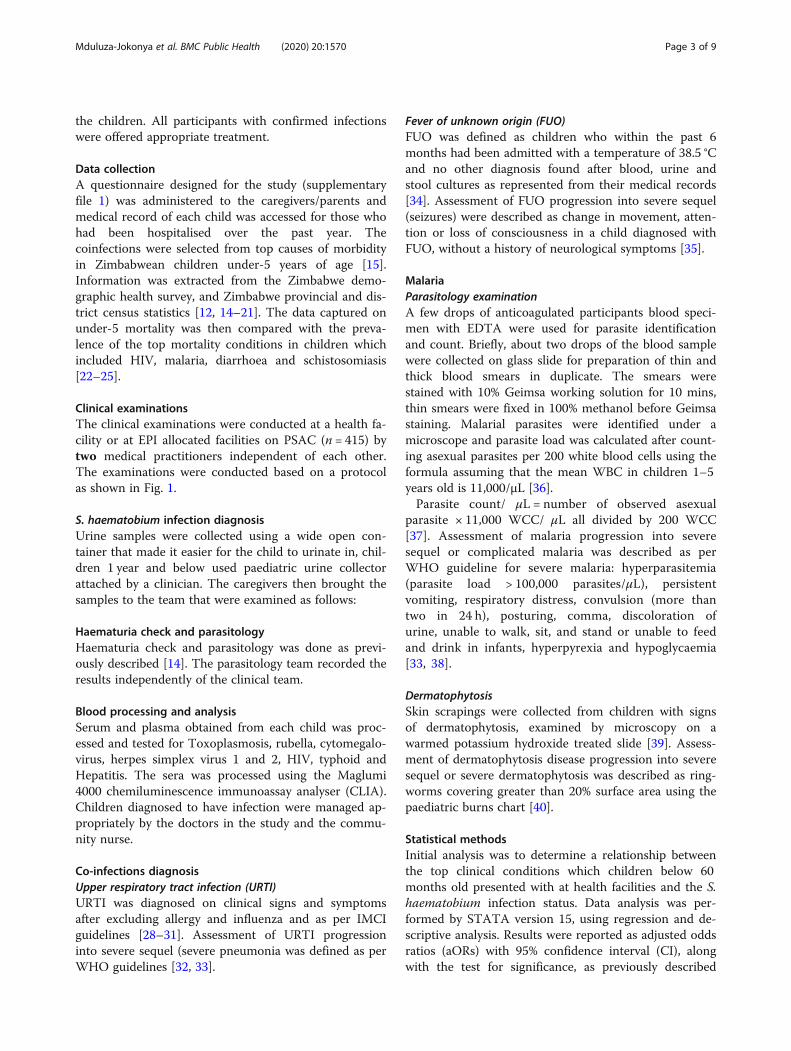
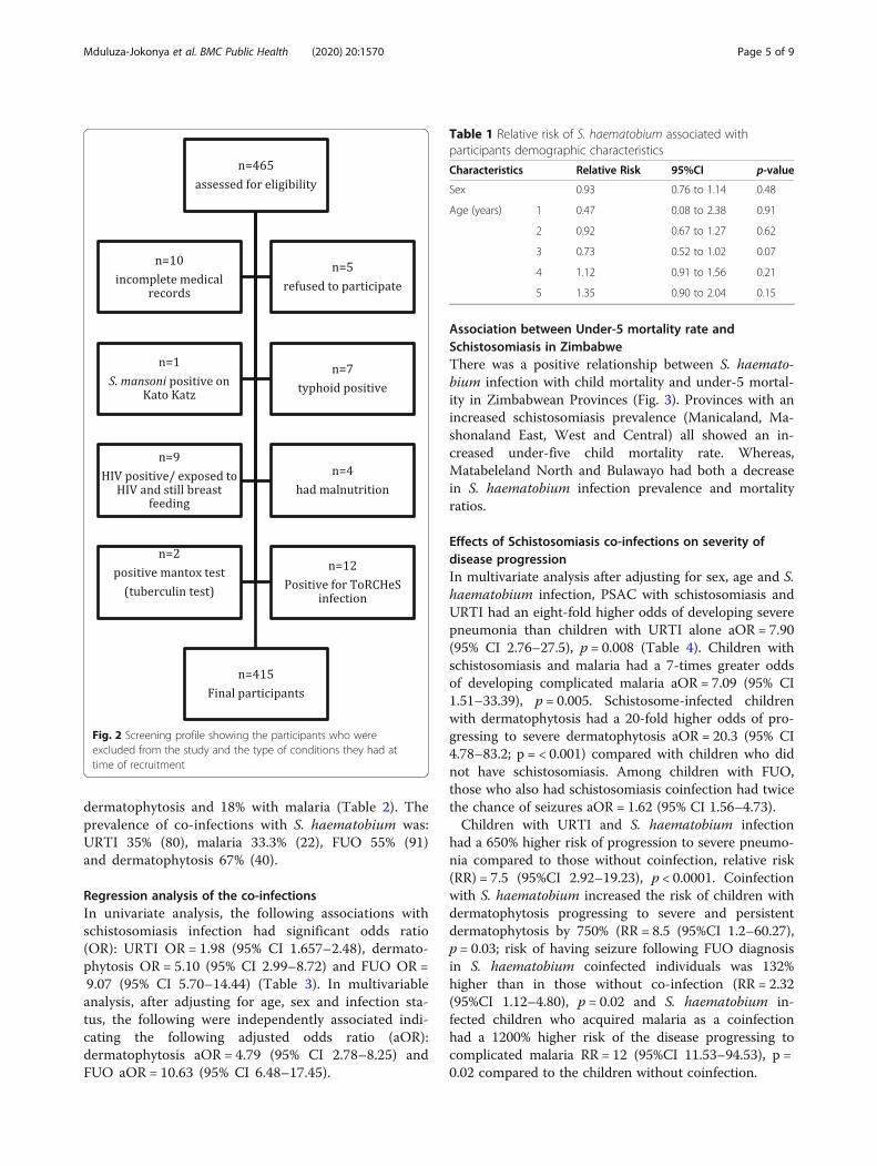
Clinical examinationsThe clinical examinations were conducted at a health fa-cility or at EPI allocated facilities on PSAC (n = 415) bytwo medical practitioners independent of each other.The examinations were conducted based on a protocolas shown in Fig. 1.
S. haematobium infection diagnosisUrine samples were collected using a wide open con-tainer that made it easier for the child to urinate in, chil-dren 1 year and below used paediatric urine collectorattached by a clinician. The caregivers then brought thesamples to the team that were examined as follows:
Haematuria check and parasitologyHaematuria check and parasitology was done as previ-ously described [14]. The parasitology team recorded theresults independently of the clinical team.
Blood processing and analysisSerum and plasma obtained from each child was proc-essed and tested for Toxoplasmosis, rubella, cytomegalo-virus, herpes simplex virus 1 and 2, HIV, typhoid andHepatitis. The sera was processed using the Maglumi4000 chemiluminescence immunoassay analyser (CLIA).Children diagnosed to have infection were managed ap-propriately by the doctors in the study and the commu-nity nurse.
Co-infections diagnosisUpper respiratory tract infection (URTI)URTI was diagnosed on clinical signs and symptomsafter excluding allergy and influenza and as per IMCIguidelines [28–31]. Assessment of URTI progressioninto severe sequel (severe pneumonia was defined as perWHO guidelines [32, 33].
Fever of unknown origin (FUO)FUO was defined as children who within the past 6months had been admitted with a temperature of 38.5 °Cand no other diagnosis found after blood, urine andstool cultures as represented from their medical records[34]. Assessment of FUO progression into severe sequel(seizures) were described as change in movement, atten-tion or loss of consciousness in a child diagnosed withFUO, without a history of neurological symptoms [35].
MalariaParasitology examinationA few drops of anticoagulated participants blood speci-men with EDTA were used for parasite identificationand count. Briefly, about two drops of the blood samplewere collected on glass slide for preparation of thin andthick blood smears in duplicate. The smears werestained with 10% Geimsa working solution for 10 mins,thin smears were fixed in 100% methanol before Geimsastaining. Malarial parasites were identified under amicroscope and parasite load was calculated after count-ing asexual parasites per 200 white blood cells using theformula assuming that the mean WBC in children 1–5years old is 11,000/μL [36].Parasite count/ μL = number of observed asexual
parasite × 11,000 WCC/ μL all divided by 200 WCC[37]. Assessment of malaria progression into severesequel or complicated malaria was described as perWHO guideline for severe malaria: hyperparasitemia(parasite load > 100,000 parasites/μL), persistentvomiting, respiratory distress, convulsion (more thantwo in 24 h), posturing, comma, discoloration ofurine, unable to walk, sit, and stand or unable to feedand drink in infants, hyperpyrexia and hypoglycaemia[33, 38].
DermatophytosisSkin scrapings were collected from children with signsof dermatophytosis, examined by microscopy on awarmed potassium hydroxide treated slide [39]. Assess-ment of dermatophytosis disease progression into severesequel or severe dermatophytosis was described as ring-worms covering greater than 20% surface area using thepaediatric burns chart [40].
Statistical methodsInitial analysis was to determine a relationship betweenthe top clinical conditions which children below 60months old presented with at health facilities and the S.haematobium infection status. Data analysis was per-formed by STATA version 15, using regression and de-scriptive analysis. Results were reported as adjusted oddsratios (aORs) with 95% confidence interval (CI), alongwith the test for significance, as previously described
Mduluza-Jokonya et al. BMC Public Health (2020) 20:1570 Page 3 of 9
[41]. A relationship was determined of being S. haemato-bium infected and the clinical conditions advancing tothe severe sequels, this was done by multinomial regres-sion analysis adjusted for sex, age and S. haematobiuminfection which gave adjusted odds ratio. Infection in-tensity for S. haematobium was defined as previouslydescribed that is arithmetic average egg count per tenmillilitres of at least two urine samples.
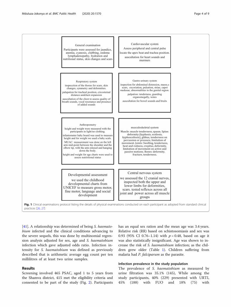
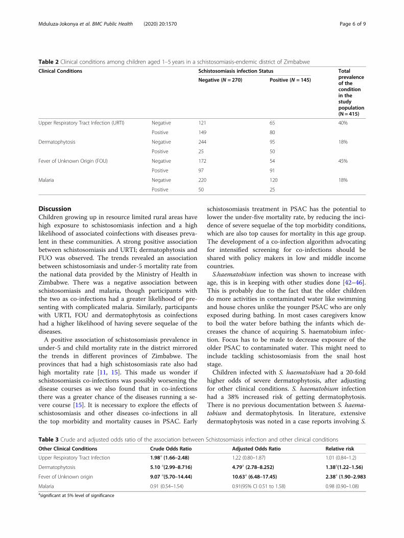
ResultsScreening involved 465 PSAC, aged 1 to 5 years fromthe Shamva district, 415 met the eligibility criteria andconsented to be part of the study (Fig. 2). Participants
has an equal sex ration and the mean age was 3.4 years.Relative risk (RR) based on schistosomiasis and sex was0.93 (95% CI 0.76–1.14) with p = 0.48, based on age itwas also statistically insignificant. Age was shown to in-crease the risk of S. haematobium infection; as the chil-dren grew older (Table 1). Children suffering frommalaria had P. falciparum as the parasite.
Infection prevalence in the study populationThe prevalence of S. haematobium as measured byurine filtration was 35.1% (145), While among thestudy participants, 40% (229) presented with URTI,45% (188) with FUO and 18% (75) with
Fig. 1 Clinical examinations protocol listing the details of physical examinations conducted on each participant as adopted from standard clinicalpractices [26, 27]
Mduluza-Jokonya et al. BMC Public Health (2020) 20:1570 Page 4 of 9
dermatophytosis and 18% with malaria (Table 2). Theprevalence of co-infections with S. haematobium was:URTI 35% (80), malaria 33.3% (22), FUO 55% (91)and dermatophytosis 67% (40).
Regression analysis of the co-infectionsIn univariate analysis, the following associations withschistosomiasis infection had significant odds ratio(OR): URTI OR = 1.98 (95% CI 1.657–2.48), dermato-phytosis OR = 5.10 (95% CI 2.99–8.72) and FUO OR =9.07 (95% CI 5.70–14.44) (Table 3). In multivariableanalysis, after adjusting for age, sex and infection sta-tus, the following were independently associated indi-cating the following adjusted odds ratio (aOR):dermatophytosis aOR = 4.79 (95% CI 2.78–8.25) andFUO aOR = 10.63 (95% CI 6.48–17.45).
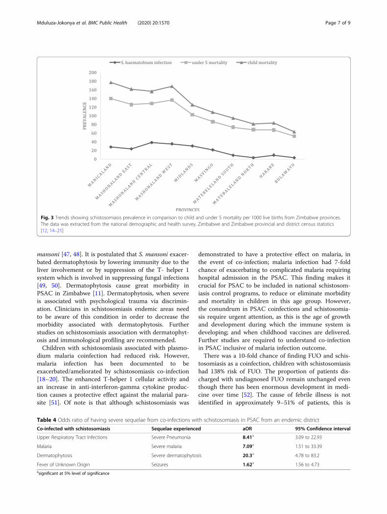
Association between Under-5 mortality rate andSchistosomiasis in ZimbabweThere was a positive relationship between S. haemato-bium infection with child mortality and under-5 mortal-ity in Zimbabwean Provinces (Fig. 3). Provinces with anincreased schistosomiasis prevalence (Manicaland, Ma-shonaland East, West and Central) all showed an in-creased under-five child mortality rate. Whereas,Matabeleland North and Bulawayo had both a decreasein S. haematobium infection prevalence and mortalityratios.
Effects of Schistosomiasis co-infections on severity ofdisease progressionIn multivariate analysis after adjusting for sex, age and S.haematobium infection, PSAC with schistosomiasis andURTI had an eight-fold higher odds of developing severepneumonia than children with URTI alone aOR = 7.90(95% CI 2.76–27.5), p = 0.008 (Table 4). Children withschistosomiasis and malaria had a 7-times greater oddsof developing complicated malaria aOR = 7.09 (95% CI1.51–33.39), p = 0.005. Schistosome-infected childrenwith dermatophytosis had a 20-fold higher odds of pro-gressing to severe dermatophytosis aOR = 20.3 (95% CI4.78–83.2; p = < 0.001) compared with children who didnot have schistosomiasis. Among children with FUO,those who also had schistosomiasis coinfection had twicethe chance of seizures aOR = 1.62 (95% CI 1.56–4.73).Children with URTI and S. haematobium infection
had a 650% higher risk of progression to severe pneumo-nia compared to those without coinfection, relative risk(RR) = 7.5 (95%CI 2.92–19.23), p < 0.0001. Coinfectionwith S. haematobium increased the risk of children withdermatophytosis progressing to severe and persistentdermatophytosis by 750% (RR = 8.5 (95%CI 1.2–60.27),p = 0.03; risk of having seizure following FUO diagnosisin S. haematobium coinfected individuals was 132%higher than in those without co-infection (RR = 2.32(95%CI 1.12–4.80), p = 0.02 and S. haematobium in-fected children who acquired malaria as a coinfectionhad a 1200% higher risk of the disease progressing tocomplicated malaria RR = 12 (95%CI 11.53–94.53), p =0.02 compared to the children without coinfection.
Fig. 2 Screening profile showing the participants who wereexcluded from the study and the type of conditions they had attime of recruitment
Table 1 Relative risk of S. haematobium associated withparticipants demographic characteristics
Characteristics Relative Risk 95%CI p-value
Sex 0.93 0.76 to 1.14 0.48
Age (years) 1 0.47 0.08 to 2.38 0.91
2 0.92 0.67 to 1.27 0.62
3 0.73 0.52 to 1.02 0.07
4 1.12 0.91 to 1.56 0.21
5 1.35 0.90 to 2.04 0.15
Mduluza-Jokonya et al. BMC Public Health (2020) 20:1570 Page 5 of 9
DiscussionChildren growing up in resource limited rural areas havehigh exposure to schistosomiasis infection and a highlikelihood of associated coinfections with diseases preva-lent in these communities. A strong positive associationbetween schistosomiasis and URTI; dermatophytosis andFUO was observed. The trends revealed an associationbetween schistosomiasis and under-5 mortality rate fromthe national data provided by the Ministry of Health inZimbabwe. There was a negative association betweenschistosomiasis and malaria, though participants withthe two as co-infections had a greater likelihood of pre-senting with complicated malaria. Similarly, participantswith URTI, FOU and dermatophytosis as coinfectionshad a higher likelihood of having severe sequelae of thediseases.A positive association of schistosomiasis prevalence in
under-5 and child mortality rate in the district mirroredthe trends in different provinces of Zimbabwe. Theprovinces that had a high schistosomiasis rate also hadhigh mortality rate [11, 15]. This made us wonder ifschistosomiasis co-infections was possibly worsening thedisease courses as we also found that in co-infectionsthere was a greater chance of the diseases running a se-vere course [15]. It is necessary to explore the effects ofschistosomiasis and other diseases co-infections in allthe top morbidity and mortality causes in PSAC. Early
schistosomiasis treatment in PSAC has the potential tolower the under-five mortality rate, by reducing the inci-dence of severe sequelae of the top morbidity conditions,which are also top causes for mortality in this age group.The development of a co-infection algorithm advocatingfor intensified screening for co-infections should beshared with policy makers in low and middle incomecountries.S.haematobium infection was shown to increase with
age, this is in keeping with other studies done [42–46].This is probably due to the fact that the older childrendo more activities in contaminated water like swimmingand house chores unlike the younger PSAC who are onlyexposed during bathing. In most cases caregivers knowto boil the water before bathing the infants which de-creases the chance of acquiring S. haematobium infec-tion. Focus has to be made to decrease exposure of theolder PSAC to contaminated water. This might need toinclude tackling schistosomiasis from the snail hoststage.Children infected with S. haematobium had a 20-fold
higher odds of severe dermatophytosis, after adjustingfor other clinical conditions. S. haematobium infectionhad a 38% increased risk of getting dermatophytosis.There is no previous documentation between S. haema-tobium and dermatophytosis. In literature, extensivedermatophytosis was noted in a case reports involving S.
Table 2 Clinical conditions among children aged 1–5 years in a schistosomiasis-endemic district of Zimbabwe
Clinical Conditions Schistosomiasis infection Status Totalprevalenceof theconditionin thestudypopulation(N = 415)
Negative (N = 270) Positive (N = 145)
Upper Respiratory Tract Infection (URTI) Negative 121 65 40%
Positive 149 80
Dermatophytosis Negative 244 95 18%
Positive 25 50
Fever of Unknown Origin (FOU) Negative 172 54 45%
Positive 97 91
Malaria Negative 220 120 18%
Positive 50 25
Table 3 Crude and adjusted odds ratio of the association between Schistosomiasis infection and other clinical conditions
Other Clinical Conditions Crude Odds Ratio Adjusted Odds Ratio Relative risk
Upper Respiratory Tract Infection 1.98a (1.66–2.48) 1.22 (0.80–1.87) 1.01 (0.84–1.2)
Dermatophytosis 5.10 a(2.99–8.716) 4.79a (2.78–8.252) 1.38a(1.22–1.56)
Fever of Unknown origin 9.07 a(5.70–14.44) 10.63a (6.48–17.45) 2.38a (1.90–2.983
Malaria 0.91 (0.54–1.54) 0.91(95% CI 0.51 to 1.58) 0.98 (0.90–1.08)asignificant at 5% level of significance
Mduluza-Jokonya et al. BMC Public Health (2020) 20:1570 Page 6 of 9
mansoni [47, 48]. It is postulated that S. mansoni exacer-bated dermatophytosis by lowering immunity due to theliver involvement or by suppression of the T- helper 1system which is involved in suppressing fungal infections[49, 50]. Dermatophytosis cause great morbidity inPSAC in Zimbabwe [11]. Dermatophytosis, when severeis associated with psychological trauma via discrimin-ation. Clinicians in schistosomiasis endemic areas needto be aware of this condition in order to decrease themorbidity associated with dermatophytosis. Furtherstudies on schistosomiasis association with dermatophyt-osis and immunological profiling are recommended.Children with schistosomiasis associated with plasmo-
dium malaria coinfection had reduced risk. However,malaria infection has been documented to beexacerbated/ameliorated by schistosomiasis co-infection[18–20]. The enhanced T-helper 1 cellular activity andan increase in anti-interferon-gamma cytokine produc-tion causes a protective effect against the malarial para-site [51]. Of note is that although schistosomiasis was
demonstrated to have a protective effect on malaria, inthe event of co-infection; malaria infection had 7-foldchance of exacerbating to complicated malaria requiringhospital admission in the PSAC. This finding makes itcrucial for PSAC to be included in national schistosom-iasis control programs, to reduce or eliminate morbidityand mortality in children in this age group. However,the conundrum in PSAC coinfections and schistosomia-sis require urgent attention, as this is the age of growthand development during which the immune system isdeveloping; and when childhood vaccines are delivered.Further studies are required to understand co-infectionin PSAC inclusive of malaria infection outcome.There was a 10-fold chance of finding FUO and schis-
tosomiasis as a coinfection, children with schistosomiasishad 138% risk of FUO. The proportion of patients dis-charged with undiagnosed FUO remain unchanged eventhough there has been enormous development in medi-cine over time [52]. The cause of febrile illness is notidentified in approximately 9–51% of patients, this is
Fig. 3 Trends showing schistosomiasis prevalence in comparison to child and under 5 mortality per 1000 live births from Zimbabwe provinces.The data was extracted from the national demographic and health survey, Zimbabwe and Zimbabwe provincial and district census statistics[12, 14–21]
Table 4 Odds ratio of having severe sequelae from co-infections with schistosomiasis in PSAC from an endemic district
Co-infected with schistosomiasis Sequelae experienced aOR 95% Confidence interval
Upper Respiratory Tract Infections Severe Pneumonia 8.41a 3.09 to 22.93
Malaria Severe malaria 7.09a 1.51 to 33.39
Dermatophytosis Severe dermatophytosis 20.3a 4.78 to 83.2
Fever of Unknown Origin Seizures 1.62a 1.56 to 4.73asignificant at 5% level of significance
Mduluza-Jokonya et al. BMC Public Health (2020) 20:1570 Page 7 of 9
even higher in resource limited areas endemic for child-hood illnesses [53]. Children who had both FOU andschistosomiasis had a 2-fold chance of having serious se-quelae such as seizures. Most clinician tend to think ofother conditions in contrast to neglected tropical diseaseswhen patients present with a fever [33]. It might be neces-sary to make it a priority to screen for schistosomiasiswhen a child from an endemic area presents with a fever.However, further immunological investigations are neces-sary in-order to find out if the fever is due to schistosom-iasis infection or exacerbation of a co-infection, during theearly immunological responses to diseases manifestation.In our study the odds of having URTI was 2% higher
in schistosomiasis infected children with a 1% increasedrisk of S. haematobium infected children acquiringURTI. Furthermore, in the coinfected cohort odds ofending up with severe pneumonia was 7-fold comparedto schistosomiasis negative population. This is a very sig-nificant finding as acute respiratory infections (ARIs) arethe leading cause of morbidity and mortality in childrenunder the age of five in Zimbabwe [11]. Thus tacklingschistosomiasis crisis in this age group will have enor-mous contribution in reducing the childhood mortalityand morbidity in schistosomiasis endemic areas.The strength of this study revealed the major morbid-
ity and mortality causes in PSAC from our study areawhich also fit into most of the low income countrieswere schistosomiasis is endemic. The main limitation ofthis study is there is a possibility that the children mayhave had more than two co-infections which mightmake our data biased, however we did thorough clinicalexaminations and laboratory tests in all the participantsto rule this out. We also considered socioeconomic sta-tus as a confounding factor by focusing on children witha similar background and in the same villages with simi-lar lifestyles. Given the likelihood of co-infections, chil-dren who present with schistosomiasis should bescreened for other conditions.
ConclusionsThis study revealed an association between schistosom-iasis and top morbidity conditions of URTI, dermato-phytosis, malaria and FUO in PSAC living in aschistosomiasis endemic area. The study also showedthat there is need for an appropriate clinical diagnosticmethod to identify associated infectious diseases and ini-tiate early treatment of schistosomiasis and co-infectionsin PSAC. Furthermore, we demonstrated that coinfec-tion led to severe sequelae of the clinical conditions, thathave a high impact on morbidity and mortality in chil-dren under the age of five. This brings out the import-ance of including PSAC in national schistosomiasiscontrol programs and the development of appropriatediagnostic tools.
Supplementary informationSupplementary information accompanies this paper at https://doi.org/10.1186/s12889-020-09634-0.
Additional file 1. Study questionaire.
AbbreviationsaOR: Adjusted odds ratios; ARI: Acute respiratory tract infection;CI: Confidence interval; EPI: Expanded program on immunization; FUO: Feverof unknown origin; HIV: Human Immunodeficiency Virus; MDA: Mass DrugAdministration; MRCZ: Medical Research Council of Zimbabwe;PSAC: Preschool age children; RR: Risk ratio/relative risk; SDG: SustainableDevelopment Goals; STH: Soil transmitted helminths;ToRCHeS: Toxoplasmosis, rubella, cytomegalovirus, hepatitis and syphilis;URTI: Upper respiratory tract infection; WBC: White blood cells; WCC: Whitecell counts; WHO: World Health Organization
AcknowledgementsWe would like to acknowledge the Ministry of Health and Child Care, theMedical Research Council of Zimbabwe, village health workers, nursing staff,parents and children from Shamva. A special thanks to members of theBiochemistry Department at the University of Zimbabwe for technicalsupport during field parasitology and sampling. Our most profoundgratitude to the participants and their parents or guardians for taking part inthis study.
Authors’ contributionsTLMJ, TN, FM and TM conceived and designed the study. TLMJ, HM, AV, MK,EC, SR, ES, LJ and TM performed the clinical examination or parasitology andthe data analysis. The authors read and approved the manuscript.
FundingTackling Infections to Benefit Africa (TIBA) supported the field and laboratoryactivities only. The funder was not involved in the design of the study,collection, analysis and interpretation of data, and in writing the manuscript.This research was commissioned by the National Institute of Health Research(NIHR), Global Health Research Programme (16/136/33) using UK aid from UKGovernment. The views expressed in this publication are those of theauthors and not necessarily those of the NIHR or the Department of Healthand Social Care.
Availability of data and materialsThe statistical data on the parasitology and clinical scores used to supportthe findings of this study are available from the corresponding author uponrequest.
Ethics approval and consent to participateEthical approval was obtained from Medical Research Council of Zimbabwe(MRCZ/A/2435). Gatekeeper approval was obtained from the Provincial andDistrict Medical Directors and Community Leaders. Written informed consentwas obtained from the parents or guardians of the children.
Consent for publicationNone required.
Competing interestsThe authors declare that there is no conflict of interest.
Author details1Optics & Imaging, Doris Duke Medical Research Institute, College of HealthSciences, University of KwaZulu-Natal, Durban, KwaZulu-Natal, South Africa.2Department of Biochemistry, University of Zimbabwe, P.O. Box MP 167, MtPleasant, Harare, Zimbabwe. 3Department of Surgery, College of HealthSciences, University of Zimbabwe, P.O. Box MP 167, Mt Pleasant, Harare,Zimbabwe. 4Department of Community Medicine, College of HealthSciences, University of Zimbabwe, P.O. Box MP 167, Mt Pleasant, Harare,Zimbabwe. 5Twin Palms Medical Centre, 113 Kwame Nkrumah Avenue,Harare, Zimbabwe. 6Institute for Immunology and Infection Research andCentre for Immunity, Infection and Evolution, School of Biological Sciences,
Mduluza-Jokonya et al. BMC Public Health (2020) 20:1570 Page 8 of 9
University of Edinburgh, Ashworth Laboratories, King’s Buildings, CharlotteAuerbach Rd., Edinburgh EH9 3JT, UK.
Received: 23 March 2020 Accepted: 29 September 2020
References1. Mutapi F, Rujeni N, Bourke C, Mitchell K, Appleby L, Nausch N, et al. Schistosoma
haematobium treatment in 1–5 year old children : safety and efficacy of theAntihelminthic drug Praziquantel. PLoS Negl Trop Dis. 2011;5(5):e1143.
2. Almeida SR. Immunology of dermatophytosis. Mycopathologia. 2008;166(5–6):277–83.
3. World Health Organisation. Schistosomiasis weekly epidemiological report.World Heal Organ. 2016;91(49):585–600.
4. World Health Organisation. Schistosomiasis. Global Health estimates 2015:deaths by cause, age, sex, by country and by region, 2000-2015; 2016.
5. Kalinda C, Chimbari MJ, Mukaratirwa S. Schistosomiasis in Zambia : asystematic review of past and present experiences; 2018. p. 1–10.
6. Abruzzi A, Fried B. Coinfection of Schistosoma (Trematoda) with bacteria,protozoa and Helminths. Adv Parasitol. 2011;77:1–85.
7. Vennervald BJ, Dunne DW. Morbidity in schistosomiasis: an update. CurrOpin Infect Dis. 2004;17(5):439–47.
8. Zhang XS, Cao KF. The impact of coinfections and their simultaneoustransmission on antigenic diversity and epidemic cycling of infectiousdiseases. Biomed Res Int. 2014;2014:8–12.
9. King HC. Parasites and poverty: the case of schistosomiasis. Acta Trop. 2011;113(2):95–104.
10. Osakunor DNM, Woolhouse MEJ, Mutapi F. Paediatric schistosomiasis: whatwe know and what we need to know. PLoS Negl Trop Dis. 2018;12(2):1–16.
11. Zimbabwe national statistics survey. Demographic and health surveys.12. Zimbabwe National Statistic Agency. Provincial report: Mashonaland Central.
Zimbabwe Popul census; 2012. http://www.zimstat.co.zw/wp-content/uploads/publications/Population/population/Mash-Central.pdf.
13. Of D. Size S. Original article. 2003;10(2):95–7.14. Mduluza-Jokonya TL, Naicker T, Kasambala M, Jokonya L, Vengesai A, Midzi
H, et al. Clinical morbidity associated with Schistosoma haematobiuminfection in pre-school age children from an endemic district in Zimbabwe.Trop Med Int Health. 2020;25(9):1110–21. https://doi.org/10.1111/tmi.13451.
15. Zimbabwe National Statistics Agency. Mashonaland West Province reportZimbabwe population. 2012;6(2):16–9 http://www.zimstat.co.zw.
16. Midzi N, Mduluza T, Chimbari MJ, Tshuma C, Charimari L, Mhlanga G, et al.Distribution of Schistosomiasis and soil transmitted Helminthiasis inZimbabwe: towards a National Plan of action for control and elimination.PLoS Negl Trop Dis. 8(8);no. e3014.
17. Zimbabwe National Statistics Agency. Provincial report Matabeleland NorthZimbabwe population. 2012;6(2):21–3 http://www.zimstat.co.zw.
18. Zimbabwe National Statistics Agency. Provincial report Masvingo.Zimbabwe Population. 2012;6(2):24–6 http://www.zimstat.co.zw.
19. Zimbabwe National Statistics Agency. Matabeleland South. ZimbabwePopulation. 2012;6(2):27–9 http://www.zimstat.co.zw.
20. Zimbabwe National Statistics Agency. Zimbabwe population census 2012.Provincial Report Harare Central Census Office. 2012;6(2):30–3 http://www.zimstat.co.zw.
21. Zimbabwe National Statistics Agency. Provincial report Bulawayo.Zimbabwe Population. 2012;6(2):34–7 http://www.zimstat.co.zw.
22. Zimbabwe National Statistics Agency. Mortality thematic report; 2015.http://www.zimstat.co.zw/sites/default/files/img/publications/Census/Mortality_Thematic.pdf.
23. Mott K, Baltes R, Bambagha J, Baldassini B. Field studies for detection of areusable polyamide filter for detection of Schistosoma haematobium eggsby urine filtration. ropenmedizin Parasitol. 1982;33:227–8.
24. Zimbabwe National Statistics Agency. Provincial report Manicaland.Zimbabwe Population. 2012;6(2):39–42 www.zimstat.co.zw/sites.
25. Zimbabwe National Statistics Agency. Polulation statistics. 2012;6(2):43–7www.zimstat.co.zw.
26. University of Glasgow. MB ChB clinical history and examination manual; 2015.27. Carter J, Müller-Stöver I, Östensen H. Good clinical diagnostic practice: a
guide for clinicians in developing countries to the clinical diagnosis ofdisease and to making proper use of clinical diagnostic services; 2005.
28. Child TTHE. IMCI chart booklet; 2002. p. 1–39.
29. Tantilipikorn P, Auewarakul P. Airway allergy and viral infection. Asian Pac JAllergy Immunol. 2011;29(2):113–9.
30. Brand PL, Mäkelä MJ, Szefler SJ, Frischer T. Price D; ERS task forcemonitoring asthma in children. Monitoring asthma in childhood: symptoms,exacerbations and quality of life. Eur Respir Rev. 2015;24(136):187–93.
31. Center for Disease Control and Prevention. Nonspecific upper respiratorytract infection. Cent Dis Control Prev. 2006;134(6):2001.
32. World Health Organization. Revised WHO classification and treatment ofchildhood pneumonia at health facilities: evidence summaries: World HealthOrganization; 2014. p. 26. Available from: https://apps.who.int/iris/bitstream/handle/10665/137319/9789241507813_.
33. World Health Organization. Recommendations for management ofcommon childhood conditions. Parassitologia. 2012;84:4-6.
34. Strauss WG. Fever of unknown origin. Postgrad Med. 1964;36(1):555–9.35. Services H, Bureau CH. Illinois emergency medical; 2016. p. 112.36. Blood Sciences Department of Haematology. Children ’ s Reference Ranges
for FBC Q Pulse Reference N. p. 369.37. World Health Organization. Giemsa staining of malaria blood films malaria
microscopy standard operating procedure, 1. Purpose and scope. MalarMicrosc Stand Oper Proced - MM-SOP-07A. 2016:1–6. http://www.wpro.who.int/mvp/lab_quality/2096_oms_gmp_sop_07a_rev.pdf.
38. World Health Organization. WHO severe malaria 2014. Trop Med Int Health.2014;19:7–131.
39. Schlenker S. Standard operating procedure. Text Chem Color. 1997;29(7):283–6.
40. Women’s and Children’s Hospital. Paediatric burn guidelines. Gov SouthAust. 2010;08:8161.
41. Wami WM, Nausch N, Bauer K, Midzi N, Gwisai R, Simmonds P, et al.Comparing parasitological vs serological determination of Schistosomahaematobium infection prevalence in preschool and primary school-agedchildren: implications for control programmes. Parasitology. 2014;141(14):1962–70.
42. Nii D, Osakunor M, Mduluza T, Midzi N, Chase-topping M, Mutsaka-Makuvaza MJ, et al. Dynamics of paediatric urogenital schistosome infection, morbidity and treatment : a longitudinal study among preschool childrenin Zimbabwe; 2018. p. 1–9.
43. Mutapi F, Burchmore R, Mduluza T, Midzi N, Turner CMR, Maizels RM. Age-relatedand infection intensity – related shifts in antibody recognition of defined proteinantigens in a Schistosome-exposed population. J Infect Dis. 2008;198:167–75.
44. Wepnje GB, Anchang-kimbi JK, Ndassi VD, Lehman LG, Kimbi HK.Schistosoma haematobium infection status and its associated risk factorsamong pregnant women in Munyenge , south west region , Cameroonfollowing scale-up of communal piped water sources from 2014 to 2017 : across-sectional study; 2019. p. 1–10.
45. Li EY, Gurarie D, Lo NC, Zhu X, King CH. Articles improving public healthcontrol of schistosomiasis with a modified WHO strategy : a model-basedcomparison study. Lancet Glob Health. 2019;7(10):e1414–22.
46. Fatimah A, Emmanuel B, Idiat B, Paul A. Review article impact of humanschistosomiasis in sub-Saharan Africa. Braz J Infect Dis. 2015;19(2):196–205.
47. Ferreira MD, Collaniere AC, Bertolini DL, Barros NC, de Moraes VasconcelosD. Cellular immunodeficiency related to chronic dermatophytosis in apatient with Schistosoma mansoni infection: can schistosomiasis induceimmunodeficiency? Rev Soc Bras Med Trop. 2017;50(1):141–4.
48. Deen J. Coinfections and Malaria. In: Hommel M., Kremsner P. (eds)Encyclopedia of Malaria. New York: Springer; 2014. https://doi.org/10.1007/978-1-4614-8757-9_113-1.
49. Grappel SF, Bishop CT, Blank F. Immunology of dermatophytes anddermatophytosis. Bacteriol Rev. 1974;38(2):222–50.
50. Woodfolk JA. Allergy and dermatophytes. Clin Microbiol Rev. 2005;18(1):30–43.51. Ekpo UF, Alabi OM, Oluwole AS, Sam-Wobo SO. Schistosoma haematobium
infections in preschool children from two rural communities in Ijebu east,South-Western Nigeria. J Helminthol. 2012;86(3):323–8.
52. Fusco FM, Pisapia R, Nardiello S, Cicala SD, Gaeta GB, Brancaccio G. Fever ofunknown origin (FUO): which are the factors influencing the final diagnosis?A 2005-2015 systematic review. BMC Infect Dis. 2019;19(1):1–11.
53. Tan Y, Liu X, Shi X. Clinical features and outcomes of patients with fever ofunknown origin: a retrospective study. BMC Infect Dis. 2019;19(1):1–7.
Publisher’s NoteSpringer Nature remains neutral with regard to jurisdictional claims inpublished maps and institutional affiliations.
Mduluza-Jokonya et al. BMC Public Health (2020) 20:1570 Page 9 of 9