associated with contrasting sperm motility in goat the
TRANSCRIPT
Page 1/31
The characteristics of proteome and metabolomeassociated with contrasting sperm motility in goatseminal plasmaBaoyu Jia
Yunnan Agricultural UniversityJiangchong Liang
Yunnan Animal Science and Veterinary InstituteChunrong Lv
Yunnan Animal Science and Veterinary InstituteSameeullah Memon
Yunnan Animal Science and Veterinary InstituteYi Fang
Chinese Academy of SciencesGuoquan Wu
Yunnan Animal Science and Veterinary InstituteGuobo Quan ( [email protected] )
Yunnan Animal Science and Veterinary Institute
Research Article
Keywords: goat, seminal plasma, proteome, metabolome, sperm motility
Posted Date: April 2nd, 2021
DOI: https://doi.org/10.21203/rs.3.rs-382781/v1
License: This work is licensed under a Creative Commons Attribution 4.0 International License. Read Full License
Version of Record: A version of this preprint was published at Scienti�c Reports on July 30th, 2021. Seethe published version at https://doi.org/10.1038/s41598-021-95138-9.
Page 2/31
AbstractSperm motility is an index tightly associated with male fertility. A close relationship between seminalplasma and sperm motility has been con�rmed. This study was to assess the protein and metabolitepro�les of seminal plasma obtained from adult goats with high or low sperm motility using the proteomicand metabolomic strategies. In total, 2098 proteins were found. 449 differentially expressed proteins(DEPs) were identi�ed, and 175 DEPs were enriched in the high motility group. The obtained DEPsprimarily exist in cytoplasma and extra-cellular portion. The Gene Ontology enrichment analysisdemonstrated the main functional roles of these DEPs in regulating biological process, metabolic processof organic substances, cellular-metabolic process, primary-metabolic process, metabolic process ofnitrogen compounds, etc. Additionally, the Kyoto-Encyclopedia of Genes and Genomes (KEGG) analysisrevealed that these DEPs were primarily involved in phosphatidylinositol signaling system, salivarysecretion, proteasome, apoptosis, mitophagy-animal, etc. Aided by the parallel reaction monitoringtechnology, the abundance changing pattern of 19 selected DEPs was consistent with that of thecorresponding proteins obtained by TMT. A total of 4603 metabolites were identi�ed in seminal plasma.1857 differential metabolites were found between the high motility group and the low motility group, and999 metabolites were up-regulated in the high motility group. The KEGG analysis demonstrated theprimary involvement of the differential metabolites in metabolic and synthetic activities. In conclusion,we �rst established the proteome and metabolome databank of goat seminal plasma, detecting someproteins and metabolites which may affect sperm motility. This study will be valuable for understandingmechanisms leading to poor sperm motility.
IntroductionArti�cial insemination (AI) can be de�nitely thought as one of the earliest and the most extensivelyutilized assisted reproductive technologies in animal breeding and production 1. In brief, AI means thatmale sperm are introduced into female reproductive tract aided by speci�c tools. At present, AI has beenrecognized as an e�cient and important approach for extending valuable genes in a stock populationand improving their production traits 1, 2. As compared to natural mating, AI largely enhance thereproductive potential of an excellent male animal. Even after its death, using some long-term storagemethods of its semen, we can still obtain its offspring 2. Furthermore, AI can be combined with the estrussynchronization technique to realize the out-of-season breeding and improve the accuracy of expectingdates of parturition. Moreover, this technology can avoid disease transmission, since males used for AIgenerally experience some strict disease examinations 3.
Currently, AI has been extensively applied in the dairy industry 4. In general, the site where semen isdeposited greatly in�uences the fertilization results of oocytes with sperm. Moreover, the number ofmotile sperm is also tightly associated with the conception rate. In comparison with fresh or cooledsemen, more motile sperm are required when frozen-thawed semen is used for AI 5. It’s known that thefeatures of sheep and goat reproductive tract are different from that of cow, which prevents lots of sperm

Page 3/31
entering uterus and makes sperm walk a long way to arrive at the oviduct and fertilize with oocytes.Currently, the fertility of AI is especially unsatis�ed with the use of frozen-thawed sperm 2. So, it isbecoming important to select those males that can produce semen with a high quality.
Healthy sperm motility is widely recognized as a core factor which can in�uence male fertility. Thosedefectively mobile or immobile sperm are usually unfruitful or sterilized except that some assistedreproductive techniques are used 6. Some AI centers are currently using wave motion of semen as a mainindex to choose ejaculates for AI in sheep 7. Furthermore, mass motility has been con�rmed to be closelyrelated to fertility in sheep 8. However, the root causes leading to poor sperm motility are complicated andhave not been determined until now.
Seminal plasma is an important part of semen, which is mainly composed of secretions derived fromtesticular, epididymis, and secondary sex gland. In seminal plasma, the components generally consist ofproteins, ions, and metabolites like nucleosides, lipids, monosaccharides, amino acids, minerals,electrolytes, and also steroid hormones 9, 10. Previous studies have proved that seminal plasmacontributes a signi�cant role in regulation of sperm motility and fertilization 11-15. Seminal plasmaprovides metabolic support, and it has a complicated and not well-understood effect on the physiologicalrole of sperm. Additionally, seminal plasma also has certain impacts on the quality of chilled 16 or frozen-thawed sperm 17.
Some studies investigated the effects of proteins presented in seminal plasma on semen quality. Insheep, some investigators have attempted to study the proteome of seminal plasma 18-21. In accordancewith the report of Richard et al., variations in freezing resistance of sheep sperm is linked with origin andcomposition of seminal plasma. It was found that freezing with seminal plasma derived from sheep withhigh resistance to cryoinjury enhanced the post-thaw quality of sperm 18. Furthermore, several proteinmarkers potentially associated with sperm freezing resistance in sheep seminal plasma were identi�ed bythe same group. The proteins strongly linked with sperm freezing resistance, including 26S proteasome-complex, acylamino-acid releasing enzyme, alpha-mannosidase class 2C, heat-shock protein 90,tripeptidyl peptidase-2, TCP-1 complex, sorbitol-dehydrogenase, and transitional-endoplasmic reticulumATPase, were detected. On the contrary, cystatin, zinc 2-alpha-glycoprotein, angiogenin 2-like protein,cartilage-acid protein-1, cathepsin-B, and ribonuclease-4 isoform-1 had negative effects on the resistanceof sperm to cryoinjury 19. In pig, the seminal plasma proteome and the associations between seminalplasma proteins and semen features have been established. Particularly, sperm motility had a stronglypositive correlation with lactadherin 13. In human, the analysis related to the seminal plasma proteomere�ects reduced mitochondrial production, acrosome disruption, and DNA fragmentation, with severalpost-genomic functions associated to these alterations 11. Similarly, in rabbit, major seminal plasma-derived proteins contribute to the prevention of lipid peroxide radical damage and oxidative stress,membrane stability, sperm membrane transport and temperature control. Additionally, sperm motility hadpositive correlations with growth factor beta-nerve and cysteine rich secreted protein-1. However, asigni�cantly negative association of sperm motility with galectin-1 existed 10.

Page 4/31
Since metabolites were end products of metabolic pathways, which also play important role in spermphysiology, such as motility, energy metabolism, and metabolic activity regulations 22. Although certainseminal plasma components have bene�cial effects on sperm cryotolerance, whereas others may havenegative effects 23, 24. In bull, some metabolites in seminal plasma, including 2 oxoglutaric-acid orfructose, can be used to evaluate their fertility as possible biomarkers 25. Hamamah et al. (1993) studiedfertile and infertile male seminal plasma using 1 H nuclear-magnetic resonance spectra (NMR), �ndingimportant variations in concentrations of glyceryl phosphorylcholine-citrate and lactate amongazoospermic and oligo-asthenozoospermic patients 26. The metabolite pro�le of rhesus macaque spermwas also investigated in order to establish the relationship among metabolism and energy source 27.Additionally, a metabolomics strategy in mouse sperm was used to evaluate the relationship betweenglycolytic-substrates and energy generation, which is critical for sperm motility 28. Furthermore, usingboth the NMR and the gas chromatography mass spectrometry (GC–MS), a total of 96 metabolites andmore than 10 biological pathways have been detected in human sperm 29. More recently, Velho et al.(2018) identi�ed 63 metabolites in bull seminal plasma, including 21 amino acids from bull with distinct�eld-fertility ratings, demonstrating the different metabolite patterns between the low-fertility and high-fertility bulls 30.
Currently, the application of AI in the goat industry is not popular as compared to the other stocks, suchas cow or sheep. Moreover, the effect of seminal plasma on sperm motility in goats until now has notbeen clari�ed. Different from the other farm animals including sheep, goat seminal plasma containsphospholipase A, which is released from the bulbourethral gland. This protein can coagulate egg-yolk andhydrolyze the lecithin to fatty acids and spermicidal lyso-lecithins 31. Therefore, during cryopreservationof goat semen, the effects of seminal plasma must be considered. In this study, we attempted to examinethe variation of proteome and metabolome in goat seminal plasma associated with sperm motility usingthe high throughput technologies. This study will enrich our omics information related to goat semen.Furthermore, some obtained speci�c proteins or metabolites may be used as biomarkers for assessingthe quality of goat semen and predicting of male fertility.
ResultsSperm quality assessment
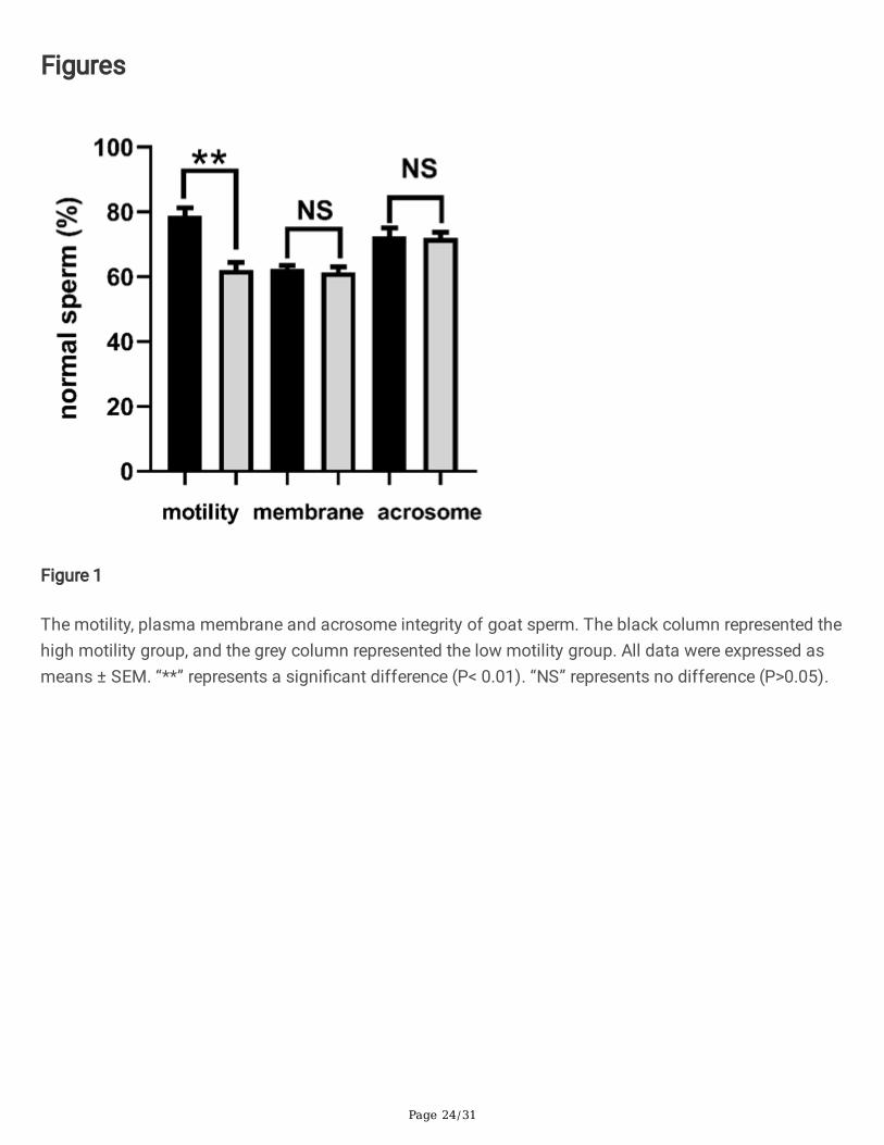
In this study, twenty goats were equally separated into two groups. As shown in Fig.1., one group had ahigh motility (78.85%±2.44%). The other one had a low motility (62.16%±2.27%). The difference betweenthese two groups was signi�cant (P<0.01). However, no differences were found among these two groupswith regards to the plasma membrane and acrosome integrity (P>0.05).
Identi�cation of proteomic information
A total of 308933 spectra were obtained after the TMT analysis and searching by Maxquant (v1.5.2.8) inthe present study. The number of the matched spectra was 36946. Furthermore, we detected 15724

Page 5/31
peptides (14861 speci�c peptides) among these spectra, and 2299 proteins including 2098 quanti�edproteins. The basic information on the protein pro�le of goat seminal plasma, such as protein accession,protein de�nition, gene name, P value, etc., was revealed in Supplementary Figure S1.
Identi�cation of DEPs
In total, 449 DEPs, with 1.5 folds shift and P-value of less than 0.05, were identi�ed in goat seminalplasma with high or low motility. The detailed information associated with the identi�ed DEPs, such asprotein accession, protein de�nition, regulated type, P-value, gene name, etc, was shown inSupplementary Table S2. In comparison with the low motility group, 175 proteins were up-regulated in thehigh motility group, such as beta-galactosidase, ATP-citrate synthase, 3-phosphoinositide-dependentprotein kinase 1-like, tra�cking protein particle compound subunit 13 isoform X2, kinesin-like protein, aprotein phosphatase inhibitor 2, etc. On the other hand, the abundance of 274 proteins signi�cantlyreduced in the high motility group, such as putative adenylate kinase 7, calpain-7-like protein,transmembrane protein 190, �brous sheath interacting protein 2, angiotensin-converting enzyme, caseinkinase II subunit beta, sperm acrosome-associated protein 5, etc.
Functional classi�cation of DEPs
The results of the GO annotation analysis were presented in Fig.2. Regarding the classi�cation ofbiological process, the acquired DEPs were signi�cantly enriched in regulating biological processes,organic substance metabolic procedure, cellular metabolic procedure, primary metabolic procedure,nitrogen compound metabolic procedure, cell component organization, localization establishment, etc.Additionally, the acquired DEPs were primarily located in the intracellular region, intracellular organelle,membrane-bound organelle, endomembrane system, organelle lumen, non-membrane-bound organelle,etc., with regard to the GO analysis of cellular component. As concern molecular function, the DEPs wereprimarily involved in protein binding, hydrolase activities, ion binding, organic cyclic complex binding,transferrase activity, heterocyclic complex binding, etc.
Moreover, the COG/KOG functional classi�cation statistics related to the acquired DEPs were presented inFig.3. 67 DEPs were found to be involved in post-translational modi�cation, protein turnover, andchaperone. 55 DEPs may have potentially functional roles in the pathways of signal transduction. Therewere 34 DEPs engaged in cytoskeleton. Additionally, some DEPs were found to function in metabolicactivities, including energy manufacturing and adaptation (20), amino acids carrying with metabolism(18), carbohydrates carrying with metabolism (22), lipids carrying with metabolism (11), etc. Interestingly,three proteins, including myosin XVB, dynein light-chain Tctex-type-1, and dynein light-chain roadblock-type-2, were found to be associated with cell motility.
Subsequently, the results of the KEGG study were presented in Fig.4., including the top 20 substantiallyenriched pathways. The main pathways were revealed, including the phosphatidylinositol signalingsystem, salivary secretion, proteasome, apoptosis, mitophagy-animal, NOD-like receptor signalingpathway, etc. Furthermore, some DEPs were detected to be potentially involved in disease or infection,
Page 6/31
such as tuberculosis, Kaposi sarcoma-associated herpesvirus infection, Huntington disease,staphylococcus aureus infection, etc.
PRM validation
In this study, in order to con�rm the accuracy of the �ndings acquired in relation to seminal plasmaproteome, 19 DEPs were selected for the PRM analysis, including phosphoglycerate mutase, ras-relatedprotein Rab-11B, ATP-citrate synthase, peroxiredoxin, spermadhesin-1, T complex protein-1 sub-unit alpha,programmed cell death protein 5, testis-tissue sperm binding protein Li 69n, ubiquitin-like modi�er-activating enzyme 1, zonadhesin, superoxide dismutase, sperm equatorial segment protein 1, thioredoxinreductase 2 (mitochondrial), acrosin-binding protein, zona pellucida binding protein, heat shock proteinfamily-E (Hsp10) member-1, peroxiredoxin-1, aquaporin 7, and izumo sperm-egg fusion protein 4. Thepattern of the fold changes in these proteins between these two groups, as shown in Table 1, wasconsistent with the results obtained by the TMT method.
Identi�cation of metabolomic data
The metabolomic analysis of goat seminal plasma was performed using a non-targeted metabolomicsstrategy, including the positive and negative modes. In the present study, a total of 4603 metabolites havebeen quanti�ed in seminal plasma derived from the two groups with high or low motility, including aminoacids, peptides, fatty acids, lipids, sugars, nucleic acid, organic acids, and other metabolites. The detailedinformation associated with these identi�ed metabolites, including index, mass, retention time (RT),compounds, formula, etc, was presented in Supplementary Table S3.
Determination of differential metabolites
Based on the Variable Importance in Projection (VIP), fold-change (FC), and P-value, the differentialmetabolites between these two groups were identi�ed. The detailed information related to these identi�edmetabolites were presented in Supplementary Table S4. Totally, 1857 differential metabolites wereidenti�ed between the high motility group and the low motility group. Among these metabolites, 999metabolites were up-regulated in the high motility group. However, 858 metabolites were signi�cantlyenriched in the low motility group. The numbers of various compounds presented in goat seminal plasmawere shown in Fig.5.
Bioinformatics analysis of differential metabolites
The cluster analysis data of the acquired differential metabolites were shown in Fig.6. As shown in Fig.6A, the cluster patterns between these two groups with high or low motility were opposite. Themetabolites enriched in the high motility group were less abundant in the group with low motility instead.The correlation analysis results of differential metabolites were demonstrated in Fig.6B. Here, thePearson correlation analysis method has been used to evaluate the correlation of differential metabolites.The top 50 differential metabolites with the largest VIP value were displayed.

Page 7/31
In accordance to the VIP-value, the top 20 differential metabolites with largest VIP value were presented inFig.6C. Among these metabolites, D-Ornithine (MW7114), ostruthin (MW2125), TRIPTOPHENOLIDE(MW2959), Ornithine (MW8535), Citrulline (MW6929), Glu-Phe (MW7553), Cocaine (MW1036), and 4-formyl Indole (MW6060) were enriched in the low motility group. However, 2-Methylguanosine (MW215),Glycerophosphorylcholine (MW7620), (3S)-3,6-Diaminohexanoate (MW5511), Deoxyadenosine(MW7155), Crotonobetaine (MW5523), Saccharopine (MW9069), Adenosine (MW6298), N,N-Dimethylguanosine (MW8389), L-Threonine (MW7963), 2,3-Bis (4 hydroxyphenyl) 1,2-propanediol(MW245), 7-Methylguanosine (MW6213), and L-Prolinamide (MW7951) were more abundant in the highmotility group.
In addition, based on the fold change between these two groups, the most enriched differentialmetabolites were shown in Fig.6D. Thioetheramide-PC (MW9485), Adenosine (MW6298), N,N-Dimethylguanosine (MW8389), Isocitric acid (MW1661), Deoxyadenosine (MW7155), Glycocholic Acid(MW7625), Crotonobetaine (MW5523), 1-O (cis-9-Octadecenyl) 2-O-acetyl-sn glycero 3-phosphocholine(MW5607), Acetylcarnitine (MW6280), and 17alpha-Hydroxypregnenolone (MW5744) were signi�cantlyenriched in high motility group. On the other hand, N6-[(Indol-3-yl)acetyl]-L-lysine (MW8382), Pyroglutamicacid (MW2427), L-Proline (MW7953), Thr Leu Arg (MW9506), Dimethylglycine (MW1228), Arg Glu Ser LeuGlu (MW6447), 2-Keto-glutaramic acid (MW206), Glu-Phe (MW7553), Cocaine (MW1036), and Citrulline(MW6929) were more abundant in the low motility group.
Functional annotation of differential metabolites
In this study, the KEGG annotation was used to identify all potential pathways which the acquireddifferential metabolites may be involved in. As shown in Fig.7., most metabolites were mainly engagedon metabolic and synthetic activities, such as biosynthesis of secondary metabolites, lysine-biosynthesis,alpha linolenic-acid metabolism, porphyrin with chlorophyll metabolism, fatty acids metabolism,peptidoglycan biosynthesis, lipopolysaccharide biosynthesis, etc. The detailed information related to theKEGG annotation were included in the Supplementary Table S5.
DiscussionIt has been reported that sperm motility is tightly associated with the results of AI 6-8. Furthermore,seminal plasma plays a critical role in sperm motility regulation 9-13. Mechanisms which lead to poorsperm motility, however, are still not clear and need to be elucidated. Therefore, the relationship betweenseminal plasma components and sperm motility needs to be explored. In the current study, we exploredthe effects of the protein and metabolite components included in seminal plasma on goat sperm motilityusing the proteomic and metabolic methods. The high-throughput technology can act as an e�cientmethod to classify and identify proteins and metabolites for the prediction of their potential roles incomplicated biological systems 25,32. We �rst established the proteome and metabolome datasets ofgoat seminal plasma, and then analyze potential functions and pathways in which the detected DEPs

Page 8/31
and differential metabolites may be involved. These outcomes may be helpful for further understandingof mechanisms leading to poor sperm motility.
Besides, the PRM, as a new-developed MS technology, was used to verify the results of the TMT methodin the present study. In previous studies, this technology has been used for the quanti�cation anddetection of speci�c proteins among biological samples 32, 34. It can calculate dozens of proteinssimultaneously with greater quantity sensitivity and assurance as compared to those conventionalmethods of protein veri�cation including western blot or immuno�uorescence approach 33. Based on theacquired results, a high consistency between the PRM results and the TMT results further con�rmed thatthe proteomic data acquired in this study were accurate and believable. In addition, the percentages ofsperm with intact plasma membrane among these samples used were similar, implying that the proteinsand metabolites identi�ed in seminal plasma cannot be derived from leakage of sperm during thetreatment.
In accordance with our study, the abundance of 445 proteins was signi�cantly different between the highmotility group and the low motility group, and these proteins may be related to the alteration of spermmotility. The GO results showed that the DEPs primarily function in metabolic activities, such as themetabolism of organic substances, the cellular metabolic procedure, the main metabolic procedure, andthe metabolic procedure of nitrogen compounds. It is known that sperm motility is highly linked withcellular metabolism. The normal moving capability of sperm requires the support of ATP. Some DEPsrelated to ATP production were found in this study. For example, ATP-citrate synthase was moreabundant in the high motility group than the low motility group. ATP-citrate synthase is involved in ATPsynthesis. In rat, reduced sperm motility and concentration have been found to be induced by a reductionin the activity of ATP-citrate synthase 35. Moreover, a decrease in the level of cellular ATP and an increasein oxidative stress were also observed 35. Additionally, phosphoglycerate kinase 2 (PGK2), an isozymethat catalyzes the �rst step in the ATP-generating glycolytic pathway, was also up-regulated within thehigh motility group. This protein is also known for its important roles in sperm motility and male fertility36. Similarly, 6-phosphogluconate dehydrogenase was involved in the pentose phosphate pathway andengaged on ATP production, consequently supporting motility of sperm 37.
84 DEPs were found to be associated with response to stress. Mature sperm are highly sensitive toenvironmental stimuli. Sperm lysozyme-like protein 1, as an intra-acrosomal oolemmal-binding spermprotein, was signi�cantly down-regulated in the high motility group. This protein has been found to beinvolved in binding of sperm to egg plasma membrane during fertilization 38. Also, we found theabundance of heat shock protein family E increased instead in the low motility group. In human, heatshock proteins, which are highly expressed in the testis, is correlated with male fertility 39. Furthermore, inbull, the level of sperm heat shock protein reduced signi�cantly after the freezing and thawing process,which possibly linked with reduced sperm motility, plasma membrane integrity and acrosome integrity 40.

Page 9/31
Regarding molecular function, 23 DEPs were found to function in regulation of oxidoreductase activity.Peroxiredoxin, an important antioxidant in mammalian semen, was less abundant in the low motilitygroup. In human, the sperm suspension supplementation obtained from asthenozoospermic mene�ciently enhanced sperm motility and DNA integrity through minimizing reactive oxygen levels 41.Superoxide dismutase 1 plays a pivotal role in antioxidation by scavenging superoxide anions. Somestudies have demonstrated that superoxide dismutase 1 is tightly associated with sperm quality,including sperm motility 42-44. But, in this study, superoxide dismutase 1 was found to be more abundantin the low motility group. The reason is not known, and we preliminarily hypothesize that thisphenomenon may be related to a compensatory increase in goat seminal plasma with low motility.
The COG/KOG functional classi�cation was also used to assess the potentially functional roles of theDEPs. 34 DEPs were found to be engaged on skeleton structure and function, including 11 DEPs up-regulated in the high motility group. A previous study has con�rmed the position of kinesin-like proteins insperm �agella, so kinesin-like proteins may play vital roles in intra-�agellar transport and �agellarformation during spermatogenesis 45. The other down-regulated DEPs, such as actin-like protein 7B,tektin-5, dynein, tubulin, etc, are well known to be involved in construction of skeleton. However, the role ofkinesin-like proteins in seminal plasma still need to be elucidated. Furthermore, three proteins, includingmyosin XVB, dynein light chain Tctex-type 1, and dynein light chain roadblock-type 2, were found todirectly in�uence cell motility in this study.
To the best of our knowledge, this study is the �rst to conduct a comprehensive assessment of smallruminant seminal plasma metabolome, including amino acids, fatty acids, peptides, sugars, nucleosides,organic and inorganic compounds. Moreover, an association of speci�c seminal plasma metabolites withgoat sperm motility were determined. Metabolites are derived from metabolic reactions and presented inmany biochemical pathways 46. Moreover, metabolites have potentiality to act as biomarkers forassessment of male fertility 47–51. Sperm are suspended in seminal plasma that shows certainqualitative and quantitative variation in its biochemical composition 52. Currently, roles of seminalplasma are still not well known. However, exposure of sperm to some metabolite-like components duringstorage of sperm, such as sugars, citric acid, amino acids, can in�uence sperm fertility 53. The removal ofseminal plasma is generally suggested for goat sperm preservation, due to the toxic interaction betweenseminal plasma and egg yolk or milk which is the main protectant in traditional extenders 31,53.Additionally, the interaction of metabolites with other molecules in the uterine environment also affectsfertilization, implantation, fetal and placental developments 54. Furthermore, some metabolites in seminalplasma, such as amino acids, peptides, sugars, fatty acids, steroids and nucleosides, are involved insome important physiological activities, in�uencing energy production, motility, pH control, membraneprotection and metabolic activity of sperm 55-58.
As presently evaluated by the untargeted metabolomics analysis, the major known metabolites in goatseminal plasma were de�ned as peptides, followed by amino acids, enzymes, carbohydrates, fatty acids,and nucleosides. We identi�ed 321 peptides and 44 amino acids in goat seminal plasma. By contrast, in

Page 10/31
bull, 21 metabolites were classi�ed as amino acids, peptides, and their analogues in seminal plasma 30.Similarly, other researchers have detected 20 59 to 23 amino acids 60 in bull seminal plasma using the GC-MS method. In addition, a large number of amino acids were found in goat epididymal �uid 58. Besidesinvolvement of composing proteins 61, amino acids have been reported to be extensively engaged onsperm biology, including protection and regulation of metabolic activity 58. Moreover, amino acids canprotect ram sperm during cryopreservation by reducing injury caused by lipid peroxidation and freeradicals 62.
49 carbohydrates were identi�ed in seminal plasma. Carbohydrates are essential for sperm functionbecause these molecules are the critical components involved in energy production pathways 57.Glycolysis is used by mammalian sperm to obtain energy. During this process, the glycolysablecarbohydrates included in seminal plasma, such as fructose, are required for ATP production, leading toincreased respiratory activity to support optimum sperm motility and survival 63, 64. According to aprevious investigation, fructose was one of the most predominant metabolites in bull seminal plasma 30.However, owing to the different analyzing method used, it cannot determine the real concentration offructose in goat seminal plasma in this study. But, the concentration of fructose in the high motility groupwas signi�cantly higher than that in the low motility group. The �nding was similar to that reported inbull. In that study, fructose was more enriched in the high fertility group as compared to the low fertilitygroup 30. Fructose is the primary energy source for sperm and the major carbohydrate in seminal plasmaof mammals 64–66. Currently, fructose has been found in seminal plasma of several species, includingbull 30, buffalo 67, goat 67, 68, ram 69, boar 70, human 71, and rabbit 72. As revealed by this study, fructose isextensively involved in fundamental pathways of energy production for goat sperm. In addition, Yousef etal. suggested that a reduction in fructose concentration observed in seminal plasma of rabbitsintoxicated with aluminum chloride may be one of factors leading to reduced sperm motility 72.Therefore, it may be concluded here that less fructose concentration in goat seminal plasma reduces theenergy supply to sperm, negatively affecting their motility.
In addition, citric acid, with a high fold change between these two groups, was more abundant in the highmotility group. Similar to our report, in bull, a signi�cant enrichment of citric acid in seminal plasma werefound in the high fertility bull 30, implying that citric acid may be act as a potential biomarker forassessment of sperm motility. Citric acid is also presented in semen of other species, such as boar 73,human 71, and rabbit 74. Citric acid is reported to be involved in pH regulation in boar semen. Furthermore,it can act as a chelator for zinc, magnesium, and calcium 73. In human, the concentration of zinc,magnesium and calcium in seminal plasma and their chelation in�uence sperm metabolism,consequently affecting sperm transport, acrosome reaction, and fertilization 75. In addition, based on arecent study, citric acid in seminal plasma was found to be associated with bull fertility by potentiallyaffecting sperm capacitation and acrosome reaction 9.

Page 11/31
In conclusion, this study �rst established the proteomic and metabolomic databanks of goat seminalplasma. Based on this, the DEPs and differential metabolites that may be involved in regulation of spermmotility were determined. There were 175 up-regulated and 274 down-regulated DEPs in high motilitygroup. The identi�ed DEPs were primarily engaged on some essential sperm functions and pathways,such as control of biological processes, metabolic processes, organization of cellular components,phosphatidylinositol signaling system, salivary secretion, proteasome, apoptosis, etc. A total of 1857differential metabolites were identi�ed between the high motility group and the low motility group, and999 metabolites were up-regulated in the high motility group. Furthermore, most differential metaboliteswere mainly involved in some metabolic and synthetic activities. Therefore, the proteins and metabolitesacquired in the present study may be helpful for us to further understand mechanisms leading to poorsperm motility. In addition, the identi�ed DEPs or metabolites can also act as biomarkers to assess goatsemen quality, and may be used for directing AI.
Materials And MethodsEthics statement
The ethical committee of Yunnan Animal Science and Veterinary Institute (Kunming city of Yunnanprovince, China) has approved all experiments including animal usage in this study (201909006). Inaddition, during the whole experiment, the authors strictly complied with Regulations on theAdministration of Laboratory Animals (Order-No.2 of the State Science and Technology Commission ofthe people's Republic of China, 1988) and Regulations on the Administration of Experimental Animals ofYunnan Province (the Standing Committee of Yunnan Provincial People's Congress 2007.10). Wecon�rmed that all authors complied with the ARRIVE guidelines.
Chemicals and reagents
Unless otherwise mentioned, the chemicals, reagents, and kits have been purchased from Sigma-Chemical Company (St. Louis, Mo, United States). The Andromed extender was purchased from MinitübGmbH (Hauptstrasse 41, 84184 Tiefenbach, Germany).
Animals and management
In this study, the semen used was collected from a newly developed breed-Yunshang black goats. Tocollect semen, 20 bucks (2–3 years old) were used during September of 2019 (their reproductive season).Routine anthelmintic handling and vaccination against rabies and tetanus were conducted. The buckswere raised under the standardized conditions of feeding, lodging and light. The daily diet consisted of29.5% maize, 23% soybean, 1.5% calcium monophosphate, 1% premis, 0.5% sodium-bicarbonate, 0.5%NaCl, 19% broad bean-bran, 10% alfalfa-Grass, and 15% corn-silage. The bucks had free access to saltand drink.
Semen collection, dilution, and motility assessment

Page 12/31
In this study, semen was collected using arti�cial vagina and directly transported to the laboratory within10 minutes. Two successive ejaculates of one buck obtained over a 10 min period were pooled for itssemen quality analysis. Instantly after collecting, we counted volume of semen and observed semencolor. Mass motility was �rst assessed by observing the wave motion pattern of fresh undiluted semen 76,
77. However, the assessment of mass motility is subjectively carried out on the basis of the experienceand knowledge of the technicians, so it is only a rough assessment. Concentrations of sperm wereanalyzed using Nucleo-Counter ® SP 100™ (Chemo-Metic AS, Allerød, Denmark). Following the initiallyassessment, quality of the used ejaculates satis�ed with the criteria in the experiments were as follows:mass motility: ≥3.0; sperm concentration: ≥2500×106 sperm/mL; normal morphology: ≥75%.
After the above mass motility assessment, the motility of sperm was analyzed using a computer-assistedsperm CASA system installed with the Sperm Class Analyzer (SCA) software (SCA Evolution; Microptic,Barcelona, Spain). A speci�c program in this software is designed for the evaluation of goat sperm. Thedetailed parameter setting for this program was as follows: Calibration name, 10×; Calibration value(μm/pixel), 0.475323; Capture method, Ph-; Grid distance (μm), 100; Analysis timeout, 15; Box size, 152;Frame rate (fps), 25; number of images, 25; Resolution, Low; Style, automatic; Minimum Area, 3μm2;Maximum Area, 70μm2; Drifting (μm/s), 0; Static (μm/s) <10; slow-medium velocity (μm/s), 45; Rapidvelocity (μm/s), 75; progressive motility (STR>), 80; connectivity (pixels), 12; VAP points (pixels), 5;VCL/VAP, VCL.
When the motility was examined, the collected semen samples were diluted using the Andromed extenderto a �nal concentration of 20×106 sperm/mL. 10μl drop of sperm solution was placed on a slide andcovered using a cover slip (18mm × 18mm). Initially, the heated plate (38°C) with a magni�cation of 100×have been installed on a phase-contrast microscope (Nikon, ECLIPSE-E200, Japan), and the progressivemotility (PM, %) values were analyzed. Ten �elds per drop including a total of 500 sperm has beenrecorded for every sample. Based on the obtained sperm motility values, the used bucks were separatedinto two groups with a higher (≥75%) or lower motility (≤65%).
When performing the proteomic analysis of goat seminal plasma, there are 5 bucks with higher or lowermotility in each group. However, when performing the metabolomic analysis of seminal plasma, there are10 bucks with higher or lower motility in each group. The semen from these bucks used were analyzedseparately and not pooled during this whole experiment.
Sperm plasma membrane and acrosome assessment
The hypo-osmotic swelling test (HOST) has been used to test the integrity of sperm plasma membrane asdescribed in a previous study 78. In brief, 20 µL of semen was incubated in 200 µL of the hypo-osmoticsolution (9 g/l fructose and 4.9 g/l sodium citrate, 100 mOsm/kg) at 37 oC for 60 minutes. Then, 10 µL ofsolution was mounted on a microscope slide and covered using a cover slip. A total of 200 sperm wereassessed in each time. Sperm with visible coiling tails were counted under the phase contrast microscopewith a magni�cation of 400× for each sample.

Page 13/31
FITC-PSA staining together with �ow cytometry was used to assess the acrosome status of goat sperm77. In brief, semen was diluted using the TALP buffer to a �xed concentration of 10×106 sperm/mL. Then,200 μL of the above sample was stained using 50μL propidium-iodide (PI) (50μg/mL) and 0.5μL FITCPSA (2mg/mL), followed by incubation in a dark and humid environment for 15 minutes at 37°C. Finally,the percentages of FITC-PSA and PI stained sperm were analyzed by �ow cytometry. The concentrationof alive sperm with intact acrosome and plasma membrane were identi�ed as PI and FITC-PSA negative.
A FacStar-plus �ow cytometer (FAC SCalibur, Becton-Dickinson and Co., Franklin Lakes, NJ, USA) wasused to perform the �ow cytometry analysis. The green �uorescence emitted from FITC-PSA weredetected on the FL1 photodetector (530/30BP-�lter). The red �uorescence generated from PI wasobserved on the FL2 photodetector (670LP-�lter). The Ar ion blue laser was used to excite those�uorochromes (488 nm). The �uorescence information was shown in the logarithmic mode using theCell-Quest Pro-3.1 program (BD-Biosciences). According to the guideline of International Society for theAdvancement of Cytometry (ISAC), the data was obtained from 100,000 events for further study using theCell-Quest program (Becton Dickinson).
Seminal plasma exaction and puri�cation
The seminal plasma exaction process was de�ned in a previous report 79. In brief, following semencollection, seminal plasma was extracted separately from sperm cells via centrifugation at 10,000 × g for10 minutes in a microfuge at 4oC. Then, the supernatants were gently collected and centrifuged again atthe same condition. The collected seminal plasma was further �ltered via a 0.22 μm Millipore �lter(Millipore). The seminal plasma samples were preserved at -80 oC for the proteomics and metabolomicanalysis.
Protein extraction and trypsin digestion
The protein extraction process has been described in a previous study 32. In brief, before the extraction oftotal proteins in seminal plasma, all samples were initially sonicated for three times using ice, applyingthe highly intensity ultrasonic-processor (Scientz) in the lysis buffer (8M urea, 1% protease inhibitorcocktail). The supernatants were collected after centrifugation at 12,000 g at 4 oC for 10 minutes, and theprotein concentrations were measured using the BCA kit as instructed by the manufacturer.
The protein mixture was reduced by 5mM dithiothreitol at 56 °C for 30 minutes and alkylated using 11mM iodoacetamide for 15 minutes at room temperature in darkness for absorption. The ureaconcentration in the protein samples were diluted to less than 2 M applying 100 mM triethylammoniumbicarbonate. After the above treatments, trypsin was applied for the �rst digestion overnight at a trypsinto protein mass ratio (1: 50), and then for the second digestion for 4 hours at a trypsin to protein massratio (1: 100).
TMT labeling, HPLC fractionation, and LC-MS/MS analysis

Page 14/31
The peptides were desalinated through the Strata X-C18 SPE column (Phenomenex) and vacuum dried,following digestion with trypsin. Peptides were reassembled into 0.5M triethylammonium bicarbonateand operated for the 10-PLEX TMT package according to the instructions of manufacture for theTMT/iTRAQ-kit. In short, one unit of the TMT/iTRAQ mixture was thawed and reassembled into 24μlacetonitrile (de�ned as the volume of mixture needed to mark of 100μg proteins). The peptide solutionswere incubated for 2 hours at room temperature, pooled, desalted, and dried through vacuumcentrifugation.
Using an Agilent-300 Extend C18 column (5 μm particles, 4.6 mm ID, 250 mm length), the samples werefractionated into various fractions through the high-pH reverse phase HPLC. In brief, peptides wereinitially separated in 10 mM ammonium-bicarbonate (pH-10) for 80 min into 80 fractions with gradient of2% to 60% acetonitrile. Later, the peptides were combined into 9 fractions and dried by vacuumcentrifugation.
The tryptic peptides were dissolved in the solvent-A (0.1 % formic-acid, 2 % acetonitrile), and straightlyloaded into a home-made reversed phase analytical column (20 cm length, 100 μm i.d.). The gradientwas comprised of an increasing from 6% to 22% solvent-B (0.1% formic acid in 90% acetonitrile) during38 minutes, 22% to 32% in 14 minutes, and an increase to 80% for 4 minutes, then maintaining at 80% forthe last 4 minutes. All processes were operated at stable �ow rate of 450 nL/min using the EASY-nLC1200 UPLC system.
The peptides were subjected to the NSI sources in Q-ExactiveTM HF X (Thermo), followed by the tandem-mass spectrometry (MS/MS) together online with the UPLC. The applied electrospray tension was 2.0 kV.The m/z assay size for a complete scan was 350 to 1600, and the Orbitrap detected the whole peptidesat resolution of 120, 000. The peptides have been chosen for MS/MS with the NCE setting at 28. Thefragments were identi�ed at a resolution of 30,000 in the Orbitrap. A data dependent process thatexchanged from one MS-scan to 20 MS/MS dynamic exclusion scans using 30.0 s. Automatic gain-control (AGC) was �xed at 1E5. The �rst set mass was �xed at 100 m/z.
Bioinformatics analysis of proteomic data
The MS/MS data was analyzed applying the explore engine Max-Quant (v-1.5.2.8). In Capra aegagrushircus database concatenated using the reversed decoy database, the tandem mass spectra weredetected. Trypsin/P has been determined as the cleavage enzyme which allows up to 2 lackingcleavages. First, the mass tolerance of precursor ions was �xed at 20 ppm in �rst check and 5 ppm wasin second check. The mass tolerance for fragments ions were �xed at 0.02 Da. Carbamido-methyl on Cyswas clari�ed for a set direction, and changeable directions were speci�ed for oxidation on Met andacetylation on the protein N-term. FDR was changed to < 1 % and a lowest score was �xed to > 40 for themodi�ed peptides. The minimum length of a peptide was �xed at 7. The TMT 10-PLEX was chosen forthe quanti�cation process. In MaxQuant, the parameters were �xed as the default values. The P valueswere calculated using the t-test of the two-sample two-tailed Student. Proteins with fold change of > 1.50and P value <0.05 were identi�ed as up-regulated DEPs between the high motility group and the low

Page 15/31
motility group. However, the proteins with fold change of <0.667 and P value < 0.05 were identi�ed as thedown-regulated proteins.
The Gene Ontology (GO) annotation was performed based on the UniProt GOA database(http://www.ebi.ac.uk/GOA/). At �rst, the IDs of identi�ed proteins were transformed to UniProt-IDs,mapping to GO IDs with the protein IDs. Unless some identi�ed proteins can be annotated by the UniProt-GOA database, the InterProScan will be applied to annotate protein’s GO functionality using the proteinsequence alignment procedure. Based on the UniProt-GOA database, the DEPs were classi�ed into threetypes: biological procedure, cell compartment, and molecular functions. For each type, two tailed Fisher’sexact-test was used to test the enrichment of the DEPs against all identi�ed proteins. The GO with amodi�ed P value < 0.05 was considered signi�cantly. In addition, the information related to subcellularlocalization of the obtained DEPs was inferred with Wolfpsort (http:/www.genscript.com/psort/wolfpsort.html).
The KEGG online service tools KAAS (http:/www.genome.jp/kaas-bin/kaas) was used to identifypathways that the obtained DEPs are enriched. Firstly, KAAS was applied to annotate the KEGG databasedescription of the identi�ed proteins. Then, the annotation results were mapped on the KEGG pathwaydatabase applying the KEGG online service tools KEGG mapping. Based on the KEGG database, the twotailed Fisher’s exact-test was used to detect the enriched channels to test the enrichment of DEPs againstthe entire detected proteins. The pathway was considered signi�cantly with a corrected P value <0.05.According to the KEGG website, these pathways were speci�ed into hierarchical groups.
The analysis of protein domain was conducted applying the InterPro domain database(http:/www.ebi.ac.uk / interpro/). For the identi�ed proteins in each category, the InterPro (resources thatallows functional evaluation of protein sequencing by identifying proteins in various groups andestimating the existence of domains and major locations) database have been scanned, and two tailedFisher’s exact-test has been applied to analyze the enrichment of DEPs against those identi�ed proteins.Proteins domain with a corrected P value < 0.05 was thought signi�cant.
Parallel reaction monitoring (PRM) validation
The seminal plasma separation and total protein exaction were the same as the above procedure. Thedigested peptides were submitted to the PRM analysis. PRM is a newly developed approach to verifyproteins using the quadrupole-Orbitrap mass spectrometer 38,80. In brief, the tryptic peptides were mixedin the solvent A and eluted in a reversed phase analytical column using the gradient solvent B (6 %-25 %over 40 minutes, 25 %-35 % over 12 minutes, 80 % over 4 minutes, and 80 % over the last 4 minutes) at arate of 500 nL/min. The peptides measured (1.5 mg per sample) were analyzed with an online QExactiveTM plus the Orbitrap mass spectrometer (ThermoFisher Scienti�c, Waltham, MA, USA) coupledwith the UPLC. The applied electrospray tension was 2.2 kV. The full MS scans (400-960 m/z) wereobtained at a resolution of 70,000 using AGC of 3E6 and a highest injection time (MIT) of 50 ms. A dataindependent protocol (one MS scan followed by 20 MS/MS scans) was used for the MS/MS scans with

Page 16/31
the following parameters,: resolution, 17,500; NEC, 27; AGC, 1E5; MIT, 120 ms; insulation window, 1.6 m/z.The PRM data was analyzed using the Skyline 3.6. The results were quanti�ed for every peptide, and theDEPs detected were screened and compared with the MS data derived from the TMT.
Seminal plasma metabolite exaction
In brief, the collected seminal plasma samples were �rst warmed on ice. The samples were vortexed for30 seconds to ensure complete mixing. Then, 3 volumes of ice-cold methanol were added to 1 volume ofseminal plasma, followed by vortexing for 3 minutes. The mixture was further centrifuged at 12,000 g for10 minutes at 4 oC. The supernatant was collected and centrifuged at 12,000 g for 5 minutes at 4 oCagain. After �ltration through a 0.22 µm �lter membrane, the supernatants were transferred into theinjection bottles. Finally, the samples were preserved at -80 oC prior to the LC-MS/MS analysis. Inaddition, the pooled QC samples were simultaneously prepared by mixing 10 μL of each exacted mixture.
HPLC
The exacted seminal plasma samples were analyzed using the LC-ESI-MS/MS system (UPLC, Shim-packUFLC SHIMADZU CBM A system, https://www.shimadzu.com/; MS, QTRAP 6500+ System, https://sciex.com/). The analytical parameters were set as following: UPLC: column, Waters ACQUITY UPLC HSS T3C18 (1.8 µm, 2.1 mm×100 mm); the column temperature, 35 oC; the �ow rate, 0.3 mL/min; the injectionvolume, 1 μL; the solvent system, water (0.01 % methanolic acid): acetonitrile; the gradient program ofpositive ion, 95:5 (V/V) at 0 minutes, 79:21 (V/V) at 3.0 minutes, 50:50 (V/V) at 5.0 minutes, 30:70 (V/V)at 9.0 minutes, 5:95 (V/V) at 10.0 minutes, and 95:5 (V/V) at 14.0 minutes; the gradient program ofnegative ion, 95:5 (V/V) at 0 minutes, 79:21 (V/V) at 3.0 minutes, 50:50 (V/V) at 5.0 minutes, 30:70 (V/V)at 9.0 minutes, 5:95 (V/V) at 10.0 minutes, and 95:5 (V/V) at 14.0 minutes.
ESI-QTRAP-MS/MS
The LIT and triple quadrupole scans were acquired on a triple quadrupole-linear ion trap massspectrometer (QTRAP) (QTRAP 6500+ LC-MS/MS System) installed with an ESI Turbo Ion-Spray interfaceand controlled by the Analyst 1.6.3 software (Sciex). The QTRAP was operated in both positive andnegative ion modes. The parameters of the ESI source operation were set as the following: the sourcetemperature: 500 oC; the ion spray voltage (IS): 5500 V (positive) and -4500 V (negative); the ion sourcegas I (GSI): 55.0 psi; the gas II (GSII): 60.0 psi; the curtain gas (CUR): 25.0 psi; the collision gas (CAD):high. The instrument tuning and mass calibration were performed in 10 and 100 μmol/L polypropyleneglycol solutions using the QQQ and LIT modes, respectively. A speci�c set of MRM transitions weremonitored during each period based on the metabolites eluted within this period.
Metabolomics data analysis
The original data �les acquired by the LC-MS analysis were �rst converted into mzML format using theProteoWizard software. Peak extraction, alignment, and retention time correction were performed by the
Page 17/31
XCMS program. The “SVR” method was used to correct the peak area. The peaks were �ltered inaccordance with a deletion rate > 50% in each group of samples. After the above treatments, the identi�edmetabolite information was obtained by searching the laboratory’s self-built database, the publicdatabase (Metlin), and metDNA. Finally, the statistical analysis was carried out by the R program. Thestatistical analysis includes the univariate analysis and the multivariate analysis. The univariatestatistical analysis was performed using Student’s t-test and variance multiple analysis. The multivariatestatistical analysis was carried out using these approaches including principal component analysis(PCA), partial least squares discriminant analysis (PLS-DA), and orthogonal partial least squaresdiscriminant analysis (OPLS-DA).
Statistical analysis
The data associated with motility, plasma membrane and acrosome integrity of goat sperm werestatistically analyzed by T-test using the JMP10.0 software (SAS Institute Inc., Cory, NC, USA). Datanormality and homogeneity of variances were veri�ed using the Shapiro–Wilk normality tests andLevene’s tests, respectively. The data were presented as means ± SEM. It was thought that the data withvalue of P<0.05 or P<0.01 was statistically signi�cant.
References1. Faigl, V., Vass, N., Jávor, A., Kulcsár,, Solti, L. &Amiridis, G. Arti�cial insemination of small ruminants -
a review. Acta. Vet. Hung. 60, 115-129 (2012).
2. Lv, C., Wu, G., Hong, Q. & Quan, G. Spermatozoa Cryopreservation: State of Art and Future in SmallRuminants. Biopreserv. Biobank. 17, 171-182 (2019).
3. Salamon, S. & Maxwell, M. C. Storage of ram semen. Anim. Reprod. Sci. 62, 77–111 (2000).
4. Vishwanath, R. Arti�cial insemination: the state of the art. Theriogenology 59, 571–584 (2003).
5. Macías, A, et al. Cervical arti�cial insemination in sheep: sperm volume and concentration using anantiretrograde �ow device. Anim Reprod Sci. 221, 106551 (2020).
�. Turner, R. M. Moving to the beat: a review of mammalian sperm motility regulation. Reprod. Fertil.Dev. 18, 25-38 (2006).
7. David, I. et al. New objective measurements of semen wave motion are associated with fertility insheep. Reprod. Fertil. Dev. 30, 889-896 (2018).
�. David, I., Kohnke, P., Lagriffoul,, Praud, O., Plouarboué, F. & Degond, P. Mass sperm motility isassociated with fertility in sheep. Anim. Reprod. Sci. 161, 75-81 (2015).
9. Cheng, Y., Yang, X., Deng, X., Zhang, X., Li, P & Tao, J. Metabolomics in bladder cancer: a systematicreview. Int. J. Clin. Exp. Med. 8, 11052–11063 (2015).
10. Egea, R. R., Puchalt, N. G., Escrivá, M. M . & Varghese, A. C. OMICS: current and future perspectives inreproductive medicine and technology. J. Hum. Reprod. Sci. 7, 73–92 (2014).

Page 18/31
11. Ugur, M. R., Dinh, T., Hitit, M., Kaya, , Topper, E & Didion, B. Amino Acids of Seminal PlasmaAssociated With Freezability of Bull Sperm. Front. Cell. Dev. Biol. 7, 347-354 (2020).
12. Bezerra, M. J. B. & Arruda-Alencar, J. M. Major seminal plasma proteome of rabbits and associationswith sperm quality. Theriogenology 128, 156-166 (2019).
13. Intasqui, P. et al. Association between the seminal plasma proteome and sperm functionaltraits. Fertil. Steril. 105, 617-628 (2016).
14. Caballero, I., Parrilla, I., Almiñana, C., Roca, , Martínez, E. A. & Vázquez, J. M. Seminal plasma proteinsas modulators of the sperm function and their application in sperm biotechnologies. Reprod.Domest. Anim. 47, 3:12-21 (2012).
15. González-Cadavid, V. et al. Seminal plasma proteins of adult boars and correlations with spermparameters. Theriogenology 82, 697-707 (2014).
1�. Soleilhavoup, C. et al. Ram seminal plasma proteome and its impact on liquid preservation ofspermatozoa. J. Proteomics 109, 245-260 (2014).
17. Parrilla, I., Perez-Patiño, C., Li, J., Barranco,, Padilla, L. & Rodriguez-Martinez, H. Boar semenproteomics and sperm preservation. Theriogenology 137, 23-29 (2019).
1�. Rickard, J. P. et al. Variation in seminal plasma alters the ability of ram spermatozoa to survivecryopreservation, Reprod Fertil Dev. 28, 516-523 (2016).
19. Rickard, j. p. et al. The identi�cation of proteomic markers of sperm freezing resilience in ramseminal plasma. J. Proteomics 126, 303-311 (2015).
20. Pini, T. et al. Proteomic investigation of ram spermatozoa and the proteins conferred by seminalplasma. J. Proteome Res. 15, 3700-3711 (2016).
21. Leahy, T., Rickard, J. P., Bernecic, N. C., Druart, X. & de Graaf, S. P. Ram seminal plasma and itsfunctional proteomic assessment. Reproduction 157, R243-R256 (2019).
22. Bieniek, J. M., Drabovich, A. P. & Lo, K. C. Seminal biomarkers for the evaluation of male infertility.Asian J. Androl. 18, 426–433 (2016).
23. Recuero, S., Fernandez-Fuertes, B., Bonet, S., Barranco, I. & Yeste, M. Potential of seminal plasma toimprove the fertility of frozen-thawed boar spermatozoa. Theriogenology 137, 36–42 (2019).
24. Yeste, M. Sperm cryopreservation update: cryodamage, markers, and factors affecting the spermfreezability in pigs. Theriogenology 85, 47–64 (2016).
25. Velho, A. L. C. et al. Metabolomic markers of fertility in bull seminal plasma. PLoS ONE 13(4),e0195279 (2018).
2�. Hamamah, S. et al. 1H nuclear magnetic resonance studies of seminal plasma from fertile andinfertile men. J. Reprod. Fertil. 97, 51–55 (1993).
27. Lin, C. Y., Hung, P. H., VandeVoort, C. A. & Miller, M. G. 1H NMR to investigate metabolism and energysupply in rhesus macaque sperm. Reprod. Toxicol. 28, 75–80 (2009).
2�. Goodson, S. G., Qiu, Y., Sutton, K. A., Xie, G., Jia, W. & O’Brien, D. A. Metabolic substrates exhibitdifferential effects on functional parameters of mouse sperm capacitation1. Biol. Reprod. 87, 75
Page 19/31
(2012).
29. Paiva, C. et al. Identi�cation of endogenous metabolites in human sperm cells using proton nuclearmagnetic resonance (1H-NMR) spectroscopy and gas chromatography-mass spectrometry (GC-MS).Andrology 3, 496–505 (2015).
30. Velho, A. L. C. et al. Metabolomic markers of fertility in bull seminal plasma. PLoS One 13, e0195279(2018).
31. Roy, A. Egg yolk-coagulating enzyme in the semen and Cowper's gland of the goat. Nature 179, 318-319 (1957).
32. Jia, B. Y., Xiang, D. C., Liu, S. N., Zhang,, Shao, Q. & Hong, Q. TMT-based quantitative proteomicanalysis of cumulus cells derived from vitri�ed porcine immature oocytes following in vitromaturation. Theriogenology 152, 8-17 (2020).
33. Bourmaud, A., Gallien, S. & Domon, B. Parallel reaction monitoring using quadrupole-Orbitrap massspectrometer: Principle and applications. Proteomics 16, 2146-2159 (2016).
34. Novikova, S. E. et al. Application of selected reaction monitoring and parallel reaction monitoring forinvestigation of HL-60 cell line differentiation. Eur. J. Mass. Spectrom. (Chichester) 23, 202-208(2017).
35. Ferramosca, A., Conte, A., Moscatelli, N. & Zara, V. A high-fat diet negatively affects rat spermmitochondrial respiration. Andrology 4, 520-525 (2016).
3�. Danshina, P. M. et al. Phosphoglycerate kinase 2 (PGK2) is essential for sperm function and malefertility in mice. Biol. Reprod. 82, 136-145 (2010).
37. Luna, C., Serrano, E., Domingo, J. & Casao, R. Expression, cellular localization, and involvement of thepentose phosphate pathway enzymes in the regulation of ram sperm capacitation. Theriogenology86, 704-714 (2016).
3�. Herrero, M. B., Mandal, A., Digilio, L.C., Coonrod, S. A., Maier, B. & Herr, J. C. Mouse SLLP1, a spermlysozyme-like protein involved in sperm-egg binding and fertilization. Dev. Biol. 284, 126-142 (2005).
39. Liu, X., Wang, X. & Liu, F. Decreased expression of heat shock protein A4L in spermatozoa ispositively related to poor human sperm quality. Mol. Reprod. Dev. 86, 379-386 (2019).
40. Zhang, X. G., Hu, S., Han, C., Zhu, Q. C., Yan, J. G. & Hu, H. J. Association of heat shock protein 90with motility of post-thawed sperm in bulls. Cryobiology 70, 164-169 (2015).
41. Liu, J., Zhu, P., Wang, W. T., Li,, Liu, X. & Shen, X. F. TAT-peroxiredoxin 2 Fusion ProteinSupplementation Improves Sperm Motility and DNA Integrity in Sperm Samples fromAsthenozoospermic Men. J. Urol. 195, 706-712 (2016).
42. Buffone, M. G., Calamera, J. C., Brugo-Olmedo, S. & Vincentiis, Superoxide dismutase content insperm correlates with motility recovery after thawing of cryopreserved human spermatozoa. Fertil.Steril. 97, 293-298 (2012).
43. Otasevic, V., Korac, A., Vucetic, M. & Macanovic, Is manganese (II) pentaazamacrocyclic superoxidedismutase mimic bene�cial for human sperm mitochondria function and motility?. Antioxid. Redox.

Page 20/31
Signal. 18, 170-178 (2013).
44. Kobayashi, T., Miyazaki, T., Natori, M. & Nozawa, S. Protective role of superoxide dismutase in humansperm motility: superoxide dismutase activity and lipid peroxide in human seminal plasma andspermatozoa. Hum. Reprod. 6, 987-991 (1991).
45. Henson, J. H. et al. The heterotrimeric motor protein kinesin-II localizes to the midpiece and �agellumof sea urchin and sand dollar sperm. Cell. Motil. Cytoskeleton 38, 29-37 (1997).
4�. Dunn, W. B., Broadhurst, D. I., Atherton, H. J., Goodacre, R. & Gri�n, J. L. Systems level studies ofmammalian metabolomes: the roles of mass spectrometry and nuclear magnetic resonancespectroscopy. Chem Soc Rev. 40(1), 387–426 (2011).
47. Kovac, J. R., Pastuszak, A. W. & Lamb, D. J. The use of genomics, proteomics, and metabolomics inidentifying biomarkers of male infertility. Fertil Steril. 99(4), 998–1007 (2013).
4�. Qiao, S. et al. Seminal plasma metabolomics approach for the diagnosis of unexplained maleinfertility. PLoS One 12(8), e0181115 (2017).
49. Gilany, K. et al. Untargeted metabolomic pro�ling of seminal plasma in nonobstructive azoospermiamen: A noninvasive detection of spermatogenesis. Biomed Chromatogr. 31(8), 3931-3941 (2017).
50. Tang, B. et al. Metabonomic analysis of fatty acids in seminal plasma between healthy andasthenozoospermic men based on gas chromatography mass spectrometry. Andrologia. 49(9),12744 (2017).
51. Zhou, X. et al. A potential tool for diagnosis of male infertility: Plasma metabolomics based on GC-MS. Talanta. 147, 82–9 (2016).
52. Einarsson, S. Studies on the composition of epididymal content and semen in the boar. Acta VetScand Suppl. 36, 1–80 (1971).
53. Lv, C., Wu, G., Hong, Q. & Quan, G. Spermatozoa Cryopreservation: State of Art and Future in SmallRuminants. Biopreserv Biobank. 17(2), 171-182 (2019).
54. Brom�eld, J. J. A role for seminal plasma in modulating pregnancy outcomes in domestic species.Reproduction 152(6), R223–R32 (2016).
55. Deepinder, F., Chowdary, H. T. & Agarwal, A. Role of metabolomic analysis of biomarkers in themanagement of male infertility. Expert Rev Mol Diagn. 7(4), 351–8 (2007).
5�. Williams, A. C. & Ford, W. C. The role of glucose in supporting motility and capacitation in humanspermatozoa. J Androl. 22(4), 680–95 (2001).
57. Juyena, N. S. & Stelletta, C. Seminal plasma: an essential attribute to spermatozoa. J Androl. 33(4),536–51 (2012).
5�. Patel, A. B., Srivastava, S., Phadke, R. S. & Govil, G. Identi�cation of low-molecular-weight compoundsin goat epididymis using multinuclear nuclear magnetic resonance. Anal Biochem. 266(2), 205–15(1999).
59. Al-Hakim, M. K., Graham, E. F. & Schmehl, M. L. Free amino acids and amino compounds in bovineseminal plasma. J Dairy Sci. 53(1), 84–8 (1970).

Page 21/31
�0. Holden, S. A. et al. Relationship between in vitro sperm functional assessments, seminal plasmacomposition,and �eld fertility after AI with either non-sorted or sex-sorted bull semen. Theriogenology87, 221–8 (2017).
�1. Papp, G., Grof, J. & Menyhart, J. The role of basic amino acids of the seminal plasma in fertility. IntUrol Nephrol. 15(2), 195–203 (1983).
�2. Sangeeta, S., Arangasamy, A., Kulkarni, S. & Selvaraju, S. Role of amino acids as additives on spermmotility, plasma membrane integrity and lipid peroxidation levels at pre-freeze and post-thawed ramsemen. Anim. Reprod. Sci. 161, 82–8 (2015).
�3. Ford, W. C. Glycolysisand sperm motility: does a spoonful of sugar help the �agellum go round?Hum. Reprod. Update. 12(3), 269–74 (2006).
�4. Mann, T. Studies on the metabolism of semen: 3. Fructose as a normal constituentof seminalplasma. Site of formation and function of fructose in semen. Biochem. J. 40(4), 481–91 (1946).
�5. TE, K. T. M. Sorbitol metabolism in spermatozoa. Proc. R. Soc. Lond. B. Biol. Sci. 151, 226–46(1959).
��. Liberda, J., Kraus, M., Ryslava, H., Vlasakova, M., Jonakova, V. & Ticha, M. D-fructose-bindingproteins in bull seminal plasma: isolation and characterization. Folia. Biol. (Praha). 47(4):113–9(2001).
�7. Anand, S. R. The carbohydrates of buffalo and goat semen. J. Reprod. Fertil. 32(1), 97–100 (1973).
��. Mendoza, G., White, I. G. & Chow, P. Studies of chemical components of Angora goat seminalplasma. Theriogenology 32(3),455–66 (1989).
�9. Matsuoka, T., Imai, H., Asakuma, S., Kohno, H. & Fukui, Y. Changes of fructose concentrations inseminal plasma and glucose and testosterone concentrations in blood plasma in rams over thecourse of a year. J. Reprod. Dev. 52(6), 805–10 (2006).
70. Baronos, S. Seminal carbohydrate in boar and stallion. J. Reprod. Fertil. 24(2), 303–5 (1971).
71. Jayaraman, V., Ghosh, S., Sengupta, A., Srivastava, S., Sonawat, H. M. & Narayan, P. K. Identi�cationof biochemical differences between different forms of male infertility by nuclear magnetic resonance(NMR) spectroscopy. J. Assist. Reprod. Genet. 31(9), 1195–204 (2014).
72. Yousef, M. I., El-Morsy, A. M. & Hassan, M. S. Aluminium-induced deterioration in reproductiveperformance and seminal plasma biochemistry of male rabbits: protective role of ascorbic acid.Toxicology 215(1–2), 97–107 (2005).
73. Kamp, G. & Lauterwein, J. Multinuclear magnetic resonance studies of boar seminal plasma.Biochim. Biophys. Acta. 1243(1), 101–9 (1995).
74. Williams, J., Gladen, B. C., Schrader, S. M., Turner, T. W., Phelps, J. L. & Chapin, R. E. Semen analysisand fertility assessment in rabbits: statistical power and design considerations for toxicologystudies.Fundam. Appl. Toxicol. 15(4), 651–65 (1990).
75. Sorensen, M. B., Bergdahl, I. A., Hjollund, N. H., Bonde, J. P., Stoltenberg, M. & Ernst, E. Metabonomicanalysis of fatty acids in seminal plasma between healthy and asthenozoospermic men based on

Page 22/31
gas chromatography mass spectrometry. Zinc, magnesium and calcium in human seminal �uid:relations to other semen parameters and fertility. Mol Hum Reprod. 5(4), 331–7 (1999).
7�. David, I., Kohnke, P., Fehrenbach,, Lopes Simoes, A. R., Debreuve, E. & Descombes, X. New objectivemeasurements of semen wave motion are associated with fertility in sheep. Reprod. Fertil. Dev. 30,889-896 (2018).
77. Lv, C., Larbi, A., Wu, G., Hong, Q. & Quan, G. Improving the quality of cryopreserved goat semen with acommercial bull extender supplemented with resveratrol. Anim. Reprod. Sci. 208, 106127 (2019).
7�. Revell, S. G. & Mrode, R. A. An osmotic resistance test for bovine semen, Anim Reprod Sci 36, 77–86(1994).
79. Marti, E., Mara, L., Marti, J. I., Muiño-Blanco, T. & Cebrián-Pérez, J. A. Seasonal variations inantioxidant enzyme activity in ram seminal plasma. Theriogenology 67, 1446-1454 (2007).
�0. Chen, X., Liu, H., Sun, W., Guo, Z. & Lang, J. Elevated urine histone 4 levels in women with ovarianendometriosis revealed by discovery and parallel reaction monitoring proteomics. Proteonomics 204,103398 (2019).
DeclarationsAcknowledgements
The authors are grateful to PTM Biolab, Inc (Hangzhou, China) for performing the MS analysis.
Funding
Funding for the study was provided by the National Nature Science Foundation of China (Grant No.31960663), Yunnan Applied Basic Research Projects (Grant No. 202001AS070001), and Yunnan YoungAcademic Leaders Program (Grant No. 202005AC160004).
Author Contribution
B.J and J.L: Conceptualization, Data curation, Formal analysis, Investigation, Methodology, Software,Validation, Writing - original draft, Writing - review & editing. C.L and S.M: Conceptualization, Datacuration, Formal analysis, Investigation, Methodology, Writing - review & editing. Y.F: Conceptualization,Formal analysis, Methodology, Review & editing. Q.W and G.Q: Conceptualization, Funding acquisition,Project administration, Resources, Supervision, Visualization, Writing - review & editing.
Competing interests

Page 23/31
The authors declare no competing interests.
TablesTable 1. Validation of DEPs using parallel reaction monitoring (PRM) analysis
oteincession
Protein name Protein gene G/B Ratio(PRM)*
G/B Pvalue(PRM)
G/B Ratio(TMT)*
A452EPH7 Phosphoglycerate mutase BPGM 3.54 1.84E-02 1.64
I1W1N4 Ras-related protein Rab-11B RAB11B 4.28 5.70E-03 1.73
A452FSK9 ATP-citrate synthase ACLY 4.40 3.88E-02 1.52
A452E5B4 Peroxiredoxin PRDX5 2.88 3.19E-02 1.53
A452DLM2 Spermadhesin-1 LOC102175091 2.84 8.60E-02 1.75
A452EVX4 T-complex protein 1 subunitalpha
TCP1 2.57 3.16E-02 1.59
A452DSU1 Programmed cell death protein5
PDCD5 1.86 2.08E-01 1.53
A452G6T9 Testis tissue sperm-bindingprotein Li 69n
PSMD14 3.72 6.53E-03 1.65
0A452FJ29 Ubiquitin-like modifier-activating enzyme 1
UBA1 2.78 1.23E-02 1.50
A452EC76 Zonadhesin ZAN 0.01 3.27E-03 0.35
A452E294 Superoxide dismutase SOD2 0.24 1.00E-03 0.41
A452EFP3 Sperm equatorial segmentprotein 1
SPESP1 0.05 8.31E-05 0.51
A452DL42 Thioredoxin reductase 2,mitochondrial
TXNRD2 0.24 1.48E-02 0.57
A452E876 Acrosin-binding protein ACRBP 0.19 2.95E-02 0.41
A452DWD6 Zona pellucida binding protein ZPBP 0.23 7.26E-03 0.38
A452E0C3 Heat shock protein family E(Hsp10) member 1
HSPE1 0.60 7.50E-02 0.66
A452E1R9 Peroxiredoxin 1 PRDX1 0.39 1.60E-01 0.61
A452DNC1 Aquaporin 7 AQP7 0.22 4.31E-06 0.60
A452EAI1 Izumo sperm-egg fusion protein4
IZUMO4 0.08 6.55E-03 0.44
Page 24/31
Figures
Figure 1
The motility, plasma membrane and acrosome integrity of goat sperm. The black column represented thehigh motility group, and the grey column represented the low motility group. All data were expressed asmeans ± SEM. “**” represents a signi�cant difference (P< 0.01). “NS” represents no difference (P>0.05).

Page 25/31
Figure 2
Statistical distribution chart of the identi�ed DEPs based on each GO category. The DEPs were classi�edinto biological process, cellular compartment and molecular function. The bar represents the number ofDEPs.

Page 26/31
Figure 3
COG/KOG functional classi�cation chart of the identi�ed DEPs. The values on each bar represented thenumber of proteins that are involved in this function.

Page 27/31
Figure 4
Kyoto Encyclopedia of Genes and Genomes (KEGG) pathway enrichment analysis of DEPs. The P valuewas calculated using a Fisher’s exact test. The X-axis represents the Log2 transformed ratio of DEPcompared to the identi�ed proteins, and the Y-axis means KEGG pathway. The size of each bubble refersto the number of the DEPs. The color indicates the signi�cance P value, and the size of circle indicatesthe number of DEPs in terms.

Page 28/31
Figure 5
The classi�cation of differential metabolites presented in goat seminal plasma between these twogroups. The number on each column represents the number of this kind of metabolites.

Page 29/31
Figure 6
The bioinformatics analysis of differential metabolites between these two groups. The �gure Arepresented the cluster heatmap of different metabolites. The metabolites with signi�cant difference werenormalized and clustered in this map. The X-axis represented the samples, and the Y-axis represented thedifferential metabolites. Red represented the highly expressed metabolites, and green represented thelowly expressed metabolites. The �gure B represented the correlation results of differential metabolitesanalyzed by the Pearson correlation analysis method. Red color indicates strong positive correlation andgreen color indicates strong negative correlation. The �gure C represented the top 20 differentialmetabolites with the largest VIP value. Abscissa represents the VIP value, and ordinate representsdifferential metabolite. Red represents up-regulated metabolites, and green represents down-regulated

Page 30/31
metabolites. The �gure D represented the top 20 differential metabolites with the largest FC value.Abscissa represents log2FC value, and ordinate represents differential metabolites. Red represents up-regulated metabolites, and green represents down-regulated metabolites.
Figure 7
The KEGG enrichment map of differential metabolites. Abscissa represents Rich factor, and ordinaterepresents the pathway name. The color of the point represented the P value. Red indicates that theenrichment is more signi�cant. The size of the points represents the number of metabolites involved inthis pathway.
Supplementary Files
This is a list of supplementary �les associated with this preprint. Click to download.
SupplementaryFigureS1.png
SupplementaryTableS1.xlsx
SupplementaryTableS2.xlsx
SupplementaryTableS3.xlsx

Page 31/31
SupplementaryTableS4.xlsx
SupplementaryTableS5.xlsx