associate editor: m. kimura pharmacological control of gastric acid...
TRANSCRIPT
www.elsevier.com/locate/pharmthera
Pharmacology & Therapeutics 98 (2003) 109–127
Associate editor: M. Kimura
Pharmacological control of gastric acid secretion for the treatment of
acid-related peptic disease: past, present, and future
Takeshi Aihara, Eiji Nakamura, Kikuko Amagase, Kazuyoshi Tomita, Teruaki Fujishita,Kazuharu Furutani, Susumu Okabe*
Department of Applied Pharmacology, Kyoto Pharmaceutical University, Misasagi, Yamashina, Kyoto 607-8414, Japan
Abstract
Pharmacological agents, such as histamine H2 receptor antagonists and acid pump inhibitors, are now the most frequently used treatment
for such acid-related diseases as gastroduodenal ulcers and reflux esophagitis. Based on increased understanding of the precise mechanisms
of gastric acid secretion at the level of receptors, enzymes, and cytoplasmic signal transduction systems, further possibilities exist for the
development of effective antisecretory pharmacotherapy. Gastrin CCK2 receptor antagonists and locally active agents appear to represent
promising therapies for the future. Development of gene targeting techniques has allowed production of genetically engineered transgenic
and knockout mice. Such genetic technology has increased the investigative power for pharmacotherapy for not only antisecretory agents, but
also treatment of mucosal diseases, such as atrophy, hyperplasia, and cancer. Elucidation of the origin of gastric parietal cells also represents
an interesting investigative target that should allow a better understanding of not only acid-related diseases, but also the evolution of the
stomach as an acid-secreting organ.
D 2003 Elsevier Science Inc. All rights reserved.
Keywords: Gastric acid secretion; Ulcer disease; H2 receptor; M3 receptor; Gastrin/CCK2 receptor; Histidine decarboxylase
Abbreviations: ACh, acetylcholine; cAMP, cyclic AMP; ECL, enterochromaffin-like; HDC, histidine decarboxylase; KO, knockout.
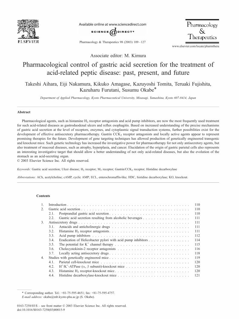
Contents
1. Introduction . . . . . . . . . . . . . . . . . . . . . . . . . . . . . . . . . . . . . . . . . . . . 110
2. Gastric acid secretion . . . . . . . . . . . . . . . . . . . . . . . . . . . . . . . . . . . . . . . 110
2.1. Postprandial gastric acid secretion . . . . . . . . . . . . . . . . . . . . . . . . . . . . . 110
2.2. Gastric acid secretion resulting from alcoholic beverages . . . . . . . . . . . . . . . . . 111
3. Antisecretory drugs . . . . . . . . . . . . . . . . . . . . . . . . . . . . . . . . . . . . . . . . 111
3.1. Antacids and anticholinergic drugs . . . . . . . . . . . . . . . . . . . . . . . . . . . . 111
3.2. Histamine H2 receptor antagonists . . . . . . . . . . . . . . . . . . . . . . . . . . . . . 111
3.3. Acid pump inhibitors . . . . . . . . . . . . . . . . . . . . . . . . . . . . . . . . . . . 112
3.4. Eradication of Helicobacter pylori with acid pump inhibitors . . . . . . . . . . . . . . . 114
3.5. The potential for K+ channel therapy . . . . . . . . . . . . . . . . . . . . . . . . . . . 115
3.6. Cholecystokinin-2 receptor antagonists . . . . . . . . . . . . . . . . . . . . . . . . . . 116
3.7. Locally acting antisecretory drugs . . . . . . . . . . . . . . . . . . . . . . . . . . . . . 118
4. Studies with genetically engineered mice . . . . . . . . . . . . . . . . . . . . . . . . . . . . . 119
4.1. Parietal cell-knockout mice . . . . . . . . . . . . . . . . . . . . . . . . . . . . . . . . 120
4.2. H+/K+-ATPase (a, b subunit)-knockout mice . . . . . . . . . . . . . . . . . . . . . . . 120
4.3. Histamine H2 receptor-knockout mice . . . . . . . . . . . . . . . . . . . . . . . . . . . 120
4.4. Histidine decarboxylase-knockout mice . . . . . . . . . . . . . . . . . . . . . . . . . . 121
0163-7258/03/$ – see front matter D 2003 Elsevier Science Inc. All rights reserved.
doi:10.1016/S0163-7258(03)00015-9
* Corresponding author. Tel.: +81-75-595-4651; fax: +81-75-595-4757.
E-mail address: [email protected] (S. Okabe).

T. Aihara et al. / Pharmacology & Therapeutics 98 (2003) 109–127110
4.5. Muscarinic M3R-knockout mice . . . . . . . . . . . . . . . . . . . . . . . . . . . . . . 121
4.6. Gastrin-knockout mice . . . . . . . . . . . . . . . . . . . . . . . . . . . . . . . . . . . 122
4.7. Gastrin transgenic mice . . . . . . . . . . . . . . . . . . . . . . . . . . . . . . . . . . 122
4.8. Cholecystokinin-2 receptor knockout mice . . . . . . . . . . . . . . . . . . . . . . . . 122
4.9. Somatostatin receptor subtype 2 SST2R-knockout mice . . . . . . . . . . . . . . . . . . 122
5. Origin of parietal cells . . . . . . . . . . . . . . . . . . . . . . . . . . . . . . . . . . . . . . 123
6. Conclusion . . . . . . . . . . . . . . . . . . . . . . . . . . . . . . . . . . . . . . . . . . . . 123
Acknowledgements . . . . . . . . . . . . . . . . . . . . . . . . . . . . . . . . . . . . . . . . . . . 123
References . . . . . . . . . . . . . . . . . . . . . . . . . . . . . . . . . . . . . . . . . . . . . . . 124
1. Introduction
Since Prout’s (1823) discovery of gastric hydrochloric
acid and Heidenhain’s (1875) and Golgi’s (1893) iden-
tification of oxyntic gland parietal cells as the gastric acid
secretory cells, the mechanisms underlying acid secretion
have proven an interesting area of investigation. To date,
the topic of acid secretion has been well reviewed (Daven-
port, 1992; Hirschowitz et al., 1995; Modlin & Sachs,
1998; Sachs et al., 1995). It has been found that gastric
acid is secreted at the time of ingestion of food or
alcoholic beverages via both a conditioned and an uncon-
ditioned reflex. Both reflexes involve stimulation of sev-
eral parietal cell receptors, which, in turn, results in
transfer of the signal to H + /K + -ATPase molecules. In
addition, it has been empirically determined that gastric
acid represents the underlying cause of acid-related peptic
disease. In an attempt to protect the gastric mucosa from
gastric acid, enhance ulcer healing, and prevent ulcer
recurrence, pharmacological control of gastric acid secre-
tion has long represented a desirable goal. Through the
painstaking efforts of inspired researchers, various phar-
maceuticals, such as histamine H2 receptor (H2R) antago-
nists (Black et al., 1972; Brimblecombe et al., 1975) and
H + /K + -ATPase (acid pump) inhibitors (Fellenius et al.,
1981, 1982; Larsson et al., 1983; Sachs & Wallmark,
1989), have been developed and utilized for the treatment
of acid-related peptic diseases (Huang & Hunt, 2001).
Through such pharmacotherapy, the mechanism by which
parietal cells secrete hydrochloric acid was elucidated in
detail. In brief, neural and hormonal systems stimulate
various receptors on the basolateral membrane, as well as
enzymes located on the surface of parietal cell secretory
canaliculi, to secrete acid. The mediators for the neural and
hormonal pathways are acetylcholine (ACh), released from
the vagus nerve; gastrin, released from antral G cells; and
histamine, released from enterochromaffin-like (ECL)
cells.
Recent genetic technology has allowed deletion and
overexpression of specific receptors and enzymes that are
involved in gastric acid secretion in mice. The following
represents a current review both of the physiology of gastric
acid secretion in humans and laboratory animals, with
emphasis on data elucidated with genetically engineered
knockout (KO) mice, and of the pharmacological control of
gastric acid secretion in acid-related peptic disease.
2. Gastric acid secretion
2.1. Postprandial gastric acid secretion
In daily life, gastric acid is secreted upon ingestion of food
or alcoholic beverages. Initially, gastric secretion is com-
menced by the thought, sight, smell, or taste of appetizing
food products via the vagus nerve and enteric nerve system;
this process represents the conditioned reflex. Neural path-
ways directly stimulate parietal cells and antral G cells,
leading to gastrin release, as well as oxyntic gland ECL cells,
leading to histamine release. Such direct and indirect parietal
cell stimulation is principally mediated by three receptors,
that is, cholinergic muscarinic M3 receptors (M3R), H2R, and
CCK-B/gastrin receptors (CCK2R). Of these three receptors,
however, H2R appears to represent the key receptor, as H2R
antagonists inhibit not only histamine-stimulated acid secre-
tion, but also gastrin- and ACh-stimulated acid secretion.
H2R activation results in an increase in intracellular cyclic
AMP (cAMP) levels, which acts as a second messenger to
transfer the signal to the final step of acid secretion (i.e., the
acid pump). In contrast, stimulation of either M3R by ACh or
CCK2R by gastrin results in a signal mediated by an intra-
cellular increase in free Ca2 + ions. It is of interest that a
threshold concentration of cAMP is required for gastrin-
induced acid secretion (Geibel et al., 1995). Furthermore,
both gastrin and vagal stimulation enhance histamine release
from histamine-containing cells, such as ECL cells, via
CCK2R or pituitary adenylate cyclase-activating polypeptide
(PACAP) receptors (Zeng et al., 1999; Sundvik et al., 2001).
Activation of such receptors eventually stimulates gastric
acid pumps in secretory canaliculi of parietal cell. After
sufficient acid secretion has occurred, a feedback system
terminates gastric acid secretion. A decrease in intragastric
pH stimulates somatostatin release from antral D cells so as to
inhibit gastrin release from G cells and to inhibit the stimu-
lative effect of gastrin on CCK2R located on parietal and ECL
cells. Furthermore, acidification of the duodenum also causes
a release of the hormone secretin, which acts to inhibit gastric
acid secretion.

& Therapeutics 98 (2003) 109–127 111
2.2. Gastric acid secretion resulting from alcoholic
beverages
In addition to food, alcoholic beverages, such as beer and
red wine, are known to represent potent secretagogues in
humans and animals (Chari et al., 1993; Lenz et al., 1983;
Teyssen et al., 1997). We have confirmed in dogs that oral
administration of either beer or wine, both alcoholic and
nonalcoholic, markedly stimulates gastric acid secretion for
� 30–45 min in denervated gastric pouch dogs. During
active gastric acid secretion, serum gastrin levels were
observed to increase. Both Singer et al. (Chari et al.,
1993; Teyssen et al., 1997) and our group (Matsuno et al.,
2000; Sasaki et al., 2000) have provided evidence that the
stimulatory effect of beer and wine on acid secretion is due
to not only alcohol, but also other unidentified substances. It
is of interest that acid secretion stimulated by beer or wine
was found to be significantly inhibited by anticholinergic
drugs (e.g., atropine and pirenzepine), H2R antagonists (e.g.,
cimetidine and famotidine), a CCK2R antagonist (e.g., S-
0509), and acid pump inhibitors (e.g., omeprazole). These
results strongly suggest that substances present in alcoholic
beverages stimulate gastric acid secretion via M3R, muscar-
inic M1 receptor (M1R), H2R, and CCK2R on parietal or
ECL cells. Identification of such substances might enhance
the understanding of the physiology underlying gastric acid
secretion.
For the pharmacological control of either gastric acid
secretion stimulated by food or alcoholic beverages or
diseases involving excessive gastric acid secretion, such as
Zollinger-Ellison syndrome or reflux diseases, the following
medications are currently used to treat acid-related peptic
disease.
T. Aihara et al. / Pharmacology
3. Antisecretory drugs
3.1. Antacids and anticholinergic drugs
Long ago, people with an upset stomach commonly
ingested powdered shells to alleviate the discomfort. Accord-
ingly, it was empirically discovered that CaCO3, a natural
antacid, buffered gastric acid. After demonstration of the
presence of hydrochloric acid in the stomach, antacid therapy
became the popular treatment for peptic acid-related diseases,
such as peptic ulcers. Antacids, such as sodium bicarbonate,
calcium carbonate, aluminum hydroxide, magnesium
hydroxide, or combined preparations, promptly provide
effective pain relief via neutralization of intraluminal acid
(Feurle, 1975). The effective time for antacids to last in the
human stomach, however, is too short to exert a neutralizing
effect. Moreover, given that more potent and safe antisecre-
tory drugs, such as H2R antagonists and acid pump inhibitors,
are readily available, antacid therapy is not commonly
utilized for current peptic ulcer treatment. To prolong the
effect of antacids, anticholinergic drugs, such as propanthe-
line bromide and benactidine methobromide, have been
concurrently administered to delay emptying of the agents
into the duodenum. Anticholinergics can also inhibit acid
secretion by themselves. Similar to antacids, however, the use
of anticholinergic drugs is generally limited, as anticholiner-
gics delivered at dosages capable of inhibiting acid secretion
almost invariably induce adverse effects, such as dry mouth,
blurred vision, tachycardia, and bladder dysfunction. In
contrast to anticholinergics, selective M1R antagonists, such
as pirenzepine and terenzepine, have been clinically utilized,
as side effects occur less frequent (Fig. 1A). Such agents are
useful in both providing pain relief and enhancing ulcer
healing via suppression of gastric acid secretion.
3.2. Histamine H2 receptor antagonists
Since histamine was considered to represent a final
common mediator for acid secretion, several groups fer-
vently sought an antagonist capable of inhibiting histamine-
stimulated acid secretion. After painstaking efforts, Black
and colleagues (1972) succeeded in developing the first H2R
antagonist, burimamide, followed by metiamide. With
minor alterations, Brimblecombe and colleagues (1975)
eventually developed cimetidine, which is widely prescribed
throughout the world for treatment of acid-related peptic
disease (Brimblecombe et al., 1975). By modifying the
chemical structure of cimetidine, the potent H2R antagonists
ranitidine, famotidine, and nizatidine were all developed,
resulting in remarkable treatment for acid-related disease,
including reflux esophagitis (Fig. 1B). These new com-
pounds were later found to inhibit gastric acid secretion
stimulated by not only histamine, but also carbachol and
gastrin in both humans and animals. These findings suggest
that H2R stimulation might be required for the effect of such
secretagogues. Recently, H2R antagonists have become
first-line therapy for acid-related peptic disease, leading to
a marked improvement in the quality of life for a large
number of patients. Paralleling the development of such
pharmacotherapy, there has been a dramatic reduction in the
use of surgical intervention for ulcer treatment. Interestingly,
in the beginning of clinical application, many physicians
expressed concern over potential regurgitation of intestinal
bacteria into the stomach, as the antisecretory effect of H2R
antagonists is much more powerful than conventional anti-
cholinergic drugs. Sustained inhibition of gastric acid might
predispose to gastric bacterial contamination, resulting in an
increase in N-nitrosoamine, a metabolite of ingested nitrites
and a known carcinogen. Accordingly, the use of H2R
antagonists initially was limited to only 4 weeks. Nonethe-
less, long-term clinical experience has demonstrated that
such a risk was a groundless fear. In fact, certain H2R
antagonists are currently even considered safe enough to be
marketed as over-the-counter pharmaceuticals.
It is well known that cimetidine strongly inhibits cyto-
chrome P450 (CYP) enzymes, particularly CYP3A4 (Ren-
dic, 1999). Accordingly, caution is warranted when

Fig. 1. Chemical structures of muscarinic cholinergic M1 receptor (A) and histamine H2 receptor (B) antagonists. Note that roxatidine and lafutidine possess
structures completely different from cimetidine, ranitidine, famotidine, and nizatidine.
T. Aihara et al. / Pharmacology & Therapeutics 98 (2003) 109–127112
cimetidine is concurrently prescribed with other pharmaco-
logic agents, such as warfarin and diazepam, that are
metabolized by this enzyme. In contrast, it has been reported
that CYP inhibition by ranitidine is relatively weak com-
pared to cimetidine.
Black and colleagues (1972) first contended that
histamine H2R antagonists require an imidazole ring in
their chemical structure to antagonize the effect of
histamine. Nonetheless, ranitidine and famotidine were
found to exert a stronger effect on H2R than cimetidine,
despite the absence of imidazole rings and the presence
of furan and thiazole rings, respectively. Furthermore,
new H2R antagonists, such as roxatidine and lafutidine,
which were developed by random screening in Japan,
appear to have no relationship to the chemical structure
of either the histamine molecule or the established H2R
antagonists (Shibata et al., 1993; Shiratsuchi et al., 1988;
Tarutani et al., 1985a, 1985b; Umeda et al., 1999). The
effects of such newly developed antagonists were much
the same as cimetidine, raniditine, and famotidine in both
clinical trials and animal studies. Animal studies demon-
strated that cimetidine and other representative H2R
antagonists had little or no effect on pharmacologic
agent-induced necrotizing gastric mucosal damage (Robert
et al., 1979). In contrast, the two newly developed
antagonists were found to have a cytoprotective effect
for gastric mucosa against pharmacologic agent-induced
necrotizing damage in animal studies.
Although pharmacotherapy for acid-related peptic dis-
eases has greatly improved since the development of H2R
antagonists, the agents still possess a number of shortcom-
ings. For instance, it was found that H2R antagonists have
little effect on daytime acid control, despite marked sup-
pression of nocturnal acid output. It has also been reported
that H2R antagonists evoke rapid tolerance during therapy.
Such tolerance does not depend on overexpression of H2R,
but rather, appears to be related to up-regulation of other
pathways that elevate cAMP levels in parietal cells. In
addition, acid rebound following H2R antagonist withdrawal
represents another disadvantage. Such a phenomenon is
considered to represent the result of either up-regulation of
H2R on parietal cells or increased production of H +/K + -
ATPase in parietal cells.
3.3. Acid pump inhibitors
Identification of the acid pump, H +/K + -ATPase, as the
final pathway of gastric acid secretion provided a unique
opportunity to develop a new class of agents that inhibit acid
secretion. Research groups at AB Hassle Company in
Sweden serendipitously discovered that benzimidazole
derivatives could potently inhibit gastric acid secretion in
rabbit or guinea pig isolated gastric glands stimulated by
dibutyryl-cAMP (Fellenius et al., 1981, 1982). This finding
resulted in the hypothesis that such compounds might
inhibit parietal cell acid pumps. After screening a vast

T. Aihara et al. / Pharmacology & Therapeutics 98 (2003) 109–127 113
number of derivatives, the first acid pump inhibitor (i.e.,
omeprazole) was finally discovered. Omeprazole was
promptly utilized for the treatment of acid-related peptic
diseases, including upper gastrointestinal ulcers, reflux
esophagitis, and Zollinger-Ellison syndrome. Clinical and
animal studies demonstrated that the antisecretory effect of
omeprazole persisted for a long time ( > 24 hr after a single
dose) due to covalent binding of omeprazole to H +/K + -
ATPase (the precise mechanism is explained in the follow-
ing section). Similar to the development of H2R antagonists,
lansoprazole, pantoprazole, and rabeprazole were subse-
quently developed by modifying the chemical structure of
omeprazole (Fig. 2A) (Lee et al., 1992; Robinson et al.,
1997; Satoh et al., 1989; Williams & Pounder, 1999). In
contrast to H2R antagonists, however, all acid pump inhib-
itors possessed a common structural element (i.e., a benzi-
midazole ring).
As expected based on the sight of inhibition, omeprazole
and lansoprazole are able to inhibit gastric acid secretion
stimulated by ACh, gastrin, and histamine in a dose-related
manner (Fig. 3). It was demonstrated that omeprazole and
other inhibitors represent prodrugs that are converted into
active forms in acidic environments (Nagaya et al., 1989;
Satoh et al., 1989). It is of note that acid pump inhibitors
were found to accumulate in parietal cell secretory canal-
iculi, resulting in an antisecretory effect that lasts much
longer than that of receptor antagonists. Animal studies have
Fig. 2. Chemical structures of acid pump inhibitors with benzim
indicated that as a result of long-term inhibition of gastric
acid secretion, circulating gastrin levels increase, resulting
in mucosal hyperplasia and carcinoid tumor development in
rat gastric mucosa (Carlsson et al., 1990; Hakanson et al.,
1986). Nonetheless, there has been no report of devel-
opment of mucosal hyperplasia in human stomachs, despite
presence of hypergastrinemia. It remains most likely that the
degree of increase in circulating gastrin in humans is much
less than that observed in rodents.
Animal studies have clearly demonstrated the tremendous
difference between H2R antagonists and acid pump inhib-
itors in terms of efficacy for ulcer treatment. Indeed, ulcer
healing following administration of H2R antagonists in rats
is rather inconsistent, despite frequent and adequate dosings.
In contrast, ulcer healing following administration of acid
pump inhibitors is reproducible, even with only single daily
dosing (Satoh et al., 1989; Yamamoto et al., 1984). Such a
marked difference might stem from the differing effective
time for acid inhibition.
As described above, increased serum gastrin levels, due
to suppressed acid secretion, are observed during omepra-
zole administration. Consequently, it was suggested that the
ulcer healing effect of omeprazole might result from
increased gastrin levels, as gastrin exerts a potent trophic
effect on gastric mucosa. Nonetheless, it was demonstrated
that the ulcer healing effect of omeprazole was not affected
by concomitant administration of a somatostatin derivative
idazole rings (A) and the K + channel blocker 293B (B).

Fig. 3. Antisecretory effects of omeprazole and lansoprazole on histamine-stimulated gastric acid secretion in the denervated (Heidenhain) pouch in dogs. Data
are presented as means ± SEM (n= 3–4). * Significantly different from control values, P< 0.05.
T. Aihara et al. / Pharmacology & Therapeutics 98 (2003) 109–127114
and the CCK2R antagonist YM022, indicating that
increased gastrin failed to exert an influence on ulcer
healing (Hirschowitz et al., 1995; Okabe et al., 1997; Okabe
& Tsukimi, 1996). Accordingly, omeprazole most likely
enhances ulcer healing due to its potent and persistent
antisecretory effect (Fig. 4).
Fig. 4. Effects of omeprazole, YM022 (a selective CCK2R antagonist), and their c
Acetic acid ulcers were used as the experimental ulcer model. Note that the ulc
suggesting that increased gastrin does not contribute to ulcer healing. Data are pre
values, P< 0.05.
3.4. Eradication of Helicobacter pylori with acid pump
inhibitors
Since Marshall and Warren (1984) discovered the bac-
teria Helicobacter pylori in stomachs, H. pylori has been
known to represent a risk factor for not only development of
ombination on ulcer healing, acid secretion, and serum gastrin levels in rats.
er healing effect of omeprazole was not affected by combined treatment,
sented as means ± SEM (n= 10–17). * Significantly different from control

T. Aihara et al. / Pharmacology & Therapeutics 98 (2003) 109–127 115
gastritis and ulcers, but also relapse of healed ulcers (Osato
& Graham, 1999). In addition, epidemiological investi-
gations have demonstrated that H. pylori infection directly
and indirectly induces neoplasms, such as gastric carcinoma
and MALT lymphoma (Danesh, 1999; Huang et al., 1998;
Konturek et al., 1999; Uemura et al., 2001). Various studies
with animals, including primates, have demonstrated that
long-term infection with H. pylori induces atrophic gastritis,
ulcers, and intestinal metaplasia at a high incidence. Our
group found that H. pylori infection invariably causes acute
gastritis, ulcers, and a relapse of healed gastric ulcers in
Mongolian gerbils (Keto et al., 1999, 2001). In addition,
Watanabe et al. (1998) demonstrated that H. pylori infection
induced intestinal-type gastric adenocarcinoma 62 weeks
following inoculation in Mongolian gerbils. Our group
(Keto et al., 2001) has also found that both atrophic gastritis
with intestinal metaplasia and intestinal-type adenocarci-
noma develop 18–24 months after H. pylori inoculation
in Mongolian gerbils. A recent study by Higashi and
colleagues (2002) demonstrated that cytotoxin associated
gene (CagA) protein highjacks a signaling pathway in
gastric cells, prompting a morphological change that begins
an oncogenic cascade. Given such findings, it is reasonable
to consider that H. pylori represents a carcinogenic agent for
the stomach that probably acts as an initiator and/or a
promoter. Accordingly, eradication of H. pylori in humans
with or without current gastric disease doubtless represents
the most important initial step for the prevention of benign
and malignant gastric disease.
Crabtree and Figura (1999) penned the following state-
ment, ‘‘The key current questions are whether the long-term
consequences of mucosal damage, such as atrophy and
intestinal metaplasia, can be reversed after elimination of
infection and whether, with the development of a simple 1-
week therapeutic regimen for eradicating H. pylori, large-
scale treatment of infected patients will have a beneficial
effect on the incidence of gastric cancer.’’
In response to the above proposal, we examined the
timing of eradication following H. pylori (VacA- and CagA-
positive strains) inoculation in Mongolian gerbils (Keto et
al., 2001). Eradication was achieved 4 or 8 months post-
inoculation with a combined treatment of omeprazole (4
weeks) plus clarithromycin (2 weeks). Upon autopsy, 18–
24 months postinoculation, it was demonstrated that both
visible and histological changes were clearly prevented by
H. pylori eradication 4 months postinfection. In contrast,
atrophy and intestinal metaplasia persisted in animals that
underwent H. pylori eradication 8 months postinoculation.
Accordingly, it was concluded that although early eradica-
tion of H. pylori clearly prevented development of visible
and histologic changes in Mongolian gerbils stomachs 18
and 24 months postinoculation, late eradication failed to
prevent development of atrophic gastritis with intestinal
metaplasia. These results strongly suggest that the timing
of eradication plays a critical role in the prevention of H.
pylori-associated mucosal changes. Namely, eradication
should be undertaken as early as possible to prevent the
occurrence of irreversible gastric damage.
3.5. The potential for K+ channel therapy
During gastric acid secretion, H +/K + -ATPase couples
H + exit to K + uptake. Consequently, luminal K + recycling
and Cl � exit are essential for HCl secretion. Nonetheless,
the molecular characterization of luminal K + channels that
enable K + recycling in parietal cells remains unknown.
Recently, several luminal K + channels were reported and
their potential to inhibit gastric acid secretion was demon-
strated with in vitro cell cultures and in vivo animal models.
KCNQ1, which is mutated in the hereditary long QT
syndrome type 1, is present in abundance in a variety of
epithelia, including kidney, pancreas, small and large intes-
tines, and stomach epithelia (Bleich & Warth, 2000;
Chouabe et al., 1997; Yang et al., 1997). KCNQ1 coassem-
bles with several different subunits, such as KCNE1,
KCNE2, and KCNE3, to form native K + channels. As
KCNE2 and KCNE3 are expressed to a high degree in the
stomach, it has been postulated that KCNQ1 coassembled
with such subunits may constitute the gastric K + channels
that contribute to luminal K + recycling in parietal cells
(Grahammer et al., 2001). 293B is a kind of chromanol
compound known as KCNQ1 inhibitor (Bleich et al., 1997;
Lohrmann et al., 1995; Yang et al., 1997). Interestingly,
293B (Fig. 2B) reversibly and completely inhibited gastric
acid secretion stimulated by histamine, carbachol, and
gastrin (Grahammer et al., 2001). Consequently, 293B
might represent a potential candidate for inhibiting gastric
acid secretion via a new mechanism.
Another K + channel designated as inwardly rectifying
K + channel 4.1 (Kir4.1) has also been reported to localize
to the apical membrane of gastric parietal cells (Fujita et al.,
2002; Hagen & Ouellette, 2001). Blockade of Kir4.1 led to
inhibition of acid secretion stimulated by both histamine
and carbachol in isolated parietal cells. Kir4.1 channel
activity is sensitive to intracellular H +, but resistant to
extracellular H +, which might reflect the channel’s involve-
ment in the K + recycling pathway for H +/K + -ATPase at
the apical membrane of parietal cells. Accordingly, Kir4.1
might also represent a potential therapeutic target for acid-
related diseases. ROMK channels are one of the other
inwardly rectifying ATP-sensitive K + channels (Sritharan
et al., 2001; Vucic et al., 2002). Gastric glands isolated
from ROMK channel gene-KO mice exhibited no H+/K + -
ATPase activity, indicating that ROMK channel is also
associated with gastric acid secretion. ROMK channels
thus might also represent an interesting target for acid
inhibition.
As many kinds of K + channels have been identified
(Shieh et al., 2000; Yamada et al., 1998), specific and
selective pharmacotherapy that inhibits distinct K + channel
activities might prove to be ideal for treating acid-related
diseases.
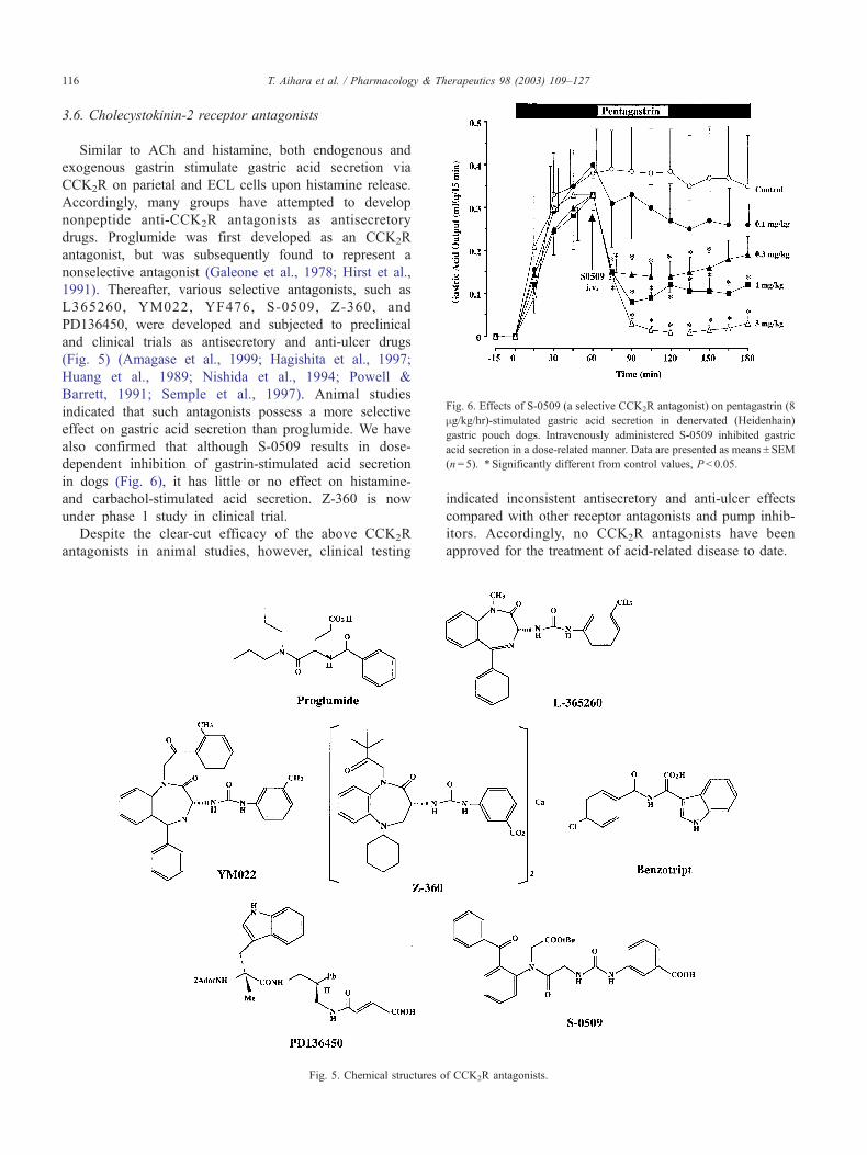
Fig. 6. Effects of S-0509 (a selective CCK2R antagonist) on pentagastrin (8
mg/kg/hr)-stimulated gastric acid secretion in denervated (Heidenhain)
gastric pouch dogs. Intravenously administered S-0509 inhibited gastric
acid secretion in a dose-related manner. Data are presented as means ± SEM
(n= 5). * Significantly different from control values, P< 0.05.
& Therapeutics 98 (2003) 109–127
3.6. Cholecystokinin-2 receptor antagonists
Similar to ACh and histamine, both endogenous and
exogenous gastrin stimulate gastric acid secretion via
CCK2R on parietal and ECL cells upon histamine release.
Accordingly, many groups have attempted to develop
nonpeptide anti-CCK2R antagonists as antisecretory
drugs. Proglumide was first developed as an CCK2R
antagonist, but was subsequently found to represent a
nonselective antagonist (Galeone et al., 1978; Hirst et al.,
1991). Thereafter, various selective antagonists, such as
L365260, YM022, YF476, S-0509, Z-360, and
PD136450, were developed and subjected to preclinical
and clinical trials as antisecretory and anti-ulcer drugs
(Fig. 5) (Amagase et al., 1999; Hagishita et al., 1997;
Huang et al., 1989; Nishida et al., 1994; Powell &
Barrett, 1991; Semple et al., 1997). Animal studies
indicated that such antagonists possess a more selective
effect on gastric acid secretion than proglumide. We have
also confirmed that although S-0509 results in dose-
dependent inhibition of gastrin-stimulated acid secretion
in dogs (Fig. 6), it has little or no effect on histamine-
and carbachol-stimulated acid secretion. Z-360 is now
under phase 1 study in clinical trial.
Despite the clear-cut efficacy of the above CCK2R
antagonists in animal studies, however, clinical testing
T. Aihara et al. / Pharmacology116
Fig. 5. Chemical structures o
indicated inconsistent antisecretory and anti-ulcer effects
compared with other receptor antagonists and pump inhib-
itors. Accordingly, no CCK2R antagonists have been
approved for the treatment of acid-related disease to date.
f CCK2R antagonists.

Fig. 7. Chemical structures of locally active antisecretory drugs and effects of H +/K + -ATPase.
Fig. 8. Local and systemic effects of ME3407 on histamine-stimulated gastric acid secretion (80 mg/kg/hr) in denervated gastric pouch dogs. ME3407 was
administered into the pouch for 30 min, intravenously or orally either before or after histamine infusion. Note that while ME3407 significantly inhibited acid
secretion, it had no effect with intravenous or oral administration. Data are presented as means ± SEM (n= 4). * Significantly different from control values,
P < 0.05. Data from Okabe et al. (2002a, 2002b).
T. Aihara et al. / Pharmacology & Therapeutics 98 (2003) 109–127 117

Fig. 9. Local effects of Wortmannin on histamine-stimulated gastric acid
secretion (80 mg/kg/hr) in denervated gastric pouch dogs. Wortmannin was
locally applied to the pouch in a volume of 15 mL for either 15 or 30 min.
Data are presented as means ± SEM (n= 5). * Significantly different from
control values, P < 0.05.
T. Aihara et al. / Pharmacology & Therapeutics 98 (2003) 109–127118
3.7. Locally acting antisecretory drugs
Several drugs have been reported to exert an antisecre-
tory effect when locally applied to the stomach (Bond &
Hunt, 1956; Canfield & Curwain, 1983; Curwain & Can-
field, 1983; Konturek et al., 1981, 1984; Nicol et al., 1981;
Reed & Smy, 1980; Urushidani et al., 1997). Nonetheless,
attention has not been directed toward such drugs as either
antisecretory drugs or investigational tools useful for the
elucidation of parietal cell function. We examined whether
or not certain agents could inhibit secretagogue-stimulated
Fig. 10. Duration of antisecretory effects of locally applied Wortmannin. Wortmann
histamine infusion (80 mg/kg/hr). It is of note that the antisecretory effects sign
means ± SEM (n= 5). * Significantly different from control values, P < 0.05.
gastric acid secretion when applied to gastric pouches in
dogs. Our studies demonstrated that omeprazole, lemino-
prazole (an acid pump inhibitor), FPL52694 (a mast cell
stabilizer), ME3407 (a myosin light chain kinase inhibitor
and functional analogue of Wortmannin) (Fig. 7) (Okabe et
al., 1995, 2002), and 16,16-dimethyl prostaglandin E2
significantly inhibited gastric acid secretion stimulated by
histamine, gastrin, or carbachol by local application. The
duration of the antisecretory activity following local
application for 30 min was � 5–6 hr for FPL52694 and
ME3407; that is, inhibition was reversible. For omeprazole,
leminoprazole, and the prostaglandin analogue, it is possible
that the compounds will have inhibited acid pumps follow-
ing either absorption from the gastric mucosa or interaction
with prostaglandin receptors on parietal cells. In contrast,
neither FPL52694 nor ME3407 exerted an inhibitory effect
on acid pumps or an antisecretory effect following oral or
intravenous delivery (Fig. 8). Since ME3407 resulted in
marked inhibition of gastric acid secretion, we further
examined whether or not another inhibitor of myosin light
chain kinase, Wortmannin, achieved a similar local effect on
acid secretion. Our studies demonstrated that locally applied
Wortmannin, administered at 1 mg per pouch either before
or after histamine infusion, resulted in a strong antisecretory
effect on histamine-stimulated acid secretion (Figs. 9 and
10). It is of interest that a 15-min application of the agent to
the pouch significantly inhibited acid secretion. The anti-
secretory effect of Wortmannin persisted for more than 10 hr
after a single application, suggesting that the duration of the
effect is nearly the same as that of acid pump inhibitors
systemically delivered (Fig. 10). Accordingly, it remains
most likely that FPL52694, ME3407, and Wortmannin act
on a pharmacologically sensitive portion of the apical
membrane of parietal cells. Other MLCK inhibitors, such
in was administered for 30 min to dog denervated gastric pouches 1 hr after
ificantly persisted >10 hr after a single application. Data are presented as

Fig. 11. Schematic drawing of a parietal cell and the various targets for antisecretory drugs.
T. Aihara et al. / Pharmacology & Therapeutics 98 (2003) 109–127 119
as HA-100 and HA-1077, failed to exert an effect on acid
secretion, despite administration of a dose of 6 mg per
pouch. The lipid-soluble substances ME3407 and Wortman-
nin appeared to penetrate the apical membrane and to
proceed into the cytoplasm, while the water-soluble sub-
stances HA-100 and HA1077 did not penetrate the mem-
brane, which probably explains their inability to achieve an
effect. Accordingly, it is most likely that both ME3407 and
Wortmannin act in the cytoplasm to inhibit acid secretion by
first inhibiting MLCK, which represents a key point in the
acid-secreting process. The exact mechanism by which
MLCK effects the normal functioning of gastric acid secre-
tion remains unknown. One consideration is that the sub-
stances might prevent the fusion of tubulovesicles to
secretory canaliculi. Incidentally, both ME3407 and Wort-
mannin failed to exert a direct effect on H + /K + -ATPase
isolated from rabbit oxyntic mucosa (Fig. 7). In any event,
the apical membrane represents not only an intriguing target
Fig. 12. Genetically engineered KO and transgenic mice. KO mice stand to
for new antisecretory drugs, but also a new medium to
further elucidate parietal cell functioning. Finally, the site of
action of various antisecretory drugs is illustrated as shown
(Fig. 11).
4. Studies with genetically engineered mice
The understanding of the regulatory mechanisms of
gastric acid secretion has been greatly deepened by the
use of various animal models, including dogs, cats, rabbits,
guinea pigs, rats, and mice, as well as clinical studies in both
ailing and healthy humans. In the vast majority of cases, the
underlying mechanism for acid secretion was elucidated by
means of the mechanism of action of the pharmacologic
agents, such as receptor antagonists or acid pump inhibitors.
Nonetheless, it proved quite difficult to precisely understand
the mechanisms underlying gastric acid secretion via phar-
offer many important findings that selective antagonists cannot reveal.

T. Aihara et al. / Pharmacology & Therapeutics 98 (2003) 109–127120
macotherapy, as most of agents were not selective enough to
inhibit each and every receptor for a sufficient length of time
due to rapid metabolism. Recently, most of the genes that
express the above-mentioned receptors and related enzymes
involved in acid secretion were cloned, allowing production
of genetic modification (i.e., KO or transgenic) animals
(Fig. 13). H +/K + -ATPase (a, b subunit)-KO mice (Franic et
al., 2001; Scarff et al., 1999; Spicer et al., 2000), histamine
H2R-KO mice (Kobayashi et al., 2000; Ogawa et al., 2003),
histidine decarboxylase (HDC)-KO mice (Tanaka et al.,
2002), muscarinic M3R-KO mice (Aihara et al., 2002;
Matsui et al., 2000; Yamada et al., 2001), gastrin-KO mice
(Franic et al., 2001; Friis-Hansen et al., 1998; Hinkle &
Samuelson, 1999; Hinkle et al., 2003; Koh et al., 1997;
Zavros et al., 2002a, 2002b), gastrin transgenic mice (Wang
& Dockray, 1999; Wang et al., 2000), CCK2R-KO mice
(Chen et al., 2002; Langhans et al., 1997; Nagata et al.,
1996), and somatostatin SST2R-KO mice (Martinez et al.,
1998) have been generated to date (Fig. 12).
4.1. Parietal cell-knockout mice
It is of note that even parietal cell-deficient mice were
generated without manifestation of any apparent aberrancy
(Canfield et al., 1996; Li et al., 1996), suggesting that
parietal cells are not requisite for the stomach. Interestingly,
the stomachs of mice lacking parietal cells exhibited extens-
ive hyperplasia within the oxyntic mucosa. These findings
strongly suggest that parietal cells greatly contribute to not
only gastric acid secretion, but also to maintenance of
gastric mucosal integrity.
Fig. 13. The stomach of a 17-month-old wild-type mouse (A) and a
histamine H2R knockout mouse (B). Note that the stomach of the KO
mouse is enlarged with a mucosal surface exhibiting large folds reminiscent
of Menetrier’s disease.
4.2. H +/K + -ATPase (�, � subunit)-knockout mice
Gastric H +/K + -ATPase, present in tubulovesicular and
secretory canalicular of gastric parietal cells, exchanges
luminal K + for cytoplasmic H + and is thus primarily
responsible for gastric acid secretion. This enzyme is
composed of two noncovalently linked subunits, a and b.The a subunit contains catalytic sites of the enzyme, while
the b subunit is required for transport of an active complex
from the endoplasmic reticulum to the apical membrane.
Recently, mice lacking the genes encoding for both of these
subunits were reported (Franic et al., 2001; Scarff et al.,
1999; Spicer et al., 2000). It is of note that both mutant mice
exhibit achlorhydria and hypergastrinemia (Franic et al.,
2001; Scarff et al., 1999; Spicer et al., 2000) compared with
wild-type mice. Gastrin, released from G cells, is the major
hormone to stimulate gastric acid secretion in response to
elevated gastric luminal pH. For instance, treatment with
anti-ulcer drugs, such as H2R antagonists and acid pump
inhibitors, induce hypergastrinemia and gastric mucosal
hypertrophy (Larsson et al., 1986). Accordingly, increased
serum gastrin in such KO mice may also be due to a positive
feedback response by G cells in response to achlorhydria. In
adult (10–12 weeks old) H +/K + -ATPase a subunit-KO
mice, severe histopathological alterations were observed,
including metaplasia and serious disruption of gastric gland
architecture (Spicer et al., 2000). The number of parietal and
chief cells appeared to be equivalent for wild-type and H +/
K + -ATPase a subunit-KO mice; however, pepsinogen
expression in chief cells was significantly reduced and
parietal cell secretory membranes were severely perturbated.
The overall thickness of the mucosa in H +/K + -ATPase bsubunit-KO mice (15 days old) was comparable with that of
wild-type mice; however, the pit and neck/isthmus regions
of the gastric units appeared enlarged, while the bases were
reduced in size. In 35-day-old H +/K + -ATPase b subunit-
KO mice, most parietal cells did not contain typical tubu-
lovesicular membranes or canaliculi; the number of zymo-
genic cells was also greatly reduced (Franic et al., 2001;
Scarff et al., 1999). Nonetheless, the total number of parietal
cells in each gastric unit did not greatly differ between KO
mice and wild-type mice. These results indicate that imma-
ture parietal cells resulting from H +/K + -ATPase subunit
KO-induced abnormal differentiation of gastric mucosal
cells, suggesting that unknown factors might be supplied
from parietal cells to maintain gastric mucosal integrity.
4.3. Histamine H2 receptor-knockout mice
We recently reported on gastric phenotypes observed in
H2R-KO mice (Kobayashi et al., 2000; Ogawa et al., 2003;
Okabe et al., 2001, 2002a). H2R-KO mice were viable and
developed without obvious abnormalities noted on gross
inspection. Unexpectedly, contrary to long-term effects on
inhibition of gastric acid secretion of H2R blocking with
antagonists, H2R-KO mice (12–16 weeks old) exhibited no

T. Aihara et al. / Pharmacology & Therapeutics 98 (2003) 109–127 121
change in basal gastric pH and basal gastric acid output
compared with wild-type mice. Although both histamine-
and gastrin-stimulated gastric acid secretion were com-
pletely abolished in H2R-KO mice, carbachol-stimulated
gastric acid secretion in these KO mice was similar to that
of wild-type mice. These results suggest both that gastrin
stimulates gastric acid secretion mainly via histamine
released from ECL cells and that carbachol stimulates
gastric acid secretion independently of histamine secretion.
In addition, it would appear that basal gastric acid secretion
in H2R-KO mice is retained by cholinergic regulation.
Interestingly, H2R-KO mice exhibited hypergastrinemia
(� 4-fold higher than wild-type mice) without elevation of
basal gastric pH. Hypergastrinemia is generally considered
to result from a simple positive feedback response of G cells
to achlorhydria; however, our study suggests that, at least in
H2R-KO mice, other factors unrelated to decreased acid
secretion can also induce hypergastrinemia.
In H2R-KO mice, gastric wet weight and DNA levels
were significantly higher than those measured for wild-type
mice, suggesting that H2R-KO mice develop gastric hyper-
trophy resulting from an increased number of mucosal cells.
Histologic studies in H2R-KO mice demonstrated a remark-
able increase in gastric mucosal thickness. Furthermore,
such hypertrophy resulted in formation of enlarged gastric
folds in the glandular region. The number of parietal and
endocrine cells in H2R-KO mice gastric mucosa was
increased compared with wild-type mice. It should be noted
that the size of parietal cells in H2R-KO mice was remark-
ably smaller. Moreover, electron microscopy of these pari-
etal cells revealed an abnormal structure with enlarged
secretory canaliculi, a lower density of microvilli, and few
typical tubulovesicles in the narrow cytoplasm. These find-
ings suggest that H2R-mediated histamine signaling controls
the cellular size of parietal cells and preserves the normal
structure of secretory membranes in parietal cells. Following
a study by our group that demonstrated that H2R-KO mice
develop Menetrier’s disease-like gastric mucosa 6–17
months after birth (Fig. 13), it was proposed that increased
gastrin and/or TGF-a are involved in such a hypertrophic
mechanism (Ogawa et al., 2003). Accordingly, H2R might
play an important role in not only gastric acid secretion, but
also gastric mucosal integrity. H2R-KO mice have afforded
new information that had not been discovered with previous
pharmacological studies. Further studies with H2R-KO mice
will increasingly elucidate the relationship between H2R and
gastric functioning.
4.4. Histidine decarboxylase-knockout mice
Histamine is synthesized from histidine by the enzyme
HDC and is stored in mast cells, ECL cells, and enteric nerve
fibers in the stomach. Our group reported that HDC-KO
mice express trace levels of histamine secretion and exhibit
very little de novo histamine synthesis in gastric mucosa
(Tanaka et al., 2002). In addition, in HDC-KO mice, low
basal acid secretion, increased sensitivity of acid secretion to
exogenous histamine, and hypergastrinemia were observed.
Interestingly, in HDC-KO mice, both carbachol and gastrin
alone exerted little to no effect on acid secretion; however,
the presence of a small amount of exogenous histamine
resulted in significant stimulation of gastric acid secretion. In
addition, both carbachol and gastrin significantly increased
gastric mucosal histamine levels in wild-type mice. These
results both indicate that the presence of histamine is
essential for carbachol and gastrin to stimulate acid secretion
in vivo and suggest that both carbachol and gastrin indirectly
stimulate parietal cells to secrete acid via release of histamine
from ECL cells. It follows that HDC-KO mice represent a
convenient model to observe the direct effect of acid secre-
tagogues on parietal cells.
No significant changes were observed in the general
histological appearance of gastric mucosal cells in young
HDC-KO mice (8–12 weeks old). Nonetheless, in HDC-
KO mice older than 3 months, the stomach weight was
significantly greater than wild-type mice. Histological ana-
lysis revealed that the gastric mucosa in HDC-KO mice was
hypertrophic at the glandular base region, with increased
parietal and ECL cells. In addition, the morphology of
HDC-KO mice appeared to differ from that of H2R-KO
mice. Further studies are needed to compare HDC-KO and
H2R-KO mice.
4.5. Muscarinic M3R-knockout mice
It has been recognized that ACh activates M3R on
parietal cells, resulting in acid secretion. In addition, in vivo
cholinergic stimulation of acid secretion appears to involve
M1R, as evidenced by a high sensitivity of acid secretion to
a selective M1R antagonist, pirenzepine. Nonetheless, due to
a lack of ligands with sufficient subtype selectivity, it has
been difficult to study the specific role of each subtype
(Caulfield & Birdsall, 1998). Consequently, the precise in
vivo role of each muscarinic receptor subtype with respect
to gastric acid secretion remained undetermined. Recently,
availability of M3R-KO mice (Matsui et al., 2000; Yamada
et al., 2001) enabled us to study the physiological signific-
ance of M3R in the regulation of gastric acid secretion
(Aihara et al., 2002). Twelve-week-old M3R-KO mice
exhibited decreased gastric acid secretion, as evidenced by
a significantly higher gastric pH and a significantly lower
gastric acid output. At this age, histologic observation
revealed no difference in the number of parietal and endo-
crine cells in the gastric mucosa between M3R-KO and
wild-type mice. Nonetheless, electron microscopy demon-
strated that the number of active (secreting) parietal cells
was significantly reduced in M3R-KO mice. Carbachol-
stimulated gastric acid secretion in M3R-KO mice was� 30% of that measured in wild-type mice. Histamine and
gastrin were still able to stimulate acid secretion in M3R-KO
mice, but the acid output was � 50% of the respective
values in wild-type mice. These results suggest that M3R

T. Aihara et al. / Pharmacology & Therapeutics 98 (2003) 109–127122
stimulation via ACh signaling is required for not only basal
acid secretion, but also stimulated gastric acid secretion. In
addition, vagal stimulation with 2-deoxy-D-glucose in-
creased the acid output in wild-type mice by 6.5-fold, but
had little impact in M3R-KO mice, strongly suggesting the
physiological importance of M3R in gastric acid secretion. It
is of great interest that carbachol is still able to stimulate
gastric acid secretion in M3R-KO mice, albeit to a degree
much less than that observed in wild-type mice. Carbachol-
stimulated gastric acid secretion was clearly inhibited by
either pirenzepine or famotidine in M3R-KO mice, suggest-
ing that histamine and activation of M1R appear to be
involved in the mechanism underlying carbachol stimu-
lation. It is of note, however, that it has been reported that
ECL cells lack functional muscarinic receptors (Helander et
al., 1996; Lindstrom et al., 1997). In the future, further
investigations to elucidate the detailed mechanisms under-
lying carbachol-induced gastric acid secretion in M3R-KO
mice are requisite. In addition, improved understanding of
the particular in vivo participation of each muscarinic
receptor subtype in gastric acid secretion will be achieved
with phenotype studies, such as the investigation we are
currently conducting with M1R-KO mice.
4.6. Gastrin-knockout mice
Gastrin-KO mice are achlorhydric and fail to respond to
exogenously administered histamine, carbachol, and gastrin
(Friis-Hansen et al., 1998; Hinkle & Samuelson, 1999;
Zavros et al., 2002a, 2002b). Histological analysis revealed
a reduction in the gastric mucosal thickness and marked
changes in the number of parietal and ECL cells in the
oxyntic mucosa of gastrin-KO mice (Franic et al., 2001;
Friis-Hansen et al., 1998; Hinkle & Samuelson, 1999; Koh
et al., 1997; Zavros et al., 2002a, 2002b). The number of
parietal cells was remarkably decreased with a distinct
accumulation of immature cells lacking H +/K + -ATPase.
In comparison with wild-type mice, gastrin-KO mice exhib-
ited a marked reduction in HDC-positive cells, with very
weak staining in the gastric glands of gastrin-KO mice; the
number of chromogranin A-positive cells, however, was
unchanged. These findings suggest that gastrin is critical for
functional maturation of the acid-secretory system (Friis-
Hansen et al., 1998; Hinkle & Samuelson, 1999; Koh et al.,
1997). These findings are consistent with results obtained
with H +/K + -ATPase b subunit and gastrin-double KO
mice. Namely, hypergastrinemia in H +/K + -ATPase b sub-
unit-deficient mice is responsible for mucosal cell hyper-
trophy, but not for depletion of zymogenic cells (Franic et
al., 2001).
4.7. Gastrin transgenic mice
Insulin-gastrin (INS-GAS) transgenic mice that overex-
press amidated gastrin in pancreatic islets exhibit early (1–4
months) mild hypergastrinemia, that is, a near 2-fold
increase in plasma gastrin levels compared with wild-type
mice (Wang & Dockray, 1999; Wang et al., 2000). These
INS-GASmice demonstrate an initial increase in the maximal
gastric acid output and the number of both parietal and ECL
cells; however, the mice later progress to hypochlorhydria,
with decreased numbers of parietal and ECL cells. Hyper-
gastrinemia leads to marked thickening of the fundic mucosa
and multifocal hyperplasia in INS-GAS mice after 1 year. At
20 months, INS-GAS mice exhibit gastric metaplasia, dys-
plasia, and invasive gastric carcinoma. Interestingly, H. felis
infection accelerates development of intramucosal carcinoma
with submucosal and intravascular invasion in INS-GAS
mice (Wang et al., 2000).
4.8. Cholecystokinin-2 receptor knockout mice
Several groups have reported on the functional and
morphological changes observed in the gastric mucosa of
CCK2R-KO mice (Chen et al., 2002; Langhans et al.,
1997; Nagata et al., 1996). We also confirmed that
CCK2R-KO mice exhibit achlorhydria and hypergastrine-
mia. In comparison with wild-type mice, the basal gastric
acid output was lower and intragastric pH was higher in
CCK2R-KO mice (Langhans et al., 1997; Nagata et al.,
1996). Histamine and carbachol increased gastric acid
secretion in CCK2R-KO mice; however, the degree of
increase in gastric acid output in CCK2R-KO mice was
less than that observed for wild-type mice. Gastrin did not
increase gastric acid secretion in CCK2R-KO mice. In spite
of hypergastrinemia, CCK2R-KO mice had a decreased
stomach wet weight with severe atrophy of the gastric
oxyntic mucosa accompanied by a reduced number of
parietal and ECL cells. These findings are consistent with
northern blot analysis that revealed decreased levels of
HDC and H +/K + -ATPase mRNA. In addition, electron
microscopy demonstrated a decreased number of active
parietal cells and an increased number of immature ECL
cells in CCK2R-KO mice compared with wild-type mice.
These results suggest CCK2R directly or indirectly regulate
differentiation of gastric mucosal cells, including parietal
and ECL cells.
4.9. Somatostatin receptor subtype 2 SST2R-knockout mice
Somatostatin is a physiological inhibitor of gastric acid
secretion that is released from D cells in gastric fundic and
antral mucosa (Patel et al., 1996). Somatostatin is known for
its potent inhibition of histamine release by ECL cells,
gastrin production by G cells, and acid secretion by parietal
cells. Somatostatin has five different receptor subtypes (i.e.,
SST1R to SST5R) (Makhlouf & Schubert, 1990). It has been
previously reported that inhibition of gastric acid secretion
is mediated by SSTR2 in ECL and parietal cells. SST2R has
two different isoforms with long (SST2AR, in ECL cells)
and short (SST2BR, in parietal cells) C-terminal variants
generated by alternative mRNA splicing (Vanetti et al.,

T. Aihara et al. / Pharmacology & Therapeutics 98 (2003) 109–127 123
1993). Martinez et al. (1998) reported that SST2R-KO mice
exhibited high basal gastric acid secretion without a change
in serum gastrin compared with wild-type mice. Somatos-
tatin antibodies were found to increase basal secretion by 4-
fold in wild-type mice, but failed to exert an effect on
SST2R-KO mice. In addition, somatostatin analogues inhib-
ited gastrin-stimulated acid secretion in wild-type mice, but
did not affect basal secretion in SST2R-KO mice. These
results indicate that somatostatin suppresses gastrin-stimu-
lated gastric acid secretion via SST2R. Consequently, the
role of somatostatin in hypergastrinemia should prove an
interesting area of investigation for a variety of KO mice.
Based upon physiological and pharmacological analysis
with the above KO mice, regulatory mechanisms for gastric
acid secretion are rapidly being developed. Double or triple
receptor KO mice, such as H2R +M3R, H2R +CCK2R,
M3R +HDC, H2R +HDC, and H2R +M3R +CCK2R, as
well as the previously generated M3R+M1R, will afford
new insight into the mechanisms underlying gastric acid
secretion. The above results indicate that KO mice stand to
offer many important findings that selective antagonists
cannot reveal. In addition, these results suggest that abnor-
malities, particularly regulatory factors for gastric acid
secretion, can induce gastric mucosal cancer-like diseases
similar to those observed in clinical situations. It follows
that these mice models may prove useful for clarifying the
unknown mechanisms underlying such diseases, allowing
development of novel treatment strategies.
5. Origin of parietal cells
One of the unique and unusual properties of the mam-
malian stomach is its ability to secrete a highly concentrated
inorganic acid (i.e., hydrochloric acid [> 0.15 N]) from
parietal cells in the oxyntic mucosa. The rationality under-
lying stomach acid secretion remains a much debated topic.
Nevertheless, it has been postulated that acidification of the
gastric contents both assists in digestion of ingested food
and sterilization of contaminant virulent and/or regurgitated
intestinal bacteria. It is interesting, however, that the origin
of parietal cells remains unstudied. The most intriguing
issue remains the evolution of parietal cells, which are
characterized by a p-type H+ /K + -ATPase from stem cells.
Namely, the evolution of the H +/K + -ATPase gene from
stem cells located in the fundic mucosa represents the most
puzzling topic of discussion. One of the present authors
(S.O.) hypothesized that parietal cells might have evolved
via transfer of the H +/K + -ATPase gene from a microbe that
possessed a p-type H + -ATPase or H +/K + -ATPase during
the early Cambrian era (Okabe, 1997, 1999). The hypothesis
is grounded upon the following observations. Microorgan-
isms, including aerobic and anaerobic bacteria, are able to
pump H + into the extracellular environment by means of an
H + -ATPase that is coupled to nutrient influx. It is of interest
that 19% of the human H +/K + -ATPase (a subunit) is
comprised of amino acid residues identical to those of an
H + -ATPase found in Neurospora crassa. In addition, the
amino acid sequence for the ATP binding sites of animal
Na +/K + -ATPase, which resembles H +/K + -ATPase in
terms of amino acid residues (Maeda, 1994), and the
phosphorylated intermediates of yeast H + -ATPase is highly
conserved (Serrano et al., 1986). Such data appear to
indicate that parietal cells might have originated from a
parabiotic microorganism that was subsequently incorpo-
rated into a stem cell. Thereafter, the gene encoding for H +/
K + -ATPase and/or GATA DNA-binding proteins (transcrip-
tional regulators of the gastric H +/K + -ATPase gene) could
have been translocated into the nucleus, most likely with the
aid of a virus and/or transposon. Such a gene translocation
most likely occurred during the Cambrian era when Pro-
chordata and Chordata, which have no parietal cells, were
abundant. Accordingly, during evolution, stem cells for
Chordata digestive organs might have differentiated into
two cell types (i.e., surface epithelial cells and parietal cells)
prior to the appearance of fish, which possess parietal cells
with H +/K + -ATPase.
6. Conclusion
Although the origin of parietal cells has yet to be
elucidated, it appears that parietal cells function to secrete
highly concentrated gastric acid following ingestion of food
or alcoholic beverages to permit digestion of food and/or
prevention of pathogenic disease. It is well known that
excessive, or even normal, acid secretion is problematic
for maintenance of the normal integrity of the upper gastro-
intestinal tract, resulting in acid-related peptic disease.
Accordingly, rigid control of acid secretion via a therapeutic
modality is necessary to enhance healing of gastroduodenal
ulcers and reflux esophagitis. To that end, currently avail-
able H2R antagonists and acid pump inhibitors represent
powerful and reliable pharmacotherapy for the treatment of
acid-related peptic disease with few side effects. Since a
prototype for locally acting drugs was discovered, new
classes of antisecretory drugs, as well as CCK2R antago-
nists, represent the next target for drug development. Util-
izing genetically engineered KO and transgenic mice, the
mechanisms by which parietal cells secrete acid will be
further characterized. Finally, early eradication of H. pylori
infection with acid pump inhibitors and antibiotics will most
likely reduce the potential for development of atrophic
gastritis, intestinal metaplasia, and gastric carcinoma.
Acknowledgements
The authors wish to thank C. J. Hurt (John Hopkins
University School of Medicine, USA) and Dr. Y. Tsukimi
(Bayer Yakuhin, Research Center Kyoto) for a critical
reading of the manuscript. Various gene KO mice were

T. Aihara et al. / Pharmacology & Therapeutics 98 (2003) 109–127124
provided by the following colleagues: H2R-KO mice, Drs.
T. Kobayashi and T. Watanabe (Department of Molecular
Immunology, Medical Institute of Bioregulation, Kyushu
University); M3R-KO mice, Drs. M. Matsui (Division of
Neuronal Network, Department of Basic Medical Sciences,
The Institute of Medical Science, The University of Tokyo)
and M. M. Taketo (Department of Pharmacology, Graduate
School of Medicine, Kyoto University); CCK2R-KO mice,
Dr. M. Matsui (Department of Medicine, Kobe University
School of Medicine); HDC-KO mice, Drs. A. Ichikawa and
S. Tanaka (Department of Physiological Chemistry, Faculty
of Pharmaceutical Sciences, Kyoto University) and H.
Ohtsu (Department of Cellular Pharmacology, Tohoku
University School of Medicine).
References
Aihara, T., Fujishita, T., Kataoka, N., Kanatani, K., Amagase, K., Okabe,
S., Matsui, M., Taketo, M. M., & Chen, D. (2002). Role of muscarinic
acetylcholine M3- and M1-receptors in gastric acid secretion in mice;
studies with receptor knockout mice. Gastroenterology 122, A-252.
Amagase, K., Ikeda, K., & Okabe, S. (1999). Antisecretory and ulcer
healing effects of S-0509, a novel CCK-B/gastrin receptor antagonist,
in rats. Dig Dis Sci 44, 879–888.
Black, J. W., Duncan, W. A., Durant, C. J., Ganellin, C. R., & Parsons,
E. M. (1972). Definition and antagonism of histamine H2-receptors.
Nature 236, 385–390.
Bleich, M., & Warth, R. (2000). The very small-conductance K + channel
KvLQT1 and epithelial function. Pflugers Arch 440, 202–206.
Bleich, M., Briel, M., Busch, A. E., Lang, H. J., Gerlach, U., Gogelein, H.,
Greger, R., & Kunzelmann, K. (1997). KvLQT channels are inhibited
by the K + channel blocker 293B. Pflugers Arch 434, 499–501.
Bond, A., & Hunt, J. (1956). The effects of sodium fluoride on the output
of some electrolytes from the gastric mucosa of cats. J Physiol 133,
317–329.
Brimblecombe, R. W., Duncan, W. A. M., Durant, D. J., Ganellin, C. R.,
& Parsons, E. M. (1975). Cimetidine, a non-thiourea H2-receptors. J
Int Med Res 3, 86–92.
Canfield, S. P., & Curwain, B. P. (1983). Inhibition of gastric acid secretion
in the conscious dog by the mast-cell stabilizing agent, FPL 52694.
Br J Pharmacol 80, 27–32.
Canfield, V., West, A. B., Goldenring, J. R., & Levenson, R. (1996). Ge-
netic ablation of parietal cells in transgenic mice: a new model for
analyzing cell lineage relationships in the gastric mucosa. Proc Natl
Acad Sci USA 93, 2431–2435.
Carlsson, E., Havu, N., Mattsson, H., & Ekman, L. (1990). Gastrin and
gastric enterochromaffin-like cell carcinoids in the rat. Digestion 47
(suppl. 1), 17–23 (discussion 49–52).
Caulfield, M. P., & Birdsall, N. J. (1998). International Union of Pharma-
cology: XVII. Classification of muscarinic acetylcholine receptors.
Pharmacol Rev 50, 279–290.
Chari, S., Teyssen, S., & Singer, M. V. (1993). Alcohol and gastric acid
secretion in humans. Gut 34, 843–847.
Chen, D., Zhao, C. M., Al-Haider, W., Hakanson, R., Rehfeld, J. F., &
Kopin, A. S. (2002). Differentiation of gastric ECL cells is altered in
CCK2 receptor-deficient mice. Gastroenterology 123, 577–585.
Chouabe, C., Neyroud, N., Guicheney, P., Lazdunski, M., Romey, G., &
Barhanin, J. (1997). Properties of KvLQT1 K + channel mutations in
Romano-Ward and Jervell and Lange-Nielsen inherited cardiac arrhyth-
mias. EMBO J 16, 5472–5479.
Crabtree, J., & Figura, N. (1999). Mechanism of Helicobacter pylori-In-
duced Mucosal Damage, vol. 11 ( pp. 21–43). Basel: Karger.
Curwain, B. P., & Canfield, S. P. (1983). Inhibition of gastric acid secretion
in conscious dog with fistula following intragastric administration of
FPL 52694. Int J Tissue React 5, 245–247.
Danesh, J. (1999). Helicobacter pylori infection and gastric cancer: system-
atic review of the epidemiological studies. Aliment Pharmacol Ther 13,
851–856.
Davenport, H. (1992). A history of gastric acid secretion and digestion
( pp. 1–75). Oxford: Oxford University Press.
Fellenius, E., Berglindh, T., Sachs, G., Olbe, L., Elander, B., Sjostrand,
S. E., & Wallmark, B. (1981). Substituted benzimidazoles inhibit gastric
acid secretion by blocking (H + +K + )ATPase. Nature 290, 159–161.
Fellenius, E., Elander, B., Wallmark, B., Helander, H. F., & Berglindh, T.
(1982). Inhibition of acid secretion in isolated gastric glands by sub-
stituted benzimidazoles. Am J Physiol 243, G505–G510.
Feurle, G. E. (1975). Effect of rising intragastric pH induced by several
antacids on serum gastrin concentrations in duodenal ulcer patients and
in a control group. Gastroenterology 68, 1–7.
Franic, T. V., Judd, L. M., Robinson, D., Barrett, S. P., Scarff, K. L.,
Gleeson, P. A., Samuelson, L. C., & Van Driel, I. R. (2001). Regulation
of gastric epithelial cell development revealed in H +/K + -ATPase beta-
subunit- and gastrin-deficient mice. Am J Physiol Gastrointest Liver
Physiol 281, G1502–G1511.
Friis-Hansen, L., Sundler, F., Li, Y., Gillespie, P. J., Saunders, T. L., Green-
son, J. K., Owyang, C., Rehfeld, J. F., & Samuelson, L. C. (1998).
Impaired gastric acid secretion in gastrin-deficient mice. Am J Physiol
274, G561–G568.
Fujita, A., Horio, Y., Higashi, K., Mouri, T., Hata, F., Takeguchi, N., &
Kurachi, Y. (2002). Specific localization of an inwardly rectifying K +
channel, Kir4.1, at the apical membrane of rat gastric parietal cells; its
possible involvement in K + recycling for the H + -K + -pump. J Physiol
540, 85–92.
Galeone, M., Moise, G., Ferrante, F., Cacioli, D., Casula, P. L., & Bigna-
mini, A. A. (1978). Double-blind clinical comparison between a gastrin-
receptor antagonist, proglumide, and a histamine H2-blocker, cimeti-
dine. Curr Med Res Opin 5, 376–382.
Geibel, J., Abraham, R., Modlin, I., & Sachs, G. (1995). Gastrin-stimulated
changes in Ca2 + concentration in parietal cells depends on adenosine
30,50-cyclic monophosphate levels. Gastroenterology 109, 1060–1067.
Grahammer, F., Herling, A. W., Lang, H. J., Schmitt-Graff, A., Wittekindt,
O. H., Nitschke, R., Bleich, M., Barhanin, J., & Warth, R. (2001). The
cardiac K + channel KCNQ1 is essential for gastric acid secretion.
Gastroenterology 120, 1363–1371.
Hagen, S. H., & Ouellette, A. J. (2001). Identification of inwardly rectifying
K + channels at the apical surface of rat parietal cells: implications for
the regulation of gastric acid secretion. Gastroenterology 120, A-155.
Hagishita, S., Murakami, Y., Seno, K., Kamata, S., Haga, N., Konoike, T.,
Kanda, Y., Kiyama, R., Shiota, T., Ishihara, Y., Ishikawa, M., Shima-
mura, M., Abe, K., & Yoshimura, K. (1997). Synthesis and pharmaco-
logical properties of ureidomethylcarbamoylphenylketone derivatives.
A new potent and subtype-selective nonpeptide CCK-B/gastrin receptor
antagonist, S-0509. Bioorganic Med Chem 5, 1695–1714.
Hakanson, R., Bottcher, G., Sundler, F., & Vallgren, S. (1986). Activation
and hyperplasia of gastrin and enterochromaffin-like cells in the stom-
ach. Digestion 35 (suppl. 1), 23–41.
Helander, K. G., Bamberg, K., Sachs, G., Melle, D., & Helander, H. F.
(1996). Localization of mRNA for the muscarinic M1 receptor in rat
stomach. Biochim Biophys Acta 1312, 158–162.
Higashi, H., Tsutsumi, R., Muto, S., Sugiyama, T., Azuma, T., Asaka, M., &
Hatakeyama, M. (2002). SHP-2 tyrosine phosphatase as an intracellular
target of Helicobacter pylori CagA protein. Science 295, 683–686.
Hinkle, K. L., & Samuelson, L. C. (1999). Lessons from genetically en-
gineered animal models: III. Lessons learned from gastrin gene deletion
in mice. Am J Physiol 277, G500–G505.
Hinkle, K. L., Bane, G. C., Jazayeri, A., & Samuelson, L. C. (2003).
Enhanced calcium signaling and acid secretion in parietal cells isolated
from gastrin-deficient mice. Am J Physiol Gastrointest Liver Physiol
284, G145–G153.

T. Aihara et al. / Pharmacology & Therapeutics 98 (2003) 109–127 125
Hirschowitz, B. I., Keeling, D., Lewin, M., Okabe, S., Parsons, M., Sewing,
K., Wallmark, B., & Sachs, G. (1995). Pharmacological aspects of acid
secretion. Dig Dis Sci 40, 3S–23S.
Hirst, B. H., Elliott, K. J., Ryder, H., & Szelke, M. (1991). Inhibition of
gastrin- and histamine-stimulated gastric acid secretion by gastrin and
cholecystokinin antagonists in the rat.Aliment Pharmacol Ther 5, 31–39.
Huang, J. Q., & Hunt, R. H. (2001). Pharmacological and pharmacody-
namic essentials of H2-receptor antagonists and proton pump inhibitors
for the practising physician. Best Pract Res Clin Gastroenterol 15,
355–370.
Huang, S. C., Zhang, L., Chiang, H. C., Wank, S. A., Maton, P. N., Gard-
ner, J. D., & Jensen, R. T. (1989). Benzodiazepine analogues L365,260
and L364,718 as gastrin and pancreatic CCK receptor antagonists. Am J
Physiol 257, G169–G174.
Huang, J. Q., Sridhar, S., Chen, Y., & Hunt, R. H. (1998). Meta-analysis of
the relationship between Helicobacter pylori seropositivity and gastric
cancer. Gastroenterology 114, 1169–1179.
Keto, Y., Takahashi, S., & Okabe, S. (1999). Healing of Helicobacter
pylori-induced gastric ulcers in Mongolian gerbils: combined treatment
with omeprazole and clarithromycin. Dig Dis Sci 44, 257–265.
Keto, Y., Ebata, M., & Okabe, S. (2001). Gastric mucosal changes induced
by long term infection with Helicobacter pylori in Mongolian gerbils:
effects of bacteria eradication. J Physiol (Paris) 95, 429–436.
Kobayashi, T., Tonai, S., Ishihara, Y., Koga, R., Okabe, S., & Watanabe, T.
(2000). Abnormal functional and morphological regulation of the gas-
tric mucosa in histamine H2 receptor-deficient mice. J Clin Invest 105,
1741–1749.
Koh, T. J., Goldenring, J. R., Ito, S., Mashimo, H., Kopin, A. S., Varro, A.,
Dockray, G. J., & Wang, T. C. (1997). Gastrin deficiency results in
altered gastric differentiation and decreased colonic proliferation in
mice. Gastroenterology 113, 1015–1025.
Konturek, S. J., Cieszkowski, M., Kwiecien, N., & Harrison, C. (1981).
Gastric acid response to topical or intravenous histamine and topical
H2-receptor blockade in dogs. Agents Actions 11, 437–441.
Konturek, S. J., Cieszkowski, M., Kwiecien, N., Konturek, J., Tasler, J., &
Bilski, J. (1984). Effects of omeprazole, a substituted benzimidazole, on
gastrointestinal secretions, serum gastrin, and gastric mucosal blood
flow in dogs. Gastroenterology 86, 71–77.
Konturek, P. C., Bielanski, W., Konturek, S. J., & Hahn, E. G. (1999).
Helicobacter pylori associated gastric pathology. J Physiol Pharmacol
50, 695–710.
Langhans, N., Rindi, G., Chiu, M., Rehfeld, J. F., Ardman, B., Beinborn,
M., & Kopin, A. S. (1997). Abnormal gastric histology and decreased
acid production in cholecystokinin-B/gastrin receptor-deficient mice.
Gastroenterology 112, 280–286.
Larsson, H., Carlsson, E., Junggren, U., Olbe, L., Sjostrand, S. E., Skan-
berg, I., & Sundell, G. (1983). Inhibition of gastric acid secretion by
omeprazole in the dog and rat. Gastroenterology 85, 900–907.
Larsson, H., Carlsson, E., Mattsson, H., Lundell, L., Sundler, F., Sundell,
G., Wallmark, B., Watanabe, T., & Hakanson, R. (1986). Plasma gastrin
and gastric enterochromaffinlike cell activation and proliferation. Stud-
ies with omeprazole and ranitidine in intact and antrectomized rats.
Gastroenterology 90, 391–399.
Lee, H., Hakanson, R., Karlsson, A., Mattsson, H., & Sundler, F. (1992).
Lansoprazole and omeprazole have similar effects on plasma gastrin
levels, enterochromaffin-like cells, gastrin cells and somatostatin cells
in the rat stomach. Digestion 51, 125–132.
Lenz, H. J., Ferrari-Taylor, J., & Isenberg, J. I. (1983). Wine and five
percent ethanol are potent stimulants of gastric acid secretion in hu-
mans. Gastroenterology 85, 1082–1087.
Li, Q., Karam, S. M., & Gordon, J. I. (1996). Diphtheria toxin-mediated
ablation of parietal cells in the stomach of transgenic mice. J Biol Chem
271, 3671–3676.
Lindstrom, E., Bjorkqvist, M., Boketoft, A., Chen, D., Zhao, C. M., Ki-
mura, K., & Hakanson, R. (1997). Neurohormonal regulation of hista-
mine and pancreastatin secretion from isolated rat stomach ECL cells.
Regul Pept 71, 73–86.
Lohrmann, E., Burhoff, I., Nitschke, R. B., Lang, H. J., Mania, D., Englert,
H. C., Hropot, M., Warth, R., Rohm, W., Bleich, M., et al (1995). A
new class of inhibitors of cAMP-mediated Cl � secretion in rabbit
colon, acting by the reduction of cAMP-activated K + conductance.
Pflugers Arch 429, 517–530.
Maeda, M. (1994). Gastric proton pump (H +/K + -ATPase): structure and
gene regulation through GATA DNA-binding protein(s). J Biochem
(Tokyo) 115, 6–14.
Makhlouf, G. M., & Schubert, M. L. (1990). Gastric somatostatin: a para-
crine regulator of acid secretion. Metabolism 39, 138–142.
Marshall, B. J., & Warren, J. R. (1984). Unidentified curved bacilli in the
stomach of patients with gastritis and peptic ulceration. Lancet i,
1311–1315.
Martinez, V., Curi, A. P., Torkian, B., Schaeffer, J. M., Wilkinson, H. A.,
Walsh, J. H., & Tache, Y. (1998). High basal gastric acid secretion in
somatostatin receptor subtype 2 knockout mice. Gastroenterology 114,
1125–1132.
Matsui, M., Motomura, D., Karasawa, H., Fujikawa, T., Jiang, J., Komiya,
Y., Takahashi, S., & Taketo, M. M. (2000). Multiple functional defects
in peripheral autonomic organs in mice lacking muscarinic acetylcho-
line receptor gene for the M3 subtype. Proc Natl Acad Sci USA 97,
9579–9584.
Matsuno, K., Sasaki, N., & Okabe, S. (2000). Gastric acid secretion in dogs
in response to combinations of beer, ethanol and peptone meal—the
role of endogenous gastrin. Aliment Pharmacol Ther 14 (suppl. 1),
109–115.
Modlin, I., & Sachs, G. (1998). Acid Related Diseases ( pp. 39–111).
Konstanz: Schnetztor-Verlag.
Nagata, A., Ito, M., Iwata, N., Kuno, J., Takano, H., Minowa, O., Chihara,
K., Matsui, T., & Noda, T. (1996). G protein-coupled cholecystokinin-
B/gastrin receptors are responsible for physiological cell growth of the
stomach mucosa in vivo. Proc Natl Acad Sci USA 93, 11825–11830.
Nagaya, H., Satoh, H., Kubo, K., & Maki, Y. (1989). Possible mechanism
for the inhibition of gastric (H+ + K+)-adenosine triphosphatase by the
proton pump inhibitor AG-1749. J Pharmacol Exp Ther 248, 799–805.
Nicol, A. K., Thomas, M., & Wilson, J. (1981). Inhibition of gastric acid
secretion by sodium cromoglycate and FPL 52694. J Pharm Pharmacol
33, 554–556.
Nishida, A., Takinami, Y., Yuki, H., Kobayashi, A., Akuzawa, S., Kamato,
T., Ito, H., Yamano, M., Nagakura, Y., & Miyata, K. (1994). YM022
[(R)-1-[2,3-dihydro-1-(20-methylphenacyl)-2-oxo-5-phenyl-1H-1,4-ben-
zodiazepin-3-yl]-3-(3-methylphenyl)urea], a potent and selective gas-
trin/cholecystokinin-B receptor antagonist, prevents gastric and
duodenal lesions in rats. J Pharmacol Exp Ther 270, 1256–1261.
Ogawa, T., Maeda, K., Tonai, S., Kobayashi, T., Watanabe, T., & Okabe, S.
(2003). Utilization of knockout mice to examine the potential role of
gastric histamine H2-receptors in Menetrier’s disease. J Pharmacol Sci
91, 61–70.
Okabe, S. (1997). Hypothesis-origin of the parietal cell: microorganism? J
Clin Gastroenterol 25 (suppl. 1), S141–S148.
Okabe, S. (1999). Hypothesis-origin of parietal cells: transfer of the
H +K + -ATPase gene from parasitic microorganisms to Cnidaria? Chin
J Physiol 42, 121–128.
Okabe, S., & Tsukimi, Y. (1996). Does omeprazole-induced hypergastrine-
mia contribute to the enhanced healing of acetic acid-induced gastric
ulcers in rats? Inflammopharmacology 4, 267–277.
Okabe, S., Shimosako, K., & Harada, H. (1995). Antisecretory effect of
leminoprazole on histamine-stimulated gastric acid secretion in dogs:
potent local effect. Jpn J Pharmacol 69, 91–100.
Okabe, S., Nozue, C., & Amagase, K. (1997). Mechanism by which
omeprazole and leminoprazole enhance healing of acetic acid gastric
ulcers in rats: role of circulating gastrin ( pp. 169–182). Milan:
Mediserve.
Okabe, S., Tonai, S., Furutani, K., Amagase, K., Kobayashi, T., & Wata-
nabe, T. (1999). Menetrier’s disease-like changes in the gastric mucosa
of histamine H2 receptor-deficient mice: role of gastrin. Gastroenterol-
ogy 120, A-19.

T. Aihara et al. / Pharmacology & Therapeutics 98 (2003) 109–127126
Okabe, S., Maeda, K., Ogawa, T., Kataoka, T., Furutani, K., Watanabe, T.,
Tanaka, S., & Ichikawa, A. (2002a). Comparative studies of gastric
mucosal changes in histamine H2R-KO and histidine decarboxylase
(HDC)-KO mice. Gastroenterology 122, A-252.
Okabe, S., Shimosako, K., Amagase, K., & Fujishita, T. (2002b). Phar-
macological control of gastric acid secretion via the apical membrane
of parietal cells in dogs ( pp. 305–315). Boston: Kluwer Academic
Publishing.
Osato, M., & Graham, D. (1999). Clinical spectrum of Helicobacter pylori
infection, vol. 11 ( pp. 2–20). Basel: Karger.
Patel, Y. C., Greenwood, M., Panetta, R., Hukovic, N., Grigorakis, S.,
Robertson, L. A., & Srikant, C. B. (1996). Molecular biology of soma-
tostatin receptor subtypes. Metabolism 45, 31–38.
Powell, K. R., & Barrett, J. E. (1991). Evaluation of the effects of PD
134308 (CI-988), a CCK-B antagonist, on the punished responding of
squirrel monkeys. Neuropeptides 19 (suppl.), 75–78.
Reed, J. D., & Smy, J. R. (1980). The effects of sodium fluoride on
gastric acid and electrolyte output in the anaesthetized cat. J Physiol
301, 39–48.
Rendic, S. (1999). Drug interactions of H2-receptor antagonists involving
cytochrome P450 (CYPs) enzymes: from the laboratory to the clinic.
Croat Med J 40, 357–367.
Robert, A., Nezamis, J. E., Lancaster, C., & Hanchar, A. J. (1979). Cyto-
protection by prostaglandins in rats. Prevention of gastric necrosis pro-
duced by alcohol, HCl, NaOH, hypertonic NaCl, and thermal injury.
Gastroenterology 77, 433–443.
Robinson, M., Maton, P. N., Rodriguez, S., Greenwood, B., & Humphries,
T. J. (1997). Effects of oral rabeprazole on oesophageal and gastric pH
in patients with gastro-oesophageal reflux disease. Aliment Pharmacol
Ther 11, 973–980.
Sachs, G., & Wallmark, B. (1989). The gastric H +/K + -ATPase: the site of
action of omeprazole. Scand J Gastroenterol Suppl 166, 3–11.
Sachs, G., Prinz, C. K., & Hersey, S. J. (1995). Acid related disorders. Palm
Beach: Sashu.
Sandvik, A. K., Cui, G., Bakke, I., Munkvold, B., & Waldum, H. L. (2001).
PACAP stimulates gastric acid secretion in the rat by inducing hista-
mine release. Am J Physiol 281, G997–G1003.
Sasaki, N., Matsuno, K., & Okabe, S. (2000). Selective action of a CCK-B/
gastrin receptor antagonist, S-0509, on pentagastrin-, peptone meal- and
beer-stimulated gastric acid secretion in dogs. Aliment Pharmacol Ther
14, 479–488.
Satoh, H., Inatomi, N., Nagaya, H., Inada, I., Nohara, A., Nakamura, N., &
Maki, Y. (1989). Antisecretory and anti-ulcer activities of a novel pro-
ton pump inhibitor AG-1749 in dogs and rats. J Pharmacol Exp Ther
248, 806–815.
Scarff, K. L., Judd, L. M., Toh, B. H., Gleeson, P. A., & Van Driel, I. R.
(1999). Gastric H +/K + -adenosine triphosphatase beta subunit is re-
quired for normal function, development, and membrane structure of
mouse parietal cells. Gastroenterology 117, 605–618.
Semple, G., Ryder, H., Rooker, D. P., Batt, A. R., Kendrick, D. A., Szelke,
M., Ohta, M., Satoh, M., Nishida, A., Akuzawa, S., & Miyata, K.
(1997). (3R)-N-(1-(tert-butylcarbonylmethyl)-2,3-dihydro-2-oxo-5-(2-
pyridyl)-1H-1, 4-benzodiazepin-3-yl)-N 0-(3-(methylamino)phenyl)urea
(YF476): a potent and orally active gastrin/CCK-B antagonist. J Med
Chem 40, 331–341.
Serrano, R., Kielland-Brandt, M. C., & Fink, G. R. (1986). Yeast plasma
membrane ATPase is essential for growth and has homology with
(Na + +K + ), K + - and Ca2 + -ATPases. Nature 319, 689–693.
Shibata, M., Yamaura, T., Inaba, N., Onodera, S., Chida, Y., & Ohnishi, H.
(1993). Gastric antisecretory effect of FRG-8813, a new histamine H2
receptor antagonist, in rats and dogs. Eur J Pharmacol 235, 245–253.
Shieh, C. C., Coghlan, M., Sullivan, J. P., & Gopalakrishnan, M. (2000).
Potassium channels: molecular defects, diseases, and therapeutic oppor-
tunities. Pharmacol Rev 52, 557–594.
Shiratsuchi, K., Fuse, H., Hagiwara, M., Mikami, T., Miyasaka, K., &
Sakuma, H. (1988). Cytoprotective action of roxatidine acetate HCl.
Arch Int Pharmacodyn Ther 294, 295–304.
Spicer, Z., Miller, M. L., Andringa, A., Riddle, T. M., Duffy, J. J., Doetsch-
man, T., & Shull, G. E. (2000). Stomachs of mice lacking the gastric
H,K-ATPase alpha-subunit have achlorhydria, abnormal parietal cells,
and ciliated metaplasia. J Biol Chem 275, 21555–21565.
Sritharan, K., Wagner, C. A., Dong, K., Radebold, K., Hebert, S. C., &
Geibel, J. P. (2001). ROMK is the apical membrane K + recycling
pathway in the gastric gland. Gastroenterology 120, A-102.
Tanaka, S., Hamada, K., Yamada, N., Sugita, Y., Tonai, S., Hunyady, B.,
Palkovits, M., Falus, A., Watanabe, T., Okabe, S., Ohtsu, H., Ichikawa,
A., & Nagy, A. (2002). Gastric acid secretion in L-histidine decarboxy-
lase-deficient mice. Gastroenterology 122, 145–155.
Tarutani, M., Sakuma, H., Shiratsuchi, K., & Mieda, M. (1985a). Histamine
H2-receptor antagonistic action of N-(3-[3-(1-piperidinylmethyl)phe-
noxy]propyl)acetoxyacetamide hydrochloride (TZU-0460). Arzneim-
Forsch 35, 703–706.
Tarutani, M., Sakuma, H., Shiratsuchi, K., & Mieda, M. (1985b). Effects of
N-(3-[3-(1-piperidinylmethyl)phenoxy]propyl)-acetoxyacetamide hy-
drochloride (TZU-0460), a histamine H2-receptor antagonist, on gastric
acid secretion and ulcer formation. Arzneim-Forsch 35, 844–848.
Teyssen, S., Lenzing, T., Gonzalez-Calero, G., Korn, A., Riepl, R. L., &
Singer, M. V. (1997). Alcoholic beverages produced by alcoholic fer-
mentation but not by distillation are powerful stimulants of gastric acid
secretion in humans. Gut 40, 49–56.
Uemura, N., Okamoto, S., Yamamoto, S., Matsumura, N., Yamaguchi, S.,
Yamakido, M., Taniyama, K., Sasaki, N., & Schlemper, R. J. (2001).
Helicobacter pylori infection and the development of gastric cancer. N
Engl J Med 345, 784–789.
Umeda, M., Fujita, A., Nishiwaki, H., & Takeuchi, K. (1999). Effect of
lafutidine, a novel histamine H2-receptor antagonist, on monochlor-
amine-induced gastric lesions in rats: role of capsaicin-sensitive sensory
neurons. J Gastroenterol Hepatol 14, 859–865.
Urushidani, T., Muto, Y., Nagao, T., Yao, X., & Forte, J. G. (1997). ME-
3407, a new antiulcer agent, inhibits acid secretion by interfering with
redistribution of H(+)-K(+)-ATPase. Am J Physiol 272, G1122–G1134.
Vanetti, M., Vogt, G., & Hollt, V. (1993). The two isoforms of the mouse
somatostatin receptor (mSSTR2A and mSSTR2B) differ in coupling
efficiency to adenylate cyclase and in agonist-induced receptor desensi-
tization. FEBS Lett 331, 260–266.
Vucic, E., Radebold, K., MacGregor, G., Shull, G. E., Spicer, Z., Wang, T.,
Hebert, S., & Geibel, J. P. (2002). ROMK is the apical potassium
regulatory channel in the parietal cell. Gastroenterology 122, A-244.
Wang, T. C., & Dockray, G. J. (1999). Lessons from genetically engineered
animal models: I. Physiological studies with gastrin in transgenic mice.
Am J Physiol 277, G6–G11.
Wang, T. C., Dangler, C. A., Chen, D., Goldenring, J. R., Koh, T., Ray-
chowdhury, R., Coffey, R. J., Ito, S., Varro, A., Dockray, G. J., & Fox,
J. G. (2000). Synergistic interaction between hypergastrinemia and
Helicobacter infection in a mouse model of gastric cancer. Gastroen-
terology 118, 36–47.
Watanabe, T., Tada, M., Nagai, H., Sasaki, S., & Nakao, M. (1998). Hel-
icobacter pylori infection induces gastric cancer in Mongolian gerbils.
Gastroenterology 115, 642–648.
Williams, M. P., & Pounder, R. E. (1999). Review article: the pharmacol-
ogy of rabeprazole. Aliment Pharmacol Ther 13(suppl. 3), 3–10.
Yamada, M., Inanobe, A., & Kurachi, Y. (1998). G protein regulation of
potassium ion channels. Pharmacol Rev 50, 723–760.
Yamada, M., Miyakawa, T., Duttaroy, A., Yamanaka, A., Moriguchi, T.,
Makita, R., Ogawa, M., Chou, C. J., Xia, B., Crawley, J. N., Felder,
C. C., Deng, C. X., & Wess, J. (2001). Mice lacking the M3 muscarinic
acetylcholine receptor are hypophagic and lean. Nature 410, 207–212.
Yamamoto, O., Okada, Y., & Okabe, S. (1984). Effects of a proton pump
inhibitor, omeprazole, on gastric secretion and gastric and duodenal
ulcers or erosions in rats. Dig Dis Sci 29, 394–401.
Yang, W. P., Levesque, P. C., Little, W. A., Conder, M. L., Shalaby, F. Y., &
Blanar, M. A. (1997). KvLQT1, a voltage-gated potassium channel
responsible for human cardiac arrhythmias. Proc Natl Acad Sci USA
94, 4017–4021.

T. Aihara et al. / Pharmacology & Therapeutics 98 (2003) 109–127 127
Zavros, Y., Rieder, G., Ferguson, A., Samuelson, L. C., & Merchant, J. L.
(2002a). Genetic or chemical hypochlorhydria is associated with in-
flammation that modulates parietal and G-cell populations in mice.
Gastroenterology 122, 119–133.
Zavros, Y., Rieder, G., Ferguson, A., Samuelson, L. C., & Merchant, J. L.
(2002b). Hypergastrinemia in response to gastric inflammation sup-
presses somatostatin. Am J Physiol Gastrointest Liver Physiol 282,
G175–G183.
Zeng, N., Athmann, C., Kang, T., Lyu, R. M., Walsh, J. H., Ohning, G. V.,
Sachs, G., & Pisegna, J. R. (1999). PACAP type 1 receptor activation
regulates ECL cells and gastric acid secretion. J Clin Invest 104, 1383–
1391.