assessment of liver function. histology of the iver the parenchymal cells of the liver are...
TRANSCRIPT
ASSESSMENT OF LIVER FUNCTION
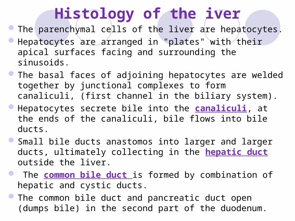
Histology of the iverThe parenchymal cells of the liver are hepatocytes. Hepatocytes are arranged in "plates" with their apical
surfaces facing and surrounding the sinusoids. The basal faces of adjoining hepatocytes are welded
together by junctional complexes to form canaliculi, (first channel in the biliary system).
Hepatocytes secrete bile into the canaliculi, at the ends of the canaliculi, bile flows into bile ducts.
Small bile ducts anastomos into larger and larger ducts, ultimately collecting in the hepatic duct outside the liver.
The common bile duct is formed by combination of hepatic and cystic ducts.
The common bile duct and pancreatic duct open (dumps bile) in the second part of the duodenum.
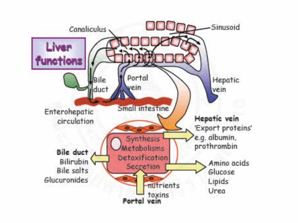
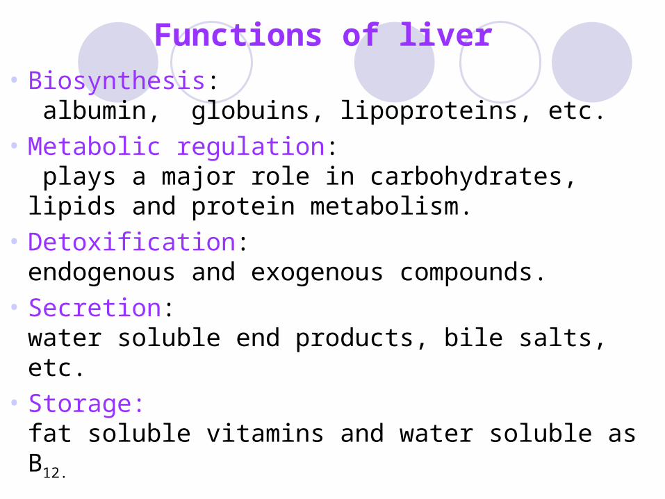
Functions of liver
• Biosynthesis: albumin, globuins, lipoproteins, etc.
• Metabolic regulation: plays a major role in carbohydrates, lipids and protein metabolism.
• Detoxification:endogenous and exogenous compounds.
• Secretion:water soluble end products, bile salts, etc.
• Storage:fat soluble vitamins and water soluble as B12.
Biochemical tests of liver diseases (Liver function tests)
Because the liver is the site for metabolism of carbohydrate, protein, and lipids, as well as for the synthesis of many proteins, the conjugation of bilirubin, and detoxification of drugs and other substances, the liver may be assessed by measurement of:- total and direct bilirubin- total protein and albumin- cholesterol and triglycerides, - urea and ammonia.
Liver function tests assess:synthetic function: serum albuminhepatocyte damage: parenchymal injury:
serum aminotransferases: aspartate transaminase (AST), alanine transaminase (ALT).
Biliary epithelial damage and biliary obstruction:serum alkaline phosphatase (ALP), glutamyltranspeptidase (GT).
Cholestasis: obstruction of the flow of bile; standing bile: serum bilirubin
Clotting (dynamic indicator): Prothrombin Time PT - (INR).
Liver enzymesEnzyme analysis is used to aid in diagnosis
and treatment of disease. In particular, enzymes that are synthesized
within cellular organelles carry out their functions within cells and are released into body fluids when those cells become diseased.
Thus, an increase in enzyme activity when compared to the reference range can indicate pathological changes in certain types of cells and tissues.
Enzyme activity levels in body fluids can reflect: 1- leakage from cells due to cellular injury, or 2- changes in enzyme production rate or actual enzyme
induction due to:- metabolic -proliferation of neoplasms - genetic states.
Damage to tissue can release different types of enzymes based on their location.
Mild inflammation of the liver reversibly increases the permeability of the cell membrane and releases cytoplasmic enzymes:lactate dehydrogenase (LD), alkaline phosphatase (ALP), ALT, and AST.
while cellular death (necrosis) will release mitochrondrial sources of ALT and AST.
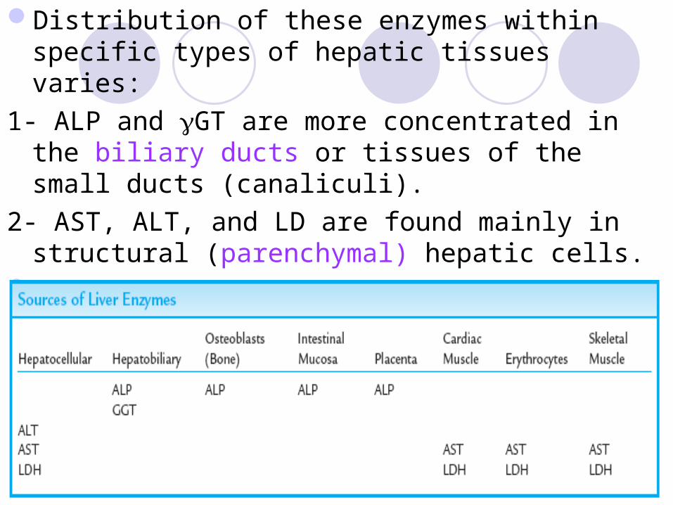
Distribution of these enzymes within specific types of hepatic tissues varies:
1- ALP and GT are more concentrated in the biliary ducts or tissues of the small ducts (canaliculi).
2- AST, ALT, and LD are found mainly in structural (parenchymal) hepatic cells.
Multiple forms of enyzmes (isoenzymes) are distributed in several different tissue types.
Alkaline phosphatase (ALP)It hydrolyses phosphate esters in alkaline solutions.Is found in high conc. in hepatobiliary cells.Inflammation or obstruction of the biliary ducts cause
disruption of these cells resulting in release of ALP into the circulation.
In cholestasis serum ALP levels may be increased 3 to 10 times the normal levels:-extrahepatic biliary obstruction: stone, tumour: pancreatic/biliary.- intrahepatic cholestasis (drug cholestasis, primary biliary cirrhosis).
ALP isoenzymes is also found in all cytoplasmic membranes of all cells of the body, especially in osteoblasts (bone forming cells: cells rebuilding bone), intestinal mucosa, placenta, germ cell and renal tubules.

Differentiation of the isoforms by:electrophoresis, heat stability and chemical inactivation.
It can be used to identify pathological processes in these tissues:
Serum ALP levels may be increased in a high osteoblastic activity up to 10 to 25 times normal .
Most of the ALP that is found in the circulation has its origin in liver or bone tissue cells.
- - glutamyl transpeptidaseIt transfer glutamyl residue from gamma-glutamyl
peptides to amino acids and small peptides.it present in liver, heart, kidneys, pancreas and
prostate.In liver, it is located in epithelial cell lining of biliary
ductules, so it is elevated in all hepatobiliary disorders.
It confirms liver source of ↑ ALP together with 5´-nucleotidase.
A raised ALP conc. in presence of a raised glutamyl transpeptidase implies that ALP is of hepatic orgin.
It is an microsomal enzyme so, it induced by drugs and alcohol.
Elevated level may indicate chronic alcoholism.

AminotransferasesEnzymes that transfer amino group from an amino acid to an α-keto
acid.Assess hepatocyte (parenchymal) damage.Of diagnostic importance are ALT and AST.AST:Present in liver, cardiac & skeletal muscles, kidney, brain,
pancreas, lung, WBC and RBCs.Has both mitochondrial and cytoplasmic forms.Liver cells contain more AST than ALT.AST more sensitive than ALT. Infiltrative disorders resulting in greater increase in
plasma AST than ALT activity. ALT: Is more liver specific than AST. Has cytoplasmic form only. Inflammatory or infective conditions as viral hepatitis, causes greater
increase in ALT than AST activities.

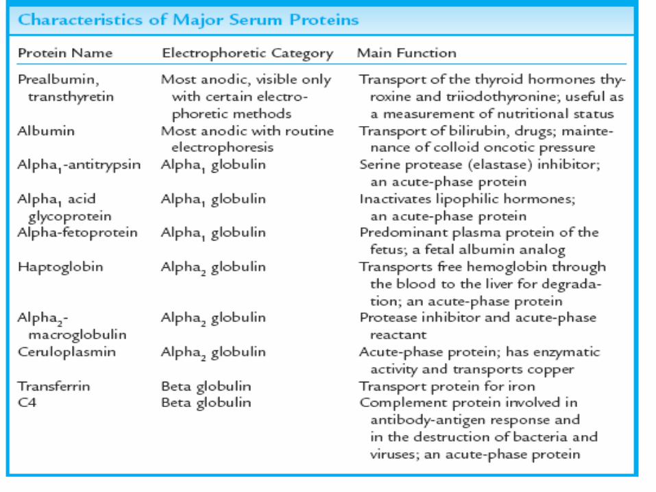
Serum proteinsThe majority of proteins found in plasma (except
immunoglobulins) are synthesized by the liver from amino acids, including serine protease, coagulation factors, prothrombin time, serum albumin.
Metal and vitamin transport proteins e.g. transferrin, ceruloplasmin and vitamin D binding protein
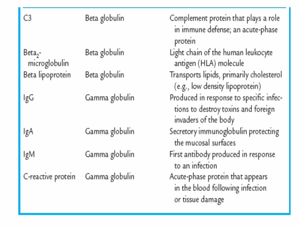
There are two main types of serum proteins, albumin and globulins.
They are grouped into five classes as determined by their electrophoretic separation: albumin, alpha1 globulins, alpha2 globulins, beta globulins, and gamma globulins.
Interpretation of total serum protein levels
Total serum protein levels are affected by: changes in one or more of the individual protein levelschanges in plasma water voume.
Causes of hyperproteinemia: increased serum protein. 1- Hemoconcentration:
decreased plasma water volume, will cause total serum protein levels to be increased.
The usual cause of hemoconcentration is dehydration, which is secondary to:diarrhea, severe vomiting, and water deprivation.
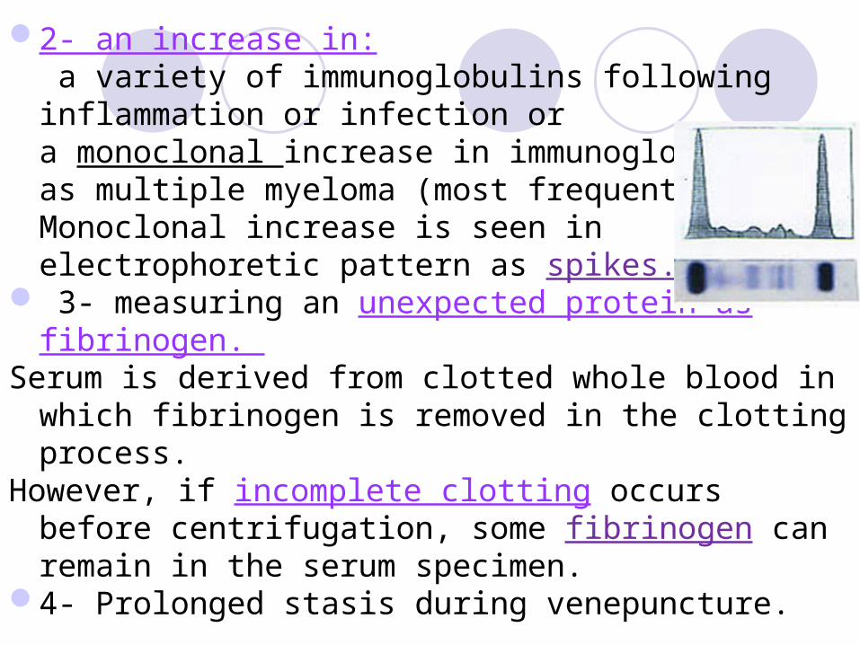
2- an increase in: a variety of immunoglobulins following inflammation or infection or a monoclonal increase in immunoglobulins as multiple myeloma (most frequently IgG). Monoclonal increase is seen in electrophoretic pattern as spikes.
3- measuring an unexpected protein as fibrinogen. Serum is derived from clotted whole blood in which
fibrinogen is removed in the clotting process. However, if incomplete clotting occurs before
centrifugation, some fibrinogen can remain in the serum specimen.
4- Prolonged stasis during venepuncture.

Hypoproteinemia: decreased protein levels in the blood
Causes of hypoproteinemia:- hypoalbuminemia, since albumin is the most abundant single protein.- starvation -nutritional deficiency of essential AA. -renal loss as in nephrotic syndrome.-gastrointestinal loss as in enteropathy
-hepatic failure: the liver is unable to synthesize proteins.
- dilution: blood is taken near the site of intravenous saline infusion.
Interpretation of Serum protein electrophoresis
inflammatory states of the liver: elevated gamma globulin protein fractions, especially immunoglobulin A (IgA) and IgM levels.
In cirrhosis: show that fast-moving gamma globulins often migrate in the beta to gamma region, causing a beta-gamma bridge.
The beta-gamma bridge can be seen as a lack of separation of the beta globulins from the gamma globulins.

Protein electrophoresis pattern, result in cirrhosis

Plasma albuminmakes up roughly half of the total serum proteinsIt assess the synthetic capacity and severity of liver
disease.Maintenance of plasma albumin conc. can be
achieved with only 10% of normal hepatocyte mass.Normal level: 3.5-5.2 g/dl (around 4 g/dL)It is a critical osmotic regulator. Plasma albumin below 2 g/dl:
↑ risk of ascites and oedema. ↓drug binding/transport.
A low plasma albumin below the lower reference limit suggests chronic liver disease, providing low protein intake, renal and GIT loss are excluded.

serum albumin decreases and prothrombin time becomes prolonged with liver failure.
Protein levels also reflect disorders in which:- essential amino acids are not provided by the diet or - proteins are lost by the kidneys or GIT.
A simple way to assess the balance between the patient’s albumin and globulins in serum is to calculate the albumin:globulin, or A/G, ratio. Globulins (G) are calculated as:- albumin (A) subtracted from total serum proteins. - albumin is then divided by globulin.
Interpretation of serum albumin levels Albumin levels are affected by changes in albumin level, and
changes in plasma water volumeCauses of hypoalbuminemia:
1- loss from:GIT: malabsorption or protein-losing enteropathy Renal system: glomerulonephritis or nephrotic syndromeskin: severe burns.
2-increased catabolism as a result of tissue damage and inflammation: neoplasms or autoimmunity.
3- malnutrition and inadequate amino acid intake.
4- declining synthesis in the liver, as associated with cirrhosis or other situations of liver failure.
5- fluid redistribution:albumin distribution between the extra- and intravascular compartments as in ascites may cause hypoalbuminemia
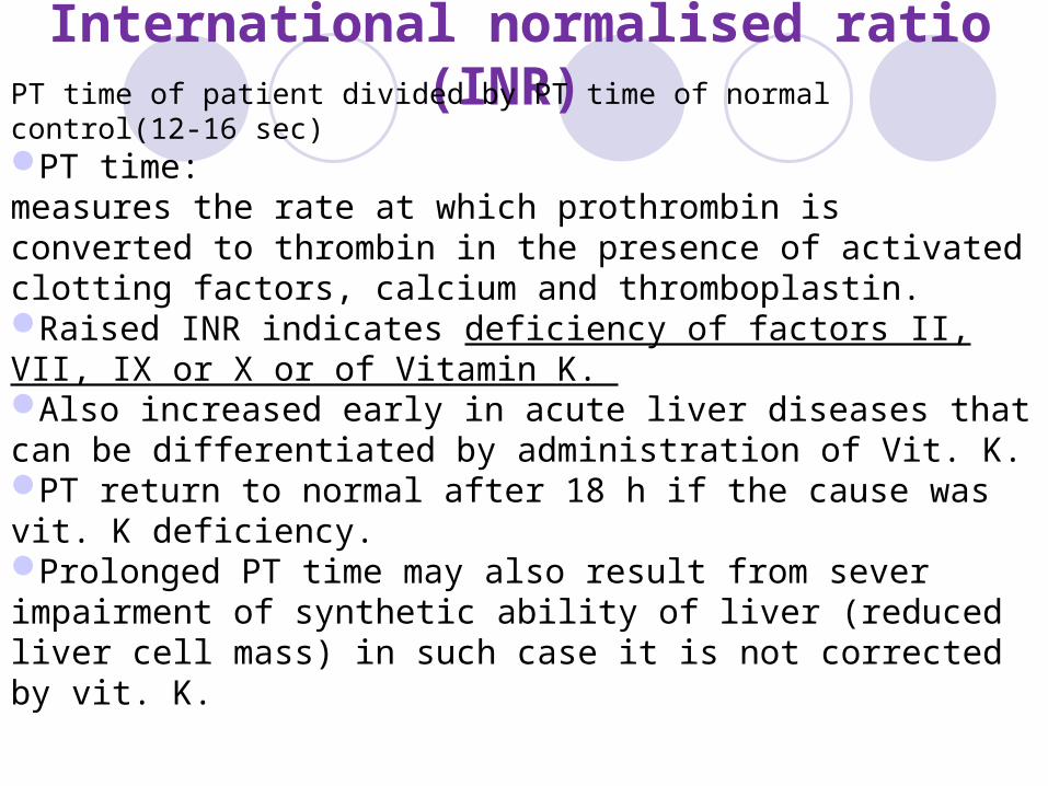
International normalised ratio (INR) PT time of patient divided by PT time of normal control(12-16 sec)PT time:measures the rate at which prothrombin is converted to thrombin in the presence of activated clotting factors, calcium and thromboplastin.Raised INR indicates deficiency of factors II, VII, IX or X or of Vitamin K. Also increased early in acute liver diseases that can be differentiated by administration of Vit. K.PT return to normal after 18 h if the cause was vit. K deficiency.Prolonged PT time may also result from sever impairment of synthetic ability of liver (reduced liver cell mass) in such case it is not corrected by vit. K.

Ammonia toxicityAmmonia is produced in:
- parenchymal liver cells during the deamination of amino acids- Intestinal tract and skeletal muscles,
but the majority of ammonia production is associated with hepatic function.
Ammonia is converted to urea in the liver. Urea is the major nonprotein nitrogen waste product,
excreted by the kidneys. Thus, in normal conditions, plasma ammonia levels are
expected to be low, 20 µg/dL, while urinary urea levels are expected to be higher 20 mg/dL.
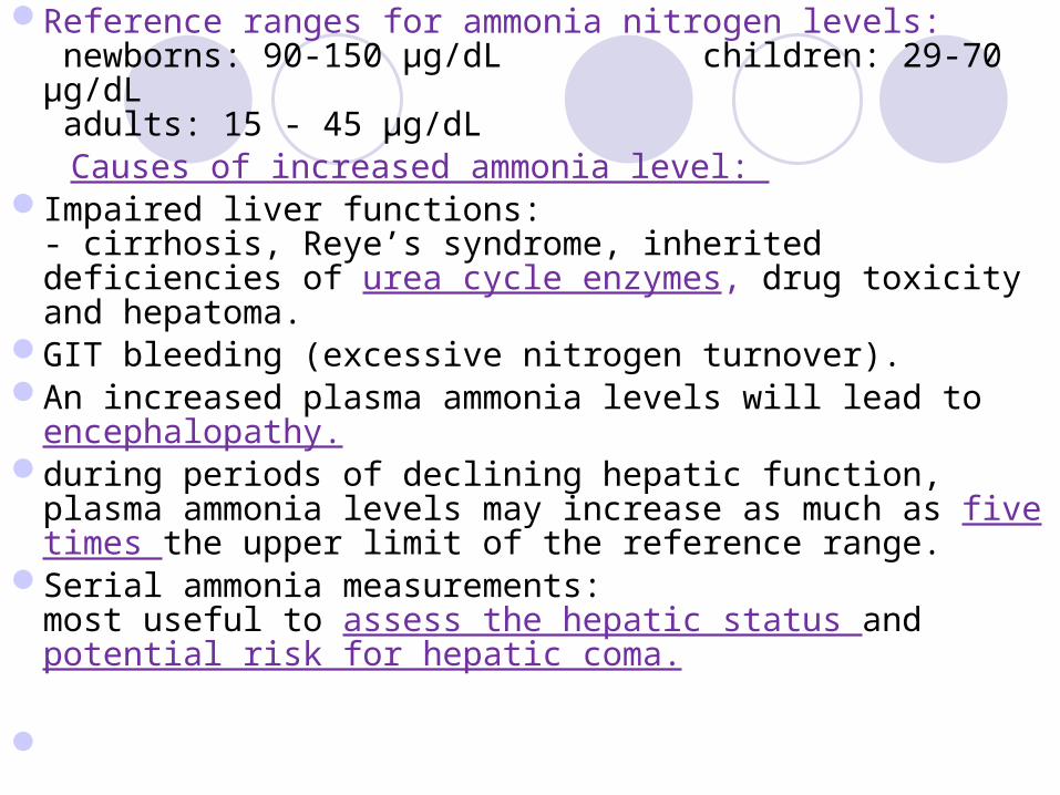
Reference ranges for ammonia nitrogen levels: newborns: 90-150 µg/dL children: 29-70 µg/dL adults: 15 - 45 µg/dL
Causes of increased ammonia level: Impaired liver functions:
- cirrhosis, Reye’s syndrome, inherited deficiencies of urea cycle enzymes, drug toxicity and hepatoma.
GIT bleeding (excessive nitrogen turnover).An increased plasma ammonia levels will lead to
encephalopathy.during periods of declining hepatic function, plasma
ammonia levels may increase as much as five times the upper limit of the reference range.
Serial ammonia measurements:most useful to assess the hepatic status and potential risk for hepatic coma.

Cirrhosisis a common and serious disease of the liver resulting
from chronic inflammation of the liver. The result is replacement of normal liver tissue with
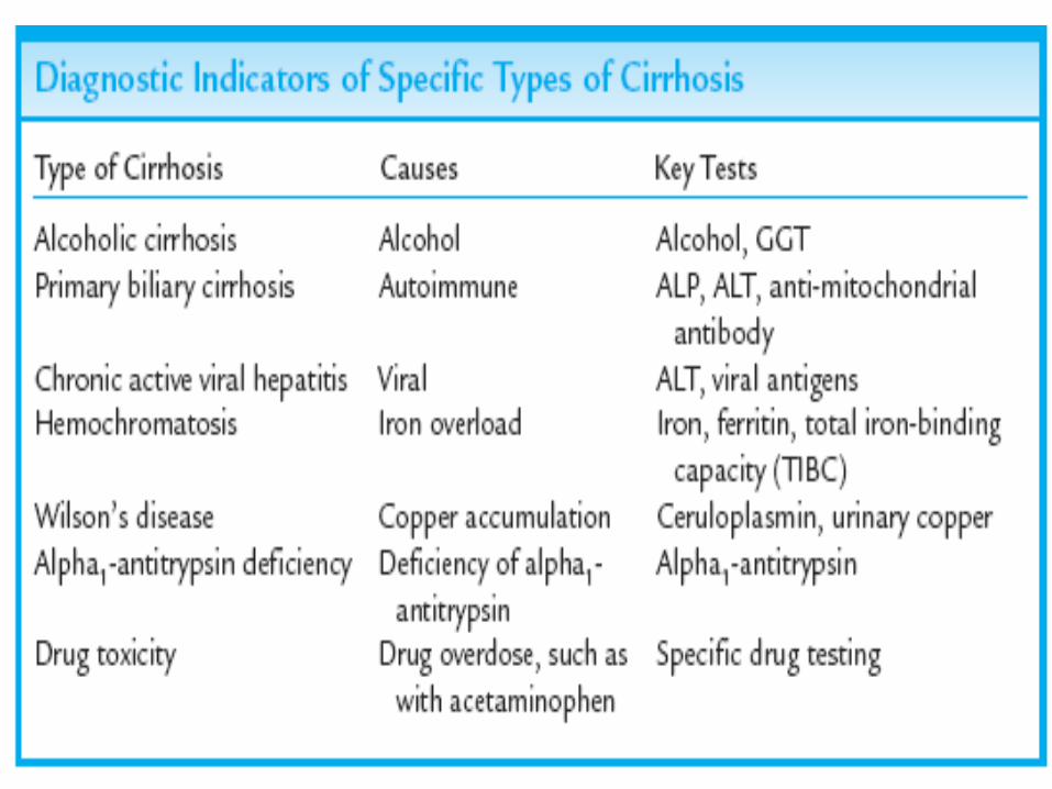
nonfunctional, nodular scar (fibrotic) tissue. It involves the entire liver. There are specific types of cirrhosis based on origin
and classification and based upon types of nodular tissue, such as micronodular, macronodular, and mixed nodular tissue.
Cirrhosis most commonly arises from chronic alcohol abuse, although chronic viral hepatitis can be a major cause and is more common in some parts of the world.
Primary biliary cirrhosis: autoimmun, unusual cause of chronic liver disease
Diagnosis: Immunoglobulin levels and autoantibodies
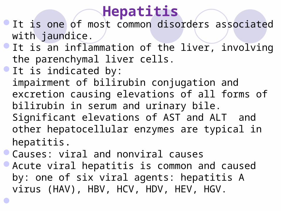
HepatitisIt is one of most common disorders associated with
jaundice. It is an inflammation of the liver, involving the
parenchymal liver cells. It is indicated by:
impairment of bilirubin conjugation and excretion causing elevations of all forms of bilirubin in serum and urinary bile. Significant elevations of AST and ALT and other hepatocellular enzymes are typical in hepatitis.
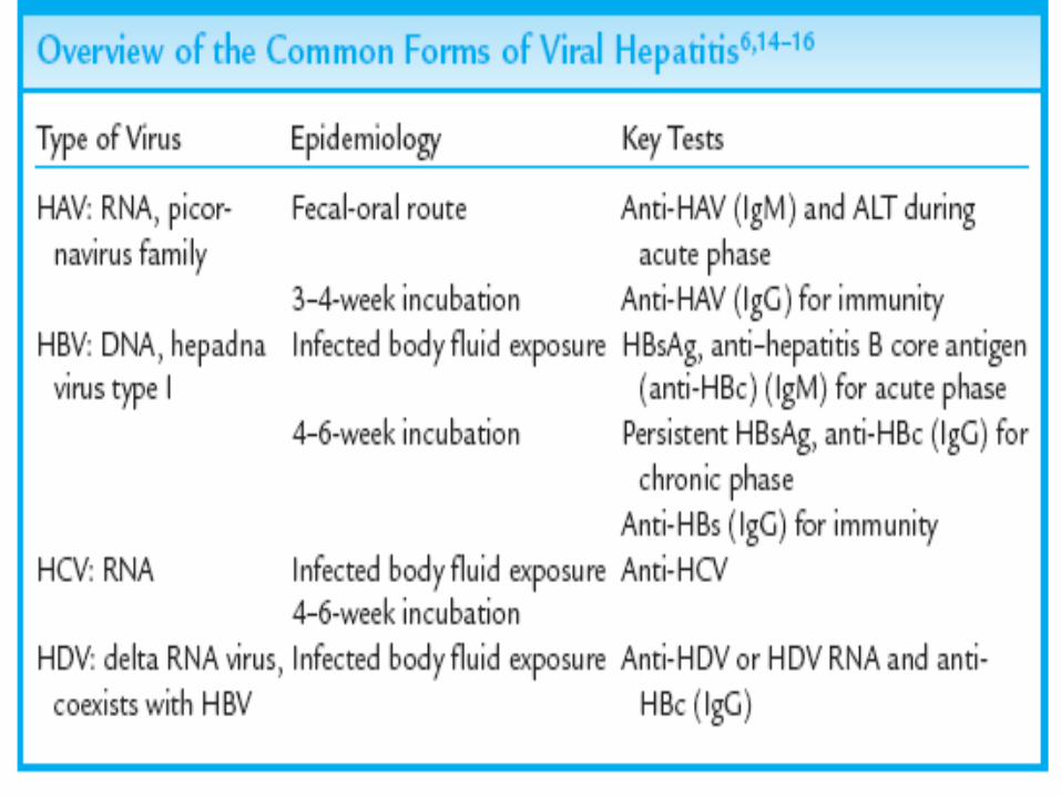
Causes: viral and nonviral causesAcute viral hepatitis is common and caused by: one of
six viral agents: hepatitis A virus (HAV), HBV, HCV, HDV, HEV, HGV.

They produce similar hepatic illnesses but with varied severity.
The outcome of viral hepatitis range from:no symptoms (asymptomatic)acute fatal liver pathology. undetected (subclinical), for years and then present with periods of active inflammation.
Long-term viral hepatitis may cause cirrhosis or hepatic cancer.
Mode of infection: exposure to blood and body fluids from an infected person.
HAV and HEV are food-borne (enterically transmitted). Non viral cause: Hepatotoxicity:
liver inflammation due to chemicals as alcohol or drugs. It is rare in young adults but common in older.
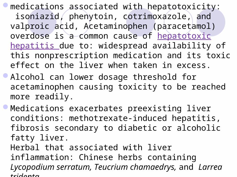
medications associated with hepatotoxicity: isoniazid, phenytoin, cotrimoxazole, and valproic acid, Acetaminophen (paracetamol) overdose is a common cause of hepatotoxic hepatitis due to: widespread availability of this nonprescription medication and its toxic effect on the liver when taken in excess.
Alcohol can lower dosage threshold for acetaminophen causing toxicity to be reached more readily.
Medications exacerbates preexisting liver conditions: methotrexate-induced hepatitis, fibrosis secondary to diabetic or alcoholic fatty liver. Herbal that associated with liver inflammation: Chinese herbs containing Lycopodium serratum, Teucrium chamaedrys, and Larrea tridenta.

Liver Function varies with ageIn elderly: hepatic function declines, indicated by
a slight decline in serum albumin and a slight increase in urea synthesis and detoxification processes.
In pediatric: liver function is less than adult hepatic function.
In neonates, the liver is not functioning at full capacity, due to the immature hepatic circulation, and plasma ammonia levels are usually higher than 60 µg/dL.
Ammonia levels remain slightly higher than adult levels until the patient nears the teen years, when the reference range for ammonia is the same as the adult level.