assessment of dysphotopsia in pseudophakic subjects with ... · iol power, the hoffer q iol formula...
TRANSCRIPT

To cite: Buckhurst PJ,Naroo SA, Davies LN, et al.Assessment of dysphotopsiain pseudophakic subjectswith multifocal intraocularlenses. BMJ Open Ophth2017;1:e000064.doi:10.1136/bmjophth-2016-000064
Received 27 December 2016Revised 04 May 2017Accepted 07 May 2017
1School of HealthProfessions, University ofPlymouth, Plymouth, UK2Ophthalmic ResearchGroup, Life and HealthSciences, Aston University,Birmingham, UK3Midland Eye,Ophthalmology, Solihull, UK
Correspondence to
Professor James SWolffsohn; [email protected]
Assessment of dysphotopsia inpseudophakic subjects withmultifocal intraocular lenses
Phillip J Buckhurst,1 Shehzad A Naroo,2 Leon N Davies,2 Sunil Shah,2,3
Tom Drew,2 James S Wolffsohn2
ABSTRACTAim To better understand the phenomenon ofdysphotopsia in patients implanted with multifocalintraocular lenses (IOLs).Methods Forty-five patients (aged 61.8�8.9 years)implanted bilaterally with Tecnis ZM900 (diffractivemultifocal), Lentis Mplus MF30 (segmented refractivemultifocal) or Softec-1 (monofocal) IOLs (each n=15)4–6 months previously and who had achieved a goodsurgical outcome were examined. Each reported theirdysphotopsia symptoms subjectively, identified itsform (EyeVisPod illustrations), quantified retinalstraylight (C-Quant) and halo perception (Astonhalometer). Retinal straylight and halometry wasrepeated by a second masked clinician to determineinterobserver repeatability.Results Subjective dysphotopsia ratings were able todifferentiate Tecnis ZM900 from Lentis Mplus MF30(p<0.001), but not Lentis Mplus MF30 from groupsimplanted with Softec-1 (p=0.290). Straylight wassimilar between the monofocal and multifocal IOLdesigns (p=0.664). ZM900 IOLs demonstrated auniform increase in dysphotopsia in comparison withthe monofocal IOL (p<0.001) as measured with thehalometer, whereas sectorial refractive multifocal IOLsdemonstrated a localised increase in dysphotopsia overthe inferior visual field. Intraobserver repeatability wasgood for the straylight (intraclass correlationcoefficients (ICC)=0.77) and halometry (ICC=0.89).There was no significant correlation between thesubjective dysphotopsia severity and the straylight(p=0.503) or halometry (p>0.10) quantification orbetween straylight and the halo area (p>0.30).Conclusions Multifocal IOLs induce symptoms ofdysphotopsia. Straylight did not differentiate betweenIOL designs, however halometry identified cleardifferences in light scatter due to the IOL optics.Whereas, subjective rating of overall dysphotopsia arenot strongly associated with straylight or haloperception, the halometry polar diagram reflected thesubjective descriptions of dysphotopsia.
INTRODUCTIONUnderstanding dysphotopsia is vital inachieving a high quality of life in allpatients following multifocal intraocularlens (IOL) implantation. Dysphotopsia is a
disturbance of vision and includes lightphenomena such as haloes, the subjectiveperception of a bright ring around a lightsource; it occurs due to optical non-confor-mities in the optical path such as cataract oroptical boundaries.1 The current literatureshows that implantation of a multifocalrather than a monofocal IOL can lead tounwanted optical phenomenon termeddysphotopsia.1 2 However, the literaturecomparing IOLs is equivocal as to whichdesign features minimise dysphotopsia, dueprincipally to the lack of objective methodsfor assessing dysphotopsia. The majority ofstudies examining dysphotopsia use varioussubjective questioning in the form of verbalinterviews,3 4 bespoke questionnaires,5 avalidated questionnaire6 7 or throughsubject-initiated complaints.8 An alternativemethod is to use graphics depicting visualdemonstrations of different types ofdysphotopsia allowing the subject to indi-cate which is most representative of whatthey perceive.9 10
Instruments designed to measure theeffects of disability glare have also beenused in multifocal IOL studies. Disabilityglare is usually quantified as the reductionin vision from a glare source present within
Key messages
" Dysphotopsia such as glare is commonlyreported following multifocal intraocular lensimplantation. but is generally only quantifiedsubjectively as self-reported severity.
" The results of this study demonstrate that aclinically applicable halometry technique canquantify light scatter and differentiate betweenmultifocal intraocular lens designs.
" This new, rapid to perform, measure could beadopted in clinical practice to identify anindividual patient’s subjective tolerance toobjective glare and to assess the amount ofglare resulting from complex optical refractivecorrection.
Buckhurst PJ, et al. BMJ Open Ophth 2017;1:e000064. doi:10.1136/bmjophth-2016-000064 1
Original article
on 27 March 2019 by guest. P
rotected by copyright.http://bm
jophth.bmj.com
/B
MJ O
pen Ophth: first published as 10.1136/bm
jophth-2016-000064 on 19 June 2017. Dow
nloaded from

the visual field and is due to the spread of light (orstraylight) across the retina.11 The majority of techni-ques used to assess disability glare are composed of acentral optotype chart of varying spatial frequency orcontrast surrounded by a glare source. The intensity ofthe ambient light is changed or a glare source is addedto determine the effect this has on measures of visualacuity or contrast sensitivity. Examples of this approachcan be found in the form of the Brightness Acuity Tester(BAT; Marco, Florida, USA), Mesoptometer II (OculusOptikgera¨te GmbH, Wetzlar-Dutenhofen, Germany)or digital view-in visual testing units, such as the Optec6500 (Stereo Optical Co, Chicago, Illinois). Severalcustom-built glare testing units have also been devel-oped.12 13 However, these testing units do not quantifythe extent of dysphotopsia and the literature showsvariable results. Similar studies involving the C-Quant(Oculus Optikgera¨te GmbH, Wetzlar-Dutenhofen,Germany), an instrument for evaluating the quantity ofocular straylight, have shown similar variability with amarked difference in straylight identified in somestudies,14–16 but not others,2 17–19 and higher straylightwith diffractive designs than refractive and segmenteddesigns.20 The difference in light scatter between amonofocal IOL and diffractive bifocal and trifocalIOLs has been recently reported with the light-distor-tion analyser (HLMP-CW47-RU000, AgilentTechnologies) an experimental device consisting of acentral white light–emitting diode (LED) surroundedby 240 small, white LEDs distributed in 24 meridians15 degrees apart;21–23 a difference was found betweenthem, but this was not correlated with visual acuity.The disparity between reported dysphotopsia and theresults recorded with glare testing units may be due tothe optical properties of multifocal IOLs. Pupil sizedoes not seem to affect straylight measures, but thishas only been assessed in spherical IOLs.24 Dyspho-topsia due to multifocal IOLs may primarily be theresult of a second out of focus image being present onthe retina rather than diffuse straylight over the retinalsurface (scatter affecting a much broader area) asinduced by conditions such as cataract.13 17 Tomeasure the qualitatively described light surroundingretinal blur circle or halo, several instruments oftenreferred to as ‘halometers’ have been created.25–27
These devices measure the size of a photopic scotomacreated by a central glare source.The purpose of this study was to examine the
phenomenon of dysphotopsia in patients implantedwith two multifocal IOL designs (refractive and diffrac-tive) and to compare subjective symptoms to thequantification of straylight and halo size. A standar-dised preoperative measure of halo size compared withsubjectively reported symptoms due to cataract, mighthelp to identify individuals who are more likely tobetter tolerate dysphotopsia effects potentially inducedby multifocal IOLs.
METHODSPatients were recruited from Solihull Hospital andMidland Eye (Solihull UK). The NHS local researchethics committee of Solihull approved the study, andinformed consent was obtained from each patient. Theconsequences and details of the study were explainedto each patient, and the research followed the tenets ofthe declaration of Helsinki.The recruitment inclusion criteria were patients
requiring bilateral cataract surgery, with an expectedpostoperative best corrected distance visual acuity of atleast 0.1 log of the minimum angle of resolution(logMAR), not having any ocular pathology or previoussurgery, with corneal astigmatism less than 1.00 D,aged between 40 and 70 years, deemed suitable by thetreating surgeon and willing to be implanted withmultifocal IOLs. The IOLs, implanted bilaterally,depended on the availability of the IOLs: TecnisZM900, addition power +4.0D at the IOL plane(Abbott Medical Optics, Santa Ana, California); LentisMplus, addition power +3.0D (MF30) at the IOL plane(Oculentis, Berlin, Germany); and Softec-1 monofocalIOL (Lenstec, St Petersburg, Florida).Preoperatively, biometry was conducted with an
IOLMaster (Carl Zeiss Meditec AG, Jena, Germany)running V.5.2 analysis software and a NIDEK OPD-Scan II wavefront aberrometer (Optical Path DifferenceScanning System II; NIDEK Co, Gamagori, Japan) tomeasure axial length and corneal power. To determineIOL power, the Hoffer Q IOL formula was used forshort axial lengths (<22mm) and the SRK/T was usedfor all other axial lengths; emmetropia was the targetin all cases (College of Ophthalmologists guidelines,2010).All operations were performed by one of three
surgeons (SS, TAK and MTB) experienced in fittingmultifocal IOL, under topical or local anaesthetic. A2.85mm clear corneal incision, widening to 3.20mmafter injection, was placed on the steepest corneal axisby all surgeons to reduce residual levels of postopera-tive astigmatism. Phacoemulsification, aspiration andirrigation were performed through a 5.50mm capsu-larhexis using the Millennium Phacoemulsification System(Bausch and Lomb, Rochester, New York, USA.). Thesurgeons used personalised A constants based on theirdocumented surgically inducted refractive error foreach lens type and used the same type of corneal inci-sions. The number of cases performed in each type ofIOL was balanced for each of the three surgeons. AllIOLs were implanted into the capsular bag.Patients were re-examined 4–6 months after surgery
with slit-lamp biomicroscopy. All patients were asked toevaluate their experience of photopic phenomenonpost-IOL implantation using the question ‘Please canyou rate your experience of glare or unusual phenom-enon around lights such as haloes on a scale of 0 to 10;zero indicating no glare experiences and 10 denotingextreme symptoms’.
2 Buckhurst PJ, et al. BMJ Open Ophth 2017;1:e000064. doi:10.1136/bmjophth-2016-000064
Open Access
on 27 March 2019 by guest. P
rotected by copyright.http://bm
jophth.bmj.com
/B
MJ O
pen Ophth: first published as 10.1136/bm
jophth-2016-000064 on 19 June 2017. Dow
nloaded from
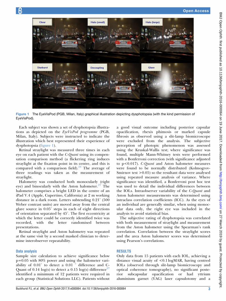
Each subject was shown a set of dysphotopsia illustra-tions as depicted on the EyeVisPod programme (PGB,Milan, Italy). Subjects were instructed to indicate theillustration which best represented their experience ofdysphotopsia (figure 1).Retinal straylight was measured three times in each
eye on each patient with the C-Quant using its compen-sation comparison method (a flickering ring inducesstraylight at the fixation point in its centre, and this iscompared with a comparison field).28 The average ofthree readings was taken as the measurement ofstraylight.Halometry was conducted both monocularly (right
eye) and binocularly with the Aston halometer.27 Thehalometer comprises a bright LED in the centre of aniPad V.4 (Apple, Cupertino, California) at 2 m workingdistance in a dark room. Letters subtending 0.21� (500Weber contrast units) are moved away from the centralglare source in 0.05� steps in each of eight directionsof orientation separated by 45�. The first eccentricity atwhich the letter could be correctly identified twice wasrecorded, with the letter randomised betweenpresentations.Retinal straylight and Aston halometry was repeated
at the same visit by a second masked clinician to deter-mine interobserver repeatability.
Data analysisSample size calculation to achieve significance belowp=0.05 with 80% power and using the halometer vari-ability of 0.01� to detect a 0.01 � difference and C-Quant of 0.14 log(s) to detect a 0.15 log(s) difference27
identified a minimum of 12 patients were required ineach group (Statistical Solutions LLC). Patients without
a good visual outcome including posterior capsularopacification, rhexis phimosis or marked capsulefibrosis as observed using a slit-lamp biomicroscopewere excluded from the analysis. The subjectiveperception of photopic phenomenon was assessedusing the Kruskal-Wallis test; where significance wasfound, multiple Mann-Whitney tests were performedwith a Bonferroni correction (with significance adjustedto p<0.017). C-Quant and Aston halometer measureswere found to be normally distributed (Kolmogrov-Smirnov test >0.05) so the resultant data were analysedusing repeated measure analysis of variance. Wheresignificance was identified, a Bonferroni post hoc testwas used to detail the individual differences betweenthe IOLs. Intraobserver variability of the C-Quant andAston halometer measurements was determined usingintraclass correlation coefficients (ICC). As the eyes ofan individual are generally similar, when using monoc-ular data only, the right eye was included in theanalysis to avoid statistical bias.The subjective rating of dysphotopsia was correlated
with both measurement of straylight and measurementfrom the Aston halometer using the Spearman’s rankcorrelation. Correlation between the straylight scoresand the area Aston halometer scores was determinedusing Pearson’s correlations.
RESULTSOnly data from 15 patients with each IOL, achieving adistance visual acuity of <0.1 logMAR, having centredIOLs (observed through slit-lamp biomicroscopy andoptical coherence tomography), no significant poste-rior subcapsular opacification or had yttriumaluminium garnet (YAG) laser capsulotomy and a
Figure 1 The EyeVisPod (PGB, Milan, Italy) graphical illustration depicting dysphotopsia (with the kind permission of
EyeVisPod).
Buckhurst PJ, et al. BMJ Open Ophth 2017;1:e000064. doi:10.1136/bmjophth-2016-000064 3
Open Access
on 27 March 2019 by guest. P
rotected by copyright.http://bm
jophth.bmj.com
/B
MJ O
pen Ophth: first published as 10.1136/bm
jophth-2016-000064 on 19 June 2017. Dow
nloaded from
residual refractive error of ��0.25D sphereand ��0.50D astigmatism by subjective refraction, wasincluded in the analysis (14 men, 31 women; mean age61.8�8.9 years). The patient demographics were60.7�11.0 years, 4:11 (men:women), in the bilateralTecnis ZM900 group, 62.3�9.0 years, 7:8, in the bilat-eral Lentis Mplus MF30 group and 62.1�6.8 years,3:12 in the bilateral Softec-1 group. The ages weresimilar for each IOL group (F2=0.177, p=0.838),hence no age correction was applied.A significant difference in subjectively rated glare was
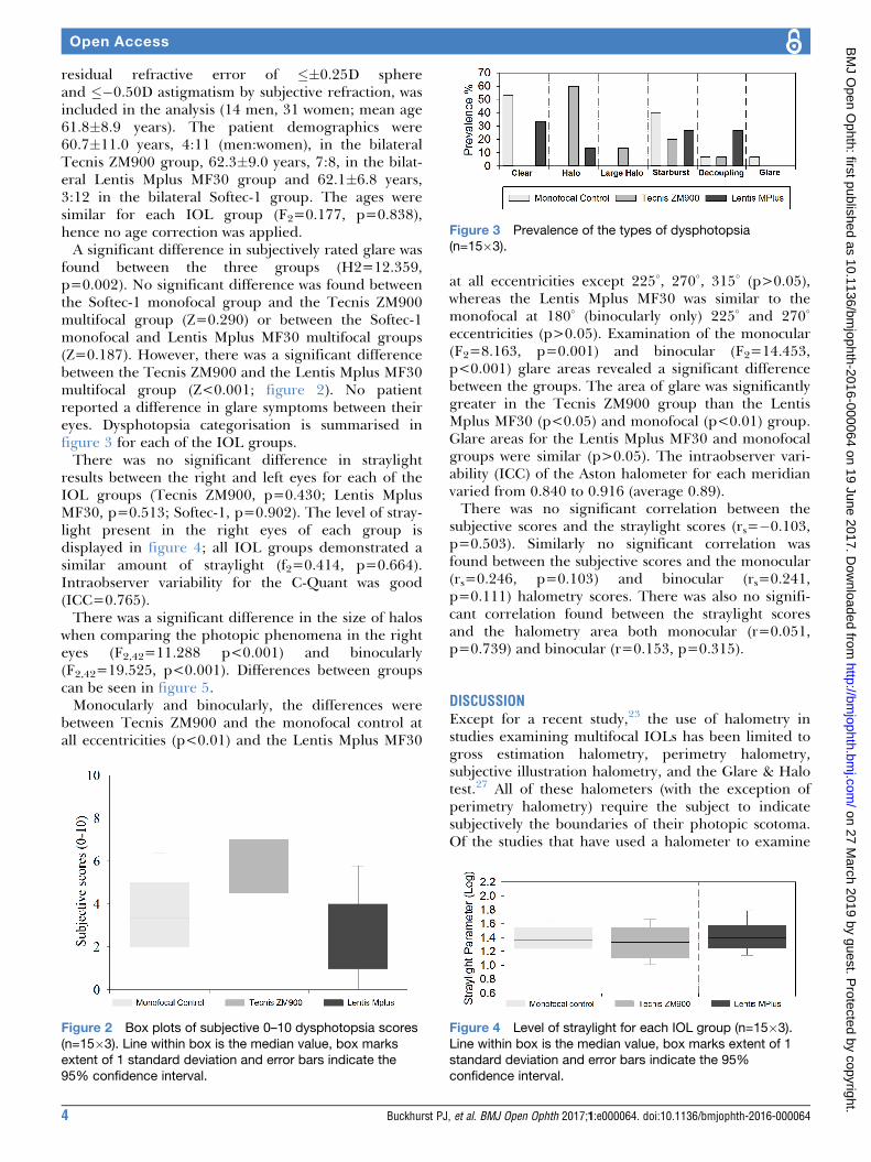
found between the three groups (H2=12.359,p=0.002). No significant difference was found betweenthe Softec-1 monofocal group and the Tecnis ZM900multifocal group (Z=0.290) or between the Softec-1monofocal and Lentis Mplus MF30 multifocal groups(Z=0.187). However, there was a significant differencebetween the Tecnis ZM900 and the Lentis Mplus MF30multifocal group (Z<0.001; figure 2). No patientreported a difference in glare symptoms between theireyes. Dysphotopsia categorisation is summarised infigure 3 for each of the IOL groups.There was no significant difference in straylight
results between the right and left eyes for each of theIOL groups (Tecnis ZM900, p=0.430; Lentis MplusMF30, p=0.513; Softec-1, p=0.902). The level of stray-light present in the right eyes of each group isdisplayed in figure 4; all IOL groups demonstrated asimilar amount of straylight (f2=0.414, p=0.664).Intraobserver variability for the C-Quant was good(ICC=0.765).There was a significant difference in the size of halos
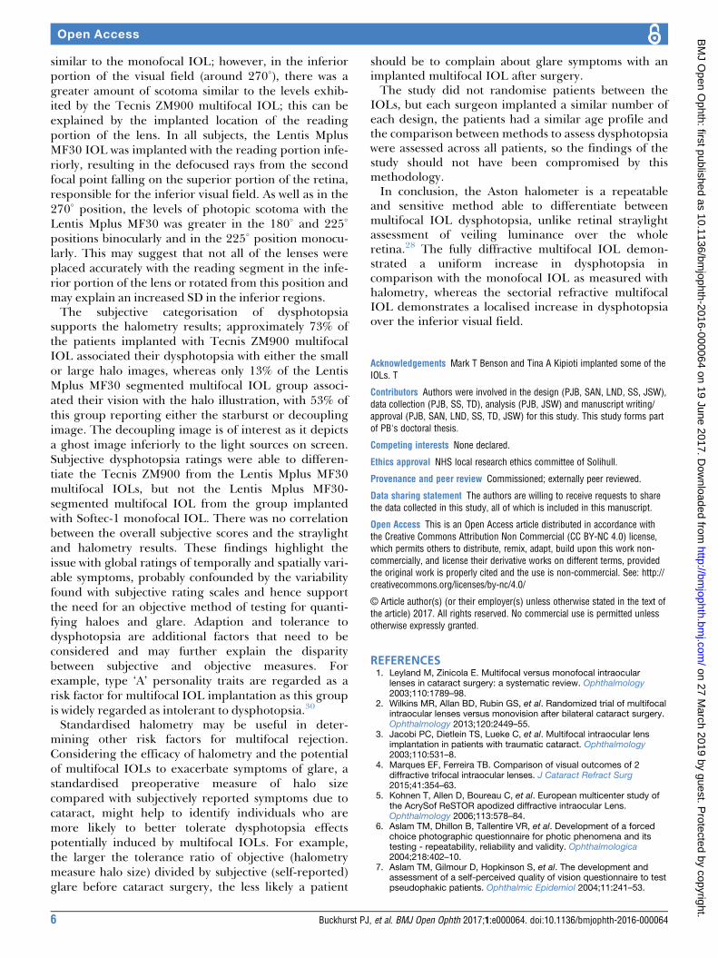
when comparing the photopic phenomena in the righteyes (F2,42=11.288 p<0.001) and binocularly(F2,42=19.525, p<0.001). Differences between groupscan be seen in figure 5.Monocularly and binocularly, the differences were
between Tecnis ZM900 and the monofocal control atall eccentricities (p<0.01) and the Lentis Mplus MF30
at all eccentricities except 225�, 270�, 315� (p>0.05),whereas the Lentis Mplus MF30 was similar to themonofocal at 180� (binocularly only) 225� and 270�
eccentricities (p>0.05). Examination of the monocular(F2=8.163, p=0.001) and binocular (F2=14.453,p<0.001) glare areas revealed a significant differencebetween the groups. The area of glare was significantlygreater in the Tecnis ZM900 group than the LentisMplus MF30 (p<0.05) and monofocal (p<0.01) group.Glare areas for the Lentis Mplus MF30 and monofocalgroups were similar (p>0.05). The intraobserver vari-ability (ICC) of the Aston halometer for each meridianvaried from 0.840 to 0.916 (average 0.89).There was no significant correlation between the
subjective scores and the straylight scores (rs=�0.103,p=0.503). Similarly no significant correlation wasfound between the subjective scores and the monocular(rs=0.246, p=0.103) and binocular (rs=0.241,p=0.111) halometry scores. There was also no signifi-cant correlation found between the straylight scoresand the halometry area both monocular (r=0.051,p=0.739) and binocular (r=0.153, p=0.315).
DISCUSSIONExcept for a recent study,23 the use of halometry instudies examining multifocal IOLs has been limited togross estimation halometry, perimetry halometry,subjective illustration halometry, and the Glare & Halotest.27 All of these halometers (with the exception ofperimetry halometry) require the subject to indicatesubjectively the boundaries of their photopic scotoma.Of the studies that have used a halometer to examine
Figure 2 Box plots of subjective 0–10 dysphotopsia scores
(n=15�3). Line within box is the median value, box marks
extent of 1 standard deviation and error bars indicate the
95% confidence interval.
Figure 3 Prevalence of the types of dysphotopsia
(n=15�3).
Figure 4 Level of straylight for each IOL group (n=15�3).
Line within box is the median value, box marks extent of 1
standard deviation and error bars indicate the 95%
confidence interval.
4 Buckhurst PJ, et al. BMJ Open Ophth 2017;1:e000064. doi:10.1136/bmjophth-2016-000064
Open Access
on 27 March 2019 by guest. P
rotected by copyright.http://bm
jophth.bmj.com
/B
MJ O
pen Ophth: first published as 10.1136/bm
jophth-2016-000064 on 19 June 2017. Dow
nloaded from

dysphotopsia with multifocal IOL subjects, five failed todemonstrate an increase in dysphotopsia after multi-focal IOL implantation.9 23 29 However, a commoncomplaint after multifocal IOL implantation is anincrease in dysphotopsia.1 2
This study shows that the Aston Halometer is arepeatable and sensitive method for the assessment ofdysphotopsia. The C-Quant detected no significantdifferences in the level of straylight regardless of theimplanted IOL; this concurs with some,2 17–19 but notall14–16 previous studies and suggests that measures ofstraylight are not inferred measures of dysphotopsia
caused by multifocal IOLs. Of note, the C-Quant valueswere on the higher side of what has been reported asthe average for this age group,19 but this was the casewith all three IOL groups.At each meridian the Tecnis ZM900 multifocal IOL
displayed a larger amount of photopic scotoma incomparison with the Softec-1 monofocal IOL. This is inkeeping with the known descriptions of haloes caused bymultifocal IOLs1 2 and predominance of ‘halo’ describedwith the EyeVisPod. The Lentis Mplus MF30-segmentedmultifocal IOL did not demonstrate the same appear-ance; superiorly, the photopic scotoma region was
Figure 5 Monocular (top) and binocular (bottom) results of the Aston Halometer for each of the IOL groups. Right polar plot,
left box plots (n=15�3). Line within box is the median value, box marks extent of 1 standard deviation and error bars indicate
the 95% confidence interval.
Buckhurst PJ, et al. BMJ Open Ophth 2017;1:e000064. doi:10.1136/bmjophth-2016-000064 5
Open Access
on 27 March 2019 by guest. P
rotected by copyright.http://bm
jophth.bmj.com
/B
MJ O
pen Ophth: first published as 10.1136/bm
jophth-2016-000064 on 19 June 2017. Dow
nloaded from
similar to the monofocal IOL; however, in the inferiorportion of the visual field (around 270�), there was agreater amount of scotoma similar to the levels exhib-ited by the Tecnis ZM900 multifocal IOL; this can beexplained by the implanted location of the readingportion of the lens. In all subjects, the Lentis MplusMF30 IOL was implanted with the reading portion infe-riorly, resulting in the defocused rays from the secondfocal point falling on the superior portion of the retina,responsible for the inferior visual field. As well as in the270� position, the levels of photopic scotoma with theLentis Mplus MF30 was greater in the 180� and 225�
positions binocularly and in the 225� position monocu-larly. This may suggest that not all of the lenses wereplaced accurately with the reading segment in the infe-rior portion of the lens or rotated from this position andmay explain an increased SD in the inferior regions.The subjective categorisation of dysphotopsia
supports the halometry results; approximately 73% ofthe patients implanted with Tecnis ZM900 multifocalIOL associated their dysphotopsia with either the smallor large halo images, whereas only 13% of the LentisMplus MF30 segmented multifocal IOL group associ-ated their vision with the halo illustration, with 53% ofthis group reporting either the starburst or decouplingimage. The decoupling image is of interest as it depictsa ghost image inferiorly to the light sources on screen.Subjective dysphotopsia ratings were able to differen-tiate the Tecnis ZM900 from the Lentis Mplus MF30multifocal IOLs, but not the Lentis Mplus MF30-segmented multifocal IOL from the group implantedwith Softec-1 monofocal IOL. There was no correlationbetween the overall subjective scores and the straylightand halometry results. These findings highlight theissue with global ratings of temporally and spatially vari-able symptoms, probably confounded by the variabilityfound with subjective rating scales and hence supportthe need for an objective method of testing for quanti-fying haloes and glare. Adaption and tolerance todysphotopsia are additional factors that need to beconsidered and may further explain the disparitybetween subjective and objective measures. Forexample, type ‘A’ personality traits are regarded as arisk factor for multifocal IOL implantation as this groupis widely regarded as intolerant to dysphotopsia.30
Standardised halometry may be useful in deter-mining other risk factors for multifocal rejection.Considering the efficacy of halometry and the potentialof multifocal IOLs to exacerbate symptoms of glare, astandardised preoperative measure of halo sizecompared with subjectively reported symptoms due tocataract, might help to identify individuals who aremore likely to better tolerate dysphotopsia effectspotentially induced by multifocal IOLs. For example,the larger the tolerance ratio of objective (halometrymeasure halo size) divided by subjective (self-reported)glare before cataract surgery, the less likely a patient
should be to complain about glare symptoms with animplanted multifocal IOL after surgery.The study did not randomise patients between the
IOLs, but each surgeon implanted a similar number ofeach design, the patients had a similar age profile andthe comparison between methods to assess dysphotopsiawere assessed across all patients, so the findings of thestudy should not have been compromised by thismethodology.In conclusion, the Aston halometer is a repeatable
and sensitive method able to differentiate betweenmultifocal IOL dysphotopsia, unlike retinal straylightassessment of veiling luminance over the wholeretina.28 The fully diffractive multifocal IOL demon-strated a uniform increase in dysphotopsia incomparison with the monofocal IOL as measured withhalometry, whereas the sectorial refractive multifocalIOL demonstrates a localised increase in dysphotopsiaover the inferior visual field.
Acknowledgements Mark T Benson and Tina A Kipioti implanted some of theIOLs. T
Contributors Authors were involved in the design (PJB, SAN, LND, SS, JSW),data collection (PJB, SS, TD), analysis (PJB, JSW) and manuscript writing/approval (PJB, SAN, LND, SS, TD, JSW) for this study. This study forms partof PB's doctoral thesis.
Competing interests None declared.
Ethics approval NHS local research ethics committee of Solihull.
Provenance and peer review Commissioned; externally peer reviewed.
Data sharing statement The authors are willing to receive requests to sharethe data collected in this study, all of which is included in this manuscript.
Open Access This is an Open Access article distributed in accordance withthe Creative Commons Attribution Non Commercial (CC BY-NC 4.0) license,which permits others to distribute, remix, adapt, build upon this work non-commercially, and license their derivative works on different terms, providedthe original work is properly cited and the use is non-commercial. See: http://creativecommons.org/licenses/by-nc/4.0/
© Article author(s) (or their employer(s) unless otherwise stated in the text ofthe article) 2017. All rights reserved. No commercial use is permitted unlessotherwise expressly granted.
REFERENCES
1. Leyland M, Zinicola E. Multifocal versus monofocal intraocularlenses in cataract surgery: a systematic review. Ophthalmology2003;110:1789–98.
2. Wilkins MR, Allan BD, Rubin GS, et al. Randomized trial of multifocalintraocular lenses versus monovision after bilateral cataract surgery.Ophthalmology 2013;120:2449–55.
3. Jacobi PC, Dietlein TS, Lueke C, et al. Multifocal intraocular lensimplantation in patients with traumatic cataract. Ophthalmology2003;110:531–8.
4. Marques EF, Ferreira TB. Comparison of visual outcomes of 2diffractive trifocal intraocular lenses. J Cataract Refract Surg2015;41:354–63.
5. Kohnen T, Allen D, Boureau C, et al. European multicenter study ofthe AcrySof ReSTOR apodized diffractive intraocular Lens.Ophthalmology 2006;113:578–84.
6. Aslam TM, Dhillon B, Tallentire VR, et al. Development of a forcedchoice photographic questionnaire for photic phenomena and itstesting - repeatability, reliability and validity. Ophthalmologica2004;218:402–10.
7. Aslam TM, Gilmour D, Hopkinson S, et al. The development andassessment of a self-perceived quality of vision questionnaire to testpseudophakic patients. Ophthalmic Epidemiol 2004;11:241–53.
6 Buckhurst PJ, et al. BMJ Open Ophth 2017;1:e000064. doi:10.1136/bmjophth-2016-000064
Open Access
on 27 March 2019 by guest. P
rotected by copyright.http://bm
jophth.bmj.com
/B
MJ O
pen Ophth: first published as 10.1136/bm
jophth-2016-000064 on 19 June 2017. Dow
nloaded from

8. Shoji N, Shimizu K. Clinical evaluation of a 5.5 mm three-zonerefractive multifocal intraocular lens. J Cataract Refract Surg1996;22:1097–101.
9. Hunkeler JD, Coffman TM, Paugh J, et al. Characterization of visualphenomena with the Array multifocal intraocular Lens. J CataractRefract Surg 2002;28:1195–204.
10. McAlinden C, Pesudovs K, Moore JE. The development of aninstrument to measure quality of vision: the Quality of Vision (QoV)questionnaire. Invest Ophthalmol Vis Sci 2010;51:5537–45.
11. Vos JJ. Disability glare state of the art report. CIE-Journal1984;3:39–53.
12. Bailey IL, Bullimore MA. A new test for the evaluation of disabilityglare. Optom Vis Sci 1991;68:911–7.
13. Epitropoulos AT, Fram NR, Masket S, et al. Evaluation of a newcontrolled point source LED glare tester for disability glare detectionin participants with and without cataracts. J Refract Surg2015;31:196–201.
14. de Vries NE, Franssen L, Webers CA, et al. Intraocular straylight afterimplantation of the multifocal AcrySof ReSTOR SA60D3 diffractiveintraocular lens. J Cataract Refract Surg 2008;34:957–62.
15. Peng C, Zhao J, Ma L, et al. Optical performance after bilateralimplantation of apodized aspheric diffractive multifocal intraocularlenses with +3.00-D addition power. Acta Ophthalmol2012;90:e586–e593.
16. Lapid-Gortzak R, Labuz G, van der Meulen IJ, et al. Straylightmeasurements in two different apodized diffractive multifocalintraocular lenses. J Refract Surg 2015;31:746–51.
17. Hofmann T, Zuberbuhler B, Cervino A, et al. Retinal straylight andcomplaint scores 18 months after implantation of the AcrySofmonofocal and ReSTOR diffractive intraocular lenses. J Refract Surg2009;25:485–92.
18. Cervino A, Hosking SL, Mont�es-Mic�o R, et al. Retinal straylight inpatients with monofocal and multifocal intraocular lenses. J CataractRefract Surg 2008;34:441–6.
19. Łabuz G, Reus NJ, van den Berg TJ, et al. Comparison of ocularstraylight after implantation of multifocal intraocular lenses. JCataract Refract Surg 2016;42:618–25.
20. Ehmer A, Rabsilber TM, Mannsfeld A, et al. Influence of differentmultifocal intraocular lens concepts on retinal stray light parameters.
Ophthalmologe 2011;108:952–6.21. Ferreira-Neves H, Macedo-de-Araujo R, Rico-Del-Viejo L, et al.
Validation of a method to measure light distortion surrounding a
source of glare. J Biomed Opt 2015;20:075002.22. Guti�errez R, Jim�enez JR, Villa C, et al. Simple device for quantifying
the influence of halos after lasik surgery. J Biomed Opt
2003;8:663–7.23. Brito P, Salgado-Borges J, Neves H, et al. Light-distortion analysis
as a possible indicator of visual quality after refractive lens exchange
with diffractive multifocal intraocular lenses. J Cataract Refract Surg
2015;41:613–22.24. Guo YW, Li J, Song H, et al. Comparison of the retinal straylight in
pseudophakic eyes with PMMA, hydrophobic acrylic, and
hydrophilic acrylic spherical intraocular lens. J Ophthalmol
2014;2014:1–6.25. Babizhayev MA, Minasyan H, Richer SP. Cataract halos: a driving
hazard in aging populations. implication of the Halometer DG
test for assessment of intraocular light scatter. Appl Ergon
2009;40:545–53.26. Meikies D, van der Mooren M, Terwee T, et al. Rostock glare
perimeter: a distinctive method for quantification of glare. Optom Vis
Sci 2013;90:1143–8.27. Buckhurst PJ, Naroo SA, Davies LN, et al. Tablet app halometer for
the assessment of dysphotopsia. J Cataract Refract Surg
2015;41:2424–9.28. Guber I, Bachmann LM, Guber J, et al. Reproducibility of straylight
measurement by C-Quant for assessment of retinal straylight using
the compensation comparison method. Graefes Arch Clin Exp
Ophthalmol 2011;249:1367–71.29. Łabuz G, Vargas-Martın F, van den Berg TJ, et al. Method for in vitro
assessment of straylight from intraocular lenses. Biomed Opt
Express 2015;6:4457–64.30. Koch DD, Wang L. Custom optimization of intraocular Lens
asphericity. Trans Am Ophthalmol Soc 2007;105:36–41.
Buckhurst PJ, et al. BMJ Open Ophth 2017;1:e000064. doi:10.1136/bmjophth-2016-000064 7
Open Access
on 27 March 2019 by guest. P
rotected by copyright.http://bm
jophth.bmj.com
/B
MJ O
pen Ophth: first published as 10.1136/bm
jophth-2016-000064 on 19 June 2017. Dow
nloaded from