assessment of biomimetic modification on a novel low ... · pdf filecitation: paredes v,...
TRANSCRIPT

Research Article Open Access
Paredes et al., J Material Sci Eng 2015, 4:5 DOI: 10.4172/2169-0022.1000193
Research Article Open Access
Volume 4 • Issue 5 • 1000193J Material Sci EngISSN: 2169-0022 JME, an open access journal
Keywords: Dental implant; Biomaterial; Ti alloy; Biomimeticmodification; Surface activation; Silanization; Short peptide sequences
IntroductionThe implants have a purpose to replace a part or a function of
the body in a manner that will be safe and physiologically acceptable. However, the material to replace teeth, bones or joints are complex, because the selection of materials depend of different ideals factors, such as biocompatibility, excellent corrosion resistance, high mechanical strength, good wear resistance and low elastic modulus [1]. Inequality of Young´s moduli has been identified as one of the main causes for implant loosening followed by stress shielding [2].
Currently, the material most commonly used as implants to bone regeneration, have elastic modulus between 220 GPa (CoCr) and 110 GPa (Ti), however the cortical bone has an elastic modulus of 7-25 GPa [3,4], then, there is a tendency to show a stress shielding effect [5-7]. Therefore, low modulus alloy are nowadays desired, because the modulus required for implant must be more similar to that of natural bone, which will inhibit bone atrophy and induce good bone remodeling as stated above [4-6].
The number of dental implant has increased in the past decades, reaching about one million per year. Their success are related to the osseointegration rate, which depends of their chemical composition, geometry and topography [8,9]. Moreover, the dental implants interact with physiological fluids, containing biologically ion species able to affect the surface, the environment and the biological system [10].
Between the dental metallic materials for implants, the Ti alloys have favorable specific strength (strength to density ratio) and lowest elasticity´s modulus [11]. For that reason the Ti-6Al-4V and NiTi were of the first Ti biomaterials used in implantable devices. However, some studies have shown that either Al or V ions are able to cause long-term health problems, such as: reaction with tissue in animals, neurological disorders, Alzheimer´s disease, and also this alloy have elastic modulus higher than bone [1]. Also NiTi (Nitinol) has been used as implant due
Assessment of Biomimetic Modification on a Novel Low Elastic Modulus Ti–Nb–Hf Alloy for Dental ImplantsParedes V*1,3, Salvagni E2 and Manero JM2,3
1Ecci University, Research Mechanical Design and Materials Group (GIDMyM), Bogotá, Colombia 2Biomaterials, Biomechanics and Tissue Engineering Group (BIBITE), Department of Materials Science and Metallurgy, Technical University of Catalonia (UPC), Barcelona, Spain 3Nanoengineering Research Centre (CRnE), Barcelona, Spain
AbstractA simple review of research literature and Research and Development trends (R&D) in the field of dental implants
shows an exponential increase in their quantity. The impressive growth in the contributions of nanoscience, in general and nanotechnology/nanomaterials applied to dental care, in particular, also shows the priorities investments and substantive effort of the global research community in this area. Nevertheless, there are still many research challenges and huge opportunities to optimize dental implants. The essential purpose of this study is to assess biomimetic modifications on a novel low elastic modulus TiHfNb Alloy Ni-free. For the mimic surface samples were initially bioactive with Oxygen Plasma (OP) or Piranha etching (Pi). Later three different silanes were used (CPTES, GPTES or APTES+CL) and finally RGD or mixtures 1:1 of RGD:FHRRIKA or RGD:PHSRN were immobilized on the surface. To evaluate the biomimetic modification, cells adhesion studies were performed. For the chemical characterization different techniques were used, including, inter alia: X-ray Photoelectron Spectroscopy (XPS), Zinc-substitution complex technique (ZC), and immunofluorescence. Experimental results showed that the best activation method was the OP, and the biomimetic, modification is a well-grounded research strategy to improve the cell adhesion.
*Corresponding author: Paredes V, Biomaterials, Biomechanics and TissueEngineering Group (BIBITE), Department of Materials Science and Metallurgy, Technical University of Catalonia (UPC), Barcelona, Spain, Tel: 34934010717; E-mail: [email protected]
Received October 07, 2015; Accepted October 20, 2015; Published October 29, 2015
Citation: Paredes V, Salvagni E, Manero JM (2015) Assessment of Biomimetic Modification on a Novel Low Elastic Modulus Ti–Nb–Hf Alloy for Dental Implants. J Material Sci Eng 4: 193. doi:10.4172/2169-0022.1000193
Copyright: © 2015 Paredes V, et al. This is an open-access article distributed under the terms of the Creative Commons Attribution License, which permits unrestricted use, distribution, and reproduction in any medium, provided the original author and source are credited.
a shape memory effect and superelasticity [12], though Ni ions released produce adverse reactions (allergy) [13].
Thereby, some metallic materials could be considered well tolerated, however, these materials are not inert, and consequently Ni-free, Al-free and V-free alloys are part of the current trend to develop alloys able to overcome the incompatibility of providing a biological balance [11]. Different authors have reported that Ti alloy containing elements no toxic as Nb, Zr, Ta, Mo, Hf, Fe, Sn, Zr; and β-Ti phase, improve mechanical properties, low elastic modulus, biocorrosion resistance, also they are biocompatible and have not allergic problems [1, 4-7].
Once selected the material, another important parameter to considerer is the biological response, Thus, the future dental implant should be developed understanding the interaction metal-tissues through the evaluation of the relation between implant, proteins and cells.
How to improve the interaction between material and cells? The stabilization of the bone–implant interface can be achieved by the adhesion of a series of proteins, because inside the body we have different sequences of peptide called proteins. For this purpose we have to know what kind of protein we will use. Osteoblast adhesion takes place by different mechanisms: the most investigated implies the interaction with RGD sequences (Arg-Gly-Asp), because this can
Journal of Material Sciences & Engineering Jo
urna
l of M
aterial Sciences &Engineering
ISSN: 2169-0022
Citation: Paredes V, Salvagni E, Manero JM (2015) Assessment of Biomimetic Modification on a Novel Low Elastic Modulus Ti–Nb–Hf Alloy for Dental Implants. J Material Sci Eng 4: 193. doi:10.4172/2169-0022.1000193
Page 2 of 6
Volume 4 • Issue 5 • 1000193J Material Sci EngISSN: 2169-0022 JME, an open access journal
be recognized in a wide range of cells. Secchi indicate that RGD on Ti stimulates cell differentiation and confers to the surface a resistance property to apoptosis (cell death) [14].
Recent studies have concluded that a mixture of RGD with other sequences improves the cell response [15-18]. Rezania worked with RGD and FHRRIKA (Phe-His-Arg-Arg-Ile-Lys-Ala). Their results showed that by using short peptide sequences with heparin-adhesive motifs is possible to improve the cell interaction and affect the long-term formation of mineralized ECM [19]. Also different author had indicated that FHRRIKA add specific advantage for substrates and promote the osteoblast adhesion [15,20-23].
Researchers such as Benoit, uncovered that PHSRN improve the adhesion and proliferation of osteoblast, and also contribute with the production of phosphaphate alkaline, which is an indicator of osteoblastic function [24]. Other studies indicate that mixtures RGD and PHRSN mimic the microenvironment of the cells. Consequently this information provides opportunities for developing biomaterials [15].
Therefore in this study a Ti (TiHfNb) Ni free alloy was used. This shows good resistance and presents an elastic modulus of 74 GPa [12,13,25-29]. Although, excellent qualities of this material lacks bioactivity. To overcome this drawback different biomolecules were incorporate. Namely CGGRGD (Cys-Gly-Gly-Arg-Gly-Asp), and mixtures 1:1 CGGRGD: CGGFHRRIKA (Cys-Gly-Gly-Phe-His-Arg-Arg-Ile-Lys-Ala) and CGGRGD: CGGPHSRN (Cys-Gly-Gly-Pro-His-Ser-Arg-Asn) [30,31]. The Cys sequence was used due to the fact that this has a Sulphur atom in its lateral chain that this element could be detected by X-ray Photoelectron Spectroscopy (XPS) and is highly reactive. Finally, cells adhesion studies on modified surfaces were made to evaluate the biomimetic modification.
Experimental MethodsMaterials
The Ti alloy (TiHfNb) was fabricated by vacuum arc melting into UPC laboratory using high purity metals 99,99% (Table 1). Thus the buttons were homogenized at 1100°C for to 12 h, treated for 1.5 h at the same temperature, and finally quenched in ice water.
The buttons were cut of 8mm × 5 mm and 2 mm of thickness. The samples were polished using SiC abrasive papers (600 to 1200) and finally were used napped polishing pad with colloidal alumina (1 and 0.05 µm). The samples were degreased by ultrasonication with acetone, water and ethanol, and dried with compressed air.
Biomimetic modification
That objective can be realized through the biomimetic modification of substrate. When exposed to the air the metals are oxidized. Thus they have an oxide layer with impurities that are not convenient. Therefore it is necessary to do the activation, in order to obtain the cleaning and activation of the surface. Once the surface is clean and active it is necessary to make the silanization, followed of the adhesion of peptide. In order to know which kind of sequence is the best and at the same time to show that biomimetic process produces a good cell response studies of cell adhesion were make.
Activation process: Smooth Ti alloys samples were bioactivated
with i) Oxygen Plasma (OP) or ii) Piranha (Pi), to remove the impurities of carbon and to generate highly active surface through the addition of OH- groups [32,33]. Based on the highest concentration of the OH-
groups and the removal of carbon the best treatment was selected.
Silanization process: Thus for the silanization were used three different silanes: i) CPTES (3-chloropropyltriethoxysilane), ii) GPTES (3-Glycidyloxypropyltrimethoxysilane) or iii) APTES (3-aminopropyltriethoxysilane). In this last case was necessary to use a crosslinker called Maleimide (3-(maleimido)-propionic acid N-hydroxysuccinimide ester) to get the union with the amino functional group. (APTES+CL) [34-38]. For the silanization the samples were immersed in pentane solution and organosiles for 1 h with 1 min sonication every 20 min. After that the samples were washed with water, ethanol and acetones, dried with compressed air and stored in a desiccator.
Depending on the two higher atomic percentage of Si the two silanes were selected for the next step of biomolecule immobilization.
Immobilization of peptide sequences: The best silanization samples were coated with different peptides as CGGRGDS, CGGPHSRN, CGGFHRRIKA, or mixtures of them (1:1): CGGRGDS + CGGPHSRN or CGGRGDS + CGGFHRRIKA. Non-treated Ti alloys samples were used as control [15,19,22,38-41]. For the immobilization of the peptide sequences, the silanization samples were incubated overnight in phosphate buffer solution (PBS) containing one peptide or one mixture. Finally samples were extracted from PBS solution and stored in a desiccator.
Cell response
For the cell adhesion assays, were used a mesenchymal cell of rat to induce the osteoblast response. Prior studies of viability show that the optimal time adhesion is to 6 h in serum free media. For the seeding of cells samples were treated with 1% bovine serum albumin solution (PBS-BSA (1%)) for 30 min in order to inhibit the unspecific protein binding, Thus the sterilization was realized immersing samples in ethanol for 10 min and finally samples were washed with sterile PBS. The best cell response will depend on cell number.
Surface characterization
Surfaces were characterized by X-ray Photoelectron Spectroscopy (XPS), and Zinc-complex Substitution Technique (ZT). The in vitro effect of modified surfaces was evaluated by immunofluorescence. Mann-Whitney and Kruskal-Wallis statistical tests were used (p-value<0.05).
X-ray Photoelectron Spectroscopy (XPS): The ESCA 5701 (Physical Electronics, PHI 10) with monochromatic Mg Kα X-ray source (300.0 W, E = 1,253.6 eV) was used for the measurements. Survey spectra were collected with pass energy of 190 eV. High resolution (HR) spectra were collected with pass energy of 24.95 eV. The binding energies were corrected based on C 1s peak at 284.8 Ev [42,43]. Multipak spectrum data analysis software was used to do curve-fitting of the spectra and to calculate component composition from the peak areas. Such analyses are accurate to ±10 % [44], also the binding energies reported have an error in the range of ±0.1 eV [43].
Zinc-complex Substitution Technique (ZT): This is an experimental technique reported in the literature [32,33], which consists of immersing the samples in a mixture 2:1 of ammonium chloride solution (4.0M) and zinc chloride solution (0.4M). During immersion, one zinc complex is formed with two hydroxyl groups,
Element Ti Hf Nb Zr% weight 59.10 16.19 23.59 1.01
Table 1: Chemical composition for Ti alloy by manufacturer XRD.
Citation: Paredes V, Salvagni E, Manero JM (2015) Assessment of Biomimetic Modification on a Novel Low Elastic Modulus Ti–Nb–Hf Alloy for Dental Implants. J Material Sci Eng 4: 193. doi:10.4172/2169-0022.1000193
Page 3 of 6
Volume 4 • Issue 5 • 1000193J Material Sci EngISSN: 2169-0022 JME, an open access journal
after the samples are immersed in nitric acid to release zinc ions into the nitric acid solution. Thus the zinc ions concentrations (CZn (in ppb)) was determined by inductively coupled plasma-atomic emission spectrometer (ICP-AES). Finally the concentration of active hydroxyl groups COH (number/nm2) was calculated using the equation:
( )6CZn 10 V A 2COH
M S
−× × × ×=
×
V: volume of nitric acid,
S: surface area of sample (nm2).
A: Avogadro’s number
M: molecular weight of zinc.
Cell response: For each of the adhesion assays 6×10³ cells were seed per well of 0.95 cm2. The cell adhesion process was characterized by i) immunofluorescence through the staining of cell nuclei with DAPI (4,6-diamino-2fenol- dihydrochloride) the quantification was performed through Image J software. For this characterization was necessary to do the fixation and dehydration of cells.
Experimental Results and DiscussionActivation process
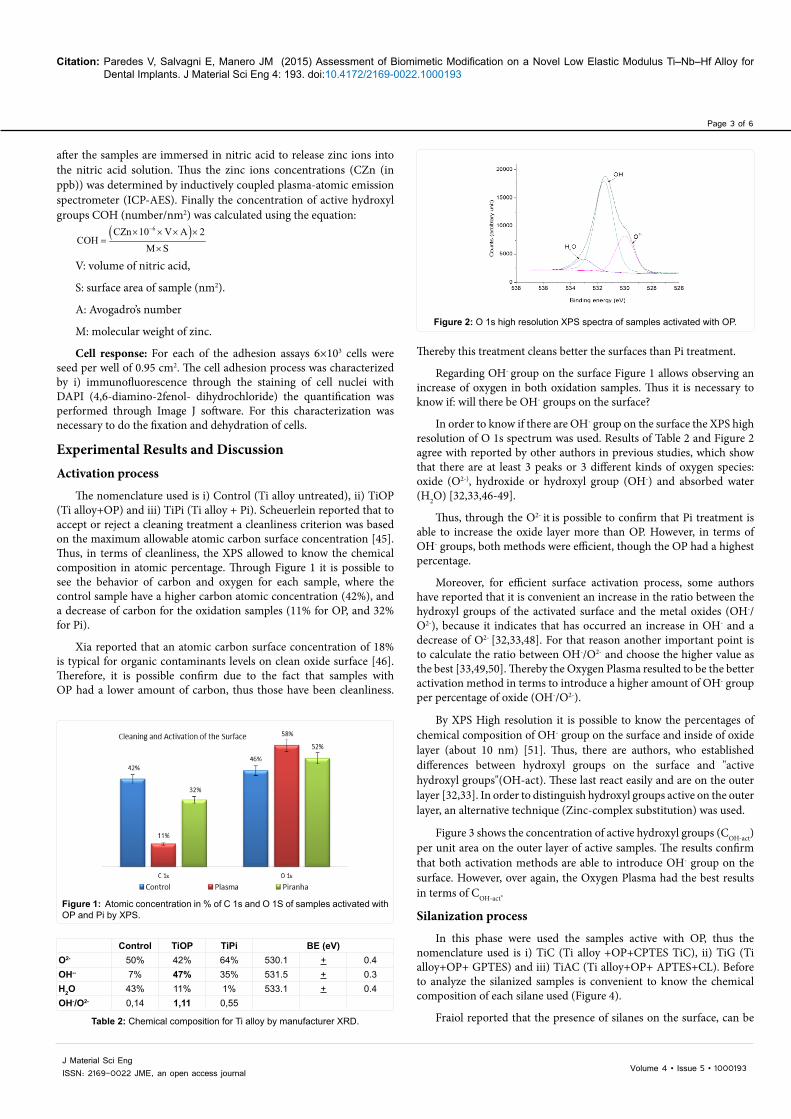
The nomenclature used is i) Control (Ti alloy untreated), ii) TiOP (Ti alloy+OP) and iii) TiPi (Ti alloy + Pi). Scheuerlein reported that to accept or reject a cleaning treatment a cleanliness criterion was based on the maximum allowable atomic carbon surface concentration [45]. Thus, in terms of cleanliness, the XPS allowed to know the chemical composition in atomic percentage. Through Figure 1 it is possible to see the behavior of carbon and oxygen for each sample, where the control sample have a higher carbon atomic concentration (42%), and a decrease of carbon for the oxidation samples (11% for OP, and 32% for Pi).
Xia reported that an atomic carbon surface concentration of 18% is typical for organic contaminants levels on clean oxide surface [46]. Therefore, it is possible confirm due to the fact that samples with OP had a lower amount of carbon, thus those have been cleanliness.
Thereby this treatment cleans better the surfaces than Pi treatment.
Regarding OH- group on the surface Figure 1 allows observing an increase of oxygen in both oxidation samples. Thus it is necessary to know if: will there be OH- groups on the surface?
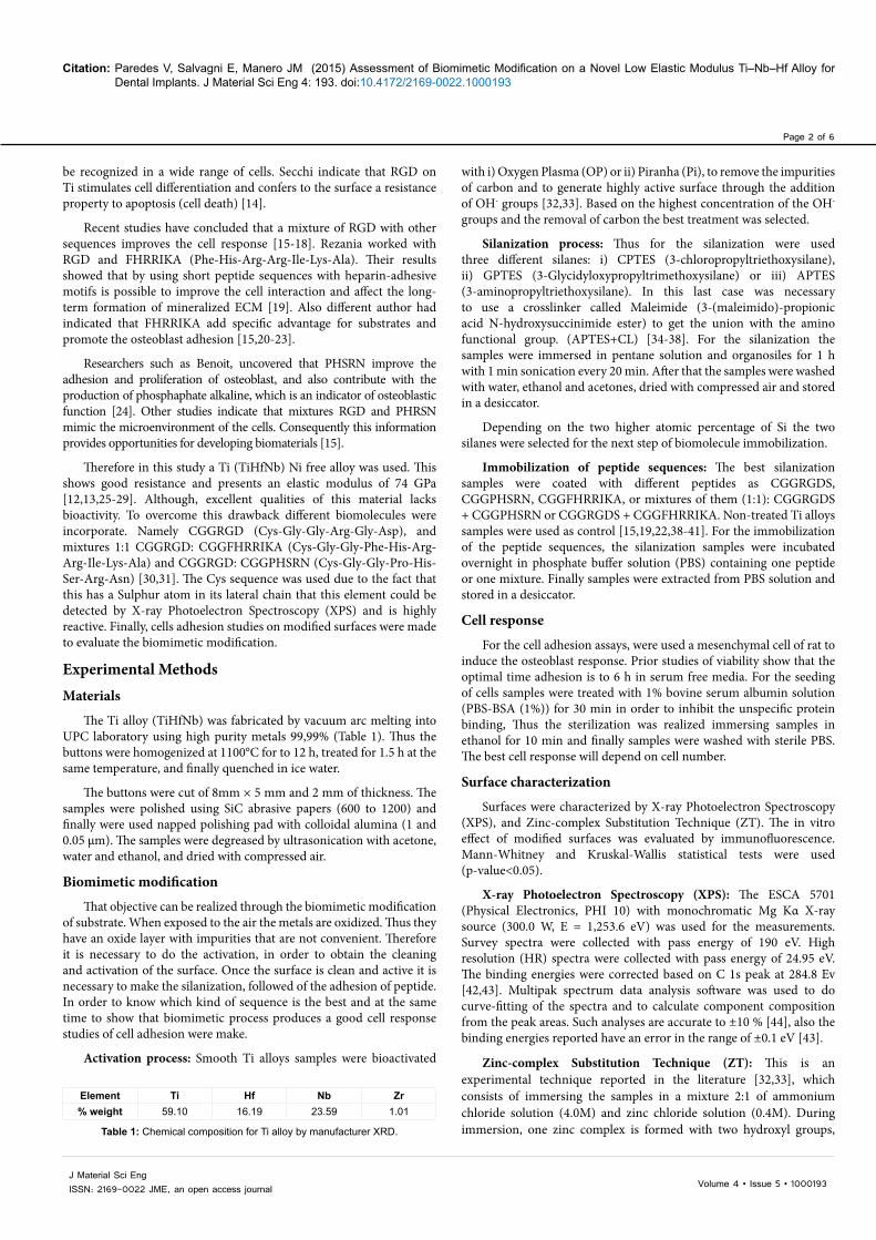
In order to know if there are OH- group on the surface the XPS high resolution of O 1s spectrum was used. Results of Table 2 and Figure 2 agree with reported by other authors in previous studies, which show that there are at least 3 peaks or 3 different kinds of oxygen species: oxide (O2-), hydroxide or hydroxyl group (OH-) and absorbed water (H2O) [32,33,46-49].
Thus, through the O2- it is possible to confirm that Pi treatment is able to increase the oxide layer more than OP. However, in terms of OH- groups, both methods were efficient, though the OP had a highest percentage.
Moreover, for efficient surface activation process, some authors have reported that it is convenient an increase in the ratio between the hydroxyl groups of the activated surface and the metal oxides (OH-/O2-), because it indicates that has occurred an increase in OH- and a decrease of O2- [32,33,48]. For that reason another important point is to calculate the ratio between OH-/O2- and choose the higher value as the best [33,49,50]. Thereby the Oxygen Plasma resulted to be the better activation method in terms to introduce a higher amount of OH- group per percentage of oxide (OH-/O2-).
By XPS High resolution it is possible to know the percentages of chemical composition of OH- group on the surface and inside of oxide layer (about 10 nm) [51]. Thus, there are authors, who established differences between hydroxyl groups on the surface and "active hydroxyl groups"(OH-act). These last react easily and are on the outer layer [32,33]. In order to distinguish hydroxyl groups active on the outer layer, an alternative technique (Zinc-complex substitution) was used.
Figure 3 shows the concentration of active hydroxyl groups (COH-act) per unit area on the outer layer of active samples. The results confirm that both activation methods are able to introduce OH- group on the surface. However, over again, the Oxygen Plasma had the best results in terms of COH-act.
Silanization process
In this phase were used the samples active with OP, thus the nomenclature used is i) TiC (Ti alloy +OP+CPTES TiC), ii) TiG (Ti alloy+OP+ GPTES) and iii) TiAC (Ti alloy+OP+ APTES+CL). Before to analyze the silanized samples is convenient to know the chemical composition of each silane used (Figure 4).
Fraiol reported that the presence of silanes on the surface, can be
Figure 1: Atomic concentration in % of C 1s and O 1S of samples activated with OP and Pi by XPS.
Control TiOP TiPi BE (eV)O2-
50% 42% 64% 530.1 + 0.4
OH-- 7% 47% 35% 531.5 + 0.3H2O 43% 11% 1% 533.1 + 0.4OH-/O2- 0,14 1,11 0,55
Table 2: Chemical composition for Ti alloy by manufacturer XRD.
Figure 2: O 1s high resolution XPS spectra of samples activated with OP.

Citation: Paredes V, Salvagni E, Manero JM (2015) Assessment of Biomimetic Modification on a Novel Low Elastic Modulus Ti–Nb–Hf Alloy for Dental Implants. J Material Sci Eng 4: 193. doi:10.4172/2169-0022.1000193
Page 4 of 6
Volume 4 • Issue 5 • 1000193J Material Sci EngISSN: 2169-0022 JME, an open access journal
Figure 3: Concentration of OH-act of samples activated with OP and Pi by Zinc-complex substitution technique.
Figure 4: Silanes used (CPTES, GPTES and APTED + Crosslinker).
detected by the appearance of the peak of silicon (Si 2p) on the surface by XPS [52]. Also different studies indicate that the peak Si 2p emerges on silanized samples because these have bonds of Si-O-metal and O-Si-O (siloxane) [53-56].
Table 3 shows all silanized samples have silicon on the surface. In particular the TiAC has greatest amount of Silicone (12% of Si 1s). This is a lowest amount of metallic elements (0%), for that it seems that TiAC have a thicker layer. This is followed by TiC samples which show a higher amount of Silicon than TiG (5% and 2% respectively). Otherwise the existence of Cl (2% Cl 2p) to the TiC confirms the presence of this silane, because CPTES has a Cl atom in their lateral chain.
Also an increase of C 1s in all silanized samples, because each silane contains carbon chains. Therefor the higher amount corresponds to TiAC (55%), due to the fact that it has longer carbon chains. Alike the TiAC showed major percentage of N 1s (10% of N 1s compared to 2% for untreated sample) due to the chemical composition of silane.
In sum, depending on the XPS information it can be inferred that sample TiAC generates a better result, because: i) TiAC contains as many Si 2p (12%) than TiC (5%) and TiG (2%), and ii) TiAC has a lower percentage of metallic elements, due to the fact that it has a thicker layer because it has a longer chain. Therefore the immobilization of peptide sequences will be used TiC and TiAC.
Immobilization of peptide sequences
How know, if there is a binding of the peptide. The strategy used peptide sequences with Cysteine, because this could be detected by XPS trough of S 2p, considering that the Cys has a thiol (-SH) in its lateral chain.
By XPS it was possible to identify all samples with biomolecules by the presence of S 2p, which indicate the existence of peptide sequence on the surface (Table 4). Regarding of C 1s, the results of TiC samples show an increase to the surface with mixture, because to the presence on dipeptide and carboxyl groups.
However, no statistically significant differences occurred between samples TiAC+peptide and TiC+peptide, although it has been found that TiAC have higher atomic percentage of silicon surface. Thus, the assessment the biomimetic modification will be depending on which peptide sequence improve the cell response.
Cell response
In order to evaluate the cell adhesion process it was used the immunofluorescence to staining the nuclei, actin filament and focal points. The experimental results are showed in Figure 5, where is possible to observe that higher amount if cell are present on samples with APTES+CL (TiAC).
For quantification of cell adhesion process, a study was conducted by staining of cell nuclei with DAPI and the results are shown in Figure 6. Depending on the results obtained it can be summarized in terms of the density of cells, the biomimetic modification with APTES +CL generates better results.
ConclusionsThis research demonstrated that by Oxygen Plasma it is possible
to obtain a best removal of carbon from contamination. Furthermore,
SamplesAtomic concentration (%)
C1s N1s O1s Si2p Cl2p Ti2p Nb3d Hf4fControl 42 2 46 0 0 7 2 1TiOP 11 1 58 0 0 11 2 1TiC 31 1 50 5 2 8 2 1TiG 28 1 56 2 0 10 2 1TiAC 55 10 23 12 0 0 0 0
Table 3: Chemical composition for Ti alloy silanized samples by XPS. Element
Atomic Concentration (%)
ControlAPTES CL CPTES
TiAC RGD RGD + FHRRIKA
RGD + PHSRN TiC RGD RGD +
FHRRIKARGD + PHSRN
C 1s 42 55 43 38 41 31 29 52 42Cl 2p 0 0 3 4 4 2 9 1 1Hf 4f 0 0 0 0 0 1 1 0 1N 1s 2 10 6 6 7 1 1 12 6Nb 3d 2 0 0 1 1 2 4 1 1O 1s 46 23 36 39 36 50 46 26 39S 2p 0 0 1 1 1 0 1 2 1Si 2p 1 12 10 10 8 5 1 4 3Ti 2p 7 0 1 1 2 8 8 2 6
Table 4: Chemical composition for Ti alloy samples with peptide sequences by XPS.

Citation: Paredes V, Salvagni E, Manero JM (2015) Assessment of Biomimetic Modification on a Novel Low Elastic Modulus Ti–Nb–Hf Alloy for Dental Implants. J Material Sci Eng 4: 193. doi:10.4172/2169-0022.1000193
Page 5 of 6
Volume 4 • Issue 5 • 1000193J Material Sci EngISSN: 2169-0022 JME, an open access journal
that method was able to produce a best result in terms of activation of the surface. XPS allowed us to do the detection of silanes on the surface, through of Si 2s, and also identify of peptide on the surface, through of S 2p.
Preliminary results of cells studies showed that the best cell response was for the samples with APTES+CL, however, there is not significative difference among peptide sequences. Therefore as Future Studies it is proposed to do the quantification of Adhesion of Peptide Sequence and cell studies of Proliferation and Differentiation.
Acknowledgments
The authors gratefully thank: Ministry of Science and Innovation; Spain MAT2008-06887-C03-03 (Biofunctionalized surfaces for tissue repair and regeneration), for financial support, Contract grant sponsor: Fundación Gran Mariscal de Ayacucho Venezuela.
References
1. Li C, Zhan Y, Jiang W (2011) Zr-Si biomaterials with high strength and low elastic modulus. Mater Des 32: 4598-4602.
2. Ryan G, Pandit A, Apatsidis DP (2006) Fabrication methods of porous metals for use in orthopaedic applications. Biomaterials 27: 2651-2670.
3. Rho JY, Tsui TY, Pharr GM (1997) Elastic properties of human cortical and trabecular lamellar bone measured by nanoindentation. Biomaterials 18: 1325-1330.
4. Oldani C, Dominguez A (2012) Titanium as a Biomaterial for Implants. Recent Adv Arthroplast 2012: 149-162.
5. Niinomi M (2011) Low Modulus Titanium Alloys for Inhibiting Bone Atrophy. Biomater Sci Eng 2011: 249-268.
6. Niinomi M, Nakai M (2011) Titanium-based biomaterials for preventing stress shielding between implant devices and bone. Int J Biomater 2011: 10.
7. Mohammed MT, Khan ZA, Siddiquee AN (2014) Beta Titanium Alloys : The Lowest Elastic Modulus for Biomedical Applications : A Review. Int J Chem Nucl Mater Metall Eng 8: 726-731.
8. Le Guéhennec L, Soueidan A, Layrolle P, Amouriq Y (2007) Surface treatments of titanium dental implants for rapid osseointegration. Dent Mater 23: 844-854.
9. Vanegas Acosta JC, Garzón-alvarado D, Casale M (2010) Interacción entre osteoblastos y superficies de titanio: aplicación en implantes dentales. Rev Cuba Investig Biomédicas 29: 51-68.
10. Jones F (2001) Teeth and bones: applications of surface science to dental materials and related biomaterials. Surf Sci Rep 42: 75-205.
11. Goriainov V, Cook R, Latham JM, Dunlop GD, Oreffo ROC (2014) Bone and metal: An orthopaedic perspective on osseointegration of metals. Acta Biomater 10: 4043-4057.
12. Sargeant TD, Rao MS, Koh C-Y, Stupp SI (2008) Covalent functionalization of NiTi surfaces with bioactive peptide amphiphile nanofibers. Biomaterials 29: 1085-1098.
13. González M, Peña J, Manero JM, Arciniegas M, Gil FJ (2009) Design and Characterization of New Ti-Nb-Hf Alloys. J Mater Eng Perform 18: 490-495.
14. Secchi AG, Grigoriou V, Shapiro IM, Composto RJ, Ducheyne P (2007) RGDS peptides immobilized on titanium alloy stimulate bone cell attachment, differentiation and confer resistance to apoptosis. J Biomed Mater Res Part A 83: 577-584.
15. Ochsenhirt SE, Kokkolia E, McCarthy JBJB, Tirrell M, Kokkoli E (2006) Effect of RGD Secondary Structure and the Synergy Site PHSRN on Cell Adhesion, Spreading and Specific Integrin Engagement. Biomaterials 27: 3863-3874.
16. Beuvelot J, Portet D, Lecollinet G, Moreau MF, Baslé MF, et al. (2009) In vitro Kinetic Study of Growth and Mineralization of Osteoblast-Like Cells (Saos-2) on Titanium Surface Coated with a RGD Functionalized Bisphosphonate. J Biomed Mater Res B Appl Biomater 90: 873-881.
17. Poulin S, Durrieu MC, Polizu S, Yahia LH (2006) Bioactive Molecules for Biomimetic Materials: Identification of RGD Peptide Sequences by ToF-S-SIMS Analysis. Appl Surf Sci 252: 6738-6741.
18. Milburn C, Chen J, Cao Y, Oparindeb GM, Adeoyeb MO, et al. (2009) Investigation of Effects of Argenine–Glycine–Aspartate (RGD) and Nano-scale Titanium Coatings on Cell Spreading and Adhesion. Mater Sci Eng C 29: 306-314.
19. Rezania A, Healy KE (1999) Biomimetic peptide surfaces that regulate adhesion, spreading, cytoskeletal organization, and mineralization of the matrix deposited by osteoblast-like cells. Biotechnol Prog 15: 19-32.
20. Schuler M, Hamilton DW, Kunzler TP, Sprecher CM, de Wild M, et al. (2009) Comparison of the response of cultured osteoblasts and osteoblasts outgrown from rat calvarial bone chips to nonfouling KRSR and FHRRIKA-peptide modified rough titanium surfaces. J Biomed Mater Res B Appl Biomater 91: 517-527.
21. Tirrell M, Kokkoli E, Biesalski M (2002) The role of surface science in bioengineered materials. Surf Sci 500: 61-83.
22. Rezania A, Healy KE (1999) Integrin subunits responsible for adhesion of human osteoblast-like cells to biomimetic peptide surfaces. J Orthop Res Off Publ Orthop Res Soc 17: 615-623.
23. Schaffner P, Dard MM (2003) Review: Structure and function of RGD peptides involved in bone biology. Cell Mol Life Sci 60: 119-132.
24. Benoit DSW, Anseth KS (2005) The Effect on Osteoblast Function of Colocalized
Figure 5: Cell Adhesion of surface with different peptide sequences by immunofluorescence.
Figure 6: Number of Cell Adhesion of surface with different peptide sequences by DAPI.
Citation: Paredes V, Salvagni E, Manero JM (2015) Assessment of Biomimetic Modification on a Novel Low Elastic Modulus Ti–Nb–Hf Alloy for Dental Implants. J Material Sci Eng 4: 193. doi:10.4172/2169-0022.1000193
Page 6 of 6
Volume 4 • Issue 5 • 1000193J Material Sci EngISSN: 2169-0022 JME, an open access journal
RGD and PHSRN Epitopes on PEG Surfaces. Biomaterials 26: 5209-5220.
25. Pegueroles Neyra M (2009) Interactions Between Titanium Surfaces and Biological Components.
26. Mjöberg B (1997) The Theory of Early Loosening of Hip Prostheses. Orthopedics 20: 1169-1175.
27. Sundfeldt M, Carlsson L V, Johansson CB, Thomsen P, Gretzer C (2006) Aseptic loosening, not only a question of wear: a review of different theories. Acta Orthop 77: 177-197.
28. González M, Peña J, Manero JM, Arciniegas M, Gil FJ (2009) Optimization of the Ti-16.2Hf-24.8Nb-1Zr Alloy by Cold Working. J Mater Eng Perform 18: 506-510.
29. Zorn G, Gotman I, Gutmanas EY, Adadi R, Sukenik CN (2007) Surface modification of Ti45Nb alloy by immobilization of RGD peptide via self assembled monolayer. J Mater Sci Mater Med 18: 1309-1315.
30. Schuler M, Trentin D, Textor M, Tosatti SGP (2006) Biomedical interfaces: titanium surface technology for implants and cell carriers. NanomedicineLondon Engl 1: 449-463.
31. Feng Y, Mrksich M (2004) The Synergy Peptide PHSRN and the Adhesion Peptide RGD Mediate Cell Adhesion through a Common Mechanism. Biochemistry 43: 15811-15821.
32. Tanaka Y, Saito H, Tsutsumi Y, Doi H, Imai H, et al. (2008) Active Hydroxyl Groups on Surface Oxide Film of Titanium, 316L Stainless Steel, and Cobalt-Chromium-Molybdenum Alloy and Its Effect on the Immobilization of Poly(Ethylene Glycol). Mater Trans 49: 805-811.
33. Sakamoto H, Hirohashi Y, Saito H, Doi H, Tsutsumi Y, Suzuki Y, et al. (2008) Effect of active hydroxyl groups on the interfacial bond strength of titanium with segmented polyurethane through gamma-mercapto propyl trimethoxysilane. Dent Mater J 27: 81-92.
34. Sevilla P, Godoy M, Salvagni E, Rodríguez D, Gil FJ (2010) Biofunctionalization of titanium surfaces for osseintegration process improvement. Journal of Physics: Conference Series 252: 1-6.
35. Shircliff RA, Martin IT, Pankow JW, Fennell J, Stradins P, et al. (2011) High-Resolution X-ray Photoelectron Spectroscopy of Mixed Silane Monolayers for DNA Attachment. ACS Appl Mater Interfaces 3: 3285-3292.
36. Ratner BD, Hoffman AS, Schoen FJ, Lemon JE (2004) The Role of Adsorbed Proteins in Tissue Response to Biomaterials. In: Biomaterials Science: An Introduction to Materials in Medicine. Academic Press, San Diego.
37. Tsiourvas D, Tsetsekou A, Arkas M, Diplas S, Mastrogianni E (2010) Covalent attachment of a bioactive hyperbranched polymeric layer to titanium surface forthe biomimetic growth of calcium phosphates. J Mater Sci Mater Med 22: 85-96.
38. Fernandez-garcia E, Chen X, Gutierrez-gonzalez CF, Fernandez A, Lopez-esteban S, et al. (2015) Peptide-functionalized zirconia and new zirconia / titanium biocermets for dental applications. J Dent 43:1162-1174.
39. Kokkoli E, Mardilovich A, Wedekind A, Rexeisen EL, Garg A, et al. (2006) Self-assembly and applications of biomimetic and bioactive peptide-amphiphiles. Soft Matter 2: 1015-1024.
40. Sowmiya M, Senthilkumar K (2015) Adsorption of RGD tripeptide on anatase ( 0 0 1 ) surface – A first principle study. Computational Materials Science 104:124-129.
41. Zheng Y, Xiong C, Li X, Zhang L (2014) Covalent attachment of cell-adhesive peptide Gly-Arg-Gly-Asp ( GRGD ) to poly ( etheretherketone ) surface by tailored silanization layers technique 320: 93-101.
42. Lobo AO, Ramos SC, Antunes EF, Marciano FR, Trava-Airoldi VJ, et al. (2012) Fast Functionalization of Vertically Aligned Multiwalled Carbon Nanotubes Using Oxygen Plasma. Mater Lett 70: 89-93.
43. Moulder JF, Stickle WF, Sobol KDB PE (1992) Handbook of X-Ray Photoelectron Spectroscopy.
44. Smart R, Mcintyre S, Bello I (2001) X - ray Photoelectron Spectroscopy. Surf Sci West.
45. Scheuerlein C, Taborelli M (2006) The assessment of metal surface cleanliness by XPS. Appl Surf Sci 252: 4279-4288.
46. Xiao SJ, Textor M, Spencer ND, Wieland M, Keller B, et al. (1997) Immobilization of the cell-adhesive peptide Arg–Gly –Asp–Cys (RGDC) on titanium surfaces by covalent chemical attachment. J Mater Sci Mater Med 8: 867-872.
47. Viornery C, Chevolot Y, Léonard D, Aronsson BO, Péchy P, et al. (2002) Surface Modification of Titanium with Phosphonic Acid To Improve Bone Bonding: Characterization by XPS and ToF-SIMS. Langmuir 18: 2582-2589.
48. Hanawa T, Hiromoto S, Asami K (2001) Characterization of the surface oxide film of a Co–Cr–Mo alloy after being located in quasi-biological environments using XPS. Appl Surf Sci 183: 68-75.
49. Hiromoto S, Onodera E, Chiba A, Asami K, Hanawa T (2005) Microstructure and Corrosion Behaviour in Biological Environments of the New Forged Low-Ni Co-Cr-Mo Alloys. Biomaterials 26: 4912-4923.
50. Paredes V, Salvagni E, Rodriguez E, Gil FJ, Manero JM (2013) Assessment and comparison of surface chemical composition and oxide layer modification upon two different activation methods on a cocrmo alloy. J Mater Sci Mater Med 25: 311-320.
51. Ma Z, Mao Z, Gao C (2007) Surface Modification and Property Analysis of Biomedical Polymers Used for Tissue Engineering. Colloids Surf B Biointerfaces 60: 137-157.
52. Fraioli R, Rechenmacher F, Neubauer S (2015) Colloids and Surfaces B : Biointerfaces Mimicking bone extracellular matrix : Integrin-binding peptidomimetics enhance osteoblast-like cells adhesion , proliferation , and differentiation on titanium 128:191-200.
53. Landoulsi J, Genet MJ, Kirat K El, Richard C, Pulvin S, et al. (2011) Silanization with APTES for Controlling the Interactions Between Stainless Steel and Biocomponents: Reality vs Expectation. In: Biomaterials - Physics and Chemistry 5: 99-126.
54. Martin HJ, Schulz KH, Bumgardner JD, Walters KB (2007) XPS Study on the Use of 3-Aminopropyltriethoxysilane to Bond Chitosan to a Titanium Surface. Langmuir 23: 6645-6651.
55. Mani G, Feldman MD, Oh S, Agrawal CM (2009) Surface Modification of Cobalt–Chromium–Tungsten–Nickel Alloy Using Octadecyltrichlorosilanes. Appl Surf Sci 255: 5961-5970.
56. Iucci G, Battocchio C, Dettin M, Ghezzo F, Polzonetti G (2010) An XPS Study on the Covalent Immobilization of Adhesion Peptides on a Glass Surface. Solid State Sci 12: 1861-1865.