assessing the fate and bioavailability of glucosinolates ... the fate...concomitant increase in the...
TRANSCRIPT

Assessing the Fate and Bioavailability of Glucosinolates in Kale(Brassica oleracea) Using Simulated Human Digestion and Caco‑2Cell Uptake ModelsEun-Sun Hwang,† Gail M. Bornhorst,‡,∥ Patricia I. Oteiza,§ and Alyson E. Mitchell*,∥
†Department of Nutrition and Culinary Science, Hankyong National University, 327 Chungang-Ro, Anseong-Si, Kyonggi-do 17579,Korea‡Department of Biological and Agricultural Engineering, Department of Food Science and Technology, University of California,Davis, One Shields Avenue, Davis, California 95616, United States§Department of Nutrition and Department of Environmental Toxicology, University of California, Davis, One Shields Avenue,Davis, California 95616, United States∥Department of Food Science and Technology, University of California, Davis, One Shields Avenue, Davis, California 95616, UnitedStates
ABSTRACT: Glucosinolates and their hydrolysis products were characterized in fresh and in in vitro gastric and intestinaldigesta of Dinosaur kale (Brassica oleracea L var. palmifolia DC). In fresh kale, glucoraphanin, sinigrin, gluconapin,gluconasturtiin, glucoerucin, glucobrasscin, and 4-methoxylglucobrassicin were identified. After 120 min of gastric digestion, thelevels of glucoraphanin, sinigrin, and gluconapin decreased, and no glucoerucin or glucobrasscin was detected. However, aconcomitant increase in the glucosinolate hydrolysis products allyl nitrile, 3-butenyl isothiocyanate, phenylacetonitrile, andsulforaphane was observed. This trend continued through intestinal digestion. After 120 min, the levels of allyl nitrile, 3-butenylisothiocyanate, phenylacetonitrile, and sulforaphane were 88.19 ± 5.85, 222.15 ± 30.26, 129.17 ± 17.57, and 13.71 ± 0.62pmol/g fresh weight, respectively. Intestinal digesta were then applied to Caco-2 cell monolayers to assess the bioavailability.After 6 h of incubation, no glucosinolates were detected and the percentage of total cellular uptake of the glucosinolatehydrolysis products ranged from 29.35% (sulforaphane) to 46.60% (allyl nitrile).
KEYWORDS: kale, glucosinolate, in vitro digestion, caco-2 cell, bioavailability
■ INTRODUCTION
Cruciferous vegetables are a popular food crop consumedworldwide and include numerous species and cultivars such askale, broccoli, cabbage, cauliflower, watercress, and Brusselssprouts.1 Kale (Brassica oleracea var. acephala L.), often toutedas a superfood, is grown in the United States, Europe, and Asiaand is cultivated throughout the year for its leaves and flowerbuds (i.e., kale raab).2 The tender leaves, which usually appear3 months after budding, are consumed either fresh, steamed,stir-fried, or boiled.3 Kale contains a high content of dietaryfiber, essential amino acids, vitamins, flavonoids, and bio-logically active glucosinolates.4,5
Glucosinolates are unique to cruciferous vegetables,6 anddemonstrate protective effects against many types of cancersincluding gastric, bladder, colorectal, prostate, and breastcancers.7−9 These exist in plant tissues as glycosides.Glucosinolates have no reported health benefits, but whenplant tissue is damaged (e.g., chewed or cut) glucosinolatesglycosides are hydrolyzed by the enzyme myrosinase(thioglucoside glucohydrolase, EC 3.2.1.147). This activitygenerates biologically active products that include isothiocya-nates, thiocyanates, nitriles, and indoles.10,11 The hydrolysisproducts of glucosinolates are thought to be primarilyresponsible for the chemoprotective effects observed withcruciferous vegetables.11 Particularly, the isothiocyanates(ITCs) act as anticancer agents by inhibiting phase I enzymes
responsible for bioactivation of carcinogenic compounds andthrough the induction of phase II detoxification enzymes thataffect xenobiotic transformation.12,13 Many studies showedthat ITCs have also have potent bactericidal, fungistatic, andfungicidal activities.14,15
Human digestion is a complex multistage process. It involvesthe mechanical and chemical breakdown of foods whichfacilitates the release of embedded nutrients so they can beabsorbed into the body through intestinal mucosal cells.16,17
Food is reduced in size in the mouth and stomach whereas thesmall intestines are the major site of nutrient absorption. Thestomach contains gastric acids, bile salts and digestive enzymeswhich work to homogenize and transform food.18 In theintestine, the gastric digesta is further dissolved and nutrientsare absorbed through the intestinal walls. Bioaccessibility isdefined as the amount or fraction of food that is released froma food into the digestive juices with the potential to beabsorbed by the small intestine during digestion.19 In contrast,bioavailability refers to the fraction of compound absorbedacross the gastric and intestinal walls into the bloodstream andtherefore has the potential to enter systemic circulation.20
Received: May 28, 2019Revised: July 30, 2019Accepted: August 2, 2019Published: August 2, 2019
Article
pubs.acs.org/JAFCCite This: J. Agric. Food Chem. 2019, 67, 9492−9500
© 2019 American Chemical Society 9492 DOI: 10.1021/acs.jafc.9b03329J. Agric. Food Chem. 2019, 67, 9492−9500
Dow
nloa
ded
via
UN
IV O
F C
AL
IFO
RN
IA D
AV
IS o
n Se
ptem
ber
27, 2
019
at 1
7:20
:32
(UT
C).
See
http
s://p
ubs.
acs.
org/
shar
ingg
uide
lines
for
opt
ions
on
how
to le
gitim
atel
y sh
are
publ
ishe
d ar
ticle
s.

Increasingly, various in vitro digestion models are used toestimate bioaccessibility and bioavailability.21−24 In vitrodigestion models offer the advantage of cost-efficiency, easeof control, and independence from physiological effects. Invitro digestion models also have excellent reproducibility andcan provide a good approximation of in vivo digestion.24
The human epithelial cell line Caco-2 is widely used as an invitro model of the human epithelial barrier in order to helpidentify the transport, and retention of substances acrossgastrointestinal tissues.25 This cell line has similar morpho-logical and biochemical characteristics as enterocytes (e.g.,polarity, tight junctions, specific transport systems, andenzymes) after it becomes differentiated in culture.26 Caco-2cells are used in numerous studies to estimate thebioavailability of important classes of phytochemicals includingcarotenoids, polyphenols, and anthocyanins from whole foodsincluding cabbage.23,27,28 To date, there are no studiesassessing the bioaccessibility and bioavailability of glucosino-lates from whole kale, and little is known regarding the profileof glucosinolates and their hydrolysis products available ingastrointestinal contents with respect to digestion time.To address this, the human digestion of fresh kale was
simulated using an in vitro digestion model.21 This system wassampled at 30 min intervals during 2 h of gastric digestion andduring 2 h of intestinal digestion. The range of glucosinolatesand their hydrolysis products in the gastric and intestinaldigesta were identified using ultra high-performance liquidchromatography−quadrupole time-of-flight tandem mass spec-trometry (UHPLC−QTOF-MS/MS) and quantified usingHPLC. The transport of the identified glucosinolates andglucosinolate hydrolysis products in gastric and gastro-intestinal digesta was evaluated in Caco-2 cell monolayersdifferentiated into intestinal epithelial cells. Measuring theglucosinolates and glucosinolate hydrolysis products in gastricand intestinal digesta provides key information on thebioaccessibility of these compounds, while measuring thetransport across Caco-2 cell monolayers models provides keyinformation on the bioavailability of these compounds.
■ MATERIALS AND METHODSChemicals and Kale Samples. Glucosinolates standards,
including glucoraphanin (≥98%), sinigrin (≥99%), gluconapin(≥98%), and gluconasturtiin (≥98%) were purchased fromExtrasynthese (Genay Cedex, France). All solvents used were ofHPLC and LC/MS grade and purchased from Sigma-Aldrich (St.Louis, MO) and Fisher Scientific (Fairlawn, NJ).Dinosaur kale was grown at the Student Farm at the University of
California Davis under agronomic supervision. The kale was grownfrom seedlings and harvested at ∼110 days from sowing from at least10 different randomly selected plants in December 2017 and in April2018 to give two harvest replicates. Fresh samples were stored at 4 °Cfor <24 h prior to in vitro digestion.Simulated in Vitro Digestion. Fresh kale was digested following
previously reported methods.21,29,30 Fresh kale was cut with a knifeapproximately 1 cm length and finely chopped with a food processor(Black and Decker, FP2500B) at low speed for 60 s (15 s × 4 times)to simulate breakdown during mastication. A 70 mL aliquot ofsimulated gastric juice was loaded into a human gastric simulator(HGS) before adding samples.31 The chopped kale (100 g) wasmixed with 20 mL of simulated saliva for 30 s, and then transferredinto the HGS. The gastric juice secretion started immediately after thesample was introduced into the HGS and continued at 2.5 mL/min.Every 30 min, 50 mL of digesta fluid was collected in a glass bottle.After 2 h of gastric digestion, the HGS was stopped. The digestaremaining in the HGS was pooled, and 80 g of digesta was mixed with
54 mL of intestinal juice to generate the intestinal digesta. Theintestinal digestion was completed by incubation in a shaking waterbath (Thermo Scientific Inc., Marietta, OH) at 37 °C and 100 cyclesper minute for 2 h. Every 60 min, 30 g of digested sample wascollected in a glass bottle. For each sample from the gastric phase, pH,total acidity, particle size, moisture, and glucosinolate andglucosinolate hydrolysis products content were measured. For eachsample from the small intestinal phase, glucosinolates and theirglucosinolate hydrolysis products were measured.
Moisture Content, pH, Total Acidity of Fresh and DigestedKale. Moisture content of the sample was measured gravimetricallyusing a vacuum oven (Thermo Scientific Inc., Marietta, OH) at 110°C for 20 h. The pH was measured using a pH meter (FisherScientific Inc.). Titratable acidity (expressed as w/w % citric acid) wasmeasured via potentiometric titration with 0.1 N NaOH until the pHreached 8.2 ± 0.05.
Particle Size and of Fresh and Digested Kale. To measure theparticle size, the solid fraction of the gastric digesta samples wereseparated from the liquid phase by filtration with a double layeredcotton cloth (17.3 × 14.2 threads per cm). Aliquots of solid particles(∼1 g) were transferred onto a Petri dish. A volume of ∼10 mL ofwater was added to disperse the particles, and samples were gentlyagitated for 5 min on an orbital shaker to allow particles to separatewithout causing any further particle hydrolysis. Images were takenusing a digital camera (Canon Powershot SD 1300IS, Canon USA,Melville, NY). Images were analyzed as previously described todetermine the median particle area and the number of particles pergram of sample.32
Caco-2 Cells: Uptake and Transport of Glucosinolates andTheir Hydrolysis Compounds. Caco-2 cell monolayers were usedto evaluate the bioavailability of the glucosinolates and theirhydrolysis products in the intestinal digesta. Caco-2 human intestinalcells were cultured at 37 °C and 5% (v/v) CO2 atmosphere inminimum essential medium (MEM) supplemented with 10% (v/v)fetal bovine serum, antibiotics (50 U/mL penicillin, and 50 μg/mLstreptomycin), 1% (v/v) of 100× nonessential amino acids, and 1mM sodium pyruvate. The medium was replaced every 3 days duringcell growth and differentiation. For the experiments, cells were seededin 6-well plate polyester membrane permeable support inserts (30mm, 0.4 μm pore size, Corning Inc., Corning, NY) at a density of 1 ×10⁵ cells/well. Monolayers were used 21 days after reachingconfluence to allow for full differentiation into intestinal epithelialcells. Cells were used between passages 3 and 15. All the experimentswere performed in serum- and phenol red-free MEM. Transepithelialelectrical resistance (TEER, Ω cm2) values were measured before andafter adding the sample using a Millicell-ERS Volt-Ohm Meter(Millipore, Bedford, MA) as previously described.33
Caco-2 cells were washed twice with MEM medium. An aliquot of250 μL of freshly prepared gastro-intestinal digesta (obtained after 2 hdigestion) was added to the upper chamber (apical side) and 750 μLof MEM was added to the lower chamber (basolateral side). Cellswere incubated at 37 °C for 6 h, and the TEER was measured toensure the integrity of the monolayer. Medium from both sides of theinsets was collected, and the cell monolayers were washed twice withice-cold PBS. Cells were collected in 200 μL of PBS, sonicated, andcentrifuged, and the supernatants were collected and stored at −20 °Cfor a maximum of 2 days until HPLC analysis. Cell studies wereperformed in triplicate.
Absorption (cellular uptake) and transport (basolateral secretion)efficiency are expressed as the percentage of glucosinolate hydrolysisproducts detected inside Caco-2 cells and that in the basolateralcompartment, with respect to the original glucosinolates and theirhydrolysis products originally added to the apical side. Thebioavailability of glucosinolate hydrolysis products is defined as thetotal content of each glucosinolate hydrolysis compound absorbed bythe Caco-2 cells (retention and transport) from the intestinal digesta(at 120 min), divided by the total added in the intestinal digesta.Typically, percentage bioavailability is expressed as a percent of theinitial amount in the food product. However, as glucosinolates areextensively hydrolyzed in the stomach and intestine, the hydrolysis
Journal of Agricultural and Food Chemistry Article
DOI: 10.1021/acs.jafc.9b03329J. Agric. Food Chem. 2019, 67, 9492−9500
9493

products are reported as a percent of the original sample afterintestinal digestion.
=[ ] + [ ]
[ ]×
bioavailability (%)compound in basolateral compound in cell monolayer
compound in intestinal digesta100
Extraction of Glucosinolates. Glucosinolates were extractedfrom all samples (0.5 g) with 2 mL of boiling 70% (v/v) methanol ina hot water bath (90−92 °C) for 5 min and centrifuged at 20 000g for10 min at 4 °C. The pellet was extracted a second time, and the twosupernatants were combined. The glucosinolate extract was applied toa Mini Bio-Spin chromatography column (Bio-Rad Laboratories,Hercules, CA) containing 1 mL of cross-linked dextran gel (type G-25) anion exchange resin, which was preactivated with 20 mM sodiumacetate (pH 5.5). Glucosinolates were removed from resins usingdesulfation carried out by the addition of 75 μL of purified arylsulfatase (EC 3.1.6.1, type H-1 from Helix pomatia). The column wascapped and allowed to stand at room temperature for 24 h. Thedesulfo-glucosinolates were eluted from the column with 1.5 mLdistilled water. Eluates were filtered through a 0.2 μm syringe filterprior to injection onto the HPLC.Authentic standards of glucosinolates were desulfated as described
above and used for the identification and quantification of the peaks.Concentrations of individual desulfo-glucosinolates were determinedfrom the experimental peak area using external standard calibrationcurves for each desulfo-glucosinolate across different ranges (depend-ing upon the glucosinolate) and are expressed as micromoles per gram(μmol/g).Desulpho-Glucosinolate Analysis Using HPLC. HPLC analysis
was performed on an Agilent 1200 HPLC system coupled with aphotodiode array (PDA) detector (Agilent Technologies, Memphis,TN). The chromatographic column used was a Waters symmetry 300C18 column (75 mm × 4.6 mm, i.d. with 3.5 μm particle diameter,Waters, Franklin, MA) at 40 °C. A mobile phase composed of A(water) and B (acetonitrile) with a gradient elution of 0 min (0% B),0−1 min (2% B), 2−35 min (2−35% B), 35−40 min (35−2% B), 41min (0% B) was used in this study. The sample injection volume was20 μL, and the flow-rate was set at 0.5 mL/min. Peaks were detectedat 229 nm. Briefly, individual glucosinolates were identified andquantified in comparison with the retention time and externalstandard curve of four glucosinolate standards (glucoraphanin,sinigrin, gluconapin, and gluconasturtiin). Relative quantification ofindividual glucosinolates with no authentic standard was achievedusing standard methods reported by ISO 9167-1.34
Identification of Desulpho-Glucosinolates Using UHPLC−QTOF-MS/MS. The identification of glucosinolates present in theHPLC peaks of extracts of fresh kale were confirmed using authenticstandards and by collecting high-resolution QTOF-MS/MS spectraon an Agilent 1290 Infinity ultrahigh-pressure liquid chromatographysystem (UHPLC) interfaced to a 6445 quadrupole time-of-flight-tandem mass spectrometer (QTOF-MS/MS) with electrosprayionization (ESI) via Jet Stream technology (Agilent Technologies,
Santa Clara, CA). The UHPLC was equipped with a binary pumpwith an integrated vacuum degasser (G4220A), an autosampler(G4226A) with thermostat (G1330B), and a thermostated columncompartment (G1316C). Positive ESI mode was used for allcompounds. The drying gas temperature and flow rate was 320 °Cand 8.0 L/min, respectively. The sheath gas temperature and flow ratewere 350 °C and 11 L/min, respectively. The nebulizer gas pressure,capillary voltage, and nozzle voltage were 35 psi, 3500 V, and 1000 V,respectively. Analysis was carried out using a scan from m/z 50 to m/z1000. Mass accuracy was maintained by the use of a second referencenebulizer that continuously introduced purine and Hexakis(1H,1H,3H-tetrafluoropropoxy) phosphazine at a flow rate of 8−9μL/min. Tandem mass spectrometry experiments were performedwith the quadrupole set at medium resolution (m/z 4 amu) and fixedcollision energies of 10, 20, and 40 were used to facilitate theidentification of the glucosinolates based on the parent andfragmented ions.
Glucosinolate hydrolysis products in gastric and intestinal digestawere also confirmed using high-resolution QTOF MS/MS spectrawith MassHunter software B07 (Agilent Technologies, Memphis,TN) and by comparing with authentic standards.
Statistical Analysis. HPLC peak areas were obtained in triplicatefor each of the three independent measurements of the sample. HPLCpeak areas were averaged, and the standard deviation (SD) wasdetermined from the average of the three measurements. Statisticalanalysis was performed with the statistical analysis system (SPSSsoftware package, version 22.0). Data were compared using repeatedmeasures analysis of variance (ANOVA); p < 0.05 was consideredsignificant.
■ RESULTS AND DISCUSSION
Measurement of Moisture Content, pH, and TotalAcidity. Moisture content was determined in fresh and
Table 1. Moisture Content, pH, and Total Acidity of Fresh Kale Before and During Simulated Gastric and Small IntestinalDigestiona
moisture content (%, wet basis) pH total acidity (%) (citric acid)
fresh kale (before digestion) 83.39 ± 0.51 a 6.62 ± 0.12 c 0.23 ± 0.01 agastric digestion30 min 96.45 ± 1.43 b 5.20 ± 0.29 b 0.62 ± 0.01 c60 min 96.22 ± 1.40 b 4.96 ± 0.26 ab 0.65 ± 0.00 c90 min 96.08 ± 1.08 b 4.75 ± 0.11 a 0.69 ± 0.02 c120 min 96.58 ± 0.72 b 4.47 ± 0.07 a 0.70 ± 0.02 cintestinal digestion60 min 95.62 ± 1.39 b 7.01 ± 0.21 d 0.27 ± 0.00 a120 min 95.51 ± 1.23 b 7.00 ± 0.16 d 0.35 ± 0.00 b
aData are shown as mean ± SD of triplicate experiment. Means with different superscript letters in the same column are significantly different at p <0.05.
Table 2. Median Particle Area and Number of Particles perGram of Sample in Fresh Kale and Kale Undergoing GastricDigestiona
median particle area(mm2)
no. of particles pergram
fresh kale(no gastric digestion)
5.52 ± 2.75 c 1038 ± 354 d
gastric digestion30 min 5.93 ± 4.16 c 939 ± 785 d60 min 5.58 ± 3.03 c 777 ± 561 d90 min 4.15 ± 2.22 ab 844 ± 684 d120 min 3.64 ± 0.23 a 945 ± 139 d
aData are shown as mean ± SD of triplicate experiment. Means withdifferent superscript letters in the same column are significantlydifferent at p < 0.05.
Journal of Agricultural and Food Chemistry Article
DOI: 10.1021/acs.jafc.9b03329J. Agric. Food Chem. 2019, 67, 9492−9500
9494
digested kale samples (Table 1). Fresh kale contained 83.39%moisture, while gastric and intestinal digested kale containedhigher moisture contents ranging from 95.51 to 96.58%. Themoisture content was increased in the samples taken during invitro gastric and intestinal digestion relative to the fresh kale.Similar to our results, previous studies have shown that themoisture content of apple or almond increased during in vitrogastrointestinal digestion.35,36 This increase is due to areduction in the particle size of the kale during in vitrodigestion, which may result in damage to the cell structure,leading to water uptake when mixed with gastric secretions.The pH and total acidity, which represent the amount of
citric acid, is shown in Table 1. The pH of fresh kale was 6.62and decreased with digestion time to 5.20−4.47. During theintestinal digestion, the pH was 7.00−7.01. The total aciditycorrelated with pH changes. The total acidity of fresh kale was0.23% and increased during gastric digestion up to 0.62−0.70
but decreased again during the intestinal digestion step by0.27−0.35%. The change in pH in the in vitro digestion modelcan be a very important factor because the action of digestiveenzymes or digestive juice secreted in digestive organs isclosely related to pH. The changes of pH and total acidity areconsidered to be closely related to the pH of the digestivefluids at each digestion stage. The pH of the oral cavity isweakly acidic (pH 5−7), and the pH of the mouth is rapidlydecreased from 1 to 3 pH units due to the release ofhydrochloric acid in the stomach and restored to a slightlyacidic (pH 5−7) due to the high pH of bile acids andbicarbonate.37
Median Particle Area and Number of Particles perGram Samples. The particle size distribution of kale beforeand during in vitro gastric digestion are given in Table 2. Themedian particle area in fresh kale (5.52 ± 2.75 mm2)significantly (p < 0.05) decreased with digestion time to 3.64
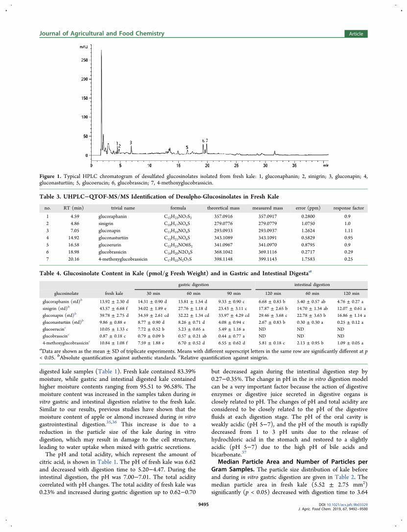
Figure 1. Typical HPLC chromatogram of desulfated glucosinolates isolated from fresh kale: 1, gluconaphanin; 2, sinigrin; 3, gluconapin; 4,gluconasturtiin; 5, glucoerucin; 6, glucobrasscin; 7, 4-methoxyglucobrassicin.
Table 3. UHPLC−QTOF-MS/MS Identification of Desulpho-Glucosinolates in Fresh Kale
no. RT (min) trivial name formula theoretical mass measured mass error (ppm) response factor
1 4.59 glucoraphanin C12H23NO7S2 357.0916 357.0917 0.2800 0.92 4.86 sinigrin C10H17NO6S 279.0776 279.0779 1.0750 1.03 7.05 gluconapin C11H19NO6S 293.0933 293.0937 1.2624 1.114 14.92 gluconasturtiin C15H21NO6S 343.1089 343.1091 0.5829 0.955 16.58 glucoerurin C12H23NO6S2 341.0967 341.0970 0.8795 0.96 18.98 glucobrassicin C16H20N2O6S 368.1042 369.1116 0.2717 0.297 20.16 4-methoxyglucobrassicin C17H22N2O7S 398.1148 399.1143 1.7583 0.25
Table 4. Glucosinolate Content in Kale (pmol/g Fresh Weight) and in Gastric and Intestinal Digestaa
gastric digestion intestinal digestion
glucosinolate fresh kale 30 min 60 min 90 min 120 min 60 min 120 min
glucoraphanin (std)b 13.92 ± 2.30 d 14.31 ± 0.90 d 13.81 ± 1.54 d 9.33 ± 0.90 c 6.68 ± 0.83 b 5.40 ± 0.57 ab 4.76 ± 0.27 a
sinigrin (std)b 43.57 ± 6.68 f 34.02 ± 1.89 e 27.76 ± 1.18 d 23.43 ± 3.11 c 17.87 ± 2.65 b 14.70 ± 1.34 ab 12.07 ± 0.61 a
gluconapin (std)b 39.78 ± 2.75 d 34.59 ± 2.61 cd 32.22 ± 1.34 cd 33.97 ± 4.29 cd 29.46 ± 3.68 c 22.78 ± 3.63 b 16.86 ± 1.14 a
gluconasturtiin (std)b 9.86 ± 0.88 e 8.77 ± 0.90 d 8.26 ± 0.71 d 4.08 ± 0.94 c 2.67 ± 0.83 b 0.30 ± 0.30 a 0.25 ± 0.12 a
glucoerucinc 10.05 ± 1.33 c 7.72 ± 0.52 b 5.23 ± 0.65 a 5.49 ± 1.16 a ND ND ND
glucobrasscinc 0.87 ± 0.18 c 0.79 ± 0.09 b 0.57 ± 0.21 ab 0.44 ± 0.77 a ND ND ND
4-methoxyglucobrassicinc 10.84 ± 1.08 f 7.59 ± 1.88 e 6.70 ± 0.52 d 6.55 ± 0.62 d 5.81 ± 0.18 c 2.13 ± 0.95 b 1.09 ± 0.05 aaData are shown as the mean ± SD of triplicate experiments. Means with different superscript letters in the same row are significantly different at p< 0.05. bAbsolute quantification against authentic standards. cRelative quantification against sinigrin.
Journal of Agricultural and Food Chemistry Article
DOI: 10.1021/acs.jafc.9b03329J. Agric. Food Chem. 2019, 67, 9492−9500
9495

± 0.23 mm2 at 120 min. Physical breakdown during gastricdigestion would be expected and has been observed inprevious in vitro and in vivo studies.31,32
The number of particles per g sample appeared to increasebetween 90 and 120 min; however, this increase was notstatistically significant. When the median particle area wasconsidered, it was observed that the area of the kale particlesdecreased to 24.82% and 34.06%, compared to the fresh kalesamples after 90 and 120 min of gastric digestion, respectively.Glucosinolates in Fresh Kale. A typical HPLC chromato-
gram of the desulfated glucosinolates isolated from fresh kale isgiven in Figure 1. Seven peaks were identified as glucosinolates(Table 3). Peaks 1, 2, and 4 corresponded to glucoraphanin (tR4.56 min), sinigrin (tR 4.86 min), gluconapin (tR 7.05 min),
and gluconasturtiin (tR 14.92 min), respectively, as confirmedwith authentic standards. High-resolution QTOF-MS/MSallowed for the tentative identification of three additionalglucosinolates corresponding to peaks 5 (glucoerucin), 6(glucobrassicin), and 7 (4-methoxylglucobrassici) at retentiontimes 16.58, 18.98, and 20.16 min, respectively. Ascommercially available standards are not available for allcompounds, concentrations were established using relativequantitation against sinigrin.Glucosinolates were quantified in undigested kale (Table 4).
Sinigrin (43.57 ± 6.68 pmol/g FW) was the predominantglucosinolate, which correlates with previously reported datafor kale.38 Relatively high amounts of gluconapin (39.78 ±
Figure 2. Extracted ion chromatogram of glucosinolate hydrolysis compounds and qTOF spectra obtained in ESI positive mode after in vitrodigestion of kale. Allyl nitrile (a,b); 3-butenyl isothiocyanate (c,d); phenylacetonitrile (e,f); and sulforophane (g,h).
Table 5. Content of Glucosinolate Hydrolysis Products (pmol/g Fresh Weight) in Fresh Kale and in Gastric and IntestinalDigestaa
gastric digestion intestinal digestion
glucosinolatehydrolysis compounds fresh kale 30 min 60 min 90 min 120 min 60 min 120 min
allyl nitrilec 14.90 ± 1.37 a 59.68 ± 3.44 b 103.50 ± 11.45 d 197.98 ± 16.70 f 135.89 ± 5.39 e 88.19 ± 5.85 c 123.26 ± 3.83 e
butenylisothiocyanatec
32.28 ± 8.35 a 61.65 ± 4.74 b 79.65 ± 9.79 c 107.65 ± 22.22 d 102.55 ± 5.05 d 222.15 ± 30.26 e 299.31 ± 25.43 f
phenylacetonitrilec 45.59 ± 1.21 a 43.43 ± 6.84 a 45.35 ± 10.91 a 50.55 ± 7.40 ab 46.32 ± 12.63 a 129.17 ± 17.57 b 200.43 ± 17.34 c
sulforaphaneb ND ND 10.09 ± 0.98 a 11.54 ± 0.45 a 13.74 ± 0.45 b 13.71 ± 0.62 b 14.55 ± 1.03 caData are shown as mean ± SD of triplicate experiment. Means with different superscript letters in the same row are significantly different at p <0.05. bAbsolute quantification against authentic standards. cRelative quantification against sulforaphane.
Journal of Agricultural and Food Chemistry Article
DOI: 10.1021/acs.jafc.9b03329J. Agric. Food Chem. 2019, 67, 9492−9500
9496

2.75 pmol/g FW) and glucoraphanin (13.92 ± 2.30 pmol/gFW) were also found in undigested kale.Influence of Gastric and Intestinal Digestion on
Glucosinolates. The gastric digestion of glucosinolates wasevaluated at 30, 60, 90, and 120 min in the HGS (Table 4). At30 and 60 min, the levels of sinigrin, gluconastrurtiin,glucoerucin, glubrasscin, and 4-methoxyglucobrassicin weresignificantly reduced as compared with the levels in fresh kale.After 90 min of gastric digestion, levels of all glucosinolateswere significantly reduced from levels in fresh kale with theexception of gluconapin. At 120 min, the levels of allglucosinolates were reduced and no glucoerucin or gluco-brasscin could be detected in digesta. Data demonstrate thatthe profile of glucosinolates in the gastric digesta will varybased upon gastric digestion time and that gluconapin is themost resistant to hydrolysis during gastric digestion.To evaluate intestinal digestion, the gastric digesta were
allowed to remain in the HGS using conditions that simulateintestinal digestion and were sampled at 60 and 120 min. Thelevels of glucoraphanin, sinigrin, and gluconapin decreased by29%, 32%, and 43%, respectively, in intestinal digesta ascompared with gastric digesta at 120 min whereas losses of
gluconasturtiin (99%) and 4-methoxyglucobrassicin (81%)were even more substantial (p < 0.05). Glucoerucin andglucobrasscin were not present in gastric digests after 120 minand were therefore absent from intestinal digesta. In a similarstudy, Vallejo et al.39 found that the in vitro gastric digestion ofbroccoli resulted in 69% reduction of total glucosinolates,which were further decreased after intestinal digestion.
Glucosinolate Hydrolysis Products. Glucosinolates arethioglycosides that differ in the structure of the aglycone sidechain. Glucosinolates can undergo hydrolysis via the activity ofmyrosinase to form unstable aglycones that can rearrange toform a wide array of biologically active compounds includingepithionitriles, nitriles, thiocyanates, isothiocyantes, andoxazolidine-thiones. Myrosinase is active in fresh vegetablesand in the stomach and small intestine.40 Glucosinolatehydrolytic rearrangement is dependent upon pH. At a lowpH ≤ 3 (i.e., the pH of the stomach), hydrolytic rearrangementfavors formation of nitriles and at pH ≥ 7 (i.e., the pH of thesmall intestines) hydrolytic rearrangement favors formation ofisothiocyanates.41 Isothiocyanates are absorbed from the smallintestine and in the colon, and metabolites are detectable in
Figure 3. Typical HPLC chromatogram of glucosinolate hydrolysis compounds (a) after 2 h of in vitro gastro digestion, (b) after 2 h of in vitrointestinal digestion, and (c) after 6 h of incubation with the Caco-2 monolayer. 1, allyl nitrile (RT = 1.947); 2, butenyl isothiocyanate (RT =3.621); 3, phenylacetonitrile (RT = 6.564); 4, sulforaphane (RT = 15.396).
Journal of Agricultural and Food Chemistry Article
DOI: 10.1021/acs.jafc.9b03329J. Agric. Food Chem. 2019, 67, 9492−9500
9497

urine 2−3 h after consumption of Brassica vegetables inhumans.40
Glucosinolate hydrolysis products are primarily responsiblefor the anticarcinogenic activity associated with consumingBrassica vegetables. These low-molecular weight compoundsare able to rapidly diffuse into the cells of the intestinalepitheilium and modulate the expression of genes important tochemoprevention (e.g., those associated with xenobioticmetabolism, antioxidation, cell cycle regulation, apoptoticpathways, and stress response).41−48 Understanding theformation and disposition of glucosinolate hydrolysis productsin the gut is key for understanding the bioactive potential ofBrassica foods. In general, levels of glucosinolate hydrolysisproducts are lower in undamaged fresh Brassica as comparedto Brassica with insect and/or physical damage as damageallows glucosinolates contact with myrosinase. Herein, thehydrolyzed products identified in fresh kale include phenyl-acetonitrile (45.59 ± 1.21 pmol/g FW), 3-butenyl isothiocya-nate (32.28 ± 8.35 pmol/g FW), and allyl nitrile (14.90 ± 1.37pmol/g FW) (Figure 2 and Table 5).
Glucosinolate Hydrolysis Products in Gastric andIntestinal Digesta. Intact glucosinolates are not absorbedthrough the intestinal epithelium, and the hydrolysis products,which are absorbed, are the bioactive forms of glucosinolates.40
Therefore, it is important to understand the disposition ofbioactive glucosinolate hydrolysis products available forabsorption in the small intestine. Phenylacetonitrile 3-butenylisothiocyanate, allyl nitrile, and sulforaphane were identified ingastric digesta by 60 min of HGS (Figures 2 and 3 and Table5). Allyl nitrile, 3-butenyl isothiocyanate, phenylacetonitrile,and sulforophane are the hydrolysis products of sinigrin,gluconapin, glucotropaeolin, and glucoraphanin, respectively.40
Identities of these compounds were confirmed using high-resolution QTOF-MS/MS (Figure 2). Levels of allyl nitrileand 3-butenyl isothiocyanate increased significantly (228% and166%, respectively) between 30 and 120 min of gastricdigestion whereas levels of phenylacetonitrile did not changesignificantly. Sulforaphane, which was not detected in freshkale, was observed only after 60 min of gastric digestion (10.09± 0.98 pmol/g FW), and levels were increased significantly at120 min of gastric digestion (13.74 ± 0.45 pmol/g FW).During intestinal digestion, the levels of allyl nitrile initially
decreased and then increased after 120 min of intestinaldigestion (Figure 3 and Table 5). Allyl nitrile is a hydrolysisproduct of sinigrin.46 The levels of sinigrin decreased duringintestinal digestion and may have contributed to the increase inallyl nitrile as the in vitro digestion model is a closed system.Butenyl isothiocyanate levels were significantly higher in
intestinal digesta (299.31 ± 25.43 pmol/g FW at 120 min) ascompared with levels in gastric digesta at 120 min (102.55 ±5.05 pmol/g FW). The levels of phenylacetonitrile, thehydrolysis product of gluconasturtiin, which did not changesignificantly during gastric digestion, increased significantlyduring intestinal digestion (280% at 60 min and 656% at 120min). Sulforaphane levels were relatively low in intestinaldigests as compared with the other glucosinolate hydrolysisproducts, reaching only 14.55 ± 1.03 pmol/g FW after 2 h ofintestinal digestion.In similar studies, Rodriguez-Hernandez et al.49 performed
in vitro digestion of different cultivars of raw freeze-driedbroccoli and measured corresponding levels of glucoraphaninand sulforaphane. Both glucoraphanin and sulforaphanedecreased during digestion of broccoli florets (cv. Naxos)T
able
6.UptakeandTranspo
rtof
Glucosino
latesandTheirHydrolysisProdu
ctsafter6hIncubation
ofIntestinal
Digesta
withCaco-2Cellsa
apicalcompartment
basolateralcompartment(transport)
cellmonolayer
(retentio
n)
cmpd
totaladded
[intestin
aldigesta]
(ng)
(ng)
(%)
(ng)
(%)
(ng)
(%)
bioavailabilityd
(totalcellularuptake,%
)
allylnitrilec
8.51
±0.26
b4.54
±0.40
b53.35±
6.30
a2.61
±0.15
b30.67±
0.82
b1.53
±0.38
a17.98±
0.74
b48.60±
1.54
cbutenylisothiocyanatec
46.71±
3.97
d26.97±
0.23
d57.73±
4.41
ab13.15±
0.47
d28.15±
1.40
b5.08
±2.21
b10.88±
3.82
a39.03±
2.42
bphenylacetonitrilec
23.46±
2.03
c13.06±
1.22
c55.67±
0.37
a5.88
±0.11
c25.06±
2.67
a2.52
±0.92
a10.74±
2.99
a35.81±
0.32
bsulforaphaneb
2.58
±0.18
a1.54
±0.31
a59.69±
7.95
b0.76
±0.22
a29.46±
1.43
bND
029.35±
1.43
aaThe
contento
fglucosinolatehydrolysisproducts(ng/mLintestinaldigesta)
wasmeasuredincells
andintheapicalandbasolateralcom
partmentsafterincubatingcellmonolayersfor6hwith
250μL
ofintestinaldigestaobtained
after120min
ofdigestionaddedto
theapicalcompartment.Dataareshow
nas
mean±
SDof
triplicateexperiments.M
eans
with
differentsuperscriptlettersin
thesame
columnaresignificantly
differentat
p<0.05.b
Absolutequantifi
catio
nagainstauthentic
standards.c Relativequantifi
catio
nagainstsulforaphane.dbioavailability(%
)=[(compoundin
basolateral+
compoundin
cellmonolayer)/(com
poundin
intestinaldigesta)]×100.
Journal of Agricultural and Food Chemistry Article
DOI: 10.1021/acs.jafc.9b03329J. Agric. Food Chem. 2019, 67, 9492−9500
9498

whereas levels of glucoraphanin decreased by 7% andsulforaphane increased by 23% in the cultivar Viola. Inadditional studies in fresh broccoli, Ghawai et al.47
demonstrated a 1.5−3.0 increase in the levels of sulforaphaneafter intestinal digestion showing that myrosinase in theintestinal digestion process was still active.Together these results demonstrate that sinigrin is the most
abundant glucosinolate in fresh kale (43.57 ± 6.68 pmol/gFW) and that other glucosinolates identified ranged from 0.87± 0.18 to 39.78 ± 2.75 pmol/g FW kale. In general, levels of allidentified glucosinolates decreased during gastric and intestinaldigestions. Whereas levels of glucosinolate hydrolysis productsincreased through gastric and intestinal digestions. Theprimary hydrolysis products formed in gastric digesta areallyl nitrile, butenyl isothiocyanate, and phenylacetonitrile.Assessing Bioavailabilily. Caco-2 cells are a widely used
and accepted model to evaluate the bioavailability of nutrientsand drugs after oral ingestion.48 The intestinal digesta at 120min, and added to the Caco-2 cells, contained theglucosinolate hydrolysis products allyl nitrile (8.51 ± 0.26ng), butenyl isothiocyanate (46.71 ± 3.97 ng), phenyl-acetonitrile (23.46 ± 2.03 ng), and sulforaphane (2.58 ±0.18 ng) (Table 6). The intestinal digesta also contained theglucosinolates glucoraphanin (3 332.94 ± 0.77 ng), sinigrin(10 813.22 ± 2.18 ng), gluconapin (14 383.63 ± 3.87 ng),gluconasturtiin (182.09 ± 0.39 ng), and 4-methoxygluco-brassicin (684.12 ± 0.13 ng). Although both glucosinolatesand the hydrolysis products were present in the intestinaldigesta at 120 min, only the hydrolysis products weremeasured in the Caco-2 cells as no further hydrolysis wasobserved in intestinal digesta and because only the hydrolysisproducts are transported across intestinal epithelial cells.Recoveries of hydrolysis products in the Caco-2 cells were101% allyl nitrile, 97% butenyl isothiocyanate, 91% phenyl-acetonitrile, and 89% for sulforophane.After 6 h, the percentage of glucosinolate hydrolysis
compounds in the apical compartment varied between 53.44and 59.47%, whereas 25.19−30.61% of the glucosinolatehydrolysis products were transported into the basolateralcompartment and 0−15.68% were retained in the cells in theCaco-2 monolayer. The percentage of total cellular uptake inthe intestinal digesta ranged from 29.39% (sulforaphane) to48.60% (allyl nitrile). These results indicate that buteneylisothocyanate is significantly higher than other glucosinolatehydrolysis products after the consumption, and digestion ofkale although allyl nitrile is the most bioavailable glucosinolatehydrolysis.
■ AUTHOR INFORMATIONCorresponding Author*Phone: (530) 304-6618. Fax: (530) 752-4759. E-mail:[email protected] E. Mitchell: 0000-0003-0286-5238FundingThis research was supported by Basic Science ResearchProgram through the National Research Foundation ofKorea (Grant NRF-2016R1A2B4014977), NIFA-USDA(Grant CA-D*-XXX-7244-H), and NIFA-USDA (Grant CA-D-FST-6975-H).NotesThe authors declare no competing financial interest.
■ ACKNOWLEDGMENTSThe authors would like to acknowledge the assistance of Dr.Larry Lerno of the UC Davis Food Safety and MeasurementFacility and Yamile Mennah Govela and Alex Olenskyj forassistance with the in vitro digestion experiments.
■ REFERENCES(1) Abdull Razis, A. F.; Noor, N. M. Cruciferous vegetables: dietaryphytochemicals for cancer prevention. Asian Pac. J. Cancer Prev. 2013,14 (3), 1565−1570.(2) Hahn, C.; Muller, A.; Kuhnert, N.; Albach, D. Diversity of kale(Brassica oleracea var. sabellica): Glucosinolate content andphylogenetic relationships. J. Agric. Food Chem. 2016, 64 (16),3215−3225.(3) Cartea, M. E.; Picoaga, A.; Soengas, P.; Ordas, A. Morphologicalcharacterization of kale populations from northwestern Spain.Euphytica 2003, 129 (1), 25−32.(4) Ayaz, F. A.; Glew, R. H.; Millson, M.; Huang, H.S.; Chuang,L.T.; Sanz, C.; Hayırlıoglu-Ayaz, S. Nutrient contents of kale (Brassicaoleraceae L. var. acephala DC.). Food Chem. 2006, 96 (4), 572−579.(5) Velasco, P.; Cartea, M. E.; Gonzalez, C.; Vilar, M.; Ordas, A.Factors affecting the glucosinolate content of kale (Brassica oleraceaacephala group). J. Agric. Food Chem. 2007, 55 (3), 955−962.(6) Holst, B.; Williamson, G. A critical review of the bioavailability ofglucosinolates and related compounds. Nat. Prod. Rep. 2004, 21 (3),425−447.(7) Atwell, L. L.; Beaver, L. M.; Shannon, J.; Williams, D. E.;Dashwood, R. H.; Ho, E. Epigenetic regulation by sulforaphane:Opportunities for breast and prostate cancer chemoprevention. Curr.Pharmacol. Rep. 2015, 1 (2), 102−111.(8) Kumar, G.; Tuli, H. S.; Mittal, S.; Shandilya, J. K.; Tiwari, A.;Sandhu, S. S. Isothiocyanates: a class of bioactive metabolites withchemopreventive potential. Tumor Biol. 2015, 36 (6), 4005−4016.(9) Veeranki, O. L.; Bhattacharya, A.; Tang, L.; Marshall, J. R.;Zhang, Y. Cruciferous vegetables, isothiocyanates, and prevention ofbladder cancer. Curr. Pharmacol. Rep. 2015, 1 (4), 272−282.(10) Halkier, B. A.; Gershenzon, J. Biology and biochemistry ofglucosinolates. Annu. Rev. Plant Biol. 2006, 57, 303−333.(11) Hossain, M. S.; Ye, W.; Hossain, M. A.; Okuma, E.; Uraji, M.;Nakamura, Y.; Mori, I. C.; Murata, Y. Glucosinolate hydrolysisproducts, isothiocyanates, nitriles, and thiocyanates, induce stomatalclosure accompanied by peroxidase-mediated reactive oxygen speciesproduction in Arabidopsis thaliana. Biosci., Biotechnol., Biochem. 2013,77 (5), 977−983.(12) James, D.; Devaraj, S.; Bellur, P.; Lakkanna, S.; Vicini, J.;Boddupalli, S. Novel concepts of broccoli sulforaphanes and disease:induction of phase II antioxidant and detoxification enzymes byenhanced-glucoraphanin broccoli. Nutr. Rev. 2012, 70 (11), 654−665.(13) Lubelska, K.; Wiktorska, K.; Mielczarek, L.; Milczarek, M.;Zbroin ska-Bregisz, I.; Chilmonczyk, Z. Sulforaphane regulatesNFE2L2/Nrf2-dependent xenobiotic metabolism phase II and phaseIII enzymes differently in human colorectal cancer and untransformedepithelial colon cells. Nutr. Cancer 2016, 68 (8), 1338−1348.(14) Dias, C.; Aires, A.; Saavedra, M. J. Antimicrobial activity ofisothiocyanates from cruciferous plants against methicillin-resistantStaphylococcus aureus (MRSA). Int. J. Mol. Sci. 2014, 15 (11), 19552−19561.(15) Manyes, L.; Luciano, F. B.; Manes, J.; Meca, G. In vitroantifungal activity of allyl isothiocyanate (AITC) against Aspergillusparasiticus and Penicillium expansum and evaluation of the AITCestimated daily intake. Food Chem. Toxicol. 2015, 83, 293−299.(16) Alminger, M.; Aura, A.-M.; Bohn, T.; Dufour, C.; El, S.N.;Gomes, A.; Karakaya, S.; Martinez-Cuesta, M.C.; McDougall, G.J.;Requena, T.; Santos, C.N. In vitro models for studying secondaryplant metabolite digestion and bioaccessibility. Compr. Rev. Food Sci.Food Saf. 2014, 13 (4), 413−436.(17) Mennah-Govela, Y. A.; Bornhorst, G. M. Fresh-squeezedorange juice properties before and during in vitro digestion as
Journal of Agricultural and Food Chemistry Article
DOI: 10.1021/acs.jafc.9b03329J. Agric. Food Chem. 2019, 67, 9492−9500
9499

influenced by orange variety and processing method. J. Food Sci. 2017,82 (10), 2438−2447.(18) Kong, F.; Singh, R. P. Disintegration of solid foods in humanstomach. J. Food Sci. 2008, 73 (5), R67−R80.(19) Bornhorst, G. M.; Roman, M. J.; Dreschler, K. C.; Singh, R.Physical property changes in raw and roasted almonds during gastricdigestion in vivo and in vitro. Food Biophys. 2014, 9 (1), 39−48.(20) Heaney, R. P. Factors influencing the measurement ofbioavailability, taking calcium as a model. J. Nutr. 2001, 131 (4),1344S−1348S.(21) Bornhorst, G. M.; Singh, R. P. Kinetics of in vitro bread bolusdigestion with varying oral and gastric digestion parameters. FoodBiophys. 2013, 8 (1), 50−9.(22) Read, A.; Wright, A.; Abdel-Aal, El-S. M. In vitrobioaccessibility and monolayer uptake of lutein from wholegrainbaked food. Food Chem. 2015, 174, 263−269.(23) Kaulmann, A.; Andre, C. M.; Schneider, Y.; Hoffmann, L.;Bohn, T. Carotenoid and polyphenol bioaccessibility and cellularuptake from plum and cabbage varieties. Food Chem. 2016, 197, 325−332.(24) Moreda-Pineiro, J.; Moreda-Pineiro, A.; Romarís-Hortas, V.;Moscoso-Perez, C.; Lo pez-Mahía, P.; Muniategui-Lorenzo, S.;Bermejo-Barrera, P.; Prada-Rodríguez, D. In-vivo and in-vitro testingto assess the bioaccessibility and the bioavailability of arsenic,selenium and mercury species in food samples. TrAC, Trends Anal.Chem. 2011, 30 (2), 324−345.(25) Liu, D.; Cheng, B.; Li, D.; Li, J.; Wu, Q.; Pan, H. Investigationson the interactions between curcumin loaded vitamin E TPGS coatednanodiamond and Caco-2 cell monolayer. Int. J. Pharm. 2018, 551(1−2), 177−183.(26) Primavera, R.; Palumbo, P.; Celia, C.; Cinque, B.; Carata, E.;Carafa, M.; Paolino, D.; Cifone, M. G.; Di Marzio, L. An insight on invitro transport of PEGylated non-ionic surfactant vesicles (NSVs)across the intestinal polarized enterocyte monolayers. Eur. J. Pharm.Biopharm. 2018, 127, 432−442.(27) Liu, C. S.; Glahn, R. P.; Liu, R. H. Assessment of carotenoidbioavailability of whole foods using a Caco-2 cell culture modelcoupled with an in vitro digestion. J. Agric. Food Chem. 2004, 52 (13),4330−4337.(28) Sun, D.; Huang, S.; Cai, S.; Cao, J.; Han, P. Digestion propertyand synergistic effect on biological activity of purple rice (Oryza sativaL.) anthocyanins subjected to a simulated gastrointestinal digestion invitro. Food Res. Int. 2015, 78, 114−123.(29) Roman, M. J.; Burri, B. J.; Singh, R. P. Release andbioaccessibility of beta-carotene from fortified almond butter duringin vitro digestion. J. Agric. Food Chem. 2012, 60 (38), 9659−9666.(30) Phinney, D. M. Design, Construction, And Evaluation of a ReactorDesigned to Mimic Human Gastric Digestion. Masters Thesis, Universityof California, Davis, CA, 2013.(31) Guo, Q.; Ye, A.; Lad, M.; Ferrua, M.; Dalgleish, D.; Singh, H.Disintegration kinetics of food gels during gastric digestion and itsrole on gastric emptying: an in vitro analysis. Food Funct. 2015, 6 (3),756−764.(32) Bornhorst, G. M.; Kostlan, K.; Singh, R. P. Particle SizeDistribution of Brown and White Rice during Gastric DigestionMeasured by Image Analysis. J. Food Sci. 2013, 78 (9), E1383−E1391.(33) Cremonini, E.; Mastaloudis, A.; Hester, S. N.; Verstraeten, S.V.; Anderson, M.; Wood, S. M.; Waterhouse, A. L.; Fraga, C. G.;Oteiza, P. I. Anthocyanins inhibit tumor necrosis alpha-induced lossof Caco-2 cell barrier integrity. Food Funct. 2017, 8 (8), 2915−2923.(34) ISO, International Organization for Standardization. Method9167-1 (E), 1992.(35) Bornhorst, G. M.; Singh, R. P. Gastric digestion in vivo and invitro: how that structural aspects of food influence the digestionprocess. Annu. Rev. Food Sci. Technol. 2014, 5, 111−132.(36) Dalmau, M. E.; Bornhorst, G. M.; Eim, V.; Rossello, C.; Simal,S. Effect of freezing, freeze drying and convective drying on in vitrogastric digestion of apples. Food Chem. 2017, 215, 7−16.
(37) Hur, S. J.; Lee, S. K.; Kim, Y. C.; Choi, I. W. Development of invitro human digestion models for health functional food research.Food Sci. Industry 2012, 45, 40−49.(38) Jeon, J.; Kim, J. K.; Kim, H.; Kim, Y. J.; Park, Y. J.; Kim, S. J.;Kim, C.; Park, S. U. Transcriptome analysis and metabolic profiling ofgreen and red kale (Brassica oleracea var. acephala) seedlings. FoodChem. 2018, 241, 7−13.(39) Vallejo, F.; Gil-Izquierdo, A.; Perez-Vicente, A.; García-Viguera,C. In vitro gastrointestinal digestion study of broccoli inflorescencephenolic compounds, glucosinolates, and vitamin C. J. Agric. FoodChem. 2004, 52 (1), 135−138.(40) Johnson, I. T. Glucosinolates: bioavailability and importance tohealth. Int. J. Vitam. Nutr. Res. 2002, 72 (1), 26−31.(41) Fenwick, G. R.; Heaney, R. K.; Mullin, W. J.; VanEtten, C. H.Glucosinolates and their breakdown products in food and food plants.Crit. Rev. Food Sci. Nutr. 1983, 18 (2), 123−201.(42) Al-Gendy, A. A.; Lockwood, G. B. GC-MS analysis of volatilehydrolysis products from glucosinolates in Farsetia aegyptia var.Flavour Fragrance J. 2003, 18, 148−152.(43) Arora, R.; Kumar, R.; Mahajan, J.; Vig, A. P.; Singh, B.; Singh,B.; Arora, S. 3-Butenyl isothiocyanate: a hydrolytic product ofglucosinolate as a potential cytotoxic agent against human cancer celllines. J. Food Sci. Technol. 2016, 53 (9), 3437−3445.(44) Nunez-Iglesias, M. J.; Novio, S.; García-Santiago, C.; Cartea, M.E.; Soengas, P.; Velasco, P.; Freire-Garabal, M. Effect of 3-butenylisothiocyanate on phenotypically different prostate cancer cells. Int. J.Oncol. 2018, 53 (5), 2213−2223.(45) Matusheski, N. V.; Jeffery, E. H. Comparison of the bioactivityof two glucoraphanin hydrolysis products found in broccoli,sulforaphane and sulforaphane nitrile. J. Agric. Food Chem. 2001, 49(12), 5743−5749.(46) Tanii, H.; Higashi, T.; Nishimura, F.; Higuchi, Y.; Saijoh, K.Effects of cruciferous allyl nitrile on phase 2 antioxidant anddetoxification enzymes. Med. Sci. Monit. 2008, 14 (10), BR189−192.(47) Rodríguez-Hernandez, M.; Medina, S.; Gil-Izquierdo, A.;Martinez-Ballesta, M.; Moreno, D. Broccoli isothiocyanate contentand in vitro availability according to variety and origin. Macedonian J.Chem. Chem. Eng. 2013, 32 (2), 251−264.(48) Ghawi, S. K.; Methven, L.; Niranjan, K. The potential tointensify sulforaphane formation in cooked broccoli (Brassica oleraceavar. italica) using mustard seeds (Sinapis alba). Food Chem. 2013, 138(2−3), 1734−1741.(49) Chen, X. M.; Dai, Y.; Kitts, D. D. Detection of Maillardreaction product [5-(5, 6-Dihydro-4 H-pyridin-3-ylidenemethyl)furan-2-yl] methanol (F3-A) in breads and demonstration ofbioavailability in Caco-2 intestinal cells. J. Agric. Food Chem. 2016,64 (47), 9072−9077.
Journal of Agricultural and Food Chemistry Article
DOI: 10.1021/acs.jafc.9b03329J. Agric. Food Chem. 2019, 67, 9492−9500
9500