assembly of the chloroplast atp-dependent clp protease … · assembly of the chloroplast...
TRANSCRIPT
Assembly of the Chloroplast ATP-Dependent Clp Protease inArabidopsis Is Regulated by the ClpT Accessory Proteins C W
Lars L.E. Sjogren and Adrian K. Clarke1
Department of Plant and Environmental Sciences, Gothenburg University, 40530 Gothenburg, Sweden
The ATP-dependent caseinolytic protease (Clp) is an essential housekeeping enzyme in plant chloroplasts. It is by far the
most complex of all known Clp proteases, with a proteolytic core consisting of multiple catalytic ClpP and noncatalytic ClpR
subunits. It also includes a unique form of Clp protein of unknown function designated ClpT, two of which exist in the model
species Arabidopsis thaliana. Inactivation of ClpT1 or ClpT2 significantly reduces the amount of Clp proteolytic core,
whereas loss of both proves seedling lethal under autotrophic conditions. During assembly of the Clp proteolytic core,
ClpT1 first binds to the P-ring (consisting of ClpP3-6 subunits) followed by ClpT2, and only then does the P-ring combine
with the R-ring (ClpP1, ClpR1-4 subunits). Most of the ClpT proteins in chloroplasts exist in vivo as homodimers, which then
apparently monomerize prior to association with the P-ring. Despite their relative abundance, however, the availability of
both ClpT proteins is rate limiting for the core assembly, with the addition of recombinant ClpT1 and ClpT2 increasing core
content up to fourfold. Overall, ClpT appears to regulate the assembly of the chloroplast Clp protease, revealing a new and
sophisticated control mechanism on the activity of this vital protease in plants.
INTRODUCTION
Energy-dependent proteases perform a crucial role in protein
quality control by removing short-lived regulatory proteins and
misfolded or otherwise damaged polypeptides. Most of these
proteases consist of two distinct components, one with chap-
erone characteristics that uses ATP for substrate selection,
unfolding, and translocation and the other a proteolytic core for
degradation (Baker and Sauer, 2006). Many of these also share a
common architecture and are best exemplified by the 26S
proteasome in eukaryotes and the Clp protease in eubacteria.
The Ser-type Clp protease in Escherichia coli consists of a
central proteolytic core comprising two apposing heptameric
rings of ClpP. This cylindrical structure forms an internal chamber
that houses the proteolytic active sites within (Wang et al., 1997).
The proteolytic core is flanked on one or both ends by a single
hexameric ring of an Hsp100 partner, either ClpA or -X (Grimaud
et al., 1998). In an energy-dependent manner, the Hsp100
chaperone translocates the unfolded protein substrate through
the narrow aperture of the core complex into the inner cavity for
rapid degradation (Ishikawa et al., 2001; Kim et al., 2001; Ortega
et al., 2002).
Consistent with their endosymbiotic origin, plant chloroplasts
have a range of proteases of bacterial ancestry. These include
the FtsH and Deg proteases associated with the thylakoid
membranes and Clp protease localized within the stroma
(Adam and Clarke, 2002). The chloroplast Clp protease in vas-
cular plants is by far the most structurally diverse member of the
Clp protease family. In the model species Arabidopsis thaliana, it
consists of a single proteolytic core complex with 11 distinct
subunits (ClpP1, ClpP3-6, ClpR1-4, and ClpT1-2), along with
three potential chaperone partners (ClpC1-2 and ClpD) (Adam
et al., 2001; Peltier et al., 2004; Clarke et al., 2005). All of these
proteins with the exception of ClpP1 are nuclear encoded; thus,
they are posttranslationally imported into chloroplasts (Adam
et al., 2001; Adam and Clarke, 2002). The protease is also one of
the most functionally important, being the principle constitutive
housekeeping protease in the stroma whose activity is essential
for plant viability (Shikanai et al., 2001; Kuroda andMaliga, 2003;
Sjogren et al., 2006; Zheng et al., 2006; Koussevitzky et al., 2007;
Kim et al., 2009). Up to 25 stromal proteins have now been
identified as potential substrates for the Clp protease, ranging
from different regulatory proteins to various metabolic enzymes
(Sjogren et al., 2006; Stanne et al., 2009).
One of the most intriguing features of the chloroplast Clp
protease is the intricate subunit composition of the central
proteolytic core complex. By analogy to bacterial orthologs,
the core complex presumably consists of twin heptameric rings,
one of which contains ClpP1 and ClpR1-4 and the other ClpP3-6
(Sjogren et al., 2006). Little redundancy exists within this config-
uration (Stanne et al., 2009), with loss of any one subunit typically
disrupting the assembly of the entire protease. Even more
perplexing is the association of the unusual ClpT accessory
proteins that have so far been found only in plant chloroplasts
and whose function remains a mystery. The relatively small ClpT
proteins have high sequence similarity to theN-terminal region of
ClpC but lack the AAA+ ATPase domains common to Hsp100
proteins (Kress et al., 2009). Two ClpT paralogs exist in Arabi-
dopsis, with a single subunit of each thought to associate with
1 Address correspondence to [email protected] author responsible for distribution of materials integral to thefindings presented in this article in accordance with the policy describedin the Instructions for Authors (www.plantcell.org) is: Adrian K. Clarke([email protected]).CSome figures in this article are displayed in color online but in blackand white in the print edition.WOnline version contains Web-only data.www.plantcell.org/cgi/doi/10.1105/tpc.110.082321
The Plant Cell, Vol. 23: 322–332, January 2011, www.plantcell.org ã 2011 American Society of Plant Biologists
the peripheral surface of theClp proteolytic core complex (Peltier
et al., 2004). Within the core structure, ClpT1 binds to the ClpP3-6
ring, whereas the location of ClpT2 has yet to be resolved
(Sjogren et al., 2006). In this study, we demonstrate that ClpT is a
crucial component of the chloroplast Clp protease in Arabidop-
sis. ClpT2 is shown to associate with the ClpP3-6 ring along with
ClpT1, although most of both proteins exist as homodimers
within the stroma. Overall, the availability of ClpT1 and ClpT2
appears to regulate the assembly of the Clp proteolytic core,
revealing a previously undiscovered control mechanism on the
formation of the chloroplast Clp protease in vascular plants.
RESULTS
VerificationandPhenotypicAppearanceofArabidopsisclpT
T-DNA Insertion Lines
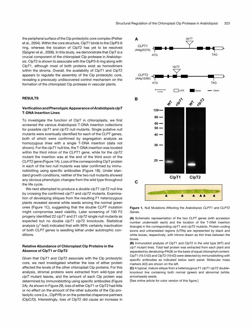
To investigate the function of ClpT in chloroplasts, we first
screened the various Arabidopsis T-DNA insertion collections
for possible clpT1 and clpT2 null mutants. Single putative null
mutants were eventually identified for each of the CLPT genes,
both of which were confirmed by segregation analysis as
homozygous lines with a single T-DNA insertion (data not
shown). For the clpT1 null line, the T-DNA insertion was located
within the third intron of the CLPT1 gene, while for the clpT2
mutant the insertion was at the end of the third exon of the
CLPT2 gene (Figure 1A). Loss of the correspondingClpT protein
in each of the two null mutants was later confirmed by immu-
noblotting using specific antibodies (Figure 1B). Under stan-
dard growth conditions, neither of the two null mutants showed
any obvious phenotypic changes from the wild type throughout
the life cycle.
We next attempted to produce a double clpT1 clpT2 null line
by crossing the confirmed clpT1 and clpT2 mutants. Examina-
tion of developing siliques from the resulting F1 heterozygous
plants revealed several white seeds among the normal green
ones (Figure 1C), suggesting that the double CLPT mutation
might compromise seed viability. Later screening of 100 F2
progeny identified 22 clpT1 and 21 clpT2 single null mutants as
expected but no double clpT1 clpT2 knockouts. Statistical
analysis (x2 test) indicated that with 99% certainty inactivation
of both CLPT genes is seedling lethal under autotrophic con-
ditions.
Relative Abundance of Chloroplast Clp Proteins in the
Absence of ClpT1 or ClpT2
Given that ClpT1 and ClpT2 associate with the Clp proteolytic
core, we next investigated whether the loss of either protein
affected the levels of the other chloroplast Clp proteins. For this
analysis, stromal proteins were extracted from wild-type and
clpT mutant leaves, and the amount of each Clp protein was
determined by immunoblotting using specific antibodies (Figure
2A). As shown in Figure 2B, loss of either ClpT1 or ClpT2 had little
or no effect on the amount of the other subunits of the Clp pro-
teolytic core (i.e., ClpP/R) or on the potential chaperone partners
(ClpC/D). Interestingly, loss of ClpT2 did cause an increase in
Figure 1. Null Mutations Affecting the Arabidopsis CLPT1 and CLPT2
Genes.
(A) Schematic representation of the two CLPT genes (with accession
number underneath each) and the location of the T-DNA insertion
(triangle) in the corresponding clpT1 and clpT2 mutants. Protein-coding
exons and untranslated regions (UTRs) are represented by black and
white boxes, respectively, with introns drawn as thin lines between the
boxes.
(B) Immunoblot analysis of ClpT1 and ClpT2 in the wild type (WT) and
clpT mutant lines. Total leaf protein was extracted from each plant and
separated by denaturing-PAGE on the basis of equal chlorophyll content.
ClpT1 (19.5 kD) and ClpT2 (19 kD) were detected by immunoblotting with
specific antibodies as indicated below each panel. Molecular mass
markers (kD) are shown on the left.
(C) A typical, mature silique from a heterozygous F1 clpT1 clpT2 double-
knockout line containing both normal (green) and abnormal (white;
arrowheads) seeds.
[See online article for color version of this figure.]
Structural Regulation of the Chloroplast Clp Protease in Arabidopsis 323
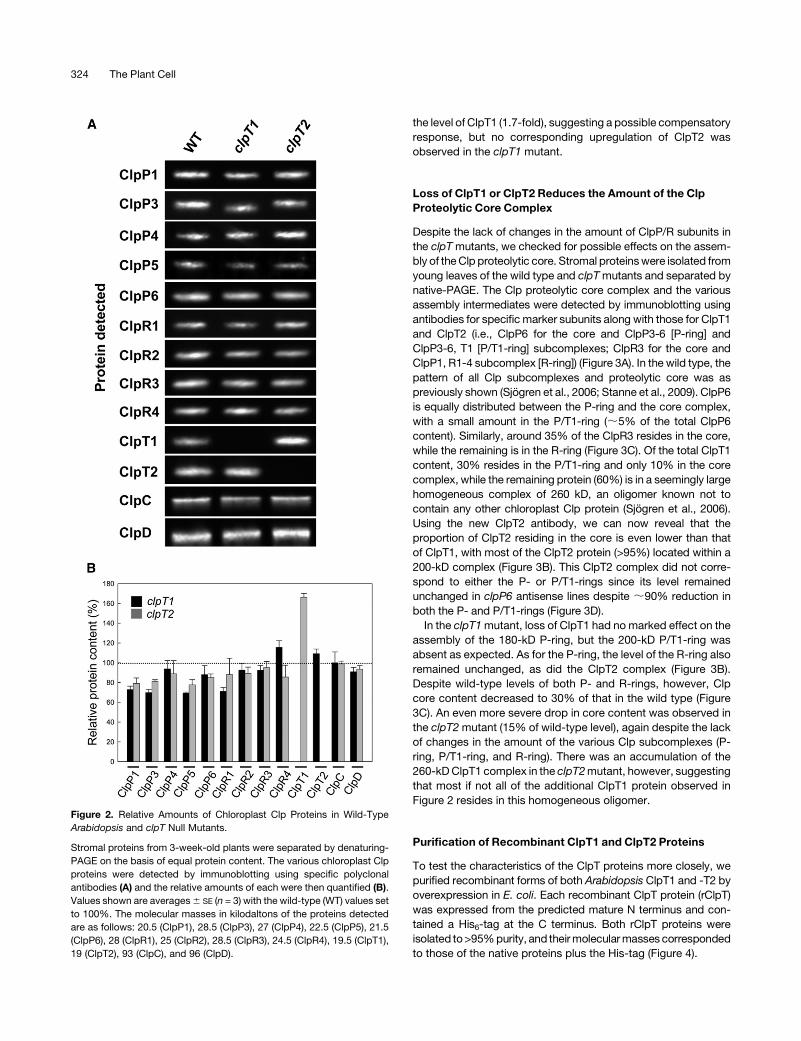
the level of ClpT1 (1.7-fold), suggesting a possible compensatory
response, but no corresponding upregulation of ClpT2 was
observed in the clpT1 mutant.
Loss of ClpT1 or ClpT2 Reduces the Amount of the Clp
Proteolytic Core Complex
Despite the lack of changes in the amount of ClpP/R subunits in
the clpTmutants, we checked for possible effects on the assem-
bly of theClp proteolytic core. Stromal proteinswere isolated from
young leaves of the wild type and clpTmutants and separated by
native-PAGE. The Clp proteolytic core complex and the various
assembly intermediates were detected by immunoblotting using
antibodies for specificmarker subunits along with those for ClpT1
and ClpT2 (i.e., ClpP6 for the core and ClpP3-6 [P-ring] and
ClpP3-6, T1 [P/T1-ring] subcomplexes; ClpR3 for the core and
ClpP1, R1-4 subcomplex [R-ring]) (Figure 3A). In the wild type, the
pattern of all Clp subcomplexes and proteolytic core was as
previously shown (Sjogren et al., 2006; Stanne et al., 2009). ClpP6
is equally distributed between the P-ring and the core complex,
with a small amount in the P/T1-ring (;5% of the total ClpP6
content). Similarly, around 35% of the ClpR3 resides in the core,
while the remaining is in the R-ring (Figure 3C). Of the total ClpT1
content, 30% resides in the P/T1-ring and only 10% in the core
complex, while the remaining protein (60%) is in a seemingly large
homogeneous complex of 260 kD, an oligomer known not to
contain any other chloroplast Clp protein (Sjogren et al., 2006).
Using the new ClpT2 antibody, we can now reveal that the
proportion of ClpT2 residing in the core is even lower than that
of ClpT1, with most of the ClpT2 protein (>95%) located within a
200-kD complex (Figure 3B). This ClpT2 complex did not corre-
spond to either the P- or P/T1-rings since its level remained
unchanged in clpP6 antisense lines despite ;90% reduction in
both the P- and P/T1-rings (Figure 3D).
In the clpT1mutant, loss of ClpT1 had no marked effect on the
assembly of the 180-kD P-ring, but the 200-kD P/T1-ring was
absent as expected. As for the P-ring, the level of the R-ring also
remained unchanged, as did the ClpT2 complex (Figure 3B).
Despite wild-type levels of both P- and R-rings, however, Clp
core content decreased to 30% of that in the wild type (Figure
3C). An even more severe drop in core content was observed in
the clpT2mutant (15% of wild-type level), again despite the lack
of changes in the amount of the various Clp subcomplexes (P-
ring, P/T1-ring, and R-ring). There was an accumulation of the
260-kDClpT1 complex in the clpT2mutant, however, suggesting
that most if not all of the additional ClpT1 protein observed in
Figure 2 resides in this homogeneous oligomer.
Purification of Recombinant ClpT1 and ClpT2 Proteins
To test the characteristics of the ClpT proteins more closely, we
purified recombinant forms of both Arabidopsis ClpT1 and -T2 by
overexpression in E. coli. Each recombinant ClpT protein (rClpT)
was expressed from the predicted mature N terminus and con-
tained a His6-tag at the C terminus. Both rClpT proteins were
isolated to>95%purity, and theirmolecularmasses corresponded
to those of the native proteins plus the His-tag (Figure 4).
Figure 2. Relative Amounts of Chloroplast Clp Proteins in Wild-Type
Arabidopsis and clpT Null Mutants.
Stromal proteins from 3-week-old plants were separated by denaturing-
PAGE on the basis of equal protein content. The various chloroplast Clp
proteins were detected by immunoblotting using specific polyclonal
antibodies (A) and the relative amounts of each were then quantified (B).
Values shown are averages6 SE (n = 3) with the wild-type (WT) values set
to 100%. The molecular masses in kilodaltons of the proteins detected
are as follows: 20.5 (ClpP1), 28.5 (ClpP3), 27 (ClpP4), 22.5 (ClpP5), 21.5
(ClpP6), 28 (ClpR1), 25 (ClpR2), 28.5 (ClpR3), 24.5 (ClpR4), 19.5 (ClpT1),
19 (ClpT2), 93 (ClpC), and 96 (ClpD).
324 The Plant Cell

Native Homogeneous ClpT Complexes
Since the native ClpT proteins appear to form large homoge-
neous complexes in addition to their association with the various
other Clp complexes, we tested if the recombinant ClpT proteins
would form similar homo-oligomers. To size the ClpT complexes
accurately, we first used the same native-PAGE system previ-
ously optimized for the other Clp complexes (Sjogren et al.,
2006). In principle, proteins are electrophoresed until they reach
their pore limitation within the gel matrix, thereby negating the
effect of net charge on the mobility of each protein complex.
Preliminary tests, however, revealed that both the native ClpT
protein complexes migrated much more slowly than the other
Clp complexes did, so the electrophoresis conditions had to be
modified. Later optimization of these conditions (see Supple-
mental Figure 1 online) revealed that the native ClpT complexes
were considerably smaller than previously observed (Figure 5).
Apart from the 200-kD P1/T-ring, the native ClpT1 protein was
found in two similarly sized complexes between 45 and 60 kD.
Native ClpT2 also formed a single complex in the same size
range (45 kD), suggesting that both ClpT proteins form homo-
geneous dimers in vivo. In comparison, the rClpT proteins sep-
arated as a single complex, both slightly larger than their native
counterparts. Taking into account the inclusion of theHis-tags, the
recombinant proteins alsoappear to form the samehomogeneous
dimers as the native ClpT1 and -T2. Interestingly, the protein with
highest sequence similarity to ClpT, ClpC, is also known to form
dimers in vivo in the absence of ATP (Clarke et al., 2005).
Recombinant ClpT Facilitates Assembly of the Clp
Proteolytic Core Complex
Next, we tested if the rClpT proteins could restore wild-type
levels of the Clp proteolytic core within the clpT mutants. First,
we determined the relative amount of ClpT1 in the wild type. By
comparing known amounts of rClpT1 with the native protein, we
determined that ClpT1 accounts for ;0.3% of the total protein
in the stroma (see Supplemental Figure 2 online). Knowing
the relative level of native ClpT1 in wild-type chloroplasts, dif-
ferent amounts of rClpT1 were added to stroma extracts from
the clpT1 mutant (Figure 6A). Adding only 10% wild-type levels
of rClpT1 restored most of the core complex (90%) that was lost
in the clpT1 mutant. It also restored the formation of the 200-kD
P/T1-ring, actually converting more of the 180-kD P-ring than
observed in the wild type. Addition of rClpT1 to wild-type levels
converted almost all the P-ring to the P/T1-ring but only further
increased the amount of the core complex to the wild-type level.
An excess amount of rClpT1 (3% of total stromal protein)
converted all the P-ring to the P/T1-ring and again increased
the core content to 20% above wild-type levels.
The corresponding experiments were performed for ClpT2,
adding similar amounts of rClpT2 to stroma extracts from the
Figure 3. Clp Protein Complexes in Wild-Type Arabidopsis and clpT Null
Mutants.
(A) Schematic representation of the chloroplast Clp proteolytic core and
its various subcomplexes as previously identified, indicating relative size
and subunit composition.
(B) Clp proteolytic core complexes in stromal fractions from 3-week-old
wild type (WT) and clpT null mutants were separated by native-PAGE on
the basis of equal protein content. The different complexes of the Clp
proteolytic core (indicated on the left) were visualized by immunoblotting
with antibodies specific for selected marker subunits of each as indi-
cated below the panels (ClpP6 for the core, P-ring, and P/T1-ring; ClpR3
for the core and R-ring; ClpT1 for the core, P/T1-ring, and unknown T1
oligomer; ClpT2 for the core and unknown T2 oligomer).
(C) Quantification of the relative amount of the Clp proteolytic core and
other Clp subcomplexes in the clpT null mutants based on the immu-
noblots with the ClpP6 and ClpR3 antibodies. Values shown are aver-
ages 6 SE (n = 6) and plotted relative to the wild-type value for the core
complex, which was set to 100%.
(D) Detection of the 200-kD ClpT2 oligomer in stromal fractions from
3-week-old wild-type Arabidopsis and a clpP6 antisense line in which the
Clp core, P-ring, and ClpP/T1-ring are reduced to;10%wild-type levels
(as confirmed using the ClpP3 antibody).
[See online article for color version of this figure.]
Structural Regulation of the Chloroplast Clp Protease in Arabidopsis 325

clpT2 mutant (Figure 6A). As for rClpT1, low levels of rClpT2
(0.03%) restored almost wild-type levels (90%) of the core
complex in the corresponding mutant. Interestingly, additional
rClpT2 (0.3%) formed even more core complex; almost double
that in the wild type. Excess rClpT2 (3%), however, had less
effect on the core complex but appeared to convert all the P-ring
to the P/T1-ring, suggesting it can either substitute for ClpT1 in
this 200-kD complex or promote the binding of preexisting
ClpT1. A novel 220-kD complex was also observed, almost
certainly corresponding to a P/T1/T2-ring complex. It should be
noted that all the changes described above occurredwithin 1min
of adding either rClpT1 or rClpT2 and that longer incubations (up
to 20 min) had no observable effect (data not shown). Further-
more, the fact that the in vitro reconstitution experiments also
complement the reduced core phenotypes of the clpT1 and
clpT2 null mutants confirms the causal link between phenotype
and gene mutation.
We next examined more closely the ability of the two ClpT
proteins to compensate for each other in the assembly of the
various Clp complexes (Figure 6B). First, increasing amounts of
rClpT2were added to stromal extracts from the clpT1mutant. As
the amount of added rClpT2 rose from 0.03 to 0.3% of the total
stromal protein, more of the P-ring was converted to the 200-kD
form. More of the core complex was also formed but still
markedly less (65%) than the wild-type level. Excess rClpT2 fully
converted the P-ring in the clpT1 mutant to the 220-kD form but
failed to restore any additional core complex. This confirmed that
high concentrations of ClpT2 can indeed substitute for ClpT1 in
the P/T1-ring, but it is less capable of forming stable core
complex. The corresponding experiment of adding rClpT1 to
stromal extracts from the clpT2 mutant revealed that ClpT1 has
little or no ability to substitute for ClpT2. As shown in Figure 6B,
increasing amounts of rClpT1 converted the P-ring until all
formed the P/T1-ring, but only a marginal amount of the core
complex was restored. It should be noted that the slightly larger
sizes of the core and P-ring complexes when rClpT proteins are
bound is due to reduced mobility of the oligomers on the stan-
dard native-PAGE conditions, which is used for optimal resolu-
tion of the various Clp complexes. Extended electrophoresis
conditions similar to those used in Figure 5 confirmed that these
complexes with rClpTmatch the sizes of their native equivalents.
ClpT1 and ClpT2 Regulate the Assembly of the Clp
Proteolytic Core
Given that addition of rClpT proteins could not only restore wild-
type levels of the Clp core complex in the clpT mutants but also
increase the amount further,we tested if theavailability ofClpTwas
in fact limiting the assembly of the Clp core in the wild type (Figure
7A). Sincewithin the assembly pathwayof the core complexClpT1
appears to associate with the P-ring before ClpT2, we first added
an excess amount of rClpT1 (1%) to wild-type stroma. Upon
addition of rClpT1, all the relatively abundant P-ring was rapidly
converted to the P/T1-ring, suggesting the availability of native
ClpT1 normally restricts this assembly step. The amount of the Clp
core complex also increased but only by ;50% (Figure 7B),
suggesting that the level of the P/T1-ring (and thereby ClpT1 itself)
also limits the assembly of the proteolytic core.
We then added an excess amount of rClpT2 (1%) to the wild-
type stroma containing rClpT1. Addition of rClpT2 resulted in a
Figure 4. Purification of Recombinant ClpT1 and ClpT2.
Soluble C-terminally His-tagged Arabidopsis ClpT1 (A) and ClpT2 (B) were overexpressed in E. coli upon induction with IPTG. Each ClpT protein was
purified from cell lysates by sequential Ni2+ affinity chromatography and gel filtration. Fractions before (�IPTG) and after (+IPTG) induction in E. coli and
after each chromatography step were analyzed by denaturing-PAGE and Coomassie blue staining. The molecular masses of the recombinant ClpT1
(rClpT1) and ClpT2 (rClpT2) were then compared with those of the native stromal ClpT proteins (stroma) isolated from wild-type Arabidopsis by
immunoblotting with specific ClpT antibodies.
326 The Plant Cell

relatively large increase in the amount of the core complex, over 4
times the normal wild-type level (Figure 7B). This shows that the
availability of native ClpT2 also regulates the assembly of the
core complex. The fact that the P/T1/T2-ring also forms a
subcomplex that normally does not accumulate in the wild type
suggests that the assembly of additional core complex is re-
stricted by another as yet unknown factor, a conclusion sup-
ported by the remaining amounts of R-ring. Indeed, only;20%
of the R-ring is converted to core upon the addition of the two
rClpT proteins, whereas;50% of the P-ring remains in the form
of the P/T1/T2-ring. This suggests that the R-ring is relatively
more abundant than the P-ring, highlighting again the importance
of ClpT regulation on the P-ring for controlling core formation.
When adding the rClpT proteins in reverse order, a similar
increase in core content was observed. After first adding an
excess amount of rClpT2 to wild-type stroma, all the available
P-ring was converted to the P/T1-ring, again highlighting the
ability of ClpT2 at high concentrations to substitute for ClpT1.
The amount of core also increased by ;threefold, confirming
that the availability of native ClpT2 restricts core formation.
Subsequent addition of excess rClpT1 converted all the P/T1-
ring to the P/T1/T2-ring but did not further increase core content.
The fact that the rise in core content was less when rClpT2 was
added before rClpT1 supports the conclusion from the recon-
stitution experiments (Figure 6) that ClpT1 associates first with
the P-ring followed by ClpT2 during normal core assembly.
DISCUSSION
In this study, we have shown that the function of ClpT in
chloroplasts of Arabidopsis is essential for plant viability. It now
seems clear that ClpT specifically regulates the assembly of the
Clp proteolytic core and thereby the Clp protease itself. We
Figure 5. Size Determination of the ClpT Oligomers.
Native and recombinant ClpT1 (A) and ClpT2 (B) were separated by a
modified form of native-PAGE designed specifically to size slow migrat-
ing protein complexes accurately. The rClpT oligomers were visualized
by Coomassie blue staining (Stain), the sizes of which were then
compared with those of native ClpT1 and ClpT2 in stromal fractions
(Stroma) from wild-type Arabidopsis by immunoblotting with ClpT-spe-
cific antibodies. Molecular mass standards in kilodaltons are shown on
the left.
Figure 6. Restored Assembly of the Clp Proteolytic Core by Addition of
Recombinant ClpT Proteins.
Stromal fractions from wild-type (WT) Arabidopsis and the two clpT
mutants were isolated from 3-week-old plants. Different amounts of
rClpT (0.03 to 3% of total stromal protein content) were then added to the
stroma from the clpTmutants and left for 1 min at 228C. Stromal fractions
were then separated by native-PAGE and the various complexes of the
Clp proteolytic core identified by immunoblotting with specific marker
antibodies (ClpP6 and ClpR3).
(A) rClpT1 was added to stroma from the clpT1 mutant and rClpT2 to
stroma from clpT2 mutant.
(B) rClpT1 was added to stroma from clpT2 and rClpT2 added to stroma
from clpT1.
Structural Regulation of the Chloroplast Clp Protease in Arabidopsis 327

propose that this assembly process follows the steps outlined in
Figure 8. As shown previously, the core complex consists of two
distinct rings: the P- and R-rings (Sjogren et al., 2006). More than
50% of the various ClpP and ClpR subunits within wild-type
Arabidopsis accumulate in either of these two ring structures,
suggesting their association to form the core complex is re-
stricted in some way. The next step in the assembly pathway is
ClpT1 association with the P-ring. The increase in size of the
P-ring is consistent with a ClpT1 monomer binding per ring,
which is also consistent with previously quantifications of ClpT1
content in the core complex (Peltier et al., 2004). ClpT1 mono-
mers presumably originate from the free ClpT1 dimers and have
high affinity for the P-ring since they do not accumulate in thewild
type. The amount of the P/T1-ring is relatively small in the wild
type, but it does accumulate to observable levels, implying that
core complex formation is again restricted at a later step.
Formation of the P/T1-ring itself is clearly regulated by the
availability of ClpT1, as demonstrated by the complete conver-
sion of the P-ring to the P/T1-ring by adding sufficient rClpT1.
However, levels of the P/T1-ring within the wild type limit core
assembly to a lesser extent. Evenwhen all the P-ring is converted
to the P/T1 form by the addition of excess rClpT1, core content
increases by only 50% above wild-type levels.
Association between the P/T1-ring and the R-ring is also
clearly regulated by the availability of ClpT2. Like ClpT1, ClpT2
appears to associate with the P-ring but only when ClpT1 is
already bound. This P/T1/T2 complex is not observable in wild-
type chloroplasts and accumulates only when excessive amounts
of rClpT2 are added. The size of the P/T1/T2-ring is again
consistent with a ClpT2 monomer per core complex (Peltier
et al., 2004). As for ClpT1, ClpT2 monomers almost certainly
derive from the abundant free ClpT2 dimers, themonomerization
of which is probably regulated in the same way as for ClpT1. It is
the P/T1/T2-ring that almost certainly associates with the R-ring
to form the core complex, and it is the availability of ClpT2 that
clearly limits this interaction. This is highlighted by the large
increase in core content (fourfold) by the addition of excess
rClpT2 when sufficient P/T1-ring is present. The P/T1/T2-ring
therefore appears absent in the wild type since the relatively
small amount of it formed immediately associates with the
abundant R-ring to form the proteolytic core complex. Since
the relative amount of the core complex is lower than that of the
chaperone partners, it is the assembly of the core regulated by
the availability of ClpT that controls the formation of the Clp
protease itself.
The apparent existence of free P- and R-rings within the
stroma raises the question of how their potential proteolytic
activity is regulated to prevent inadvertent protein degradation.
Such a problem is normally avoided structurally by both narrow
entrance apertures to the inner chamber of the core complex and
the sequestering of the active sites of the catalytic ClpP subunits
within (Wang et al., 1997). Although little is known about the
structure of such Clp subcomplexes and how accessible the
active site of each ClpP subunit is, evidence that they are likely
to be proteolytically inactive comes from recent studies of the
cyanobacterial counterpart to the chloroplast Clp protease
(Andersson et al., 2009). In the model cyanobacterium Synecho-
coccus, themainClp proteolytic core consists of amixture of one
catalytic (ClpP3) and one noncatalytic subunit (ClpR). A func-
tional recombinant form of this core complex was able to be
purified but only by coexpressing both clpP3 and clpR genes
in the same bacterial cell. Earlier purifications of ClpP3 alone
showed that it assembled only into a single heptameric ring and
Figure 7. Increased Clp Proteolytic Core Content in Wild-Type Arabi-
dopsis by Addition of Recombinant ClpT.
Stroma was extracted from 3-week-old wild-type leaves and then incu-
bated with excess rClpT1 for 1 min at 228C and then excess rClpT2 for a
further 1min (both ClpT proteins were added at 1%of total stromal protein
content). Aliquots of stroma were taken prior to rClpT1 addition (WT), af-
ter 1 min incubation with added rClpT1 (WT + rClpT1), and after 1 min
incubation with rClpT2 (WT + rClpT1 + rClpT2). The reverse experiment
was then performed adding first rClpT2 and then rClpT1 following
the same procedure as above. Aliquots were separated by native-
PAGE, and complexes of the Clp proteolytic core identified by immuno-
blotting with specific marker antibodies (ClpP6 and ClpR3) (A) with the
relative amount of the Clp proteolytic core then quantified (B). Values
shown are averages 6 SE (n = 3) with the wild-type values set to 100%.
328 The Plant Cell

was unable to form a stable core complex (Andersson et al.,
2009). Despite this, a-casein degradation assays with this puri-
fied ClpP3 protein showed no degradation activity, with or
without ClpC. This suggests that a single ring of catalytic ClpP
subunits is proteolytically inactive, which might explain how free
P- and R-rings could exist in the chloroplast.
Although the two ClpT proteins have similar amino acid se-
quences, they have limited ability to compensate for each other.
This was first evident in the single-gene knockout mutants in
which the remaining ClpT could only partially substitute for the
missing paralog in the assembly of the core complex. This was
particularly so for ClpT1, which despite being upregulated in the
clpT2 mutant could maintain only 15% of the core complex.
Conversely, ClpT2 was better able to replace ClpT1 function,
with 30% of the core complex present in the clpT1 mutant. This
was also shown by addition of rClpT2 restoring up to 50% wild-
type levels of the core complex in stroma from the clpT1mutant.
Interestingly, we have observed that the amount of Clp core
complex must drop to 10% of wild-type levels or lower before
phenotypic changes are observed in Arabidopsis (Sjogren et al.,
2006; Stanne et al., 2009). This threshold might well explain why
ClpT1was induced in the clpT2mutant but not ClpT2 in the clpT1
mutant.
One seemingly paradoxical feature arising from this study is
that even though the amount of ClpT1 and ClpT2 clearly
restricts the assembly of the Clp proteolytic core, both proteins
are present in the stroma at levels more than sufficient to
facilitate this process. Of the total ClpT1 within the stroma, less
than half associates with the core complex or the P-ring. The
situation is more extreme for ClpT2, with <5% of total ClpT2
bound to the core. Instead, themajority of both proteins exist as
homogeneous dimers, suggesting that in this state, ClpT is
unavailable for association with the other Clp complexes. If so,
then the monomerization of these ClpT dimers might well
regulate the availability of ClpT1 and ClpT2 for association
with the core complex. This possibility, however, is seemingly
contradicted by the results with the rClpT proteins. Both rClpT1
and rClpT2 form a dimer corresponding to that of their native
counterparts and yet they were freely available to bind to the
P-ring and core complex. This suggests that monomerization of
the ClpT dimers per se is not rate limiting for core assembly.
Interestingly, the monomerization step itself could well be
facilitated by ClpC, given that its bacterial counterpart ClpA is
known to possess such a chaperone activity (i.e., the activation
of RepA via monomerization of RepA dimers; Wickner et al.,
1994). Indeed, ClpC could have been the factor limiting the
increase in core assembly when excess rClpT proteins were
added to wild-type stroma. We recently estimated that the
stromal ClpC content is ;4 times higher than that of the Clp
proteolytic core under our standard growth conditions. It might
be that once the core increases four- to fivefold by the addition
of rClpT1/2 as shown in Figure 7 that all the available ClpC in the
stroma is then complexed to the core, and none remains to
monomerize additional rClpT for continued core assembly.
Despite this, it seems clear that the native ClpT dimers differ
from those of the recombinant ones in a way that somehow
restricts their monomerization. Such a difference could be some
form of posttranslational modification (e.g., phosphorylation)
that needs to be removed prior to monomerization, a possibility
we are now in the process of examining.
Given the need for ClpT in the assembly of the Clp proteolytic
core, the question arises as to why such a regulatory protein is
needed for the chloroplast Clp protease in plants but not those in
green algae (Majeran et al., 2005) or for the cyanobacterial
progenitor (Stanne et al., 2007). One possible explanation might
lie in the structural dissimilarity between the different types of
core complexes. The main proteolytic core in Synechococcus
consists of two identical heptameric rings, each containing three
Figure 8. Model for the Assembly Pathway of the Chloroplast Clp Protease in Arabidopsis.
The bulk of ClpT1 and ClpT2 in the stroma exists as homogeneous dimers. Prior to association with the Clp proteolytic core, both ClpT dimers undergo
monomerization by an unknown mechanism. Both ClpT monomers presumably have high affinity for the P-ring since they do not accumulate within the
wild-type stroma (as indicated by parentheses). ClpT1 binds first to the P-ring followed by ClpT2. It is the P/T1/T2-ring that has high affinity for the R-ring
to form the intact Clp proteolytic core complex. Later association of the ClpC chaperone partner completes the assembly of the active Clp protease.
[See online article for color version of this figure.]
Structural Regulation of the Chloroplast Clp Protease in Arabidopsis 329

ClpP3 and four ClpR subunits arranged in a defined alternating
pattern (Andersson et al., 2009). The two identical rings almost
certainly bind symmetrically to each other, with each ClpP and
ClpR subunit in one ring aligning with the corresponding subunit
in the adjacent ring. Assuming this was the original type of Clp
proteolytic core during the evolution of chloroplasts, it has since
changed in plants to a more asymmetrical unit in which one ring
contains only ClpP subunits and the other all the ClpR subunits.
Whatever the underlying reason for this extensive change to the
chloroplast core complex, it might well have inadvertently
destabilized the association between the two rings. If so, there
would have been a strong selection pressure to restabilize core
formation, possibly giving rise toClpT that binds to the P-ring and
facilitates its association to the R-ring.
An alternative explanation for the development of ClpT might
lie in the specific form of regulation it confers to the assembly of
the Clp core complex (this explanation must have in fact been a
consequence from the original need to stabilize the core com-
plex). It is clear from this study that the availability of both ClpT1
and ClpT2 affects the amount of core complex that forms and
thereby the Clp protease itself. Providing more ClpT protein can
rapidly increase the amount of the core complex without the
need for new protein synthesis. This regulatory mechanism
would therefore enable plant chloroplasts to adjust quickly the
levels of Clp protease within the stroma as needed. Such fine-
tuning of the plastidic proteolytic activity could be important
during plant development, as has been suggested for plants
exhibiting leaf variegated phenotypes in which chloroplast pro-
tein expression and retrograde signals are somehowdisrupted at
an early developmental stage (Liu et al., 2010). This form of
regulation would also be an obvious advantage to sessile plants
that must respond to often fluctuating environmental conditions
throughout their lifetime. Indeed, it has been previously shown
that the level of almost all of the chloroplast Clp proteins in
Arabidopsis remains unchanged during many different types of
stresses (Shanklin et al., 1995; Ostersetzer and Adam, 1996;
Zheng et al., 2002), whereas the Clp proteins in eubacteria are
often highly stress inducible (Porankiewicz et al., 1999). By
maintaining excess pools of the ClpT dimers and the various
assembly intermediates of the Clp protease in the stroma (i.e.,
the P- and R-rings and ClpC), the amount of the chloroplast Clp
protease can be readily and simply adjusted by the release of
available ClpT monomers depending on the required proteolytic
activity. Such a regulatory mechanism conferred by ClpT would
therefore add a new dimension to the functional importance of
chloroplast Clp protease in plants, which has until now been
considered exclusively as a constitutive housekeeping en-
zyme.
METHODS
Plant Growth Conditions
Seeds for Arabidopsis thaliana wild type (ecotype Columbia-0), the clpT1
and clpT2 T-DNA insertionmutants, and the clpP6 antisense line (Sjogren
et al., 2006) were sown in a perlite/soil mix (1:5) after vernalization at 48C
for at least 48 h to break dormancy. Plants were cultivated individually in
pots or as lawns under the following standard conditions: 8-h photope-
riod with white light (;150 mmol photons m22 s21), 23/188C day-night
temperatures, and 65% relative air humidity.
Identification of Arabidopsis clpT Null Mutants
Putative clpT mutants were screened by electronic BLAST searches of
available populations of Arabidopsis T-DNA insertion mutants using
genomic sequences for CLPT1 and CLPT2. One possible clpT1 and
one possible clpT2 mutant were identified in the SALK (SALK_05772;
Alonso et al., 2003) and SAIL (SAIL_340-A10; Sessions et al., 2002)
collections, respectively. Seeds from both lines are obtained and
screened using either kanamycin or BASTA resistance as selective
marker. Homozygous lines with a single T-DNA insertion were later
identified by segregation analysis as previously described (Sjogren et al.,
2004).
Crossing of clpT1 and clpT2 Null Mutants
The confirmed clpT1 and clpT2 null mutants were crossed together using
clpT2 as a pollen donor. The F1 progeny were selected on BASTA and
then screened by immunoblotting using the ClpT1- and ClpT2-specific
antibodies, confirming all as heterozygotes. Immunoblotting was again
used to screen 100 individual plants from the F2 population in search of
viable clpT1 clpT2 double null mutants. F2 seeds in mature siliques of F1
plants were dissected and visualized by light microscopy (Olympus
SZ40).
Production of ClpT2-Specific Antibody
A polyclonal antibody specific to Arabidopsis ClpT2 was generated
using a synthetic peptide corresponding to the unique amino acid
sequence CELESFASESGFLDE. The peptide was conjugated to BSA
and then injected into rabbits intramuscularly and subcutaneously
(AgriSera).
Purification of Recombinant ClpT1 and ClpT2
To prepare recombinant forms of Arabidopsis ClpT1 and ClpT2, the
mature N terminus of both proteins was first determined using prediction
software (TargetP; Emanuelsson et al., 2007), sequence alignment with
ClpT orthologs in other plant species, and the available online mass
spectrometry database PPDB (http:/ppdb.tc.cornell.edu/) so as to ex-
clude sequences for the chloroplast transit peptide. The Arabidopsis
CLPT1 and CLPT2 genes were then amplified from cDNA clones using
high-fidelity pfx DNA polymerase and specific primers containing NdeI or
KpnI restriction sites to facilitate directional cloning. Also included in the
39 primer was additional sequence to add His6-tags to the C termini of
both ClpT proteins. The primer sequences were as follows: CLPT1 59
primer, 59-CATATGTCGGCCAGCACGGTCTTAAACGTC-39; CLPT1 39
primer, 59-GGTACCCTAGTGATGGTGATGGTGATGTTCACCTTGTTTC-
TTGAAGCTCAAATCTACATC-39;CLPT2 59 primer, 59-CATATGAGCTTA-
CCCACCGCGATTCCAG-39; CLPT2 39 primer, 59-GGTACCCTAGTGAT-
GGTGATGGTGATGTTCATCTAAAAAGCCAGATTCAGAGGCAAAAG-39 .
The digested PCR products were ligated into the pCDFDuet-1 vector
(Novagen) digested with the same enzymes and transformed into Esch-
erichia coli BL-21 CodonPlus cells (Clontech). The cloned clpT1 and
clpT2 genes were sequenced to verify their integrity. Overexpression of
ClpT1 and ClpT2 in E. coli and their subsequent purification from cell
lysates by sequential Ni2+ affinity and gel filtration chromatography was
performed as previously described (Andersson et al., 2006). The purified
rClpT proteins were stored in 20 mM Tris-HCl, pH 7.5, 75 mM NaCl, and
20% (w/v) glycerol.
330 The Plant Cell
Protein Extractions
Total leaf proteins and intact chloroplasts were isolated from 3-week-old
lawns of the wild type, clpT1 and clpT2 null mutants, and clpP6 antisense
lines as previously described (Sjogren et al., 2006). Stromal proteins were
fractionated from purified intact chloroplasts and the protein concentra-
tion determined as detailed earlier (Sjogren et al., 2006).
PAGE and Immunoblotting
All protein sampleswere separated under denaturing conditions using the
NuPAGE system as previously described (Sjogren et al., 2006). Protein
complexes from stromal fractions were separated by native-PAGE using
a Tris-borate systemas previously described (Sjogren et al., 2006; Stanne
et al., 2009). Prior to separation by native-PAGE, the rClpT proteins were
diluted in native sample buffer (45 mM Tris-HCl, 45 mM boric acid,
0.002% [w/v] bromphenol blue, and 7% [w/v] glycerol, final concentra-
tion). For more accurate size determination of ClpT1 and ClpT2, a
modified form of the Tris-borate system was used in which the native
protein samples were separated on 7 to 23% polyacrylamide gradient,
Tris-borate (45 mM Tris-borate, pH 8.3) gels (16 cm long, 1 mm thick).
Gels were electrophoresed at a constant current of 8 mA for up to 69 h at
48C. Native size markers used were lactalbumin (14 kD), BSA (66-kD
monomer, 132-kD dimer), and ferritin (440-kD monomer, 880-kD dimer).
Once separated, proteins were either stained with Coomassie Brilliant
Blue or transferred to nitrocellulose membranes for immunoblotting
(Sjogren et al., 2006). Specific polyclonal antibodies were used to detect
the following Clp proteins: ClpC, dilution 1:20,000 (Porankiewicz and
Clarke, 1997); ClpD, 1:10,000 (Zheng et al., 2002); ClpP1, 1:1000 (Zheng
et al., 2002); ClpP3, 1:5000 (Zheng et al., 2002); ClpP4, 1:3000 (Zheng et al.,
2002); ClpP5, 1:1000 (Zheng et al., 2002); ClpP6, 1:10,000 (Zheng et al.,
2002); ClpR1, 1:500 (Sjogren et al., 2004); ClpR2, 1:1000 (Sjogren et al., 2004);
ClpR3, 1:5000 (Sjogren et al., 2004); ClpR4, 1:4000 (Sjogren et al., 2004);
and ClpT1, 1:4000 (Sjogren et al., 2006). Primary antibodies were detected
with the horseradish peroxidize–linked anti-rabbit IgG secondary antibody
and visualized by enhanced chemiluminescence (GE Healthcare). Chemilu-
minescent signals were captured and quantified using the ChemiGenius2
imaging system and associated software (Syngene).
Addition of rClpT Proteins to Stroma from clpT Null Mutants
Different amounts (0.03, 0.3, or 3% of total stromal protein content) of
rClpT1 or rClpT2 were added to isolated stroma (3.8 mg protein/mL final
concentration) from the clpT1 or clpT2 null mutants. As a negative control,
an equal volume of the rClpT storage buffer was added to the stromal
fractions from the wild type and the two clpT mutants. All samples were
incubated at 228C for 1 min and then placed at 48C with an equal volume
of 23 TB loading buffer (45 mM Tris-HCl, pH 8.3, 45 mM boric acid, 20%
glycerol, and 0.02% bromophenol blue) added. Samples were separated
by native-PAGE (Sjogren et al., 2006; Stanne et al., 2009) and transferred
to nitrocellulose membranes for immunoblotting (Sjogren et al., 2006).
The Clp proteolytic core and its various assembly intermediates were
detected using specific marker antibodies: ClpP6 antibody for the Clp
core, P-ring (ClpP3-6) and P/T1-ring (ClpP3-6 and ClpT1), ClpR3 for the
Clp core and R-ring (ClpP1 and ClpR1-4).
Addition of rClpT Proteins to Wild-Type Stroma
Excess amounts of rClpT1 and rClpT2 (1% of total stromal protein
content) were added to isolated wild-type stroma stepwise. The
rClpT1 protein (0.4 mg) was first added to isolated wild-type stroma
(40 mg) and incubated for 1 min at 28C. The rClpT2 protein (0.4mg) was
then added and the sample incubated for another 1 min at 228C. The
reverse experiment was also performed in which the rClpT2 protein
was added before ClpT1 using the same procedure as above. As a
negative control, an equal volume of storage buffer was added to a
wild-type stroma sample when each rClpT protein was added to the
experimental samples. All samples were separated by native-PAGE
and the Clp proteolytic core and subcomplexes detected by immu-
noblotting as described above.
Accession Numbers
Sequence data from this article can be found in the Arabidopsis Genome
Initiative or GenBank/EMBL databases under the following accession
numbers: CLPP1, ATCG00670; CLPP3, At1g66670; CLPP4, At5g45390;
CLPP5, At1g02560; CLPP6, At1g11750; CLPR1, At1g49970; CLPR2,
At1g12410;CLPR3, At1g09130; CLPR4, At4g17040;CLPT1, At4g25370;
and CLPT2, At4g12060.
Supplemental Data
The following materials are available in the online version of this article.
Supplemental Figure 1. Size Determination of rClpT1 and rClpT2 by
Native-PAGE.
Supplemental Figure 2. Relative Amount of Stromal ClpT1 in Wild-
Type Arabidopsis.
ACKNOWLEDGMENTS
We thank Mats Andersson for technical advice on the crossing of the
Arabidopsis clpT mutants and Henrik Antonsson for help with the
statistical analysis of the F2 progeny. This work was supported by
grants to A.K.C. from The Swedish Research Council for Environment,
Agricultural Science, and Spatial Planning (Formas) and from The
Swedish Research Council (Vetenskapsradet).
Received December 15, 2010; revised December 15, 2010; accepted
January 9, 2011; published January 25, 2011.
REFERENCES
Adam, Z., Adamska, I., Nakabayashi, K., Ostersetzer, O., Haussuhl,
K., Manuell, A., Zheng, B., Vallon, O., Rodermel, S.R., Shinozaki,
K., and Clarke, A.K. (2001). Chloroplast and mitochondrial proteases
in Arabidopsis. A proposed nomenclature. Plant Physiol. 125: 1912–
1918.
Adam, Z., and Clarke, A.K. (2002). Cutting edge of chloroplast prote-
olysis. Trends Plant Sci. 7: 451–456.
Alonso, J.M., et al. (2003). Genome-wide insertional mutagenesis of
Arabidopsis thaliana. Science 301: 653–657.
Andersson, F.I., Blakytny, R., Kirstein, J., Turgay, K., Bukau, B.,
Mogk, A., and Clarke, A.K. (2006). Cyanobacterial ClpC/HSP100
protein displays intrinsic chaperone activity. J. Biol. Chem. 281: 5468–
5475.
Andersson, F.I., et al. (2009). Structure and function of a novel type of
ATP-dependent Clp protease. J. Biol. Chem. 284: 13519–13532.
Baker, T.A., and Sauer, R.T. (2006). ATP-dependent proteases of
bacteria: Recognition logic and operating principles. Trends Biochem.
Sci. 31: 647–653.
Clarke, A.K., MacDonald, T.M., and Sjogren, L.L.E. (2005). The ATP-
dependent Clp protease in chloroplasts of higher plants. Physiol.
Plant. 123: 406–412.
Structural Regulation of the Chloroplast Clp Protease in Arabidopsis 331
Emanuelsson, O., Brunak, S., von Heijne, G., and Nielsen, H. (2007).
Locating proteins in the cell using TargetP, SignalP and related tools.
Nat. Protoc. 2: 953–971.
Grimaud, R., Kessel, M., Beuron, F., Steven, A.C., and Maurizi, M.R.
(1998). Enzymatic and structural similarities between the Escherichia
coli ATP-dependent proteases, ClpXP and ClpAP. J. Biol. Chem. 273:
12476–12481.
Ishikawa, T., Beuron, F., Kessel, M., Wickner, S., Maurizi, M.R., and
Steven, A.C. (2001). Translocation pathway of protein substrates in
ClpAP protease. Proc. Natl. Acad. Sci. USA 98: 4328–4333.
Kim, J., Rudella, A., Ramirez Rodriguez, V., Zybailov, B., Olinares,
P.D.B., and van Wijk, K.J. (2009). Subunits of the plastid ClpPR
protease complex have differential contributions to embryogenesis,
plastid biogenesis, and plant development in Arabidopsis. Plant Cell
21: 1669–1692.
Kim, Y.I., Levchenko, I., Fraczkowska, K., Woodruff, R.V., Sauer,
R.T., and Baker, T.A. (2001). Molecular determinants of complex
formation between Clp/Hsp100 ATPases and the ClpP peptidase.
Nat. Struct. Biol. 8: 230–233.
Koussevitzky, S., Stanne, T.M., Peto, C.A., Giap, T., Sjogren, L.L.E.,
Zhao, Y., Clarke, A.K., and Chory, J. (2007). An Arabidopsis thaliana
virescent mutant reveals a role for ClpR1 in plastid development. Plant
Mol. Biol. 63: 85–96.
Kress, W., Maglica, Z., and Weber-Ban, E. (2009). Clp chaperone-
proteases: Structure and function. Res. Microbiol. 160: 618–628.
Kuroda, H., and Maliga, P. (2003). The plastid clpP1 protease gene is
essential for plant development. Nature 425: 86–89.
Liu, X., Yu, F., and Rodermel, S. (2010). An Arabidopsis pentatrico-
peptide repeat protein, SUPPRESSOR OF VARIEGATION7, is re-
quired for FtsH-mediated chloroplast biogenesis. Plant Physiol. 154:
1588–1601.
Majeran, W., Friso, G., van Wijk, K.J., and Vallon, O. (2005). The
chloroplast ClpP complex in Chlamydomonas reinhardtii contains an
unusual high molecular mass subunit with a large apical domain.
FEBS J. 272: 5558–5571.
Ortega, J., Lee, H.S., Maurizi, M.R., and Steven, A.C. (2002). Alter-
nating translocation of protein substrates from both ends of ClpXP
protease. EMBO J. 21: 4938–4949.
Ostersetzer, O., and Adam, Z. (1996). Effects of light and temperature
on expression of ClpC, the regulatory subunit of chloroplastic Clp
protease, in pea seedlings. Plant Mol. Biol. 31: 673–676.
Peltier, J.B., Ripoll, D.R., Friso, G., Rudella, A., Cai, Y., Ytterberg, J.,
Giacomelli, L., Pillardy, J., and van Wijk, K.J. (2004). Clp protease
complexes from photosynthetic and non-photosynthetic plastids and
mitochondria of plants, their predicted three-dimensional structures,
and functional implications. J. Biol. Chem. 279: 4768–4781.
Porankiewicz, J., and Clarke, A.K. (1997). Induction of the heat shock
protein ClpB affects cold acclimation in the cyanobacterium Syne-
chococcus sp. strain PCC 7942. J. Bacteriol. 179: 5111–5117.
Porankiewicz, J., Wang, J., and Clarke, A.K. (1999). New insights into
the ATP-dependent Clp protease: Escherichia coli and beyond. Mol.
Microbiol. 32: 449–458.
Sessions, A., et al. (2002). A high-throughput Arabidopsis reverse
genetics system. Plant Cell 14: 2985–2994.
Shanklin, J., DeWitt, N.D., and Flanagan, J.M. (1995). The stroma of
higher plant plastids contain ClpP and ClpC, functional homologs of
Escherichia coli ClpP and ClpA: An archetypal two-component ATP-
dependent protease. Plant Cell 7: 1713–1722.
Shikanai, T., Shimizu, K., Ueda, K., Nishimura, Y., Kuroiwa, T., and
Hashimoto, T. (2001). The chloroplast clpP gene, encoding a proteo-
lytic subunit of ATP-dependent protease, is indispensable for chloro-
plast development in tobacco. Plant Cell Physiol. 42: 264–273.
Sjogren, L.L.E., MacDonald, T.M., Sutinen, S., and Clarke, A.K.
(2004). Inactivation of the clpC1 gene encoding a chloroplast Hsp100
molecular chaperone causes growth retardation, leaf chlorosis, lower
photosynthetic activity, and a specific reduction in photosystem
content. Plant Physiol. 136: 4114–4126.
Sjogren, L.L.E., Stanne, T.M., Zheng, B., Sutinen, S., and Clarke,
A.K. (2006). Structural and functional insights into the chloroplast
ATP-dependent Clp protease in Arabidopsis. Plant Cell 18: 2635–
2649.
Stanne, T.M., Pojidaeva, E., Andersson, F.I., and Clarke, A.K. (2007).
Distinctive types of ATP-dependent Clp proteases in cyanobacteria.
J. Biol. Chem. 282: 14394–14402.
Stanne, T.M., Sjogren, L.L.E., Koussevitzky, S., and Clarke, A.K.
(2009). Identification of new protein substrates for the chloroplast
ATP-dependent Clp protease supports its constitutive role in Arabi-
dopsis. Biochem. J. 417: 257–268.
Wang, J., Hartling, J.A., and Flanagan, J.M. (1997). The structure of
ClpP at 2.3 A resolution suggests a model for ATP-dependent
proteolysis. Cell 91: 447–456.
Wickner, S., Gottesman, S., Skowyra, D., Hoskins, J., McKenney, K.,
and Maurizi, M.R. (1994). A molecular chaperone, ClpA, functions
like DnaK and DnaJ. Proc. Natl. Acad. Sci. USA 91: 12218–12222.
Zheng, B., Halperin, T., Hruskova-Heidingsfeldova, O., Adam, Z.,
and Clarke, A.K. (2002). Characterization of Chloroplast Clp proteins
in Arabidopsis: Localization, tissue specificity and stress responses.
Physiol. Plant. 114: 92–101.
Zheng, B., MacDonald, T.M., Sutinen, S., Hurry, V., and Clarke, A.K.
(2006). A nuclear-encoded ClpP subunit of the chloroplast ATP-
dependent Clp protease is essential for early development in Arabi-
dopsis thaliana. Planta 224: 1103–1115.
332 The Plant Cell

DOI 10.1105/tpc.110.082321; originally published online January 25, 2011; 2011;23;322-332Plant Cell
Lars L.E. Sjögren and Adrian K. ClarkeClpT Accessory Proteins
Is Regulated by theArabidopsisAssembly of the Chloroplast ATP-Dependent Clp Protease in
This information is current as of June 29, 2018
Supplemental Data /content/suppl/2011/01/13/tpc.110.082321.DC1.html
References /content/23/1/322.full.html#ref-list-1
This article cites 33 articles, 18 of which can be accessed free at:
Permissions https://www.copyright.com/ccc/openurl.do?sid=pd_hw1532298X&issn=1532298X&WT.mc_id=pd_hw1532298X
eTOCs http://www.plantcell.org/cgi/alerts/ctmain
Sign up for eTOCs at:
CiteTrack Alerts http://www.plantcell.org/cgi/alerts/ctmain
Sign up for CiteTrack Alerts at:
Subscription Information http://www.aspb.org/publications/subscriptions.cfm
is available at:Plant Physiology and The Plant CellSubscription Information for
ADVANCING THE SCIENCE OF PLANT BIOLOGY © American Society of Plant Biologists