aspergillus nidulans pmts form heterodimers in all pairwise combinations
TRANSCRIPT
FEBS Open Bio 4 (2014) 335–341
journal homepage: www.elsevier .com/locate / febsopenbio
Aspergillus nidulans Pmts form heterodimers in all pairwise combinations
http://dx.doi.org/10.1016/j.fob.2014.03.0062211-5463/� 2014 The Authors. Published by Elsevier B.V. on behalf of the Federation of European Biochemical Societies.This is an open access article under the CC BY-NC-ND license (http://creativecommons.org/licenses/by-nc-nd/3.0/).
Abbreviations: HA, hemagglutinin; HOG, high osmolarity glycerol; Pmt/PMT,protein O-mannosyltransferase⇑ Corresponding author. Address: Department of Plant Biology, Miller Plant
Sciences, 120 Carlton Street, University of Georgia, Athens, GA 30602, USA.Tel.: +1 706 542 2014; fax: +1 706 542 1805.
E-mail address: [email protected] (M. Momany).1 Current address: Department of Microbiology, Faculty of Science, Chulalongkorn
University, Phayathai Rd., Pathumwan, Bangkok 10330, Thailand.
Thanyanuch Kriangkripipat 1, Michelle Momany ⇑Department of Plant Biology, University of Georgia, Athens, GA 30602, USA
a r t i c l e i n f o a b s t r a c t
Article history:Received 11 March 2014Revised 13 March 2014Accepted 13 March 2014
Keywords:Aspergillus nidulansProtein O-mannosyltransferaseO-mannosylationMsb2PMT
Eukaryotic protein O-mannosyltransferases (Pmts) are divided into three subfamilies (Pmt1, Pmt2,and Pmt4) and activity of Pmts in yeasts and animals requires assembly into complexes. In Saccha-romyces cerevisiae, Pmt1 and Pmt2 form a heteromeric complex and Pmt 4 forms a homomeric com-plex. The filamentous fungus Aspergillus nidulans has three Pmts: PmtA (subfamily 2), PmtB(subfamily 1), and PmtC (subfamily 4). In this study we show that A. nidulans Pmts form heteromericcomplexes in all possible pairwise combinations and that PmtC forms homomeric complexes. Wealso show that MsbA, an ortholog of a Pmt4-modified protein, is not modified by PmtC.� 2014 The Authors. Published by Elsevier B.V. on behalf of the Federation of European Biochemical Societies. This
is an open access article under the CC BY-NC-ND license (http://creativecommons.org/licenses/by-nc-nd/3.0/).
1. Introduction In S. cerevisiae Ser/Thr-rich domains of secreted proteins are
Protein O-mannosylation is a type of protein glycosylation foundin prokaryotes and eukaryotes [1–3]. In eukaryotes, proteinO-mannosyltransferases (Pmts) are integral membrane proteinslocalized in the endoplasmic reticulum (ER) [4,5]. Pmts transfer amannose residue from dolichyl phosphate mannose to the hydroxylresidue of serine or threonine in secreted proteins facing the luminalside of the ER [6,7]. Further elongation of O-mannosyl glycans takesplace in the Golgi using a different set of enzymes and GDP-sugar [1].O-mannosylation is important for stability, localization and functionof secreted proteins [8–10] and a total lack of O-mannosylation islethal in eukaryotes. Pmts in eukaryotes are grouped into 3 subfam-ilies with names based on the Saccharomyces cerevisiae enzymesPmt1, Pmt2 and Pmt4 [11]. S. cerevisiae has 7 Pmts; Candida albicanshas 5 [12]; and Aspergillus nidulans and other filamentous fungi have3, one from each subfamily [2,13].
likely to be mannosylated and membrane-associated proteins aremannosylated by Pmt4 [14,15]. But beyond these generalizations,the consensus sequence directing O-mannosylation is not known[3]. Nonetheless, some targets of Pmts have been found empiricallyand these exhibit specificity toward individual target proteins[8,16]. However, there is evidence of limited substrate overlap.For example, the S. cerevisiae Pmt4 and Pmt1/Pmt2 complex man-nosylate different domains of Ccw5p [17].
O-mannosyltransferase activity requires a Pmt complex[18–20]. In S. cerevisiae and S. pombe, members of the Pmt1 sub-family form heteromeric complexes with members of the Pmt2subfamily [13,21]. The Pmt4 subfamily of S. cerevisiae forms ahomomeric complex [13].
In previous work, both our group and another group indepen-dently showed that the filamentous fungus A. nidulans has threepmts each representing a different subfamily: PmtA from subfamily2, PmtB from subfamily 1, and PmtC from subfamily 4. Both groupsalso showed that DpmtA, DpmtB, DpmtC and the double DpmtADpmtB were viable and that each null mutant had a distinctivephenotype [22,23]. These results strongly suggested that eitherPmtA and Pmt B do not form complexes in A. nidulans as the orthol-ogous Pmt2 and Pmt1 do in S. cerevisiae, or that such subfamily1/subfamily 2 complexes are not required for viability inA. nidulans as they are in S. cerevisiae. In this study we tested thethree A. nidulans Pmts for the ability to form heteromeric andhomomeric complexes. We also examined modifications of the A.nidulans ortholog of S. cerevisiae Msb2, a HOG pathway osmosensormodified by Pmt4 [24,25].
336 T. Kriangkripipat, M. Momany / FEBS Open Bio 4 (2014) 335–341
2. Materials and methods
2.1. Aspergillus strains and media
The A. nidulans strains used in this study (Table 1) were incu-bated in complete and minimal media (CM and MM) with supple-ments as previously described [22]. Genetic manipulations werecarried out using standard A. nidulans protocols as previously de-scribed [22].
2.2. Construction of tagged Pmts
Strains bearing single copy epitope tagged Pmts were con-structed by fusion PCR [26] using primers listed in Table 2. Primernames in Table 2 indicate tag identity and whether primers are up-stream or downstream of the designated pmt gene. Ampliconswere purified and transformed into ATK45. Homologous integra-tion resulting in strains bearing a single tagged Pmt replacing theoriginal Pmt was verified by PCR and Southern. All strains con-structed along with detailed genotypes are shown in Table 1.
2.3. Construction of S-tagged Pmt target proteins
The GA4 S-tag fragment with stop codon was amplified frompAO81 and the gene of interest was amplified from the start codonto one codon before the stop codon. Amplicons were fused by PCR,ligated into the pENTR/D-TOPO vector using the pENTR/D-TOPOCloning Kit (Invitrogen Co., CA) and transferred into the pMT-DV2 destination vector using Gateway LR Clonase II (InvitrogenCorp., CA) and transformed into A850 and Dpmt strains. All strainsconstructed along with detailed genotypes are shown in Table 1.All primer sequences are shown in Table 2. Primer names in Table 2
Table 1A. nidulans strains and plasmids.
Strain Genotype/phenotype
A850 argB2::trpC_B methGATK08 pyrG89 argB2::trpC_B pyroA4 DpmtA::AfpyrGATK16 pyrG89 DpmtB::AfpyrG argB2 pyroA4ATK38 pyrG89 wA3 argB2 pyroA4 DpmtC::AfpyrGATK42 pyrG89 wA3 argB::trpC_B pyroA4ATK45 pyrG89 argB::trpC_B nkuA::Afpyro pyroA4ATK89 pyrG89 argB::trpC_B nkuA::Afpyro pyroA4 pmtA::S-tag-AATK95 pyrG89 argB2::trpC_B pyroA4 DpmtA::AfpyroATK103 ATK38::AfargB-gpd(P)- ANID_01359-S-tagATK104 pyrG89 wA3 DpmtB::Afpyro argB2 pyroA4ATK154 pyrG89 argB::trpC_B nkuA::Afpyro pyroA4 pmtC::HA-tagATK165 pyrG89 argB::trpC_B pyroA4 pmtA::S-tag-AfpyrG pmtC::HATK168 pyrG89 DpmtB::Afpyro argB::trpC_B pyroA4 pmtA::S-tagATK172 A850::AfargB-gpd(P)-ANID_07041(MsbA)-S-tagATK177 ATK16::AfargB-gpd(P)-AN ID_07041(MsbA)-S-tagATK179 ATK08::AfargB-gpd(P)-AN ID_07041(MsbA)-S-tagATK184 ATK38::AfargB-gpd(P)-AN ID_07041(MsbA)-S-tagATK187 pyrG89 pmtB::HA-tag-AfargB argB::trpC_B nkuA::AfpyroATK192 pyrG89 pmtB::HA-tag-AfargB argB::trpC_B pyroA4 pmtAATK193 pyrG89 pmtB::HA-tag-AfargB argB::trpC_B pyroA4 pmtC:ATK195 ATK149 X ATK104ATK200 pyrG89 pmtB::HA-tag-AfargB argB::trpC_B pyroA4DpmtAATK208 pyrG89 argB::trpC_B pyroA4 pmtC::S-tag-AfpyrGATK211 ATK177 X ATK95ATK217 pyrG89/pyrG89 argB::trpC_B/argB::trpC_B pyroA4/pyroA4
PlasmidspAfargB2 Ampr argB2pAO81 GA4-S-Tag AfpyrGpDV2 Ampr argB-gpd(p)-ccdB-sgfppMT-3xHA Ampr argB-alcA(p)-ccdB-3xHApFNO3 Kanr GA5-GFP AfpyrGpTK74 pDV2::ANID_07041-S-tag
* Fungal Genetics Stock Center School of Biological Sciences, University of Missouri, Kan
indicate tag identity and whether primers are upstream or down-stream of the designated pmt gene.
2.4. Membrane fraction preparation
1 � 108 conidia/ml of the specified tagged-PMT strain wereinoculated to CM (50 mL for target protein extraction and 1 L forimmunoprecipitation) and shaken at 200 rpm and 30�C for 8 h.Mycelia were filtered, washed with cold stop buffer (0.9% NaCl,1 mM NaN3, 10 mM EDTA, 50 mM NaF, pH 7.0), and ground in li-quid nitrogen. Two milliliters of cold extraction buffer (50 mMTris–HCl, pH 7.5, 0.3 mM MgCl2 plus protease Inhibitors (Complete,Mini, EDTA-free; Protease Inhibitor Cocktail Tablets, Roche) wereadded to 1 g ground mycelia and vortexed for 10 min at 4 �C. Thecell suspension was centrifuged at 500�g for 10 min at 4�C. Thesupernatant was collected and centrifuged for 30 min at 20,000rpm at 4 �C (Sorvall SS34 rotor). One mL buffer containing 50 mMTris–HCl, pH 7.5, 7.5 mM MgCl2, and 15% glycerol was added per1 mL of pellet and stored at �80�C. Protein was quantified withRC DC Protein Assay Kit (Bio-Rad Laboratories, CA) using bovineserum albumin as a standard.
2.5. Immunoprecipitation
Immunoprecipitation methods were adapted from Girrbach andcolleagues [7]. Twenty milligrams of membrane fraction was solu-bilized in 4 mL of lysis buffer (20 mM Tris–HCl, pH 7.5, 140 mMNaCl, 0.3 mM MgCl2, 10% glycerol, 0.35% sodium deoxycholate,0.5% Triton X-100 plus Protease Inhibitor (Complete, Mini, EDTA-free; Protease Inhibitor Cocktail Tablets, Roche). One hundred lLof agarose immobilized anti-epitope tag antibody slurry was addedper 20 mg of protein. Agarose immobilized rabbit anti-S tag or
Source or reference
FGSC*
[22][22][22]This study[22]
fpyrG This studyThis studyThis study[22]
-AfargB This studyA-tag-AfargB This study
-AfpyrG pmtC::HA-tag-AfargB This studyThis studyThis studyThis studyThis study
pyroA4 This study::S-tag-AfpyrG This study:S-tag-AfpyrG This study
This study::Afpyro pmtC::S-tag-AfpyrG This study
This studyThis study
pmtC::S-tag-AfpyrG/pmtC::HA-tag-AfargB This study
G. S. May[26][28][28][26]This study
sas City, Kansas City, Missouri, USA
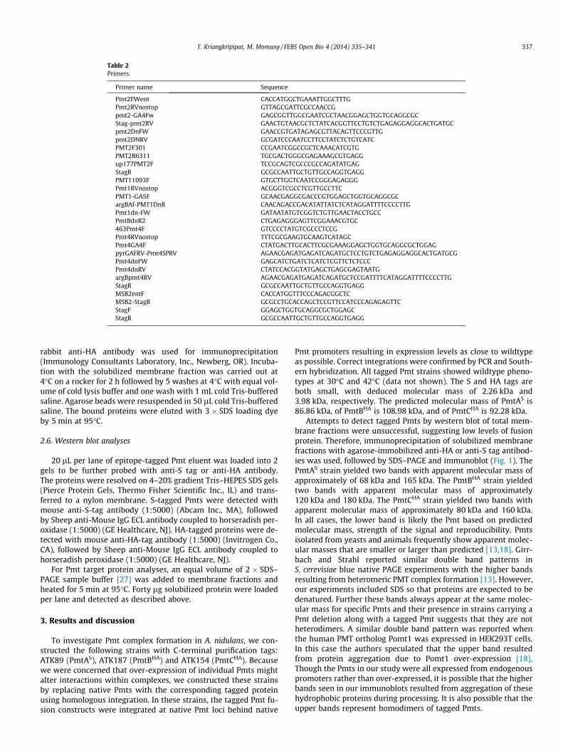
Table 2Primers.
Primer name Sequence
Pmt2FWent CACCATGGCTGAAATTGGCTTTGPmt2RVnostop GTTAGCGATTCGCCAACCGpmt2-GA4Fw GAGCGGTTGGCGAATCGCTAACGGAGCTGGTGCAGGCGCStag-pmt2RV GAACTGTAACGCTCTATCACGGTTCCTGTCTGAGAGGAGGCACTGATGCpmt2DnFW GAACCGTGATAGAGCGTTACAGTTCCCGTTGpmt2DNRV GCGATCCCAATCCTTCCTATCTCTGTCATCPMT2F301 CCGAATCGGCCGCTCAAACATCGTGPMT2R6311 TGCGACTGGGCGAGAAAGCGTGAGGup177PMT2F TCCGCAGTCGCCCGCCAGATATGAGStagR GCGCCAATTGCTGTTGCCAGGTGAGGPMT11093F GTGCTTGGTCAATCCGGGAGAGGGPmt1RVnostop ACGGGTCGCCTCGTTGCCTTCPMT1-GA5F GCAACGAGGCGACCCGTGGAGCTGGTGCAGGCGCargBAf-PMT1DnR CAACAGACCGACATATTATCTCATAGGATTTTCCCCTTGPmt1dn-FW GATAATATGTCGGTCTGTTGAACTACCTGCCPmtBdnR2 CTGAGAGGGAGTTCGGAAACGTGC463Pmt4F GTCCCCTATGTCGCCCTCCGPmt4RVnostop TTTCGCGAAGTGCAAGTCATAGCPmt4GA4F CTATGACTTGCACTTCGCGAAAGGAGCTGGTGCAGGCGCTGGAGpyrGAFRV-Pmt4SPRV AGAACGAGATGAGATCAGATGCTCCTGTCTGAGAGGAGGCACTGATGCGPmt4dnFW GAGCATCTGATCTCATCTCGTTCTCTCCCPmt4dnRV CTATCCACGGTATGAGCTGAGCGAGTAATGargBpmt4RV AGAACGAGATGAGATCAGATGCTCCGATTTTCATAGGATTTTCCCCTTGStagR GCGCCAATTGCTGTTGCCAGGTGAGGMSB2entF CACCATGGTTTCCCAGACGGCTCMSB2-StagR GCGCCTGCACCAGCTCCGTTCCATCCCAGAGAGTTCStagF GGAGCTGGTGCAGGCGCTGGAGCStagR GCGCCAATTGCTGTTGCCAGGTGAGG
T. Kriangkripipat, M. Momany / FEBS Open Bio 4 (2014) 335–341 337
rabbit anti-HA antibody was used for immunoprecipitation(Immunology Consultants Laboratory, Inc., Newberg, OR). Incuba-tion with the solubilized membrane fraction was carried out at4�C on a rocker for 2 h followed by 5 washes at 4�C with equal vol-ume of cold lysis buffer and one wash with 1 mL cold Tris-bufferedsaline. Agarose beads were resuspended in 50 lL cold Tris-bufferedsaline. The bound proteins were eluted with 3 � SDS loading dyeby 5 min at 95�C.
2.6. Western blot analyses
20 lL per lane of epitope-tagged Pmt eluent was loaded into 2gels to be further probed with anti-S tag or anti-HA antibody.The proteins were resolved on 4–20% gradient Tris–HEPES SDS gels(Pierce Protein Gels, Thermo Fisher Scientific Inc., IL) and trans-ferred to a nylon membrane. S-tagged Pmts were detected withmouse anti-S-tag antibody (1:5000) (Abcam Inc., MA), followedby Sheep anti-Mouse IgG ECL antibody coupled to horseradish per-oxidase (1:5000) (GE Healthcare, NJ). HA-tagged proteins were de-tected with mouse anti-HA-tag antibody (1:5000) (Invitrogen Co.,CA), followed by Sheep anti-Mouse IgG ECL antibody coupled tohorseradish peroxidase (1:5000) (GE Healthcare, NJ).
For Pmt target protein analyses, an equal volume of 2 � SDS–PAGE sample buffer [27] was added to membrane fractions andheated for 5 min at 95�C. Forty lg solubilized protein were loadedper lane and detected as described above.
3. Results and discussion
To investigate Pmt complex formation in A. nidulans, we con-structed the following strains with C-terminal purification tags:ATK89 (PmtAS), ATK187 (PmtBHA) and ATK154 (PmtCHA). Becausewe were concerned that over-expression of individual Pmts mightalter interactions within complexes, we constructed these strainsby replacing native Pmts with the corresponding tagged proteinusing homologous integration. In these strains, the tagged Pmt fu-sion constructs were integrated at native Pmt loci behind native
Pmt promoters resulting in expression levels as close to wildtypeas possible. Correct integrations were confirmed by PCR and South-ern hybridization. All tagged Pmt strains showed wildtype pheno-types at 30�C and 42�C (data not shown). The S and HA tags areboth small, with deduced molecular mass of 2.26 kDa and3.98 kDa, respectively. The predicted molecular mass of PmtAS is86.86 kDa, of PmtBHA is 108.98 kDa, and of PmtCHA is 92.28 kDa.
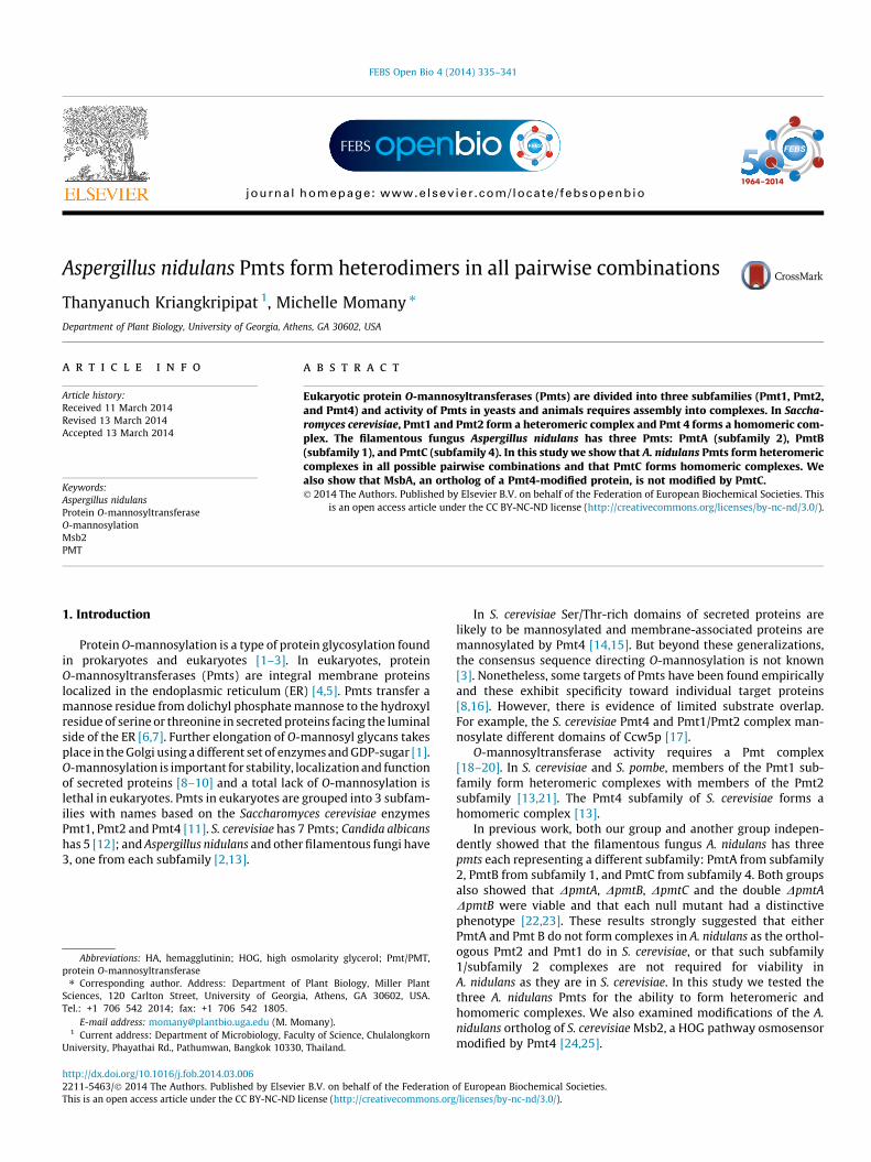
Attempts to detect tagged Pmts by western blot of total mem-brane fractions were unsuccessful, suggesting low levels of fusionprotein. Therefore, immunoprecipitation of solubilized membranefractions with agarose-immobilized anti-HA or anti-S tag antibod-ies was used, followed by SDS–PAGE and immunoblot (Fig. 1). ThePmtAS strain yielded two bands with apparent molecular mass ofapproximately of 68 kDa and 165 kDa. The PmtBHA strain yieldedtwo bands with apparent molecular mass of approximately120 kDa and 180 kDa. The PmtCHA strain yielded two bands withapparent molecular mass of approximately 80 kDa and 160 kDa.In all cases, the lower band is likely the Pmt based on predictedmolecular mass, strength of the signal and reproducibility. Pmtsisolated from yeasts and animals frequently show apparent molec-ular masses that are smaller or larger than predicted [13,18]. Girr-bach and Strahl reported similar double band patterns inS. cerevisiae blue native PAGE experiments with the higher bandsresulting from heteromeric PMT complex formation [13]. However,our experiments included SDS so that proteins are expected to bedenatured. Further these bands always appear at the same molec-ular mass for specific Pmts and their presence in strains carrying aPmt deletion along with a tagged Pmt suggests that they are notheterodimers. A similar double band pattern was reported whenthe human PMT ortholog Pomt1 was expressed in HEK293T cells.In this case the authors speculated that the upper band resultedfrom protein aggregation due to Pomt1 over-expression [18].Though the Pmts in our study were all expressed from endogenouspromoters rather than over-expressed, it is possible that the higherbands seen in our immunoblots resulted from aggregation of thesehydrophobic proteins during processing. It is also possible that theupper bands represent homodimers of tagged Pmts.
Fig. 1. Immunoprecipitation of Epitope-tagged Pmts. Western blot analyses of immunoprecipitates from ATK89 (PmtAS), ATK187 (PmtBHA) and ATK154 (PmtCHA). Proteinswere expressed from their native loci under the control of their endogenous promoters. Solubilized membrane-enriched fractions were immunoprecipitated with thecorresponding agarose immobilized antibody against the epitope tag. Immunoprecipitates were resuspended in SDS loading dye and resolved on 4–20% gradient Tris–HEPES–SDS–polyacrylamide gels, transferred to membranes, and probed with anti-S tag antibody (left) or anti-HA antibody (two blots on right). Arrows indicate Pmts.
338 T. Kriangkripipat, M. Momany / FEBS Open Bio 4 (2014) 335–341
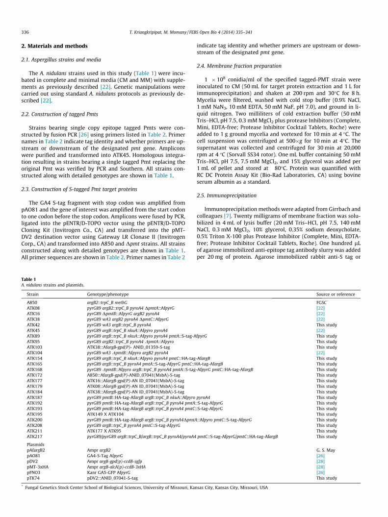
3.1. PmtA (subfamily 2) forms heteromeric complexes with PmtB(subfamily 1)
To investigate whether A. nidulans PmtA and PmtB form a het-eromeric complex like their S. cerevisiae orthologs, Pmt2 andPmt1 [13], we derived a PmtAS PmtBHA strain (ATK192) from sex-ual crosses. Immunoprecipitation was performed with agarose-immobilized anti-HA antibodies and agarose-immobilized anti-Stag antibody. The immunoprecipitate was divided into two ali-quots. One aliquot was probed with anti-S tag antibody and theother was probed with anti-HA antibody. When probed withanti-S tag antibody, the same bands that were seen in the PmtAS
strain were visible (approximately 68 kDa and 165 kDa) (Fig. 2).When probed with anti-HA antibody, the same bands that wereseen in the PmtBHA strain were visible (approximately 120 kDaand 180 kDa) (Fig. 2). Reciprocal experiments performed usinganti-HA antibodies for immunoprecipitation before immunoblot-ting showed very high background, though the results were consis-tent with heteromeric complex formation by PmtA and PmB (datanot shown). Our results show that PmtA and PmtB form hetero-meric complexes in A. nidulans.
3.2. PmtA (subfamily 2) forms heteromeric complexes with PmtC(subfamily 4)
In S. cerevisiae, Pmt2 and Pmt4 do not form complexes [13]. Toinvestigate the interactions between the A. nidulans orthologs,
Fig. 2. Immunoprecipitation of PmtA-PmtB heteromeric complexes. Solubilizedmembrane-enriched fractions from ATK192 (PmtAS PmtBHA) were immunoprecip-itated with agarose immobilized anti-S tag antibody. Co-immunoprecipitates wereresuspended in SDS loading dye and resolved on 4–20% gradient Tris–HEPES–SDS–polyacrylamide gels, transferred to membranes, and probed with anti-S tagantibody (left) or anti-HA antibody (right). Arrows indicate Pmts.
strains carrying PmtAS and PmtCHA in the presence of PmtB(pmtA::S tag, pmtC::HA, ATK165) and absence of PmtB (pmtA::Stag, pmtC::HA, DpmtB; ATK168) were derived from sexual crosses.Immunoprecipitations were performed with agarose-immobilizedanti-S tag antibody as described above. When probed with anti-HA antibody, Western blots of immunoprecipitates from bothstrains showed a band of approximately 80 kDa whether or notPmtB was present (Fig. 3). Reciprocal experiments performed usinganti-HA antibodies for immunoprecipitation before immunoblot-ting were consistent with heteromeric complex formation by PmtAand PmtC (Fig. S1). Our results show that A. nidulans PmtA forms aheteromeric complex with PmtC in the presence and absence ofPmtB.
3.3. PmtB (subfamily 1) forms heteromeric complexes with PmtC(subfamily 4)
To investigate the interactions between PmtB and PmtC, a strainbearing PmtBHA and PmtCS in the presence of PmtA (pmtB::HA,pmtC::S tag; ATK193) and absence of PmtA (pmtB::HA, pmtC::Stag, DpmtA; ATK200) were derived from sexual crosses. Immuno-precipitation and Western blots were performed as describedabove. Immunoblots probed with anti-HA antibody showed a bandof approximately 120 kDa whether or not PmtA was present(Fig. 4). Reciprocal experiments performed using anti-HA antibod-ies for immunoprecipitation before immunoblotting showed veryhigh background, though the results were consistent with hetero-meric complex formation by PmtB and PmtC (data not shown). Our
Fig. 3. Immunoprecipitation of PmtA-PmtC heteromeric complexes. Solubilizedmembrane-enriched fractions from ATK168 (PmtAS, PmtCHA, DpmtB; lane 1) andATK165 (PmtAS, PmtCHA; lane 2) were immunoprecipitated with agarose immobi-lized anti-S-tag antibody. Co-immunoprecipitates were resuspended in SDS loadingdye and resolved on 4–20% gradient Tris–HEPES–SDS–polyacrylamide gels, trans-ferred to membranes, and probed with anti-S tag antibody (left) or anti-HAantibody (right). Arrows indicate Pmts.
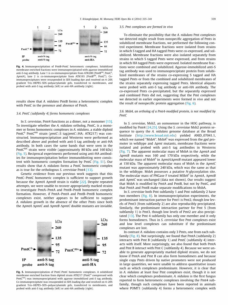
Fig. 4. Immunoprecipitation of PmtB-PmtC heteromeric complexes. Solubilizedmembrane-enriched fractions were immunoprecipitated with agarose immobilizedanti-S-tag antibody. Lane 1 is co-immunoprecipitate from ATK200 (PmtBHA, PmtCS,DpmtA), lane 2 is co-immunoprecipitate from ATK193 (PmtBHA, PmtCS). Co-immunoprecipitates were resuspended in SDS loading dye and resolved on 4–20%gradient Tris–HEPES–SDS–polyacrylamide gels, transferred to membranes, andprobed with anti-S tag antibody (left) or anti-HA antibody (right).
T. Kriangkripipat, M. Momany / FEBS Open Bio 4 (2014) 335–341 339
results show that A. nidulans PmtB forms a heteromeric complexwith PmtC in the presence and absence of PmtA.
3.4. PmtC (subfamily 4) forms homomeric complexes
In S. cerevisiae, Pmt4 functions as a dimer, not a monomer [13].To investigate whether the A. nidulans ortholog, PmtC, is a mono-mer or forms homomeric complexes in A. nidulans, a stable diploidPmtCS PmtCHA strain (pmtC::S tag/pmtC::HA; ATK217) was con-structed. Immunoprecipitation and Westerns were performed asdescribed above and probed with anti-S tag antibody or anti-HAantibody. In both cases the same bands that were seen in thePmtCHA strain were visible (approximately 80 kDa and 160 kDa)(Fig. 5). Reciprocal experiments performed using anti-HA antibod-ies for immunoprecipitation before immunoblotting were consis-tent with homomeric complex formation by PmtC (Fig. S1). Ourresults show that A. nidulans forms a PmtC homomeric complexas is true for the orthologous S. cerevisiae Pmt4 [13].
Genetic evidence from our previous work suggests that thisPmtC–PmtC homomeric complex is sufficient to support growthbecause the DpmtA DpmtB strain is viable [22]. Despite repeatedattempts, we were unable to recover appropriately marked strainsto investigate PmtA–PmtA and PmtB–PmtB homomeric complexformation. However, if PmtA–PmtA and PmtB–PmtB homomericcomplexes exist, neither appears to be sufficient to supportA. nidulans growth in the absence of the other Pmts since boththe DpmtA DpmtC and DpmtB DpmtC double mutants are inviable.
Fig. 5. Immunoprecipitation of PmtC-PmtC homomeric complexes. A solubilizedmembrane enriched fraction from diploid strain ATK217 (PmtCS coexpressed withPmtCHA) was immunoprecipitated with agarose immobilized anti-S tag antibody.Co-immunoprecipitate was resuspended in SDS loading dye and resolved on 4–20%gradient Tris–HEPES–SDS–polyacrylamide gels, transferred to membranes, andprobed with anti-S tag antibody (left) or anti-HA antibody (right).
3.5. Pmt complexes are formed in vivo
To eliminate the possibility that the A. nidulans Pmt complexeswe detected might result from nonspecific aggregation of Pmts insolubilized membrane fractions, we performed the following con-trol experiment. Membrane fractions were isolated from strainsin which S tagged and HA tagged Pmts were co-expressed, and sol-ubilized. Membrane fractions were also isolated separately fromstrains in which S tagged Pmts were expressed, and from strainsin which HA tagged Pmts were expressed. Isolated membrane frac-tions were combined and solubilized. Agarose-immobilized anti-Stag antibody was used to immunoprecipitate protein from solubi-lized membranes of the strains co-expressing S tagged and HAtagged Pmts or from the combined and solubilized membranes ofthe strains separately expressing tagged Pmts. Identical aliquotswere probed with anti-S tag antibody or anti-HA antibody. Theco-expressed Pmts co-precipitated, but the separately expressedand combined Pmts did not, suggesting that the Pmt complexeswe detected in earlier experiments were formed in vivo and notthe result of nonspecific protein aggregation (Fig. 6).
3.6. MsbA, an ortholog of a Pmt4-modified protein, is not modified byPmtC
In S. cerevisiae, Msb2, an osmosensor in the HOG pathway, ismodified by Pmt4 [24,25]. Using the S. cerevisiae Msb2 protein se-quence to query the A. nidulans genome database at the BroadInstitute (http://www.broad.mit.edu) yielded ANID_07041.1,which we named ‘‘MsbA’’. MsbAS was expressed from the gpd pro-moter in wildtype and Dpmt mutants, membrane fractions wereisolated and probed with anti-S tag antibodies in Westerns(Fig. 7). The apparent molecular mass of MsbA in the DpmtA andDpmtB mutants was 160 and 170 kDa respectively, while themolecular mass of MsbAS in DpmtADpmtB mutant appeared lowerat 150 kDa. The apparent molecular mass of MsbA in the DpmtCmutant was approximately 240 kDa, which was the same as thatin the wildtype. MsbA possesses a putative N-glycosylation site.The molecular mass of PNGase F treated MSbAS in DpmtA, DpmtBand DpmtC was unchanged (data not shown). Our results suggestthat MsbA is modified by PmtA and PmtB, but not by PmtC, andthat PmtA and PmtB make separate modifications to MsbA.
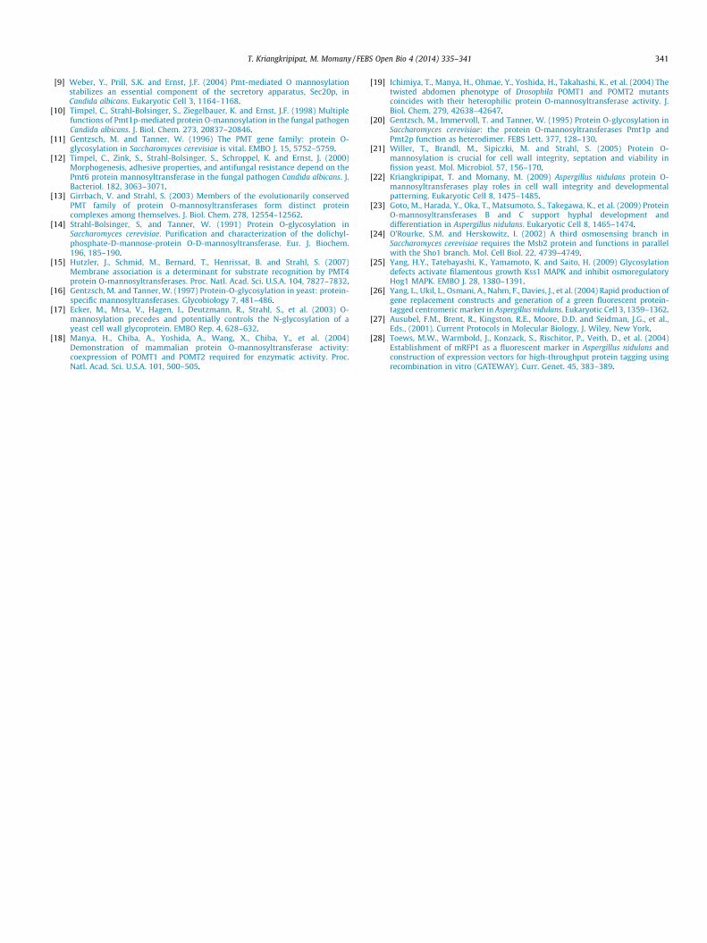
In S. cerevisiae both Pmt subfamily 1 and Pmt subfamily 2 havetwo members (Fig. 8). In immunoprecipitation experiments, thepredominant interaction partner for Pmt1 is Pmt2, though low lev-els of Pmt3 (from subfamily 2) are also reproducibly precipitated.Similarly, the predominant interaction partner for Pmt 5 (fromsubfamily 1) is Pmt3, though low levels of Pmt2 are also precipi-tated [13]. The Pmt 4 subfamily has only one member and it onlyforms homodimers. Thus in S. cerevisiae five Pmt complexes existand low level complexes can substitute if the predominantcomplexes are lost.
In contrast A. nidulans contains only 3 Pmts, one from each sub-family (Fig. 8). Not surprisingly, we found that PmtA (subfamily 2)interacts with Pmt B (subfamily 1) and PmtC (subfamily 4) inter-acts with itself. More surprisingly, we also found that both PmtAand Pmt B interact with Pmt C (subfamily 4). Because we were un-able to generate appropriately marked diploid strains, we do notknow if PmtA and Pmt B can also form homodimers and becausesingle copy Pmts driven by native promoters were not producedin large quantities, we were unable to address quantitative issuessuch as which complexes predominate. However, it is clear thatin A. nidulans at least four Pmt complexes exist, though it is notclear which complexes predominate. A. nidulans is the first fungusreported to form heteromeric complexes involving the Pmt4 sub-family, though such complexes have been reported in animalswhere POMT1 (subfamily 4) forms a heteromeric complex with
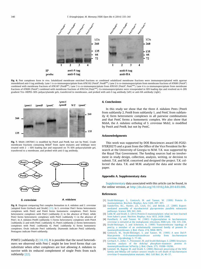
Fig. 6. Pmt complexes form in vivo. Solubilized membrane enriched fractions or combined solubilized membrane fractions were immunoprecipitated with agaroseimmobilized anti-S tag antibody. Lane 1 is co-immunoprecipitate from ATK192 (PmtAS, PmtBHA). Lane 2 is co-immunoprecipitate from membrane fractions of ATK89 (PmtAS)combined with membrane fractions of ATK187 (PmtBHA). Lane 3 is co-immunoprecipitate from ATK165 (PmtAS, PmtCHA). Lane 4 is co-immunoprecipitate from membranefractions of ATK89 (PmtAS) combined with membrane fractions of ATK154 (PmtCHA). Co-immunoprecipitates were resuspended in SDS loading dye and resolved on 4–20%gradient Tris–HEPES–SDS–polyacrylamide gels, transferred to membranes, and probed with anti-S tag antibody (left) or anti-HA antibody (right).
Fig. 7. MsbA (AN7041) is modified by PmtA and PmtB, but not by PmtC. Crudemembrane fractions containing MsbAS from Dpmt mutants and wildtype weretreated with 2 � SDS loading dye and separated on 7% SDS–polyacrylamide gel,transferred to a membrane, and probed with anti-S tag antibody.
Fig. 8. Diagram comparing Pmt complex formation in A. nidulans and S. cerevisiae(adapted from Girrbach and Strahl) [13]. In S. cerevisiae Pmt1 forms heteromericcomplexes with Pmt2 and Pmt4 forms homomeric complexes. Pmt1 formsheteromeric complexes with Pmt3 (subfamily 2) in the absence of Pmt2, whilePmt2 forms heteromeric complexes with Pmt5 (subfamily 1) in the absence ofPmt1. In A. nidulans PmtB (subfamily 1) forms heteromeric complexes with PmtA(subfamily 2) and with PmtC (subfamily 4). PmtA (subfamily 2) forms heteromericcomplexes with PmtC (subfamily 4). PmtC (subfamily 4) forms homomericcomplexes. Ovals indicate Pmt1 subfamily. Diamonds indicate Pmt2 subfamily.Pentagons indicate Pmt4 subfamily.
340 T. Kriangkripipat, M. Momany / FEBS Open Bio 4 (2014) 335–341
POMT2 (subfamily 2) [18,19]. It is possible that the novel heterodi-mers we observed with Pmt C might be low level forms that cansubstitute when other complexes are lost allowing A. nidulans tosurvive with its reduced complement of single Pmts from eachsubfamily [22].
4. Conclusions
In this study we show that the three A. nidulans Pmts (PmtAfrom subfamily 2, PmtB from subfamily 1, and PmtC from subfam-ily 4) form heteromeric complexes in all pairwise combinationsand that PmtC forms a homomeric complex. We also show thatMsbA, the A. nidulans ortholog of S. cerevisiae Msb2, is modifiedby PmtA and PmtB, but not by PmtC.
Acknowledgments
This work was supported by DOE Biosciences award DE-FG02-97ER20275 and a grant from the Office of the Vice President for Re-search at the University of Georgia to M.M. T.K. was supported bythe Royal Thai Government. The funding sources had no involve-ment in study design, collection, analysis, writing, or decision tosubmit. T.K. and M.M. conceived and designed the project. T.K. col-lected the data. T.K. and M.M. analyzed the data and wrote thepaper.
Appendix A. Supplementary data
Supplementary data associated with this article can be found, inthe online version, at http://dx.doi.org/10.1016/j.fob.2014.03.006.
References
[1] Strahl-Bolsinger, S., Gentzsch, M. and Tanner, W. (1999) Protein O-mannosylation. Biochim. Biophys. Acta 1426, 297–307.
[2] VanderVen, B.C., Harder, J.D., Crick, D.C. and Belisle, J.T. (2005) Export-mediated assembly of mycobacterial glycoproteins parallels eukaryoticpathways. Science 309, 941–943.
[3] Loibl, M. and Strahl, S. (2013) Protein O-mannosylation: what we have learnedfrom baker’s yeast. Biochim. Biophys. Acta 1833, 2438–2446.
[4] Haselbeck, A. and Tanner, W. (1983) O-glycosylation in Saccharomycescerevisiae is initiated at the endoplasmic reticulum. FEBS Lett. 158, 335–338.
[5] Strahl-Bolsinger, S. and Scheinost, A. (1999) Transmembrane topology ofpmt1p, a member of an evolutionarily conserved family of protein O-mannosyltransferases. J. Biol. Chem. 274, 9068–9075.
[6] Gentzsch, M., Strahl-Bolsinger, S. and Tanner, W. (1995) A new Dol-P-Man:protein O-D-mannosyltransferase activity from Saccharomycescerevisiae. Glycobiology 5, 77–82.
[7] Girrbach, V., Zeller, T., Priesmeier, M. and Strahl-Bolsinger, S. (2000) Structure-function analysis of the dolichyl phosphate-mannose: protein O-mannosyltransferase ScPmt1p. J. Biol. Chem. 275, 19288–19296.
[8] Lommel, M., Bagnat, M. and Strahl, S. (2004) Aberrant processing of the WSCfamily and Mid2p cell surface sensors results in cell death of Saccharomycescerevisiae O-mannosylation mutants. Mol. Cell Biol. 24, 46–57.
T. Kriangkripipat, M. Momany / FEBS Open Bio 4 (2014) 335–341 341
[9] Weber, Y., Prill, S.K. and Ernst, J.F. (2004) Pmt-mediated O mannosylationstabilizes an essential component of the secretory apparatus, Sec20p, inCandida albicans. Eukaryotic Cell 3, 1164–1168.
[10] Timpel, C., Strahl-Bolsinger, S., Ziegelbauer, K. and Ernst, J.F. (1998) Multiplefunctions of Pmt1p-mediated protein O-mannosylation in the fungal pathogenCandida albicans. J. Biol. Chem. 273, 20837–20846.
[11] Gentzsch, M. and Tanner, W. (1996) The PMT gene family: protein O-glycosylation in Saccharomyces cerevisiae is vital. EMBO J. 15, 5752–5759.
[12] Timpel, C., Zink, S., Strahl-Bolsinger, S., Schroppel, K. and Ernst, J. (2000)Morphogenesis, adhesive properties, and antifungal resistance depend on thePmt6 protein mannosyltransferase in the fungal pathogen Candida albicans. J.Bacteriol. 182, 3063–3071.
[13] Girrbach, V. and Strahl, S. (2003) Members of the evolutionarily conservedPMT family of protein O-mannosyltransferases form distinct proteincomplexes among themselves. J. Biol. Chem. 278, 12554–12562.
[14] Strahl-Bolsinger, S. and Tanner, W. (1991) Protein O-glycosylation inSaccharomyces cerevisiae. Purification and characterization of the dolichyl-phosphate-D-mannose-protein O-D-mannosyltransferase. Eur. J. Biochem.196, 185–190.
[15] Hutzler, J., Schmid, M., Bernard, T., Henrissat, B. and Strahl, S. (2007)Membrane association is a determinant for substrate recognition by PMT4protein O-mannosyltransferases. Proc. Natl. Acad. Sci. U.S.A. 104, 7827–7832.
[16] Gentzsch, M. and Tanner, W. (1997) Protein-O-glycosylation in yeast: protein-specific mannosyltransferases. Glycobiology 7, 481–486.
[17] Ecker, M., Mrsa, V., Hagen, I., Deutzmann, R., Strahl, S., et al. (2003) O-mannosylation precedes and potentially controls the N-glycosylation of ayeast cell wall glycoprotein. EMBO Rep. 4, 628–632.
[18] Manya, H., Chiba, A., Yoshida, A., Wang, X., Chiba, Y., et al. (2004)Demonstration of mammalian protein O-mannosyltransferase activity:coexpression of POMT1 and POMT2 required for enzymatic activity. Proc.Natl. Acad. Sci. U.S.A. 101, 500–505.
[19] Ichimiya, T., Manya, H., Ohmae, Y., Yoshida, H., Takahashi, K., et al. (2004) Thetwisted abdomen phenotype of Drosophila POMT1 and POMT2 mutantscoincides with their heterophilic protein O-mannosyltransferase activity. J.Biol. Chem. 279, 42638–42647.
[20] Gentzsch, M., Immervoll, T. and Tanner, W. (1995) Protein O-glycosylation inSaccharomyces cerevisiae: the protein O-mannosyltransferases Pmt1p andPmt2p function as heterodimer. FEBS Lett. 377, 128–130.
[21] Willer, T., Brandl, M., Sipiczki, M. and Strahl, S. (2005) Protein O-mannosylation is crucial for cell wall integrity, septation and viability infission yeast. Mol. Microbiol. 57, 156–170.
[22] Kriangkripipat, T. and Momany, M. (2009) Aspergillus nidulans protein O-mannosyltransferases play roles in cell wall integrity and developmentalpatterning. Eukaryotic Cell 8, 1475–1485.
[23] Goto, M., Harada, Y., Oka, T., Matsumoto, S., Takegawa, K., et al. (2009) ProteinO-mannosyltransferases B and C support hyphal development anddifferentiation in Aspergillus nidulans. Eukaryotic Cell 8, 1465–1474.
[24] O’Rourke, S.M. and Herskowitz, I. (2002) A third osmosensing branch inSaccharomyces cerevisiae requires the Msb2 protein and functions in parallelwith the Sho1 branch. Mol. Cell Biol. 22, 4739–4749.
[25] Yang, H.Y., Tatebayashi, K., Yamamoto, K. and Saito, H. (2009) Glycosylationdefects activate filamentous growth Kss1 MAPK and inhibit osmoregulatoryHog1 MAPK. EMBO J. 28, 1380–1391.
[26] Yang, L., Ukil, L., Osmani, A., Nahm, F., Davies, J., et al. (2004) Rapid production ofgene replacement constructs and generation of a green fluorescent protein-tagged centromeric marker in Aspergillus nidulans. Eukaryotic Cell 3, 1359–1362.
[27] Ausubel, F.M., Brent, R., Kingston, R.E., Moore, D.D. and Seidman, J.G., et al.,Eds., (2001). Current Protocols in Molecular Biology, J. Wiley, New York.
[28] Toews, M.W., Warmbold, J., Konzack, S., Rischitor, P., Veith, D., et al. (2004)Establishment of mRFP1 as a fluorescent marker in Aspergillus nidulans andconstruction of expression vectors for high-throughput protein tagging usingrecombination in vitro (GATEWAY). Curr. Genet. 45, 383–389.