asn 38th annual meeting neuroimaging for clinicians, … - carotid duplex protocol.pdf · asn 38th...
TRANSCRIPT

Carotid Duplex Protocol ASN 38th Annual Meeting
Neuroimaging for Clinicians, by Clinicians January 15-18, 2015
Carefree, AZ
Patricia A. (Tish) Poe, BA RVT FSVU Director, Quality Assurance
NAVIX Diagnostix

No disclosures

Policy and Procedure
Policy and Procedure
These documents cover the big picture of how an area of testing is approached including:
• Indications
• Patient history
• Physical exam
• Risk factors
• Patient positioning
• Exam technique considerations

Protocol: image sequence Sequence Location Level Orientation Mode Label
1. Common carotid artery Trans/Long Split B CCA
2. Bifurcation Trans/Long Split B Bif (Label ICA/ECA)
3. Internal carotid artery Proximal Trans/Long Split B ICA Proximal
4. Internal carotid artery Mid Trans/Long Split B ICA Mid
5. Common carotid artery Proximal Long C/D Calcs Package
6. Common carotid artery Mid Long C/D Calcs Package
7. Common carotid artery Distal Long C/D Calcs Package
8. External carotid artery Long C/D Calcs Package
9. Internal carotid artery Proximal Long C/D Calcs Package
10. Internal carotid artery Mid Long C/D Calcs Package
11. Internal carotid artery Distal Long C/D Calcs Package
12. Vertebral Artery Mid Long C/D Calcs Package
13. Subclavian Artery Proximal Long B SCA
14. Subclavian Artery Proximal Long C/D Calcs Package

Evaluate fully
B-mode Imaging
1. Transverse from clavicle to mandible
a. Proximal common carotid artery (CCA)
b. Mid CCA
c. Distal CCA
d. Bifurcation
e. Proximal internal carotid artery (ICA)
f. Mid/distal ICA

Evaluate fully
2. Longitudinal plane from clavicle to mandible
a. Proximal common carotid artery (CCA)
b. Mid CCA
c. Distal CCA
d. External carotid artery (ECA)
e. Proximal internal carotid artery (ICA)
f. Mid ICA
g. Distal ICA

Identify the location in the CCA where velocity is uses to calculate ICA/CCA ratio
• Mid CCA velocity to be used in the ratio should be obtained about 2 cm proximal to the flow divider
• Do not say “bulb” as this is not a region but an anatomic widening that occurs in the distal CCA to proximal ICA
• Watch angle correction: align to vessel walls

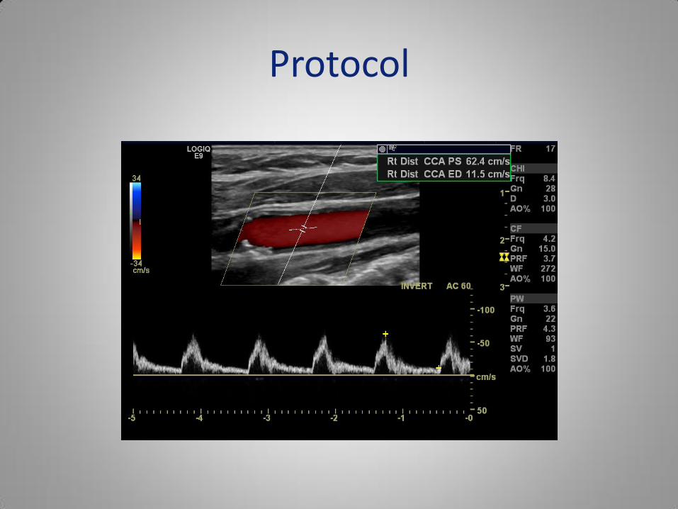
Protocol

Protocol

Protocol
Protocol

Protocol

Protocol

Protocol

Protocol

Protocol

Protocol

Protocol

Protocol

Protocol

Protocol
What is missing from our protocol for the image sequence on the right?
Common carotid artery, Mid, Long, C/D
Protocol
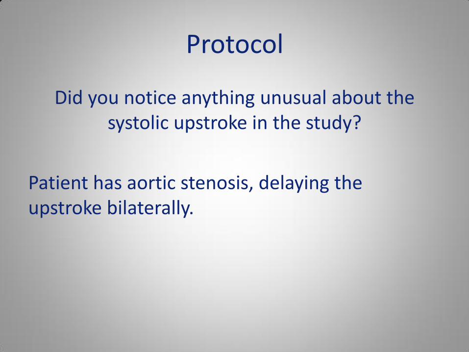
Did you notice anything unusual about the systolic upstroke in the study?
Patient has aortic stenosis, delaying the upstroke bilaterally.
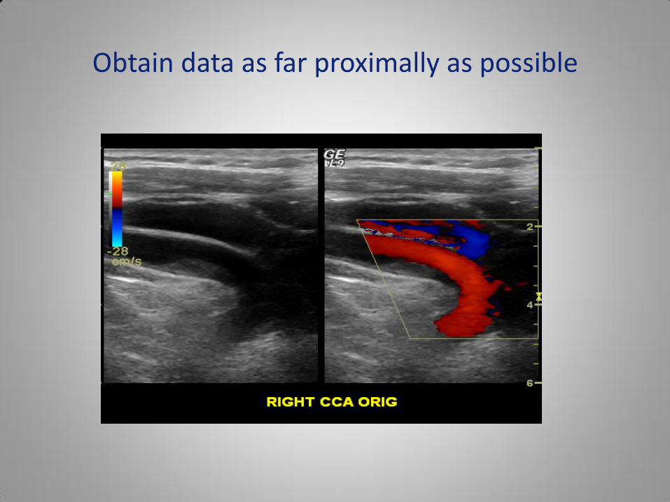
Obtain data as far proximally as possible

Subclavian artery evaluation Obtain and document bilateral brachial blood pressures prior to performing cerebrovascular examinations.
Blood pressure symmetry is important in determining the presence and severity of subclavian artery stenosis.
If you find a pressure gradient of ≥20 mmHg, this may be indicative of a significant pressure reducing lesion in the upper extremity arteries.
Bilateral subclavian artery stenosis may be present creating lowered
BP bilaterally.

Subclavian Artery
The proximal subclavian artery is evaluated in all patients, with additional images added when stenosis is identified. The following signs, symptoms, or conditions increase suspicion for hemodynamically significant subclavian artery disease:
• Discrepancy in brachial blood pressures ≥20 mmHg
• Abnormal flow in the extracranial vertebral artery
• Bruit of unknown origin
• Velocity increase in the proximal subclavian artery

Subclavian Artery
• There are no firm velocity criteria for the subclavian or brachiocephalic (innominate) arteries
• Considering that the normal velocity range in the adult aorta, carotid and femoral arteries is 60-100 cm/sec, velocities over 200 cm/sec are suspicious for >50% stenosis
• Proceed as far proximally as possible and include waveforms more distally to look for turbulence or delayed upstroke

Carotid Duplex Cases: Case 5
Right Subclavian prox
341 PSV

Carotid Duplex Cases: Case 5
Right Subclavian mid
203 PSV w/ turbulence

Carotid Duplex Cases: Case 5
Left Subclavian
326 PSV
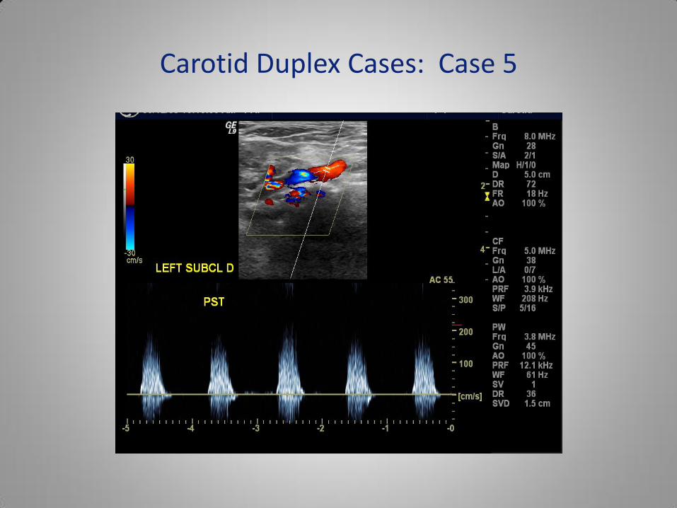
Carotid Duplex Cases: Case 5

Optimize B-mode

Optimize B-mode
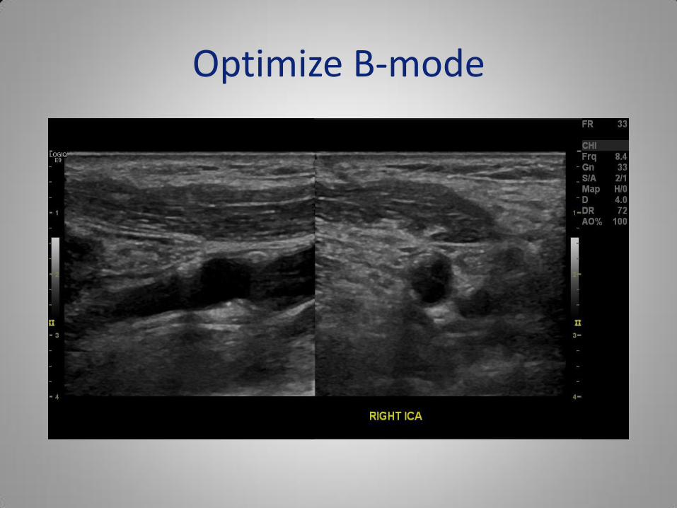
Optimize B-mode

Optimize gain settings
Spectral Doppler gain appropriate Spectral Doppler gain too high and then decreased

Color and spectral Gain
Spectral Doppler gain too high Color Doppler gain too high

Fully document occlusion

Fully document occlusion
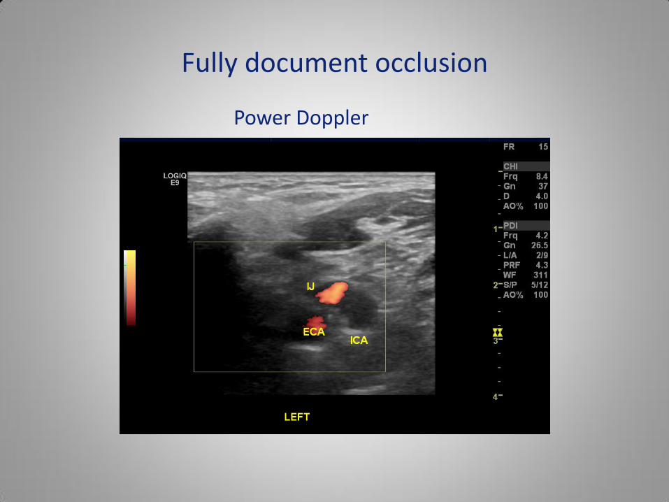
Fully document occlusion
Power Doppler

Reporting basics
The technologist should get the following information from each patient about the indications/symptoms for which he/she is complaining and document the answers in the patient record:
Onset Frequency
Duration Radiation
Methods of relief Associated symptoms

Reporting basics
Identification of physical findings must be evaluated and documented in the patient record, noting location and severity.
These physical signs include, but are not limited to:
Gait
Strength of grip
Facial drooping
Speech patterns or slurring

Identify when to add TCD: always of in view of findings or symptoms
In NAVIX:
Complete TCD added
• >50% ICA stenosis
• Symptoms of TIA, CVA or VBI with no significant extracranial carotid disease

Identify when to add TCD: always of in view of findings or symptoms
In NAVIX:
Limited TCD (OA and Siphon) added:
• Plaque noted during the Extracranial evaluation
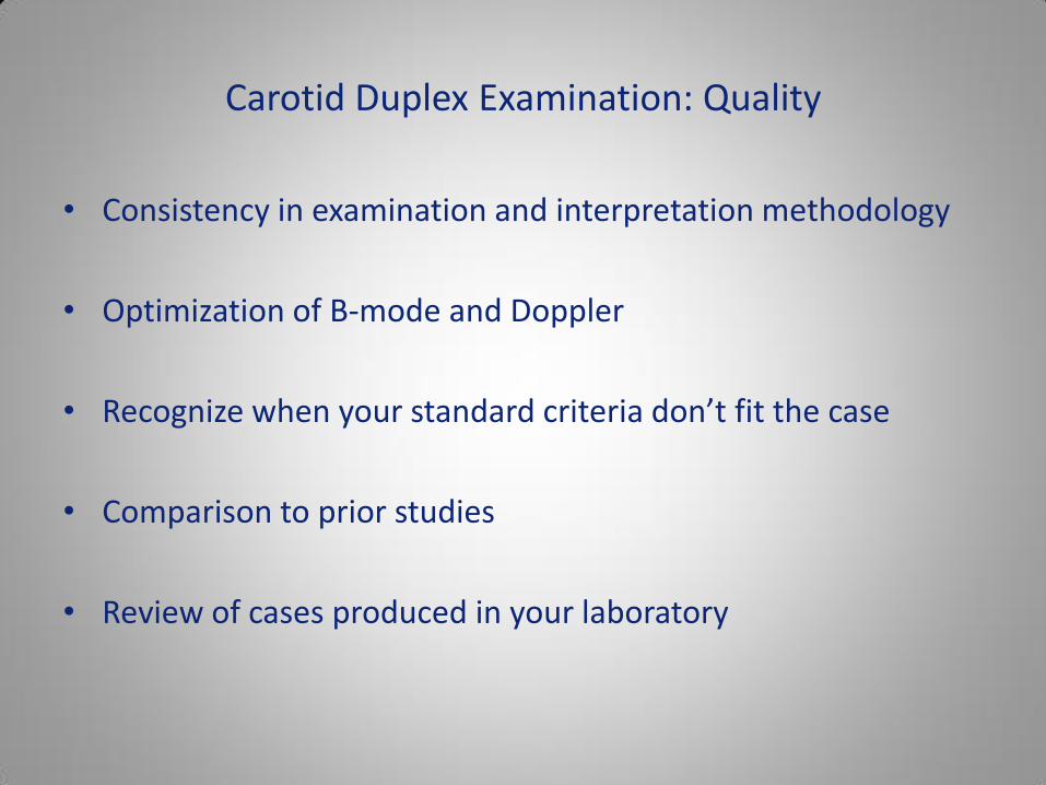
Carotid Duplex Examination: Quality
• Consistency in examination and interpretation methodology
• Optimization of B-mode and Doppler
• Recognize when your standard criteria don’t fit the case
• Comparison to prior studies
• Review of cases produced in your laboratory

Thank you for your attention and for this beautiful meeting location!