asian archives of pathology
TRANSCRIPT

Print ISSN: 1905-9183 Online ISSN: 2673-0499
ASIAN ARCHIVES OF PATHOLOGY THE OFFICIAL JOURNAL OF THE ROYAL COLLEGE OF PATHOLOGISTS OF THAILAND
Volume 1
Number 2
April – June 2019
EDITORIAL BOARD
Editor-in-Chief
Dr Chetana Ruangpratheep MD, FRCPath (Thailand), MSc, PhD Phramongkutklao College of Medicine, Bangkok, Thailand
Associate Editors ▪ Associate Professor Dr Mongkol Kunakorn
MD, FRCPath (Thailand) Ramathibodi Hospital, Mahidol University, Bangkok, Thailand
▪ Associate Professor Dr Theerapong Krajaejun MD, FRCPath (Thailand) Ramathibodi Hospital, Mahidol University, Bangkok, Thailand
▪ Assistant Professor Dr Thirayost Nimmanon MD, FRCPath (Thailand), MRes, PhD Phramongkutklao College of Medicine, Bangkok, Thailand
▪ Assistant Professor Dr Wisarn Worasuwannarak MD, FRCPath (Thailand) Ramathibodi Hospital, Mahidol University, Bangkok, Thailand
▪ Dr Anirut Worawat MD, FRCPath (Thailand) Siriraj Hospital, Mahidol University, Bangkok, Thailand
▪ Dr Panuwat Chutivongse MD, FRCPath (Thailand) Chulalongkorn University, Bangkok, Thailand
Editorial Consultant
Professor Dr Vorachai Sirikulchayanonta MD, FRCPath (Thailand) Rangsit University, Pathumtani, Thailand

Asian Archives of Pathology i
Volume 1 | Number 2 | April – June 2019
ABOUT THE JOURNAL
Aims and Scope Asian Archives of Pathology (AAP) is an open access, peer-reviewed journal. The journal was first published in 2002 under the Thai name “วารสารราชวทยาลยพยาธแพทยแหงประเทศไทย” and English name “Journal of the Royal College of Pathologists of Thailand”. The journal is a publication for workers in all disciplines of pathology and forensic medicine. In the first 3 years (volumes), the journal was published every 4 months. Until 2005, the journal has changed its name to be “Asian Archives of Pathology: The Official Journal of the Royal College of Pathologists of Thailand”, published quarterly to expand the collaboration among people in the fields of pathology and forensic medicine in the Asia-Pacific regions and the Western countries. The full articles of the journal are appeared in either Thai or English. However, the abstracts of all Thai articles are published in both Thai and English languages. The journal features letters to the editor, original articles, review articles, case reports, case illustrations, and technical notes. Diagnostic and research areas covered consist of (1) Anatomical Pathology (including cellular pathology, cytopathology, haematopathology, histopathology, immunopathology, and surgical pathology); (2) Clinical Pathology (Laboratory Medicine) [including blood banking and transfusion medicine, clinical chemistry (chemical pathology or clinical biochemistry), clinical immunology, clinical microbiology, clinical toxicology, cytogenetics, parasitology, and point-of-care testing]; (3) Forensic Medicine (Legal Medicine or Medical Jurisprudence) (including forensic science and forensic pathology); (4) Molecular Medicine (including molecular genetics, molecular oncology, and molecular pathology); (5) Pathobiology; and (6) Pathophysiology. All issues of our journal have been printed in hard copy since the beginning. Around the late 2014, we developed our website (www.asianarchpath.com) in order to increase our visibility. We would like to acknowledge that our journal has been sponsored by the Royal College of Pathologists of Thailand. We have the policy to disseminate the verified scientific knowledge to the public on a non-profit basis. Hence, we have not charged the authors whose manuscripts have been submitted or accepted for publication in our journal. On the other hand, if any authors request a printed copy of the journal issue containing the articles, each of the copied journals costs 450 bahts for Thai authors and 30 United States dollars (USD) for international authors. Publication Frequency Four issues per year

Asian Archives of Pathology ii
Volume 1 | Number 2 | April – June 2019
Disclaimer The Royal College of Pathologists of Thailand and Editorial Board cannot be held responsible for errors or any consequences arising from the use of information contained in Asian Archives of Pathology. It should also be noted that the views and opinions expressed in this journal do not necessarily reflect those of The Royal College of Pathologists of Thailand and Editorial Board.

Asian Archives of Pathology iii
Volume 1 | Number 2 | April – June 2019
MANUSCRIPT REVIEWERS
▪ Professor Dr Aileen Wee
MBBS, FRCPath, FRCPA National University Hospital, Singapore
▪ Professor Dr Eiichi Morii MD, PhD Osaka University Hospital, Osaka, Japan
▪ Professor Dr Jasvir Khurana MBBS, FCAP Temple University, Lewis Katz School of Medicine, Pennsylvania, The United States of America
▪ Professor Dr Paisit Paueksakon MD, FRCPath (Thailand), FCAP Vanderbilt University School of Medicine, Tennessee, The United States of America
▪ Professor Dr Nidhi Chongchitnant MD, FRCPath (Thailand) Bangkok Hospital, Bangkok, Thailand
▪ Professor Dr Vorachai Sirikulchayanonta MD, FRCPath (Thailand) Rangsit University, Pathumtani, Thailand
▪ Professor Dr Oytip Na-thalang PhD Thammasat University Rangsit Campus, Pathumtani, Thailand
▪ Associate Professor Dr Phaibul Punyarit MD, FCAP, FRCPath (Thailand) Bumrungrad International Hospital, Bangkok, Thailand
▪ Assistant Professor Dr Yingluck Visessiri MD, FRCPath (Thailand) Ramathibodi Hospital, Mahidol University, Bangkok, Thailand
▪ Assistant Professor Dr Pasra Arnutti PhD Phramongkutklao College of Medicine, Bangkok, Thailand
▪ Dr Jutatip Kintarak MD, FRCPath (Thailand) Thammasat University Rangsit Campus, Pathumtani, Thailand

Asian Archives of Pathology iv
Volume 1 | Number 2 | April – June 2019
▪ Dr Kantang Satayasoontorn MD, FRCPath (Thailand) Army Institute of Pathology, Bangkok, Thailand
▪ Dr Mongkon Charoenpitakchai MD, FRCPath (Thailand) Phramongkutklao College of Medicine, Bangkok, Thailand
▪ Dr Sivinee Charoenthammaraksa MD, FRCPath (Thailand) Bumrungrad International Hospital, Bangkok, Thailand
▪ Dr Sorranart Muangsomboon MD, FRCPath (Thailand) Siriraj Hospital, Mahidol University, Bangkok, Thailand

Asian Archives of Pathology
Volume 1 | Number 2 | April – June 2019
CONTENTS
About the journal ………………………………………………………………………………………………………… i
Aims and scope ……………………………………………………………………………………………………………. i Publication frequency …………………………………………………………………………………………………. i Disclaimer …………………………………………………………………………………………………………………….. ii
Manuscript reviewers …………………………………………………………………………………………………… iii Original Article ……………………………………………………………………………………………………………… 1 ▪ Assessment of the usefulness of the knowledge of pathology ………………………….
for clinical medical students – a multicentre study 1
Ijeoma Angela Meka, Helen Chioma Okoye, Angela Ogechukwu Ugwu, Isah Adagiri Yahaya, Ochukor Otokunefor, Olugbenga Olalekan Ojo, and Emmanuel Onyebuchi Ugwu
Review Article ………………………………………………………………………………………………………………. 13 ▪ The essentials of vascular pathology ………………………………………………………………….. 13
Chetana Ruangpratheep
Case Report ………………………………………………………………………………………………………………….. 34 ▪ Heart transplant for multiple recurrences of familial cardiac ……………………………
myxomas in an adolescent patient: a case report and literature review
34
Thiyaphat Laohawetwanit, Poonchavist Chantranuwatana, and Pat Ongcharit
Appendix 1: Information for authors ………………………………………………………………………….. 45 Categories of manuscripts …………………………………………………………………………………………… 46 Organisation of manuscripts ……………………………………………………………………………………….. 48 Proofreading ………………………………………………………………………………………………………………… 54 Revised manuscripts ……………………………………………………………………………………………………. 54
Appendix 2: Benefits of publishing with Asian Archives of Pathology ……………………… 55 Appendix 3: Submission of the manuscripts ……………………………………………………………… 56 Appendix 4: Contact the journal ………………………………………………………………………………… 57 Appendix 5: Support the journal ………………………………………………………………………………… 58

Asian Archives of Pathology 1
Volume 1 | Number 2 | April – June 2019
ORIGINAL ARTICLE
Assessment of the usefulness of the knowledge of pathology for clinical medical
students – a multicentre study
Ijeoma Angela Meka1*, Helen Chioma Okoye2, Angela Ogechukwu Ugwu2, Isah Adagiri Yahaya3, Ochukor Otokunefor4, Olugbenga Olalekan Ojo5, and
Emmanuel Onyebuchi Ugwu6
1 Department of Chemical Pathology, College of Medicine, University of Nigeria, Ituku-Ozalla Campus, Enugu, Nigeria
2 Department of Haematology and Immunology, College of Medicine, University of Nigeria, Ituku-Ozalla Campus, Enugu, Nigeria
3 Department of Chemical Pathology and Immunology, Bayero University / Aminu Kano Teaching Hospital, Kano, Nigeria
4 Department of Chemical Pathology, College of Medicine, University of Port Harcourt, Rivers State, Nigeria
5 Department of Surgery, Lagos University Teaching Hospital, Lagos, Nigeria 6 Department of Obstetrics and Gynaecology, College of Medicine, University of Nigeria,
Ituku-Ozalla Campus, Enugu, Nigeria * Correspondence to: Ijeoma Angela Meka, Department of Chemical Pathology, College of Medicine, University of Nigeria,
Ituku-Ozalla Campus, Enugu, Nigeria. PO Box 15514, UNEC Post Office Enugu, Nigeria. Telephone: (+234) 703 096 7673 Email: [email protected], [email protected]
Conflict of interest: The authors declare that they have no conflicts of interest with the contents of this article.

Asian Archives of Pathology 2
Volume 1 | Number 2 | April – June 2019
Abstract Pathology courses are taught to undergraduate medical students to provide them with basic foundation for clinical sciences. However, there is hardly any form of assessment of how relevant and helpful these pathology courses are to the students in the clinical years and even beyond. The authors set out to determine the extent of use of the knowledge of pathology in students’ appreciation of clinical medicine. The study involved final year medical students recruited from four accredited medical schools in Nigeria. Data was obtained using self-administered semi-structured questionnaires. A total of 310 final year medical students with mean (standard deviation) age of 22.5 (3.8) years participated in the study. Most useful pathology courses versus clinical courses for respondents were: 155 (50.0%) Morbid Anatomy and Haematology vs Obstetrics and Gynaecology; 181 (58.4%) Haematology vs Paediatrics; 239 (77.1%) Morbid Anatomy vs Surgery; and 246 (79.4%) Haematology vs Medicine. The proportion of respondents who indicated interest to pursue a career in pathology was 83 (26.8 %). Pathology remains relevant and very useful for students’ understanding of clinical courses. However, further work needs to be done to elucidate steps required to attract the younger generation into this branch of medicine. Keywords: clinical; haematology; medical students; morbid anatomy; pathology

Asian Archives of Pathology 3
Volume 1 | Number 2 | April – June 2019
Introduction Pathology may be defined as the science of the causes and effects of diseases, especially the branch of medicine that deals with the laboratory examination of samples of body tissue for diagnostic or forensic purposes(1). It also refers to the study of abnormal anatomy, biochemistry, and physiology at gross (organ), tissue, cellular, biochemical, and molecular levels; for the purpose of diagnosis and management of diseases. A good knowledge of pathology is essential to effective clinical practice. It is a medical specialty which deals with pathological processes underlying disease processes. In many medical schools, it is incorporated into the undergraduate medical school curriculum in the third or fourth year. Pathology generally has four arms/branches which are Chemical/Clinical Pathology, Haematology, Morbid Anatomy, and Medical Microbiology. Medical students are trained in these four areas while studying pathology as a basic and compulsory requirement for proceeding to the clinical courses. It thus serves as a first introduction to human disease processes in the undergraduate years. Chemical Pathology: This is also known as Clinical Chemistry, Clinical Pathology or Clinical Biochemistry. This branch of pathology deals with biochemically investigating bodily fluids such as blood, urine, saliva, pleural fluid, ascetic fluid, and cerebrospinal fluid(2). It is the study of the biochemical basis of diseases, and the application of biochemical and molecular techniques in diagnosis. The purpose of Chemical Pathology is the understanding of the biochemical derangements due to disease processes. The ultimate target is to ensure that physicians make use of Chemical Pathology investigations in a cost effective manner by rational test selection and thoughtful interpretation. Haematology: This is the branch of medical science concerned with diseases of the blood and blood-forming tissues(3). It deals with current and evolving knowledge of the pathogenesis, clinical and laboratory features, management and treatment of a wide range of blood and bone marrow disorders. Morbid Anatomy: This is also known as Anatomic or Anatomical Pathology. This medical specialty is concerned with the diagnosis of disease based on morphology of cells and tissues, the macroscopic and microscopic examination of organs and tissues(4). The procedures used in Anatomic Pathology include gross and microscopic examination, immunohistochemistry, cytopathology, tissue cytogenetic, and in situ hybridisation among others. Medical Microbiology: This arm of pathology works to support and oversee the prevention, diagnosis and treatment of illness caused by microorganisms (viruses, fungi, and parasites). Medical microbiologists strive to identify the best treatment for particular infectious diseases and give advice on the best samples to collect and also the technique of the sample collection to diagnose an infection, such as a swab, blood, cerebrospinal fluid or urine test(5). Medical Microbiology is also strongly integrated in the study and identification of strategies to combating antimicrobial resistance.

Asian Archives of Pathology 4
Volume 1 | Number 2 | April – June 2019
Evaluation of students’ perception in the use of pathology is essential to ascertain their knowledge and understanding of the information passed in the course of pathology teaching. There is indeed a dearth of data in Nigeria regarding this subject. It is important to know if students have acquired adequate knowledge to help them in their clinical courses and how effective the knowledge gained is in aiding understanding of clinical courses. In line with this evaluation, a study(6) in the United Kingdom has reported that the majority, 47 (67%) trainees in their study felt that their undergraduate courses had not prepared them for their membership exams, and that they were disadvantaged in having to learn pathology from first principles rather than build on the basics they hoped to know already. In terms of career choices among medical students, previous studies(7-10) both within and outside Nigeria, have documented low preferences of pathology as a career when compared with other sub-specialties. However, there is yet to be a description of the students’ choices among the various components of pathology. This study therefore aims to explore the extent of the use of the knowledge of pathology by medical students in their clinical years, and go further to characterise the pattern of students’ career choices among the various components of pathology. This will help to provide much needed data and fill the currently existing gap in literature on this topic, especially in Nigeria. Materials and Methods Study location: This comparative cross-sectional study was carried out in four accredited Nigerian Universities namely, University of Nigeria (UNN), University of Lagos (UNILAG), University of Port Harcourt (UNIPORT), and Bayero University, Kano (BUK) between March and October, 2018. These universities were selected to represent four major regions of Nigeria thus; University of Nigeria, Nsukka represented the South-East region; University of Port Harcourt represented the South-South region; University of Lagos represented the South-West; and Bayero University, Kano represented the Northern region. Study design: Participants were recruited from the final year medical students of the participating universities who had been exposed to all aspects of clinical medicine. Data was collected using pretested self-administered semi-structured questionnaires. The questionnaire used was designed by the researchers. Participants completed the questionnaires after the purpose of the study was explained to them and confidentiality of data assured. The questionnaires were pretested using ten students, and then assessed for completeness of data, ease of filling the questionnaires, clarity of questions and appropriate response options. The final draft of the questionnaires used for data collection assessed socio-demographic characteristics like age,

Asian Archives of Pathology 5
Volume 1 | Number 2 | April – June 2019
sex, institution and class of study. Seven questions were used to assess use of knowledge of pathology while two questions assessed possibility of pursuing a career in pathology. Inclusion criteria: Consenting final year medical students from aforementioned universities. Exclusion criteria: Non-medical students, non-final year medical students, students not from the afore-mentioned universities, and individuals who declined consent. Ethical considerations: Informed consent was obtained from participants and ethical clearance obtained from University of Nigeria College of Medicine Research Ethics Committee. Statistical Analysis: This was done using SPSS version 20. Continuous variables were presented as mean, standard deviation (SD), number and percentages while categorical variables were presented in frequency tables as counts (number) and percentages. The Chi Square (χ2) test of statistical significance was used to determine the relationship of age, sex, institutions, and career interest in pathology. All p-values were bidirectional, and a p-value < 0.05 was considered statistically significant. Results A total of 310 final year medical students participated in the survey giving a response rate of 74.2%. The age range among participants was 20 – 38 years with mean [standard deviation (SD)] of 22.5 (3.8) years. There was a slight male sex preponderance with a male (M) to female (F) ratio of 1.4 to 1, with males making up 58.7% of respondents. Majority were in the 20 – 25 years age group as shown in Table 1. Distribution of respondents according to institutions is as shown in Figure 1. All respondents were in the final year of study and have gone through all clinical postings. Table 1 Age and sex distribution of 310 respondents.
Category Number of respondents
Sex Male 182 (58.7%)
Female 128 (41.3%)
Age group (Years)
20 – 25 222 (71.6%) 26 – 30 80 (25.8%)
> 30 8 (2.6%)

Asian Archives of Pathology 6
Volume 1 | Number 2 | April – June 2019
Figure 1 Distribution of respondents according to institutions (UNN = University of Nigeria; UNILAG = University of Lagos; UNIPORT = University of Port Harcourt; and BUK = Bayero University, Kano). Of the 4 Pathology Departments, majority 273 (88.1%) stated that they enjoyed Haematology courses most. In responding to how often they encounter pathology in clinical courses, majority 251 (81.0%) indicated encountering Morbid Anatomy most often. Most respondents 224 (72.3%) revised Haematology after leaving pathology class. These are outlined in Table 2. Table 2 Assessment of 310 respondents on pathology courses.
Variable Number of respondents
Haematology Chemical Pathology Microbiology Morbid Anatomy Yes No Yes No Yes No Yes No
Courses aid understanding of clinical courses?
272 (87.7%)
38 (12.3%)
256 (82.6%)
54 (17.4%)
256 (82.6%)
54 (17.4%)
272 (87.7%)
38 (12.3%)
How respondents enjoyed pathology courses?
273 (88.1%)
37 (11.9%)
220 (71.0%)
90 (29.0%)
196 (63.2%)
114 (36.8%)
220 (71.0%)
90 (29.0%)
Pathology courses encountered most often in clinical courses.
243 (78.4%)
67 (21.6%)
242 (78.1%)
68 (21.9%)
243 (78.4%)
67 (21.6%)
251 (81.0%)
59 (19.0%)
Revised pathology lectures after classes?
224 (72.3%)
86 (27.7%)
206 (66.5%)
104 (33.5%)
200 (64.5%)
110 (35.5%)
219 (70.6%)
91 (29.4%)

Asian Archives of Pathology 7
Volume 1 | Number 2 | April – June 2019
Most respondents 304 (98.1%) affirmed that pathology courses were useful to them beyond pathology class. Most useful pathology courses versus clinical courses for respondents were: 155 (50.0 %) Morbid Anatomy and Haematology vs Obstetrics and Gynaecology; 181 (58.4%) Haematology vs Paediatrics; 239 (77.1%) Morbid Anatomy vs Surgery; and 246 (79.4%) Haematology vs Medicine as depicted in Table 3. When asked the course they would concentrate on if they were to re-learn pathology, most 272 (87.7%) indicated Haematology and Morbid Anatomy, as shown in Table 4. Table 3 Response of 310 participants to the most useful pathology courses in clinical classes.
The most useful pathology course in clinical classes
Number of participants Obstetrics & Gynaecology Paediatrics Medicine Surgery
Yes No Yes No Yes No Yes No Haematology 155
(50.0%) 155
(50.0%) 181
(58.4%) 129
(41.6%) 246
(79.4%) 64
(20.6%) 182
(58.7%) 128
(41.3%) Chemical Pathology 119
(38.4%) 191
(61.6%) 166
(53.5%) 144
(46.5%) 236
(76.1%) 74
(23.8%) 161
(51.9%) 149
(48.1%) Microbiology 140
(45.2%) 170
(54.8%) 156
(50.3%) 154
(49.7%) 233
(75.2%) 77
(24.8%) 170
(54.8%) 140
(45.2%) Morbid Anatomy 155
(50.0%) 155
(50.0%) 145
(46.8%) 165
(53.2%) 212
(68.4%) 98
(31.6%) 239
(77.1%) 71
(22.9%)
Table 4 Area of concentration in event of re-learning pathology courses of 310 respondents.
Re-learning pathology courses Number of respondents
Yes No Haematology 272
(87.7%) 38
(12.3%) Chemical Pathology 255
(82.3%) 55
(17.7%) Microbiology 256
(82.6%) 54
(17.4%) Morbid Anatomy 272
(87.7%) 38
(12.3%)

Asian Archives of Pathology 8
Volume 1 | Number 2 | April – June 2019
The proportion of respondents who indicated interest to pursue a career in pathology was 83 (26.8 %), out of which 35 (42.2%) opted for Haematology. This is shown in Figure 2. The response to pursue a career in pathology is highest among those in the age group 20 – 25 years (53.0%), while it is lowest among respondents above age of 30 years (4.8%). The differences observed in the age groups was statistically significant (χ2 = 22.754, p < 0.001). Those who indicated interest to pursue a career in pathology courses were higher among males (57.8%), compared to females (42.2%), but the differences observed were not statistically significant (χ2 = 0.404, p = 0.817). Differences observed among institutions were statistically significant (χ2 = 39.310, p < 0.001) as depicted in Table 5.
Figure 2 Potential career choices of respondents according to departments.

Asian Archives of Pathology 9
Volume 1 | Number 2 | April – June 2019
Table 5 Relationship of age, sex, institutions and career interest in pathology of 310 respondents.
Variable Number of respondent having career interest in pathology χ2 p-value
Yes No Not sure Age
(Years) 20 – 25 44
(53.0%) 97
(81.5%) 81
(76.42%)
22.754 < 0.001
26 – 30 35 (42.2%)
21 (17.6%)
24 (22.64%)
> 30 4 (4.8%)
1 (0.9%)
1 (0.94%)
Total 83 (100%)
119 (100%)
106 (100%)
Sex Male 48
(57.8%) 68
(57.1%) 66
(61.1%)
0.404 0.817 Female 35
(42.2%) 51
(42.9%) 42
(38.9%) Total 83
(100%) 119
(100%) 108
(100%)
Institution UNN 13 (15.7%)
25 (21.0%)
29 (26.8%)
39.310 < 0.001
UNILAG 13 (15.7%)
59 (49.6%)
32 (29.6%)
BUK 35 (42.2%)
25 (21.0%)
34 (31.5%)
UNIPORT 22 (26.4%)
10 (8.4%)
13 (12.1%)
Total 83 (100%)
119 (100%)
108 (100%)
Note: UNN = University of Nigeria; UNILAG = University of Lagos; UNIPORT = University of Port Harcourt; and BUK = Bayero University, Kano
Asian Archives of Pathology 10
Volume 1 | Number 2 | April – June 2019
Discussions In the present study, there was a slight male preponderance among respondents. This is in agreement with other studies(9-11) carried out among medical students in Nigeria, and America(12) till 2017 when the trend changed in America with female medical students enrollees being more than their male conterparts(13). This is not surprising as medicine has long been a male-dominated profession. The mean age of 22.5 years in this study is lower than the 25.5 years reported by Ossai et al(9) in South-East Nigeria. The present study included participants from four geopolitical zones in Nigeria whereas the study by Ossai et al(9) focused only on one geopolitical zone (South-East Nigeria), and this may account for the difference. The proportion of respondents who enjoyed Morbid Anatomy was 220 (71.0%). This contrasts with 65.5%(14) and 64.3%(15) of participants reported in two different studies who found Morbid Anatomy interesting. The course mostly enjoyed by participants was Haematology and this probably might have influenced their career choices as majority of those who indicated interest to pursue a career in pathology chose Haematology. This is equally a good feedback for the lecturers in Haematology as it seems that their positive influence may have translated into stimulation of career interest in students. Another explanation to this may be that out of the four arms of pathology, Haematology is mostly involved in direct patient-care as it often has the highest patient load when compared with Chemical Pathology and Microbiology which are also involved in direct patient-care. As medical students often view medical practice as basically direct patient-care, their career choices are usually influenced along that line. Most respondents encountered Morbid Anatomy most often in their clinical courses. But interestingly, this did not translate to respondents revising their Morbid Anatomy courses after pathology classes. While 251 (81.0%) admitted to encountering Morbid Anatomy most often in clinical courses, and 272 (87.7%) indicated they would focus on Morbid Anatomy if they are to re-learn pathology, only 219 (70.6%) revised the course during the clinical years. Though the reason behind this is not clear but it may be attributed to the level of interest in the course as the proportion of those who revised (70.6%) agrees with that of those who enjoyed the course (71.0%). This should then encourage lecturers in the field of Morbid Anatomy to search for ways of stimulating interest in students in order to further help them in appreciating clinical courses. The proportion of respondents who indicated interest to pursue a career in pathology is higher than 2.0% recorded by Ossai et al(9) in 2016. This could mean a renewed interest in pathology as a career choice among medical students. Again, the wider coverage of the current study might account for the difference. A statistically significant greater proportion of respondents from BUK indicated interest in pursuing a career in pathology. This is

Asian Archives of Pathology 11
Volume 1 | Number 2 | April – June 2019
commendable on the part of pathology lecturers in BUK for stimulating their students’ interest as pathology has long been a specialty with low students’ career preference. Though there is a dearth of data on this topic but among the four arms of pathology, Morbid Anatomy has been the most widely studied in terms of students’ career choices. The reason for this is not clear but this observation might encourage researchers in the other arms of pathology to carry out more research in their respective specialties. In the current study, 33.7% of participants indicated interest to pursue a career in Morbid anatomy. This is higher than 12.7%(14) and 21.3%(15) recorded by previous researchers in Nigeria. The study by Vhriterhire et al was done in two institutions in the same geopolitical zone of Nigeria while the study by Ojo et al was carried out in a single institution, and these could account for the differences seen. However, the higher proportion of participants who enjoyed Morbid Anatomy as a course and those who indicated career interest in Morbid Anatomy in the current study is a positive picture which may depict renewed interest among students in Morbid Anatomy. Most respondents indicated they would concentrate on Haematology and Morbid Anatomy if they were to re-learn Pathology (Table 4). This could be explained by the observation that both courses were the most cited in responding to the question on the most useful pathology courses versus clinical courses (Table 3). The observations captured in Tables 3 and 4 give great insight to how much pathology courses aid students in appreciation of clinical courses, hence this study calls for more emphasis to be placed on the teaching and learning of pathology by both lecturers and students. Conclusions The study of pathology remains very fundamental in medical education. However this study portrays the rubrics of the relationship between pathology and clinical courses in terms of students’ perception and use of knowledge gained in clinical years. Again, though previous studies have demonstrated low interest in pathology as a career choice but this study goes further to characterise medical students’ pattern of career choices between the four arms of pathology. Strengths and Limitations of Study It would have been interesting to know the factors influencing the career choices of the respondents. However, despite these limitations, the major strength of this study is the multicentre representation of four major geopolitical zones in Nigeria.
Asian Archives of Pathology 12
Volume 1 | Number 2 | April – June 2019
References (1). English Oxford Living dictionaries https://en.oxforddictionaries.com/definition/pathology
assessed on 9/8/2018. (2). Royal College of Pathologists https://www.rcpath.org/discover-pathology/news/fact-
sheets/what-is-chemical-pathology-.html assessed on 9/8/2018. (3). Collins English dictionary https://www.collinsdictionary.com/dictionary/english/
haematology assessed on 9/8/2018. (4). Anatomic Pathology https://www.humpath.com/spip.php?article21281 assessed on
9/8/2018. (5). Royal College of Pathologists https://www.rcpath.org/discover-pathology/careers-in-
pathology/careers-in-medicine/become-a-microbiologist.html assessed on 6/7/2018. (6). Marsdin E and Biswas S. Are we learning enough pathology in medical school to prepare
us for postgraduate training and examinations? Journal of Biomedical Education 2013, Article ID 165691, 3 pages.
(7). Hung T, Jarvis-Selinger S, Ford JC. Residency choices by graduating medical students: why not pathology? Hum Pathol 2011;42:802-807.
(8). Newton DA, Grayson MS. Trends in career choice by US medical school graduates. JAMA 2003;290:1179-1182.
(9). Ossai EN, Uwakwe KA, Anyanwagu UC, Ibiok NC, Azuogu BN, Ekeke N. Specialty preferences among final year medical students in medical schools of southeast Nigeria: need for career guidance. BMC Medical Education. 2016;16:259.
(10). Asani M O, Gwarzo G D, Gambo M J. Preference of specialty choices among final year medical students of Bayero University Kano. Sahel Med J 2016;19:155-8.
(11). Oku OO, Oku AO, Edentekhe T, Kalu Q, Edem BE. Specialty choices among graduating medical students in University of Calabar, Nigeria: implications for anesthesia practice. Ain-Shams J Anaesthesiol 2014;7:485-90.
(12). Sanfey HA, Saalwachter-Schulman AR, Nyhof-Young JM, Eidelson B, Mann BD. Influences on medical student career choice: gender or generation? Arch Surg. 2006 Nov; 141(11):1086-94.
(13). Association of American Medical Colleges. More women than men enrolled in U.S. medical schools in 2017. Available at https://news.aamc.org/press-releases/article/applicant-enrollment-2017/ assessed on 9/12/2018.
(14). Vhriterhire RA, Orkuma JA, Jegede OO, Omotosho AJ, Adekwu A. Technology-enhanced pathology education: Nigerian medical students perspectives. Journal of Education and Practice. 2016;7(35):103-108.
(15). Ojo BA, Abdulkareem IS, Izegbu MC. The choice of morbid anatomy as a career by medical undergraduates in a developing country. NQJHM 2005;15(2):64-66.

Asian Archives of Pathology 13
Volume 1 | Number 2 | April – June 2019
REVIEW ARTICLE
The essentials of vascular pathology
Chetana Ruangpratheep Department of Pathology, Floor 6, Her Royal Highness Princess Bejaratana Building,
Phramongkutklao College of Medicine, 315 Rajavithi Road, Rajadevi, Bangkok 10400 Thailand Telephone: +66 (0) 90 132 2047 Fax: +66 (0) 2 354 7791 Email: [email protected]
Abstract Vascular pathology is defined as the abnormalities of the arterial and venous blood vessels and the lymphatic vessels. Endothelial cell injury usually leads to the development of either arterial or venous change. The lymphatic diseases mostly result from inflammation, infection, and neoplasm. Keywords: endothelial cell injury; lymphatic diseases; vascular pathology
สาระส าคญเกยวกบพยาธวทยาของหลอดเลอด
เจตนา เรองประทป ภาควชาพยาธวทยา ชน 6 อาคารเจาฟาเพชรรตน วทยาลยแพทยศาสตรพระมงกฎเกลา
เลขท 315 ถนนราชวถ แขวงทงพญาไท เขตราชเทว จงหวดกรงเทพมหานคร รหสไปรษณย 10400 โทรศพท: +66 (0) 90 132 2047 โทรสาร: +66 (0) 2 354 7791 Email: [email protected]
บทคดยอ พยาธวทยาของหลอดเลอดหมายถงความผดปกตของหลอดเลอดแดง หลอดเลอดด า และหลอดน าเหลอง โดยปกตการบาดเจบของเซลลบผนงชนในของหลอดเลอดจะน าไปสการเปลยนแปลงไดทงของหลอดเลอดแดงและหลอดเลอดด า ส าหรบโรคของหลอดน าเหลองมกเปนผลจากการอกเสบ การตดเชอ และเนองอก ค าส าคญ: การบาดเจบของเซลลบผนงชนในของหลอดเลอด; โรคของหลอดน าเหลอง; พยาธวทยาของหลอดเลอด

Asian Archives of Pathology 14
Volume 1 | Number 2 | April – June 2019
โดยปกตแลวระบบหมนเวยนโลหต [Circulatory (Vascular) system] ประกอบดวยระบบหลอดเลอด (Blood vascular system) และระบบหลอดน าเหลอง (Lymphatic vascular system)(1,2) แมกระนนกตามค าวา “Vascular system” มกจะเปนทเขาใจกนโดยทววาหมายถงระบบหลอดเลอดเปนหลก ส าหรบเนอหาของพยาธวทยาของระบบหลอดเลอด (Pathology of the vascular system) ทปรากฏในบทความนจะเปนการอธบายถงภาวะและความผดปกตทพบบอยของทงระบบหลอดเลอดและหลอดน าเหลอง ดงตอไปน
1. ภาวะและ/หรอความผดปกตทเกยวของกบหลอดเลอดแดง (Arterial blood vessels) 1.1. ภาวะหลอดเลอดแดงแขง (Arteriosclerosis)
1.1.1. ภาวะหลอดเลอดแดงแขงเนองจากความดนโลหตสง (Hypertensive arteriosclerosis) 1.1.1.1. Hyaline arteriolosclerosis 1.1.1.2. Hyperplastic arteriolosclerosis
1.1.2. Atherosclerosis 1.1.3. Mönckeberg’s medial calcific sclerosis
1.2. ภาวะหลอดเลอดโปงพอง (Aneurysm) 1.3. การเซาะแยกของผนงหลอดเลอดแดงใหญ (Aortic Dissection) 1.4. การอกเสบของหลอดเลอดแดงใหญเนองจากโรคซฟลส (Syphilitic Aortitis) 1.5. การอกเสบของหลอดเลอด (Vasculitis)
2. ภาวะและ/หรอความผดปกตทเกยวของกบหลอดเลอดด า (Venous blood vessels) 2.1. ภาวะหลอดเลอดขอดทขา (Varicose Veins) 2.2. ภาวะลมเลอดอดหลอดเลอดด าสวนลกทขา [Deep Vein Thrombosis (DVT)] 2.3. ภาวะลมเลอดอดหลอดเลอดแดงทเขาสปอด (Pulmonary Thromboembolism)
3. เนองอกของหลอดเลอด (Vascular tumours) 3.1. เนองอกแบบธรรมดาของหลอดเลอด (Benign vascular tumours)
3.1.1. Capillary haemangioma 3.1.2. Cavernous haemangioma 3.1.3. Glomangioma (Glomus tumour)
3.2. มะเรงหลอดเลอด (Malignant vascular tumours) 3.2.1. Kaposi’s sarcoma 3.2.2. Angiosarcoma
4. ภาวะและ/หรอความผดปกตของหลอดน าเหลอง (Lymphatic vessels) 4.1. การอกเสบของหลอดน าเหลอง (Lymphangitis) 4.2. ภาวะบวมน าเหลอง (Lymphoedema) 4.3. เนองอกแบบธรรมดาของหลอดน าเหลอง (Lymphangioma)

Asian Archives of Pathology 15
Volume 1 | Number 2 | April – June 2019
1. ภาวะและ/หรอความผดปกตทเกยวของกบหลอดเลอดแดง (Arterial blood vessels) 1.1. ภาวะหลอดเลอดแดงแขง (Arteriosclerosis)
Arteriosclerosis หมายถง ภาวะทผนงของหลอดเลอดแดงเกดการหนาตวและมลกษณะแขงมากขน ไดแก ภาวะหลอดเลอดแดงแขงเนองจากความดนโลหตสง ( Hypertensive arteriosclerosis) ภาวะหลอดเลอดแดงแขงเนองจาก Atherosclerosis และภาวะหลอดเลอดแดงแขงเนองจาก Mönckeberg’s medial calcific sclerosis(3-5) 1.1.1. ภา ว ะ หล อด เ ล อ ด แ ด ง แ ข ง เ น อ ง จ า ก ค ว า ม ด น โ ล ห ต ส ง ( Hypertensive
arteriosclerosis) ในป พ.ศ. 2560 (ค.ศ. 2017) วทยาลยแพทยโรคหวใจแหงสหรฐอเมรกา (American College of Cardiology) และสมาคมโรคหวใจแหงสหรฐอเมรกา (American Heart Association) ไดก าหนดเกณฑใหมส าหรบความดนโลหตสง (Hypertension) ในผใหญ คอ เมอท าการวดความดนโลหต [Blood pressure (BP)] อยางนอย 2 ครงแลวพบวา มคาเฉลยของความดนโลหตซสโตลก [Systolic blood pressure (SBP)] ตงแต 130 มลลเมตรปรอท (mmHg) เปนตนไป หรอ มคาเฉลยของความดนโลหตไดแอสโตลก [Diastolic blood pressure (DBP)] มากกวาหรอเทากบ 80 mmHg(6) ความดนโลหตเปนผลจากปรมาตรเลอดทสงออกจากหวใจตอนาท (Cardiac output) และ แรงตานทานของหลอดเลอดสวนปลาย (Peripheral vascular resistance) ทงนการเปลยนแปลงของปรมาตรเลอดทสงออกจากหวใจตอนาท (Cardiac output) มความเกยวของกบ (ก) ปรมาณของเกลอโซเดยมทรางกายไดรบและขบออก และ (ข) การท างานของระบบเรนน-แองจโอเทนซน-อลโดสเตอโรน [Renin-Angiotensin-Aldosterone system (RAAS)] จากปอด ไต และตอมหมวกไตสวนนอก (Adrenal cortex) ส าหร บแรงต านทานของหลอดเล อดสวนปลาย ( Peripheral vascular resistance) จะถกเปลยนแปลงไดดวย (ก) การท างานของระบบประสาท และ (ข) การท างานของฮอรโมน (Hormones) ทหลงจากระบบตอมไรทอ (Endocrine system)(3) ความดนโลหตสงแบงออกเปน 2 ประเภท คอ ความดนโลหตสงชนดปฐมภม [Primary (Essential or Idiopathic) hypertension] และ ความดนโลหตสงชนดทตยภม (Secondary hypertension)(3,4,7,8) ก. ค ว า ม ด น โ ล ห ต ส ง ช น ด ป ฐ ม ภ ม [Primary (Essential or Idiopathic)
hypertension] รอยละ 90 ของผปวยทไดรบการวนจฉยวามความดนโลหตสงจะเปนชนดปฐมภม ซงสาเหตของการเกดความดนโลหตสงชนดปฐมภมนยงไมทราบแนชด แตอาจเกดจากปจจยทหลากหลายดงตอไปน(4)
▪ การถายทอดทางกรรมพนธ ▪ การท างานทมากเกนไปของระบบประสาทซมพาเทตก (Sympathetic
nervous system) ▪ ความผดปกตในการขนสงประจโซเดยมและประจโพแทสเซยมผานเยอหม
เซลล (Na+/K+ membrane transport)

Asian Archives of Pathology 16
Volume 1 | Number 2 | April – June 2019
▪ การบรโภคอาหารทมความเคมสงอยเปนประจ า ▪ ความผดปกตของระบบ RAAS
ข. ความดนโลหตสงชนดทตยภม (Secondary hypertension) ความดนโลหตสงชนดทตยภม หมายถง การทรางกายมความดนโลหตสงเนองจากภาวะ/โรคตางๆ เนองอกของตอมไรทอ หรอการไดรบยาบางชนดอยางตอเนองเปนเวลานาน ซงสาเหตของความดนโลหตสงชนดทตยภมไดแก(4,7-9)
▪ ภาวะความดนในกะโหลกศรษะสง (Increased intracranial pressure) ▪ ความผดปกตในการท างานของตอมไทรอยด (Thyroid gland) ตอมพารา
ไทรอยด (Parathyroid gland) หรอตอมหมวกไตสวนนอก ▪ การตบแคบทบรเวณสวนโคงของหลอดเลอดแดงใหญ (Coarctation of
aorta) ▪ ความผดปกตของเนอไต (Renal parenchyma) หรอหลอดเลอดแดงของ
ไต (Renal arteries) ทงนการตบของหลอดเลอดแดงทไต (Renal artery stenosis) เปนสาเหตทพบไดบอยของการเกดความดนโลหตสงในผทอายนอยกวา 20 ปหรอมากกวา 50 ป
▪ เนองอกของตอมใตสมอง (Pituitary gland) ตอมหมวกไตสวนนอก หรอตอมหมวกไตสวนใน (Adrenal medulla)
▪ การตงครรภ ▪ การเกดความเครยดอยางเฉยบพลน (Acute stress) ของรางกายหรอจตใจ ▪ การร บประทานยา คมก า เน ดหร อยากล มคอร ต โ คส เต ย ร อยด
(Corticosteroids) เปนระยะเวลานาน
เมอเกดความดนโลหตสงอยางเรอรง (Chronic hypertension) จะกอใหเกดพยาธสภาพของเนอเยอ/อวยวะตางๆ ดงนคอ(7)
▪ เซลลกลามเนอหวใจหองลางดานซายมขนาดใหญขน จงท าใหผนงหวใจหองลางดานซายนนเกดการหนาตวขนดวย เรยกวาการเปลยนแปลงนวา “Left ventricular hypertrophy (LVH)” ซ งจะท าให เหนหวใจมขนาดโตมากกวาปกต (Cardiomegaly)
▪ ภาวะ Atherosclerosis ของหลอดเลอดแดงขนาดใหญ (Aorta) ซงภาวะหลอดเลอดแดงแขงชนดนจะเกดขนกบหลอดเลอดแดงขนาดกลาง (Arteries) และหลอดเลอดแดงขนาดเลกมาก (Arterioles) ของสมอง จอประสาทตา (Retina) หวใจ ไต และขาดวยเชนกน เมอผปวยโรคความดนโลหตสงเรอรงเกดภาวะหลอดเลอดแดงแขงเนองจาก Atherosclerosis ทหลอดเลอดแดงโคโรนาร (Coronary arteries) ของหวใจรวมกบการเกด LVH จะสงผลใหมการตายของกลามเนอหวใจหองลางดานซายอยางเฉยบพลน (Acute myocardial infarction) ไดงายมากขน
▪ ภาวะ ไตวาย [Kidney (Renal) failure] จ ากการขาด เล อดไปเล ยง (Ischaemia) เนอเยอของไต เนองจากการหนาตวขนของผนงชนในสด

Asian Archives of Pathology 17
Volume 1 | Number 2 | April – June 2019
(Tunica intima) ของหลอดเลอดแดงขนาดกลางและหลอดเลอดแดงขนาดเลกมากทไตนน โดยเปนผลทเกดตามมาหลงจากการบาดเจบของเซลลบผนงชนในของหลอดเลอด (Endothelial injury) แลวท าใหเกดการเพมจ านวนของเซลลสรางเสนใย (Fibroblasts) ในเนอเยอเกยวพนทอยใตเซลลบผนงชนในของหลอดเลอด (Subendothelial connective tissue) และกลายเปนเยอพงผด (Fibrosis) ตรงบรเวณดงกลาว จงท าใหรของหลอดเลอดแดงทไปเลยงเนอเยอของไตมขนาดเลกลงกวาปกตนนเอง อนงความดนโลหตสงสามารถกอใหเกดการเปลยนแปลงเพมเตมแกหลอดเลอดแดงขนาดเลกมากทไตไดอก 2 แบบคอ Hyaline arteriolosclerosis และ Hyperplastic arteriolosclerosis
1.1.1.1. Hyaline arteriolosclerosis ความดนโลหตสงเรอรงนอกจากจะกอใหเกดการบาดเจบของเซลลบผนงชนในของหลอดเลอดแดงขนาดเลกมากทไตแลว ยงกอใหเกดการเสอม (Degeneration) ของเซลลกลามเนอเรยบ (Smooth muscle cells) ในผนงชนกลาง (Tunica media) ของหลอดเลอดแดงขนาดเลกมากนนดวย ซงผนงชนกลางของหลอดเลอดดงกลาวจะถกแทนทดวยสสารทมชอวา “ไฮยาลน (Hyaline)” โดยเมอน าเนอเยอไตทมการเปลยนแปลงดงกลาวมายอมส Haematoxylin และ Eosin (H&E) และดดวยกลองจลทรรศนจะพบวา ผนงชนกลางของหลอดเลอดแดงขนาดเลกมากซงถกแทนทดวย Hyaline นนจะเหนเปนสชมพทบทงหมด ผลจากการหนาและแขงตวของหลอดเลอดแดงขนาดเลกมากนจะกอใหเกดการขาดเลอดและการตายของเนอเยอไตเปนบรเวณโดยทว ซงเนอเยอไตทตายกจะถกแทนทดวยเยอพงผดในเวลาตอมาและเรยกการเปลยนแปลงนวา “Nephrosclerosis” อนน าไปสภาวะไตวายไดในทสด(3,4,7)
1.1.1.2. Hyperplastic arteriolosclerosis ขณะตรวจรางกายหากผปวยโรคความดนโลหตสงม DBP สงกวา 130 mmHg พรอมกบมเลอดออกทจอประสาทตาทงสองขาง (Bilateral retinal haemorrhages) และ/หรอพบลกษณะเหมอนปยฝายสขาวทจอประสาทตา [Cotton wool spots (exudates)] ทงนสามารถพบการบวมของขวประสาทตา (Papilloedema) รวมดวยหรอไมกได นนคอผปวยเปน “โรคความดนโลหตสงชนดรายแรง [Malignant (Accelerated) hypertension]”(10) ซงจะกอใหเกดการเปลยนแปลงกบหลอดเลอดแดงขนาดกลาง หลอดเลอดแดงขนาดเลก (Small arteries) และหลอดเลอดแดงขนาดเลกมากของไตได 2 ลกษณะดงนคอ
▪ ลกษณะแรกจะเกดการตายของเซลลแบบ Fibrinoid necrosis ทผนงของหลอดเลอดแดงขนาดเลกมากซงน าเลอดเขา (Afferent arterioles) กลมหลอดเลอดฝอยในเนอไต (Glomerulus)(4,5,10) ท าใหเกดการสรางลมเลอดขนภายในหลอดเลอดแดงนนขณะทยงมชวตอย (Thrombosis) ซงน าไปสความบกพรองในการท างานของไต
▪ ลกษณะทสองจะเกดการอกเสบทผนงชนใน (Endarteritis) ของหลอดเลอดแดงขนาดกลางและหลอดเลอดแดงขนาดเลกในเนอไต ท าใหเซลลสรางเสนใยและเซลลทมคณสมบตคลายเซลลกลามเนอเรยบ (Myointimal cells)(2) ในเนอเยอเกยวพนทอยใตเซลลบผนงชนในของหลอดเลอด จะถกกระตนใหแบงตวและเพมจ านวนมากขนจนเรยงตวซอนเปนชนคลายเปลอกหวหอม (Onion skin) ซงน าไปสการหนาตวขนของผนงชนในของหลอดเลอดแดงทงสองชนดดงกลาวขางตน จ ง เรยกการ เปล ยนแปลงของหลอดเลอดแดงลกษณะน วา “Hyperplastic

Asian Archives of Pathology 18
Volume 1 | Number 2 | April – June 2019
arteriolosclerosis” โดยผลจากการเปลยนแปลงนจะท าใหเกดการตบแคบของหลอดเลอดแดง และน าไปสการขาดเลอดไปเลยงเนอเยอของไต จนในทสดเกดภาวะไตวายขนมา(3,5,10)
1.1.2. Atherosclerosis Atherosclerosis หมายถง ภาวะทผนงของหลอดเลอดแดงมความแขงมากกวาปกต เนองจากเกดการบาดเจบของเซลลบผนงชนในของหลอดเลอด (Endothelial injury) แลวท าใหสารไขมน (Lipid) ไปสะสมอยทเนอเยอเกยวพนซงอยใตเซลลบผนงชนในของหลอดเลอดนน รวมกบเกดการสรางเยอพงผดไปลอมรอบบรเวณทมไขมนสะสมอยดงกลาวขางตนดวย เมอดดวยตาเปลาจะพบวาผวของผนงชนในของหลอดเลอดแดงมลกษณะเปนรอยนนเลกนอยและมสเหลอง (Fatty streaks) ซงคลายกบสของขาวโอตตม [ค ำวำ “Athero” มำจำกภำษำกรก แปลวำ ขำวโอตตม (Gruel)] โดยบรเวณของผนงหลอดเลอดแดงทเกดการเปลยนแปลงนจะมความแขงเพมขนจากการสรางเยอพงผดนนเอง (ค ำวำ “Scleros” มำจำกภำษำกรก แปลวำ แขง)(3) อนงเมออายมากขน Myointimal cells ซงอยใตเซลลบผนงชนในของหลอดเลอดแดงนนจะเรมมการสะสมไขมนภายในไซโตพลาสซม (Cytoplasm) ของเซลลเพมขนดวย(2) นอกจากนนยงมการเปลยนแปลงอนเกดขนกบผนงของหลอดเลอดแดงในผสงอายดงนคอ (ก) ผนงชนในของหลอดเลอดแดงหนาตวขนเรอยๆ จากการเกดเยอพงผดสะสมอยางตอเนอง; (ข) ผนงชนกลางของหลอดเลอดแดงถกแทนทดวยเยอพงผด ; (ค) เกดการสะสมสารประกอบมวโคโพลแซกคาไรด (Mucopolysaccharide) ระหวางเซลลตางๆ ในผนงของหลอดเลอดแดง; และ (ง) เสนใยยดหยนภายในผนงของหลอดเลอดแดงเกดการขาดเปนทอนๆ (Fragmentation of the elastic laminae) โดยปจจยดงกลาวขางตนสามารถกอใหเกด Atherosclerosis ไดงายมากขนกบหลอดเลอดแดงของผสงอาย(4) ปจจยเสยงหลกส าหรบการเกด Atherosclerosis แตเปนปจจยเสยงทสามารถควบคมไดมอย 4 อยาง คอ (ก) ภาวะสารไขมนสงในเลอด (Hyperlipidaemia) โดยเฉพาะระดบทเพมสงขนของสารไขมนทมความหนาแนนต า [Low-density lipoprotein (LDL)]; (ข) การสบบหร; (ค) เบาหวาน [Diabetes mellitus (DM)]; และ (ง) ความดนโลหตสง (3) นนคอหากรางกายปราศจากเงอนไขดงกลาวขางตนทง 4 อยางน กจะลดการเกด Atherosclerosis กบผนงของหลอดเลอดแดงไดอยางมาก
กลไกการเกด Atherosclerosis เมอเกดการบาดเจบของเซลลบผนงชนในของหลอดเลอดไมวาจากสาเหตใดกตาม จะเพมสภาพซมผานได (Permeability) ของเซลลบผนงชนในของหลอดเลอด ท าใหสารไขมนชนด LDL ทอยในกระแสเลอดผานบรเวณเซลลบผนงชนในของหลอดเลอดทถกท าลายนน เขาไปสะสมอยทเนอเยอเกยวพนซงอยใตเซลลบผนงชนในของหลอดเลอด แลวสารไขมนนจะท าปฏกรยาออกซเดชน (Oxidation) กบสสารทอยในเนอเยอเกยวพนนน โดย Oxidised LDL จะเหนยวน าใหเซลลเมดเลอดขาวชนดโมโนไซต (Monocytes) และทลมโฟไซต (T lymphocytes) ทอยในกระแสเลอด เกดการยดเกาะกบเซลลบผนงชนในของหลอดเลอดแลวผานรอยตอระหวางเซลลบผนงน เขาไปสเนอเยอเกยวพนซงอยใตเซลลบผนงชนในของหลอดเลอด จากนน Monocytes จะถกกระตนใหเปลยนเปนเซลลแมคโครเฟจ (Macrophages) เพอท าหนาทกลนกน (Phagocytosis)

Asian Archives of Pathology 19
Volume 1 | Number 2 | April – June 2019
Oxidised LDL เมอน าเนอเยอหลอดเลอดแดงทมการเปลยนแปลงดงกลาวมายอมส H&E และดดวยกลองจลทรรศนจะพบวา สารไขมนชนด LDL จ านวนมากทถกกลนกนเขาไปอยใน Cytoplasm ของ Macrophages นนจะดเหมอนเปนฟองอากาศเลกๆ จงเรยก Macrophages ทมลกษณะนวา “Foam cells” และเมอดดวยตาเปลากจะเหนผนงชนในของหลอดเลอดแดงมลกษณะเปน Fatty streaks ดงทกลาวไวในชวงแรกนนเอง ส าหรบ T lymphocytes ทเขามาอยในเนอเยอเกยวพนซงอยใตเซลลบผนงชนในของหลอดเลอดจะกอใหเกดการอกเสบแบบเรอรงขนมารวมดวย(3,5,7) ทงนการเกดเปน Fatty streaks จนสามารถเหนไดดวยตาเปลานนตองใชเวลาประมาณ 11 – 12 ปภายหลงจากการบาดเจบของเซลลบผนงชนในของหลอดเลอด(11) ในขณะท Macrophages กลนกน Oxidised LDL อยนนจะมการปลอยสารเคมซงไปกระตนเซลลกลามเนอเรยบ (Smooth muscle cells) ในผนงชนกลางของหลอดเลอดแดงใหเกดการแบงตว โดยเซลลกลามเนอเรยบทเพมจ านวนขนมาจะเคลอนยาย (Migration) ไปทเนอเยอเกยวพนซงอยใตเซลลบผนงชนในของหลอดเลอด จากนนเซลลกลามเนอเรยบทเคลอนยายมาเหลานจะสรางสารประกอบโปรตนชนดคอลลาเจน (Collagen) จ านวนมาก และสารเคลอบเซลล (Extracellular matrix) ชนดอนอกเลกนอย ไดแก สารประกอบโปรตนอลาสตน (Elastin) และสารไกลโคสะมโนไกลแคนส (Glycosaminoglycans) ซงสารทเซลลกลามเนอเรยบสรางขนมาดงกลาวขางตนนนจะกลายเปนพงผดปกคลมหรอหอหมบรเวณทมสารไขมนชนด LDL สะสมและ/หรอม Foam cells รวมตวกนอย เมอน าเนอเยอหลอดเลอดแดงทมการเปลยนแปลงดงกลาวนมายอมส H&E และดดวยกลองจลทรรศนจะพบวา ลกษณะของพงผดทปกคลมหรอหอหมนนคลายกบหมวกทมปกขางหนาจงเรยกวา “Fibrous cap” ส าหรบสารไขมนทสะสมอยใตพงผดนนจะถกละลายไปเมอเนอเยอหลอดเลอดแดงผานกระบวนการทางมญชวทยา (Histology) ซงตองใชทงสารละลายเอทานอล (Ethanol) หรอเอทลแอลกอฮอล (Ethyl alcohol) และสารละลายไซลน (Xylene) ทสามารถละลายสารประกอบไขมนได จงท าใหเหนบรเวณทเคยมสารไขมนอยเปนชองวางรปกระสวยเรยกวา “Cholesterol clefts” อนงเซลลกลามเนอเรยบทเคลอนยายมานนยงสามารถกอใหเกดการสะสมหนปน (Calcification) ปะปนอยกบสารไขมนและ Foam cells ดวย บรเวณผนงชนในของหลอดเลอดแดงทเกด Fibrous cap เมอมองดวยตาเปลาจะเหนเปนแผนนนขนมาเ ร ย ก ว า “Atheroma [Atherosclerotic (Fibrofatty or Fibroinflammatory lipid or Fibrous) plaques]” ดงนนหลอดเลอดแดงจงมผนงทหนาและมความแขงมากยงขนโดยเฉพาะหากเกดการสะสมหนปนใน Atheroma นรวมดวย(3-5,7) ส าหรบการเกดเปน Atheroma จนสามารถเหนไดดวยตาเปลานนตองใชเวลาประมาณ 15 – 30 ปภายหลงจากการเกด Fatty streaks(11) การเกด Atherosclerosis ของหลอดเลอดแดงสามารถแบงออกไดเปน 3 ระดบคอ ระดบเลกนอย (Mild) ระดบปานกลาง (Moderate) และระดบรนแรง (Severe) ซง Mild atherosclerosis จะเหนผนงชนในของหลอดเลอดแดงมลกษณะเปนเพยง Fatty streaks เทานน ส าหรบ Severe atherosclerosis จะเหน Atheroma จ านวนมากกระจายไปตลอดผนงชนในของหลอดเลอดแดง หากเปนหลอดเลอดแดงขนาดกลางหรอขนาดเลกจะพบวา Atheroma จะมความหนาอยางนอยครงหนงของเสนผานศนยกลางของหลอดเลอดแดงนน ทงนสามารถเรยงล าดบความบอยของหลอดเลอดแดงทจะเกด Severe atherosclerosis จากมากไปนอยไดดงน คอ (I) Abdominal aorta และ Iliac arteries; (II) Proximal coronary arteries; (III) Thoracic aorta, Femoral arteries แล ะ Popliteal arteries; (IV) Internal carotid arteries; แล ะ (V) Middle cerebral arteries, Basilar artery และ Vertebral arteries(5)

Asian Archives of Pathology 20
Volume 1 | Number 2 | April – June 2019
ความผดปกตสบเนองทางกาย (Clinical sequelae) ของ Atherosclerosis(12) (ก). เมอหลอดเลอดแดงเกด Atherosclerotic plaque ขนทผนงชนในของหลอดเลอด ซงจะเปนปจจย
ทชวยท าใหเกดการสรางลมเลอดขนภายในหลอดเลอดแดงขณะทยงมชวตอยไดงายมากขน ทงนหากหลอดเลอดแดงทเกด Atherosclerosis นนจะมหรอไมมลมเลอดดงกลาว (Thrombus) เกดขนรวมดวยกตาม แตท าใหเกดการอดตนของหลอดเลอดแดงอยางนอยรอยละ 75 กจะท าใหเนอเยอทอยปลายตอหลอดเลอดแดงนนไดรบเลอดทมออกซเจนไปเลยงไมเพยงพอ และสงผลใหเกดการตายของเซลลในเนอเยอจากการขาดเลอด (Infarction) ตามมา(13)
(ข). สวนผวของ Atherosclerotic plaque สามารถแตกหรอกะเทาะออก และหลดลอยไปตามกระแสเลอดกลายเปน Atheromatous emboli ซงจะไปอดกนหลอดเลอดแดงขนาดเลกและหลอดเลอดแดงขนาดเลกมากได จากนนจะกอใหเกดการตายของเซลลจากการขาดเลอดในเนอเยอทอยปลายตอหลอดเลอดแดงซงถกอดกนนนเอง
(ค). หลอดเลอดแดงขนาดใหญทเกด Atherosclerosis ระดบปานกลางหรอรนแรง จะสญเสยความแขงแรงของผนงของหลอดเลอด จนท าใหเกดการโปงพองของหลอดเลอด (Aneurysm) โดยเฉพาะหลอดเลอดแดงขนาดใหญตรงสวนชองทอง (Abdominal aorta)
1.1.3. Mönckeberg’s medial calcific sclerosis
Mönckeberg’s medial calcific sclerosis หมายถง การทผนงของหลอดเลอดแดงมความแขงเพมมากขนจากการสะสมหนปนชนด Dystrophic calcification ในผนงชนกลางของหลอดเลอดแดงขนาดกลาง ซงเปนผลสบเนองมาจากการเสอมสภาพตามวย (Age-related degeneration) ของเซลลกลามเนอเรยบในผนงชนกลางของหลอดเลอดแดงนนเอง ภาวะหลอดเลอดแดงแขงชนดนพบไดบอยในผทอายมากกวา 50 ป โดยมกจะเกดขนกบหลอดเลอดแดงตอไปน Radial, Ulnar, Femoral, Tibial และ Uterine arteries อยางไรกตาม Mönckeberg’s medial calcific sclerosis จะไมกอใหเกดอาการผดปกตทางรางกายแตอยางใด(5)
1.2. ภาวะหลอดเลอดโปงพอง (Aneurysm) ภาวะหลอดเลอดโปงพอง (Aneurysm) หมายถง การขยายตวของหลอดเลอดแบบเฉพาะบางสวนของหลอดเลอดนน โดยการขยายตวแบบเฉพาะสวนนเปนแบบถาวรอกดวย(4,5,7) อนงค าวา Aneurysm นกน ามาใชเรยกการโปงพองออกเฉพาะสวนแบบถาวรของผนงกลามเนอหวใจหองลางซายภายหลงการตายของเซลลกลามเนอหวใจจากการขาดเลอดไปเลยง ( Myocardial infarction) ดวยเชนกน(7)
สาเหตของการเกดภาวะหลอดเลอดแดงโปงพอง การเกดภาวะหลอดเลอดแดงโปงพองเปนผลจากความออนแอของผนงชนกลางของหลอดเลอดแดง (5) โดยมสาเหตทส าคญดงนคอ (ก) Cystic medial degeneration (necrosis); (ข) Atherosclerosis; (ค) การตดเชอแบคทเรยหรอเชอราทผนงหลอดเลอดแดง; (ง) โรคซฟลสตตยภม (โรคซฟลสระยะทสาม) (Tertiary

Asian Archives of Pathology 21
Volume 1 | Number 2 | April – June 2019
syphilis); (จ) การอกเสบของหลอดเลอดแดง (Arteritis) จากโรคภมตานตนเอง (Autoimmune diseases); และ (ฉ) ความบกพรองแตก าเนด (Congenital defect) ของผนงของหลอดเลอดแดงในสมอง
(ก). Cystic medial degeneration (necrosis) Cystic medial degeneration (necrosis) คอ การเสอมสภาพและการขาดเปนทอนของเสนใยยดหยน (Elastic fibres) ทอยในผนงชนกลางของหลอดเลอดแดง เปนผลใหเกดสารประกอบมวโคโพลแซกคาไรด (Mucopolysaccharide) สะสมอยภายในผนงชนกลางของหลอดเลอดแดงนน โดยการเกดภาวะนสามารถพบไดในผสงอาย ผทมความดนโลหตสง และผทความผดปกตทางกรรมพนธซงมชอวา “กลมอาการมารแฟน (Marfan syndrome)” กลมอาการมารแฟน (Marfan syndrome) เปนความผดปกตของรางกายอนเปนผลจากการถายทอดหนวยพนธกรรมหรอยน (Gene) ทถกเปลยนแปลงไปจากปกต (Mutation) โดยยนทถกเปลยนแปลงไปนคอ “ยนไฟบรลลน-1 (ยนเอฟบเอน 1) [Fibrillin-1 (FBN1) gene]” จงท าใหเกดความบกพรองในการสรางสารประกอบไกลโคโปรตน (Glycoprotein) ชอ “ไฟบรลลน (Fibrillin)” ซงสารประกอบนจะอยนอกเซลลและเปนสวนประกอบหลกของไมโครไฟบรลส (Microfibrils) ในเสนใยยดหยน (Elastic fibres) นนเอง ปกตแลว Microfibrils จะอยเปนจ านวนมากในผนงชนกลางของหลอดเลอดแดงขนาดใหญ (Aorta) เอนยดขอ (Ligaments) และเอนยดเลนสตา (Suspensory ligament of the crystalline lens of the eye) จงเปนเหตใหผทเปน Marfan syndrome มขอตอทหลวมและสามารถยดขอตอไดมากกวาปกต ลกษณะอนทเดนชดของกลมอาการนคอ รปรางผอมสงรวมกบมแขน ขา และนวมอทยาวกวาปกต ซงจากการท Marfan syndrome มการสรางเสนใยยดหยนทไมสมบรณ ดงนนผนงชนกลางของหลอดเลอดแดงใหญจะไมมความแขงแรงมากพอทจะตานทานแรงดนเลอดได จงท าใหเกดภาวะหลอดเลอดแดงใหญโปงพองนนเองโดยเฉพาะสวนของหลอดเลอดแดงใหญชวงทรวงอก (Thoracic aorta) อนงผลของการเกด Cystic medial degeneration (necrosis) กบผนงชนกลางของหลอดเลอดแดงใหญอาจน าไปสการเซาะแยกของผนงหลอดเลอดแดงใหญ (Aortic Dissection) ดวยเชนกน นอกจากนนแลวการเกดการโปงพองของหลอดเลอดแดงใหญชวงทรวงอก (Thoracic aortic aneurysm) ใน Marfan syndrome มกพบรวมกบการเกดภาวะมลนหวใจเอออรตกสองลนแตก าเนด (Bicuspid aortic valve)(14)
(ข). Atherosclerosis ภาวะหลอดเลอดแดงใหญโปงพอง (Aortic aneurysm) จาก Atherosclerosis มกจะเกดขนทหลอดเลอดแดงใหญในสวนชองทอง (Abdominal aorta) ชวงทอยระหวางหลอดเลอดแดงของไต (Renal arteries) กบทางแยกของหลอดเลอดแดงใหญ (Aortic bifurcation) ซงภาวะหลอดเลอดแดงใหญโปงพองชนดนเรยกชอวา “Abdominal aortic aneurysm (AAA)” โดยมอาการแสดงทพบเปนสวนใหญคอ กอนในทองทสามารถคล าไดและมการเตนตามจงหวะชพจร (Palpable and pulsatile abdominal mass) ทงนภาวะแทรกซอนทส าคญทสดและน าไปสการเสยชวตไดนนคอ การแตก (Rupture) ของหลอดเลอดแดงใหญสวนทโปงพองและท าใหมเลอดออกในชองวางหลงเยอบชองทองเปนปรมาณมาก (Massive retroperitoneal haemorrhage) ซงจะท าใหผปวยเสยชวตจากการทรางกายเกดภาวะชอกจากปรมาตรของเลอดลดลง (Hypovolaemic shock) อยางทนทนนเอง(4,5)

Asian Archives of Pathology 22
Volume 1 | Number 2 | April – June 2019
(ค). กำรตดเชอแบคทเรยหรอเชอรำทผนงหลอดเลอดแดง ภาวะหลอดเลอดแดงโปงพองจากการตดเชอแบคทเรยหรอเชอราทผนงหลอดเลอดแดง จะท าใหผนงของหลอดเลอดแดงออนแอจากการถกท าลายและไมสามารถทนตอแรงดนเลอดในหลอดเลอดแดงนนได เรยกวา “Mycotic (Infectious) aneurysm” ซงเชอแบคทเรยหรอเชอราท เปนสาเหตของการท าลายผนงหลอดเลอดแดงมาจากการตดเชอในกระแสเลอด (Septicaemia) แลวเชอเหลานเขาสผนงชนกลางของหลอดเลอดแดงผานทางหลอดเลอดแดงขนาดเลกซงมชอวา “Vasa vasorum” ทอยในผนงชนนอกของหลอดเลอดแดง (Tunica adventitia) [ดรายละเอยดของหลอดเลอดแดง Vasa vasorum ในหวขอ 1.4 การอกเสบของหลอดเลอดแดงใหญเนองจากโรคซฟลส (Syphilitic Aortitis)] ส าหรบแหลงของเชอแบคทเรยและเชอราอนน าไปสการตดเชอในกระแสเลอดนนสวนใหญมาจากการตดเชอของเยอบชนในของหวใจและลนหวใจ [Bacterial (Infective) endocarditis] โดยหลอดเลอดแดงทพบบอยส าหรบการเกดการโปงพองของผนงหลอดเลอดแดงจากการตดเชอ ไดแก หลอดเลอดแดงใหญและหลอดเลอดแดงของสมอง (Cerebral arteries)(4)
(ง). โรคซฟลสตตยภม (โรคซฟลสระยะทสำม) (Tertiary syphilis) โรคซฟลสตตยภม (โรคซฟลสระยะทสาม) จะท าใหเกดการอกเสบของผนงของหลอดเลอดแดงใหญและน าไปสการเกดการโปงพองของหลอดเลอดแดงใหญสวนขน ( Ascending aorta)(4,15) [ดรายละเอยดในหวขอ 1.4 การอกเสบของหลอดเลอดแดงใหญเนองจากโรคซฟลส (Syphilitic Aortitis)]
(จ). กำรอกเสบของหลอดเลอดแดง (Arteritis) จำกโรคภมตำนตนเอง (Autoimmune diseases) การอกเสบของผนงของหลอดเลอดแดงใหญจนน าไปสการเกดภาวะหลอดเลอดแดงใหญโปงพอง สามารถพบไดในโรคภมตานตนเองทท าใหเกดการการอกเสบของหลอดเลอดแดง (Arteritis) 2 โรค ดงนคอ Giant cell arteritis และ Takayasu arteritis(14) [ดรายละเอยดในหวขอ 1.5 การอกเสบของหลอดเลอด (Vasculitis)]
(ฉ). ควำมบกพรองแตก ำเนด (Congenital defect) ทผนงของหลอดเลอดแดงในสมอง ความบกพรองแตก าเนดทผนงของหลอดเลอดแดงในสมองคอ เซลลกลามเนอเรยบทอยในผนงชนกลางของหลอดเลอดแดงถกแทนทดวยเนอเยอพงผด(4) จงท าใหผนงของหลอดเลอดแดงในสมองนนไมมความแขงแรงและไมสามารถทนตอแรงดนเลอดในหลอดเลอดแดงได เปนผลใหหลอดเลอดแดงในสมองเกดการโปงพองโดยมลกษณะคลายผลเบอรร (Berry) จงเรยกวา “Berry aneurysm” ซงต าแหนงทพบบอยคอหลอดเลอดแดงในสมองทวงของวลลส (Circle of Willis) ไดแก (I) บรเวณระหวาง Anterior cerebral artery กบ Anterior communicating artery; (II) บรเวณระหวาง Internal carotid artery กบ Posterior communicating artery; และ (III) บรเวณระหวางสวนหลกของ Middle cerebral artery กบทางแยก (Bifurcation) ของ Internal carotid artery(5) ทงนภาวะแทรกซอนทส าคญคอการแตกของ Berry aneurysm แลวท าใหเกดเลอดออกใตเยอหมสมองชนกลาง (Subarachnoid haemorrhage)(4,5)
Asian Archives of Pathology 23
Volume 1 | Number 2 | April – June 2019
1.3. การเซาะแยกของผนงหลอดเลอดแดงใหญ (Aortic Dissection) การเซาะแยกของผนงหลอดเลอดแดงใหญ (Aortic Dissection) เปนการฉกขาดตามแนวยาวของผนงของหลอดเลอดแดงใหญ โดยเรมตนจากผนงชนในแลวเลอดทอยในหลอดเลอดจะคอยๆ เซาะแยกไปจนถงผนงชนกลาง และกลายเปนชองทมเลอดขง (Blood-filled space) อยในผนงชนกลางของหลอดเลอดแดงใหญ การเซาะแยกของผนงของหลอดเลอดแดงใหญมกเกดขนบอยในผทมอาย 60 – 70 ป ทงนผปวยสวนใหญจะมประวตของความดนโลหตสงเปนปจจยส าคญตอการเกดภาวะน อนง Atherosclerosis และ Bicuspid aortic valve กมความเกยวของกบการเกดการเซาะแยกของผนงของหลอดเลอดแดงใหญดวยเชนกน ผปวยจะมอาการเจบหนาอกอยางรนแรงและเฉยบพลน ซงอาการเจบนอาจราวไปทคอ ดานหลง และบรเวณทอง จงท าใหแพทยวนจฉยโรคผดวาผปวยมอาการของโรคกลามเนอหวใจตายอยางเฉยบพลนได สาเหตของการเสยชวตในผปวยทเกดการเซาะแยกของผนงของหลอดเลอดแดงใหญ มกเปนผลจากการฉกขาดโดยตลอดความหนาของผนงของหลอดเลอดแดงใหญ จนเลอดทอยในหลอดเลอดแดงใหญไหลเขาไปอยในชองเยอหมหวใจ (Pericardial cavity) เปนปรมาณมากเรยกวา “Haemopericardium” หรอเลอดไหลเขาไปอยในชองประจนอก (Mediastinum) เปนปรมาณมากเรยกวา “Haemomediastinum” หรอเลอดไหลเขาไปอยในโพรงเยอหมปอด (Pleural cavity) เปนปรมาณมากเรยกวา “Haemothorax” โดยเฉพาะในโพรงเยอหมปอดดานซาย หรอเลอดไหลเขาไปอยในชองทอง (Abdominal cavity) เปนปรมาณมากเรยกวา “Haemoperitoneum” หรอเลอดไหลเขาไปอยในชองวางหลงเยอบชองทองเปนปรมาณมาก อนน าไปสการเกดภาวะชอกจากปรมาตรของเลอดลดลงอยางทนทและท าใหผปวยเสยชวตในเวลาตอมานนเอง(3-5,14,16)
1.4. การอกเสบของหลอดเลอดแดงใหญเนองจากโรคซฟลส (Syphilitic Aortitis)
โรคซฟลสตตยภมหรอโรคซฟลสระยะทสามจะกอใหเกดการอกเสบแบบเรอรงของหลอดเลอดแดงขนาดเลกทมชอวา “Vasa vasorum” โดยเปนการอกเสบของ Vasa vasorum ทเกดขนอยางชาๆ และตอเนองซงเรยกวา “Endarteritis obliterans”(15) ส าหรบหลอดเลอดแดง Vasa vasorum นนเปนหลอดเลอดแดงขนาดเลกทอยในเนอเยอเกยวพนของผนงชนนอกของหลอดเลอดทมขนาดเสนผานศนยกลางอยางนอย 0.5 มลลเมตร (17) ซงแขนงของ Vasa vasorum นจะเขาไปเลยงบรเวณสวนครงนอกของผนงชนกลางของหลอดเลอดแดง ส าหรบผนงชนในและบรเวณสวนครงในของผนงชนกลางของหลอดเลอดแดงนนจะไดรบสารอาหารและออกซเจนซงแพรผานมาจากเลอดทไหลอยในรของหลอดเลอดแดงนนเอง(2,17) ดงนนเมอเกด Endarteritis obliterans ของ Vasa vasorum กจะท าใหมการสรางลมเลอดขนภายในหลอดเลอดขณะทยงมชวตอยไดงายขน ซงเปนผลใหมเลอดไปเลยงเซลลกลามเนอเรยบทอยในบรเวณสวนครงนอกของผนงชนกลางของหลอดเลอดแดงใหญนนไมเพยงพอ น าไปสการตายของเซลลบรเวณดงกลาวได พรอมกนนการอกเสบเรอรงทเกดขนกบ Vasa vasorum นจะลามไปตามแขนงของหลอดเลอดแดง Vasa vasorum ทไปเลยงผนงชนกลางของหลอดเลอดแดงใหญดวย จงท าใหมการท าลายเสนใยยดหยนซงอยรอบแขนงของหลอดเลอดนนในบรเวณสวนครงนอกของผนงชนกลางของหลอดเลอดแดงใหญ ตอมาผนงชนกลางของหลอดเลอดแดงใหญทเกดการตายจากการขาดเลอดไปเลยงนจะถกแทนทดวยเยอพงผด โดยเยอพงผดทเกดขนจะดงรงผนงชนในและ

Asian Archives of Pathology 24
Volume 1 | Number 2 | April – June 2019
บรเวณสวนครงในของผนงชนกลางของหลอดเลอดแดงใหญ เมอมองผวของผนงชนในของหลอดเลอดแดงใหญดวยตาเปลาจะเหนวาสวนทถกดงรงดวยเยอพงผดเกดเปนรอยบมลงไป สวนบรเวณผวของผนงชนในของหลอดเลอดแดงใหญทไมไดถกดงรงกจะมลกษณะเรยบและดเหมอนเปนรอยนนเลกนอย ซงลกษณะทปรากฏดงกลาวนดคลายกบเปลอกไม (Tree bark appearance) อนงผลของการเกดเยอพงผดและการท าลายเสนใยยดหยนในบรเวณสวนครงนอกของผนงชนกลาง เปนผลใหผนงของหลอดเลอดแดงใหญเกดความออนแอและสญเสยความยดหยนตอแรงดนเลอดทอยในหลอดเลอดจนเกดการโปงพองของหลอดเลอดแดงใหญตามมา ซงต าแหนงทพบไดบอยคอหลอดเลอดแดงใหญสวนขน (Ascending aorta) การโปงพองของหลอดเลอดแดงใหญสวนขนเนองจากการอกเสบของหลอดเลอดในโรคซฟลสระยะทสาม (Syphilitic Aortitis) ท าใหเสนรอบวงของลนหวใจเอออรตก (Aortic valve) มความยาวมากกวาปกต ลนหวใจเอออรตกทงสามลนจงปดได ไมสนท เลอดทถกสงผานหลอดเลอดแดงใหญสวนขนไปแลวเกดการไหลยอนกลบเขาสหวใจหองลางดานซายอกครง เรยกวา “ภาวะลนหวใจเอออรตกรว [Aortic regurgitation (AR)]” โดยขณะทเลอดไหลยอนกลบผานลนหวใจเอออรตกจะเกดเสยงฟ (Murmur) ทสามารถไดยนขณะตรวจรางกายผปวย(15,18) การอกเสบของหลอดเลอดแดงใหญสวนขนนอาจท าใหเกดการตบแคบหรอการอดตนของรเปดหลอดเลอดแดงโคโรนาร (Coronary arteries) จนน าไปสการขาดเลอดไปเลยงผนงของกลามเนอหวใจและเกดการตายของกลามเนอหวใจอยางเฉยบพลนไดอกดวย(15)
1.5. การอกเสบของหลอดเลอด (Vasculitis)
โดยปกตแลวการอกเสบของหลอดเลอด (Vasculitis) จะเกยวของการโรคภมตานตนเอง ซงจะกอใหเกดการตายของเซลลแบบ Fibrinoid necrosis ในผนงของหลอดเลอดแดง นนคอมการท าลายคอลลาเจนและเซลลกลามเนอเรยบทผนงชนกลางของหลอดเลอดแดง เมอน าเนอเยอของผปวยโรคภมตานตนเองทมการอกเสบของหลอดเลอดมาท าการยอมดวยส H&E และดดวยกลองจลทรรศน จะพบวาเนอเยอตลอดเสนรอบวงของผนงหลอดเลอดถกแทรกดวยเซลลเมดเลอดขาว และมวตถสชมพสดสม าเสมอลกษณะคลายไฟบรน (Fibrinoid material) เขาไปแทนทเนอเยอปกตของผนงหลอดเลอดนน ทงน Fibrinoid material ซงถกสรางขนจากกระบวนการอกเสบของหลอดเลอด ประกอบดวยสารตางๆ ไดแก โปรตนจากเซลลทถกท าลาย สารจากการสลายตวของคอลลาเจน สารภมต านทาน ( Immunoglobulins) สารโปรตนจากระบบคอมพลเมนต (Complement system) อลบมน (Albumin) และไฟบรน(19) ผลของการอกเสบของหลอดเลอดแดงรวมกบการเกด Fibrinoid necrosis นน าไปสการสรางลมเลอดขนภายในหลอดเลอดแดงขณะทยงมชวตอยจนเกดการอดตนของหลอดเลอดแดง ท าใหเลอดไปเลยงไดไมเพยงพอแกเนอเยอตางๆ ซงอยปลายตอหลอดเลอดแดงทมการอดตนนน และเกดการตายของเซลลชนด Coagulative necrosis, Gangrenous necrosis (Gangrene)(20) หรอ Colliquative (Liquefactive) necrosis ตามมา โดยขนอยกบชนดของเนอเยอทเกดการขาดเลอดไปเลยงหลงจากมการอกเสบของหลอดเลอดนนเอง การอกเสบของหลอดเลอดแดงทควรรมดงตอไปน คอ Polyarteritis nodosa (PAN), Temporal (Giant cell) arteritis, Kawasaki disease (Mucocutaneous lymph node syndrome), Buerger disease (Thromboangiitis obliterans) และ Raynaud syndrome

Asian Archives of Pathology 25
Volume 1 | Number 2 | April – June 2019
1.5.1. Polyarteritis nodosa (PAN) Polyarteritis nodosa (PAN) เปนการอกเสบทเกดขนกบหลอดเลอดแดงขนาดเลกและขนาดกลางในเนอเยอตางๆ ของรางกาย โดยอาจเกยวของกบการตดเชอไวรสตบอกเสบชนดบ [Hepatitis B virus (HBV)] เชอไวรสตบอกเสบชนดซ [Hepatitis C virus (HCV)] และเชอเฮชไอว [Human immunodeficiency virus (HIV)](5)
1.5.2. Temporal (Giant cell) arteritis Temporal (Giant cell) arteritis เปนการอกเสบของหลอดเลอดแดงแบบแกรนโลมา (Granulomatous arteritis) โดยเกดขนไดบอยกบ Temporal artery ทงนสามารถเกดขนกบหลอดเลอดแดงเสนอนบรเวณศรษะ หลอดเลอดแดงใหญ และแขนงของหลอดเลอดแดงใหญไดอกดวย ผปวยมกมอายตงแต 70 ปขนไป ทงนสามารถพบ Temporal (Giant cell) arteritis เกดขนในเพศหญงไดบอยมากกวาเกดขนในเพศชายเปนจ านวนเลกนอย อนงหากการอกเสบของหลอดเลอดแดงชนดนเกดขนในผหญงทอายนอยกวา 50 ปจะเรยกวา “Takayasu disease” ผปวยจะแสดงอาการปวดศรษะแบบตบๆ บรเวณขมบ บางครงหากการอกเสบนเกดขนกบ Ophthalmic artery หรอ Posterior ciliary arteries รวมดวย อาจท าใหเกดอาการตาบอดขางเดยวหรอสองขางแบบชวคราวหรอถาวรได อยางไรกตามอาการของ Temporal (Giant cell) arteritis จะคอยๆ บรรเทาลงในระยะเวลา 6 – 12 เดอนและสามารถหายเองได(4,5)
1.5.3. Kawasaki disease (Mucocutaneous lymph node syndrome) Kawasaki disease (Mucocutaneous lymph node syndrome) เปนการอกเสบอยางเฉยบพลนของหลอดเลอดรวมกบเกดการตายของผนงของหลอดเลอด ซงเกยวของกบการตดเชอไวรสหรอแบคทเรย โรคนจะเกดขนไดบอยในเดกอาย 1 เดอน – 2 ป (Infant) โดยมอาการและอาการแสดงดงนคอ ไขสง ผนตามผวหนง (Rash) รอยโรคทเยอบตา (Conjunctiva) รอยโรคทปาก และตอมน าเหลองอกเสบ (Lymphadenitis) ปกตแลว Kawasaki disease เปนโรคทสามารถหายเองได แมกระนนกตามผปวยรอยละ 70 จะเกดการอกเสบของหลอดเลอดแดงโคโรนารและน าไปสการโปงพองของหลอดเลอดแดงโคโรนาร (Coronary artery aneurysms) ซงรอยละ 1 – 2 ของผปวยโรคนและมการอกเสบของหลอดเลอดแดงโคโรนารเกดขนรวมดวยจะเสยชวต(5)
1.5.4. Buerger disease (Thromboangiitis obliterans) Buerger disease (Thromboangiitis obliterans) เ ป น โ รคหาย าก ( Rare disease) แตเปนการอกเสบของหลอดเลอดแดงทเกยวของกบการสบบหรมากทสดเมอเปรยบเทยบกบการอกเสบของหลอดเลอดแดงชนดอน ผปวยสวนใหญเปนเพศชายอายระหวาง 25 – 40 ปทมประวตสบบหรจด โดยโรคนจะมการอกเสบของหลอดเลอดแดงขนาดเลกและขนาดกลางทอยบรเวณสวนปลายของแขนและขา ซงผลของการอกเสบทเกดขนกบหลอดเลอดแดงนท าใหผนงชนในของหลอดเลอดถกแทนทดวยเยอพงผดอยางมาก

Asian Archives of Pathology 26
Volume 1 | Number 2 | April – June 2019
รวมกบการสรางลมเลอดขนภายในหลอดเลอดแดงขณะทยงมชวตอย ทงนการอกเสบทเกดขนจะลกลามออกมายงผนงชนนอกและเนอเยอเกยวพนทอยรอบๆ หลอดเลอดแดง (Periarteritis) จงท าใหหลอดเลอดด า (Veins) และเสนประสาท (Nerves) ทอยขางเคยงเกดการอกเสบตามมาดวย จากพยาธสภาพของหลอดเลอดดงกลาวขางตนท าใหเกดการขาดเลอดไปเลยงเนอเยอตางๆ ทอยปลายตอหลอดเลอดแดงทมการอดตน ผปวยจะมอาการปวดแขนหรอขาเปนพกๆ เมอออกแรง (Intermittent claudication) นนคอขณะทกลามเนอของแขนหรอขามการเคลอนไหว ผปวยจะมอาการปวดเกรงคลายเปนตะครว (Cramping pain) ของแขนหรอขา แตเมอหยดการใชงานของกลามเนอ ความตองการออกซเจนส าหรบการใชพลงงานของกลามเนอกจะลดลง เปนผลใหอาการปวดลดลงอยางรวดเรวดวย อนงผลของการอดตนของหลอดเลอดแดงนจะสงผลใหเกดการตายของเซลลแบบ Gangrenous necrosis (Gangrene) ในเนอเยอของนวมอหรอนวเทาทอยปลายตอหลอดเลอดแดงทมการอดตนนนดวย ซงบอยครงทการเกด Gangrene นเรมจากการปรากฏเปนแผลทท าใหรสกเจบ (Painful ulceration) ตรงสวนปลายของนวมอหรอนวเทา โดยการตายของเซลลทเกดขนดงกลาวจะด าเนนไปอยางตอเนองและน าไปสการรกษาดวยการตดนวมอหรอนวเทา (Amputation) หากผปวยโรคนยงคงสบบหรจดอยกอาจสงผลใหเกด Gangrene ตอไปเรอยๆ จนตองอาจถกรกษาดวยการตดมอหรอเทาตออกดวย(4,5) เมอผปวยหยดสบบหรกจะท าใหการอกเสบของหลอดเลอดแดงทเกดขนชนดนบรรเทาเบาบางลงจนไมมอาการของโรคปรากฏ (Remission) เลยกได ทวาหากผปวยกลบมาสบบหรอกครงกจะเปนผลใหเกดอาการก าเรบของโรค (Exacerbation) ขนมาไดเชนกน(5)
1.5.5. Raynaud syndrome(3) กลมอาการเรยโน (Raynaud syndrome) เปนภาวะทพบไดบอยอนเนองจากหลอดเลอดแดงขนาดเลกและหลอดเลอดแดงขนาดเลกมากทนวมอและทมอ เกดการหดตวและการขยายตวมากกวาปกตตอความเยน อารมณเครยด หรอสารนโคตน (Nicotine) ซงบางครงอาจเกดขนทจมก ตงห หรอรมฝปากกได โดยอาการแสดงทพบไดบอยคอเมอรางกายสมผสกบความเยนจะท าใหผวหนงบรเวณปลายนวมอมสซดลงแลวตอมากลายเปนสเขยว (Cyanosis) บางครงอาจเกดอาการชา (Numbness) รวมดวย แตจะไมคอยเกดอาการเจบปวด เมอรางกายไดสมผสกบความอบอนอกครง บรเวณดงกลาวกจะมเลอดไหลกลบเขามาเชนเดมจงท าใหผวหนงดมสแดงมากขน (Hyperaemia) ซงกลมอาการเรยโนนจะไมปรากฏพยาธสภาพใดๆ ใหเหนจงถอวาเปนสภาวะปกตในทางสรรวทยา โดยเพศหญงมกจะพบภาวะนไดบอยมากกวาเพศชาย สามารถแบงกลมอาการเรยโนออกไดเปน 2 ชนด คอ กลมอาการเรยโนชนดปฐมภม (Primary Raynaud syndrome) แล ะ กล ม อากา ร เ รย โ นชน ด ทต ย ภม (Secondary Raynaud syndrome)

Asian Archives of Pathology 27
Volume 1 | Number 2 | April – June 2019
1.5.5.1. กลมอาการเรยโนชนดปฐมภม (Primary Raynaud syndrome) โดยทวไปพบวาจ านวนมากกวารอยละ 80 ของผทมกลมอาการเรยโนเปนชนดปฐมภม ซงพบวาจะไมมโรคประจ าตวแตอยางใด
1.5.5.2. กลมอาการเรยโนชนดทตยภม (Secondary Raynaud syndrome) กลมอาการเรยโนชนดทตยภมมกจะเกดขนในผปวยโรคภมตานตนเอง โดยรอยละ 80 พบในโรค Systemic sclerosis และอกรอยละ 20 พบในโรค Systemic lupus erythematosus (SLE)
2. ภาวะและ/หรอความผดปกตทเกยวของกบหลอดเลอดด า (Venous blood vessels)
2.1. ภาวะหลอดเลอดขอดทขา (Varicose Veins) ภาวะหลอดเลอดขอดทขาพบไดบอยในผทประกอบอาชพซงตองยนนงอยกบทเปนเวลานาน จงท าใหความดนของหลอดเลอดด าทขาสงขนกวาปกต ทงนยงสามารถพบรวมกบโรค/ภาวะอนทกอใหเกดการเพมขนของความดนในหลอดเลอดด าทขา ไดแก ภาวะหวใจลมเหลว (Congestive heart failure) และเนองอกในองเชงกราน (Pelvic tumours) อาการส าคญของภาวะหลอดเลอดขอดทขา คอ มอาการปวดขาเมอตองยนอยนงๆ และอาการปวดจะลดลงเมอยกขาขน หากเกดหลอดเลอดขอดทขาในขนรนแรงจะเปนผลใหผวหนงบรเวณนนมการอกเสบและเปนแผลได (Stasis dermatitis)(5)
2.2. ภาวะลมเลอดอดหลอดเลอดด าสวนลกทขา [Deep Vein Thrombosis (DVT)]
ภาวะลมเลอดอดหลอดเลอดด าสวนลกทขาสามารถเกดขนดวยปจจยเดยวกนกบการสรางลมเลอดขนภายในหลอดเลอดแดงและในชองหวใจขณะทยงมชวตอย ดงนคอ(14,21) 2.2.1. การบาดเจบของเซลลบผนงชนในของหลอดเลอดด า ไดแก
− การใสสายสวนหลอดเลอดด า (Intravenous catheter) − การอกเสบของหลอดเลอดด า − การตดเชอของหลอดเลอดด า − การฉกขาดของผนงหลอดเลอดด าจากการผาตดหรอจากอบตเหต
2.2.2. การไหลชาของเลอดในหลอดเลอดด า ไดแก − ภาวะหวใจวาย (Heart failure) − การทรางกายตองอยนงเปนเวลานานขณะโดยสารรถหรอเครองบนเปนระยะทางไกล − การนอนอยตดเตยงเปนเวลานานภายหลงการเจบปวย การผาตด หรอกระดกหก − ภาวะทมเมดเลอดแดงมากกวาปกตหรอโรคเลอดขน (Polycythaemia vera)
2.2.3. ภาวะเลอดแขงตวงายกวาปกต (Hypercoagulability) ไดแก − การรบประทานยาคมก าเนด − ระยะทายของการตงครรภ − มะเรงเตานม ปอด กระเพาะอาหาร หรอตบออน

Asian Archives of Pathology 28
Volume 1 | Number 2 | April – June 2019
ภาวะแทรกซอนเมอเกดลมเลอดอดหลอดเลอดด าสวนลกทขา(21) (ก). Phlegmasia cerulea dolens
เกดขนเมอ DVT ทขาอยในขนรนแรงกลาวคอ มการสรางลมเลอดอดตนหลอดเลอดด าเกอบสมบรณหรออดตนอยางสมบรณ จนท าใหขาสวนนนเกดอาการบวม เจบปวด และผวหนงมสเขยวคล า
(ข). ภำวะลมเลอดอดหลอดเลอดแดงทเขำสปอด (Pulmonary Thromboembolism) เมอเกดลมเลอดขนาดใหญอดตนหลอดเลอดด าทขา ลมเลอดนมโอกาสทจะหลดลอยไปตามกระแสเลอดในหลอดเลอดด าเขาสหองหวใจดานขวา แลวตอมาไปอดอยทหลอดเลอดแดงซงออกจากหวใจเขาสปอด (Pulmonary artery) ซงเปนเหตใหผปวยเสยชวตได (ดรายละเอยดดานลาง)
2.3. ภาวะลมเลอดอดหลอดเลอดแดงทเขาสปอด (Pulmonary Thromboembolism)
เมอท าการผาชนสตรศพ (Autopsy) ผปวยทเสยชวตขณะเขารบการรกษาในโรงพยาบาล จะพบภาวะลมเลอดอดหลอดเลอดแดงทเขาสปอดไดเปนจ านวนมากกวารอยละ 50 ของศพผปวยเหลาน ทวาส าหรบผทมอายมากกวา 40 ปและไดรบการผาตดจะเกดภาวะนเปนอาการแทรกซอนหลงการผาตดเพยงรอยละ 1 – 2 เทานน โดยปจจยทสงผลใหผปวยทไดรบการผาตดมความเสยงทเพมสงขนส าหรบการเกด Pulmonary thromboembolism ภายหลงการผาตดจะประกอบดวย (ก) อายมาก; (ข) รปรางอวน; (ค) ขนตอนและระยะเวลาของกระบวนการผาตด ; (ง) การตดเชอภายหลงการผาตด; (จ) มะเรง; และ (ฉ) โรคของหลอดเลอดด าทเปนมากอนจะไดรบการผาตด ประมาณรอยละ 90 ของกอนลมเลอดซงลอยมาตามกระแสเลอดแลวอดหลอดเลอดแดงทเขาสปอดขณะทยงมชวตอย (Pulmonary emboli) มาจากหลอดเลอดด าสวนลกทขา โดยเฉพาะลมเลอดทเกดขนใน Iliofemoral veins มกจะน าไปสการเกด Pulmonary thromboembolism ทท าใหเกดอาการรนแรงจนเสยชวตได ซงอาการและอาการแสดงของผปวยทเกดภาวะนขนอยกบขนาดของกอนลมเลอดทลอยอยในกระแสเลอด (Embolus) สขภาพของผปวย และระยะเวลาทเกดภาวะนเปนแบบเฉยบพลนหรอแบบเรอรง ภาวะล ม เลอดอดหลอดเลอดแดงท เ ขาสปอดแบบเฉยบพลน ( Acute pulmonary thromboembolism) จะกอใหเกดอาการและอาการแสดงไดหลายรปแบบดงนคอ ( I) ไมแสดงอาการ (Asymptomatic) เนองจากกอนลมเลอดขนาดเลกลอยมาตามกระแสเลอดแลวอดหลอดเลอดแดงท เขาสปอด (Small pulmonary emboli) ; ( II) หายใจล าบากชวคราว (Transient dyspnoea) และหายใจเรวกวาปกต (Tachypnoea) แตไมมอาการทางกายอยางอนรวมดวย; (III) การตายของเนอเยอปอดจากการขาดเลอดไปเลยง (Pulmonary infarction) รวมกบมอาการเจบหนาอกเวลาหายใจเขาหรอเวลาไอ (Pleuritic chest pain) ไอเปนเลอด (Haemoptysis) และมน าในชองเยอหมปอด (Pleural effusion); และ (IV) การท างานของระบบหวใจและหลอดเลอดลมเหลว (Cardiovascular collapse) และเสยชวตอยางฉบพลน (Sudden death)(21)

Asian Archives of Pathology 29
Volume 1 | Number 2 | April – June 2019
3. เนองอกของหลอดเลอด (Vascular tumours) 3.1. เนองอกแบบธรรมดาของหลอดเลอด (Benign vascular tumours)
3.1.1. Capillary haemangioma Capillary haemangioma เปนเนองอกแบบธรรมดาของหลอดเลอดทสามารถเกดขนทเนอเยอสวนใดกไดของรางกาย เมอ Capillary haemangioma นเกดทผวหนงจะรจกกนโดยทวไปวาเปนปานแดงแตก าเนด ซงบางครงจะเรยกรอยปานแดงบนผวหนงของเดกแรกเกดวา “ปานสตรอวเบอรร [Strawberry naevi (haemangioma)]” โดยขนาดของรอยปานแดงจะโตขนอยางรวดเรวในชวงอาย 1 เดอนแรก และสของรอยปานแดงจะเรมจางลงเมอเดกอาย 1 – 3 ป ซงรอยละ 80 ของเดกทมรอยปานแดงบนผวหนงและมอาย 5 ป รอยดงกลาวนจะยบหายไป (Regression) เนองจากรอยปานแดงบนผวหนงของเดกประกอบขนจากหลอดเลอดฝอยจ านวนมากทยงเจรญไมเตมท ( Immature capillaries) เมอเดกอายมากขนหลอดเลอดฝอยเหลานมการเจรญเตบโตอยางสมบรณแลว จงท าใหรอยปานแดงจางหายไปได เนองอกของหลอดเลอดชนดนจะไมมการรกรานเนอเยอขางเคยงและไมมการกระจายไปยงอวยวะอนในรางกายอกดวย เมอน าเนอเยอผวหนงทม Capillary haemangioma มายอมส H&E และดดวยกลองจลทรรศนจะพบวา เนองอกนประกอบดวยชองวางขนาดเลกจ านวนมากคลายหลอดเลอดฝอย โดยชองวางแตละชองนนถกบดวยเซลลผนงชนในของหลอดเลอด (Endothelial cells) และมเมดเลอดแดงบรรจอยในชองวางเหลาน อกทงบรเวณระหวางชองวางจะมเนอเยอเกยวพนจ านวนเลกนอยแทรกอย(4,5)
3.1.2. Cavernous haemangioma Cavernous haemangioma เปนเนองอกแบบธรรมดาของหลอดเลอดทสามารถเกดขนไดกบผวหนง เยอเมอก (Mucosa) มาม ตบ และตบออน ส าหรบ Cavernous haemangioma ทเกดบนผวหนงจะเรยกวา “Port wine stain” จะไมมการยบหายไปไดเอง เมอน าเนอเยอผวหนงทมเนองอกชนดนมายอมส H&E และดดวยกลองจลทรรศนจะพบวาประกอบดวยชองวางขนาดใหญจ านวนมากคลายหลอดเลอดด า โดยบรเวณระหวางชองวางขนาดใหญจะมเนอเยอเกยวพนจ านวนเลกนอยและชองวางขนาดเลกคลายหลอดเลอดฝอยแทรกอย ซงชองวางทงขนาดเลกและขนาดใหญจะถกบดวย Endothelial cells และมเมดเลอดแดงบรรจอยในชองวางเหลาน(5)
3.1.3. Glomangioma (Glomus tumour) Glomangioma (Glomus tumour) เปนเนองอกแบบธรรมดาซงก าเนดจากเนอเยอหลอดเลอดทมชอวา “Glomus bodies”(5) โดยปกตแลว Glomus bodies เปนเนอเยอทประกอบดวยกลมของหลอดเลอดแดงขนาดเลกมากตอเชอมโดยตรงกบหลอดเลอดด าขนาดเลกมาก ซงการตอเชอมนไมมการผานหลอดเลอดฝอย (Arteriovenous shunt) เนอเยอ Glomus bodies จะท าหนาทควบคมการไหลเวยนของเลอดในชนหนงแท (Dermis) ของผวหนงทหชนนอก ปลายนวมอ และเทา เพอรกษาระดบอณหภมของ
Asian Archives of Pathology 30
Volume 1 | Number 2 | April – June 2019
รางกายใหคงทในบรเวณดงกลาว ซงการท างานของ Glomus bodies นอยภายใตการควบคมของระบบประสาท(22) โดยทวไป Glomangioma จะมขนาดเลกกวา 1 เซนตเมตร และต าแหนงทพบการเกดเนองอกชนดนไดบอยกคอใตเลบ จงท าใหผปวยมอาการเจบปวดอยางรนแรงตรงบรเวณทเกดเนองอกนนเอง(4,5)
3.2. มะเรงหลอดเลอด (Malignant vascular tumours)(4,5)
3.2.1. Kaposi’s sarcoma Kaposi’s sarcoma เปนมะเรงของหลอดเลอดทเกดขนไดบอยในผทตดเชอ HIV แลวตอมากลายเปนโรคเอดส [Acquired immune deficiency syndrome (AIDS)] โดยเชอไวรสทเปนสาเหตของการก าเนดมะเรงชนดนคอ “Human herpes virus type 8 (HHV8) หรอ Kaposi sarcoma–associated herpes virus (KSHV)” ซงสามารถตรวจพบอนภาคของไวรส (Virus particles) ดงกลาวไดในนวเคลยส (Nucleus) ของเซลลมะเรงชนดน
3.2.2. Angiosarcoma ถงแม Angiosarcoma จะเปนมะเรงของหลอดเลอดแบบทวไป แตเปนชนดของมะเรงทพบไดยาก ซงมะเรงชนดนเกดขนไดในทกกลมอายทงเพศชายและเพศหญง โดยต าแหนงของรางกายทมกพบการเกด Angiosarcoma ไดแก ผวหนง เตานม ตบ มาม กระดก และเนอเยอออน (Soft tissues) ของขา ส าหรบมะเรงของหลอดเลอดซงเกดขนทตบ (Hepatic angiosarcoma) จะมความเกยวของกบการทรางกายไดรบสารเคมกอมะเรง (Chemical carcinogens) เปนระยะเวลานาน ซ งสารเคมท เปนสาเหตคอ สารหน (Arsenic) ในสารเคมก าจดศตรพช (Pesticides) และสารไวนลคลอไรด (Vinyl chloride) ในผลตภณฑพลาสตก (Plastics)
4. ภาวะและ/หรอความผดปกตของหลอดน าเหลอง (Lymphatic vessels)(5)
4.1. การอกเสบของหลอดน าเหลอง (Lymphangitis) การอกเสบของหลอดน าเหลอง (Lymphangitis) มสาเหตสวนใหญจากการตดเชอแบคท เ ร ย ด ง ต อ ไปน ค อ Group A (Beta-haemolytic) Streptococcus pyogenes แล ะ Staphylococcus aureus โดยจะเกดเปนรอยสแดงทผวหนงและมอาการเจบปวดตามรอยน อนงมกจะพบการอกเสบของตอมน าเหลอง (Lymphadenitis) ทอยใกลๆ ดวย(5,23)
4.2. ภาวะบวมน าเหลอง (Lymphoedema) ภาวะบวมน าเหลอง (Lymphoedema) มกเกดจากการอดตนของหลอดน าเหลอง (Lymphatic obstruction) ท าใหน าเหลอง (Lymph) ทอยในหลอดน าเหลองซงมการอดตนนนซมผานออกมาแลวไปสะสมอยในเนอเยอทอยรอบๆ หลอดน าเหลองดงกลาว โดยน าเหลองทซมออกมานอกหลอดน าเหลองนมปรมาณสารประกอบโปรตนอยเปนจ านวนมาก จงท าใหเมอกดผวหนงของเนอเยอทเกดภาวะบวมน าเหลองจะรสกถงความแนนและไมบมลงไปเชนเดยวกบกรณของการบวม

Asian Archives of Pathology 31
Volume 1 | Number 2 | April – June 2019
น า (Pitting oedema) นอกจากนเมอมน าเหลองสะสมอยในเนอเยอเปนเวลานาน (Chronic lymphoedema) จะกระตนใหเกดการสรางเยอพงผดขนมาในชนหนงแทและเนอเยอบรเวณนนได
สาเหตของการเกดหลอดน าเหลองอดตน ไดแก − หนอนพยาธทท าใหเกดโรคเทาชาง (Elephantiasis หรอ Lymphatic filariasis) คอ Brugia malayi
และ Wuchereria bancrofti − มะเรง − เยอพงผดทถกสรางขนภายหลงการเกดการอกเสบของเนอเยอหรอภายหลงการฉายรงส − การใชความรอนท าลายเนอเยอในกระบวนการทางศลยกรรม (Surgical ablation)
4.3. เนองอกแบบธรรมดาของหลอดน าเหลอง (Lymphangioma)
เมอเนองอกแบบธรรมดาของหลอดน าเหลอง (Lymphangioma) ประกอบดวยชองวางทคลายหลอดน าเหลองขนาดใหญจ านวนมากจะเรยกวา “Cystic lymphangioma (Cystic hygroma)” โดยเนองอกของหลอดน าเหลองชนดนมกจะเปนรอยโรคทมขนาดใหญกวา 10 เซนตเมตรตงแตแรกเกด ซงจะเกดไดบอยในบรเวณคอและรกแร ทงนยงสามารถเกดขนทประจนอกและบร เวณหลง เย อบ ชองทอง ส าหรบพยาธก า เนดของ Cystic lymphangioma (Cystic hygroma) นนอาจเกยวของกบความลมเหลวของการสรางความเชอมตอระหวางระบบหลอดน าเหลองกบระบบหลอดเลอดด า
สรป ▪ การบาดเจบของเซลลบผนงชนในของหลอดเลอดมกน าไปสการเกดพยาธสภาพของหลอดเลอดแดงและ
หลอดเลอดด า ▪ ภาวะสารไขมนสงในเลอด การสบบหร เบาหวาน และความดนโลหตสงเปนปจจยเสยงทส าคญส าหรบการ
เกดการบาดเจบของเซลลบผนงชนในของหลอดเลอดแดง จนท าใหเกดภาวะหลอดเลอดแดงแขงเนองจาก Atherosclerosis ตามมา
▪ Atherosclerosis เปนสาเหตทพบไดบอยของการเกดการโปงพองและการเซาะแยกของผนงของหลอดเลอดแดงใหญในสวนชองทอง
▪ การอกเสบของหลอดเลอดแดงมกจะเปนผลจากโรคภมตานตนเอง และท าใหเกดการขาดเลอดไปเลยงเนอเยอตางๆ ทอยปลายตอหลอดเลอดแดงนนดวย
▪ กอนลมเลอดซงลอยมาตามกระแสเลอดแลวอดหลอดเลอดแดงทเขาสปอดขณะทยงมชวตอย สวนใหญมาจากหลอดเลอดด าสวนลกทขา
▪ เนองอกของหลอดเลอดมทงแบบเนองอกธรรมดาและแบบมะเรง โดยมะเรงของหลอดเลอดนมกจะพบไดไมบอย
▪ รอยโรคของหลอดน าเหลองทเกดขนบอยมสาเหตจากการอกเสบ การตดเชอ และเนองอก

Asian Archives of Pathology 32
Volume 1 | Number 2 | April – June 2019
เอกสารอางอง (1). Pugsley MK, Tabrizchi R. The vascular system. An overview of structure and function. J
Pharmacol Toxicol Methods 2000 Sep-Oct;44(2):333-340. (2). Young B, O'Dowd G, Woodford P. Chapter 8: Circulatory system. Wheater’s Functional
Histology: A Text and Colour Atlas. Sixth ed. United States of America: Elsevier, Churchill Livingstone; 2014. p. 144-158.
(3). Chapter 8: Disorders of Blood Vessels. In: McConnell TH, Paulson VA, Valasek MA, editors. The Nature of Disease: Pathology for the Health Professions. Second ed. China: Wolters Kluwer Health | Lippincott Williams & Wilkins; 2014. p. 210-236.
(4). Gallagher PJ, van der Wal AC. Diseases of the arteries and other vessels. In: Cross SS, editor. Underwood's Pathology: A Clinical Approach. Sixth ed. China: Churchill Livingstone, Elsevier; 2013. p. 248-263.
(5). Gotlieb AI, Liu A. Chapter 16: Blood Vessels. In: Strayer DS, Saffitz JE, Schiller AL, editors. Rubin’s Pathology: Clinicopathologic Foundations of Medicine. Seventh ed. China: Wolters Kluwer Health; 2015. p. 577-619.
(6). Whelton PK, Carey RM, Aronow WS, Casey DE,Jr, Collins KJ, Dennison Himmelfarb C, et al. 2017 ACC/AHA/AAPA/ABC/ACPM/AGS/APhA/ASH/ASPC/NMA/PCNA Guideline for the Prevention, Detection, Evaluation, and Management of High Blood Pressure in Adults: A Report of the American College of Cardiology/American Heart Association Task Force on Clinical Practice Guidelines. Hypertension 2018 Jun;71(6):e13-e115.
(7). Brashers VL. Diseases of the arteries. In: McCance KL, Huether SE, Brashers VL, Rote NS, editors. Pathophysiology: The Biologic Basis for Disease in Adults and Children. Seventh ed. Canada: Elsevier, Mosby; 2014. p. 1132-1162.
(8). Drago J, Williams GH, Lilly LS. Chapter 13: Hypertension. In: Lilly LS, editor. Pathophysiology of Heart Disease: A Collaborative Project of Medical Students and Faculty. Sixth ed. China: Wolters Kluwer; 2016. p. 310-333.
(9). Kaplan NM, Victor RG. Causes of hypertension. Kaplan’s Clinical Hypertension. Eleventh ed. China: Wolters Kluwer; 2015. p. 13-14.
(10). Shantsila A, Lip GYH. Malignant Hypertension Revisited-Does This Still Exist? Am J Hypertens 2017 Jun 1;30(6):543-549.
(11). Aziz M, Yadav KS. Pathogenesis of atherosclerosis: a review. Med Clin Rev 2016;2(3):1-6. (12). Shahawy S, Libby P. Chapter 5: Atherosclerosis. In: Lilly LS, editor. Pathophysiology of
Heart Disease: A Collaborative Project of Medical Students and Faculty. Sixth ed. China: Wolters Kluwer; 2016. p. 112-133.
(13). Lipinski M, Do D, Morise A, Froelicher V. What percent luminal stenosis should be used to define angiographic coronary artery disease for noninvasive test evaluation? Ann Noninvasive Electrocardiol 2002 Apr;7(2):98-105.
Asian Archives of Pathology 33
Volume 1 | Number 2 | April – June 2019
(14). Renati S, Creager MA. Chapter 15: Diseases of the Peripheral Vasculature. In: Lilly LS, editor. Pathophysiology of Heart Disease: A Collaborative Project of Medical Students and Faculty. Sixth ed. China: Wolters Kluwer; 2016. p. 350-372.
(15). Schwartz DA. Tertiary syphilis causes neurologic and vascular diseases. In: Strayer DS, Saffitz JE, Schiller AL, editors. Rubin’s Pathology: Clinicopathologic Foundations of Medicine. Seventh ed. China: Wolters Kluwer Health; 2015. p. 415-416.
(16). Owens CD, Gasper WJ, Johnson MD. Aortic dissection. In: Papadakis MA, McPhee SJ, Rabow MW, editors. 2017 Current Medical Diagnosis & Treatment. Fifty-sixth ed. United States of America: McGraw-Hill Education; 2017. p. 484-486.
(17). Ross MH, Pawlina W. Chapter 13: Cardiovascular System. Histology: A Text and Atlas with Correlated Cell and Molecular Biology. Seventh ed. China: Wolters Kluwer Health; 2016. p. 404-441.
(18). Roberts WC, Ko JM, Vowels TJ. Natural history of syphilitic aortitis. Am J Cardiol 2009 Dec 1;104(11):1578-1587.
(19). Chapter 2: Cell Injury, Cell Death, and Adaptatiions. In: Kumar V, Abbas AK, Aster JC, editors. Robbins Basic Pathology. Tenth ed. Canada: Elsevier; 2018. p. 31-54.
(20). Choi SW, Lew S, Cho SD, Cha HJ, Eum EA, Jung HC, et al. Cutaneous polyarteritis nodosa presented with digital gangrene: a case report. J Korean Med Sci 2006 Apr;21(2):371-373.
(21). McManus BM, Allard MF, Yanagawa R. Chapter 7: Haemodynamic Disorders. In: Strayer DS, Saffitz JE, Schiller AL, editors. Rubin’s Pathology: Clinicopathologic Foundations of Medicine. Seventh ed. China: Wolters Kluwer Health; 2015. p. 299-326.
(22). Young B, O'Dowd G, Woodford P. Chapter 9: Skin. Wheater’s Functional Histology: A Text and Colour Atlas. Sixth ed. United States of America: Elsevier, Churchill Livingstone; 2014. p. 159-179.
(23). Owens CD, Gasper WJ, Johnson MD. Diseases of the lymphatic channels. In: Papadakis MA, McPhee SJ, Rabow MW, editors. 2017 Current Medical Diagnosis & Treatment. Fifty-sixth ed. United States of America: McGraw-Hill Education; 2017. p. 491-493.

Asian Archives of Pathology 34
Volume 1 | Number 2 | April – June 2019
CASE REPORT
Heart transplant for multiple recurrences of familial cardiac myxomas in an adolescent patient: a case report and literature review
Thiyaphat Laohawetwanit1*, Poonchavist Chantranuwatana1, and
Pat Ongcharit2
1 Department of Pathology, Faculty of Medicine, Chulalongkorn University, Bangkok, Thailand 2 Division of Cardiothoracic Surgery, Department of Surgery, Faculty of Medicine,
Chulalongkorn University, Bangkok, Thailand * Correspondence to: Thiyaphat Laohawetwanit, Department of Pathology, Faculty of Medicine, Chulalongkorn University ,
1873 Rama 4 Road, Pathumwan, Bangkok 10330, Thailand. Email: [email protected]
Abstract Cardiac myxoma is the most common primary cardiac neoplasm occurring in all age groups. After resection, familial cardiac myxoma is more likely to recur than that arises in sporadic fashion. In this report, we describe an adolescent patient experiencing multiple recurrences of cardiac myxomas and underwent heart transplant. This is the first report of such a case at our institution. Keywords: cardiac myxoma; Carney complex; embolisation; heart transplant

Asian Archives of Pathology 35
Volume 1 | Number 2 | April – June 2019
Introduction Primary cardiac tumours are rare, with an incidence rate of 0. 0017% to 0. 19% in unselected autopsies(1). The majority of these tumours are benign. Rhabdomyomas and myxomas are the most common primary tumours of the heart in children and adults, respectively. Cardiac myxoma is the most common primary neoplasm of the cardiac muscle commonly arising from the endocardium. It can be detected in all age groups (prenatally to 97 years old). Mean age at presentation is between the fourth and seventh decades of life. More than 90% of cardiac myxomas occur sporadically. Less than 10% of them are associated with Carney complex (myxoma syndrome), which is an autosomal dominant pattern of inheritance. This tumour is commonly seen in younger patients without sex predominance(2). High recurrence rate is reported for familial cardiac myxomas; nevertheless, multiple recurrences are very rare(3). In this report, we describe a patient having multiple recurrences of multicentric cardiac myxomas and underwent cardiac transplantation. This is the first report of such a case at King Chulalongkorn Memorial Hospital. The study was approved by the institute’s ethics committee (COA No. 293/2018 and IRB No. 109/61). Case report In 2010, a 10-year-old girl was referred to our hospital due to a mass in left atrium and congestive heart failure. Two months earlier, she developed subacute progressive dyspnoea, which was worsened by exertion. She also had orthopnoea and chest tightness. Initial physical examination revealed right ventricular heave and thrill, pansystolic murmur grade 4/6 at apex and hepatosplenomegaly. Chest x-ray displayed cardiomegaly and pulmonary venous congestion. Neither rash nor endocrinopathy was identified. Her father had recurrent cardiac myxoma with cerebral embolic stroke. He underwent two episodes of surgical removal in 2003 and 2010. Other family members were healthy. She was diagnosed as familial cardiac myxoma. Surgical removal of the mass was performed. Two years after initial resection of the mass, she developed acute onset of right leg pain and pallor along with new onset of hypertension. Five intracardiac masses were also identified. Two of them were in left atrium. Others were in left ventricle, pulmonary vein and interatrial septum. She was diagnosed as recurrent cardiac myxomas, acute right superficial femoral artery and bilateral renal artery embolism with bilateral renal infarction. Surgical removal of masses, fasciotomy and embolectomy were performed. Bilateral renal infarction was clinically improved by medical treatment. A right ventricular mass was noted in 2 years after the second episode of surgical removal. Excision was done. In three years afterward, she developed multiple intracardiac masses and underwent heart transplantation. During the course of gross examination, six intramural masses (0.8 – 3.5 cm in the greatest dimension) are identified (Figure 1). Four of them are in the right ventricle. One of them is in

Asian Archives of Pathology 36
Volume 1 | Number 2 | April – June 2019
the left atrium. The other mass is in the left ventricle. All of these masses are of solid subtype showing globular, with a smooth and shiny surface. The cut surface is variegated. Whereas most of them show mucoid appearance with tan white cut surface, some of them displayed dark red discolouration (Figure 2). Microscopically, these masses reveal several clusters of myxoma cells, which are characterised by cytologically bland, plump spindle cells possessing oval nuclei and eosinophilic cytoplasm. These cells are around small blood vessels which are surrounded by oedematous stroma (Figure 3). After the patient had undergone heart transplantation, the clinical course was uneventful. Nine months later, she stopped immunosuppressive agents by herself and developed subacute progressive dyspnoea. Initial investigation revealed markedly diminished left ventricular ejection fraction. She also experienced acute acalculous cholecystitis with septic shock. Finally, she passed away. Her body was sent for autopsy. The heart shows multiple foci of myocardial necrosis with adjacent loose fibrocollagenous stroma containing some lymphocytes (Figure 4). These lesions account for approximately 30% of the myocardium. These lymphocytes reveal CD3 immunoreactivity. Subendocardial myocytolysis is occasionally seen. The aorta and coronary arteries are unremarkable. Scattered fibrin thrombi are also observed within pulmonary capillaries and glomeruli.
Figure 1 Multiple cardiac myxomas in the explanted (native) heart (LA = Left atrium;
RV = Right ventricle; and LV = Left ventricle).

Asian Archives of Pathology 37
Volume 1 | Number 2 | April – June 2019
Figure 2 Excised solid myxoma.
Figure 3 Histologic section of a myxoma (Haematoxylin and eosin, x400).

Asian Archives of Pathology 38
Volume 1 | Number 2 | April – June 2019
Figure 4 Autopsy finding of the heart (Haematoxylin and eosin, x200).
Discussion Primary cardiac neoplasms are very uncommon in children and adolescents. The clinical presentation is varied, including dyspnoea, chest pain, and cyanosis. Cardiac myxoma is the one of the most common cardiac neoplasms in this subpopulation. According to a systematic review, its prevalence is highest in 10 – 18 years of age. After being resected, this tumour rarely recurred(4). Less than 10% of cardiac myxomas are presented in the context of Carney complex(2). Its diagnostic criteria are listed in Table 1(3). The patient and her father were presented with cardiac myxomas which were histologically proven. Neither endocrinopathy nor spotty skin pigmentation was identified. This is consistent with the diagnostic criteria of Carney complex. Cardiac myxomas occur at a younger age comparing with those of sporadic cases, multicentrically, and in any, or all, cardiac chambers. After resection, cardiac myxoma arising in the setting of Carney complex sometimes recurs. Its recurrence rate is approximately 22% comparing with that of 3% observed in non-syndromic tumour(5). Other risk factor for tumour recurrence is margin status. Embolisation is associated with cardiac myxoma which is villous, soft gelatinous and less than 4.5 cm in the greatest dimension(2,6). Embolisation to both systemic and pulmonary circulation can occur. Upon examination of the masses, they are soft gelatinous and less than 4.5 cm in the greatest dimension. The patient also developed splenic infarction, bilateral renal infarction and peripheral arterial occlusion.

Asian Archives of Pathology 39
Volume 1 | Number 2 | April – June 2019
Table 1 Diagnosis of Carney complex needs the presence of two manifestations of the disease listed or one of these major criteria and one of the supplemental criteria. Major criteria
1. Spotty skin pigmentation with a typical distribution (lips, conjunctiva and inner or outer canthi, vaginal and penile mucosa)
2. Myxoma (cutaneous and mucosal) 3. Cardiac myxoma 4. Breast myxomatosis or fat-suppressed magnetic resonance imaging findings
suggestive of this diagnosis 5. Primary pigmented nodular adrenocortical disease (PPNAD) or paradoxical positive
response of urinary glucocorticosteroids to dexamethasone administration during Liddle’s test
6. Acromegaly due to GH-producing adenoma 7. Large-cell calcifying Sertoli cell tumour (LCCSCT) or characteristic calcification on
testicular ultrasonography 8. Thyroid carcinoma or multiple, hypoechoic nodules on thyroid ultrasonography, in
a young patient 9. Psammomatous melanotic schwannoma 10. Blue nevus, epithelioid blue nevus (multiple) 11. Breast ductal adenoma (multiple) 12. Osteochondromyxoma
Supplemental criteria 1. Affected first-degree relative 2. Inactivating mutation of PRKAR1A gene
Multiple recurrent familial cardiac myxomas are previously reported (Table 2)(5,7-20). These tumours usually occurred in female with varied age of onset, ranging from 10 to 73 years old. The most common site at initial presentation was left atrium. All of these patients were treated with tumour resection. None of them underwent cardiac transplantation. Cardiac myxoma is a rare underlying heart disease underwent heart transplant. Comparing with the previously reported cases, the current case was presented with the early-onset disease. Among Asian countries, common causes of end- stage heart diseases to undergo heart transplant were comparable with those of the global registry. Ischaemic cardiomyopathy, non-ischaemic cardiomyopathy and valvular heart disease were leading causes for recipients to undergo heart transplant, respectively(21).
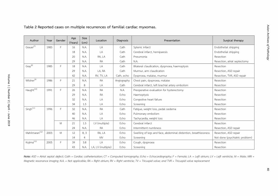
Asian Archives of Pathology 40
Table 2 Reported cases on multiple recurrences of familial cardiac myxomas.
Author Year Gender Age
(Years) Size (cm)
Location Diagnosis Presentation Surgical therapy
Grauer(7) 1983 F 16 N.A. LA Cath Splenic infarct Endothelial stripping 18 N.A. LA Cath Cerebral infarct, hemiparesis Endothelial stripping 20 N.A. RA, LA Cath Pneumonia Resection 29 N.A. RA Cath N.A. Resection, atrial septectomy Gray(8) 1985 F 18 N.A. LA Cath Bilateral claudication, dyspnoea, haemoptysis Resection 29 N.A. LA, RA Cath Murmur, arm claudication Resection, ASD repair 42 N.A. RV, TV, LA Cath, echo Dyspnoea, malaise, murmur Resection, TVR, ASD repair Wilsher(9) 1986 F 21 N.A. RA Angiography Chest pain, dyspnoea, malaise Resection 29 8 LA Cath Cerebral infarct, left brachial artery embolism Resection Haught(10) 1991 F 26 N.A. RA N.A. Preoperative evaluation for hysterectomy Resection 29 N.A. RA Echo Haemoptysis Resection 32 N.A. LA Echo Congestive heart failure Resection 34 1.5 LA Echo Screening Resection Singh(11) 1996 F 32 N.A. RA Cath Fatigue, weight loss, pedal oedema Resection 40 N.A. LA Echo Pulmonary embolism Resection 46 N.A. LA Echo Tachycardia, weight loss Resection M 21 1.5 LV (multiple) Echo Cerebral infarct Resection 24 N.A. RA Echo Intermittent numbness Resection, ASD repair Mahilmaran(12) 2003 M 12 8, 3 RA, LA Echo Swelling of legs and face, abdominal distention, breathlessness Resection, ASD repair 14 4 MV Echo Screening Not done (psychiatric problem) Kojima(13) 2005 F 39 3.8 LA Echo Cough, dyspnoea Resection 43 N.A. LA, LV (multiple) Echo Screening Resection
Note: ASD = Atrial septal defect; Cath = Cardiac catheterisation; CT = Computed tomography; Echo = Echocardiography; F = Female; LA = Left atrium; LV = Left ventricle; M = Male; MRI = Magnetic resonance imaging; N.A. = Not applicable; RA = Right atrium; RV = Right ventricle; TV = Tricuspid valve; and TVR = Tricuspid valve replacement
Volume 1 | Num
ber 2 | April – June 2019

Asian Archives of Pathology 41
Table 2 (Continued) Reported cases on multiple recurrences of familial cardiac myxomas.
Author Year Gender Age
(Years) Size (cm)
Location Diagnosis Presentation Surgical therapy
Akbarzadeh(14) 2005 F 17 N.A. LA N.A. Cerebral infarct Resection 33 N.A. LA N.A. N.A. Resection 35 N.A. LA (multiple), LV Echo Dyspnoea, paresis of the left hand Resection Turhan(15) 2008 F 10 N.A. LA Echo N.A. Resection 14 1.4 LA Echo Screening Follow-up 42 N.A. LA N.A. N.A. Resection 44 N.A. LA N.A. N.A. Resection 46 5.7 LA Echo Screening Resection Roy(16) 2011 F 31 N.A. LA N.A. N.A. Resection 38 2.5, 4 LA, RA Echo, MRI Visual disturbance Resection, ASD repair Cao(17) 2011 F 21 4 LA Echo N.A. Resection 26 4, 0.4 LA (x2) Echo Dyspnoea, palpitation and fatigability Resection Tamura(18) 2014 F 20 N.A. LA N.A. N.A. Resection 28 N.A. LA N.A. N.A. Resection 45 N.A. RV Echo Loss of consciousness, infective endocarditis Resection, tricuspid valve annuloplasty Azzam(19) 2014 M 36 N.A. LA N.A. N.A. Resection 40 N.A. LA Echo, CT N.A. Resection 41 N.A. LA Echo, CT N.A. Resection 51 3 LA Echo, CT Screening Resection Kwon(5) 2016 F 14 N.A. RA, LA N.A. N.A. Resection, ASD repair 24 N.A. LA, LV N.A. N.A. Resection, ASD repair 40 N.A. RA N.A. N.A. Resection, ASD repair 46 10 RA Echo, CT Chest discomfort, dyspnoea Resection
Note: ASD = Atrial septal defect; Cath = Cardiac catheterisation; CT = Computed tomography; Echo = Echocardiography; F = Female; LA = Left atrium; LV = Left ventricle; M = Male; MRI = Magnetic resonance imaging; N.A. = Not applicable; RA = Right atrium; RV = Right ventricle; TV = Tricuspid valve; and TVR = Tricuspid valve replacement
Volume 1 | Num
ber 2 | April – June 2019

Asian Archives of Pathology 42
Table 2 (Continued) Reported cases on multiple recurrences of familial cardiac myxomas.
Author Year Gender Age
(Years) Size (cm)
Location Diagnosis Presentation Surgical therapy
Schmidt(20) 2017 M 73 N.A. LA N.A. N.A. N.A. 78 N.A. Intra-atrial septum Echo Visual loss of right eye, pre-syncopal episodes Resection Current case 2018 F 10 5.5 LA Echo Congestive heart failure Resection 12 0.5 – 4.5 LA (x2), LV, pulmonary vein,
interatrial septum Echo Acute arterial occlusion Resection, splenectomy, fasciotomy,
embolectomy 14 6 RV Echo Screening Resection 17 0.8 – 3.5 RV (x4), LA, LV Echo Screening Heart transplant
Note: ASD = Atrial septal defect; Cath = Cardiac catheterisation; CT = Computed tomography; Echo = Echocardiography; F = Female; LA = Left atrium; LV = Left ventricle; M = Male; MRI = Magnetic resonance imaging; N.A. = Not applicable; RA = Right atrium; RV = Right ventricle; TV = Tricuspid valve; and TVR = Tricuspid valve replacement
Volume 1 | Num
ber 2 | April – June 2019

Asian Archives of Pathology 43
Volume 1 | Number 2 | April – June 2019
Historically, we initiated a heart transplant program in 1987 with our first orthotopic heart transplant in December 1988, which was also the first case in Southeast Asia(22). According to Thai Transplantation Society report, intrathoracic organ transplantation has been most frequently performed at King Chulalongkorn Memorial Hospital since 2008(23). The presented case is the first case of end-stage heart disease due to multiple recurrences of familial cardiac myxomas to undergo heart transplant at our institution. Conclusions We presented a case with multiple recurrences of familial cardiac myxomas who underwent heart transplant at our institution. Multifocal and recurrent cardiac myxomas seem more common in younger age group who has a family history of tumour. After nine months of heart transplantation, she passed away as a result of acute cellular rejection and septic shock. References (1). Reynen K. Cardiac myxomas. N Engl J Med. 1995;333(24):1610-7. (2). Travis WD, Brambilla E, Burke A, Marx A, Nicholson AG. WHO Classification of Tumours of
the Lung, Pleura, Thymus and Heart. Lyon: International Agency for Research on Cancer; 2015.
(3). Stratakis CA, Kirschner LS, Carney JA. Clinical and molecular features of the Carney complex: diagnostic criteria and recommendations for patient evaluation. J Clin Endocrinol Metab. 2001;86(9):4041-6.
(4). Tzani A, Doulamis IP, Mylonas KS, Avgerinos DV, Nasioudis D. Cardiac Tumors in Pediatric Patients: A Systematic Review. World J Pediatr Congenit Heart Surg. 2017;8(5):624-32.
(5). Kwon OY, Kim GJ, Jang WS, Lee YO, Cho JY, Lee JT. Fourth Recurrence of Cardiac Myxoma in a Patient with the Carney Complex. Korean J Thorac Cardiovasc Surg. 2016;49(2) :119-21.
(6). Wang Z, Chen S, Zhu M, Zhang W, Zhang H, Li H, et al. Risk prediction for emboli and recurrence of primary cardiac myxomas after resection. J Cardiothorac Surg. 2016;11:22.
(7). Grauer K, Grauer MC. Familial atrial myxoma with bilateral recurrence. Heart Lung. 1983;12(6):600-2.
(8). Gray IR, Williams WG. Recurring cardiac myxoma. Br Heart J. 1985;53(6):645-9. (9). Wilsher ML, Roche AH, Neutze JM, Synek BJ, Holdaway IM, Nicholson GI. A familial
syndrome of cardiac myxomas, myxoid neurofibromata, cutaneous pigmented lesions, and endocrine abnormalities. Aust N Z J Med. 1986;16(3):393-6.
(10). Haught WH, Alexander JA, Conti CR. Familial recurring cardiac myxoma. Clin Cardiol. 1991;14(8):692-5.6.

Asian Archives of Pathology 44
Volume 1 | Number 2 | April – June 2019
(11). Singh SD, Lansing AM. Familial cardiac myxoma--a comprehensive review of reported cases. J Ky Med Assoc. 1996;94(3):96-104.
(12). Mahilmaran A, Seshadri M, Nayar PG, Sudarsana G, Abraham KA. Familial cardiac myxoma: Carney's complex. Tex Heart Inst J. 2003;30(1):80-2.
(13). Kojima S, Sumiyoshi M, Watanabe Y, Suwa S, Matsumoto M, Nakata Y, et al. A Japanese case of familial cardiac myxoma associated with a mutation of the PRKAR1alpha gene. Intern Med. 2005;44(6):607-10.
(14). Akbarzadeh Z, Esmailzadeh M, Yousefi A, Safaei A, Raisi K, Sharifi F. Multicentric familial cardiac myxoma. Eur J Echocardiogr. 2005;6(2):148-50.
(15). Turhan S, Tulunay C, Altin T, Dincer I. Second recurrence of familial cardiac myxomas in atypical locations. Can J Cardiol. 2008;24(9):715-6.
(16). Shetty Roy AN, Radin M, Sarabi D, Shaoulian E. Familial recurrent atrial myxoma: Carney's complex. Clin Cardiol. 2011;34(2):83-6.
(17). Cao H, Wu Y, Zhu J, Chen Y. Familial cardiac myxoma with multifocal recurrences: a case report and review of the literature. J Biomed Res. 2011;25(5):368-72.
(18). Tamura Y, Seki T. Carney complex with right ventricular myxoma following second excision of left atrial myxoma. Ann Thorac Cardiovasc Surg. 2014;20 Suppl:882-4.
(19). Azzam R, Abdelbar A, Yap KH, Abousteit A. Carney complex: fourth time excision of recurrent atrial myxoma via left thoracotomy. BMJ Case Rep. 2014;2014.
(20). Schmidt C, Doi A, Ura M, Cole C, Mundy J. Familial Atrial Myxoma: Three Related Cases at an Australian Tertiary Institution. Ann Thorac Cardiovasc Surg. 2017;23(4):203-6.
(21). Krittayaphong R, Ariyachaipanich A. Heart Transplant in Asia. Heart Fail Clin. 2015;11(4):563-72.
(22). Ongcharit P, Wongkietkachorn K, Sritangsirikul S, Namchaisiri J, Singhatanatkit S, Luengtaviboon K, et al. Heart transplantation 1987- 2007: 20 years' experience at Chulalongkorn hospital. Transplant Proc. 2008;40(8):2591-3.
(23). Thai-transplant.org. 2016 Annual Report of Organ Transplantation in Thailand [Internet]. 2016 [cited 27 January 2018]. Available from: http://www.thai-transplant.org/pic/File/ Translate%20Registry%202016%20_English_.pdf.

Asian Archives of Pathology 45
Volume 1 | Number 2 | April – June 2019
APPENDIX 1
INFORMATION FOR AUTHORS
All authors listed in a paper submitted to Asian Archives of Pathology (AAP) must have contributed substantially to the work. It is the corresponding author who takes responsibility for obtaining permission from all co-authors for the submission. When submitting the paper, the corresponding author is encouraged to indicate the specific contributions of all authors (the author statement, with signatures from all authors and percentage of each contribution can be accepted). Examples of contributions include: designed research, performed research, contributed vital new reagents or analytical tools, analysed data, and wrote the paper. An author may list more than one type of contribution, and more than one author may have contributed to the same aspect of the work. Authors should take care to exclude overlap and duplication in papers dealing with related materials. See also paragraph on Redundant or Duplicate Publication in “Uniform Requirements for Manuscripts Submitted to Biomedical Journals” at http://www.icmje.org/index.html. The submitted manuscripts will be reviewed by the members of the Editorial Board or the expert reviewers. At the discretion of the Editorial Board, the manuscripts may be returned immediately without full review, if deemed not competitive or outside the realm of interests of the majority of the readership of the Journal. The decision (reject, invite revision, and accept) letter will be coming from the Editorial Board who has assumed responsibility for the manuscript’s review. The editor’s decision is based not just on technical merit of the work, but also on other factors such as the priority for publication and the relevance to the Journal’s general readership. All papers are judged in relation to other submissions currently under consideration.

Asian Archives of Pathology 46
Volume 1 | Number 2 | April – June 2019
Categories of Manuscripts 1. Letters to the Editor
The letters to the editor are the reactions to any papers published in AAP. These letters will be reviewed by the Editorial Board and sent to the authors of the original paper with an invitation to respond. Letters and eventual responses will be published together, when appropriate.
▪ Word Count: 300 – 500 words (excluding references and figure or table legends) ▪ Abstract: Not required ▪ References: Maximum of 10 ▪ Figure or Table: Maximum of 1 (if needed)
2. Original Articles
The original articles are the researches describing the novel understanding of anatomical pathology, clinical pathology (laboratory medicine), forensic medicine (legal medicine or medical jurisprudence), molecular medicine or pathobiology. Systematic reviews, meta-analyses and clinical trials are classified as articles. The articles should be clearly and concisely written in the well-organised form (see Organisation of Manuscripts): abstract; introduction; materials and methods; results; discussion; and conclusions. The manuscripts that have passed an initial screening by the Editorial Board will be reviewed by two or more experts in the field.
▪ Word Count: 3,000 – 5,000 words (excluding abstract, references, and figure or table legends)
▪ Structured Abstract (see Organisation of Manuscripts): 150 – 200 words ▪ References: Maximum of 150 ▪ Figures or Tables: Maximum of 6
3. Review Articles
The review articles are generally invited by the Editor-in-Chief. They should focus on a topic of broad scientific interest and on recent advances. These articles are peer-reviewed before the final decision to accept or reject the manuscript for publication. Therefore, revisions may be required.
▪ Word Count: 3,000 – 5,000 words (excluding abstract, references, and figure or table legends)
▪ Unstructured Abstract: 150 – 200 words ▪ References: Maximum of 150 ▪ Figures or Tables: Maximum of 4

Asian Archives of Pathology 47
Volume 1 | Number 2 | April – June 2019
4. Case Reports AAP limits publication of case reports to those that are truly novel, unexpected or unusual, provide new information about anatomical pathology, clinical pathology (laboratory medicine) or forensic medicine (legal medicine or medical jurisprudence). In addition, they must have educational value for the aforementioned fields. The journal will not consider case reports describing preventive or therapeutic interventions, as these generally require stronger evidence. Case reports that involve a substantial literature review should be submitted as a review article. The submitted case reports will undergo the usual peer-reviewed process.
▪ Word Count: 1,200 – 2,000 words (excluding abstract, references, and figure or table legends)
▪ Unstructured Abstract: 150 – 200 words ▪ References: Maximum of 20 ▪ Figures or Tables: Maximum of 4
5. Case Illustrations
Case illustrations are aimed to provide education to readers through multidisciplinary clinicopathological discussions of interesting cases. The manuscript consists of a clinical presentation or description, laboratory investigations, discussion, final diagnosis, and up to 5 take-home messages (learning points). Regarding continuous learning through self-assessment, each of the case illustrations will contain 3 – 5 multiple choice questions (MCQs) with 4 – 5 suggested answers for each question. These MCQs are placed after the final diagnosis and the correct answers should be revealed after the references. The questions and take-home messages (learning points) are included in the total word count. The manuscripts that have passed an initial screening by the Editorial Board will be reviewed by two experts in the field.
▪ Word Count: 1,000 – 2,000 words (excluding references and figure or table legends)
▪ Abstract: Not required ▪ References: Maximum of 10 ▪ Figures: Maximum of 2 ▪ Tables: Maximum of 5
6. Technical Notes
The technical notes are brief descriptions of scientific techniques used in the anatomical pathology, clinical pathology (laboratory medicine), forensic medicine (legal medicine or medical jurisprudence), molecular medicine or pathobiology. The submitted manuscripts are usually peer-reviewed.

Asian Archives of Pathology 48
Volume 1 | Number 2 | April – June 2019
▪ Word Count: Maximum of 1,000 words (excluding references and figure or table legends)
▪ Abstract: Not required ▪ References: Maximum of 5 ▪ Figures or Tables: Maximum of 2
Organisation of Manuscripts 1. General Format
The manuscripts written in English language are preferable. However, Thai papers are also acceptable, but their title pages, abstracts, and keywords must contain both Thai and English. These English and Thai manuscripts are prepared in A4-sized Microsoft Word documents with leaving 2.54-cm (1-inch) margins on all sides. All documents are required to be aligned left and double-spaced throughout the entire manuscript. The text should be typed in 12-point regular Times New Roman font for English manuscript and 16-point regular TH SarabunPSK font for Thai manuscript. The running titles of English and Thai manuscripts are placed in the top left-hand corner of each page. They cannot exceed 50 characters, including spaces between words and punctuation. For the header of English paper, the running title will be typed in all capital letters. The page number goes on the top right-hand corner. Footnotes are not used in the manuscripts, but parenthetical statements within text are applied instead and sparingly. Abbreviations should be defined at first mention and thereafter used consistently throughout the article. The standard abbreviations for units of measure must be used in conjunction with numbers. All studies that involve human subjects should not mention subjects’ identifying information (e.g. initials) unless the information is essential for scientific purposes and the patients (or parents or guardians) give written informed consent for publication.
2. Title Page
The title page is the first page of the manuscripts and must contain the following: ▪ The title of the paper (not more than 150 characters, including spaces between
words) ▪ The full names, institutional addresses, and email addresses for all authors (If
authors regard it as essential to indicate that two or more co-authors are equal in status, they may be identified by an asterisk symbol with the caption “These authors contributed equally to this work” immediately under the address list.)

Asian Archives of Pathology 49
Volume 1 | Number 2 | April – June 2019
▪ The name, surname, full postal address, telephone number, facsimile number, and email address of the corresponding author who will take primary responsibility for communication with AAP.
▪ Conflict of interest statement (If there are no conflicts of interest for any author, the following statement should be inserted: “The authors declare that they have no conflicts of interest with the contents of this article.”)
3. Abstract
A structured form of abstract is used in all Original Article manuscripts and must include the following separate sections:
▪ Background: The main context of the study ▪ Objective: The main purpose of the study ▪ Materials and Methods: How the study was performed ▪ Results: The main findings ▪ Conclusions: Brief summary and potential implications ▪ Keywords: 3 – 5 words or phrases (listed in alphabetical order) representing the
main content of the article 4. Introduction
The Introduction section should clearly explain the background to the study, its aims, a summary of the existing literature and why this study was necessary or its contribution to the field.
5. Materials and Methods
The Materials and Methods section must be described in sufficient detail to allow the experiments or data collection to be reproduced by others. Common routine methods that have been published in detail elsewhere should not be described in detail. They need only be described in outline with an appropriate reference to a full description. Authors should provide the names of the manufacturers and their locations for any specifically named medical equipment and instruments, and all chemicals and drugs should be identified by their systematic and pharmaceutical names, and by their trivial and trade names if relevant, respectively. Calculations and the statistical methods employed must be described in this section. All studies involving animal or human subjects must abide by the rules of the appropriate Internal Review Board and the tenets of the recently revised Helsinki protocol. Hence, the manuscripts must include the name of the ethics committee that approved the study and the committee’s reference number if appropriate.

Asian Archives of Pathology 50
Volume 1 | Number 2 | April – June 2019
6. Results The Results section should concisely describe the findings of the study including, if appropriate, results of statistical analysis which must be presented either in the text or as tables and figures. It should follow a logical sequence. However, the description of results should not simply repeat the data that appear in tables and figures and, likewise, the same data should not be displayed in both tables and figures. Any chemical equations, structural formulas or mathematical equations should be placed between successive lines of text. The authors do not discuss the results or draw any conclusions in this section.
7. Discussion
The Discussion section should focus on the interpretation and the significance of the findings against the background of existing knowledge. The discussion should not repeat information in the results. The authors will clearly identify any aspects that are novel. In addition, there is the relation between the results and other work in the area.
8. Conclusions
The Conclusions section should state clearly the main summaries and provide an explanation of the importance and relevance of the study reported. The author will also describe some indication of the direction future research should take.
9. Acknowledgements
The Acknowledgements section should be any brief notes of thanks to the following: ▪ Funding sources ▪ A person who provided purely technical help or writing assistance ▪ A department chair who provided only general support ▪ Sources of material (e.g. novel drugs) not available commercially
Thanks to anonymous reviewers are not allowed. If you do not have anyone to acknowledge, please write “Not applicable” in this section.
10. References
The Vancouver system of referencing should be used in the manuscripts. References should be cited numerically in the order they appear in the text. The authors should identify references in text, tables, and legends by Arabic numerals in parentheses or as superscripts. Please give names of all authors and editors. The references should be numbered and listed in order of appearance in the text. The names of all authors are cited when there are six or fewer. When there are seven or more, only the first three followed by “et al.” should be given. The names of journals should be abbreviated in the style used in Index Medicus (see examples below). Reference to unpublished data and personal

Asian Archives of Pathology 51
Volume 1 | Number 2 | April – June 2019
communications should not appear in the list but should be cited in the text only (e.g. A Smith, unpubl. Data, 2000).
▪ Journal article 1. Sibai BM. Magnesium sulfate is the ideal anticonvulsant in preeclampsia –
eclampsia. Am J Obstet Gynecol 1990; 162: 1141 – 5. ▪ Books
2. Remington JS, Swartz MN. Current Topics in Infectious Diseases, Vol 21. Boston: Blackwell Science Publication, 2001.
▪ Chapter in a book 3. Cunningham FG, Hauth JC, Leveno KJ, Gilstrap L III, Bloom SL, Wenstrom KD.
Hypertensive disorders in pregnancy. In: Cunningham FG, Hauth JC, Leveno KJ, Gilstrap L III, Brom SL, Wenstrom KD, eds. Williams Obstetrics, 22nd ed. New York: McGraw-Hill, 2005: 761 – 808.
11. Tables
The tables should be self-contained and complement, but without duplication, information contained in the text. They should be numbered consecutively in Arabic numerals (Table 1, Table 2, etc.). Each table should be presented on a separate page with a comprehensive but concise legend above the table. The tables should be double-spaced and vertical lines should not be used to separate the columns. The column headings should be brief, with units of measurement in parentheses. All abbreviations should be defined in footnotes. The tables and their legends and footnotes should be understandable without reference to the text. The authors should ensure that the data in the tables are consistent with those cited in the relevant places in the text, totals add up correctly, and percentages have been calculated correctly.
12. Figure Legends
The legends should be self-explanatory and typed on a separate page titled “Figure Legends”. They should incorporate definitions of any symbols used and all abbreviations and units of measurement should be explained so that the figures and their legends are understandable without reference to the text. If the tables or figures have been published before, the authors must obtain written permission to reproduce the materials in both print and electronic formats from the copyright owner and submit them with the manuscripts. These also follow for quotes, illustrations, and other materials taken from previously published works not in the public domain. The original resources should be cited in the figure captions or table footnotes.

Asian Archives of Pathology 52
Volume 1 | Number 2 | April – June 2019
13. Figures All illustrations (line drawings and photographs) are classified as figures. The figures should be numbered consecutively in Arabic numerals (Figure 1, Figure 2, etc.). They are submitted electronically along with the manuscripts. These figures should be referred to specifically in the text of the papers but should not be embedded within the text. The following information must be stated to each microscopic image: staining method, magnification (especially for electron micrograph), and numerical aperture of the objective lens. The authors are encouraged to use digital images (at least 300 d.p.i.) in .jpg or .tif formats. The use of three-dimensional histograms is strongly discouraged when the addition of these histograms give no extra information.
14. Components
14.1. Letters to the Editor The Letter to the Editor manuscripts consist of the following order:
▪ Title Page ▪ Main Text ▪ References ▪ Table (if needed) ▪ Figure Legend (if needed) ▪ Figure (if needed)
14.2. Original Articles The Original Article manuscripts consist of the following order:
▪ Title Page ▪ Structured Abstract ▪ Introduction ▪ Materials and Methods ▪ Results ▪ Discussion ▪ Conclusions ▪ Acknowledgements ▪ References ▪ Table (s) ▪ Figure Legend (s) ▪ Figure (s)
14.3. Review Articles The Review Article manuscripts consist of the following order:
▪ Title Page ▪ Unstructured Abstract

Asian Archives of Pathology 53
Volume 1 | Number 2 | April – June 2019
▪ Introduction ▪ Main Text ▪ Conclusions ▪ Acknowledgements ▪ References ▪ Table (s) ▪ Figure Legend (s) ▪ Figure (s)
14.4. Case Reports The Case Report manuscripts consist of the following order:
▪ Title Page ▪ Unstructured Abstract ▪ Introduction ▪ Case Description ▪ Discussion ▪ Conclusions ▪ Acknowledgements ▪ References ▪ Table (s) ▪ Figure Legend (s) ▪ Figure (s)
14.5. Case Illustrations The Case Illustration manuscripts consist of the following order:
▪ Title Page ▪ Clinical Presentation or Description ▪ Laboratory Investigations ▪ Discussion ▪ Final Diagnosis ▪ Multiple Choice Questions (MCQs) ▪ Take-Home Messages (Learning Points) ▪ Acknowledgements ▪ References ▪ Correct Answers to MCQs ▪ Table (s) ▪ Figure Legend (s) ▪ Figure (s)

Asian Archives of Pathology 54
Volume 1 | Number 2 | April – June 2019
14.6. Technical Notes The Technical Note manuscripts consist of the following order:
▪ Title Page ▪ Introduction ▪ Main text ▪ Conclusions ▪ Acknowledgements ▪ References ▪ Table (s) ▪ Figure Legend (s) ▪ Figure (s)
Proofreading The authors of the accepted manuscripts will receive proofs and are responsible for proofreading and checking the entire article, including tables, figures, and references. These authors should correct only typesetting errors at this stage and may be charged for extensive alterations. Page proofs must be returned within 48 hours to avoid delays in publication. Revised Manuscripts In many cases, the authors will be invited to make revisions to their manuscripts. The revised manuscripts must generally be received by the Editorial Board within 3 months of the date on the decision letter or they will be considered a new submission. An extension can sometimes be negotiated with the Editorial Board.
Asian Archives of Pathology 55
Volume 1 | Number 2 | April – June 2019
APPENDIX 2
BENEFITS OF PUBLISHING WITH
ASIAN ARCHIVES OF PATHOLOGY
Asian Archives of Pathology (AAP) is an open access journal. Open Access makes your works freely available to everyone in the world. It provides a significant boost to the readership of your articles, and has been shown to have an increase in positive influence on citations and reuse. Hence, open-access leads to more recognition for our esteemed authors. The journal has been sponsored by the Royal College of Pathologists of Thailand. We have the policy to disseminate the verified scientific knowledge to the public on a non-profit basis. Hence, we have not charged the authors whose manuscripts have been submitted or accepted for publication in our journal. Since AAP is also a peer-reviewed journal, the submitted manuscripts will be reviewed by the members of the Editorial Board or the expert reviewers. The decision on these manuscripts is processed very fast without any delay and in shortest possible time. The processing period is 1 – 2 weeks. These decisions of the reviewers are unbiased and the decision (reject, invite revision, and accept) letter coming from the Editorial Board is always conveyed to the authors.

Asian Archives of Pathology 56
Volume 1 | Number 2 | April – June 2019
APPENDIX 3
SUBMISSION OF THE MANUSCRIPTS
Step 1: Access www.asianarchpath.com Step 2: If you did not register before, please create an account first. Step 3: Login with your username and password. Step 4: Click the “+ New Submission” button on the upper right-hand side of the page. Step 5: Proceed to fill up the Submission Form online and follow the directions given
therein. Step 6: Upload your manuscript file (s). Step 7: Re-check the content of your manuscript (s) and the uploaded file (s) more
carefully prior to the submission. If you have submitted your manuscript file (s) incorrectly, you must contact Editor-in-Chief of Asian Archives of Pathology immediately. The Editor-in-Chief can clear the incorrect attempt and allow you another submission.
Step 8: Click the “Submit Manuscript” button under Important Notice.
If you have any further enquires, please do not hesitate to contact the Journal.

Asian Archives of Pathology 57
Volume 1 | Number 2 | April – June 2019
APPENDIX 4
CONTACT THE JOURNAL
The Editorial Office of Asian Archives of Pathology
Department of Pathology, Floor 6, Her Royal Highness Princess Bejaratana Building Phramongkutklao College of Medicine
317 Rajavithi Road, Rajadevi, Bangkok 10400 Thailand
Telephone: +66 (0) 90 132 2047
Fax: +66 (0) 2 354 7791
Email: [email protected]

Asian Archives of Pathology 58
Volume 1 | Number 2 | April – June 2019
APPENDIX 5
SUPPORT THE JOURNAL
Asian Archives of Pathology (AAP) has a mission of disseminating the unbiased and reliable medical knowledge on a non-profit basis. If you consider that this journal is useful for the public, you can support us by submitting your advertisements via the contact information below.
Dr Chetana Ruangpratheep The Editorial Office of Asian Archives of Pathology
Department of Pathology, Floor 6, Her Royal Highness Princess Bejaratana Building Phramongkutklao College of Medicine
317 Rajavithi Road, Rajadevi, Bangkok 10400 Thailand Telephone: +66 (0) 90 132 2047
Fax: +66 (0) 2 354 7791 Email: [email protected]
Every support, small or big, can make a difference.
Thank you
Dr Chetana Ruangpratheep
MD, FRCPath (Thailand), MSc, PhD Editor-in-Chief of Asian Archives of Pathology

Volume 1 | Number 2 | April – June 2019
ACADEMIC MEETINGS AND CONFERENCES
Announcements of academic meetings and conferences that are of interest to the readers of
Asian Archives of Pathology (AAP) should be sent to the Editor-in-Chief at least 3 months before the first day of the month of issue. The contact information is shown below.
Dr Chetana Ruangpratheep
The Editorial Office of Asian Archives of Pathology Department of Pathology, Floor 6, Her Royal Highness Princess Bejaratana Building
Phramongkutklao College of Medicine 317 Rajavithi Road, Rajadevi, Bangkok 10400 Thailand
Telephone: +66 (0) 90 132 2047
Fax: +66 (0) 2 354 7791
Email: [email protected]

Volume 1 | Number 2 | April – June 2019
WHAT IS INSIDE THIS ISSUE?
Original Article: Assessment of the usefulness of the knowledge of pathology ………………………………… for clinical medical students – a multicentre study
1
Ijeoma Angela Meka, Helen Chioma Okoye, Angela Ogechukwu Ugwu, Isah Adagiri Yahaya, Ochukor Otokunefor, Olugbenga Olalekan Ojo, and Emmanuel Onyebuchi Ugwu
Review Article: The essentials of vascular pathology …………………………………………………………………………. 13 Chetana Ruangpratheep
Case Report: Heart transplant for multiple recurrences of familial cardiac ………………………………….. myxomas in an adolescent patient: a case report and literature review
34
Thiyaphat Laohawetwanit, Poonchavist Chantranuwatana, and Pat Ongcharit