ascites and right pleural effusion - journal of nuclear medicine
TRANSCRIPT

leural effusions occur in 5% to 10% of patients withcirrhosis of the liver (1 ). In these patients, ascites is
usually evident but a pleural effusion may develop in acirrhotic patient in the absence ofdetectable ascites (2).The effusions are usually right-sided, but may be bilateral or left-sided. The precise mechanism of fluid accumulation is not clear.
We describe a case where a peritoneopleural communication has been demonstrated by radioisotopicmethod. The possible mechanisms involved in the formation of pleural effusion in liver cirrhosis are discussed.
CASE REPORT
A 54-yr-old white female was admitted for progressivedyspnea of recent onset and signs of upper respiratory infection. Her past history included a background of considerablealcohol ingestion, and a diagnosis of Laennec cirrhosis hadbeen previously made by liver biopsy.
On admission, the patient looked chronically ill. She wasafebrile with regular pulse rate of 120/mm and a blood pressure of 180/100. There were clinical signs of cirrhosis andfrank jaundice. Chest examination revealed evidence for amassive right pleural effusion. Other physical findings in
Received Dec. 13, 1985;revision accepted Apr. 23, 1986.For reprints contact: Jean Verreault, MD, Dept. of Nuclear
Medicine,Centre Hospitalier Universitaire, Sherbrooke, Québec,Canada, J 1H-5N4.
cluded enlarged liver and evidence of free liquid in the pentoneal cavity.
The chest x-ray demonstrated a large right pleural effusion.Other laboratory tests were compatible with liver cirrhosis andtoxic effects ofalcohol. Thoracentesis revealed a fluid that hadthe characteristics ofa transudate as did a sample of pentonealfluid. Forty-eight hours after the thoracentesis of 1,800 cc, theright pleural space was completely refilled (Fig. 1).
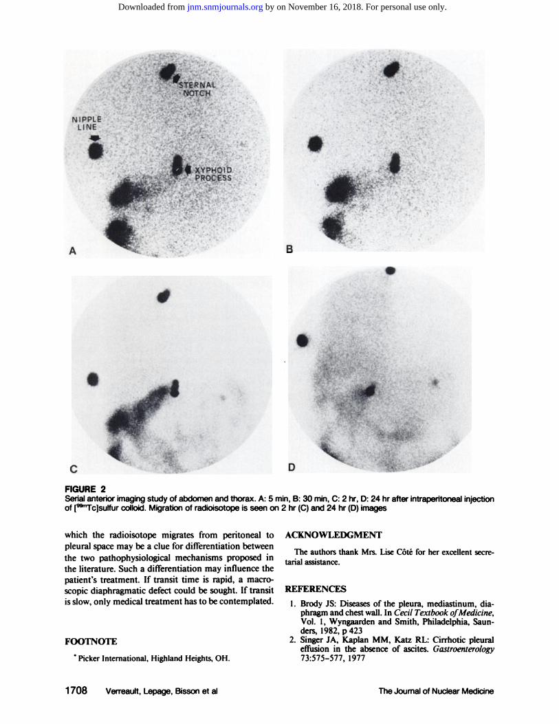
To prove that right pleural effusion was related to ascites,5 mCi of technetium-99m (@mTc) sulfur colloid was injectedwith aseptic technique into the right lower abdominal cavity.A serial imaging study was done in anterior view at intervalsof 5 mm for 30 mm and at 2 and 24 hr postinjection (Fig. 2).Images were obtained on a large field-of-view camera with anall-purpose, parallel collimator. At 2 hr the radiocolloid appeared in the right hemithorax and at 24 hr activity wasfurther increased. It confirmed the presence of a peritoneopleural communication. The patient responded well to medical therapy and pleural effusion resolvedcompletely in 10days.
DISCUSSION
The association ofhydrothorax with hepatic cirrhosishas been recognized for many years but there has beenmuch speculation as to its origin.
In 1947, Higgins and his colleagues suggested thatpleural effusion was related to the fall in the concentration of plasma proteins (3). In 1958, Morrow, Kantorand Armen suggested that the fluid might come fromthe blood stream as a result of hypoalbuminemia or
1706 Verreault,Lepage,Bissonetal The Journal of Nuclear Medicine
Case Reports
Ascites and RightPleural Effusion: Demonstrationof a Pentoneo-Pleural CommunicationJean Verreault, Serge Lepage, Guy Bisson, and Andre Plante
Departments ofNuc/ear Medicine and Internal Medicine, Centre Hospitalier Universitaire,University ofSherbrooke, Sherbrooke, Québec,Canada
A 54-yr-oldfemale with known livercirrhosis presented with a right transudative pleuraleffusion and ascites. To find the source of pleural fluid,[@“TcJsuIfurcolloidwas injectedintraperitoneallyand a serial imagingstudy revealed its passage to the right pleural space on2-hr and 24-hr images. Mechanisms proposed in the formation of pleural effusion in livercirrhosis are (a) lymphatic drainage and (b) diaphragmatic defect. Radioisotope migrationspeed may be a clue for differentiating these two mechanisms, being more rapid in thepresence of a diaphragmatic defect.
J NucIMed 27:1706—1709,1986
by on November 16, 2018. For personal use only. jnm.snmjournals.org Downloaded from

bumin from the peritoneal to pleural spaces in a patientwith right hepatic hydrothorax. The autopsy showed nogross defect in the right diaphragm.
Since lymphatics are more abundant in the right than£ in the left diaphragm (10), this mechanism might ex
plain that hepatic hydrothorax are more frequently rightsided. In addition to this, Leak (11) used electronmicroscopy to show that pores of 4—12 @zin diameterexist between mesothelial cells and diaphragmatic lymphatics. They provide a system of open channelsthrough which fluids and cells may rapidly be removedfrom the serous cavities. What remains unclear is howand why the fluid leaves the lymphatic system andenters the pleural space instead ofbeing expelled towardthe larger collecting vessels. It may result from lymphatic system overload (12).
There is strong evidence to support the two mechanisms. We think that each of these can explain theformation of hydrothorax from ascites depending onthe case. Lymphatic drainage is probably the first mechanism to occur in reaction to the presence of ascites. Itcan be the way by which hydrothorax will be formed ifthere is an overload of the lymphatic system. If it isinsufficient, the abdominal pressure increases and maycause subsequent rupture ofthe diaphragm. One objection to this assumption may be that pleural effusionscan be present in liver cirrhosis without ascites. In thesecases, the alternative explanation is that there is congenital diaphragmatic weakness caused by an insuffident deposition of muscle and tendon bundles. Weproposed that the clue to distinguish between these twomechanisms may be the use of radioisotope injectedintraperitoneally as we did.
Indeed, when we assume that the radiocolloid injected intrapentoneally is mixed with ascites fluid andconsider that its migration from peritoneal to pleuralspace was right sided as was the pleural effusion, wecan presume that there is a relationship between ascitesfluid and pleural effusion. Furthermore, our proposalextends to the time required to show accumulation ofthe agent in the pleural space. If it happens within afew minutes, as it was reported by Faiyaz and Goyal(13), a diaphragmatic defect is probably present particularly when the accumulation is as intense as peritonealactivity. If it takes a longer period of time as was seenfor our patient, the peritoneopleural communicationmay be attributed to diaphragmatic lymphatics particularly when that accumulation is less intense than peritoneal activity.
Such a study can be done either by the use of[@mTc1sulfur colloid or [99mTc]macroaW@ted albumin. Theradiopharmaceutical can be injected intraperitoneallyat the end of the usual diagnostic or therapeutic peritoneal puncture. It can show the source of pleuraleffusions in liver cirrhosis and also in patients withperitoneal dialysis (14). We postulate that the speed at
4
I
hypertension in the azygous and hemiazygous systemsor be secondary to ascites either by direct passage offluid through a diaphragmatic defect or by its transportthrough the diaphragmatic lymphatics (4).
Hypoproteinemia as a sole cause can be eliminatedfor there are many patients, cirrhotic or not, who havelow serum protein levels but never develop hydrothorax(5). Ifazygous hypertension is present due to collateralsbetween this system and the portal system, transudationof fluid into the chest might appear. However, thiscannot explain hydrothorax occurring in Meigs' syndrome where there is no portal hypertension. The mostprobable explanation is that hydrothorax is deriveddirectly from the peritoneal fluid either through a defectin the diaphragm or through the lymphatic channelsthat penetrate it.
At least three studies (6—8)have provided evidencethat hydrothorax complicating cirrhosis is generatedfrom ascites through a diaphragmatic defect acquiredas a result of increased intra-abdominal pressure. Thishas been demonstrated either by rapid passage of dyefrom the ascites into the pleural effusion, induction ofa hydropneumothorax by the intra-abdominal instillation of oxygen, thoracoscopy, or by necropsy findings.The other mechanism proposed in literature is thatperitoneal fluid passes to the pleural space through thediaphragmatic lymphatics. Johnston and Rodolfo (9)support it by the demonstration of unidirectional transport of carbon particles and radioiodinated serum al
FIGURE 1Chest x-ray showing large right pleural effusion
Volume27 •Number 11 •November1986 1707
by on November 16, 2018. For personal use only. jnm.snmjournals.org Downloaded from
,)s@@
... •@:,@
N IPPLE@@@ :@@ :@,@‘&@@-4'@ •
LINE . •@@@@ e@ . . . . @:@@@ .
. 1.@@@ ‘@@ ‘ .‘—.@: : ‘.@.: _k@
.;@ ).
r'@@ —@ ..
•;@; •@‘‘;!‘@@@ ., @:@@ £@•@
. ,,‘@
A
2@:@. %1@i;@@•
. ..@ • •@
@ ...@ .@.
@.,.@. @h@i:@
@ •
@-1@
I
B
.
S.@
S.@:
@::@DC
FIGURE 2Serial anterior imagingstudy of abdomen and thorax. A: 5 mm, B: 30 mm, C: 2 hr. D:24 hr after intraperitoneal injectionof [@‘TcJsuffurcolloid.Migrationof radioisotope is seen on 2 hr (C)and 24 hr (D)images
which the radioisotope migrates from peritoneal topleural space may be a clue for differentiation betweenthe two pathophysiological mechanisms proposed inthe literature. Such a differentiation may influence thepatient's treatment. If transit time is rapid, a macroscopic diaphragmatic defect could be sought. If transitis slow, only medical treatment has to be contemplated.
FOOTNOTE
. Picker International, Highland Heights, OH.
1708 Verreauft,Lepage,Bissonetal
ACKNOWLEDGMENT
The authors thank Mrs. LiseCôtéfor her excellentsecretanal assistance.
REFERENCES
1. Brody iS: Diseases of the pleura, mediastinum, diaphragm and chest wall. In Cecil TextbookofMedicine,Vol. 1, Wyngaarden and Smith, Philadelphia, Saunders,1982,p 423
2. Singer JA, Kaplan MM, Katz RL: Cirrhotic pleuraleffusion in the absence of ascites. Gastroenterology73:575—577,1977
The Journal of Nudear Medicine
by on November 16, 2018. For personal use only. jnm.snmjournals.org Downloaded from
3. Higgins G, Kelsall AR, O'Brien JR. et al: Ascites inchronic diseases of the liver. QJ Med 16:263—274,1947
4. Morrow CS, Kantor M, Armen RN: Hepatic hydrothorax. Ann Internal Med 49:193—202,1958
5. McKay DG, Sparling Hi, Robbins SL: Cirrhosis ofthe liver with massive hydrothorax. Arch Internal Med79:501—509,1947
6. Williams MH: Pleural effusion produced by abdommo-pleural communication in a patient with Laennec's cirrhosis ofthe liver and ascites.Ann Intern Med33:216—221,1950
7. Emerson PA, Davies JH: Hydrothorax complicatingascites. Lancet 1:487—488,1955
8. Lieberman FL, Hidemura R, Peters RL, et al: Pathogenesis and treatment of hydrothorax complicatingcirrhosis with ascites. Ann Intern Med 64:341—351,1966
9. Johnston RF, Loo RV: Hepatic hydrothorax: Studiesto determine the source offluid and report of thirteencases.Ann InternMed 61:385—401,I964
10. Brash JC: Cunningham ‘sTextbook ofAnatomy, 9thed., London, Oxford University Press, 1951, p 1423
11. Leak L: Gross and ultrastructural morphologic features of the diaphragm. Am Rev Resp Dis 119:3—21,1979
12. Dumont AE, Mulholland JH: Flow rate and composition ofthoracic duct lymph in patients with cirrhosis.N EnglJ Med 263:47 1—474,1960
13. Faiyaz U, Goyal PC: Unilateral pleural effusion without ascites in liver cirrhosis. Postgrad Med 74:309—315,1983
14. Spadaro JJ, Thakur V. Nolph KD: Technetium-99m-labelled macroaggregated albumin in demonstrationof trans-diaphragmatic leakage of dialysate in peritoneal dialysis. Am J Nephrol 2:36—38,1982
Volume 27 •Number 11 •November1986 1709
by on November 16, 2018. For personal use only. jnm.snmjournals.org Downloaded from
1986;27:1706-1709.J Nucl Med. Jean Verreault, Serge Lepage, Guy Bisson and Andre Plante CommunicationAscites and Right Pleural Effusion: Demonstration of a Peritoneo-Pleural
http://jnm.snmjournals.org/content/27/11/1706This article and updated information are available at:
http://jnm.snmjournals.org/site/subscriptions/online.xhtml
Information about subscriptions to JNM can be found at:
http://jnm.snmjournals.org/site/misc/permission.xhtmlInformation about reproducing figures, tables, or other portions of this article can be found online at:
(Print ISSN: 0161-5505, Online ISSN: 2159-662X)1850 Samuel Morse Drive, Reston, VA 20190.SNMMI | Society of Nuclear Medicine and Molecular Imaging
is published monthly.The Journal of Nuclear Medicine
© Copyright 1986 SNMMI; all rights reserved.
by on November 16, 2018. For personal use only. jnm.snmjournals.org Downloaded from