asare, g. a.; afriyie, d.; ngala, r. a.; abutiate, h.; doku, d.; -- antiproliferative activity of...
TRANSCRIPT

8/9/2019 Asare, G. A.; Afriyie, D.; Ngala, R. A.; Abutiate, H.; Doku, D.; -- Antiproliferative Activity of Aqueous Leaf Extract
http://slidepdf.com/reader/full/asare-g-a-afriyie-d-ngala-r-a-abutiate-h-doku-d-antiproliferative 1/11
http://ict.sagepub.com/ Integrative Cancer Therapies
http://ict.sagepub.com/content/early/2014/11/18/1534735414550198The online version of this article can be found at:
DOI: 10.1177/1534735414550198
published online 18 November 2014Integr Cancer Ther orge Awuku Asare, Dan Afriyie, Robert A. Ngala, Harry Abutiate, Derek Doku, Seidu A. Mahmood and Habibur Ra
and Some Target Genes on the Prostate, BPH-1 CAnnona muricata L.ntiproliferative Activity of Aqueous Leaf Extract of
Published by:
http://www.sagepublications.com
can be found at:Integrative Cancer Therapies Additional services and information for
http://ict.sagepub.com/cgi/alertsEmail Alerts:
http://ict.sagepub.com/subscriptionsSubscriptions:
http://www.sagepub.com/journalsReprints.navReprints:
http://www.sagepub.com/journalsPermissions.navPermissions:
http://ict.sagepub.com/content/early/2014/11/18/1534735414550198.refs.htmlCitations:
What is This?
- Nov 18, 2014OnlineFirst Version of Record>>
at UNIV OF GEORGIA LIBRARIES on November 25, 2014ict.sagepub.comDownloaded from at UNIV OF GEORGIA LIBRARIES on November 25, 2014ict.sagepub.comDownloaded from
8/9/2019 Asare, G. A.; Afriyie, D.; Ngala, R. A.; Abutiate, H.; Doku, D.; -- Antiproliferative Activity of Aqueous Leaf Extract
http://slidepdf.com/reader/full/asare-g-a-afriyie-d-ngala-r-a-abutiate-h-doku-d-antiproliferative 2/11
Integrative Cancer Therapies
1 –10© The Author(s) 2014
Reprints and permissions:sagepub.com/journalsPermissions.nav
DOI: 10.1177/1534735414550198ict.sagepub.com
Original Article
Introduction
Prostate cancer is the most frequent cancer in men and the
second highest cause of mortality by cancer for the male
population. Approximately 29% and 9% of leading new
cancer cases and deaths, respectively, in the United States
were attributed to the prostate in 2012.
1
A 1.33-fold increas-ing trend of incidence rate between 1999 and 2002 was
reported in Korean men.2 Furthermore, prostate cancer may
become problematic if a less than 15-year survival is pre-
dicted.3 In Ghana, 17.35% of male cancer death is attributed
to the prostate.4 Treatment, on the other hand, has adverse
effects,5 and in some cases unneeded, as some men do not
die from their cancer and may harbor tumors that are indo-
lent even in the absence of therapy.3,6 Benign prostatic
hyperplasia (BPH) affects more than 50% of men in their
60s and as much as 90% in their 70s and 80s. In the United
States in 2000, there were 4.5 million visits to physicians
with issues relating to BPH. Globally and nationally, more
and more people are turning to complementary and alterna-
tive medicine for various ailments of which the use of
medicinal plants is foremost.
Annona muricata L., commonly called soursop, is a
small erect evergreen tropical fruit tree plant belonging to
198 ICTXXX10.1177/1534735414550198IntegrativeCancer TherapiesA sareeta l
1University of Ghana, Accra, Ghana2Kwame Nkrumah University of Science and Technology, Kumasi, Ghana3West Africa Postgraduate College of Pharmacists, Lagos, Nigeria Ghana4Bangladesh Agricultural University, Mymesingh, Bangladesh
Corresponding Author:
George Awuku Asare, Chemical Pathology Unit, Department of Medical
Laboratory Sciences, School of Allied Health Sciences, College of Health
Sciences, University of Ghana, PO Box KB 143, Korle Bu, Accra, Ghana.
Email: [email protected]
Antiproliferative Activity of AqueousLeaf Extract of Annona muricata L. on theProstate, BPH-1 Cells, and Some TargetGenes
George Awuku Asare, PhD1, Dan Afriyie, MSc, MPhil1, Robert A. Ngala, PhD2,
Harry Abutiate, MPhil3, Derek Doku, MSc1,2, Seidu A. Mahmood, PhD1,
and Habibur Rahman, PhD1,4
Abstract
Background. Annona muricata L. has been reported to possess antitumor and antiproliferative properties. Not much work has
been done on its effect on BPH-1 cell lines, and no in vivo studies targeting the prostate organ exist. The study determined the
effect of A muricata on human BPH-1 cells and prostate organ. Methods. The MTT assay was performed on BPH-1 cells using theaqueous leaf extract of A muricata. Cells (1 × 10⁵ per well) were challenged with 0.5, 1.0, and 1.5 mg/mL extract for 24, 48, and
72 hours. Cell proliferation and morphology were examined microscopically. BPH-1 cells (1 × 10⁴ per well) were seeded into
6-well plates and incubated for 48 hours with 0.5, 1.0, and 1.5 mg/mL A muricata extract. Reverse transcriptase polymerase chain
reaction was performed using mRNA extracted from the cells. Possible target genes, Bax and Bcl-2, were examined. TwentyF344 male rats (≈200 g) were gavaged 30 mg/mL (10 rats) and 300 mg/mL (10 rats) and fed ad libitum alongside 10 control
rats. Rats were sacrificed after 60 days. The prostate, seminal vesicles, and testes were harvested for histological examination.
Results. Annona muricata demonstrated antiproliferative effects with an IC50
of 1.36 mg/mL. Best results were obtained after 48
hours, with near cell extinction at 72 hours. Bax gene was upregulated, while Bcl-2 was downregulated. Normal histologicalarchitecture was observed for all testes. Seminal vesicle was significantly reduced in test groups (P < .05) and demonstrated
marked atrophy with increased cellularity and the acinii, empty of secretion. Prostate of test groups were reduced with epithelial
lining showing pyknotic nucleus, condensation, and marginalization of the nuclear material, characteristic of apoptosis of the
glandular epithelium. Furthermore, scanty prostatic secretion with flattening of acinar epithelial lining occurred. Conclusion. Annonamuricata has antiproliferative effects on BPH-1 cells and reduces prostate size, possibly through apoptosis.
Keywords
Annona muricata, BPH-1, prostate, proliferation, apoptosis, rats
at UNIV OF GEORGIA LIBRARIES on November 25, 2014ict.sagepub.comDownloaded from

8/9/2019 Asare, G. A.; Afriyie, D.; Ngala, R. A.; Abutiate, H.; Doku, D.; -- Antiproliferative Activity of Aqueous Leaf Extract
http://slidepdf.com/reader/full/asare-g-a-afriyie-d-ngala-r-a-abutiate-h-doku-d-antiproliferative 3/11
2 Integrative Cancer Therapies
the family Annonaceae, growing 5 to 6 meters in height.
The leaves of A muricata have been reported to contain sev-
eral groups of substances collectively called annonaceous
acetogenins. Monotetrahydrofuran annonaceous aceto-
genins, cis-corossolone (4) annocatalin (5), annonacin,
annonacinone, solamin, and corossolone have been isolated
from the leaves of A muricata. The first 2 isolates have sig-
nificant cytotoxic activity in vitro against 2 human hepa-
toma cell lines, Hep G(2) and 2,2,15. Compound 5 showed
a high selectivity toward the Hep 2,2,15 cell line.7
Additionally, acetogenins 1 (annoreticuin-9-one) and 2 (cis-
annoreticuin) isolated from A reticulata and A montana,
respectively, have been reported to have cytotoxicity against
certain cancer cell lines. Acetogenin 1 targets the human
pancreatic tumor cell line (PACA-2), human prostate ade-
nocarcinoma (PC-3), and human lung carcinoma (A-549),
while acetogenin 2, targets human hepatoma carcinoma cell
line (Hep G2). The dichloromethane extract of the seeds of
A muricata yielded annoreticuin-9-one (1), while the flesh
of the fruit yielded cis-annoreticuin (2).8
The presence ofAnnonaceous acetogenins, muricoreacin (1) and murihexo-
cin C (2) (mono-tetrahydrofuran acetogenins) in the leaves
of A muricata (Annonaceae) with significant cytotoxic
activities targeting human prostate adenocarcinoma (PC-3)
and pancreatic carcinoma (PACA-2) cell lines has been
demonstrated.9 Leaves of A muricata in ethyl acetate
showed a higher death rate to HeLa cells than the ethanol
distilled water extract. Similarly, chloroform extract appli-
cation to HeLa cells showed a higher death rate than ethyl
acetate extract. The chloroform extracts appear to be a bet-
ter option for cancer causing viruses.10
The aqueous extract
is said to contain general glycosides, saponins, and flavo-
noids.11 In an acute toxicity study (LD50
< 5000 mg/kg body
wt), the aqueous extract did not show any toxicity on sys-
temic organs.11
However, the use of an aqueous infusion of
about 140 µg/cup is said to have caused neurotoxicity
related to atypical parkinsonism in Guadeloupe.12 The etha-
nolic extract of the leaves of A muricata is said to have
hypoglycemic and antidiabetic effects.13
Furthermore, its
protective effect on lipid profile has been documented.14
The aim of the study therefore was to investigate the
effect of A muricata on human benign prostate cells (BPH-
1) and whole prostate organ in male F344 rats.
Materials and Methods
Plant Material Extraction
The leaves of A muricata were collected from the outskirts
of the capital city Accra from July to August 2013 and
authenticated by the national herbarium. Specimens were
deposited with voucher number UG 00178.AM.215/13.
Leaves were hand-washed by rubbing the surface gently
under running water. They were later sun-dried for 3 days.
Leaves were milled and soaked by the proportion of 1 kg of
the milled substance soaked in 4000 mL of water for 24
hours. The mixture was then boiled for 1 hour and filtered
through fine linen gauze. The marc was then soaked with
another 3000 mL of water for another 24 hours and filtered.
The 2 filtered solutions were then pooled and freeze-dried.
The yield from 1 kg of ground substance was 25.2 g.
High-Performance Liquid Chromatography
(HPLC) Analysis
Different batches of the extract were monitored by chro-
matographic fingerprint. Samples were analyzed on a
Shimadzu HPLC system (Kyoto, Japan), Ultimate XB-C18
column (150 × 4.6 mm, 5 µm), and the absorbance was
measured at 208 nm. The mobile phase solvent A was water
and solvent B acetonitrile (ACE) at a flow rate of 1 mL/min
and an injection volume of 1 µL. The gradient run ACE–
H2O was as follows: from 10%:90% to 10%:90% (0-10
minutes); from 10%:90% to 85%:15% (10-30 minutes);from 85%:15% to 85%:15% (30-40 minutes). An optimum
easily controlled and reproducible procedure of extraction
described previously was established from the fingerprint
results.
Effect of A muricata on BPH-1 Cell Viability
Cell viability assays were performed on BPH-1 cells. In
brief, cells were seeded into 96-well plates at a density of
1 × 10⁵ cells/well in 0.1 mL RPMI 1640, 10% fetal bovine
serum (FBS) medium. Cells were treated with 0.5, 1.0,
and 1.5 mg/mL extract (in phosphate-buffered saline[PBS]) and incubated at 37°C for 24, 48, and 72 hours. At
the end of treatment time for various plates, the medium
was replaced by 100 µL MTT (Sigma, St Louis, MO) per
well and incubated for an additional 4 hours at 37°C. The
reaction was stopped by adding 100 µL DMSO, AR grade
(Sigma) to each well to dissolve the purple-blue MTT
formazan precipitate. The absorbance was read at 570 nm
on an ELISA microplate reader (BioTek, Elx800, VT).
The inhibition of growth was assessed as percent viability
where vehicle treated cells were considered as 100%
viable.
RNA Extraction and Reverse Transcriptase
Polymerase Chain Reaction (RT-PCR) Analysis
BPH-1 cells were seeded into 6-well plates at a density of 1 ×
104 per well in 2 mL medium (in 10% FBS) and treated with
0.5, 1.0, and 1.5 mg/mL plant extract (in PBS) for 48 hours.
Total RNA was isolated using TriZol reagent (Invitrogen,
Carlsbad, CA). Oligo(dT)-primed RNA (1 µg) was reverse-
transcribed using the SuperScript II transcriptase kit (RR047A,
at UNIV OF GEORGIA LIBRARIES on November 25, 2014ict.sagepub.comDownloaded from
8/9/2019 Asare, G. A.; Afriyie, D.; Ngala, R. A.; Abutiate, H.; Doku, D.; -- Antiproliferative Activity of Aqueous Leaf Extract
http://slidepdf.com/reader/full/asare-g-a-afriyie-d-ngala-r-a-abutiate-h-doku-d-antiproliferative 4/11
Asare et al 3
Takara, Shiga, Japan) according to the manufacturer’s instruc-
tions. cDNA obtained was amplified by PCR with TaqDNA
polymerase (Fermentas, Burlington, Canada). The presence
of possible target genes, Bax and Bcl-2, was determined using
the obtained cDNA and glyceraldehyde-3-phosphate dehy-
drogenase (GAPDH) as the internal control. The sequence of
primers used for amplification were as follows: Bcl-2—for-
ward 5′-GG TGGTGGAGG AACTCT TCA-3′ and reverse
5′-GAGCAGCGTCT TCAGAGACA-3′; Bax—forward
5′-CCAAGAAGCTG AGCGAG TGT-3′ and reverse 5′-TC
ACGGAG GAAGTCCAG TGT-3′; GAPDH—forward 5′
TGCTGAGTATGTCGTGGAG-3′ and reverse
5′-GTGTTCTGAGTGGCAGTGAT-3′ (bcl2—268 bp; bax—
248 bp; GAPDH—240 bp). The PCR reaction was performed
under the following conditions: Bcl-2, denaturation at 94°C
for 30 seconds, annealing at 58°C for 60 seconds, and exten-
sion at 72°C for 60 seconds. For Bax, denaturation at 94°C for
30 seconds, annealing at 55°C for 30 seconds, and extension
at 72°C for 45 seconds; for GAPDH, denaturation at 94°C for
30 seconds, annealing at 58°C for 60 seconds, and extensionat 72°C for 60 seconds. The samples were analyzed by run-
ning 1.5% agarose gel electrophoresis and DNA bands exam-
ined using a Bio-Rad 2000 gel documentation system.
Animal Study
The protocol adopted followed the OECD15
document on
the use of laboratory animals and was approved by the ethics
committee of the Noguchi Memorial Institute for Medical
Research. Thirty male F344 rats were divided into 3 groups
of 10 rats each and housed in stainless steel cages. Group I
(normal control) was fed the standard diet and water. Rats in
group II (low dose [LD]) were orally administered A muri-
cata extract at a dose of 30 mg/kg body wt of A muricata,
while rats in groups III (high dose [HD]) were orally admin-
istered extract at 300 mg/kg body wt. Plant extract adminis-
tration was repeated for 60 days. After 60 days of extract
administration, all animals were sacrificed and the prostate,
seminal vesicles, and testes were harvested.
Histopathological Analysis
Fat- and connective tissue-free prostate, seminal vesicles,
and testes were harvested, blotted with clean tissue, exam-
ined, and weighed to obtain organ to body weight ratios.Thereafter, prostate and seminal vesicle were immediately
fixed in 10% buffered formaldehyde solution. Testes were
fixed in Bouin’s solution. Three-micrometer sectioned
slides of prostate were hematoxylin and eosin (H&E)
stained and evaluated microscopically for histological
changes using Olympus BX 51TF (Olympus Corporation,
Tokyo, Japan) light microscope connected to a digital cam-
era. Images of selected sections were captured at 100×,
200×, and 400× magnifications.
Statistical Analysis and Data Evaluation
Statistical analysis of the data was done using Graph Pad
Software, Version 5.0, for Windows (Graph Pad software,
San Diego, CA). Results were expressed as mean ± SEM, n =
10. Significance of difference between controls and dosegroups were evaluated by performing a 1-way ANOVA. Post
hoc analysis was performed with Bonferroni multiple com-
parison test where ANOVA showed significant differences. P
values ≤.05 were considered statistically significant.
Results
In Vitro Assays
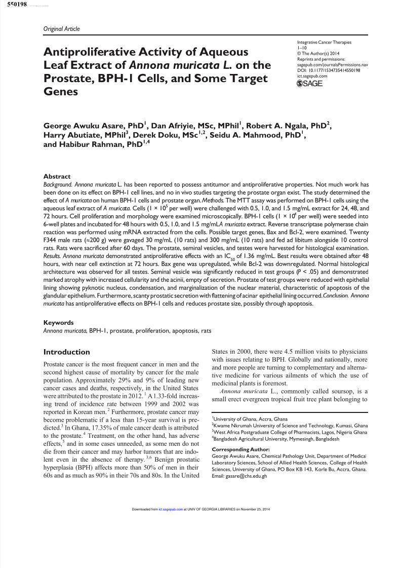
From Figure 1 and Table 1, it can be seen that the areas of
peaks 3, 4, 6, 5, 2, 1, 8, 7 were in the ratio of 27.1:18.8:15.6
:12.5:11.4:7.8:3.5:3.4. Results from the study are therefore
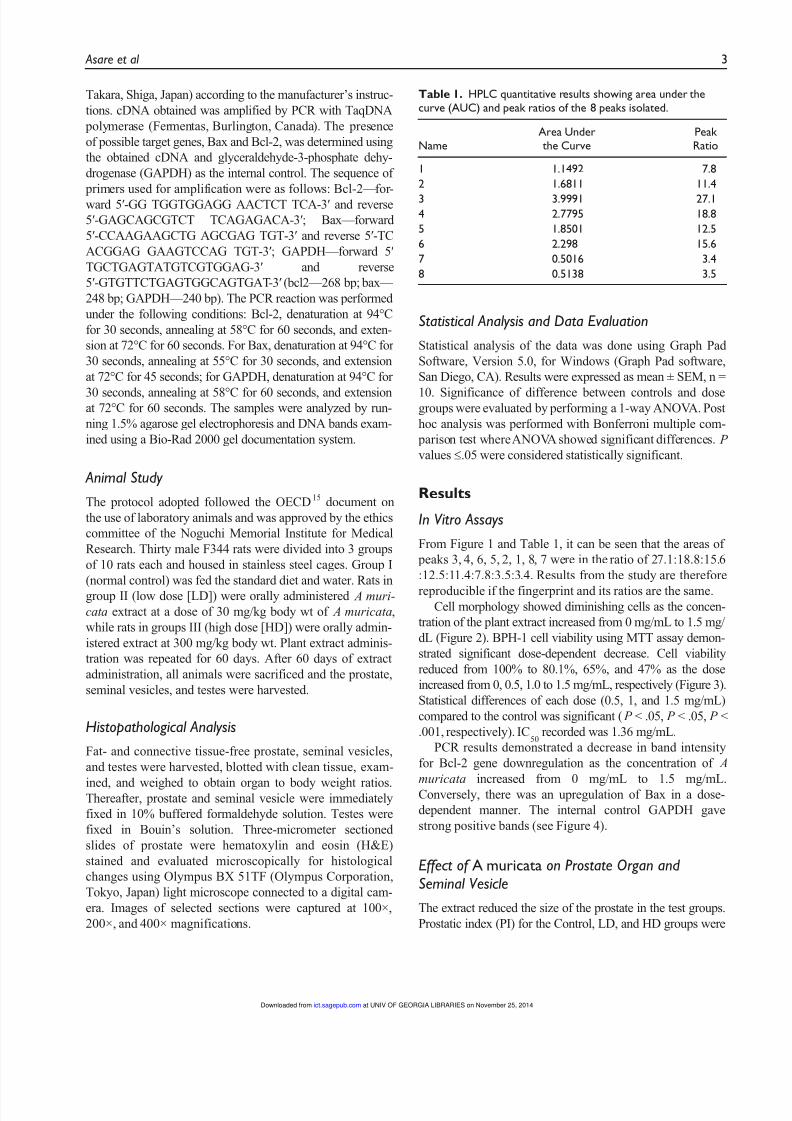
reproducible if the fingerprint and its ratios are the same.Cell morphology showed diminishing cells as the concen-
tration of the plant extract increased from 0 mg/mL to 1.5 mg/
dL (Figure 2). BPH-1 cell viability using MTT assay demon-
strated significant dose-dependent decrease. Cell viability
reduced from 100% to 80.1%, 65%, and 47% as the dose
increased from 0, 0.5, 1.0 to 1.5 mg/mL, respectively (Figure 3).
Statistical differences of each dose (0.5, 1, and 1.5 mg/mL)
compared to the control was significant ( P < .05, P < .05, P <
.001, respectively). IC50
recorded was 1.36 mg/mL.
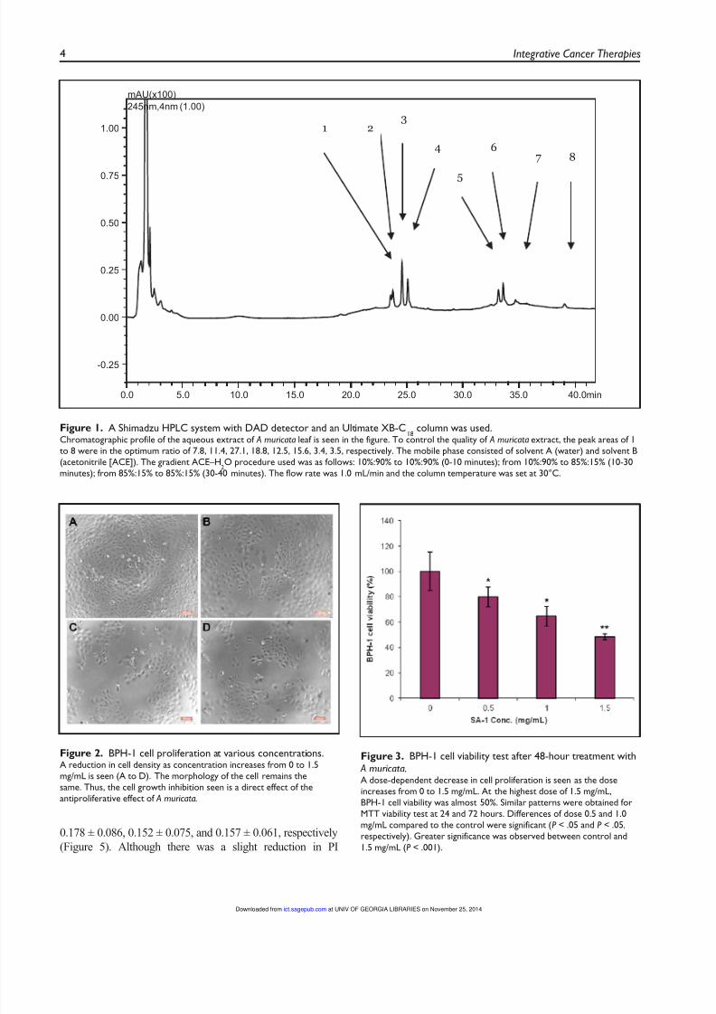
PCR results demonstrated a decrease in band intensity
for Bcl-2 gene downregulation as the concentration of A
muricata increased from 0 mg/mL to 1.5 mg/mL.
Conversely, there was an upregulation of Bax in a dose-
dependent manner. The internal control GAPDH gave
strong positive bands (see Figure 4).
Effect of A muricata on Prostate Organ and
Seminal Vesicle
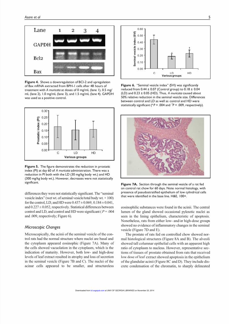
The extract reduced the size of the prostate in the test groups.
Prostatic index (PI) for the Control, LD, and HD groups were
Table 1. HPLC quantitative results showing area under thecurve (AUC) and peak ratios of the 8 peaks isolated.
NameArea Underthe Curve
PeakRatio
1 1.1492 7.8
2 1.6811 11.4
3 3.9991 27.14 2.7795 18.8
5 1.8501 12.5
6 2.298 15.6
7 0.5016 3.4
8 0.5138 3.5
at UNIV OF GEORGIA LIBRARIES on November 25, 2014ict.sagepub.comDownloaded from
8/9/2019 Asare, G. A.; Afriyie, D.; Ngala, R. A.; Abutiate, H.; Doku, D.; -- Antiproliferative Activity of Aqueous Leaf Extract
http://slidepdf.com/reader/full/asare-g-a-afriyie-d-ngala-r-a-abutiate-h-doku-d-antiproliferative 5/11
4 Integrative Cancer Therapies
0.178 ± 0.086, 0.152 ± 0.075, and 0.157 ± 0.061, respectively
(Figure 5). Although there was a slight reduction in PI
0.0 5.0 10.0 15.0 20.0 25.0 30.0 35.0 40.0min
-0.25
0.00
0.25
0.50
0.75
1.00
mAU(x100)
245nm,4nm (1.00)
1 23
4
5
67 8
Figure 1. A Shimadzu HPLC system with DAD detector and an Ultimate XB-C18
column was used.Chromatographic profile of the aqueous extract of A muricata leaf is seen in the figure. To control the quality of A muricata extract, the peak areas of 1to 8 were in the optimum ratio of 7.8, 11.4, 27.1, 18.8, 12.5, 15.6, 3.4, 3.5, respectively. The mobile phase consisted of solvent A (water) and solvent B(acetonitrile [ACE]). The gradient ACE–H
2O procedure used was as follows: 10%:90% to 10%:90% (0-10 minutes); from 10%:90% to 85%:15% (10-30
minutes); from 85%:15% to 85%:15% (30-40 minutes). The flow rate was 1.0 mL/min and the column temperature was set at 30°C.
Figure 2. BPH-1 cell proliferation at various concentrations.A reduction in cell density as concentration increases from 0 to 1.5mg/mL is seen (A to D). The morphology of the cell remains thesame. Thus, the cell growth inhibition seen is a direct effect of theantiproliferative effect of A muricata.
Figure 3. BPH-1 cell viability test after 48-hour treatment with A muricata.A dose-dependent decrease in cell proliferation is seen as the doseincreases from 0 to 1.5 mg/mL. At the highest dose of 1.5 mg/mL,BPH-1 cell viability was almost 50%. Similar patterns were obtained forMTT viability test at 24 and 72 hours. Differences of dose 0.5 and 1.0mg/mL compared to the control were significant (P < .05 and P < .05,respectively). Greater significance was observed between control and1.5 mg/mL (P < .001).
at UNIV OF GEORGIA LIBRARIES on November 25, 2014ict.sagepub.comDownloaded from
8/9/2019 Asare, G. A.; Afriyie, D.; Ngala, R. A.; Abutiate, H.; Doku, D.; -- Antiproliferative Activity of Aqueous Leaf Extract
http://slidepdf.com/reader/full/asare-g-a-afriyie-d-ngala-r-a-abutiate-h-doku-d-antiproliferative 6/11
Asare et al 5
differences they were not statistically significant. The “seminal
vesicle index” (wet wt. of seminal vesicle/total body wt. × 100)
for the control, LD, and HD were 0.437 ± 0.069, 0.184 ± 0.041,
and 0.227 ± 0.052, respectively. Statistical differences between
control and LD, and control and HD were significant ( P = .004
and .009, respectively; Figure 6).
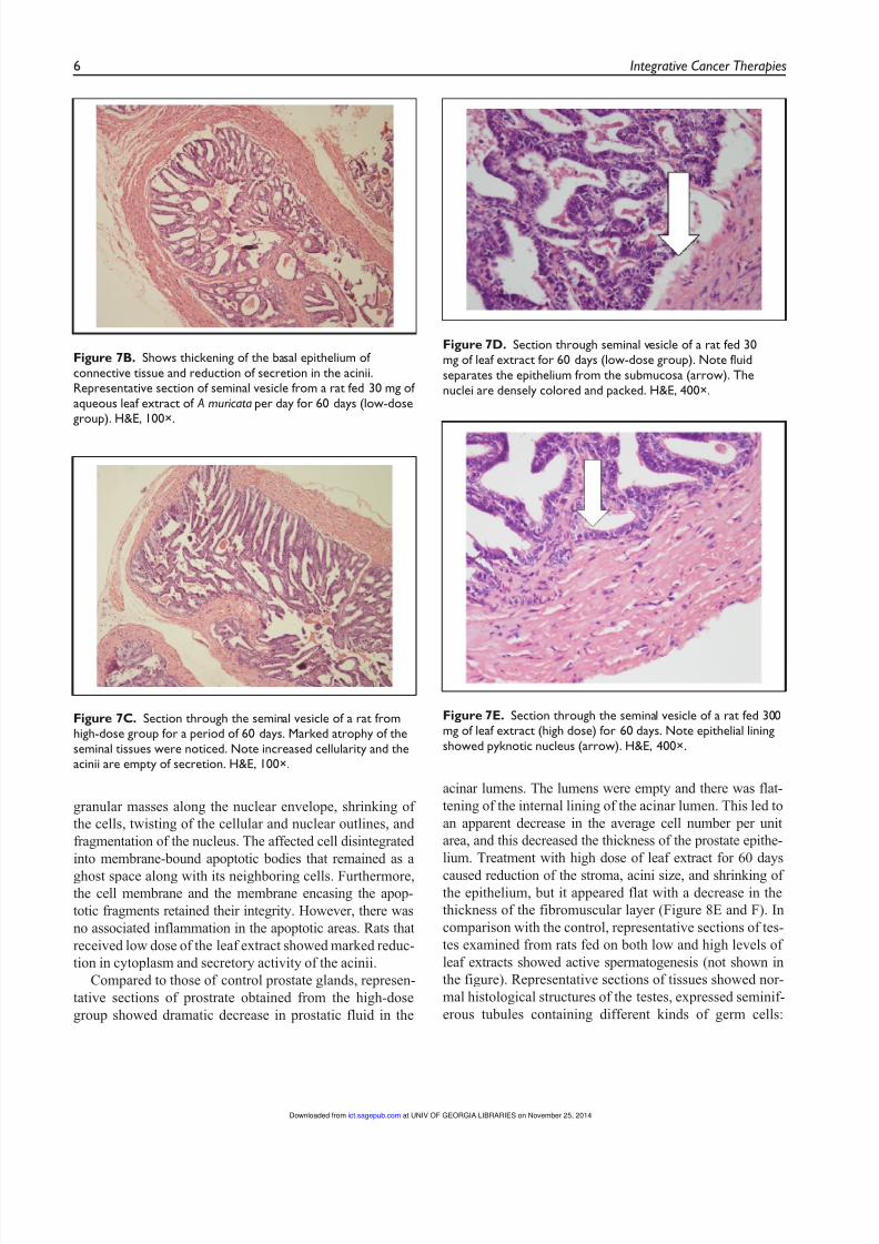
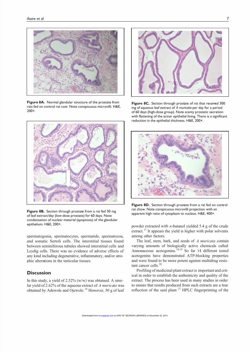
Microscopic ChangesMicroscopically, the acinii of the seminal vesicle of the con-
trol rats had the normal structure where nuclei are basal and
the cytoplasm appeared eosinophic (Figure 7A). Many of
the cells showed vacuolation in the cytoplasm, which is the
indication of maturity. However, both low- and high-dose
levels of leaf extract resulted in atrophy and loss of secretion
in the seminal vesicle (Figure 7B and C). The nuclei of the
acinar cells appeared to be smaller, and structureless
eosinophilic substances were found in the acinii. The central
lumen of the gland showed occasional pyknotic nuclei as
seen in the lining epithelium, characteristic of apoptosis.
Nonetheless, rats from either low- and/or high-dose groups
showed no evidence of inflammatory changes in the seminalvesicle (Figure 7D and E).
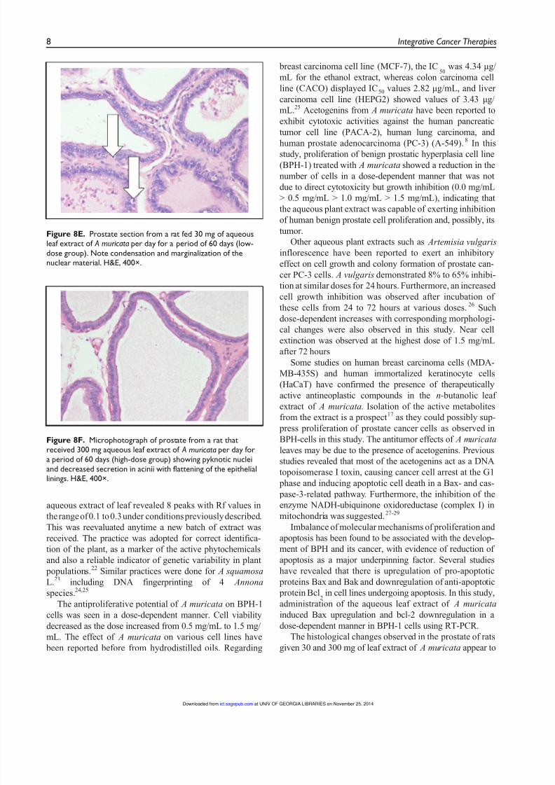
The prostate of rats fed on controlled chow showed nor-
mal histological structures (Figure 8A and B). The alveoli
showed tall columnar epithelial cells with an apparent high
ratio of cytoplasm to nucleus. However, representative sec-
tions of tissues of prostate obtained from rats that received
low dose of leaf extract showed apoptosis in the epithelium
of the glandular acinii (Figure 8C and D). They include dis-
crete condensation of the chromatin, to sharply delineated
Figure 4. Shows a downregulation of BCl-2 and upregulationof Bax mRNA extracted from BPH-1 cells after 48 hours oftreatment with A muricata at doses of 0 mg/mL (lane 1), 0.5 mg/mL (lane 2), 1.0 mg/mL (lane 3), and 1.5 mg/mL (lane 4). GAPDHwas used as a positive control.
0.00
0.05
0.10
0.15
0.20
0.25
0.30
C LD HD
Various groups
P r o s t a t i c
i n d e x
( P I )
Figure 5. The figure demonstrates the reduction in prostatic
index (PI) at day 60 of A muricata administration. There was areduction in PI both with the LD (30 mg/kg body wt.) and HD(300 mg/kg body wt.). However, decreases were not statisticallysignificant.
0.00
0.10
0.20
0.30
0.40
0.50
0.60
C LD HD
Various groups
S e m i n a l v e s i c l e i n d e x ( S V I )
*
†
Figure 6. “Seminal vesicle index” (SVI) was significantlyreduced from 0.44 ± 0.07 (Control group) to 0.18 ± 0.04(LD) and 0.23 ± 0.05 (HD). Thus, A muricata caused about50% relative reduction in the seminal vesicle size. Differencesbetween control and LD as well as control and HD werestatistically significant (*P = .004 and †P = .009, respectively).
Figure 7A. Section through the seminal vesicle of a rat fedon control rat chow for 60 days. Note normal histology, withpresence of pseudostratified epithelium of low cylindrical cellsthat were identified in the base line. H&E, 100×.
at UNIV OF GEORGIA LIBRARIES on November 25, 2014ict.sagepub.comDownloaded from
8/9/2019 Asare, G. A.; Afriyie, D.; Ngala, R. A.; Abutiate, H.; Doku, D.; -- Antiproliferative Activity of Aqueous Leaf Extract
http://slidepdf.com/reader/full/asare-g-a-afriyie-d-ngala-r-a-abutiate-h-doku-d-antiproliferative 7/11
6 Integrative Cancer Therapies
granular masses along the nuclear envelope, shrinking of
the cells, twisting of the cellular and nuclear outlines, and
fragmentation of the nucleus. The affected cell disintegrated
into membrane-bound apoptotic bodies that remained as a
ghost space along with its neighboring cells. Furthermore,the cell membrane and the membrane encasing the apop-
totic fragments retained their integrity. However, there was
no associated inflammation in the apoptotic areas. Rats that
received low dose of the leaf extract showed marked reduc-
tion in cytoplasm and secretory activity of the acinii.
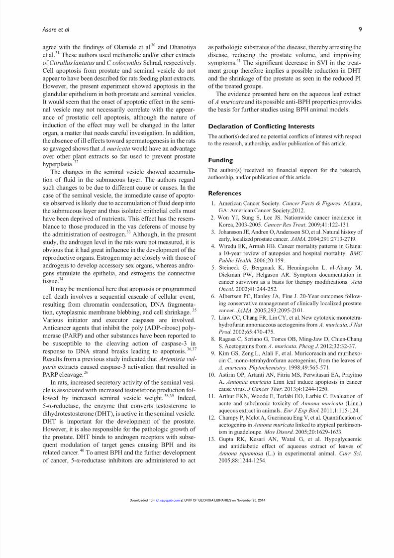
Compared to those of control prostate glands, represen-
tative sections of prostrate obtained from the high-dose
group showed dramatic decrease in prostatic fluid in the
acinar lumens. The lumens were empty and there was flat-
tening of the internal lining of the acinar lumen. This led to
an apparent decrease in the average cell number per unit
area, and this decreased the thickness of the prostate epithe-
lium. Treatment with high dose of leaf extract for 60 days
caused reduction of the stroma, acini size, and shrinking ofthe epithelium, but it appeared flat with a decrease in the
thickness of the fibromuscular layer (Figure 8E and F). In
comparison with the control, representative sections of tes-
tes examined from rats fed on both low and high levels of
leaf extracts showed active spermatogenesis (not shown in
the figure). Representative sections of tissues showed nor-
mal histological structures of the testes, expressed seminif-
erous tubules containing different kinds of germ cells:
Figure 7B. Shows thickening of the basal epithelium ofconnective tissue and reduction of secretion in the acinii.Representative section of seminal vesicle from a rat fed 30 mg ofaqueous leaf extract of A muricata per day for 60 days (low-dosegroup). H&E, 100×.
Figure 7C. Section through the seminal vesicle of a rat fromhigh-dose group for a period of 60 days. Marked atrophy of theseminal tissues were noticed. Note increased cellularity and theacinii are empty of secretion. H&E, 100×.
Figure 7D. Section through seminal vesicle of a rat fed 30mg of leaf extract for 60 days (low-dose group). Note fluidseparates the epithelium from the submucosa (arrow). Thenuclei are densely colored and packed. H&E, 400×.
Figure 7E. Section through the seminal vesicle of a rat fed 300mg of leaf extract (high dose) for 60 days. Note epithelial liningshowed pyknotic nucleus (arrow). H&E, 400×.
at UNIV OF GEORGIA LIBRARIES on November 25, 2014ict.sagepub.comDownloaded from
8/9/2019 Asare, G. A.; Afriyie, D.; Ngala, R. A.; Abutiate, H.; Doku, D.; -- Antiproliferative Activity of Aqueous Leaf Extract
http://slidepdf.com/reader/full/asare-g-a-afriyie-d-ngala-r-a-abutiate-h-doku-d-antiproliferative 8/11
Asare et al 7
spermatogonia, spermatocytes, spermatids, spermatozoa,
and somatic Sertoli cells. The interstitial tissues found
between seminiferous tubules showed interstitial cells andLeydig cells. There was no evidence of adverse effects of
any kind including degenerative, inflammatory, and/or atro-
phic alterations in the testicular tissues.
Discussion
In this study, a yield of 2.52% (w/w) was obtained. A simi-
lar yield of 2.62% of the aqueous extract of A muricata was
obtained by Adewole and Ojewole.16
However, 50 g of leaf
powder extracted with n-butanol yielded 5.4 g of the crude
extract.17 It appears the yield is higher with polar solvents
among other factors.
The leaf, stem, bark, and seeds of A muricata contain
varying amounts of biologically active chemicals calledAnnonaceous acetogenins.18,19 So far 14 different tested
acetogenins have demonstrated ATP-blocking properties
and were found to be more potent against multidrug resis-
tant cancer cells.20
Profiling of medicinal plant extract is important and crit-
ical in order to establish the authenticity and quality of the
extract. The process has been used in many studies in order
to ensure that results produced from such extracts are a true
reflection of the said plant.21 HPLC fingerprinting of the
Figure 8A. Normal glandular structure of the prostate fromrats fed on control rat cow. Note conspicuous microvilli. H&E,200×.
Figure 8B. Section through prostate from a rat fed 30 mgof leaf extract/day (low-dose prostate) for 60 days. Notecondensation of nuclear material (apoptosis) of the glandularepithelium. H&E, 200×.
Figure 8C. Section through prostate of rat that received 300mg of aqueous leaf extract of A muricata per day for a periodof 60 days (high-dose group). Note scanty prostatic secretionwith flattening of the acinar epithelial lining. There is a significantreduction in the epithelial thickness. H&E, 200×.
Figure 8D. Section through prostate from a rat fed on controlrat chow. Note conspicuous microvilli projection with anapparent high ratio of cytoplasm to nucleus. H&E, 400×.
at UNIV OF GEORGIA LIBRARIES on November 25, 2014ict.sagepub.comDownloaded from
8/9/2019 Asare, G. A.; Afriyie, D.; Ngala, R. A.; Abutiate, H.; Doku, D.; -- Antiproliferative Activity of Aqueous Leaf Extract
http://slidepdf.com/reader/full/asare-g-a-afriyie-d-ngala-r-a-abutiate-h-doku-d-antiproliferative 9/11
8 Integrative Cancer Therapies
aqueous extract of leaf revealed 8 peaks with Rf values in
the range of 0.1 to 0.3 under conditions previously described.
This was reevaluated anytime a new batch of extract was
received. The practice was adopted for correct identifica-
tion of the plant, as a marker of the active phytochemicalsand also a reliable indicator of genetic variability in plant
populations.22 Similar practices were done for A squamosa
L.23
including DNA fingerprinting of 4 Annona
species.24,25
The antiproliferative potential of A muricata on BPH-1
cells was seen in a dose-dependent manner. Cell viability
decreased as the dose increased from 0.5 mg/mL to 1.5 mg/
mL. The effect of A muricata on various cell lines have
been reported before from hydrodistilled oils. Regarding
breast carcinoma cell line (MCF-7), the IC50
was 4.34 µg/
mL for the ethanol extract, whereas colon carcinoma cell
line (CACO) displayed IC50 values 2.82 µg/mL, and liver
carcinoma cell line (HEPG2) showed values of 3.43 µg/
mL.25 Acetogenins from A muricata have been reported to
exhibit cytotoxic activities against the human pancreatic
tumor cell line (PACA-2), human lung carcinoma, and
human prostate adenocarcinoma (PC-3) (A-549).8 In this
study, proliferation of benign prostatic hyperplasia cell line
(BPH-1) treated with A muricata showed a reduction in the
number of cells in a dose-dependent manner that was not
due to direct cytotoxicity but growth inhibition (0.0 mg/mL
> 0.5 mg/mL > 1.0 mg/mL > 1.5 mg/mL), indicating that
the aqueous plant extract was capable of exerting inhibition
of human benign prostate cell proliferation and, possibly, its
tumor.
Other aqueous plant extracts such as Artemisia vulgaris
inflorescence have been reported to exert an inhibitory
effect on cell growth and colony formation of prostate can-
cer PC-3 cells. A vulgaris demonstrated 8% to 65% inhibi-tion at similar doses for 24 hours. Furthermore, an increased
cell growth inhibition was observed after incubation of
these cells from 24 to 72 hours at various doses.26
Such
dose-dependent increases with corresponding morphologi-
cal changes were also observed in this study. Near cell
extinction was observed at the highest dose of 1.5 mg/mL
after 72 hours
Some studies on human breast carcinoma cells (MDA-
MB-435S) and human immortalized keratinocyte cells
(HaCaT) have confirmed the presence of therapeutically
active antineoplastic compounds in the n-butanolic leaf
extract of A muricata. Isolation of the active metabolites
from the extract is a prospect17 as they could possibly sup-
press proliferation of prostate cancer cells as observed in
BPH-cells in this study. The antitumor effects of A muricata
leaves may be due to the presence of acetogenins. Previous
studies revealed that most of the acetogenins act as a DNA
topoisomerase I toxin, causing cancer cell arrest at the G1
phase and inducing apoptotic cell death in a Bax- and cas-
pase-3-related pathway. Furthermore, the inhibition of the
enzyme NADH-ubiquinone oxidoreductase (complex I) in
mitochondria was suggested.27-29
Imbalance of molecular mechanisms of proliferation and
apoptosis has been found to be associated with the develop-
ment of BPH and its cancer, with evidence of reduction ofapoptosis as a major underpinning factor. Several studies
have revealed that there is upregulation of pro-apoptotic
proteins Bax and Bak and downregulation of anti-apoptotic
protein Bcl2 in cell lines undergoing apoptosis. In this study,
administration of the aqueous leaf extract of A muricata
induced Bax upregulation and bcl-2 downregulation in a
dose-dependent manner in BPH-1 cells using RT-PCR.
The histological changes observed in the prostate of rats
given 30 and 300 mg of leaf extract of A muricata appear to
Figure 8E. Prostate section from a rat fed 30 mg of aqueousleaf extract of A muricata per day for a period of 60 days (low-dose group). Note condensation and marginalization of thenuclear material. H&E, 400×.
Figure 8F. Microphotograph of prostate from a rat thatreceived 300 mg aqueous leaf extract of A muricata per day fora period of 60 days (high-dose group) showing pyknotic nucleiand decreased secretion in acinii with flattening of the epitheliallinings. H&E, 400×.
at UNIV OF GEORGIA LIBRARIES on November 25, 2014ict.sagepub.comDownloaded from
8/9/2019 Asare, G. A.; Afriyie, D.; Ngala, R. A.; Abutiate, H.; Doku, D.; -- Antiproliferative Activity of Aqueous Leaf Extract
http://slidepdf.com/reader/full/asare-g-a-afriyie-d-ngala-r-a-abutiate-h-doku-d-antiproliferative 10/11
Asare et al 9
agree with the findings of Olamide et al30 and Dhanotiya
et al.31 These authors used methanolic and/or ether extracts
of Citrullus lantatus and C colocynthis Schrad, respectively.
Cell apoptosis from prostate and seminal vesicle do not
appear to have been described for rats feeding plant extracts.
However, the present experiment showed apoptosis in the
glandular epithelium in both prostate and seminal vesicles.
It would seem that the onset of apoptotic effect in the semi-
nal vesicle may not necessarily correlate with the appear-
ance of prostatic cell apoptosis, although the nature of
induction of the effect may well be changed in the latter
organ, a matter that needs careful investigation. In addition,
the absence of ill effects toward spermatogenesis in the rats
so gavaged shows that A muricata would have an advantage
over other plant extracts so far used to prevent prostate
hyperplasia.32
The changes in the seminal vesicle showed accumula-
tion of fluid in the submucous layer. The authors regard
such changes to be due to different cause or causes. In the
case of the seminal vesicle, the immediate cause of apopto-sis observed is likely due to accumulation of fluid deep into
the submucous layer and thus isolated epithelial cells must
have been deprived of nutrients. This effect has the resem-
blance to those produced in the vas deferens of mouse by
the administration of oestrogen.33 Although, in the present
study, the androgen level in the rats were not measured, it is
obvious that it had great influence in the development of the
reproductive organs. Estrogen may act closely with those of
androgens to develop accessory sex organs, whereas andro-
gens stimulate the epithelia, and estrogens the connective
tissue.34
It may be mentioned here that apoptosis or programmed
cell death involves a sequential cascade of cellular event,
resulting from chromatin condensation, DNA fragmenta-
tion, cytoplasmic membrane blebbing, and cell shrinkage.35
Various initiator and executor caspases are involved.
Anticancer agents that inhibit the poly (ADP-ribose) poly-
merase (PARP) and other substances have been reported to
be susceptible to the cleaving action of caspase-3 in
response to DNA strand breaks leading to apoptosis.36,37
Results from a previous study indicated that Artemisia vul-
garis extracts caused caspase-3 activation that resulted in
PARP cleavage.26
In rats, increased secretory activity of the seminal vesi-
cle is associated with increased testosterone production fol-lowed by increased seminal vesicle weight.38,39 Indeed,
5-α-reductase, the enzyme that converts testosterone to
dihydrotestosterone (DHT), is active in the seminal vesicle.
DHT is important for the development of the prostate.
However, it is also responsible for the pathologic growth of
the prostate. DHT binds to androgen receptors with subse-
quent modulation of target genes causing BPH and its
related cancer.40 To arrest BPH and the further development
of cancer, 5-α-reductase inhibitors are administered to act
as pathologic substrates of the disease, thereby arresting the
disease, reducing the prostate volume, and improving
symptoms.41
The significant decrease in SVI in the treat-
ment group therefore implies a possible reduction in DHT
and the shrinkage of the prostate as seen in the reduced PI
of the treated groups.
The evidence presented here on the aqueous leaf extract
of A muricata and its possible anti-BPH properties provides
the basis for further studies using BPH animal models.
Declaration of Conflicting Interests
The author(s) declared no potential conflicts of interest with respect
to the research, authorship, and/or publication of this article.
Funding
The author(s) received no financial support for the research,
authorship, and/or publication of this article.
References 1. American Cancer Society. Cancer Facts & Figures. Atlanta,
GA: American Cancer Society;2012.
2. Won YJ, Sung S, Lee JS. Nationwide cancer incidence in
Korea, 2003-2005. Cancer Res Treat . 2009;41:122-131.
3. Johansson JE, Andren O, Andersson SO, et al. Natural history of
early, localized prostate cancer. JAMA. 2004;291:2713-2719.
4. Wiredu EK, Armah HB. Cancer mortality patterns in Ghana:
a 10-year review of autopsies and hospital mortality. BMC
Public Health. 2006;20:159.
5. Steineck G, Bergmark K, Henningsohn L, al-Abany M,
Dickman PW, Helgason AR. Symptom documentation in
cancer survivors as a basis for therapy modifications. Acta
Oncol . 2002;41:244-252.
6. Albertsen PC, Hanley JA, Fine J. 20-Year outcomes follow-
ing conservative management of clinically localized prostate
cancer. JAMA. 2005;293:2095-2101.
7. Liaw CC, Chang FR, Lin CY, et al. New cytotoxic monotetra-
hydrofuran annonaceous acetogenins from A. muricata. J Nat
Prod . 2002;65:470-475.
8. Ragasa C, Soriano G, Torres OB, Ming-Jaw D, Chien-Chang
S. Acetogenins from A. muricata. Phcog J . 2012;32:32-37.
9. Kim GS, Zeng L, Alali F, et al. Muricoreacin and murihexo-
cin C, mono-tetrahydrofuran acetogenins, from the leaves of
A. muricata. Phytochemistry. 1998;49:565-571.
10. Astirin OP, Artanti AN, Fitria MS, Perwitasari EA, Prayitno
A. Annonaa muricata Linn leaf induce apoptosis in cancer
cause virus. J Cancer Ther . 2013;4:1244-1250. 11. Arthur FKN, Woode E, Terlabi EO, Larbie C. Evaluation of
acute and subchronic toxicity of Annona muricata (Linn.)
aqueous extract in animals. Eur J Exp Biol . 2011;1:115-124.
12. Champy P, Melot A, Guerineau Eng V, et al. Quantification of
acetogenins in Annona muricata linked to atypical parkinson-
ism in guadeloupe. Mov Disord . 2005;20:1629-1633.
13. Gupta RK, Kesari AN, Watal G, et al. Hypoglycaemic
and antidiabetic effect of aqueous extract of leaves of
Annona squamosa (L.) in experimental animal. Curr Sci.
2005;88:1244-1254.
at UNIV OF GEORGIA LIBRARIES on November 25, 2014ict.sagepub.comDownloaded from

8/9/2019 Asare, G. A.; Afriyie, D.; Ngala, R. A.; Abutiate, H.; Doku, D.; -- Antiproliferative Activity of Aqueous Leaf Extract
http://slidepdf.com/reader/full/asare-g-a-afriyie-d-ngala-r-a-abutiate-h-doku-d-antiproliferative 11/11
10 Integrative Cancer Therapies
14. Adewole SO, Ojewole JAO. Protective effects of Annona
muricata Linn. (Annonaceae) leaf aqueous extract on serum
lipid profiles and oxidative stress in hepatocytes of strep-
tozontocin-treated rats. Afr J Tradit Complement Altern Med .
2009;6:30-41.
15. OECD. Test No. 408: Repeated Dose 90-Day Oral Toxicity
Study in Rodents; OECD Guidelines for the Testing of
Chemicals, Section 4: Health Effects. Paris, France: OECDPublishing; 1998.
16. Adewole SO, Caxton-Martins EA. Morphological changes
and hypoglycemic effects of A. muricata (Annonaceae) leaf
aqueous extract on pancreatic Β-cells of streptozotocin-
treated diabetic rats. Afr J Biomed Res. 2006;9:173-187.
17. George VC, Kumar DRN, Rajkumar V, Suresh PK, Kumar
RA. Quantitative assessment of the relative antineoplastic
potential of the n-butanolic leaf extract of A. muricata in nor-
mal and immortalized human cell lines. Asian Pac J Cancer
Prev. 2012;13:699-704.
18. Tormo JR, Royo I, Gallardo T, et al. In vitro antitumor struc-
ture activity relationships of threo/trans/three mono-tetrahy-
drofuranic acetogenins: correlations with their inhibition of
mitochondial complex 1. Oncol Res. 2003;14:147-154. 19. Kojima N. Systemic synthesis of antitumor Annonaceous ace-
togenins. Yakugaku Zasshi. 2004;124:673-681.
20. Oberlies NH, Jones JL, Corbett TH, Fotopoulos SS,
McLaughlin JL. Tumor cell growth inhibition by several
Annonaceous acetogenins in an in vitro disk diffusion assay.
Cancer Lett . 1995;96:55-62.
21. Chatterjee S, Srivastava S, Khalid A, et al. Comprehensive
metabolic fingerprinting of Withania somnifera leaf and root
extracts. Phytochemistry. 2010;71:1085-1094.
22. Maier TS, Kuhn J, Müller C. Proposal for field sampling of
plants and processing in the lab for environmental metabolic
fingerprinting. Plant Methods. 2010;6:6.
23. Abhishek SL, Vd Goray Namrata P. Pharmacognostical stud-
ies on the leaf of Annona squamosa Linn. Phcog J . 2009;1:1.
24. Ahmad I, Bhagat S, Sharma TVRS, Krishna K, Simachalam
P, Srivastava RC. ISSR and RAPD marker based DNA fin-
gerprinting and diversity assessment of Annona spp. in South
Andaman. Ind J Horticulture. 2010;67:147-151.
25. Elhawary SS, El Tantawy ME, Rabeh MA, Fawaz NE. DNA
fingerprinting, chemical composition, antitumor and anti-
microbial activities of the essential oils and extractives of
four Annona species from Egypt. J Natural Sci Res . 2013;3:
59-68.
26. Nawab A, Yunus M, Mahdi AA, Gupta S. Evaluation of anti-
cancer properties of medicinal plants from the Indian sub-
continent. Mol Cell Pharmacol . 2011;3:21-29.
27. Lopez LM, Martin CC, Bermejo A, Cortes D, AyusoMJ. Cytotoxic compounds from annonaceous species as
DNA topoisomerase I poisons. Anticancer Res. 2001;21:
3493-3497.
28. Yuan SS, Chang HL, Chen HW, et al. Annonacin, a mono-
tetrahydrofuran acetogenin, arrests cancer cells at the G1
phase and causes cytotoxicity in a bax- and caspase-3-related
pathway. Life Sci. 2003;72:2853-2861.
29. Kojima N, Morioka T, Urabe D, et al. Convergent synthesis
of fluorescence-labeled probes of annonaceous acetogeninsand visualization of their cell distribution. Bioorg Med Chem.
2010;18:8630-8641.
30. Olamide AA, Olayemi OO, Demetrius OO, Olatoye OJ,
Kehinde AA. Effects of methanolic extract of Citrullus lana-
tus seed on experimentally induced prostatic hyperplasia. Eur
J Med Plants. 2011;1:171-179.
31. Dhanotiya R, Chauhan NS, Saraf DK, Dixit VK. Effect of
Citrullus colocynthis Schard on testosterone-induced benign
prostatic hyperplasia. J Comp Integr Med . 2009;6:29.
32. Torres MP, Rachagani S, Purohit V, et al. Graviola: a novel
promising natural-derived drug that inhibits tumorigenic-
ity and metastasis of pancreatic cancer cells in vitro and in
vivo through altering cell metabolism. Cancer Lett . 2012;323:
29-40. 33. Harsh R, Overholser MD, Wells LJ. Effects of estrogen and
androgen injections on reproductive organs in male rats and
mice. J Endocrinol . 1939;1:261-267.
34. Clegg EJ. Postnatal changes in the histology of the semi-
nal vesicle and coagulating gland in the rat. J Anatomy.
1953;93:361-367.
35. Bøe R, Gjertsen BT, Vintermyr OK, Houge G, Lanotte M,
Døskeland SO. The protein phosphatase inhibitor okadaic
acid induces morphological changes typical of apoptosis in
mammalian cells. Exp Cell Res. 1991;195:237-246.
36. Mancini M, Nicholson DW, Roy S, et al. The caspase-3 pre-
cursor has a cytosolic and mitochondrial distribution: implica-
tions for apoptotic signaling. J Cell Biol . 1998;140:1485-1495.
37. Nicholson DW, Thornberry NA. Caspases: killer proteases.
Trends Biochem Sci. 1997;22:299-306.
38. Higgins SJ, Burchell JM. Effects of testosterone on mes-
senger ribonucleic acid and protein synthesis in rat seminal
vesicle. Biochem J . 1978;174:543-551.
39. Zanato VF, Martins MP, Anselmo-Franci JA. Sexual devel-
opment of male Wistar rats. Braz J Med Biol Res. 1994;27:
1273-1280.
40. Bartsch G, Rittmaster RS, Klocker H. Dihydrotestosterone
and the concept of 5-alpha-reductase inhibition in human
benign prostatic hyperplasia. World J Urol . 2002;19:413-425.
41. Andriole G, Bruchovsky N, Chung LW, et al. Dihydrotesto-
sterone and the prostate: the scientific rationale for 5-alpha-
reductase inhibitors in the treatment of benign prostatichyperplasia. J Urol . 2004;172:1399-403.