arterivirus molecular biology and pathogenesis 2013
DESCRIPTION
Arterivirus Molecular Biology and Pathogenesis 2013TRANSCRIPT

Review Arterivirus molecular biology and pathogenesis
Eric J. Snijder,1 Marjolein Kikkert1 and Ying Fang2,3
Correspondence
Eric J. Snijder
Received 25 June 2013
Accepted 7 August 2013
1Molecular Virology Department, Department of Medical Microbiology, Leiden University MedicalCenter, Leiden, The Netherlands
2Department of Veterinary and Biomedical Sciences, South Dakota State University, Brookings,South Dakota, USA
3Department of Diagnostic Medicine and Pathobiology, College of Veterinary Medicine,Kansas State University, Manhattan, Kansas, USA
Arteriviruses are positive-stranded RNA viruses that infect mammals. They can cause persistent or
asymptomatic infections, but also acute disease associated with a respiratory syndrome, abortion or
lethal haemorrhagic fever. During the past two decades, porcine reproductive and respiratory
syndrome virus (PRRSV) and, to a lesser extent, equine arteritis virus (EAV) have attracted attention as
veterinary pathogens with significant economic impact. Particularly noteworthy were the ‘porcine high
fever disease’ outbreaks in South-East Asia and the emergence of new virulent PRRSV strains in the
USA. Recently, the family was expanded with several previously unknown arteriviruses isolated from
different African monkey species. At the molecular level, arteriviruses share an intriguing but distant
evolutionary relationship with coronaviruses and other members of the order Nidovirales. Nevertheless,
several of their characteristics are unique, including virion composition and structure, and the
conservation of only a subset of the replicase domains encountered in nidoviruses with larger
genomes. During the past 15 years, the advent of reverse genetics systems for EAV and PRRSV has
changed and accelerated the structure–function analysis of arterivirus RNA and protein sequences.
These systems now also facilitate studies into host immune responses and arterivirus immune evasion
and pathogenesis. In this review, we have summarized recent advances in the areas of arterivirus
genome expression, RNA and protein functions, virion architecture, virus–host interactions, immunity,
and pathogenesis. We have also briefly reviewed the impact of these advances on disease
management, the engineering of novel candidate live vaccines and the diagnosis of arterivirus infection.
Introduction
About two decades ago, genome sequencing establishedthat arteriviruses qualified as a separate family of positive-stranded RNA (+RNA) viruses (reviewed by Cavanagh,1997; Snijder & Meulenberg, 1998). A family with, on theone hand, an intriguing but distant evolutionary relation-ship to coronaviruses and other members of the orderNidovirales but, on the other hand, characteristics uniqueamong currently known +RNA viruses. These includevirion composition and structure, and the conservation ofonly a subset of the replicase domains encountered innidoviruses with larger genomes. Shortly after the familyArteriviridae was created, equine arteritis virus (EAV) andporcine reproductive and respiratory syndrome virus(PRRSV) were the first nidoviruses for which reversegenetics systems became available (Meulenberg et al., 1998;van Dinten et al., 1997). This breakthrough not onlyrevolutionized the structure–function analysis of RNA andprotein sequences, but also facilitated studies into patho-genesis and immune responses, and the engineering of novelcandidate live vaccines.
The consequences of arterivirus infection can range frompersistent asymptomatic infection to acute disease, asso-ciated with abortion, fatal age-dependent poliomyelitis orlethal haemorrhagic fever. Three of the currently recognizedarterivirus species [EAV, lactate dehydrogenase-elevatingvirus (LDV) of mice and simian haemorrhagic fever virus(SHFV)] were first isolated about 50 years ago (reviewed bySnijder & Kikkert, 2013; Snijder & Meulenberg, 1998).However, during the past two decades, it was particularly thefourth arterivirus, PRRSV, that moved into the spotlight bycausing tremendous economic losses to the swine industryworldwide. In the late 1980s, highly diverged variants of thisvirus emerged almost simultaneously in Western Europe(genotype 1) and North America (genotype 2), causingnumerous acute respiratory disease outbreaks and abortionstorms. Subsequently, the ‘porcine high fever disease’outbreaks in South-East Asia (Tian et al., 2007) and theemergence of the highly virulent MN184 strain in the USA(Han et al., 2006) have attracted special attention.
Disease prevention and control will clearly benefit fromthe dissection of the molecular and cellular biology of
Journal of General Virology (2013), 94, 2141–2163 DOI 10.1099/vir.0.056341-0
056341 G 2013 SGM Printed in Great Britain 2141

arterivirus infection. In this review, we will summarize whathas been learned, in particular since the advent of reversegenetics, with respect to arterivirus genome expression, RNAand protein functions, virion architecture, host responses,and pathogenesis. We will also briefly review the impact ofthese advances on disease management, vaccine develop-ment and the diagnosis of arterivirus infection.
The expanding order Nidovirales
About 15 years ago, the family Arteriviridae was unitedwith the family Coronaviridae in a new order Nidovirales(Cavanagh, 1997; Snijder & Meulenberg, 1998). In spite ofstriking differences in genome size and virion structure, thegenome organization and expression of arteri- andcoronaviruses are strikingly similar, and key replicasedomains were postulated to share a common ancestry (denBoon et al., 1991). A prominent feature of nidovirusgenome expression is the nested set of subgenomic (sg)mRNAs that was the basis for the order name Nidovirales(Latin nidus5nest). The complex evolutionary relationshipbetween arteriviruses and other nidoviruses has been reviewedextensively elsewhere (Gorbalenya et al., 2006; Nga et al., 2011;Snijder & Meulenberg, 1998). Essentially, related replicasegenes have become associated with seemingly unrelated sets ofstructural protein genes, with RNA recombination likelyplaying an important role in nidovirus evolution.
As nidovirus discovery continued, in a remarkably widevariety of hosts, the order was further expanded withnidoviruses infecting shrimp (family Roniviridae; Cowleyet al., 2000), fish (genus Bafinivirus; subfamily Torovirinaein the family Coronaviridae; Schutze et al., 2006) and mostrecently also insects (proposed family Mesoniviridae; Ngaet al., 2011; Zirkel et al., 2011). The recently identifiedwobbly possum disease virus (WPDV) appears most closelyrelated to arteriviruses (Dunowska et al., 2012). Further-more, several additional monkey arteriviruses, only distantlyrelated to SHFV, were discovered recently (Lauck et al., 2013).Together with the large evolutionary distances between currentarterivirus species (Fig. 1), this may well prompt a futurerevision of the internal taxonomic structure of the family.
Genome structure and expression
Genome properties and organization
The arterivirus genome is 12–16 kb long, 39-polyadenylatedand presumably 59-capped. Full-length genome sequences(Table 1) have been obtained for European and NorthAmerican EAV isolates, a large number of genotype 1 and 2PRRSVs, two LDV strains, and five distantly related monkeyviruses, tentatively grouped under the name SHFV.
The arterivirus genome is a polycistronic +RNA (Figs 2and 3), with 59 and 39 NTRs of 156–224 and 59–117 nt,respectively, that flank an array of 10–15 known ORFs. Thelarge replicase ORFs 1a and 1b occupy the 59-proximalthree-quarters of the genome, with the size of ORF1a beingmuch more variable than that of ORF1b. ORF1a translation
yields replicase polyprotein (pp) 1a (1727–2502 aa), whereasORF1b is expressed by 21 programmed ribosomal frame-shifting (PRF) (den Boon et al., 1991), which C-terminallyextends pp1a into pp1ab (3175–3959 aa). Recently, a shorttransframe (TF) ORF was found to overlap the nsp2-codingregion of ORF1a in the +1 frame and to be expressed by 22PRF (Fang et al., 2012). Remarkably, the TF ORF isconserved in the genomes of PRRSV, LDV and SHFV, butthis part of the EAV genome is considerably truncated.
The 39-proximal genome part has a compact organizationand contains eight to 12 relatively small genes, most ofwhich overlap with neighbouring genes (Fig. 2). TheseORFs encode structural proteins and are expressed from a39-co-terminal nested set of sg mRNAs (Fig. 3). Theorganization of these ORFs is conserved, but downstreamof ORF1b, SHFV and all recently identified SHFV-likeviruses contain three or four additional ORFs (~1.6 kb)that may be derived from an ancient duplication of ORFs2–4 (Godeny et al., 1998; Lauck et al., 2013). Together withthe size variation in ORF1a, this presumed duplicationexplains the genome size differences among arteriviruses.
SHFVkrc2
SHFVkrc1
SHFVkrtg1
SHFVLVR
SHFVkrtg2
100
10010010
0
100100
100
10
0
60
WPDV0.1
LDV-PLDV-C
PRRSV
Lelystad
PRRSV
VR2332
EAVBucyrus
EAVs3685
Fig. 1. Phylogeny of representatives of currently recognizedarterivirus species (EAV, PRRSV, LDV and SHFV), four recentlydiscovered monkey arteriviruses distantly related to SHFV (Laucket al., 2013), and WPDV (Dunowska et al., 2012). This neighbour-joining phylogenetic tree is based on amino acid sequencestranslated from a codon-based ORF1b nucleotide sequencealignment. The analysis highlights the distances between currentand tentative members of the family Arteriviridae. Bootstrap values(based on 1000 replicates of the data) are shown on branches;scale bar, amino acid substitutions per site. Courtesy of Dr Tony L.Goldberg, University of Wisconsin, Madison, WI, USA.
E. J. Snijder, M. Kikkert and Y. Fang
2142 Journal of General Virology 94

RNA structures in replication, transcription and translation
Several RNA signals involved in arterivirus replica-tion have been identified. In EAV, a minimum of~300 nt from both genome termini is required for efficientreplication, meaning that replication signals extend into
the coding sequences (Molenkamp et al., 2000a; Tijms et al.,
2001). Detailed RNA secondary structure models were
developed for the EAV 59 and 39 NTRs (Fig. 4). The 59
NTR is involved in translation, replication and transcrip-tion (van den Born et al., 2004), and contains the so-called
Table 1. Arterivirus genomes and database accession numbers
Virus (isolate) Host Genome size (kb) GenBank accession no.
EAV (Bucyrus) Horse, donkey 12.7 NC_002532
LDV (P) Mouse 14.1 NC_001639
PRRSV genotype 1 (Lelystad) Swine 15.1 M96262
PRRSV genotype 2 (16244B) Swine 15.4 NC_001961
SHFV (LVR 42-0) Macaques 15.7 NC_003092
SHFV-like (krc1) Red colobus 15.5 HQ845737
SHFV-like (krc2) Red colobus 15.2 HQ845738
SHFV-like (krtg1) Red-tailed guenon 15.2 JX473847
SHFV-like (krtg2) Red-tailed guenon 15.3 JX473849
14 1513121110987654
TM
TM
TM TM TM
TM–2
–1
–1
–1
–1
–2
–2
TF
TF
TF
TM TM TM
TMP2Pβ
P2PβPα
P2
P2PPP
PβPα
EAV
LDV
PRRSV
SHFV
TM TMS
S
S
STM TM TM
R Z H N
R Z H N
R Z H N
R Z H N
3210 16 kb
N(A)n
(A)n
(A)n
(A)n
M
GP
GPGP
GPE
N
M
GP
GPGP
GPE
N
M
GPGPE
GPGP
N
M
GP
GP 5a
5a
5a
5a
GP
2b
2a
replicase ORF1b
replicase ORF1a
replicase ORF1a
replicase ORF1a
replicase ORF1a
replicase ORF1b
replicase ORF1b
replicase ORF1b
3 5 7
2a 3 5 7
2b 3
2b′
2a′ 4′
3′
5 7
2a 3 5 7
5a4 6
2b 5a4 6
2a 5a4 6
2b 5a4 6
GPE
Expressed from genomic RNA Expressed from sg mRNAs
Fig. 2. Arterivirus genome organization. The family prototype EAV is shown at the top. The replicase ORFs 1a and 1b [the latterexpressed by ”1 programmed ribosomal frameshifting (PRF)] are followed by the genes encoding the minor and major envelopeproteins and the N protein. GP, glycoprotein; M, membrane; E, envelope; N, nucleocapsid. The 39-proximal region of the SHFVgenome carries a large insertion (pink) containing four ORFs that may encode additional virion proteins. With the exception ofEAV, arterivirus genomes contain an alternative transframe (TF) ORF in the non-structural protein 2 (nsp2)-coding region, whichis expressed by ”2 PRF (Fang et al., 2012). The positions corresponding to (known or predicted) polyprotein cleavage sites aredepicted above the replicase ORFs; red arrowheads, sites cleaved by the nsp4 SP (S); blue arrowheads, sites cleaved by PLPdomains (P) in the nsp1/nsp2 region. The processing scheme of the SHFV nsp1 region remains to be elucidated. ThreeORF1a-encoded (putative) transmembrane domains (TM) and four highly conserved ORF1b-encoded domains are depicted:RNA-dependent RNA polymerase (R), (putative) multinuclear zinc-binding domain (Z), RNA helicase (H), and NendoUendoribonuclease domain (N). Modified from Snijder & Kikkert (2013).
Arterivirus molecular biology and pathogenesis
http://vir.sgmjournals.org 2143

leader TRS hairpin (LTH) that is crucial for sg mRNAproduction. The relevance of other EAV 59 NTR structuresremains to be investigated as few of these are conserved inother arteriviruses (Lu et al., 2011; van den Born et al., 2004).
A 39-proximal EAV RNA hairpin is required for RNAsynthesis and its loop was implicated in an essentialpseudoknot interaction with an upstream hairpin withinthe N protein gene (Beerens & Snijder, 2007). This confor-mation appears conserved in all arteriviruses and mayconstitute a molecular switch that, for example, regulates astep in negative-strand RNA synthesis. In addition, a 39-terminal CC motif upstream of the poly(A) tail was impli-cated in a critical step in replication, possibly the initiationof negative-strand RNA synthesis (Beerens et al., 2007). InPRRSV, a ‘kissing interaction’ between the loops of RNAhairpins in the 39 NTR and N protein gene was found to becrucial for replication (Verheije et al., 2002a).
Arterivirus genome translation presumably initiates through‘conventional’ ribosomal scanning of the 59 NTR (van denBorn et al., 2005), but also entails one or two ribosomalframeshifting events. As in all other nidoviruses, a 21 PRFin the short ORF1a/1b overlap region is used to expressORF1b. The estimated frameshift efficiency is 15–20 % (in areporter system), which derives from the concerted action ofa ‘slippery’ sequence and a downstream RNA pseudoknotstructure (den Boon et al., 1991; Firth & Brierley, 2012). Inaddition, all arteriviruses except EAV employ 22 PRF toexpress a conserved TF ORF that overlaps the nsp2-codingregion (Fig. 2) (Fang et al., 2012). This frameshift site
dictates both efficient 22 and 21 PRF (estimatedefficiencies in PRRSV-infected cells of 16–20 % and 7 %,respectively) and consequently three N-terminally collinearproducts are produced: nsp2, nsp2TF and nsp2N. Adownstream RNA sequence (CCCANCUCC) is a criticaldeterminant of this first documented case of 22 PRF ineukaryotic cells, but is not predicted to be part of a particularframeshift-directing RNA structure. The two PRF mecha-nisms create a complex series of non-structural proteinexpression ratios. Of the ribosomes that translate the PRRSVnsp1 region, y20 % and y7 % synthesize nsp2TF andnsp2N, respectively, with y73 % translating the remainderof ORF1a (nsp3–8) and probably only y15 % subsequentlytranslating ORF1b (nsp9–12).
Proteinases and replicase polyprotein processing
As in all nidoviruses (Ziebuhr et al., 2000), the post-translational processing of arterivirus replicase polypro-teins involves a complex proteolytic cascade that is directedby three (EAV) or four (PRRSV/LDV) ORF1a-encodedproteinase domains (Figs 2 and 5). Cleavage of pp1a andpp1ab generates 13 (EAV) or 14 (PRRSV/LDV) processingend products, named nsp1–12, including nsp7a/7b (PRRSV/LDV/EAV) and nsp1a/1b (PRRSV/LDV). In PRRSV, LDVand SHFV, the 22 PRF event described above yields thetruncated nsp2 variants nsp2TF and nsp2N (Fang et al.,2012). The nsp3–8 region is subject to two alternativeprocessing cascades, with cleaved nsp2 acting as a co-factorto promote the nsp4/5 cleavage in the ‘major’ processing
Binding
Release
Endocytosis
RTCassembly
pp CleavageTranslation
RTCmembraneassociation
Exocytosis
Golgi
ReticulumEndoplasmic
Nucleus
sg mRNAs
AAA
AAA
AAA
AAA
AAA
AAA
AAA
AAAAAA
AAA AAA
AAA
AA
A
DMV
Genome
(–)Genomesg (–)RNAs
Genome
Budding
Genome
N protein
Envelope proteins
encapsidation
Envelopeprotein
maturation
Fusion
Fig. 3. Overview of the arterivirus infectioncycle. Following entry by receptor-mediatedendocytosis and disassembly, genome trans-lation yields replicase polyproteins pp1a andpp1ab. These polyproteins are cleaved byinternal proteinases and the viral non-structuralproteins assemble into a replication andtranscription complex (RTC) that first engagesin minus-strand RNA synthesis. Both full-length and subgenome-length minus strandsare produced, the latter serving as templatesfor the synthesis of sg mRNAs required toexpress the structural protein genes, whichreside in the 39-proximal quarter of thegenome. Novel genomes are packaged intonucleocapsids that become enveloped bybudding from smooth intracellular membranes,after which the new virions leave the cell usingthe exocytic pathway. For further details, seetext. Modified from Snijder & Kikkert (2013).
E. J. Snijder, M. Kikkert and Y. Fang
2144 Journal of General Virology 94

pathway (Wassenaar et al., 1997). EAV cleavage-sitemutagenesis underlined the critical role of replicase poly-protein processing in virus replication (van Aken et al.,2006b; van Dinten et al., 1999).
Two papain-like proteinases (PLPs) and one chymotrypsin-like serine proteinase (SP) are functional in all arteriviruses,and reside in nsp1/1b (PLP1b), nsp2 (PLP2) and nsp4 (SP)(for reviews, see Fang & Snijder, 2010; Snijder &Meulenberg, 1998; Ziebuhr et al., 2000). In PRRSV andLDV, a fourth non-structural proteinase (PLP1a) med-iates internal nsp1 cleavage into nsp1a and nsp1b (denBoon et al., 1995). The presence of two PLP domains mayreflect an ancient duplication event, but PLP1a hasbecome inactivated in EAV. The sequence analysis ofthe SHFV nsp1 region revealed a potentially even morecomplex organization, with an array of three (potential) PLPdomains in the 480 aa region upstream of the (predicted)nsp1/2 junction.
During the past decade, crystal structures have been reportedfor all four arterivirus proteinase domains (Fig. 5). Consistentwith earlier bioinformatics, expression and mutagenesisstudies (den Boon et al., 1995), PLP1a (Sun et al., 2009) andPLP1b (Xue et al., 2010) are very compact PLP domainsemploying a Cys–His tandem as active-site residues. Theyappear to exclusively cleave their own C-terminus, which isretained in the substrate-binding pocket, thus precludingfurther proteolytic activity.
In contrast, the PLP2 domain in nsp2 exhibits both cis andtrans cleavage activities (Han et al., 2009; Snijder et al.,1995). In addition to performing the critical nsp2/3 cleavage,PLP2 is capable of removing ubiquitin (Ub) and Ub-likemodifiers like ISG15 (an IFN-stimulated gene) from hostcell substrates (Frias-Staheli et al., 2007; Sun et al., 2010,2012b; van Kasteren et al., 2012). Structurally, PLP2 alsodiffers from PLP1a/b and was proposed to be distantlyrelated to so-called ovarian tumour domain-containing(OTU) deubiquitinases (DUBs) (Makarova et al., 2000).However, the compact EAV PLP2 structure (Fig. 5d; vanKasteren et al., 2013) is distinct from other OTU superfamilymembers and incorporates a structurally important zincfinger (ZF) domain. Given these features, PLP2 represents aunique subclass of zinc-dependent OTU DUBs.
Crystal structures were solved for both EAV and PRRSV nsp4(Fig. 5e), which includes the SP that is the arteriviral mainproteinase and cleaves all sites downstream of nsp3 (Barrette-Ng et al., 2002; Tian et al., 2009). The protein contains twodomains that form the typical chymotrypsin-like two-b-barrelfold and a C-terminal domain that is dispensable forproteolytic activity, but may be involved in fine-tuning ofreplicase polyprotein processing (van Aken et al., 2006a).
Non-structural protein functions
During or following pp1a and pp1ab cleavage, arterivirusnon-structural proteins assemble into a membrane-associated
12520
12560
12440
1260012650
(A)n
(a)
(b)
A B C D E F G H I
LeaderTRS
LTH
SL5
SL4SL1
SL2
SL3
Pseudokn
ot
J313
5′
3′
1
12690
Fig. 4. RNA structures at the EAV genome termini. (a) Secondary structure model of the 59-proximal 313 nt of the EAV genome(van den Born et al., 2004). The leader TRS hairpin (LTH; hairpin G), leader transcription-regulatory sequence (TRS) region andreplicase ORF1a initiation codon are highlighted. (b) RNA secondary structure model of the 39-proximal 300 nt, with stem–loop(SL) structures 1–5 indicated. An RNA pseudoknot (conserved in all arteriviruses) that can be formed between the SL4 andSL5 loops was found to be critical for EAV replication. Modified from Beerens & Snijder (2007) with permission.
Arterivirus molecular biology and pathogenesis
http://vir.sgmjournals.org 2145

enzyme complex that directs viral replication and transcrip-tion. In addition to encoding the proteinases discussed above,ORF1a encodes three (putative) transmembrane proteins(nsp2, nsp3 and nsp5) that are thought to anchor the viralRNA-synthesizing machinery to modified intracellular mem-branes. These membrane structures as well as the role of nsp1proteins in transcriptional control and innate immuneevasion will be discussed separately below.
The functions of nsp6–8 and a conserved cysteine-richdomain in the C-terminal part of nsp2 have remained
enigmatic thus far. Interestingly, the latter domain is lackingfrom the truncated, 22 PRF-derived nsp2TF and nsp2N
proteins. nsp2N lacks a hydrophobic domain and is predictedto be cytosolic, whereas nsp2TF is fitted with an alternative
transmembrane region that appears to target the protein to
the exocytic pathway (Fang et al., 2012). Full-length nsp2,however, is a key component of the viral replication
structures. Additional nsp2- and nsp2TF-related proteins,likely resulting from further post-translational modificationand/or cleavage, were observed in PRRSV-infected cells
(Fang et al., 2012; Han et al., 2010), but their biologicalsignificance needs to be studied in more detail. It isinteresting to note that nsp2N and nsp2TF both containthe N-terminal PLP2 domain, and may thus target this viralDUB to alternative locations in the cell.
In addition to PLP2 and the 22 PRF mechanism, arterivirusnsp2 stands out for its ‘hypervariable region’ in whichdeletions and insertions have occurred in both EAV andPRRSV (Balasuriya et al., 2004b; reviewed by Fang & Snijder,2010). Although its interactions with the immune systemremain to be elucidated, a cluster of immunodominant Bcell epitopes and potential T-cell epitopes is associated withthe hypervariable region. Variation in this nsp2 region wasinitially linked to viral virulence, in particular for the highlypathogenic Asian PRRSVs, but this claim continues to bedebated (reviewed by Fang & Snijder, 2010). Nevertheless,certain nsp2 regions that are non-essential for replicationmay play an important role in PRRSV pathogenesis in vivo.
Arterivirus ORF1b encodes the two core enzymes for viralRNA synthesis: the RNA-dependent RNA polymerase
PRF
HVR TM TM TM R Z H N
1211109867α 7β54321βnsp1α
Pα P2Pβ
Pα P2Pβ R Z H N
S
TM TM TMS
PRRSV
(a)
(b) (c) (d)
EAV
Zn
Zn
Zn
ZF domain NTD
CTD
PRRSV nsp1α PRRSV nsp1β EAV PLP2
ZF
CTE
His
His Linker
Linker
His
Cys
CysCys
Asp HisSer
Asn
CTE
(e) PRRSV nsp4
PRF
PRF
Fig. 5. Arterivirus proteinases and replicase polyprotein processing. (a) Domain organization and proteolytic processing of EAVand PRRSV pp1ab. For domain abbreviations, see Fig. 2; HVR, hypervariable region. Demonstrated and predicted zinc-bindingdomains are indicated in green. Adapted from Snijder & Kikkert (2013). (b–e) Crystal structures of arterivirus-encodedproteases made using PYMOL (http://www.pymol.org/) and Protein Data Bank (PDB) entries (b) 3IFU, (c) 3MTV, (d) 4IUM and(e) 3FAN, respectively. Proteinase active-site residues are highlighted in red. Courtesy of Puck van Kasteren (Leiden UniversityMedical Center). (b) PRRSV nsp1a (Sun et al., 2009), including the N-terminal zinc finger (ZF) domain (blue, zinc-bindingresidues highlighted in yellow) and C-terminal PLP1a domain (red/pink). After cleavage, the ‘C-terminal extension’ (CTE)domain (green) remains in the substrate-binding pocket. (c) PRRSV nsp1b (Xue et al., 2010), including N-terminal domain(NTD; blue), linker sequence (grey), PLP1b domain (red/pink) and CTE (green). (d) EAV PLP2 proteinase domain (van Kasterenet al., 2013), including its ZF with zinc-binding residues highlighted (yellow). (e) PRRSV nsp4 (Tian et al., 2009), including thetypical chymotrypsin-like two-b-barrel fold of the SP domain (red/pink) that is the arterivirus main proteinase. The pinkbidirectional arrow represents a loop devoid of visible electron density in the crystal structure. The nsp4 C-terminal domain(CTD; blue) is dispensable for proteolytic activity in EAV.
E. J. Snijder, M. Kikkert and Y. Fang
2146 Journal of General Virology 94

(RdRp) in nsp9 and the helicase domain in nsp10.Phylogenetic analyses provided strong evidence for acommon ancestry of these arterivirus enzymes and theircoronavirus equivalents (den Boon et al., 1991; Gorbalenyaet al., 2006). Nsp10, which has been studied quiteextensively, also contains a nidovirus-wide conserved N-terminal zinc-binding domain, which was implicated in sgmRNA synthesis (van Dinten et al., 1997). The predictedNTP-binding and superfamily 1 helicase activities of nsp10were corroborated by in vitro assays, which also revealed the59A39 polarity of the unwinding reaction (Seybert et al.,2000).
A fourth conserved arterivirus ORF1b domain encodes apuzzling endoribonuclease function (originally namedNendoU, for nidovirus endonuclease specific for uridylate;Ivanov et al., 2004; Snijder et al., 2003a). Its importance forarterivirus replication was established by site-directedmutagenesis (Posthuma et al., 2006), but the exact functionof NendoU and, in particular, the identity of its naturalsubstrate(s) in infected cells remains enigmatic. Recom-binant EAV and PRRSV nsp11 display broad substratespecificity in vitro, cleaving ssRNA and dsRNA substrates 39
of pyrimidines (Nedialkova et al., 2009). The individualexpression of nsp11 is extremely toxic and a similar broadactivity towards viral RNA substrates in infected cellswould clearly make NendoU expression potentially ‘sui-cidal’. This suggests that its access to RNA substrates isstrictly controlled in the context of infection, possibly bycompartmentalization of nsp11 in replication structures.
Two ORF1b-encoded replicase domains that remain to becharacterized are the conserved N-terminal third of nsp9and the small C-terminal nsp12 cleavage product. It is also ofnote that the enzymes and signals ensuring the 59 cappingand 39 polyadenylation of arterivirus mRNAs remain to beidentified.
Synthesis and translation of sg mRNAs
A hallmark of arteriviruses and other nidoviruses is thesynthesis of a 39-co-terminal nested set of sg mRNAs, fromwhich the structural proteins are translated (Fig. 6). Eacharterivirus sg mRNA contains a common 59 end ‘leadersequence’ (156–211 nt) that is identical to the 59-proximalpart of the genome (de Vries et al., 1990). This property isshared with coronaviruses, but not with several other groupsof nidoviruses. The fusion of the 59 ‘leader’ to the codingpart (or ‘body’) of the sg mRNA is thought to rely ondiscontinuous RNA synthesis – a mechanism that resemblescopy-choice RNA recombination and that has been reviewedextensively elsewhere (Pasternak et al., 2006; Sawicki et al.,2007; Sola et al., 2011). Briefly, discontinuous negative-strand RNA synthesis is thought to generate a nested set ofsubgenome-length, negative-stranded RNAs, which subse-quently serve as templates for sg mRNA synthesis. Basepairing of short conserved transcription regulatory sequences(TRSs) plays a key role, as was first verified for EAV usingreverse genetics (Pasternak et al., 2001; van Marle et al., 1999).
At the 39 end of the nascent subgenome-length negativestrand, the body TRS complement can base pair with the TRSthat is present at the 39 end of the leader sequence (‘leaderTRS’) in the genomic template.
Arterivirus sg mRNAs are produced in non-equimolar, butrelatively constant amounts, thus providing a mechanismto regulate structural protein expression. Transcriptiondepends on TRS duplex formation and, in general, therelative amounts of sg mRNA correlate with the calculatedstability of this duplex (Pasternak et al., 2003). Structuralstudies (van den Born et al., 2004) placed the leader TRS ina single-stranded loop referred to as the LTH (Fig. 4a) – acritical signal in EAV discontinuous RNA synthesis (vanden Born et al., 2005).
Several replicase subunits have been implicated in trans-criptional regulation, with specific mutations in both EAVnsp1 and nsp10 causing the (near-) complete inactivationof sg mRNA synthesis (Tijms et al., 2001; van Dinten et al.,1997). EAV nsp1 is thought to control the accumulation ofgenome and sg mRNAs by determining the levels at whichtheir negative-stranded templates are produced (Nedialkovaet al., 2010). An N-terminal ZF domain was implicated inthis function, but other nsp1 domains are also important(Tijms et al., 2001). In PRRSV, where nsp1 is internallycleaved into nsp1a and nsp1b, the ZF-containing nsp1asubunit appears to fulfil a similar role in transcriptionalregulation (Fig. 5b; Kroese et al., 2008; Sun et al., 2009).
With the exception of the smallest transcript, arterivirus sgmRNAs are structurally polycistronic but are presumed tobe functionally monocistronic. Notable exceptions are thefunctionally bicistronic mRNAs from which the partiallyoverlapping gene sets E/GP2 and ORF5a/GP5 are expressed(Firth et al., 2011; Godeny et al., 1998; Johnson et al., 2011;Snijder et al., 1999).
Virion, structural proteins and assembly
Virion and nucleocapsid properties
The arterivirus genome is encapsulated by a single Nprotein of 110–128 aa, forming a core that is wrapped in alipid envelope containing various surface GPs and othermembrane proteins (Fig. 7). The roughly spherical or oval-shaped virions have a diameter of 50–60 nm and reportedbuoyant densities of 1.13–1.17 g cm23 in sucrose (Snijder &Meulenberg, 1998; Spilman et al., 2009). They possess arelatively smooth envelope (Fig. 7), which is likely explainedby the small ectodomains of the two major envelopeproteins, GP5 and M protein.
The arterivirus nucleocapsid has long been assumed to beisometric, but recent cryo-electron tomography studiesof PRRSV particles rather suggest a pleomorphic corestructure (mean diameter 39 nm), possibly resembling thatproposed for coronaviruses in terms of being a helical coilor an even more loosely organized filamentous structure(Dokland, 2010; Spilman et al., 2009). The N protein isphosphorylated, which may modulate nucleic acid binding
Arterivirus molecular biology and pathogenesis
http://vir.sgmjournals.org 2147

or protein–protein interactions (de Vries et al., 1992;Wootton et al., 2002). Crystal structures were obtained forthe capsid-forming C-terminal domain of the PRRSV andEAV N proteins (Fig. 7f). The protein’s N-terminal domaincontains many positively charged residues and presumablybinds RNA (reviewed by Dokland, 2010). Based on cryo-EM studies of frozen hydrated PRRSV particles, the corewas proposed to consist of two intertwined layers of Nprotein dimers that ‘sandwich’ the genome (Fig. 7g–i;Spilman et al., 2009). Specific RNA encapsidation signalsthat interact with the N protein have not been identifiedthus far.
The arterivirus envelope is now presumed to contain twomajor and five minor envelope proteins (Fig. 7e). With theexception of the recently discovered ORF5a protein (Firthet al., 2011), they are all critical requirements to produceinfectious progeny (Molenkamp et al., 2000b; Wissink et al.,2005). In reverse genetics studies, EAV mutants lacking oneof the minor GP genes (see below) produced non-infec-tious subviral particles that consisted of RNA, and the N,M and GP5 proteins, suggesting that these form the basic
virion scaffold. The defect could be complemented in transby expression of the deleted gene, thus generating a systemto produce ‘disabled infectious single-cycle viruses’ withpotential vaccine applications (Welch et al., 2004; Wieringaet al., 2004; Zevenhoven-Dobbe et al., 2004).
Major envelope proteins
The two major envelope proteins are the ‘major’ GP (GP5in EAV, PRRSV and LDV) and the non-glycosylated Mprotein, which form a disulphide-linked heterodimer (deVries et al., 1995; Faaberg & Plagemann, 1995; Snijder et al.,2003b). The EAV GP5/M heterodimer is essential for virusassembly, possibly by inducing the membrane curvaturerequired for virus budding. The N-terminal half of theconserved M protein presumably traverses the membranethree times, exposing only a short ectodomain (10–18 aa)on the virion surface (de Vries et al., 1992; Faaberg &Plagemann, 1995). The major GP is the most variablestructural protein, although important structural featuresare conserved between arteriviruses. The proteins consist of
Anti-genome
Genome5′ 3′
5′
3′
3′3′ 5′
3′5′5′3′
5′3′
+L
LTH
Nascent
subgenome-length minus strand
–B –B
+B +B +B
RdRp complex
–B
nsp1
Nested set of subgenome-length minus strands Nested set of sg mRNAs
3′
3′5′
5′5′
5′3′
3′
3′
5′
5′
Progeny genomes
Leader
Leader
Leader
Leader
Anti-leader
Anti-leader
Anti-leader
Leader
Leader
Leader
Anti-lea
Genome
Genome
REPLICATION
TRANSCRIPTION
Fig. 6. Arterivirus RNA synthesis. Model for replication and transcription using a hypothetical genome encoding three sgmRNAs. The top half of the scheme depicts the replication of the genome from a full-length minus-strand intermediate (anti-genome). The bottom half illustrates how minus-strand RNA synthesis can be interrupted at a body TRS (+B), after which thenascent minus strand, having a body TRS complement (–B) at its 39 end, is redirected to the leader TRS (+L) near the 59 end ofthe genome. Guided by a base-pairing interaction between the –B and +L sequences, RNA synthesis is resumed to add theanti-leader sequence to each nascent subgenome-length minus strand. Subsequently, the latter serves as template to producea sg mRNA. The RdRp complexes engaged in replication and transcription may differ, as transcription-specific regulatoryprotein factors, like EAV nsp1, have been described (Nedialkova et al., 2010). For further details, see text. Adapted from Snijder& Kikkert (2013).
E. J. Snijder, M. Kikkert and Y. Fang
2148 Journal of General Virology 94

an ectodomain, a central hydrophobic region predicted tospan the membrane three times and a sizeable cytoplasmicC-terminal domain of 50–75 aa. An N-terminal signalsequence is predicted to be cleaved from the ectodomain(Dokland, 2010), which is post-translationally modifiedwith one to three N-linked glycans (de Vries et al., 1992;Faaberg & Plagemann, 1995; Zhang et al., 2010) that can
become very heterogeneous due to the attachment of poly-N-acetyllactosamine structures of different size during GP5maturation in the Golgi complex. The GP5 ectodomain isan important target for neutralizing antibodies and forLDV (Li et al., 1998), for example, its variable N-glycosylationwas linked to important differences in neutralization andpathogenesis (see below).
(a)
(e)
+RNA genome
N protein
Small hydrophobic proteins
Minor GP trimer
Major envelopeprotein dimerENVELOPE
f~55 nm
Lipid bilayer
N-linked glycans
GP5GP2
GP4
5a
E
GP3
M
NUCLEOCAPSID
f~39 nm
(f)
(g) (h) (i)
(b) (c) (d)
Spikecomplex
TM domains
6.5 nm3 nm
N
C
42 nm
52 nm
α2 α3
α1
β1β2
Fig. 7. Arterivirus structure. Transmission electron microscopy (EM) images of (a) extracellular PRRSV particles and (b) abudding EAV particle. Adapted from Snijder & Meulenberg (1998). (c) Negatively stained, purified PRRSV particles. All bars,25 nm. Adapted from Spilman et al. (2009). (d) Cryo-EM of a representative PRRSV particle in vitreous ice. A (putative) spikeprotein complex and striations possibly corresponding to the transmembrane domains of envelope proteins are indicated.Adapted from Spilman et al. (2009). (e) Schematic representation of the structure of an arterivirus particle. The stoichiometry ofvirion components was chosen (more or less) arbitrarily. For abbreviations, see main text. Adapted from Snijder & Kikkert(2013). (f) Ribbon diagram of the crystal structure of the dimer of the C-terminal ‘dimerization’ domain of the PRRSV N protein(PDB entry 1P65). Adapted from Snijder & Kikkert (2013). (g–i) Cryo-EM-based tomographic reconstruction of a PRRSVparticle, suggesting that the virion core is not solid but consists of a two-layered shell that surrounds a hollow central cavity.Adapted from Spilman et al. (2009). (g) Cutaway view of the internal core, which appears to be disorganized and to consist ofdensity strands bundled together into a ball. The core is shown as an isosurface, coloured by the radius from the centre of theparticle (from red to blue). (h) The core has been cut open to show its internal structure, including the central density (red)typically seen in the tomograms. (i) A 6.3 nm thick slab through the centre of one PRRSV particle tomogram, with several copiesof the crystal structure of the dimeric C-terminal domain of N [see (f)], rendered at a comparable resolution and superimposedon the oblong densities in the core. (c, d, f–i) Courtesy of Dr Terje Dokland, University of Alabama at Birmingham, Birmingham,AL, USA.
Arterivirus molecular biology and pathogenesis
http://vir.sgmjournals.org 2149

Minor envelope proteins
The structural properties of arterivirus minor envelopeproteins have been reviewed recently by Dokland (2010).The minor GPs (GP2, GP3 and GP4) are present in virionsas disulphide-linked GP2–GP3–GP4 heterotrimers andGP2–GP4 heterodimers (Das et al., 2010; de Lima et al.,2009; Wieringa et al., 2003a, b; Wissink et al., 2005). GP2 isa conventional class I integral membrane protein contain-ing an N-terminal signal peptide, an ectodomain with one tofour potential N-glycosylation sites, and a C-terminaltransmembrane segment and short cytoplasmic tail.Cysteine residues in the GP2 ectodomain are critical forthe formation of intramolecular and intermolecular disulph-ide bridges, and the interaction with the GP4 ectodomain(Wieringa et al., 2003a). Arterivirus GP3 is a heavilyglycosylated integral membrane protein containing a non-cleaved N-terminal signal sequence and a hydrophobic C-terminal domain (de Lima et al., 2009; Wieringa et al.,2002). The protein may be anchored in the membrane withboth termini (Wieringa et al., 2002). Like GP2, GP4 is a classI membrane protein with an N-glycosylated ectodomainfrom which a signal peptide is removed (Meulenberg et al.,1997; Wieringa et al., 2002). A recent PRRSV study suggeststhat, in addition to being part of the GP2–GP3–GP4heterotrimer, PRRSV GP4 interacts with the major GP, GP5(Das et al., 2010).
The two remaining arterivirus envelope proteins are smallnon-glycosylated poly peptides that are predicted to belargely embedded in the membrane. In EAV (Snijder et al.,1999) and PRRSV (Wu et al., 2001), the small E proteinwas identified as a minor structural protein, whichassociates with the minor GP heterotrimer (Wieringaet al., 2004). The E protein is essential for virus infectivityand, although its membrane topology is unknown, wasproposed to oligomerize and form an ion channel thatcould play a role during viral entry and/or fusion (Lee &Yoo, 2006). The E protein contains a conserved myristoy-lation signal (Thaa et al., 2009) that is important but notabsolutely required for virion infectivity.
Recently, a second small hydrophobic protein, encoded bythe previously undiscovered ORF5a, was identified for EAVand PRRSV (Firth et al., 2011; Johnson et al., 2011). TheORF5a protein was detected in purified PRRSV. AlthoughEAV reverse genetics demonstrated that it is not essentialfor infectivity, knockout mutants produce significantlyreduced progeny titres.
Assembly and release
Since the N protein is found in association with viralreplication structures, arterivirus genome encapsidationmay be initiated at the site of viral RNA synthesis (Tijmset al., 2002). Recent electron tomography studies of EAV-infected cells revealed a network of N protein-containingsheets and tubules of unknown function, which appearintertwined with the membranous replication structures(see below). Arterivirus budding involves the wrapping of a
preformed nucleocapsid by membranes of the smoothendoplasmic reticulum (ER) or Golgi complex (Magnussonet al., 1970; Wood et al., 1970), in which the viral envelopeproteins appear to be retained. The formation of the GP5/Mheterodimer is a primary determinant of virus assembly andtriggers the transport of both proteins to the Golgi complex– a step correlated with the production of infectious progeny(Snijder et al., 2003a; Verheije et al., 2002b; Wieringa et al.,2004; Wissink et al., 2005). Once released into the lumen ofsecretory pathway compartments, virions are transported tothe plasma membrane and released, while undergoingextensive glycan processing in the Golgi complex.
Arterivirus–host interactions
Tropism, entry and receptors
With the exception of EAV, arterivirus tropism is veryrestricted (Plagemann & Moennig, 1992; Snijder &Meulenberg, 1998). EAV replicates efficiently in primaryhorse macrophages and kidney cells, and in a remarkablevariety of cell lines. LDV grows in primary mousemacrophages, but not in macrophage or other cell lines. Inaddition to replicating in primary macrophages from theirrespective hosts, SHFV and PRRSV replicate in certainAfrican green monkey kidney cell lines (MA-104) andderivatives thereof, such as MARC-145 (Duan et al., 1998).
Arterivirus entry occurs through standard clathrin-mediatedendocytosis, and the viral nucleocapsid is released into thecytosol following endosome acidification and membranefusion (Kreutz & Ackermann, 1996; Nauwynck et al., 1999).However, fusion activity has not been convincingly assignedto any of the viral envelope proteins. Likewise, the debate onwhich protein(s) determine tropism and receptor bindinghas not been settled. In cell culture, a chimeric PRRSVequipped with the EAV minor GPs and E protein gained thebroad tropism that is typical of EAV (Tian et al., 2012),whereas EAV/PRRSV chimeras with swapped GP5 and Mectodomains did not display such a changed tropism(Dobbe et al., 2001; Verheije et al., 2002b). Thus, the GP5/M heterodimer may be the key player in envelope formationand budding, while the minor GPs are the prime deter-minants of host cell binding and possibly also fusion andentry.
The host factors that mediate entry have only been studied indetail for PRRSV (for recent reviews, see Van Breedam et al.,2010a; Welch & Calvert, 2010). Heparin-like molecules onthe cell surface (Delputte et al., 2002) and sialic acids on thevirion surface (Delputte & Nauwynck, 2004) were impli-cated in PRRSV binding to porcine alveolar macrophages.Subsequently, internalization would be mediated by siaload-hesin (CD169) – a macrophage-restricted member of thesiglec family of immunoglobulin-like lectins that carriessialic acids with which the ectodomains of the GP5/Mheterodimer can interact (Van Breedam et al., 2010b).Expression of porcine CD169 in non-susceptible cell linescan mediate PRRSV internalization, but not disassembly andproductive infection. However, a CD169 homologue is not
E. J. Snijder, M. Kikkert and Y. Fang
2150 Journal of General Virology 94

expressed by the MARC-145 cell line that fully supportsPRRSV infection (Duan et al., 1998), suggesting the existenceof alternative receptors. In particular, CD163, a member ofthe scavenger receptor cysteine-rich family, was identified asits expression rendered a variety of non-permissive cell linessusceptible to PRRSV infection (Welch & Calvert, 2010). Itwas postulated that CD169 and CD163 work together inporcine macrophages, with the former serving as a receptor forinternalization and the latter playing a key role in uncoatingand genome release (Van Gorp et al., 2008). Clearly, severalquestions regarding arterivirus entry remain to be addressed,despite the considerable recent progress made for PRRSV.
Replication structures
As in all +RNA viruses, arterivirus replicative proteinsassemble into an enzyme complex for viral RNA synthesisthat is associated with virus-induced membrane structures.In the case of arteriviruses and other nidoviruses, these arenot ‘simple’ invaginations, but more complex networks ofmodified intracellular membranes in the perinuclear regionof the infected cell (Fig. 8a–c; Li et al., 2012; van der Meeret al., 1998). All arterivirus replicase subunits studied todate co-localize in this area, except for the nsp1 andnsp2TF proteins (Chen et al., 2010; Fang et al., 2012; Tijmset al., 2002). Infection triggers the formation of largenumbers of ‘double membrane vesicles’ (DMVs) (Fig. 8d),with which the viral replication and transcription complex(RTC) is thought to be associated (Pedersen et al., 1999; vanHemert et al., 2008). The formation of this membranous‘scaffold’ for the RTC has been attributed to the ORF1a-encoded (putative) membrane-spanning proteins nsp2, nsp3and nsp5, and paired membranes and DMVs can be inducedby the expression of nsp2 and nsp3 alone (Snijder et al., 2001).A recent electron tomography study revealed that, as incoronaviruses, DMVs can be interconnected by their outermembranes (Fig. 8e–h), thus forming a network of modifiedER in EAV-infected cells (Knoops et al., 2012). A strikingadditional similarity with coronaviruses is the presence ofdsRNAs inside the DMVs. Although these may representdouble-stranded intermediates of replication and transcrip-tion, their enclosure by a double membrane is puzzling andtheir functionality in RNA synthesis remains to be demon-strated.
Little is known about host proteins that may interact witharterivirus RNA or transmembrane non-structural pro-teins, or that may be recruited to replication structures.Common sets of (as-yet unidentified) host proteins bind invitro to the 3’ end of the genome or anti-genome of SHFV,EAV and PRRSV, and were implicated in the initiation ofarterivirus RNA synthesis (Hwang & Brinton, 1998; Maineset al., 2005). The RNA-synthesizing activity of EAV RTCsassociated with crude membrane fractions from infectedcells does depend on membrane integrity. Moreover, an as-yet unidentified host protein factor or complex from thecytosolic fraction of the cells is required for in vitro RTCfunction (van Hemert et al., 2008). Recent EAV and
PRRSV inhibitor studies with the drug cyclosporin Asuggest the involvement of members of the cyclophilinfamily, most likely cyclophilin A, in arterivirus RNAsynthesis (de Wilde et al., 2013). Furthermore, it has beensuggested that the autophagy marker microtubule-asso-ciated protein 1 light chain 3 (LC3) and the ER degradation-enhancing a-mannosidase-like1 (EDEM1) associate withEAV DMVs (Monastyrska et al., 2013), but the functionalrelevance of this observation for viral RNA synthesis remainsunclear since the analysis was only performed late ininfection. In any case, in this study EAV replication wasunchanged in autophagy-deficient cells, although it wasaffected by the depletion of LC3, specifically in its non-lipidated form. In contrast, several recent PRRSV studies(for example Sun et al., 2012; Liu et al., 2012) suggested thatautophagy promotes arterivirus replication, as judged fromthe effect of inhibiting the autophagy pathway usingpharmacological and siRNA approaches. Clearly, additionalresearch is needed to establish the precise role of autophagyor individual factors from this pathway in the arterivirusreplicative cycle.
Arterivirus proteins in the nucleus
Despite the cytoplasmic replication cycle, some arterivirusproteins are directed (in part) to the nucleus of infectedcells, specifically the nsp1 proteins and the N protein. Theirpossible involvement in modulating host cell responses isdiscussed in the next section, together with other viralproteins implicated in similar functions.
Whereas the signals or interactions that target arterivirusnsp1 proteins to the nucleus remain to be defined, residues41–47 of the PRRSV N protein were identified as a nuclearlocalization signal that interacts with the nuclear transpor-ters importin a and b (Rowland et al., 2003). Like its EAVcounterpart (Tijms et al., 2002), the PRRSV N proteinaccumulates in the nucleoli of infected cells. Remarkably, aleptomycin B-induced block of CRM1-mediated nuclearexport resulted in the nuclear accumulation of the EAV Nprotein, indicating that the protein must shuttle betweenthe cytoplasm and nucleus before playing its role in cyto-plasmic virus assembly. Various nuclear host proteinsinteract with the PRRSV N protein (reviewed by Sun et al.,2012a), including fibrillarin, nucleolin and poly(A)-bindingprotein, but the functional implications of these findingsremain to be studied. Using reverse genetics, a knockoutmutant for the nuclear localization signal in the PRRSV Nprotein was engineered, which was viable, but seriouslyattenuated. Pigs infected with this mutant developedreduced viraemia and significantly higher neutralizingantibody titres (Lee et al., 2006).
Immune responses
Innate immune responses
Arterivirus infections generally elicit poor innate immuneresponses, probably explaining the weak adaptive immune
Arterivirus molecular biology and pathogenesis
http://vir.sgmjournals.org 2151

responses and viral persistence. For a detailed overview ofthe immune response to arterivirus infection, the reader isreferred to other reviews (Darwich et al., 2010; Kimman et al.,2009; Sun et al., 2012a) and references therein. Viral proteinfunctions that may help arteriviruses to modulate or evade hostimmune responses will be discussed in the next section.
It has not been firmly established which pattern recog-nition receptors of the innate immune system are involvedin the sensing of arterivirus infection. The involvement ofToll-like receptor 3 (TLR3) during PRRSV infection inmacrophages has been suggested (Darwich et al., 2010 andreferences therein), but also other TLRs were implicated in
(a)
(d)
dsRNAnsp3 nsp9 nsp2TF
nsp2
(e)
(g) (h)
(f)
ER
(b) (c)
Fig. 8. Arterivirus replication structures. (a–c) Subcellular localization of EAV (a, b) and PRRSV (c) non-structural proteins ininfected Vero-E6 (a, b) or MARC-145 (c) cells as observed by immunofluorescence microscopy. (a) Double labelling for EAVnsp3 (red) and dsRNA (green), presumably highlighting structures involved in viral RNA synthesis. (b) Staining for EAV nsp9,which contains the RdRp domain. (c) Double labelling showing that PRRSV nsp2TF (red) is not targeted to replicationstructures (labelled for nsp2; green). (d–h) Electron micrographs and tomograms of EAV-infected cells. (d) Membrane changesin infected BHK-21 cells with the rectangle indicating a region containing many DMVs. Arrows, virions budding from smoothintracellular membranes. Bar, 250 nm. (e, f). Tomogram slices showing close-ups of DMVs in EAV-infected Vero-E6 cellspreserved by high-pressure freezing. Arrows indicate connections of DMV outer membranes with ER (e) or other DMVs (f).Bars, 50 nm. (g, h) Three-dimensional surface-rendered model of DMVs from a tomogram of EAV-infected Vero-E6 cells (7 hpost-infection). DMV membranes, brown; DMV cores, blue; ER, beige; mitochondria, red; N protein tubules, green. Bar,250 nm. (h) Close-up showing neck-like connections of DMVs to ER (arrows). (a, b) Courtesy of Dr Yvonne van der Meer(Leiden University Medical Center); adapted from Snijder & Kikkert (2013). (c, d, e–h) Adapted from Fang et al. (2012), Fang &Snijder (2010) and Knoops et al. (2012), respectively, with permission.
E. J. Snijder, M. Kikkert and Y. Fang
2152 Journal of General Virology 94

triggering innate immune response. Experimental LDVinfection of knockout mice established the importance ofTLR7 in the induction of the type I IFN response (Ammannet al., 2009). An EAV study in knockout mouse embryonicfibroblasts suggested that the retinoic acid inducible I(RIG-I)-like receptors, including melanoma differentiation-associated protein 5 (MDA5) and RIG-I, both have a role incountering arterivirus infection (van Kasteren et al., 2012).
The extent to which arterivirus infection suppresses theinnate immune response varies among genetically diversi-fied isolates. It was demonstrated that different PRRSVfield isolates varied in their ability to induce IFN-aexpression in porcine alveolar macrophages – an obser-vation that may account for differences in virulence andpathogenicity (Lee et al., 2004). Multiple studies reportedthat PRRSV infection induces IL-8 expression, while theinduction of IL-6 and IL-10 is still debated (Darwich et al.,2010), most likely due to variation between PRRSV isolatestested. In EAV, virulent and non-virulent strains differed intheir ability to induce the expression of TNF-a, IL-1b, IL-6and IL-8, and the magnitude of the cytokine response incultured macrophages reflects virus virulence in the field(Balasuriya et al., 2004a; Moore et al., 2003).
The role of innate cytokines in LDV infection is ratherunclear. LDV normally induces early and transient expres-sion of pro-inflammatory cytokines, including IL-6 and IL-12, but their effect on virus replication and pathogenesisremains to be elucidated (Coutelier et al., 1995; Markine-Goriaynoff et al., 2001). LDV-induced IFN-a expressionwas also reported, but the virus is insensitive to a systemicIFN-a response in mice (Ammann et al., 2009). LDV infec-tion activates NK cells and consequently a large increase inIFN-c production was observed, but these responses wereunable to control LDV replication in vivo (Markine-Goriaynoff et al., 2002).
Innate immune evasion
Arteriviruses likely employ a combination of functions tomodulate the host’s innate immune response and severalproteins have been implicated: PRRSV nsp1a, nsp1b, nsp2,nsp11 and N protein, and EAV nsp2 and N protein.However, many of these studies employed expression ofindividual viral proteins rather than infected cells, whichmay influence parameters like expression kinetics andsubcellular localization of viral proteins. Furthermore, thusfar only a few studies addressed the (postulated) immune-modulating activities of PRRSV non-structural proteinsduring replication and pathogenesis in animals. Thus,corroborating these (proposed) innate immune evasionfunctions in infected cells and animal models remains acritical step. An interesting example is the proposedmodulation of IFN-b production by the PRRSV nsp11NendoU endoribonuclease (Sun et al., 2012a), which isknown to be highly cytotoxic upon its individual expressionin a variety of systems. The cytosolic version of the enzymemay target the overall RNA population of the cell, thus
inducing a translational shut-off (see above; Nedialkova etal., 2009), whereas NendoU activity in infected cells is likelyto be restricted by, for example, association with membran-ous replication structures.
PRRSV nsp1a and nsp1b were proposed to be majormodulators of the type I IFN response (reviewed by Fang &Snijder, 2010; Sun et al., 2012a). Their expression suppressesIFN-b activation following the induction of innate immuneresponses by Sendai virus infection or dsRNA transfection(Beura et al., 2010; Chen et al., 2010; Song et al., 2010). Thensp1a is able to suppress the activation of NFkB, whilensp1b has the ability to inhibit IFN regulatory factor 3(IRF3)-mediated type I IFN production. Both proteinslocalize to the nucleus of infected cells and a recent studysuggests that nsp1a interferes with IFN-b productionthrough degradation of the transcription factor CREB(cAMP response element-binding protein)-binding protein(CBP) in the nucleus (Han et al., 2013). In addition, nsp1b isable to inhibit downstream IFN-induced signalling pathwaysby interrupting STAT1 phosphorylation and nucleartranslocation of IFN-stimulated gene factor 3 (ISGF3) inthe JAK (Janus kinase)–STAT (signal transducer andactivator of transcription) pathway (Chen et al., 2010;Patel et al., 2010). The nsp1a and nsp1b domains critical fortheir function in IFN suppression have been identified (e.g.by alanine-scanning mutagenesis) (Beura et al., 2012). Mostrecently, a highly conserved nsp1b motif was implicated ininhibiting the IFN response (Li et al., 2013), as itsmutagenesis yielded viable recombinant viruses inducingincreased expression of IFN-a, IFN-b and ISG15.
The arterivirus PLP2 (Fig. 5) is a dual-specificity proteinasethat not only cleaves the nsp2/3 junction in the replicasepolyproteins (see above), but is also able to disrupt innateimmune signalling by removing Ub and Ub-like modifiersfrom host cell substrates. EAV and PRRSV PLP2 werefound to exhibit general DUB activity towards cellular Ubconjugates and to also remove the IFN-induced Ubhomologue ISG15, which is thought to have antiviralactivity (Frias-Staheli et al., 2007; Sun et al., 2012b). Thebiological significance of this activity was supported by thefact that PLP2-DUB could inhibit type I IFN production byinterfering with NFkB activation, which is regulated bypolyubiquitination (Sun et al., 2010). PLP2 overexpressionin cell culture can suppress IFN-b induction by RIG-I-likereceptors. The proteinase is able to remove K63-linkedpolyubiquitin from RIG-I in order to inhibit downstreamsignalling (van Kasteren et al., 2012). Recently, based onthe structure of the EAV PLP2–Ub complex, targetedmutagenesis was used to inhibit DUB activity withoutaffecting nsp2/3 cleavage (van Kasteren et al., 2013). Theresulting virus mutants displayed WT replication proper-ties and their strongly reduced PLP2-DUB activity trans-lated into strikingly enhanced innate immune signalling ininfected equine cells. The mRNA levels for IFN-b increasedby nearly an order of magnitude, suggesting that thisapproach could be applied to next-generation vaccinedevelopment (see below).
Arterivirus molecular biology and pathogenesis
http://vir.sgmjournals.org 2153

As described above, arterivirus N proteins are transportedto the nucleus and interactions with various host cell proteinshave been described (reviewed by Sun et al., 2012a).However, no firm conclusions on the functional significanceof these interactions have been drawn. The PRRSV N proteinmay be responsible for the early upregulation of IL-10 duringinfection, suggesting that it modulates the production ofregulatory T cells to suppress the innate immune response(Wongyanin et al., 2012), but this hypothesis requires furtherinvestigation.
Humoral immune responses
In general, arterivirus infection induces the early produc-tion of high antibody levels. Antibodies in EAV-infectedhorses mainly recognize the structural proteins of GP2,GP5, M and N (Chirnside et al., 1995; MacLachlan et al.,1998), and non-strutural proteins of nsp2, nsp4, nsp5 andnsp12 (Go et al., 2011b). In PRRSV-infected animals,antibody responses against all GPs, M and N proteins weredescribed, with the anti-N response being the first andstrongest (Darwich et al., 2010; Lopez & Osorio, 2004).PRRSV nsp1a, nsp1b, nsp2 and nsp7 also induced ahumoral immune response (Brown et al., 2009; Johnsonet al., 2007), with antibodies specific for these proteinsbeing detected as early as 14 days post-infection (d.p.i.)and lasting to at least 202 d.p.i.
Neutralizing antibodies in EAV-infected horses can developas early as 7–14 d.p.i. In contrast, neutralizing antibodies inPRRSV- or LDV-infected animals are generated late andtheir titres remain low (reviewed by Balasuriya et al., 2004a;Lopez & Osorio, 2004). In arterivirus-infected animals,neutralizing antibodies are predominantly directed to themajor GP, GP5. Various neutralizing epitopes weremapped to the GP5 ectodomain in EAV, PRRSV and LDV(Balasuriya et al., 1997; Plagemann et al., 2002; Plagemann,2004), and also to the PRRSV GP4 ectodomain (Meulenberget al., 1997). Synthetic peptides representing neutralizingepitopes from LDV and PRRSV GP5 did not induce virus-neutralizing antibodies, suggesting that additional residuesor structural features are critical for antigenicity (Lopez &Osorio, 2004; Plagemann, 2001). The four major neutral-izing epitopes in EAV GP5 are conformation dependent andGP5/M heterodimerization is thought to be critical for theinduction of neutralizing responses in both PRRSV and EAV(Balasuriya et al., 2004a; Lopez & Osorio, 2004).
The delayed or weak induction of neutralizing antibodiesupon arterivirus infection has frequently been linked toGP5 glycosylation, particularly for PRRSV and LDV. Itwas postulated that non-neuropathogenic LDV strainscan establish persistent infections due to the fact thatneutralizing antibodies bind less efficiently to theirhighly glycosylated GP5 ectodomain (Chen et al., 1997;Li et al., 1998). In PRRSV, expression of a hypoglyco-sylated GP5 variant induced significantly higher neutral-izing antibody levels in infected piglets (Vu et al., 2011).An immuno-dominant decoy epitope was identified in
PRRSV GP5, which is thought to subvert the immunesystem’s ability to focus on an adjacent neutralizingepitope (Ostrowski et al., 2002). It has been proposedthat LDV- and PRRSV-specific antibodies may contrib-ute to antibody-dependent enhancement of infection(Cafruny & Plagemann, 1982; Yoon et al., 1996), inparticular through virus opsonization by antibodiesrecognizing certain non-neutralizing N and GP5 epi-topes, leading to enhanced virus internalization bymacrophages (Cancel-Tirado et al., 2004). Finally,PRRSV infection was proposed to manipulate and delayneutralizing antibody production by preventing thedevelopment of a normal B cell repertoire in piglets(Butler et al., 2008).
The humoral immune response against SHFV infectionvaries between monkey species and virus isolates tested(Gravell et al., 1986). Virulent SHFV strains, which causeacute disease in patas monkeys, induced neutralizingantibodies by 7 d.p.i., leading to complete virus clearanceby 21 d.p.i. In contrast, SHFV strains that cause persistentinfections in these monkeys induce very low antibodytitres.
Cell-mediated immune responses
The cell-mediated immune (CMI) response to arterivirusinfection has not been characterized in great detail. Cell-mediated cytotoxic responses against EAV were studied inexperimentally infected ponies, in which CD8+ T-cell-mediated cytotoxicity was virus strain-specific and genetic-ally restricted (Castillo-Olivares et al., 2003). The EAV-specific CTL precursors persisted for at least 1 year afterinfection.
CMI responses, including CD4+, CD8+ and CD4+/CD8+
double-positive T cells, have been detected in PRRSV-infected animals, and they appear transiently between 2and 8 weeks post-infection (reviewed by Darwich et al.,2010; Murtaugh et al., 2002). The abundance of PRRSV-specific T cells and IFN-c-producing cells in both acute andpersistently infected swine appears to be highly variable,and there is no apparent correlation with the viral load inlymphoid tissues. It was reported that PRRSV has theability to downregulate the expression of MHC class I andII molecules on the surface of antigen-presenting cells.Several PRRSV structural proteins (GP4, GP5, M and N)were found to be strong CMI inducers and a set of T-cellepitopes was identified (reviewed by Darwich et al., 2010).In addition, potential T-cell epitopes were identified innsp9 and nsp10 (Parida et al., 2012).
LDV-specific CD4+ and CD8+ T-cell immune responsesin infected mice did not suffice to achieve virus clearance(Even et al., 1995; van den Broek et al., 1997). Additionalstudies are required to determine whether there is acorrelation between T-cell responses in vitro and protectionin vivo, and overall it seems likely that arteriviruses mani-pulate the CMI response, directly or indirectly.
E. J. Snijder, M. Kikkert and Y. Fang
2154 Journal of General Virology 94
Arterivirus disease, diagnostics and prevention
Epidemiology and disease
The natural host range of arteriviruses is restricted toequids (EAV), swine (PRRSV), mice (LDV), and severalgenera of African and Asian monkeys (SHFV).Macrophages appear to be the primary target cell for allarteriviruses (Plagemann & Moennig, 1992). Receptors forarterivirus entry have only been studied for PRRSV and havebeen discussed above.
Equine viral arteritis is a respiratory and reproductivedisease that is encountered in equine populations world-wide. In the field, EAV infections are frequently subclinicalor persistent, but the virus is also capable of inducing avariety of symptoms, including characteristic lesions of thesmall muscular arteries (arteritis), acute anorexia and fever,oedema, abortion in pregnant mares, and interstitialpneumonia in neonates (for reviews, see Balasuriya &MacLachlan, 2004; Holyoak et al., 2008; Timoney &McCollum, 1993). EAV persistence in the ampulla of thereproductive tract of infected stallions can result in a‘carrier state’ and the risk of virus transmission throughinfected semen. The prevalence of infection varies betweenhorse breeds and recent genome-wide association studiespinpointed genetic differences associated with susceptibilityof CD3+ T lymphocytes to EAV infection in vitro (Go et al.,2011a), and possibly also with the risk of persistent infectionin stallions (Go et al., 2012).
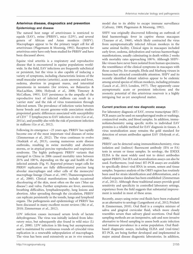
Following its emergence ~25 years ago, PRRSV has rapidlybecome one of the most important viral diseases of swine(Zimmerman et al., 2012). The virus continues to evolve(Stadejek et al., 2013), causing numerous acute diseaseoutbreaks, resulting in swine mortality and abortionstorms, or in atypical porcine reproductive and respiratorysyndrome. The highly pathogenic PRRSV variants thatemerged in China in 2006 caused mortality rates between20 % and 100 %, depending on the age and health of theinfected animals (Fig. 9). Reported primary target cells forPRRSV replication are fully differentiated porcine lungalveolar macrophages and other cells of the monocyte/macrophage lineage (Duan et al., 1997; Thanawongnuwechet al., 2000). Clinical manifestations include occasionaldiscolouring of the skin, most often on the ears (‘blue eardisease’) and vulva. Further symptoms are fever, anorexia,breathing difficulties, lymphadenopathy, lung lesions andabortion. After spreading through the circulation, PRRSVcan replicate persistently in the tonsils, lungs and lymphoidorgans. The pathogenesis and epidemiology of PRRSV hasbeen discussed in many excellent recent reviews (Shi et al.,2010; Zimmerman et al., 2012).
LDV infection causes increased serum levels of lactatedehydrogenase. The virus was initially isolated from labo-ratory mice, but subsequently also found in wild mice (Liet al., 2000). LDV infection is life-long but asymptomaticand is maintained by continuous rounds of cytocidal virusreplication in a renewable subpopulation of macrophages.The virus has been used extensively as an in vivo research
model due to its ability to escape immune surveillance(Cafruny, 1989; Plagemann & Moennig, 1992).
SHFV was originally discovered following an outbreak offatal haemorrhagic fever in captive rhesus macaques(Tauraso et al., 1968), which likely contracted the virusfrom asymptomatically infected African monkeys in thesame animal facility. Clinical signs in macaques includedearly fever, oedema, dehydration and various haemorrhagicmanifestations, usually culminating in death within 2 weekswith mortality rates approaching 100 %. Although SHFV-like viruses have never been isolated from human specimens,the resemblance of the clinical symptoms in macaques withthose caused by, for example, Ebola and Marburg viruses inhumans has attracted considerable attention. SHFV and itsrecently identified distant relatives appear to be endemicamong several species of African monkeys, including primates(Lauck et al., 2013; London, 1977). They apparently causeasymptomatic acute or persistent infections and thezoonotic potential of this arterivirus reservoir is a highlyintriguing but as-yet unexplored matter.
Current practices and new diagnostic assays
For laboratory diagnosis of EAV, reverse transcriptase (RT)-PCR assays can be used on nasopharyngeal swabs or washings,conjunctival swabs, and blood samples. In addition, immu-nohistochemistry using specific mAbs to individual EAVproteins is a reliable method for EAV diagnosis in tissues. Avirus neutralization assay remains the gold standard fordetection of serum antibodies against EAV (Holyoak et al.,2008).
PRRSV can be detected using immunohistochemistry, virusisolation and (indirect) fluorescent antibody (IFA or FA)tests in serum or tissue samples. In serology, the IDEXXELISA is the most widely used test to detect antibodiesagainst PRRSV, but IFA and neutralization assays can also beused. Furthermore, (real-time) RT-PCR assays are availableto specifically detect viral RNA in serum, semen and tissuesamples. Sequence analysis of the ORF5 region has routinelybeen used for strain identification and differentiation, and arelated sequence database has been established (Zimmermanet al., 2012). Although these traditional assays provide goodsensitivity and specificity in controlled laboratory settings,experience from the field suggests that substantial improve-ment is needed in most of these areas.
Recently, assays using swine oral fluids have been evaluatedas an alternative to serology (Langenhorst et al., 2012; Prickett& Zimmerman, 2010). Oral fluid is a complex mixture ofsaliva and gingival crevicular fluid, which more closelyresembles serum than salivary gland secretions. Oral fluidsampling methods are an inexpensive, safe and non-invasivealternative to blood sampling to assess both acute infectionand immune prevalence in a swine population. Oral fluid-based diagnostic assays, including ELISA and (real-time)RT-PCR, are being further developed and implemented inmajor animal disease diagnostic laboratories. In addition,
Arterivirus molecular biology and pathogenesis
http://vir.sgmjournals.org 2155

the fluorescent microsphere immunoassay (Luminex tech-nology) has been adapted to the use of oral fluid and serumin order to improve the sensitivity of the assay. It allows theuniform, simultaneous detection of multiple antigens orantibodies within a small volume of a single sample(Langenhorst et al., 2012). In traditional antibody detectionassays, PRRSV N protein was used as antigen, but recentlyalso certain non-structural proteins (nsp1a, nsp1b, nsp2 andnsp7) have been explored as more accurate indicators ofinfection (Brown et al., 2009; Langenhorst et al., 2012).
Arterivirus vaccines
Both modified live virus (MLV) and inactivated vaccinesare commercially available for the arteriviruses of veterinaryimportance. For EAV, Arvac (MLV; Ford DodgeLaboratories) and Artervac (inactivated; Ford DodgeLaboratories) have been used in the field. Since the firstPRRSV MLV (RespPRRS/Ingelvac PRRS MLV; BoerhringerIngelheim Animal Health) became available in 1994, variousother MLV and inactivated vaccines have been released inthe USA and Europe (Mengeling, 2005). Together withmanagement strategies, these vaccines suffice to controldisease outbreaks. However, they are derived from singlevirus strains, and are not always efficacious in preventingreinfection and transmission. Furthermore, there are MLVsafety concerns, in particular regarding reversion tovirulence and the possibility of recombination with field
isolates (Bøtner et al., 1997; Li et al., 2009; Storgaard et al.,1999). The efficacy of MLV vaccines in protecting against abroad spectrum of heterologous field isolates has also beenquestioned (Kimman et al., 2009). In general, killed vaccinesare safer but less efficacious in inducing protection, whichmakes the development of a safe and broadly protectivePRRSV vaccine a major challenge.
For EAV, several genetically engineered candidate vaccineshave been tested in vivo. Vaccination of ponies with asubunit vaccine based on the GP5 ectodomain conferredcertain levels of protection (Castillo-Olivares et al., 2001).More promising results were obtained with GP5/M-expressing Venezuelan equine encephalitis virus repliconparticles. Horses vaccinated with this candidate vaccineproduced neutralizing antibodies, shed little or no virusand developed only mild symptoms after a challenge(Balasuriya et al., 2002). Using reverse genetics, a candidateEAV live marker vaccine was constructed by deletion of themajor neutralization domain of GP5 (Castillo-Olivares et al.,2003). This recombinant virus produced an asymptomaticinfection in ponies, which were subsequently protectedagainst a challenge with virulent EAV. A GP5 peptide-basedELISA was developed to distinguish vaccinated animalsfrom those infected with WT virus.
Several experimental subunit/vectored vaccines based onplasmid DNA and viral vectors that carry PRRSV structuralproteins (GP3, M and/or GP5) have been developed (reviewed
(a)
(d) (e) (f)
(b) (c)
Fig. 9. Clinical symptoms typical of infection with highly pathogenic PRRSV. (a) Infected piglet shivering with high fever. (b)Characteristic purple/blue colouring of the ears. (c) Adult sow that has succumbed to porcine high-fever disease. In mostcases, infection in pregnant sows leads to abortion, but occasionally also to death of the sow. (d) Aborted fetuses as a result ofinfection. (e) Kidney from an infected pig showing numerous blood spots (red arrows), found in 20–30 % of the cases. (f)Severe lesions and haemorrhage (red arrows) of lungs unique to highly pathogenic PRRSV infection. (a, e, f) Courtesy of Dr K.Tian and Professor G. F. Gao, Chinese Academy of Sciences, Beijing, PR China; reprinted from Tian et al. (2007) under theCreative Commons license. (c, d) Courtesy of the Institute of Animal Husbandry and Veterinary Medicine, Fujian Academy ofAgriculture Sciences, Fuzhou, PR China.
E. J. Snijder, M. Kikkert and Y. Fang
2156 Journal of General Virology 94

by Huang & Meng, 2010; Kimman et al., 2009). However, itremains to be determined whether such vaccines couldgenerate better protection than the existing MLV and killedwhole-viral vaccines. In terms of efficacy in the field, PRRSVMLV vaccines still are the most promising approach (Rock,2007), and attempts to improve their safety and efficacy areongoing. A recent development is the application ofknowledge regarding determinants of virulence and immuneantagonist functions. Using reverse genetics, recombinantchimeras of virulent field strains and attenuated vaccinestrains were generated, which were attenuated to variabledegrees in animal models (Kwon et al., 2008; Wang et al.,2008). Furthermore, recombinant viruses carrying specificmutations or deletions in viral immune evasion proteinshave been engineered (Beura et al., 2012; Li et al., 2013; Sunet al., 2010, 2012b; van Kasteren et al., 2013).
Another drawback of current PRRSV vaccines is thatvaccinated animals cannot be distinguished from pigs thathave recovered from a natural infection. Several laboratorieshave explored the possibility of constructing geneticallyengineered marker vaccines (reviewed by Fang & Snijder,2010). Using type 1 or 2 PRRSV infectious clones, an nsp2 Bcell epitope was deleted to create a negative marker, whereasan antigenic protein or peptide tag was inserted into thensp2-coding sequence of the same construct to add a posi-tive marker. To monitor the vaccination status of animals,companion diagnostic assays were designed (Fang et al.,2008). These genetically modified live (marker) viruses maywell provide the most rational approach to future PRRSVvaccine development, although the use of recombinantviruses in the field continues to be a matter of debatebetween vaccine developers, swine practitioners and animalhealth authorities.
Outlook
The arterivirus genome expression strategy is among themost complex of currently known +RNA viruses andincludes an intriguing variety of regulatory mechanisms thatoperate at the co- and post-translational level. Consequently,EAV and increasingly also PRRSV are being used extensivelyas research models to study basic aspects of +RNA virusreplication at large, and nidovirus molecular biology inparticular. Thus far most of the ~25 arterivirus proteins haveonly been defined in basic terms and their functional charac-terization is one of the major challenges for future research.This will enhance our understanding of, for example, theintricacies of arterivirus RNA synthesis, replicase function,replication structures and virus–host interactions. Therecently discovered non-canonical translation mechanismthat produces nsp2TF/nsp2N adds another layer of com-plexity to arterivirus genome expression. This highlyefficient 22 PRF event is unprecedented in eukaryoticsystems and may have implications beyond the field of RNAvirology. Furthermore, the in-depth characterization ofnsp2TF/nsp2N function will likely reveal novel arterivirus–host interactions.
Following the discovery of the ORF5a protein as theeighth arterivirus structural protein, the dissection of thefunctional interactions between structural proteins duringparticle assembly and disassembly has become an evenmore complex issue. Recent studies revealed severalunique features of the arterivirus nucleocapsid andenvelope proteins, which in part link to unansweredquestions regarding attachment and entry. Althoughprogress has been made towards understanding hostfactors involved in PRRSV entry, issues like the specificfunctions of these host proteins and the role ofcarbohydrate moieties on the virion and cell surfaceremain to be addressed. The arterivirus–host molecularinterplay may ultimately provide an explanation for thehighly specific tropism of this group of viruses. Thisproperty, in turn, connects to the intriguing questionwhether arteriviruses are able to cross species barriers andemerge in other hosts. Considering the unapparent andpersistent infections caused by currently known arter-iviruses, it may be difficult to detect arterivirus-induceddisease in other species. However, modern virus detectiontechniques may compensate for this and are increasinglylikely to identify additional family members, as evidencedby the recent discovery of multiple additional SHFV-likeviruses in African monkeys.
The development of improved vaccines and disease controlstrategies has been a major driving force behind studiesinto arterivirus molecular biology. EAV and PRRSV aresignificant veterinary pathogens, and the latter is one ofthe economically most important swine pathogens. Therecent Asian PRRSV outbreaks continue to cause enorm-ous economic losses. The complete control of PRRSV andEAV infection is hampered by natural characteristicscommon to all RNA viruses, in particular rapid evolutionyielding genetically and antigenically diverse virus popula-tions. Our poor understanding of arterivirus–host interac-tions and immunology further complicates the control ofinfection (e.g. due to major deficits in our knowledge of theevents initiating the immune response upon infection, thekey targets for B and T-cell-based protection, and themechanisms regulating the maturation of the immuneresponse). Persistent infection is another significant factorimpeding arterivirus control, as current diagnostic meth-ods are unable to reliably identify persistently infectedanimals. Recent advances in arterivirus non-structuralprotein research has resulted in the development ofimproved diagnostic assays and the identification ofvarious potential innate immune antagonists, with impli-cations for the rational design of novel vaccines. Reversegenetics has become a key tool to manipulate immuneantagonist functions in the context of vaccine devel-opment. The initial steps toward genetically engineeredMLV vaccines have been taken and illustrate howmolecular virological knowledge of arterivirus infection,supported by structural biology and immunology, mayprovide a rational basis for future disease prevention andcontrol.
Arterivirus molecular biology and pathogenesis
http://vir.sgmjournals.org 2157

Acknowledgements
We would like to acknowledge the contributions, over many years, of
a large number of colleagues who worked with us in the arterivirus
field. Our research was and is supported by the Netherlands Organi-
zation of Scientific Research (NWO grants 700.57.301, 700.10.352 and
700.59.008), the European Commission (grants 260644 and 264286),
the US Department of Agriculture (grants 2005-35204, 2007-01745,
2008-55620 and 2011-02925) and the US National Pork Board (grants
05-155, 06-173, 09-234, 11-037, 11-143 and 12-127).
References
Ammann, C. G., Messer, R. J., Peterson, K. E. & Hasenkrug, K. J.(2009). Lactate dehydrogenase-elevating virus induces systemic
lymphocyte activation via TLR7-dependent IFNalpha responses by
plasmacytoid dendritic cells. PLoS ONE 4, e6105.
Balasuriya, U. B. & MacLachlan, N. J. (2004). The immune response
to equine arteritis virus: potential lessons for other arteriviruses. Vet
Immunol Immunopathol 102, 107–129.
Balasuriya, U. B., Patton, J. F., Rossitto, P. V., Timoney, P. J.,McCollum, W. H. & MacLachlan, N. J. (1997). Neutralization
determinants of laboratory strains and field isolates of equine arteritis
virus: identification of four neutralization sites in the amino-terminal
ectodomain of the G(L) envelope glycoprotein. Virology 232, 114–128.
Balasuriya, U. B., Heidner, H. W., Davis, N. L., Wagner, H. M.,Hullinger, P. J., Hedges, J. F., Williams, J. C., Johnston, R. E., DavidWilson, W. & other authors (2002). Alphavirus replicon particles
expressing the two major envelope proteins of equine arteritis virus
induce high level protection against challenge with virulent virus in
vaccinated horses. Vaccine 20, 1609–1617.
Balasuriya, U. B. R., Dobbe, J. C., Heidner, H. W., Smalley, V. L.,Navarrette, A., Snijder, E. J. & MacLachlan, N. J. (2004a).Characterization of the neutralization determinants of equine arteritis
virus using recombinant chimeric viruses and site-specific mutagen-
esis of an infectious cDNA clone. Virology 321, 235–246.
Balasuriya, U. B. R., Hedges, J. F., Smalley, V. L., Navarrette, A.,McCollum, W. H., Timoney, P. J., Snijder, E. J. & MacLachlan, N. J.(2004b). Genetic characterization of equine arteritis virus during
persistent infection of stallions. J Gen Virol 85, 379–390.
Barrette-Ng, I. H., Ng, K. K. S., Mark, B. L., Van Aken, D., Cherney,M. M., Garen, C., Kolodenko, Y., Gorbalenya, A. E., Snijder, E. J. &James, M. N. (2002). Structure of arterivirus nsp4. The smallest
chymotrypsin-like proteinase with an alpha/beta C-terminal exten-
sion and alternate conformations of the oxyanion hole. J Biol Chem
277, 39960–39966.
Beerens, N. & Snijder, E. J. (2007). An RNA pseudoknot in the 39 end
of the arterivirus genome has a critical role in regulating viral RNA
synthesis. J Virol 81, 9426–9436.
Beerens, N., Selisko, B., Ricagno, S., Imbert, I., van der Zanden, L.,Snijder, E. J. & Canard, B. (2007). De novo initiation of RNA
synthesis by the arterivirus RNA-dependent RNA polymerase. J Virol
81, 8384–8395.
Beura, L. K., Sarkar, S. N., Kwon, B., Subramaniam, S., Jones, C.,Pattnaik, A. K. & Osorio, F. A. (2010). Porcine reproductive and
respiratory syndrome virus nonstructural protein 1beta modulates
host innate immune response by antagonizing IRF3 activation. J Virol
84, 1574–1584.
Beura, L. K., Subramaniam, S., Vu, H. L., Kwon, B., Pattnaik, A. K. &Osorio, F. A. (2012). Identification of amino acid residues important
for anti-IFN activity of porcine reproductive and respiratory
syndrome virus non-structural protein 1. Virology 433, 431–439.
Bøtner, A., Strandbygaard, B., Sørensen, K. J., Have, P., Madsen,K. G., Madsen, E. S. & Alexandersen, S. (1997). Appearance of acutePRRS-like symptoms in sow herds after vaccination with a modified
live PRRS vaccine. Vet Rec 141, 497–499.
Brown, E., Lawson, S., Welbon, C., Gnanandarajah, J., Li, J.,Murtaugh, M. P., Nelson, E. A., Molina, R. M., Zimmerman, J. J. &other authors (2009). Antibody response to porcine reproductive andrespiratory syndrome virus (PRRSV) nonstructural proteins and
implications for diagnostic detection and differentiation of PRRSV
types I and II. Clin Vaccine Immunol 16, 628–635.
Butler, J. E., Wertz, N., Weber, P. & Lager, K. M. (2008). Porcine
reproductive and respiratory syndrome virus subverts repertoire
development by proliferation of germline-encoded B cells of allisotypes bearing hydrophobic heavy chain CDR3. J Immunol 180,
2347–2356.
Cafruny, W. A. (1989). Lactate dehydrogenase-elevating virus: biology
and pathogenesis. Crit Rev Microbiol 17, 107–119.
Cafruny, W. A. & Plagemann, P. G. W. (1982). Immune responseto lactate dehydrogenase-elevating virus: serologically specific
rabbit neutralizing antibody to the virus. Infect Immun 37, 1007–
1012.
Cancel-Tirado, S. M., Evans, R. B. & Yoon, K. J. (2004). Monoclonal
antibody analysis of porcine reproductive and respiratory syndromevirus epitopes associated with antibody-dependent enhancement and
neutralization of virus infection. Vet Immunol Immunopathol 102,
249–262.
Castillo-Olivares, J., de Vries, A. A. F., Raamsman, M. J. B., Rottier,P. J. M., Lakhani, K., Westcott, D., Tearle, J. P., Wood, J. L., Mumford,J. A. & other authors (2001). Evaluation of a prototype sub-unitvaccine against equine arteritis virus comprising the entire ectodo-
main of the virus large envelope glycoprotein (G(L)): induction of
virus-neutralizing antibody and assessment of protection in ponies.J Gen Virol 82, 2425–2435.
Castillo-Olivares, J., Wieringa, R., Bakonyi, T., de Vries, A. A. F.,Davis-Poynter, N. J. & Rottier, P. J. M. (2003). Generation of acandidate live marker vaccine for equine arteritis virus by deletion of
the major virus neutralization domain. J Virol 77, 8470–8480.
Cavanagh, D. (1997). Nidovirales: a new order comprising
Coronaviridae and Arteriviridae. Arch Virol 142, 629–633.
Chen, Z., Rowland, R. R. R., Anderson, G. W., Palmer, G. A. &Plagemann, P. G. W. (1997). Coexistence in lactate dehydrogenase-
elevating virus pools of variants that differ in neuropathogenicity and
ability to establish a persistent infection. J Virol 71, 2913–2920.
Chen, Z., Lawson, S., Sun, Z., Zhou, X., Guan, X., Christopher-Hennings, J., Nelson, E. A. & Fang, Y. (2010). Identification of twoauto-cleavage products of nonstructural protein 1 (nsp1) in porcine
reproductive and respiratory syndrome virus infected cells: nsp1
function as interferon antagonist. Virology 398, 87–97.
Chirnside, E. D., Francis, P. M., de Vries, A. A. F., Sinclair, R. &Mumford, J. A. (1995). Development and evaluation of an ELISA
using recombinant fusion protein to detect the presence of hostantibody to equine arteritis virus. J Virol Methods 54, 1–13.
Coutelier, J. P., Van Broeck, J. & Wolf, S. F. (1995). Interleukin-12gene expression after viral infection in the mouse. J Virol 69, 1955–
1958.
Cowley, J. A., Dimmock, C. M., Spann, K. M. & Walker, P. J. (2000).Gill-associated virus of Penaeus monodon prawns: an invertebrate
virus with ORF1a and ORF1b genes related to arteri- and
coronaviruses. J Gen Virol 81, 1473–1484.
Darwich, L., Dıaz, I. & Mateu, E. (2010). Certainties, doubts and
hypotheses in porcine reproductive and respiratory syndrome virusimmunobiology. Virus Res 154, 123–132.
E. J. Snijder, M. Kikkert and Y. Fang
2158 Journal of General Virology 94

Das, P. B., Dinh, P. X., Ansari, I. H., de Lima, M., Osorio, F. A. &Pattnaik, A. K. (2010). The minor envelope glycoproteins GP2a and
GP4 of porcine reproductive and respiratory syndrome virus interact
with the receptor CD163. J Virol 84, 1731–1740.
de Lima, M., Ansari, I. H., Das, P. B., Ku, B. J., Martinez-Lobo, F. J.,Pattnaik, A. K. & Osorio, F. A. (2009). GP3 is a structural component
of the PRRSV type II (US) virion. Virology 390, 31–36.
de Vries, A. A. F., Chirnside, E. D., Bredenbeek, P. J., Gravestein,L. A., Horzinek, M. C. & Spaan, W. J. M. (1990). All subgenomic
mRNAs of equine arteritis virus contain a common leader sequence.
Nucleic Acids Res 18, 3241–3247.
de Vries, A. A. F., Chirnside, E. D., Horzinek, M. C. & Rottier, P. J. M.
(1992). Structural proteins of equine arteritis virus. J Virol 66, 6294–
6303.
de Vries, A. A. F., Post, S. M., Raamsman, M. J. B., Horzinek, M. C. &
Rottier, P. J. M. (1995). The two major envelope proteins of equine
arteritis virus associate into disulfide-linked heterodimers. J Virol 69,
4668–4674.
de Wilde, A. H., Li, Y., van der Meer, Y., Vuagniaux, G., Lysek, R.,Fang, Y., Snijder, E. J. & van Hemert, M. J. (2013). Cyclophilin
inhibitors block arterivirus replication by interfering with viral RNA
synthesis. J Virol 87, 1454–1464.
Delputte, P. L. & Nauwynck, H. J. (2004). Porcine arterivirus infection
of alveolar macrophages is mediated by sialic acid on the virus. J Virol
78, 8094–8101.
Delputte, P. L., Vanderheijden, N., Nauwynck, H. J. & Pensaert, M. B.(2002). Involvement of the matrix protein in attachment of porcine
reproductive and respiratory syndrome virus to a heparinlike receptor
on porcine alveolar macrophages. J Virol 76, 4312–4320.
den Boon, J. A., Snijder, E. J., Chirnside, E. D., de Vries, A. A. F.,Horzinek, M. C. & Spaan, W. J. M. (1991). Equine arteritis virus is not
a togavirus but belongs to the coronaviruslike superfamily. J Virol 65,
2910–2920.
den Boon, J. A., Faaberg, K. S., Meulenberg, J. J. M., Wassenaar,A. L. M., Plagemann, P. G. W., Gorbalenya, A. E. & Snijder, E. J.
(1995). Processing and evolution of the N-terminal region of the
arterivirus replicase ORF1a protein: identification of two papainlike
cysteine proteases. J Virol 69, 4500–4505.
Dobbe, J. C., van der Meer, Y., Spaan, W. J. M. & Snijder, E. J. (2001).Construction of chimeric arteriviruses reveals that the ectodomain of
the major glycoprotein is not the main determinant of equine arteritis
virus tropism in cell culture. Virology 288, 283–294.
Dokland, T. (2010). The structural biology of PRRSV. Virus Res 154,
86–97.
Duan, X., Nauwynck, H. J. & Pensaert, M. B. (1997). Virus
quantification and identification of cellular targets in the lungs and
lymphoid tissues of pigs at different time intervals after inoculation
with porcine reproductive and respiratory syndrome virus (PRRSV).
Vet Microbiol 56, 9–19.
Duan, X., Nauwynck, H. J., Favoreel, H. W. & Pensaert, M. B. (1998).Identification of a putative receptor for porcine reproductive and
respiratory syndrome virus on porcine alveolar macrophages. J Virol
72, 4520–4523.
Dunowska, M., Biggs, P. J., Zheng, T. & Perrott, M. R. (2012).Identification of a novel nidovirus associated with a neurological
disease of the Australian brushtail possum (Trichosurus vulpecula).
Vet Microbiol 156, 418–424.
Even, C., Rowland, R. R. R. & Plagemann, P. G. W. (1995). Cytotoxic
T cells are elicited during acute infection of mice with lactate
dehydrogenase-elevating virus but disappear during the chronic phase
of infection. J Virol 69, 5666–5676.
Faaberg, K. S. & Plagemann, P. G. W. (1995). The envelope proteins
of lactate dehydrogenase-elevating virus and their membranetopography. Virology 212, 512–525.
Fang, Y. & Snijder, E. J. (2010). The PRRSV replicase: exploring themultifunctionality of an intriguing set of nonstructural proteins.
Virus Res 154, 61–76.
Fang, Y., Christopher-Hennings, J., Brown, E., Liu, H., Chen, Z.,Lawson, S. R., Breen, R., Clement, T., Gao, X. & other authors(2008). Development of genetic markers in the non-structural protein
2 region of a US type 1 porcine reproductive and respiratorysyndrome virus: implications for future recombinant marker vaccine
development. J Gen Virol 89, 3086–3096.
Fang, Y., Treffers, E. E., Li, Y., Tas, A., Sun, Z., van der Meer, Y., deRu, A. H., van Veelen, P. A., Atkins, J. F. & other authors (2012).Efficient -2 frameshifting by mammalian ribosomes to synthesize anadditional arterivirus protein. Proc Natl Acad Sci U S A 109, E2920–
E2928.
Firth, A. E. & Brierley, I. (2012). Non-canonical translation in RNAviruses. J Gen Virol 93, 1385–1409.
Firth, A. E., Zevenhoven-Dobbe, J. C., Wills, N. M., Go, Y. Y.,Balasuriya, U. B., Atkins, J. F., Snijder, E. J. & Posthuma, C. C. (2011).Discovery of a small arterivirus gene that overlaps the GP5 coding
sequence and is important for virus production. J Gen Virol 92, 1097–1106.
Frias-Staheli, N., Giannakopoulos, N. V., Kikkert, M., Taylor, S. L.,Bridgen, A., Paragas, J., Richt, J. A., Rowland, R. R., Schmaljohn, C. S.& other authors (2007). Ovarian tumor domain-containing viral
proteases evade ubiquitin- and ISG15-dependent innate immune
responses. Cell Host Microbe 2, 404–416.
Go, Y. Y., Bailey, E., Cook, D. G., Coleman, S. J., Macleod, J. N., Chen,K. C., Timoney, P. J. & Balasuriya, U. B. (2011a). Genome-wideassociation study among four horse breeds identifies a common
haplotype associated with in vitro CD3+ T cell susceptibility/
resistance to equine arteritis virus infection. J Virol 85, 13174–
13184.
Go, Y. Y., Snijder, E. J., Timoney, P. J. & Balasuriya, U. B. (2011b).Characterization of equine humoral antibody response to the non-structural proteins of equine arteritis virus. Clin Vaccine Immunol 18,
268–279.
Go, Y. Y., Bailey, E., Timoney, P. J., Shuck, K. M. & Balasuriya, U. B.(2012). Evidence that in vitro susceptibility of CD3+ T lymphocytes
to equine arteritis virus infection reflects genetic predisposition of
naturally infected stallions to become carriers of the virus. J Virol 86,12407–12410.
Godeny, E. K., de Vries, A. A. F., Wang, X. C., Smith, S. L. & de Groot,R. J. (1998). Identification of the leader-body junctions for the viral
subgenomic mRNAs and organization of the simian hemorrhagic
fever virus genome: evidence for gene duplication during arterivirusevolution. J Virol 72, 862–867.
Gorbalenya, A. E., Enjuanes, L., Ziebuhr, J. & Snijder, E. J. (2006).Nidovirales: evolving the largest RNA virus genome. Virus Res 117,17–37.
Gravell, M., London, W. T., Leon, M. E., Palmer, A. E. & Hamilton, R. S.(1986). Elimination of persistent simian hemorrhagic fever (SHF)
virus infection in patas monkeys. Proc Soc Exp Biol Med 181, 219–225.
Han, J., Wang, Y. & Faaberg, K. S. (2006). Complete genome analysisof RFLP 184 isolates of porcine reproductive and respiratory
syndrome virus. Virus Res 122, 175–182.
Han, J., Rutherford, M. S. & Faaberg, K. S. (2009). The porcine
reproductive and respiratory syndrome virus nsp2 cysteine protease
domain possesses both trans- and cis-cleavage activities. J Virol 83,9449–9463.
Arterivirus molecular biology and pathogenesis
http://vir.sgmjournals.org 2159

Han, J., Rutherford, M. S. & Faaberg, K. S. (2010). Proteolyticproducts of the porcine reproductive and respiratory syndrome virusnsp2 replicase protein. J Virol 84, 10102–10112.
Han, M., Du, Y., Song, C. & Yoo, D. (2013). Degradation of CREB-binding protein and modulation of type I interferon induction by thezinc finger motif of the porcine reproductive and respiratorysyndrome virus nsp1a subunit. Virus Res 172, 54–65.
Holyoak, G. R., Balasuriya, U. B., Broaddus, C. C. & Timoney, P. J.(2008). Equine viral arteritis: current status and prevention.Theriogenology 70, 403–414.
Huang, Y. W. & Meng, X. J. (2010). Novel strategies and approaches todevelop the next generation of vaccines against porcine reproductiveand respiratory syndrome virus (PRRSV). Virus Res 154, 141–149.
Hwang, Y.-K. & Brinton, M. A. (1998). A 68-nucleotide sequencewithin the 39 noncoding region of simian hemorrhagic fever virusnegative-strand RNA binds to four MA104 cell proteins. J Virol 72,4341–4351.
Ivanov, K. A., Hertzig, T., Rozanov, M., Bayer, S., Thiel, V.,Gorbalenya, A. E. & Ziebuhr, J. (2004). Major genetic marker ofnidoviruses encodes a replicative endoribonuclease. Proc Natl AcadSci U S A 101, 12694–12699.
Johnson, C. R., Yu, W. & Murtaugh, M. P. (2007). Cross-reactiveantibody responses to nsp1 and nsp2 of Porcine reproductive andrespiratory syndrome virus. J Gen Virol 88, 1184–1195.
Johnson, C. R., Griggs, T. F., Gnanandarajah, J. & Murtaugh, M. P.(2011). Novel structural protein in porcine reproductive andrespiratory syndrome virus encoded by an alternative ORF5 presentin all arteriviruses. J Gen Virol 92, 1107–1116.
Kimman, T. G., Cornelissen, L. A., Moormann, R. J. M., Rebel, J. M. &Stockhofe-Zurwieden, N. (2009). Challenges for porcine reproductiveand respiratory syndrome virus (PRRSV) vaccinology. Vaccine 27,3704–3718.
Knoops, K., Barcena, M., Limpens, R. W. A. L., Koster, A. J.,Mommaas, A. M. & Snijder, E. J. (2012). Ultrastructural character-ization of arterivirus replication structures: reshaping the endoplasmicreticulum to accommodate viral RNA synthesis. J Virol 86, 2474–2487.
Kreutz, L. C. & Ackermann, M. R. (1996). Porcine reproductive andrespiratory syndrome virus enters cells through a low pH-dependentendocytic pathway. Virus Res 42, 137–147.
Kroese, M. V., Zevenhoven-Dobbe, J. C., Bos-de Ruijter, J. N.,Peeters, B. P., Meulenberg, J. J., Cornelissen, L. A. & Snijder, E. J.(2008). The nsp1a and nsp1b papain-like autoproteinases are essentialfor porcine reproductive and respiratory syndrome virus RNAsynthesis. J Gen Virol 89, 494–499.
Kwon, B., Ansari, I. H., Pattnaik, A. K. & Osorio, F. A. (2008).Identification of virulence determinants of porcine reproductive andrespiratory syndrome virus through construction of chimeric clones.Virology 380, 371–378.
Langenhorst, R. J., Lawson, S., Kittawornrat, A., Zimmerman, J. J.,Sun, Z., Li, Y., Christopher-Hennings, J., Nelson, E. A. & Fang, Y.(2012). Development of a fluorescent microsphere immunoassay fordetection of antibodies against porcine reproductive and respiratorysyndrome virus using oral fluid samples as an alternative to serum-based assays. Clin Vaccine Immunol 19, 180–189.
Lauck, M., Sibley, S. D., Hyeroba, D., Tumukunde, A., Weny, G.,Chapman, C. A., Ting, N., Switzer, W. M., Kuhn, J. H. & other authors(2013). Exceptional simian hemorrhagic fever virus diversity in a wildAfrican primate community. J Virol 87, 688–691.
Lee, S. M., Schommer, S. K. & Kleiboeker, S. B. (2004). Porcinereproductive and respiratory syndrome virus field isolates differ in invitro interferon phenotypes. Vet Immunol Immunopathol 102, 217–231.
Lee, C. & Yoo, D. (2006). The small envelope protein of porcine
reproductive and respiratory syndrome virus possesses ion channelprotein-like properties. Virology 355, 30–43.
Lee, C., Hodgins, D., Calvert, J. G., Welch, S. K., Jolie, R. & Yoo, D.(2006). Mutations within the nuclear localization signal of the
porcine reproductive and respiratory syndrome virus nucleocapsid
protein attenuate virus replication. Virology 346, 238–250.
Li, K., Chen, Z. & Plagemann, P. G. W. (1998). The neutralization epitope
of lactate dehydrogenase-elevating virus is located on the short
ectodomain of the primary envelope glycoprotein. Virology 242, 239–245.
Li, K., Schuler, T., Chen, Z., Glass, G. E., Childs, J. E. & Plagemann,P. G. (2000). Isolation of lactate dehydrogenase-elevating viruses fromwild house mice and their biological and molecular characterization.
Virus Res 67, 153–162.
Li, B., Fang, L., Xu, Z., Liu, S., Gao, J., Jiang, Y., Chen, H. & Xiao, S.(2009). Recombination in vaccine and circulating strains of porcine
reproductive and respiratory syndrome viruses. Emerg Infect Dis 15,
2032–2035.
Li, Y., Tas, A., Snijder, E. J. & Fang, Y. (2012). Identification of porcine
reproductive and respiratory syndrome virus ORF1a-encoded non-
structural proteins in virus-infected cells. J Gen Virol 93, 829–839.
Li, Y., Zhu, L., Lawson, S. R. & Fang, Y. (2013). Targeted mutations in
a highly conserved motif of the nsp1b protein impair the interferonantagonizing activity of porcine reproductive and respiratory
syndrome virus. J Gen Virol 94, 1972–1983.
Liu, Q., Qin, Y., Zhou, L., Kou, Q., Guo, X., Ge, X., Yang, H. & Hu, H.(2012). Autophagy sustains the replication of porcine reproductive
and respiratory virus in host cells. Virology 429, 136–147.
London, W. T. (1977). Epizootiology, transmission and approach to
prevention of fatal simian haemorrhagic fever in rhesus monkeys.
Nature 268, 344–345.
Lopez, O. J. & Osorio, F. A. (2004). Role of neutralizing antibodies
in PRRSV protective immunity. Vet Immunol Immunopathol 102,
155–163.
Lu, J., Gao, F., Wei, Z., Liu, P., Liu, C., Zheng, H., Li, Y., Lin, T. & Yuan,S. (2011). A 59-proximal stem-loop structure of 59 untranslatedregion of porcine reproductive and respiratory syndrome virus
genome is key for virus replication. Virol J 8, 172.
MacLachlan, N. J., Balasuriya, U. B., Hedges, J. F., Schweidler, T. M.,McCollum, W. H., Timoney, P. J., Hullinger, P. J. & Patton, J. F. (1998).Serologic response of horses to the structural proteins of equine
arteritis virus. J Vet Diagn Invest 10, 229–236.
Magnusson, P., Hyllseth, B. & Marusyk, H. (1970). Morphological
studies on equine arteritis virus. Arch Gesamte Virusforsch 30, 105–112.
Maines, T. R., Young, M., Dinh, N. N. N. & Brinton, M. A. (2005). Two
cellular proteins that interact with a stem loop in the simian
hemorrhagic fever virus 39(+)NCR RNA. Virus Res 109, 109–124.
Makarova, K. S., Aravind, L. & Koonin, E. V. (2000). A novel
superfamily of predicted cysteine proteases from eukaryotes, virusesand Chlamydia pneumoniae. Trends Biochem Sci 25, 50–52.
Markine-Goriaynoff, D., Nguyen, T. D., Bigaignon, G., Van Snick, J. &Coutelier, J. P. (2001). Distinct requirements for IL-6 in polyclonaland specific Ig production induced by microorganisms. Int Immunol
13, 1185–1192.
Markine-Goriaynoff, D., Hulhoven, X., Cambiaso, C. L., Monteyne, P.,Briet, T., Gonzalez, M. D., Coulie, P. & Coutelier, J. P. (2002). Natural
killer cell activation after infection with lactate dehydrogenase-elevating virus. J Gen Virol 83, 2709–2716.
Mengeling, W. L. (2005). The porcine reproductive and respiratory
syndrome quandary. Part II: Vaccines and vaccination strategy.J Swine Health Prod 13, 152–156.
E. J. Snijder, M. Kikkert and Y. Fang
2160 Journal of General Virology 94

Meulenberg, J. J. M., van Nieuwstadt, A. P., van Essen-Zandbergen,A. & Langeveld, J. P. M. (1997). Posttranslational processing andidentification of a neutralization domain of the GP4 protein encodedby ORF4 of Lelystad virus. J Virol 71, 6061–6067.
Meulenberg, J. J. M., Bos-de Ruijter, J. N. A., van de Graaf, R.,Wensvoort, G. & Moormann, R. J. (1998). Infectious transcripts fromcloned genome-length cDNA of porcine reproductive and respiratorysyndrome virus. J Virol 72, 380–387.
Molenkamp, R., Rozier, B. C. D., Greve, S., Spaan, W. J. M. & Snijder,E. J. (2000a). Isolation and characterization of an arterivirus defectiveinterfering RNA genome. J Virol 74, 3156–3165.
Molenkamp, R., van Tol, H., Rozier, B. C. D., van der Meer, Y., Spaan,W. J. M. & Snijder, E. J. (2000b). The arterivirus replicase is the onlyviral protein required for genome replication and subgenomic mRNAtranscription. J Gen Virol 81, 2491–2496.
Monastyrska, I., Ulasli, M., Rottier, P. J., Guan, J. L., Reggiori, F. & deHaan, C. A. (2013). An autophagy-independent role for LC3 in equinearteritis virus replication. Autophagy 9, 164–174.
Moore, B. D., Balasuriya, U. B. R., Watson, J. L., Bosio, C. M., MacKay,R. J. & MacLachlan, N. J. (2003). Virulent and avirulent strains ofequine arteritis virus induce different quantities of TNF-alpha andother proinflammatory cytokines in alveolar and blood-derivedequine macrophages. Virology 314, 662–670.
Murtaugh, M. P., Xiao, Z. G. & Zuckermann, F. (2002). Immunologicalresponses of swine to porcine reproductive and respiratory syndromevirus infection. Viral Immunol 15, 533–547.
Nauwynck, H. J., Duan, X., Favoreel, H. W., Van Oostveldt, P. &Pensaert, M. B. (1999). Entry of porcine reproductive and respiratorysyndrome virus into porcine alveolar macrophages via receptor-mediated endocytosis. J Gen Virol 80, 297–305.
Nedialkova, D. D., Ulferts, R., van den Born, E., Lauber, C., Gorbalenya,A. E., Ziebuhr, J. & Snijder, E. J. (2009). Biochemical characterization ofarterivirus nonstructural protein 11 reveals the nidovirus-wideconservation of a replicative endoribonuclease. J Virol 83, 5671–5682.
Nedialkova, D. D., Gorbalenya, A. E. & Snijder, E. J. (2010).Arterivirus Nsp1 modulates the accumulation of minus-strandtemplates to control the relative abundance of viral mRNAs. PLoSPathog 6, e1000772.
Nga, P. T., Parquet, M. C., Lauber, C., Parida, M., Nabeshima, T., Yu,F., Thuy, N. T., Inoue, S., Ito, T. & other authors (2011). Discovery ofthe first insect nidovirus, a missing evolutionary link in the emergenceof the largest RNA virus genomes. PLoS Pathog 7, e1002215.
Ostrowski, M., Galeota, J. A., Jar, A. M., Platt, K. B., Osorio, F. A. &Lopez, O. J. (2002). Identification of neutralizing and nonneutralizingepitopes in the porcine reproductive and respiratory syndrome virusGP5 ectodomain. J Virol 76, 4241–4250.
Parida, R., Choi, I. S., Peterson, D. A., Pattnaik, A. K., Laegreid, W.,Zuckermann, F. A. & Osorio, F. A. (2012). Location of T-cell epitopesin nonstructural proteins 9 and 10 of type-II porcine reproductiveand respiratory syndrome virus. Virus Res 169, 13–21.
Pasternak, A. O., van den Born, E., Spaan, W. J. M. & Snijder, E. J.(2001). Sequence requirements for RNA strand transfer duringnidovirus discontinuous subgenomic RNA synthesis. EMBO J 20,7220–7228.
Pasternak, A. O., van den Born, E., Spaan, W. J. M. & Snijder, E. J.(2003). The stability of the duplex between sense and antisensetranscription-regulating sequences is a crucial factor in arterivirussubgenomic mRNA synthesis. J Virol 77, 1175–1183.
Pasternak, A. O., Spaan, W. J. M. & Snijder, E. J. (2006). Nidovirustranscription: how to make sense...? J Gen Virol 87, 1403–1421.
Patel, D., Nan, Y., Shen, M., Ritthipichai, K., Zhu, X. & Zhang, Y. J.(2010). Porcine reproductive and respiratory syndrome virus inhibits
type I interferon signaling by blocking STAT1/STAT2 nuclear
translocation. J Virol 84, 11045–11055.
Pedersen, K. W., van der Meer, Y., Roos, N. & Snijder, E. J. (1999).Open reading frame 1a-encoded subunits of the arterivirus replicaseinduce endoplasmic reticulum-derived double-membrane vesicles
which carry the viral replication complex. J Virol 73, 2016–2026.
Plagemann, P. G. W. (2001). Complexity of the single linearneutralization epitope of the mouse arterivirus lactate dehydrogenase-
elevating virus. Virology 290, 11–20.
Plagemann, P. G. W. (2004). GP5 ectodomain epitope of porcine
reproductive and respiratory syndrome virus, strain Lelystad virus.
Virus Res 102, 225–230.
Plagemann, P. G. W. & Moennig, V. (1992). Lactate dehydrogenase-
elevating virus, equine arteritis virus, and simian hemorrhagic fever
virus: a new group of positive-strand RNA viruses. Adv Virus Res 41,99–192.
Plagemann, P. G. W., Rowland, R. R. R. & Faaberg, K. S. (2002). Theprimary neutralization epitope of porcine respiratory and reproductive
syndrome virus strain VR-2332 is located in the middle of the GP5
ectodomain. Arch Virol 147, 2327–2347.
Posthuma, C. C., Nedialkova, D. D., Zevenhoven-Dobbe, J. C.,Blokhuis, J. H., Gorbalenya, A. E. & Snijder, E. J. (2006). Site-directed
mutagenesis of the Nidovirus replicative endoribonuclease NendoUexerts pleiotropic effects on the arterivirus life cycle. J Virol 80, 1653–
1661.
Prickett, J. R. & Zimmerman, J. J. (2010). The development of oralfluid-based diagnostics and applications in veterinary medicine. Anim
Health Res Rev 11, 207–216.
Rock, D. L. (2007). Colloquium on prospects for development of an
effective PRRS vaccine. AASV News Archive. http://www.aasv.org/
news/story.php?id=2527.
Rowland, R. R. R., Schneider, P., Fang, Y., Wootton, S., Yoo, D. &Benfield, D. A. (2003). Peptide domains involved in the localization of
the porcine reproductive and respiratory syndrome virus nucleocapsidprotein to the nucleolus. Virology 316, 135–145.
Sawicki, S. G., Sawicki, D. L. & Siddell, S. G. (2007). A contemporaryview of coronavirus transcription. J Virol 81, 20–29.
Schutze, H., Ulferts, R., Schelle, B., Bayer, S., Granzow, H.,Hoffmann, B., Mettenleiter, T. C. & Ziebuhr, J. (2006). Characte-rization of White bream virus reveals a novel genetic cluster of
nidoviruses. J Virol 80, 11598–11609.
Seybert, A., van Dinten, L. C., Snijder, E. J. & Ziebuhr, J. (2000).Biochemical characterization of the equine arteritis virus helicase
suggests a close functional relationship between arterivirus andcoronavirus helicases. J Virol 74, 9586–9593.
Shi, M., Lam, T. T., Hon, C. C., Hui, R. K., Faaberg, K. S., Wennblom,T., Murtaugh, M. P., Stadejek, T. & Leung, F. C. (2010). Molecularepidemiology of PRRSV: a phylogenetic perspective. Virus Res 154,
7–17.
Snijder, E. J. & Kikkert, M. (2013). Arteriviruses. In Fields Virology, 6th
edn, pp. 859–879. Edited by D. M. Knipe & P. M. Howley.
Philadelphia, PA: Lippincott Williams & Wilkins.
Snijder, E. J. & Meulenberg, J. J. M. (1998). The molecular biology of
arteriviruses. J Gen Virol 79, 961–979.
Snijder, E. J., Wassenaar, A. L. M., Spaan, W. J. M. & Gorbalenya,A. E. (1995). The arterivirus Nsp2 protease. An unusual cysteine
protease with primary structure similarities to both papain-like andchymotrypsin-like proteases. J Biol Chem 270, 16671–16676.
Snijder, E. J., van Tol, H., Pedersen, K. W., Raamsman, M. J. B. & deVries, A. A. F. (1999). Identification of a novel structural protein ofarteriviruses. J Virol 73, 6335–6345.
Arterivirus molecular biology and pathogenesis
http://vir.sgmjournals.org 2161

Snijder, E. J., van Tol, H., Roos, N. & Pedersen, K. W. (2001). Non-
structural proteins 2 and 3 interact to modify host cell membranes
during the formation of the arterivirus replication complex. J Gen
Virol 82, 985–994.
Snijder, E. J., Bredenbeek, P. J., Dobbe, J. C., Thiel, V., Ziebuhr, J.,Poon, L. L. M., Guan, Y., Rozanov, M., Spaan, W. J. M. & Gorbalenya,A. E. (2003a). Unique and conserved features of genome and
proteome of SARS-coronavirus, an early split-off from the corona-
virus group 2 lineage. J Mol Biol 331, 991–1004.
Snijder, E. J., Dobbe, J. C. & Spaan, W. J. M. (2003b). Heterodimeri-
zation of the two major envelope proteins is essential for arterivirus
infectivity. J Virol 77, 97–104.
Sola, I., Mateos-Gomez, P. A., Almazan, F., Zuniga, S. & Enjuanes, L.(2011). RNA-RNA and RNA-protein interactions in coronavirus
replication and transcription. RNA Biol 8, 237–248.
Song, C., Krell, P. & Yoo, D. (2010). Nonstructural protein 1a
subunit-based inhibition of NF-kB activation and suppression of
interferon-b production by porcine reproductive and respiratory
syndrome virus. Virology 407, 268–280.
Spilman, M. S., Welbon, C., Nelson, E. & Dokland, T. (2009). Cryo-
electron tomography of porcine reproductive and respiratory
syndrome virus: organization of the nucleocapsid. J Gen Virol 90,
527–535.
Stadejek, T., Stankevicius, A., Murtaugh, M. P. & Oleksiewicz, M. B.(2013). Molecular evolution of PRRSV in Europe: current state of
play. Vet Microbiol 165, 21–28.
Storgaard, T., Oleksiewicz, M. B. & Bøtner, A. (1999). Examination of
the selective pressures on a live PRRS vaccine virus. Arch Virol 144,
2389–2401.
Sun, M. X., Huang, L., Wang, R., Yu, Y. L., Li, C., Li, P. P., Hu, X. C., Hao,H. P., Ishag, H. A. & Mao, X. (2012). Porcine reproductive and
respiratory syndrome virus induces autophagy to promote virus
replication. Autophagy 8, 1434–1447.
Sun, Y., Xue, F., Guo, Y., Ma, M., Hao, N., Zhang, X. C., Lou, Z., Li, X. &Rao, Z. (2009). Crystal structure of porcine reproductive and
respiratory syndrome virus leader protease Nsp1alpha. J Virol 83,
10931–10940.
Sun, Z., Chen, Z., Lawson, S. R. & Fang, Y. (2010). The cysteine
protease domain of porcine reproductive and respiratory syndrome
virus nonstructural protein 2 possesses deubiquitinating and inter-
feron antagonism functions. J Virol 84, 7832–7846.
Sun, Y., Han, M., Kim, C., Calvert, J. G. & Yoo, D. (2012a). Interplay
between interferon-mediated innate immunity and porcine repro-
ductive and respiratory syndrome virus. Viruses 4, 424–446.
Sun, Z., Li, Y., Ransburgh, R., Snijder, E. J. & Fang, Y. (2012b).Nonstructural protein 2 of porcine reproductive and respiratory
syndrome virus inhibits the antiviral function of interferon-
stimulated gene 15. J Virol 86, 3839–3850.
Tauraso, N. M., Shelokov, A., Palmer, A. E. & Allen, A. M. (1968).Simian hemorrhagic fever. 3. Isolation and characterization of a viral
agent. Am J Trop Med Hyg 17, 422–431.
Thaa, B., Kabatek, A., Zevenhoven-Dobbe, J. C., Snijder, E. J.,Herrmann, A. & Veit, M. (2009). Myristoylation of the arterivirus E
protein: the fatty acid modification is not essential for membrane
association but contributes significantly to virus infectivity. J Gen
Virol 90, 2704–2712.
Thanawongnuwech, R., Halbur, P. G. & Thacker, E. L. (2000). The role
of pulmonary intravascular macrophages in porcine reproductive and
respiratory syndrome virus infection. Anim Health Res Rev 1, 95–102.
Tian, K., Yu, X., Zhao, T., Feng, Y., Cao, Z., Wang, C., Hu, Y., Chen, X.,Hu, D. & other authors (2007). Emergence of fatal PRRSV variants:
unparalleled outbreaks of atypical PRRS in China and moleculardissection of the unique hallmark. PLoS ONE 2, e526.
Tian, X., Lu, G., Gao, F., Peng, H., Feng, Y., Ma, G., Bartlam, M., Tian,K., Yan, J. & other authors (2009). Structure and cleavage specificityof the chymotrypsin-like serine protease (3CLSP/nsp4) of PorcineReproductive and Respiratory Syndrome Virus (PRRSV). J Mol Biol392, 977–993.
Tian, D., Wei, Z., Zevenhoven-Dobbe, J. C., Liu, R., Tong, G., Snijder,E. J. & Yuan, S. (2012). Arterivirus minor envelope proteins are a majordeterminant of viral tropism in cell culture. J Virol 86, 3701–3712.
Tijms, M. A., van Dinten, L. C., Gorbalenya, A. E. & Snijder, E. J.(2001). A zinc finger-containing papain-like protease couplessubgenomic mRNA synthesis to genome translation in a positive-stranded RNA virus. Proc Natl Acad Sci U S A 98, 1889–1894.
Tijms, M. A., van der Meer, Y. & Snijder, E. J. (2002). Nuclearlocalization of non-structural protein 1 and nucleocapsid protein ofequine arteritis virus. J Gen Virol 83, 795–800.
Timoney, P. J. & McCollum, W. H. (1993). Equine viral arteritis. VetClin North Am Equine Pract 9, 295–309.
van Aken, D., Snijder, E. J. & Gorbalenya, A. E. (2006a). Mutagenesisanalysis of the nsp4 main proteinase reveals determinants of arte-rivirus replicase polyprotein autoprocessing. J Virol 80, 3428–3437.
van Aken, D., Zevenhoven-Dobbe, J. C., Gorbalenya, A. E. & Snijder,E. J. (2006b). Proteolytic maturation of replicase polyprotein pp1a bythe nsp4 main proteinase is essential for equine arteritis virusreplication and includes internal cleavage of nsp7. J Gen Virol 87,3473–3482.
Van Breedam, W., Delputte, P. L., Van Gorp, H., Misinzo, G.,Vanderheijden, N., Duan, X. & Nauwynck, H. J. (2010a). Porcinereproductive and respiratory syndrome virus entry into the porcinemacrophage. J Gen Virol 91, 1659–1667.
Van Breedam, W., Van Gorp, H., Zhang, J. Q., Crocker, P. R.,Delputte, P. L. & Nauwynck, H. J. (2010b). The M/GP(5) glycoproteincomplex of porcine reproductive and respiratory syndrome virusbinds the sialoadhesin receptor in a sialic acid-dependent manner.PLoS Pathog 6, e1000730.
van den Born, E., Gultyaev, A. P. & Snijder, E. J. (2004). Secondarystructure and function of the 59-proximal region of the equinearteritis virus RNA genome. RNA 10, 424–437.
van den Born, E., Posthuma, C. C., Gultyaev, A. P. & Snijder, E. J.(2005). Discontinuous subgenomic RNA synthesis in arteriviruses isguided by an RNA hairpin structure located in the genomic leaderregion. J Virol 79, 6312–6324.
van den Broek, M. F., Sporri, R., Even, C., Plagemann, P. G. W.,Hanseler, E., Hengartner, H. & Zinkernagel, R. M. (1997). Lactatedehydrogenase-elevating virus (LDV): lifelong coexistence of virusand LDV-specific immunity. J Immunol 159, 1585–1588.
van der Meer, Y., van Tol, H., Locker, J. K. & Snijder, E. J. (1998).ORF1a-encoded replicase subunits are involved in the membraneassociation of the arterivirus replication complex. J Virol 72, 6689–6698.
van Dinten, L. C., den Boon, J. A., Wassenaar, A. L. M., Spaan, W. J. M.& Snijder, E. J. (1997). An infectious arterivirus cDNA clone:identification of a replicase point mutation that abolishes discon-tinuous mRNA transcription. Proc Natl Acad Sci U S A 94, 991–996.
van Dinten, L. C., Rensen, S., Gorbalenya, A. E. & Snijder, E. J.(1999). Proteolytic processing of the open reading frame 1b-encodedpart of arterivirus replicase is mediated by nsp4 serine protease and Isessential for virus replication. J Virol 73, 2027–2037.
Van Gorp, H., Van Breedam, W., Delputte, P. L. & Nauwynck, H. J.(2008). Sialoadhesin and CD163 join forces during entry of theporcine reproductive and respiratory syndrome virus. J Gen Virol 89,2943–2953.
E. J. Snijder, M. Kikkert and Y. Fang
2162 Journal of General Virology 94

van Hemert, M. J., de Wilde, A. H., Gorbalenya, A. E. & Snijder, E. J.(2008). The in vitro RNA synthesizing activity of the isolatedarterivirus replication/transcription complex is dependent on a hostfactor. J Biol Chem 283, 16525–16536.
van Kasteren, P. B., Beugeling, C., Ninaber, D. K., Frias-Staheli, N.,van Boheemen, S., Garcia-Sastre, A., Snijder, E. J. & Kikkert, M.(2012). Arterivirus and nairovirus ovarian tumor domain-containingdeubiquitinases target activated RIG-I to downregulate innateimmunity. J Virol 86, 773–785.
van Kasteren, P. B., Bailey-Elkin, B. A., James, T. W., Ninaber, D. K.,Beugeling, C., Khajehpour, M., Snijder, E. J., Mark, B. L. & Kikkert, M.(2013). Deubiquitinase function of arterivirus papain-like protease 2suppresses the innate immune response in infected host cells. ProcNatl Acad Sci U S A 110, E838–E847.
van Marle, G., Dobbe, J. C., Gultyaev, A. P., Luytjes, W., Spaan,W. J. M. & Snijder, E. J. (1999). Arterivirus discontinuous mRNAtranscription is guided by base pairing between sense and antisensetranscription-regulating sequences. Proc Natl Acad Sci U S A 96,12056–12061.
Verheije, M. H., Olsthoorn, R. C. L., Kroese, M. V., Rottier, P. J. M. &Meulenberg, J. J. M. (2002a). Kissing interaction between 39
noncoding and coding sequences is essential for porcine arterivirusRNA replication. J Virol 76, 1521–1526.
Verheije, M. H., Welting, T. J. M., Jansen, H. T., Rottier, P. J. M. &Meulenberg, J. J. M. (2002b). Chimeric arteriviruses generated byswapping of the M protein ectodomain rule out a role of this domainin viral targeting. Virology 303, 364–373.
Vu, H. L., Kwon, B., Yoon, K. J., Laegreid, W. W., Pattnaik, A. K. &Osorio, F. A. (2011). Immune evasion of porcine reproductive andrespiratory syndrome virus through glycan shielding involves bothglycoprotein 5 as well as glycoprotein 3. J Virol 85, 5555–5564.
Wang, Y., Liang, Y., Han, J., Burkhart, K. M., Vaughn, E. M., Roof, M. B.& Faaberg, K. S. (2008). Attenuation of porcine reproductive andrespiratory syndrome virus strain MN184 using chimeric construc-tion with vaccine sequence. Virology 371, 418–429.
Wassenaar, A. L. M., Spaan, W. J. M., Gorbalenya, A. E. & Snijder, E. J.(1997). Alternative proteolytic processing of the arterivirus replicaseORF1a polyprotein: evidence that NSP2 acts as a cofactor for theNSP4 serine protease. J Virol 71, 9313–9322.
Welch, S. K. & Calvert, J. G. (2010). A brief review of CD163 and itsrole in PRRSV infection. Virus Res 154, 98–103.
Welch, S. K., Jolie, R., Pearce, D. S., Koertje, W. D., Fuog, E., Shields,S. L., Yoo, D. & Calvert, J. G. (2004). Construction and evaluation ofgenetically engineered replication-defective porcine reproductive andrespiratory syndrome virus vaccine candidates. Vet Immunol Immuno-pathol 102, 277–290.
Wieringa, R., de Vries, A. A. F., Raamsman, M. J. B. & Rottier, P. J. M.(2002). Characterization of two new structural glycoproteins, GP(3)and GP(4), of equine arteritis virus. J Virol 76, 10829–10840.
Wieringa, R., De Vries, A. A. F., Post, S. M. & Rottier, P. J. M. (2003a).Intra- and intermolecular disulfide bonds of the GP2b glycoprotein ofequine arteritis virus: relevance for virus assembly and infectivity.J Virol 77, 12996–13004.
Wieringa, R., de Vries, A. A. F. & Rottier, P. J. M. (2003b). Formationof disulfide-linked complexes between the three minor envelope
glycoproteins (GP2b, GP3, and GP4) of equine arteritis virus. J Virol77, 6216–6226.
Wieringa, R., de Vries, A. A. F., van der Meulen, J., Godeke, G. J.,Onderwater, J. J. M., van Tol, H., Koerten, H. K., Mommaas, A. M.,Snijder, E. J. & Rottier, P. J. (2004). Structural protein requirements inequine arteritis virus assembly. J Virol 78, 13019–13027.
Wissink, E. H. J., Kroese, M. V., van Wijk, H. A. R., Rijsewijk, F. A. M.,Meulenberg, J. J. M. & Rottier, P. J. M. (2005). Envelope proteinrequirements for the assembly of infectious virions of porcinereproductive and respiratory syndrome virus. J Virol 79, 12495–12506.
Wongyanin, P., Buranapraditkul, S., Yoo, D., Thanawongnuwech, R.,Roth, J. A. & Suradhat, S. (2012). Role of porcine reproductive andrespiratory syndrome virus nucleocapsid protein in induction ofinterleukin-10 and regulatory T-lymphocytes (Treg). J Gen Virol 93,1236–1246.
Wood, O., Tauraso, N. M. & Liebhaber, H. (1970). Electron microscopicstudy of tissue cultures infected with simian haemorrhagic fever virus.J Gen Virol 7, 129–136.
Wootton, S. K., Rowland, R. R. R. & Yoo, D. (2002). Phosphorylation ofthe porcine reproductive and respiratory syndrome virus nucleocapsidprotein. J Virol 76, 10569–10576.
Wu, W. H., Fang, Y., Farwell, R., Steffen-Bien, M., Rowland, R. R.,Christopher-Hennings, J. & Nelson, E. A. (2001). A 10-kDa structuralprotein of porcine reproductive and respiratory syndrome virusencoded by ORF2b. Virology 287, 183–191.
Xue, F., Sun, Y., Yan, L., Zhao, C., Chen, J., Bartlam, M., Li, X., Lou, Z.& Rao, Z. (2010). The crystal structure of porcine reproductive andrespiratory syndrome virus nonstructural protein Nsp1beta reveals anovel metal-dependent nuclease. J Virol 84, 6461–6471.
Yoon, K. J., Wu, L. L., Zimmerman, J. J., Hill, H. T. & Platt, K. B. (1996).Antibody-dependent enhancement (ADE) of porcine reproductiveand respiratory syndrome virus (PRRSV) infection in pigs. ViralImmunol 9, 51–63.
Zevenhoven-Dobbe, J. C., Greve, S., van Tol, H., Spaan, W. J. M. &Snijder, E. J. (2004). Rescue of disabled infectious single-cycle (DISC)equine arteritis virus by using complementing cell lines that expressminor structural glycoproteins. J Gen Virol 85, 3709–3714.
Zhang, J., Timoney, P. J., Shuck, K. M., Seoul, G., Go, Y. Y., Lu, Z.,Powell, D. G., Meade, B. J. & Balasuriya, U. B. (2010). Molecularepidemiology and genetic characterization of equine arteritis virusisolates associated with the 2006–2007 multi-state disease occurrencein the USA. J Gen Virol 91, 2286–2301.
Ziebuhr, J., Snijder, E. J. & Gorbalenya, A. E. (2000). Virus-encodedproteinases and proteolytic processing in the Nidovirales. J Gen Virol81, 853–879.
Zimmerman, J. J., Benfield, D. A., Dee, S. A., Murtaugh, M. P., Stadejek, T.,Stevenson, G. W. & Torremorell, M. (2012). Porcine reproductive andrespiratory syndrome virus (porcine arterivirus). In Disease of Swine,10th edn, pp. 461–486. Edited by J. J. Zimmerman, L. A. Karriker,A. Ramirez, K. J. Schwartz & G. W. Stevenson. Chichester: Wiley.
Zirkel, F., Kurth, A., Quan, P. L., Briese, T., Ellerbrok, H., Pauli, G.,Leendertz, F. H., Lipkin, W. I., Ziebuhr, J. & other authors (2011). Aninsect nidovirus emerging from a primary tropical rainforest. MBio 2,e00077-11.
Arterivirus molecular biology and pathogenesis
http://vir.sgmjournals.org 2163