arterial stiffness and indices of left ventricular
TRANSCRIPT

Research ArticleArterial Stiffness and Indices of Left VentricularDiastolic Dysfunction in Patients with Embolic Stroke ofUndetermined Etiology
Paulina Gąsiorek ,1 Agata Sakowicz ,2 Maciej Banach ,3,4 Stephan von Haehling,5,6
and Agata Bielecka-Dabrowa3,4
1Department of Neurology and Ischemic Strokes, Medical University of Lodz, Zeromskiego 113, 90-549 Lodz, Poland2Department of Medical Biotechnology, Medical University of Lodz, Poland3Department of Hypertension, Chair of Nephrology and Hypertension, Medical University of Lodz, Poland4Department of Cardiology and Congenital Diseases of Adults, Polish Mother’s Memorial Hospital Research Institute (PMMHRI),Rzgowska 281/289, 93-338 Lodz, Poland5Department of Cardiology and Pneumology, University of Göttingen Medical Center, Göttingen, Germany6German Center for Cardiovascular Research (DZHK), Partner Site Göttingen, Germany
Correspondence should be addressed to Paulina Gąsiorek; [email protected]
Received 15 April 2019; Revised 4 July 2019; Accepted 27 July 2019; Published 12 September 2019
Academic Editor: Alexandra Scholze
Copyright © 2019 Paulina Gąsiorek et al. This is an open access article distributed under the Creative Commons AttributionLicense, which permits unrestricted use, distribution, and reproduction in any medium, provided the original work isproperly cited.
Purpose. The study is aimed at identifying echocardiographic and circulating biomarkers as well as hemodynamic indices ofembolic stroke of undetermined etiology (ESUS) in patients aged <65. Methods. We prospectively investigated 520 patients withconfirmed ischemic stroke and selected those 65 patients who were diagnosed with ESUS (age 54 (47-58) years, 42% male). Anadditional 36 without stroke but with a similar risk profile were included as a control group (age 53 (47-58) years, 61% male).All patients underwent echocardiography, noninvasive assessment of hemodynamic parameters using a SphygmoCor tonometer(AtCor Med., Australia), and measurements of selected biomarkers. Results. ESUS patients and controls were well matched forbaseline characteristics including blood pressure and left ventricular ejection fraction (LVEF). Compared to controls, patientswith ESUS had lower mean early diastolic (E′) and systolic (S′) mitral annular velocities and a higher ratio of the peak velocityof early diastolic transmitral flow to the peak velocity of early diastolic mitral annular motion (all p < 0 01). The peak velocityflow in the late diastole (A wave) value and LV mass indexed to the body surface area (LVMI) (g/m2) were higher in the ESUSgroup than in the control group (both p < 0 01). The isovolumetric relaxation time (IVRT) was longer and the mean left atrialvolume index (LAVI) was higher in ESUS patients compared to the control group. Parameters of arterial stiffness such asaugmentation pressure, augmentation index, and augmentation index adjusted to a heart rate of 75 bpm (AIx75) were higher inESUS patients compared to controls (p < 0 05). Patients in the ESUS group had higher levels of asymmetric dimethylarginine,interleukin 6, and N-terminal probrain natriuretic peptide (NT-proBNP, all p < 0 05) than those in the control group. Inmultivariate analysis, the following factors were significantly associated with the presence of ESUS: AIx75 (odds ratio (OR)1.095, 95% confidence interval (CI) 1.004-1.194; p = 0 04), IVRT (OR 1.045, 95% CI: 1.009-1.082; p = 0 014), LAVI (OR 1.3,95% CI: 1.099-1.537; p = 0 002), and NT-proBNP (OR 1.003, 95% CI: 1.001-1.005; p = 0 005). Conclusions. Increased arterialstiffness and indices of diastolic dysfunction as well as a higher NT-proBNP level are significantly associated with ESUS. Theseparameters require further scrutiny over time to understand their impact on the development of symptomatic heart failure. TheClinicalTrials.gov identifier is NCT03377465.
HindawiDisease MarkersVolume 2019, Article ID 9636197, 10 pageshttps://doi.org/10.1155/2019/9636197

1. Introduction
More than one million inhabitants of Europe suffer fromstroke yearly, and ischemic stroke accounts for approxi-mately 80% of all cases. Despite the reduction in stroke mor-tality, the absolute number of people with stroke-relateddeath has increased greatly in the past two decades [1, 2].Identification of the etiology of stroke is necessary to preparean adequate prevention strategy [3]. The term embolic strokeof undetermined etiology (ESUS) was introduced by theCryptogenic Stroke (CS)/ESUS International WorkingGroup in 2014 [4]. ESUS refers to a nonlacunar infarct,which means a subcortical infarct ≤ 1 5 cm on computedtomography or ≤2.0 cm on magnetic resonance imaging inthe absence of the following: cardioembolic sources suchas permanent or paroxysmal atrial fibrillation (AF) oratrial flutter, intracardiac thrombus or tumors, prostheticcardiac valve, mitral stenosis, myocardial infarction withinthe past 4 weeks, left ventricular ejection fraction < 30%,valvular vegetations, or infective endocarditis as well asextracranial or intracranial atherosclerosis causing >50%luminal stenosis in the artery supplying the ischemicregion and other specific causes of stroke (e.g., dissection,arteritis, migraine/vasospasm, and drug misuse) [4, 5].Approximately one-fourth of all strokes are ESUS. Identi-fication of the prognostic factors is necessary in order tooptimize the preventive strategy [6]. The presence of ESUSstrokes indicates that the conventional risk factors cannotfully account for the pathogenesis of stroke. The character-istics and predictors of ESUS stroke in patients with heartfailure without significant LVEF reduction and without AFare unknown [7]. A growing number of studies have dem-onstrated the association between parameters of arterialstiffness and stroke [8]. Endothelial dysfunction assessedby an increased level of asymmetric dimethylarginine(ADMA) may affect the inflammatory state in patientswith ESUS [9]. It is very important to detect useful bio-markers of the risk of ESUS for appropriate intervention.The aim of this study was to identify echocardiographicand circulating biomarkers as well as hemodynamic indi-ces of embolic stroke of undetermined etiology (ESUS) inpatients aged <65.
2. Methods
2.1. Study Population. We prospectively investigated 520patients with confirmed ischemic stroke hospitalized in theDepartment of Neurology and Ischemic Strokes, MedicalUniversity of Lodz [10]. We enrolled patients (males,females; age median 54 (interquartile range, IQR 47-58)years) with ESUS and 36 to the control group (median 53age 47-58 years, 61% male) from the Department of Hyper-tension, Medical University of Lodz. All patients underwentneuroimaging examination, arterial ultrasound examination,electrocardiogram (ECG) monitoring, echocardiography,and noninvasive assessment of hemodynamic parametersusing a SphygmoCor tonometer [9, 11]. Other measurementsobtained included the levels of selected biochemicalbiomarkers.
We define ESUS as nonlacunar stroke with no major-riskcardioembolic source of embolism and with the absence ofextracranial or intracranial atherosclerosis causing 50% ste-nosis in the arteries supplying the area of ischemia and withno other specific causes of stroke [4, 5].
The exclusion criteria were as follows: unstable hyper-tension, atrial fibrillation, hyperthyroidism, pregnancy andbreastfeeding, dialysis, cancer, autoimmunologic disease,reception of cytostatic, immunosuppression drugs, gluco-corticosteroids, antiretroviral drugs, transplant and treat-ment with hematogenous preparation during the last 6months, active infection, alcoholism, addiction to medi-cines, infection with hepatitis B virus (HBV), hepatitis Cvirus (HCV), human immunodeficiency virus (HIV), surgi-cal intervention or serious injury during the last 1 month,vaccination during the last 3 months, and incapable ofgiving agreement.
All enrolled patients underwent blinded adjudication bycardiologists experienced in adjudication. Detailed clinical,imaging, and biomarker data were collected at the time ofenrollment, and echocardiogram and SphygmoCor analyseswere performed and interpreted by doctors blinded tobiomarker analysis. Central systolic and diastolic pressureswere measured using a sphygmomanometer and peripheralpressures using a stethoscope.
All methods in this study were performed in accordancewith the guidelines and regulations approved by the BioethicsCommission of the Medical University of Lodz, and approvalfrom this commission (no. RNN/272/16/KE) was obtained.Written informed consent was obtained from all the patients.The study was registered at ClinicalTrials.gov—identifiernumber: NCT03377465 (Biomarkers, Hemodynamic andEchocardiographic Predictors of Ischemic Strokes and TheirInfluence on the Course and Prognosis) [9, 12].
2.2. Echocardiography. All patients were examined followinga standardized protocol using an ALOKA Alpha 10 Premier(Tokyo, Japan) with a 3–11MHz probe after inclusion.Quantitative echocardiography was used following currentguidelines [13]. Left ventricular volumes and ejection frac-tion (EF) were determined by biplane Simpson’s method[14]. The left ventricular mass was calculated using the Dev-ereux formula. The early (E) and atrial filling (A) peak veloc-ities, E/A ratio, deceleration time of early filling, andisovolumic relaxation time were measured from the transmi-tral flow. Peak systolic (S′), early diastolic (E′), and late dia-stolic (A′) mitral annular myocardial velocities of the leftventricle septal and lateral walls were recorded from the api-cal 4-chamber view with pulsed wave tissue Doppler, andresults were averaged. The E/E′ was calculated as an indexof LV filling pressure [13].
2.3. Laboratory Tests and Biomarkers. Blood samples for lab-oratory tests were collected from patients assigned to eithergroup in a hospital setting, thus minimizing the risk of infec-tion in both the subject and the person collecting the sample.Laboratory tests were performed in fasting subjects in a lab-oratory of WAM Hospital, following a minimum 12-hourperiod after the last meal. At the initial time point of the
2 Disease Markers

study, 19.5mL of blood was collected with a vacuum bloodcollection system from the basilic vein into 8.5mL, 5mL,and 4mL clot activator plastic Vacutainer tubes and into2mL Vacutainer tubes containing ethylenediaminetetraace-tic acid (EDTA), for routine laboratory tests. The bloodsamples to perform biomarker analysis were taken on the7th day after stroke. Enzyme-linked immunosorbent assay(ELISA) tests were conducted for quantitative determinationof N-terminal probrain natriuretic peptide (NT-proBNP)(Cloud-Clone-Corp., China), interleukin 6 (IL-6) (Gen-Probe, France), and asymmetric dimethylarginine (ADMA)(Immundiagnostik, Bensheim) in human serum [9].
2.4. Holter ECG. 72 h Holter ECG was recorded in a 2-channel, 5-electrode paradigm with a GE SEER LightAmbulatory Recorder and analyzed with the GE MarquetteMARS Holter System (GE Medical Systems, Milwaukee, WI53223, USA) [15].
2.5. Noninvasive Assessment of Hemodynamic ParametersUsing the SphygmoCor System
2.5.1. Central Blood Pressure. The central (ascending aortic)pressure waveform was derived by radial applanationtonometry 7 days after stroke and application of a general-ized transfer function to the radial pressure waveform usinga commercial device (SphygmoCor 9.0; AtCor Medical,Sydney, Australia) [16]. Central augmented pressure (AP)was calculated as the difference between the first and secondsystolic peaks on the central pressure waveform. The aorticaugmentation index (AIx), a composite marker of systemicarterial stiffness and left ventricular afterload, was calculatedby AP as a percentage of the total pressure waveform height.Our aim was to achieve high-quality waveforms indicated bya pulse height of >100, with a pulse length and diastolicvariation ≤ 5.
2.5.2. Arterial Stiffness. Central arterial stiffness wasassessed by aortic pulse wave velocity (PWV) usingelectrocardiogram-gated sequential tonometry at the carotidand radial sites (SphygmoCor 9.0; AtCor Medical, Sydney,Australia) [16]. The path length was calculated by subtractingthe distance between the sternal notch and carotid record-ing site from the distance between the sternal notch andthe radial site. The aortic systolic pressure, aortic diastolicpressure, aortic pulse pressure, mean aortic pressure [17],pulse wave velocity (PWV), augmentation pressure (AP),and augmentation index (AIx) were obtained 7 days afterstroke. AIx and AP were derived by pulse wave analysis(PWA) [18]. AP is the maximum systolic pressure minuspressure at the inflection point. PWV was calculated asthe path length divided by transit time (meters/second).The average of measurements over a period of 11 s (9–10 car-diac cycles) was calculated after the exclusion of extremevalues [18].
2.5.3. The SphygmoCor Heart Rate Variability AssessmentSystem. The SphygmoCor heart rate variability system isa sophisticated system for noninvasively assessing theautonomic nervous system (ANS) based on heart rate
variability (HRV) analysis. HRV analysis is based onmeasuring variability in intervals between R waves (i.e.,R-R intervals) [18].
2.6. Statistical Analysis. The STATISTICA 13.1 softwarepackage (StatSoft, Poland) was used for analysis. Results wereconsidered significant if p < 0 05. The Shapiro-Wilk test wasused to assess the normality of distribution. Data werepresented as mean and standard deviation or median andinterquartile range (25%-75%), depending on the data scaleand distribution. To compare two groups, Student’s t-testfor continuous variables with normal distribution and withhomogeneity of variance was used. For data with normal dis-tribution but lacking homogeneity of variance, theWelsh testwas conducted. The Mann-Whitney U test for nonnormallydistributed variables was used.
The dichotomous data were analyzed by the chi-squaretest or chi-square with Yates correction. Variables significantin univariate analysis (significance level p < 0 05) were usedfor the construction of a multivariate logistic regressionmodel; logistic regression was conducted among the patientsfrom the ESUS group (n = 65) vs controls (n = 36). Thequality of the models and the usefulness of the markers wereevaluated using receiver operating characteristic (ROC)curves and tables of reclassification. For quantitative vari-ables (continuous and discrete) to evaluate correlationsbetween variables, Spearman’s rank correlation coefficientwas used.
3. Results
3.1. General Characteristics of Patients. There were no signif-icant differences between groups in the body mass index(BMI), peripheral systolic and diastolic blood pressures, oradditional diseases such as coronary artery disease (CAD),hypertension, and diabetes. Patients with ESUS more fre-quently were smokers (38% vs 13%; p = 0 02). In the groupwith ESUS, 23% of patients received ASA (acetylsalicylicacid) and statin, 28% beta blocker, 37% ACE (angiotensin-converting enzyme), and 20% CCB (calcium channelblocker) and 20% took a diuretic and 8% insulin. In the con-trol group, 9% of patients received ASA, 17% statin, 12% betablocker, 24% ACE, 9% CCB, and 15% diuretic and none ofthem took insulin before admission to the hospital. Therewere no differences between the stroke and control groupsin the levels of low-density lipoprotein (LDL) cholesterol,total cholesterol, triglycerides, prothrombin time, or acti-vated partial thromboplastin time. The level of high-densitylipoprotein (HDL) cholesterol was significantly lower in theESUS group than in controls (1.19mmol/L (0.95-1.46) vs1.37 (1.19-1.6); p = 0 02). Patients in the stroke group hadhigher levels of NT-proBNP pg/mL (391 (107-1249) vs 109(46-236); p = 0 003), IL-6 pg/mL (2.6 (0.8-8.1) vs 0.7 (0.4-1.2); p = 0 002), and ADMA μmol/L (0.44 (0.39-0.55) vs0.36 (0.32-0.4); p = 0 0002) than the control group (18congress abstract). Patients in the ESUS group had higherlevels of glomerular filtration rate (GFR) mL/min/1.73m3
(75 (64-89) vs 68 (62-78); p = 0 002) compared to thecontrol group.
3Disease Markers

The basic characteristics of patients in groups are pre-sented in Tables 1 and 2.
3.2. Findings on Echocardiography. There were no differencesin aortic diastolic pressure (DP) and systolic pressure(SP) between ESUS and control groups. ESUS patientshad a lower value of left ventricular ejection fraction(LVEF) than patients from the control group, but in bothgroups, the values were proper (60 (55-64) % vs 63 (60-66) %;p = 0 009). ESUS patients also had lower mean early diastolic(E′) (median 8.6 cm/s (7.1-10.3) vs 12.5 cm/s (9.6-14); p =0 0008) and systolic (S′) mitral annular velocities (mean7 ± 1 vs 8 ± 1 cm/s; p = 0 03) and a higher E/E′ ratio com-pared to the control group (median 7.6 (6.1-8.9) vs 6.0(5.3-6.9), p = 0 0002). The peak velocity flow in the latediastole (A wave) value and LV mass indexed to the bodysurface area (LVMI) (g/m2) were higher in the ESUSgroup than in controls (80 ± 19 vs 64 ± 17 cm/s; p = 0 01and 112 (90-125) vs 89 (77-101); p = 0 0004). Isovolu-metric relaxation time (IVRT) was longer in ESUS patientscompared to the control group (113 ± 23 vs 97 ± 30ms;p = 0 001). The mean left atrial volume index (LAVI)was higher in the ESUS group (27 ± 11 vs 21 ± 5;p = 0.01). ESUS patients (25% of them) more frequentlyhad nonhemodynamically significant liquid in the pericar-dium (p = 0 04) compared to the control group (6%) (11).
The evaluation of selected echocardiographic parametersin groups is presented in Table 3.
3.3. Noninvasive Assessment of Hemodynamic ParametersUsing the SphygmoCor System. The parameters of arterialstiffness augmentation pressure (AP), augmentation index
(AIx), and augmentation index adjusted to a heart rate of75 bpm (AIx75) were higher in ESUS patients compared tocontrols (11mmHg (7-18) vs 6 (3-13); p = 0 001, 27 ± 13 vs22 ± 13%; p = 0 03, and 25 ± 11 vs 18 ± 12; p = 0 009, respec-tively). Aortic systolic pressure (SP) was higher in the ESUSgroup (125 ± 16mmHg vs 116 ± 7; p = 0 01), and the heartrate variability (HRV) index was lower in the ESUS groupcompared to controls (6.6 (4.7-9) vs 8.7 (5.9-12); p = 0 006).The evaluation of hemodynamic parameters using theSphygmoCor system is presented in Table 4.
3.4. Multivariate Logistic Regression Analysis. The signifi-cantly associated parameters in the univariate logistic regres-sion analysis presented in Table 5 were used in themultivariate regression analysis. In the multivariate analysis,the factors independently associated with the presence ofESUS were AIx75 (odds ratio (OR) 1.095, 95% CI 1.004-1.194; p = 0 04), IVRT (OR 1.045, 95% CI: 1.009-1.082; p =0 014), LAVI (OR 1.3, 95% CI: 1.099-1.537; p = 0 002), andNT-proBNP (OR 1.003, 95% CI: 1.001-1.005; p = 0 005).This analysis is presented in Table 6.
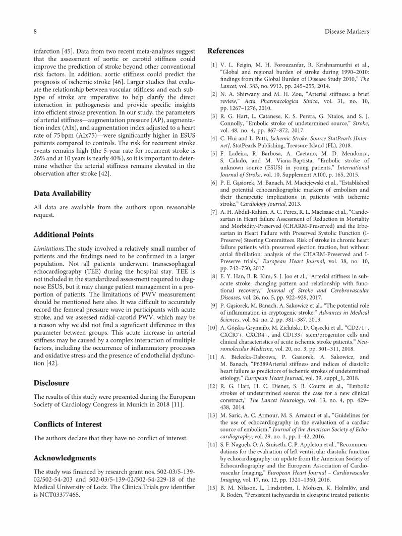
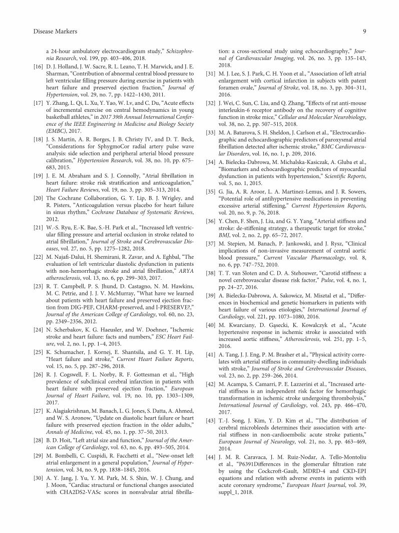
The value of LAVI more than 24mL/m2 and NT-proBNPvalues higher than 99.5 pg/mL were associated with the pres-ence of ESUS. The ROC charts for LAVI and for NT-proBNPare presented accordingly in Figures 1 and 2 (11).
4. Conclusions
Increased arterial stiffness and indices of left ventriculardiastolic dysfunction as well as a higher NT-proBNP levelare associated independently with the presence of ESUS.Our results suggest that there may be a role for increasedheart rhythm surveillance in patients with indices of dia-stolic heart failure to prevent stroke. Despite the lack oflong-term randomized double-blind controlled therapeutictrials, there is high potential to reduce stroke prevalencethrough a significant reduction of arterial stiffness. Phar-macological interventions and lifestyle modification thatcan influence blood pressure, arterial function, or struc-ture in either the short or long term are promising ther-apies reversing arterial stiffness, which can prevent ESUSstrokes.
5. Discussion
5.1. Principle Findings. The results of this study revealed thatESUS patients had lower mean early diastolic (E′) and sys-tolic (S′) mitral annular velocities and a higher E/E′ ratioas well as longer IVRT compared to the control group (11).Also, mean LAVI and the level of NT-proBNP werehigher in the ESUS group, which suggests that despitethe proper values of LVEF, the indices of diastolic heartfailure could be a predictor of this type of stroke. Theparameters of arterial stiffness—augmentation pressure(AP), augmentation index (AIx), and augmentation indexadjusted to a heart rate of 75 bpm (AIx75)—were alsohigher in ESUS patients compared to controls. The param-eters independently associated with the presence of ESUSwere as follows: higher AIx75, longer IVRT, higher LAVI,
Table 1: Basic characteristics of patients in both groups.
ParameterPatients withESUS (n = 65)
Controls(n = 36) p
Gender (male) (%) 42 61 0.059
Median age (years) 54 (IQR 47-58) 53 (47-58) 0,89
SP (mmHg) 136 ± 18 7 128 ± 19 7 0.05
DP (mmHg) 82 ± 9 9 81, 8 ± 9 0 0.72
BMI (kg/m2) 26.1 (22.4-28.7) 25 (21.8-28.1) 0.49
Hypertension (%) 52 43 0.36
CAD (%) 9 9 0.76
Diabetes 8 0 0.23
Smoking (%) 38 13 0.02
ASA (%) 23 9 0.12
Statin (%) 23 17 0.66
Beta blocker (%) 28 12 0.13
ACE (%) 37 24 0.2
CCB (%) 20 9 0.27
Insulin (%) 8 0 0.23
Diuretic (%) 20 15 0.75
ASA: acetylsalicylic acid; ACE: angiotensin-converting enzyme; BMI: bodymass index; CAD: coronary artery disease; CCB: calcium-channel blocker;DP: diastolic pressure; SP: systolic pressure.
4 Disease Markers

and higher NT-proBNP levels. The cutoff points for LAVIand NT-proBNP indicating an increased risk of ESUSwere lower in our study than those accepted in the stan-dards for the diagnosis of heart failure (125 pg/mL com-pared to 99 pg/mL in our study) and LAVI (34mL/m2
compared to 24mL/m2 in our study). Increased arterial
stiffness and indices of diastolic heart failure are associatedindependently with the occurrence of ESUS (11).
5.2. Indices of Diastolic LV and LA Dysfunctions and Risk ofStroke. Patients with heart failure with a reduced ejectionfraction (HFrEF) are at risk from thromboembolic eventsoriginating from both the arterial and venous circulation,which is in part a result of Virchow’s triad of risk factorsfor thrombus formation strictly connected with heart failuresyndrome [19–21]. Kodiak et al. investigated the associationbetween left ventricular diastolic dysfunction (LVDD) andstroke of different origins as well as cryptogenic stroke. Theirresults showed that the CHA2DS2-VASc score was higher inpatients with LVDD [22]. In addition, LVDD compared tothe CHA2DS2-VASc score was a stronger predictor of strokein AF patients. In the study of Najafi-Dalui et al., LVDD wasnot associated with the CHA2DS2-VASc score in patientswith nonhemorrhagic stroke and coexisting AF [22, 23]. Upto half of patients with heart failure have heart failure withpreserved ejection fraction (HFpEF). The prognosis ofHFpEF patients is considerably worse than that of patientswith coronary artery disease, hypertension, AF, or diabetesin the same age range and gender distribution. Little isknown about the incidence of stroke in HFpEF, particularlyin the absence of AF. The Atrial Fibrillation ClopidogrelTrial with Irbesartan for Prevention of Vascular Events(ACTIVE trial) which included over 3400 patients withAF showed similar risks (hazard ratio of 1.01; 95% confi-dence interval, 0.78–1.31) of 4.3% and 4.4% per 100 per-son years for embolic events in noncoagulated patientswith HFpEF and in HFrEF, respectively [24, 25]. Abdul-Rahim et al. revealed a similar risk of stroke in patientswithout AF with HFpEF (1.0% per year) and HFrEF(1.2% per year) and concluded that routinely collectedclinical variables may help clinicians to identify patientswith HFpEF, who may have sufficiently high risk of strokealthough they do not have AF potentially to justify antic-oagulation [7]. Cogswell et al. hypothesized a possibleinfluence of silent paroxysmal AF on stroke risk in HFpEF
Table 2: Basic characteristics of patients in both groups—evaluation of biochemical parameters in groups.
Parameter Patients with ESUS (median with IQR) Controls (median with IQR) p
K+ (mmol/L) 4.08 (±0.35) 4.21 (±0.28) 0.058
Creatinine (μm/L) 75.0 (64.0-89.0) 68.0 (62.0-78.0) 0.21
GFR (mL/min/1.73m3) 75 (64-89) 68 (62-78) 0.002
NT-proBNP (pg/mL) 391 (107.9-1249.22) 109 (46.22-236.90) 0.0003
Total cholesterol (mmol/L) 4 9 ± 1 4 5 1 ± 1 17 0.057
LDL cholesterol (mmol/L) 2.83 (2.07-4.0) 3.02 (2.66-3.67) 0.37
HDL cholesterol (mmol/L) 1.19 (0.95-1.46) 1.37 (1.19-1.6) 0.02
Triglycerides (mmol/L) 1.58 (1.11-2.0) 1.33 (0.86-1.7) 0.16
APTT (s) 28.1 (25.9-31.3) 28.7 (26.5-31.7) 0.42
PT (s) 12.0 (11.5-12.6) 11.9 (11.7-12.4) 0.90
ADMA (μmol/L) 0.44 (0.39-0.55) 0.36 (0.32-0.40) 0.0002
IL-6 (pg/mL) 2.6 (0.8-8.1) 0.7 (0.4- 1.2) 0.002
ADMA: asymmetric dimethylarginine; APTT: activated partial thromboplastin time; HDL: high-density lipoprotein; IL-6: interleukin 6; LDL: low-densitylipoprotein; K+: potassium; NT-proBNP: N-terminal probrain natriuretic peptide; GFR: glomerular filtration rate; PT: prothrombin time.
Table 3: Evaluation of selected echocardiographic parameters inboth groups.
Parameter Patients with ESUS Controls p
E 69 ± 17 76 ± 17 0.09
A 79 ± 19 64 ± 17 0.001
E′ (cm/s) 8.6 (7.1-10.3)∗ 12.5 (9.6-14)∗ 0.0008
S′ (cm/s) 7 0 ± 1 0 8 0 ± 1 0 0.03
A′ (cm/s) 15 54 ± 4 95 16 50 ± 3 86 0.39
E/E′ (cm/s) 7.6 (6.1-8.9)∗ 6.0 (5.3-6.9)∗ 0.0002
LVMI (g/m2) 112.0 (90.0-125.5)∗ 89.5 (77.0-101.0)∗ 0.0004
LA (mm) 36.0 (33.0-41.0)∗ 35.0 (32.0-38)∗ 0.07
LAVI (mL/m2) 27 0 ± 11 21 0 ± 5 0.01
LVEF (%) 60 (55-64) 63 (60-66) 0.009
IVRT (m/s) 113 0 ± 23 97 ± 30 0.001
TAPSE (mm) 24.0 (21-27)∗ 25.0 (22.0-28.0)∗ 0.34
PL (%) 25 16 0.04
For parameters with nonnormal distribution median values, lower andhigher values are given. For parameters with normal distribution mean,values ± standard deviation (SD) are given; A′: late mitral annular motion;A: late diastolic filling velocity; E/E′: ratio of peak velocity of early diastolictransmitral flow to peak velocity of early diastolic mitral annular motion asdetermined by pulsed wave Doppler; E: early diastolic filling velocity; E′:early diastolic mitral annular velocity; HF: high frequency; IVRT:isovolumic relaxation time; LA: left atrium; LF: low frequency; LAVI: leftatrial volume index; LV: left ventricle; LVEF: left ventricular ejectionfraction; LVMI: left ventricular mass index; S′: systolic mitral annularvelocity; PL: pericardial liquid; TAPSE: tricuspid annular plane systolicexcursion; ∗median.
5Disease Markers

patients, given that stroke risk in patients with HFpEFwithout AF and HFpEF with AF as well as AF only wassimilar [26, 27]. Based on the Atherosclerosis Risk inCommunities (ARIC) Study with 1,527 participants, theauthors concluded that undetected AF may be commonin patients with HFpEF and not detecting this may leadto associated cerebral infarcts. Risk factors for having cere-bral infarcts in the HFpEF/no AF group included left atrialenlargement [25]. Left atrium (LA) function comprisesreservoir, conduit, and pump functions, which are depen-dent on left ventricular diastolic function. The left atrialvolume index (LAVI), a biomarker of LA dysfunctionreflecting the aggravation of diastolic LV function, isstrongly associated with cardiovascular disease and out-comes [28]. In the study by Lee et al., the authorscompared the LAVI values between ESUS patients withpatent foramen ovale (PFO) and healthy subjects withPFO and found that the ESUS patients had larger LAvolumes than controls regardless of the presence of PFO.What is interesting is that LA enlargement, but not theamount of shunting, was associated with cortical infarctions,which could imply recurrent embolic stroke [29, 30]. Also,Lee et al. suggested that LA dysfunction could be a markerof incident AF, atrial thrombi, and thromboembolic risksof AF [31]. CHA2DS2-VASc is the most widely acceptedscoring system to assess stroke risk in AF patients,
Table 4: Evaluation of hemodynamic parameters using theSphygmoCor system in both groups.
Parameter Patients with ESUS Controls p
AP (mmHg) 11.0 (7-18)∗ 6.0 (3.0-13.0)∗ 0.0018
AIx (%) 27 ± 13 22 ± 13 0.03
AIx75 (%) 25 ± 11 18 ± 12 0.009
PWV (m/s) 7.2 (6.1-8.4)∗ 7,4 (6,2-9,4)∗ 0.24
HRV index 6.6 (4.7-9)∗ 8.7 (5.9-12.1)∗ 0.006
DP aortic (mmHg) 83.0 (78.0-90.0)∗81.0 (76.0-90.0)∗ 0.59
SP aortic (mmHg) 121 28 ± 17, 72 124 64 ± 18 27 0.42
AP: augmentation pressure; AIx: augmentation index; AIx75: adjustedaugmentation index at a heart rate of 75 beats per minute; DP: diastolicpressure; HRV index: heart rate velocity; SP: systolic pressure; PWV: pulsewave velocity.
Table 5: Univariate logistic regression analysis of the parameters inwhich the univariate analysis using Mann-WhitneyU test, Student’st-test, or chi2 test differs significantly between ESUS and Controlgroups.
Parameter OR 95% CI p
SP (mmHg) 1,023 0,99-1,05 0,057
Smoking 4,375 1,37-13,96 0,013
Cholesterol HDL (mmol/L) 0,248 0,07-0,09 0,028
GFR (mL/min/1.73m3) 0,953 0,93-0,98 0,002
ADMA (μmol/L) 9042 27,5-29,6 0,002
IL-6 (pg/mL) 1,859 1,06-3,26 0,031
NT-proBNP (pg/mL) 1,002 1,001-1,003 0,004
LAVI (mL/m2) 1,064 1,01-1,21 0,021
LVEF (%) 0,914 0,85-0,99 0,019
A 1,050 1,02-1,08 0,001
IVRT (m/s) 1,037 1,02-1,06 0,001
S′ (cm/s) 0,763 0,59-0,99 0,044
E′ (cm/s) 0,087 0,77-0,99 0,027
E/E′ (cm/s) 1,465 1,14-1,88 0,003
LVMI (g/m2) 1,034 1,01-1,06 0,002
PL (%) 5,277 1,13-24,57 0,034
HRV index 0,995 0,96-1,03 0,784
AP (mmHg) 1,100 1,03-1,18 0,005
AIx (%) 1,035 1,01-1,07 0,038
AIx75 (%) 1,048 1,01-1,09 0,014
SP: systolic pressure; HDL: high-density lipoprotein; GFR: glomerularfiltration rate; ADMA: asymmetric dimethylarginine; IL-6: interleukin 6;NT-proBNP: N-terminal probrain natriuretic peptide; LAVI: left atrialvolume index; LVEF: left ventricular ejection fraction; A: late diastolicfilling velocity; IVRT: isovolumic relaxation time; S′: systolic mitralannular velocity; E′: early diastolic mitral annular velocity; E/E′: ratio ofpeak velocity of early diastolic transmitral flow to peak velocity of earlydiastolic mitral annular motion as determined by pulsed wave Doppler;LVMI: left ventricular mass index; PL: pericardial liquid; HRV index: heartrate velocity; AP: augmentation pressure; AIx: augmentation index; AIx75:adjusted augmentation index at a heart rate of 75 beats per minute.
Table 6: Multivariate analysis—stepwise logistic regression.
Variable OR95% CI for OR
p valueLower limit Upper limit
IVRT (ms) 1.045 1.009 1.982 0.01
NT-proBNP (pg/mL) 1.003 1.001 1.005 0.007
LAVI (mL/m2) 1.3 1.099 1.537 0.002
AIx75 1.095 1.004 1.194 0.04
AIx75: adjusted augmentation index at a heart rate of 75 beats per minute;IVRT: isovolumic relaxation time; LAVI: left atrial volume index; NT-proBNP: N-terminal probrain natriuretic peptide.
0.0 0.2 0.4 0.6 0.8 1.0
0.2
0.0
0.4
0.6
0.8
1.0
Sensitivity
Specificity
24.242
Figure 1: ROC chart for LAVI.
6 Disease Markers

although still we do not have enough information about theaccompanying cardiac functional/structural changes. In a totalof 4,795 patients with nonvalvular AF, increases in the leftventricular mass index (LVMI) and prevalence of left ventric-ular hypertrophy (LVH) as well as LAVI and E/E′ wereobserved with elevating CHA2DS2-VASc scores (p < 0 05 forLVMI and LVH and p < 0 001 for LAVI and E/E′). LVH (haz-ard ratio (HR), 3.609; confidence interval (CI), 2.426–5.369;p < 0 001) and E/E′ (HR, 1.087; CI, 1.054–1.121; p < 0 001) were independent risk factors for a CHA2DS2-VAScscore of 2 or higher. The authors stated that higherCHA2DS2-VASc scores are associated with impaired dia-stolic function, reflecting high left atrial pressure andincreased risk of thromboembolism [31]. Perhaps, loweringthe left ventricular end-diastolic pressure should be a ther-apeutic target for HFpEF patients with high CHA2DS2-VASc scores to decrease the prevalence of thromboembolicevents in this group of patients [32]. The increased LAVIin the aspect of ESUS stroke may also be important as apredictor of paroxysmal AF as a true cause of stroke.Detecting AF after ischemic stroke is challenging becauseof its paroxysmal nature and often silent, asymptomaticcourse, as was confirmed in studies with implantabledevices. Baturova et al. reported that left atrial dilatationassessed by LAVI independently predicted AF after strokein patients without prior AF history, while the other clin-ical or ECG markers were not predictive for AF detectionearly after ischemic stroke. The authors suggest that ini-tially, there is development of subtle structural changespredictive for future AF seen in echocardiography (forexample, increased LAVI) [33]. The level of NT-proBNPmay participate in pathogenesis and pathophysiology of ische-mic stroke. The efforts to find a correlation between the NT-proBNP concentration and stroke topography, size, or gravityof neurological deficit do not give clear-cut results. The sameconcerns the prognostic value of BNP concentration duringischemic stroke. Despite conflicting reports, it is worth con-tinuing this research, because among other things, there is a
connection between cardiac insufficiency and prognosis inacute cerebrovascular incidents [34, 35]. In our study, ESUSpatients had lower mean early diastolic (E′) and systolic (S′)mitral annular velocities and a higher E/E′ ratio as well as lon-ger IVRT compared to the control group, which confirms theconnection between indices of diastolic heart failure andESUS stroke. Even slightly increased values of LAVI (cutoffpoint 24mL/m2) and NT-proBNP (cutoff point 99pg/mL)were predictors of this type of stroke [11]. Further investi-gation is necessary to attribute the additive values of echo-cardiographic parameters for stroke prediction and theeffect of left ventricular end-diastolic pressure (LVEDP)control on the reduction of ischemic stroke events. LAVImay be a new noninvasive tool to identify patients afterstroke who would benefit the most from continuous screen-ing for AF.
5.3. Arterial Stiffness and the Risk of ESUS. Arterial stiffnesshas been regarded as a reliable marker of arterial structuraland functional alterations after abundant experimental andclinical studies. Vascular structure, vascular function, andBP are the three major components that are involved in arte-rial stiffness [36]. Factors such as inflammation, oxide stress,the renin-angiotensin-aldosterone system (RAAS), andgenetic factors that influence the vascular function in theshort term or the structure in the long term can induce arte-rial stiffness [37]. Stiffening of the cervical elastic arteries maylead to cerebrovascular disease via multiple mechanisms.Increased stiffness of the carotid artery leads to a higher pul-satile pressure and flow load on the brain, which can pene-trate distally into the cerebral microcirculation, causingcerebral ischemia and hemorrhage. The increased stiffnessof elastic arteries may also cause excessive blood pressurevariability, which may further sensitize the brain to theharmful effects of impaired microvascular vasoreactivity[38]. The increased pulsatile load may induce a hypertrophicremodeling response of small cerebral arteries, which initiallylimit the penetration of the pulsatile load into the microcircu-latory system by raising vascular resistance, leading toimpaired vasoreactivity, hypoperfusion, and chronic ische-mia [39]. One of the causes of ischemic stroke is chronic ath-erosclerosis. The atherosclerotic state might be reflected byincreased arterial stiffness, whereby the aortic pressure isaugmented, resulting in increased arterial wall stress and leftventricular afterload [40]. Arterial stiffness provides impor-tant information regarding the progression of atherosclerosisand can be measured noninvasively [41, 42]. Increasedarterial stiffness causes vessel damage and is independentlyassociated with deep or infratentorial cerebral microbleeds[43, 44]. After an average 7.9 years of follow-up of middle-aged patients with essential hypertension, Laurent et al.found that a 1-SD elevation (4 cm/s) in PWV was associatedwith a 72% higher risk of fatal stroke. High PWV remainedsignificantly predictive of stroke death after adjustment forclassical cardiovascular risk factors. Other researchersassessed its predictive value in the elderly and general popu-lation. Byun et al. reported that increased arterial stiffnessassessed based on higher values of AIx75 in acute lacunarinfarction may be related to the pathogenesis of lacunar
0.0 0.2 0.4 0.6 0.8 1.0
0.2
0.0
0.4
0.6
0.8
1.0
Sensitivity
Specificity
99.495
Figure 2: ROC chart for NT-proBNP.
7Disease Markers
infarction [45]. Data from two recent meta-analyses suggestthat the assessment of aortic or carotid stiffness couldimprove the prediction of stroke beyond other conventionalrisk factors. In addition, aortic stiffness could predict theprognosis of ischemic stroke [46]. Larger studies that evalu-ate the relationship between vascular stiffness and each sub-type of stroke are imperative to help clarify the directinteraction in pathogenesis and provide specific insightsinto efficient stroke prevention. In our study, the parametersof arterial stiffness—augmentation pressure (AP), augmenta-tion index (AIx), and augmentation index adjusted to a heartrate of 75 bpm (AIx75)—were significantly higher in ESUSpatients compared to controls. The risk for recurrent strokeevents remains high (the 5-year rate for recurrent stroke is26% and at 10 years is nearly 40%), so it is important to deter-mine whether the arterial stiffness remains elevated in theobservation after stroke [42].
Data Availability
All data are available from the authors upon reasonablerequest.
Additional Points
Limitations.The study involved a relatively small number ofpatients and the findings need to be confirmed in a largerpopulation. Not all patients underwent transesophagealechocardiography (TEE) during the hospital stay. TEE isnot included in the standardized assessment required to diag-nose ESUS, but it may change patient management in a pro-portion of patients. The limitations of PWV measurementshould be mentioned here also. It was difficult to accuratelyrecord the femoral pressure wave in participants with acutestroke, and we assessed radial-carotid PWV, which may bea reason why we did not find a significant difference in thisparameter between groups. This acute increase in arterialstiffness may be caused by a complex interaction of multiplefactors, including the occurrence of inflammatory processesand oxidative stress and the presence of endothelial dysfunc-tion [42].
Disclosure
The results of this study were presented during the EuropeanSociety of Cardiology Congress in Munich in 2018 [11].
Conflicts of Interest
The authors declare that they have no conflict of interest.
Acknowledgments
The study was financed by research grant nos. 502-03/5-139-02/502-54-203 and 502-03/5-139-02/502-54-229-18 of theMedical University of Lodz. The ClinicalTrials.gov identifieris NCT03377465.
References
[1] V. L. Feigin, M. H. Forouzanfar, R. Krishnamurthi et al.,“Global and regional burden of stroke during 1990–2010:findings from the Global Burden of Disease Study 2010,” TheLancet, vol. 383, no. 9913, pp. 245–255, 2014.
[2] N. A. Shirwany and M. H. Zou, “Arterial stiffness: a briefreview,” Acta Pharmacologica Sinica, vol. 31, no. 10,pp. 1267–1276, 2010.
[3] R. G. Hart, L. Catanese, K. S. Perera, G. Ntaios, and S. J.Connolly, “Embolic stroke of undetermined source,” Stroke,vol. 48, no. 4, pp. 867–872, 2017.
[4] C. Hui and L. Patti, Ischemic Stroke. Source StatPearls [Inter-net], StatPearls Publishing, Treasure Island (FL), 2018.
[5] F. Ladeira, R. Barbosa, A. Caetano, M. D. Mendonça,S. Calado, and M. Viana-Baptista, “Embolic stroke ofunknown source (ESUS) in young patients,” InternationalJournal of Stroke, vol. 10, Supplement A100, p. 165, 2015.
[6] P. E. Gąsiorek, M. Banach, M. Maciejewski et al., “Establishedand potential echocardiographic markers of embolism andtheir therapeutic implications in patients with ischemicstroke,” Cardiology Journal, 2013.
[7] A. H. Abdul-Rahim, A. C. Perez, R. L. MacIsaac et al., “Cande-sartan in Heart failure Assessment of Reduction in Mortalityand Morbidity-Preserved (CHARM-Preserved) and the Irbe-sartan in Heart Failure with Preserved Systolic Function (I-Preserve) Steering Committees. Risk of stroke in chronic heartfailure patients with preserved ejection fraction, but withoutatrial fibrillation: analysis of the CHARM-Preserved and I-Preserve trials,” European Heart Journal, vol. 38, no. 10,pp. 742–750, 2017.
[8] E. Y. Han, B. R. Kim, S. J. Joo et al., “Arterial stiffness in sub-acute stroke: changing pattern and relationship with func-tional recovery,” Journal of Stroke and CerebrovascularDiseases, vol. 26, no. 5, pp. 922–929, 2017.
[9] P. Gąsiorek, M. Banach, A. Sakowicz et al., “The potential roleof inflammation in cryptogenic stroke,” Advances in MedicalSciences, vol. 64, no. 2, pp. 381–387, 2019.
[10] A. Gójska-Grymajło, M. Zieliński, D. Gąsecki et al., “CD271+,CXCR7+, CXCR4+, and CD133+ stem/progenitor cells andclinical characteristics of acute ischemic stroke patients,” Neu-romolecular Medicine, vol. 20, no. 3, pp. 301–311, 2018.
[11] A. Bielecka-Dabrowa, P. Gasiorek, A. Sakowicz, andM. Banach, “P6389Arterial stiffness and indices of diastolicheart failure as predictors of ischemic strokes of undeterminedetiology,” European Heart Journal, vol. 39, suppl_1, 2018.
[12] R. G. Hart, H. C. Diener, S. B. Coutts et al., “Embolicstrokes of undetermined source: the case for a new clinicalconstruct,” The Lancet Neurology, vol. 13, no. 4, pp. 429–438, 2014.
[13] M. Saric, A. C. Armour, M. S. Arnaout et al., “Guidelines forthe use of echocardiography in the evaluation of a cardiacsource of embolism,” Journal of the American Society of Echo-cardiography, vol. 29, no. 1, pp. 1–42, 2016.
[14] S. F. Nagueh, O. A. Smiseth, C. P. Appleton et al., “Recommen-dations for the evaluation of left ventricular diastolic functionby echocardiography: an update from the American Society ofEchocardiography and the European Association of Cardio-vascular Imaging,” European Heart Journal – CardiovascularImaging, vol. 17, no. 12, pp. 1321–1360, 2016.
[15] B. M. Nilsson, L. Lindström, I. Mohsen, K. Holmlöv, andR. Bodén, “Persistent tachycardia in clozapine treated patients:
8 Disease Markers
a 24-hour ambulatory electrocardiogram study,” Schizophre-nia Research, vol. 199, pp. 403–406, 2018.
[16] D. J. Holland, J. W. Sacre, R. L. Leano, T. H. Marwick, and J. E.Sharman, “Contribution of abnormal central blood pressure toleft ventricular filling pressure during exercise in patients withheart failure and preserved ejection fraction,” Journal ofHypertension, vol. 29, no. 7, pp. 1422–1430, 2011.
[17] Y. Zhang, L. Qi, L. Xu, Y. Yao, W. Lv, and C. Du, “Acute effectsof incremental exercise on central hemodynamics in youngbasketball athletes,” in 2017 39th Annual International Confer-ence of the IEEE Engineering in Medicine and Biology Society(EMBC), 2017.
[18] J. S. Martin, A. R. Borges, J. B. Christy IV, and D. T. Beck,“Considerations for SphygmoCor radial artery pulse waveanalysis: side selection and peripheral arterial blood pressurecalibration,” Hypertension Research, vol. 38, no. 10, pp. 675–683, 2015.
[19] J. E. M. Abraham and S. J. Connolly, “Atrial fibrillation inheart failure: stroke risk stratification and anticoagulation,”Heart Failure Reviews, vol. 19, no. 3, pp. 305–313, 2014.
[20] The Cochrane Collaboration, G. Y. Lip, B. J. Wrigley, andR. Pisters, “Anticoagulation versus placebo for heart failurein sinus rhythm,” Cochrane Database of Systematic Reviews,2012.
[21] W.-S. Ryu, E.-K. Bae, S.-H. Park et al., “Increased left ventric-ular filling pressure and arterial occlusion in stroke related toatrial fibrillation,” Journal of Stroke and Cerebrovascular Dis-eases, vol. 27, no. 5, pp. 1275–1282, 2018.
[22] M. Najafi-Dalui, H. Shemirani, R. Zavar, and A. Eghbal, “Theevaluation of left ventricular diastolic dysfunction in patientswith non-hemorrhagic stroke and atrial fibrillation,” ARYAatherosclerosis, vol. 13, no. 6, pp. 299–303, 2017.
[23] R. T. Campbell, P. S. Jhund, D. Castagno, N. M. Hawkins,M. C. Petrie, and J. J. V. McMurray, “What have we learnedabout patients with heart failure and preserved ejection frac-tion from DIG-PEF, CHARM-preserved, and I-PRESERVE?,”Journal of the American College of Cardiology, vol. 60, no. 23,pp. 2349–2356, 2012.
[24] N. Scherbakov, K. G. Haeusler, and W. Doehner, “Ischemicstroke and heart failure: facts and numbers,” ESC Heart Fail-ure, vol. 2, no. 1, pp. 1–4, 2015.
[25] K. Schumacher, J. Kornej, E. Shantsila, and G. Y. H. Lip,“Heart failure and stroke,” Current Heart Failure Reports,vol. 15, no. 5, pp. 287–296, 2018.
[26] R. J. Cogswell, F. L. Norby, R. F. Gottesman et al., “Highprevalence of subclinical cerebral infarction in patients withheart failure with preserved ejection fraction,” EuropeanJournal of Heart Failure, vol. 19, no. 10, pp. 1303–1309,2017.
[27] K. Alagiakrishnan, M. Banach, L. G. Jones, S. Datta, A. Ahmed,and W. S. Aronow, “Update on diastolic heart failure or heartfailure with preserved ejection fraction in the older adults,”Annals of Medicine, vol. 45, no. 1, pp. 37–50, 2013.
[28] B. D. Hoit, “Left atrial size and function,” Journal of the Amer-ican College of Cardiology, vol. 63, no. 6, pp. 493–505, 2014.
[29] M. Bombelli, C. Cuspidi, R. Facchetti et al., “New-onset leftatrial enlargement in a general population,” Journal of Hyper-tension, vol. 34, no. 9, pp. 1838–1845, 2016.
[30] A. Y. Jang, J. Yu, Y. M. Park, M. S. Shin, W. J. Chung, andJ. Moon, “Cardiac structural or functional changes associatedwith CHA2DS2-VASc scores in nonvalvular atrial fibrilla-
tion: a cross-sectional study using echocardiography,” Jour-nal of Cardiovascular Imaging, vol. 26, no. 3, pp. 135–143,2018.
[31] M. J. Lee, S. J. Park, C. H. Yoon et al., “Association of left atrialenlargement with cortical infarction in subjects with patentforamen ovale,” Journal of Stroke, vol. 18, no. 3, pp. 304–311,2016.
[32] J. Wei, C. Sun, C. Liu, and Q. Zhang, “Effects of rat anti-mouseinterleukin-6 receptor antibody on the recovery of cognitivefunction in stroke mice,” Cellular and Molecular Neurobiology,vol. 38, no. 2, pp. 507–515, 2018.
[33] M. A. Baturova, S. H. Sheldon, J. Carlson et al., “Electrocardio-graphic and echocardiographic predictors of paroxysmal atrialfibrillation detected after ischemic stroke,” BMC Cardiovascu-lar Disorders, vol. 16, no. 1, p. 209, 2016.
[34] A. Bielecka-Dabrowa, M. Michalska-Kasiczak, A. Gluba et al.,“Biomarkers and echocardiographic predictors of myocardialdysfunction in patients with hypertension,” Scientific Reports,vol. 5, no. 1, 2015.
[35] G. Jia, A. R. Aroor, L. A. Martinez-Lemus, and J. R. Sowers,“Potential role of antihypertensive medications in preventingexcessive arterial stiffening,” Current Hypertension Reports,vol. 20, no. 9, p. 76, 2018.
[36] Y. Chen, F. Shen, J. Liu, and G. Y. Yang, “Arterial stiffness andstroke: de-stiffening strategy, a therapeutic target for stroke,”BMJ, vol. 2, no. 2, pp. 65–72, 2017.
[37] M. Stepien, M. Banach, P. Jankowski, and J. Rysz, “Clinicalimplications of non-invasive measurement of central aorticblood pressure,” Current Vascular Pharmacology, vol. 8,no. 6, pp. 747–752, 2010.
[38] T. T. van Sloten and C. D. A. Stehouwer, “Carotid stiffness: anovel cerebrovascular disease risk factor,” Pulse, vol. 4, no. 1,pp. 24–27, 2016.
[39] A. Bielecka-Dabrowa, A. Sakowicz, M. Misztal et al., “Differ-ences in biochemical and genetic biomarkers in patients withheart failure of various etiologies,” International Journal ofCardiology, vol. 221, pp. 1073–1080, 2016.
[40] M. Kwarciany, D. Gąsecki, K. Kowalczyk et al., “Acutehypertensive response in ischemic stroke is associated withincreased aortic stiffness,” Atherosclerosis, vol. 251, pp. 1–5,2016.
[41] A. Tang, J. J. Eng, P. M. Brasher et al., “Physical activity corre-lates with arterial stiffness in community-dwelling individualswith stroke,” Journal of Stroke and Cerebrovascular Diseases,vol. 23, no. 2, pp. 259–266, 2014.
[42] M. Acampa, S. Camarri, P. E. Lazzerini et al., “Increased arte-rial stiffness is an independent risk factor for hemorrhagictransformation in ischemic stroke undergoing thrombolysis,”International Journal of Cardiology, vol. 243, pp. 466–470,2017.
[43] T.-J. Song, J. Kim, Y. D. Kim et al., “The distribution ofcerebral microbleeds determines their association with arte-rial stiffness in non-cardioembolic acute stroke patients,”European Journal of Neurology, vol. 21, no. 3, pp. 463–469,2014.
[44] J. M. R. Caravaca, J. M. Ruiz-Nodar, A. Tello-Montoliuet al., “P6391Differences in the glomerular filtration rateby using the Cockcroft-Gault, MDRD-4 and CKD-EPIequations and relation with adverse events in patients withacute coronary syndrome,” European Heart Journal, vol. 39,suppl_1, 2018.
9Disease Markers

[45] D. S. Byun, S. W. Han, J. H. Park, J. Y. Kim, J. S. Baik, and J. H.Park, “Relationship between augmentation index and acuteischemic stroke subtype,” Journal of Clinical Neuroscience,vol. 21, no. 7, pp. 1220–1224, 2014.
[46] T. Sanna, P. D. Ziegler, and F. Crea, “Detection and manage-ment of atrial fibrillation after cryptogenic stroke or embolicstroke of undetermined source,” Clinical Cardiology, vol. 41,no. 3, pp. 426–432, 2018.
10 Disease Markers

Stem Cells International
Hindawiwww.hindawi.com Volume 2018
Hindawiwww.hindawi.com Volume 2018
MEDIATORSINFLAMMATION
of
EndocrinologyInternational Journal of
Hindawiwww.hindawi.com Volume 2018
Hindawiwww.hindawi.com Volume 2018
Disease Markers
Hindawiwww.hindawi.com Volume 2018
BioMed Research International
OncologyJournal of
Hindawiwww.hindawi.com Volume 2013
Hindawiwww.hindawi.com Volume 2018
Oxidative Medicine and Cellular Longevity
Hindawiwww.hindawi.com Volume 2018
PPAR Research
Hindawi Publishing Corporation http://www.hindawi.com Volume 2013Hindawiwww.hindawi.com
The Scientific World Journal
Volume 2018
Immunology ResearchHindawiwww.hindawi.com Volume 2018
Journal of
ObesityJournal of
Hindawiwww.hindawi.com Volume 2018
Hindawiwww.hindawi.com Volume 2018
Computational and Mathematical Methods in Medicine
Hindawiwww.hindawi.com Volume 2018
Behavioural Neurology
OphthalmologyJournal of
Hindawiwww.hindawi.com Volume 2018
Diabetes ResearchJournal of
Hindawiwww.hindawi.com Volume 2018
Hindawiwww.hindawi.com Volume 2018
Research and TreatmentAIDS
Hindawiwww.hindawi.com Volume 2018
Gastroenterology Research and Practice
Hindawiwww.hindawi.com Volume 2018
Parkinson’s Disease
Evidence-Based Complementary andAlternative Medicine
Volume 2018Hindawiwww.hindawi.com
Submit your manuscripts atwww.hindawi.com