ars.els-cdn.com · web viewthe target lesion is the treated segment starting 10 mm proximal and...
TRANSCRIPT

Appendix 1. Authors and Affiliations and SCTI ContributorsName AffiliationKaren A. Hicks, MD Division of Cardiovascular and Renal Products
Office of Drug Evaluation ICenter for Drug Evaluation and Research (CDER)
United States Food and Drug Administration (FDA)Silver Spring, Maryland
Kenneth W. Mahaffey, MD Stanford Center for Clinical ResearchDepartment of Medicine
Stanford University School of MedicineStanford, California
Roxana Mehran, MD Division of CardiologyIcahn School of Medicine at Mount Sinai
New York, New YorkSteven E. Nissen, MD Department of Cardiovascular Medicine
Cleveland ClinicCleveland, Ohio
Stephen D. Wiviott, MD TIMI Study GroupCardiovascular Division
Brigham and Women’s HospitalBoston, Massachusetts
Billy Dunn, MD Division of Neurology ProductsOffice of Drug Evaluation I
Center for Drug Evaluation and Research (CDER)United States Food and Drug Administration (FDA)
Silver Spring, MarylandScott D. Solomon, MD Cardiovascular Division
Brigham and Women’s HospitalBoston, Massachusetts
John R. Marler, MD Division of Neurology ProductsOffice of Drug Evaluation I
Center for Drug Evaluation and Research (CDER)United States Food and Drug Administration (FDA)
Silver Spring, MarylandJohn R. Teerlink, MD Section of Cardiology
San Francisco Veterans Affairs Medical Center and School of MedicineUniversity of California San Francisco
San Francisco, CaliforniaAndrew Farb, MD Division of Cardiovascular Devices
Center for Devices and Radiological Health (CDRH)United States Food and Drug Administration (FDA)
Silver Spring, MarylandDavid A. Morrow, MD, MPH TIMI Study Group
Cardiovascular DivisionBrigham and Women’s Hospital
Boston, MassachusettsShari L. Targum, MD, MPH Division of Cardiovascular and Renal Products
Office of Drug Evaluation ICenter for Drug Evaluation and Research (CDER)
United States Food and Drug Administration (FDA)Silver Spring, Maryland
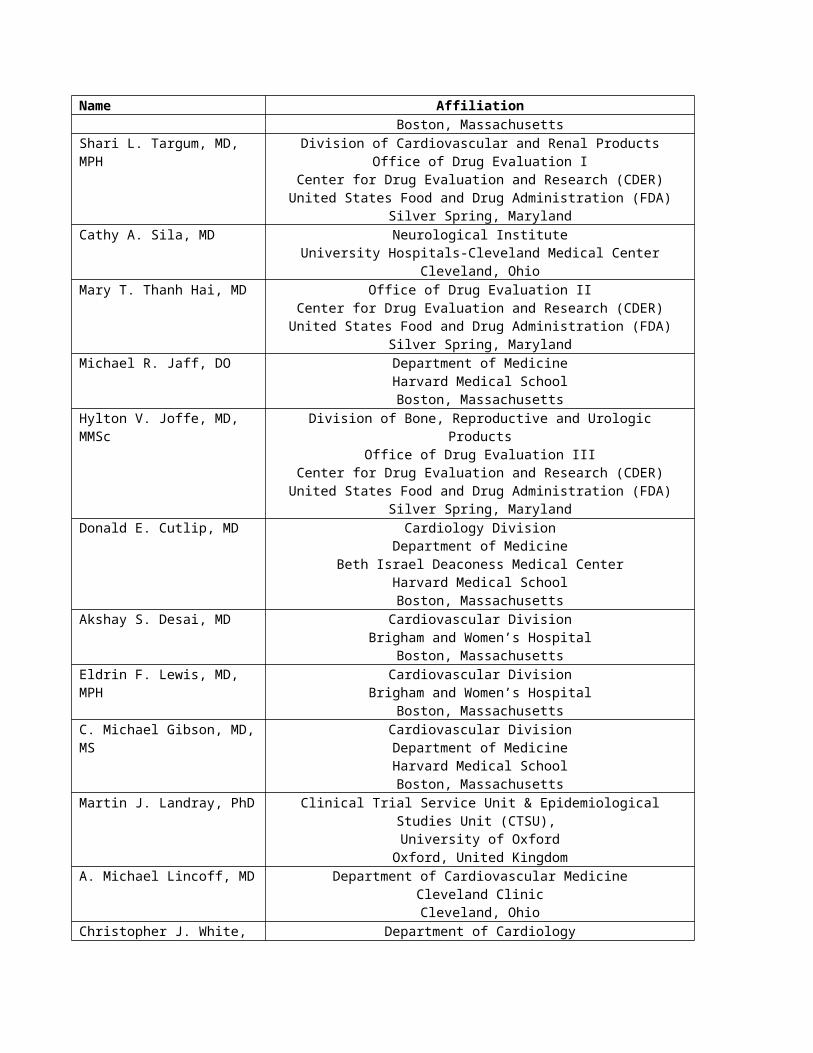
Name AffiliationCathy A. Sila, MD Neurological Institute
University Hospitals-Cleveland Medical CenterCleveland, Ohio
Mary T. Thanh Hai, MD Office of Drug Evaluation IICenter for Drug Evaluation and Research (CDER)
United States Food and Drug Administration (FDA)Silver Spring, Maryland
Michael R. Jaff, DO Department of MedicineHarvard Medical SchoolBoston, Massachusetts
Hylton V. Joffe, MD, MMSc Division of Bone, Reproductive and Urologic ProductsOffice of Drug Evaluation III
Center for Drug Evaluation and Research (CDER)United States Food and Drug Administration (FDA)
Silver Spring, MarylandDonald E. Cutlip, MD Cardiology Division
Department of MedicineBeth Israel Deaconess Medical Center
Harvard Medical SchoolBoston, Massachusetts
Akshay S. Desai, MD Cardiovascular DivisionBrigham and Women’s Hospital
Boston, MassachusettsEldrin F. Lewis, MD, MPH Cardiovascular Division
Brigham and Women’s HospitalBoston, Massachusetts
C. Michael Gibson, MD, MS Cardiovascular DivisionDepartment of MedicineHarvard Medical SchoolBoston, Massachusetts
Martin J. Landray, PhD Clinical Trial Service Unit & Epidemiological Studies Unit (CTSU), University of Oxford
Oxford, United KingdomA. Michael Lincoff, MD Department of Cardiovascular Medicine
Cleveland ClinicCleveland, Ohio
Christopher J. White, MD Department of CardiologyOchsner Clinical SchoolNew Orleans, Louisiana
Steven S. Brooks, MD, MBA Brooks MedTech, LLCReisterstown, Maryland
Kenneth Rosenfield, MD Vascular Medicine and InterventionCorrigan Minehan Heart CenterMassachusetts General Hospital
Boston, MassachusettsMichael J. Domanski, MD Peter Munk Cardiac Centre
University Health NetworkMount Sinai HospitalUniversity of Toronto
Toronto, Ontario, CanadaAlexandra J. Lansky, MD Department of Internal Medicine
Section of CardiologyYale School of MedicineNew Haven, Connecticut

Name AffiliationJohn J. V. McMurray, MD Institute of Cardiovascular & Medical Sciences
BHF Cardiovascular Research CentreUniversity of Glasgow
Glasgow, ScotlandJames E. Tcheng, MD Division of Cardiovascular Medicine
Duke University Medical CenterDuke University Medical Center
Durham, North CarolinaSteven R. Steinhubl, MD Division of Digital Medicine
Scripps Translational Science InstituteLa Jolla, California
Paul Burton, MD, PhD Cardiovascular and Metabolism Medical AffairsJanssen Pharmaceuticals Inc.
Titusville, New JerseyLaura Mauri, MD Cardiovascular Division
Department of MedicineBrigham and Women’s Hospital
Harvard Medical SchoolBoston, Massachusetts
Christopher M. O’Connor, MD Division of CardiologyInova Heart & Vascular Institute
Falls Church, VirginiaMarc A. Pfeffer, MD, PhD Cardiovascular Division
Brigham and Women’s HospitalBoston, Massachusetts
H.M. James Hung, Ph.D. Division of Biometrics IOffice of Biostatistics
Center for Drug Evaluation and Research (CDER)United States Food and Drug Administration (FDA)
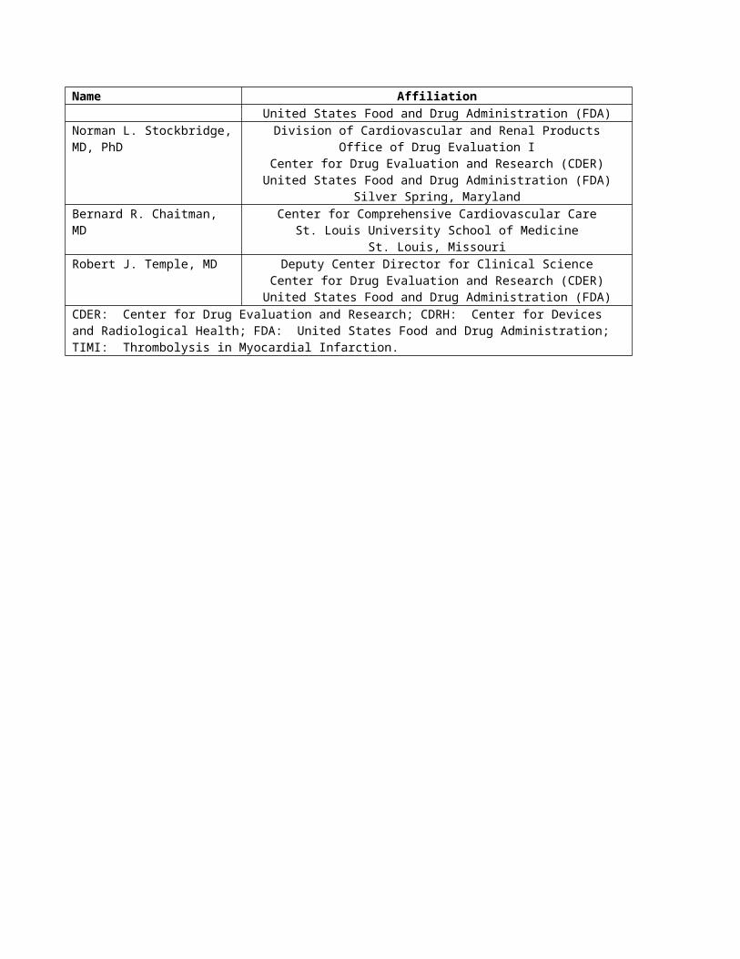
Norman L. Stockbridge, MD, PhD
Division of Cardiovascular and Renal ProductsOffice of Drug Evaluation I
Center for Drug Evaluation and Research (CDER)United States Food and Drug Administration (FDA)
Silver Spring, MarylandBernard R. Chaitman, MD Center for Comprehensive Cardiovascular Care
St. Louis University School of MedicineSt. Louis, Missouri
Robert J. Temple, MD Deputy Center Director for Clinical ScienceCenter for Drug Evaluation and Research (CDER)
United States Food and Drug Administration (FDA)CDER: Center for Drug Evaluation and Research; CDRH: Center for Devices and Radiological Health; FDA: United States Food and Drug Administration; TIMI: Thrombolysis in Myocardial Infarction.
SCTI Contributors
Name Affiliation ContributionsStroke and Transient Ischemic Attack SubcommitteeHeather D. Fitter, MD Division of Neurology Products,
Office of Drug Evaluation I (ODE I),
Center for Drug Evaluation and Research (CDER),
United States Food and Drug Administration (FDA),Silver Spring, Maryland
Participated in discussions regarding the stroke and TIA definitions
Kachikwu Illoh, MD Office of Scientific Investigation,Office of Compliance,
CDER,FDA,
Silver Spring, Maryland
Participated in discussions regarding the stroke and TIA definitions
Percutaneous Coronary Intervention, Stent Thrombosis, and Peripheral Vascular Intervention SubcommitteeKenneth J. Cavanaugh, Jr., PhD Division of Peripheral Vascular
Devices,Center for Devices and
Radiological Health (CDRH),FDA,
Silver Spring, Maryland
Provided input into the development of the PVI definitions
Hospitalization for Unstable Angina and Heart Failure Event SubcommitteeBenjamin M. Scirica, MD, MPH TIMI Study Group,
Cardiovascular Division, Brigham and Women’s Hospital,
Boston, Massachusetts
Participated in discussions regarding the HF and HUA definitions
Regulatory SubcommitteeIlan Irony, MD Division of Clinical Evaluation,
Pharmacology and Toxicology, Center for Biologics Evaluation
and Research (CBER),FDA,
Silver Spring, Maryland
Implemented the CV and stroke endpoint definitions in clinical trials
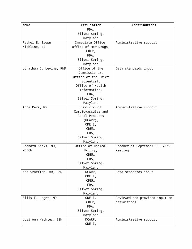
Rachel E. Brown Kichline, BS Immediate Office,Office of New Drugs,
CDER,FDA,
Silver Spring, Maryland
Administrative support
Jonathan G. Levine, PhD Office of the Commissioner,Office of the Chief Scientist,Office of Health Informatics,
FDA,Silver Spring, Maryland
Data standards input

Name Affiliation ContributionsAnna Park, MS Division of Cardiovascular and
Renal Products (DCARP),ODE I,CDER,FDA,
Silver Spring, Maryland
Administrative support
Leonard Sacks, MD, MBBCh Office of Medical Policy,CDER,FDA,
Silver Spring, Maryland
Speaker at September 11, 2009 Meeting
Ana Szarfman, MD, PhD DCARP,ODE I,CDER,FDA,
Silver Spring, Maryland
Data standards input
Ellis F. Unger, MD ODE I,CDER,FDA,
Silver Spring, Maryland
Reviewed and provided input on definitions
Lori Ann Wachter, BSN DCARP,ODE I,CDER,FDA,
Silver Spring, Maryland
Administrative support
Bram Zuckerman, MD Division of Cardiovascular Devices,CDRH,
Silver Spring, Maryland
Provided input regarding the Interventional Cardiology definitions
Industry, External Consultants, and Academic Research OrganizationYale Mitchel, MD Cardiovascular Disease,
Merck Research Laboratories,Rahway, New Jersey
Attended public meetings and participated in the discussion of the endpoint definitions
Douglas Peddicord, PhD Association of Clinical Research Organizations (ACRO),
Washington, DC
Attended public meetings and participated in the discussion of the endpoint definitions
Thomas Shook, MD PAREXEL International,Waltham, Massachusetts
Attended public meeting and participated in the discussion of the endpoint definitions
Data Standards SubcommitteeBron Kisler, BS Biomedical Informatics
Specialist, National Cancer Institute [c],
Bethesda, Maryland;Co-Founder, Clinical Data
Interchange Standards Consortium (CDISC)
Posted draft definitions on CDISC website for public comment in 2010 – 2011. Supported Webinar to discuss the CV and stroke endpoint definitions in 2010. Posted updated definitions dated August 20, 2014 on the CDISC website. Worked with FDA to develop the CV TAUG (data standards for current CV and stroke endpoint definitions).
Charles Jaffe, MD, PhD Health Level 7 (HL7) International,
Del Mar, California
Attended public meetings and participated in the discussion of the endpoint definitions
Clinical Trials Transformation Initiative (CTTI) SubcommitteeRhonda Bartley, AA Infectious Disease and Global
Health,Duke University Medical Center,
Durham, North Carolina
Administrative Support
Name Affiliation ContributionsDavid L. DeMets Department of Biostatistics and
Medical Informatics,University of Wisconsin,
Madison, Wisconsin
Participated in a 2010 teleconference with FDA to discuss the development of uniform CV and stroke definitions and data standards for clinical trials
MariJo Mencini Duke Clinical Research Institute,Durham, North Carolina
Administrative Support
Cheri Janning Duke Clinical Translation Science Institute,
Durham, North Carolina
Administrative Support
Biometrics SubcommitteeSteve Bai, PhD Division of Biometrics I,
Office of Biostatistics,CDER,FDA,
Silver Spring, Maryland
Development of Statistical Analysis Plan (SAP); Data Analysis for Testing of the Definitions
John Lawrence, PhD Division of Biometrics I,Office of Biostatistics,
CDER,FDA,
Silver Spring, Maryland
Development of SAP; Data Analysis for Testing of the Definitions
Ralph B. D’Agostino, Sr., PhD Mathematics and Statistics Department,
Boston University,Boston, Massachusetts
External Review of SAP and input
Stuart J. Pocock, PhD Department of Medical Statistics,London School of Hygiene and
Tropical Medicine,London, United Kingdom
External Review of SAP and input
Adjudication SubcommitteeTIMI Study Group Clinical Events Committee (TIMI Study Group CEC)Stephen D. Wiviott, MD† TIMI Study Group,
Cardiovascular Division,Brigham and Women’s Hospital,
Boston, Massachusetts
CEC Chairman
Cheryl Lowe, RN TIMI Study Group CEC CEC DirectorKristen Mills, MS TIMI Study Group CEC Sr. CEC Project ManagerLeah Zahn TIMI Study Group CEC CEC Project ManagerEric Awtry, MD TIMI Study Group CEC CEC AdjudicatorClifford Berger, MD TIMI Study Group CEC CEC AdjudicatorKevin Croce, MD, PhD TIMI Study Group CEC CEC AdjudicatorEli Gelfand, MD TIMI Study Group CEC CEC AdjudicatorCarolyn Ho, MD TIMI Study Group CEC CEC AdjudicatorDavid Leeman, MD TIMI Study Group CEC CEC AdjudicatorMark Link, MD TIMI Study Group CEC CEC AdjudicatorAndrew Norden, MD TIMI Study Group CEC CEC AdjudicatorAshvin Pande, MD TIMI Study Group CEC CEC AdjudicatorNatalia Rost, MD TIMI Study Group CEC CEC AdjudicatorFredrick Ruberg, MD TIMI Study Group CEC CEC AdjudicatorScott Silverman, MD TIMI Study Group CEC CEC AdjudicatorAneesh Singhal, MD TIMI Study Group CEC CEC AdjudicatorJoseph Vita, MD(in memoriam) TIMI Study Group CEC CEC Adjudicator

Name Affiliation ContributionsCardiovascular Research Foundation (CRF)Roxana Mehran, MD† Division of Cardiology,
Icahn School of Medicine at Mount Sinai,
New York, New York
CEC Chairman
Steven Marx, MD Department of Medicine,Columbia University,
College of Physicians and Surgeons,
New York, New York
CEC Adjudicator
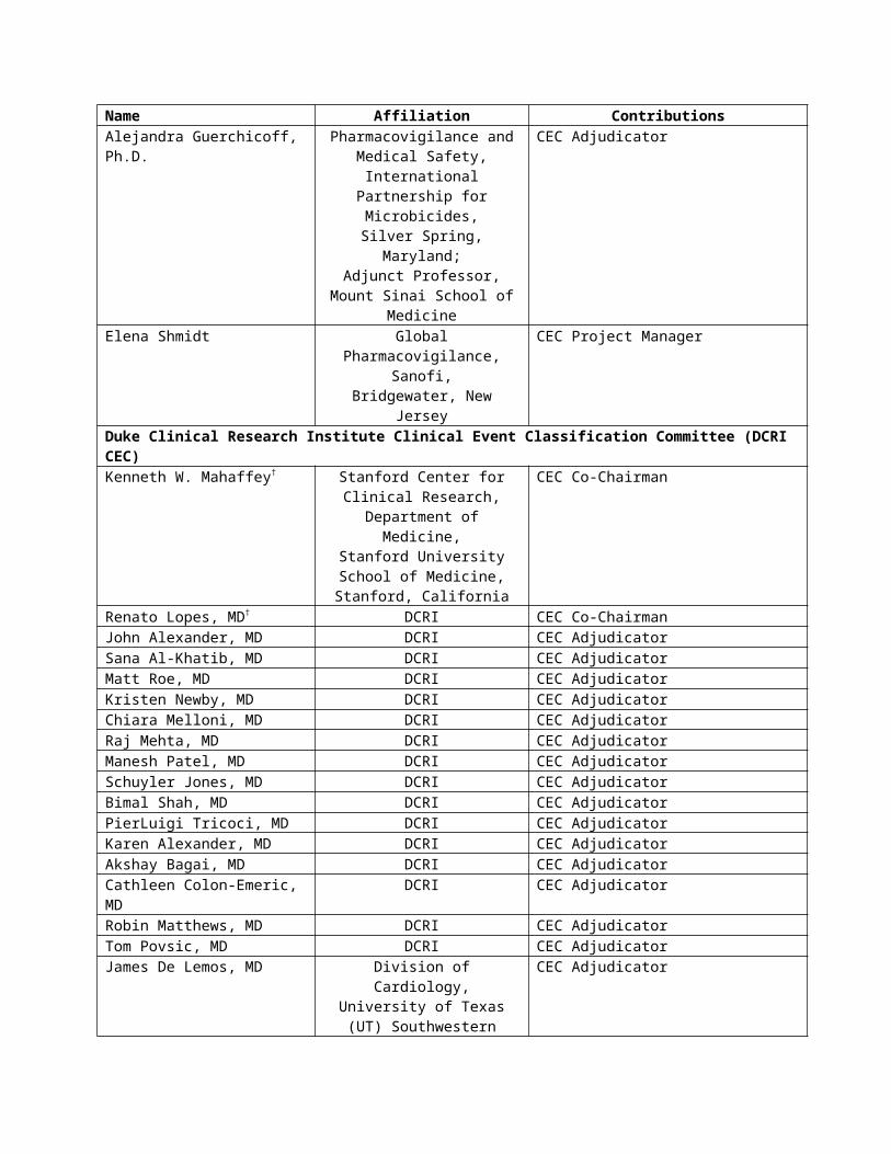
Alejandra Guerchicoff, Ph.D. Pharmacovigilance and Medical Safety,
International Partnership for Microbicides,
Silver Spring, Maryland;Adjunct Professor,
Mount Sinai School of Medicine
CEC Adjudicator
Elena Shmidt Global Pharmacovigilance,Sanofi,
Bridgewater, New Jersey
CEC Project Manager
Duke Clinical Research Institute Clinical Event Classification Committee (DCRI CEC)Kenneth W. Mahaffey† Stanford Center for Clinical
Research,Department of Medicine,
Stanford University School of Medicine,
Stanford, California
CEC Co-Chairman
Renato Lopes, MD† DCRI CEC Co-ChairmanJohn Alexander, MD DCRI CEC AdjudicatorSana Al-Khatib, MD DCRI CEC AdjudicatorMatt Roe, MD DCRI CEC AdjudicatorKristen Newby, MD DCRI CEC AdjudicatorChiara Melloni, MD DCRI CEC AdjudicatorRaj Mehta, MD DCRI CEC AdjudicatorManesh Patel, MD DCRI CEC AdjudicatorSchuyler Jones, MD DCRI CEC AdjudicatorBimal Shah, MD DCRI CEC AdjudicatorPierLuigi Tricoci, MD DCRI CEC AdjudicatorKaren Alexander, MD DCRI CEC AdjudicatorAkshay Bagai, MD DCRI CEC AdjudicatorCathleen Colon-Emeric, MD DCRI CEC AdjudicatorRobin Matthews, MD DCRI CEC AdjudicatorTom Povsic, MD DCRI CEC AdjudicatorJames De Lemos, MD Division of Cardiology,
University of Texas (UT) Southwestern Medical Center,
Dallas, Texas
CEC Adjudicator
Shaun Goodman, MD Canadian Heart Research Centre,Toronto, Ontario, Canada
CEC Adjudicator
Darren McGuire, MD Division of Cardiology,University of Texas (UT)
Southwestern Medical Center,Dallas, Texas
CEC Adjudicator

Name Affiliation ContributionsJohn Petersen, MD Swedish Heart and Vascular
Institute,Seattle, Washington
CEC Adjudicator
Mina Madan, MD Sunnybrook Health Sciences Centre,
Toronto, Ontario, Canada
CEC Adjudicator
Sergio Leonardi, MD Fondazione IRCCS Policlinico San Matteo,Pavia, Italy
CEC Adjudicator
Sunil Rao, MD DCRI CEC AdjudicatorBrad Kolls, MD DCRI (Neurology) CEC AdjudicatorDedrick Jordan, MD Department of Neurology,
University of North Carolina, Chapel Hill, North Carolina
CEC Adjudicator
Adriana Bertolami, MD Brazil Clinical Research Institute,São Paulo, Brazil
CEC Adjudicator
Humberto Moreira, MD Brazil Clinical Research Institute,São Paulo, Brazil
CEC Adjudicator
Luciana Armaganigan, MD Brazil Clinical Research Institute,São Paulo, Brazil
CEC Adjudicator
Pedro Barros Silva Brazil Clinical Research Institute,São Paulo, Brazil
CEC Adjudicator
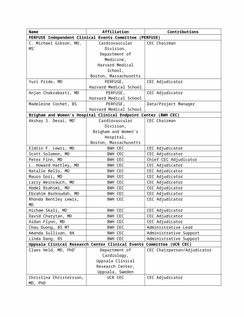
Matthew D. Wilson, RN DCRI CEC OperationsChristine Anderson, RN DCRI CEC OperationsShaun Clifton, BS DCRI CEC OperationsPERFUSE Independent Clinical Events Committee (PERFUSE)C. Michael Gibson, MD, MS† Cardiovascular Division,
Department of Medicine,Harvard Medical School,Boston, Massachusetts
CEC Chairman
Yuri Pride, MD PERFUSE, Harvard Medical School
CEC Adjudicator
Anjan Chakrabarti, MD PERFUSE,Harvard Medical School
CEC Adjudicator
Madeleine Cochet, BS PERFUSE,Harvard Medical School
Data/Project Manager
Brigham and Women’s Hospital Clinical Endpoint Center (BWH CEC)Akshay S. Desai, MD† Cardiovascular Division,
Brigham and Women’s Hospital,Boston, Massachusetts
CEC Chairman
Eldrin F. Lewis, MD BWH CEC CEC AdjudicatorScott Solomon, MD BWH CEC CEC AdjudicatorPeter Finn, MD BWH CEC Chief CEC AdjudicatorL. Howard Hartley, MD BWH CEC CEC AdjudicatorNatalie Bello, MD BWH CEC CEC AdjudicatorMauro Gori, MD BWH CEC CEC AdjudicatorLarry Weinrauch, MD BWH CEC CEC AdjudicatorAbdel Brahimi, MD BWH CEC CEC AdjudicatorEbrahim Barkoudah, MD BWH CEC CEC AdjudicatorRhonda Bentley Lewis, MD BWH CEC CEC AdjudicatorHicham Skali, MD BWH CEC CEC AdjudicatorDavid Charytan, MD BWH CEC CEC AdjudicatorAidan Flynn, MD BWH CEC CEC Adjudicator

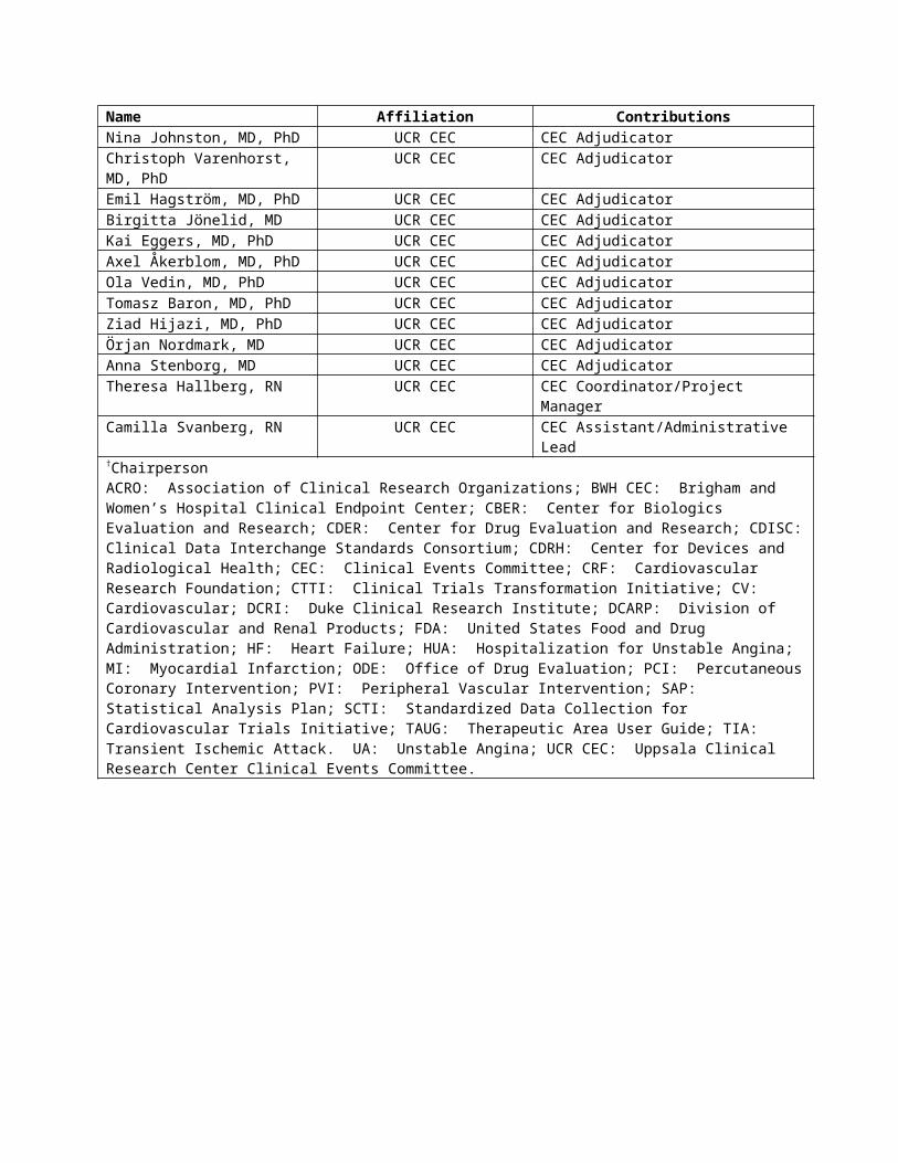
Name Affiliation ContributionsChau Duong, BS MT BWH CEC Administrative LeadAmanda Sullivan, BA BWH CEC Administrative SupportLinda Dang, BS BWH CEC Administrative SupportUppsala Clinical Research Center Clinical Events Committee (UCR CEC)Claes Held, MD, PhD† Department of Cardiology,
Uppsala Clinical Research Center,
Uppsala, Sweden
CEC Chairperson/Adjudicator
Christina Christersson, MD, PhD UCR CEC CEC AdjudicatorNina Johnston, MD, PhD UCR CEC CEC AdjudicatorChristoph Varenhorst, MD, PhD UCR CEC CEC AdjudicatorEmil Hagström, MD, PhD UCR CEC CEC AdjudicatorBirgitta Jönelid, MD UCR CEC CEC AdjudicatorKai Eggers, MD, PhD UCR CEC CEC AdjudicatorAxel Åkerblom, MD, PhD UCR CEC CEC AdjudicatorOla Vedin, MD, PhD UCR CEC CEC AdjudicatorTomasz Baron, MD, PhD UCR CEC CEC AdjudicatorZiad Hijazi, MD, PhD UCR CEC CEC AdjudicatorÖrjan Nordmark, MD UCR CEC CEC AdjudicatorAnna Stenborg, MD UCR CEC CEC AdjudicatorTheresa Hallberg, RN UCR CEC CEC Coordinator/Project ManagerCamilla Svanberg, RN UCR CEC CEC Assistant/Administrative Lead†ChairpersonACRO: Association of Clinical Research Organizations; BWH CEC: Brigham and Women’s Hospital Clinical Endpoint Center; CBER: Center for Biologics Evaluation and Research; CDER: Center for Drug Evaluation and Research; CDISC: Clinical Data Interchange Standards Consortium; CDRH: Center for Devices and Radiological Health; CEC: Clinical Events Committee; CRF: Cardiovascular Research Foundation; CTTI: Clinical Trials Transformation Initiative; CV: Cardiovascular; DCRI: Duke Clinical Research Institute; DCARP: Division of Cardiovascular and Renal Products; FDA: United States Food and Drug Administration; HF: Heart Failure; HUA: Hospitalization for Unstable Angina; MI: Myocardial Infarction; ODE: Office of Drug Evaluation; PCI: Percutaneous Coronary Intervention; PVI: Peripheral Vascular Intervention; SAP: Statistical Analysis Plan; SCTI: Standardized Data Collection for Cardiovascular Trials Initiative; TAUG: Therapeutic Area User Guide; TIA: Transient Ischemic Attack. UA: Unstable Angina; UCR CEC: Uppsala Clinical Research Center Clinical Events Committee.
Appendix 2. Introduction
The purpose of this document is to provide a framework of definitions for cardiovascular (CV) and stroke outcomes in clinical trials. These definitions are based on clinical and research expertise, published guidelines and definitions, and our current understanding of the specific laboratory tests, diagnostic tests, and imaging techniques used in clinical practice to diagnose these events.
It is recognized that definitions of CV and stroke outcomes may change over time, as new biomarkers or other diagnostic tests become available, or as standards evolve and perceptions of clinical importance become modified.
Endpoint definitions are necessary in clinical trials so that events are clearly characterized by objective criteria and reported uniformly. However, some events may be complex and may not neatly fulfill the specified criteria. Furthermore, within a large-scale, multicenter, international study, some results may not be available because they were never measured by the physician responsible for their care at the time, because the test was not available locally, or because the results can no longer be found. In all cases, clinical judgment should be used to determine the most likely cause of an event. Where the person performing the adjudication of an event is blind to the treatment allocation, any errors will be random, rather than systematic. As a consequence, any noise introduced by slight misclassifications of events will not bias the result towards one arm or another, but may mask a true difference in effectiveness or safety or increase the chance of concluding non-inferiority.
Advances in database technologies and statistical methodologies have created opportunities to aggregate large trial datasets. If uniformly defined, events in drug development programs or among different clinical trials may be analyzed more easily and trends and other safety signals may be identified. More consistent definitions could improve the ability to estimate event rates in a contemplated clinical trial.
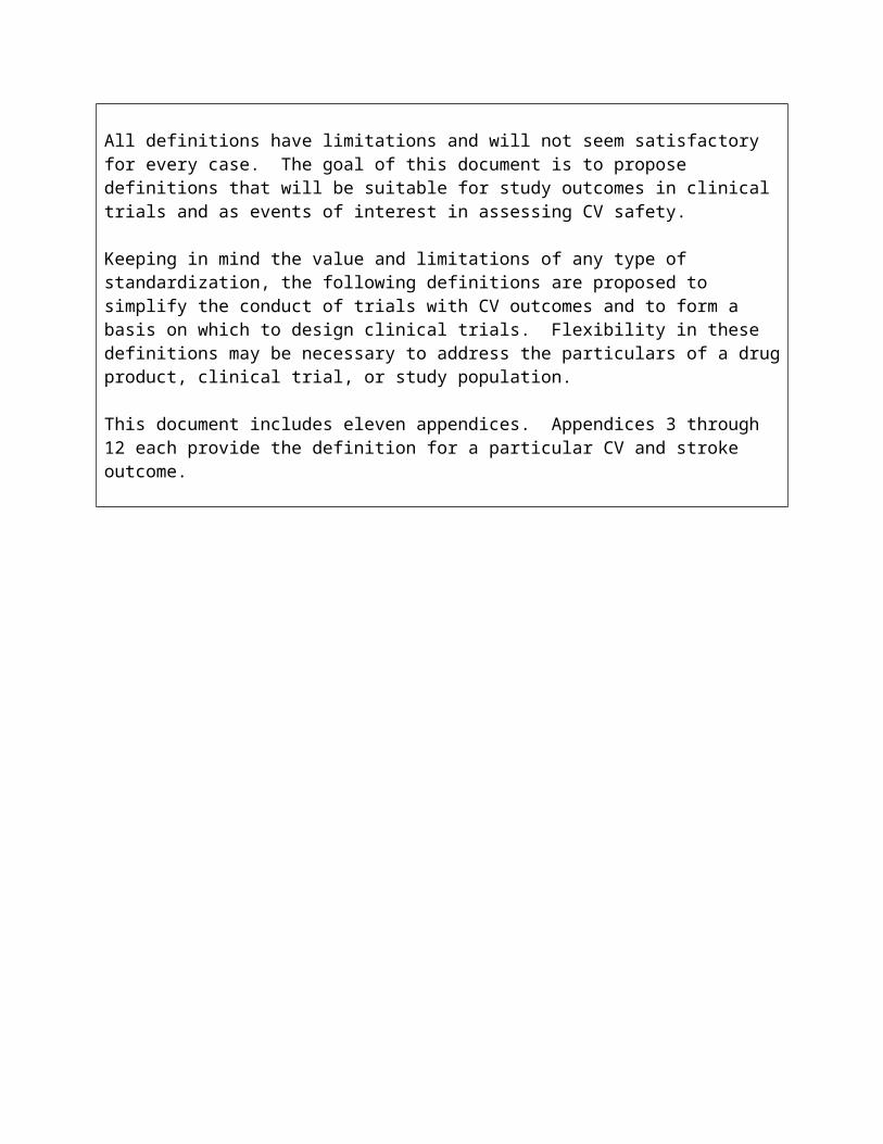
All definitions have limitations and will not seem satisfactory for every case. The goal of this document is to propose definitions that will be suitable for study outcomes in clinical trials and as events of interest in assessing CV safety.
Keeping in mind the value and limitations of any type of standardization, the following definitions are proposed to simplify the conduct of trials with CV outcomes and to form a basis on which to design clinical trials. Flexibility in these definitions may be necessary to address the particulars of a drug product, clinical trial, or study population.
This document includes eleven appendices. Appendices 3 through 12 each provide the definition for a particular CV and stroke outcome.

Appendix 3. Definition of Cardiovascular Death
Cardiovascular death includes death resulting from an acute myocardial infarction (MI), sudden cardiac death, death due to heart failure (HF), death due to stroke, death due to cardiovascular (CV) procedures, death due to CV hemorrhage, and death due to other CV causes.
Classifying CV mortality more specifically (MI, sudden death etc.) is usually not needed for outcome trials. Moreover, such classification is difficult because the classifications refer both to underlying cause (e.g., acute MI, which can cause fatal arrhythmias or HF) and to mode of death (sudden/arrhythmic; HF, which can result from an MI or worsening HF), and they overlap substantially. The following definitions can, however, be used if desired.
1. Death due to Acute Myocardial Infarction refers to a death by any CV mechanism (e.g., arrhythmia, sudden death, HF, stroke, pulmonary embolus, peripheral arterial disease) ≤ 30 days1 after a MI, related to the immediate consequences of the MI, such as progressive HF or recalcitrant arrhythmia. We note that there may be assessable mechanisms of CV death during this time period, but for simplicity, if the CV death occurs ≤ 30 days of the MI, it will be considered a death due to MI.
Acute MI should be verified to the extent possible by the diagnostic criteria outlined for acute MI (see Appendix 6) or by autopsy findings showing recent MI or recent coronary thrombosis.
Death resulting from a procedure to treat a MI (percutaneous coronary intervention (PCI), coronary artery bypass graft surgery (CABG)), or to treat a complication resulting from MI, should also be considered death due to acute MI.
Death resulting from an elective coronary procedure to treat myocardial ischemia (i.e., chronic stable angina) or death due to a MI that occurs as a direct consequence of a CV investigation/procedure/operation should be considered as a death due to a CV procedure.
2. Sudden Cardiac Death refers to a death that occurs unexpectedly and not within 30 days of an acute MI. Sudden cardiac death includes the following scenarios:
a. Death witnessed and occurring without new or worsening symptoms
b. Death witnessed within 60 minutes of the onset of new or worsening cardiac symptoms, unless the symptoms suggest acute MI
c. Death witnessed and attributed to an identified arrhythmia (e.g., captured on an electrocardiographic (ECG) recording, witnessed on a monitor, or unwitnessed but found on implantable cardioverter-defibrillator review)
1The 30 day cut-off is arbitrary.
d. Death after unsuccessful resuscitation from cardiac arrest (e.g., implantable cardioverter defibrillator (ICD) unresponsive sudden cardiac death, pulseless electrical activity arrest)
e. Death after successful resuscitation from cardiac arrest and without identification of a specific cardiac or non-cardiac etiology
f. Unwitnessed death in a subject seen alive and clinically stable ≤ 24 hours prior to being found dead without any evidence supporting a specific non-cardiovascular cause of death (information regarding the patient’s clinical status preceding death should be provided, if available)
General Considerations
o Unless additional information suggests an alternate specific cause of death (e.g., Death due to Other Cardiovascular Causes), if a patient is seen alive ≤ 24 hours of being found dead, sudden cardiac death (criterion 2f) should be recorded. For patients who were not observed alive within 24 hours of death, undetermined cause of death should be recorded (e.g., a subject found dead in bed, but who had not been seen by family for > 24 hours).
3. Death due to Heart Failure refers to a death in association with clinically worsening symptoms and/or signs of HF regardless of HF etiology (see Appendix 9). Deaths due to HF can have various etiologies, including single or recurrent MIs, ischemic or non-ischemic cardiomyopathy, hypertension, or valvular disease.
4. Death due to Stroke refers to death after a stroke that is either a direct consequence of the stroke or a complication of the stroke. Acute stroke should be verified to the extent possible by the diagnostic criteria outlined for stroke (see Appendix 8).
5. Death due to Cardiovascular Procedures refers to death caused by the immediate complications of a cardiac procedure.
6. Death due to Cardiovascular Hemorrhage refers to death related to hemorrhage such as a non-stroke intracranial hemorrhage (e.g., subdural hematoma) (see Appendix 8), non-procedural or non-traumatic vascular rupture (e.g., aortic aneurysm), or hemorrhage causing cardiac tamponade.
7. Death due to Other Cardiovascular Causes refers to a CV death not included in the above categories but with a specific, known cause (e.g., pulmonary embolism or peripheral arterial disease).
Appendix 4. Definition of Non-Cardiovascular Death
Non-cardiovascular death is defined as any death with a specific cause that is not thought to be CV in nature, as listed in Appendix 3. Detailed recommendations on the classification of non-CV causes of death are beyond the scope of this document. The level of detail required and the optimum classification will depend on the nature of the study population and the anticipated number and type of non-CV deaths. Any specific anticipated safety concern should be included as a separate cause of death. The following is a suggested list of non-CV causes of death:
Pulmonary Renal Gastrointestinal Hepatobiliary Pancreatic Infection (includes sepsis) Inflammatory (e.g., Systemic Inflammatory Response Syndrome (SIRS) / Immune
(including autoimmune) (may include anaphylaxis from environmental (e.g., food allergies))
Hemorrhage that is neither CV bleeding or a stroke (see Appendix 3, Section 6, and Appendix 8)
Non-CV procedure or surgery Trauma (includes homicide) Suicide Non-prescription drug reaction or overdose Prescription drug reaction or overdose (may include anaphylaxis) Neurological (non-CV) (excludes CV death from ischemic stroke, hemorrhagic stroke, or
undetermined cause of stroke or CV hemorrhage of central nervous system) Malignancy (e.g., leukemia, lymphoma, or other malignancy) Other non-CV, specify: _________________

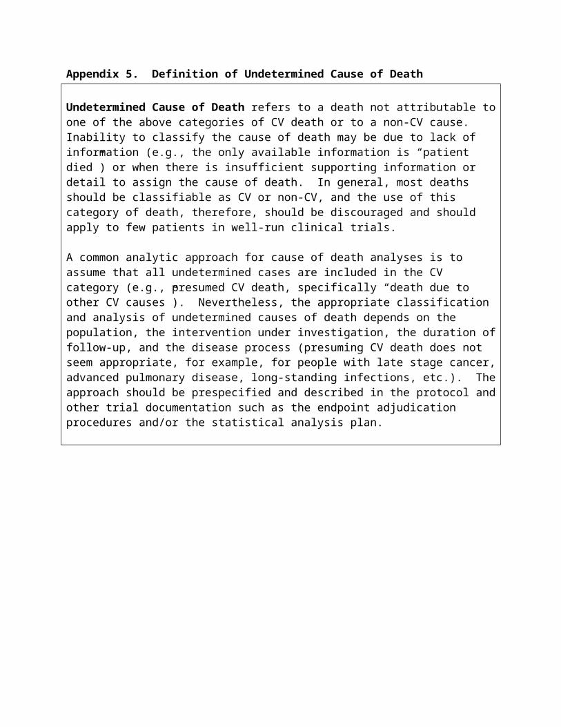
Appendix 5. Definition of Undetermined Cause of Death
Undetermined Cause of Death refers to a death not attributable to one of the above categories of CV death or to a non-CV cause. Inability to classify the cause of death may be due to lack of information (e.g., the only available information is “patient died”) or when there is insufficient supporting information or detail to assign the cause of death. In general, most deaths should be classifiable as CV or non-CV, and the use of this category of death, therefore, should be discouraged and should apply to few patients in well-run clinical trials.
A common analytic approach for cause of death analyses is to assume that all undetermined cases are included in the CV category (e.g., presumed CV death, specifically “death due to other CV causes”). Nevertheless, the appropriate classification and analysis of undetermined causes of death depends on the population, the intervention under investigation, the duration of follow-up, and the disease process (presuming CV death does not seem appropriate, for example, for people with late stage cancer, advanced pulmonary disease, long-standing infections, etc.). The approach should be prespecified and described in the protocol and other trial documentation such as the endpoint adjudication procedures and/or the statistical analysis plan.
Appendix 6. Definition of Myocardial Infarction
Please also see the 2012 Third Universal Definition of Myocardial Infarction.
1. General Considerations
The term myocardial infarction (MI) should be used when there is evidence of myocardial necrosis in a clinical setting consistent with myocardial ischemia.
In general, the diagnosis of MI requires the combination of: Evidence of myocardial necrosis (either changes in cardiac biomarkers or post-
mortem pathological findings); and Supporting information derived from the clinical presentation, electrocardiographic
changes, or the results of myocardial or coronary artery imaging
The totality of the clinical, electrocardiographic, and cardiac biomarker information should be considered to determine whether or not a MI has occurred. Specifically, timing and trends in cardiac biomarkers and electrocardiographic information require careful analysis. The adjudication of MI should also take into account the clinical setting in which the event occurs. MI may be adjudicated for an event that has characteristics of a MI but which does not meet the strict definition because biomarker or electrocardiographic results are not available.
2. Criteria for Myocardial Infarction
a. Clinical PresentationThe clinical presentation should be consistent with diagnosis of myocardial ischemia and infarction. Other findings that might support the diagnosis of MI should be taken into account because a number of conditions are associated with chronic or transient elevations in cardiac biomarkers (e.g., trauma, surgery, pacing, ablation, HF, hypertrophic cardiomyopathy, pulmonary embolism, severe pulmonary hypertension, stroke or subarachnoid hemorrhage, infiltrative and inflammatory disorders of cardiac muscle, drug toxicity, burns, critical illness, extreme exertion, and chronic kidney disease). Supporting information can also be considered from myocardial imaging and coronary imaging. The totality of the data may help differentiate acute MI from the background disease process.
b. Biomarker ElevationsFor cardiac biomarkers, the 99th percentile of the upper reference limit (URL) should be used. The URL is determined by the manufacturer of the assay and can be determined by an individual hospital that has studied a normal reference population. Because hospitals may use different URLs for the identical assay without studying a normal reference population and for reasons not evident to a Clinical Events Committee (CEC), variability in MI reporting due to the URL for specific assays may be reduced by using the manufacturer’s recommended 99th percentile or a core biochemistry laboratory. An

alternate approach would be to use the local laboratory value, but this may be less satisfactory if the local laboratory did not study a normal reference population.
Cardiac troponins are the preferred biomarker to diagnose MI. If cTn values are not available, then CK-MB mass should be used.
For types 4a and 5 MI events, different biomarker elevations for cTn and CK-MB are required. The specific criteria will be referenced to the URL.
In many studies, particularly those in which patients present acutely to hospitals which are not participating sites, it is not practical to stipulate the use of a single biomarker or assay, and the locally available results will need to be used as the basis for adjudication unless the assay type can be determined. However, if possible, using the same cardiac biomarker assay and preferably, a core laboratory, for all measurements reduces inter-assay variability.
Since the prognostic significance of different types of myocardial infarctions (e.g., periprocedural MI versus spontaneous MI) may be different, MI subtypes should be reported separately, in addition to total number of MI events. Reporting the ratio of peak biomarker value observed/URL should be considered to provide a rough estimate of MI size.
c. Electrocardiogram (ECG) ChangesElectrocardiographic changes can be used to support or confirm a MI. Supporting evidence may be ischemic changes and confirmatory information may be new Q waves.
ECG manifestations of acute myocardial ischemia (in absence of left ventricular hypertrophy (LVH) and left bundle branch block (LBBB)):
o ST elevationNew ST elevation at the J point in two contiguous leads with the cut-points: ≥ 0.1 mV in all leads other than leads V2-V3 where the following cut-points apply: ≥ 0.2 mV in men ≥ 40 years (≥ 0.25 mV in men < 40 years) or ≥ 0.15 mV in women.
o ST depression and T-wave changesNew horizontal or down-sloping ST depression ≥ 0.05 mV in two contiguous leads and/or new T inversion ≥ 0.1 mV in two contiguous leads with prominent R wave or R/S ratio > 1.
The above ECG criteria illustrate patterns consistent with myocardial ischemia. In patients with abnormal biomarkers, it is recognized that lesser ECG abnormalities may represent an ischemic response and may be accepted under the category of abnormal ECG findings.

Criteria for pathological Q-wave
o Any Q-wave in leads V2-V3 ≥ 0.02 seconds or QS complex in leads V2 and V3o Q-wave ≥ 0.03 seconds and ≥ 0.1 mV deep or QS complex in leads I, II, aVL, aVF, or V4-V6
in any two leads of a contiguous lead grouping (I, aVL; V1-V6; II, III, and aVF)a
aThe same criteria are used for supplemental leads V7-V9, and for the Cabrera frontal plane lead grouping.
ECG changes associated with prior myocardial infarction
o Pathological Q-waves, as defined aboveo R-wave ≥ 0.04 seconds in V1-V2 and R/S ≥ 1 with a concordant positive T-wave
in the absence of a conduction defect
Criteria for prior myocardial infarction (e.g., silent MI)
Any one of the following criteria meets the diagnosis for prior MI:o Pathological Q waves with or without symptoms in the absence of non-ischemic
causeso Imaging evidence of a region of loss of viable myocardium that is thinned and
fails to contract, in the absence of a non-ischemic causeo Pathological findings of a prior MI

Appendix 7. Definition of Hospitalization for Unstable Angina
Unstable angina requiring hospitalization is defined as
1. Ischemic discomfort (angina, or symptoms thought to be equivalent) ≥ 10 minutes in duration occurring
at rest, or in an accelerating pattern with frequent episodes associated with progressively
decreased exercise capacity.
AND
2. Prompting an unscheduled hospitalization within 24 hours of the most recent symptoms. Hospitalization is defined as an admission to an inpatient unit or a visit to an emergency department that results in at least a 24 hour stay (or a change in calendar date if the hospital admission or discharge times are not available).
AND
3. At least one of the following:
a. New or worsening ST or T wave changes on resting ECG (in the absence of confounders, such as LBBB or LVH)
Transient ST elevation (duration < 20 minutes)New ST elevation at the J point in two contiguous leads with the cut-points: ≥ 0.1 mV in all leads other than leads V2-V3 where the following cut-points apply: ≥ 0.2 mV in men ≥ 40 years (≥ 0.25 mV in men < 40 years) or ≥ 0.15 mV in women.
ST depression and T-wave changesNew horizontal or down-sloping ST depression ≥ 0.05 mV in two contiguous leads and/or new T-wave inversion ≥ 0.3 mV in two contiguous leads with prominent R wave or R/S ratio > 1.
b. Definite evidence of inducible myocardial ischemia as demonstrated by: an early positive exercise stress test, defined as ST elevation or ≥ 2 mm ST
depression prior to 5 metsOR stress echocardiography (reversible wall motion abnormality) OR myocardial scintigraphy (reversible perfusion defect), OR MRI (myocardial perfusion deficit under pharmacologic stress).
and believed to be responsible for the myocardial ischemic symptoms/signs.

c. Angiographic evidence of new or worse ≥ 70% lesion (≥ 50% for left main lesion) and/or thrombus in an epicardial coronary artery that is believed to be responsible for the myocardial ischemic symptoms/signs.
d. Need for coronary revascularization procedure (PCI or CABG) for the presumed culprit lesion(s), as defined in 3c. This criterion would be fulfilled if revascularization was undertaken during the unscheduled hospitalization, or subsequent to transfer to another institution without interceding home discharge.
AND
4. Negative cardiac biomarkers and no evidence of acute MI
General Considerations
1. Escalation of pharmacotherapy for ischemia, such as intravenous nitrates or increasing dosages of β-blockers, should be considered supportive but not diagnostic of unstable angina. However, a typical presentation and admission to the hospital with escalation of pharmacotherapy, without any of the additional findings listed under category 3, would be insufficient to support classification as hospitalization for unstable angina.
2. If subjects are admitted with suspected unstable angina, and subsequent testing reveals a non-cardiac or non-ischemic etiology, this event should not be recorded as hospitalization for unstable angina. Potential ischemic events meeting the criteria for MI should not be adjudicated as unstable angina.
3. Planned hospitalization or rehospitalization for performance of an elective revascularization in patients who do not fulfill the criteria for unstable angina should not be considered a hospitalization for unstable angina. For example,
Hospitalization of a patient with stable exertional angina for coronary angiography and PCI that is prompted by a positive outpatient stress test should not be considered hospitalization for unstable angina.
Rehospitalization of a patient meeting the criteria for unstable angina who was stabilized, discharged, and subsequently readmitted for revascularization, does not constitute a second hospitalization for unstable angina.
4. A patient who undergoes an elective catheterization where incidental coronary artery disease is found and who subsequently undergoes coronary revascularization will not be considered as meeting the hospitalization for unstable angina endpoint.

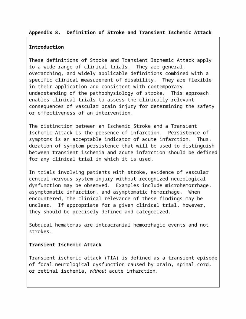
Appendix 8. Definition of Stroke and Transient Ischemic Attack
Introduction
These definitions of Stroke and Transient Ischemic Attack apply to a wide range of clinical trials. They are general, overarching, and widely applicable definitions combined with a specific clinical measurement of disability. They are flexible in their application and consistent with contemporary understanding of the pathophysiology of stroke. This approach enables clinical trials to assess the clinically relevant consequences of vascular brain injury for determining the safety or effectiveness of an intervention.
The distinction between an Ischemic Stroke and a Transient Ischemic Attack is the presence of infarction. Persistence of symptoms is an acceptable indicator of acute infarction. Thus, duration of symptom persistence that will be used to distinguish between transient ischemia and acute infarction should be defined for any clinical trial in which it is used.
In trials involving patients with stroke, evidence of vascular central nervous system injury without recognized neurological dysfunction may be observed. Examples include microhemorrhage, asymptomatic infarction, and asymptomatic hemorrhage. When encountered, the clinical relevance of these findings may be unclear. If appropriate for a given clinical trial, however, they should be precisely defined and categorized.
Subdural hematomas are intracranial hemorrhagic events and not strokes.
Transient Ischemic Attack
Transient ischemic attack (TIA) is defined as a transient episode of focal neurological dysfunction caused by brain, spinal cord, or retinal ischemia, without acute infarction.
Stroke
Stroke is defined as an acute episode of focal or global neurological dysfunction caused by brain, spinal cord, or retinal vascular injury as a result of hemorrhage or infarction.
Classification:
A. Ischemic Stroke
Ischemic stroke is defined as an acute episode of focal cerebral, spinal, or retinal dysfunction caused by infarction of central nervous system tissue.
Hemorrhage may be a consequence of ischemic stroke. In this situation, the stroke is an ischemic stroke with hemorrhagic transformation and not a hemorrhagic stroke.
B. Hemorrhagic Stroke
Hemorrhagic stroke is defined as an acute episode of focal or global cerebral or spinal dysfunction caused by intraparenchymal, intraventricular, or subarachnoid hemorrhage.
C. Undetermined Stroke
Undetermined stroke is defined as an acute episode of focal or global neurological dysfunction caused by presumed brain, spinal cord, or retinal vascular injury as a result of hemorrhage or infarction but with insufficient information to allow categorization as either ischemic or hemorrhagic.
Stroke Disability
Disability should be measured by a reliable and valid scale in all cases, typically at each visit and 90 days after the event. For example, the modified Rankin Scale may be used to address this requirement:
Scale
Disability
0 No symptoms at all1 No significant disability despite symptoms; able to carry out all usual duties and
activities2 Slight disability; unable to carry out all previous activities, but able to look after own
affairs without assistance3 Moderate disability; requiring some help, but able to walk without assistance4 Moderately severe disability; unable to walk without assistance and unable to attend to
own bodily needs without assistance5 Severe disability; bedridden, incontinent and requiring constant nursing care and
attention6 Dead

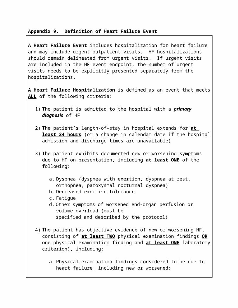
Appendix 9. Definition of Heart Failure Event
A Heart Failure Event includes hospitalization for heart failure and may include urgent outpatient visits. HF hospitalizations should remain delineated from urgent visits. If urgent visits are included in the HF event endpoint, the number of urgent visits needs to be explicitly presented separately from the hospitalizations.
A Heart Failure Hospitalization is defined as an event that meets ALL of the following criteria:
1) The patient is admitted to the hospital with a primary diagnosis of HF
2) The patient’s length-of-stay in hospital extends for at least 24 hours (or a change in calendar date if the hospital admission and discharge times are unavailable)
3) The patient exhibits documented new or worsening symptoms due to HF on presentation, including at least ONE of the following:
a. Dyspnea (dyspnea with exertion, dyspnea at rest, orthopnea, paroxysmal nocturnal dyspnea)
b. Decreased exercise tolerancec. Fatigued. Other symptoms of worsened end-organ perfusion or volume overload (must be
specified and described by the protocol)
4) The patient has objective evidence of new or worsening HF, consisting of at least TWO physical examination findings OR one physical examination finding and at least ONE laboratory criterion), including:
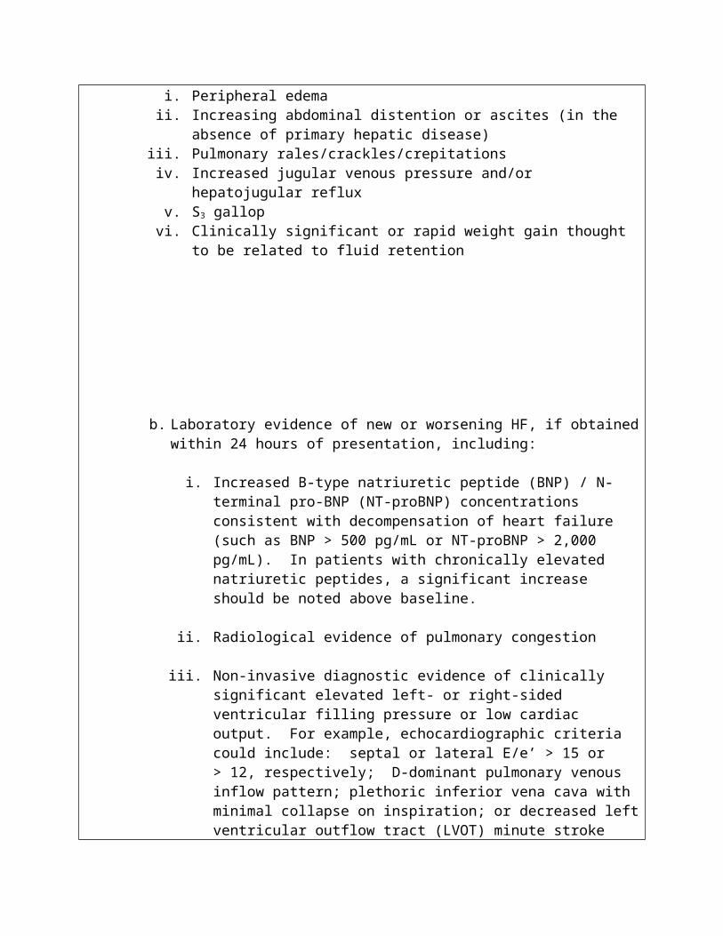
a. Physical examination findings considered to be due to heart failure, including new or worsened:
i. Peripheral edemaii. Increasing abdominal distention or ascites (in the absence of primary hepatic
disease)iii. Pulmonary rales/crackles/crepitationsiv. Increased jugular venous pressure and/or hepatojugular refluxv. S3 gallop
vi. Clinically significant or rapid weight gain thought to be related to fluid retention

b. Laboratory evidence of new or worsening HF, if obtained within 24 hours of presentation, including:
i. Increased B-type natriuretic peptide (BNP) / N-terminal pro-BNP (NT-proBNP) concentrations consistent with decompensation of heart failure (such as BNP > 500 pg/mL or NT-proBNP > 2,000 pg/mL). In patients with chronically elevated natriuretic peptides, a significant increase should be noted above baseline.
ii. Radiological evidence of pulmonary congestion
iii. Non-invasive diagnostic evidence of clinically significant elevated left- or right-sided ventricular filling pressure or low cardiac output. For example, echocardiographic criteria could include: septal or lateral E/e’ > 15 or > 12, respectively; D-dominant pulmonary venous inflow pattern; plethoric inferior vena cava with minimal collapse on inspiration; or decreased left ventricular outflow tract (LVOT) minute stroke distance (time velocity integral [TVI])
OR
iv. Invasive diagnostic evidence with right heart catheterization showing a pulmonary capillary wedge pressure (pulmonary artery occlusion pressure) ≥ 18 mmHg, central venous pressure ≥ 12 mmHg, or a cardiac index < 2.2 L/min/m2
Note: All results from diagnostic tests should be reported, if available, even if they do not meet the above criteria, because they provide important information for the adjudication of these events.
5) The patient receives at least ONE of the following treatments specifically for HF:
a. Significant augmentation in oral diuretic therapy (e.g., doubling of loop diuretic dose, initiation of maintenance loop diuretic therapy, initiation of combination diuretic therapy)
b. Initiation of intravenous diuretic (even a single dose) or vasoactive agent (e.g., inotrope, vasopressor, vasodilator)
c. Mechanical or surgical intervention, including:i. Mechanical circulatory support (e.g., intra-aortic balloon pump,
ventricular assist device, extracorporeal membrane oxygenation, total artificial heart)
ii. Mechanical fluid removal (e.g., ultrafiltration, hemofiltration, dialysis)

General Considerations (HF Hospitalization)
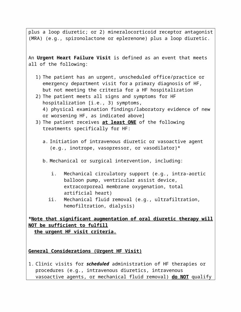
Combination diuretic therapy could include 1) a thiazide-type diuretic (e.g., hydrochlorothiazide, metolazone, chlorothiazide) plus a loop diuretic; or 2) mineralocorticoid receptor antagonist (MRA) (e.g., spironolactone or eplerenone) plus a loop diuretic.
An Urgent Heart Failure Visit is defined as an event that meets all of the following:
1) The patient has an urgent, unscheduled office/practice or emergency department visit for
a primary diagnosis of HF, but not meeting the criteria for a HF hospitalization2) The patient meets all signs and symptoms for HF hospitalization [i.e., 3) symptoms,
4) physical examination findings/laboratory evidence of new or worsening HF, as indicated above]
3) The patient receives at least ONE of the following treatments specifically for HF:
a. Initiation of intravenous diuretic or vasoactive agent (e.g., inotrope, vasopressor, or vasodilator)*
b. Mechanical or surgical intervention, including:
i. Mechanical circulatory support (e.g., intra-aortic balloon pump, ventricular assist device, extracorporeal membrane oxygenation, total artificial heart)
ii. Mechanical fluid removal (e.g., ultrafiltration, hemofiltration, dialysis)
*Note that significant augmentation of oral diuretic therapy will NOT be sufficient to fulfill the urgent HF visit criteria.
General Considerations (Urgent HF Visit)
1. Clinic visits for scheduled administration of HF therapies or procedures (e.g., intravenous diuretics, intravenous vasoactive agents, or mechanical fluid removal) do NOT qualify as non-hospitalized HF events.

Appendix 10. Interventional Cardiology Definitions

A. Clinical Definitions
1. Clinically-Driven Target Lesion Revascularization : Revascularization is clinically-driven if the target lesion diameter stenosis is > 50% by quantitative coronary angiography (QCA) and the subject has clinical or functional ischemia which cannot be explained by another native coronary or bypass graft lesion. Clinical or functional ischemia includes any of the following:
a. A history of angina pectoris, presumably related to the target vesselb. Objective signs of ischemia at rest (electrocardiographic changes) or during exercise
test (or equivalent), presumably related to the target vesselc. Abnormal results of any invasive functional diagnostic test [e.g., coronary flow
reserve (CFR) or fractional flow reserve (FFR)]
Comment: Target lesion revascularization (TLR) of a > 70% diameter stenosis by QCA in the absence of the above signs or symptoms may be considered clinically-driven.
Comment: In the absence of QCA data or if a < 50% stenosis is present, TLR may be considered clinically-driven by the Clinical Events Committee (CEC) if severe ischemic signs and symptoms attributed to the target lesion are present.
2. Non-Target Lesion and Non-Target Lesion Revascularization : A lesion for which revascularization is not attempted or one in which revascularization is performed using a non-study device, respectively.
3. Non-Target Vessel and Non-Target Vessel Revascularization : A vessel for which revascularization is not attempted or one in which revascularization is performed using a non-study device, respectively.
4. Percutaneous Coronary Intervention (PCI) Status :
a. Elective: The procedure can be performed on an outpatient basis or during a subsequent hospitalization without significant risk of myocardial infarction (MI) or death. For stable in-patients, the procedure is being performed during this hospitalization for convenience and ease of scheduling and NOT because the patient's clinical situation demands the procedure prior to discharge.
b. Urgent: The procedure should be performed on an inpatient basis and prior to discharge because of significant concerns that there is risk of myocardial ischemia, MI, and/or death. Patients who are outpatients or in the emergency department at the time that the cardiac catheterization is requested would warrant hospital admission based on their clinical presentation.

c. Emergency: The procedure should be performed as soon as possible because of substantial concerns that ongoing myocardial ischemia and/or MI could lead to death. "As soon as possible" refers to a patient who is of sufficient acuity that one would cancel a scheduled case to perform this procedure immediately in the next available room during business hours, or one would activate the on-call team were this to occur during off-hours.
d. Salvage: The procedure is a last resort. The patient is in cardiogenic shock when the PCI begins (i.e., the time at which the first guide wire or intracoronary device is introduced into a coronary artery or bypass graft for the purpose of mechanical revascularization) OR within the last ten minutes prior to the start of the case or during the diagnostic portion of the case, the patient has also received chest compressions or has been on unanticipated circulatory support (e.g., intra-aortic balloon pump, extracorporeal membrane oxygenation, or cardiopulmonary support).
5. Percutaneous Coronary Intervention (PCI): Placement of an angioplasty guide wire, balloon, or other device (e.g., stent, atherectomy catheter, brachytherapy delivery device, or thrombectomy catheter) into a native coronary artery or coronary artery bypass graft for the purpose of mechanical coronary revascularization. In the assessment of the severity of coronary lesions with the use of intravascular ultrasound, coronary flow reserve (CFR), or fractional flow reserve (FFR), insertion of a guide wire will NOT be considered PCI.
6. Procedural Success : Achievement of < 30 % residual diameter stenosis of the target lesion assessed by visual inspection or QCA and no in-hospital major adverse cardiac events (MACE, a composite of death, MI, or repeat coronary revascularization of the target lesion). Ideally, the assessment of the residual stenosis at the end of the procedure should be performed by an angiographic core laboratory.
Comment: For some device interventions (e.g., balloon angioplasty), achievement of < 50% diameter stenosis by visual inspection or QCA is an acceptable definition for procedural success.
7. Target Lesion : Any lesion treated or attempted to be treated during the PCI with the study device. The target lesion includes the arterial segment treated with the study device (stent, in most cases) plus 5 mm proximal and 5 mm distal to the treatment site.
8. Target Lesion Failure (TLF): The composite of ischemia-driven revascularization of the target lesion, MI related to the target vessel, or cardiac death related to the target vessel. If it cannot be determined with certainty whether the MI or death was related to the target vessel, it is considered a TLF.

9. Target Lesion Revascularization (TLR): Any repeat percutaneous intervention of the target lesion (including 5 mm proximal and 5 mm distal to the target lesion) or surgical bypass of the target vessel performed for restenosis or other complication involving the target lesion. In the assessment of TLR, angiograms should be assessed by an angiographic core laboratory (if designated) and made available to the CEC for review upon request.
10. Target Vessel : A major native coronary artery (e.g., left main coronary artery, left anterior descending coronary artery, left circumflex coronary artery, or right coronary artery) or bypass graft containing the target lesion. A native coronary artery target vessel includes the arterial segments upstream and downstream to the target lesion plus major side branches.
11. Target Vessel Failure (TVF): The composite of ischemia-driven revascularization of the target vessel, MI related to the target vessel, or cardiac death related to the target vessel. If it cannot be determined with certainty whether the MI or death was related to the target vessel, it is considered a TVF.
12. Target Vessel, Non-Target Lesion, and Target Vessel, Non-Target Lesion Revascularization: Any lesion or revascularization of a lesion in the target vessel other than the target lesion, respectively.
13. Target Vessel Revascularization (TVR): Any repeat percutaneous intervention or surgical bypass of any segment of the target vessel. In the assessment of TVR, angiograms should be assessed by an angiographic core laboratory (if designated) and made available to the CEC for review upon request.
14. Vascular Complications : Access site hematoma: Development of a new, localized collection of blood at a
vascular access site sufficient to produce a palpable mass within 72 hours of a procedure.
Arteriovenous fistula: Development of a new, unintended communication between an artery and a vein occurring at a vascular access site within 72 hours of a procedure.
Peripheral ischemia: Development of new arterial insufficiency sufficient to produce clinical signs or symptoms of ischemia (pallor, pain, paresthesia) distal to a vascular access site within 72 hours of a procedure.
Peripheral nerve injury: Development of new sensory or motor loss of peripheral nerve function from external nerve compression (e.g., as a result of positioning during a procedure), or internal compression or direct nerve damage from the procedure, occurring within 72 hours of a procedure.
Pseudoaneurysm: Development of a new localized collection of blood with a persistent communication (neck) originating at a vascular access site and occurring within 72 hours of a procedure.
Retroperitoneal hemorrhage: Development of new bleeding into the retroperitoneal space originating at a vascular access site and occurring within 72hours of a procedure.
B. Angiographic Definitions
1. Abrupt Closure : New intra-procedural severely reduced flow (TIMI grade 0-1) within the target vessel that persists and requires intervention by stenting or other treatment, or results in MI or death. Abrupt closure requires an association with a vascular dissection, thrombus, or severe spasm at the treatment site or within the instrumented vessel.
2. Coronary Lesions Treated
Coronary Artery Segments Definition
Right coronary artery ostium Origin of the right coronary artery, including the first 3 mm of the artery
Proximal right coronary artery
Proximal portion of the right coronary artery, from the ostium of the right coronary artery to the origin of the first right ventricular branch (pRCA)
Mid right coronary arteryMiddle portion of the right coronary artery, from the origin of the first right ventricular branch to the acute margin (mRCA)
Distal right coronary arteryDistal portion of the right coronary artery, from the acute margin to the origin of the posterior descending artery (dRCA)
Right posterior descending artery
In right dominant and mixed circulations, the vessel that runs in the posterior interventricular groove and supplies septal perforator branches (PDA)
Posterolateral segmental artery
In right dominant circulations, the distal continuation of the right coronary artery in the posterior atrioventricular groove after the origin of the right posterior descending artery (PLSA)
First right posterolateral branchIn right dominant circulations, the first posterolateral branch originating from the right posterior atrioventricular artery (RPL1)
Second right posterolateral branchIn right dominant circulations, the second posterolateral branch originating from the right posterior atrioventricular artery (RPL2)
Third right posterolateral branchIn right dominant circulations, the third posterolateral branch originating from the right posterior atrioventricular artery (RPL3)
Posterior descending septal perforator Septal perforator vessel originating from the posterior descending artery

Coronary Artery Segments Definition
Right ventricular branch Branch arising from the right coronary artery to supply the right ventricular wall (RV)
Left main coronary artery ostium Origin of the left coronary artery, including the first 3 mm of the artery
Left main coronary artery body Body of the left main coronary artery, from the ostium to the bifurcation (LM)
Left main coronary artery bifurcation
Distal end of the left main, including the terminal 3 mm through the bifurcation of the left main into the left anterior descending and left circumflex arteries
Left anterior descending artery ostium
Origin of the left anterior descending coronary artery, including the first 3 mm of the artery
Proximal left anterior descending artery
Proximal portion of the left anterior descending coronary artery, from the ostium to the origin of the first septal (pLAD)
Mid left anterior descending artery
Middle portion of the left anterior descending coronary artery, from the origin of the first septal artery to the origin of the third septal artery (mLAD)
Distal left anterior descending arteryDistal portion of the left anterior descending coronary artery, from the origin of the third septal artery to the terminus (dLAD)
First diagonal branch
First of the three longest branches originating from the left anterior descending artery to supply the anterolateral wall of the left ventricle (D1)
First diagonal lateral branch Branch of the first diagonal branch
Second diagonal branch
Second of the three longest branches originating from the left anterior descending artery to supply the anterolateral wall of the left ventricle (D2)
Second diagonal lateral branch Branch of the second diagonal branch
Third diagonal branch
Third of the three longest branches originating from the left anterior descending artery to supply the anterolateral wall of the left ventricle (D3)
Third diagonal lateral branch Branch of the third diagonal branch
Anterior descending septal perforatorSeptal perforator vessel originating from the left anterior descending artery to supply the interventricular septum
Left circumflex artery ostium Origin of the left circumflex coronary artery, including the first 3 mm of the artery
Proximal left circumflex artery Proximal portion of the left circumflex coronary artery, from the ostium to the origin (or the nominal location of) the first marginal branch

Coronary Artery Segments Definition(pLCX)
Mid left circumflex artery
Middle portion of the left circumflex coronary artery, from the origins of (or nominal locations of) the first marginal to the second marginal (mLCX)
Distal left circumflex artery
Distal portion of the left circumflex coronary artery, from the origin of (or the nominal location of) the second marginal to the terminus (in right dominant systems), or to the origin of the 1st left posterolateral in all other dominance systems (dLCX)
First obtuse marginal branchFirst of the three longest branches originating from the left circumflex artery to supply the lateral wall of the left ventricle (OM1)
First obtuse marginal lateral branch Branch of the first marginal branch
Second obtuse marginal branchSecond of the three longest branches originating from the left circumflex artery to supply the lateral wall of the left ventricle (OM2)
Second obtuse marginal lateral branch Branch of the second marginal branch
Third obtuse marginal branchThird of the three longest branches originating from the left circumflex artery to supply the lateral wall of the left ventricle (OM3)
Third obtuse marginal lateral branch Branch of the third marginal branch
Left atrioventricular artery
In left dominant and mixed circulations, the distal continuation of the left circumflex coronary artery in the posterior atrioventricular groove
Left posterior descending artery
In left dominant circulations, the vessel that arises from the distal continuation of the left atrioventricular artery, travels in the posterior interventricular groove, and supplies septal perforator branches (LPDA)
First left posterolateral branch
In left dominant and mixed circulations, the first posterolateral branch originating from the posterior atrioventricular left circumflex artery (LPL1)
Second left posterolateral branch
In left dominant and mixed circulations, the second posterolateral branch originating from the posterior atrioventricular left circumflex artery (LPL2)
Third left posterolateral branch
In left dominant and mixed circulations, the third posterolateral branch originating from the posterior atrioventricular left circumflex artery (LPL3)
Coronary Artery Segments Definition
Ramus intermedius branchBranch vessel whose origin bisects the origins of the left anterior descending and circumflex arteries (RI)
Ramus intermedius lateral branch Branch of the ramus intermedius branch
3. Dissection :
Based on the National Heart, Lung, and Blood Institute (NHLBI) Dissection Classification System: Grade A: Minor radiolucencies within the lumen during contrast injection with no
persistence after dye clearance Grade B: Parallel tracts or double lumen separated by a radiolucent area during
contrast injection with no persistence after dye clearance Grade C: Extraluminal cap with persistence of contrast after dye clearance from the
lumen Grade D: Spiral luminal filling defect with delayed but complete distal flow Grade E: New persistent filling defect with delayed antegrade flow Grade F: Non-A-E types with total coronary occlusion and no distal antegrade flow
Note: Grade E and F dissections may represent thrombus
4. Late Loss : Minimum lumen diameter (MLD) assessed at follow-up angiography minus the MLD assessed immediately after the index procedure. MLDs are measured by QCA.
5. Minimum Lumen Diameter (MLD): The mean minimum lumen diameter (typically measured in-lesion, in-stent, and in-segment) derived from two orthogonal views by QCA.
6. No Reflow : An acute reduction in coronary flow (TIMI grade 0-1) in the absence of dissection, thrombus, spasm, or high-grade residual stenosis at the original target lesion.
7. Percent Diameter Stenosis (% DS): The value calculated as 100 x (1 – MLD/RVD) using the mean values determined by QCA from two orthogonal views (when possible).
8. Reference Vessel Diameter (RVD): Defined as the average of normal segments within 10 mm proximal and 10 mm distal to the target lesion from two orthogonal views using QCA.
9. Restenosis : Re-narrowing of the vessel following the treatment of a prior stenosis
Binary restenosis : A diameter stenosis of > 50% at the previously treated lesion site, including the originally treated site plus the adjacent vascular segments 5 mm proximal and 5 mm distal to the site.
In-stent restenosis (ISR) : A previously stented lesion with a > 50% diameter. stenosis.
10. Thrombus (Angiographic) : A discrete, mobile, intraluminal filling defect with defined borders with or without associated contrast staining.
11. TIMI (Thrombolysis in Myocardial Infarction) Flow Grades :
Grade 0 (no perfusion): There is no antegrade flow beyond the point of occlusion.
Grade 1 (penetration without perfusion): The contrast material passes beyond the area of obstruction but “hangs up” and fails to opacify the entire coronary bed distal to the obstruction for the duration of the cineangiographic filming sequence.
Grade 2 (partial perfusion): The contrast material passes across the obstruction and opacifies the coronary bed distal to the obstruction. However, the rate of entry of contrast material into the vessel distal to the obstruction or its rate of clearance from the distal bed (or both) is perceptibly slower than its entry into or clearance from comparable areas not perfused by the previously occluded vessel (e.g., the opposite coronary artery or the coronary bed proximal to the obstruction).
Grade 3 (complete perfusion): Antegrade flow into the bed distal to the obstruction occurs as promptly as antegrade flow into the bed proximal to the obstruction and clearance of contrast material from the involved bed is as rapid as from an uninvolved bed in the same vessel or the opposite artery.
12. Vessels Left main coronary artery (LMCA) Left anterior descending artery (LAD) with septal and diagonal branches Left circumflex artery (LCX) with obtuse marginal branches Ramus intermedius artery Right coronary artery (RCA) and any of its branches Posterior descending artery Saphenous vein bypass graft(s) Arterial bypass graft(s): Right internal mammary graft, left internal mammary graft,
radial artery graft, and gastroepiploic artery graft.

Appendix 11. Definition of Peripheral Vascular Intervention
1. Peripheral Vascular Intervention (PVI): Peripheral vascular intervention2 is a catheter-based or open surgical procedure designed to improve arterial or venous blood flow or otherwise modify or revise vascular conduits. Procedures may include, but are not limited to, percutaneous transluminal balloon angioplasty, stent placement, thrombectomy, embolectomy, atherectomy, dissection repair, aneurysm exclusion, treatment of dialysis conduits, placement of various devices, intravascular thrombolysis or other pharmacotherapies, and open surgical bypass or revision.
In general, the intention to perform percutaneous peripheral vascular intervention is denoted by the insertion of a guide wire into a peripheral artery or vein.
The target vessel(s) and the type of revascularization procedure (e.g., surgical bypass, thrombectomy, endarterectomy, percutaneous transluminal angioplasty, stent placement, thromboembolectomy, and thrombolysis) should be specified and recorded. For the sake of simplicity, this definition applies to the extracranial carotid artery and other non-cardiac arteries and veins and excludes the intracranial vessels and lymphatics.
2. Procedural Success : In the case of percutaneous intervention for obstructive lesions, procedural success is defined as the achievement of a satisfactory final residual diameter stenosis by angiography at the end of the procedure (and without flow limiting dissection or hemodynamically significant translesional pressure gradient). The specific parameter for final percent residual stenosis is typically between < 30% and < 50%; selection of the appropriate percentage may vary depending upon the specific intervention applied, the vascular territory, and anticipated or desired therapeutic response. Procedural success also implies absence of in-hospital major adverse events (e.g., death, stroke, myocardial infarction, acute onset of limb ischemia, need for urgent/emergent vascular surgery, and other procedure-specific major adverse events). The balloon inflation, stent placement, or other therapeutic intervention may be preceded by use of adjunctive devices (e.g., percutaneous mechanical thrombectomy, directional or rotational atherectomy, laser, and chronic total occlusion crossing device), as predefined in the protocol.
3. Procedural Status: Non-Elective and Elective:
a. Non-Elective : Non-elective procedures include emergent and urgent procedures. A non-elective procedure is a procedure that is performed without delay, because there is clinical consensus that the procedure should occur imminently. Non-elective procedures imply a degree of instability of the patient, urgency of the medical condition, or instability of the threatening lesion.
2 We note that peripheral vascular disease includes veins, arteries, and lymphatics. However, for simplicity, this definition will focus on peripheral artery and venous interventions.
Emergent : A procedure that is performed immediately because of the acute nature of the medical condition (e.g., acute limb ischemia, acute aortic dissection), and theincreased morbidity or mortality associated with a temporal delay in treatment.
Urgent : An urgent procedure is one that is not an emergency but is required to be performed on a timely basis (≤ 24 hrs) (e.g., a patient who has been stabilized following initial treatment of acute limb ischemia, and there is clinical consensus that a definitive procedure should occur within the next 24 hours).
b. Elective : An elective procedure is one that is scheduled and is performed on a patient with stable disease, or in whom there is no urgency and/or increased morbidity or mortality associated with a planned procedure.
4. Target Lesion : A target lesion is any vascular segment treated or attempted to be treated during the trial procedure with the index device. The target lesion is the treated segment starting 10 mm proximal and ending 10 mm distal to the index device or therapy (stent, balloon, atherectomy catheter, or aortic stent-graft).
5. Target Vessel : A target vessel is any vessel (e.g., non-cardiac or non-intracranial) that contains the target lesion treated with the study device. The target vessel includes the target lesion as well as the entire length of native vessel upstream and downstream from the target lesion, including side branches. For the arteries of the leg, the vasculature is divided into 3 vessel “levels:” aorto-iliac, femoral-popliteal, and tibial-crural.
6. Non-Target Lesion and Non-Target Lesion Revascularization : A lesion for which revascularization is not attempted or one in which revascularization is performed using a non-study device, respectively.
7. Non-Target Vessel and Non-Target Vessel Revascularization : A vessel for which revascularization is not attempted or one in which revascularization is performed using a non-study device, respectively.
8. Target Vessel, Non-Target Lesion and Target Vessel, Non-Target Lesion Revascularization: Any lesion or revascularization of a lesion in the target vessel other than the target lesion, respectively.
9. Target Lesion Revascularization (TLR): Target lesion revascularization is any repeat intervention of the target lesion (including 10 mm proximal and 10 mm distal to the index device, as target lesion is defined above) or surgical intervention/bypass of the target vessel performed for restenosis or other complication involving the target lesion. In the assessment of TLR, angiograms should be assessed by an angiographic core laboratory (if designated). Angiograms (and core laboratory assessment thereof) and other source documentation should be made available to the CEC for review upon request.

10. Target Vessel Revascularization (TVR): Target vessel revascularization is any repeat intervention or surgical bypass of any segment of the target vessel. In the assessment of TVR, angiograms should be assessed by an angiographic core laboratory (if designated). Angiograms (and core laboratory assessment thereof) and other source documentation should be made available to the CEC for review upon request.
11. Clinically-Driven Target Lesion Revascularization : Clinically-driven target lesion revascularization is defined as target lesion revascularization performed due to target lesion diameter stenosis > 50% AND either evidence of clinical or functional ischemia (e.g. recurrent/progressive intermittent claudication, critical limb ischemia) OR recurrence of the clinical syndrome for which the initial procedure was performed. Clinically-driven target lesion revascularization occurs in the absence of protocol-directed surveillance ultrasound or angiography.
12. Vessel Patency : Vessel patency at a given time point will be determined by the absence of clinically-driven target lesion revascularization and/or absence of recurrent target lesion diameter stenosis > 50% by imaging (e.g., invasive angiography or most commonly, duplex ultrasonography). If patency data are incorporated within the primary endpoint of a clinical trial, the angiographic images or duplex ultrasonographic images should be assessed by appropriate core laboratories and made available to the CEC for review upon request.
13. Restenosis : Re-narrowing of the artery following the treatment of a prior stenosis
Binary restenosis : A diameter stenosis of > 50% at the previously treated lesion site, including the originally treated site plus the adjacent vascular segments 10 mm proximal and 10 mm distal to the site (or as otherwise defined by the protocol, as noted above).
In-stent restenosis (ISR): A previously stented lesion that has > 50% diameter stenosis.

Appendix 12. Definition of Stent Thrombosis
Stent Thrombosis: Timing
Stent thrombosis should be reported as a cumulative value over time and at the various individual time points as specified below. Time 0 is defined as the time point after the guiding catheter has been removed and the subject has left the cardiac catheterization laboratory.
Stent Thrombosis: TimingAcute stent thrombosis1 0-24 hours post stent implantationSubacute stent thrombosis1 > 24 hours – 30 days post stent implantationLate stent thrombosis2 > 30 days – 1 year post stent implantationVery late stent thrombosis2 > 1 year post stent implantation1Acute or subacute can also be replaced by the term early stent thrombosis. Early stent thrombosis (0-30 days) will be used herein.
2Includes “primary” as well as “secondary” late stent thrombosis; “secondary” late stent thrombosis is a stent thrombosis after a target lesion revascularization.
Stent Thrombosis: Categories
We propose three categories of evidence to define stent thrombosis, as follows:
1. Definite Stent Thrombosis
Definite stent thrombosis is considered to have occurred by either angiographic or pathological confirmation:
a. Angiographic confirmation of stent thrombosis3
The presence of a thrombus4 that originates in the stent or in the segment 5 mm proximal or distal to the stent and presence of at least 1 of the following criteria within a 48-hour time window:
o Acute onset of ischemic symptoms at rest
o New ischemic ECG changes that suggest acute ischemia
o Typical rise and fall in cardiac biomarkers (refer to definition of spontaneous MI)
3 The incidental angiographic documentation of stent occlusion in the absence of clinical signs or symptoms is not considered a confirmed stent thrombosis (silent occlusion).
4Intracoronary thrombus
o Nonocclusive thrombusIntracoronary thrombus is defined as a (spheric, ovoid, or irregular) noncalcified filling defect or lucency surrounded by contrast material (on 3 sides or within a coronary stenosis) seen in multiple projections, or persistence of contrast material within the lumen, or a visible embolization of intraluminal material downstream
o Occlusive thrombusTIMI 0 or TIMI 1 intrastent or proximal to a stent up to the most adjacent proximal side branch or main branch (if originates from the side branch).
b. Pathological Confirmation of Stent Thrombosis
Evidence of recent thrombus within the stent determined at autopsy or via examination of tissue retrieved following thrombectomy.
2. Probable Stent Thrombosis
Clinical definition of probable stent thrombosis is considered to have occurred after intracoronary stenting in the following cases:
a. Any unexplained death within the first 30 days5
b. Irrespective of the time after the index procedure, any MI that is related to documented acute ischemia in the territory of the implanted stent without angiographic confirmation of stent thrombosis and in the absence of any other obvious cause
3. Possible Stent Thrombosis
Clinical definition of possible stent thrombosis is considered to have occurred with any unexplained death from 30 days after intracoronary stenting until end of trial follow-up.
5For studies with ST-elevation MI population, one may consider the exclusion of unexplained death within 30 days as evidence of probable stent thrombosis