areamyloiddiseasescausedbyproteinaggregates ... vitro studies aimed at the characterization of an...
TRANSCRIPT
Areamyloiddiseases causedbyproteinaggregatesthatmimicbacterial pore-forming toxins ?
Hilal A. Lashuel1* and Peter T. Lansbury Jr.2*1 Laboratory of Molecular Neurobiology and Neuroproteomics, Brain Mind Institute, Ecole PolytechniqueFederale de Lausanne (EPFL), CH-1015 Lausanne, Switzerland2Harvard Center for Neurodegeneration and Repair, Center for Neurologic Diseases, Brigham and Women’sHospital and Department of Neurology, Harvard Medical School, 65 Landsdowne St, Cambridge, MA 02139, USA
Abstract. Protein fibrillization is implicated in the pathogenesis of most, if not all, age-associated neurodegenerative diseases, but the mechanism(s) by which it triggers neuronaldeath is unknown. Reductionist in vitro studies suggest that the amyloid protofibril may be thetoxic species and that it may amplify itself by inhibiting proteasome-dependent proteindegradation. Although its pathogenic target has not been identified, the properties of theprotofibril suggest that neurons could be killed by unregulated membrane permeabilization,possibly by a type of protofibril referred to here as the ‘amyloid pore ’. The purpose of thisreview is to summarize the existing supportive circumstantial evidence and to stimulate furtherstudies designed to test the validity of this hypothesis.
1. Introduction 168
2. What is the significance of the shared structural properties of disease-associated
protein fibrils? 169
2.1 Mechanism of amyloid fibril formation in vitro 172
2.1.1 In vitro fibril formation involves transient population of ordered aggregates of intermediate
stability, or protofibrils 172
3. Toxic properties of protofibrils 173
3.1 Protofibrils, rather than fibrils, are likely to be pathogenic 173
3.2 The toxic protofibril may be a mixture of related species 174
3.3 Morphological similarities of protofibrils suggest a common mechanism of toxicity 175
3.4 Are the amyloid diseases a subset of a much larger class of previously unrecognized protofibril
diseases ? 175
3.5 Fibrils, in the form of aggresomes, may function to sequester toxic protofibrils 175
4. Amyloid pores, a common structural link among protein aggregation
neurodegenerative diseases 176
4.1 Mechanistic studies of amyloid fibril formation reveal common features, including pore-like
protofibrils 176
4.1.1 Amyloid-b (Ab) (Alzheimer’s disease) 176
4.1.2 a-Synuclein (PD and diffuse Lewy body disease) 178
4.1.3 ABri (familial British dementia) 179
* Correspondence may be addressed to either author.
Email : [email protected] or [email protected]
Quarterly Reviews of Biophysics 39, 2 (2006), pp. 167–201. f 2006 Cambridge University Press 167doi:10.1017/S0033583506004422 Printed in the United Kingdom
First published online 18 September 2006

4.1.4 Superoxide dismutase-1 (amyotrophic lateral sclerosis) 179
4.1.5 Prion protein (Creutzfeldt–Jakob disease, bovine spongiform encephalopathy, etc.) 180
4.1.6 Huntingtin (Huntington’s disease) 180
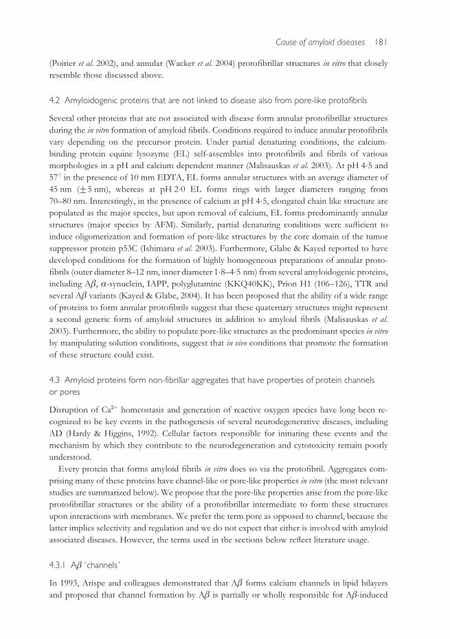
4.2 Amyloidogenic proteins that are not linked to disease also from pore-like protofibrils 181
4.3 Amyloid proteins form non-fibrillar aggregates that have properties of protein channels or
pores 181
4.3.1 Ab ‘ channels ’ 181
4.3.2 a-Synuclein ‘pores ’ 182
4.3.3 PrP ‘channels ’ 182
4.3.4 Polyglutamine ‘channels ’ 183
4.4 Nature uses b-strand-mediated protein oligomerization to construct pore-forming toxins 183
5. Mechanisms of protofibril induced toxicity in protein aggregation diseases 185
5.1 The amyloid pore can explain the age-association and cell-type selectivity of the
neurodegenerative diseases 185
5.2 Protofibrils may promote their own accumulation by inhibiting the proteasome 186
6. Testing the amyloid pore hypothesis by attempting to disprove it 187
7. Acknowledgments 188
8. References 188
1. Introduction
A compelling circumstantial case can be made that amyloid fibril formation is a primary cause of
neurodegeneration in Alzheimer’s disease (AD), Parkinson’s disease (PD), prion diseases, and
related diseases (see Table 1) (Rochet & Lansbury, 2000 ; Stefani & Dobson, 2003). Supporting
evidence for this case, is derived from (1) pathology : fibrillar protein aggregates often colocalize
with neuronal loss, (2) genetics : the gene encoding the fibrillar protein is linked to disease
(autosomal dominant mutations cause familial disease, polymorphisms can be susceptibility
factors, and some haplotypes are linked to risk), (3) animal modeling : overexpression of
the fibrillar protein reproduces many features of the human disease, and (4) biophysics :
proteins containing disease-associated mutations aggregate more rapidly than wild-type proteins.
However, the mechanism by which amyloid fibril formation causes neurodegeneration and the
identity of the pathogenic species have not been determined. It is not possible to directly observe
the pathogenic event. Therefore, one is left to extrapolate from simplified models in order
to produce a working hypothesis, which can then be tested experimentally. The design of a
successful therapeutic strategy based on a working hypothesis will be taken as a ‘proof ’ of its
veracity.
The pathogenic pathway is likely to be much more complicated than an evolutionarily
optimized process like, say, ribosome assembly. Furthermore, there is no a priori reason to
assume that cell death in neurodegenerative disease occurs by a single mechanism or that, if
protein aggregates are responsible, a single species is capable of acting alone as the pathogenic
trigger. Despite these caveats, this review will apply Occam’s razor and start with the working
hypothesis that a single species of protein aggregate may be responsible for initiating a cascade
of events that culminate in neurodegeneration. It is our hope that this review will stimulate
others to design experiments to test the relevance, or lack of thereof, of this working hypothesis.
168 H. A. Lashuel and P. T. Lansbury Jr.

The majority of research in the field of neurodegeneration has focused on the cause of
neuronal death, rather than on the identity of the neurotoxic species. This review will focus on
in vitro studies aimed at the characterization of an intermediate on the amyloid pathway as one
potential toxic species. This review is based on the premise that understanding the structures of
the protein aggregates and the dynamics of their interconversion in vitro, can establish potential
links between aggregation and toxicity. The key to such an approach is to allow genetic
information, especially regarding autosomal dominant (gain of function) mutations, to guide
these experiments. Before summarizing these studies, it is important to emphasize that any
reductionist approach must ultimately explain essential features of the in vivo process that it is
seeking to elucidate. In the case of neurodegeneration, these are :
(1) Aging is one of the strongest susceptibility factor for all of the neurodegenerative diseases.
Even those that are purely genetic, like Huntington’s disease, are primarily adult-onset. This
does not necessarily mean that the pathogenic process takes a long time, but only that
neurons become more susceptible to occurrence of a pathogenic event as they age.
(2) All of these diseases show selectivity to particular types of neurons (hence the diverse
symptoms).
(3) Mutations cause early-onset forms of these diseases that are transmitted in an autosomal
dominant manner.
Points (2) and (3) are linked, since different mutations in the same protein can produce distinct
clinical entities that affect different neuronal subpopulations (three examples are fatal insomnia,
which results from a point mutation in the prion protein (Gambetti et al. 1995), hereditary
cerebral haemorrhage with amyloidosis of the Dutch type, caused by a mutation in amyloid
precursor protein (APP)/Ab (Maat-Schieman et al. 1992), and dementia with Lewy bodies,
caused by a mutation in a-synuclein (Zarranz et al. 2004)).
Several lines of evidence suggest that in vitro amyloid fibril formation mimics the in vivo pro-
cess : (a) fibrils formed in vitro strongly resemble those in diseased tissues (Sunde et al. 1997),
(b) protofibrillar intermediates, first detected in vitro, and later in vivo (see Table 1) exhibit strik-
ingly similar structural and neurotoxic properties (Roher et al. 1996) and (c ) the specificity of
in vitro assembly is reflected in vivo (Wetzel, 1994 ; Helms & Wetzel, 1996 ; Rajan et al. 2001 ;
Kheterpal et al. 2003). Taken together, these observations suggest that structural and mechanistic
clues derived from in vitro studies are relevant to the role of protein fibrillogenesis in neuro-
degenerative diseases and may provide molecular targets for the design of desperately needed
therapeutic agents.
2. What is the significance of the shared structural properties of
disease-associated protein fibrils?
Approximately 24 human proteins form amyloid fibrils in vivo (Stefani & Dobson, 2003). These
proteins are unrelated at the level of primary structure, consistent with the finding that many
proteins with no connection to disease can form amyloid fibrils with a common core structure
in vitro, suggesting that the amyloid fibril is an intrinsically stable structure (Dobson, 2001).
Amyloid fibrils are not crystalline, so their structural similarity is based on lower-resolution
approaches : X-ray fibril diffraction, electron and atomic force microscopy, and their ability to
bind histopathological dyes like Congo Red and Thioflavin T (Sunde et al. 1997). The structural
convergence among various amyloid fibrils is also corroborated by the findings that antibodies
Cause of amyloid diseases 169

Table 1. A summary of studies examining the structure and formation of protofibrils, including amyloid pores by several amyloid-forming priteins in vitro, cell cultures and
in vivo grouped according to the amyloid disease to which they are relevant. Due to space limitations only the most relevant references are cited
DiseaseAmyloid-formingproteins and peptides
Evidence forprotofibrils in vitro
Evidence for protofibrilsIn cell culture and in vivo
Evidence for annularstructures/amyloid pores
Evidence forchannel/pore activity
Alzheimer’sdisease
Ab 1-40 (WT) Goldsbury et al. 2005 ;Harper et al. 1997a, b,1999; Huang et al. 2000 ;Kayed et al. 2004 ;Lambert et al. 1998 ; Stineet al. 2003 ; Walsh et al.1997, 1999; Yong et al.2002)
Funato et al. 1999 ; Gonget al. 2003 ; Kuo et al. 1996 ;Lambert et al. 2001 ;Pitschke et al. 1998 ;Roher et al. 1996
Hafner et al. 2001 ; Kayed& Glabe, 2004; Klug et al.2003 ; Lashuel et al. 2002b,2003
Alarcon et al. 2006 ; Arispeet al. 1993a, b, 1994, 1996;Kagan et al. 2002 ;Kawahara & Kuroda,2000, 2001; Kawaharaet al. 2000 ; Kourie et al.2001b; Lin et al. 1999 ;Singer & Dewji, 2006
Ab 1-40 (E22G) Dahlgren et al. 2002 ;Lashuel et al. 2002b, 2003;Nilsberth et al. 2001
Morishima-Kawashima& Ihara, 1998 ; Podlisnyet al. 1995 ; Walsh et al.2000, 2002
Lashuel et al. 2002b, 2003
Ab 1-42(WT & E22G)
Dahlgren et al. 2002 ;El-Agnaf et al. 2000 ;Parbhu et al. 2002 ; Roheret al. 1996 ; Stine et al.2003 ; Wang et al. 2002a
Chromy et al. 2003 ; Kayedet al. 2004 ; Lashuel et al.2003 ; Lin et al. 2001
Bahadi et al. 2003a ; Bhatiaet al. 2000 ; Hirakura et al.1999 ; Lin et al. 2001 ; Rheeet al. 1998
Parkinson’sdisease
a-Synuclein (WT, A53T& A30P)
Cappai et al. 2005 ; Conwayet al. 2000, 2001; Ding et al.2002 ; Kaylor et al. 2005 ;Lashuel et al. 2002a ;Rochet et al. 2000 ;Shtilerman et al. 2002 ;Volles & Lansbury, 2003;Zhu & Fink, 2003
El-Agnaf et al. 2006 ; Feany& Bender, 2000; Gosaviet al. 2002 ; Lee & Lee,2002; Lee et al. 2002 ; LoBianco et al. 2002 ; Masliahet al. 2000 ; Sharon et al.2001, 2003a, b.
Lashuel et al. 2002a, b ;Ding et al. 2002 ;Shtilerman et al. 2002 ;Rochet et al. 2000 ; Zhu &Fink, 2003 ; Crystal et al.2003 ; Pountney et al.2004 ; Zhu et al. 2004 ;Lowe et al. 2004 ; Quistet al. 2005
Volles & Lansbury, 2002;Volles et al. 2001
170
H.A.Lashueland
P.T.LansburyJr.

Familial Britishdementia
ABri El-Agnaf et al. 2001a, b ;Srinivasan et al. 2003
Holton et al. 2001 Srinivasan et al. 2003 Quist et al. 2005
Dialysis-associatedamyloidosis
b2-Microglobulin Kad et al. 2003 Kayed & Glabe, 2004 Hirakura & Kagan, 2001
Secondary systemicamyloidoses
Serum amyloid A Wang et al. 2002b Wang et al. 2002b Hirakura et al. 2002
Prion-relateddiseases
MHM2 PrP 106 (D23-88and D141-176)
Baskakov et al. 2000 Riesner et al. 1996 ;Supattapone et al. 1999
Kawahara et al. 2000 ;Kourie, 2002; Kourie &Culverson, 2000;Kourie et al. 2001a, 2003 ;Lin et al. 1997
Prion, PrP 106-126 Kayed & Glabe, 2004 Kayed & Glabe, 200484-146 Sokolowski et al. 2003SHa PrP (90-232) Baskakov et al. 2002 ;
Lu & Chang, 2002;Vendrely et al. 2005
Sokolowski et al. 2003 Hirakura et al. 2000bPrP (23-231) Bahadi et al. 2003b, c
Huntington’sdisease
(polyglutamine) Poirier et al. 2002a Kayed & Glabe, 2004 Hirakura et al. 2000a ;Kagan et al. 2001 ;Monoi et al. 2000Demuro et al. 2005 ;Kayed et al. 2004
Amyotrophic lateralsclerosis
Superoxide dismutase-1(WT & A4V, G37R,G85R)
Chung, 2003; Rakhit et al.2002, 2004; Ray et al. 2004
Turner et al. 2003 Chung, 2003; Ray et al.2004
Senile & familalamyloidosis
Transthyretin Cardoso et al. 2002a, b ;Kayed et al. 2003, 2004 ;Lashuel et al. 1998, 1999
Sousa et al. 2001, 2002 ;Teng et al. 2001
Kayed & Glabe, 2004 Azimov et al. 2001
Type II diabetes IAPP Anguiano et al. 2002 ; Greenet al. 2003 ; Green, 2004;Kayed & Glabe, 2004;Kayed et al. 2003 ; Poratet al. 2003 ; Rhoades &Gafni, 2003
Butler et al. 2003 ;de Koning et al. 1994 ;Janson et al. 1996 ;O’Brien et al. 1994
Janson et al. 1999 ;Anguiano, 2002 ;Kayed & Glabe, 2004 ;Porat et al. 2003
Anguiano et al. 2002 ;Harroun et al. 2001 ;Hirakura et al. 2000b;Kawahara et al. 2000 ;Mirzabekov et al. 1996 ;Porat et al. 2003
P53 Ishimaru et al. 2003 Ishimaru et al. 2003Equine lysozyme Malisauskas et al. 2003 Malisauskas et al. 2003Insulin Dzwolak et al. 2005 Dzwolak et al. 2005
Cause
ofam
yloiddiseases
171

raised against the fibrillar form of the amyloidogenic protein amyloid-b (Ab ) recognize
amyloid fibrils derived from other amyloid-forming proteins (O’Nuallain & Wetzel, 2002).
The shared conformational epitopes have not been identified, but may be involved in patho-
genesis. Moreover, the structural similarities suggest a shared mechanism of amyloid fibril
formation.
2.1 Mechanism of amyloid fibril formation in vitro
2.1.1 In vitro fibril formation involves transient population of ordered aggregates of
intermediate stability, or protofibrils
In order to form an amyloid fibril, proteins must adopt a b-sheet rich conformation(s). This
may involve induction of structure in a disordered monomer, or the partial unfolding of a
b-sheet-containing globular protein. The b-sheet-rich species has a high propensity to aggregate/
fibrillize (Kelly, 1998). In vitro amyloid fibril formation does not proceed via a simple two-state,
‘ intermediate-less ’ mechanism akin to microtuble formation from tubulin (these processes have
evolved to be efficient and non-toxic), although that model can be instructive ( Jarrett &
Lansbury, 1993). Rather, discrete b-sheet rich oligomeric intermediates appear before fibrils
form and disappear upon fibril formation (Harper et al. 1997a ; Walsh et al. 1997 ; Lambert et al.
1998) (Fig. 1). These are collectively designated protofibrils. Protofibrils are more stable than
the monomer, but less stable than the fibrillar product. They appear to be obligate intermediates
in fibril formation ; the transient formation of protofibrils has been observed in every well-
characterized in vitro case, suggesting that they are obligate intermediates that are likely to exist,
albeit transiently, in vivo.
OligomersMonomer-dimer-tetramer
α-Synuclein
ABri
Spherical Chain-like Annular
n Protofibrils
Amyloid fibrils
Plaques
Lewy bodies
Fig. 1. In vitro fibril formation involves transient population of ordered aggregates, or protofibrils.
Schematic representation summarizing the quaternary structural species populated during amyloid fibril
formation in vitro and are thought to form during protein fibrillogenesis and deposition in vivo. For simplicity,
only the most commonly observed (globular, chain-like and annular structures) protofibrillar structures are
shown. (The ABri images were kindly provided by Dr Michael Zagorski.)
172 H. A. Lashuel and P. T. Lansbury Jr.

3. Toxic properties of protofibrils
3.1 Protofibrils, rather than fibrils, are likely to be pathogenic
The presence of amyloid fibrils in the post-mortem brains of demented patients led to the first
description of AD and resulted in the hypothesis that fibrils themselves are the primary patho-
genic species. This hypothesis fails to explain several important pathological and clinical
characteristics of AD, raising the possibility that a species other than amyloid fibrils could be the
toxic species (Goldberg & Lansbury, 2000). First, there is no correlation between the amounts of
fibrillar Ab deposits at autopsy and the clinical severity of AD (Lemere et al. 1996 ; Terry et al.
1991). Such a correlation does exist between ‘soluble Ab ’ (monomers plus protofibrils) in the
brain and early cognitive dysfunction (Lue et al. 1999 ; McLean et al. 1999 ; Naslund et al. 2000).
Second, transgenic animals that overproduce APP exhibit neuronal and behavioral abnormalities
before amyloid plaques can be detected (Chui et al. 1999 ; Hsia et al. 1999 ; Moechars et al. 1999 ;
Mucke et al. 2000). Third, inhibiting Ab amyloid fibril formation does not necessarily attenuate
Ab-associated toxicity towards cultured neurons (Aksenova et al. 1996 ; Stege et al. 1999). Fourth,
one autosomal dominant form of AD results from a mutation [APP (E693G)=Ab (E22G)] that
decreases Ab production in vivo and promotes protofibril formation and toxicity in vitro
(Nilsberth et al. 2001 ; Lashuel et al. 2003 ; Whalen, 2005). Fifth, in some AD transgenic mice,
vaccination with an Ab-directed antibody prevented or reversed age-dependent memory decline
without reducing the amyloid burden (Morgan et al. 2000) [in other cases, vaccination has been
shown to reduce behavioral and cognitive deficits and the amyloid burden ( Janus et al. 2000 ;
Morgan et al. 2000)]. Finally, Ab aggregates with properties that are indistinguishable from Ab
protofibrils formed in vitro have been found in human CSF, human cerebral cortex (McLean et al.
1999), and in neuritic amyloid plaques from AD brains (Roher et al. 1996), raising the possibility
that soluble prefibrillar oligomeric species, rather than the fibrils, could be the pathogenic species
in AD and related amyloid diseases. Consistent with this hypothesis, oligomeric (dimer, trimers
and tetramers) low-molecular-weight oligomers (Walsh et al. 2002 ; Wang et al. 2004; Cleary et al.
2005 ; Townsend et al. 2006) and protofibrillar forms of Ab altered neuronal function and/or
caused neuronal death in culture (Lambert et al. 1998 ; Hartley et al. 1999 ; Walsh et al. 2002 ;
Whalen et al. 2005).
The toxic protofibril hypothesis also explains features of PD and other neurodegenerative
diseases that are inconsistent with the fibril being the pathogenic species (Goldberg & Lansbury,
2000 ; Caughey & Lansbury, 2003 ; Silveira et al. 2005). In PD, fibrillar intraneuronal inclusions
comprising a-synuclein, known as Lewy bodies (LBs), are an invariant feature of sporadic and
autosomal dominant forms of PD, but are absent in autosomal recessive juvenile Parkinsonism
(AJRP) (Shimura et al. 2001). It is likely that AJRP brains have such high levels of protofibrils that
complete neurodegeneration occurs before LBs have a chance to form [AJRP results from a
deletion in the parkin gene ; point mutations produce a later-onset form of PD that is charac-
terized by LBs (Kitada et al. 1998)]. Elevated levels of soluble oligomeric forms of a-synulcien
have been detected in plasma samples of PD patients compared to controls (El-Agnaf et al.
2006). Similarly, transgenic mice that overexpress the human a-synuclein protein become
symptomatic (movement disorder plus dopaminergic abnormalities) but do not produce fibrillar
deposits (Masliah et al. 2000). Furthermore, virus-mediated overexpression of human a-synuclein
into the substantia nigra of rats (Kirik et al. 2002 ; Lo Bianco et al. 2002) and primates (Kirik et al.
2003) results in selective dopaminergic neuronal death with non-fibrillar, a-synuclein-containing
inclusions. In vitro studies of a-synuclein aggregation provide possible hints as to the underlying
Cause of amyloid diseases 173

situation : mixtures of human and mouse a-synuclein (as exist in the transgenic mouse brain)
accumulate protofibrils, but fibrillize very slowly (Rochet et al. 2000). Interestingly, virus-
mediated expression of rat a-synuclein in rat brain results in aggregation, but no neuro-
degeneration (Kirik et al. 2002 ; Lo Bianco et al. 2002). Further characterization of the aggregates
formed by human and rat a-synuclein could provide important insights into the role of
a-synuclein aggregation in the degeneration of dopaminergic neurons.
Other neurodegenerative diseases fail to support the proposal that amyloid fibrils are patho-
genic. Prion protein (PrP) fibrils are observed in some prion diseases, but are not an invariant
feature (Chiesa & Harris, 2001). In yeast and mouse models of prion disease, toxicity was
produced in the absence of the stable, protease-resistant aggregated form of the prion protein
(PrP-Sc) (Ma & Lindquist, 2002 ; Ma et al. 2002). A transgenic mouse model of a familial prion
disease became symptomatic before PrP-Sc could be detected (Chiesa et al. 2003). Recent studies
by Caughey and co-workers suggest that the most infectious PrP particles represent protofibrillar
particles with molecular masses ranging from 300–600 kDa (corresponding to 14–28 PrP
molecules) (Silveira et al. 2005). Finally, in systemic amyloid diseases, where amyloid was thought
to produce disease by physically interfering with organ function, studies by Reixach et al. suggest
a primary pathogenic role for protofibrils in initiating cytotoxicity and organ dysfunction
(Reixach et al. 2004). For example, non-fibrillar aggregates of transthyretin (TTR), a protein
associated with systemic amyloidosis and familial amyloid polyneuropathy (FAP), have been
detected in transgenic mice expressing wild-type and mutant TTR (Teng et al. 2001) and in the
nerves of FAP patients (Sousa et al. 2001, 2002). These aggregates were linked to significant
clinical pathology early in the disease before the fibrillar deposits could be detected.
3.2 The toxic protofibril may be a mixture of related species
In an effort to more precisely identify the neurotoxic protein aggregate, cell culture models of
extracellular toxicity have been utilized. Although these simplified models allow more detailed
analysis, their relevance to AD is debatable. Given that caveat, as well as the fact that Ab
oligomers added to cell culture media are heterogeneous (Lashuel et al. 2003; Goldsbury et al.
2005) and are likely to change during the course of an experiment, there is remarkable agreement
among these studies that the protofibril, and not the monomer or the fibril, is a toxic entity
(Lambert et al. 1998 ; Hartley et al. 1999 ; White et al. 2005 ; Townsend et al. 2006). In the case of
Ab, the amyloid protein of AD, globular oligomers (designated ADDLs) were toxic to cultured
neurons (Lambert et al. 1998) and inhibited hippocampal long-term potentiation (LTP) when
introduced to brain slices (Wang et al. 2002a). Globular oligomers of similar sizes (based on AFM
and EM measurement) have been described by other research groups and were shown to be
toxic to cultured neurons (Barghorn et al. 2005). In a related study, small spherical and chain-like
Ab protofibrils (but not monomer, dimer, trimer) induced acute electrophysiological changes
and progressive neurotoxicity in cortical neurons (Hartley et al. 1999). Large spherical aggregates
of Ab (average diameter of >10 nm), termed amylospheroids, exhibited significantly higher
toxicity than small spherical Ab oligomers (<10 nm) (Hoshi et al. 2003). Ab42 forms spherical
aggregates (diameter >10 nm) more rapidly and exhibits significantly more (>100-fold) toxicity
to neuronal cultures than those formed by Ab40. Similar, apparently spherical structures with
diameters ranging from 8–24 nm have also been observed as transient intermediates during the
fibrillogenesis of the arctic variant (E22G) of Ab40 and Ab42 (Lashuel et al. 2003), a-synuclein
(Lashuel et al. 2002a) and mutant SOD1 (Ray et al. 2004). The toxicity of TTR towards cultured
174 H. A. Lashuel and P. T. Lansbury Jr.

cells is also linked to a non-fibrillar oligomeric species, possibly an octamer (Reixach et al. 2004).
Finally, it is probable that the protofibril structure, rather than the protein primary sequence,
produces toxicity. For example, small spherical protofibrils, formed in vitro by an amyloidogenic
SH3 domain from bovine phosphatidyl-inositol-3 kinase (which is not linked to any disease)
are toxic to cultured cells (Bucciantini et al. 2002).
3.3 Morphological similarities of protofibrils suggest a common mechanism of toxicity
The shared morphological and toxic properties of amyloid protofibrils suggest that toxicity
depends on shared structural features. Consistent with this hypothesis, antibodies raised against
protofibrillar Ab were reported to recognize protofibrillar species derived from other amyloi-
dogenic proteins (e.g. a-synuclein, polyglutamine, TTR, IAPP, lysozyme, human insulin, and
PrP106-126), but not monomeric or fibrillar forms of these proteins (Kayed et al. 2003). These
antibodies also inhibit the toxicity of these proteins. These findings support detailed structural
studies of protofibrils, some of which are summarized below. Furthermore, These observations
raise the possibility that these diverse (with respect to sequence, at least) structures may exert
their toxicity through common mechanisms. It may be that an intermediate on the assembly
pathway that is in equilibrium with the protofibril could be the toxic species. The most likely
candidates are low-molecular-weight oligomers (e.g. dimers, trimers or tetramers) or a high-
molecular-weight protofibrillar species, most likely the 4 nm (¡1 nm) spherical species. This is
consistent with biophysical studies (AFM and EM) demonstrating that these spheres are the
precursors to the chain-like and annular protofibrillar structures and appear to exist in equilib-
rium with the large spherical aggregates (8–70 nm). Several laboratories have demonstrated that
these species are capable of self-assembling into pore-like structures on artificial or biological
membranes (Lin et al. 2001 ; Ding et al. 2002).
3.4 Are the amyloid diseases a subset of a much larger class of previously unrecognized
protofibril diseases ?
The hypothesis that small protofibrillar aggregates are pathogenic has an interesting corollary :
there may be many other diseases that, like ARJP, are not characterized by easily detected protein
aggregates. Unlike ARJP, which is genetically and clinically linked to the amyloid disease PD,
these diseases may not be recognized as involving protein aggregation at all. In contrast to our
expectation that the primary sequence efficiently directs protein folding, a significant portion
of newly synthesized proteins (y35%) are not correctly folded in vivo (Schubert et al. 2000).
We expect that many more protein aggregation-driven pathologies remain to be discovered.
3.5 Fibrils, in the form of aggresomes, may function to sequester toxic protofibrils
According to the toxic protofibril hypothesis, the role of the amyloid fibril is uncertain ; it could
have some greatly reduced toxicity, it could be inert, or it could protect neurons by sequestering
toxic protofibrils and/or by consuming protein monomers, inhibiting continued protofibril
formation (Caughey & Lansbury, 2003). There is circumstantial evidence that fibrils may have
a protective function. In PD brains, dopaminergic neurons that contain LBs appear to be
‘healthier ’ than neighboring neurons based on morphological and biochemical criteria
(Tompkins & Hill, 1997). Several transgenic models of polyglutamine diseases are characterized
Cause of amyloid diseases 175

by polyglutamine inclusions in healthy neuron populations, but not in vulnerable neurons
(Kuemmerle et al. 1999). In a transgenic mouse model of one polyglutamine disease, spino-
cerebellar ataxia-type1 (SCA1), there was a direct correlation between the ability of neurons to
sequester ataxin-1 into inclusions and their resistance to cell death (Watase et al. 2002). Taken
together, these observations suggest that formation of fibrillar inclusions may protect neurons
against protofibril-induced neurotoxicity. There is mounting evidence that the sequestration
process may be an active one. Expression of any one of the four disease-associated proteins ;
cystic fibrosis transmembrane conductance regulator (CFTR, associated with cystic fibrosis)
( Johnston et al. 1998), SOD1 (amyotrophic lateral sclerosis) ( Johnston et al. 2000), the androgen
receptor (AR, X-linked spinobulbar muscular atrophy) (Taylor et al. 2003), or Parkin ( Junn et al.
2002) in cultured cells under conditions of oxidative stress or proteasome impairment leads to
sequestration of the mutant protein within multicomponent proteinaceous inclusions known as
aggresomes. Sequestration is actively mediated by microtubules ( Johnston et al. 1998). In the case
of Parkin, these inclusions exhibit morphological and immunohistochemical features similar
to LBs (Junn et al. 2002). In addition to sequestering toxic protein aggregates, aggresomes may
play an active role in facilitating the clearance of these toxic species by lysosomal mediated
degradation mechanisms (Taylor et al. 2003).
4. Amyloid pores, a common structural link among protein aggregation
neurodegenerative diseases
4.1 Mechanistic studies of amyloid fibril formation reveal common features, including
pore-like protofibrils
The convergent circumstantial evidence implicating protofibrils as pathogens has motivated a
more detailed analysis of protofibril structure and formation in vitro. These studies are briefly
summarized below and in Table 1, grouped according to the amyloid disease to which they are
relevant. Similar pathways are followed by other fibrillogenic proteins that are not known to be
related to disease (see Table 1 and below).
4.1.1 Amyloid-b (Ab) (Alzheimer’s disease)
Alzheimer’s disease (AD) is characterized by the presence of extracellular fibrillar amyloid
plaques and intraneuronal neurofibrillary tangles in brain areas associated with memory and
learning. The main fibrillar constituent of amyloid plaques is amyloid-b (b(Glenner & Wong,
1984), a 39-42 amino acid peptide that is produced by endoproteolytic processing of the APP
(Selkoe, 1994). A central role for Ab amyloid fibril formation in the etiology of AD is supported
by extensive genetic and biochemical evidence (Hardy & Selkoe, 2002). In solution, Ab
is unstructured, but Ab protofibrils, including spherical (average diameter of 2�4–4�5 nm,
containing y40 monomers) and chain-like protofibrils and are rich in b-sheet structure (Fig. 1)
(Harper et al. 1997a, b ; Walsh et al. 1997 ; Lambert et al. 1998 ; Parbhu et al. 2002 ; Stine et al. 2003).
Smaller oligomers (e.g. dimers, trimers and tetramers) likely exist, but are not populated in
solution (they can only be observed by SDS–PAGE). Once the critical concentration of the
spherical and later chain-like species is reached, they are rapidly converted into amyloid fibrils. In
addition to the globular and chain-like protofibrils, annular structures with variable diameters
(outer diameter 6–9 nm, inner diameter 1�5–2 nm) have also been observed during the
176 H. A. Lashuel and P. T. Lansbury Jr.

fibrillogenesis of Ab in vitro (Fig. 2a) and during the reconstitution of Ab in lipid bilayers (Fig. 3a)
(Hafner et al. 2001 ; Lin et al. 2001 ; Chromy et al. 2003 ; Klug et al. 2003; Lashuel et al. 2003 ; Kayed
& Glabe, 2004). The similar heights of the spheres, chain-like and annular protofibrils suggests
that the spheres are the precursors of the others (Lashuel et al. 2003). Formation of annular
protofibrils is promoted by a pathogenic mutation [APP (E693G)=Ab(E22G)] that is linked to
familial AD and accelerates the oligomerization of Ab in vitro (Nilsberth et al. 2001 ; Lashuel et al.
2002a, b). Annular protofibrils represent only a small fraction of the total population of Ab
(a) Aβ
Arctic (E22G)
(b) Aα-Synuclein
A53T
(d)
A30P
(e)
ABri EquinLysozyme
IAPP SAA
(c) SOD
A4V
Fig. 2.Many amyloid proteins form annular pore-like protofibrils and have channel- or pore-like properties
in vitro. Annular pore-like structures with variable diameters form during the in vitro fibrillogenesis of disease
associated mutants of Ab [Arctic variant (E22G) of Ab40] (a, f ), a-synuclein (A53T & A30P) (b, d ), and
SOD1 (A4V) (c), and other amyloid-forming proteins (e).
Cause of amyloid diseases 177

protofibrils at any given time, which explains why these structures were missed or ignored in
previous studies.
4.1.2 a-Synuclein (PD and diffuse Lewy body disease)
PD is associated with the formation of intraneuronal fibrillar inclusions (LBs) (Pollanen et al.
1993 ; Shults, 2006). a-Synuclein is the primary component of LBs in all PD patients (Spillantini
et al. 1998b) and is strongly implicated as a cause of PD by biochemical (Cookson, 2005) and
genetic studies of familial forms of PD (Polymeropoulos et al. 1997 ; Kruger et al. 1998 ; Singleton
et al. 2003 ; Zarranz et al. 2004) and by mouse (Masliah et al. 2000) and Drosophila (Feany &
Bender, 2000 ; Chen & Feany, 2005) modeling studies. Three different a-synuclein missense
mutations (A30P, A53T and E46K) are associated with rare, autosomal dominant forms of early-
onset PD (Polymeropoulos et al. 1997 ; Kruger et al. 1998 ; Zarranz et al. 2004). In addition,
triplication of the wild-type a-synuclein gene causes autosomal dominant PD in an Iowan
(a) (c)
Aβ (1–40)
α -synuclein
ABri
ADan
Amylin
SAA
(b)
1. 50.000 mm/div2. 15.000 mm/div
mm
50
100
150
200
Fig. 3. Amyloid forming proteins form pore-like/channel structures on artificial as well as biological
membranes. (a) AFM images of pore-like structures formed during the reconstitution of Ab in lipid bilayers
(adapted from Lin et al. 2001). (b) Pore-like structures that were obtained by incubating predominantly
spherical a-synuclein protofibrils (WT and A53T) with brain-derived vesicles (adapted from Ding et al.
2002). (c) Amyloid-forming proteins/peptides reconstituted in lipid bilayers form pore/channel-like struc-
tures (adapted from Quist et al. 2005).
178 H. A. Lashuel and P. T. Lansbury Jr.

kindred (Singleton et al. 2003). Transgenic mice (Masliah et al. 2000) expressing human wild-type
a-synuclein and Drosophila (Feany & Bender, 2000 ; Chen & Feany, 2005) expressing WT, A30P,
or A53T are characterized by a-synuclein non-fibrillar (mice and fly) or fibrillar (Flynn &
Theesen, 1999) inclusions and a Parkinsonian phenotype. Like Ab, a-synuclein is not folded
under ‘native ’ conditions (Weinreb et al. 1996), but will form b-sheet rich protofibrils and
amyloid fibrils in vitro. Three mutations (A53T, E46K and A30P) linked to early-onset PD
promote the formation of a-synuclein protofibrils, but the A30P mutation was shown to form
fibrils more slowly than WT a-synuclein, suggesting that a-synuclein protofibrils may cause
neurodegeneration in PD (Conway et al. 1998 ; Conway et al. 2000 ; El-Agnaf et al. 1998 ; Li et al.
2001 ; Pandey et al. 2006). Amyloid fibril formation by a-synuclein occurs by a hierarchical
assembly mechanism (random coilpspherespchain-like and annular protofibrilspfibrils)
similar to that observed for Ab in AD (Conway et al. 2000 ; Ding et al. 2002). The PD-linked
mutations promote formation of annular and tubular protofibrillar structures (wild-type
a-synuclein forms annular protofibrils after extended incubation) (Conway et al. 2000 ; Ding et al.
2002 ; Lashuel et al. 2002a, b) (Fig. 2b, d ). The diameter of a-synuclein annular protofibrils
detected in vitro is similar to that of the a-synuclein fibrils. Zhu and colleagues observed the
formation of a-synuclein annular protofibrils during drug-induced disaggregation of a-synuclein
fibrils in vitro (Zhu et al. 2004), suggesting that a direct relationship between the two structures
may exist. These observations are consistent with cross-sectional analysis of ex vivo amyloid fibrils
demonstrating that amyloid fibrils have an electron-lucent center, indicative of the presence of
a hollow center (Serpell et al. 2000).
4.1.3 ABri (familial British dementia)
ABri is the major component of amyloid deposits in the brain of patients with familial British
dementia (FBD). FBD is an autosomal dominant neurodegenerative disorder associated with a
stop codon mutation in the BRI gene that results in the production of ABri (Vidal et al. 1999 ;
Ghiso et al. 2000). In vitro fibrillogenesis of ABri produced protofibrils and subsequently, amyloid
fibrils (El-Agnaf et al. 2001a, b ; Srinivasan et al. 2003, 2004), by a mechanism that is reminiscent
of that of Ab and a-synuclein, including the formation of spherical, chain-like and annular
protofibrillar intermediates. Spherical protofibrils exhibited more toxicity to cultured neurons
than chain-like protofibrils and fibrils (El-Agnaf et al. 2001a). Annular ABri protofibrils
have been characterized in vitro (see Fig. 2e) (Srinivasan et al. 2004 ; O. M. El-Agnaf, personal
communication) but their toxicity has not been determined.
4.1.4 Superoxide dismutase-1 (amyotrophic lateral sclerosis)
Amyotrophic lateral sclerosis (ALS), also known as Lou Gehrig’s disease (Rosen, 1978 ; Julien,
2001), is linked to the gene encoding superoxide dismutase-type 1 (SOD1) (Rosen et al. 1993 ;
Brown, 1997). Cytoplasmic inclusions containing mutant SOD1 are present in the motor
neurons of familial ALS patients (Shibata et al. 1996) and transgenic mouse models (Bruijn et al.
1998 ; Watanabe et al. 2001). Significantly, these inclusions do not clearly contain fibrillar sub-
structure. SOD1 is a structured homodimer. FALS mutations destabilize the native SOD1 dimer
(Nakano et al. 1996 ; Cardoso et al. 2002b; Hayward et al. 2002 ; Lindberg et al. 2002 ; Rodriguez
et al. 2002 ; Tiwari & Hayward, 2003 ; Rakhit et al. 2004 ; Ray et al. 2004) and promote SOD1
aggregation in vitro (Ray et al. 2004). Significantly, SOD1 has not been induced to form amyloid
Cause of amyloid diseases 179

fibrils in vitro. Instead, spherical and annular oligomeric structures have been observed (Chung,
2003 ; Ray et al. 2004). As expected, the pathogenic SOD1 mutants aggregate more rapidly than
WT SOD1 (Chung, 2003 ; Ray et al. 2004), consistent with what is observed with Ab and
a-synuclein (Lashuel et al. 2003 ; Lashuel et al. 2002a, b). Their findings suggest that common
pathogenic assemblies may underlie all of these diseases. In addition to pathogenic mutations,
conditions that promote metal depletion or oxidative damage of SOD1 accelerate the formation
of annular structures by mutants and wild-type SOD (A4V, H46R, G37R, and G85R) (Chung,
2003 ; Elam et al. 2003 ; Ray et al. 2004). The morphology of these oligomeric forms of SOD1
formed in vitro resembles that of protofibrillar species of Ab and a-synuclein, in particular the
annular and small spherical species (Fig. 2c). Thus, these species have been designated SOD1
protofibrils, yet they do not convert to fibrils over several weeks of incubation (Ray et al. 2004).
4.1.5 Prion protein (Creutzfeldt–Jakob disease, bovine spongiform encephalopathy, etc.)
The prion diseases [Creuzfeldt-Jakob disease (CJD), Gertsmann-Straussler syndrome (GSS), and
fatal familial insomnia (FFI) in humans, bovine spongiform encephalopathy (BSE) in cattle,
scrapie in sheep, and chronic wasting disease in elk] are characterized by the presence of an
abnormal form of the PrP in the brain. The ability of these diseases to be transmitted, apparently
by an abnormal form of the PrP, distinguishes them from the others discussed here, but there are
also sporadic forms of human prion disease as well as forms that are purely genetic ; caused by
mutations in the gene encoding PrP (CJD, GSS, and FFI). The disease-associated form of the
PrP, or PrP-Sc, is resistant to proteolysis and rich in b-sheet structure, relative to the normal
form of the PrP, PrP-C (Cohen & Prusiner, 1998). PrP-Sc can also be isolated in fibrous form
(Rubenstein et al. 1987). However, several lines of evidence suggest that PrP-Sc, like other
amyloid fibrils, may not be the neurotoxic form of the protein. Transgenic mice that over-
produce a disease-associated form of PrP exhibit disease-like phenotypes before PrP-Sc can be
detected (Chiesa & Harris, 2001 ; Harris et al. 2003). During this phase, the protease-resistance of
PrP gradually increased, suggesting the stepwise formation of structured oligomers of increasing
stability. In vitro fibrillization of a fragment of PrP (residues 89–231) involves multiple oligomeric
species, some of which appear to be off-pathway b-sheet-rich oligomers (Baskakov et al. 2002).
Truncated variants of PrP form spherical and elongated protofibril-like structures that share
properties with PrP-Sc and are capable of seeding the conversion of PrPC into amyloid fibrils
(Lee & Eisenberg, 2003). Finally, a truncated variant of PrP (SHaPrP 90-232) forms spherical
and annular oligomeric structures in vitro, similar to those formed by Ab and a-synuclein
(Sokolowski et al. 2003). It was estimated that 8 subunits of SHaPrP 90-232 are required to form
the b-sheet-rich annular structures (Sokolowski et al. 2003).
4.1.6 Huntingtin (Huntington’s disease)
Huntington’s disease is the most prevalent of a group of purely genetic neurodegenerative
movement disorders that are caused by expansions of naturally occurring polyglutamine (polyQ)
tracts in several proteins (the normal function of these tracts is not known) (Zoghbi & Orr,
2000). The magnitude of the expansion is related to the age of onset of the disease, with large
expansions causing juvenile onset disease. The Huntington’s disease brain is characterized by
nuclear inclusions comprising the protein huntingtin (htn). Truncated mutant versions of htn
with expanded polyQ repeat form spherical (Poirier et al. 2002 ; Wacker et al. 2004) chain-like
180 H. A. Lashuel and P. T. Lansbury Jr.
(Poirier et al. 2002), and annular (Wacker et al. 2004) protofibrillar structures in vitro that closely
resemble those discussed above.
4.2 Amyloidogenic proteins that are not linked to disease also from pore-like protofibrils
Several other proteins that are not associated with disease form annular protofibrillar structures
during the in vitro formation of amyloid fibrils. Conditions required to induce annular protofibrils
vary depending on the precursor protein. Under partial denaturing conditions, the calcium-
binding protein equine lysozyme (EL) self-assembles into protofibrils and fibrils of various
morphologies in a pH and calcium dependent manner (Malisauskas et al. 2003). At pH 4�5 and
57x in the presence of 10 mm EDTA, EL forms annular structures with an average diameter of
45 nm (¡5 nm), whereas at pH 2�0 EL forms rings with larger diameters ranging from
70–80 nm. Interestingly, in the presence of calcium at pH 4�5, elongated chain like structure are
populated as the major species, but upon removal of calcium, EL forms predominantly annular
structures (major species by AFM). Similarly, partial denaturing conditions were sufficient to
induce oligomerization and formation of pore-like structures by the core domain of the tumor
suppressor protein p53C (Ishimaru et al. 2003). Furthermore, Glabe & Kayed reported to have
developed conditions for the formation of highly homogeneous preparations of annular proto-
fibrils (outer diameter 8–12 nm, inner diameter 1�8–4�5 nm) from several amyloidogenic proteins,
including Ab, a-synuclein, IAPP, polyglutamine (KKQ40KK), Prion H1 (106–126), TTR and
several Ab variants (Kayed & Glabe, 2004). It has been proposed that the ability of a wide range
of proteins to form annular protofibrils suggest that these quaternary structures might represent
a second generic form of amyloid structures in addition to amyloid fibrils (Malisauskas et al.
2003). Furthermore, the ability to populate pore-like structures as the predominant species in vitro
by manipulating solution conditions, suggest that in vivo conditions that promote the formation
of these structure could exist.
4.3 Amyloid proteins form non-fibrillar aggregates that have properties of protein channels
or pores
Disruption of Ca2+ homeostasis and generation of reactive oxygen species have long been re-
cognized to be key events in the pathogenesis of several neurodegenerative diseases, including
AD (Hardy & Higgins, 1992). Cellular factors responsible for initiating these events and the
mechanism by which they contribute to the neurodegeneration and cytotoxicity remain poorly
understood.
Every protein that forms amyloid fibrils in vitro does so via the protofibril. Aggregates com-
prising many of these proteins have channel-like or pore-like properties in vitro (the most relevant
studies are summarized below). We propose that the pore-like properties arise from the pore-like
protofibrillar structures or the ability of a protofibrillar intermediate to form these structures
upon interactions with membranes. We prefer the term pore as opposed to channel, because the
latter implies selectivity and regulation and we do not expect that either is involved with amyloid
associated diseases. However, the terms used in the sections below reflect literature usage.
4.3.1 Ab ‘ channels ’
In 1993, Arispe and colleagues demonstrated that Ab forms calcium channels in lipid bilayers
and proposed that channel formation by Ab is partially or wholly responsible for Ab-induced
Cause of amyloid diseases 181
toxicity in AD (Arispe et al. 1993a, b). This finding has been reproduced many times, in several
different laboratories, using many membrane models (Arispe et al. 1993b, 1994 ; Kawahara et al.
1997, 2000 ; Sanderson et al. 1997 ; Rhee et al. 1998 ; Hirakura et al. 1999; Lin et al. 1999, 2001 ;
Bhatia et al. 2000 ; Kourie et al. 2001b; Kagan et al. 2002 ; Lin & Kagan, 2002 ; Bahadi et al. 2003a ;
Alarcon et al. 2006). The form of Ab that is closely linked to AD pathogenesis (Ab42) exhibits
higher propensity to form channels, consistent with its increased propensity to aggregate and
form annular protofibrils in vitro (H. Lashuel & P. T. Lansbury Jr., unpublished observations) as
compared to Ab40, the predominant form ( Jarrett et al. 1993). b-sheet formation is also linked to
Ab-channel formation (and aggregation) (Sanderson et al. 1997). When incorporated into arti-
ficial lipid bilayers, Ab produces uniform pore-like structures with an outer diameter of 8–12 nm
and inner diameter of 2 nm (Fig. 3a, c) (Lin et al. 2001 ; Quist et al. 2005). These aggregates,
formed at the membrane surface, resemble, pore-like aggregates formed in solution using Ab42
or the disease-associated mutant form of Ab40 (E22G) (Lashuel et al. 2002b, 2003). Addition of
Ab40 oligomers to hypothalamic GT1-7 neuronal cells results in simultaneous formation of Ca2+
channels and increase in intracellular Ca2+ levels, suggesting that Ab40 oligomers is capable of
disrupting biological as well as artificial membranes, possibly via pore formation (Kawahara &
Kuroda, 2001, 2000 ; Singer & Dewji, 2006). The nature of the oligomer responsible for this
activity was not investigated. AD-derived Ab oligomers were observed to exhibit high affinity
and selective attachment to membranes, suggesting that AD-derived oligomers are either directly
integrated into the membranes or bind tightly to other cell surface molecules of cultured hip-
pocampal neurons (Gong et al. 2003). Oligomer-specific antibodies were shown to block their
interaction with membranes.
4.3.2 a-Synuclein ‘pores ’
The a-synuclein protofibril (but not the monomer or the fibril) binds very strongly to vesicle
membranes and causes leakage of small compounds (less than y2�5 nm hydrodynamic radius)
entrapped within synthetic vesicles (Volles et al. 2001 ; Volles & Lansbury, 2002). This typical
pore-like behavior was consistent with the observation that addition of spherical protofibrils of
a-synuclein to purified brain-derived vesicle (BDV) fractions resulted in the formation of pore-
like structures (Fig. 3b) (Ding et al. 2002). Additionally, reconstitution of a-synuclein in lipid
bilayers also results in the formation of pore-like structures that exhibit channel-like properties
(Quist et al. 2005). Mutations linked to familial PD promoted the formation of the b-sheet-rich
annular, pore-like (d=8–12 nm, 2–2�5 nm inner diameter) structures in solution (Lashuel et al.
2002a, b). These structures resembled in morphology and dimension, membrane-spanning pores
that are formed by protein toxins (e.g. hemolysin, latrotoxin, and aerolysin) (Valeva et al. 1997 ;
Orlova et al. 2000 ; Wallace et al. 2000). Vesicle permeabilization was also demonstrated for
protofibrils comprising the congener c-synuclein, but not those comprising b-synuclein (Park &
Lansbury, 2003). At the same time, c-synuclein, but not b-synuclein, produced pore-like
structures in solution (H. Lashuel, unpublished results), strengthening the case that the pore-like
structures are responsible for the pore-like behavior of a-synuclein.
4.3.3 PrP ‘channels ’
Aguzzi and colleagues have proposed that the pathogenicity of the PrP may be related to
abnormal pore formation (Moore et al. 1999 ; Rossi et al. 2001 ; Behrens & Aguzzi, 2002).
This proposal, based on studies of a PrP homolog, Doppel, is supported by circumstantial
182 H. A. Lashuel and P. T. Lansbury Jr.

experimental evidence. First, an abnormal transmembrane (translocation-incompetent) form of
PrP has been linked to disease (Hegde et al. 1999). Second, a peptide based on the proposed
transmembrane domain of PrP produce non-fibrillar aggregates that disrupt membranes (Pillot
et al. 1997) and induce toxicity in rat cortical neurons (Pillot et al. 2000). Third, several PrP-based
peptides interacts with lipid membranes and form ion channels (Lin et al. 1997 ; Kourie &
Culverson, 2000 ; Bahadi et al. 2003b, c). Fourth, a truncated form of PrP permeabilizes synthetic
vesicles (Sanghera & Pinheiro, 2002). Fifth, when disease-associated mutant forms of PrP were
expressed in CHO cells, PrP was found to be very tightly associated with plasma membranes
(Lehmann & Harris, 1996).
4.3.4 Polyglutamine ‘channels ’
Peptides with pathological polyglutamine repeats (>35–40 residues) insert into artificial mem-
branes (Hirakura et al. 2000a ; Monoi et al. 2000 ; Kagan et al. 2001) and also affect mitochondrial
Ca2+ homeostasis (Panov et al. 2002). Furthermore a polyglutamine (Gln40) peptide produced
homogeneous pore-like protofibrillar structures in vitro, while a shortened peptide did not
(Monoi et al. 2000). Importantly, b-sheet formation, oligomerization, amyloid pore/channel
formation, and fibrillogenesis are also very sensitive to polyGln repeat length (Scherzinger et al.
1999). Circumstantial evidence suggests that membrane disruption is mediated by direct inter-
action between the polyglutamine repeats or an aggregated form of these proteins resulting in the
formation of ion channels in lipid bilayers and in mitochondrial membranes. Interestingly, only
pathological polyglutamine repeat (>35–40) were observed to insert into artificial membranes
and exhibited direct effects on mitochondrial Ca2+ homeostasis, reproducing the mitochondrial
deficit seen in Huntington’s disease patients and in transgenic animals expressing these repeats
(Hirakura et al. 2000a ; Monoi et al. 2000 ; Kagan et al. 2001 ; Panov et al. 2002). Furthermore,
Kayed & Glabe (2004) reported that a polyglutamine protein containing 40 repeat (KKQ40KK)
forms homogeneous annular pore-like protofibrillar structures in vitro, suggesting that protofibril
formation and channel activity are linked.
Monoi and colleagues proposed that a single chain of poly-glutamine polypeptide is capable of
forming cylindrical pores by forming a right handed helix (6�2 residues/turn), termed m-helix,
that is further stabilized by backbone side-chain hydrogen bonding interactions between the
amide groups and glutmaine side-chain (Monoi, 1995 ; Monoi et al. 2000). What makes this model
an attractive one is that it offers an explanation for the requirement of >35 polyglutamine tract
to confer pathogenecity in polyglutamine diseases, because 37 residues (each contributing 0�81 Ato the length) would be required to form a m-helix that spans the hydrophobic region of the
bilayer (30 A). A normal transmembrane domain composed of an a-helix is y20 amino acids
long. However, b-sheet formation, oligomerization and fibrillogenesis by poly-glutamine pro-
teins are highly dependent on the polyQ repeat length with 36 or more glutamines favoring these
tertiary and quaternary structure changes (Scherzinger et al. 1999). However, further studies are
required to elucidate the nature of the membrane active species responsible for polyglutamine
membrane disruption properties.
4.4 Nature uses b-strand-mediated protein oligomerization to construct pore-forming toxins
A simple working hypothesis emerging from the studies summarized above holds that five prop-
erties of disease-associated amyloid proteins are linked: (1) aggregation, (2) b-sheet formation,
Cause of amyloid diseases 183

(3) amyloid pore formation, (4) membrane permeabilization, and (5) toxicity (see Fig. 4). This
proposal is more compelling because natural protein toxins utilize an analogous mechanism.
Latrotoxin, a-hemolysin, and aerolysin all form well-ordered, oligomeric membrane-spanning
pores characterized by b-sheet structure. It is important to remember that while these toxins are
Synucleinoverexpression
Proteasome inhibitionmutations
UCH-L1 & Parkin
Ubiquitin
Parkin
UCH-L1
Oxidative stressPathogenic mutations
(A53T, A30P and E46K)
Lewy bodies
DA
MitochondriaROS
Ca2+
Cytochrome C
Neurodegeneration
Fig. 4. Schematic depiction of the various steps involved in a-synuclein aggregation and Lewy Body
formation and potential mechanisms by which these processes could contribute to the pathogenesis of PD.
Several factors have been shown to play a role in initiating and/or accelerating a-synuclein aggregation and
fibrillogenesis including, increased protein expression, mutations, oxidative stress induced modifications
and crosslinking, and phosphorylation at S129 (Chen & Feany, 2005; Fujiwara et al. 2002 ; Smith et al. 2005).
The scheme depicts amyloid protofibril as the toxic species and that it may amplify itself by inhibiting
proteasome-dependent protein degradation, disrupting dompaniergic vesicles, formation of reactive oxygen
species and increased oxidative stress, causing mitochondrial dysfunction (Abou-Sleiman et al. 2006), all of
which have the affect of increasing levels of a-synuclein and/or accelerating a-synuclien oligomerization
and protofibril formation. This hypothesis is supported by the findings that triplication of the a-synucleingene causes and autosomal dominant PD in Iowan kindred (Singleton et al. 2003). Protofibrils may promote
their own accumulation and toxicity by inhibiting the proteasome. Two gene products in the protein
degradation pathway have been implicated in early onset forms of PD (Lansbury & Brice, 2002). One form
is associated with genetic mutations in the gene encoding parkin (a protein with an E3 (ubiquitin ligase)-like
activity), where the mutations reduce or abolish parkin’s E3-like ligase activity (Kitada et al. 1998). A rare
point mutation in the gene encoding ubiquitin C-terminal hydrolase (UCH-L1), a neuronal-specific deubi-
quitinating enzyme, is thought to lead to reduced catalytic activity of UCH-L1, and, thus, indirectly affect the
proteasomal function (Leroy et al. 1998). Furthermore, reduced expression of UCH-L1 in the substantia
nigra of DLB and PD brains suggest that it plays a role in modulating protein ag gregation and LB
formation (Barrachina et al. 2005). In addition to its beneficial hydrolase activity (hydrolysis of C-terminal
ubiquityl esters and amides), UCH-L1 exhibits a concentration dependent ubiquityl ligase activity that is
thought to be pathogenic for PD. Taking together, these findings demonstrate that accumulation of
a-synuclein either due to increased expression or decreased degradation is sufficient to cause PD, as
predicted by the amyloid hypothesis.
184 H. A. Lashuel and P. T. Lansbury Jr.
optimized by evolution to permeabilize target membranes, amyloid pore formation is likely to be
an accident and will therefore be very inefficient in vitro. Pore formation by b-sheet bacterial
toxins involves a series of complex events that include membrane association, oligomerization,
and insertion of a b-barrel. The pore-forming protein toxins (PFTs) typically exist as structured,
water-soluble monomers ; conversion into the membrane-inserted pore requires that these pro-
teins undergo oligomerization and a conformational change. In the case of aerolysin, proteolytic
processing by the target is required to initiate the formation of the transmembrane b-barrel
domain (Abrami et al. 1998). The PFT proteins typically aggregate and/or are activated (Walker
et al. 1992 ; Song et al. 1996; Sellman et al. 1997 ; Heuck et al. 2003) at the membrane surface, a
feature that promotes membrane selectivity. Up to 100 b-hairpins can be inserted on activation,
to produce the transmembrane b-sheet pore (Hotze et al. 2001). b-barrels that span the
membrane (Valeva et al. 1997 ; Heuck et al. 2001) typically comprise 8–22 b-strands (each 10 13
residues long) and have an average diameter of 1�5–3�5 nm (Heuck et al. 2001, 2003). In the
a-hemolysin barrel, each monomer contributes an amphipathic hairpin to a 14-stranded hepta-
meric b-barrel (Menestrina et al. 2001). PFTs form pores ranging in diameter from 1 to 2 nm for
staphylococcal a-hemolysin (Fussle et al. 1981) and Vibrio cholerae cytolysin (Zitzer et al. 1995),
and from 15 to 45 nm for streptolysin O (Sekiya et al. 1993) and perfringolysin O (Olofsson et al.
1993). For some PFTs, some degree of heterogeneity (pore size and oligomeric state) has been
reported (Sharpe & London, 1999 ; Tadjibaeva et al. 2000). PFT membrane binding and oligomer-
ization are dependent on the membrane composition and microenvironment and, in some cases,
are mediated by interaction with receptor molecules. Cholesterol, for example, can play a critical
role in PFT insertion (Cabiaux et al. 1997), and may also play a role in amyloid pore toxicity
(Arispe & Doh, 2002 ; Curtain et al. 2003 ; Eckert et al. 2003).
5. Mechanisms of protofibril induced toxicity in protein aggregation diseases
5.1 The amyloid pore can explain the age-association and cell-type selectivity of the
neurodegenerative diseases
Most cases of AD, PD and ALS are sporadic, that is, they do not involve mutant protein, and are
strongly age-associated. Any model for pathogenesis must explain these cases. It is important to
emphasize that the wild-type proteins will also form amyloid pores in vitro, albeit more reluctantly
than the disease-associated mutants (Hafner et al. 2001; Ding et al. 2002 ; Lashuel et al. 2002a, b,
2003 ; Shtilerman et al. 2002 ; Chromy et al. 2003 ; Chung, 2003 ; Klug et al. 2003 ; Kayed & Glabe,
2004). Factors other than mutations could trigger pore formation pathogenesis in sporadic dis-
ease (Fig. 4). These factors could include (but are not limited to) : (1) increased expression of the
amyloid-forming protein (Singleton et al. 2004 ; Uryu et al. 2003) ; (2) impaired degradation of the
monomeric protein by the proteasome (Giasson & Lee, 2003) ; (3) impaired degradation of
the protofibrils by a lysosome-mediated autophagic process (Lee et al. 2004) ; (4) changes in
chaperone activity and/or expression of homologs that act as specific chaperones (Rochet et al.
2000 ; Conway et al. 2001 ; Hashimoto et al. 2001 ; Masliah & Hashimoto, 2002) ; (5) changes in
levels of post-translational modification, both enzymatic (Shimura et al. 2001 ; Fujiwara et al.
2002 ; Chen & Feany, 2005 ; Smith et al. 2005) and non-enzymatic (Conway et al. 2001) ; (6)
changes in the pathogenic microenvironment (pH, membrane composition, increased oxi-
dation). Oxidative damage of amyloid-forming proteins increases their propensity to misfold and
aggregate in vitro (Rakhit et al. 2002 ; Dauer & Przedborski, 2003). Expression of a-synuclein in
Cause of amyloid diseases 185

cells, under oxidizing conditions, promotes cytoplasmic aggregation and cell death (Kim et al.
2003 ; Smith et al. 2005). Many of the factors listed above are ATP-dependent and would
therefore be expected to emerge on aging (Heydari et al. 1995 ; Gaczynska et al. 2001 ; Soti &
Csermely, 2002 ; Ferrington et al. 2005 ; Chondrogianni, 2005), when ATP production becomes
less efficient. Aging may also affect the susceptibility of membranes to permeabilization;
age-dependent changes in the distribution of cholesterol in neuronal membranes can facilitate
oligomerization and accumulation of Ab in plasma membranes (Wood et al. 2002) and choles-
terol content influences Ab toxicity in cell culture (Arispe & Doh, 2002). It must be emphasized
that of the factors listed above, all except membrane composition can be easily rationalized by
other pathogenic mechanisms.
Another striking feature of the neurodegenerative diseases is that they are selective for certain
neuronal populations (hence the clinical diversity). This feature can be explained by an amyloid
pore model. It is likely that structural differences among protofibrils formed by the various
amyloid-forming proteins would confer some specificity in the way they interact with cellular
membranes. In addition, accessory molecules could play a role in modulating pore formation.
Finally, conversion of annular protofibrils into amyloid pores may occur at the membrane sur-
face in vivo, triggered by specific environmental factors, changes in fluidity of the membrane or
interaction with specific proteins. For example, the selective vulnerability of dopaminergic neur-
ons of the substantia nigra to a-synuclein toxicity in PD may be related to (1) the relatively low
expression of b-synuclein (Rockenstein et al. 2001), a homolog that prevents a-synuclein pro-
tofibril formation in vitro (Park & Lansbury, 2003), (2) the high concentration of cytoplasmic
dopamine (Lotharius & Brundin, 2002 ; Xu et al. 2002) in these cells (some populations of
dopaminergic neurons, that have more efficient machinery for removal of cytoplasmic dopa-
mine, are resistant to PD), or (3) highly oxidizing conditions (higher levels of free radical scav-
engers are expressed in PD-resistant populations of dopaminergic neurons) (Hirsch et al. 1997),
A combination of the latter two factors may generate dopamine ortho-quinone, which, in vitro,
covalently modifies a-synuclein and stabilizes protofibrils (Conway et al. 2001 ; Li et al. 2004). The
extreme sensitivity of dopaminergic neurons to proteasomal inhibition (McNaught et al. 2002)
may be related to the importance of keeping cytoplasmic a-synuclein levels low in these cells.
Second, the differences between cell-types would be expected to influence the specific activity of
the amyloid pores in a way that depended on the protein constituent. Microinjection of proto-
fibrillar Ab42 induces death of human neurons, but not neuroblastoma cells, astrocytes or other
non-neuronal cell lines (Zhang et al. 2002). Decreased susceptibility to intracellular toxicity could
arise from the absence of ‘ receptor ’ molecules that mediate Ab toxicity, the presence of neu-
roprotective factors, or just the changed composition of target membranes.
5.2 Protofibrils may promote their own accumulation by inhibiting the proteasome
Neurons have developed housekeeping systems to avoid misfolded proteins accumulating in the
cytoplasm. These include the chaperone system [some of which is ATP-dependent and would be
compromised on aging (Heydari et al. 1995 ; Soti & Csermely, 2002)], which prevents aggregation
(Muchowski, 2002 ; McClellan et al. 2005) and the proteasomal system, which degrades misfolded
and damaged proteins (Berke & Paulson, 2003 ; Ross & Pickart, 2004). The importance of
ubiquitin-dependent proteasomal degradation in PD is illustrated by the fact that parkin and
UCH-L1, two PD-linked gene products, are involved in this process (Lansbury & Brice, 2002)
(Fig. 4). Once protein aggregation has occurred, several backup systems may take over, including
186 H. A. Lashuel and P. T. Lansbury Jr.

the HSP90 heat-shock protein, that is capable of disaggregating these species (Ben-Zvi &
Goloubinoff, 2001 ; Ben-Zvi et al. 2004), the autophagy system, which engulfs and degrades
protein aggregates [including protofibrils (Lee et al. 2004)] via fusion with the lysosome, and the
‘aggresome’ system (Kopito, 2000). Complete or partial failure of any of these systems could
have detrimental consequences for the cell. Convergent evidence indicates that the protofibrils
themselves could compromise proteasomal degradation. First, overexpression of disease-
associated proteins (SOD1 or a-synuclein) in non-neuronal cells resulted in inhibition of pro-
teasome activity (Tanaka et al. 2001 ; Urushitani et al. 2002). Second, aggregated (protofibrillar and
fibrillar) a-synuclein binds directly to the proteasome and inhibits proteasomal activity (Snyder
et al. 2003 ; Lindersson et al. 2004). Third, inhibition of the neuronal proteasome causes cytosolic
inclusion formation (Ma & Lindquist, 2002) and neuronal death (Ma et al. 2002). It is not known
whether neuronal death is a result of an increase in protein aggregation due to decreased
degradation or of another proteasome-mediated event (Bennett et al. 2005). The former scenario
offers a mechanism whereby toxicity can amplify itself.
6. Testing the amyloid pore hypothesis by attempting to disprove it
In the past 4 years, 18 different amyloid-forming proteins have been reported to form prefibrillar
intermediates with pore-like morphologies (Table 1), supporting the notion that formation of the
pore and formation of the fibril are tightly linked. However, whether the pore has any relevance
to disease, as proposed here, is not at all clear. It must be emphasized that there is a much work
to be done to strengthen the case, which must, by definition, be based on circumstantial
evidence. A goal of this review is to encourage experiments designed to disprove the hypothesis.
In closing, we make three points, each based on different experimental approaches :
(1) Detection of amyloid pores in tissue from patients or animal models does not support the hypothesis nor does
the failure to detect these structures disprove it. This point is illustrated by the flawed logic that led to
the toxic fibril hypothesis. However, one could argue that, in the absence of evidence for the
existence of pores in tissue, there is no proof that they are able to form under physiological
conditions. Several studies of a-synuclein aggregates extracted from brains affected by rare
neurodegenerative diseases related to PD, provide tantalizing evidence that pore-like struc-
tures do exist in vivo. Several studies of a-synuclein fibrils from diffuse Lewy body disease
(DLBD) reveal pore-like structures that co-purify with a-synuclein fibrils (Spillantini et al.
1998a ; Fujiwara et al. 2002). The resemblance of these structures to the amyloid pores pro-
duced from a-synuclein in vitro (Lashuel et al. 2002a) is striking. Direct evidence for the
existence of amyloid pore-like structures in vivo has been provided by the extraction of
annular a-synuclein structures, similar to those seen in in vitro preparations, from inclusions
from post-mortem brain tissues of a multiple system atrophy (MSA, another a-synuclein
aggregation disease) patient (Pountney et al. 2004).
(2) Evidence for membrane abnormalities is consistent with the amyloid pore, but these abnormalities could easily
have arisen by another mechanism. Formation of unregulated pores at the mitochondrial mem-
brane could result in altered Ca2+ homeostasis, the release of cytochrome c and other proa-
poptotic molecules, ultimately causing increased oxidative stress and apoptosis. These are
features that are clearly seen in the motor neurons of the post-mortem ALS brain, where
mitochondrial swelling and cytochrome c release are invariant features (Menzies et al. 2002).
In cellular models of PD, Golgi fragmentation, which is a common feature of human
Cause of amyloid diseases 187

neurodegenerative diseases, including PD, AD, ALS, CJD, is specifically linked to protofibril
formation (Gosavi et al. 2002).
(3) Determination of the toxicity of a mutant protein that efficiently forms very stable pores would be very
informative. According to the amyloid pore hypothesis, mutations in amyloid proteins that
stabilized pores to a great extent may have been disfavored by natural selection, since they
would have led to juvenile-onset disease. If one could discover a mutation that had this effect
in vitro, one could then determine its toxicity relative to disease-linked mutants in cellular
and/or animal models of disease. If such a mutant lacked in vivo toxicity, then the amyloid
pore hypothesis would be untenable. If such a mutant was extremely toxic, the circumstantial
case would be considerably stronger. We are working hard to find such a mutant. Hopefully,
the identification of the pathogenic species will motivate drug discovery efforts aimed at
treating the neurodegenerative processes that underlie these devastating diseases.
7. Acknowledgments
We thank members of the Lansbury and Walz laboratories for their contribution to this field and
for thoughtful discussions on the subject, Mr Asad Qureshi for reviewing the manuscript and
assistance with the references, and Dr Michael Zagorski for providing the protofibril images of
ABri.
8. References
ABOU-SLEIMAN, P. M., MUQIT, M. M. & WOOD, N. W.
(2006). Expanding insights of mitochondrial dysfunc-
tion in Parkinson’s disease.Nature Reviews Neuroscience 7,
207–219.
ABRAMI, L., FIVAZ, M., DECROLY, E., SEIDAH, N. G., JEAN,
F., THOMAS, G., LEPPLA, S. H., BUCKLEY, J. T. & VAN
DER GOOT, F. G. (1998). The pore-forming toxin
proaerolysin is activated by furin. Journal of Biological
Chemistry 273, 32656–32661.
AKSENOVA, M. V., AKSENOV, M. Y., BUTTERFIELD, D. A. &
CARNEY, J. M. (1996). alpha-1-antichymotrypsin inter-
action with A beta (1-40) inhibits fibril formation but
does not affect the peptide toxicity. Neuroscience Letters
211, 45–48.
ALARCON, J. M., BRITO, J. A., HERMOSILLA, T., ATWATER, I.,
MEARS, D. & ROJAS, E. (2006). Ion channel formation
by Alzheimer’s disease amyloid beta-peptide (Abeta40)
in unilamellar liposomes is determined by anionic
phospholipids. Peptides 27, 95–104.
ANGUIANO, M., NOWAK, R. J. & LANSBURY JR., P. T. (2002).
Protofibrillar islet amyloid polypeptide permeabilizes
synthetic vesicles by a pore-like mechanism that may be
relevant to type II diabetes. Biochemistry 41,
11338–11343.
ARISPE, N. & DOH, M. (2002). Plasma membrane choles-
terol controls the cytotoxicity of Alzheimer’s disease
AbetaP (1-40) and (1-42) Peptides. Faseb Journal 16,
1526–1536.
ARISPE, N., POLLARD, H. B. & ROJAS, E. (1993a). Giant
multilevel cation channels formed by Alzheimer disease
amyloid beta-protein [A beta P-(1-40)] in bilayer mem-
branes. Proceedings of the National Academy of Sciences USA
90, 10573–10577.
ARISPE, N., POLLARD, H. B. & ROJAS, E. (1994). beta-
Amyloid Ca (2+)-channel hypothesis for neuronal death
in Alzheimer disease. Molecular and Cellular Biochemistry
140, 119–125.
ARISPE, N., POLLARD, H. B. & ROJAS, E. (1996). Zn2+ in-
teraction with Alzheimer amyloid beta protein calcium
channels. Proceedings of the National Academy of Sciences
USA 93, 1710–1715.
ARISPE, N., ROJAS, E. & POLLARD, H. B. (1993b).
Alzheimer disease amyloid beta protein forms calcium
channels in bilayer membranes: blockade by trometh-
amine and aluminum. Proceedings of the National Academy
of Sciences USA 90, 567–571.
AZIMOV, R., AZIMOVA, R., HIRAKURA, Y. & KAGAN, B. L.
(2001). Ion channels of different selectivity formed by
transthyretin. Biophysical Journal 80, 129a.
BAHADI, R., FARRELLY, P. V., KENNA, B. L., CURTAIN, C. C.,
MASTERS, C. L., CAPPAI, R., BARNHAM, K. J. & KOURIE,
J. I. (2003a). Cu2+-induced modification of the kinetics
of Ab[[LB]]1-42[[RB]] channels. American Journal of
Physiology – Cell Physiology 285, C873–C880.
BAHADI, R., FARRELLY, P. V., KENNA, B. L., KOURIE, J. I.,
TAGLIAVINI, F., FORLONI, G. & SALMONA, M. (2003b).
188 H. A. Lashuel and P. T. Lansbury Jr.

Channels formed with a mutant prion protein PrP
(82-146) homologous to a 7-kDa fragment in diseased
brain of GSS patients.American Journal of Physiology – Cell
Physiology 285, C862–872.
BAHADI, R., FARRELLY, P. V., KENNA, B. L., KOURIE, J. I.,
TAGLIAVINI, F., FORLONI, G. & SALMONA, M. (2003c).
Ion channels formed with a synthetic mutant prion
protein (PrP[[LB]]82-146[[RB]]) homologous to a
7 kDa fragment found in the diseased brain of
Gerstmann-Straussler-Scheinker syndrome. American
Journal of Physiology – Cell Physiology 285, C862–C872.
BARGHORN, S., NIMMRICH, V., STRIEBINGER, A., KRANTZ, C.,
KELLER, P., JANSON, B., BAHR, M., SCHMIDT, M., BITNER,
R. S., HARLAN, J., BARLOW, E., EBERT, U. & HILLEN, H.
(2005). Globular amyloid beta-peptide oligomer – a
homogenous and stable neuropathological protein
in Alzheimer’s disease. Journal of Neurochemistry 95,
834–847.
BARRACHINA, M., CASTANO, E., DALFO, E., MAES, T.,
BUESA, C. & FERRER, I. (2005). Reduced ubiquitin C-
terminal hydrolase-1 expression levels in dementia with
Lewy bodies. Neurobiology of Disease Journal 22, 265–273.
BASKAKOV, I. V., AAGAARD, C., MEHLHORN, I., WILLE, H.,
GROTH, D., BALDWIN, M. A., PRUSINER, S. B. & COHEN,
F. E. (2000). Self-assembly of recombinant prion pro-
tein of 106 residues. Biochemistry 39, 2792–2804.
BASKAKOV, I. V., LEGNAME, G., BALDWIN, M. A., PRUSINER,
S. B. & COHEN, F. E. (2002). Pathway complexity of
prion protein assembly into amyloid. Journal of Biological
Chemistry 277, 21140–21148.
BEHRENS, A. & AGUZZI, A. (2002). Small is not beautiful :
antagonizing functions for the prion protein PrP(C) and
its homologue Dpl. Trends in Neurosciences 25, 150–154.
BEN-ZVI, A., DE LOS RIOS, P., DIETLER, G. &
GOLOUBINOFF, P. (2004). Active solubilization and re-
folding of stable protein aggregates by cooperative un-
folding action of individual hsp70 chaperones. Journal of
Biological Chemistry 279, 37298–37303.
BEN-ZVI, A. P. & GOLOUBINOFF, P. (2001). Review: mech-
anisms of disaggregation and refolding of stable
protein aggregates by molecular chaperones. Journal of
Structural Biology 135, 84–93.
BENNETT, E. J., BENCE, N. F., JAYAKUMAR, R. & KOPITO,
R. R. (2005). Global impairment of the ubiquitin-
proteasome system by nuclear or cytoplasmic protein
aggregates precedes inclusion body formation. Molecular
Cell 17, 351–365.
BERKE, S. J. & PAULSON, H. L. (2003). Protein aggregation
and the ubiquitin proteasome pathway: gaining the
upper hand on neurodegeneration. Current Opinion in
Genetics & Development 13, 253–261.
BHATIA, R., LIN, H. & LAL, R. (2000). Fresh and globular
amyloid beta protein (1-42) induces rapid cellular
degeneration : evidence for AbetaP channel-mediated
cellular toxicity. FASEB Journal 14, 1233–1243.
BROWN JR., R. H. (1997). Amyotrophic lateral sclerosis.
Insights from genetics. Archives of Neurology 54, 1246–
1250.
BRUIJN, L. I., HOUSEWEART, M. K., KATO, S., ANDERSON,
K. L., ANDERSON, S. D., OHAMA, E., REAUME, A. G.,
SCOTT, R. W. & CLEVELAND, D. W. (1998). Aggregation
and motor neuron toxicity of an ALS-linked SOD1
mutant independent from wild-type SOD1. Science 281,
1851–1854.
BUCCIANTINI, M., GIANNONI, E., CHITI, F., BARONI, F.,
FORMIGLI, L., ZURDO, J., TADDEI, N., RAMPONI, G.,
DOBSON, C. M. & STEFANI, M. (2002). Inherent toxicity
of aggregates implies a common mechanism for protein
misfolding diseases. Nature 416, 507–511.
BUTLER, A. E., JANSON, J., SOELLER, W. C. & BUTLER, P. C.
(2003). Increased beta-cell apoptosis prevents adaptive
increase in beta-cell mass in mouse model of type 2
diabetes : evidence for role of islet amyloid formation
rather than direct action of amyloid. Diabetes 52,
2304–2314.
CABIAUX, V., WOLFF, C. & RUYSSCHAERT, J. M. (1997).
Interaction with a lipid membrane: a key step in bac-
terial toxins virulence. International Journal of Biological
Macromolecules 21, 285–298.
CAPPAI, R., LECK, S. L., TEW, D. J., WILLIAMSON, N. A.,
SMITH, D. P., GALATIS, D., SHARPLES, R. A., CURTAIN,
C. C., ALI, F. E., CHERNY, R. A., CULVENOR, J. G.,
BOTTOMLEY, S. P., MASTERS, C. L., BARNHAM, K. J. &
HILL, A. F. (2005). Dopamine promotes alpha-
synuclein aggregation into SDS-resistant soluble oli-
gomers via a distinct folding pathway. FASEB Journal
19, 1377–1379.
CARDOSO, I., GOLDSBURY, C. S., MULLER, S. A., OLIVIERI,
V., WIRTZ, S., DAMAS, A. M., AEBI, U. & SARAIVA, M. J.
(2002a). Transthyretin fibrillogenesis entails the as-
sembly of monomers : a molecular model for in vitro
assembled transthyretin amyloid-like fibrils. Journal of
Molecular Biology 317, 683–695.
CARDOSO, R. M., THAYER, M. M., DIDONATO, M., LO, T. P.,
BRUNS, C. K., GETZOFF, E. D. & TAINER, J. A. (2002b).
Insights into Lou Gehrig’s disease from the structure
and instability of the A4V mutant of human Cu,Zn
superoxide dismutase. Journal of Molecular Biology 324,
247–256.
CAUGHEY, B. & LANSBURY, P. T. (2003). Protofibrils, pores,
fibrils, and neurodegeneration : separating the respon-
sible protein aggregates from the innocent bystanders.
Annual Review of Neuroscience 26, 267–298.
CHEN, L. & FEANY, M. B. (2005). Alpha-synuclein phos-
phorylation controls neurotoxicity and inclusion for-
mation in a Drosophila model of Parkinson disease.
Nature Neuroscience 8, 657–663.
CHIESA, R. & HARRIS, D. A. (2001). Prion diseases : what is
the neurotoxic molecule? Neurobiology of Disease Journal
8, 743–763.
Cause of amyloid diseases 189

CHIESA, R., PICCARDO, P., QUAGLIO, E., DRISALDI, B., SI-
HOE, S. L., TAKAO, M., GHETTI, B. & HARRIS, D. A.
(2003). Molecular distinction between pathogenic and
infectious properties of the prion protein. Journal of
Virology 77, 7611–7622.
CHROMY, B. A., NOWAK, R. J., LAMBERT, M. P., VIOLA,
K. L., CHANG, L., VELASCO, P. T., JONES, B. W.,
FERNANDEZ, S. J., LACOR, P. N., HOROWITZ, P., FINCH,
C. E., KRAFFT, G. A. & KLEIN, W. L. (2003). Self-
assembly of Abeta (1-42) into globular neurotoxins.
Biochemistry 42, 12749–12760.
CHUI, D. H., TANAHASHI, H., OZAWA, K., IKEDA, S.,
CHECLER, F., UEDA, O., SUZUKI, H., ARAKI, W., INOUE,
H., SHIROTANI, K., TAKAHASHI, K., GALLYAS, F. &
TABIRA, T. (1999). Transgenic mice with Alzheimer
presenilin 1 mutations show accelerated neurode-
generation without amyloid plaque formation. Nature
Medicine 5, 560–564.
CHUNG, J., YANG, H., DE BEUS, M. D., RYU, C. Y., CHO, K,
COLON, W. (2003). Cu/Zn superoxide dismutase can
form pore-like structures. Biochemical and Biophysical
Research Communications 312, 873–876.
CLEARY, J. P., WALSH, D. M., HOFMEISTER, J. J., SHANKAR,
G. M., KUSKOWSKI, M. A., SELKOE, D. J. & ASHE, K. H.
(2005). Natural oligomers of the amyloid-beta protein
specifically disrupt cognitive function. Nature Neuro-
science 8, 79–84.
COHEN, F. E. & PRUSINER, S. B. (1998). Pathologic
conformations of prion proteins. Annual Review of
Biochemistry 67, 793–819.
CONWAY, K. A., HARPER, J. D. & LANSBURY, P. T. (1998).
Accelerated in vitro fibril formation by a mutant alpha-
synuclein linked to early-onset Parkinson disease.
Nature Medicine 4, 1318–1320.
CONWAY, K. A., LEE, S. J., ROCHET, J. C., DING, T. T.,
WILLIAMSON, R. E. & LANSBURY JR., P. T. (2000).
Acceleration of oligomerization, not fibrillization, is a
shared property of both alpha-synuclein mutations
linked to early-onset Parkinson’s disease : implications
for pathogenesis and therapy. Proceedings of the National
Academy of Sciences USA 97, 571–576.
CONWAY, K. A., ROCHET, J. C., BIEGANSKI, R. M. &
LANSBURY JR., P. T. (2001). Kinetic stabilization of
the alpha-synuclein protofibril by a dopamine-alpha-
synuclein adduct. Science 294, 1346–1349.
COOKSON, M. R. (2005). The biochemistry of Parkinson’s
disease. Annual Review of Biochemistry 74, 29–52.
CRYSTAL, A. S., GIASSON, B. I., CROWE, A., KUNG, M. P.,
ZHUANG, Z. P., TROJANOWSKI, J. Q. & LEE, V. M.
(2003). A comparison of amyloid fibrillogenesis using
the novel fluorescent compound K114. Journal of
Neurochemistry 86, 1359–1368.
CURTAIN, C. C., ALI, F. E., SMITH, D. G., BUSH, A. I.,
MASTERS, C. L. & BARNHAM, K. J. (2003). Metal ions, pH,
and cholesterol regulate the interactions of Alzheimer’s
disease amyloid-beta peptide with membrane lipid.
Journal of Biological Chemistry 278, 2977–2982.
DAHLGREN, K. N., MANELLI, A. M., STINE JR., W. B.,
BAKER, L. K., KRAFFT, G. A. & LADU, M. J. (2002).
Oligomeric and fibrillar species of amyloid-beta
peptides differentially affect neuronal viability. Journal of
Biological Chemistry 277, 32046–32053.
DAUER, W. & PRZEDBORSKI, S. (2003). Parkinson’s disease :
mechanisms and models. Neuron 39, 889–909.
DE KONING, E. J., HOPPENER, J. W., VERBEEK, J. S.,
OOSTERWIJK, C., VAN HULST, K. L., BAKER, C. A., LIPS,
C. J., MORRIS, J. F. & CLARK, A. (1994). Human islet
amyloid polypeptide accumulates at similar sites in islets
of transgenic mice and humans. Diabetes 43, 640–644.
DEMURO, A., MINA, E., KAYED, R., MILTON, S. C., PARKER,
I. & GLABE, C. G. (2005). Calcium dysregulation and
membrane disruption as a ubiquitous neurotoxic
mechanism of soluble amyloid oligomers. Journal of
Biological Chemistry 280, 17294–17300.
DING, T. T., LEE, S. J., ROCHET, J. C. & LANSBURY JR., P. T.
(2002). Annular alpha-synuclein protofibrils are pro-
duced when spherical protofibrils are incubated in
solution or bound to brain-derived membranes.
Biochemistry 41, 10209–10217.
DOBSON, C. M. (2001). The structural basis of protein
folding and its links with human disease. Philosophical
Transactions of the Royal Society B : Biological Sciences 356,
133–145.
DZWOLAK, W., GRUDZIELANEK, S., SMIRNOVAS, V.,
RAVINDRA, R., NICOLINI, C., JANSEN, R., LOKSZTEJN, A.,
POROWSKI, S. & WINTER, R. (2005). Ethanol-perturbed
amyloidogenic self-assembly of insulin : looking for
origins of amyloid strains. Biochemistry 44, 8948–8958.
ECKERT, G. P., KIRSCH, C., LEUTZ, S., WOOD, W. G.
& MULLER, W. E. (2003). Cholesterol modulates amy-
loid beta-peptide’s membrane interactions. Pharmaco-
psychiatry 36 (Suppl. 2), S136–S143.
EL-AGNAF, O. M., JAKES, R., CURRAN, M. D. & WALLACE,
A. (1998). Effects of the mutations Ala30 to Pro and
Ala53 to Thr on the physical and morphological
properties of alpha-synuclein protein implicated in
Parkinson’s disease. FEBS Letters 440, 67–70.
EL-AGNAF, O. M., MAHIL, D. S., PATEL, B. P. & AUSTEN,
B. M. (2000). Oligomerization and toxicity of beta-
amyloid-42 implicated in Alzheimer’s disease. Bio-
chemical and Biophysical Research Communications 273,
1003–1007.
EL-AGNAF, O. M., NAGALA, S., PATEL, B. P. & AUSTEN,
B. M. (2001a). Non-fibrillar oligomeric species of the
amyloid ABri peptide, implicated in familial British de-
mentia, are more potent at inducing apoptotic cell death
than protofibrils or mature fibrils. Journal of Molecular
Biology 310, 157–168.
EL-AGNAF, O. M., SALEM, S. A., PALEOLOGOU, K. E.,
CURRAN, M. D., GIBSON, M. J., COURT, J. A.,
SCHLOSSMACHER, M. G. & ALLSOP, D. (2006). Detection
190 H. A. Lashuel and P. T. Lansbury Jr.

of oligomeric forms of alpha-synuclein protein in hu-
man plasma as a potential biomarker for Parkinson’s
disease. FASEB Journal 20, 419–425.
EL-AGNAF, O. M., SHERIDAN, J. M., SIDERA, C., SILIGARDI,
G., HUSSAIN, R., HARIS, P. I. & AUSTEN, B. M. (2001b).
Effect of the disulfide bridge and the C-terminal ex-
tension on the oligomerization of the amyloid peptide
ABri implicated in familial British dementia. Biochemistry
40, 3449–3457.
ELAM, J. S., TAYLOR, A. B., STRANGE, R., ANTONYUK, S.,
DOUCETTE, P. A., RODRIGUEZ, J. A., HASNAIN, S. S.,
HAYWARD, L. J., VALENTINE, J. S., YEATES, T. O. &
HART, P. J. (2003). Amyloid-like filaments and water-
filled nanotubes formed by SOD1 mutant proteins
linked to familial ALS. Nature Structural Biology 10,
461–467.
FALSEY, A. R., WALSH, E. E., FRANCIS, C. W., LOONEY,
R. J., KOLASSA, J. E., HALL, W. J. & ABRAHAM, G. N.
(2001). Response of C-reactive protein and serum
amyloid A to influenza A infection in older adults.
Journal of Infectious Diseases 183, 995–999.
FEANY, M. B. & BENDER, W. W. (2000). A Drosophila
model of Parkinson’s disease. Nature 404, 394–398.
FERRINGTON, D. A., HUSOM, A. D. & THOMPSON, L. V.
(2005). Altered proteasome structure, function, and
oxidation in aged muscle. FASEB Journal 19, 644–646.
FLYNN, B. L. & THEESEN, K. A. (1999). Pharmacologic
management of Alzheimer disease part III : non-
steroidal antiinflammatory drugs – emerging protective
evidence? Annals of Pharmacotherapy 33, 840–849.
FUJIWARA, H., HASEGAWA, M., DOHMAE, N., KAWASHIMA,
A., MASLIAH, E., GOLDBERG, M. S., SHEN, J., TAKIO, K.
& IWATSUBO, T. (2002). alpha-Synuclein is phosphory-
lated in synucleinopathy lesions. Nature Cell Biology 4,
160–164.
FUNATO, H., ENYA, M., YOSHIMURA, M., MORISHIMA-
KAWASHIMA, M. & IHARA, Y. (1999). Presence of sodium
dodecyl sulfate-stable amyloid beta-protein dimers in
the hippocampus CA1 not exhibiting neurofibrillary
tangle formation. American Journal of Pathology 155,
23–28.
FUSSLE, R., BHAKDI, S., SZIEGOLEIT, A., TRANUM-JENSEN, J.,
KRANZ, T. & WELLENSIEK, H. J. (1981). On the mech-
anism of membrane damage by Staphylococcus aureus
alpha-toxin. Journal of Cell Biology 91, 83–94.
GACZYNSKA, M., OSMULSKI, P. A. & WARD, W. F. (2001).
Caretaker or undertaker? The role of the proteasome in
aging.Mechanisms of Ageing and Development 122, 235–254.
GAMBETTI, P., PARCHI, P., PETERSEN, R. B., CHEN, S. G. &
LUGARESI, E. (1995). Fatal familial insomnia and familial
Creutzfeldt-Jakob disease : clinical, pathological and
molecular features. Brain Pathology 5, 43–51.
GHISO, J., VIDAL, R., ROSTAGNO, A., MIRAVALLE, L.,
HOLTON, J. L., MEAD, S., REVESZ, T., PLANT, G. &
FRANGIONE, B. (2000). Amyloidogenesis in familial
British dementia is associated with a genetic defect on
chromosome 13. Annals of the New York Academy of
Sciences 920, 84–92.
GIASSON, B. I. & LEE, V. M. (2003). Are ubiquitination
pathways central to Parkinson’s disease? Cell 114, 1–8.
GLENNER, G. G. & WONG, C. W. (1984). Alzheimer’s dis-
ease : initial report of the purification and characteri-
zation of a novel cerebrovascular amyloid protein.
Biochemical and Biophysical Research Communications 120,
885–890.
GOLDBERG, M. S. & LANSBURY JR., P. T. (2000). Is there a
cause-and-effect relationship between alpha-synuclein
fibrillization and Parkinson’s disease?Nature Cell Biology
2, E115–E119.
GOLDSBURY, C., FREY, P., OLIVIERI, V., AEBI, U. & MULLER,
S. A. (2005). Multiple assembly pathways underlie
amyloid-beta fibril polymorphisms. Journal of Molecular
Biology 352, 282–298.
GONG, Y., CHANG, L., VIOLA, K. L., LACOR, P. N.,
LAMBERT, M. P., FINCH, C. E., KRAFFT, G. A. & KLEIN,
W. L. (2003). Alzheimer’s disease-affected brain : pres-
ence of oligomeric A{beta} ligands (ADDLs) suggests
a molecular basis for reversible memory loss. Proceedings
of the National Academy of Sciences USA 100, 10417–10422.
GOSAVI, N., LEE, H. J., LEE, J. S., PATEL, S. & LEE, S. J.
(2002). Golgi fragmentation occurs in the cells with
prefibrillar alpha-synuclein aggregates and precedes the
formation of fibrillar inclusion. Journal of Biological
Chemistry 277, 48984–48992.
GREEN, J., GOLDSBURY, C., MINI, T., SUNDERJI, S., FREY, P.,
KISTLER, J., COOPER, G. & AEBI, U. (2003). Full-length
rat amylin forms fibrils following substitution of single
residues from human amylin. Journal of Molecular Biology
326, 1147–1156.
GREEN, J. D., GOLDSBURY, C., KISTLER, J., COOPER, G. S. &
AEBI, U. (2004). Human amylin oligomer growth and
fibril elongation define two distinct phases in amyloid
formation. Journal of Biological Chemistry 279, 12206–
12212.
HAFNER, J. H., CHEUNG, C. L., WOOLLEY, A. T. & LIEBER,
C. M. (2001). Structural and functional imaging with
carbon nanotube AFM probes. Progress in Biophysics &
Molecular Biology 77, 73–110.
HARDY, J. & SELKOE, D. J. (2002). The amyloid hypothesis
of Alzheimer’s disease : progress and problems on the
road to therapeutics. Science 297, 353–356.
HARDY, J. A. & HIGGINS, G. A. (1992). Alzheimer’s dis-
ease : the amyloid cascade hypothesis. Science 256,
184–185.
HARPER, J. D., LIEBER, C. M. & LANSBURY JR., P. T.
(1997a). Atomic force microscopic imaging of seeded
fibril formation and fibril branching by the Alzheimer’s
disease amyloid-beta protein. Chemistry & Biology 4,
951–959.
HARPER, J. D., WONG, S. S., LIEBER, C. M. & LANSBURY,
P. T. (1997b). Observation of metastable Abeta
Cause of amyloid diseases 191

amyloid protofibrils by atomic force microscopy.
Chemistry & Biology 4, 119–125.
HARPER, J. D., WONG, S. S., LIEBER, C. M. & LANSBURY JR.,
P. T. (1999). Assembly of A beta amyloid protofibrils :
an in vitro model for a possible early event in
Alzheimer’s disease. Biochemistry 38, 8972–8980.
HARRIS, D. A., CHIESA, R., DRISALDI, B., QUAGLIO, E.,
MIGHELI, A., PICCARDO, P. & GHETTI, B. (2003). A
murine model of a familial prion disease. Clinical
Laboratory Medicine 23, 175–186.
HARROUN, T. A., BRADSHAW, J. P. & ASHLEY, R. H. (2001).
Inhibitors can arrest the membrane activity of human
islet amyloid polypeptide independently of amyloid
formation. FEBS Letters 507, 200–204.
HARTLEY, D. M., WALSH, D. M., YE, C. P., DIEHL, T.,
VASQUEZ, S., VASSILEV, P. M., TEPLOW, D. B. & SELKOE,
D. J. (1999). Protofibrillar intermediates of amyloid
beta-protein induce acute electrophysiological changes
and progressive neurotoxicity in cortical neurons.
Journal of Neuroscience 19, 8876–8884.
HASHIMOTO, M., ROCKENSTEIN, E., MANTE, M., MALLORY,
M. & MASLIAH, E. (2001). beta-Synuclein inhibits
alpha-synuclein aggregation: a possible role as an anti-
parkinsonian factor. Neuron 32, 213–223.
HAYWARD, L. J., RODRIGUEZ, J. A., KIM, J. W., TIWARI, A.,
GOTO, J. J., CABELLI, D. E., VALENTINE, J. S. & BROWN
JR., R. H. (2002). Decreased metallation and activity in
subsets of mutant superoxide dismutases associated
with familial amyotrophic lateral sclerosis. Journal of
Biological Chemistry 277, 15923–15931.
HEGDE, R. S., TREMBLAY, P., GROTH, D., DEARMOND, S. J.,
PRUSINER, S. B. & LINGAPPA, V. R. (1999). Transmissible
and genetic prion diseases share a common pathway of
neurodegeneration. Nature 402, 822–826.
HELMS, L. R. & WETZEL, R. (1996). Specificity of abnormal
assembly in immunoglobulin light chain deposition
disease and amyloidosis. Journal of Molecular Biology 257,
77–86.
HEUCK, A. P., TWETEN, R. K. & JOHNSON, A. E. (2001).
Beta-barrel pore-forming toxins : intriguing dimorphic
proteins. Biochemistry 40, 9065–9073.
HEUCK, A. P., TWETEN, R. K. & JOHNSON, A. E. (2003).
Assembly and topography of the prepore complex in
cholesterol-dependent cytolysins. Journal of Biological
Chemistry 278, 31218–31225.
HEYDARI, A. R., CONRAD, C. C. & RICHARDSON, A. (1995).
Expression of heat shock genes in hepatocytes is af-
fected by age and food restriction in rats. Journal of
Nutrition 125, 410–418.
HIRAKURA, Y., AZIMOV, R., AZIMOVA, R. & KAGAN, B. L.
(2000a). Polyglutamine-induced ion channels : a poss-
ible mechanism for the neurotoxicity of Huntington
and other CAG repeat diseases. Journal of Neuroscience
Research 60, 490–494.
HIRAKURA, Y., CARRERAS, I., SIPE, J. D. & KAGAN, B. L.
(2002). Channel formation by serum amyloid A: a
potential mechanism for amyloid pathogenesis and host
defense. Amyloid 9, 13–23.
HIRAKURA, Y. & KAGAN, B. L. (2001). Pore formation by
beta-2-microglobulin: a mechanism for the pathogen-
esis of dialysis associated amyloidosis. Amyloid 8,
94–100.
HIRAKURA, Y., LIN, M. C. & KAGAN, B. L. (1999).
Alzheimer amyloid abeta 1-42 channels : effects of sol-
vent, pH, and Congo Red. Journal of Neuroscience Research
57, 458–466.
HIRAKURA, Y., YIU, W. W., YAMAMOTO, A. & KAGAN, B. L.
(2000b). Amyloid peptide channels : blockade by zinc
and inhibition by Congo red (amyloid channel block).
Amyloid 7, 194–199.
HIRSCH, E. C., FAUCHEUX, B., DAMIER, P., MOUATT-
PRIGENT, A. & AGID, Y. (1997). Neuronal vulnerability
in Parkinson’s disease. Journal of Neural Transmission 50
(Suppl.), 79–88.
HOLTON, J. L., GHISO, J., LASHLEY, T., ROSTAGNO, A.,
GUERIN, C. J., GIBB, G., HOULDEN, H., AYLING, H.,
MARTINIAN, L., ANDERTON, B. H., WOOD, N. W., VIDAL,
R., PLANT, G., FRANGIONE, B. & REVESZ, T. (2001).
Regional distribution of amyloid-Bri deposition and its
association with neurofibrillary degeneration in familial
British dementia. American Journal of Pathology 158,
515–526.
HOSHI, M., SATO, M., MATSUMOTO, S., NOGUCHI, A.,
YASUTAKE, K., YOSHIDA, N. & SATO, K. (2003).
Spherical aggregates of beta-amyloid (amylospheroid)
show high neurotoxicity and activate tau protein
kinase I/glycogen synthase kinase-3beta. Proceedings
of the National Academy of Sciences USA 100, 6370–
6375.
HOTZE, E. M., WILSON-KUBALEK, E. M., ROSSJOHN, J.,
PARKER, M. W., JOHNSON, A. E. & TWETEN, R. K.
(2001). Arresting pore formation of a cholesterol-
dependent cytolysin by disulfide trapping synchronizes
the insertion of the transmembrane beta-sheet from a
prepore intermediate. Journal of Biological Chemistry 276,
8261–8268.
HSIA, A. Y., MASLIAH, E., MCCONLOGUE, L., YU, G. Q.,
TATSUNO, G., HU, K., KHOLODENKO, D., MALENKA,
R. C., NICOLL, R. A. & MUCKE, L. (1999). Plaque-
independent disruption of neural circuits in Alzheimer’s
disease mouse models. Proceedings of the National Academy
of Sciences USA 96, 3228–3233.
HUANG, T. H., YANG, D. S., PLASKOS, N. P., GO, S., YIP,
C. M., FRASER, P. E. & CHAKRABARTTY, A. (2000).
Structural studies of soluble oligomers of the Alzheimer
beta-amyloid peptide. Journal of Molecular Biology 297,
73–87.
ISHIMARU, D., ANDRADE, L. R., TEIXEIRA, L. S., QUESADO,
P. A., MAIOLINO, L. M., LOPEZ, P. M., CORDEIRO, Y.,
COSTA, L. T., HECKL, W. M., WEISSMULLER, G., FOGUEL,
D. & SILVA, J. L. (2003). Fibrillar aggregates of the
192 H. A. Lashuel and P. T. Lansbury Jr.

tumor suppressor p53 core domain. Biochemistry 42,
9022–9027.
JANSON, J., ASHLEY, R. H., HARRISON, D., MCINTYRE, S. &
BUTLER, P. C. (1999). The mechanism of islet amyloid
polypeptide toxicity is membrane disruption by inter-
mediate-sized toxic amyloid particles. Diabetes 48,
491–498.
JANSON, J., SOELLER, W. C., ROCHE, P. C., NELSON, R. T.,
TORCHIA, A. J., KREUTTER, D. K. & BUTLER, P. C.
(1996). Spontaneous diabetes mellitus in transgenic
mice expressing human islet amyloid polypeptide.
Proceedings of the National Academy of Sciences USA 93,
7283–7288.
JANUS, C., PEARSON, J., MCLAURIN, J., MATHEWS, P. M.,
JIANG, Y., SCHMIDT, S. D., CHISHTI, M. A., HORNE, P.,
HESLIN, D., FRENCH, J., MOUNT, H. T., NIXON, R. A.,
MERCKEN, M., BERGERON, C., FRASER, P. E., ST
GEORGE-HYSLOP, P. & WESTAWAY, D. (2000). A beta
peptide immunization reduces behavioural impairment
and plaques in a model of Alzheimer’s disease. Nature
408, 979–982.
JARRETT, J. T., BERGER, E. P. & LANSBURY JR., P. T. (1993).
The carboxy terminus of the beta amyloid protein is
critical for the seeding of amyloid formation: implica-
tions for the pathogenesis of Alzheimer’s disease.
Biochemistry 32, 4693–4697.
JARRETT, J. T. & LANSBURY JR., P. T. (1993). Seeding ‘one-
dimensional crystallization ’ of amyloid: a pathogenic
mechanism in Alzheimer’s disease and scrapie? Cell
73, 1055–1058.
JOHNSTON, J. A., DALTON, M. J., GURNEY, M. E. & KOPITO,
R. R. (2000). Formation of high molecular weight
complexes of mutant Cu, Zn-superoxide dismutase in a
mouse model for familial amyotrophic lateral sclerosis.
Proceedings of the National Academy of Sciences USA 97,
12571–12576.
JOHNSTON, J. A., WARD, C. L. & KOPITO, R. R. (1998).
Aggresomes: a cellular response to misfolded proteins.
Journal of Cell Biology 143, 1883–1898.
JULIEN, J. P. (2001). Amyotrophic lateral sclerosis. un-
folding the toxicity of the misfolded. Cell 104, 581–591.
JUNN, E., LEE, S. S., SUHR, U. T. & MOURADIAN, M. M.
(2002). Parkin accumulation in aggresomes due to pro-
teasome impairment. Journal of Biological Chemistry 277,
47870–47877.
KAD, N. M., MYERS, S. L., SMITH, D. P., SMITH, D. A.,
RADFORD, S. E. & THOMSON, N. H. (2003). Hierarchical
assembly of beta2-microglobulin amyloid in vitro re-
vealed by atomic force microscopy. Journal of Molecular
Biology 330, 785–797.
KAGAN, B. L., HIRAKURA, Y., AZIMOV, R. & AZIMOVA, R.
(2001). The channel hypothesis of Huntington’s dis-
ease. Brain Research Bulletin 56, 281–284.
KAGAN, B. L., HIRAKURA, Y., AZIMOV, R., AZIMOVA, R. &
LIN, M. C. (2002). The channel hypothesis of Alz-
heimer’s disease : current status. Peptides 23, 1311–1315.
KAWAHARA, M., ARISPE, N., KURODA, Y. & ROJAS, E.
(1997). Alzheimer’s disease amyloid beta-protein forms
Zn (2+)-sensitive, cation-selective channels across ex-
cised membrane patches from hypothalamic neurons.
Biophysical Journal 73, 67–75.
KAWAHARA, M. & KURODA, Y. (2000). Molecular mechan-
ism of neurodegeneration induced by Alzheimer’s beta-
amyloid protein : channel formation and disruption of
calcium homeostasis. Brain Research Bulletin 53, 389–397.
KAWAHARA, M. & KURODA, Y. (2001). Intracellular calcium
changes in neuronal cells induced by Alzheimer’s beta-
amyloid protein are blocked by estradiol and choles-
terol. Cellular and Molecular Neurobiology 21, 1–13.
KAWAHARA, M., KURODA, Y., ARISPE, N. & ROJAS, E.
(2000). Alzheimer’s beta-amyloid, human islet amylin,
and prion protein fragment evoke intracellular free
calcium elevations by a common mechanism in a hy-
pothalamic GnRH neuronal cell line. Journal of Biological
Chemistry 275, 14077–14083.
KAYED, R. & GLABE, C. G. (2004). Formation, stability and
toxicity of annular protofibrils from different amyloid
forming proteins. Neurobiology of Aging 25 (Suppl. 2),
S144.
KAYED, R., HEAD, E., THOMPSON, J. L., MCINTIRE, T. M.,
MILTON, S. C., COTMAN, C. W. & GLABE, C. G. (2003).
Common structure of soluble amyloid oligomers im-
plies common mechanism of pathogenesis. Science 300,
486–489.
KAYED, R., SOKOLOV, Y., EDMONDS, B., MCINTIRE, T. M.,
MILTON, S. C., HALL, J. E. & GLABE, C. G. (2004).
Permeabilization of lipid bilayers is a common confor-
mation-dependent activity of soluble amyloid oligomers
in protein misfolding diseases. Journal of Biological
Chemistry 279, 46363–46366.
KAYLOR, J., BODNER, N., EDRIDGE, S., YAMIN, G., HONG,
D. P. & FINK, A. L. (2005). Characterization of oligo-
meric intermediates in alpha-synuclein fibrillation:
FRET studies of Y125W/Y133F/Y136F alpha-synu-
clein. Journal of Molecular Biology 353, 357–372.
KELLY, J. W. (1998). The alternative conformations of
amyloidogenic proteins and their multi-step assembly
pathways. Current Opinion in Structural Biology 8, 101–106.
KHETERPAL, I., LASHUEL, H., HARTLEY, D., WALZ, T.,
LANSBURY JR., P. & WETZEL, R. (2003). Aß protofibrils
possess a stable core structure resistant to hydrogen
exchange. Biochemistry 42, 14092–14098.
KIM, H. J., CHAE, S. C., LEE, D. K., CHROMY, B., LEE, S. C.,
PARK, Y. C., KLEIN, W. L., KRAFFT, G. A. & HONG,
S. T. (2003). Selective neuronal degeneration induced
by soluble oligomeric amyloid beta protein. FASEB
Journal 17, 118–120.
KIRIK, D., ANNETT, L. E., BURGER, C., MUZYCZKA, N.,
MANDEL, R. J. & BJORKLUND, A. (2003). Nigrostriatal
alpha-synucleinopathy induced by viral vector-mediated
overexpression of human alpha-synuclein : a new
Cause of amyloid diseases 193

primate model of Parkinson’s disease. Proceedings of the
National Academy of Sciences USA 100, 2884–2889.
KIRIK, D., ROSENBLAD, C., BURGER, C., LUNDBERG, C.,
JOHANSEN, T. E., MUZYCZKA, N., MANDEL, R. J.
& BJORKLUND, A. (2002). Parkinson-like neuro-
degeneration induced by targeted overexpression of
alpha-synuclein in the nigrostriatal system. Journal of
Neuroscience 22, 2780–2791.
KITADA, T., ASAKAWA, S., HATTORI, N., MATSUMINE, H.,
YAMAMURA, Y., MINOSHIMA, S., YOKOCHI, M., MIZUNO,
Y. & SHIMIZU, N. (1998). Mutations in the parkin gene
cause autosomal recessive juvenile parkinsonism.
Nature 392, 605–608.
KLUG, G. M., LOSIC, D., SUBASINGHE, S. S., AGUILAR, M. I.,
MARTIN, L. L. & SMALL, D. H. (2003). Beta-amyloid
protein oligomers induced by metal ions and acid pH
are distinct from those generated by slow spontaneous
ageing at neutral pH. European Journal of Biochemistry 270,
4282–4293.
KOPITO, R. R. (2000). Aggresomes, inclusion bodies and
protein aggregation. Trends in Cell Biology 10, 524–530.
KOURIE, J. I. (2002). Prion channel proteins and their role
in vacuolation and neurodegenerative diseases.European
Biophysics Journal 31, 409–416.
KOURIE, J. I. & CULVERSON, A. (2000). Prion peptide
fragment PrP[106-126] forms distinct cation channel
types. Journal of Neuroscience Research 62, 120–133.
KOURIE, J. I., FARRELLY, P. V. & HENRY, C. L. (2001a).
Channel activity of deamidated isoforms of prion pro-
tein fragment 106-126 in planar lipid bilayers. Journal of
Neuroscience Research 66, 214–220.
KOURIE, J. I., HENRY, C. L. & FARRELLY, P. (2001b).
Diversity of amyloid beta protein fragment [1-40]-
formed channels. Cellular and Molecular Neurobiology 21,
255–284.
KOURIE, J. I., KENNA, B. L., TEW, D., JOBLING, M. F.,
CURTAIN, C. C., MASTERS, C. L., BARNHAM, K. J. &
CAPPAI, R. (2003). Copper modulation of ion channels
of PrP[106-126] mutant prion peptide fragments.
Journal of Membrane Biology 193, 35–45.
KRUGER, R., KUHN, W., MULLER, T., WOITALLA, D.,
GRAEBER, M., KOSEL, S., PRZUNTEK, H., EPPLEN, J. T.,
SCHOLS, L. & RIESS, O. (1998). Ala30Pro mutation in the
gene encoding alpha-synuclein in Parkinson’s disease.
Nature Genetics 18, 106–108.
KUEMMERLE, S., GUTEKUNST, C. A., KLEIN, A. M., LI, X. J.,
LI, S. H., BEAL, M. F., HERSCH, S. M. & FERRANTE, R. J.
(1999). Huntington aggregates may not predict
neuronal death in Huntington’s disease. Annals of
Neurology 46, 842–849.
KUO, Y. M., EMMERLING, M. R., VIGO-PELFREY, C.,
KASUNIC, T. C., KIRKPATRICK, J. B., MURDOCH, G. H.,
BALL, M. J. & ROHER, A. E. (1996). Water-soluble Abeta
(N-40, N-42) oligomers in normal and Alzheimer dis-
ease brains. Journal of Biological Chemistry 271, 4077–4081.
LAMBERT, M. P., BARLOW, A. K., CHROMY, B. A., EDWARDS,
C., FREED, R., LIOSATOS, M., MORGAN, T. E., ROZOVSKY,
I., TROMMER, B., VIOLA, K. L., WALS, P., ZHANG, C.,
FINCH, C. E., KRAFFT, G. A. & KLEIN, W. L. (1998).
Diffusible, nonfibrillar ligands derived from Abeta1-42
are potent central nervous system neurotoxins.
Proceedings of the National Academy of Sciences USA 95,
6448–6453.
LAMBERT, M. P., VIOLA, K. L., CHROMY, B. A., CHANG, L.,
MORGAN, T. E., YU, J., VENTON, D. L., KRAFFT, G. A.,
FINCH, C. E. & KLEIN, W. L. (2001). Vaccination with
soluble Abeta oligomers generates toxicity-neutralizing
antibodies. Journal of Neurochemistry 79, 595–605.
LANSBURY JR., P. T. & BRICE, A. (2002). Genetics of
Parkinson’s disease and biochemical studies of
implicated gene products. Current Opinion in Genetics &
Development 12, 299–306.
LASHUEL, H., HARTLEY, D., PETRE, B., WALL, J., SIMON, M.,
WALZ, T. & LANSBURY, P. (2003). Mixtures of wild-type
and ‘Arctic ’ Abeta40 in vitro accumulate protofibrils,
including amyloid pores. Journal of Molecular Biology 332,
795–808.
LASHUEL, H., PETRE, B., WALL, J., SIMON, M., NOWAK, R.,
WALZ, T. & LANSBURY, P. (2002a). alpha-Synuclein,
Especially the Parkinson’s Disease-associated Mutants,
Forms Pore-like Annular and Tubular Protofibrils.
Journal of Molecular Biology 322, 1089.
LASHUEL, H. A., HARTLEY, D., PETRE, B. M., WALZ, T. &
LANSBURY JR., P. T. (2002b). Neurodegenerative dis-
ease : amyloid pores from pathogenic mutations. Nature
418, 291.
LASHUEL, H. A., LAI, Z. & KELLY, J. W. (1998).
Characterization of the transthyretin acid denaturation
pathways by analytical ultracentrifugation: implications
for wild-type, V30M, and L55P amyloid fibril for-
mation. Biochemistry 37, 17851–17864.
LASHUEL, H. A., WURTH, C., WOO, L. & KELLY, J. W.
(1999). The most pathogenic transthyretin variant,
L55P, forms amyloid fibrils under acidic conditions
and protofilaments under physiological conditions.
Biochemistry 38, 13560–13573.
LEE, H. J., KHOSHAGHIDEH, F., PATEL, S. & LEE, S. J.
(2004). Clearance of alpha-synuclein oligomeric inter-
mediates via the lysosomal degradation pathway. Journal
of Neuroscience 24, 1888–1896.
LEE, H. J. & LEE, S. J. (2002). Characterization of cyto-
plasmic alpha-synuclein aggregates : Fibril formation is
tightly linked to the inclusion forming process in cells.
Journal of Biological Chemistry 277, 48976–48983.
LEE, H. J., SHIN, S. Y., CHOI, C., LEE, Y. H. & LEE, S. J.
(2002). Formation and removal of alpha-synuclein ag-
gregates in cells exposed to mitochondrial inhibitors.
Journal of Biological Chemistry 277, 5411–5417.
LEE, S. & EISENBERG, D. (2003). Seeded conversion of
recombinant prion protein to a disulfide-bonded
194 H. A. Lashuel and P. T. Lansbury Jr.

oligomer by a reduction-oxidation process. Nature
Structural Biology 10, 725–730.
LEHMANN, S. & HARRIS, D. A. (1996). Mutant and infec-
tious prion proteins display common biochemical
properties in cultured cells. Journal of Biological Chemistry
271, 1633–1637.
LEMERE, C. A., BLUSZTAJN, J. K., YAMAGUCHI, H.,
WISNIEWSKI, T., SAIDO, T. C. & SELKOE, D. J. (1996).
Sequence of deposition of heterogeneous amyloid beta-
peptides and APO E in Down syndrome: implications
for initial events in amyloid plaque formation.
Neurobiology of Disease Journal 3, 16–32.
LEROY, E., BOYER, R., AUBURGER, G., LEUBE, B., ULM, G.,
MEZEY, E., HARTA, G., BROWNSTEIN, M. J.,
JONNALAGADA, S., CHERNOVA, T., DEHEJIA, A., LAVEDAN,
C., GASSER, T., STEINBACH, P. J., WILKINSON, K. D. &
POLYMEROPOULOS, M. H. (1998). The ubiquitin pathway
in Parkinson’s disease. Nature 395, 451–452.
LI, J., UVERSKY, V. N. & FINK, A. L. (2001). Effect of
familial Parkinson’s disease point mutations A30P and
A53T on the structural properties, aggregation, and
fibrillation of human alpha-synuclein. Biochemistry 40,
11604–11613.
LI, J., ZHU, M., MANNING-BOG, A. B., DI MONTE, D. A. &
FINK, A. L. (2004). Dopamine and L-dopa disaggregate
amyloid fibrils : implications for Parkinson’s and
Alzheimer’s disease. FASEB Journal 18, 962–964.
LIN, H., BHATIA, R. & LAL, R. (2001). Amyloid beta protein
forms ion channels : implications for Alzheimer’s dis-
ease pathophysiology. FASEB Journal 15, 2433–2444.
LIN, H., ZHU, Y. J. & LAL, R. (1999). Amyloid beta protein
(1-40) forms calcium-permeable, Zn2+-sensitive chan-
nel in reconstituted lipid vesicles. Biochemistry 38,
11189–11196.
LIN, M. C. & KAGAN, B. L. (2002). Electrophysiologic
properties of channels induced by Abeta25-35 in planar
lipid bilayers. Peptides 23, 1215–1228.
LIN, M. C., MIRZABEKOV, T. & KAGAN, B. L. (1997).
Channel formation by a neurotoxic prion protein frag-
ment. Journal of Biological Chemistry 272, 44–47.
LINDBERG, M. J., TIBELL, L. & OLIVEBERG, M. (2002).
Common denominator of Cu/Zn superoxide dis-
mutase mutants associated with amyotrophic lateral
sclerosis : decreased stability of the apo state. Proceedings
of the National Academy of Sciences USA 99, 16607–16612.
LINDERSSON, E., BEEDHOLM, R., HOJRUP, P., MOOS, T., GAI,
W., HENDIL, K. B. & JENSEN, P. H. (2004). Proteasomal
inhibition by a-synuclein filaments and oligomers.
Journal of Biological Chemistry 279, 12924–12934.
LO BIANCO, C., RIDET, J. L., SCHNEIDER, B. L., DEGLON, N.
& AEBISCHER, P. (2002). alpha-Synucleinopathy and
selective dopaminergic neuron loss in a rat lentiviral-
based model of Parkinson’s disease. Proceedings of the
National Academy of Sciences USA 99, 10813–10818.
LOTHARIUS, J. & BRUNDIN, P. (2002). Pathogenesis of
Parkinson’s disease : dopamine, vesicles and alpha-
synuclein. Nature Reviews Neuroscience 3, 932–942.
LOWE, R., POUNTNEY, D. L., JENSEN, P. H., GAI, W. P. &
VOELCKER, N. H. (2004). Calcium(II) selectively induces
alpha-synuclein annular oligomers via interaction with
the C-terminal domain. Protein Science 13, 3245–3252.
LU, B. Y. & CHANG, J. Y. (2002). Isolation and character-
ization of a polymerized prion protein. Biochemical
Journal 364, 81–87.
LUE, L. F., KUO, Y. M., ROHER, A. E., BRACHOVA, L., SHEN,
Y., SUE, L., BEACH, T., KURTH, J. H., RYDEL, R. E. &
ROGERS, J. (1999). Soluble amyloid beta peptide con-
centration as a predictor of synaptic change in
Alzheimer’s disease. American Journal of Pathology 155,
853–862.
MA, J. & LINDQUIST, S. (2002). Conversion of PrP to a self-
perpetuating PrPSc-like conformation in the cytosol.
Science 298, 1785–1788.
MA, J., WOLLMANN, R. & LINDQUIST, S. (2002).
Neurotoxicity and neurodegeneration when PrP ac-
cumulates in the cytosol. Science 298, 1781–1785.
MAAT-SCHIEMAN, M. L., VAN DUINEN, S. G., HAAN, J. &
ROOS, R. A. (1992). Morphology of cerebral plaque-like
lesions in hereditary cerebral hemorrhage with amyloi-
dosis [in Dutch]. Acta Neuropathologica (Berlin) 84,
674–679.
MALISAUSKAS, M., ZAMOTIN, V., JASS, J., NOPPE, W.,
DOBSON, C. M. & MOROZOVA-ROCHE, L. A. (2003).
Amyloid protofilaments from the calcium-binding
protein equine lysozyme: formation of ring and linear
structures depends on pH and metal ion concentration.
Journal of Molecular Biology 330, 879–890.
MASLIAH, E. & HASHIMOTO, M. (2002). Development of
new treatments for Parkinson’s disease in transgenic
animal models : a role for beta-synuclein. Neurotoxicology
23, 461–468.
MASLIAH, E., ROCKENSTEIN, E., VEINBERGS, I., MALLORY,
M., HASHIMOTO, M., TAKEDA, A., SAGARA, Y., SISK, A. &
MUCKE, L. (2000). Dopaminergic loss and inclusion
body formation in alpha-synuclein mice : implications
for neurodegenerative disorders. Science 287, 1265–1269.
MCCLELLAN, A. J., TAM, S., KAGANOVICH, D. & FRYDMAN, J.
(2005). Protein quality control : chaperones culling cor-
rupt conformations. Nature Cell Biology 7, 736–741.
MCLEAN, C. A., CHERNY, R. A., FRASER, F. W., FULLER,
S. J., SMITH, M. J., BEYREUTHER, K., BUSH, A. I. &
MASTERS, C. L. (1999). Soluble pool of Abeta amyloid as
a determinant of severity of neurodegeneration in
Alzheimer’s disease. Annals of Neurology 46, 860–866.
MCNAUGHT, K. S., MYTILINEOU, C., JNOBAPTISTE, R.,
YABUT, J., SHASHIDHARAN, P., JENNERT, P. & OLANOW,
C. W. (2002). Impairment of the ubiquitin-proteasome
system causes dopaminergic cell death and inclusion
body formation in ventral mesencephalic cultures.
Journal of Neurochemistry 81, 301–306.
Cause of amyloid diseases 195

MENESTRINA, G., SERRA, M. D. & PREVOST, G. (2001).
Mode of action of beta-barrel pore-forming toxins of
the staphylococcal alpha-hemolysin family. Toxicon 39,
1661–1672.
MENZIES, F. M., INCE, P. G. & SHAW, P. J. (2002).
Mitochondrial involvement in amyotrophic lateral
sclerosis. Neurochemistry International 40, 543–551.
MIRZABEKOV, T. A., LIN, M. C. & KAGAN, B. L. (1996).
Pore formation by the cytotoxic islet amyloid peptide
amylin. Journal of Biological Chemistry 271, 1988–1992.
MOECHARS, D., DEWACHTER, I., LORENT, K., REVERSE, D.,
BAEKELANDT, V., NAIDU, A., TESSEUR, I., SPITTAELS, K.,
HAUTE, C. V., CHECLER, F., GODAUX, E., CORDELL, B. &
VAN LEUVEN, F. (1999). Early phenotypic changes in
transgenic mice that overexpress different mutants of
amyloid precursor protein in brain. Journal of Biological
Chemistry 274, 6483–6492.
MONOI, H. (1995). New tubular single-stranded helix of
poly-L-amino acids suggested by molecular mechanics
calculations: I. Homopolypeptides in isolated environ-
ments. Biophysical Journal 69, 1130–1141.
MONOI, H., FUTAKI, S., KUGIMIYA, S., MINAKATA, H. &
YOSHIHARA, K. (2000). Poly-L-glutamine forms cation
channels : relevance to the pathogenesis of the poly-
glutamine diseases. Biophysical Journal 78, 2892–2899.
MOORE, R. C., LEE, I. Y., SILVERMAN, G. L., HARRISON,
P. M., STROME, R., HEINRICH, C., KARUNARATNE, A.,
PASTERNAK, S. H., CHISHTI, M. A., LIANG, Y.,
MASTRANGELO, P., WANG, K., SMIT, A. F., KATAMINE,
S., CARLSON, G. A., COHEN, F. E., PRUSINER, S. B.,
MELTON, D. W., TREMBLAY, P., HOOD, L. E. &
WESTAWAY, D. (1999). Ataxia in prion protein (PrP)-
deficient mice is associated with upregulation of the
novel PrP-like protein doppel. Journal of Molecular Biology
292, 797–817.
MORGAN, D., DIAMOND, D. M., GOTTSCHALL, P. E., UGEN,
K. E., DICKEY, C., HARDY, J., DUFF, K., JANTZEN, P.,
DICARLO, G., WILCOCK, D., CONNOR, K., HATCHER,
J., HOPE, C., GORDON, M. & ARENDASH, G. W. (2000).
A beta peptide vaccination prevents memory loss in an
animal model of Alzheimer’s disease. Nature 408,
982–985.
MORISHIMA-KAWASHIMA, M. & IHARA, Y. (1998). The
presence of amyloid beta-protein in the detergent-
insoluble membrane compartment of human neuro-
blastoma cells. Biochemistry 37, 15247–15253.
MUCHOWSKI, P. J. (2002). Protein misfolding, amyloid
formation, and neurodegeneration : a critical role for
molecular chaperones? Neuron 35, 9–12.
MUCKE, L., MASLIAH, E., YU, G. Q., MALLORY, M.,
ROCKENSTEIN, E. M., TATSUNO, G., HU, K.,
KHOLODENKO, D., JOHNSON-WOOD, K. &
MCCONLOGUE, L. (2000). High-level neuronal ex-
pression of abeta 1-42 in wild-type human amyloid
protein precursor transgenic mice : synaptotoxicity
without plaque formation. Journal of Neuroscience 20,
4050–4058.
NAKANO, R., INUZUKA, T., KIKUGAWA, K., TAKAHASHI, H.,
SAKIMURA, K., FUJII, J., TANIGUCHI, N. & TSUJI, S. (1996).
Instability of mutant Cu/Zn superoxide dismutase
(Ala4Thr) associated with familial amyotrophic lateral
sclerosis. Neuroscience Letters 211, 129–131.
NASLUND, J., HAROUTUNIAN, V., MOHS, R., DAVIS, K. L.,
DAVIES, P., GREENGARD, P. & BUXBAUM, J. D. (2000).
Correlation between elevated levels of amyloid beta-
peptide in the brain and cognitive decline. Journal of the
American Medical Association 283, 1571–1577.
NILSBERTH, C., WESTLIND-DANIELSSON, A., ECKMAN, C. B.,
CONDRON, M. M., AXELMAN, K., FORSELL, C., STENH, C.,
LUTHMAN, J., TEPLOW, D. B., YOUNKIN, S. G., NASLUND,
J. & LANNFELT, L. (2001). The ‘Arctic ’ APP mutation
(E693G) causes Alzheimer’s disease by enhanced Abeta
protofibril formation. Nature Neuroscience 4, 887–893.
O’BRIEN, T. D., BUTLER, A. E., ROCHE, P. C., JOHNSON,
K. H. & BUTLER, P. C. (1994). Islet amyloid polypeptide
in human insulinomas. Evidence for intracellular amy-
loidogenesis. Diabetes 43, 329–336.
O’NUALLAIN, B. &WETZEL, R. (2002). Conformational Abs
recognizing a generic amyloid fibril epitope. Proceedings
of the National Academy of Sciences USA 99, 1485–1490.
OLOFSSON, A., HEBERT, H. & THELESTAM, M. (1993). The
projection structure of perfringolysin O (Clostridium
perfringens theta-toxin). FEBS Letters 319, 125–127.
ORLOVA, E. V., RAHMAN, M. A., GOWEN, B., VOLYNSKI,
K. E., ASHTON, A. C., MANSER, C., VAN HEEL, M. &
USHKARYOV, Y. A. (2000). Structure of alpha-latrotoxin
oligomers reveals that divalent cation-dependent tetra-
mers form membrane pores. Nature Structural Biology 7,
48–53.
PANDEY, N., SCHMIDT, R. E. & GALVIN, J. E. (2006). The
alpha-synuclein mutation E46K promotes aggregation
in cultured cells. Experimental Neurology 197, 515–520.
PANOV, A. V., GUTEKUNST, C. A., LEAVITT, B. R., HAYDEN,
M. R., BURKE, J. R., STRITTMATTER, W. J. &
GREENAMYRE, J. T. (2002). Early mitochondrial calcium
defects in Huntington’s disease are a direct effect of
polyglutamines. Nature Neuroscience 5, 731–736.
PARBHU, A., LIN, H., THIMM, J. & LAL, R. (2002). Imaging
real-time aggregation of amyloid beta protein (1-42) by
atomic force microscopy. Peptides 23, 1265–1270.
PARK, J. Y. & LANSBURY JR., P. T. (2003). Beta-synuclein
inhibits formation of alpha-synuclein protofibrils : a
possible therapeutic strategy against Parkinson’s dis-
ease. Biochemistry 42, 3696–3700.
PILLOT, T., DROUET, B., PINCON-RAYMOND, M.,
VANDEKERCKHOVE, J., ROSSENEU, M. & CHAMBAZ, J.
(2000).Anonfibrillar formof the fusogenic prionprotein
fragment [118-135] induces apoptotic cell death in rat
cortical neurons. Journal of Neurochemistry 75, 2298–2308.
PILLOT, T., LINS, L., GOETHALS, M., VANLOO, B., BAERT, J.,
VANDEKERCKHOVE, J., ROSSENEU, M. & BRASSEUR, R.
196 H. A. Lashuel and P. T. Lansbury Jr.

(1997). The 118-135 peptide of the human prion pro-
tein forms amyloid fibrils and induces liposome fusion.
Journal of Molecular Biology 274, 381–393.
PITSCHKE, M., PRIOR, R., HAUPT, M. & RIESNER, D. (1998).
Detection of single amyloid beta-protein aggregates in
the cerebrospinal fluid of Alzheimer’s patients by flu-
orescence correlation spectroscopy. Nature Medicine 4,
832–834.
PODLISNY, M. B., OSTASZEWSKI, B. L., SQUAZZO, S. L., KOO,
E. H., RYDELL, R. E., TEPLOW, D. B. & SELKOE, D. J.
(1995). Aggregation of secreted amyloid beta-protein
into sodium dodecyl sulfate-stable oligomers in cell
culture. Journal of Biological Chemistry 270, 9564–9570.
POIRIER, M. A., LI, H., MACOSKO, J., CAI, S., AMZEL, M. &
ROSS, C. A. (2002). Huntingtin spheroids and proto-
fibrils as precursors in polyglutamine fibrilization.
Journal of Biological Chemistry 277, 41032–41037.
POLLANEN, M. S., DICKSON, D. W. & BERGERON, C. (1993).
Pathology and biology of the Lewy body. Journal of
Neuropathology & Experimental Neurology 52, 183–191.
POLYMEROPOULOS, M. H., LAVEDAN, C., LEROY, E., IDE,
S. E., DEHEJIA, A., DUTRA, A., PIKE, B., ROOT, H.,
RUBENSTEIN, J., BOYER, R., STENROOS, E. S.,
CHANDRASEKHARAPPA, S., ATHANASSIADOU, A.,
PAPAPETROPOULOS, T., JOHNSON, W. G., LAZZARINI,
A. M., DUVOISIN, R. C., DI IORIO, G., GOLBE, L. I. &
NUSSBAUM, R. L. (1997). Mutation in the alpha-
synuclein gene identified in families with Parkinson’s
disease. Science 276, 2045–2047.
PORAT, Y., KOLUSHEVA, S., JELINEK, R. & GAZIT, E. (2003).
The human islet amyloid polypeptide forms transient
membrane-active prefibrillar assemblies. Biochemistry 42,
10971–10977.
POUNTNEY, D. L., LOWE, R., QUILTY, M., VICKERS, J. C.,
VOELCKER, N. H. & GAI, W. P. (2004). Annular alpha-
synuclein species from purified multiple system atrophy
inclusions. Journal of Neurochemistry 90, 502–512.
QUIST, A., DOUDEVSKI, I., LIN, H., AZIMOVA, R., NG, D.,
FRANGIONE, B., KAGAN, B., GHISO, J. & LAL, R. (2005).
Amyloid ion channels : a common structural link for
protein-misfolding disease. Proceedings of the National
Academy of Sciences USA 102, 10427–10432.
RAJAN, R. S., ILLING, M. E., BENCE, N. F. & KOPITO, R. R.
(2001). Specificity in intracellular protein aggregation
and inclusion body formation. Proceedings of the National
Academy of Sciences USA 98, 13060–13065.
RAKHIT, R., CROW, J. P., LEPOCK, J. R., KONDEJEWSKI,
L. H., CASHMAN, N. R. & CHAKRABARTTY, A. (2004).
Monomeric Cu/Zn superoxide dismutase is a common
misfolding intermediate in the oxidation models of
sporadic and familial ALS. Journal of Biological Chemistry
279, 15499–15504.
RAKHIT, R., CUNNINGHAM, P., FURTOS-MATEI, A., DAHAN,
S., QI, X. F., CROW, J. P., CASHMAN, N. R.,
KONDEJEWSKI, L. H. & CHAKRABARTTY, A. (2002).
Oxidation-induced misfolding and aggregation of
superoxide dismutase and its implications for amyo-
trophic lateral sclerosis. Journal of Biological Chemistry 277,
47551–47556.
RAY, S. S., NOWAK, R. J., STROKOVICH, K., BROWN JR., R.
H, WALZ, T. & LANSBURY JR., P. T. (2004). An inter-
subunit disulfide bond prevents in vitro aggregation of
a superoxide dismutase-1 mutant linked to familial
amytrophic lateral sclerosis. Biochemistry 43, 4899–4905.
REIXACH, N., DEECHONGKIT, S., JIANG, X., KELLY, J. W. &
BUXBAUM, J. N. (2004). Tissue damage in the amyloid-
oses : transthyretin monomers and nonnative oli-
gomers are the major cytotoxic species in tissue culture.
Proceedings of the National Academy of Sciences USA 101,
2817–2822.
RHEE, S. K., QUIST, A. P. & LAL, R. (1998). Amyloid beta
protein-(1-42) forms calcium-permeable, Zn2+-
sensitive channel. Journal of Biological Chemistry 273,
13379–13382.
RHOADES, E. & GAFNI, A. (2003). Micelle formation by a
fragment of human islet amyloid polypeptide. Biophysical
Journal 84, 3480–3487.
RIESNER, D., KELLINGS, K., POST, K., WILLE, H., SERBAN,
H., GROTH, D., BALDWIN, M. A. & PRUSINER, S. B.
(1996). Disruption of prion rods generates 10-nm
spherical particles having high alpha-helical content and
lacking scrapie infectivity. Journal of Virology 70,
1714–1722.
ROCHET, J. C., CONWAY, K. A. & LANSBURY JR., P. T.
(2000). Inhibition of fibrillization and accumulation of
prefibrillar oligomers in mixtures of human and mouse
alpha-synuclein. Biochemistry 39, 10619–10626.
ROCHET, J. C. & LANSBURY JR., P. T. (2000). Amyloid fi-
brillogenesis : themes and variations. Current Opinion in
Structural Biology 10, 60–68.
ROCKENSTEIN, E., HANSEN, L. A., MALLORY, M.,
TROJANOWSKI, J. Q., GALASKO, D. & MASLIAH, E. (2001).
Altered expression of the synuclein family mRNA in
Lewy body and Alzheimer’s disease. Brain Research 914,
48–56.
RODRIGUEZ, J. A., VALENTINE, J. S., EGGERS, D. K., ROE,
J. A., TIWARI, A., BROWN JR., R. H. & HAYWARD, L. J.
(2002). Familial amyotrophic lateral sclerosis-associated
mutations decrease the thermal stability of distinctly
metallated species of human copper/zinc superoxide
dismutase. Journal of Biological Chemistry 277, 15932–
15937.
ROHER, A. E., CHANEY, M. O., KUO, Y. M., WEBSTER,
S. D., STINE, W. B., HAVERKAMP, L. J., WOODS, A. S.,
COTTER, R. J., TUOHY, J. M., KRAFFT, G. A., BONNELL,
B. S. & EMMERLING, M. R. (1996). Morphology and
toxicity of Abeta-(1-42) dimer derived from neuritic
and vascular amyloid deposits of Alzheimer’s disease.
Journal of Biological Chemistry 271, 20631–20635.
ROSEN, A. D. (1978). Amyotrophic lateral sclerosis.
Clinical features and prognosis. Archives of Neurology 35,
638–642.
Cause of amyloid diseases 197

ROSEN, D. R., SIDDIQUE, T., PATTERSON, D., FIGLEWICZ,
D. A., SAPP, P., HENTATI, A., DONALDSON, D., GOTO, J.,
O’REGAN, J. P., DENG, H. X., et al. (1993). Mutations in
Cu/Zn superoxide dismutase gene are associated with
familial amyotrophic lateral sclerosis. Nature 362,
59–62.
ROSS, C. A. & PICKART, C. M. (2004). The ubiquitin-pro-
teasome pathway in Parkinson’s disease and other
neurodegenerative diseases. Trends in Cell Biology 14,
703–711.
ROSSI, D., COZZIO, A., FLECHSIG, E., KLEIN, M. A.,
RULICKE, T., AGUZZI, A. &WEISSMANN, C. (2001). Onset
of ataxia and Purkinje cell loss in PrP null mice in-
versely correlated with Dpl level in brain.EMBO Journal
20, 694–702.
RUBENSTEIN, R., MERZ, P. A., KASCSAK, R. J., CARP, R. I.,
SCALICI, C. L., FAMA, C. L. & WISNIEWSKI, H. M. (1987).
Detection of scrapie-associated fibrils (SAF) and SAF
proteins from scrapie-affected sheep. Journal of Infectious
Diseases 156, 36–42.
SANDERSON, K. L., BUTLER, L. & INGRAM, V. M. (1997).
Aggregates of a beta-amyloid peptide are required to
induce calcium currents in neuron-like human terato-
carcinoma cells : relation to Alzheimer’s disease. Brain
Research 744, 7–14.
SANGHERA, N. & PINHEIRO, T. J. (2002). Binding of prion
protein to lipid membranes and implications for prion
conversion. Journal of Molecular Biology 315, 1241–1256.
SCHERZINGER, E., SITTLER, A., SCHWEIGER, K., HEISER, V.,
LURZ, R., HASENBANK, R., BATES, G. P., LEHRACH, H. &
WANKER, E. E. (1999). Self-assembly of polyglutamine-
containing huntingtin fragments into amyloid-like fi-
brils : implications for Huntington’s disease pathology.
Proceedings of the National Academy of Sciences USA 96,
4604–4609.
SCHUBERT, U., ANTON, L. C., GIBBS, J., NORBURY, C. C.,
YEWDELL, J. W. & BENNINK, J. R. (2000). Rapid degra-
dation of a large fraction of newly synthesized proteins
by proteasomes. Nature 404, 770–774.
SEKIYA, K., SATOH, R., DANBARA, H. & FUTAESAKU, Y.
(1993). A ring-shaped structure with a crown formed by
streptolysin O on the erythrocyte membrane. Journal of
Bacteriology 175, 5953–5961.
SELKOE, D. J. (1994). Cell biology of the amyloid beta-
protein precursor and the mechanism of Alzheimer’s
disease. Annual Reviews of Cell Biology 10, 373–403.
SELLMAN, B. R., KAGAN, B. L. & TWETEN, R. K. (1997).
Generation of a membrane-bound, oligomerized pre-
pore complex is necessary for pore formation by
Clostridium septicum alpha toxin. Molecular Microbiology 23,
551–558.
SERPELL, L. C., SUNDE, M., BENSON, M. D., TENNENT,
G. A., PEPYS, M. B. & FRASER, P. E. (2000). The pro-
tofilament substructure of amyloid fibrils. Journal of
Molecular Biology 300, 1033–1039.
SHARON, R., BAR-JOSEPH, I., FROSCH, M. P., WALSH, D. M.,
HAMILTON, J. A. & SELKOE, D. J. (2003a). The for-
mation of highly soluble oligomers of alpha-synuclein is
regulated by fatty acids and enhanced in Parkinson’s
disease. Neuron 37, 583–595.
SHARON, R., BAR-JOSEPH, I., MIRICK, G. E., SERHAN, C. N.
& SELKOE, D. J. (2003b). Altered fatty acid composition
of dopaminergic neurons expressing alpha-synuclein
and human brains with alpha-synucleinopathies. Journal
of Biological Chemistry 278, 49874–49881.
SHARON, R., GOLDBERG, M. S., BAR-JOSEF, I., BETENSKY,
R. A., SHEN, J. & SELKOE, D. J. (2001). alpha-Synuclein
occurs in lipid-rich high molecular weight complexes,
binds fatty acids, and shows homology to the fatty acid-
binding proteins. Proceedings of the National Academy of
Sciences USA 98, 9110–9115.
SHARPE, J. C. & LONDON, E. (1999). Diphtheria toxin
forms pores of different sizes depending on its con-
centration in membranes: probable relationship to oli-
gomerization. Journal of Membrane Biology 171, 209–221.
SHIBATA, N., HIRANO, A., KOBAYASHI, M., SIDDIQUE, T.,
DENG, H. X., HUNG, W. Y., KATO, T. & ASAYAMA, K.
(1996). Intense superoxide dismutase-1 immuno-
reactivity in intracytoplasmic hyaline inclusions of
familial amyotrophic lateral sclerosis with posterior
column involvement. Journal of Neuropathology &
Experimental Neurology 55, 481–490.
SHIMURA, H., SCHLOSSMACHER, M. G., HATTORI, N.,
FROSCH, M. P., TROCKENBACHER, A., SCHNEIDER, R.,
MIZUNO, Y., KOSIK, K. S. & SELKOE, D. J. (2001).
Ubiquitination of a new form of alpha-synuclein by
parkin from human brain : implications for Parkinson’s
disease. Science 293, 263–269.
SHTILERMAN, M. D., DING, T. T. & LANSBURY JR., P. T.
(2002). Molecular crowding accelerates fibrillization of
alpha-synuclein : could an increase in the cytoplasmic
protein concentration induce Parkinson’s disease?
Biochemistry 41, 3855–3860.
SHULTS, C. W. (2006). Lewy bodies. Proceedings of the
National Academy of Sciences USA 103, 1661–1668.
SILVEIRA, J. R., RAYMOND, G. J., HUGHSON, A. G., RACE,
R. E., SIM, V. L., HAYES, S. F. & CAUGHEY, B. (2005).
The most infectious prion protein particles. Nature 437,
257–261.
SINGER, S. J. & DEWJI, N. N. (2006). Evidence that
Perutz’s double-beta-stranded subunit structure for
beta-amyloids also applies to their channel-forming
structures in membranes. Proceedings of the National
Academy of Sciences USA 103, 1546–1550.
SINGLETON, A., MYERS, A. & HARDY, J. (2004). The law of
mass action applied to neurodegenerative disease : a
hypothesis concerning the aetiology and pathogenesis
of complex diseases. Human Molecular Genetics 13,
R123–R126.
SINGLETON, A. B., FARRER, M., JOHNSON, J., SINGLETON, A.,
HAGUE, S., KACHERGUS, J., HULIHAN, M., PEURALINNA,
198 H. A. Lashuel and P. T. Lansbury Jr.

T., DUTRA, A., NUSSBAUM, R., LINCOLN, S., CRAWLEY,
A., HANSON, M., MARAGANORE, D., ADLER, C.,
COOKSON, M. R., MUENTER, M., BAPTISTA, M., MILLER,
D., BLANCATO, J., HARDY, J. & GWINN-HARDY, K.
(2003). alpha-Synuclein locus triplication causes
Parkinson’s disease. Science 302, 841.
SMITH, W. W., MARGOLIS, R. L., LI, X., TRONCOSO, J. C.,
LEE, M. K., DAWSON, V. L., DAWSON, T. M., IWATSUBO,
T. & ROSS, C. A. (2005). Alpha-synuclein phosphoryla-
tion enhances eosinophilic cytoplasmic inclusion
formation in SH-SY5Y cells. Journal of Neuroscience 25,
5544–5552.
SNYDER, H., MENSAH, K., THEISLER, C., LEE, J.,
MATOUSCHEK, A. & WOLOZIN, B. (2003). Aggregated
and monomeric alpha-synuclein bind to the S6k pro-teasomal protein and inhibit proteasomal function.
Journal of Biological Chemistry 278, 11753–11759.
SOKOLOWSKI, F., MODLER, A. J., MASUCH, R., ZIRWER, D.,
BAIER, M., LUTSCH, G., MOSS, D. A., GAST, K. &
NAUMANN, D. (2003). Formation of critical oligomers is
a key event during conformational transition of re-
combinant syrian hamster prion protein. Journal of
Biological Chemistry 278, 40481–40492.
SONG, L., HOBAUGH, M. R., SHUSTAK, C., CHELEY, S.,
BAYLEY, H. & GOUAUX, J. E. (1996). Structure of sta-
phylococcal alpha-hemolysin, a heptameric transmem-
brane pore. Science 274, 1859–1866.
SOTI, C. & CSERMELY, P. (2002). Chaperones and aging:
role in neurodegeneration and in other civilizational
diseases. Neurochemistry International 41, 383–389.
SOUSA, M. M., CARDOSO, I., FERNANDES, R., GUIMARAES, A.
& SARAIVA, M. J. (2001). Deposition of transthyretin in
early stages of familial amyloidotic polyneuropathy :
evidence for toxicity of nonfibrillar aggregates. American
Journal of Pathology 159, 1993–2000.
SOUSA, M. M., FERNANDES, R., PALHA, J. A., TABOADA, A.,
VIEIRA, P. & SARAIVA, M. J. (2002). Evidence for early
cytotoxic aggregates in transgenic mice for human
transthyretin Leu55Pro.American Journal of Pathology 161,
1935–1948.
SPILLANTINI, M. G., CROWTHER, R. A., JAKES, R., CAIRNS,
N. J., LANTOS, P. L. & GOEDERT, M. (1998a).
Filamentous alpha-synuclein inclusions link multiple
system atrophy with Parkinson’s disease and dementia
with Lewy bodies. Neuroscience Letters 251, 205–208.
SPILLANTINI, M. G., CROWTHER, R. A., JAKES, R.,
HASEGAWA, M. & GOEDERT, M. (1998b). alpha-
Synuclein in filamentous inclusions of Lewy bodies
from Parkinson’s disease and dementia with lewy
bodies. Proceedings of the National Academy of Sciences USA
95, 6469–6473.
SRINIVASAN, R., JONES, E. M., LIU, K., GHISO, J.,
MARCHANT, R. E. & ZAGORSKI, M. G. (2003). pH-de-
pendent amyloid and protofibril formation by the ABri
peptide of familial British dementia. Journal of Molecular
Biology 333, 1003–1023.
SRINIVASAN, R., MARCHANT, R. & ZAGORSKI, M. (2004).
ABri peptide associated with familial British dementia
forms pore-like protofibrillar structures. Amyloid 11,
10–13.
STEFANI, M. & DOBSON, C. M. (2003). Protein aggregation
and aggregate toxicity : new insights into protein fold-
ing, misfolding diseases and biological evolution. Journal
of Molecular Medicine 81, 678–699.
STEGE, G. J., RENKAWEK, K., OVERKAMP, P. S.,
VERSCHUURE, P., VAN RIJK, A. F., REIJNEN-AALBERS, A.,
BOELENS, W. C., BOSMAN, G. J. & DE JONG, W. W.
(1999). The molecular chaperone alphaB-crystallin en-
hances amyloid beta neurotoxicity. Biochemical and
Biophysical Research Communications 262, 152–156.
STINE JR., W. B., DAHLGREN, K. N., KRAFFT, G. A. &
LADU, M. J. (2003). In vitro characterization of con-
ditions for amyloid-beta peptide oligomerization and
fibrillogenesis. Journal of Biological Chemistry 278,
11612–11622.
SUNDE, M., SERPELL, L. C., BARTLAM, M., FRASER, P. E.,
PEPYS, M. B. & BLAKE, C. C. (1997). Common core
structure of amyloid fibrils by synchrotron X-ray dif-
fraction. Journal of Molecular Biology 273, 729–739.
SUPATTAPONE, S., BOSQUE, P., MURAMOTO, T., WILLE, H.,
AAGAARD, C., PERETZ, D., NGUYEN, H. O., HEINRICH,
C., TORCHIA, M., SAFAR, J., COHEN, F. E., DEARMOND,
S. J., PRUSINER, S. B. & SCOTT, M. (1999). Prion protein
of 106 residues creates an artifical transmission barrier
for prion replication in transgenic mice. Cell 96,
869–878.
TADJIBAEVA, G., SABIROV, R. & TOMITA, T. (2000).
Flammutoxin, a cytolysin from the edible mushroom
Flammulina velutipes, forms two different types of
voltage-gated channels in lipid bilayer membranes.
Biochimica et Biophysica Acta 1467, 431–443.
TANAKA, Y., ENGELENDER, S., IGARASHI, S., RAO, R. K.,
WANNER, T., TANZI, R. E., SAWA, A., DAWSON, V. L.,
DAWSON, T. M. & ROSS, C. A. (2001). Inducible
expression of mutant alpha-synuclein decreases pro-
teasome activity and increases sensitivity to mitochon-
dria-dependent apoptosis. Human Molecular Genetics 10,
919–926.
TAYLOR, J. P., TANAKA, F., ROBITSCHEK, J., SANDOVAL,
C. M., TAYE, A., MARKOVIC-PLESE, S. & FISCHBECK,
K. H. (2003). Aggresomes protect cells by enhancing
the degradation of toxic polyglutamine-containing pro-
tein. Human Molecular Genetics 12, 749–757.
TENG, M. H., YIN, J. Y., VIDAL, R., GHISO, J., KUMAR, A.,
RABENOU, R., SHAH, A., JACOBSON, D. R., TAGOE, C.,
GALLO, G. & BUXBAUM, J. (2001). Amyloid and non-
fibrillar deposits in mice transgenic for wild-type human
transthyretin : a possible model for senile systemic
amyloidosis. Laboratory Investigation 81, 385–396.
TERRY, R. D., MASLIAH, E., SALMON, D. P., BUTTERS, N.,
DETERESA, R., HILL, R., HANSEN, L. A. & KATZMAN, R.
(1991). Physical basis of cognitive alterations in
Cause of amyloid diseases 199

Alzheimer’s disease : synapse loss is the major correlate
of cognitive impairment. Annals of Neurology 30,
572–580.
TIWARI, A. & HAYWARD, L. J. (2003). Familial amyotrophic
lateral sclerosis mutants of copper/zinc superoxide
dismutase are susceptible to disulfide reduction. Journal
of Biological Chemistry 278, 5984–5992.
TOMPKINS, M. M. & HILL, W. D. (1997). Contribution of
somal Lewy bodies to neuronal death. Brain Research
775, 24–29.
TOWNSEND, M., SHANKAR, G. M., MEHTA, T., WALSH,
D. M. & SELKOE, D. J. (2006). Effects of secreted oli-
gomers of amyloid b-protein on hippocampal synaptic
plasticity : a potent role for trimers. Journal of Physiology
572, 477–492.
TURNER, B. J., LOPES, E. C. & CHEEMA, S. S. (2003).
Neuromuscular accumulation of mutant superoxide
dismutase 1 aggregates in a transgenic mouse model of
familial amyotrophic lateral sclerosis. Neuroscience Letters
350, 132–136.
URUSHITANI, M., KURISU, J., TSUKITA, K. & TAKAHASHI, R.
(2002). Proteasomal inhibition by misfolded mutant
superoxide dismutase 1 induces selective motor neuron
death in familial amyotrophic lateral sclerosis. Journal of
Neurochemistry 83, 1030–1042.
URYU, K., GIASSON, B. I., LONGHI, L., MARTINEZ, D.,
MURRAY, I., CONTE, V., NAKAMURA, M., SAATMAN, K.,
TALBOT, K., HORIGUCHI, T., MCINTOSH, T., LEE, V. M. &
TROJANOWSKI, J. Q. (2003). Age-dependent synuclein
pathology following traumatic brain injury in mice.
Experimental Neurology 184, 214–224.
VALEVA, A., PALMER, M. & BHAKDI, S. (1997).
Staphylococcal alpha-toxin: formation of the hepta-
meric pore is partially cooperative and proceeds
through multiple intermediate stages. Biochemistry 36,
13298–13304.
VENDRELY, C., VALADIE, H., BEDNAROVA, L., CARDIN, L.,
PASDELOUP, M., CAPPADORO, J., BEDNAR, J., RINAUDO,
M. & JAMIN, M. (2005). Assembly of the full-length re-
combinant mouse prion protein I. Formation of soluble
oligomers. Biochimica et Biophysica Acta 1724, 355–366.
VIDAL, R., FRANGIONE, B., ROSTAGNO, A., MEAD, S.,
REVESZ, T., PLANT, G. & GHISO, J. (1999). A stop-codon
mutation in the BRI gene associated with familial
British dementia. Nature 399, 776–781.
VOLLES, M. J. & LANSBURY JR., P. T. (2002). Vesicle per-
meabilization by protofibrillar alpha-synuclein is sensi-
tive to Parkinson’s disease-linked mutations and occurs
by a pore-like mechanism. Biochemistry 41, 4595–4602.
VOLLES, M. J. & LANSBURY JR., P. T. (2003). Zeroing in on
the pathogenic form of alpha-synuclein and its mech-
anism of neurotoxicity in Parkinson’s disease.
Biochemistry 42, 7871–7878.
VOLLES, M. J., LEE, S. J., ROCHET, J. C., SHTILERMAN,
M. D., DING, T. T., KESSLER, J. C. & LANSBURY JR., P. T.
(2001). Vesicle permeabilization by protofibrillar
alpha-synuclein : implications for the pathogenesis
and treatment of Parkinson’s disease. Biochemistry 40,
7812–7819.
WACKER, J. L., ZAREIE, M. H., FONG, H., SARIKAYA, M. &
MUCHOWSKI, P. J. (2004). Hsp70 and Hsp40 attenuate
formation of spherical and annular polyglutamine oli-
gomers by partitioning monomer. Nature Structural &
Molecular Biology 11, 1215–1222.
WALKER, B., KRISHNASASTRY, M., ZORN, L. & BAYLEY, H.
(1992). Assembly of the oligomeric membrane pore
formed by Staphylococcal alpha-hemolysin examined
by truncation mutagenesis. Journal of Biological Chemistry
267, 21782–21786.
WALLACE, A. J., STILLMAN, T. J., ATKINS, A., JAMIESON, S. J.,
BULLOUGH, P. A., GREEN, J. & ARTYMIUK, P. J. (2000).
E. coli hemolysin E (HlyE, ClyA, SheA): X-ray crystal
structure of the toxin and observation of membrane
pores by electron microscopy. Cell 100, 265–276.
WALSH, D. M., HARTLEY, D. M., KUSUMOTO, Y., FEZOUI,
Y., CONDRON, M. M., LOMAKIN, A., BENEDEK, G. B.,
SELKOE, D. J. & TEPLOW, D. B. (1999). Amyloid beta-
protein fibrillogenesis. Structure and biological activity
of protofibrillar intermediates. Journal of Biological
Chemistry 274, 25945–25952.
WALSH, D. M., KLYUBIN, I., FADEEVA, J. V., CULLEN,
W. K., ANWYL, R., WOLFE, M. S., ROWAN, M. J. &
SELKOE, D. J. (2002). Naturally secreted oligomers of
amyloid beta protein potently inhibit hippocampal
long-term potentiation in vivo. Nature 416, 535–539.
WALSH, D. M., LOMAKIN, A., BENEDEK, G. B., CONDRON,
M. M. & TEPLOW, D. B. (1997). Amyloid beta-protein
fibrillogenesis. Detection of a protofibrillar intermedi-
ate. Journal of Biological Chemistry 272, 22364–22372.
WALSH, D. M., TSENG, B. P., RYDEL, R. E., PODLISNY,
M. B. & SELKOE, D. J. (2000). The oligomerization of
amyloid beta-protein begins intracellularly in cells de-
rived from human brain. Biochemistry 39, 10831–10839.
WANG, H. W., PASTERNAK, J. F., KUO, H., RISTIC, H.,
LAMBERT, M. P., CHROMY, B., VIOLA, K. L., KLEIN,
W. L., STINE, W. B., KRAFFT, G. A. & TROMMER, B. L.
(2002a). Soluble oligomers of beta amyloid (1-42)
inhibit long-term potentiation but not long-term
depression in rat dentate gyrus. Brain Research 924,
133–140.
WANG, L., LASHUEL, H. A., WALZ, T. & COLON, W.
(2002b). Murine apolipoprotein serum amyloid A in
solution forms a hexamer containing a central channel.
Proceedings of the National Academy of Sciences USA 99,
15947–15952.
WANG, Q., WALSH, D. M., ROWAN, M. J., SELKOE, D. J. &
ANWYL, R. (2004). Block of long-term potentiation by
naturally secreted and synthetic amyloid beta-peptide in
hippocampal slices is mediated via activation of the ki-
nases c-Jun N-terminal kinase, cyclin-dependent kinase
5, and p38 mitogen-activated protein kinase as well as
200 H. A. Lashuel and P. T. Lansbury Jr.

metabotropic glutamate receptor type 5. Journal of
Neuroscience 24, 3370–3378.
WATANABE, M., DYKES-HOBERG, M., CULOTTA, V. C.,
PRICE, D. L., WONG, P. C. & ROTHSTEIN, J. D. (2001).
Histological evidence of protein aggregation in mutant
SOD1 transgenic mice and in amyotrophic lateral
sclerosis neural tissues. Neurobiology of Disease Journal 8,
933–941.
WATASE, K., WEEBER, E. J., XU, B., ANTALFFY, B., YUVA-
PAYLOR, L., HASHIMOTO, K., KANO, M., ATKINSON, R.,
SUN, Y., ARMSTRONG, D. L., SWEATT, J. D., ORR, H. T.,
PAYLOR, R. & ZOGHBI, H. Y. (2002). A long CAG repeat
in the mouse Sca1 locus replicates SCA1 features and
reveals the impact of protein solubility on selective
neurodegeneration. Neuron 34, 905–919.
WEINREB, P. H., ZHEN, W., POON, A. W., CONWAY, K. A.
& LANSBURY JR., P. T. (1996). NACP, a protein im-
plicated in Alzheimer’s disease and learning, is natively
unfolded. Biochemistry 35, 13709–13715.
WETZEL, R. (1994). Mutations and off-pathway aggre-
gation of proteins. Trends in Biotechnology 12, 193–198.
WHALEN, B. M., SELKOE, D. J. & HARTLEY, D. M. (2005).
Small non-fibrillar assemblies of amyloid beta-protein
bearing the Arctic mutation induce rapid neuritic de-
generation. Neurobiology of Disease Journal 20, 254–266.
WHITE, J. A., MANELLI, A. M., HOLMBERG, K. H., VAN
ELDIK, L. J. & LADU, M. J. (2005). Differential effects of
oligomeric and fibrillar amyloid-beta 1-42 on astrocyte-
mediated inflammation.Neurobiology of Disease Journal 18,
459–465.
WOOD, W. G., SCHROEDER, F., IGBAVBOA, U., AVDULOV,
N. A. & CHOCHINA, S. V. (2002). Brain membrane
cholesterol domains, aging and amyloid beta-peptides.
Neurobiology of Aging 23, 685–694.
XU, J., KAO, S. Y., LEE, F. J., SONG, W., JIN, L. W.
& YANKNER, B. A. (2002). Dopamine-dependent
neurotoxicity of alpha-synuclein : a mechanism for
selective neurodegeneration in Parkinson disease.
Nature Medicine 8, 600–606.
YONG, W., LOMAKIN, A., KIRKITADZE, M. D., TEPLOW,
D. B., CHEN, S. H. & BENEDEK, G. B. (2002). Structure
determination of micelle-like intermediates in amyloid
beta -protein fibril assembly by using small angle neu-
tron scattering. Proceedings of the National Academy of
Sciences USA 99, 150–154.
ZARRANZ, J. J., ALEGRE, J., GOMEZ-ESTEBAN, J. C.,
LEZCANO, E., ROS, R., AMPUERO, I., VIDAL, L.,
HOENICKA, J., RODRIGUEZ, O., ATARES, B., LLORENS, V.,
TORTOSA, E. G., DEL SER, T., MUNOZ, D. G. & DE
YEBENES, J. G. (2004). The new mutation, E46K, of
alpha-synuclein causes parkinson and Lewy body de-
mentia. Annals of Neurology 55, 164–173.
ZHANG, Y., MCLAUGHLIN, R., GOODYER, C. & LEBLANC, A.
(2002). Selective cytotoxicity of intracellular amyloid
beta peptide 1-42 through p53 and Bax in cultured
primary human neurons. Journal of Cell Biology 156,
519–529.
ZHU, M. & FINK, A. L. (2003). Abstract Viewer/Itinerary
Planner. Washington, DC: Society for Neuroscience,
Program No. 132.12.
ZHU, M., RAJMANI, S., KAYLOR, J., HAN, S., ZHOU, F. &
FINK, A. L. (2004). The flavonoid baicalein inhibits
fibrillation of alpha-synuclein and disaggregates existing
fibrils. Journal of Biological Chemistry 279, 26846–26857.
ZITZER, A., WALEV, I., PALMER, M. & BHAKDI, S. (1995).
Characterization of Vibrio cholerae El Tor cytolysin as an
oligomerizing pore-forming toxin. Medical Microbiology
and Immunology (Berlin) 184, 37–44.
ZOGHBI, H. Y. & ORR, H. T. (2000). Glutamine repeats
and neurodegeneration. Annual Review of Neuroscience 23,
217–247.
Cause of amyloid diseases 201