archives of otolaryngology- head*neck surgery volume 112 ... · for the archives of...
TRANSCRIPT

archives of
Otolaryngology-Head*Neck Surgery VOLUME 112, NUMBER 4, APRIL 1986
COMMENTARY
Have I Performed the Right Operat ion? John Conley, MD, New York 385
ORIGINAL ARTICLES
Surgical Concepts for Reconstruct ion of the Auricle Alexander Berghaus, MD, Frank Toplak, MD, Berlin 388
The Human Endolymphat ic Sac Dan Bagger-SjÖbäck, MD, PhD, Stockholm; Ulla Friberg, MD, Helge Rask-Andersen, MD, PhD, Uppsala, Sweden 398
Magnetic Resonance Imaging of Temporal Bone and Cerebel lopont ine Angle Lesions
Mark J. Maslan, MD; Joseph T. Latack, MD; John L Kemink, MD; Malcolm D. Graham, MD, Ann Arbor, Mich 410
Temporal Bone Findings in VATER Syndrome Noboru Sakai, MD; Makoto Igarashi, MD; Robert H. Miller, MD, Houston 416
Newborn Hearing Screening Using the Crib-O-Gram Kevin C. Webb, PhD; Venkatesan Krishnan, MD; Gerald Katzman, MD, Toledo 420
Experimental Endolymphat ic Hydrops in the Rat Johannes Jan Mannt, MD; Wim Kuijpers, PhD; Piet van Wiehern, PhD, Hilmegen, the Netherlands 423
Cholinergic Receptors in the Upper Respiratory System of the Rat Andre B. M. Klaassen, PhD; Wim Kuijpers, PhD; Huub M. E Scheres, MSc; Jaap F. Rodrigues de Miranda, PhD; Arie J. Beld, PhD, Nijmegen, the Netherlands 428
Histamines Antagonists in Al lergic Rhinitis Benedetto Testa, MD; Carlo Mesolella, MD; Federico Costa, MD; Pier Giorgio Catalanotti, MD; Nunzia Benedetto, MD; Silvana Mattere, PhD, Naples 432
Frontoethmoidectomy in the Treatment of Mucoceles John S. Rubin, MD, Baltimore; Valerie J. Lund, PRCS, Barbara Salmon, MB, BS, London 434
Surgical Compl icat ions in Patients With Head and Neck Cancer Receiving Chemotherapy
Jacquelynne P. Corey, MD; David D. Caldarelli, MD; James C. Hutchinson, Jr, MD; Lauren D. Holinger, MD; Samuel G. Taylor IV, MD; John L. Showel, MD; Judith A. Kooser, MD, Chicago 437
A Prospective Study of Tracheoesophageal Speech Eric D. Blom, PhD; Mark I. Singer, MD; Ronald C. Hamaker, MD, Indianapolis 440
CLINICAL NOTE
Acute Uvulit is Associated With Epiglot t i t is Eric L. Westerman, MD, James P. Hutton, MD, Tulsa, Okla 448
REGULAR DEPARTMENTS
Resident 's Page 450
Let ters to the Editor 454
News and Comment 456
Medical News 373
Instruct ions for Authors 376
Index to Advert isers 476
Copyright © 1986 by the AMERICAN MEDICAL ASSOCIATION
Official Publication for American Academy of Facial Plastic and Reconstructive Surgery, Inc., and American Society for Head and Neck Surgery
The ARCHIVES OF OTOLARYNGOLOGY—HEAD & NECK SURGERY (ISSN-0886-4470) is published monthly by the American Medical Association, 535 N Dearborn St, Chicago, IL 60610, and is an official publication of the Association. Second-class postage paid at Chicago, IL 60610 and at additional mailing office.
CHANGE OF ADDRESS—POSTMASTER, send all address changes to Caryl L. Wertheimer, Director of Fulfillment, 535 N Dearborn St, Chicago, IL 60610. Notification of address change must be made at least six weeks in advance, include both old and new addresses, a recent mailing label, and your new zip code.
SUBSCRIPTION RATES—The subscription rates for the ARCHIVES OF OTOLARYNGOLOGY—HEAD & NECK SURGERY are as follows: for members of the AMA, $1.50 included in the annual membership dues; for nonmembers, $44 for one year, $79 for two years in the United States and US possessions; ait other countries, one year, $54; two years, $99 (add $10 surcharge to each one-year subscription, $20 for two years for air delivery to the United Kingdom and Europe only). Special yearly rates to residents and medical students in the United States and US possessions, one year, $22; two years, $39.50. Rates for subscriptions for delivery to Australia, Bangladesh, India, Japan, Nepal, New Zealand, South Korea, and Sri Lanka are available through respective exclusive agents. Address all subscription communications to the American Medical Association, Circulation and Fulfillment Division, 535 N Dearborn St. Chicago, IL 60610. Phone: (312) 280-7154.
ADVERTISING OFFICES: Eastern: 600 Third Ave, Suite 700. New York. NY 10016 (Manager: Robert C. Corcoran [212-867-6640]; Representat ives: John L. Reeves); M idwes t /Fa rwes t : 535 N Dearborn St. Chicago. IL 60610 (Manager: Thomas J. Carroll [312-280-7190]; Representat ives: John P. Cahill. Midwest; Kenneth H. Jones, Farwest)
ADVERTISING PRINCIPLES: Each advertisement in this issue has been reviewed and complies with the principles governing advertising in AMA scientific publications. A copy of these principles is available on request. The appearance of advertising in AMA publications is not an AMA guarantee or endorsement of the product or the claims made for the product by the manufacturer.
362

Original Articles
Surgical Concepts for Reconstruction of the Auricle History and Current State of the Art Alexander Berghaus, MD, Frank Toplak, MD
• W e c o m p i l e d a n d e v a l u a t e d t h e w o r l d l i t e r a t u r e o n a u r i c u l a r r e c o n s t r u c t i o n , f o r a t o t a l o f o v e r 4 0 0 p u b l i c a t i o n s , m o r e t h a n 2 0 0 a u t h o r s , a n d o v e r 3 , 3 0 0 r e p o r t e d c a s e s . W e f o u n d t h a t p a r t i a l r e c o n s t r u c t i o n s w e r e a l r e a d y p e r f o r m e d a s e a r l y a s 6 0 0 B C ; t o t a l r e c o n s t r u c t i o n s w e r e s t i l l c o n s i d e r e d i m p r a c t i c a b l e i n 1 8 3 0 . B u t s i n c e 1 8 9 1 , m o r e t h a n 4 0 d i f f e r e n t c a r t i l a g i n o u s , o s s e o u s , a n d a l l o -p l a s t i c f r a m e m a t e r i a l s h a v e b e e n d e s c r i b e d . O n l y e i g h t o f t h e s e w e r e s t i l l b e i n g a p p l i e d in t h e l a s t d e c a d e , w i t h a u t o g e n o u s c o s t a l c a r t i l a g e a n d s i l i c o n e a s t h e l e a d i n g s u b s t a n c e s . R e s u l t s o f t h e o p e r a t i o n c a n b e i m p r o v e d b y s p e c i a l s u r g i c a l m a n i p u l a t i o n s , e g , t h e " f a n - f l a p " t e c h n i q u e . T a k i n g i n t o c o n s i d e r a t i o n t h e c o m p l i c a t i o n r a t e , t h e n u m b e r o f i n d i v i d u a l i n t e r v e n t i o n s , a n d t h e s t a b i l i t y o f t h e r e s u l t s , w e d e v i s e d a s p e c i a l p o i n t s y s t e m t h a t m a k e s p o s s i b l e a l i m i t e d a s s e s s m e n t o f t h e d i f f e r e n t s u r g i c a l t e c h n i q u e s .
(Arch Otolaryngol Head Neck Surg 1 9 8 6 ; 1 1 2 : 3 8 8 - 3 9 7 )
Accepted for publication Dec 23, 1985. From the Ear, Nose, and Throat Department,
Steglitz Medical Center, Free University of Berlin.
Read before the American Academy of Facial, Plastic, and Reconstructive Surgery, Miami Beach, Fla, May 31, 1985.
Reprint requests to Hals-Nasen-Ohrenklinik und Poliklinik, Klinikum Steglitz der Freien Universität Berlin, Hindenburgdamm 30, B 1000 Berlin 45, West Germany (Dr Berghaus).
The two major problems in reconstruction of the auricle are as old
as the history of this operation, and they have so far lost nothing of their importance. These problems are—as stated by Eduard Zeis1 as early as 1838—the provision of a suitable supporting frame and its coverage by skin. As an optimal solution has not yet been found, the search continues for the best frame material and the most suitable surgical technique. As this is a relatively rare operation, the experience gained with it is mainly described in scattered individual representations, thus making it difficult to obtain a comprehensive overview.
We compiled all published reports dealing with reconstruction of the auricle in an attempt to provide an overview of all known implants, transplants, and surgical techniques that have been used. In addition, we tried to establish objective criteria for weighing the pros and cons of different surgical techniques on the basis of information given in the literature; we did this by recording all complications mentioned in the cases reported and by simultaneously listing all individual operations necessary for correction. Finally, we wanted to answer the question of whether the prefer
ence of a certain material or surgical technique can be derived from the available literature.
The completeness of the literature collection was not limited by the number of sources (>400) or their age; reliable reports on complete auricular reconstructions date from the second half of the 19th century at the earliest, a time from which it is still relatively easy to obtain references. On the other hand, it would have involved a disproportionate amount of effort to procure or translate some studies, mainly from East Bloc countries or east Asia. Approximately ten publications were thus excluded.
The thoroughly compiled sources date from pre-Christian times2 (600 BC in India) to 1984. As the search for literature was not regionally restricted, we cannot claim our study to be complete; the investigation is, however, continuously under way. We registered 205 authors, 404 publications, 3,346 cases of reconstructed auricles, and 42 different types of frame materials. From the mass of data and findings, only some individual aspects can be mentioned herein. It is interesting from the historical point of view that even famous surgeons, such as Dieffenbach,3 Nelaton and Ombre-
3 8 8 A r c h O t o l a r y n g o l H e a d N e c k S u r g — V o l 1 1 2 , Apr i l 1 9 8 6 Aur icu lar R e c o n s t r u c t i o n — B e r g h a u s & Top lak

danne,4 and Fritze and Reich,5 considered total reconstruction of an auricle to be impracticable for a long time— until the end of the 19th century— whereas partial reconstructions are reported to have already been carried out in ancient India and Alexandria and, later, Italy.
Apart from the merely theoretical proposal for the reconstruction of an auricle without a supporting frame, never carried out by von Szymanows-ki,6 who gave the first description in 1870 (Fig 1), and the inappropriate attempt to improve this technique by subcutaneous injections of petroleum jelly (Vaseline) by von Hacker7 in 1901, we found that the first reports on total surgical reconstruction of an auricle were the following. Kuhnt, and his assistant, Schanz,8 in Jena, Germany, in 1890, used the ipsilateral auricular cartilage rudiment (Fig 2). Randall,9 in Philadelphia in 1893, used
a frame of fresh rabbit cartilage (Fig 3). Körte,10 in 1905 at the municipal hospital Am Urban in Berlin, used (inspired by König from Altona, Germany) a "wedge-shaped part of full thickness" from the healthy ear, which would be called "composite graft" today, together with the mastoid skin of the affected side and a free skin transplant for the operation. Lexer1112 and Eitner13 are among the dozen of Körte's followers who have adopted this technique. The last reports on it were presented by Gab-ka14 in 1972, Gorney et al15 in 1971, and Gorney,16 who carried out a systematic analysis of the applicable parts of the nonaffected ear (Fig 4).
F R A M E M A T E R I A L S
Although isolated attempts have been made again and again to reconstruct an auricle without a supporting frame, eg, Beck17 in 1925, De River18 in
Fig 2 . — T e c h n i q u e of Kuhnt in 1890 wi th f igures p repa red by his ass is tant ( f rom S c h a n z 8 ) .
1927, and, most recently, Sarig et al19 in 1982, attention was focused on the search for optimal supporting material during the following half century. At the same time, the demands for aesthetic results continued to grow. Later, in 1944, Suraci20 suggested the following seven criteria that must be met in reconstruction of the auricle: (1) correct size, (2) appearance identical to that of the contralateral ear, (3) identical ear-head angles, (4) identical levels of the ears, (5) durability of the results in terms of size and shape, (6) selection of adequate supporting and soft tissue for precise molding, and (7) the color of the skin has to correspond to that of the contralateral ear.
Autogenous costal cartilage, which later became the most widely applied material, was used very early. In 1908, Schmieden21 at the Royal Surgical University Clinic, Berlin, used this material to form an ear under the abdominal skin and then moved it to the defect area using a "plastic migrating pedicle flap" (Fig 5). According to our investigations, Gillies,22 in 1920, was the first to recon-
A r c h O t o l a r y n g o l H e a d N e c k S u r g — V o l 1 1 2 , Apr i l 1 9 8 6 Aur icu lar R e c o n s t r u c t i o n — B e r g h a u s & Top lak 3 8 9

Fig 3 . — S u r g i c a l p rocedu re wi th f resh rabbi t car t i lage t ransp lant in 1893 ( f rom Randa l l 9 ) .
F ig 4 . — A p p l i c a b l e par ts of nonaf fec ted ear car t i lage, wi th f ronta l v iew on left s ide and rear view on r ight ( f rom G o r n e y 1 6 ) . struct an auricle with autogenous cos
tal cartilage in Anglo-American countries.
Of a total of more than 40 surgical techniques distinguishable by the material used for the supporting structure (Table 1), some are mentioned here briefly as follows: autogenous tibial bone,23 iliac bone,24 mastoid bone,24'26 autogenous nasoseptal cartilage,27 maternal auricular cartilage,24-28 allogenous conserved auricular cartilage,2930 allogenous costal cartilage,31'33 autogenous diced cartilage34*37 (Fig 6), allogenous diced cartilage,37'39 ox cartilage,40 calf cartilage,41 and allogenous meniscus.42'44 In addition to allogenous meniscus, Pitanguy, in 1958, also used autogenous meniscus. The above-mentioned materials have also been combined with each other and with other materials.
In view of the disadvantages for the patient that result from taking autogenous material, eg, additional surgical areas and risk of bending or resorption, scientists have always tried to find uncomplicated materials for implantation that are easily obtained without interventions in the patient and well tolerated at the same time. As Tanzer45 stated in 1963, the ideal supporting framework consists of anorganic material that can be preop-eratively formed, then sterilized and applied.
This resulted in the use of ivory, for
example46,47 (Fig 7), and in the early search for a suitable alloplastic material on the other hand. Thus, the observed resorption, even of autogenous costal cartilage, caused Ombre-danne48 and others to use acrylic material and later polyethylene instead of autogenous material (Table 2). The following alloplastic materials have been used: caoutchouc,49 tantalum wire,50-51 acrylic glass52'54 (Fig 8), polyethylene,55 x-ray film,51 polyam-ide,56 silicone5758 (Fig 9), Teflon,59 and porous polyethylene.60-61
We were able to differentiate 42 cartilaginous, osseous, alloplastic, and combined frame materials; the classification provides for a subdivision into autogenous, allogenous, and xenogenous types based on origin, since different behavior in the implant bed can be assumed in each case. It was problematic to classify the use of the microtic auricular-cartilage rudiment (always autogenous). Some surgeons reject the use of this cartilage, others consider its use valuable, and a third group does not mention it when describing the intervention. Finally, its specification as an independent material combined with others always appeared useful in cases in which the author emphasized the processing of this rudiment in a
particular manner as an operative principle.
R E C O N S T R U C T I O N PROCEDURES
According to the information we compiled, only eight of more than 40 separate procedures were still being applied internationally within the last ten years. They are (in the order of the number of cases reported in the literature) as follows: autogenous costal cartilage, silicone, allogenous diced cartilage, allogenous costal cartilage, autogenous cartilage combinations, autogenous mastoid bone, allogenous auricular cartilage, and porous polyethylene.
A u t o g e n o u s C o s t a l C a r t i l a g e
Although the general view is that autogenous cartilage cannot be regarded as an optimal implant material in some respects, it has been applied far more frequently than seven other frame materials in the last ten years. While the development of additional risks (eg, pleural ruptures or thoracic deformities) and scars are obvious disadvantages, numerous surgeons disregard or underestimate the danger of partial or complete resorption of autogenous costal cartilage. Incomplete data in the publications render it impossible to determine the
3 9 0 A r c h O t o l a r y n g o l H e a d N e c k S u r g — V o l 1 1 2 , Apr i l 1 9 8 6 Aur icu lar R e c o n s t r u c t i o n — B e r g h a u s & Toplak

Fig 5 .—Aur icu la r recons t ruc t ion w i th au togenous f l a p " in 1908 ( f rom S c h m i e d e n 2 1 ) .
precise percentage of resorption related to the total number of ears (1,592) with autogenous costal cartilage as the implant material. It is nevertheless noteworthy that 13 (19%) of 69 authors report on "resorption" in their group of patients (Table 3). This number is increased by an additional 14 authors, if—as appears justifiable—the "spreading" and "shrinking" of the new auricle that they describe is interpreted as resorption. If these are included, as many as 39% (27/67) of all surgeons with published results on autogenous costal cartilage as the supporting structure would have observed this unfortunate complication. One can only speculate as to how many of the remaining authors observed a resorption but did not report it. Other not uncommon phenomena are skin necroses and frame protrusions, which were described by 20 (29%) of the 69 authors. Thus, unproblematic healing cannot
cos ta l car t i lage and "p l as t i c migrat ing ped ic le
be taken for granted with costal cartilage, even though it is an autogenous material.
S i l i c o n e
The possibility of complications is even greater with silicone frames, which have more recently been ranging second in their frequency of application (we found reports of over 679 ears reconstructed with this material). Of 15 authors, 12 (80%) had to remove the silicone frames for various reasons. Nine (60%) of the authors observed spontaneous skin perforations, and seven (47%) an infection (compared with 9% [6/69] with autogenous costal cartilage); again it was not possible to determine the percentages in relation to the total number of ears that were operated on (Table 3).
O t h e r M a t e r i a l s
The other six current frame materials are used much less often,
Table 1 .—Suppor t i ng F rame Mater ia ls Used for Aur icu lar Recons t ruc t ion
Cartilage Autogenous
Contralateral auricular Costal Diced Septonasal Meniscus
Allogenous Fresh auricular Conserved auricular Fresh costal Conserved costal Diced Septonasal Meniscus
Xenogenous Rabbit Ox Calf
Autogenous bone Tibial Iliac Mastoid
Ivory
Combinations Autogenous ipsilateral auricular/costal
cartilage Autogenous contralateral auricular
costal cartilage Autogenous costal cartilage/bone Autogenous auricular cartilage / ivory Autogenous auricular cartilage / acrylics Autogenous auricular
cartilage / polyethylene Autogenous costal cartilage/
polyethylene
Alloplastic materials Petroleum jelly (Vaseline) Caoutchouc
Metals Silver wire Tantalum wire Steel wire
Synthetic materials Acrylic glass Acrylics, soft Polyethylene, massive Polyethylene, porous Polyamide (Supramide, Nylon), massive Polyamide (Nylon), porous Polyamide (Nylon), fiber Polyester x-ray film Polyester fiber Polytetrafluoroethylene (Teflon) Silicone (Silastic, Etheron)
judging from the literature references that mention them. The materials listed under items 3 through 8 in Table 4, taken together, were used in operations on about 400 ears.
The application of allogenous costal
A r c h O t o l a r y n g o l H e a d N e c k S u r g — V o l 1 1 2 , Apr i l 1 9 8 6 Aur i cu la r R e c o n s t r u c t i o n — B e r g h a u s & Top lak 3 9 1

Fig 7 . — I v o r y f ramework in 1931 ( f rom J o s e p h 4 7 ) .
Fig 6 . — P r e c a s t d iced car t i lage graft ( b o t t o m ) on its removal f rom meta l form ( top) in 1 9 4 4 . Drawing of meta l fo rm shown at cen te r ( f r om Y o u n g 3 5 ) .
cartilage has so far been reported by 15 authors (as with silicone) but only once in the last ten years (in 1980).33 On closer consideration, however, this material cannot be recommended, because resorption has been observed with striking frequency (by some authors in as many as 100% of all cases).
It is surprising that comparatively fewer resorptions were described with diced allogenous costal cartilage, which was implanted by Limberg in 196137 and others with a "cartilage injection." Of the three Russian authors concerned, Yarchuk62 mentions an "absorption" in 33 of 100 cases, but Limberg37 (41 cases) and Aleksandrov39 (one case) apparently did not observe this complication.
It appears that somewhat more favorable results can be achieved than with autogenous costal cartilage alone by combining autogenous auricular cartilage from the healthy contralateral side with costal cartilage. Walter,63 particularly, achieved favorable results with this material. This cannot be judged with certainty, however, because of the small number of cases (72 cases from three authors) and the altogether limited data published on
the postoperative course. Unfavorable, in any case, are the three to four additional operating areas in the patient.
There are still more uncertainties in connection with autogenous mastoid bone, which was suggested by Gillies24 in 1937 and adopted as a frame material by Cotin et al25 in 1978. Apparently, resorptions and deformities develop, but the small number of cases and limited data of the authors do not permit a well-founded assessment.
Allogenous conserved auricular cartilage was reported to be used as frame material in 1983 by Campbell30 but in only one patient whose long-term course is not known. It is interesting, however, that Kruchinskii64 had previously (1973) reported only one frame removal over a period of four years in 32 ears constructed of this material. As early as 1940, Kirk-ham29 also assessed the allogenous auricular-cartilage frame quite favorably.
A T T E M P T A T A N U M E R I C A L A S S E S S M E N T
Is it now possible to somehow objectively compare the results of all the authors considered? This project certainly involves some problems, for example, the nonuniform nomenclature in specifying complications and the frequent lack of data in the publications. Nevertheless, we have attempted to assign a mathematically determined point value to each of the different reports, taking into consideration the objectively positive and objectively negative aspects of the surgical procedures. Based on the
Tab le 2 . — S u r g e o n s Who C h a n g e d F rame Mater ia ls
Surgeon Year Frame Material Gillies 1920 Autogenous
costal cartilage 1937 Autogenous bone 1937 Allogenous
auricular cartilage
1951 Xenogenous cartilage
Pierce 1930 Autogenous costal cartilage
1938 Allogenous costal cartilage
1962 Polyethylene Ombredanne 1944 Autogenous
costal cartilage 1951 Acrylics 1958 Polyethylene
Bäckdahl 1939 Allogenous auricular cartilage
1946 Rustproof steel wire
1948 X-ray film 1949 Autogenous
costal cartilage 1950 Xenogenous
cartilage
briefly and appropriately formulated demands made by Sanvenero-Rossel-li65,66 to achieve "the best result within the shortest period of time with as few interventions as possible," a long postoperative observation period with maintenance of the surgical results is assessed as positive by our calculation procedure. The following phenomena, on the other hand, appear to be negative: many separate surgical sessions, additional operating areas outside the ear region, and, of course, the postoperative complications, comprising early and late complications. For example, it is possible that an early postoperative suppuration may at first heal without loss of the entire ear but that, years later, the same ear may nevertheless end up as a failure through rejection of the implant or shrinking of the cartilage.
The total number of ears reconstructed by one author using a particular technique is considered by specifying the complications as a percentage of all reconstructions. To avoid putting disproportionate emphasis on the early complications, which do not necessarily result in a loss of the new auricle, their percentage is only assessed as one tenth in each case, while the percentage of late complica-
3 9 2 A r c h O t o l a r y n g o l H e a d N e c k S u r g — V o l 1 1 2 , Ap r i l 1 9 8 6 Aur icu lar R e c o n s t r u c t i o n — B e r g h a u s & Toplak

Fig 8 . — C o l l e c t i o n of acry l ic suppor ts in 1961 ( f rom M a t t h e w s 5 3 ) .
F ig 9 . — A u r i c u l a r recons t ruc t ion wi th s i l icone f r amework in 1966 ( f rom C r o n i n 5 7 ) .
Tab le 3 . — C o m p l i c a t i o n s of Au togenous Costa l Car t i lage and Si l icone Mos t Frequent ly
Men t ioned in the Ana lyzed Pub l i ca t ions*
No. (%) of Compl icat ions Authors
Autogenous costal cartilage Skin necrosis and frame
protrusion 20 /69 (29) Bending, spreading, and
shrinking 14/69 (20) Cartilage resorption 13/69 (19) Pleural rupture 7 /69 (10) Infection 6 /69 (9)
Silicone Frame removal 12/15 (80) Frame protrusion 9 /15 (60) Infection 7 /15 (47)
*The frequency of complications in relation to the number of ears that were operated on could not be evaluated due to incomplete data.
tions (eg, resorption), which can signify the failure of the operation, is entered into the calculation undivided. If authors did not mention in later publications the development of complications in previously reported cases, we assumed that the long-term results were good.
The mode of calculation used for the point value that we determined for the individual procedures is illustrated in the examples of authors A and B in Table 5. The individual points are totaled after assessment. A value around 0 speaks favorably for the surgical technique, while increasingly negative values—particularly around —100—are considered unfavorable. In principle, our calculating methods are also conceivable, eg, forming a quotient, but the relations should remain the same. It becomes clear anyhow that this assessment can only be carried out if the results are published by the authors with sufficient transparency. The gaps found in the reports that we assessed are in part considerable. Of current interest is the evaluation of techniques of the last ten years, as shown in Table 5.
I n f l u e n c e o f S u r g i c a l T e c h n i q u e o n R e s u l t s
Apart from the implanted material, the surgical technique can also exert an influence on the long-term result; it is possible to demonstrate this impressively by using as an example the "fan-flap" technique (the term alludes to the fan shape of the fascia
A r c h O t o l a r y n g o l H e a d N e c k S u r g — V o l 1 1 2 , Apr i l 1 9 8 6 Aur i cu la r R e c o n s t r u c t i o n — B e r g h a u s & Toplak 3 9 3

Table 4 . — A s s e s s m e n t of Current Aur icu lar Recons t ruc t ion P rocedures in the L i te ra ture
Frame Material Publ icat ion
Period *
Total No. of Authors
(Assessed) t
Tota l No. of Cases
(Assessed) t Mean Value* 1. Autogenous costal
cartilage 1908-1983 69 (32) 1,590 (1,238) - 2 2 . 6 2. Silicone 1966-1984 15(12) 679 (412) - 4 6 . 3 3. Allogenous costal
cartilage particles 1958-1975 3 ( 2 ) 142 (141) - 1 0 . 5 4. Allogenous costal
cartilage 1939-1980 15 (10) 104 (86) - 6 4 . 0 5. Autogenous cartilage
combination: contralateral auricular cartilage and costal cartilage 1963-1983 5 ( 3 ) 77 (72) 0
6. Autogenous bone (mastoid) 1937-1983 3 ( 2 ) 51 (51) - 5 2 . 5
7. Allogenous auricular cartilage (conserved) 1940-1983 4 ( 2 ) 40 (34) - 4 . 8
8. Porous polyethylene 1981-1984 2 ( 2 ) 5 ( 5 ) - 0 . 8 9. Autogenous costal
cartilage 2 ( 2 ) 44 (44) - 0 . 6 10. Silicone 4 ( 4 ) 250 (250) + 1.7 11. Allogenous costal
cartilage 1 ( D 8 ( 8 ) - 1 0 1 . 0 12. Porous polyethylene 1 ( D 2 ( 2 ) - 2 . 5
' * Items 9 through 12 included the application of the "fan-flap" technique; all appeared in the literature after 1976.
fParenthetical values indicate the number of authors and cases that we evaluated on the basis of sufficient data, using our point system.
^Determined from the total of individual values for each author.
Tab le 5 . — T w o Assessmen t Examp les
Assessment Cri ter ia Author A: Autogenous
Costal Cart i lage Author B: Si l icone Observation period of years +8 .0 +5.0 Operation sessions - 4 . 0 - 4 . 0 Operating areas outside the ear region - 3 . 0 - 1 . 0 Percentage of early complications (XO. 1) - 1 6 . 0 - 5 . 6 Percentage of late complications - 9 5 . 0 - 9 . 0 Tota l Points - 1 1 0 . 0 - 1 4 . 6
flap). This procedure, which, in its essential aspects, had already been described and applied by Herrmann and Zuhlke54 in 1964 as "perichondri-zation" of the supporting frame, became generally well known in the modified form published independently by Fox and Edgerton67 in 1976. The procedure is based on the principle that coverage with pericranium54 or vascularized temporal fascia67 provides an implant or transplant with a vital protective coat, which not only reduces the danger of skin perforations and necroses but also permits a frame thus coated to be covered with a free-skin transplant in one session (Fig 10). Brent and Byrd68 developed the fan-flap technique in detail in
1983 by utilizing the healthy contralateral temporal fascia as a free flap and connecting it, such as in burn injuries, to the affected side by microvascular anastomosis. Although Dufourmentel,69 in 1958, had already utilized the temporal vessels covered with a skin transplant for reconstruction of the auricle, the vessels only served as a protection for the helix margin of the implant.
The enthusiasm recognizable in the reports of those authors who apply the fan-flap technique can be objectively explained by our assessment system. If the point values are compared when the same frame material is used with and without application of the fan-flap technique, a marked
improvement of the results obtained with autogenous costal cartilage and silicone can be achieved by the use of a covering composed of temporal fascia (Table 4, items 9 and 10). However, this hope must be expressed with caution, particularly for autogenous costal cartilage, since reports by only two authors with 44 fan-flap operations could be evaluated. The calculation for silicone frames is more reliable, because this technique was reported by four authors with 250 cases, which can better be compared with 12 authors and 412 cases without application of the fan flap. Ohmori70 and Ohmori and Sekiguchi,58 who have had experience with both methods, have observed only very few frame protrusions since they started using temporal fascia. For their results, we calculated a point value of -14.6 from the publication in 1974 reporting 116 cases without application of the temporal fascia70; later, in 1984, Ohmori and Sekiguchi58 achieved a mean value of +0.6 with 156 reconstructed ears using the fan-flap technique. This observation is also supported by our experience71 with the fan-flap technique in combination with a frame of porous polyethylene, although our cases are limited in relation to the number of cases discussed herein.
The fact that our assessment system shows no improvement for allogenous costal cartilage with the fan-flap technique is most probably due to the strong resorptive tendency of the material rather than to the technique. If the feared frame protrusions do not again occur with alloplastic implants, application of the fan-flap technique would for the first time render a modern synthetic material clearly superior to the cartilage frame.
A s s e s s m e n t o f Our R e s u l t s
The great advantage of an alloplastic frame compared with that of autogenous costal cartilage l i e s -above all, for the patient—in the fact that it obviates the removal of autogenous cartilage, which has its own surgical risks and scars. Due to the high failure rate (especially without the fan flap) and the known risk of infection associated with silicone
3 9 4 A r c h O t o l a r y n g o l H e a d N e c k S u r g — V o l 112 , Apr i l 1 9 8 6 Aur icu lar R e c o n s t r u c t i o n — B e r g h a u s & Toplak

c
Fig 10 .—Pr inc ip le of " f a n - f l a p " techn ique in 1976 ( f rom Fox and E d g e r t o n 6 7 ) .
Fig 1 1 . — S c a n n i n g e lec t ron pho tom ic rog raph of porous h igh-dens i ty po lye thy lene ( X 1 0 0 ) .
frames, however, silicone did not seem optimal. In searching for a tissue-compatible and stable alloplastic frame with low susceptibility to infection, Berghaus et al,61 in comparative animal experiments with four materials, found porous high-density polyethylene to be particularly suit
able (Fig 11). Since report of these experiments in
1983, Berghaus (unpublished data, August 1983), has used this easily pliable material to develop rounded implants in which the synthetic mass is reduced to a minimum (Fig 12). As already shown by preliminary experi-
Fig 1 2 . — F r a m e w o r k of po rous po lyethy lene, as des igned by Be rghaus (unpub l ished da ta , Augus t 1983) .
Fig 1 3 . — N o r m a l pos taur icu la r su lcus th ree years after per ichondr i t i s wi th des t ruc t ion of large por t ion of car t i lage and implantat ion of po rous po lye thy lene suppor t .
mental studies, the adaptation of the skin to the frame is very good and increases continually with time, so that a marked relief formation is achieved. The implants permit a total auricular reconstruction and can also be cut down to achieve a partial one.
Important for the stability of the result is coverage with a fan flap, which we prepare as described by Brent and Byrd68 and supplement by a postoperative suction drainage according to Cronin.57 Skin coverage is performed with a thick split-skin flap.
We first used a still rather crudely formed porous high-density polyethylene frame in 1982 in a case of auricular cartilage loss due to abscess-
A r c h O t o l a r y n g o l H e a d N e c k S u r g — V o l 1 1 2 , Apr i l 1 9 8 6 Aur icu lar R e c o n s t r u c t i o n — B e r g h a u s & Top lak 3 9 5
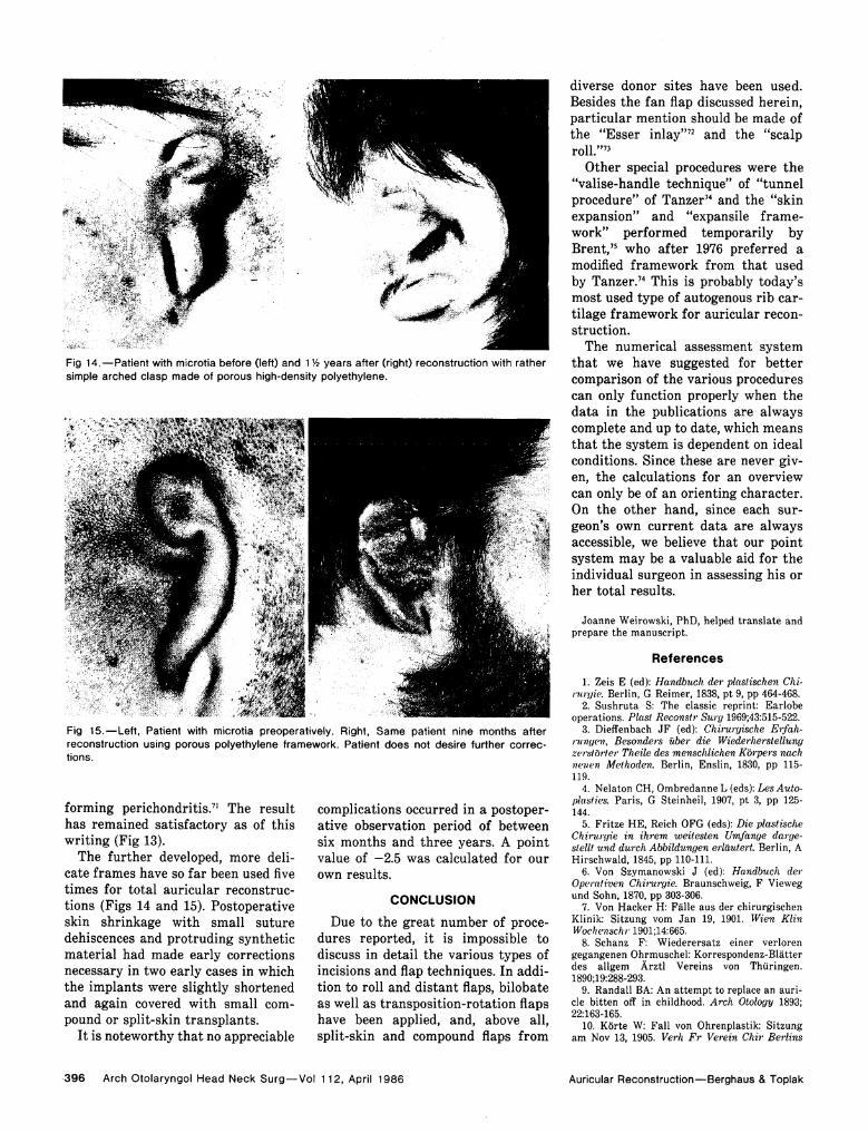
Fig 1 4 . — P a t i e n t w i th microt ia be fore (left) and 1V2 yea rs af ter ( r ight) recons t ruc t ion wi th ra ther s imple a rched c lasp m a d e of po rous h igh-dens i ty po lye thy lene.
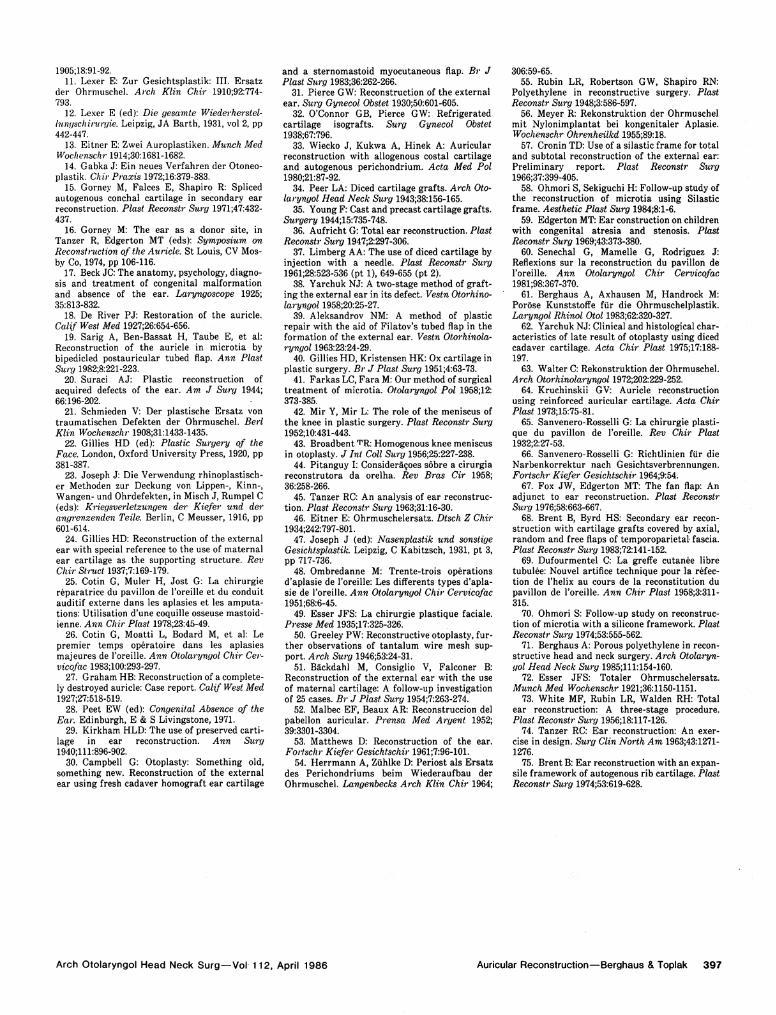
Fig 1 5 . — L e f t , Pat ient w i th microt ia preopera t ive ly . Right , S a m e pat ient nine mon ths af ter recons t ruc t ion us ing po rous po lye thy lene f ramework . Pat ient does not des i re fur ther co r rec t ions.
forming perichondritis.71 The result has remained satisfactory as of this writing (Fig 13).
The further developed, more delicate frames have so far been used five times for total auricular reconstructions (Figs 14 and 15). Postoperative skin shrinkage with small suture dehiscences and protruding synthetic material had made early corrections necessary in two early cases in which the implants were slightly shortened and again covered with small compound or split-skin transplants.
It is noteworthy that no appreciable
complications occurred in a postoperative observation period of between six months and three years. A point value of —2.5 was calculated for our own results.
C O N C L U S I O N
Due to the great number of procedures reported, it is impossible to discuss in detail the various types of incisions and flap techniques. In addition to roll and distant flaps, bilobate as well as transposition-rotation flaps have been applied, and, above all, split-skin and compound flaps from
diverse donor sites have been used. Besides the fan flap discussed herein, particular mention should be made of the "Esser inlay"72 and the "scalp roll."73
Other special procedures were the "valise-handle technique" of "tunnel procedure" of Tanzer74 and the "skin expansion" and "expansile framework" performed temporarily by Brent,75 who after 1976 preferred a modified framework from that used by Tanzer.74 This is probably today's most used type of autogenous rib cartilage framework for auricular reconstruction.
The numerical assessment system that we have suggested for better comparison of the various procedures can only function properly when the data in the publications are always complete and up to date, which means that the system is dependent on ideal conditions. Since these are never given, the calculations for an overview can only be of an orienting character. On the other hand, since each surgeon's own current data are always accessible, we believe that our point system may be a valuable aid for the individual surgeon in assessing his or her total results.
Joanne Weirowski, PhD, helped translate and prepare the manuscript
R e f e r e n c e s
1. Zeis E (ed): Handbuch der plastischen Chirurgie. Berlin, G Reimer, 1838, pt 9, pp 464-468.
2. Sushruta S: The classic reprint: Earlobe operations. Plast Reconstr Surg 1969;43:515-522.
3. Diefenbach JF (ed): Chirurgische Erfahrungen, Besonders über die Wiederherstellung zerstörter Theile des menschlichen Körpers nach neuen Methoden. Berlin, Enslin, 1830, pp 115-119.
4. Nelaton CH, Ombredanne L (eds): Les Autopiasties. Paris, G Steinheil, 1907, pt 3, pp 125-144.
5. Fritze HE, Reich OFG (eds): Die plastische Chirurgie in ihrem weitesten Umfange dargestellt und durch Abbildungen erläutert. Berlin, A Hirschwald, 1845, pp 110-111.
6. Von Szymanowski J (ed): Handbuch der Operativen Chirurgie. Braunschweig, F Vieweg und Sohn, 1870, pp 303-306.
7. Von Hacker H: Fälle aus der chirurgischen Klinik: Sitzung vom Jan 19, 1901. Wien Klin Wochenschr 1901;14:665.
8. Schanz F: Wiederersatz einer verloren gegangenen Ohrmuschel: Korrespondenz-Blätter des allgem Ärztl Vereins von Thüringen. 1890;19:288-293.
9. Randall BA: An attempt to replace an auricle bitten off in childhood. Arch Otology 1893; 22:163-165.
10. Körte W: Fall von Ohrenplastik: Sitzung am Nov 13, 1905. Verh Fr Verein Chir Berlins
3 9 6 A r c h O t o l a r y n g o l H e a d N e c k S u r g — V o l 1 1 2 , Apr i l 1 9 8 6 Aur icu lar R e c o n s t r u c t i o n — B e r g h a u s & Top lak
1905;18:91-92. 11. Lexer E: Zur Gesichtsplastik: III. Ersatz
der Ohrmuschel. Arch Klin Chir 1910;92:774-793.
12. Lexer E (ed): Die gesamte Wiederherstellungschirurgie. Leipzig, JA Barth, 1931, vol 2, pp 442-447.
13. Eitner E: Zwei Auroplastiken. Munch Med Wochenschr 1914;30:1681-1682.
14. Gabka J: Ein neues Verfahren der Otoneo-plastik. Chir Praxis 1972;16:379-383.
15. Gorney M, Falces E, Shapiro R: Spliced autogenous conchal cartilage in secondary ear reconstruction. Plast Reconstr Surg 1971;47:432-437.
16. Gorney M: The ear as a donor site, in Tanzer R, Edgerton MT (eds): Symposium on Reconstruction of the Auricle. St Louis, CV Mos-by Co, 1974, pp 106-116.
17. Beck JC: The anatomy, psychology, diagnosis and treatment of congenital malformation and absence of the ear. Laryngoscope 1925; 35:813-832.
18. De River PJ: Restoration of the auricle. Calif West Med 1927;26:654-656.
19. Sarig A, Ben-Bassat H, Taube E, et al: Reconstruction of the auricle in microtia by bipedicled postauricular tubed flap. Ann Plast Surg 1982;8:221-223.
20. Suraci AJ: Plastic reconstruction of acquired defects of the ear. Am J Surg 1944; 66:196-202.
21. Schmieden V: Der plastische Ersatz von traumatischen Defekten der Ohrmuschel. Berl Klin Wochenschr 1908;31:1433-1435.
22. Gillies HD (ed): Plastic Surgery of the Face. London, Oxford University Press, 1920, pp 381-387.
23. Joseph J: Die Verwendung rhinoplastisch-er Methoden zur Deckung von Lippen-, Kinn-, Wangen- und Ohrdefekten, in Misch J, Rumpel C (eds): Kriegsverletzungen der Kiefer und der angrenzenden Teile. Berlin, C Meusser, 1916, pp 601-614.
24. Gillies HD: Reconstruction of the external ear with special reference to the use of maternal ear cartilage as the supporting structure. Rev Chir Struct 1937;7:169-179.
25. Cotin G, Muler H, Jost G: La Chirurgie reparatrice du pavilion de l'oreille et du conduit auditif externe dans les aplasies et les amputations: Utilisation d'une coquille osseuse mastoid-ienne. Ann Chir Plast 1978;23:45-49.
26. Cotin G, Moatti L, Bodard M, et al: Le premier temps operatoire dans les aplasies majeures de l'oreille. Ann Otolaryngol Chir Cer-vicofac 1983;100:293-297.
27. Graham HB: Reconstruction of a completely destroyed auricle: Case report. Calif West Med 1927;27:518-519.
28. Peet EW (ed): Congenital Absence of the Ear. Edinburgh, E & S Livingstone, 1971.
29. Kirkham HLD: The use of preserved cartilage in ear reconstruction. Ann Surg 1940;111:896-902.
30. Campbell G: Otoplasty: Something old, something new. Reconstruction of the external ear using fresh cadaver homograft ear cartilage
and a sternomastoid myocutaneous flap. Br J Plast Surg 1983;36:262-266.
31. Pierce GW: Reconstruction of the external ear. Surg Gynecol Obstet 1930;50:601-605.
32. O'Connor GB, Pierce GW: Refrigerated cartilage isografts. Surg Gynecol Obstet 1938;67:796.
33. Wiecko J, Kukwa A, Hinek A: Auricular reconstruction with allogenous costal cartilage and autogenous perichondrium. Acta Med Pol 1980;21:87-92.
34. Peer LA: Diced cartilage grafts. Arch Otolaryngol Head Neck Surg 1943;38:156-165.
35. Young F: Cast and precast cartilage grafts. Surgery 1944;15:735-748.
36. Auf rieht G: Total ear reconstruction. Plast Reconstr Surg 1947;2:297-306.
37. Limberg AA: The use of diced cartilage by injection with a needle. Plast Reconstr Surg 1961;28:523-536 (pt 1), 649-655 (pt 2).
38. Yarchuk NJ: A two-stage method of grafting the external ear in its defect. Vestn Otorhino-laryngol 1958;20:25-27.
39. Aleksandrov NM: A method of plastic repair with the aid of Filatov's tubed flap in the formation of the external ear. Vestn Otorhinola-ryngol 1963:23:24-29.
40. Gillies HD, Kristensen HK: Ox cartilage in plastic surgery. Br J Plast Surg 1951;4:63-73.
41. Farkas LC, Fara M: Our method of surgical treatment of microtia. Otolaryngol Pol 1958;12: 373-385.
42. Mir Y, Mir L: The role of the meniscus of the knee in plastic surgery. Plast Reconstr Surg 1952;10:431-443.
43. Broadbent TR: Homogenous knee meniscus in otoplasty. J Int Coll Surg 1956;25:227-238.
44. Pitanguy I: Consideräcoes sobre a cirurgia reconstrutora da orelha. Rev Bras Cir 1958; 36:258-266.
45. Tanzer RC: An analysis of ear reconstruction. Plast. Reconstr Surg 1963;31:16-30.
46. Eitner E: Ohrmuschelersatz. Dtsch Z Chir 1934;242:797-801.
47. Joseph J (ed): Nasenplastik und sonstige Gesichtsplastik. Leipzig, C Kabitzsch, 1931, pt 3, pp 717-736.
48. Ombredanne M: Trente-trois operations d'aplasie de l'oreille: Les differents types d'apla-sie de l'oreille. Ann Otolaryngol Chir Cervicofac 1951;68:6-45.
49. Esser JFS: La Chirurgie plastique faciale. Presse Med 1935;17:325-326.
50. Greeley PW: Reconstructive otoplasty, further observations of tantalum wire mesh support. Arch Surg 1946;53:24-31.
51. Bäckdahl M, Consiglio V, Falconer B: Reconstruction of the external ear with the use of maternal cartilage: A follow-up investigation of 25 cases. Br J Plast Surg 1954;7:263-274.
52. Malbec EF, Beaux AR: Reconstruccion del pabellon auricular. Prensa Med Argent 1952; 39:3301-3304.
53. Matthews D: Reconstruction of the ear. Fortschr Kiefer Gesichtschir 1961;7:96-101.
54. Herrmann A, Zühlke D: Periost als Ersatz des Perichondriums beim Wiederaufbau der Ohrmuschel. Langenbecks Arch Klin Chir 1964;
306:59-65. 55. Rubin LR, Robertson GW, Shapiro RN:
Polyethylene in reconstructive surgery. Plast Reconstr Surg 1948;3:586-597.
56. Meyer R: Rekonstruktion der Ohrmuschel mit Nylon implan tat bei kongenitaler Aplasie. Wochenschr Ohrenheilkd 1955;89:18.
57. Cronin TD: Use of a silastic frame for total and subtotal reconstruction of the external ear: Preliminary report. Plast Reconstr Surg 1966;37:399-405.
58. Ohmori S, Sekiguchi H: Follow-up study of the reconstruction of microtia using Silastic frame. Aesthetic Plast Surg 1984;8:1-6.
59. Edgerton MT: Ear construction on children with congenital atresia and stenosis. Plast Reconstr Surg 1969;43:373-380.
60. Senechal G, Mamelle G, Rodriguez J: Reflexions sur la reconstruction du pavilion de l'oreille. Ann Otolaryngol Chir Cervicofac 1981;98:367-370.
61. Berghaus A, Axhausen M, Handrock M: Poröse Kunststoffe für die Ohrmuschelplastik. Laryngol Rhinol Otol 1983;62:320-327.
62. Yarchuk NJ: Clinical and histological characteristics of late result of otoplasty using diced cadaver cartilage. Acta Chir Plast 1975;17:188-197.
63. Walter C: Rekonstruktion der Ohrmuschel. Arch Otorhinolaryngol 1972;202:229-252.
64. Kruchinskii GV: Auricle reconstruction using reinforced auricular cartilage. Acta Chir Plast 1973;15:75-81.
65. Sanvenero-Rosselli G: La Chirurgie plastique du pavilion de l'oreille. Rev Chir Plast 1932;2:27-53.
66. Sanvenero-Rosselli G: Richtlinien für die Narbenkorrektur nach Gesichtsverbrennungen. Fortschr Kiefer Gesichtschir 1964;9:54.
67. Fox JW, Edgerton MT: The fan flap: An adjunct to ear reconstruction. Plast Reconstr Surg 1976;58:663-667.
68. Brent B, Byrd HS: Secondary ear reconstruction with cartilage grafts covered by axial, random and free flaps of temporoparietal- fascia. Plast Reconstr Surg 1983;72:141-152.
69. Dufourmentel C: La greffe cutanee libre tubulee: Nouvel artifice technique pour la refection de l'helix au cours de la reconstitution du pavilion de l'oreille. Ann Chir Plast 1958;3:311-315.
70. Ohmori S: Follow-up study on reconstruction of microtia with a silicone framework. Plast Reconstr Surg 1974;53:555-562.
71. Berghaus A: Porous polyethylene in reconstructive head and neck surgery. Arch Otolaryngol Head Neck Surg 1985;111:154-160.
72. Esser JFS: Totaler Ohrmuschelersatz. Munch Med Wochenschr 1921;36:1150-1151.
73. White MF, Rubin LR, Waiden RH: Total ear reconstruction: A three-stage procedure. Plast Reconstr Surg 1956;18:117-126.
74. Tanzer RC: Ear reconstruction: An exercise in design. Surg Clin North Am 1963;43:1271-1276.
75. Brent B: Ear reconstruction with an expansile framework of autogenous rib cartilage. Plast Reconstr Surg 1974;53:619-628.
A r c h O t o l a r y n g o l H e a d N e c k S u r g — V o l 1 1 2 , A p r i l 1 9 8 6 Aur icu lar R e c o n s t r u c t i o n — B e r g h a u s & Toplak 3 9 7