archives of oral biology -...
TRANSCRIPT
Archives of Oral Biology 63 (2016) 47–52
Interaction of Candida albicans with periodontal ligament fibroblastslimits biofilm formation over elastomer silicone disks
F. Alsalleeha,b,*, S. Williamsb, H. Jaberb
aKing Saud University, College of Dentistry, Restorative Dental Sciences, Saudi ArabiabDepartment of Surgical Specialties, University of Nebraska Medical Center, College of Dentistry, USA
A R T I C L E I N F O
Article history:Received 2 February 2015Received in revised form 18 June 2015Accepted 18 November 2015
Keywords:Candida albicansHyphaeAntifungalFibroblastsHost cells
A B S T R A C T
Objective: Candida albicans is the most numerous commensal and potentially pathological yeast in thehuman oral cavity. The purpose herein is to investigate the ability of C. albicans to form a biofilm in thepresence of periodontal ligament (PDL) fibroblasts.Material and methods: Silicone elastomer disks (SE) were transferred to wells containing PDL cells. C.albicans suspension was added to each well. The whole mixed culture was then allowed to form a biofilmfor 48 h. Biofilms were quantified by tetrazolium-salt-based (2,3-bis(2-methoxy-4-nitro-5-sulfophenyl)-5-[(phenyl amino) carbonyl]- 2H-tetrazolium hydroxide (XTT). Furthermore, biofilm was visualized byconfocal scanning laser and scanning electron microscopy. Migration of C. albicans and its ability to formbiofilms in presence of PDL cells was determined by using a transwell system. Last, elutes obtained fromco-culturing C. albicans and PDL cells were added to SE disks and covered with C. albicans. The cultureplate was then incubated to allow biofilm formation. Biofilms formed over SE disks were quantified usingXTT.Results: PDL cells significantly limited the biofilm formation at incubation interval of 48 h. PDL cellsinduced less biofilm compared to mature and thick hyphae in the absence of PDL cells as seen in confocalscanning laser and scanning electron microscopy. The presence of PDL cells limited the migration andformation of biofilm by C. albicans. Elutes obtained from co-culturing PDL cells with C. albicans for onehour induced significantly less biofilm.Conclusions: This is the first study to report that PDL cells exhibit antifungal activity. While the exactmechanism of how PDL cells limited biofilm formation is yet unknown, it was clear that competent PDLcells promote resistance to C. albicans biofilm formation.
ã 2015 Elsevier Ltd. All rights reserved.
Contents lists available at ScienceDirect
Archives of Oral Biology
journa l homepage: www.e lsev ier .com/ locate /aob
1. Introduction
Candida albicans, being the most frequent commensal andperiodically pathogenic yeast in the oral cavity (Conti & Gaffen,2010). Like many oral microbes, C. albicans form and live within abiofilm matrix composed of exopolysaccharides, proteins, andnucleic acids that protect them from the environment and immunesystem (Siqueira & Sen, 2004; Gomes, Fidel, Fidel, & de MouraSarquis, 2010). Biofilm formation, leading to immune-evasion andimmune-modulation of the host defense, is considered a keyvirulence factor of C. albicans. Formation of a biofilm can providethe C. albicans community protection against antimicrobial agentsas compared with those in a nomadic state (e.g. planktonic cells). C.
* Corresponding author at: King Saud University College of Dentistry RestorativeDental Sciences, Riyadh, Saudi Arabia. Fax: +966 14679016.
E-mail address: [email protected] (F. Alsalleeh).
http://dx.doi.org/10.1016/j.archoralbio.2015.11.0120003-9969/ã 2015 Elsevier Ltd. All rights reserved.
albicans in biofilm can be 100-fold more resistant to antifungalfluconazole and 20- to 30-fold more resistant to antifungalamphotericin B than planktonic cells (Kumamoto, 2002). Severalstudies and in vitro models have been established to characterize C.albicans biofilm formation on common bio-prosthetic materialssuch as polymethylmethacrylate, which is used in the constructionof dentures as well as silicone elastomer, a model material used forindwelling devices including catheters. Previous studies haveindicated that biofilm development occurs in three distinct phases.The first phase begins with the adherence of C. albicans; yeastforms, to its substrate (�0 to 11 h). Intermediate developmentalphase features attached cells proliferation to form microcoloniesand begin to deposit an extracellular matrix (�12 to 14 h). Finally,the maturation phase (�24 to 72 h) characterized by forming adense network of filamentous forms (pseudohyphae and hyphae),and become encased in the exopolymeric matrix (Ramage, Mowat,Jones, Williams, & Lopez-Ribot, 2009; Chandra et al., 2001).

0
0.2
0.4
0.6
0.8
1
1.2
1.4
1.6
Disk Disk/CA 80 CA42 Disk/CA 42 PDL/Disk/ CA42
mn294ta
eulavTTX
Biofilm Form a�on a�er 48 h+VE con trol
-64%
-VE control
*
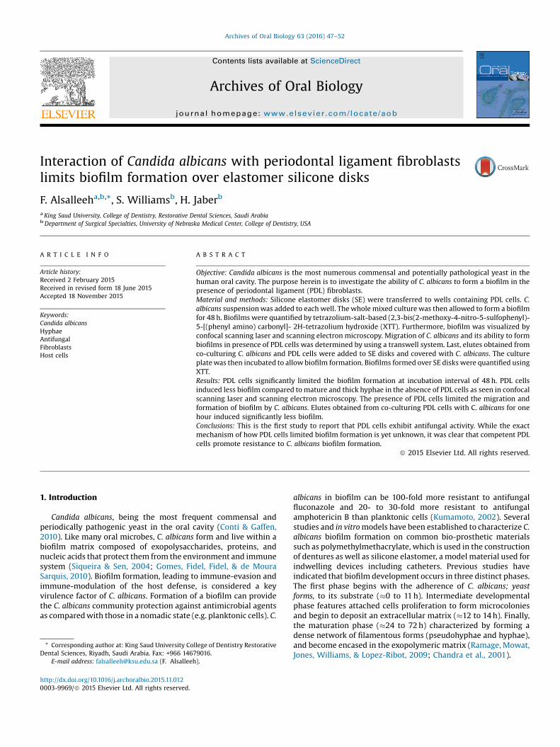
Fig. 1. Metabolic activity (XTT) of C. albicans biofilms formed for 48 h on SE disks inthe presence (PDL/Disk/CA42) or absence (Disk/CA42) of PDL cells. Disks withoutinoculation of C. albicans was used as negative control. C. albicans (CA42) grownonto wells was used as positive control. Disks inoculated with C. albicans (CA80) wasused to validate model used. All groups had n = 6. Metabolic activity is presented asan optical density at 492 nm. *P value of groups compared was significantlydifferent at a = 0.05.
48 F. Alsalleeh et al. / Archives of Oral Biology 63 (2016) 47–52
Periodontitis is a chronic inflammatory disease affecting theintegrity of tooth supporting tissues induced by variety ofmicroorganisms, including yeast (Slots, Rams, & Listgarten,1988). More recently, 30% of patients with chronic periodontitiscompared to 15% with healthy subjects had C. albicans inperiodontal pockets (Canabarro et al., 2013). C. albicans can evadethe host defense and induce a complex immune response thatultimately determines the clinical outcome of the infection.Polymorphonuclear neutrophils and macrophages are known tobe the most important inflammatory cells involved in the defenseagainst C. albicans (Ashman & Papadimitriou, 1995). Periodontalligament fibroblasts (PDL cells) are involved in the formation andmaintenance of periodontal fibrous tissue connecting teeth to thealveolar bone (Beertsen, McCulloch, & Sodek,1997). PDL cells play acrucial role in the early infection as well as resolution stage ofinfection at root surfaces (Jonsson, Nebel, Bratthall, & Nilsson,2011). Thus, studying the interaction between C. albicans infectionand periodontal cells should advance current understanding of themechanism involved in the etiology of chronic periodontitisrelated to fungal infections.
The central hypothesis of this investigation was that PDL cellsmay influence the biofilm formation by C. albicans. There were nodata published on the interaction of PDL cells with C. albicans. It isunknown whether PDL cells have any antifungal activity during C.albicans colonization. Using co-culture model system, the currentstudy sought to determine whether PDL cells results in less biofilmformation by C. albicans on silicone disks (SE). The flat sheeting ofSE disks was chosen to facilitate biofilm formation and produceundistorted images of biofilm formed by microscopic techniques asdescribed previously (Kuhn, Chandra, Mukherjee, & Ghannoum,2002). It also allows to design future studies aimed to understandthe pathogenicity in other ex/in vivo oral models.
2. Material and methods
2.1. Cell culture, C. albicans, and growth conditions
Primary human PDL fibroblasts isolated from human periodon-tal tissues were obtained from ScienCell (Carlsbad, CA) and grownin complete culture medium; Dulbecco’s modified Eagle's medium(Life Technologies, CA) supplemented with antibiotics and 10%fetal bovine serum (FBS), at 37 �C in a humidified 5% CO2
atmosphere. Cells between the 4th and 6th passages were usedin the present study.
The C. albicans wild strain CA42 (formerly known as SC5314(Fonzi & Irwin, 1993) was used. A non-filamentous; unable to formbiofilms, cph1D/efg1D double mutant strain CA80 (Lo et al., 1997),was used to validate model presented. Both strains were a courtesyof Dr. Audrey Atkin, University of Nebraska, Lincoln and weregrown in yeast extract-peptone-dextrose (YPD) medium (DifcoLaboratories, Detroit, MI.) from fresh Sabouraud dextrose agarplates (Difco) and incubated for 24 h at 37 �C in a shaker at 250 rpm.Cells were harvested and washed twice with phosphate bufferedsaline (PBS). Cells were then re-suspended in 10 mL of PBS, countedfollowing serial dilution, standardized, and used immediately.
2.2. Silicone disks preparation
Silicone elastomer (SE) was purchased (Invotec InternationalIncorporated, Jacksonville, FL). This material was shown topromote C. albicans biofilm and often used in indwelling devices(Hawser & Douglas, 1994). The SE is supplied as a flat sheet thatfacilitates quantifications and imaging the biofilm formed. Theywere cut with a carpenter's hole punch to produce standardizedsamples of 1.5 mm thick, and 3 mm diameter. SE disks werewashed, autoclaved and incubated in fetal bovine serum (FBS) for
24 h at 37 �C. The pretreatment with FBS is known to promotehyphal formation (Chandra et al., 2001).
2.3. Quantitative measurement of biofilms
PDL cells that reached confluence in culture medium werecollected, washed, and counted with a haemocytometer. A total of105 cells/well were plated in complete culture medium onto 24-well plates and grown overnight in 5% CO2 at 37 �C to allowadherence to the surface. The SE disks were transferred to wellsaccording to following groups (n = 6, repeated three times): (Group1) no PDL cells or C. albicans, (Group 2) CA80 strain, (Group 3)CA42 strain, and (Group 4) CA42 strain and PDL cells. Group 1 and2 served as negative controls. Additional wells pretreated with FBSwith no SE disks received CA42 strain served as positive control. C.albicans suspension containing 105 cells was used. The wholemixed culture, in complete culture medium; was then allowed toform a biofilm at 37 �C in a humidified 5% CO2 atmosphere.Following maturation for 48 h, SE disks were transferred to newculture plates and biofilms were quantified by tetrazolium-salt-based (2,3-bis(2-methoxy-4-nitro-5-sulfophenyl)-5-[(phenyl ami-no) carbonyl]-2H-tetrazolium hydroxide (XTT) as describedpreviously (Chandra et al., 2001). The reduction of XTT to formazancrystals can only take place in the presence of viable cells and thenecessary reductase enzymes.
2.4. Confocal microscopy analysis of biofilms
Biofilm formation by C. albicans on SE disks in the presence ofPDL cells was investigated using confocal scanning laser micros-copy(CSLM) as described previously (Chandra, Mukherjee, &Ghannoum, 2008). New experiments were designed as describedabove. After co-culturing SE disks with C. albicans in the presenceof PDL cells, SE disks were stained with FUN-1 (L7009, MolecularProbes, Eugene, Oreg.). Following biofilm formation, disks wereremoved and transferred to new 24-well plates. Wells containingbiofilm disks were submerged in FUN-1 at a final concentration of10 mM (from 10 mM stock). The plates were then incubated for30 min at 37 �C. The disks were removed from the wells, placed in35-mm glass-bottom microwell dishes (MatTek Corp., Ashland,

Fig. 2. Confocal laser scanning microscopy (CLSM) of C. albicans (CA42) biofilms grown on SE disks and stained with 10 mM FUN-1 in the absence (A) or presence (C) of PDLcells. (B) and (D) are differential interference contrast images of (A) and (C) respectively. Images taken with an Olympus FV500-IX81 inverted CSLM system. Fluorescenceoptical and DIC images were collected with a 20� lens using 488 nm excitation and 522 nm emission (n = 3).
F. Alsalleeh et al. / Archives of Oral Biology 63 (2016) 47–52 49
Mass.), and observed by using an Olympus FV500-IX81 invertedCSLM system. Fluorescence optical and DIC images were collectedwith a 20x lens using 488 nm excitation and 522 nm emission.
2.5. Scanning electron microscopy
In a parallel experiment, SE disks were prepared for SEM usingstandardized method described previously (Chandra et al., 2008).They were fixed with 10% glutaraldehyde, followed by fixation withosmium tetroxide. This was followed by a series of ethanoldehydration steps, and the prepared samples were sputter coatedwith Au-Pd (60/40 ratio) and viewed with a model JEOL6100 microscope.
2.6. Migration of C. albicans in the presence of PDL cells
Migration of C. albicans and its ability to form biofilms wasinspired from previous studies (Wozniok et al., 2008), by using thesame experimental design described above. First, migration of C.albicans was studied using transwell system without a substrate;SE disks. A total of 105 of PDL cells were plated in culture mediumonto a transwell system (0.4 mm pore size, Corning Incorporated,Lowell, MA) for 24-well plates and grown overnight in 5% CO2 at37 �C to allow adherence to the surface. The SE disks were placedonto lower compartment of the system of wells had PDL cells. C.
albicans suspension containing 105 cells was added to uppercompartment of the system. An addition SE disks were placed ontoempty well; with no PDL cells which served as positive controls.Biofilms formed over SE disks were quantified using XTT after 72 has described above.
2.7. C. albicans growth in conditioned media
Supernatant obtained from co-culturing C. albicans (CA42) withPDL cells was used as previously described (Chandra, McCormick,Imamura, Mukherjee, & Ghannoum, 2007) to analyze biofilmsformation. PDL cells were seeded at a concentration of 2 � 106 cellsonto 75-cm2
flasks and grown overnight in 5% CO2 at 37 �C to allowadherence to the surface. A suspension of C. albicans wild strainCA42 was added to flasks containing PDL cells at ratio of 1:1, andwas labeled solution A. An additional flask of PDL cells was leftuntreated, and was labeled solution B. After one hour of incubation,elutes were collected and centrifuged at low speed (1000 RPM) for10 min. Each solution was filtered through a 0.2 mm syringe filter.SE disks treated with FBS overnight were placed onto flat-bottom96-well plates. All SE disks received 2 �104 cells of C. albicans(CA42) and covered with 200 mL of elute A or B (n = 6). The cultureplate was then incubated for 48 h at 37 �C, 5% CO2 to allow biofilmformation. Biofilms formed over SE disks were quantified usingXTT described above.

Fig. 3. SEM showing biofilms formed by C. albicans (CA42) alone on SE disks (A and B), or with PDL cells (C and D). SEM analyses at accelerating voltage of 10 kV andmagnifications at 1.00 k and 5.0 k (n = 3).
50 F. Alsalleeh et al. / Archives of Oral Biology 63 (2016) 47–52
2.8. Statistical analysis
Each experiment was independently performed at least threetimes on separate days; data shown in the figures are from onerepresentative experiment. For quantification of biofilms by XTT(Sections 2.3,2.6 and 2.7), one-way analysis of variance wasperformed to compare means of multiple groups, and the one-tailed Student’s t-test was used for analysis of two groups (n = 6).Results with a P-value less than 0.05 were considered statisticallysignificant.
3. Results
3.1. Quantitative and microscopy analysis of C. albicans biofilms in thepresence or absence of PDL cells
The results presented have shown that PDL cells significantlylimited the biofilm formation at incubation interval of 48 h(P < 0.001) (Fig. 1). The reduction in biofilm formation was 64%(Fig. 1). The mutant form of C. albicans strain (CA80) was unable toform biofilm and was used to validate model presented. C. albicansthat was allowed to grow on FBS-coated wells formed significantbiofilm and was used as positive control (Fig. 1).
To visualize the biofilm formed over SE after 48 h, CLSM wasutilized. In the absence of PDL cells, C. albicans (CA42) consistentlyproduced mature and thick hyphae (Fig. 2A and B). The presence ofPDL cells induced less mature and scattered hyphae formation(Fig. 2C and D). Furthermore, SE disks were analyzed by SEM.Biofilm formation was more mature and thick (Fig. 3A and B)compared to disks co-cultured with PDL cells (Fig. 3C and D).
3.2. PDL cells and conditioned media limit C. albicans migration andbiofilms formation
It was noted that C. albicans were able to migrate and formmature biofilms on FBS-coated wells after 72 h compared to wells
with PDL cells (data not shown). Therefore, studies were designedto analyze the effect of PDL cells on the migration of C. albicans toform biofilm on SE disks. The presence of PDL cells limited themigration and formation of biofilm by C. albicans (Fig. 4A).Furthermore, elutes obtained from co-culturing PDL cells with C.albicans for one hour induced significantly less robust biofilmscompared to elutes obtained from C. albicans culture; solution B(Fig. 4B; P < 0.05).
4. Discussion
Oral C. albicans infections have to overcome the immuneresponse mediated by supporting tissue and cells of roots.Therefore, the present study has utilized an in vitro biofilm modelto investigate the ability of C. albicans to form a biofilm on SE disksin the presence of PDL cells. Results presented clearly demonstrat-ed that PDL cells limit the formation of biofilms by C. albicans on itssubstrates. To our knowledge, this is the first report to study theinteraction between PDL cells and C. albicans and its ability to forma biofilm.
The present study was motivated by Chandra et al. (2007) whodemonstrated that co-cultured C. albicans with blood mononuclearcells (BMCs) enhanced biofilm formation. The current studiesshowed that the coculture of PDL cells with C. albicans limits theability of this pathogen to form biofilms. Therefore, the dynamicinteractions of C. albicans with different immune cells may result indifferent outcome. In their study, C. albicans were allowed toadhere to synthetic disks prior to co-culturing with BMCs. It isgenerally accepted that the phagocytosis-induced apoptosis ofBMCs and subsequent clearance by macrophages is a prerequisitefor the resolution of infection-associated inflammation, while thefailure to undergo apoptosis creates a pathological situation(Zhang, Hirahashi, Cullere, & Mayadas, 2003; Kobayashi et al.,2003). One aspect of C. albicans pathogenicity is related to itsability to form biofilms (Kumamoto & Vinces, 2005). Utilizing aquantitative viability assay (XTT), it was shown that PDL cells

Fig. 4. (A) Metabolic activity (XTT) of C. albicans biofilms formed after migrationthrough transwell system for 72 h on SE disks in the absence or presence of PDL cells(n = 6). (B) Metabolic activity (XTT) of C. albicans biofilms formed after 48 h on SEdisks submerged in solution A (PDL cells stimulated by C. albicans, CA42); orsolution B (C. albicans alone, CA42) for one hour (n = 6). Metabolic activity ispresented as an optical density at 492 nm. *P value of groups compared wassignificantly different at a = 0.05.
F. Alsalleeh et al. / Archives of Oral Biology 63 (2016) 47–52 51
limited C. albicans biofilm formation. The CLSM and SEM analysiswas conducted to visualize the morphology and architecture ofbiofilms. It was clear that the presence of PDL cells resulted in lessmature biofilms over SE disks. While the mechanism of how PDLcells limited biofilm formation is yet unknown, it is clear thatcompetent PDL cells promote resistance to C. albicans biofilmformation.
Although, C. albicans can cause infectious lesions in yeast andfilamentous forms, studies have shown that its ability to formbiofilm is a key factor to host invasion and tissue destruction(Kurzai, Schmitt, Brocker, Frosch, & Kolb-Maurer, 2005; Farrell,Hawkins, & Ryder, 1983; Hausauer, Gerami-Nejad, Kistler-Ander-son, & Gale, 2005). Several studies have indicated that Efg1 and/orCph1 transcription factors of C. albicans are involved in the biofilmformation (Kumamoto & Vinces, 2005). The strain CA80, used inherein, lacks both genes and was used to validate model presented.XTT assay has been used as a viability assay of different organismsincluding mammalian cells, bacteria and fungi (Chandra et al.,2001; Scudiero et al., 1988; McCluskey, Quinn, & McGrath, 2005).In the present studies, PDL cells alone also metabolized XTT (Fig.1).
However, signals produced by C. albicans biofilm in the absence ofPDL cells were significantly higher than that of biofilm formed by C.albicans co-cultured with PDL cells. These findings confirm thatPDL cells exert anti-biofilm activity and perhaps down regulatedEfg1 and/or Cph1 genes. The current model presented is able to testsuch hypothesis.
The current study shed some light on antifungal activity of PDLcells. By studying the interaction of PDL cells and C. albicans, it wasdemonstrated that PDL cells exerted a slow-down of C. albicansmigration (Fig. 4A). Furthermore, results presented clearlydemonstrated that elutes from such interaction caused a reductionin biofilm formation (Fig. 4B). It is plausible that PDL cells are ableto change the environment by secreting inflammatory cytokines.IL-17 is proinflammatory cytokine secreted by a subset of CD4+ Thelper cells, named Th17 cells. It has been shown that Th17 cellsregulate host defense through neutrophil trafficking (Yu et al.,2007) and are involved in several inflammatory diseases includingchronic lesions of human periodontal disease (Cardoso et al.,2009). Interestingly, the IL-17 receptor (IL-17R) is expressed byhuman PDL cells (Zhu et al., 2011), suggesting that PDL cellsrespond to Th17 cells of adaptive immunity to promote inflamma-tion. It should be noted that Th17 cells are known to play aprotective role against C. albicans infection in the oral cavity (Conti& Gaffen, 2010). PDL cells also secrete cytokines such as IL-10 andtransforming growth factor-b (TGF-b), which are anti-inflamma-tory and necessary for tissue repair during the healing process(Colic et al., 2009; Deschner et al., 2000; Pinkerton et al., 2008).Therefore PDL cells may play an integral role in the resistance to C.albicans infection.
In conclusion, the present studies have shown that PDL cellslimit the formation of biofilms by C. albicans. Furthermore, PDLcells were able to change the environment and inhibit migration ofC. albicans to form biofilm. The current in vitro model presentedwill allow the conduction of further studies to understand thepathogenicity of C. albicans biofilms formation, as well as the hostresponse.
Conflict of interest
The authors deny any conflicts of interest.
Ethical approval
None.
Acknowledgments
We would like to thank Dennis Feely, Ph.D and You Zhou, Ph.Dfor the assistance to conduct CSLM and SEM analysis.
References
Ashman, R. B., & Papadimitriou, J. M. (1995). Production and function of cytokines innatural and acquired immunity to Candida albicans infection. MicrobiologicalReviews, 59(4), 646–672.
Beertsen, W., McCulloch, C. A., & Sodek, J. (1997). The periodontal ligament: aunique, multifunctional connective tissue. Periodontology 2000, 13, 20–40.
Canabarro, A., Valle, C., Farias, M. R., Santos, F. B., Lazera, M., & Wanke, B. (2013).Association of subgingival colonization of Candida albicans and other yeastswith severity of chronic periodontitis. Journal of Periodontal Research, 48(4),428–432.
Cardoso, C. R., Garlet, G. P., Crippa, G. E., Rosa, A. L., Junior, W. M., Rossi, M. A., et al.(2009). Evidence of the presence of T helper type 17 cells in chronic lesions ofhuman periodontal disease. Oral Microbiology and Immunology, 24(1), 1–6.
Chandra, J., Kuhn, D. M., Mukherjee, P. K., Hoyer, L. L., McCormick, T., & Ghannoum,M. A. (2001). Biofilm formation by the fungal pathogen Candida albicans:development, architecture, and drug resistance. Journal of Bacteriology, 183(18),5385–5394.
Chandra, J., McCormick, T. S., Imamura, Y., Mukherjee, P. K., & Ghannoum, M. A.(2007). Interaction of Candida albicans with adherent human peripheral blood

52 F. Alsalleeh et al. / Archives of Oral Biology 63 (2016) 47–52
mononuclear cells increases C. albicans biofilm formation and results indifferential expression of pro- and anti-inflammatory cytokines. Infection andImmunity, 75(5), 2612–2620.
Chandra, J., Mukherjee, P. K., & Ghannoum, M. A. (2008). In vitro growth and analysisof Candida biofilms. Nature Protocols, 3(12), 1909–1924.
Colic, M., Gazivoda, D., Vucevic, D., Vasilijic, S., Rudolf, R., & Lukic, A. (2009).Proinflammatory and immunoregulatory mechanisms in periapical lesions.Molecular Immunology, 47(1), 101–113.
Conti, H. R., & Gaffen, S. L. (2010). Host responses to Candida albicans: Th17 cells andmucosal candidiasis. Microbes and Infection, 12(7), 518–527.
Deschner, J., Arnold, B., Kage, A., Zimmermann, B., Kanitz, V., & Bernimoulin, J. P.(2000). Suppression of interleukin-10 release from human periodontalligament cells by interleukin-1beta in vitro. Archives of Oral Biology, 45(2),179–183.
Farrell, S. M., Hawkins, D. F., & Ryder, T. A. (1983). Scanning electron microscopestudy of Candida albicans invasion of cultured human cervical epithelial cells.Sabouraudia, 21(3), 251–254.
Fonzi, W. A., & Irwin, M. Y. (1993). Isogenic strain construction and gene mapping inCandida albicans. Genetics, 134(3), 717–728.
Gomes, C., Fidel, S., Fidel, R., & de Moura Sarquis, M. I. (2010). Isolation andtaxonomy of filamentous fungi in endodontic infections. Journal of Endodontics,36(4), 626–629.
Hausauer, D. L., Gerami-Nejad, M., Kistler-Anderson, C., & Gale, C. A. (2005). Hyphalguidance and invasive growth in Candida albicans require the Ras-like GTPaseRsr1p and its GTPase-activating protein Bud2p. Eukaryotic Cell, 4(7), 1273–1286.
Hawser, S. P., & Douglas, L. J. (1994). Biofilm formation by Candida species on thesurface of catheter materials in vitro. Infection and Immunity, 62(3), 915–921.
Jonsson, D., Nebel, D., Bratthall, G., & Nilsson, B. O. (2011). The human periodontalligament cell: a fibroblast-like cell acting as an immune cell. Journal ofPeriodontal Research, 46(2), 153–157.
Kobayashi, S. D., Voyich, J. M., Somerville, G. A., Braughton, K. R., Malech, H. L.,Musser, J. M., et al. (2003). An apoptosis-differentiation program in humanpolymorphonuclear leukocytes facilitates resolution of inflammation. Journal ofLeukocyte Biology, 73(2), 315–322.
Kuhn, D. M., Chandra, J., Mukherjee, P. K., & Ghannoum, M. A. (2002). Comparison ofbiofilms formed by Candida albicans and Candida parapsilosis on bioprostheticsurfaces. Infection and Immunity, 70(2), 878–888.
Kumamoto, C. A., & Vinces, M. D. (2005). Alternative Candida albicans lifestyles:growth on surfaces. Annual Review of Microbiology, 59, 113–133.
Kumamoto, C. A. (2002). Candida biofilms. Current Opinion in Microbiology, 5(6),608–611.
Kurzai, O., Schmitt, C., Brocker, E., Frosch, M., & Kolb-Maurer, A. (2005).Polymorphism of Candida albicans is a major factor in the interaction withhuman dendritic cells. International Journal of Medical Microbiology, 295(2),121–127.
Lo, H. J., Kohler, J. R., DiDomenico, B., Loebenberg, D., Cacciapuoti, A., & Fink, G. R.(1997). Nonfilamentous C. albicans mutants are avirulent. Cell, 90(5), 939–949.
McCluskey, C., Quinn, J. P., & McGrath, J. W. (2005). An evaluation of three new-generation tetrazolium salts for the measurement of respiratory activity inactivated sludge microorganisms. Microbial Ecology, 49(3), 379–387.
Pinkerton, M. N., Wescott, D. C., Gaffey, B. J., Beggs, K. T., Milne, T. J., & Meikle, M. C.(2008). Cultured human periodontal ligament cells constitutively expressmultiple osteotropic cytokines and growth factors, several of which areresponsive to mechanical deformation. Journal of Periodontal Research, 43(3),343–351.
Ramage, G., Mowat, E., Jones, B., Williams, C., & Lopez-Ribot, J. (2009). Our currentunderstanding of fungal biofilms. Critical Reviews in Microbiology, 35(4), 340–355.
Scudiero, D. A., Shoemaker, R. H., Paull, K. D., Monks, A., Tierney, S., Nofziger, T. H., etal. (1988). Evaluation of a soluble tetrazolium/formazan assay for cell growthand drug sensitivity in culture using human and other tumor cell lines. CancerResearch, 48(17), 4827–4833.
Siqueira, J. F. Jr., & Sen, B. H. (2004). Fungi in endodontic infections. Oral Surgery, OralMedicine, Oral Pathology, Oral Radiology, and Endodontics, 97(5), 632–641.
Slots, J., Rams, T. E., & Listgarten, M. A. (1988). Yeasts, enteric rods andpseudomonads in the subgingival flora of severe adult periodontitis. OralMicrobiology and Immunology, 3(2), 47–52.
Wozniok, I., Hornbach, A., Schmitt, C., Frosch, M., Einsele, H., Hube, B., et al. (2008).Induction of ERK-kinase signalling triggers morphotype-specific killing ofCandida albicans filaments by human neutrophils. Cellular Microbiology, 10(3),807–820.
Yu, J. J., Ruddy, M. J., Wong, G. C., Sfintescu, C., Baker, P. J., Smith, J. B., et al. (2007). Anessential role for IL-17 in preventing pathogen-initiated bone destruction:recruitment of neutrophils to inflamed bone requires IL-17 receptor-dependentsignals. Blood, 109(9), 3794–3802.
Zhang, B., Hirahashi, J., Cullere, X., & Mayadas, T. N. (2003). Elucidation of molecularevents leading to neutrophil apoptosis following phagocytosis: cross-talkbetween caspase 8, reactive oxygen species, and MAPK/ERK activation. Journalof Biological Chemistry, 278(31), 28443–28454.
Zhu, L., Wu, Y., Wei, H., Xing, X., Zhan, N., Xiong, H., et al. (2011). IL-17R activation ofhuman periodontal ligament fibroblasts induces IL-23 p19 production:Differential involvement of NF-kappaB versus JNK/AP-1 pathways. MolecularImmunology, 48(4), 647–656.