archaea in coastal marine environments
TRANSCRIPT

Proc. Natl. Acad. Sci. USAVol. 89, pp. 5685-5689, June 1992Ecology
Archaea in coastal marine environments(achabaterla/phyoey/batwe nt/n a eclogU)
EDWARD F. DELONG*Biology Department, Woods Hole Oceanographic Institution, Woods Hole, MA 02543
Communicated by George N. Somero, March 17, 1992 (received for review February 4, 1992)
ABSTRACT Archaea (archaebacteria) are a phenotypi-cally diverse group of microorganisms that share a commonevolutionary history. There are four general phenotypic groupsof archaea: the methanogens, the extreme halophiles, thesulfate-reducing archaea, and the extreme thermophiles. In themarine environment, archaeal habitats are generally limited toshallow or deep-sea anaerobic sediments (free-living and en-dosymbiotic methanogens), hot springs or deep-sea hydrother-mal vents (methanogens, sulfate reducers, and extreme ther-mophiles), and highly saline land-locked seas (halophiles). Thisreport provides evidence for the widespread occurrence ofunusual archaea in oxygenated coasl surface waters of NorthAmerica. Quantitative mates indicated that up to 2% of thetotal ribosomal RNA extracted fom coastal bacterioplanktonassemblages was archaeal. Archaeal small-subunit ribosomalRNA-encoding DNAs (rDNAs) were coned from mixed bac-terioplankton populations collected at geographically distantsampling sites. Phylogenetic and nucleotide signature analysesof these cloned rDNAs revealed the presence of two lin ofarchaea, each sharing the dinic signatures and structuralfeatures previously established for the domain Archaea. Bothof these lineages were found in bacterioplankton populationscollected off the east and west coass of North America. Theabundance and distribution of these arcaea in oxic coastalsurface waters suggests that these microorgnisms representundescribed physiological types of arcaea, which reside andcompete with aerobic, mesophilic eubacteria in marine coastalenvironments.
Application of molecular phylogenetic analyses to ecologicalquestions has recently enhanced the ability of microbialecologists to assess naturally occurring diversity in mixedmicrobial assemblages (1-6). In this approach, genes encod-ing phylogenetically informative macromolecules, derivedfrom extracted nucleic acids of mixed microbial populations,are clonally isolated, sorted, and sequenced. Analysis of therecovered sequences allows inference of the phylogeneticaffiliation of individual population constituents. Addition-ally, this sequence information aids in the design of taxa-specific oligodeoxynucleotide probes (7, 8), for monitoringthe spatial and temporal variability of specific groups. Thisapproach has led to the phylogenetic identification of previ-ously uncultured microbes (3-5, 7), as well as estimates oftheir abundance or distribution (3, 5, 8).To characterize planktonic versus surface-attached marine
bacteria, we cloned and analyzed small-subunit rRNA se-quences derived from microbial assemblages occupyingthese different habitats. Ribosomal RNA genes were ampli-fied (9, 10) from purified, mixed-population nucleic acids (3).The amplified rRNA genes were then cloned, sorted, andsequenced, and compared with a data base of aligned rRNAsequences from well-characterized microorganisms (11). Dueto the predominance of eukaryotic nucleic acids in many
macroaggregate samples, eubacterial- or archaeal-biasedPCR primers were routinely used to exclude the amplificationof eukaryotic ribosomal RNA-encoding DNA (rDNA). Sur-prisingly, archaeal rDNA was detected in many samples.This report describes the detection of two marine archaeallineages and their preliminary phylogenetic and ecologicalcharacterization.
METHODSBacterioplankton Collection. Coastal water samples were
collected and screened through a 10-L&m Nytex mesh prefil-ter. Bacterioplankton were concentrated from these 10-pm-filtered water samples by using a CH2PR filtration unit(Amicon) fitted with a polysulfone hollow-fiber filter (30-kDacutoff). Twenty-liter samples were concentrated to a finalvolume of 100-150 ml. The resulting bacterioplankton con-centrates were centrifuged (27,500 x g, 30 min, 40C), and thecell pellets were stored at -800C. Bacterial-cell densities inseawater and bacterioplankton concentrates were deter-mined by epifluorescence microscopy of glutaraldehyde-fixed, acridine orange-stained samples (12). Cell recoveries inthe concentrates ranged from 78% to 100o of the total cellsfiltered.
Extraction of Nucleic Acids and PCR Amplification. Cellpellets were lysed, and crude nucleic acids were purified asdescribed (5). Approximately 5-10 jug of this crude nucleicacid preparation was purified by CsCl equilibrium density-gradient centrifugation (100,000 x g, 5-16 hr, 200C) on aBeckman TL 100 ultracentrifuge using a TLA 100 rotor.Ribosomal DNA was amplified from purified DNA usingGeneAMP kit reagents (Perkin-Elmer/Cetus), as recom-mended by the manufacturer. Reaction mixtures contained 2mM MgCl2, 10 mM Tris'HCl, pH 8.3/50 mM KCI/200 pMdeoxynucleotide triphosphates/2.5 units of Thermus aquat-icus DNA polymerase/0.2 juM each of oligonucleotide prim-er/DNA template at 1 ng/pul. Thermal cycling was as follows:denaturation at 950C for 1.5 min, annealing at 550C for 1.5min, and extension at 720C for 1.5 min for a total of 30 cycles.The oligonucleotide primer sequences were as follows:
Eubac27F (13): AGA1492R (13): GGTEukF (10): AACEukR (10): TGAArch2lF: TTCArch958R: YCC
GTTTACCTGTCCCGGGGC
TGACTTGTTTTCTTGGTT
TCCGTTGATTGCATCGAM
TGGACGCCTAGGCYGTCC
CTCACTGCCTTCCCGAAT
AGTAGTACC TACGAT
Quantitative Hybridization Experiments. Crude nucleic ac-ids and purified rRNA standards were denatured in 0.5%glutaraldehyde, serially diluted, applied to nylon membranes(Hybond-N; Amersham) with a slot-blotting apparatus, andimmobilized by baking in a vacuum at 800C for 1 hr (14).Membranes were preincubated at 45TC for 0.5 hr in hybrid-
Abbreviation: rDNA, ribosomal RNA-encoding DNA.*Present address: Department of Biological Sciences, University ofCalifornia, Santa Barbara, CA 93106.
5685
The publication costs of this article were defrayed in part by page chargepayment. This article must therefore be hereby marked "advertisement"in accordance with 18 U.S.C. §1734 solely to indicate this fact.
Proc. Natl. Acad. Sci. USA 89 (1992)
ization buffer (0.9 M NaCI/50mM NaH2PO4, pH 7.0/5.0mMNa2 EDTA/0.5% SDS/10x Denhardt's solution/polyriboad-enylic acid at 0.5 mg/ml), followed by the addition of 2 x 10⁷cpm (specific activity 4-9 x 10⁸ cpm/,.g) of 32P-end-labeledoligonucleotide probe (14). After 12- to 16-hr incubation, themembranes were washed for 30 min at room temperature in1x SET (150 mM NaCl/20 mM Tris HCl, pH 7.8/1 mM Na2EDTA) containing 0.5% SDS, followed by a 30-min wash atthe indicated temperature. Oligodeoxynucleotide probe se-quences and wash temperatures were as follows:
Universal probe (8):Archaeal probe (14):Eubacterial probe (14):Eukaryote probe (15):Negative control (15):
ACG GGC GGT GTG TRC (450C)GTG CTC CCC CGC CAA TTC CT (56°C)GCT GCC TCC CGT AGG AGT (450C)GGG CAT CAC AGA CCT G (400C)GTG CCA GCM GCC GCG G (450C)
Dried membranes were exposed to preflashed film (KodakXRP-5) in the presence of an intensifying screen for 1-24 hrat -80°C. Autoradiographic signals were quantified fromvideo images with an interpretive densitometer (Scanalytics,Billerica, MA) with zero-dimensional analysis software. Thepercentage group-specific rRNA was estimated as the slopeof the group-specific probe bound per unit of rRNA, dividedby the slope of the universal probe bound per unit of rRNA.Values were background corrected and normalized to theslopes of group-specific probe bound per unit of rRNA forheterologous or homologous rRNA standards, as describedby Giovannoni et al. (3). The standard homologous andheterologous rRNAs used were as follows: Archaea: Halo-ferax volcandi, Desulfurococcus strain SY, Sulfolobus solfa-taricus, Methanobacterium thermoautotrophicum, Metha-nococcus jannaschii. Bacteria: Synechococcus PCC 6301,Bacillus megaterium, Escherichia coli, Oceanospirillum li-num, Alteromonas macleodii; Eukarya: Aequorea victoria,Saccharomyces cerevisiae, Alexandrium fundyense.Ribosomal DNA Cloning and Sequencing. Amplified DNA
from three to five separate reactions was pooled, phenol/chloroform, 1:1-extracted, chloroform-extracted, ethanol-precipitated, and resuspended in one-tenth vol sterile dis-tilled water (16). The purified, amplified archaeal rDNAswere cloned by using a commercially prepared vector (TAcloning system; Invitrogen, San Diego). Insert-containingclones were identified by agarose gel electrophoresis ofsmall-scale plasmid preparations (16). Denatured, double-stranded plasmid templates were sequenced by the dideoxynucleotide chain-termination method with Sequenase 2.0(United States Biochemical) following the manufacturer'srecommendations. Universal rRNA-specific sequencingprimers (13) and M13 forward and reverse primers (16) wereused in sequencing reactions.
Phylogenetic Analyses. Sequences were aligned to a database of previously determined rRNA sequences obtainedfrom the Ribosomal RNA Database Project (11). GenBankaccession numbers for sequences determined in this study areas follows: SBAR 5, M88075; SBAR 1A, M88074; WHAR Q,M88079; WHAR N, M88078; SBAR 12, M88076; and SBAR16, M88077. Least-squares distance matrix analyses (17)were based on evolutionary distances estimated from simi-larity values and using the correction of Jukes and Cantor(18). The sequence-editing and distance-analyses software ofOlsen (17) was obtained through the Ribosomal RNA Data-base Project (11). Parsimony "bootstrap" analyses (19, 20)were performed by using PAUP (version 3.0s; D. L. Swof-ford). The PHYLIP package was used for maximum-likelihoodanalyses (version 3.4; J. Felsenstein). All analyses wererestricted to comparison of 740 highly to moderately con-served sequence positions. Only those positions representedby a known base in all sequences were used in the analyses.
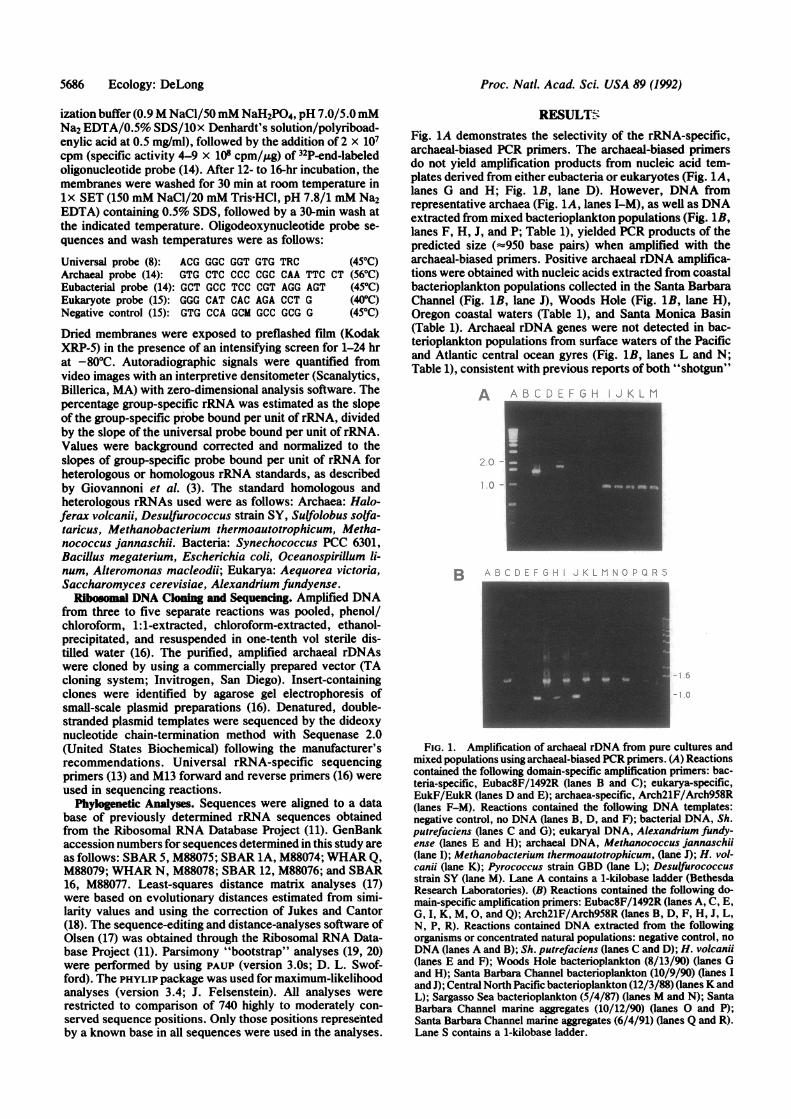
RESULTSFig. 1A demonstrates the selectivity of the rRNA-specific,archaeal-biased PCR primers. The archaeal-biased primersdo not yield amplification products from nucleic acid tem-plates derived from either eubacteria or eukaryotes (Fig. 1A,lanes G and H; Fig. 1B, lane D). However, DNA fromrepresentative archaea (Fig. 1A, lanes I-M), as well as DNAextracted from mixed bacterioplankton populations (Fig. 1B,lanes F, H, J, and P; Table 1), yielded PCR products of thepredicted size (=w950 base pairs) when amplified with thearchaeal-biased primers. Positive archaeal rDNA amplifica-tions were obtained with nucleic acids extracted from coastalbacterioplankton populations collected in the Santa BarbaraChannel (Fig. 1B, lane J), Woods Hole (Fig. 1B, lane H),Oregon coastal waters (Table 1), and Santa Monica Basin(Table 1). Archaeal rDNA genes were not detected in bac-terioplankton populations from surface waters of the Pacificand Atlantic central ocean gyres (Fig. 1B, lanes L and N;Table 1), consistent with previous reports of both "shotgun"
A
B_
FIG. 1. Amplification of archaeal rDNA from pure cultures andmixed populations using archaeal-biased PCR primers. (A) Reactionscontained the following domain-specific amplification primers: bac-teria-specific, Eubac8F/1492R (lanes B and C); eukarya-specific,EukF/EukR (lanes D and E); archaea-specific, Arch21F/Arch958R(lanes F-M). Reactions contained the following DNA templates:negative control, no DNA (lanes B, D, and F); bacterial DNA, Sh.putrefaciens (lanes C and G); eukaryal DNA, Alexandrium fundy-ense (lanes E and H); archaeal DNA, Methanococcus jannaschii(lane I); Methanobacterium thermoautotrophicum, (lane J); H. vol-canii (lane K); Pyrococcus strain GBD (lane L); Desulfurococcusstrain SY (lane M). Lane A contains a 1-kilobase ladder (BethesdaResearch Laboratories). (B) Reactions contained the following do-main-specific amplification primers: Eubac8F/1492R (lanes A, C, E,G, I, K, M, 0, and Q); Arch21F/Arch958R (lanes B, D, F, H, J, L,N, P, R). Reactions contained DNA extracted from the followingorganisms or concentrated natural populations: negative control, noDNA (lanes A and B); Sh. putrefaciens (lanes C and D); H. volcanii(lanes E and F); Woods Hole bacterioplankton (8/13/90) (lanes Gand H); Santa Barbara Channel bacterioplankton (10/9/90) (lanes IandJ); Central North Pacific bacterioplankton (12/3/88) (lanes K andL); Sargasso Sea bacterioplankton (5/4/87) (lanes M and N); SantaBarbara Channel marine aggregates (10/12/90) (lanes 0 and P);Santa Barbara Channel marine aggregates (6/4/91) (lanes Q and R).Lane S contains a 1-kilobase ladder.
5686 Ecology: DeLong
!:O -
Proc. Natl. Acad. Sci. USA 89 (1992) 5687
Table 1. Detection of archaeal rDNA and rRNA inbacterioplankton nucleic acid extracts
Group-specific rRNA,* %Sample site Date Ampt Archaea Bacteria Eucarya
Central Pacific 12/3/88 - ND 98.3 0.4Woods Hole 8/13/90 + 0.1 67.7 6.8Santa Barbara 10/9/90 +Woods Hole 3/9/91 + ND 70.0 22.9Santa Monica Basin 3/12/91 +Santa Barbara 6/4/91 + 2.3 54.5 41.2Santa Barbara 6/5/91 + 1.0 52.5 46.9Santa Barbara 6/6/91 + 1.6 64.2 52.6Oregon coast 7/23/91 +ND, not detected; -, experiment not done.
*See text for methods.tDNA that yielded PCR amplified (Amp) products of the predictedsize with archaeal-biased primers are indicated by a +.
cloning (5) and archaeal-specific hybridization probe analy-ses (3).The relative proportions of eubacterial, eukaryotic, and
archaeal rRNA in nucleic acid extracts were estimated toverify the presence of archaea in coastal bacterioplanktonpopulations. Specific rRNAs were quantified by measuringthe amount of radiolabeled, group-specific oligonucleotideprobe that bound to serial dilutions of mixed-populationrRNAs (refs. 3 and 8; Table 1). Archaeal rRNA accounted foras much as 1.0-2.3% of the total rRNA (up to 4% of the totalprokaryotic rRNA) in samples taken in June 1991 in the SantaBarbara Channel (Table 1). A smaller but measurable pro-portion of archaeal rRNA was detected in one Woods Holebacterioplankton sample.
Archaeal rDNA libraries were prepared from bacterio-plankton DNA samples collected from several different sam-pling sites and times (Woods Hole, 8/13/90; Santa BarbaraChannel, 10/9/90, 10/12/90, 6/4/91; Oregon coast, 7/23/
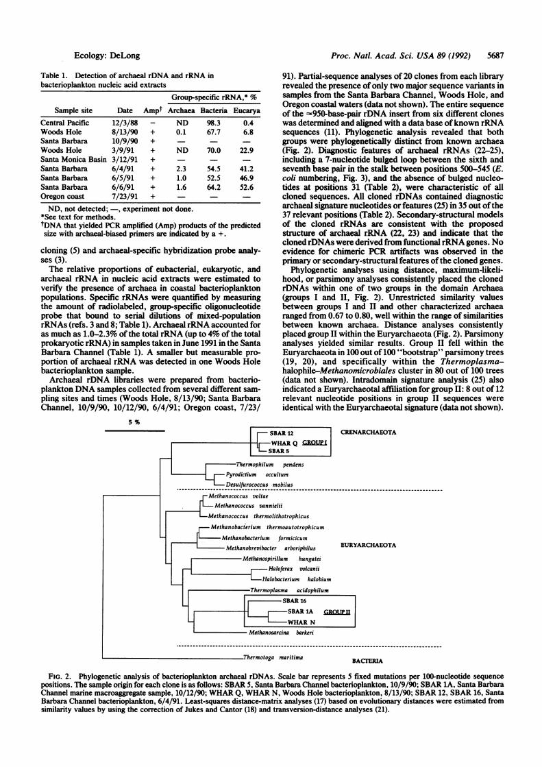
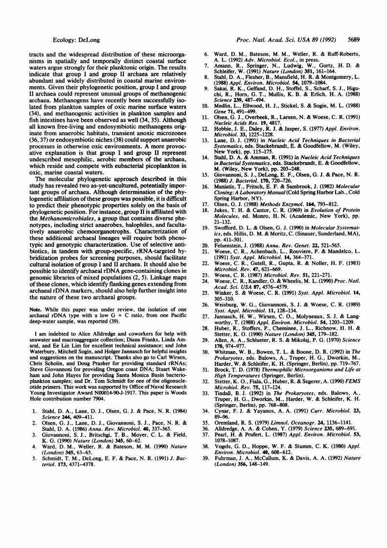
91). Partial-sequence analyses of 20 clones from each libraryrevealed the presence of only two major sequence variants insamples from the Santa Barbara Channel, Woods Hole, andOregon coastal waters (data not shown). The entire sequenceof the =950-base-pair rDNA insert from six different cloneswas determined and aligned with a data base ofknown rRNAsequences (11). Phylogenetic analysis revealed that bothgroups were phylogenetically distinct from known archaea(Fig. 2). Diagnostic features of archaeal rRNAs (22-25),including a 7-nucleotide bulged loop between the sixth andseventh base pair in the stalk between positions 500-545 (E.coli numbering, Fig. 3), and the absence of bulged nucleo-tides at positions 31 (Table 2), were characteristic of allcloned sequences. All cloned rDNAs contained diagnosticarchaeal signature nucleotides or features (25) in 35 out ofthe37 relevant positions (Table 2). Secondary-structural modelsof the cloned rRNAs are consistent with the proposedstructure of archaeal rRNA (22, 23) and indicate that thecloned rDNAs were derived from functionalrRNA genes. Noevidence for chimeric PCR artifacts was observed in theprimary or secondary-structural features ofthe cloned genes.
Phylogenetic analyses using distance, maximum-likeli-hood, or parsimony analyses consistently placed the clonedrDNAs within one of two groups in the domain Archaea(groups I and II, Fig. 2). Unrestricted similarity valuesbetween groups I and II and other characterized archaearanged from 0.67 to 0.80, well within the range of similaritiesbetween known archaea. Distance analyses consistentlyplaced group II within the Euryarchaeota (Fig. 2). Parsimonyanalyses yielded similar results. Group II fell within theEuryarchaeota in 100 out of 100 "bootstrap" parsimony trees(19, 20), and specifically within the Thermoplasma-halophile-Methanomicrobiales cluster in 80 out of 100 trees(data not shown). Intradomain signature analysis (25) alsoindicated a Euryarchaeotal affiliation for group II: 8 out of 12relevant nucleotide positions in group II sequences wereidentical with the Euryarchaeotal signature (data not shown).
5 %
SBAR 12 CRENARCHAEOTAWHAR Q GROUP ISBAR 5
IThermophilum pendensPyrodictium occultum
Desulfurococcus mobilus
Methanococcus voltaeMethanococcus vannielii
Methanococcus thermolithotrophicus
Methanobacterium thermoautotrophicum
Methanobacterium formicicumEURYARCHAEOTA
Methanospirillum hungatei
t Haloferax volcaniiI Halobacterium halobium
Methanosarcina barkeri
_Thermotoga maritima BACTERIA
FIG. 2. Phylogenetic analysis of bacterioplankton archaeal rDNAs. Scale bar represents 5 fixed mutations per 100-nucleotide sequencepositions. The sample origin for each clone is as follows: SBAR 5, Santa Barbara Channel bacterioplankton, 10/9/90; SBAR 1A, Santa BarbaraChannel marine macroaggregate sample, 10/12/90; WHAR Q, WHAR N, Woods Hole bacterioplankton, 8/13/90; SBAR 12, SBAR 16, SantaBarbara Channel bacterioplankton, 6/4/91. Least-squares distance-matrix analyses (17) based on evolutionary distances were estimated fromsimilarity values by using the correction of Jukes and Cantor (18) and transversion-distance analyses (21).
Ecology: DeLong
metnanovrevivacter arvorspitstus

Proc. Natl. Acad. Sci. USA 89 (1992)
NGCCGcC GG G-530
520-A U
C AN AGU-AYG-CG-CY-R
AG NY-RA
CG GG-C-540N-NN-NG-CN-NG-CA NA
Arch_
cG CCGcC GG G-530
520-A UC AU AGU-AAG-CG-CU-A
AG UC-GA
CGGG-C -540U-AG-CG-CGo UG-CA AAGroup I
c GG G-530
520-A UC AC AG UU-AG-CG-CC-G
AG R M-GAWGGG-C -540
U * G(A)(C) G-C (G)G-CRe UG-CA AAGroup 11
FIG. 3. Conserved archaeal secondary structure in the regionbetween positions 499-546 (E. coli numbering). The archaeal con-
sensus secondary structure (25) is compared with that of group I andgroup II. The 7-nucleotide bulged loop and flanking base pairs are
outlined in boldface type. Nucleotides in parentheses were presentin only one member of the group.
Phylogenetic placement of group I was more problematic.The large evolutionary distance separating group I from otherarchaea may indicate that it is a more rapidly evolving, "fastclock" lineage, complicating the phylogenetic analysis (23).Base-compositional differences may also lead to artifacts inanalyses, as has been shown for thermophilic lineages thathave higher than average G + C ratios in their rDNAs (21,26). The Crenarchaeota lineage consists entirely of extremethermophiles, which have relatively high rDNA G + C ratios(-0.63-0.67; ref. 21). Paradoxically, although the rDNA G +C ratio of group I archaea is low (0.51), this group shares 7out of 12 relevant signature nucleotides in common with theCrenarchaeota (data not shown; ref. 25). Hence, low G + Ccontent of group I may, in part, account for difficultiesencountered in its phylogenetic placement. Transversionanalysis, which removes some of the biases associated withbase-compositional differences (21), supports this hypothesis(purine content in groups I and II rDNAs is constant, as inother archaea; ref. 21): Transversion-distance analysis mostfrequently placed group I within the Crenarchaeota, whereasthe phylogenetic placement of group II remained unchanged(Fig. 2). In transversion-parsimony bootstrap analyses,group I was specifically affiliated with the Crenarchaeota in67 out of 100 trees. Exact phylogenetic placement of group Inecessarily awaits the acquisition of more extensive small-subunit rRNA sequence data, as well as other phenotypic andgenotypic data, from members of this group.
DISCUSSIONThe diversity of archaeal rDNA clones recovered frommarine habitats was limited, in striking contrast to thephylogenetic diversity found in eubacterial rDNA librariesoriginating from these and other samples (3, 5). Only twomajor archaeal lineages were detected in bacterioplanktonpopulations collected from both coasts. Each of these lin-eages is distinct from any previously cultured archaeal group.Phylogenetic analysis revealed that one lineage, group II, isloosely affiliated with the most physiologically diversebranch of the Euryarchaeota, the Methanomicrobiales. Theother archaeal lineage identified in this study, group I, sharesno close evolutionary relationship with previously culturedArchaea. Nevertheless, it is clear from the secondary struc-ture and nucleotide-signature analysis that group I shares thediagnostic features common to all Archaea (Table 2; refs.23-25).Although collected in oxygenated surface waters, the pos-
sibility that these recently detected archaea emanate from
Table 2. Interdomain signature analysis of group I and groupII archaea
Nucleotide Base pair or feature
position Eukarya Bacteria Archaea Group I Group II
31 bulged base? No Yes No No No33:551 A-U A-U Y-R C'G C-G44.1:397 --A --A U-A U-A U-A47.1 extra base? Yes No Yes Yes Yes52:359 G-C Y-R G'C G&C G-C53:358 C-G A-U CG C-G COG (A-U)113:314 CG G-C CG CG C-G121 A Y C C C292:308 RU G-C G-C G-C G-C307 Y Y G G G335 A C C C C338 A A G G G339:350 CG CG GY G-C R Y341:348 U-A C-G CG CG COG361 C R C A C365 A U A A A367 U U C C C377:386 Y-R R-Y Y-G U-A CG393 A A G G G500:545 UA GC G-C G&C G-C514:537 G-C Y'R GC G-C G-C549 C C U U U558 A G Y U C(A)569:881 GC YR YR C-G C-G585:756 U-A RY C-G C G C G674:716 R Y GA G-C G&C G&C675:715 U-A A-A U-A U-A U-A684:706 G-Y U'A G&Y G-C G-C716 Y A C C C867 Y R Y C G(A)880 U C C C U884 G U U U U923 A A G G G928 A G G G G930 G Y A A A931 G C G G G933 A G A A A
Relevant signature nucleotides or structural features defining thethree domains Eukarya, Bacteria, and Archaea (25) were comparedwith group I and group II archaea. Nucleotides that were present inonly one member of a given group are shown in parentheses.
some other source must be considered. Hyperthermophilicarchaea survive oxic conditions well at low temperatures (27)and have been detected in marine surface waters after violentvolcanic eruptions, arriving with the advecting hydrothermalplume (28). It seems unlikely that the marine archaeal groupsreported here are hyperthermophiles because their rDNA G+ C ratios range from 0.51 (group I) to 0.55-0.57 (group II).Most known hyperthermophilic archaea have rDNA G + Ccontents ranging from 0.60 to 0.69 (21). Potential anaerobicniches, found in submarine hydrocarbon seeps of the SantaBarbara Channel (29) or in marine sediments in general,represent other possible allochthonous sources for theseunusual microbes. Although it is possible that these unusualarchaeal groups originate from resuspended sediment mate-rial, their consistent presence and relative abundance insurface waters render this explanation unlikely.
In the sea, archaeal habitats are thought to be limited toshallow or deep-sea anaerobic sediments (free-living andendosymbiotic methanogens; ref. 30), deep-sea hydrother-mal vents (methanogens, sulfate reducers, and extreme ther-mophiles; refs. 31 and 32), and highly saline land-locked seas(halophiles; ref. 33). The relatively large proportion of ar-chaeal rRNA in coastal bacterioplankton nucleic acid ex-
5688 Ecology: DeLong
Proc. Natl. Acad. Sci. USA 89 (1992) 5689
tracts and the widespread distribution of these microorga-nisms in spatially and temporally distinct coastal surfacewaters argue strongly for their planktonic origin. The resultsindicate that group I and group II archaea are relativelyabundant and widely distributed in coastal marine environ-ments. Given their phylogenetic position, group I and groupII archaea could represent unusual groups of methanogenicarchaea. Methanogens have recently been successfully iso-lated from plankton samples of oxic marine surface waters(34), and methanogenic activities in plankton samples andfish intestines have been observed as well (34, 35). Althoughall known free-living and endosymbiotic methanogens orig-inate from anaerobic habitats, transient anoxic microzones(36, 37) or endosymbiotic niches (38) could support anaerobicprocesses in otherwise oxic environments. A more provoc-ative explanation is that group I and group II representundescribed mesophilic, aerobic members of the archaea,which reside and compete with eubacterial picoplankton inoxic, marine coastal waters.The molecular phylogenetic approach described in this
study has revealed two as-yet-uncultured, potentially impor-tant groups of archaea. Although determination of the phy-logenetic affiliation ofthese groups was possible, it is difficultto predict their phenotypic properties solely on the basis ofphylogenetic position. For instance, group II is affiliated withthe Methanomicrobiales, a group that contains diverse phe-notypes, including strict anaerobes, halophiles, and faculta-tively anaerobic chemoorganotrophs. Characterization ofthese additional archaeal lineages will require both pheno-typic and genotypic characterization. Use of selective anti-biotics, in tandem with group-specific, rRNA-targeted hy-bridization probes for screening purposes, should facilitatecultural isolation of group I and II archaea. It should also bepossible to identify archaeal rDNA gene-containing clones ingenomic libraries of mixed populations (2, 5). Linkage mapsof these clones, which identify flanking genes extending fromarchaeal rDNA markers, should also help further insight intothe nature of these two archaeal groups.
Note. While this paper was under review, the isolation of onearchaeal rDNA type with a low G + C ratio, from one Pacificdeep-water sample, was reported (39).
I am indebted to Alice Alldredge and coworkers for help withseawater and macroaggregate collection; Diana Franks, Linda Am-aral, and Ee Lin Lim for excellent technical assistance; and JohnWaterbury, Mitchell Sogin, and Holger Jannasch for helpful insightsand suggestions on the manuscript. Thanks also go to Carl Wirsen,Chris Scholin, and Doug Prasher for providing standard rRNAs;Steve Giovannoni for providing Oregon coast DNA; Stuart Wake-ham and John Hayes for providing Santa Monica Basin bacterio-plankton samples; and Dr. Tom Schmidt for one of the oligonucle-otide primers. This work was supported by Office of Naval ResearchYoung Investigator Award N00014-90-J-1917. This paper is WoodsHole contribution number 7904.
1. Stahl, D. A., Lane, D. J., Olsen, G. J. & Pace, N. R. (1984)Science 244, 409-411.
2. Olsen, G. J., Lane, D. J., Giovannoni, S. J., Pace, N. R. &Stahl, D. A. (1986) Annu. Rev. Microbiol. 40, 337-365.
3. Giovannoni, S. J., Britschgi, T. B., Moyer, C. L. & Field,K. G. (1990) Nature (London) 345, 60-62.
4. Ward, D. M., Weller, R. & Bateson, M. M. (1990) Nature(London) 345, 63-65.
5. Schmidt, T. M., DeLong, E. F. & Pace, N. R. (1991) J. Bac-teriol. 173, 4371-4378.
6. Ward, D. M., Bateson, M. M., Weller, R. & Ruff-Roberts,A. L. (1992) Adv. Microbiol. Ecol., in press.
7. Amann, R., Springer, N., Ludwig, W., Gortz, H. D. &Schleifer, W. (1991) Nature (London) 351, 161-164.
8. Stahl, D. A., Flesher, B., Mansfield, H. R. & Montgomery, L.(1988) Appl. Environ. Microbiol. 54, 1079-1084.
9. Sakai, R. K., Gelfand, D. H., Stoffel, S., Scharf, S. J., Higu-chi, R., Horn, G. T., Mullis, K. B. & Erlich, H. A. (1988)Science 239, 487-494.
10. Medlin, L., Ellwood, H. J., Stickel, S. & Sogin, M. L. (1988)Gene 71, 491-499.
11. Olsen, G. J., Overbeek, R., Larsen, N. & Woese, C. R. (1991)Nucleic Acids Res. 19, 4817.
12. Hobbie, J. E., Daley, R. J. & Jasper, S. (1977) Appl. Environ.Microbiol. 33, 1225-1228.
13. Lane, D. J. (1991) in Nucleic Acid Techniques in BacterialSystematics, eds. Stackebrandt, E. & Goodfellow, M. (Wiley,New York), pp. 115-175.
14. Stahl, D. A. & Amman, R. (1991) in Nucleic Acid Techniquesin Bacterial Systematics, eds. Stackebrandt, E. & Goodfellow,M. (Wiley, New York), pp. 205-248.
15. Giovannoni, S. J., DeLong, E. F., Olsen, G. J. & Pace, N. R.(1988) J. Bacteriol. 170, 720-726.
16. Maniatis, T., Fritsch, E. F. & Sambrook, J. (1982) MolecularCloning:A Laboratory Manual (Cold Spring Harbor Lab., ColdSpring Harbor, NY).
17. Olsen, G. J. (1988) Methods Enzymol. 164, 793-812.18. Jukes, T. H. & Cantor, C. R. (1969) in Evolution of Protein
Molecules, ed. Monro, H. N. (Academic, New York), pp.21-132.
19. Swofford, D. L. & Olsen, G. J. (1990) in Molecular Systemat-ics, eds. Hillis, D. M. & Moritz, C. (Sinauer, Sunderland, MA),pp. 411-501.
20. Felsenstein, J. (1988) Annu. Rev. Genet. 22, 521-565.21. Woese, C. R., Achenbach, L., Rouviere, P. & Mandelco, L.
(1991) Syst. Appl. Microbiol. 14, 364-371.22. Woese, C. R., Gutell, R., Gupta, R. & Noller, H. F. (1983)
Microbiol. Rev. 47, 621-669.23. Woese, C. R. (1987) Microbiol. Rev. 51, 221-271.24. Woese, C. R., Kandler, 0. & Wheelis, M. L. (1990) Proc. Natl.
Acad. Sci. USA 87, 4576-4579.25. Winker, S. & Woese, C. R. (1991) Syst. Appl. Microbiol. 14,
305-310.26. Weisburg, W. G., Giovannoni, S. J. & Woese, C. R. (1989)
Syst. Appl. Microbiol. 11, 128-134.27. Jannasch, H. W., Wirsen, C. O., Molyneaux, S. J. & Lang-
worthy, T. (1988) Appl. Environ. Microbiol. 54, 1203-1209.28. Huber, R., Stoffers, P., Cheminee, J. L., Richnow, H. H. &
Stetter, K. 0. (1990) Nature (London) 345, 179-182.29. Allen, A. A., Schlueter, R. S. & Mikolaj, P. G. (1970) Science
170, 974-977.30. Whitman, W. B., Bowen, T. L. & Boone, D. R. (1992) in The
Prokaryotes, eds. Balows, A., Truper, H. G., Dworkin, M.,Harder, W. & Schleifer, K. H. (Springer, Berlin), pp. 719-767.
31. Brock, T. D. (1978) Thermophilic Microorganisms and Life atHigh Temperatures (Springer, Berlin).
32. Stetter, K. O., Fiala, G., Huber, R. & Segerer, A. (1990) FEMSMicrobiol. Rev. 75, 117-124.
33. Tindall, B. J. (1992) in The Prokaryotes, eds. Balows, A.,Truper, H. G., Dworkin, M., Harder, W. & Schleifer, K. H.(Springer, Berlin), pp. 768-808.
34. Cynar, F. J. & Yayanos, A. A. (1991) Curr. Microbiol. 23,89-96.
35. Oremland, R. S. (1979) Limnol. Oceanogr. 24, 1136-1141.36. Alldredge, A. A. & Cohen, Y. (1979) Science 235, 689-691.37. Pearl, H. & Prufert, L. (1987) Appl. Environ. Microbiol. 53,
1078-1087.38. Vogels, G. D., Hoppe, W. F. & Stumm, C. K. (1980) Appl.
Environ. Microbiol. 40, 608-612.39. Fuhrman, J. A., McCallum, K. & Davis, A. A. (1992) Nature
(London) 356, 148-149.
Ecology: DeLong