arabidopsis peroxin11c-e, fission1b, and … ·...
TRANSCRIPT
Arabidopsis PEROXIN11c-e, FISSION1b, andDYNAMIN-RELATED PROTEIN3A Cooperate inCell Cycle–Associated Replication of Peroxisomes W
Matthew J. Lingard,a,1 Satinder K. Gidda,b Scott Bingham,a Steven J. Rothstein,b Robert T. Mullen,b
and Richard N. Treleasea,2
a School of Life Sciences, Arizona State University, Tempe, Arizona 85287-4501b Department of Molecular and Cellular Biology, University of Guelph, Ontario, Canada N1G 2W1
Although participation of PEROXIN11 (PEX11), FISSION1 (FISl), and DYNAMIN-RELATED PROTEIN (DRP) has been well
established during induced peroxisome proliferation in response to external stimuli, their roles in cell cycle–associated
constitutive replication/duplication have not been fully explored. Herein, bimolecular fluorescence complementation exper-
iments with Arabidopsis thaliana suspension cells revealed homooligomerization of all five PEX11 isoforms (PEX11a-e) and
heterooligomerizations of all five PEX11 isoforms with FIS1b, but not FIS1a nor DRP3A. Intracellular protein targeting
experiments demonstrated that FIS1b, but not FIS1a nor DRP3A, targeted to peroxisomes only when coexpressed with PEX11d
or PEX11e. Simultaneous silencing of PEX11c-e or individual silencing of DRP3A, but not FIS1a nor FIS1b, resulted in ;40%
reductions in peroxisome number. During G2 in synchronized cell cultures, peroxisomes sequentially enlarged, elongated, and
then doubled in number, which correlated with peaks in PEX11, FIS1, and DRP3A expression. Overall, these data support a
model for the replication of preexisting peroxisomes wherein PEX11c, PEX11d, and PEX11e act cooperatively during G2 to
promote peroxisome elongation and recruitment of FIS1b to the peroxisome membrane, where DRP3A stimulates fission of
elongated peroxisomes into daughter peroxisomes, which are then distributed between daughter cells.
INTRODUCTION
Peroxisomes are relatively small, pleomorphic, single-membrane-
bound organelles that house a wide variety of critical metabolic
pathways, including fatty acid b-oxidation, auxin and jasmonate
metabolism, and photorespiration reactions (Zolman et al., 2000;
Afitlhile et al., 2005; Mano and Nishimura, 2005; Nyathi and
Baker, 2006; Reumann and Weber, 2006; Theodoulou et al.,
2006). New peroxisomes can either be derived de novo from the
endoplasmic reticulum (ER) (Tabak et al., 2003; Hoepfner et al.,
2005; Kim et al., 2006) or from fission of preexisting peroxisomes
(Schrader and Fahimi, 2006; Motley and Hettema, 2007). Preex-
isting peroxisomes can increase in number through at least two
partially overlapping mechanisms: (1) proliferation, the substan-
tive increase in peroxisome number in response to external
stimuli; or (2) replication/duplication, the binary fission of perox-
isomes in response to cues from the cell cycle machinery (Yan
et al., 2005; Schrader and Fahimi, 2006). During both proliferation
and replication, key molecules are required to stimulate fission of
the organelle. The mechanics of these fission events have been
the recent focus of an extensive body of research in plants, yeast,
and mammals (Mano et al., 2004; Thoms and Erdmann, 2005;
Yan et al., 2005; Lingard and Trelease, 2006; Schrader and
Fahimi, 2006; Fagarasanu et al., 2007; Orth et al., 2007).
At least three classes of proteins, including PEROXIN11
(PEX11), DYNAMIN-RELATED PROTEINs (DRPs), and FISSION1
(FIS1) isoforms are required for peroxisome fission. The PEX11
family in mammals is composed of three members (PEX11a,
PEX11b, and PEX11g) (Abe et al., 1998; Schrader et al., 1998b; Li
et al., 2002a, 2002b; Li and Gould, 2002) and yeasts (Pex11p,
Pex25p, and Pex27p) (Marshall et al., 1995; Smith et al., 2002;
Rottensteiner et al., 2003; Tam et al., 2003). By contrast, the
PEX11 family in Arabidopsis thaliana is composed of five mem-
bers (PEX11a, PEX11b, PEX11c, PEX11d, and PEX11e) (Lingard
and Trelease, 2006), which can be separated into three groups
based on sequence homology: PEX11a, PEX11b, and PEX11c-e
(Lingard and Trelease, 2006).
Overexpression of PEX11 homologs in mammals, plants,
trypanosomes, and fungi leads to profound increases in perox-
isome number, suggestive of a role for PEX11 proteins in the
promotion of peroxisome fission (Marshall et al., 1995; Abe et al.,
1998; Lorenz et al., 1998; Schrader et al., 1998b; Li et al., 2002a,
2002b; Li and Gould, 2002; Smith et al., 2002; Rottensteiner
et al., 2003; Tam et al., 2003; Lingard and Trelease, 2006; Orth
et al., 2007). Further evidence for the involvement of these
proteins in peroxisome fission comes from cells lacking PEX11
isoforms. Saccharomyces cerevisiae, Candidia boidinii, or Mus
musculus cells lacking one or more PEX11 homolog exhibit
reduced numbers of enlarged and/or elongated peroxisomes
1 Current address: Department of Biochemistry and Cell Biology, RiceUniversity, Houston, TX 77005.2 Address correspondence to [email protected] author responsible for distribution of materials integral to thefindings presented in this article in accordance with the policy describedin the Instructions for Authors (www.plantcell.org) is: Richard N.Trelease ([email protected]).W Online version contains Web-only data.www.plantcell.org/cgi/doi/10.1105/tpc.107.057679
The Plant Cell, Vol. 20: 1567–1585, June 2008, www.plantcell.org ª 2008 American Society of Plant Biologists

(Marshall et al., 1995; Sakai et al., 1995; Li et al., 2002a, 2002b; Li
and Gould, 2002; Rottensteiner et al., 2003; Tam et al., 2003). In
Arabidopsis plants, individual silencing of PEX11a and PEX11b,
as well as simultaneous silencing of PEX11c, PEX11d, and
PEX11e, leads to decreases of up to 75% in peroxisome number
(Orth et al., 2007). Furthermore, simultaneous silencing of
PEX11c, PEX11d, and PEX11e can also lead to dramatic in-
creases in peroxisome size, and simultaneous silencing of
PEX11a and PEX11b can lead to slight increases in peroxisome
size (Nito et al., 2007).
DRPs are large GTPases that likely are the direct actuators of
mitochondrion and peroxisome fission (Thoms and Erdmann,
2005). Mammalian, yeast, or plant cells lacking the appropriate
DRP homologs (DLP1, Vps1p/Dnm1p, or DRP3A, respectively)
have dramatically elongated peroxisomes, whereas overexpres-
sion of mammalian and yeast versions has no effect on perox-
isome abundance (Hoepfner et al., 2001; Koch et al., 2003, 2004;
Li and Gould, 2003; Kuravi et al., 2006). DRPs lack the pleckstrin
homology domain, which is necessary for membrane binding in
standard dynamins (Thoms and Erdmann, 2005); thus, DRP
associations with organelle membranes are mediated through
interactions with tether proteins.
The mitochondrial and peroxisomal membrane tether for
mammalian DLP1 is FIS1 (Yoon et al., 2003; Koch et al., 2005;
Yu et al., 2005; Schrader and Yoon, 2007). FIS1 homologs
possess only a single membrane-spanning domain located near
their C-terminal and thus are considered members of the tail-
anchored (Nout-Cin) family of membrane proteins (Borgese et al.,
2007). Human and S. cerevisiae FIS1 targets to both peroxi-
somes and mitochondria (Koch et al., 2005; Kuravi et al., 2006).
Silencing or deletion of these proteins results in a decrease in the
number of peroxisomes, whereas overexpression of human Fis1
promotes an increase in peroxisome number. In Arabidopsis,
two genes, FIS1a and FIS1b, are homologous to yeast and
mammalian FIS1. Although mutants with T-DNA insertions in
FIS1a exhibit decreased numbers of mitochondria (Scott et al.,
2006), it is not known whether FIS1b participates in mitochon-
drial division nor whether FIS1a or FIS1b is involved in the fission
of plant peroxisomes.
The sequence of events leading to peroxisome proliferation is
best characterized in mammalian cells and involves a well-
defined sequence of morphological changes, including sequen-
tial elongation, constriction, and, ultimately, fission (Schrader
et al., 1998b; Schrader and Fahimi, 2006). Interactive roles of
mammalian PEX11, FIS1, and DRP during peroxisome prolifer-
ation are beginning to emerge. Overexpression of PEX11b or
FIS1 in human fibroblasts lacking DLP1 promotes peroxisome
elongation and constriction, but not multiplication, suggesting
that both PEX11b and FIS1 act upstream of DLP1 (Li and Gould,
2003; Koch et al., 2004, 2005). Furthermore, immunoprecip-
itation experiments suggest that human FIS1 and DLP1, but
not PEX11b and DLP1, directly interact with each other (Yoon
et al., 2003; Koch et al., 2005). Likewise, in Chinese hamster
ovary cells, PEX11b, FIS1, and DLP1 occur in a trimeric com-
plex wherein the association of PEX11b with DLP1 is medi-
ated through interaction with FIS1 (Kobayashi et al., 2007). By
contrast, experimental evidence is virtually absent for any mech-
anistic relationships among these proteins during cell cycle–
related peroxisome replication. Furthermore, it has not been
established whether molecular mechanisms similar to those
cited above for mammalian peroxisomes are engaged in either
proliferation or cell cycle replication of plant peroxisomes.
We sought to ascertain the physical and temporal relation-
ships among PEX11 isoforms a-e, FIS1 isoforms a and b, and
DRP3A in preexisting peroxisomes undergoing replication in
dividing Arabidopsis cells. We report that physical interactions
exist among all five PEX11 isoforms and FIS1b, but not with
FIS1a or DRP3A. We show also that PEX11c, PEX11d, PEX11e,
FIS1b, and DRP3A play distinct yet overlapping roles during
peroxisome replication wherein PEX11 isoforms promote per-
oxisome elongation and recruitment of FIS1b to the peroxisome
membrane. FIS1b likely then recruits DRP3A to the peroxisome
membrane, stimulating fission, the final step of peroxisome rep-
lication. Overall, our data not only corroborate current models
that portray a fission process as being responsible for the rep-
lication of preexisting peroxisomes (Mullen and Trelease, 2006;
Schrader and Fahimi, 2006; Fagarasanu et al., 2007), but addi-
tionally provide new mechanistic evidence for cooperative in-
teractions that accomplish cell cycle–associated constitutive
self-replication of preexisting peroxisomes. This semiautono-
mous peroxisome growth and division process is envisaged as
the essential means for maintaining peroxisome homeostasis in
actively growing and dividing plant cells.
RESULTS
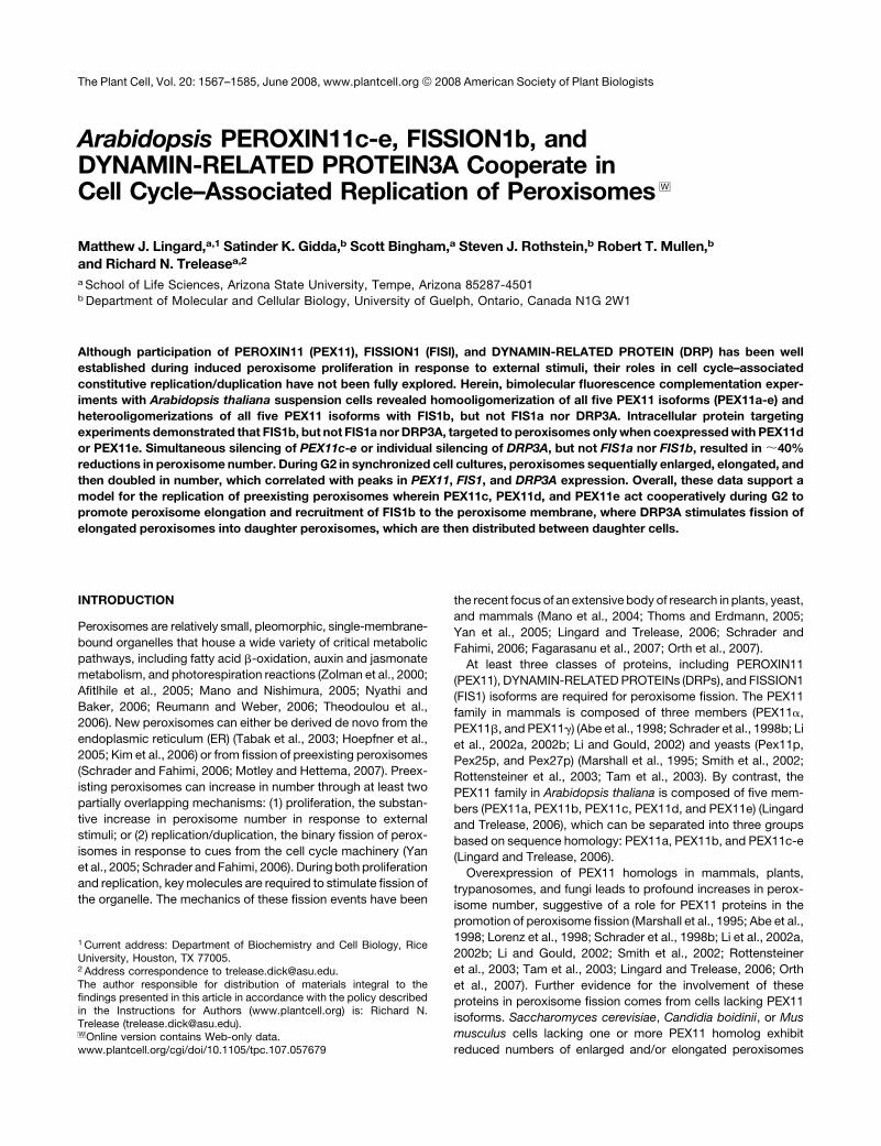
Subcellular Localizations of FIS1a, FIS1b, and DRP3A
To examine the subcellular localization of FIS1a, FIS1b, and
DRP3A, N-terminal myc epitope–tagged versions of each protein
were transiently expressed individually or in combinations with
PEX11 isoforms for ;16 h within Arabidopsis suspension cells
(Figure 1). We reported previously that all five PEX11 isoforms
were peroxisomal membrane proteins (Lingard and Trelease,
2006). Because FIS1a and FIS1b are likely tail-anchored mem-
brane proteins, which normally possess C-terminal membrane
targeting signals (Borgese et al., 2007), the myc epitope was
appended to their N termini to avoid interfering with putative
targeting information. The myc epitope was also appended to the
N terminus of DRP3A to mirror the N-terminal labeling with the
green fluorescent protein (GFP-DRP3A) used for previous local-
ization studies (Arimura and Tsutsumi, 2002; Mano et al., 2004).
The subcellular localization of each epitope-tagged protein
was assessed in individual formaldehyde-fixed cells via confocal
epifluorescence microscopy of antigen-bound anti-myc anti-
bodies. Anticatalase antibodies were also used as markers for
peroxisomes. Figures 1A and 1B show that myc-FIS1a and
myc-FIS1b sorted mostly to punctate and perinuclear reticular
structures, respectively, neither of which colocalized with en-
dogenous catalase in peroxisomes (see merged image overlay).
By contrast, Figure 1C illustrates that myc-DRP3A sorted to both
the cytosol and small punctate structures that were dispersed
throughout the cytoplasm. A subset of these myc-DRP3A-
containing punctate structures was located immediately adja-
cent to individual peroxisomes (e.g., asterisks in Figure 1C
overlay inset).
1568 The Plant Cell
To further explore the subcellular localization(s) of myc-tagged
FIS1a, FIS1b, and DRP3A, suspension cells were either cotrans-
formed with the mitochondrial marker protein MITO-GFP (con-
sisting of the F1-ATPase b-subunit N-terminal presequence
linked to GFP) or labeled with fluorophore-conjugated Conca-
navalin A, an ER marker. Although myc-FIS1a did not colocalize
with either the mitochondrial or ER marker, myc-FIS1b partially
colocalized with the ER marker (see Supplemental Figure 1A
online) but not with the mitochondrial marker. On the other hand,
the small punctate structures containing myc-DRP3A frequently
were located immediately adjacent to individual MITO-GFP-
containing mitochondria (see Supplemental Figure 1B online).
This juxtaposition of myc-DRP3A with mitochondria and perox-
isomes (Figure 1C; see Supplemental Figure 1B online) is similar
to the localizations reported previously for GFP-DRP3A in tran-
siently transformed Arabidopsis roots (Mano et al., 2004) and
tobacco (Nicotiana tabacum) suspension cells (Arimura and
Tsutsumi, 2002), suggesting that the protein targets in a similar
manner in all three cell types.
During the transient transformation experiments, we noted
that cells expressing myc-FIS1b consistently possessed an
increased number of spherical, often smaller peroxisomes (cf.
catalase images in Figure 1B with 1A and 1H), consistent with a
role for this protein in peroxisome fission as found in yeast and
mammalian cells (Yoon et al., 2003; Koch et al., 2005; Yu et al.,
2005). However, it is curious that myc-FIS1b is not associated
with peroxisomes in our suspension cells, suggesting that the
change in peroxisome number might reflect sorting to peroxi-
somes of an immunologically undetectable portion of the total
expressed myc-FIS1b.
Co-overexpression experiments were conducted to test
whether functional interactions occurred at the peroxisome
membrane between PEX11d or PEX11e, which promote perox-
isome elongation or an increase in number when expressed
Figure 1. Confocal Epifluorescence Images Illustrating Subcellular Lo-
calizations of Transiently Expressed FIS1a, FIS1b, and/or DRP3A in
Biolistically Transformed Arabidopsis Suspension Cells.
Each panel shows a single optical section of a transformed and
immunolabeled cell and is representative of $25 cells from at least two
independent biolistic bombardments. After bombardment, cells were
held for 16 h for gene expression and protein sorting, fixed in formalde-
hyde, then dual immunolabeled with anti-myc (green) and anticatalase
(magenta) antibodies. Protein colocalizations, if present, are depicted in
the far right overlay column as white images due to the overlay of the two
fluorophores in the same cell. Bar ¼ 10 mm.
(A) to (C) Single-transformed cells overexpressing myc-FIS1a (A), myc-
FIS1b (B), or myc-DRP3A (C). The box in the overlay panel of (C) outlines
the portion of the cell cytoplasm shown at higher magnification in the
inset. Asterisks in (C) indicate examples of the close associations of
punctate structures containing either expressed myc-DRP3A (green) or
endogenous peroxisomal catalase (magenta).
(D) and (E) Dual-transformed cells overexpressing myc-FIS1b and
PEX11d (D) or myc-FIS1a and PEX11d (E). Arrowheads in (D) point to
aggregated peroxisomes in the transformed cell, whereas arrows point
to normal peroxisomes in neighboring untransformed cells.
(F) Triple-transformed cell overexpressing myc-DRP3A, cYFP-FIS1b,
and PEX11d.
(G) Single-transformed cell overexpressing myc-PEX11d.
(H) Mock cell transformed with empty vector (pRTL2-myc). DIC, differ-
ential interference contrast image.
PEX11, FIS1b, and DRP3A in Peroxisome Replication 1569
singly (Figure 1G; Lingard and Trelease, 2006), and FIS1a,
FIS1b, or DRP3A. Figure 1D illustrates representative results of
coexpressing myc-FIS1b with untagged PEX11d. In these cells,
myc-FIS1b consistently colocalized with catalase within a few
relatively large cytoplasmic structures (Figure 1D, arrowheads),
distinctly different in size and number from the individual normal-
appearing peroxisomes visible in the neighboring nontrans-
formed cells (Figure 1D, arrows) or in mock-transformed cells
(Figure 1H) or from the elongated peroxisomes observed in cells
overexpressing myc-PEX11d alone (Figure 1G). The abnormally
large structures in Figure 1D, possessing catalase, myc-FIS1b,
and PEX11d likely resulted from peroxisome aggregation, as we
previously observed in cells overexpressing peroxisome mem-
brane proteins (Mullen et al., 2001; Lisenbee et al., 2003; Murphy
et al., 2003; McCartney et al., 2005). In parallel experiments,
coexpressions of myc-FIS1b with untagged PEX11e also re-
sulted in the apparent recruitment of myc-FIS1b to aggregated
peroxisomes. However, coexpressions of myc-FIS1a with
PEX11d (Figure 1E) or with PEX11e did not result in the re-
cruitment of myc-FIS1a to peroxisomes. Instead, myc-FIS1a
sorted to unidentified reticulate and globular structures, the latter
being morphologically similar to those observed when myc-
FIS1a was expressed on its own (cf. myc-FIS1a images in
Figures 1A and 1E).
Coexpression of individual untagged PEX11d or PEX11e iso-
forms with myc-DRP3A resulted in sorting of myc-DRP3A to the
cytosol and small punctate structures similar to its localization
shown in Figure 1C. Similarly, results of triple overexpression
experiments with myc-DRP3A, cYFP-FIS1b, and untagged
PEX11d (Figure 1F), or untagged PEX11e, revealed the same
localization of myc-DRP3A to the cytosol and small punctate
compartments. However, in contrast with results of the single or
double transformations with myc-DRP3A, peroxisome morphol-
ogy in triple transformed cells was profoundly altered (e.g.,
catalase image in Figure 1F); that is, all of the peroxisomes in
these cells were elongated, remarkably similar to the elongated
peroxisomes consistently observed in cells overexpressing
PEX11d alone (Figure 1G).
To verify that the fluorescent signal observed in cells over-
expressing myc-tagged proteins was attributable to expression
of the epitope-tagged protein and not background or nonspecific
labeling, mock-treated cells were transformed with the empty
vector (Figure 1H). No fluorescence signal was detected in cells,
shown in the differential interference image, labeled with anti-
myc antibodies. In the same cell, normal-appearing round per-
oxisomes were labeled with anticatalase antibodies (Figure 1H).
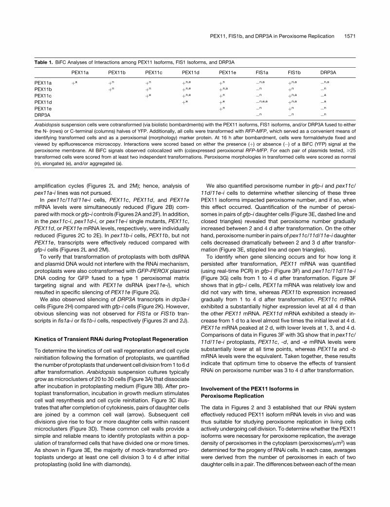
Physical Interactions among the PEX11 Isoforms, the FIS1
Isoforms, and DRP3A
We employed bimolecular fluorescence complementation (BiFC)
assays to determine whether in vivo physiological interactions
occurred among the PEX11 isoforms, the FIS1 isoforms, and/or
DRP3A (Table 1). Arabidopsis cells were transiently transformed
with pairs of plasmids encoding a PEX11 isoform, a FIS1 isoform,
and/or DRP3A. One protein of the pair was fused to the
N-terminal half of the yellow fluorescent protein (YFP) and the
other protein was fused to the C-terminal half of YFP (e.g., nYFP-
PEX11c and cYFP-FIS1b). Because each half of the YFP is not
intrinsically fluorescent, YFP fluorescence is observed only when
intermolecular interactions occur between nYFP- and cYFP-
tagged proteins (Citovsky et al., 2006). All cells were also
cobombarded with a plasmid encoding a red fluorescent protein
linked to the rice multifunctional protein (RFP-MFP), a well-
characterized peroxisomal matrix enzyme (Chuong et al., 2002,
2005). Coexpressed RFP-MFP served as an internal standard for
transformation efficiency as well as a convenient peroxisomal
marker to assess colocalizations with BiFC signals and potential
changes in peroxisome morphology.
The results presented in Table 1 show that all five PEX11
isoforms interact in vivo as homooligomers and with each other as
heterooligomers at the peroxisome membrane. Additional perox-
isomal interactions were observed among all five PEX11 isoforms
and FIS1b, but not FIS1a nor DRP3A. Furthermore, BiFC signal
was not observed for coexpressed DRP3A, FIS1a, and FIS1b,
suggesting that these proteins did not homo- or heterooligomer-
ize. In control experiments, each of the PEX11-, FIS1-, and
DRP3A-nYFP fusion constructs were expressed with a corre-
sponding cYFP empty vector. The consistent lack of peroxisome
fluorescence in these control cells provided convincing negative
evidence that fluorescence observed in experimental BiFC assays
resulted from specific protein–protein interactions.
Three categories of peroxisome morphologies were observed
in transformed cells during BiFC assays (Table 1): (1) normal
rounded or rod-shaped, as typically observed in untransformed
suspension cells (Lingard and Trelease, 2006); (2) elongated ($3
mm long; e.g., Figure 1F); and/or (3) aggregated (e.g., Figure 1D).
Elongated peroxisomes were observed in the majority of cells
coexpressing PEX11d, with the notable exception of cells coex-
pressing PEX11d with FIS1b or DRP3A. Elongated peroxisomes
also were observed in cells expressing homooligomers of
PEX11c; however, coexpression of PEX11c with any other PEX11
isoforms abrogated the peroxisome elongations that normally
were observed during PEX11c overexpression (Lingard and
Trelease, 2006).
Silencing of DRP3A, PEX11, and FIS1 Expression
To determine whether the PEX11 isoforms, FIS1 isoforms, and/or
DRP3A were necessary for peroxisome replication, transient
RNA interference (RNAi) was induced in suspension cell proto-
plasts transformed with gene-specific double-stranded RNA
(dsRNA). RNAi specificity was achieved using dsRNA comple-
mentary to either the coding sequence (CDS) or the 39 untrans-
lated region (UTR) of each respective mRNA (see Supplemental
Figure 2 online). Supplemental Table 1 online lists the cell names
and corresponding dsRNA and DNA constructs used for pre-
paring RNAi protoplasts.
In mock-transformed cells (Figure 2A) or in gfp-i cells trans-
formed with irrelevant GFP dsRNA (Figure 2F), ACTIN2 (ACT2),
PEX11c, PEX11d, and PEX11e transcripts were consistently
detected using RT-PCR (with 25 amplification cycles) at 18 h
after transformation. Conversely, the consistently low levels of
PEX11b transcripts were detected in gfp-i control cells only after
using additional PCR amplification cycles (Figure 2M). However,
PEX11a transcripts were not detected even after additional
1570 The Plant Cell
amplification cycles (Figures 2L and 2M); hence, analysis of
pex11a-i lines was not pursued.
In pex11c/11d/11e-i cells, PEX11c, PEX11d, and PEX11e
mRNA levels were simultaneously reduced (Figure 2B) com-
pared with mock or gfp-i controls (Figures 2A and 2F). In addition,
in the pex11c-i, pex11d-i, or pex11e-i single mutants, PEX11c,
PEX11d, or PEX11e mRNA levels, respectively, were individually
reduced (Figures 2C to 2E). In pex11b-i cells, PEX11b, but not
PEX11e, transcripts were effectively reduced compared with
gfp-i cells (Figures 2L and 2M).
To verify that transformation of protoplasts with both dsRNA
and plasmid DNA would not interfere with the RNAi mechanism,
protoplasts were also cotransformed with GFP-PEROX plasmid
DNA coding for GFP fused to a type 1 peroxisomal matrix
targeting signal and with PEX11e dsRNA (pex11e-i), which
resulted in specific silencing of PEX11e (Figure 2G).
We also observed silencing of DRP3A transcripts in drp3a-i
cells (Figure 2H) compared with gfp-i cells (Figure 2K). However,
obvious silencing was not observed for FIS1a or FIS1b tran-
scripts in fis1a-i or fis1b-i cells, respectively (Figures 2I and 2J).
Kinetics of Transient RNAi during Protoplast Regeneration
To determine the kinetics of cell wall regeneration and cell cycle
reinitiation following the formation of protoplasts, we quantified
the number of protoplasts that underwent cell division from 1 to 6 d
after transformation. Arabidopsis suspension cultures typically
grow as microclusters of 20 to 30 cells (Figure 3A) that dissociate
after incubation in protoplasting medium (Figure 3B). After pro-
toplast transformation, incubation in growth medium stimulates
cell wall resynthesis and cell cycle reinitiation. Figure 3C illus-
trates that after completion of cytokinesis, pairs of daughter cells
are joined by a common cell wall (arrow). Subsequent cell
divisions give rise to four or more daughter cells within nascent
microclusters (Figure 3D). These common cell walls provide a
simple and reliable means to identify protoplasts within a pop-
ulation of transformed cells that have divided one or more times.
As shown in Figure 3E, the majority of mock-transformed pro-
toplasts undergo at least one cell division 3 to 4 d after initial
protoplasting (solid line with diamonds).
We also quantified peroxisome number in gfp-i and pex11c/
11d/11e-i cells to determine whether silencing of these three
PEX11 isoforms impacted peroxisome number, and if so, when
this effect occurred. Quantification of the number of peroxi-
somes in pairs of gfp-i daughter cells (Figure 3E, dashed line and
closed triangles) revealed that peroxisome number gradually
increased between 2 and 4 d after transformation. On the other
hand, peroxisome number in pairs of pex11c/11d/11e-i daughter
cells decreased dramatically between 2 and 3 d after transfor-
mation (Figure 3E, stippled line and open triangles).
To identify when gene silencing occurs and for how long it
persisted after transformation, PEX11 mRNA was quantified
(using real-time PCR) in gfp-i (Figure 3F) and pex11c/11d/11e-i
(Figure 3G) cells from 1 to 4 d after transformation. Figure 3F
shows that in gfp-i cells, PEX11a mRNA was relatively low and
did not vary with time, whereas PEX11b expression increased
gradually from 1 to 4 d after transformation. PEX11c mRNA
exhibited a substantially higher expression level at all 4 d than
the other PEX11 mRNA. PEX11d mRNA exhibited a steady in-
crease from 1 d to a level almost five times the initial level at 4 d.
PEX11e mRNA peaked at 2 d, with lower levels at 1, 3, and 4 d.
Comparisons of data in Figures 3F with 3G show that in pex11c/
11d/11e-i protoplasts, PEX11c, -d, and -e mRNA levels were
substantially lower at all time points, whereas PEX11a and -b
mRNA levels were the equivalent. Taken together, these results
indicate that optimum time to observe the effects of transient
RNAi on peroxisome number was 3 to 4 d after transformation.
Involvement of the PEX11 Isoforms in
Peroxisome Replication
The data in Figures 2 and 3 established that our RNAi system
effectively reduced PEX11 isoform mRNA levels in vivo and was
thus suitable for studying peroxisome replication in living cells
actively undergoing cell division. To determine whether the PEX11
isoforms were necessary for peroxisome replication, the average
density of peroxisomes in the cytoplasm (peroxisomes/mm2) was
determined for the progeny of RNAi cells. In each case, averages
were derived from the number of peroxisomes in each of two
daughter cells in a pair. The differences between each of the mean
Table 1. BiFC Analyses of Interactions among PEX11 Isoforms, FIS1 Isoforms, and DRP3A
PEX11a PEX11b PEX11c PEX11d PEX11e FIS1a FIS1b DRP3A
PEX11a þa þn þn þn,e þn �n,a þn,a �n,a
PEX11b þn þn þn,e þn,a �n þn �n
PEX11c þe þn,e þn �n þn,a �a
PEX11d þe þe �n,e,a þn,a �a
PEX11e þn �n þn �n
DRP3A �n �n �n
Arabidopsis suspension cells were cotransformed (via biolistic bombardments) with the PEX11 isoforms, FIS1 isoforms, and/or DRP3A fused to either
the N- (rows) or C-terminal (columns) halves of YFP. Additionally, all cells were transformed with RFP-MFP, which served as a convenient means of
identifying transformed cells and as a peroxisomal (morphology) marker protein. At 16 h after bombardment, cells were formaldehyde fixed and
viewed by epifluorescence microscopy. Interactions were scored based on either the presence (þ) or absence (�) of a BiFC (YFP) signal at the
peroxisome membrane. All BiFC signals observed colocalized with (co)expressed peroxisomal RFP-MFP. For each pair of plasmids tested, $25
transformed cells were scored from at least two independent transformations. Peroxisome morphologies in transformed cells were scored as normal
(n), elongated (e), and/or aggregated (a).
PEX11, FIS1b, and DRP3A in Peroxisome Replication 1571

values presented in Figures 4A, 5A, 5B, and 6A were tested for
significance with t tests (see Supplemental Tables 2 to 5 online).
The histogram in Figure 4A shows that 4 d after transformation
the average density of peroxisomes in the cytoplasm of daughter
cell pairs that were either mock-transformed (without DNA
or dsRNA), transformed with plasmid DNA alone (GFP-PEROX),
or transformed with irrelevant dsRNA (gfp-i) was ;0.25 peroxi-
somes/mm2. An ;17% reduction in peroxisome number was
observed in pex11c-i, pex11d-i, and pex11e-i single mutants,
which was decreased further to an ;30% reduction in the
pex11d/11e-i, but not pex11c/11e-i or pex11d/11c-i, double mu-
tants. The greatest reduction in peroxisome number (;46%) was
observed in pex11c/11d/11e-i triple mutants. These data indicate
that peroxisome replication in actively dividing cells is dependent
upon the cooperative function of PEX11c, PEX11d, and PEX11e.
Figures 4B and 4C show representative optical micrographs of
peroxisomes in GFP-PEROX and gfp-i daughter cells. The rela-
tively small, spherical, or rod-shaped peroxisomes in these control
cells are characteristic of peroxisomes in untransformed cells. By
contrast, peroxisomes in pex11c/11d/11e-i daughter cell pairs
(Figure 4D) were fewer in number, larger than normal, and uni-
formly spherical. These results suggest that silencing of PEX11c,
PEX11d, and PEX11e restricts only peroxisome elongation and
division and that peroxisome growth is not regulated by PEX11
and can be uncoupled from peroxisome elongation and division.
Involvement of DRP3A, FIS1a, and FIS1b in
Peroxisome Replication
We next silenced DRP3A, FIS1a, and FIS1b to determine whether
these proteins were necessary for peroxisome replication. Al-
though we were not able to detect an effect on transcript levels of
the FIS1a and FIS1b targets (Figures 2I and 2J), the effects on
peroxisome density and mitochondria shown below indicate that
the dsRNA showed an effect, possibly at the translational level.
Figure 5A shows that at 4 d after transformation, drp3a-i daughter
cell pairs exhibited an ;40% reduction in peroxisome number
compared with gfp-i cells. However, the fis1a-i or fis1b-i single
mutants showed no significant reductions in peroxisome number,
in contrast with fis1a-i fis1b-i double mutants, which exhibited a
significant (;23%) reduction. Although no significantly enhanced
reduction was observed in the drp3a-i fis1a-i double mutant
compared with drp3a-i, the 40% reduction in drp3a-i peroxisome
density was significantly enhanced to ;55% in drp3a-i fis1b-i, but
not drp3a-i fis1a-i, double mutants. The 46% reduction in perox-
isome number in pex11c/11d/11e-i cells was enhanced to ;59%
in drp3a-i pex11c/11d/11e-i but was not enhanced in fis1a-i fis1b-i
pex11c/11d/11e-i. The greatest reduction in peroxisome number of
any mutant was observed in the drp3a-i fis1a-i fis1b-i triple mutant
(66% reduction). Peroxisome number in the fisla-i fis1b-i pex11c/
11d/11e-i mutant was not significantly different from peroxisome
number in pex11c/11d/11e-i, fis1b-i pex11c/11d/11e-i, or the fis1a-i
fis1b-i double mutant. Taken together, these data suggest that
FIS1b is involved in peroxisome replication, specifically in collab-
oration with DRP3A, and that DRP3A also functions along with
PEX11c, PEX11d, and PEX11e in peroxisome replication.
DRP3A and FIS1a were reported previously to be necessary
for mitochondrial division (Mano et al., 2004; Scott et al., 2006).
Figure 2. Gene-Specific Silencing of DRP3A, the Five PEX11 Isoforms,
and the Two FIS1 Isoforms.
Arabidopsis suspension cell protoplasts were transformed with polyeth-
ylene glycol (PEG) with dsRNA complementary to GFP ([F], [K], and [M]),
PEX11c/11d/11e (B), PEX11c (C), PEX11d (D), PEX11e ([E] and [G]),
PEX11b (L), DRP3A (H), FIS1a (I), or FIS1b (J). Protoplasts for (G) were
also cotransformed with plasmid DNA encoding the GFP-PEROX trans-
gene. Mock transformations in (A) were done with buffer without RNA or
DNA. After 18 h of expression, total RNA was extracted, and ACTIN2
(ACT2), FIS1a, FIS1b, DRP3A, PEX11a, PEX11b, PEX11c, PEX11d, and
PEX11e mRNA were reverse transcribed and amplified via 25, 30, or
40 PCR cycles. In (M), ACT2 (reproduced from [L]), PEX11a, PEX11b,
and PEX11e bands are from the same gel and were adjusted for
brightness simultaneously before removing adjoining wells (gap in the
image). All images are of fluorescence emitted from ethidium bromide–
labeled PCR products (;600 bp) separated in 1% ([A] to [G]) or 2% (w/v)
([H] to [M]) agarose gels.
1572 The Plant Cell
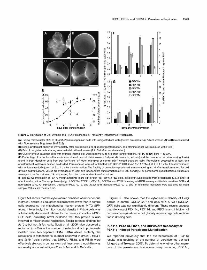
Figure 5B shows that the cytoplasmic densities of mitochondria
in drp3a-i and fis1a-i daughter cell pairs were lower than in control
cells expressing the mitochondrial marker protein, MITO-GFP,
alone. Interestingly, the mitochondrial density in fis1b-i cells was
substantially decreased relative to the density in control MITO-
GFP cells, providing novel evidence that this protein is also
involved in mitochondrial replication. Similar to these findings for
fis1b-i, but not fis1a-i cells, Scott et al. (2006) also observed a
reduction (;43%) in the number of mitochondria in protoplasts
isolated from two separate FIS1a T-DNA alleles. Notably, the
reductions in mitochondrial number observed in drp3a-i, fis1a-i,
and fis1b-i cells verified that DRP3A, FIS1a, and FIS1b were
effectively silenced in our transient cell lines, even though this was
not readily apparent in Figure 2 for fis1a-i and fis1b-i cells.
Figure 5B also shows that the cytoplasmic density of Golgi
bodies in control GOLGI-GFP and pex11c/11d/11e-i (GOLGI-
GFP) cells was not significantly different. These results suggest
that silencing of PEX11c, PEX11d, and PEX11e and inhibition of
peroxisome replication do not globally repress organelle replica-
tion in dividing cells.
PEX11c, PEX11d, FIS1b, and DRP3A Are Necessary for
PEX11e-Induced Peroxisome Multiplication
We reported previously that the overexpression of PEX11e
results in a doubling of the number of peroxisomes per cell
(Lingard and Trelease, 2006). To determine whether other mem-
bers of the peroxisome fission machinery, including PEX11c,
Figure 3. Reinitiation of Cell Division and RNAi Persistence in Transiently Transformed Protoplasts.
(A) Typical microcluster of 20 to 30 Arabidopsis suspension cells with undigested cell walls (before protoplasting). All cell walls in (A) to (D) were stained
with Fluorescence Brightener 28 (FB28).
(B) Single protoplast observed immediately after protoplasting (0 d), mock transformation, and staining of cell wall residues with FB28.
(C) Pair of daughter cells sharing an equatorial cell wall (arrow) (2 to 5 d after transformation).
(D) Cluster of four daughter cells with multiple internal cell walls (arrows) (3 to 6 d after transformation). For (A) to (D), bars ¼ 10 mm.
(E) Percentage of protoplasts that underwent at least one cell division over a 6-d period (diamonds, left axis) and the number of peroxisomes (right axis)
found in both daughter cells from pex11c/11d/11e-i (open triangles) or control gfp-i (closed triangles) cells. Protoplasts possessing at least one
equatorial cell wall were defined as divided. Peroxisomes were either labeled with GFP-PEROX (pex11c/11d/11e-i) at 1 to 4 d after transformation or
with anticatalase IgGs (gfp-i ) at 2 to 4 d after transformation. The fragility of protoplasts precluded immunolabeling at 1 d after transformation. For cell
division quantifications, values are averages of at least two independent transformations (n $ 300 per day). For peroxisome quantifications, values are
averages 6 SE from at least 19 cells arising from two independent transformations.
(F) and (G) Quantification of PEX11 mRNA amounts in gfp-i (F) or pex11c/11d/11e-i (G) cells. Total RNA was isolated from protoplasts 1, 2, 3, and 4 d
after transformation. Transcript levels (in fg) of PEX11a, PEX11b, PEX11c, PEX11d, and PEX11e in 4 ng total RNA were quantified via real-time PCR and
normalized to ACT2 expression. Duplicate (PEX11a, -b, and ACT2) and triplicate (PEX11c, -d, and -e) technical replicates were acquired for each
sample. Values are means 6 SD.
PEX11, FIS1b, and DRP3A in Peroxisome Replication 1573

PEX11d, FIS1b, and/or DRP3A, were involved in this inducible
multiplication, GFP-PEX11e was overexpressed in various RNAi
backgrounds and peroxisomes were quantified in pairs of
daughter cells. As expected, overexpression of GFP-PEX11e
induced an ;45% increase in peroxisome number compared
with gfp-i control cells (Figure 6A). However, overexpression of
GFP-PEX11e in a pex11c/11d-i double mutant background
resulted in a significant inhibition of the PEX11e-induced perox-
isome multiplication. When GFP-PEX11e was overexpressed in
fis1b-i or drp3a-i backgrounds, peroxisome number was the
same as in the normal fis1b-i and drp3a-i lines. These data sug-
gest that PEX11c, PEX11d, PEX11e, FIS1b, and DRP3A are
necessary for PEX11-induced peroxisome multiplication.
In addition to reductions in peroxisome number (Figure 6A), we
noted that these transgenic cells exhibited dramatic alterations
in peroxisome morphology. The fluorescence micrographs pre-
sented in Figures 6B to 6E illustrate that peroxisomes in drp3a-i
(GFP-PEX11e) cells were substantially elongated (Figure 6B)
compared with the uniformly spherical peroxisomes in gfp-i
(Figure 6C) or the spherical but much more numerous perox-
isomes in GFP-PEX11e cells (Figure 6D). Notably, the elongated
drp3a-i (GFP-PEX11e) peroxisomes (Figure 6B) were thicker and
shorter than peroxisomes in drp3a-i daughter cell pairs (Figure
6E),whoseelongated/tubulated peroxisomes more closely resem-
bled peroxisomes in cells overexpressing PEX11c or PEX11d
(Lingard and Trelease, 2006).
Peroxisome Replication Occurs before M-Phase in
Synchronized Suspension Cells and Is Correlated with
Expression of DRP3A, PEX11, FIS1a, and FIS1b Expression
The relationships among (1) DRP3A, PEX11, FIS1a, and FIS1b
expression, (2) peroxisome size and number, and (3) cell cycle
stages were examined in synchronized suspension cell cultures.
Cell cycle progression in nonprotoplasted suspension cells (e.g.,
Figure 3A) was synchronized using amphidicolin block and release
(Menges and Murray, 2002), and synchronization efficiency was
assessed by determining the percentage of cells in S- and M-phase
at intervals from 0 to 32 h after amphidicolin release. To visualize
and quantify the number of cells in S-phase, cells were incubated
for 1 h in 5-bromo-29-deoxyuridine (BrdU), which was incorpo-
rated into actively replicating DNA, prior to formaldehyde fixation
and immunolabeling with anti-BrdU antibodies. Mitotic profiles
(metaphase and anaphase chromosomes) typical of M-phase
cells were identified by labeling nuclei with 49,6-diamidino-
2-phenylindole (DAPI).
The dashed line in Figure 7A shows that 1 h after release of
amphidicolin, ;78% of the cells were in S-phase; thereafter, the
percentage of cells decreased within 6 to 8 h to a background of
;15%. These S-phase percentages resemble other published
values for Arabidopsis suspension cell synchronization (Menges
and Murray, 2002). The solid line in Figure 7A indicates that the
percentage of cells in M-phase rose gradually from 6 to 14 h after
release of the block and eventually reached a peak at 30 h of
;2%. This maximum percentage was somewhat lower than that
found by Menges and Murray (2002). However, Menges and
Murray (2002) employed a novel fast-growing Arabidopsis sus-
pension culture, which grew at least twice as fast as our cells
and, after synchronization, transitioned from the peak of S-phase
to the peak of M-phase in ;10 h. By contrast, the transition from
S-phase to M-phase lasted at least 24 h in our synchronized
cultures (Figure 7), suggesting that the more dispersed (broader
and shorter) M-phase peak in our synchronized culture was a
product of the slower growth rate.
To determine whether cell cycle progression is synchronized
with peroxisome replication, peroxisome number per cell, cell
Figure 4. Cytoplasmic Peroxisome Density (Peroxisomes/mm2) in pex11-i Cells Pairs at 4 d after Transformation.
Protoplasts were transformed without RNA or DNA (mock), with GFP-PEROX, or cotransformed with various dsRNA (x axis) plus GFP-PEROX. Four
days after transformation, protoplasts were fixed and stained with FB28. Bars for control cells are shaded black, and bars for single, double, and triple
silencing are shaded in dark gray, light gray, and white, respectively.
(A) Peroxisome density in the cytoplasm of each of two transformed daughter cells. Values are means 6 SE of at least 26 daughter cell pairs from at
least two independent transformations for all but pex11b-i (n ¼ 12 from one transformation). Bars with asterisks are significantly different from gfp-i
(* P # 0.05; ** P # 0.001).
(B) GFP-labeled peroxisomes in a control daughter cell pair transformed with GFP-PEROX only.
(C) Anticatalase/rhodamine redX-immunolabeled peroxisomes in a gfp-i daughter cell pair.
(D) GFP-labeled peroxisomes within a pex11d/11d/11e-i daughter cell pair cotransformed with GFP-PEROX. Images in (B) to (D) are representative of
whole-cell projections acquired via nonconfocal epifluorescence microscopy. Bar ¼ 10 mm.
1574 The Plant Cell

area, and peroxisome elongation were quantified in equatorial
focal planes at several time points after release from the
amphidicolin block (Figure 7B). The average cell area (Figure
7B, open squares) and the average number of peroxisomes per
cell (Figure 7B, open diamonds) remained relatively constant
Figure 5. Cytoplasmic Peroxisome Density in fis1a-i, fis1b-i, and drp3a-i
Daughter Cell Pairs at 4 d after Transformation.
(A) Cytoplasmic peroxisome density in single (fis1a-i, fis1b-i, and drp3a-i ),
double (fis1a-i fis1b-i, drp3a-i fis1a-i, drp3a-i fis1b-i, and drp3a-i pex11c/
11d/11e-i ), and triple (drp3a-i fis1a-i fis1b-i and fis1a-i fis1b-i pex11c/
11d/11e-i ) mutants. gfp-i and pex11c/11d/11e-i means are reproduced
from Figure 4A. Bars corresponding to the basal level of peroxisomes in
gfp-i and pex11c/11d/11e-i cells are shaded black and white, respec-
tively, and bars reflecting analyses of fis1 and drp3a silencing are shown
in dark and light gray, respectively.
(B) Cytoplasmic mitochondrial or Golgi body density in control (MITO-
GFP and GOLGI-GFP ) and RNAi (fis1a-i, fis1b-i, drp3a-i, and pex11c/
11d/11e-i ) transformed daughter cell pairs. Mitochondria and Golgi
bodies were labeled in control and RNAi lines by (co)transforming cells
with MITO-GFP or GOLGI-GFP plasmid DNA, respectively. Bars for
controls are shaded black, and bars for RNAi lines are shaded gray.
Values in (A) and (B) are means 6 SE. n values for each sample are
indicated within the histogram bars. Bars with asterisks are significantly
different from gfp-i (A) or MITO-GFP (B) (* P # 0.05; ** P # 0.001).
Figure 6. Necessity of PEX11c, PEX11d, FIS1b, and/or DRP3A for
PEX11e-Induced Peroxisome Multiplication.
(A) Mean cytoplasmic peroxisome densities in pex11c-i pex11d-i, fis1b-i,
and drp3a-i daughter cell pairs overexpressing GFP-PEX11e. Data for
the means of gfp-i, drp3a-i, fis1b-i, pex11c/11d-i, and pex11c/11d/11e-i
are reproduced from Figures 4A and 5A. All values are means 6 SE.
n values for each sample are indicated within the histogram bars. Bars
with asterisks are significantly different from GFP-PEX11e (* P # 0.05;
** P # 0.001). Bars for the control and corresponding GFP-PEX11e–
overexpressing lines are shaded the same color.
(B) GFP-labeled peroxisomes in a pair of drp3a-i daughter cells over-
expressing GFP-PEX11e.
(C) Anticatalase/rhodamine redX-immunolabeled peroxisomes in a pair
of gfp-i daughter cells.
(D) GFP-PEX11e-labeled peroxisomes in a pair of daughter cells over-
expressing GFP-PEX11e.
(E) GFP-PEROX–labeled peroxisomes in a pair of drp3a-i daughter cells.
Images in (B) to (E) are representative whole-cell projections acquired via
nonconfocal epifluorescence microscopy. Bar ¼ 10 mm.
PEX11, FIS1b, and DRP3A in Peroxisome Replication 1575
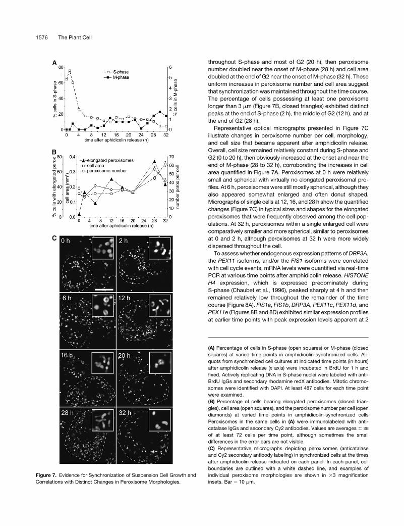
throughout S-phase and most of G2 (20 h), then peroxisome
number doubled near the onset of M-phase (28 h) and cell area
doubled at the end of G2 near the onset of M-phase (32 h). These
uniform increases in peroxisome number and cell area suggest
that synchronization was maintained throughout the time course.
The percentage of cells possessing at least one peroxisome
longer than 3 mm (Figure 7B, closed triangles) exhibited distinct
peaks at the end of S-phase (2 h), the middle of G2 (12 h), and at
the end of G2 (28 h).
Representative optical micrographs presented in Figure 7C
illustrate changes in peroxisome number per cell, morphology,
and cell size that became apparent after amphidicolin release.
Overall, cell size remained relatively constant during S-phase and
G2 (0 to 20 h), then obviously increased at the onset and near the
end of M-phase (28 to 32 h), corroborating the increases in cell
area quantified in Figure 7A. Peroxisomes at 0 h were relatively
small and spherical with virtually no elongated peroxisomal pro-
files. At 6 h, peroxisomes were still mostly spherical, although they
also appeared somewhat enlarged and often donut shaped.
Micrographs of single cells at 12, 16, and 28 h show the quantified
changes (Figure 7C) in typical sizes and shapes for the elongated
peroxisomes that were frequently observed among the cell pop-
ulations. At 32 h, peroxisomes within a single enlarged cell were
comparatively smaller and more spherical, similar to peroxisomes
at 0 and 2 h, although peroxisomes at 32 h were more widely
dispersed throughout the cell.
To assess whether endogenous expression patterns of DRP3A,
the PEX11 isoforms, and/or the FIS1 isoforms were correlated
with cell cycle events, mRNA levels were quantified via real-time
PCR at various time points after amphidicolin release. HISTONE
H4 expression, which is expressed predominately during
S-phase (Chaubet et al., 1996), peaked sharply at 4 h and then
remained relatively low throughout the remainder of the time
course (Figure 8A). FIS1a, FIS1b, DRP3A, PEX11c, PEX11d, and
PEX11e (Figures 8B and 8D) exhibited similar expression profiles
at earlier time points with peak expression levels apparent at 2
Figure 7. Evidence for Synchronization of Suspension Cell Growth and
Correlations with Distinct Changes in Peroxisome Morphologies.
(A) Percentage of cells in S-phase (open squares) or M-phase (closed
squares) at varied time points in amphidicolin-synchronized cells. Ali-
quots from synchronized cell cultures at indicated time points (in hours)
after amphidicolin release (x axis) were incubated in BrdU for 1 h and
fixed. Actively replicating DNA in S-phase nuclei were labeled with anti-
BrdU IgGs and secondary rhodamine redX antibodies. Mitotic chromo-
somes were identified with DAPI. At least 487 cells for each time point
were examined.
(B) Percentage of cells bearing elongated peroxisomes (closed trian-
gles), cell area (open squares), and the peroxisome number per cell (open
diamonds) at varied time points in amphidicolin-synchronized cells
Peroxisomes in the same cells in (A) were immunolabeled with anti-
catalase IgGs and secondary Cy2 antibodies. Values are averages 6 SE
of at least 72 cells per time point, although sometimes the small
differences in the error bars are not visible.
(C) Representative micrographs depicting peroxisomes (anticatalase
and Cy2 secondary antibody labeling) in synchronized cells at the times
after amphidicolin release indicated on each panel. In each panel, cell
boundaries are outlined with a white dashed line, and examples of
individual peroxisome morphologies are shown in 33 magnification
insets. Bar ¼ 10 mm.
1576 The Plant Cell

and 12 h after amphidicolin release. FIS1a, DRP3A, PEX11c, and
PEX11e each exhibited a third rise in expression level near the
end of G2 (22 to 26 h after amphidicolin release) coincident with
peak increases in both peroxisome elongation and number at
28 h (Figures 7B and 7C). Intriguingly, PEX11a and PEX11b,
which under other conditions we have tested were either unde-
tectable (PEX11a) or had very low expression levels (PEX11b),
both underwent a single rise in expression during G2 (8 to 12 h
after amphidicolin release) (Figure 8C).
DISCUSSION
Previously, we identified five functional Arabidopsis PEX11 ho-
mologs that, when overexpressed, sorted directly to the perox-
isome membrane and stimulated peroxisome (1) doubling in
number per cell (PEX11a and PEX11e), (2) aggregation or en-
largement (PEX11b), and/or (3) elongation (PEX11a, PEX11c,
and PEX11d) (Lingard and Trelease, 2006). In this study, we
focus on the molecular mechanisms that are involved in main-
taining a constitutive population of preexisting peroxisomes in
actively dividing plant cells. In particular, we provide new evi-
dence for the cooperative participation of PEX11c, PEX11d,
PEX11e with FIS1b and DRP3A, in growth and subsequent equal
division of preexisting peroxisomes during G2 phase just prior to
mitosis. Hence, we provide evidence that the creation of new
plant peroxisomes is not solely dependent upon direct origina-
tion from the ER, as portrayed in several recent models drawn
from research with yeast and mammalian cells (Tabak et al.,
2003; Kragt et al., 2005; Haan et al., 2006; Kim et al., 2006;
Titorenko and Mullen, 2006). Our findings do not preclude
origination of plant peroxisomes from the ER, although it is
notable that ER-dependent peroxisome formation has not yet
been described in plant cells (Trelease and Lingard, 2006).
Instead, recent research in plant peroxisome biogenesis sup-
ports participation of the ER in the overall growth and division of
preexisting plant peroxisomes through essential contributions of
new membrane components (reviewed in Mullen and Trelease,
2006). A similar role for the ER was described recently in the
replication of preexisting peroxisomes in S. cerevisiae (Motley
and Hettema, 2007).
Interactions among PEX11 Isoforms, FIS1 Isoforms,
and DRP3A
The homooligomerizations among PEX11 isoforms that we ob-
served via BiFC experiments (Table 1) were consistent with
previous published findings in mammals and yeast. For instance,
immunoprecipitation experiments showed that mammalian
PEX11a and PEX11b formed homodimers (Li and Gould, 2003),
and yeast two-hybrid assays revealed that S. cerevisiae Pex11p,
Pex25p, and Pex27p also formed homodimers (Rottensteiner
et al., 2003). On the other hand, it is curious that we observed
heterooligomerizations among all five of the Arabidopsis PEX11
isoforms (Table 1) because no heterooligomerizations were
observed among mammalian PEX11 isoforms (Li and Gould,
2003), and only weak interactions were observed between S.
cerevisiae Pex25p and Pex27p (Rottensteiner et al., 2003). This
apparent difference may be due to the stabilization of short-lived
Figure 8. Quantification of HISTONE H4, DRP3A, FIS1, and PEX11
mRNA at Varied Time Points in Amphidicolin-Synchronized Cells.
Total RNA was isolated from synchronized cells at the indicated time
points (in hours) after amphidicolin release (x axis) and reverse tran-
scribed, and 4 ng total cDNA were quantified via real-time PCR for each
data point. Values are means 6 SD of at least two technical replicates.
(A) Absolute quantifications of HISTONE H4 mRNA.
(B) Absolute quantifications of FIS1a (diamonds), FIS1b (squares), and
DRP3A (triangles) mRNA.
(C) Absolute quantifications of PEX11a (diamonds) and PEX11b (squares).
(D) Absolute quantification of PEX11c (triangles), PEX11d (x), and PEX11e
(circles) mRNA.
PEX11, FIS1b, and DRP3A in Peroxisome Replication 1577

intermolecular interactions that can occur after reconstitution
of the YFP fluorophore during BiFC (Bracha-Drori et al., 2004;
Walter et al., 2004).
An advantage of BiFC over yeast two-hybrid and immunopre-
cipitation experiments is that the subcellular localization of the
interacting species can directly be observed via microscopy. For
example, the reconstituted YFP fluorescence resulting from
interactions between the PEX11 proteins and FIS1b revealed
not only the interaction between these two molecules but also
that FIS1b was recruited to the peroxisome membrane through
its associations with the PEX11 isoforms, which was confirmed
by the PEX11-dependent sorting of overexpressed myc-FIS1b to
peroxisomes (Figures 1B and 1D).
Although the BiFC results clearly show that homo- and
heterointeractions occur among the PEX11 isoforms, as well as
FIS1b, they do not yield information about stoichiometric rela-
tionships within these complexes (e.g., whether the proteins
exist within these complexes or have a more transitory interac-
tion). Nevertheless, our BiFC results suggest that oligomerization
of the PEX11 isoforms serves a regulatory function. For exam-
ple, cells possessing PEX11d homo- and heterooligomers or
PEX11c homooligomers also possessed unusually elongated
peroxisomes (Table 1). Thus, heterooligomerization seemed
to inhibit PEX11c-stimulated elongation but had no effect
on PEX11d-stimulated elongation. Hetero-interactions among
PEX11c, PEX11d, and PEX11e also seem to be required for
PEX11e-induced peroxisome fission, as peroxisome multiplica-
tion was inhibited in pex11c/11d-i (GFP-PEX11e) cells (Figure
6A). Yeast and mammalian models also support a role for PEX11
oligomerization in the regulation of peroxisome fission. Inhibiting
mammalian PEX11 homodimerization precludes peroxisome
proliferation (Kobayashi et al., 2007), whereas inhibition of S.
cerevisiae Pex11p homodimerization promotes peroxisome pro-
liferation (Marshall et al., 1996). Given the widespread oligomer-
izations among the Arabidopsis PEX11 isoforms (Table 1), and
the ability for some of these oligomers to promote peroxisome
elongation, it seems likely that at least some types of oligomers
are active.
Our data suggest that FIS1a, unlike FIS1b, does not participate
in peroxisome replication. No interactions were observed be-
tween FIS1a and DRP3A, or between FIS1a and any PEX11
isoform (Table 1), and overexpressed FIS1a did not sort to
peroxisomes (Figures 1A and 1E). Also, the number of peroxi-
somes in fis1a-i drp3a-i cells is not significantly different from
drp3a-i cells, whereas the number of peroxisomes in fis1b-i
drp3a-i cells is significantly lower (Figure 5A), suggesting that
FIS1b, but not FIS1a, cooperates with DRP3A during peroxisome
replication.
Although our targeting (Figure 1), BiFC (Table 1), and silencing
data (Figure 5A) do not support a direct role for FIS1a in
peroxisome division, this tail-anchored protein appears to play
a role in mitochondrial biogenesis consistent with the results
from a previous study (Scott et al., 2006). For instance, we
showed in Figure 5B that the number of mitochondria in fis1a-i
daughter cell pairs was lower than in control cells. It is surprising,
however, that myc-FIS1a does not localize to mitochondria in our
suspension cells, suggesting that the targeting mechanism for
FIS1a, as in mammalian cells (Schrader and Yoon, 2007), is
complex. Experiments are currently underway to determine
whether FIS1a, similar to other tail-anchored membrane proteins
(e.g., Bcl-2 and BAX) (Leber et al., 2007), may only target to the
mitochondria from the cytosol in response to a specific devel-
opmental and/or environmental signal.
Although interactions between FIS1b and DRP3A were not
observed in our BiFC experiments (Table 1), evidence for their
cooperation can be inferred from the dual-silencing experiments.
Because mRNA silencing only knocks down protein expression
levels, double or triple silencing mutants within the same oligo-
meric complex, or acting within the same pathway, are expected
to have enhanced phenotypes compared with the single mu-
tants. Accordingly, the enhanced reduction in peroxisome num-
ber observed in drp3a-i fis1b-i, but not drp3a-i fis1a-i, cells
(Figure 5A) suggests that these two proteins are jointly involved in
peroxisome fission. It may be that the physical interaction
between FIS1b and DRP3A is mediated through the action of
an unidentified adaptor or bridge protein, as is observed during
mitochondrial fission in yeast (reviewed in Schrader and Yoon,
2007). The existence of such a bridge protein in Arabidopsis may
explain why our BiFC experiments revealed no direct contact
between FIS1b and DRP3A (Table 1).
Other Roles for the PEX11 Isoforms during
Peroxisome Fission
We reported previously that overexpression of PEX11e pro-
motes peroxisome fission without discernable elongation
(Lingard and Trelease, 2006). In this work, overexpression of
PEX11e in cells with silenced DRP3A resulted in peroxisome
elongation without fission (Figure 6B), indicating that in plants, as
in other organisms, PEX11 isoforms are not directly responsible
for peroxisome fission, but likely modify or tubulate the perox-
isome membrane in preparation for fission (Schrader and Fahimi,
2006). In plants and mammals, overexpression of certain PEX11
isoforms leads to peroxisomal elongation, which often precedes
membrane fission (Li and Gould, 2003; Koch et al., 2004; Lingard
and Trelease, 2006). This elongation may be accomplished
through direct interactions of a PEX11 isoform with phospho-
lipids in the peroxisome membrane. For example, the sequence
and predicted secondary structure of S. cerevisiae Pex11p is
similar to the ligand binding domain of the peroxisome proliferator-
activated receptor subfamily of nuclear hormone receptors
(Barnett et al., 2000), which bind to fatty acids, suggesting that
the PEX11 proteins may directly bind to phospholipids on the
exterior surface of the peroxisome membrane (Fagarasanu et al.,
2007).
The silencing experiments presented in Figures 3 to 5 dem-
onstrate that peroxisomal growth and tubulation are separable
events and that the latter is dependent upon PEX11 expression.
Specifically, even though drp3a-i and pex11c/11d/11e-i cells
had similar numbers of peroxisomes (Figure 5A), the peroxi-
somes in pex11c/11d/11e-i were rounded and enlarged (Figure
4D), whereas those in drp3a-i were elongated but not enlarged
(Figure 6E). These results confirm that DRP3A is necessary for
peroxisome fission and suggest that peroxisome elongation
occurs constitutively in plant cells, as observed in mammals
1578 The Plant Cell
(Li and Gould, 2003; Koch et al., 2004). It is significant that
peroxisomes in cells with reduced levels of PEX11c, PEX11d,
and PEX11e enlarge but do not elongate, suggesting that PEX11
isoforms are required for elongation but not for growth. Notably,
in synchronized cell cultures, the coordinated increases in per-
oxisome elongation did not always coincide with increases in
peroxisome number (Figure 7, 2 and 12 h), but always coincided
with increases in PEX11 expression (Figures 8C and 8D, 2 and
12 h), supporting a role for the PEX11 proteins in peroxisome
elongation but not fission. The constitutive high levels of PEX11c,
PEX11d, and PEX11e expression (Figures 2 and 3F) and the
dramatic decreases in peroxisome number in pex11c/11d/11e-i,
but not pex11b-i, pex11c-i, pex11d-i, or pex11e-i, cells (Figure 4),
both suggest that PEX11c, PEX11d, and PEX11e, but not
PEX11a or PEX11b, play distinct, but overlapping, roles in
peroxisome replication.
Previously, we found that cells overexpressing PEX11b have
enlarged peroxisomes (Lingard and Trelease, 2006), suggesting
either that PEX11b acts as a negative regulator of peroxisome
fission or that overexpression of PEX11b promotes peroxisome
aggregation, as has been observed for other overexpressed
peroxisome membrane proteins (Mullen et al., 2001). However,
pex11b-i cells have normal numbers of peroxisomes (Figure 4A),
which is not suggestive of a role for PEX11b in peroxisome
replication. Orth et al. (2007) recently reported that overex-
pression of PEX11b in Arabidopsis leaf mesophyll cells resulted
in peroxisome elongation, which was quite different from our
previous findings that overexpression of PEX11b in Arabidopsis
suspension-cultured cells led to peroxisome aggregation (Lingard
and Trelease, 2006). In addition, Orth et al. (2007) reported that
silencing of PEX11b led to an ;75% decrease in peroxisome
number per leaf mesophyll cell, whereas we found that silencing
of PEX11b had no apparent effect on peroxisome abundance in
suspension cells (Figure 4A). Although these results seem con-
tradictory, recent results (Desai and Hu, 2008) offer a plausible
explanation that they are not contradictory. They reported that
Arabidopsis peroxisomes first elongate and then proliferate in
number when etiolated seedlings were transferred to the light,
which coincided with an upregulation of PEX11b expression.
Furthermore, they found that silencing of PEX11b precluded this
light-induced peroxisome proliferation and that PEX11b was a
direct target of the photomorphogenic transcription factor HYH.
Thus, it seems that the primary role for PEX11b is induction of
peroxisome proliferation in response to light signals, not main-
tenance of peroxisome homeostasis within dividing cells. Be-
cause peroxisomes in our suspension cells do not increase in
number per cell in response to an overexpression of PEX11b
(Lingard and Trelease, 2006), nor do peroxisomes exhibit defects
in the cell division process within pex11b-i cells (Figure 4), it
seems likely that PEX11c, PEX11d, and PEX11e, but not
PEX11b, are the dominant PEX11 isoforms in dark-grown, rap-
idly dividing cells. In concert with this assertion are the consti-
tutive low level of PEX11b expression (Figures 2 and 3A) and
the increases in PEX11b expression during protoplast cell wall
regeneration and cell cycle reinitiation (Figure 3F). These collec-
tive results suggest that PEX11b fulfills a minor role that partially
overlaps with those of PEX11c, PEX11d, and PEX11e in the
maintenance of peroxisome homeostasis in dividing cells.
Peroxisome Division during Cell Cycle Transitions
We undertook a detailed morphological analysis of the changes
in Arabidopsis peroxisomes that occur during cell cycle transi-
tions. Numerous studies with mammalian and yeast cells de-
scribe the morphological changes associated with induced
peroxisome proliferation (Marshall et al., 1995; Schrader et al.,
1998a, 1998b, 1999; Koch et al., 2005) but do not correlate
peroxisome morphological changes with cell cycle progression.
Our data show that cell division, peroxisome elongation, and
peroxisome replication are intimately connected (Figure 7) and
that these events are coordinated with distinct peaks in PEX11,
FIS1b, and DRP3A expression (Figure 8).
The strikingly similar expression profiles for the PEX11 iso-
forms, the FIS1 isoforms, and DRP3A in the synchronized culture
(Figure 8) are highly suggestive for a role for these genes in cell
cycle progression. In addition, it is possible to infer from results of
our silencing experiments that at least part of the function of
these proteins is the replication of peroxisomes. Whether these
proteins play roles in cell cycle progression in addition to organ-
elle replication is unknown. A potentially analogous scenario has
been identified in rat kidney cells where some, but not all,
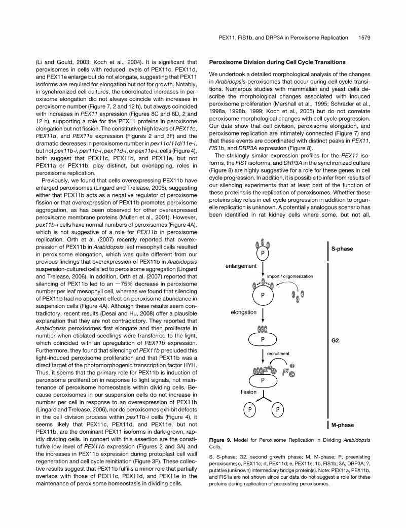
Figure 9. Model for Peroxisome Replication in Dividing Arabidopsis
Cells.
S, S-phase; G2, second growth phase; M, M-phase; P, preexisting
peroxisome; c, PEX11c; d, PEX11d; e, PEX11e; 1b, FIS1b; 3A, DRP3A; ?,
putative (unknown) intermediary bridge protein(s). Note: PEX11a, PEX11b,
and FIS1a are not shown since our data do not suggest a role for these
proteins during replication of preexisting peroxisomes.
PEX11, FIS1b, and DRP3A in Peroxisome Replication 1579
proteins that are necessary for Golgi body fission are also
necessary for entrance into mitosis (Sutterlin et al., 2002), im-
plying that organelle homeostasis is intimately linked with cell
division. Interestingly, Golgi division can also be controlled
through action of some cyclin-dependent kinases (Lowe et al.,
1998, 2000; Draviam et al., 2001), a class of proteins classically
involved in the regulation of cell cycle transitions. In plants,
CDT1a and CDT1b (for CDC10-dependent transcript 1a and b,
which encode proteins necessary for initiation of DNA replication
in yeast) are necessary for both DNA synthesis and plastid
division (Raynaud et al., 2005), supporting the hypothesis that
organelle replication and cell division are interrelated.
To fully explore the links between plant organelle replication
and cell division, it would be necessary to generate transgenic
cell lines or plants defective in key genes, such as the PEX11
isoforms, the FIS1 isoforms, or DRP3A. Although growth defects
were not observed in FIS1a or PEX11 mutants (Scott et al., 2006;
Orth et al., 2007), plants defective in DRP3A exhibited severe
growth defects (Mano et al., 2004). Further experiments will be
needed to determine whether normal peroxisome replication is
necessary for cell cycle progression and, if so, whether the
PEX11 isoforms, the FIS1 isoforms, and DRP3A are also involved
in other aspects of cell division.
Our data provide insights for updating and modifying the
Mullen and Trelease (2006) model for the biogenesis of preexist-
ing plant peroxisomes. Figure 9 presents a working model for
replication of preexisting peroxisomes in actively dividing Arabi-
dopsis cells. After entry from S-phase into the second growth
phase (G2) of the cell cycle, preexisting peroxisomes undergo a
defined series of morphological changes. First, peroxisome en-
largement, but not elongation, occurs independent of any PEX11
isoform. Then, inactive forms of membrane-bound PEX11c,
PEX11d, and/or PEX11e undergo homo/heterooligomerization
events within the membrane to promote peroxisome elongation.
Alternatively/concomitantly, newly synthesized PEX11c, PEX11d,
and/or PEX11e are inserted into the peroxisome membrane
where they promote peroxisome elongation (dashed arrows).
PEX11 oligomers next recruit from the cytosol FIS1b and DRP3A,
the latter indirectly, possibly through an intermediary bridge
protein(s). Recruited DRP3A then stimulates fission of the elon-
gated peroxisome as the cell prepares to enter M-phase. Nota-
bly, this model does not include PEX11a, PEX11b, or FIS1a, as
our data do not reveal a role for these proteins in basal perox-
isome replication. The final result is the growth and division of a
preexisting peroxisome, giving rise to daughter peroxisomes that
segregate into daughter somatic cells and thereby maintain an
ongoing population of functional peroxisomes.
METHODS
Chemicals, Reagents, and Plasmids
Enzymes and reagents used for DNA and/or RNA isolations and manip-
ulations were purchased from Eppendorf, Fermentas, New England
Biolabs, Promega, and Qiagen. Custom oligonucleotide primers were
purchased from Genetech Biosciences, Integrated DNA Technologies,
and Invitrogen. DAPI was purchased from Invitrogen. Macerozyme R10
and Cellulase Y-C were purchased from Karlan Research Products. All
other chemicals were purchased from Sigma-Aldrich.
Plant expression plasmids containing FIS1a, FIS1b, and DRP3A fused
to the myc epitope tag were constructed as follows. Sequences corre-
sponding to the entire open reading frame (ORF) of Arabidopsis thaliana
FIS1a, FIS1b, or DRP3A were amplified by PCR using the appropriate
plasmid (template) DNA and forward and reverse mutagenic primers.
Sequences of all primers used in this study are shown in Supplemental
Table 6 online. PCR products encoding FIS1A and FIS1B were digested
with BamHI and XbaI, ligated into the corresponding sites of the multiple
cloning region of pRTL2/myc-BX, a modified version of the plant expres-
sion vector pRTL2DN/S (Lee et al., 1997) that includes the 35S cauliflower
mosaic virus promoter and sequences encoding an initiator Met, Gly
linkers, and the myc epitope tag (underlined: MGEQKLISEEDLG-) (Fritze
and Anderson, 2000), followed by in-frame BamHI and XbaI sites
(Shockey et al., 2006). The resulting plasmids were referred to as
pRTL2/myc FIS1A and pRTL2/myc FIS1B. Similarly, PCR products
encoding DRP3A were digested with XmaI and subcloned in frame with
a 59 myc epitope tag into XmaI-digested pRTL2/myc-MCS, yielding
pRTL2/myc DRP3A. pRTL2/myc-MCS was constructed by inserting
sequence encoding restriction enzymes including XmaI immediately
downstream of the XbaI site in PRTL2/myc-X.
GFP-PEX11e and untagged PEX11d were prepared previously (Lingard
and Trelease, 2006). GOLGI-GFP (GmMan1-GFP) was provided by
Andreas Nebenfuhr (University of Tennessee, Knoxville, TN) and Andrew
Staehelin (University of Colorado, Boulder, CO) (Nebenfuhr et al., 1999).
GFP-PEROX encodes GFP fused to an amino acid sequence containing
a type 1 peroxisomal targeting signal -SRY (U. Schumann and R.T.
Mullen, unpublished data). GFP-MFP and RFP-MFP encode GFP and
RFP, respectively, linked to the rice peroxisomal matrix multifunctional
protein (Chuong et al., 2005). MITO-GFP encodes the 60–amino acid
N-terminal presequence of the b-subunit of F1-ATPase (Chaumont et al.,
1994) linked to GFP (R. Di Leo and R.T. Mullen, unpublished data).
Arabidopsis Cell Culture
Arabidopsis thaliana var Landsberg erecta suspension cell cultures (a gift
from Steven Neill, University of West England, Bristol, UK) were propagated
as described (Lingard and Trelease, 2006) except that the Murashige and
Skoog (MS) salt and vitamin mixture (Invitrogen; no longer available) was
replaced with a custom-made MS basal medium with vitamins (Phyto-
Technology Laboratories) modified to match the Invitrogen MS mixture by
reducing nicotinic acid and pyridoxine HCl concentrations from 0.5 mg/L to
0.05 mg/L and substituting 27.8 mg/L ferric sulfate for 27.8 mg/L ferrous
sulfate. Over a 20-d period, the same growth pattern was documented for
cells grown in both the custom PhytoTechnology and the standard
Invitrogen media. Alternatives were to prepare media with either Phyto-
Technology MS basal salt mixture without vitamins or Sigma-Aldrich MS
Basal salt mixture, both supplemented with myo-inositol (0.1 g/L), nicotinic
acid (0.05 mg/L), pyroxidine HCl (0.05 mg/L), and thiamine (0.1 mg/L). In
both media, cells exhibited normal growth, even though a fine white
precipitate was routinely found in both types of media.
Protoplast Preparation and Transformation
Solutions and procedures used for protoplast preparation and transfor-
mation were described by others (Doelling and Pikaard, 1993; Lisenbee
et al., 2005). Our protocol was modified as follows. Suspension cells were
pelleted at room temperature in sterile 50-mL conical tubes for 2.5 min at
setting 3 in an IEC HN-SII centrifuge (Thermo Electron). For washing
steps, cells were pelleted, the supernatants aspirated, and the cell pellets
resuspended in media/solution and then incubated with rocking inversion
for 5 min at room temperature.
To prepare protoplasts, 3- to 4-d-old cells were pelleted (5- to 6-mL
packed cell volume) and resuspended in 40 mL of unbuffered 0.4 M
1580 The Plant Cell
D-mannitol. After 5 min of incubation at room temperature, cells in a
15-mL aliquot of this cell suspension were pelleted (2- to 3-mL packed
cell volume). This cell pellet was resuspended in 12.5 mL of filter-sterilized
(0.2 mm filter; Nalgene) enzyme solution (Arabidopsis cell growth medium
plus 0.4 M mannitol, 0.1% [w/v] Macerozyme R10, and 1% [w/v] Cellulase
Y-C). Cells were digested at 308C for 3 h in the dark with slow rocking
inversion. Following cell wall digestions, the protoplast suspension was
poured onto a 40-mm nylon filter (BD Falcon) to remove undigested cell
aggregates. The protoplasts that passed through the filter were washed
first in 30 mL and then in 15 mL of W5 solution (154 mM NaCl, 125 mM
CaCl2, 5 mM KCl, 5 mM glucose, and 0.03% [w/v] MES, pH 5.8). An
aliquot of the 15-mL protoplast suspension was diluted (one part to nine
parts) in MaMg solution (15 mM MgCl2, 0.1% [w/v] MES, and 0.4 M
mannitol, pH 5.6), and the number of protoplasts per mL was counted
using a hemacytometer.
Protoplasts were chemically transformed essentially as described by
J. Sheen (http://genetics.mgh.harvard.edu/sheenweb/) with the following
modifications. All centrifugations to pellet-transformed protoplasts were
done in a MSE GT-2 tabletop centrifuge (setting 7) for 1 min. Prior to
transformation, protoplasts in 15 mL of W5 solution were pelleted and
resuspended in 15 mL of MaMg solution. For (immuno)fluorescence
experiments, 1.0 3 105 protoplasts in 250 mL MaMg solution were mixed
with dsRNA and/or DNA in sterile 2-mL microfuge tubes. For RT-PCR,
4.0 3 105 protoplasts in 1 mL of MaMg solution were mixed with dsRNA
and/or DNA in sterile, round-bottomed Pyrex culture tubes. One volume
[275 or 1100 mL for (immuno)fluorescence or PCR experiments, respec-
tively] of freshly prepared, filter-sterilized PEG solution [40% (w/v) PEG
4000, 0.4 M mannitol, and 0.25 M Ca(NO3)2] was added to each tube.
Each tube was then inverted six to eight times by hand to mix dsRNA,
DNA, protoplasts, and the PEG solution. The reaction mixture was then
incubated at room temperature in the dark for 30 min without mixing.
Transformed protoplasts were diluted in 2 volumes (1100 mL or 4400 mL,
respectively) W5 solution, pelleted, and washed twice with 1 or 5 mL of
recovery solution (Arabidopsis growth media plus 0.4 M mannitol),
respectively. For cell cycle initiation (Mathur et al., 1995), protoplasts
were resuspended in 250 or 1000 mL recovery solution, respectively. Pro-
toplasts were incubated in 2-mL microfuge tubes [(immuno)fluorescence
experiments] or in flat-bottomed six-well culture dishes (one reaction
per well) (RT-PCR). Protoplasts were incubated up to 6 d in darkness,
at room temperature, without agitation.
dsRNA Preparation
dsRNA complementary to the coding sequence or 39 UTRs of the PEX11
isoforms, the FIS1 isoforms, and DRP3A were synthesized using the T7
RiboMAX Express RNAi system (Promega). Primers and templates for
dsRNA synthesis are listed in Supplemental Tables 1 and 6 online,
respectively. A T7 RNA polymerase promoter (see Supplemental Table 6
online, bolded bases) was added to 59 ends of each gene-specific primer,
resulting in PCR products with T7 RNA polymerase promoter sites at 59
sense and 59 antisense ends of each UTR or CDS. pex11b-i, pex11c/11d/
11e-i, gfp-i, fis1a-i, and fis1b-i PCR products were ;600 bp and corre-
sponded to the entire CDS for each mRNA (see Supplemental Table
1 online). The drp3a-i PCR product was 102 bp and corresponded to a
unique sequence near the 59 end of the CDS. pex11c-i, pex11d-i, pex11e-i,
and fis1b-i PCR products were 200 to 300 bp and corresponded to the
entire UTR for each mRNA. RNA synthesis, annealing, and cleanup were
performed according to the T7 Ribomax Express protocol. After DNase and
RNase digestions, dsRNA integrity and concentration were estimated via
agarose gel electrophoresis. For (immuno)fluorescence experiments, pro-
toplasts were transformed with 25 mg of individual dsRNA and/or with 25 mg
plasmid DNA (e.g., pex11c-i), 20 mg each of dsRNA pairs (e.g., pex11c-i
pex11d-i), or 15 mg each of three dsRNA (e.g., pex11c-i pex11d-i fis1a-i).
Amounts of dsRNA and/or DNA were quadrupled for RT-PCR experiments.
RT-PCR
Total RNA was extracted from protoplasts and synchronized suspension
cells with the RNeasy mini kit (Qiagen). Contaminating genomic DNA was
removed with the Qiagen RNAe-Free DNase Set. For RT- PCR in Figure 2,
PCR products were amplified from 10 ng (Figures 2A to 2G) or 45 ng
(Figures 2H to 2M) cDNA (prepared from total RNA). Primers used for
these reactions are listed in Supplemental Table 6 online. The following
thermal profile was used for all RT-PCR amplifications: 948C for 5 min; 25,
30, or 40 cycles of 948C for 30 s, 57.58C for 30 s, and 728C for 1 min; and
728C for 7 min. PCR products were analyzed via agarose gel electropho-
resis as described by Lingard and Trelease (2006).
Quantitative PCR (qPCR) was performed in optical 384-well plates
using an ABI Prism 7900 HT sequence detection system (Applied Bio-
systems). Each 10-mL qPCR consisted of qPCR MasterMix for SYBR
Green (Eurogentec), 4 ng cDNA, and 0.1 mM each of isoform-specific
primer pairs (see Supplemental Table 6 online). The following thermal
profile was used for qPCR: 958C for 10 min; 40 cycles of 958C for 15 s and
608C for 1 min; and a melt curve analysis of 958C for 15 s, 608C for 15 s,
and 958C for 15 s. Because of the high nuclei acid sequence identity
among the PEX11c, PEX11d, and PEX11e coding sequences, qPCR
primers were designed that corresponded to their unique 39 UTRs (see
Supplemental Table 6 online). Absolute quantities of PEX11 mRNA were
determined from standard curves prepared from serial dilutions of known
quantities of PEX11a, PEX11b, PEX11c, PEX11d, PEX11e, ACT2, FIS1a,
FIS1b, DRP3A, and HISTONE H4 plasmid DNA. Mean absolute quantities
and SD were calculated by the Applied Biosystems SDS 2.1 software.
Melt curve analyses were used to verify that a single product was
produced from each qPCR.
BiFC
Plasmids used in BiFC experiments were constructed as follows. Se-
quences corresponding to the entire ORF of Arabidopsis PEX11a,
PEX11b, PEX11c, PEX11d, PEX11e, FIS1a, FIS1b, or DRP3A were
amplified by PCR using the appropriate plasmid (template) DNA and
forward and reverse mutagenic primers (see Supplemental Table 6
online). PCR products encoding either the PEX11 isoforms, FIS1a, or
FIS1b were digested with BamHI and ligated into the BamHI site of the
multiple cloning region of pSAT4-nEYFP-C1 and/or pSAT4-cEYFP-
C1(B), plant expression vectors that contain a tandem 35S cauliflower
mosaic virus promoter, tobacco etch virus translation leader sequence,
and encode the N-terminal (amino acid residues 1 to 174, nEYFP) and
C-terminal (amino acid residues 175 to 239, cEYFP) halves of the en-
hanced yellow fluorescent protein (EYFP), respectively (kindly provided
by Stanton Gelvin, Purdue University) (Citovsky et al., 2006). A similar
procedure was used to construct pSAT4-nEYFP-C1 and pSAT4-cEYFP-
C1(B) containing the DRP3A ORF [i.e., PCR products encoding the
DRP3A ORF were digested with XhoI and ligated into XhoI-digested
pSAT4-nEYFP-C1 and pSAT4-cEYFP-C1(B)]. Plasmids used as positive
controls for BiFC included pE3012, encoding the Arabidopsis importin a
protein (At IMPa4) fused to the N terminus of the C-terminal half of EYFP,
and pE2946, encoding At IMPa4 fused to the N terminus of the N-terminal
half of EYFP (Citovsky et al., 2006). pRTL2/RFP-MFP encodes the
monomeric RFP fused to the full-length ORF of the rice peroxisomal
MFP. pRTL2/RFP-MFP was constructed by digesting pRTL2/GFP-MFP
(Chuong et al., 2005) with XbaI and ligating the resulting fragment into
XbaI-digested pRTL2/RFP-MCS, a modified version of the plant expres-
sion vector pRTL2DN/S (Lee et al., 1997) that contains the ORF for a
monomeric version of RFP (obtained from pmRFPC1; Clontech) followed
by a multiple cloning site (MCS).
Biolistic bombardment of cells for BiFC experiments was performed
essentially as described by Lingard and Trelease (2006) with the exception
that tungsten particles were coated with 100 ng each of pSAT-nEYFP-C1
PEX11, FIS1b, and DRP3A in Peroxisome Replication 1581
and pSAT-cEYFP-C1(B) plasmids containing the appropriate combina-
tions of PEX11 isoforms, FIS1 isoforms, and DRP3A. Tungsten particles
were coated also with 200 ng of pRTL2/RFP-MFP so that the expressed
RFP-MFP fusion protein could serve as an internal reference marker to
identify cells that were transformed and could be examined for the BiFC
signal. Approximately 16 h after bombardment, cells were processed and
examined by fluorescence microscopy (see below). The 16-h time point
and the amounts of plasmid DNA employed in BiFC assays were chosen
based on preliminary optimization experiments aimed at eliminating the
possibility of any nonspecific (false positive) interactions that may occur
due to protein overexpression (reviewed in Bhat et al., 2006). Overall, the
conditions used here for BiFC were based on those in which nEYFP- or
cEYFP-tagged versions of the PEX11 isoforms, FIS1 isoforms, or DRP3A
coexpressed with empty vectors containing cEYFP or nEYFP alone
yielded no significant YFP fluorescence. Also, transformations with MFP-
RFP alone confirmed that there was no cross-bleed of the red fluores-
cence signal into the yellow channel.
Biolistic Cell Transformation and
(Immuno)Fluorescence Microscopy
Transient transformations of Arabidopsis suspension cells and immu-
nostaining were performed as previously described by Lingard and
Trelease (2006). Briefly, transient transformations of unprotoplasted
suspension cells (Figure 1) were performed using 5 mg of plasmid DNA
(or 2.5 mg of each plasmid for cotransformations) with a biolistic particle
delivery system (1000/HE; Bio-Rad Laboratories). Bombarded cells were
incubated for 16 h to allow for expression and sorting of the introduced
gene product(s), fixed in formaldehyde, incubated with 0.03% (w/v)
cellulysin (Calbiochem) and 0.1% (w/v) pectinase (Sigma-Aldrich), and
permeabilized with 0.3% Triton X-100 (v/v) (Sigma-Aldrich). Primary and
secondary antibody sources and concentrations were as follows: mouse
anti-myc monoclonal antibody 9E10 (1:250) (Santa Cruz Biotechnology)
and affinity-purified (Protein A-Sepharose) rabbit anti-cottonseed cata-
lase IgGs (1:500) (Kunce et al., 1988); goat anti-mouse Alexa Fluor 488
IgGs (1:1000) and goat anti-rabbit rhodamine red-X IgGs (1:500; Jackson
ImmunoResearch Laboratories).
Confocal laser scanning microscopic images of Arabidopsis cells were
acquired using a Leica DM RBE microscope with a Leica 363 Plan
Apochromat oil-immersion objective, a Leica TCS SP2 scanning head,
and the Leica TCS NT software package (version 2.61). Fluorophore
emissions were collected sequentially in double-labeling experiments. All
single-labeling experiments showed no detectable crossover at the
settings used for data collection. All images were acquired as single
optical sections (;0.4 mm) and were saved as 512 3 512-pixel digital
images.
To prepare transformed protoplasts for (immuno)fluorescence obser-
vations, 250 mL of 0.8% (w/v) formaldehyde (prepared from paraformal-
dehyde) was added directly to 250 mL protoplasts, which were then mixed
and incubated at room temperature in darkness for 45 min. After fixation,
1 mL of PBS (140 mM NaCl, 2.7 mM KCl, 4.3 mM Na2HPO4, and 4.4 mM
KH2PO4, pH 7.4) was added to protoplasts. GFP-transformed protoplasts
were pelleted, washed in 1.5 mL PBS, and all but 250 mL of the
supernatant was removed. To stain cell walls, the cells were incubated
in 2.4 3 10�4% (w/v) FB28 in PBS for 7 min and then washed in 1 mL of
PBS. Cells were adhered for at least 10 min to poly-L-lysine–coated
microscope slides, covered with 1 volume N-propyl gallate (0.5 mg/mL in
45% [v/v] glycerol in PBS), and cover slipped.
Immunolabeling of protoplasts was done as described previously
(Lisenbee et al., 2003; Flynn et al., 2005) with the following modifications.
Briefly, 2 to 4 d after transformation, formaldehyde-fixed protoplasts (see
above) were adhered to poly-L-lysine–coated slides for 30 min, incubated
in 0.33% (v/v) Triton X-100 in PBS for 15 min, and washed twice in PBS.
No cell wall digestion step was necessary for suitable immunolabeling of
protoplasts. Cells were incubated for 45 min in primary antibodies (diluted
in PBS and 5% [w/v] BSA), washed twice, and incubated 45 min in
dye-conjugated secondary antibodies (diluted in PBS). Following the
removal of secondary antibodies, the cell walls were stained, and then
cells were treated with N-propyl gallate and cover slipped (see above).
Cy2-conjugated secondary antibodies were purchased from Jackson
ImmunoResearch Laboratories and diluted in PBS (1:250). Synchronized
and bombarded suspension cells prepared in this study were fixed
and immunolabeled as described previously (Lingard and Trelease,
2006).
Microscope observations were performed using either a Zeiss Axiovert
100 inverted or a Zeiss Axioskop 2 MOT microscope (Carl Zeiss). For the
former, Cy2 and GFP fluorescence were observed using a modified filter
set 09 (Carl Zeiss) in which the emitter was replaced with D540/40
(Chroma Technology). FB28 and DAPI fluorescence were observed using
filter set 02 (Carl Zeiss). GFP and RFP fluorescence were observed with
the Axioskop 2 MOT microscope using filter sets (Chroma Technology)
with emitters HQ535/50 and HQ610/75, respectively.
For experiments that involved counting organelles in protoplasts,
images of all (immuno)labeled organelles in overlapping z-sections
(;25 images per cell) were acquired with a Cool Snap ES CCD digital
camera (Roper Scientific). For counting involving (unprotoplasted) syn-
chronized cells, all organelles from an approximately equatorial plane
were analyzed. Images were analyzed with MetaMorph software (Mo-
lecular Devices). Out-of-focus signal in three-dimensional reconstruc-
tions of cells was removed with the flatten background function, cell
boundaries were outlined, and the number of organelles and flattened
(two-dimensional) cell areas were recorded. Descriptive statistics and t
tests were derived in Excel or SPSS 14.0. Images of cells for BiFC
experiments were acquired with a Retiga 1300 CCD camera (Qimaging)
and OpenLab software (version 5.0; Improvision). All image levels were
adjusted and figures were compiled using Adobe Photoshop CS.
Synchronization of Arabidopsis Suspension Cells
Synchronization of cells was accomplished essentially as described
(Menges and Murray, 2002). Briefly, 20 mL of a 10-d-old cell suspension
were transferred into 40 mL of fresh growth media followed by the
addition of 10.4 mg/mL amphidicolin (Sigma-Aldrich). After 36 h of
incubation in amphidicolin, the cells were washed three times each in
100 mL growth media to remove the drug. At this point, staining with the
vital dye Evans Blue revealed that virtually all of the cells were viable. Cells
were then resuspended in 60 mL growth media and allowed to recover for
1 to 32 h. At various time points (Figures 7 and 8), a 0.5-mL aliquot of cells
was frozen in liquid nitrogen for RNA analysis (see above) and a separate
2.5-mL aliquot was incubated for 1 h with 5 mL BrdU labeling reagent (GE
Healthcare). Following BrdU labeling, cells were washed twice in PBS,
fixed in 4% (w/v) formaldehyde for 1 h, and labeled (see above) with anti-
BrdU antibodies (GE Healthcare), anticatalase antibodies, and DAPI
(Sigma-Aldrich). For determination of the S-phase labeling index, the
number of BrdU-positive nuclei was divided by the total number of DAPI-
labeled nuclei. For determination of the M-phase mitotic index, the
number of cells with obvious metaphase or anaphase mitotic profiles was
divided by the total number of nuclei.
Accession Numbers
Sequence data from this article can be found in the GenBank/EMBL data
libraries under the following accession numbers: PEX11a (At1g47750),
PEX11b (At3g47430), PEX11c (At1g01820), PEX11d (At2g45740),
PEX11e (At3g61070), ACT2 (At3g18780), FIS1a (At3g57090), FIS1b
(At5g12390), DRP3A (At4g33650), and HISTONE H4 (At2g28740).
1582 The Plant Cell
Supplemental Data
The following materials are available in the online version of this article.
Supplemental Figure 1. Colocalization of myc-DRP3A with MITO-
GFP in Cotransformed Arabidopsis Suspension Cells.
Supplemental Figure 2. Sequence Alignment of Arabidopsis
PEX11a, PEX11b, PEX11c, PEX11d, and PEX11e Coding Sequences
and 39 Untranslated Regions.
Supplemental Table 1. Names of Transient RNAi Cells, dsRNA
Constructs, and Plasmid DNA Used for Protoplast Transformations.
Supplemental Table 2. P Values (Two-Tail) for Samples in Figure 4A
from Two-Sample t Tests.
Supplemental Table 3. P Values (Two-Tail) for Samples in Figure 5A
from Two-Sample t Tests.
Supplemental Table 4. P Values (Two-Tail) for Samples in Figure 5B
from Two-Sample t Tests.
Supplemental Table 5. P Values (Two-Tail) for Samples in Figure 6A
from Two-Sample t Tests.
Supplemental Table 6. Synthetic Oligonucleotide Primers Used in
This Study.
Supplemental Data Set 1. Text File of Alignment Corresponding to
Supplemental Figure 2.
ACKNOWLEDGMENTS
This work was supported by the National Science Foundation (Grant
MCB-0091826) (R.N.T.), the Natural Sciences and Engineering Research
Council (Grant 217291) (R.T.M.), and the Ontario Research and Devel-
opment Challenge Fund (Ontario Centre for Agriculture Genomics; Grant
046061) (S.J.R.). We thank David Rhoads for the PEX11e cDNA, Stanton
Gelvin for BiFC plasmids, Uwe Schumann for constructing GFP-PEROX
and pRTL2/RFP-MFP, Rosa Di Leo for constructing MITO-GFP, Wayne
Snedden for the Arabidopsis NADK cDNA, Robert Roberson for expert
guidance on image presentation and providing FB28, and Bonnie Bartel
for donating RT-PCR supplies. We also thank Erin Chase Lingard for
direction with the statistical analyses, Graham Smith and Kimberly
Gibson for cell culture maintenance, Heather Tardif for cell culture main-
tenance and assistance with protoplasting experiments, and Bonnie
Bartel, Andrew Woodward, Sarah Ratzel, and Elizabeth Poggio for
comments on the manuscript. PEX11b, FIS1a, FIS1b, and DRP3A
cDNAs were provided by the ABRC.
Received December 19, 2007; revised May 12, 2008; accepted May 20,
2008; published June 6, 2008.
REFERENCES
Abe, I., Okumoto, K., Tamura, S., and Fujiki, Y. (1998). Clofibrate-
inducible, 28-kDa peroxisomal integral membrane protein is encoded
by PEX11. FEBS Lett. 431: 468–472.
Afitlhile, M.M., Fukaushige, H., Nishimura, M., and Hildebrand, D.F.
(2005). A defect in glyoxysomal fatty acid b-oxidation reduces
jasmonic acid accumulation in Arabidopsis. Plant Physiol. Biochem.
43: 603–609.
Arimura, S., and Tsutsumi, N. (2002). A dynamin-like protein (ADL2b),
rather than FtsZ, is involved in Arabidopsis mitochondrial division.
Proc. Natl. Acad. Sci. USA 99: 5727–5731.
Barnett, P., Tabak, H.F., and Hettema, E.H. (2000). Nuclear receptors
arose from pre-existing protein modules during evolution. Trends
Biochem. Sci. 25: 227–228.
Bhat, R.A., Lahaye, T., and Panstruga, R. (2006). The visible touch: In
planta visualization of protein-protein interactions by fluorophore-
based methods. Plant Methods 2: 12.
Borgese, N., Brambillasca, S., and Colombo, S. (2007). How tails
guide tail-anchored proteins to their destinations. Curr. Opin. Cell Biol.
19: 368–375.
Bracha-Drori, K., Shichrur, K., Katz, A., Oliva, M., Angelovici, R.,
Yalovsky, S., and Ohad, N. (2004). Detection of protein-protein
interactions in plants using bimolecular fluorescence complementa-
tion. Plant J. 40: 419–427.
Chaubet, N., Flenet, M., Clement, B., Brignon, P., and Gigot, C.
(1996). Identification of cis-elements regulating the expression of an
Arabidopsis histone H4 gene. Plant J. 10: 425–435.
Chaumont, F., Silva Filho Mde, C., Thomas, D., Leterme, S., and
Boutry, M. (1994). Truncated presequences of mitochondrial F1-
ATPase beta subunit from Nicotiana plumbaginifolia transport CAT
and GUS proteins into mitochondria of transgenic tobacco. Plant Mol.
Biol. 24: 631–641.
Chuong, S.D., Park, N.I., Freeman, M.C., Mullen, R.T., and Muench,
D.G. (2005). The peroxisomal multifunctional protein interacts with
cortical microtubules in plant cells. BMC Cell Biol. 6: 40.
Chuong, S.D.X., Mullen, R.T., and Muench, D.G. (2002). Identification
of a rice RNA- and microtubule-binding protein as the multifunctional
protein, a peroxisomal enzyme involved in the beta-oxidation of fatty
acids. J. Biol. Chem. 277: 2419–2429.
Citovsky, V., Lee, L.Y., Vyas, S., Glick, E., Chen, M.H., Vainstein, A.,
Gafni, Y., Gelvin, S.B., and Tzfira, T. (2006). Subcellular localization
of interacting proteins by bimolecular fluorescence complementation
in planta. J. Mol. Biol. 362: 1120–1131.
Desai, M., and Hu, J. (2008). Light induces peroxisome proliferation in
Arabidopsis seedlings through the photoreceptor phytochrome A, the
transcription factor HY5 HOMOLOG, and the peroxisomal protein
PEROXIN11b. Plant Physiol. 146: 1117–1127.
Doelling, J.H., and Pikaard, C.S. (1993). Transient expression in
Arabidopsis thaliana protoplasts derived from rapidly established
cell-suspension cultures. Plant Cell Rep. 12: 241–244.
Draviam, V.M., Orrechia, S., Lowe, M., Pardi, R., and Pines, J. (2001).
The localization of human cyclins B1 and B2 determines CDK1
substrate specificity and neither enzyme requires MEK to disassem-
ble the Golgi apparatus. J. Cell Biol. 152: 945–958.
Fagarasanu, A., Fagarasanu, M., and Rachubinski, R.A. (2007).
Maintaining peroxisome populations: A story of division and inheri-
tance. Annu. Rev. Cell Dev. Biol. 23: 321–344.
Flynn, C.R., Heinze, M., Schumann, U., Gietl, C., and Trelease, R.N.
(2005). Compartmentalization of the plant peroxin, AtPex10p, within
subdomain(s) of ER. Plant Sci. 168: 635–652.
Fritze, C.E., and Anderson, T.R. (2000). Epitope tagging: General
method for tracking recombinant proteins. Methods Enzymol. 327:
3–16.
Haan, G.-J., Baerends, R.J.S., Krikken, A.M., Otzen, M., Veenhuis,
M., and Klei, I.J. (2006). Reassembly of peroxisomes in Hansenula
polymorpha pex3 cells on reintroduction of Pex3p involves the nuclear
envelope. FEMS Yeast Res. 6: 186–194.
Hoepfner, D., Schidknegt, D., Braakman, I., Philippsen, P., and
Tabak, H.F. (2005). Contribution of the endoplasmic reticulum to
peroxisome formation. Cell 122: 85–95.
Hoepfner, D., van den Berg, M., Philippsen, P., Tabak, H.F., and
Hettema, E.H. (2001). A role for Vps1p, actin, and the Myo2p motor in
peroxisome abundance and inheritance in Saccharomyces cerevisiae.
J. Cell Biol. 155: 979–990.
Kim, P.K., Mullen, R.T., Schumann, U., and Lippincott-Schwartz, J.
PEX11, FIS1b, and DRP3A in Peroxisome Replication 1583
(2006). The origin and maintenance of mammalian peroxisomes
involves a de novo PEX16-dependent pathway from the ER. J. Cell
Biol. 173: 521–532.
Kobayashi, S., Tanaka, A., and Fujiki, Y. (2007). Fis1, DLP1, and
Pex11p coordinately regulate peroxisome morphogenesis. Exp. Cell
Res. 313: 1675–1686.
Koch, A., Schneider, G., Luers, G.H., and Schrader, M. (2004).
Peroxisome elongation and constriction but not fission can occur
independently of dynamin-like protein 1. J. Cell Sci. 117: 3995–4006.
Koch, A., Thiemann, M., Grabenbauer, M., Yoon, Y., McNiven, M.A.,
and Schrader, M. (2003). Dynamin-like protein 1 is involved in
peroxisomal fission. J. Biol. Chem. 278: 8597–8605.
Koch, A., Yoon, Y., Bonekamp, N.A., McNiven, M.A., and Schrader,
M. (2005). A role for Fis1 in both mitochondrial and peroxisomal
fission in mammalian cells. Mol. Biol. Cell 16: 5077–5086.
Kragt, A., Voorn-Brouwer, T., van den Berg, M., and Distel, B. (2005).
Endoplasmic reticulum-directed Pex3p routes to peroxisomes and
restores peroxisome formation in a Saccharomyces cerevisiae pex3D
strain. J. Biol. Chem. 280: 34350–34357.
Kunce, C.M., Trelease, R.N., and Turley, R.B. (1988). Purification
and biosynthesis of cottonseed (Gossypium hirsutum L) catalase.
Biochem. J. 251: 147–155.
Kuravi, K., Nagotu, S., Krikken, A.M., Sjollema, K., Deckers, M.,
Erdmann, R., Veenhuis, M., and van der Klei, I.J. (2006). Dynamin-
related proteins Vps1p and Dnm1p control peroxisome abundance in
Saccharomyces cerevisiae. J. Cell Sci. 119: 3994–4001.
Leber, B., Lin, J., and Andrews, D.W. (2007). Embedded together: the
life and death consequences of interaction of the Bcl-2 family with
membranes. Apoptosis 12: 897–911.
Lee, M.S., Mullen, R.T., and Trelease, R.N. (1997). Oilseed isocitrate
lyases lacking their essential type 1 peroxisomal targeting signal are
piggybacked to glyoxysomes. Plant Cell 9: 185–197.
Li, X., Baumgart, E., Dong, G.X., Morrell, J.C., Jimenez-Sanchez, G.,
Valle, D., Smith, K.D., and Gould, S.J. (2002b). PEX11a is required
for peroxisome proliferation in response to 4-phenylbutyrate but is
dispensable for peroxisome proliferator-activated receptor alpha-
mediated peroxisome proliferation. Mol. Cell. Biol. 22: 8226–8240.
Li, X., Baumgart, E., Morrell, J.C., Jimenez-Sanchez, G., Valle, D.,
and Gould, S.J. (2002a). PEX11b deficiency is lethal and impairs
neuronal migration but does not abrogate peroxisome function. Mol.
Cell. Biol. 22: 4358–4365.
Li, X., and Gould, S.J. (2002). PEX11 promotes peroxisome division
independently of peroxisome metabolism. J. Cell Biol. 156: 643–651.
Li, X., and Gould, S.J. (2003). The dynamin-like GTPase DLP1 is
essential for peroxisome division and is recruited to peroxisomes in
part by PEX11. J. Biol. Chem. 278: 17012–17020.
Lingard, M.J., and Trelease, R.N. (2006). Five Arabidopsis peroxin 11
homologs individually promote peroxisome elongation, duplication or
aggregation. J. Cell Sci. 119: 1961–1972.
Lisenbee, C.S., Heinze, M., and Trelease, R.N. (2003). Peroxisomal
ascorbate peroxidase resides within a subdomain of rough endo-
plasmic reticulum in wild-type Arabidopsis cells. Plant Physiol. 132:
870–882.
Lisenbee, C.S., Lingard, M.J., and Trelease, R.N. (2005). Arabidopsis
peroxisomes possess functionally redundant membrane and matrix
isoforms of monodehydroascorbate reductase. Plant J. 43: 900–914.
Lorenz, P., Maier, A.G., Baumgart, E., Erdmann, R., and Clayton, C.
(1998). Elongation and clustering of glycosomes in Trypanosoma brucei
overexpressing the glycosomal Pex11p. EMBO J. 17: 3542–3555.
Lowe, M., Gonatas, N.K., and Warren, G. (2000). The mitotic phos-
phorylation cycle of the cis-Golgi matrix protein GM130. J. Cell Biol.
149: 341–356.
Lowe, M., Rabouille, C., Nakamura, N., Watson, R., Jackman, M.,
Jamsa, E., Rahman, D., Pappin, D.J., and Warren, G. (1998). Cdc2
kinase directly phosphorylates the cis-Golgi matrix protein GM130
and is required for Golgi fragmentation in mitosis. Cell 94: 783–793.
Mano, S., Nakamori, C., Kondo, M., Hayashi, M., and Nishimura, M.
(2004). An Arabidopsis dynamin-related protein, DRP3A, controls
both peroxisomal and mitochondrial division. Plant J. 38: 487–498.
Mano, S., and Nishimura, M. (2005). Plant peroxisomes. Vitam. Horm.
72: 111–154.
Marshall, P.A., Dyer, J.M., Quick, M.E., and Goodman, J.M. (1996).
Redox-sensitive homodimerization of Pex11p: A proposed mecha-
nism to regulate peroxisomal division. J. Cell Biol. 135: 123–137.
Marshall, P.A., Krimkevich, Y.I., Lark, R.H., Dyer, J.M., Veenhuis, M.,
and Goodman, J.M. (1995). Pmp27 promotes peroxisomal prolifer-
ation. J. Cell Biol. 129: 345–355.
Mathur, J., Koncz, C., and Szabados, L. (1995). A simple method for
isolation, liquid culture, transformation and regeneration of Arabidop-
sis thaliana protoplasts. Plant Cell Rep. 14: 221–226.
McCartney, A.W., Greenwood, J.S., Fabian, M.R., White, K.A., and
Mullen, R.T. (2005). Localization of the tomato bushy stunt virus
replication protein p33 reveals a peroxisome-to-endoplasmic reticu-
lum sorting pathway. Plant Cell 17: 3513–3531.
Menges, M., and Murray, J.A.H. (2002). Synchronous Arabidopsis
suspension cultures for analysis of cell-cycle gene activity. Plant J. 30:
203–212.
Motley, A.M., and Hettema, E.H. (2007). Yeast peroxisomes multiply
by growth and division. J. Cell Biol. 178: 399–410.
Mullen, R.T., Lisenbee, C.S., Flynn, C.R., and Trelease, R.N. (2001).
Stable and transient expression of chimeric peroxisomal membrane
proteins induces an independent ‘‘zippering’’ of peroxisomes and an
endoplasmic reticulum subdomain. Planta 213: 849–863.
Mullen, R.T., and Trelease, R.N. (2006). The ER-peroxisome connec-
tion in plants: Development of the ‘‘ER semi-autonomous peroxisome
maturation and replication’’ model for plant peroxisome biogenesis.
Biochim. Biophys. Acta 1763: 1655–1668.
Murphy, M.A., Phillipson, B.A., Baker, A., and Mullen, R.T. (2003).
Characterization of the targeting signal of the Arabidopsis 22-kD
integral peroxisomal membrane protein. Plant Physiol. 133: 813–828.
Nebenfuhr, A., Gallagher, L.A., Dunahay, T.G., Frohlick, J.A.,
Mazurkiewicz, A.M., Meehl, J.B., and Staehelin, L.A. (1999).
Stop-and-go movements of plant Golgi stacks are mediated by the
acto-myosin system. Plant Physiol. 121: 1127–1141.
Nito, K., Kamigaki, A., Kondo, M., Hayashi, M., and Nishimura, M.
(2007). Functional classification of Arabidopsis peroxisome biogene-
sis factors proposed from analyses of knockdown mutants. Plant Cell
Physiol. 48: 763–774.
Nyathi, Y., and Baker, A. (2006). Plant peroxisomes as a source of
signalling molecules. Biochim. Biophys. Acta 1763: 1478–1495.
Orth, T., Reumann, S., Zhang, X., Fan, J., Wenzel, D., Quan, S., and
Hu, J. (2007). The PEROXIN11 protein family controls peroxisome
proliferation in Arabidopsis. Plant Cell 19: 333–350.
Raynaud, C., Perennes, C., Reuzeau, C., Catrice, O., Brown, S., and
Bergounioux, C. (2005). Cell and plastid division are coordinated
through the prereplication factor AtCDT1. Proc. Natl. Acad. Sci. USA
102: 8216–8221.
Reumann, S., and Weber, A.P.M. (2006). Plant peroxisomes respire
in the light: Some gaps of the photorespiratory C-2 cycle have
become filled - Others remain. Biochim. Biophys. Acta 1763: 1496–
1510.
Rottensteiner, H., Stein, K., Sonnenhol, E., and Erdmann, R. (2003).
Conserved function of Pex11p and the novel Pex25p and Pex27p in
peroxisome biogenesis. Mol. Biol. Cell 14: 4316–4328.
Sakai, Y., Marshall, P.A., Saiganji, A., Takabe, K., Saiki, H., Kato,
N., and Goodman, J.M. (1995). The Candida boidinii peroxisomal
1584 The Plant Cell

membrane protein Pmp30 has a role in peroxisomal proliferation and
is functionally homologous to Pmp27 from Saccharomyces cerevisiae.
J. Bacteriol. 177: 6773–6781.
Schrader, M., and Fahimi, H.D. (2006). Growth and division of perox-
isomes. Int. Rev. Cytol. 255: 237–289.
Schrader, M., Krieglstein, K., and Fahimi, H.D. (1998a). Tubular
peroxisomes in HepG2 cells: Selective induction by growth factors
and arachidonic acid. Eur. J. Cell Biol. 75: 87–96.
Schrader, M., Reuber, B.E., Morrell, J.C., Jimenez-Sanchez, G., Obie,
C., Stroh, T.A., Valle, D., Schroer, T.A., and Gould, S.J. (1998b).
Expression of PEX11 beta mediates peroxisome proliferation in the
absence of extracellular stimuli. J. Biol. Chem. 273: 29607–29614.
Schrader, M., Wodopia, R., and Fahimi, H.D. (1999). Induction of
tubular peroxisomes by UV irradiation and reactive oxygen species in
HepG2 cells. J. Histochem. Cytochem. 47: 1141–1148.
Schrader, M., and Yoon, Y. (2007). Mitochondria and peroxisomes: Are
the ‘big brother’ and the ‘little sister’ closer than assumed? Bioessays
29: 1105–1114.
Scott, I., Tobin, A.K., and Logan, D.C. (2006). BIGYIN, an orthologue of
human and yeast FIS1 genes functions in the control of mitochondrial
size and number in Arabidopsis thaliana. J. Exp. Bot. 57: 1275–
1280.
Shockey, J.M., Gidda, S.K., Chapital, D.C., Kuan, J.C., Dhanoa, P.K.,
Bland, J.M., Rothstein, S.J., Mullen, R.T., and Dyer, J.M. (2006).
Tung tree DGAT1 and DGAT2 have nonredundant functions in triacyl-
glycerol biosynthesis and are localized to different subdomains of the
endoplasmic reticulum. Plant Cell 18: 2294–2313.
Smith, J.J., Marelli, M., Christmas, R.H., Vizeacoumar, F.J.,
Dilworth, D.J., Ideker, T., Galitski, T., Dimitrov, K., Rachubinski,
R.A., and Aitchison, J.D. (2002). Transcriptome profiling to identify
genes involved in peroxisome assembly and function. J. Cell Biol.
158: 259–271.
Sutterlin, C., Hsu, P., Mallabiabarrena, A., and Malhotra, V. (2002).
Fragmentation and dispersal of the pericentriolar Golgi complex is
required for entry into mitosis in mammalian cells. Cell 109: 359–369.
Tabak, H.F., Murk, J.L., Braakman, I., and Geuze, H.J. (2003).
Peroxisomes start their life in the endoplasmic reticulum. Traffic 4:
512–518.
Tam, Y.Y.C., Torres-Guzman, J.C., Vizeacoumar, F.J., Smith, J.J.,
Marelli, M., Aitchison, J.D., and Rachubinski, R.A. (2003). Pex11-
related proteins in peroxisome dynamics: a role for novel peroxin
Pex27p in controlling peroxisome size and number in Saccharomyces
cerevisiae. Mol. Biol. Cell 14: 4089–4102.
Theodoulou, F.L., Holdsworth, M., and Baker, A. (2006). Peroxisomal
ABC transporters. FEBS Lett. 580: 1139–1155.
Thoms, S., and Erdmann, R. (2005). Dynamin-related proteins and
Pex11 proteins in peroxisome division and proliferation. FEBS J. 272:
5169–5181.
Titorenko, V.I., and Mullen, R.T. (2006). Peroxisome biogenesis: The
peroxisomal endomembrane system and the role of the ER. J. Cell
Biol. 174: 11–17.
Trelease, R.N., and Lingard, M.J. (2006). Particpation of the plant ER in
peroxisomal biogenesis. In The Plant Endoplasmic Reticulum, D.G.
Robinson, ed (Heidelberg, Germany: Springer-Verlag), pp. 205–232.
Walter, M., Chaban, C., Schutze, K., Batistic, O., Weckermann, K.,
Nake, C., Blazevic, D., Grefen, C., Schumacher, K., Oecking, C.,
Harter, K., and Kudla, J. (2004). Visualization of protein interactions
in living plant cells using bimolecular fluorescence complementation.
Plant J. 40: 428–438.
Yan, M., Rayapuram, N., and Subramani, S. (2005). The control of
peroxisome number and size during division and proliferation. Curr.
Opin. Cell Biol. 17: 1–8.
Yoon, Y., Krueger, E.W., Oswald, B.J., and McNiven, M.A. (2003). The
mitochondrial protein hFis1 regulates mitochondrial fission in mam-
malian cells through an interaction with the dynamin-like protein
DLP1. Mol. Cell. Biol. 23: 5409–5420.
Yu, T., Fox, R.J., Burwell, L.S., and Yoon, Y. (2005). Regulation of
mitochondrial fission and apoptosis by the mitochondrial outer mem-
brane protein hFis1. J. Cell Sci. 118: 4141–4151.
Zolman, B.K., Yoder, A., and Bartel, B. (2000). Genetic analysis of
indole-3-butyric acid responses in Arabidopsis thaliana reveals four
mutant classes. Genetics 156: 1323–1337.
PEX11, FIS1b, and DRP3A in Peroxisome Replication 1585

DOI 10.1105/tpc.107.057679; originally published online June 6, 2008; 2008;20;1567-1585Plant Cell
Richard N. TreleaseMatthew J. Lingard, Satinder K. Gidda, Scott Bingham, Steven J. Rothstein, Robert T. Mullen and
Associated Replication of Peroxisomes−Cell Cyclein PEROXIN11c-e, FISSION1b, and DYNAMIN-RELATED PROTEIN3A Cooperate Arabidopsis
This information is current as of September 16, 2018
Supplemental Data /content/suppl/2008/05/27/tpc.107.057679.DC1.html
References /content/20/6/1567.full.html#ref-list-1
This article cites 78 articles, 39 of which can be accessed free at:
Permissions https://www.copyright.com/ccc/openurl.do?sid=pd_hw1532298X&issn=1532298X&WT.mc_id=pd_hw1532298X
eTOCs http://www.plantcell.org/cgi/alerts/ctmain
Sign up for eTOCs at:
CiteTrack Alerts http://www.plantcell.org/cgi/alerts/ctmain
Sign up for CiteTrack Alerts at:
Subscription Information http://www.aspb.org/publications/subscriptions.cfm
is available at:Plant Physiology and The Plant CellSubscription Information for
ADVANCING THE SCIENCE OF PLANT BIOLOGY © American Society of Plant Biologists