aquaculture viruses. what a virus isn’t not a bacterium... not independent... cannot survive in...
TRANSCRIPT
Aquaculture Viruses

What a Virus Isn’t
• Not a bacterium...
• Not independent...
• Cannot survive in absence of a living cell within which to replicate...
• Antibiotics generally don’t work on them...
What Viruses Are...
• Infectious agents composed mainly of nucleic acid with a protein coat (capsid)
• Visible with electron microscope (10-200 nM)• Carry on normal cell-like function (unless free, then
infectious)• In infectious form: no growth; no respiration???• Can enter living plant, animal or bacterial cell
Virus Appearence?
• 1. Capsid• 2. Core and genetic material (DNA/RNA)• Capsid: outer shell of the virus which encloses genetic material
(link: chemical structure of capsid helps determine immune response to virus)
• capsid is made of many identical individual proteins• protein core under capsid protecting genetic material• sometimes an additional covering (lipid bilayer w/embedded
proteins) on outside known as an envelope ( like a baseball)• various forms: rods, filaments, spheres, cubes, crystals
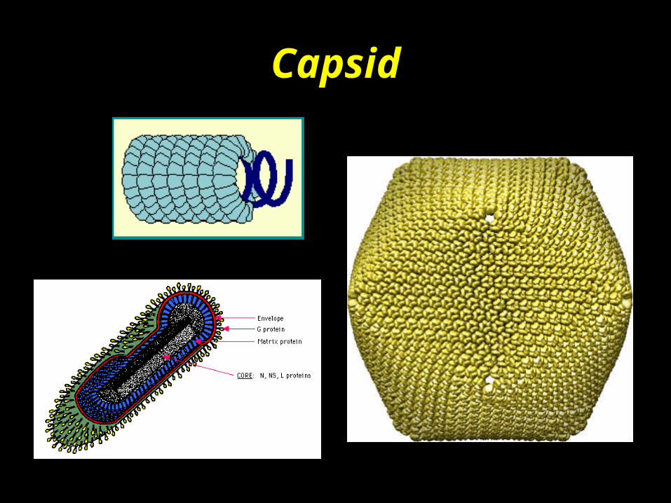
Capsid
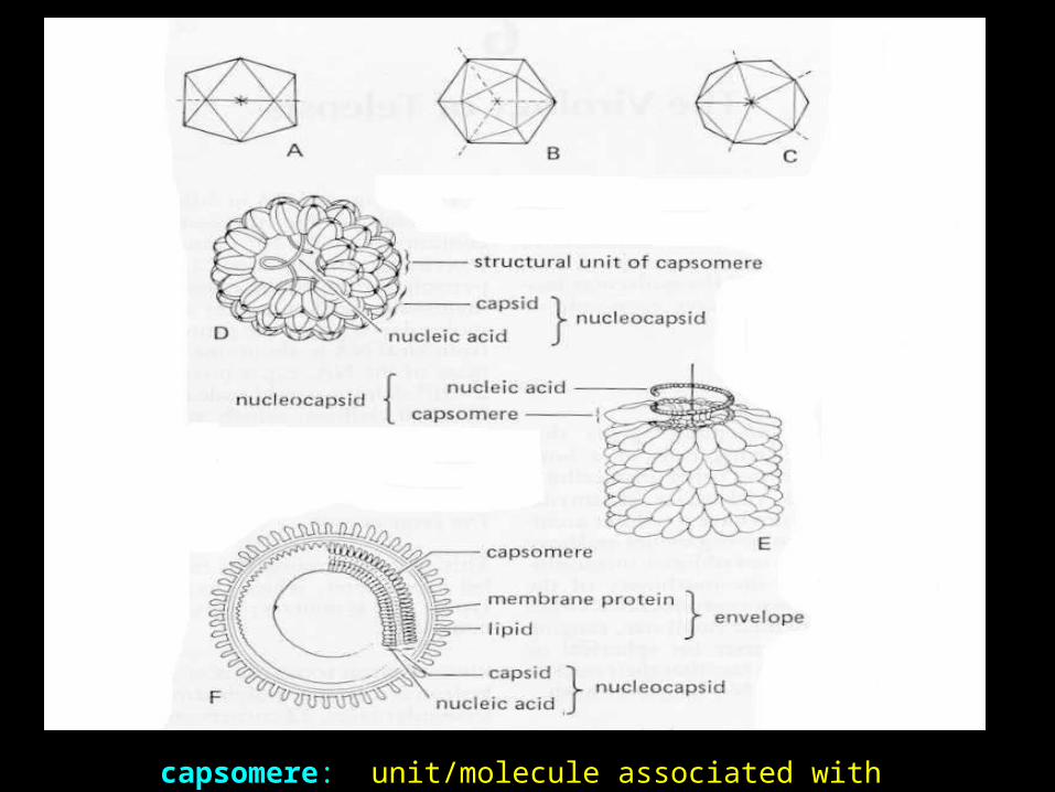
capsomere: unit/molecule associated with capsid structure
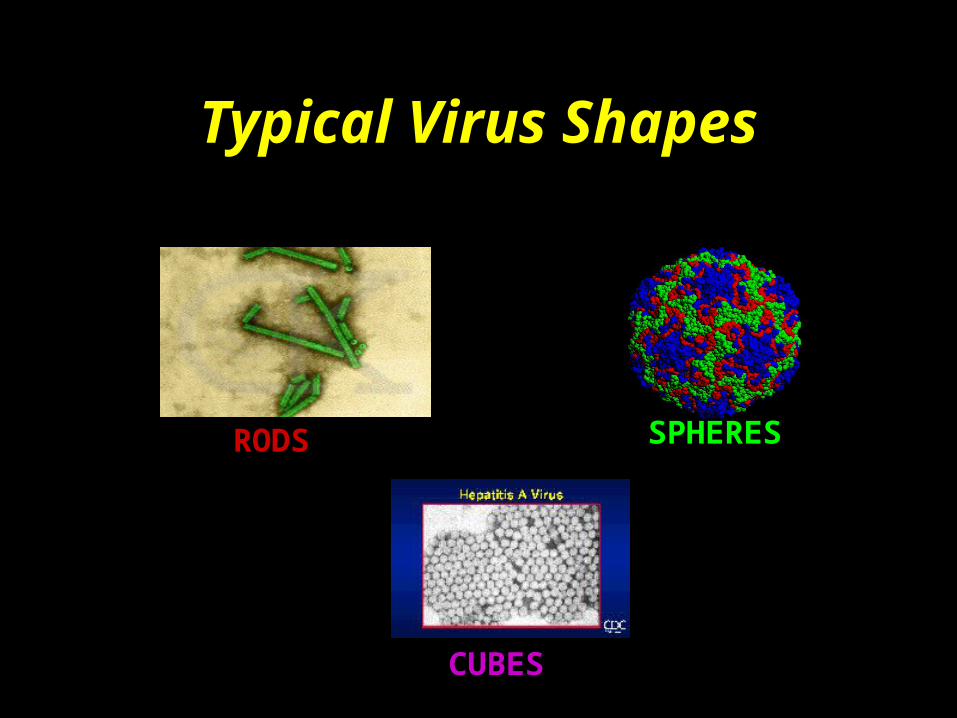
Typical Virus Shapes
RODS SPHERES
CUBES
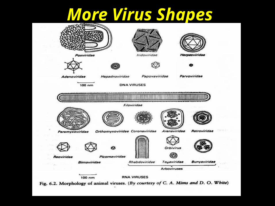
More Virus Shapes


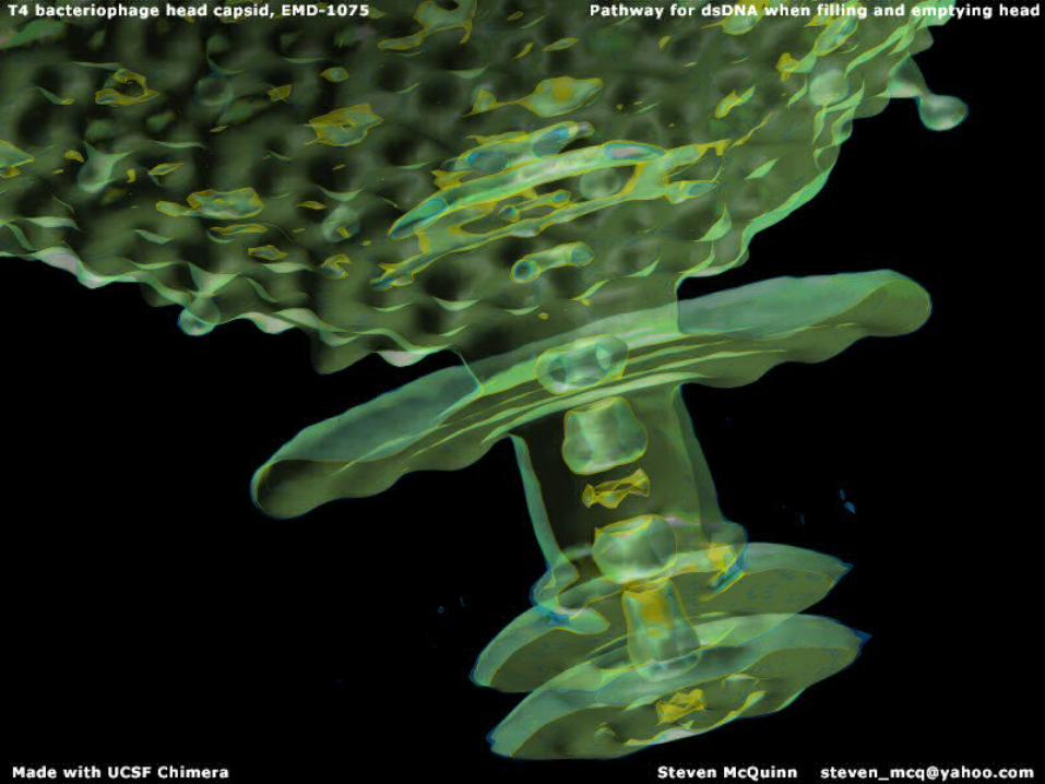
Composition of T-Even Bacteriophage
• Capsid: brains of virus, tightly-wound protein protecting nucleic acids
• Body: attached to capsid head, rod-like structure w/retractible sheath, hollow core
• Tail: at end of core is a spiked plate carrying 6 slender tail fibers, anchor virus to its host

How do viruses work?
• Viruses make use of the host cell’s chemical energy, protein and nucleic acid synthesizing ability to replicate themselves...
• each virus attacks a specific type of cell – cold viruses attack cells of the lung
– the AIDS virus attacks T4 cells of the
immune system
– fish viruses are just as specific

Bacteriophage Attack

Virusal Mechanism
• Viruses contain single- or double- stranded DNA or RNA
• Often, the virus alters the intracellular environment enough to damage or kill the cell (oops!!)
• If enough cells are destroyed, disease results!

Role of RNA/DNARole of RNA/DNA• Supplies the codes for building the protein coat
(capsid) and for producing enzymes needed to replicate more viruses
• Information given so newly-built viruses can lyse cells (e.g., bacteriophage)
• Result: cell destroyed.
Bottom Line...
• All viruses only exist to make more viruses
• Most are harmful
• Replication = host cell death.
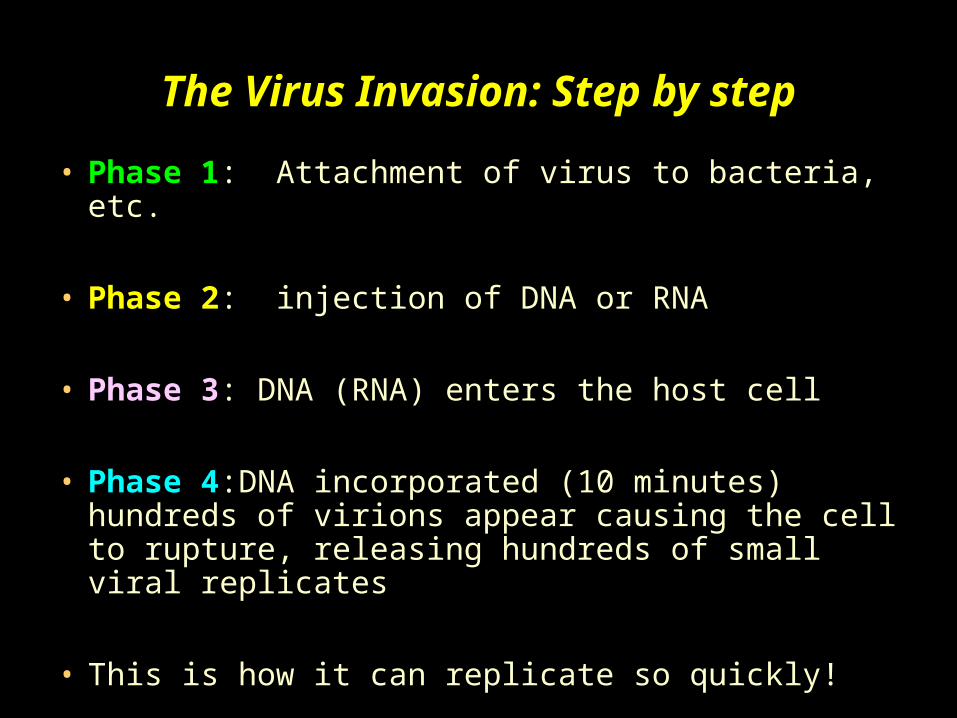
The Virus Invasion: Step by step
• Phase 1: Attachment of virus to bacteria, etc.
• Phase 2: injection of DNA or RNA
• Phase 3: DNA (RNA) enters the host cell
• Phase 4:DNA incorporated (10 minutes) hundreds of virions appear causing the cell to rupture, releasing hundreds of small viral replicates
• This is how it can replicate so quickly!

The Virus Invasion

What’s Infected by a Virus?
• All living things have some susceptibility to a particular virus
• Virus is specific for the organism• Within a species, there may be a 100 or more
different viruses which can affect that species alone
• Specific: for example, a virus that only affects one organism (humans and smallpox)
• Influenza can infect humans and two animals
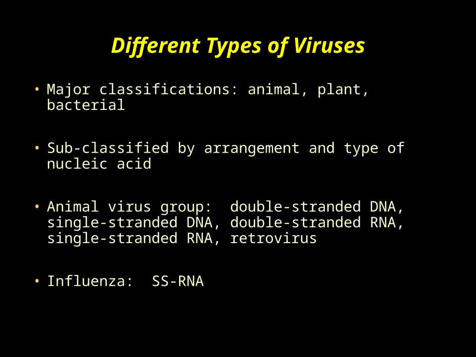
Different Types of Viruses
• Major classifications: animal, plant, bacterial
• Sub-classified by arrangement and type of nucleic acid
• Animal virus group: double-stranded DNA, single-stranded DNA, double-stranded RNA, single-stranded RNA, retrovirus
• Influenza: SS-RNA

Do Viruses ever Change?
• Mutations do occur.• If the mutation is harmful, the new virus particle
might no longer be functional (infectious)• However, because a given virus can generate many,
many copies, a small number of non-functional viruses is not important
• Mutation is not necessarily damaging to the virus -- it can lead to a functional but new strain of virus

Defense Against Viruses
• First Line: skin and mucous membrane, which also lines the gastrointestinal and respiratory passageways
• skin is tough and stomach acidity acts as a disinfectant
• Second Line: after the virus enters the blood and other tissues, white blood cells and related cells (phagocytes) consume them
• accumulation of phagocytes in area of infection is known as “puss”

Defense Against Viruses
Antibodies attacking chickenpox virus

Defense Against Viruses
• Antibodies are the best defense against viruses
• unfortunately, they are specific in their action
• chickenpox antibody will only attack a chickenpox virus
• a particular virus stimulates the production of a particular antibody
Defense Against Viral Infection
• Animals are protected in several ways:
• 1) intracellular: if a particular virus attacks cells, our bodies produce interferons
• interferons (alpha, beta or gamma) are proteins which interact with adjacent cells and cause them to become more resistant to infection by the virus
• if the resistance is not quite good enough, we become sick

Defense Against Viral Infection
• 2) immune system (extracellular): kills the virus outside the cell
• also kills the infected cells• virus cannot spread• eventually the virus is completely removed and we
get better• exception: HIV because it infects cells of the
immune system, itself• chemicals/drugs: acyclovir, AZT, HIV protease
inhibitor, several fish vaccines available.
Major Fish Viruses

Major Viral Infections in Fish
• Infectious pancreatic necrosis (IPN)
• Viral hemorrhagic septicemia (VHS)
• Infectious hematopoetic necrosis (IHN)
• Channel catfish virus disease (CCVD)
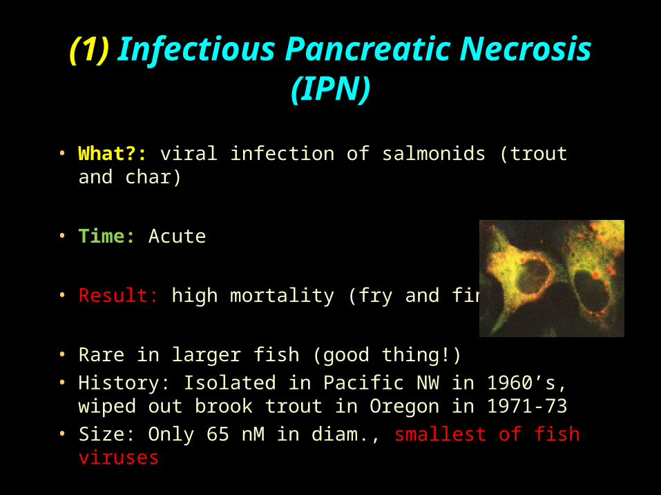
(1) Infectious Pancreatic Necrosis (IPN)
• What?: viral infection of salmonids (trout and char)
• Time: Acute
• Result: high mortality (fry and fingerlings)
• Rare in larger fish (good thing!)
• History: Isolated in Pacific NW in 1960’s, wiped out brook trout in Oregon in 1971-73
• Size: Only 65 nM in diam., smallest of fish viruses
IPN: general notes
• Single capsid shell, icosohedral symmetry, no envelope
• Contains two segments of DS-RNA
• Fairly stable and resistant to chemicals (acid, ether, etc.), variable resistance to freezing
• Remains infectious for 3 months in water (uh oh!)
• Targets pancreas and hematopoietic tissues of kidney and spleen
IPN: epizootiology (disease process)
• Who?: All salmonids, brook trout most susceptible, marine fish (flounder?)
• Reservoirs (where)?: carriers, once a carrier always a carrier, virus particles shed in feces/urine
• Transmission (how?): horizontal, by waters via carriers or infected fry; vertical from adults to progeny; experimentally by feeding infected material, IP injection
• Pathogenesis: entry via gills, digestive tract• Environmental factors: mortality reduced at lower
temps (why?); however, carriers not reduced
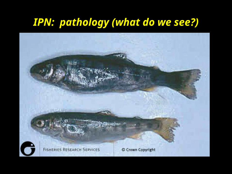
IPN: pathology (what do we see?)
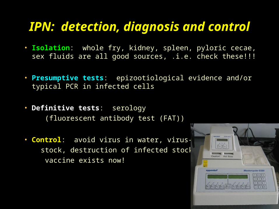
IPN: detection, diagnosis and control
• Isolation: whole fry, kidney, spleen, pyloric cecae, sex fluids are all good sources, .i.e. check these!!!
• Presumptive tests: epizootiological evidence and/or typical PCR in infected cells
• Definitive tests: serology
(fluorescent antibody test (FAT))
• Control: avoid virus in water, virus-free
stock, destruction of infected stock,
vaccine exists now!


How Bad Can It Be??

Fish severely affected by IPNV:
• Atlantic salmon* (Salmo salar)brook trout* (Salvelinus fontinalis)brown trout* (Salmo trutta)danio zebrafish* (Brachydanio rerio)rainbow trout* (Oncorhynchus mykiss)yellowtail* (Seriola lalandi)
Other species known to be susceptible…
• amago salmon (Oncorhynchus rhodurus)Arctic char (Salvelinus alpinus)Atlantic menhadden (Brevoortia tyrannus)carangids (Carangidae)chinook salmon (Oncorhynchus tshawytscha)chum salmon (Oncorhynchus keta)cichlids (Cichlidae)coho salmon (Oncorhynchus kisutch)common scallop (Pecten maximus)cutthroat trout (Salmo clarki)cyprinids (Cyprinidae)Danube salmon (Salmo hucho)drums/croakers (Sciaenidae)eels (Anguilla spp)grayling (Thymallus thymallus)
More…
• halibut (Hippoglossus stenolepis) herrings/sardines (Clupidae)Jap. amberjack (Seriola quinqueradiata) lake trout (Salvelinus namaycush)lampreys (Petromyzontyidae) left-eye flounders (Bothidae)loach (Misgurnus anguillicaudatus) loaches (Cobitidae)masou salmon (Oncorhynchus masou) Pacific salmon (Oncorhynchus spp)perches (Percidae) pikes (Esocidae)silversides (Atherinidae) sockeye salmon (Oncorhynchus nerka)soles (Soleidae) Southwest European nase (C. toxostoma) striped snakehead (Channa striatus) suckers (Cotostomidae)summer flounder (Paralichthys dentatus) turbot (Psetta maxima)white seabass (Moronidae) whitefish (Coregonidae)carp (Cyprinus carpio) goldfish (Carassius auratus)redfin perch (Perca fluviatilis) southern flounder (P. lethostigma)yellowfin bream (Acanthopagrus australis)

Asymptomatic carriers...
• coalfish (Pollachius virens)common carp (Cyprinus carpio)discus fish (Symphysodon discus)goldfish (Carrasius auratus)heron (Ardea cinerea)loach (Cobitidae)minnow (Phoxinus phoxinus)noble crayfish (Astacus astacus)pike (Esox lucius)river lamprey (Lampetra fluviatalis)shore crab (Carcinus maenas)Spanish barbel (Barbus graellsi)white suckers (Catostomas commersoni)
Infectious pancreatic necrosis in Atlantic salmon.
Note swollen stomach and 'pop eye'
Source: Australian Animal Health Laboratory
...what now???

(2) Viral Hemorrhagic Septicemia (VHS)
(2) Viral Hemorrhagic Septicemia (VHS)
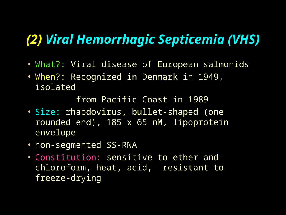
• What?: Viral disease of European salmonids• When?: Recognized in Denmark in 1949, isolated
from Pacific Coast in 1989• Size: rhabdovirus, bullet-shaped (one rounded
end), 185 x 65 nM, lipoprotein envelope• non-segmented SS-RNA• Constitution: sensitive to ether and chloroform,
heat, acid, resistant to freeze-drying

Viral Hemorrhagic Septicemia
• Produces a general viremia, tissue and organ damage, liver necrosis, spleen, kidney
• Epizootiology: cultured rainbow trout, also brown trout, steelhead, chinook, coho (most cases in WA state)
• Reservoirs: again...survivors are life-long carriers, usually rainbow trout, brown in Europe
• Transmission: horizontal through water, virus can occur on eggs spawned by carriers, IP injection, birds, hatchery equipment
Viral Hemorrhagic Septicemia (VHS)
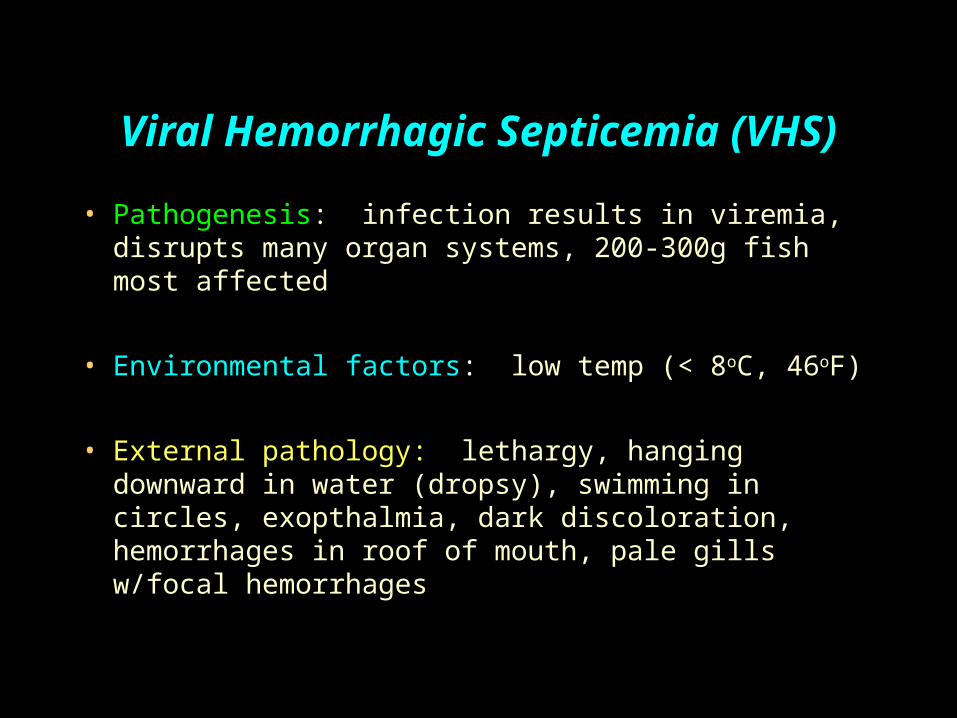
• Pathogenesis: infection results in viremia, disrupts many organ systems, 200-300g fish most affected
• Environmental factors: low temp (< 8oC, 46oF)
• External pathology: lethargy, hanging downward in water (dropsy), swimming in circles, exopthalmia, dark discoloration, hemorrhages in roof of mouth, pale gills w/focal hemorrhages
Viral Hemorrhagic Septicemia (VHS)
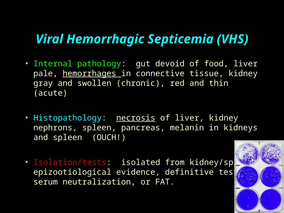
• Internal pathology: gut devoid of food, liver pale, hemorrhages in connective tissue, kidney gray and swollen (chronic), red and thin (acute)
• Histopathology: necrosis of liver, kidney nephrons, spleen, pancreas, melanin in kidneys and spleen (OUCH!)
• Isolation/tests: isolated from kidney/spleen, epizootiological evidence, definitive test is serum neutralization, or FAT.

Viral Hemorrhagic Septicemia (VHS)
External hemorrhages
Liver red in acute stage

Viral haemorrhagic septicaemia in rainbow trout.
Note swollen stomach and “pop eye”
Viral haemorrhagic septicaemia in rainbow trout.
Note pale color of stomach region, pinpoint haemorrhages in fatty tissue, and pale gills
Source: T Håstein
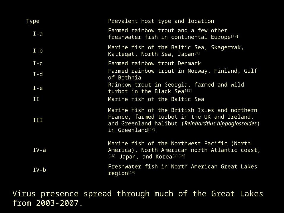
Type Prevalent host type and location
I-aFarmed rainbow trout and a few other freshwater fish in continental Europe[10]
I-bMarine fish of the Baltic Sea, Skagerrak, Kattegat, North Sea, Japan[1]
I-c Farmed rainbow trout Denmark
I-d Farmed rainbow trout in Norway, Finland, Gulf of Bothnia
I-e Rainbow trout in Georgia, farmed and wild turbot in the Black Sea [11]
II Marine fish of the Baltic Sea
IIIMarine fish of the British Isles and northern France, farmed turbot in the UK and Ireland, and Greenland halibut (Reinhardtius hippoglossoides) in Greenland[12]
IV-aMarine fish of the Northwest Pacific (North America), North American north Atlantic coast,[13] Japan, and Korea[1][14]
IV-b Freshwater fish in North American Great Lakes region[14]
Virus presence spread through much of the Great Lakes from 2003-2007.
Viral Hemorrhagic Septicemia
• Prevention: clean broodstock and water = clean fish, avoid infected broodstock, test and slaughter
• Can spread very quickly from farm to farm: avoid close proximity to other farms
• Vaccines are under development.
• One EPA-approved disinfectant: Virkon® AQUATIC (made by Dupont). Bleach kills the VHS virus.

(3 ) Infectious Hematopoietic Necrosis (IHN)
• Who: sockeye, chinook, rainbows; cohos resistant
• When?: 1950’s in Oregon hatcheries. 100 million mortalities between 1970-1980, if infected, 70% mortality likely, esp. in young fish (fry: 90-95% mort. possible)
• What?: bullet shaped rhabdovirus, non- segmented SS-RNA, sensitive to heat and pH, glycoprotein is spiked on surface of virus

Infectious Hematopoietic Necrosis (IHN)
• Reservoirs: survivors life-long carriers, adults shed virus at spawning
• Transmission: horizontal, primary mode is vertical via ovarian fluid (virus hitches ride on sperm into egg); however, feces, urine, and external mucus possible. Also, feeding and inoculation have worked experimentally
• Pathogenesis: gills suspected; incubation period depends on temp, route, dose, age; extensive hemorrhaging, necrosis of many tissues; death usually due to kidney failure
Infectious Hematopoietic Necrosis (IHN)• Environmental factors: temp. very important, slows below
10◦C, holding in tanks/handling increase severity (doesn’t occur naturally >15 ◦C)
• External pathology: lethargy, whirling, dropsy, exopthalmia, anemia, hemorrhaging of musculature/fins, scoliosis
• Internal pathology: liver, kidney, spleen pale; stomach/intestines filled with milky fluid; petechial hemorrhaging
• Histopathology: extensive necrosis of hematopoetic tissue of kidney/spleen
Infectious Hematopoietic Necrosis (IHN)
• Definitive diagnosis: serum neutralization, FAT, ELISA
• Prevention: avoidance, quarantine, clean water with UV, ozone, virus-free stock; test, slaughter, disinfect; disinfect eggs; vaccines under development; elevated water temp
• No vaccines as of June 2007.
(4) Channel Catfish Virus Disease (CCVD)
• Contagious herpes virus affecting only channel catfish less than four months old
• Occurs in SE United States, California, Honduras
• Acute hemorrhagia, high mortality, discovered in 1968
• Agent: enveloped capsid, icosohedral nucleocapsid with 162 capsomeres
• Physio/chemical properties: easy to kill, sensitive to freeze-thaw, acid, ether, etc.
Channel Catfish Virus Disease (CCVD)
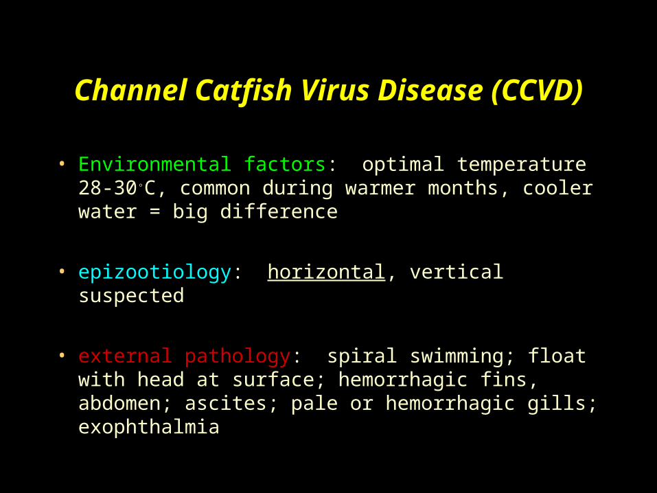
• Environmental factors: optimal temperature 28-30◦C, common during warmer months, cooler water = big difference
• epizootiology: horizontal, vertical suspected
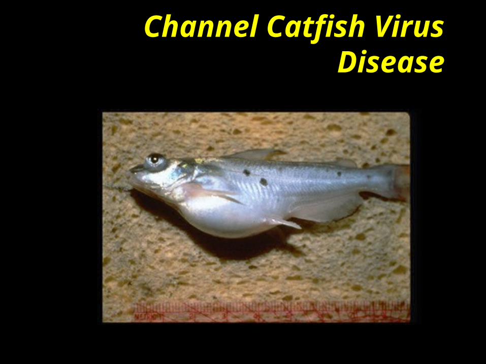
• external pathology: spiral swimming; float with head at surface; hemorrhagic fins, abdomen; ascites; pale or hemorrhagic gills; exophthalmia
Channel Catfish Virus Disease (CCVD)
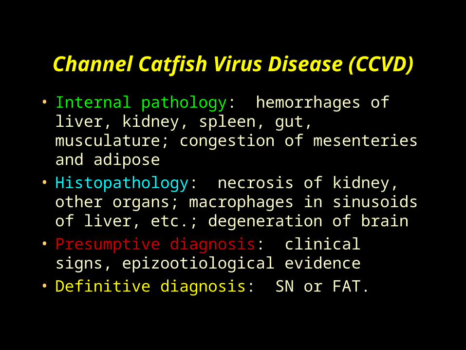
• Internal pathology: hemorrhages of liver, kidney, spleen, gut, musculature; congestion of mesenteries and adipose
• Histopathology: necrosis of kidney, other organs; macrophages in sinusoids of liver, etc.; degeneration of brain
• Presumptive diagnosis: clinical signs, epizootiological evidence
• Definitive diagnosis: SN or FAT.
Channel Catfish Virus Disease (CCVD)
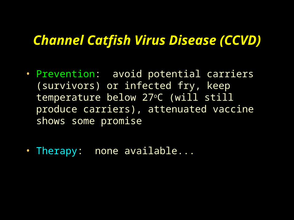
• Prevention: avoid potential carriers (survivors) or infected fry, keep temperature below 27oC (will still produce carriers), attenuated vaccine shows some promise
• Therapy: none available...
Channel Catfish Virus Disease

Channel Catfish Virus Disease

However, you can always take precautions!