april 30, 2008 10:39 wspc/115-ijprai spi-j068 00637 … the associ… · ·...
TRANSCRIPT
April 30, 2008 10:39 WSPC/115-IJPRAI SPI-J068 00637
International Journal of Pattern Recognitionand Artificial IntelligenceVol. 22, No. 3 (2008) 601–616c© World Scientific Publishing Company
DETERMINING THE ASSOCIATION BETWEENDERMATOGLYPHICS AND SCHIZOPHRENIA BYUSING FINGERPRINT ASYMMETRY MEASURES
JEN-FENG WANG, CHEN-LIANG LIN and CHEN-WEN YEN
Department of Mechanical and Electromechanical EngineeringNational Sun Yat-sen University, Kaohsiung 80424, Taiwan
YUNG-HSIEN CHANG∗, TENG-YI CHEN and KUAN-PIN SU
China Medical University and Hospital, Taichung 40402, Taiwan∗[email protected]
MARK L. NAGURKA
Department of Mechanical Engineering, Marquette UniversityMilwaukee, WI 53201, USA
Early detection and intervention strategies for schizophrenia are receiving increasinglymore attention. Dermatoglyphic patterns, such as the degree of asymmetry of the fin-gerprints, have been hypothesized to be indirect measures for early abnormal devel-opmental processes that can lead to later psychiatric disorders such as schizophrenia.However, previous results have been inconsistent in trying to establish the associationbetween dermatoglyphics and schizophrenia. The goal of this work is to try to resolve thisproblem by borrowing well-developed techniques from the field of fingerprint matching.Two dermatoglyphic asymmetry measures are proposed that draw on the orientationfield of homologous fingers. To test the capability of these measures, fingerprint imageswere acquired digitally from 40 schizophrenic patients and 51 normal individuals. Basedon these images, no statistically significant association between conventional dermato-glyphic asymmetry measures and schizophrenia was found. In contrast, the sample meansof the proposed measures consistently identified the patient group as having a higherdegree of asymmetry than the control group. These results suggest that the proposedmeasures are promising for detecting the dermatoglyphic patterns that can differentiatethe patient and control groups.
Keywords: Dermatoglyphic; fingerprint; schizophrenia; fluctuating asymmetry; ridgecount; mutual information.
1. Introduction
Schizophrenia is hypothesized to be the result of an interaction between geneticfactors and insults during embryonic development.22 While a full understanding of
∗Author for correspondence
601

April 30, 2008 10:39 WSPC/115-IJPRAI SPI-J068 00637
602 J.-F. Wang et al.
its origins and pathogenic mechanisms remain elusive, schizophrenia is generallyconsidered to be a neurodevelopmental disorder.12 This idea is supported by brainimaging studies that have found diverse abnormalities in schizophrenia.4 For exam-ple, structural brain abnormalities, such as distortion of the corpus callosum andasymmetry of brain, have been recognized on computed tomography (CT) and mag-netic resonance imaging (MRI) scans.11,14,17–19,35 Studies suggest that schizophre-nia may be due to impaired neurodevelopment and might occur before the end ofthe second gestational trimester.
Dermatoglyphics, the epidermal ridges and patterns of the hand, are establishedby the end of the second trimester and have been considered as markers of prenatalbrain injury. The rationale behind this hypothesis is that epidermal ridges shareectodermal origins with the central nervous system. Specifically, epidermal ridgesstart to develop in the 11th gestational week and their critical stage of differentiationoccurs in fetal months 3–4, coinciding with a critical phase of brain development.25
The morphology of the epidermal ridges is genetically determined but can alsobe influenced by environmental factors such as a viral infection, radiation, or alco-hol and drug abuse that can disturb brain development.2,9 However, once they areformed the epidermal ridges remain unchanged. Since both the ridges and the brainare derived from the ectoderm, it seems reasonable to use unusual dermatoglyphicpatterns to characterize disturbances to brain development. Preliminary experimen-tal results have successfully demonstrated the association between dermatoglyphicsand cerebral structural measures in patients with schizophrenia.31
Many studies have tried to establish the direct link between epidermal ridgesand schizophrenia using different features to characterize the configuration of epi-dermal ridges. Fearon et al.10 reported more than 70 such studies up to 2001. Thesestudies are valuable since they considered schizophrenia from a preventive perspec-tive reflecting an important recent trend in neurosciences.7 However, as will bereported in the subsequent section, results of these studies were inconsistent.
This paper proposes alternative dermatoglyphic asymmetry measures to differ-entiate schizophrenic patients from healthy individuals. The measures are adaptedfrom a technique that has been extensively employed in fingerprint matching. Thepaper is organized as follows. The following section reviews commonly employedfinger dermatoglyphic features as well as the results of previous studies. Section 3illustrates the proposed method. Section 4 presents the experimental results. Dis-cussion and conclusion are given in Sec. 5.
2. Previous Work
Due to the inevitability of developmental errors, no organism is perfectly symmet-rical. Individuals have bilateral distribution with different degrees of asymmetry.The degree of asymmetry has thus been considered an indirect measure for devel-opmental instability.36 Since dermatoglyphic anomalies are hypothesized to signifydisruptions in the second trimester of prenatal development, a critical time periodin the etiology of schizophrenia, the possibility that schizophrenic patients tend tohave a higher degree of dermatoglyphic asymmetry has been studied extensively.

April 30, 2008 10:39 WSPC/115-IJPRAI SPI-J068 00637
Determining the Association Between Dermatoglyphics and Schizophrenia 603
Conventional finger dermatoglyphic features that have been used to measurethe degree of asymmetry include the following five categories:
(1) Ridge Pattern: Based on the pattern of ridge configuration, several criteriahave been proposed to classify fingerprints into different pattern groups. Theseinclude the three-pattern system (loop, arch, whorl),8 the six-pattern system(plain arch, tended arch, ulnar loop, radial loop, whorl or combined figure),13
and the eight-pattern system (plain arch, tended arch, ulnar loop, radial loop,plain whorl, double loop whorl, central pocket loop whorl, and accidental whorl)adopted by the FBI.
(2) FRC and TFRC: The finger ridge count (FRC) is defined as the number ofridges intersected by a line between the triradial points (also called the deltapoint) to the point of core. As shown in Fig. 1(a), the core is the topmostpoint of the innermost curving ridge and a triradius is defined as the meetingplace of three dermal lines that make angles of approximately 120◦ with oneanother. The total finger ridge count (TFRC) is the sum of FRC for all fingersin one hand. Some fingers may have more than one triradial point, as shownin Fig. 1(b), and this results in multiple ridge counts. To resolve this problem,the largest ridge count is typically chosen as the FRC.
(3) AFRC: The absolute finger ridge count (AFRC) is equal to FRC when thefinger has only one triradial point. However, in dealing with multiple triradialpoints, unlike FRC that uses the largest ridge count, AFRC is chosen as thesum of all ridge counts. The sum of AFRC for all fingers in one hand is denotedas the total absolute finger ridge count (TAFRC) in this work.
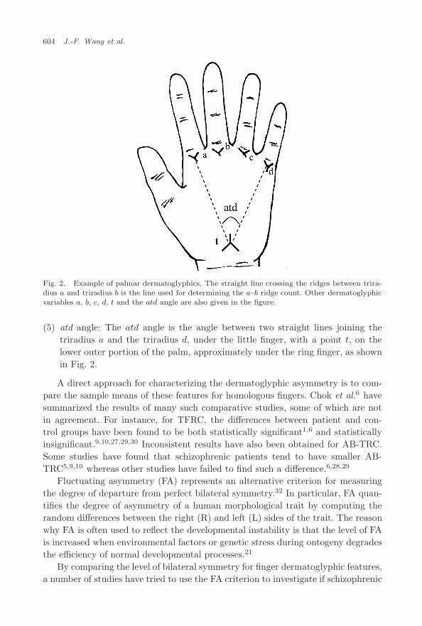
(4) AB-TRC: The total a–b ridge count is defined as the number of ridges inter-sected by a line drawn between the a triradius (at the base of the index finger)and b triradius (at the base of the middle finger) of the palm in each hand, asdepicted in Fig. 2.
(a) (b)
Fig. 1. (a) A fingerprint with one triradial point. (b) A fingerprint with two triradial points. Theridge count is the number of ridges intersected by a line between the triradial points to the pointof core.
April 30, 2008 10:39 WSPC/115-IJPRAI SPI-J068 00637
604 J.-F. Wang et al.
Fig. 2. Example of palmar dermatoglyphics. The straight line crossing the ridges between trira-dius a and triradius b is the line used for determining the a–b ridge count. Other dermatoglyphicvariables a, b, c, d, t and the atd angle are also given in the figure.
(5) atd angle: The atd angle is the angle between two straight lines joining thetriradius a and the triradius d, under the little finger, with a point t, on thelower outer portion of the palm, approximately under the ring finger, as shownin Fig. 2.
A direct approach for characterizing the dermatoglyphic asymmetry is to com-pare the sample means of these features for homologous fingers. Chok et al.6 havesummarized the results of many such comparative studies, some of which are notin agreement. For instance, for TFRC, the differences between patient and con-trol groups have been found to be both statistically significant1,6 and statisticallyinsignificant.9,10,27,29,30 Inconsistent results have also been obtained for AB-TRC.Some studies have found that schizophrenic patients tend to have smaller AB-TRC5,9,10 whereas other studies have failed to find such a difference.6,28,29
Fluctuating asymmetry (FA) represents an alternative criterion for measuringthe degree of departure from perfect bilateral symmetry.32 In particular, FA quan-tifies the degree of asymmetry of a human morphological trait by computing therandom differences between the right (R) and left (L) sides of the trait. The reasonwhy FA is often used to reflect the developmental instability is that the level of FAis increased when environmental factors or genetic stress during ontogeny degradesthe efficiency of normal developmental processes.21
By comparing the level of bilateral symmetry for finger dermatoglyphic features,a number of studies have tried to use the FA criterion to investigate if schizophrenic

April 30, 2008 10:39 WSPC/115-IJPRAI SPI-J068 00637
Determining the Association Between Dermatoglyphics and Schizophrenia 605
patients have a higher degree of asymmetry than normal individuals. Results fromthese studies are also inconsistent.6
As suggested by several authors10,29 the lack of consistency in previous workmay be due to factors such as diagnostic criteria and ethnicity. Another possibilityis that the difference between the degree of finger dermatoglyphic asymmetry forschizophrenic patients and normal individuals is relatively subtle and thus difficultto detect.5 In the subsequent section two alternative dermatoglyphic asymmetrymeasures are posited to detect such potentially subtle differences.
3. Proposed Approach
As described in the preceding section, in trying to find the association betweendermatoglyphics and schizophrenia inconsistent results have been obtained. Ratherthan repeat comparative studies using the same dermatoglyphic features, this workfocuses on the ridge orientation field (also referred to as the directional field in someliteratures) for the homologous fingers. As shown in Fig. 3, the ridge orientation
(a) Left finger (b) Right finger
(c) Orientation field of left finger (d) Orientations field of right
Fig. 3. Examples of ridge orientation field for a pair of index fingers. (a) and (b) are the originalfingerprint images and (c) and (d) illustrate the orientation field for these two fingers.

April 30, 2008 10:39 WSPC/115-IJPRAI SPI-J068 00637
606 J.-F. Wang et al.
field is a representation of the direction of the ridges throughout the image andthus represents the intrinsic nature of the fingerprint image. With the exception ofridge-pattern, which is only a qualitative measure, traditional dermatoglyphic fingerfeatures cannot provide such global information to quantitatively characterize theoverall configuration of fingerprints.
3.1. Acquisition and alignment of finger images
Fingerprints have traditionally been extracted by creating an inked impression ofthe fingertip on paper. This acquisition procedure is sensitive to environmentalfactors and the skin condition,15 and consequently many fingerprint images acquiredthis way are of poor quality. In this work fingerprint images are captured using adigital camera (Canon G3, resolution 2272× 1704). Figure 4 shows an index fingerimage acquired in such a way.
After segmenting the fingers from the background, a preprocessing step for mea-suring the degree of asymmetry, the contours of homologous fingers are alignedusing the maximization of mutual information (MMI) criterion.33 By measuringthe amount of information that one image contains relative to the other, mutualinformation (MI) has been applied extensively to image registration problems.20 Inparticular, given two images A and B, the definition of the mutual information I
(A, B) of these images is
I(A, B) = H(A) + H(B) − H(A, B) (1)
where H(A) and H(B) denote the entropies of the images A and B, respectively.Note that the entropy of an image can be thought of as a dispersion measure for thedistribution of the image gray values. H(A, B) is the joint entropy of images A andB and can be used to quantify the dispersion of the joint probability distributionp(a, b) which is the probability of the occurrence for pixels of gray value a in image
Sample of finger image
Fig. 4. An example of a digitally captured fingerprint image.

April 30, 2008 10:39 WSPC/115-IJPRAI SPI-J068 00637
Determining the Association Between Dermatoglyphics and Schizophrenia 607
A and counterpart pixels with gray value b in image B. An important propertyof the joint entropy is that H(A, B) decreases with the strength of the statisticalrelationship between images A and B. Therefore, as images A and B become moresimilar, the lower the value of H(A, B).
Since the goal is to align the contour of the fingers, finger images are firstconverted into binary images by setting the background pixels to zeros and thefinger pixels to ones. Next, for a pair of homologous fingers, one binary fingerimage is taken as the reference, and the other binary finger image is aligned byrotating and translating it until an optimal position is found that maximizes theMI. Figure 6 shows the contours of the homologous fingers of Fig. 5 before and afteralignment.
3.2. Computing orientation field for the region of interest
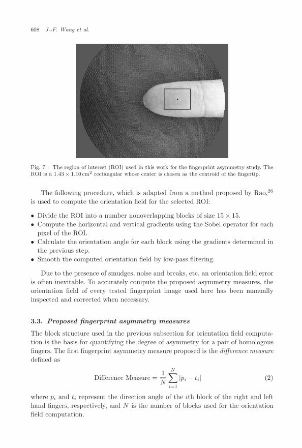
After aligning the finger images, the next step is to determine the region of interest(ROI) for each finger. As illustrated in Fig. 7, the ROI is chosen as a 1.43×1.10cm2
rectangular whose center is located at the centroid of the fingertip area.
(a) Left finger (b) Right finger
Fig. 5. Examples of a pair of homologous finger images before the operation of alignment. Thefinger image shown in (a) is set as the reference and finger image of (b) is to be adjusted by theMMI criterion.
(a) Before alignment (b) After alignment
Fig. 6. An illustration of the effect of alignment for the pair of homologous fingers shown inFig. 5. (a) Contours before alignment. (b) Contours after alignment. Note that the dotted linerepresents the contour of left finger and solid line is the contour of the right finger.
April 30, 2008 10:39 WSPC/115-IJPRAI SPI-J068 00637
608 J.-F. Wang et al.
Fig. 7. The region of interest (ROI) used in this work for the fingerprint asymmetry study. TheROI is a 1.43 × 1.10 cm2 rectangular whose center is chosen as the centroid of the fingertip.
The following procedure, which is adapted from a method proposed by Rao,26
is used to compute the orientation field for the selected ROI:
• Divide the ROI into a number nonoverlapping blocks of size 15 × 15.• Compute the horizontal and vertical gradients using the Sobel operator for each
pixel of the ROI.• Calculate the orientation angle for each block using the gradients determined in
the previous step.• Smooth the computed orientation field by low-pass filtering.
Due to the presence of smudges, noise and breaks, etc. an orientation field erroris often inevitable. To accurately compute the proposed asymmetry measures, theorientation field of every tested fingerprint image used here has been manuallyinspected and corrected when necessary.
3.3. Proposed fingerprint asymmetry measures
The block structure used in the previous subsection for orientation field computa-tion is the basis for quantifying the degree of asymmetry for a pair of homologousfingers. The first fingerprint asymmetry measure proposed is the difference measuredefined as
Difference Measure =1N
N∑i=1
|pi − ti| (2)
where pi and ti represent the direction angle of the ith block of the right and lefthand fingers, respectively, and N is the number of blocks used for the orientationfield computation.

April 30, 2008 10:39 WSPC/115-IJPRAI SPI-J068 00637
Determining the Association Between Dermatoglyphics and Schizophrenia 609
The second asymmetry measure used in this work is called the correlation mea-sure. It can be computed from
Correlation Measure =∑N
i=1 (pi − p)(ti − t)√∑Ni=1 (pi − p)2
√∑Ni=1 (ti − t)2
(3)
where p and t are the sample means of the orientation angles for the fingers of theright and left hands, respectively. This measure is often used in signal processingapplications to characterize the degree of similarity between two signals.
4. Experimental Results
Fingerprint images were acquired from 40 patients diagnosed with schizophreniafrom the Jing-Ho Mental Hospital (Changhua, Taiwan) and the China Medical Uni-versity Hospital (Taichung, Taiwan). Fingerprint images were also acquired from51 control participants. For the control group, 49 participants were students at SunYat-sen University (Kaohisung, Taiwan) and the remaining two were volunteersfrom the China Medical University Hospital. All participants were of Han national-ity and thus belong to the same ethnic group. In addition, to eliminate the potentialeffect of sexual differences, all participants were chosen to be males.
Fingerprint images were acquired by a digital camera, and for the sake of reli-ability, five consecutive shots were taken for each finger. In this work thumbs wereexcluded from the experimental study. (Taking accurate images of thumbs wasfound to be much more time consuming than other fingers.)
With the availability of five images for each finger, every image of a right-handfinger was compared with every image of the homologous finger of the left-hand.Among the 25 combinations, the one that yielded the smallest asymmetry measurevalue was adopted.
Mean values were assessed using a two-sided independent samples t-test. Thetest results were considered significant for P -values < 0.01. Each of the testedasymmetry measures was found in five comparative studies for the index, mid-dle, ring and little fingers as well as the overall result (sum or average) for thefour fingers. Since triradial points could not be found in some of our fingerprintimages, the number of samples for each comparison often differs from the number ofparticipants.
The first part of the experiment tests the hypothesis that the control group haslarger FRC or AFRC than the patient group. The results, summarized in Tables 1and 2, indicate that there are no significant differences between the groups for FRCor AFRC.
The second part of the experiment investigates the degree of bilateral symmetry.In particular, the question is if the patient group has a higher level of FRC or AFRCfluctuating asymmetry in comparison to the normal group. An FA measure adoptedin several previous fingerprint asymmetry studies1,6,21,30,34 is used here. It is the

April 30, 2008 10:39 WSPC/115-IJPRAI SPI-J068 00637
610 J.-F. Wang et al.
Table 1. Summary of the results for FRC and TFRC.
Controls Patients
Fingers Numbers Mean ± Std. Numbers Mean ± Std. P -Value
FRCLeft Hand
Index 45 13.58 ± 5.34 34 13.91 ± 6.46 0.808Middle 46 14.20 ± 4.73 33 14.12 ± 4.47 0.943Ring 45 16.49±3.82 31 16.03±4.81 0.661Little 48 14.02 ± 3.46 37 14.19 ± 5.00 0.862
Right HandIndex 43 13.47 ± 4.66 31 13.19 ± 5.67 0.828Middle 45 14.00 ± 4.15 33 13.03 ± 4.96 0.365Ring 46 15.96 ± 3.32 26 15.69 ± 5.75 0.831Little 46 13.89 ± 3.84 35 14.89 ± 5.16 0.342
TFRC 32 107.84 ± 24.46 16 97.13 ± 38.37 0.320
Table 2. Summary of the results for AFRC and TAFRC.
Controls Patients
Fingers Numbers Mean ± Std. Numbers Mean ± Std. P -Value
AFRCLeft Hand
Index 45 19.56 ± 11.06 34 20.35 ± 12.73 0.772Middle 46 19.43 ± 11.21 33 19.73 ± 10.35 0.905Ring 45 24.16 ± 9.93 31 26.45 ± 12.03 0.384Little 48 17.42 ± 7.90 37 19.22 ± 9.66 0.360
Right HandIndex 43 19.67 ± 10.88 31 18.29 ± 11.15 0.597Middle 45 19.44 ± 10.70 33 16.67 ± 9.75 0.237Ring 46 23.39 ± 8.92 26 24.73 ± 11.88 0.619Little 46 17.24 ± 7.98 35 20.69 ± 10.32 0.106
TAFRC 32 142.06 ± 57.54 16 128.81 ± 67.12 0.505
first FA asymmetry measure used in this work,
FA1 =1M
M∑i=1
|Li − Ri|, (4)
where Ri and Li are the values of the tested feature of the ith finger of the rightand left hands, respectively, and M is the number of fingers used for comparison.
The second FA measure used here is
FA2 =1M
M∑i=1
|Li − Ri||Li + Ri| . (5)
This criterion has also been used in previous fingerprint asymmetry studies.23,27,29
The results for the two tested FA asymmetry measures are shown in Tables 3 and 4,and indicate that no statistically significant difference can be found.
April 30, 2008 10:39 WSPC/115-IJPRAI SPI-J068 00637
Determining the Association Between Dermatoglyphics and Schizophrenia 611
Table 3. Summary of the results for the fluctuating dermatoglyphic asymmetry measure FA1.
Controls Patients
Ridge Count Measure Finger Numbers Mean ± Std. Numbers Mean ± Std. P -Value
FRC
Index 40 2.45 ± 2.43 28 2.11 ± 1.55 0.480Middle 42 2.45 ± 1.73 28 2.86 ± 2.32 0.434Ring 42 2.05 ± 1.77 25 2.20 ± 1.71 0.729Little 46 2.40 ± 1.88 34 2.18 ± 1.53 0.870
Total 32 2.23 ± 0.97 16 2.03 ± 1.15 0.564
AFRC
Index 40 3.95 ± 4.22 28 3.57 ± 4.62 0.732Middle 42 5.33 ± 5.64 28 4.96 ± 4.99 0.775Ring 42 5.07 ± 4.93 25 2.68 ± 2.56 0.011Little 46 3.41 ± 3.05 34 3.97 ± 4.06 0.504
Total 32 4.02 ± 2.36 16 3.33 ± 1.74 0.261
Table 4. Summary of the results for the fluctuating dermatoglyphic asymmetry measure FA2.
Controls Patients
Ridge Count Measure Finger Numbers Mean ± Std. Numbers Mean ± Std. P -Value
FRC
Index 40 0.121 ± 0.155 28 0.149 ± 0.225 0.563Middle 42 0.099 ± 0.090 28 0.145 ± 0.194 0.246Ring 42 0.066 ± 0.056 25 0.123 ± 0.220 0.216Little 46 0.095 ± 0.104 34 0.092 ± 0.095 0.892
Total 32 0.090 ± 0.051 16 0.122 ± 0.125 0.339
AFRC
Index 40 0.143 ± 0.174 28 0.170 ± 0.235 0.607Middle 42 0.147 ± 0.131 28 0.184 ± 0.202 0.399Ring 42 0.119 ± 0.116 25 0.116 ± 0.223 0.956Little 46 0.117 ± 0.114 34 0.118 ± 0.110 0.954
Total 32 0.124 ± 0.074 16 0.139 ± 0.117 0.647
Table 5. Summary of the results for the proposedorientation difference measure.
Orientation Difference
(Mean ± Standard Deviation)
Finger Controls Patients P -Value
Index 12.78 ± 5.30 13.62 ± 7.04 0.5333Middle 10.82 ± 4.23 11.13 ± 4.65 0.7405Ring 10.32 ± 4.16 10.59 ± 4.01 0.7494Little 8.87 ± 3.89 11.30 ± 4.52 0.0085
Average 10.70 ± 2.87 11.66 ± 3.20 0.1206
The third part of the experiment performs comparisons of the proposed asym-metry measures and the results are summarized in Tables 5 and 6. For thedifference measure, the results of Table 5 show that the sample means of theschizophrenic group are larger than those of the control group in all five experi-ments. In addition, the result for the little finger has achieved statistical significance

April 30, 2008 10:39 WSPC/115-IJPRAI SPI-J068 00637
612 J.-F. Wang et al.
Table 6. Summary of the results for the proposedcorrelation measure.
Correlation(Mean ± Standard Deviation)
Finger Controls Patients P -Value
Index 0.826 ± 0.096 0.785 ± 0.121 0.0809Middle 0.840 ± 0.087 0.802 ± 0.104 0.0698Ring 0.848 ± 0.086 0.813 ± 0.115 0.1110Little 0.869 ± 0.073 0.820 ± 0.120 0.0256
Average 0.846 ± 0.057 0.805 ± 0.071 0.0038
(P -value = 0.0085). These results suggest that the patient group tends to have ahigher degree of asymmetry than the control group.
For the proposed correlation measure, the results presented in Table 6 confirmthat the sample means of the patient group are all smaller than those of the normalgroup in the five tests. Statistical significance has also been reached for the averageof the correlation measure of the four tested fingers (P -value = 0.0038). The resultsobtained using the proposed correlation measure agree with the results obtainedby the difference measure in that they also suggest that the patient group tends tohave a higher degree of asymmetry than the control group.
5. Discussion and Conclusion
Schizophrenia is considered to be associated with altered prenatal neurodevelop-ment. Since dermatoglyphic patterns are formed at the second trimester of pre-natal development, a time period that appears to be etiologically relevant to thedevelopment of schizophrenia, unusual dermatoglyphic patterns have been hypoth-esized to be proxy markers of altered early development in psychosis. To test thishypothesized association, this work compares the degree of finger dermatoglyphicasymmetry for schizophrenic and normal groups. In addition to using conventionalasymmetry measures, this work proposes two alternative measures based on theorientation field of the fingerprints. For our tested sample, the conventional der-matoglyphic measures were not able to provide statistically significant results thatcan be used to differentiate the patient and the control groups. Therefore, theassociation between schizophrenia and the conventional dermatoglyphic asymme-try measures cannot be established in this study.
In contrast, as demonstrated by the results of Tables 5 and 6, the samplemeans of the proposed difference (correlation) measure of the patient group areall larger (smaller) than those of the control group. In addition among the tentests of Tables 5 and 6, two have achieved statistical significance. The limited suc-cess of the proposed measures may be due to the inadequate statistical power ofour tests since the number of participants in our experimental studies is relativelysmall. Nevertheless, compared with the conventional measures, results obtained
April 30, 2008 10:39 WSPC/115-IJPRAI SPI-J068 00637
Determining the Association Between Dermatoglyphics and Schizophrenia 613
using the proposed measures have provided evidence for an association betweenunusual dermatoglyphic characteristics and genetic vulnerability to schizophrenia.
The proposed approach extracts information from the orientation field, whichcharacterizes the global configuration of a fingerprint by describing the local direc-tions of the ridge lines. Experimental results have shown that the proposed measurescan identify the difference between the patient and normal groups when the con-ventional dermatoglyphic measures fail to detect any between group differences.It may be valuable to repeat the experiments using the proposed measure for thestudies that were unsuccessful in finding an association between finger dermato-glyphics and schizophrenia.
References
1. M. T. Avila, J. Sherr, L. E. Valentine, T. A. Blaxton and G. K. Thaker, Neurode-velopmental interactions conferring risk for schizophrenia: a study of dermatoglophicmarkers in patients and relatives, Schizophr. Bull. 29 (2003) 595–605.
2. W. J. Babler, Embryonic development of epidermal ridges and their configurations,in Dermatoglyphics: Science in Transition, Vol. 27, eds. C. C. Plato, R. M. Garrutoand B. A. Schaumman (Wiley-Liss, New York, 1991), pp. 95–112.
3. A. M. Bazen and S. H. Gerez, Systematic methods for the computation of the direc-tional fields and singular points of fingerprints, IEEE Trans. Patt. Anal. Mach. Intell.24(7) (2002) 905–919.
4. B. Bogerts, Recent advances in the neuropathology of schizophrenia, Schizophr. Bull.19 (1993) 431–445.
5. E. Bramon, M. Walshe, C. McDonald, B. Martın, T. Toulopoulou, H. Wickham,J. van Os, P. Fearon, P. C. Sham, L. Fananas and R. M. Murray, Dermatoglyph-ics and schizophrenia: a meta-analysis and investigation of the impact of obstetriccomplications upon a-b ridge count, Schizophr. Res. 75 (2005) 399–404.
6. J. T. Chok, T. R. Kwapil and A. Scheuermann, Dermatoglyphic anomalies in psycho-metrically identified schizotypic young adults, Schizophr. Res. 72 (2005) 205–214.
7. M. T. Compton, Considering schizophrenia from a prevention perspective, Am.J. Prev. Med. 26 (2004) 178–185.
8. H. Cummins and C. Midlo, Fingerprints, Palms and Soles (Blakiston, Philadelphia,1943).
9. L. Fananas, J. van Os, C. Hoyos, J. McGrath, C. S. Mellor and R. Murray, Der-matoglyphic a-b ridge count as a possible marker for developmental disturbance inschizophrenia: replication in two samples, Schizophr. Res. 20 (1996) 307–314.
10. P. Fearon, A. Lane, M. Airie, J. Scannell, A. McGowan, M. Bynre, M. Cannon, D. Cot-ter, P. Murphy, B. Cassidy, J. Waddington, C. Larkin and E. O’Callaghan, Is reduceddermatoglyphic a-b ridge count a reliable marker of developmental impairment inschizophrenia, Schizophr. Res. 50 (2001) 151–157.
11. J. Foong, M. Maier, C. A. Clark, G. J. Barker, D. H. Miller and M. A. Ron, Neu-ropathological abnormalities of the corpus callosum in schizophrenia: a diffusion tensorimaging study, J. Neurol. Neurosurg. Psychiatry 68 (2000) 242–244.
12. P. J. Harrison, The neuropathology of schizophrenia: a critical review of the data andtheir interpretation, Brain 122 (1999) 593–624.
13. E. Henry, Classification and Uses of Finger Prints, 8th edn. (H.M. Stationary Office,London, 1937).

April 30, 2008 10:39 WSPC/115-IJPRAI SPI-J068 00637
614 J.-F. Wang et al.
14. B. Ismail, E. Cantor-Graae and T. F. McNeil, Minor physical anomalies inschizophrenic patients and their siblings, Am. J. Psychiatry 155 (1998) 1695–1702.
15. L. C. Jain, U. Halici, I. Hayashi, S. B. Lee, S. Tsutsui, Intelligent Biometric Techniquesin Fingerprint and Face Recognition (CRC Press, Boca Raton, FL, 1999).
16. A. K. Jain, L. Hong, S. Pankanti and R. Bolle, An identity-authentication system byusing fingerprints, Proc. IEEE 85 (1997) 1365–1388.
17. B. Kirkpatrick, D. Litman, J. W. Kim, K. Vladar, A. Breier and R. W. Buchanan,Failure of fusion of the septum pellucidum and heterogeneity of schizophrenia, J. Nerv.Ment. Dis. 185 (1997) 639–641.
18. J. S. Kwon, M. E. Shenton, Y. Hirayasu, D. F. Salisbury, I. A. Fischer, C. C. Dickey,D. Yurgelun-Todd, M. Tohen, R. Kikinis, F. A. Jolesz and R. W. McCarley, MRIstudy of cavum septi pellucidi in schizophrenia, affective disorder, and schizotypalpersonality disorder, Am. J. Psychiatry 155 (1998) 509–515.
19. A. Lane, A. Kinsella, P. Murphy, M. Byrne, J. Keenan, K. Colgan, B. Cassidy, N. Shep-pard, R. Horgan, J. L. Waddington, C. Larkin and E. O’Callaghan, The anthropomet-ric assessment of dysmorphic features in schizophrenia as an index of its developmentalorigins, Psychol. Med. 27 (1997) 1155–1164.
20. F. Maes, D. Vandermeulen and P. Suetens, Medical image registration using mutualinformation, Proc. IEEE 91 (2003) 1699–1722.
21. T. A. Markow and K. Wandler, Fluctuating dermatoglyphic asymmetry and the genet-ics of liability to schizophrenia, Psychiatry Res. 19 (1986) 323–328.
22. G. Michael, M. Richard and G. John, Psychiatry, 2nd edn. (Oxford University Press,New York, 1999).
23. A. R. Palmer and C. Strobeck, Fluctuating asymmetry: measurement, analysis, pat-terns, Ann. Rev. Ecol. Syst. 17 (1986) 391–421.
24. J. Qi, S. Yang and Y. Wang, Fingerprint matching combining the global orientationfield with minutia, Patt. Recogn. Lett. 26 (2005) 2424–2430.
25. P. Rakic, Specification of cerebral cortical areas, Science, 241 (1988) 170–176.26. A. R. Rao, A Taxonomy for Texture Description and Identification (Springer-Verlag,
New York, 1990).27. J. L. Reilly, P. T. Murphy, M. Bynre, C. Larkin, M. Gill, E. O’Callaghan and A. Lane,
Dermatoglyphic fluctuating aasymmetry and atypical handedness in schizophrenia,Schizophr. Res. 50 (2001) 159–168.
28. A. Rosa, M. J. Cuesta, V. Peralta, A. Zarzuela, F. Serrano, A. Martınez-Larrea andL. Fananas, Dermatoglyphic anomalies and neurocognitive deficits in sibling pairsdiscordant for schizophrenia spectrum disorders, Psychiatry Res. 137 (2005) 215–221.
29. S. Saha, D. Loesch, D. Chant, J. Welham, O. El-Saadi, Fananas L., B. Mowry andJ. McGrath, Directional and fluctuating asymmetry in finger and a-b ridge counts inpsychosis: a case-control study, BMC Psychiatry 3 (2003) 1–9.
30. C. J. van Oel, W. F. C. Baare, H. E. H. Pol, J. Haag, J. Balazs, A. Dingemans, R. S.Kahn and M. M. Sitskoorn, Differentiating between low and high susceptibility toschizophrenia in twins: the significance of dermatoglyphic indices in relation to otherdeterminants of brain development, Schizophr. Res. 52 (2001) 181–193.
31. J. van Os, P. W. R. Woodruff, L. Fananas, F. Ahmad, N. Shuriquie, R. Howard andR. M. Murray, Association between cerebral structural abnormalities and dermato-glyphic ridge counts in schizophrenia, Compr. Psych. 41 (2000) 380–384.
32. L. Van Valen, A study of fluctuating asymmetry, Evolution 16 (1962) 125–142.33. P. Viola, Alignment by maximization of mutual information (Ph.D. thesis) (Mas-
sachusetts Institute of Technology, 1995).

April 30, 2008 10:39 WSPC/115-IJPRAI SPI-J068 00637
Determining the Association Between Dermatoglyphics and Schizophrenia 615
34. D. D. Weinstein, D. Diforio, J. Schiffman, E. Walker and R. Bonsall, Minor physi-cal anomalies, dermatoglyuphic asymmetries and cortisol levels in adolescents withschizotypal personality disorder, Am. J. Psychiatr. 156 (1999) 617–623.
35. P. W. Woodruff, I. C. McManus and A. S. David, Meta-analysis of corpus callosumsize in schizophrenia, J. Neurol. Neurosurg. Psychiatry 58 (1995) 457–461.
36. R. A. Yeo and S. W. Gangestad, Developmental instability and phenotypic variationin neural organization, in The Other Side of the Error Term: Aging and Developmentas Model Systems in Cognitive Neuroscience, ed. N. Raz (St. Elsevier Science, NewYork, 1998), pp. 1–51.
Jen-Feng Wang re-ceived his B.E. andM.E. degrees in mechan-ical engineering (ME)and mechanical andelectro-mechanical engi-neering (MEM) from
National Sun Yat-SenUniversity in 2001 and2003, respectively. He is
currently a Ph.D. student at National SunYat-Sen University.
His research interests include the applica-tion of image processing, and neural networksto pattern recognition problems.
Chen-Wen Yen re-ceived his M.E. andPh.D. degrees inmechanical engineeringfrom Carnegie-MellonUniversity in 1986 and1989, respectively. Fol-lowing graduation, hejoined the faculty in theDepartment of Mechan-
ical Engineering at Sun Yat-Sen University.The focus of his research work is on the
application of neural networks for medicalimaging applications.
Chen-Liang Lin re-ceived the B.E. andM.E. degrees in mechan-ical engineering (ME)from National Sun Yat-Sen University, in 1999and 2001, respectively,where he is currentlyworking toward thePh.D. degree.
His research interests include artifi-cial neural network, pattern recognition andmedical signal processing.
Yung-Hsien Chang isa consultative physi-cian in China Medi-cal University Hospital,Professor in GraduateInstitute of IntegrativeMedicine and vice pres-ident at China MedicalUniversity.
His fields of spe-cialty include Chinese medicine, acupunc-ture, rehabilitation, orthopedics, andintegrative medicine.
April 30, 2008 10:39 WSPC/115-IJPRAI SPI-J068 00637
616 J.-F. Wang et al.
Teng-Yi Chen receivedhis M.D. degree fromthe College of ChineseMedicine in 1980. He isnow a superintendent ofCHING-HO hospital inTaichung, Taiwan.
His research worksinclude psychology andschizophrenia.
Mark L. Nagurkareceived the B.S. andM.S. degrees in mechan-ical engineering andapplied mechanics fromthe University of Penn-sylvania, Philadelphia,and the Ph.D. degreein mechanical engineer-ing from Massachusetts
Institute of Technology, Cambridge. He thenjoined the Department of Mechanical andIndustrial Engineering at Marquette Univer-sity, Milwaukee, WI.
His research interests include automa-tion, mechatronics, control design and bio-mechanics.
Kuan-Pin Su is theChief and AssistantProfessor at the De-partment of GeneralPsychiatry and theDirector of Mind-BodyInterface Research Cen-ter of China MedicalUniversity Hospital inTaichung, Taiwan. In
2005, he began to pursue his PhD pro-gram in the section of Stress, Psychiatryand Immunology Laboratory at the Insti-tute of Psychiatry, King’s College Londonto study the mechanism of cytokine-induceddepression.