approach to disc pallor and automated fields in neuro-ophthalmology
TRANSCRIPT

Approach to Disc Pallor and Automated Fields in Neuro-
ophthalmology
Dr.Shah-Noor Hassan FCPS,FRCS(Glasgow)
Approach to Optic disc pallor
Types of optic atrophy• Primary• Secondary• Consecutive• Temporal pallor• Glaucomatous

Primary optic atrophy
• cause not detected ophthalmoscopically • eg, pituitary tumor, optic nerve tumor, traumatic optic
neuropathy, multiple sclerosis ,toxic/nutritional
• Fundus features of disc Color-chalky white Margins-sharp Demarcated cup Cup may be enlarged normal retinal vessels
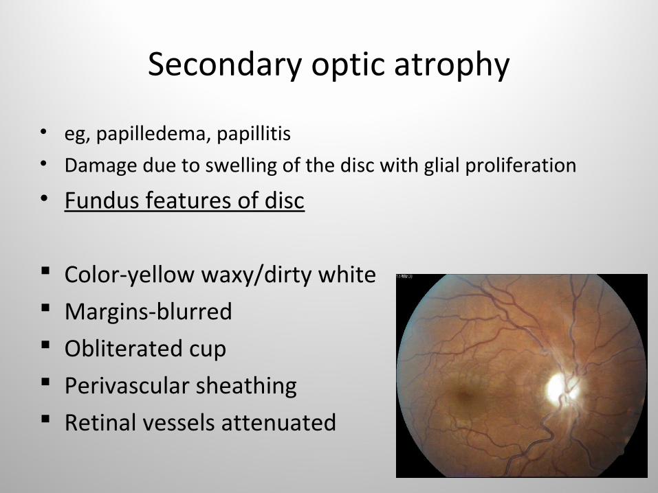
Secondary optic atrophy
• eg, papilledema, papillitis • Damage due to swelling of the disc with glial proliferation
• Fundus features of disc
Color-yellow waxy/dirty white Margins-blurred Obliterated cup Perivascular sheathing Retinal vessels attenuated

classification
Pathologic classification • Ascending optic atrophy - follows damage to
RGC/nerve fibre layer
• Descending optic atrophy - retrolaminar optic nerve/chiasma/optic tract
• Trans-synaptic degeneration- seen in patients with occipital damage incurred either in utero or in early infancy.
Unilateral optic atrophy
Ischemic (anterior ischemic optic neuropathy, retinal occlusive disease)
Compressive (orbital, anterior fossa)
Inflammatory (demyelinating, infectious,Autoimmune, sarcoid)
TraumaticInfiltrative
Bilateral optic atrophy
GlaucomaSecondary to retinal degenerationPost-papilledemaCongenital anomalies: hypoplasia, colobomaMethyl alcohol toxicityInherited ON : LHON, Auto dominantNutritional/toxic
Optic Atrophy
• Varies from temporal pallor to chalky white• Due to changes in blood supply, axonal loss,
glial tissue• Also nature and extent of injury/damage

Differentials
• Axial myopia• Optic nerve hypoplasia• Brighter-than-normal luminosity• Optic nerve pit• Myelinated nerve fibers• Scleral crescent• Optic disc drusen• Tilted disc
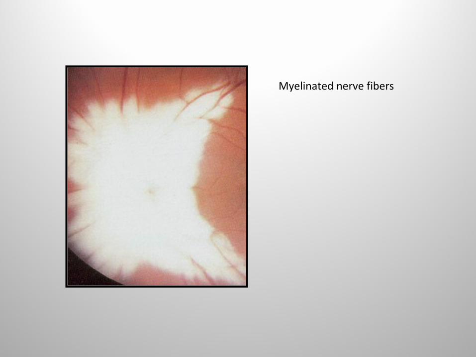
Myelinated nerve fibers
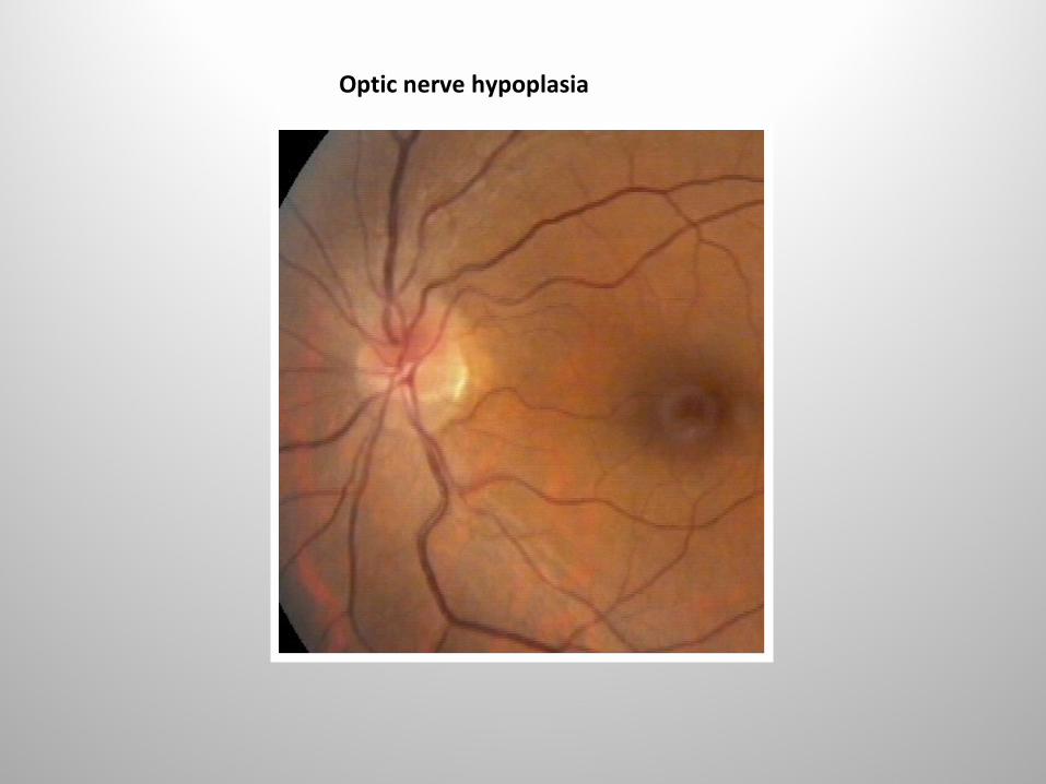
Optic nerve hypoplasia
Optic atrophy
• Slight, moderate, severe
• Diffuse or sectoral
Optic atrophy
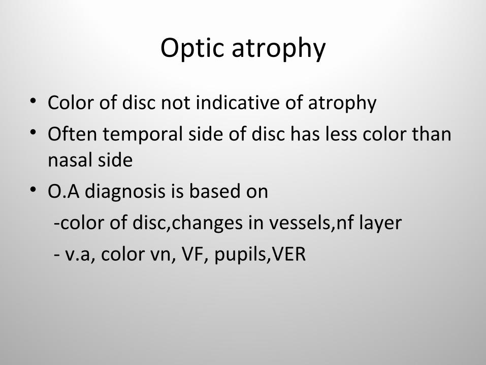
• Color of disc not indicative of atrophy• Often temporal side of disc has less color than
nasal side• O.A diagnosis is based on
-color of disc,changes in vessels,nf layer
- v.a, color vn, VF, pupils,VER

0ptic atrophy
• In AION – Limited to upper and lower quad• Chiasmal lesions – temp and nasal
pallor(bowtie or band atrophy) due to crossing nasal fibres being involved
Also seen in eye contralateral to unilateral optic tract lesion
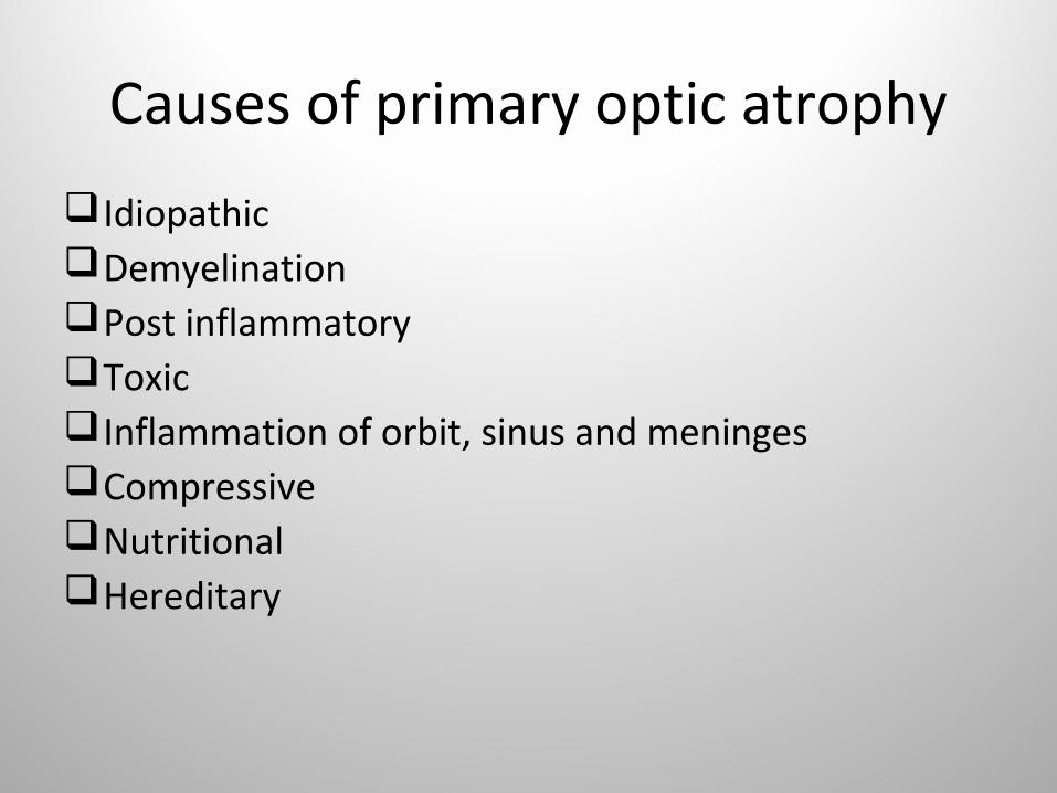
Causes of primary optic atrophy
IdiopathicDemyelination Post inflammatoryToxic Inflammation of orbit, sinus and meningesCompressive Nutritional Hereditary

Causes of secondary optic atrophy
• Anterior ischaemic optic neuropathy • Papllitis• Papilledema• Metabolic diseases (Diabetes)
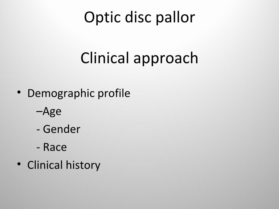
Optic disc pallor
Clinical approach
• Demographic profile
–Age
- Gender
- Race• Clinical history

Optic disc pallorDemographic profile
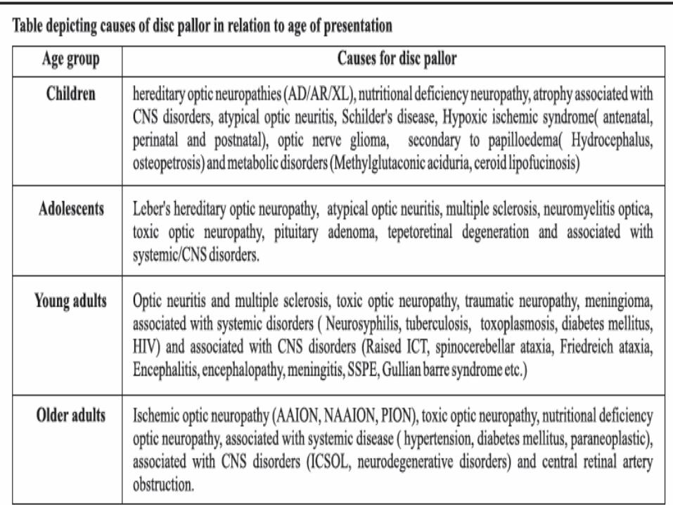
• Age – most important of demographic parameters
• No watertight compartments• Significant overlaps between age groups• Glaucoma can present with pallor at any age

Optic disc pallor
• Gender can guide in favour of a diagnosis but not always
• Male: LHON, Traumatic ON,Tapetoretinal deg,ToxicON,Occupational(heavy metal exposure), lead, arsenic,Nutritional(chronic alcoholism)
• Female: MS, Meningioma, Autoimmune/collagen vasc dis.,Sheehan synd, ecclampsia

Optic disc pallor- Racial differences
• Blacks – lower incidence of Ischaemic ON, better visual fn in IIH
• Caucasians more likely for MS than Hispanics and Asians
• Overall Optic atrophy more in blacks than whites

Optic disc pallor- Clinical history
• Onset –over hrs or days- Optic neuritis, Iscemic ON, Traumatic ON
Subacute- over few days – in demylenating, compressive
• Course- resolved naturally, recurrent episodes- demyelinating
residual poor vn ,progressive,protracted course- other pathologies

Optic disc pallor -History
• Laterality
- Unilateral – Typical ON
NAION
Traumatic ON
Compressive
- Bilateral - Toxic, Nutritional,Hereditary,Arteritic AION, Atypical Optic neuritis
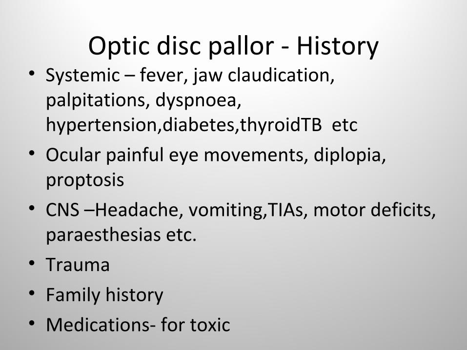
Optic disc pallor - History• Systemic – fever, jaw claudication,
palpitations, dyspnoea, hypertension,diabetes,thyroidTB etc
• Ocular painful eye movements, diplopia, proptosis
• CNS –Headache, vomiting,TIAs, motor deficits, paraesthesias etc.
• Trauma• Family history• Medications- for toxic
Optic disc pallor –Ocular Exam
• V.A.• Visual Fields• Color Vision• Pupils• Fundus – Disc color, cup, margins,
vascularity(Kestebaum count -10 capillaries on disc. In optic atrophy<6)
• RNFL Defects – Red free green filter exam

Optic disc pallor – diagnosis by optic disc appearance
• Ischaemic – pallid edema- arterial
attenuation,sheathing- superior /inferior disc
pallor- Haemorrhages- fellow eye small cup-disc
ratio(disc at risk)
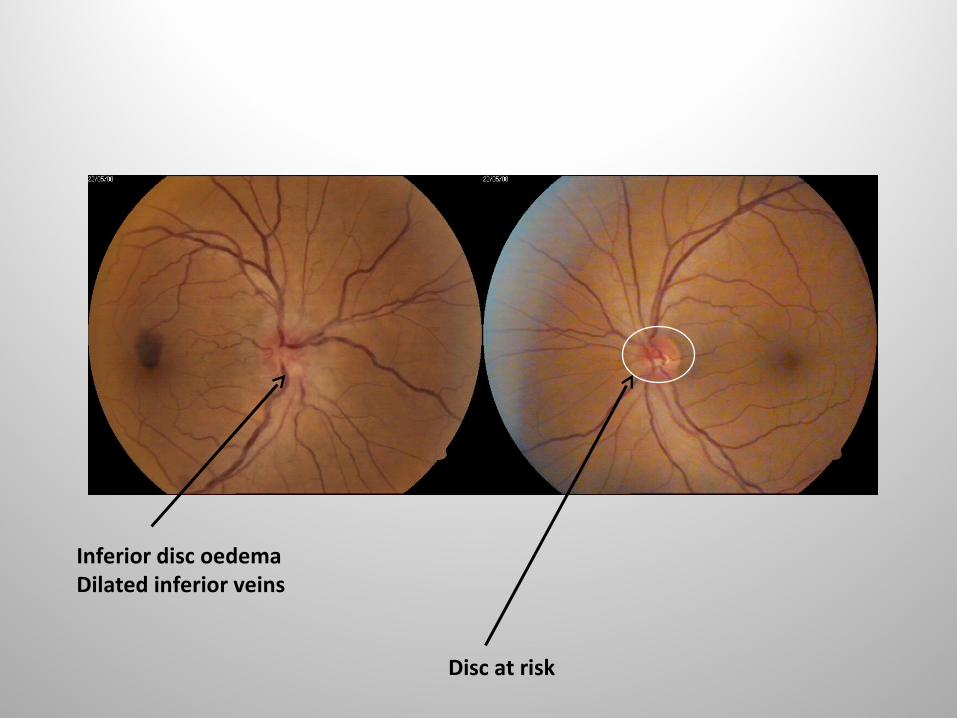
Inferior disc oedemaDilated inferior veins
Disc at risk
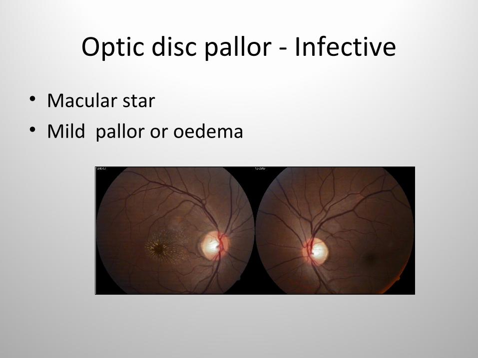
Optic disc pallor - Infective
• Macular star• Mild pallor or oedema
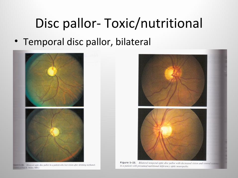
Disc pallor- Toxic/nutritional• Temporal disc pallor, bilateral

Disc pallor-LHON/DOA• Temporal, occasionally diffuse

Optic disc pallor- Chiasma/tract lesion
• Bowtie/band atrophy
Glaucomatous
Nonglaucomatous
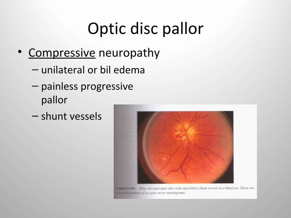
Optic disc pallor• Compressive neuropathy– unilateral or bil edema– painless progressive
pallor– shunt vessels
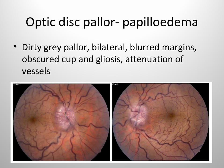
Optic disc pallor- papilloedema
• Dirty grey pallor, bilateral, blurred margins, obscured cup and gliosis, attenuation of vessels

Papilloedema -stages

Disc pallor – systemic exam
Look for• Nutritional deficiency• B.P• Anemia• Lymph nodes• CVS /CNS

Optic disc pallor-Ocular investigations
At baseline and followup• V.A• Color Vn• Contrast sensitivity• VF• FFA(if necessary)• OCT• VER/ERG

Optic disc pallor- Investigations
First line
1.Haemogram,peripheral smear,TLC,DLC
2.ESR
3.Mx
4.Chest Xray
5.MRI Head and Orbit with thin cuts and fat suppression
6.VDRL
7. LP(if reqd)
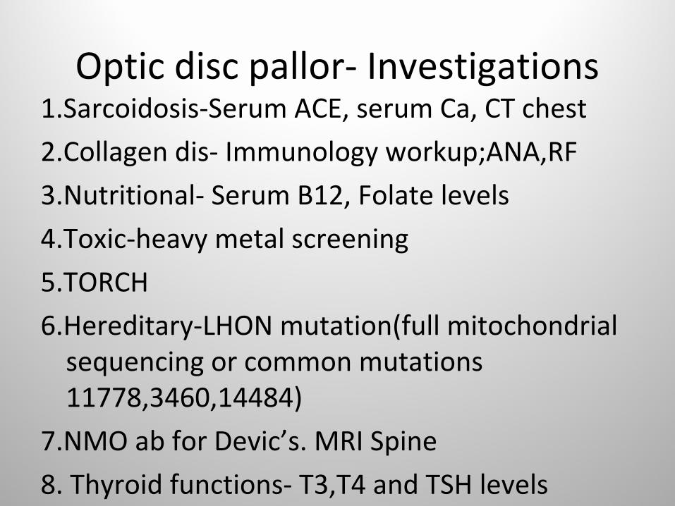
Optic disc pallor- Investigations1.Sarcoidosis-Serum ACE, serum Ca, CT chest
2.Collagen dis- Immunology workup;ANA,RF
3.Nutritional- Serum B12, Folate levels
4.Toxic-heavy metal screening
5.TORCH
6.Hereditary-LHON mutation(full mitochondrial sequencing or common mutations 11778,3460,14484)
7.NMO ab for Devic’s. MRI Spine
8. Thyroid functions- T3,T4 and TSH levels
9.Temporal artery biopsy for AAION
VISUAL FIELD EVALUATION
VF testing strategies
• Confrontation• Goldmann kinetic or static• Humphrey static automated

Confrontation fields
Presentation Outline
1. General aspects of automated perimetry
2. Components and basics of interpreting visual field reports
3. Physiological basis of visual field defects in neurological afflictions
4. Clinical examples discussing the topographic representation of visual fields
5. Visual field alterations in neuro-ophthalmological afflictions
6. Algorithm for field assessment and interpretation
7. Review of various field defects

Why choose automated perimeters: Humphrey perimeter
Advantages of Automated Perimetry •Provides more sensitive and reproducible results •Provides quantitative information•Provides results in a more timely and precise manner •Experienced perimetrist is not required
Advantages of Static Perimetry •Scotomas better defined•Temporal summation of stimulus possible•Less dependence on Perimetrist
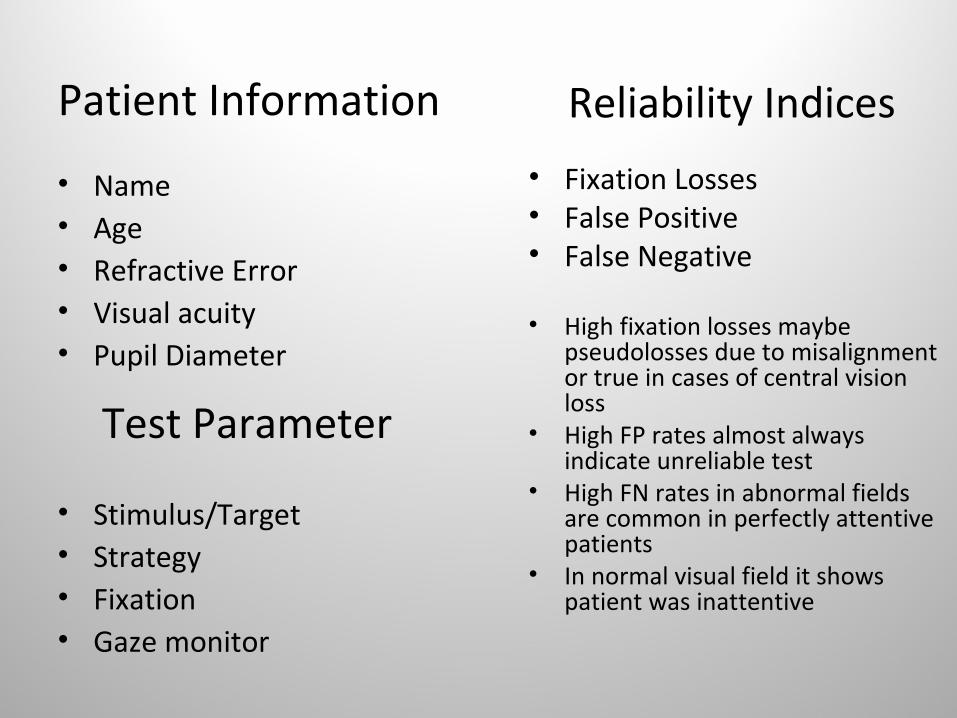
Patient Information
• Name• Age• Refractive Error• Visual acuity• Pupil Diameter
• Stimulus/Target• Strategy• Fixation• Gaze monitor
Reliability Indices
• Fixation Losses• False Positive• False Negative
• High fixation losses maybe pseudolosses due to misalignment or true in cases of central vision loss
• High FP rates almost always indicate unreliable test
• High FN rates in abnormal fields are common in perfectly attentive patients
• In normal visual field it shows patient was inattentive
Test Parameter

• Any Patient with visual loss unexplained on Ocular or refractive basis should have a visual field examinatin as the very next test.
Extent of normal visual field
• Nasally 60 degrees• Superiorly 60 degrees• Inferiorly 70 to 75 degrees• Temporally 100 to 110 degrees

Interpretation of Humphrey visual Field

Why is the Grey scale plot suited for field assessment in Neuro-ophthalmology cases
• Optic nerve diseases are more likely to result in absolute field losses
• Absolute field defects are very clearly demarcated on grey scale plots
• These visual fields often respect the vertical meridians and these are well defined on grey scale plots
• Orientation of scotoma with respect to the blind spot is best appreciated on the grey scale plots

Effects of distortion or obscuration
• Of light entering eye
by eyebrows, skin of lid
Lens rim, spectacle power(high plus/minus),incorrect lenses
• By eyelids and media
Pupil size,opacities in media
Basis of visual field defect• Anatomical structure of retinal nerve fibres-
which has 4 groupings which enter Optic disc
a.Inf. Ret n. fibres subserve Sup. visual field
b.Sup. Ret. N. fibres – Inferior visual field
c.Fibres bet macula & optic disc(papillomac bundle)- central Vn
d.Nasal fibres which enter disc in wedge shaped manner
Basis of field defect• Superior and Inferior fibres form arcuate
bundles around papillomacular bundle to enter optic nerve at 12 o’ clock and 6 o’ clock
• Peripherally in retina they join horizontal raphe. Fibres do not cross horizontal raphe.
• Arcuate n.f.bundle –arcuate field defect. Nasal extent is hor. Meridian
• Pap-mac bundle-central or centrocaecal scotoma
• Nasal wedge-temporal defect
Basis of field defects
• Optic disc lesions will produce VF defects identical to those of retina
• Posteriorly fibres rotate 90degrees• Macular fibres occupy central core of Optic
nerve• Hence retrobulbar optic nerve lesions produce
more central scotomas.


Basis of Visual Field Defects in Neuro-Ophthalmology

Arrangement of nerve fibres along the course of visual pathway
Lateral Geniculate BodyUpper retina fibers - medial partLower retina fibers - lateral partMacular fibers -posterior 2/3

Preview of field defects seen in
neuro-ophthalmology
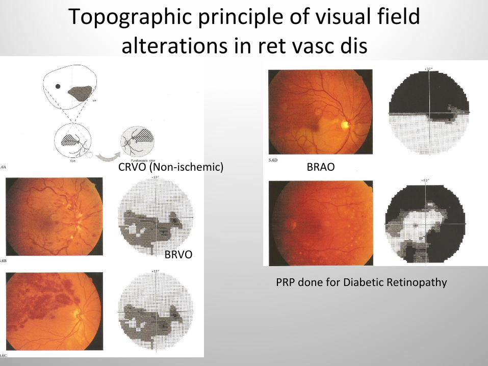
Topographic principle of visual field alterations in ret vasc dis
CRVO (Non-ischemic)
BRVO
BRAO
PRP done for Diabetic Retinopathy

Visual Field alterations in nerve fibre layer affliction

Visual field changes in neuro-ophthalmology1. Optic disc pit
2. Tilted optic disc
3. Optic disc drusen
4. Anterior ischemic optic neuropathy
5. Papilledema
6. Hereditary optic atrophy
7. Toxic optic neuropathy
8. Optic neuritis
9. Optic nerve meningioma
10. Pituitary adenoma
11. Craniopharyngioma
12. Optic tract and LGB lesions
13. Optic radiation afflictions
14. Visual cortex lesions
15. Miscellaneous field alterations
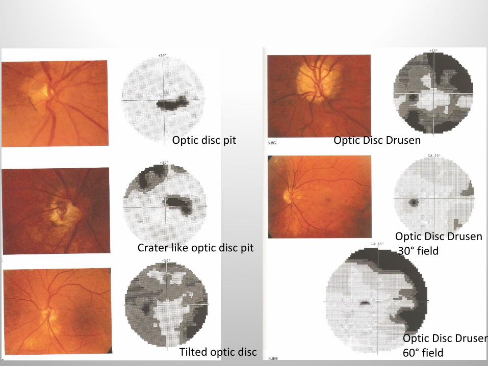
Optic disc pit
Tilted optic disc
Crater like optic disc pit
Optic Disc Drusen 60° field
Optic Disc Drusen
Optic Disc Drusen 30° field

AION with cecocentral sparing
Typical AION
Maculopapular ischemia

Papilledema (IIH)
Hemorrhagic Papilledema
Papilledema secondary to astrocytoma

Principle behind cecocentral scotoma
Hereditary optic nerve atrophy
Toxic optic neuropathy

Visual Field defects in optic neuritis
CentralDiffuse depressionAltitudnalNasal hemianopiaNerve fibre bundle defectsCombined defects
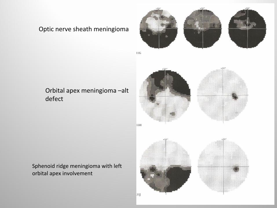
Orbital apex meningioma –alt defect
Optic nerve sheath meningioma
Sphenoid ridge meningioma with left orbital apex involvement
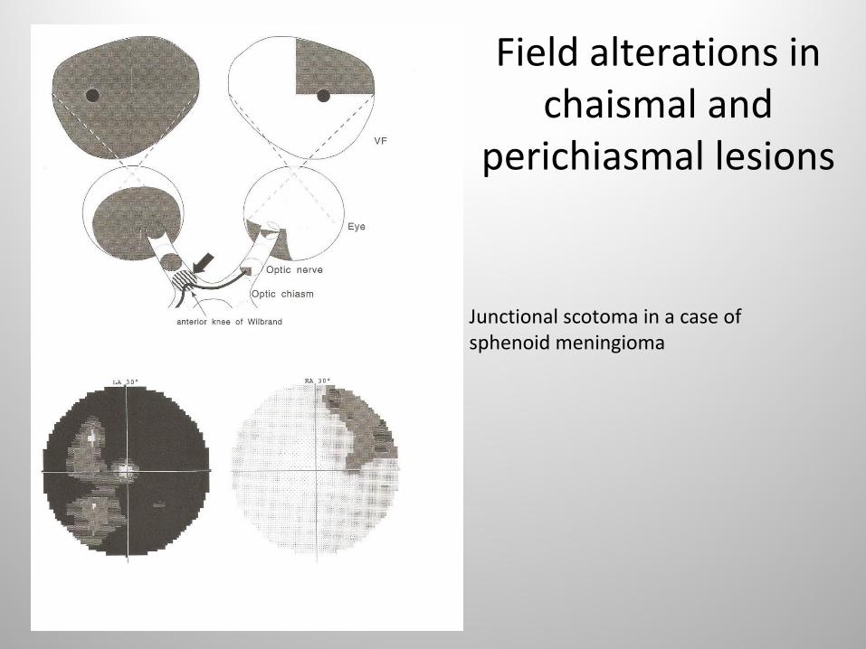
Field alterations in chaismal and
perichiasmal lesions
Junctional scotoma in a case of sphenoid meningioma

Pituitary Adenoma
Early stage of pituitary adenoma

Late stage pituitary adenoma- Hemianopia and subtotal defects
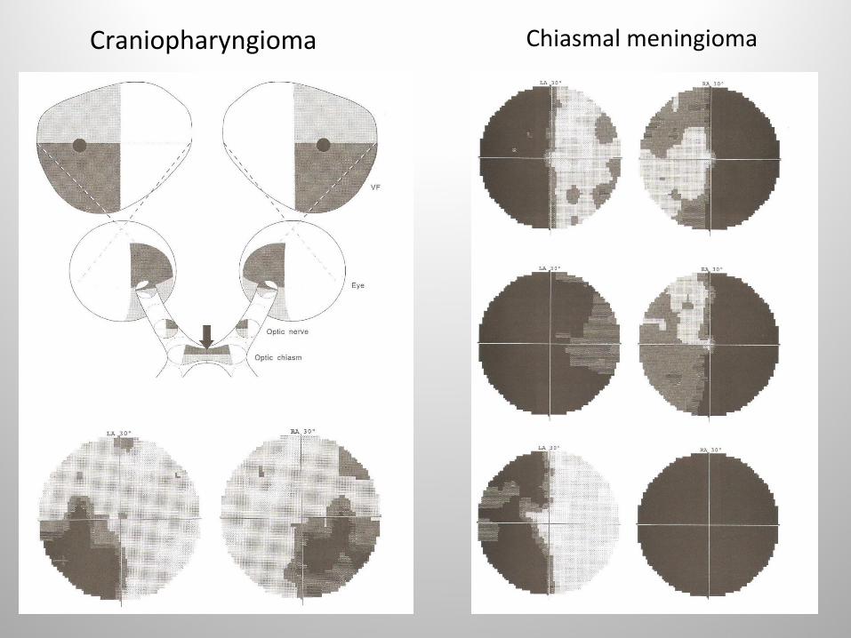
Craniopharyngioma Chiasmal meningioma

Field alterations in optic tract and lateral geniculate body lesions
Basal Ganglia neoplasm: Incongruent Hemianopia

Field alterations in lesions involving optic radiations
Astrocytoma of left temporal lobe
Substance defects in left brain
Infarct of left occipital and parietal lobe

Complete hemianopia in total involvement of all optic radiations
Hemianopia with sparing of fixation in subtotal in involvement of all optic radiations
Involvement of all optic radiations after left cystic tissue defect following neurosurgery
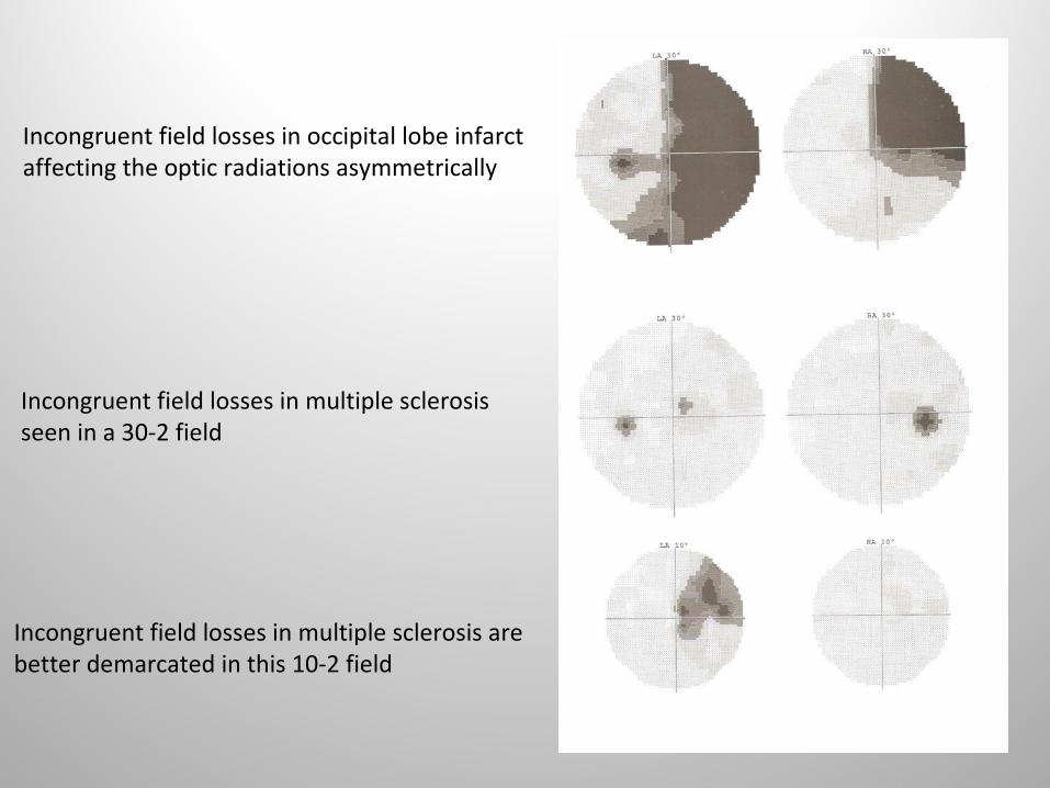
Incongruent field losses in occipital lobe infarct affecting the optic radiations asymmetrically
Incongruent field losses in multiple sclerosis seen in a 30-2 field
Incongruent field losses in multiple sclerosis are better demarcated in this 10-2 field

Field alterations in afflictions of the visual cortex
Congruent homonymous hemianopia with macular sparing
Occipital lobe infarct with complete homonymous defects
Occipital lobe infarct with incomplete homonymous defects

Miscellaneous Field Defects
Peripheral field defects
• May be out of field for 30-2• A fast 60-2 protocol used
Bilateral homonynous hemianopia• Seen in vascular lesions• May have both congruent
and incongruent areas
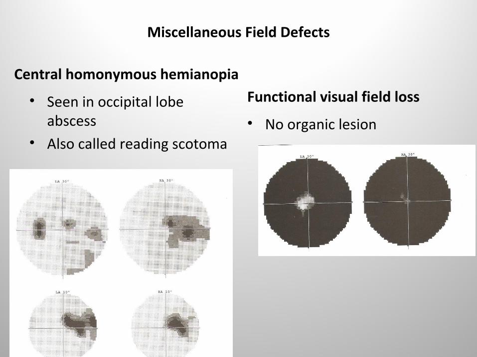
Miscellaneous Field Defects
Central homonymous hemianopia
• Seen in occipital lobe abscess
• Also called reading scotoma
Functional visual field loss
• No organic lesion

Localisation of homonymous hemianopia
• Macular sparing• Congruity• Pupillary response – normal in Op Radiation• Asymmetric OKN -Parietal• Saccadic pursuit to side of lesion-parietal

Algorithm for Field Assessment and Interpretation

• References 1. Field charts referenced from Atlas of Computerized
Perimetry by Weber and Caprioli, Published by W.B. Saunders Co.Philadelphia,2000
2.Clinical Neuro-Ophthalmology by Ulrich Scheifer et al,Publishers,Springer

THANK YOU