applied physiology and the hemodynamic management of septic shock
TRANSCRIPT

12
Applied Physiology and the Hemodynamic Management of Septic Shock Utilizing the
Physiologic Optimization Program
William McGee and Patrick Mailloux Baystate Medical Center, Tufts University School of Medicine
United States
1. Introduction
Volume management is an important aspect of caring for patients with sepsis. Multiple
factors contribute to the challenge of resuscitating septic patients, including volume
depletion, a decrease in vascular tone and myocardial depression. Goal directed therapy
incorporates the use of physiologic targets to guide fluid resuscitation in this population,
taking into account the changes in physiology of a patient with sepsis. Further, evolving
technology and knowledge is allowing for a better understanding of endpoints when
managing fluids in this critically ill patient group.
Patient’s presenting with hypo-perfusion secondary to septic shock benefit from early,
aggressive resuscitation in a protocolized manner (Rivers, 2001). The goals of initial
resuscitation, to be achieved within 6 hours of presentation, include a central venous
pressure of 8 – 12 mmHg, mean arterial pressure (MAP) of ≥ 65 mmHg, urine output ≥ 0.5
mL/kg/hr and a central venous (ScvO2) or mixed venous oxygen saturation ≥ 70% or 65%,
respectively (Rivers 2001, Dellinger, 2008). This concept is known as early goal directed
therapy (EGDT) (Rivers, 2001).
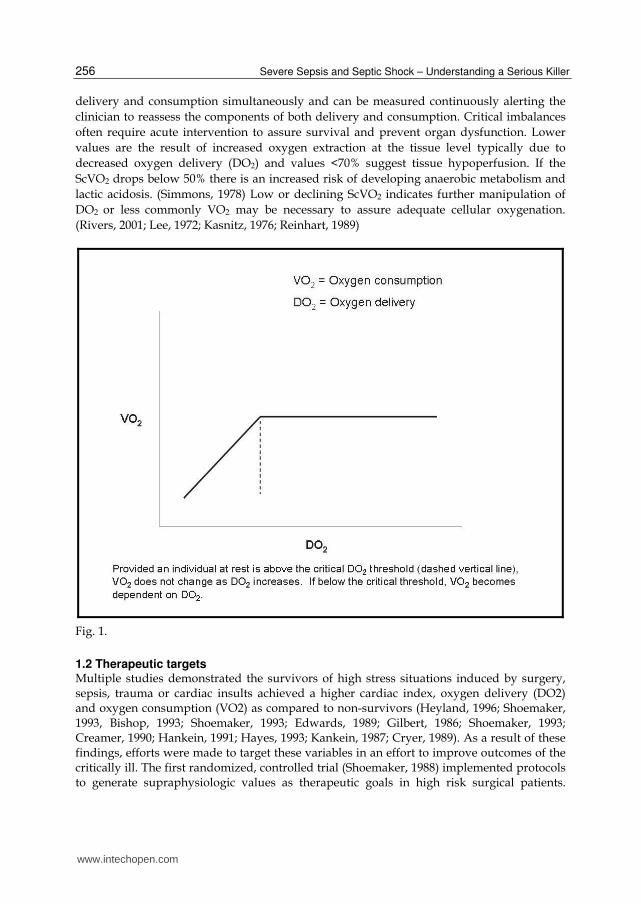
1.1 Oxygen delivery The ultimate goal of early resuscitation is to achieve adequate oxygen delivery so the
balance between supply and demand to vital organs is maintained. A critical level of oxygen
delivery (DO2) exists for septic patients and increasing the DO2 above that level does not
further increase oxygen consumption. (Ronco, 1993; Shibutani, 1983;, Danek, 1980; Nelson,
1987, 1988) When below critical DO2, oxygen utilization is dependent upon DO2 (Figure 1),
compensation via increased oxygen extraction is no longer sufficient, oxygen debt develops
and anaerobic metabolism ensues. (Pieracci, 2011) One accepted method to measure
whether DO2 is above this threshold involves determining the oxygen saturation of venous
blood returning to the heart (McGee & Jodka, 2002; Krafft, 1993). ScvO2 as a proxy for SVO2
is reasonable (Lee, 1972; Reinhart, 2004; Ladakis, 2001) and reflects the oxygen saturation of
SVC blood. As the saturation of venous blood (Figure 2) declines, oxygen consumption is
increasing for a constant value of oxygen delivery, or oxygen delivery itself may be
declining. In either circumstance assessment of ScVO2 represents the balance between
www.intechopen.com
Severe Sepsis and Septic Shock – Understanding a Serious Killer
256
delivery and consumption simultaneously and can be measured continuously alerting the
clinician to reassess the components of both delivery and consumption. Critical imbalances
often require acute intervention to assure survival and prevent organ dysfunction. Lower
values are the result of increased oxygen extraction at the tissue level typically due to
decreased oxygen delivery (DO2) and values <70% suggest tissue hypoperfusion. If the
ScVO2 drops below 50% there is an increased risk of developing anaerobic metabolism and
lactic acidosis. (Simmons, 1978) Low or declining ScVO2 indicates further manipulation of
DO2 or less commonly VO2 may be necessary to assure adequate cellular oxygenation.
(Rivers, 2001; Lee, 1972; Kasnitz, 1976; Reinhart, 1989)
Fig. 1.
1.2 Therapeutic targets Multiple studies demonstrated the survivors of high stress situations induced by surgery, sepsis, trauma or cardiac insults achieved a higher cardiac index, oxygen delivery (DO2) and oxygen consumption (VO2) as compared to non-survivors (Heyland, 1996; Shoemaker, 1993, Bishop, 1993; Shoemaker, 1993; Edwards, 1989; Gilbert, 1986; Shoemaker, 1993; Creamer, 1990; Hankein, 1991; Hayes, 1993; Kankein, 1987; Cryer, 1989). As a result of these findings, efforts were made to target these variables in an effort to improve outcomes of the critically ill. The first randomized, controlled trial (Shoemaker, 1988) implemented protocols to generate supraphysiologic values as therapeutic goals in high risk surgical patients.
www.intechopen.com

Applied Physiology and the Hemodynamic Management of Septic Shock Utilizing the Physiologic Optimization Program
257
Patients with oxygen transport maximized by a PA catheter protocol had a lower mortality, reduced duration of mechanical ventilation and ICU stay. Study limitations in baseline characteristics of the various groups along with an unblinded design raise the question of bias versus an actual treatment impact (Shoemaker, 1988; Heyland, 1996).
Fig. 2.
Hayes et al (Hayes, 1994) randomly assigned patients failing to reach established
therapeutic goals following volume resuscitation alone to continue with standard care or
receive dobutamine to increase cardiac index, oxygen delivery and oxygen consumption.
The treatment group had a higher mortality, suggesting efforts to achieve supranormal
physiologic targets may result in more risk than benefit. In addition, Gattinoni et al
(Gattinoni, 1995) failed to demonstrate a favorable impact on morbidity or mortality when
targeting hemodynamic therapy to achieve supranormal values for cardiac index or normal
values for mixed venous oxygen saturation. In 2001, Rivers et al (Rivers, 2001) published the results of a randomized trial of EGDT in the treatment of patients with severe sepsis and septic shock. By instituting a multi-faceted protocol targeted to increase oxygen delivery they were able to demonstrate significant benefits to outcome in this patient population. The main components of this approach included continuous monitoring of central venous oxygen saturation (ScvO2), treatment in a dedicated area of the emergency department for the first six hours and fluid boluses to achieve a central venous pressure (CVP) of 8 to 12 mmHg. If the MAP remained < 65 mmHg
www.intechopen.com

Severe Sepsis and Septic Shock – Understanding a Serious Killer
258
following fluid resuscitation to a CVP of 8 – 12 mmHg, vasopressor therapy began, red blood cells were transfused to achieve a hematocrit of at least 30% if the ScvO2 was less than 70% and dobutamine administration titrated to achieve an ScvO2 of at least 70% despite the other interventions. (Rivers, 2001) The intervention group was more likely to achieve the ScvO2, CVP, MAP and urine output goals along with an improvement in mortality. 46.5% in those receiving standard therapy died compared to 30.5% in the EGDT cohort (p=0.009). The EGDT group also received significantly more fluid (4981 mL vs. 3499 mL), red blood cells (64.1% transfused vs. 18.5%), and inotropic support (13.7% vs. 0.8%) during the first 6 hours. The difference in fluid balance did not persist at 72 hours (13,443 mL in EGDT vs. 13,358 in standard therapy) emphasizing the importance of rapid volume optimization for septic shock. (Rivers, 2001) This study demonstrated for the first time the importance of an aggressive, early, protocolized, goal directed treatment regimen in caring for patients with severe sepsis and septic shock. This protocol has been adopted in multiple guidelines as the standard of care for treating septic patients (Dellinger, 2008). Table 1 summarizes the findings of the above trials.
Study Intervention Results
Shoemaker, 1988 Supraphysiologic values as therapeutic goals in high risk surgical patients
Oxygen transport maximized by a PA catheter protocol lead to lower mortality, reduced duration of mechanical ventilation and shorter ICU stay
Hayes, 1994 Dobutamine to increase cardiac index, oxygen delivery and oxygen consumption
Higher mortality in patients treated with dobutamine compared to standard care
Gattinoni, 1995 Achieving supranormal values for cardiac index or normal values for mixed venous oxygen saturation
No improvement on morbidity or mortality
Rivers, 2001 Early goal directed therapy with CVP of 8 – 12 mmHg, vasopressors if MAP < 65 mmHg following fluid resuscitation, ScvO2 ≥ 70%, dobutamine and red blood cells to keep hematocrit at least 30% if ScvO2 < 70%
Improved mortality with intervention compared to standard care. More fluid, red blood cells and inotropic support during first 6 hours in intervention group.
Table 1. Summary of Studies on Goal Directed Therapy
1.3 Physiological derangements Aggressive fluid resuscitation is a mainstay of therapy for septic patients as this population tends to be severely hypovolemic related to multiple mechanisms, venodilation from altered vascular tone leads to pooling of blood in the capacitance vessels. (McGee & Jodka. 2002; Kumar, 2009, Teule 1984) Septic patients also have extravasation of fluid into the interstitium related to increased permeability of the capillary endothelium (Rivers 2008;
www.intechopen.com

Applied Physiology and the Hemodynamic Management of Septic Shock Utilizing the Physiologic Optimization Program
259
Pieracci 2011). These phenomena result in decreased preload, cardiac output and inadequate oxygen delivery. It remains essential to restore volume status and improve cardiac performance. Additionally, the body’s inflammatory response is further modulated by cellular hypoxia brought about by the decline in bulk oxygen delivery (Rivers, 2007) may compound the physiologic derangement. Concurrent with the needs to restore adequate preload and circulating blood volume, septic patients often demonstrate myocardial depression. This dysfunction is related to the presence of myocardial depressant factors early in the septic process, not decreased myocardial perfusion, and include cytokines, tumor necrosis factor alpha (TNF-┙) and interleukin one beta (IL-1┚) acting synergistically. (Court, 2002; Pathan, 2002) Additionally, nitric oxide generation, interstitial myocarditis, calcium trafficking, endothelin receptor antagonists and apoptosis likely all contribute to the ongoing process of myocardial depression. (Fernandes, 2008). This is manifest as a low cardiac output concurrent with a decrease in ejection fraction and oxygen delivery despite adequate filling pressures. Ventricular interdependence and impairment of left ventricular filling may be an important concern especially with concomitant ARDS and RV dysfunction/dilatation. (Michard, 2010; von Ballmoos, 2010) The use of echocardiogram plays an important role in quantifying the degree of dysfunction
as it is used to assess biventricular contractility and identifies hemodynamically unstable
patients who will benefit from either inotropic support or further volume expansion
(Griffee, 2010; Beaulieu, 2007). Dobutamine is a typical first line inotropic agent when
myocardial depression of sepsis is confirmed and the goal is to establish adequate oxygen
delivery or tissue perfusion as measured by ScvO2. No benefit is seen using inotropes to
create a supra-physiologic state though studies achieving this did not have echocardiogram
data to determine what proportion of subjects had impaired contractility (Hayes, 1994;
Gattinoni, 1995). Sepsis induced myocardial depression usually resolves completely over the
course of the illness for survivors. (Griffee, 2010; Parker, 1984).
1.4 Role of red blood cells Controversy exists as to the ideal hemoglobin concentration for septic patients undergoing EGDT as the risks of transfusion often outweigh the benefits. (Hebert, 1999; Marik, 2008; Fuller, 2010) The goal of transfusion of red blood cells is to improve DO2. There is no impact on the sublingual microcirculation as detected by an orthogonal polarization spectral device in septic patients receiving a red blood cell transfusion (Sakr, 2007). Further, transfusing anemic, septic patients does not improve either regional or global oxygen utilization as determined by either the Fick equation or indirect calorimetry and may increase pulmonary vascular resistance, further hindering right ventricular function. (Fernandes, 2001; Bone, 1993) At this time unless there is active myocardial ischemia, it is difficult to define exact triggers for transfusion of red blood cells in septic patients undergoing active resuscitation. (Pieracci, 2011)
1.5 Avoiding volume overload An aggressive, early intervention targeted at improving hemodynamic performance in
septic patients improves outcomes and is now recognized as the standard of care. The
trigger to transfuse red blood cells during the early resuscitation of septic patients remains
controversial and is a major criticism of EGDT, especially given the known complications of
www.intechopen.com
Severe Sepsis and Septic Shock – Understanding a Serious Killer
260
providing transfusions to critically ill patients. Critical care practitioners need to recognize
both the importance of implementing EGDT and avoiding the deleterious effects of fluid
overload. When septic patients have a more positive fluid balance both early in the
resuscitation process and cumulatively over 4 days there is an associated increased risk of
mortality (Boyd, 2011). The importance of carefully managing fluids is further illustrated in
patients with acute lung injury or ARDS as those with a lower cumulative fluid balance
demonstrate improved outcomes. (Murphy, 2009; the NHLBI ARDSNet, 2006) When
complicated by acute kidney injury, fluid overload further impacts mortality in critically ill
patients (Payen, 2008; Bouchard, 2009)
2. Applied physiology for the management of severe sepsis/septic shock
The foundation of all resuscitation strategies for severe sepsis and septic shock is volume
therapy. Early goal directed therapy is complicated by the requirement for central venous
access which is not always available especially during the initial patient evaluation, and
the use of a CVP target which has been called in to question as a meaningful measure of
preload responsiveness. (Michard, 2002; Marik, 2008;Osman, 2007) Additionally although
rapid resuscitation is important excess volume that does not improve cardiac performance
is potentially contributing to complications; excess length of stay (Wiedemann, 2006) and
mortality (Murphy, 2009). Generally as the goal of any volume therapy in the critically ill
is to improve cardiac performance, we propose physiology based hemodynamic therapy
for the severely septic patient that considers the pathophysiology already described.
2.1 Physiologic optimization program for sepsis resuscitation, dynamic variables of volume responsiveness
Utilizing dynamic variables of volume responsiveness to guide resuscitation allows precise
titration of preload with clear endpoints for fluid resuscitation to minimize the risk of
inappropriate fluid overload. Dynamic variables of volume responsiveness, stroke volume
variation (SVV) and pulse pressure variations (PPV) exploit the physiology of the
heart/lung interaction during positive pressure ventilation that relates the variation of
stroke volume or pulse pressure to the degree of volume responsiveness. The respirophasic
change in blood pressure (commonly observed in the arterial pressure tracing) of
hypovolemic patients is a well known example. (McGee, 2009) (Figure 3).
The available devices quantify the variability and display the variation as a primary output.
This variation is highly correlated with the degree of volume responsiveness, i.e., the greater
the variation, the greater the expected response to a volume challenge. (Figure 4). This approach is available only for patients with controlled positive pressure ventilation without a significant dysrhythmia as the utility of SVV or PPV to assess volume responsiveness is not valid beyond these conditions. The use of the dynamic variables SVV and PPV, especially during early resuscitation prior to intubation is generally not possible. Resuscitation of these patients, however, is straight forward and relies on titration of volume against cardiac performance measured by SV and CO. This approach provides assurance that preload optimization has occurred prior to implementation of pharmacotherapy. Our preference is to use the indexed values in an attempt to normalize targets across variability in body surface area. This may be problematic in the super obese (McGee, 2006). (Figure 5).
www.intechopen.com

Applied Physiology and the Hemodynamic Management of Septic Shock Utilizing the Physiologic Optimization Program
261
Positive Pressure Breath
MECHANISM OF SVV
↑RV Afterload
↓RV Preload
↑LV Preload
Acute ↑SV
Delayed ↓↓ SV
Empty Pulmonary Venous System
↑ Intrathoracic Pressure
Airw
ay
Pre
ssu
reA
rteria
l P
ressu
re
Time
Fig. 3. The phasic change in blood pressure and its timing to the ventilatory cycle is illustrated with four positive pressure breaths and the simultaneously displayed arterial waveform directly below it. Blood pressure goes up during the inspiratory phase of mechanical ventilation and decreases during expiration. The swings in blood pressure (pulse pressure variation, PPV) are generated by the change in stroke volume SV (stroke volume variation, SVV) effected by positive pressure ventilation. The variability is respirophasic as this figure illustrates. The impact on right (RV) and left ventricular (LV) pre and afterload induced by positive pressure ventilation is shown.
In those clinical settings where SVV or PPV does not predict volume responsiveness how
can this be determined? As long as stroke volume can be measured this question can be
answered. This represents a huge advantage of the technologies that allow measurement of
stroke volume and cardiac output, FloTrac/Vigileo, PICCO, and LiDCO, over those that
simply provide pulse pressure variation, which is now readily available on bedside
monitors that display an arterial waveform (IntelliVue MP90, 2006). This physiology also
explains why this literature has been primarily developed in the operating room where ideal
conditions often exist for the measurement and application of dynamic parameters, namely
controlled mechanical ventilation with a large enough tidal volume to induce a significant
change in pleural pressure to impact venous return. A threshold value for tidal volume of 8
cc per kilogram ideal body weight has been determined in several studies that evaluated the
tidal volume necessary to meaningfully impact venous return. (Feissel, 2001; Tavernier,
1998; Michard, 2000; Perel, 1987) Across a heterogeneous population, this may be true,
although there will be specific examples depending on the patient’s lung compliance and
intravascular volume status where it may not be.
www.intechopen.com

Severe Sepsis and Septic Shock – Understanding a Serious Killer
262
SVV high >13%
Preload
B
A
Magnitude of SVV is Related to LV Magnitude of SVV is Related to LV
PreloadPreload
Change in preload related to positive Change in preload related to positive
pressure ventilationpressure ventilation
SVV low <13%
Equal change inFilling with positive
Pressure breath
SV
Fig. 4. Legend: A and B represent different locations on the Starling Curve. The change in preload induced by the ventilator is identical. The impact on SV is not. The change in SV induced by one positive pressure breath is proportional to SVV. SVV determines the magnitude of preload dependency. Patients with higher SVV are more volume responsive (A – preload dependent) functioning on the steeper portion of the Frank Starling curve. SVV decreases as preload dependent LV function is optimized (B – preload independent). In these patients (B) volume can be safely removed as cardiac performance is not influenced by changes in preload. SVV/SVI pairs allow individual discrimination of a patient’s Starling curve that can determine when volume is required to improve cardiac performance and conversely when volume can be safely removed. SV (stroke volume) SVV (stroke volume variation)
At lower tidal volumes, false negatives where stroke volume variation is low, and the
patient is still significantly volume responsive occur. Extreme examples of this were
encountered during the H1N1 flu epidemic in the fall of 2009, where many patients were
being oscillated at very high frequencies but with minimal tidal volume and hence pleural
pressure change. These patients could be significantly volume responsive with essentially
no stroke volume or pulse pressure variation because of the very small change in pleural
pressure induced with this strategy of ventilation. Similarly, patients being ventilated using
a low tidal volume strategy for ALI/ARDS may manifest a similar phenomenon. In those
patients, teasing out volume responsiveness utilizing physiology is fairly simple. Listed
below are three possible strategies to ascertain volume responsiveness when SVV is unable
to provide direction: (DeBacker, 2005)
www.intechopen.com

Applied Physiology and the Hemodynamic Management of Septic Shock Utilizing the Physiologic Optimization Program
263
SVI Targeted ResuscitationSVI Targeted Resuscitation
Optimization of preload dependent Optimization of preload dependent
cardiac performancecardiac performance
SVIPreload dependent:
More variability
SVV is high >13%
Preload Independent:
Less variability
SVV is low < 13%
Preload
Sweet Spot
Fig. 5. The goal of volume therapy during resuscitation is to optimize the use of preload augmentation of cardiac function. Stroke volume variation is a very useful parameter to allow rapid safe resuscitation. When this parameter is not useful (dysrhythmia, small tidal volume, spontaneous breathing), simply targeting the maximum stroke volume index (SVI), is another means to guide volume therapy. Independent of which Frank-Starling Curve the patient is on ,the goal of volume therapy is always the same during resuscitation; to obtain the most benefit from the preload dependent portion of the Frank-Starling Curve. These techniques have the additional benefit of more precisely targeting the “sweet spot” of the Frank-Starling Curve without excessive volume overload.
2.2 Stroke volume index targeted resuscitation: Obtaining the sweet spot on the Frank-Starling curve (figure 5) 1. Recruitable stroke volume: Increase the tidal volume to at least 8-10 cc/kg and look for the change in SVV. For volume responsive patients, once beyond a threshold value for tidal volume, volume responsiveness becomes apparent as the SVV will increase to greater than 10-13%, a reliable cutoff above which patients are generally volume responsive. The time responsiveness of these technologies is fairly rapid and tidal volume needs to be increased for only a short period of time, typically less than 5 minutes. Physician presence at the bedside during this maneuver is important to: a. View the change; and b. Assure that the pressure encountered with the ventilation change is not harmful to the
patients. If this change in tidal volume results in SVV becoming greater than 10-13%, a
www.intechopen.com

Severe Sepsis and Septic Shock – Understanding a Serious Killer
264
fluid bolus is then given after the patient has been returned to the initial ventilator settings.
Two additional strategies are also useful for non-ventilated patients or those with significant dysrhythmias. 2. The passive leg raising maneuver (PLR) ((Cavallaro, 2010, Monnet, 2006) (Figure 6.)
Passive Leg Raising
Baseline PLR
Data recorded after 120s
Patients who respond to the PLR maneuver will also respond to a fluid bolus. Response described in text.
Fig. 6.
The patient is placed flat and the legs are elevated to 45 degrees and the change in stroke volume and cardiac output is recorded. Those patients that have a positive response usually defined as greater than 12-15% increase in cardiac output are then given a fluid challenge, typically 250-500 cc of colloid or crystalloid. This can be done numerous times until the indicator of volume responsiveness disappears or an adequate stroke volume/cardiac output is reached. Although the change in SV using aortic flow is fully apparent within 30 seconds, the increase in venous return that this maneuver induces in cardiac performance requires the legs be elevated for roughly 120 seconds to allow for the time constraints of the pulse contour technology. (Teboul, 2009; Biais, 2009) In numerous studies, this has been shown to be essentially a perfect test as a measure of volume responsiveness as long as the improvement in stroke volume/cardiac output is the cardiac performance parameter of interest. (Cavallaro, 2010) Simply looking at the change in blood pressure is not helpful. (Monnet, 2006) This technique works well especially in those patients who are either spontaneously breathing or have a significant dysrhythmia. This reversible volume challenge is most useful in patients with acute lung injury or ARDS or those with either acute or chronic renal failure, where giving a volume challenge that does not result in
www.intechopen.com

Applied Physiology and the Hemodynamic Management of Septic Shock Utilizing the Physiologic Optimization Program
265
improvement in cardiac performance may in fact be detrimental to the patient. (Cavallaro, 2010; McGee, 2009) 3. Volume challenges: As long as there is a cardiac performance measure, stroke volume
or cardiac output, simply giving a fluid bolus and assessing its impact on cardiac
performance is a reasonable way to assess volume responsiveness for patients where
we suspect that additional volume will not be injurious, i.e. clear lungs and without
renal failure. If cardiac performance does not improve, volume is not the correct
therapy (Figure 5).
It is important to recognize when the patient is on the flat part of the Frank-Starling
Curve and not responding to volume. Excess volume therapy has been associated with
increased length of stay, increased time on mechanical ventilation, and mortality.
(Boyd, 2011; Murphy, 2009; the NHLBI ARDSNet, 2006; Payen, 2008;Bouchard, 2009;
Maitland, 2011) The major impact of using these technologies well is that we now have
the ability for precise titration of volume management in the majority of critically
ill patients. The simple premise underlying volume therapy in the ICU or OR is to affect
a change in cardiac performance. This simply is not possible without a cardiac
performance measure.
2.3 Physiologic optimization program (figure 7)
Figure 7 illustrates the use of dynamic parameters of volume responsiveness for the
hemodynamic management of patients with severe sepsis or septic shock. Volume
responsive patients SVV > 13% receive volume therapy titrated against both SVV and SVI.
For non-volume responsive patients, the physiology is interrogated at the level of cardiac
performance on a beat to beat basis. Ultimately and with this approach rapidly a majority
of patients will develop a SVI > normal (pathway 1). This represents resuscitated septic
shock and these patients may be safely placed on a vasopressor, knowing that volume
resuscitation has been accomplished. Precise volume titration can be maintained but once
SVI is supraphysiologic (pathway 3) volume therapy is stopped and diuretics might be
warranted for that population who go on to develop ALI/ARDS typically after the initial
resuscitation phase. Approaches to the patient in pathway 2 will be discussed in the text
(McGee, 2009).
2.4 Assessment of oxygen delivery
Non-volume responsive patients (SVV < 13%) with low SVI or CO are a particularly
challenging population. DO2 adequacy must be determined on an individual basis. This
will also apply to some patients with normal SVI or CO. O2 extraction is particularly
useful in this regard facilitating determination of the adequacy of bulk oxygen transport.
When extraction is low or normal (<33%) augmentation of DO2 has not been shown to be
helpful. (Gattinoni, 1995, Hayes 1994) Alternatively, high extraction (>40%) usually
precipitates some attempt at DO2 optimization, although this approach has not been
rigorously evaluated, it remains the basis of all resuscitation strategies. It is clear that as
O2 extraction increases, physiologic reserve is compromised, lactic acidosis ensues, and
mortality results. Using O2 extraction as an endpoint further refines hemodynamic care of
this severely ill group of patients and is the physiologic foundation of early goal directed
therapy.
www.intechopen.com

Severe Sepsis and Septic Shock – Understanding a Serious Killer
266
Fig. 7.
Fig. 8.
www.intechopen.com
Applied Physiology and the Hemodynamic Management of Septic Shock Utilizing the Physiologic Optimization Program
267
For those patients with extraction greater than normal but not excessively elevated (>33% to < 40%). Best clinical management remains unclear. DO2 should be determined as a first step. Clinical assessment of physiology as it relates to the individual patient along with frequent reassessment of other clinical parameters would typically lead to a watchful waiting approach or more aggressive resuscitation. (Figure 8) O2 extraction data helps assess the adequacy of O2 delivery. If adequate (extraction <33%) a
vasopressor depending on the blood pressure or no further therapy is generally appropriate.
Other possible interventions are shown in Figure 8, all titrated against a change in
SVI/cardiac output and ultimately oxygen delivery.
In applying these algorithms at the bedside, the use of sound physiologic principles guides
management in a group of patients in whom advanced hemodynamic monitoring can be
easily and safely obtained (McGee, 2009).
2.5 Total physiologic assessment for septic shock
When possible but especially for those patients in whom we desire a more complete picture
of cardiac performance typically those with normal or decreased stroke volume/cardiac
output; echocardiography with assessment of biventricular function and ventricular
interdependence along with pulmonary artery pressure allows for a complete description of
cardiac performance and suggests possible therapies for augmentation if necessary.
Inotropes for pure left ventricular failure or diagnosis and treatment of impairment of left
ventricular filling from right ventricular distension as two common examples of how this
additional cardiac functional anatomic data refines clinical care. Numerous other etiologies
are possible and beyond the scope of this manuscript, but functional biventricular
assessment is perhaps the final piece to a complete hemodynamic assessment of the
critically ill septic shock patient. Flow (SV/CO and DO2), preload responsiveness
(SVV/PPV), perfusion (ScVO2 and calculation of O2 extraction) along with biventricular
function (echo) can all be obtained reasonably safely for a majority of critically ill patients
who routinely have arterial and central venous catheters and often have echocardiography
performed.
Goal directed therapy saves lives of patients with severe sepsis. Application of physiology
based volume management for the care of these patients further refines therapy while
providing assurance that preload optimization is accomplished while minimizing the
impact of excess volume. Titrated hemodynamic management using applied physiology has
further potential to improve outcomes over more traditional approaches to the management
of severe sepsis and septic shock.
3. References
Beaulieu Y. (2007) Bedside echocardiography in the assessment of the critically ill. Crit
Care Med 35, No. 5 Suppl: S235 – S249.
Biais M, Vidil L, Sarrabay P et al: (2009) Changes in stroke volume induced by passive leg
raising in spontaneously breathing patients: comparison between
echocardiography and Vigileo/FloTrac device. Crit Care 13(6):R195.
www.intechopen.com
Severe Sepsis and Septic Shock – Understanding a Serious Killer
268
Bishop MH, Shoemaker WC, Appel PL et al: (1993) Relationship between supranormal
circulatory values, time delays, and outcome in severely traumatized patients.
Crit Care Med 21:56-63.
Bone R, Marik P, Sibbald WJ. (1993) Effect of stored-blood transfusion on oxygen delivery
in patients with sepsis. JAMA 269(23):3024 – 3029.
Bouchard J, Soroko SB, Chertow GM, et al. (2009) Fluid accumulation, survival and
recovery of kidney function in critically ill patients with acute kidney injury.
Kidney International 76:422 – 427.
Boyd JH, Forbes J, Nakada T, et al. (2011) Fluid resuscitation in septic shock: A positive
fluid balance and elevated central venous pressure are associated with increased
mortality. Crit Care Med 39:259 – 265.
Cavallaro F, Sandroni C, Marano C et al: Diagnostic accuracy of passive leg raising for
prediction of fluid responsiveness in adults: systematic review and meta-analysis
of clinical studies. Intensive Care Med 2010, 36(9):1475-1483.
Court O, Kumar A, Parrillo JE, et al. (2002) Clinical Review : Myocardial depression in
sepsis and septic shock. Critical Care 6:500 – 508.
Creamer JE, Edwards JD, Nightingale P: (1990) Hemodynamic and oxygen transport
variables in cardiogenic shock secondary to acute myocardial infarction and
response to treatment. Am J Cardiol 65:1297-1300.
Cryer HG, Richardson JD, Longmire-Cook S, et al: (1989) Oxygen delivery in patients with
adult respiratory distress syndrome who undergo surgery. Correlation with
multiple system organ failure. Arch Surg 124:1378-1384.
Danek SJ, Lynch JP, Wegg JG, et al. (1980) The dependence of oxygen uptake on oxygen
delivery in the adult respiratory distress syndrome. Am Rev Respir Dis. 122: 387
– 395.
DeBacker D, Heenen S, Piagnerelli M et al: (2005)Pulse pressure variations to predict
fluid responsiveness: influence of tidal volume. Intensive Care Med 31(4):517-
523.
Dellinger RP, Levy M, Carlet JM, et al. (2008) Surviving Sepsis Campaign: International
guidelines for management of severe sepsis and septic shock: 2008. Intensive
Care Med 34:17 – 61.
Edwards JD, Brown GCS, Nightingale P, et al: (1989) Use of survivors’ cardiorespiratory
values as therapeutic goals in septic shock. Crit Care Med 17:1098-1103.
Feissel M, Michard F, Mangin I, et al: (2001) Respiratory changes in aortic blood velocity
as an indicator of fluid responsiveness in ventilated patients with septic shock.
Chest 119(3):867-873.
Fernandes CJ, Akamine N, De Marco F, et al. (2001) Red blood cell transfusion does not
increase oxygen consumption in critically ill septic patients. Critical Care 5:362 –
367.
Fernandes CJ, Akamine N, Knobel E. (2008) Myocardial depression in sepsis. Shock 30;
Suppl 1:14 – 17.
Fuller BM, Gajera M, Schorr C, et al. (2010) The impact of packed red blood cell
transfusion on clinical outcomes in patients with septic shock treated with early
goal directed therapy. Indian J Crit Care Med 14(4):165 – 169.
www.intechopen.com

Applied Physiology and the Hemodynamic Management of Septic Shock Utilizing the Physiologic Optimization Program
269
Gattinoni L, Brazzi L, Pelosi P, et al. (1995) A trial of goal-oriented hemodynamic therapy
in critically ill patients. N Engl J Med 333:1025 – 1032.
Gilbert EM, Haupt MT, Mandanas RY et al: (1986) The effect of fluid loading, blood
transfusion, and catecholamine infusion on oxygen delivery and consumption in
patients with sepsis. Am Rev Respir Dis 134:873-878.
Griffee MJ, Merkel MJ, Wei KS. (2010) The role of echocardiography in hemodynamic
assessment of septic shock. Crit Care Clin 26:365 – 382.
Hankein KB, Gronemeyer R, Held A et al: (1991) Use of continuous noninvasive
measurement of oxygen consumption in patients with adult respiratory
distress syndrome following shock of various etiologies. Crit Care Med 19:642-
649.
Hankein KB, Senker R, Schwarten JU et al: (1987) Evaluation of prognostic indices based
on hemodynamic and oxygen transport variables in shock patients with adult
respiratory distress syndrome. Crit Care Med 15:1-7.
Hayes MA, Timmins AC, Yau E, et al. (1994) Elevation of systemic oxygen delivery in the
treatment of critically ill patients. N Engl J Med 330:1717 – 1722.
Hayes MA, Yau EHS, Timmins AC et al: (1993) Response of critically ill patients to
treatment aimed at achieving supranormal oxygen delivery and consumption.
Chest 103:886-895.
Hebert PC, Wells G, Blajchman MA, et al. (1999) A multicenter, randomized, controlled
clinical trial of transfusion requirements in critical care. N Engl J Med 340:409 –
417.
Heyland DK, Cook DJ, King D, et al. (1996) Maximizing oxygen delivery in critically ill
patients: A methodologic appraisal of the evidence. Crit Care Med 24(3): 517 –
524
IntelliVue M90: (2006) Networked patient monitors with portal technology for highest
intensity care. Philips Medical Systems Brochure; Accessed on September 30,
2008, from www.medical.philips.com
Kasnitz P, Druger GL, Torra F, Simmons DH: (1976) Mixed venous oxygen tension and
hyperlactatemia. Survival in severe cardiopulmonary disease. JAMA 236(6):570-
574.
Krafft P, Steltzer H, Hiesmayr M, et al. (1993) Mixed venous oxygen saturation in
critically ill septic shock patients. The role of defined events. Chest 103:900 -
906)
Kumar A, Kumar A. (2009) Sepsis and Septic Shock. Chapter 57. Civetta, Taylor
and Kirby’s Critical Care, Fourth Edition. Lippincott, Williams and Wilkins
2009.
Ladakis C, Myrianthefs P, Karabinis A, et al. (2001) Central venous and mixed
venous oxygen saturation in critically ill patients. Respiration 68(3):279 –
285
Lee J, Wright F, Barber R, Stanley L: (1972) Central venous oxygen saturation in shock: a
study in man. Anesthesiology 36(5):472.
Magder S, Vanelli G. (1996) Circuit factors in the high cardiac output of sepsis. J Crit Care
11(4):155 – 166.
www.intechopen.com

Severe Sepsis and Septic Shock – Understanding a Serious Killer
270
Maitland K, Kiguli S, Opoka RO et al: (2011) Mortality after fluid bolus in African children
with severe infection. New England J Med 364(26):2483-2495.
Marik PE, Baram M, Vahid B: (2008) Does trhe central venous pressure predict fluid
responsiveness? A systemicatic review of the literature and the tale of severe
mares. Chest 134(1):172-178.
Marik PE, Corwin HL. (2008) Efficacy of red blood cell transfusion in the critically ill: A
systematic review of the literature. Crit Care Med 36:2667 – 2674.
McGee WT, Jodka P. (2002) Oxygen transport and tissue oxygenation. Chapter 3.
Cardiopulmonary Critical Care BIOS Scientific Publishers, LTD. 2002.
McGee WT: (2009) A simple physiologic algorithm for managing hemodynamics using
stroke volume and stroke volume variation: physiologic optimization program. J
Inten Care Med;24(6):352-360.
Michard F, Boussat S, Chemla D et al: (2000) Relation between respiratory changes in
arterial pulse pressure and fluid responsiveness in septic patients with acute
circulatory failure. Am J Respir Crit Care Med 162(1):134-138.
Michard F, Richards G, Biais M et al: (2010) Using pulse pressure variation or
stroke volume variation to diagnose right ventricular failure? Critical Care
14:451.
Michard F, Teboul JL: (2002) Predicting fluid responsiveness in ICU patients: a critical
analysis of the evidence. Chest 121(6):2000-2008.
Monnet X, Rienzo M, Osman D et al: (2006) Passive leg raising predicts fluid
responsiveness in the critically ill. Crit Care Med 34(5):1402-1407.
Murphy CV, Schramm GE, Doherty JA, et al. (2009) The importance of fluid management
in acute lung injury secondary to septic shock. Chest 136:102 – 109.
Natanson C, Fink MP, Ballantyne HK, et al. (1986) Gram-negative bacteremia produces
both severe systolic and diastolic cardiac dysfunction in a canine model that
simulates human septic shock. J Clin Invest 78:259 – 270.
Nelson DP, Beyer C, Samsel RW, et al. (1987) Pathological supply dependence of O2
uptake during bacteremia in dogs. J Appl Physiol. 63: 1487 – 1492.
Nelson DP, Samsel RW, Wood LDH, et al. (1988) Pathological supply dependence of
systemic and intestinal O2 uptake during endotoxemia. J Appl Physiol. 64: 2410 –
2419
Osman D, Ridel C, Ray P et al: (2007) Cardiac filling pressures are not appropriate
to predict hemodynamic response to volume challenge. Crit Care Med 35(1):
64-68.
Parker MM, Shelhamer JH, Bacharach SL, et al. (1984) Profound but reversible myocardial
depression in patients with septic shock. Ann of Intern Med 100:483 – 490.
Pathan N, Sandiford C, Harding SE, et al. (2002) Characterization of a myocardial
depressant factor in meningococcal septicemia. Crit Care Med 30:2191 – 2198.
Payen D, Cornelie de Pont A, Sakr Y, et al. (2008) A positive fluid balance is associated
with a worse outcome in patients with acute renal failure. Critical Care 12:R74
Perel A, Pizov R, Cotev S: (1987) Systolic blood pressure variation is a sensitive indicator
of hypovolemia in ventilated dogs subjected to graded hemorrhage.
Anesthesiology 67(4):498-502.
www.intechopen.com

Applied Physiology and the Hemodynamic Management of Septic Shock Utilizing the Physiologic Optimization Program
271
Pieracci FM, Biffl WL, Moore EE. (2011) Current concepts in resuscitation. J Intensive Care
Med published online 7 February 2011
Reinhart K: (1989) Monitoring O2 transport and tissue oxygenation in critically ill patients.
In: Reinhart K, Eyrich K eds. Clinical Aspects of O2 Transport and Tissue
Oxygenation. Berlin, Germany: Springer; 195-211.
Reinhart K, Kuhn HU, Hartog C, Bredle DL: (2004) Continuous central venous and
pulmonary artery oxygen saturation monitoring in the critically ill. Intensive
Care Med 30(8):1572-1578.
Rivers EP, Coba V, Visbal A, et al. (2008) Management of Sepsis: Early resuscitation. Clin
Chest Med 689 – 704.
Rivers EP, Kruse JA, Jacobsen G, et al. (2007) The influence of early hemodynamic
optimization on biomarker patterns of severe sepsis and septic shock. Crit Care
Med 35(9):2016 – 2024
Rivers E, Nguyen B, Havstad S, et al.(2001) Early goal-directed therapy in the treatment of
severe sepsis and septic shock. N Engl J Med 345:1368 – 77.
Ronco JJ, Fenwick JC, Tweeddale MG, et al. (1993) Identification of the critical oxygen
delivery for anaerobic metabolism in critically ill septic and non-septic humans.
JAMA 270(14): 1724 – 1730.
Sakr Y, Chierego M, Piagnerelli M, et al. (2007) Microvascular response to red blood cell
transfusion in patients with severe sepsis. Crit Care Med 35:1639 – 1644.
Shibutani k, Komatsu T, Kubal K, et al. (1983) Critical level of oxygen delivery in man.
Crit Care Med. 11:640 – 643
Shoemaker WC, Appel PL, Kram HB: (1993) Hemodynamic and oxygen transport
responses in survivors and non-survivors of high-risk surgery. Crit Care Med
21:977-1097.
Shoemaker WC, Appel PL, Kram HB et al: (1993) Temporal hemodynamic and oxygen
patterns in medical patients. Chest 104:1529-1536.
Shoemaker WC, Appel PL, Kram HB et al: (1993) Sequence of physiologic patterns in
surgical septic shock. Crit Care Med 21:1876-1889.
Simmons DH, Alpas AP, Tashkin DP, Coulson A: (1978) Hyperlactatemia due to arterial
hypoxemia or reduced cardiac output, or both. Am Phys Soc 45(2):195.
Shoemaker WC, Appel PL, Kram HB, et al. (1988) Prospective trial of supranormal values
of survivors as therapeutic goals in high risk surgical patients. Chest 94:1176 –
1186.
Tavernier B, Makhotine O, Lebuffe G et al: (1998) Systolic pressure variation as a guide to
fluid therapy in patients with sepsis-induced hypotension. Anesthesiology
89(6):1313-1321.
Teule Gjj, Van Lingen A, Verweij-van Vught MA, et al. (1984) Role of peripheral pooling
in porcine Escherichia coli sepsis. Circ Shock 12:115 – 123.
The National Heart, Lung and Blood Institute Acute Respiratory Distress Syndrome
(ARDS) Clinical Trials Network. (2006) Comparison of two fluid-management
strategies in acute lung injury. N Engl J Med 354:2564 – 2575.
www.intechopen.com

Severe Sepsis and Septic Shock – Understanding a Serious Killer
272
Von Ballmoos MW, Takala J, Roeck M et al: (2010) Pulse-pressure variation and
hemodynamic response in patients with elevated pulmonary artery pressure: a
clinical study. Critical Care 14:R111.
www.intechopen.com
Severe Sepsis and Septic Shock - Understanding a Serious KillerEdited by Dr Ricardo Fernandez
ISBN 978-953-307-950-9Hard cover, 436 pagesPublisher InTechPublished online 10, February, 2012Published in print edition February, 2012
InTech EuropeUniversity Campus STeP Ri Slavka Krautzeka 83/A 51000 Rijeka, Croatia Phone: +385 (51) 770 447 Fax: +385 (51) 686 166www.intechopen.com
InTech ChinaUnit 405, Office Block, Hotel Equatorial Shanghai No.65, Yan An Road (West), Shanghai, 200040, China
Phone: +86-21-62489820 Fax: +86-21-62489821
Despite recent advances in the management of severe sepsis and septic shock, this condition continues to bethe leading cause of death worldwide. Some experts usually consider sepsis as one of the most challengingsyndromes because of its multiple presentations and the variety of its complications. Various investigators fromall over the world got their chance in this book to provide important information regarding this deadly disease .We hope that the efforts of these investigators will result in a useful way to continue with intense work andinterest for the care of our patients.
How to referenceIn order to correctly reference this scholarly work, feel free to copy and paste the following:
William McGee and Patrick Mailloux (2012). Applied Physiology and the Hemodynamic Management of SepticShock Utilizing the Physiologic Optimization Program, Severe Sepsis and Septic Shock - Understanding aSerious Killer, Dr Ricardo Fernandez (Ed.), ISBN: 978-953-307-950-9, InTech, Available from:http://www.intechopen.com/books/severe-sepsis-and-septic-shock-understanding-a-serious-killer/applied-physiology-and-the-hemodynamic-management-of-septic-shock-utilizing-the-physiologic-optimiza

© 2012 The Author(s). Licensee IntechOpen. This is an open access articledistributed under the terms of the Creative Commons Attribution 3.0License, which permits unrestricted use, distribution, and reproduction inany medium, provided the original work is properly cited.