application of nucleic acid-lipid conjugates for the...
TRANSCRIPT

This is a repository copy of Application of nucleic acid-lipid conjugates for the programmable organisation of liposomal modules..
White Rose Research Online URL for this paper:http://eprints.whiterose.ac.uk/87004/
Version: Accepted Version
Article:
Beales, PA and Vanderlick, TK (2014) Application of nucleic acid-lipid conjugates for the programmable organisation of liposomal modules. Advances in Colloid and Interface Science, 207 (Helmut). 290 - 305. ISSN 0001-8686
https://doi.org/10.1016/j.cis.2013.12.009
(c) 2014, Elsevier. Licensed under the Creative Commons Attribution-NonCommercial-NoDerivatives 4.0 International http://creativecommons.org/licenses/by-nc-nd/4.0/
[email protected]://eprints.whiterose.ac.uk/
Reuse
Unless indicated otherwise, fulltext items are protected by copyright with all rights reserved. The copyright exception in section 29 of the Copyright, Designs and Patents Act 1988 allows the making of a single copy solely for the purpose of non-commercial research or private study within the limits of fair dealing. The publisher or other rights-holder may allow further reproduction and re-use of this version - refer to the White Rose Research Online record for this item. Where records identify the publisher as the copyright holder, users can verify any specific terms of use on the publisher’s website.
Takedown
If you consider content in White Rose Research Online to be in breach of UK law, please notify us by emailing [email protected] including the URL of the record and the reason for the withdrawal request.

1
Application of nucleic acid - lipid conjugates for the programmable
organisation of liposomal modules
Paul A. Beales1,*
and T. Kyle Vanderlick2,*
1 School of Chemistry, University of Leeds, Leeds, LS2 9JT, UK
2 Department of Chemical and Environmental Engineering, Yale University, New Haven, CT
06510, USA.
* Corresponding authors:
Dr. Paul A. Beales, School of Chemistry, University of Leeds, Woodhouse Lane, Leeds, West
Yorkshire, LS2 9JT, UK. E: [email protected]; T: +44 113 343 9101.
Prof. T. Kyle Vanderlick, Department of Chemical and Environmental Engineering, Yale
University, 10 Hillhouse Avenue, New Haven, CT 06511, USA. E: [email protected]; T:
+1 203 432 4220.
Keywords: lipid vesicles; DNA amphiphiles; directed assembly; compartmentalization;
bionanotechnology

2
Table of contents
Abstract
1. Introduction
2. Functionalisation of liposomes with DNA
a. Single hydrocarbon chains
b. Cholesterol anchors
c. Double hydrocarbon anchors
d. Other modifications
3. Properties & Assembly of DNA-functionalised liposomes
a. Basic features and reversible liposome clustering in bulk solution
b. Influence of Membrane Interactions upon DNA thermodynamic stability
c. Breaking symmetry: phase separation and aspherical structures
d. Reduced dimensions: assembly on surfaces
4. Communication between aqueous compartments
a. Irreversible liposome fusion
b. Trans-membrane channels
5. Outlook and future prospects
Acknlowledgements
References
3
Abstract
We present a critical review of recent work related to the assembly of multicompartment liposomes
clusters using nucleic acids as a specific recognition unit to link liposomal modules. The asymmetry in
nucleic acid binding to its non-self complementary strand allows the controlled association of
different compartmental modules into composite systems. These biomimetic multicompartment
architectures could have future applications in chemical process control, drug delivery and synthetic
biology. We assess the different methods of anchoring DNA to lipid membrane surfaces and discuss
how lipid and DNA properties can be tuned to control the morphology and properties of liposome
superstructures. We consider different methods for chemical communication between the contents
of liposomal compartments within these clusters and assess the progress towards making this
chemical mixing efficient, switchable and chemically specific. Finally, given the current state of the
art, we assess the outlook for future developments towards functional modular networks of
liposomes.
1. Introduction
Modular compartmentalisation of chemical processes and function is central to the
organisation of living systems. Multiscale assembly from macromolecular complexes to organelles,
cell, tissues, organs and organisms gives rise to sophisticated function across length scales from
parallel biochemical modules that are in communication with one another and can sense changes in
their environment. On colloidal lengthscales this compartmental organisation is predominately
derived from the use of lipid bilayer membranes as functional interfacial barriers. These two-
dimensional fluid interfaces host functional protein channels and receptors that regulate the
passage of specific chemicals and biochemical signalling between compartments.
Mimicry of Hキラノラェ┞げゲ Iラマヮ;ヴデマWミデ;ノキゲ;デキラミ ラa キデゲ IエWマキゲデヴ┞ on micrometer and sub-
micrometer length scales holds promise for technology development in several fields. One example
is the design of multi-step micro-/nano-reactors for chemical process control; this would allow the
maintenance of rare compounds, only available in small quantities, at high concentrations in self-
assembled compartments while also allowing multi-step reactions in chemically incompatible
environments (e.g. acidic and alkaline pH, or oxidising and reducing environments) as the reaction
steps through each compartment (Fig. 1). While rational design of such sophisticated self-assembled
multistep microreactors is some way from becoming a reality, the principle of single compartment,
self-assembled catalytic capsules has already been demonstrated [1-3]. For example, enzymes
encapsulated within polymersomes have recently been shown to be able to generate and release

4
antibiotics in bacterial cultures [4]. The next step towards design of multicompartment nanoreactors
is the controlled assembly of modular capsules within close spatial proximity that might begin to
allow communication between compartments.
A further application of compartmentalised nanostructures is within the field of
nanomedicine [5]. Nanomedicine aims to use soft and nanoscale materials to control the temporal
and spatial distribution of therapeutics within a patient by determining the biodistribution and drug
release kinetics of a particular formulation in a predictable fashion. It is also desirable to deliver
multiple therapeutic compounds simultaneously and preferably within the same particle such that
they arrive at their target simultaneously [6]. Possible clinical applications include combination
therapies to overcome multidrug resistant bacteria, codelivery of a prodrug with an activating agent
and traceable delivery of therapeutics by combining the drug with an image contrast agent. In many
cases it would be desirable that these compounds are kept physically isolated from one another
within the particle structure to prevent unfavourable drug-drug interactions or store each
compound in different favourable environments (e.g. pH). Therefore it would not always be
favourable to encapsulate multiple active agents within a single compartment: multicompartment
approaches will be required.
Synthetic biology is an emerging field broadly defined as the engineering of biological parts
and devices as well as the redesign of natural biological systems [7, 8]. A bottom-up approach to
synthetic biology refers to self-assembly approaches for engineering new systems created from
biological components [9]. Within this context lies the ambitious challenge of building a functional
cell from its fundamental molecular constituents. While many properties combine to define a living
cell, including a metabolism, responsiveness to its environment, the ability to reproduce and to
ultimately evolve [10], being able to replicate a small number of life-like properties within a
synthetic system is currently considered to be a favourable outcome. For example, in vitro synthetic
gene expression has been achieved in liposomes by encapsulation of a DNA plasmid with E-coli
W┝デヴ;Iデ ;ミS キミI┌H;デキミェ デエWゲW ノキヮラゲラマWゲ ┘キデエキミ ; さaWWSキミェ ゲラノ┌デキラミざ [10-12]. In terms of engineering
cell-like materials within synthetic biology for new functional devices, reproduction and evolution of
デエW さIWノノざ マ;┞ ミラデ HW ミWIWゲゲ;ヴ┞ aラヴ aキヴゲデ ェWミWヴ;デキラミ デWIエミラノラェキWゲく Hラ┘W┗Wヴ デエW ;Hキノキデ┞ デラ WミI;ヮゲ┌ノ;デW
metabolic processes that are responsive to their external environment will have many applications
including environmental sensing, novel medicines and catalysis. Different functional elements could
be combined in a modular fashion that is familiar to both engineering design and biological
organisation, where each module is a synthetic gene network (e.g. BioBricks [13, 14]) expressed
within a liposomal compartment in close communication with similar such modules in a

5
multicompartment architecture. This concept of modular compartmentalisation of function has
already been demonstrated in protocell design, where light-activated release of lactose from a lipid
organelle is coupled to the in vitro gene expression of green fluorescent protein within an emulsion
droplet [15]. A similar photo-responsive synthetic organelle has previously been described where
light-driven trans-membrane proton gradients are generated using the bacteriorhodopsin protein,
which then drives F0-F1 ATP synthase to generate ATP, which could provide chemical potential
energy to drive further downstream bioenergetic processes [16]. The advantage of designing
bottom-up synthetic cells over reengineering existing organisms lies in the ability to eliminate
potentially unwanted cross-talk between the host and synthetic biochemistries as well as providing a
system of minimal biochemical complexity that is easier to understand, redesign and control.
The aforementioned applications in nanoreactors, drug delivery and synthetic biology
provide ample motivation for the design and engineering of multicompartmental structures on the
micro- and nano- scales. Several approaches towards this goal exist within the published literature.
These include careful assembly protocols for encapsulation of smaller vesicles within larger ones to
create multicompartmental さvesosomesざ [17-19], さI;ヮゲラゲラマWゲざ IヴW;デWS H┞ WマHWSSキミェ ノキヮラゲラマWゲ
within layer-by-layer polymer shells [20-22], encapsulation of aqueous two phase systems within
single liposomes [23-25] and association of lipid vesicles via site-specific ligand-receptor interactions
such as biotin-avidin bonds [26-28]. Many of these approaches were discussed in a recent review
[29]. Here, the central focus will be on the use of nucleic acid functionalities on the surface of
liposomes to mediate their associations. The two key advantages of nucleic acids for this purpose
are [30]: (i) DNA (usually) forms an asymmetric interaction with its complement, allowing the
controlled assembly of different liposomal compartments, compared with symmetric binding
interactions, e.g. biotin-avidin-biotin, which cross-link liposomes from the same population; (ii) the
high fidelity, ゲWケ┌WミIW ゲヮWIキaキI ;ミS SキヴWIデキラミ;ノ ふヵ╉ т ン╉ぶ エ┞HヴキSキゲ;デキラミ ラa DNA S┌ヮノW┝Wゲ mean that, in
principle, an expansive array of specific interactions can be encoded within large multicomponent
systems utilising the same chemistry.
DNA has long been used within materials science since the pioneering work of Nadrian
Seeman on DNA nanostructures [31, 32]. The sequence specificity, binding fidelity and mechanical
rigidity of the DNA molecule have made it a promising building block for the construction of new
materials [33-35] and devices [36]. DNA has also been conjugated to other particles to direct their
assembly into higher-order superstructures [37, 38], including encoding the delicate balance
between attractive and repulsive interactions required to drive crystallisation in low density colloidal
systems [39-41]. Hydrophobic modifications to DNA strands have recently been used to for the
6
higher-order assembly of DNA-cages and the fabrication of DNA cages with hydrophobic cores that
can host poorly water soluble guest compounds [42]. Here we review the literature on
functionalisation of soft, self-assembled liposomes with DNA functionalities related to their higher
order assembly and applications within functional multicompartment materials (Fig. 2). Firstly we
will assess the choice of hydrophobic modifications and other strategies that have been used to
attach nucleic acids to the surface of membranes. We will then look at the properties of DNA when
bound to a lipid bilayer, particularly those dependent upon the lipid composition of the membrane,
that can influence the higher order assembly of liposome superstructures. Finally we will look at the
prospects for communication between the contents of individual compartments either through
irreversible fusion or specific channels embedded across the membranes.
2. Functionalisation of liposomes with DNA
In this section we look at the different strategies for encoding membranes with information
by anchoring nucleic acid strands to their surface. The majority of these approaches involve the
synthesis of nucleic acids with hydrophobic modification(s) resulting in surfactant molecules that
spontaneously insert into amphiphile aggregate structures. Here we will primarily consider their
structures and physical properties; details of their synthesis have been reviewed elsewhere [43, 44].
Cationic lipid lipoplexes designed for therapeutic delivery of nucleic acids will not be considered as
part of this review [45-47].
a. Single hydrocarbon chains
Single hydrocarbon chains varying in length between C12 and C18 have been used to
functionalise peptide nucleic acids (PNA) (Fig. 3a) [48, 49]. The modified PNAs can bind
complementary DNA sequences without any significant perturbation to the hybridization free energy
by the presence of the alkyl chain. These PNA amphiphiles bind transiently with micelles and
vesicles, particularly when bound to a complementary DNA sequence. This allows micelles to be
┌ゲWS ;ゲ さSヴ;ェ デ;ェゲざ キミ Iラミテ┌ミIデキラミ ┘キデエ デエW PNA ;マヮエキヮエキノWゲ キn electrokinetic chromatography,
allowing the rapid separation of unlabelled DNA based upon their length and sequence [48, 50-52].
While useful in biotechnology applications for biomolecular separation, the temporary nature of
single alkyl chain nucleic acid amphiphilesげ insertion into micelles and liposomes make them
unsuitable for long-term tagging of liposome populations for high fidelity superstructure assembly.
DNA block copolymers with single 1 kDa polypropyleneoxide (PPO) modifications have been used to
stably label liposomes for triggered release studies (Fig. 3a) [53]. Another polymer-based anchoring
unit, poly(2-vinyl-8-hydroxyquinoline-r-8-vinyl-1-naphthoic acid), that is photo-responsive such that

7
it becomes hydrophilic upon irradiation with ultra-violet light has been used for light-triggered
release of liposomes anchored to surface substrates [54].
b. Cholesterol anchors
By far the most commonly used modifications within the literature are cholesterol-based
anchors (Fig. 3b). The primary reason for their popularity is that they are commercially available
(including strategies for creating double cholesterol anchoring), removing the need for specialist
synthesis of modified nucleic acids and opening up their accessibility to a wider range of laboratories
in biophysics, chemical engineering, bionanotechnology and soft matter. It was found that a single
cholesterol anchor appeared to be insufficient to reliably label liposomes with 30 base DNA
sequences for robust, addressable surface arrays of liposomes; a double anchoring protocol was
devised where one short 5´ modified strand hybridizes to the lower (3´ proximal) end of a longer 3´
cholesterol modified strand (Fig. 3b) [55]. This leaves an overhanging single-stranded region of DNA
to bind anti-sense strands in solution, while having two cholesterol moieties to reinforce the
hydrophobic anchoring into a lipid bilayer. DNA strands with two parallel cholesterol modifications
attached through a Y-shaped modification on one end of the DNA have also been reported for
enhanced hydrophobic association with membrane surfaces [56, 57].
Single cholesterol anchors have been reported to quantitatively functionalise liposome
surfaces using size exclusion chromatography [58]. The stability of the hydrophobic anchor within
the membrane will, of course, be dependent on the length of the hydrophilic DNA sequence it is
attached to; a thorough systematic study of this is yet to be reported. However single cholesterol
anchors have successfully been used to functionalise liposomes with 38 base DNA-aptamers for
reversible cell targeting [59] and to anchor 272 base hexagonal DNA nanostructures to liposomes,
┘エWヴW ゲ┌ヴa;IW ;ミIエラヴキミェ ヮWヴマキデデWS キゲラデエWヴマ;ノ ミ;ミラゲデヴ┌Iデ┌ヴW ;ゲゲWマHノ┞ デエ;デ キゲミげデ ヮラゲゲキHノW キミ H┌ノニ
solution [60]. Multiple cholesterol anchors attached as modified bases along a section of the DNA
strand have also been explored [58, 61]. While this yields a lower critical micelle concentration and
stronger anchoring to the membrane, not all the cholesterol anchors insert into the membrane,
which can cause aggregation of the modifications at the membrane interface. Therefore end-
functionalised cholesterol-DNA amphiphiles (chol-DNA), in general, appear to be more compliant for
bionanotechnology and soft materials applications. Very recently, double-cholesterol modified DNA
has been used to functionalise lipid bilayer supported on microscale silica colloids, which brings the
advantageous property of DNA lateral surface mobility to the assembly of hard, inorganic colloidal
materials [62].

8
c. Double hydrocarbon anchors
Oligonucleotides with two hydrophobic tails as their hydrophobic modification, similar to the
structure of natural lipids, generally appear to form stable anchors within liposome membranes.
Thiol-derivatized DNA strands can be covalently attached to some commercially available, synthetic
functional lipids (e.g. with maleimide moieties) [63, 64]. DNA-lipid conjugates can also be
synthesized by first making a dialkyl lipid phosphoramidite, which can be added as the last base on a
standard solid-phase DNA synthesizer (Fig. 3c) [65]. These DNA-lipid conjugates can then be mixed
with preformed liposomes into which they spontaneously insert. This protocol removes the need to
rely on efficient covalent conjugation of DNA strands with reactive groups at the membrane surface.
Dipalmitoyl PNA conjugates and DNA containing two tocopherol modified deoxyuridine bases (Fig.
3c) have also been used to stably anchor nucleic acids to membranes via two hydrocarbon chains
[66].
d. Other modifications
While hydrophobic nucleic acid modifications are the most commonly chosen method to
label lipid bilayers, receptor mediated interactions have also been reported. Biotinylated DNA has
been attached to biotinylated lipids within liposomes and lipid-coated emulsions using streptavidin
cross-linkers (Fig. 3d) [67-69]. The challenge here is that streptavidin can directly cross-link
biotinylated particles [26]. Therefore much care is required when setting up the functionalisation
protocol for this technique to ensure that no direct streptavidin-driven cross-linking between
liposomes of the same population occurs. The chemical components required for DNA-
functionalisation via biotin-avidin bonds are also commercially available.
3. Properties & Assembly of DNA-functionalised liposomes
This section covers the properties of DNA-functionalized liposomes, their assembly into
higher order superstructures and how DNA localization and binding is influenced by the lipid
composition of the membrane.
a. Basic features and reversible liposome clustering in bulk solution
Detailed studies have been conducted for the functionalisation of planar bilayers [61] and
liposomes [58] with single and multiple cholesterol anchors. Eighteen base single cholesterol-
anchored DNA inserts into planar bilayers up to a saturation level of approximately 80:1
phospholipids:DNA, an average separation between strands of 5.3 nm, within the さHヴ┌ゲエざ ヴWェキマW ラa
polymer-functionalised interfaces [61]. The DNA moiety sits near-perpendicular to the bilayer,
9
moving within the volume of an inverted cone with a 2.6 nm radius; the dissociation constant with
the bilayer is measured to be 2.0 ± 0.2 nM using Quartz Crystal Microbalance with Impedance (QCM-
Z). DNA with multiple cholesteryl anchors along a poly-Thymine backbone behaves quite differently,
with three adsorption regimes: a dilute regime where the DNA strand lies near-flat to the bilayer, a
second regime where a 2nd
layer of chol-DNA interdigitates and a more comb-like distribution of
DNA aラヴマゲが ;ミS ; デエキヴS さデWデヴキゲ-ノキニWざ ヴWェキマW ラa IラマヮノW┝ マ┌ノデキノ;┞WヴWS マ┌ノデキ-cholesterol DNAs. The
aggregation between multi-cholesterol DNAs in the bilayer is thought to be driven by some of the
cholesterol anchors which do not insert into the bilayer but instead stick out into the aqueous phase.
However both single cholesterol DNAs and multi-cholesterol DNAs can reversibly bind their
complementary strand without significant perturbation by the bilayer surface.
Little difference is found between planar bilayer and vesicle modification by cholesterol-
DNAs [58]. The critical micelle concentration (c.m.c.) of an 18-mer single cholesterol DNA was
measured to be 10 µM (about an order of magnitude higher than the multi-cholesterol DNA) using a
combination of pyrene fluorescence and static light scattering. Therefore the free energy of transfer
of one chol-DNA from an aggregate to the aqueous phase is calculated to be -52 kJ mol-1
(22 kBT per
molecule at 298 K), leading to an estimate of the contribution per base counter-acting the
micellization of pure cholesterol of 0.8 kJ mol-1
(0.33 kBT per molecule at 298 K). This is one of very
few measurements of the c.m.c. of a DNA amphiphile; more such measurements are required for
accurate comparison of the stability of membrane functionalisation by nucleic acid amphiphiles, and
particularly how this changes with the increasing length of nucleic acid strands.
It should be noted that c.m.c. measurements of DNA amphiphiles need to be interpreted
with care: when used for the functionalization of lipid membranes, these molecules are forming
mixed aggregates with the lipids rather than single component micelles. When diluted within lipid
membranes, the electrostatic and steric repulsion between the bulky nucleic acid headgroups that
do not favour self-aggregation are significantly reduced and so c.m.c. measurements might provide
misleadingly pessimistic predictions for their stability of incorporation within lipid membranes.
Therefore the development of assays that are highly sensitive to the presence of free nucleic acid
amphiphiles in the bulk solution that coexist with a population of liposomes would be more
pertinent for determining their efficiency of membrane functionalisation. For example, partitioning
of chol-DNA (18-mers) into preformed liposomes has been shown to be quantitative (within
experimental error) using size exclusion chromatography [58].
The nucleic acid moieties of cholesteryl-conjugates are free to bind their complement from
the bulk aqueous environment. DNA hybridization kinetics at the liposome surface can be analyzed

10
by dynamic light scattering through changes in the hydrodynamic radius once double-stranded
(duplexed) DNA is formed [70]. DNA-decorated liposomes were also found to be stable for at least
one week (the maximum duration tested).
Cholesterol-DNA conjugates have been shown to be able to mediate direct assembly
interactions between two populations of liposomes functionalised by complementary strands [30].
This can be achieved for liposomes across a broad range of sizes from nanoscale (100 nm; LUVs) to
giant (> 5µm; GUVs) liposomes. Clusters formed between liposomes were reversible by heating
above the duplex melting temperature or reducing the salt concentration such the repulsion
between charged sugar phosphate backbones dominates inter-strand interactions; however thermal
melting of GUVs proved challenging, likely due to limitations of the temperature stage on the
microscope. Three regimes of LUV assembly were observed by dynamic light scattering. Below 2.5
DNA/vesicle no noticeable aggregation was observed. Kinetically stable, small clusters were
observed up to approximately 20 DNA/vesicle. At 39 DNA/vesicle and above, continuous
agglomeration was observed until large flocculates, visible to the naked eye, dropped out of
solution. Since chol-DNA is mobile within the bilayer, this is interpreted as the DNA saturating within
adhesion plaques between pairs of liposomes until these regions saturate at around 20 DNA per
vesicle. Above this DNA loading there is always excess DNA on the outside of clusters that can bind
additional liposomes through further collisions. This work examined the interactions between two
populations of vesicles. Other work has demonstrated that by using more complementary pairs of
DNA strands, it is possible to form clusters of three different liposomes [68], moving towards the
possibility of controlling the associations between an arbitrary number of liposome populations to
create complex interaction networks. By fluorescent tagging of the DNA linkers, this also clearly
showed the concentration of DNA-linkers within the adhesion plaques permitted by the lateral
mobility of the DNA within the fluid bilayer membrane.
Multivalent interactions between particles and surfaces via surface-;ミIエラヴWS さヴWIWヮデラヴざ
molecules can give rise to qualitatively different interaction behaviour when compared with direct
キミデWヴヮ;ヴデキIノW キミデWヴ;Iデキラミゲ S┌W デラ ┗;ヴキラ┌ゲ WミデヴラヮキI a;Iデラヴゲ ヴWノ;デキミェ デラ デエW デWデエWヴWS さヴWIWヮデラヴざ
molecules [71]. For inter-vesicle interactions, in particular, the fluidity of the membrane, which
allows receptor lateral mobility across the surface, and the deformability of the membrane during
inter-liposome adhesion processes are important physical differences when comparing liposome
;ゲゲWマHノ┞ ┘キデエ ゲキマキノ;ヴ ゲデ┌SキWゲ aラヴ さエ;ヴSざ IラノノラキS;ノ ヮ;ヴデキIノWゲ [30]. Changes in the conformational
entropy of flexible tethers upon binding, combinatorial entropy arising from the multiplicity of states
in a multivalent interaction and changes in the translational entropy of laterally mobile linkers can all
11
have significant contributions to the overall binding free energy [71]. Indeed the mobility of DNA
strands within a fluid bilayer result in maximising the combinatorial entropy favouring adhesion as
all possible DNA-DNA binding combinations can be explored, which counter-acts the unfavourable
loss in translational entropy of the tethers which disfavours binding.
Enthalpy に entropy competition can arise in interesting ways during receptor binding
interactions; for example flexible linkers or interfaces can permit optimal configurations for binding
between complementary pairs that do not strain the bonds and thereby has the most favourable
enthalpy, however flexible interfaces have a higher entropy cost than rigid interfaces during binding
interactions [71]. Weak individual receptor interactions between surfaces can also be useful in
controlling higher order assembly, where multivalent interactions that are not just sensitive to the
presence of the complementary strands but also on their local surface density can be engineered
into the system [72]. By applying a deeper understanding of the thermodynamics of multivalent
interactions between the flexible, fluid interfaces of liposomes, a greater degree of control and
complexity in the liposome assembly process should be possible, over and above what has already
been demonstrated within experimental studies of such systems.
The significant physical differences between the DNA-mediated assembly of soft particles,
such as liposomes, and hard particles has also been demonstrated using emulsion droplets, where
the deformability of the individual particles and the fluidity of their interface also come into play
[69].This study shows that the enrichment of DNA functionalities in adhesion contact sites gives the
emulsion droplets an effective valency that can be controlled through the DNA surface density on
these particles, similar to how the aggregation state of liposomes can also be controlled through
tuning the DNA loading within the membranes [30]. Entropic effects of ligand binding are also found
to be important in the growth of adhesion plaques between neighbouring droplets: DNA linkers with
rigid double-stranded DNA spacers formed contact sites that were 60% larger than DNA linkers that
were spaced from the droplet interface by flexible, single-stranded DNA [69]. A thermodynamic
model for the size of contact sites as a function of emusion droplet radius; this model would in
future be amenable to translation to liposome systems, where the deformation energy of the
particles would be described by the curvature-elastic energy of the lipid membranes instead of the
surface tension at the droplet interface. For the case of lipsomes, a further modification could be
made to this model that accounts for the role of thermally excited membrane undulations in the
thermodynamics of ligand binding, which has recently been addressed by theoretical and
computational modelling [73]. Particle valency will also be addressed later in this review when

12
considering how liposome surface anisotropy generated through lipid phase separation influences
their higher order assembly (section 3c).
In contrast to most of the examples discussed in this review, DNA strands have also been
modified to have a hydrophobic anchoring group at both the 3´ and 5´ ends [74]. When mixed with
liposomes these modified single stranded DNAs insert both hydrophobic ends into the membrane of
the same liposome with the DNA lying across the membrane surface. Upon hybridization to the
unmodified anti-sense strand, the DNA becomes near perpendicular to the membrane, exposing one
of the hydrophobic groups. This hydrophobic group can then insert into the membrane of a different
liposome, resulting in the assembly of higher-order liposome clusters. This clustering is reversible;
when the DNA duplex melts the hydrophobic groups flip back to anchoring into the same liposome,
maximising the entropy of the liposomes in the system. This approach results in reversible cross-
linking between liposomes of the same population rather than bringing together liposomes with
different contents. However this system could prove useful for DNA sensing assays: a 10 K
suppression in melting temperature is observed between a 17 base, fully complementary anti-sense
strand and strands containing a single base mismatch.
DNA functionalities have been used to form multicompartment assemblies between other
soft nanostructures, these include attaching liposomes to gas microbubbles for medical theranostics
[75], linking liposomes to layer-by-layer capsules [76] and assembly of lipid-coated oil-in-water
microemulsions [67], which would allow extension to compartmentalisation of hydrophobic
chemistries.
In the next subsections we will look beyond simply the DNA-mediated interactions between
liposomes alone and consider ways in which the lipid compositions and membrane properties might
couple with the specific DNA-binding interaction to add greater degrees of control to the assembly
of multicompartment liposome architectures. We will also explore reducing the dimensions within
which the liposomes are assembled by examining surface templating as a tool for mediating
liposome interactions.
b. Influence of Membrane Interactions upon DNA thermodynamic stability
While we have already noted that DNA-mediated assembly using DNA amphiphiles is
thermally reversible, it is of interest to explore this in more detail and in particular to investigate the
dependence of the DNA hybridization thermodynamics upon the composition of membranes within
which they are hosted. This is relevant for predicting the temperatures required for thermal
annealing of DNA-directed liposomal superstructures as well as having biological relevance by
13
probing the coupling between local membrane composition and receptor binding strength within a
simple model system with controllable parameters [77].
DNA strands anchored to lipid vesicles form thermodynamically more stable duplexes when
mediating vesicle-vesicle interactions than the unmodified strands would in bulk solution [77]. This is
not simply a local concentration effect from the localisation of the DNA at the vesicle surface; other
thermodynamic effects make significant contributions to this change in hybridization free energy.
These factors include the entropy loss from tethering the DNA to the vesicle surface, changes in
vesicle entropy brought about by tethering them into clusters and the conformational steric
restrictions imposed on the membrane-anchored DNA strands. Double-anchored chol-DNA formed
from the hybridization of a long and short cholesterol-functionalised strand also show an enhanced
thermodynamic stability of the duplex [78]; this could arise from enhanced inter-strand interactions
through the hydrophobic moieties.
Intermembrane interactions can have a significant impact on the thermodynamic stability of
DNA duplexes that act as tethers between liposomes [77]. When relatively short 10 base chol-DNA
linkers were used, the melting temperature (Tm) of vesicle agglomerates was on average 11.6 °C
lower for anionic POPG membranes than for neutral POPC membranes. While incorporating 10
mol% of cationic DOTAP lipid into POPC membranes raised Tm by an average of 8.6 °C. This is
because electrostatic repulsion between anionic membranes weakens the DNA duplex, while
attractive interactions caused by polyanionic DNA between cationic membranes stabilizes the DNA
double helix. However when longer double-anchored chol-DNA conjugates were used, no difference
in Tm was observed between POPC and POPG liposomes as the tethered membranes were now
spaced far enough apart that their interaction energies were negligible.
Traditional thermodynamic models for sequence-dependent DNA melting [79-81] were
modified to take into account inter-membrane interactions by applying a Bell-type model [82] to
account for the tilting of the free energy landscape by an applied force [77]:
Tm = (らH0 Ъ UFぶっふらS
0 + kB ln(CT/4)) (3.1)
UF = ADNA йD0
D0Щらx Ptot(x) dx (3.2)
WエWヴW らH0 ;ミS らS
0 are the enthalpy and entropy changes per molecule, respectively, CT is the
concentration of single-stranded DNA, kB キゲ Bラノデ┣マ;ミミげゲ Iラミゲデ;ミデが UF is the total work done by
intermembrane forces on the DNA duplex, ADNA is the area per DNA in the adhesion plaques
between vesicles, Ptot(x) is the intermembrane pressure as a function of intermembrane distance (x),
D0 is the equilibrium intermembrane disデ;ミIW ┌ヮラミ ゲデ;HノW S┌ヮノW┝ aラヴマ;デキラミ ;ミS らx is the distance

14
along the reaction coordinate (x) to the transition state where the DNA duplex melts. Theoretical
models for intermembrane pressures (van der Waals, electrostatic double layer (edl), steric
undulation and hydration interactions) show that edl interactions are dominant when comparing
POPC and POPG liposomes. This allowed a calculated estimation of ADNA of 41 nm2; taken together
with the estimate that adhesion plaques between 100 nm liposomes saturate at approximately 20
DNA duplexes (see section 3a), this predicts an average adhesion plaque diameter between 100 nm
liposomes of 32 nm. This model also predicts an attractive energy of -1.4 kBT per DNA duplex
between 10% DOTAP membranes tethered by the 10 base pair DNA strands.
Lipid composition has also been shown to influence the Tm of DNA linkers between bilayer
nanodiscs (Fig. 4) [83]. Anionic lipids are again found to destabilize the duplex, lowering the Tm of the
DNA-linked structures. Lowering the ionic strength of the aqueous phase also significantly reduces
Tm. Hysteresis in Tm was observed between heating and cooling directions, with the apparent Tm
being higher during the heating cycle. This is interpreted as a result of needing to melt several DNA
strands between each pair of nanodiscs in a cooperative unbinding interaction for disassembly,
whereas only one DNA bond is required to form to tether two nanodiscs together, i.e. no
cooperativity between strands is necessarily required. The cooperativity of the melting transition
(determined by its full width, half maximum, FWHM) is relatively high at 100 mM NaCl (2 に 4 °C),
compared with unmodified DNA strands (~10 に 15 °C). However the cooperativity of the melting
transition is found to decrease with decreasing ionic strength. While the solvent environment is also
known to be critical in determining the thermodynamics of DNA hybridization, e.g. nature and
concentration of salts, osmolytes and cosolvents, there has been no systematic study of the effect of
these parameters on DNA hybridization at a lipid bilayer surface. However, there are extensive
examples in the literature of the study of solution effects on the hybridization of DNA oligomers in
the bulk [84-88].
c. Breaking symmetry: phase separation and aspherical structures
To enhance the structural complexity and architectural control of liposomal assemblies it is
desirable to break the spherical symmetry of the homogeneous surface distribution of DNA around
the liposomes by clustering the nucleic acids within functional surface domains. Janus particles [89,
90], which consist of two faces of differing surface chemistry, or (more generally) particles with
patchy surface morphologies [91] increase the complexity of materials that can be formed from
assembly of the constituent building blocks. This is because by breaking the symmetry of the particle
surface chemistry, inter-particle interactions are now not just dependent upon their relative
15
separations but also have an angular dependence from the relative orientations of the particles,
thereby increasing the number of degrees of freedom within the system.
The membranes of lipid vesicles can be patterned by phase separation within
multicomponent lipid mixtures. Lipid membranes can exist in several different phase states,
including liquid disordered (Lü), liquid ordered (Lo) and several solid-like gel phases (e.g. Lé, Lé´, Pé´)
[92]; lipid mixtures can therefore be developed that phase separate into two or more of these
coexisting phase textures [93-96]. When impurities (e.g. fluorescent lipids) are incorporated into
phase separated membranes in trace compositions, they thermodynamically partition into domains
according to the relative free energy cost of insertion into each phase [97]. Therefore it is most
common for impurities to partition into the most disordered phase available so that they do not
incur a free energy penalty by disrupting the packing structure within the more ordered domains.
Similarly, DNA amphiphiles will partition thermodynamically between available phases, opening the
possibility for engineering specific functional domains within vesicle membranes.
The first demonstration of partitioning of DNA-amphiphiles within phase separated vesicles
┘;ゲ ┌ゲキミェ DNA ゲデヴ;ミSゲ マラSキaキWS H┞ デ┘ラ エ┞SヴラヮエラHキI ü-tocopherol nucleotides [98, 99]. The
partitioning behaviour of these molecules was investigated in liquid-liquid (Lü に Lo) phase separated
GUVs composed of 1:1:1 POPC:Sphingomyelin:Cholesterol. Within this system, the DNA
functionalities were observed to localize within the liquid disordered domains.
The partitioning behaviour of cholesteryl modified DNAs in phase separated GUVs has also
been characterized [56]. Single and double anchored chol-DNAs predictably partition into the Lü
phase of the various liquid-solid phase separated mixtures that were tested. However the behaviour
in liquid-liquid phase separated DOPC/DPPC/cholesterol GUVs was found to be more complex. Single
anchored chol-DNA partitioned fairly evenly between coexisting phases with only a slight
enhancement in the Lo phase. The proportion of DNA in the liquid ordered domains could be
enhanced by up to a factor of two when doubly-anchored chol-DNA was used instead. While
cholesterol is known to be enriched in the Lo domains of GUVs, chemical modification of the
cholesterol group means that these molecules cannot be assumed to have similar partitioning
behaviour [56]. In fact, the cholesteryl-TEG anchors have been shown not to have the same lipid
condensing behaviour as cholesterol in POPC membranes [78]. However it is suggested that the rigid
fused ring structure of the cholesteryl anchor imposes an unfavourable entropic penalty on the
conformational degrees of freedom ;┗;キノ;HノW デラ さニキミニWSざ ┌ミゲ;デ┌ヴ;デWS ノキヮキSゲが ┘エキIエ Sヴキ┗Wゲ デエW
moderate enhancement within the Lo phase [56].
16
It is desirable to move beyond a moderate enhancement towards a strong partitioning into
Lo domains so that these phases can host distinct functionalities to their surrounding membrane.
Two routes have been demonstrated to achieve this goal. Firstly, a physical route aimed at modifying
the physical properties of the coexisting liquid phases to further enhance the partitioning of chol-
DNAs into Lo domains has proven to be successful [100]. Incorporating highly unsaturated lipids into
the lipid mixtures was predicted to enhance the entropic free energy cost for chol-DNA to partition
into Lü domains; this indeed proved to be the case. The most potent lipid for this purpose proved to
be 10 mol.% bovine heart cardiolipin (CL), which resulted in at least an order of magnitude
enrichment of double cholesterol anchored DNA into liquid ordered domains (Fig. 5a-c). The small
head group of the CL lipid combined with its four unsaturated acyl tails form an inverted-cone
shaped lipid that exerts a lateral packing stress within the hydrophobic region of the Lü phase, into
which this lipid partitions [101]. The enhanced lateral packing stress in the liquid disordered phase is
thought to contribute to the strong enhancement of chol-DNA into the liquid ordered phase [100];
similarly a saturated dialkyl lipid-DNA was shown to strongly partition into the liquid ordered
domains of liquid-liquid phase separated GUVs containing CL. When mixed population of Janus-
textured GUVs with complementary DNA isolated to their Lo domains were studied, these liposomes
formed size-limited clusters (Fig. 5d). Liquid-ordered domains localised within the adhesion plaques
between liposomes leaving the DNA-depleted liquid disordered phase exposed on the exterior of the
composite structures that do not favour the binding of further vesicles. Thus by breaking the
symmetry of the DNA distribution on the vesicles, we have gained some control over the
superstructure morphologies that can be formed.
A second approach to functionalising liquid ordered domains, we will refer to as the
chemical route in that this solution is arrived at through chemical synthesis of novel DNA anchors
designed to prefer the liquid-ordered phase. Loew et al. demonstrated that palmitoylated peptide
nucleic acids partition almost exclusively to liquid-ordered domains [66]. When combined with the
Lü-partitioning tocopherol anchors discussed earlier, this allowed the construction of GUVs where
each phase was encoded with a different DNA functionality. These structures were shown to be
reversible between well mixed DNA-functionalities within single phase GUVs and phase separated
DNA-functionalities in liquid-liquid coexistence by heating above and cooling below the liquidus
curve within the lipid phase diagram. Furthermore, by using strands that cross-link palmitoyl-PNA
and tocopherol-DNA conjugates through hybridization into a composite molecule, the combined
complex partitioned into liquid disordered domains [102]. However enzymatic cleavage of the
linking DNA strand switched the palmitoylated PNA back into the liquid ordered domains (Fig. 6).

17
This proved an elegant demonstration of switchable domain partitioning of membrane-anchored
nucleic acid molecules that is responsive to external stimuli in the form of enzymatic catalysis.
A second, but quite different, form of domain formation has also been demonstrated within
DNA-functionalized membranes [103]. By using different length DNA tethers (24mers combined with
either 48mers or 72mers) between a surface-supported lipid bilayer and a second tethered
membrane, the laterally mobile DNA strands self-sort into domains of equal DNA length in order to
minimise the total curvature energy of the tethered bilayer (Fig. 7). However when 48mer and
72mer tethers were combined, domain formation did not occur, likely due to flexibility or tilting of
these longer DNA strands allowing for greater accommodation of DNA length difference that results
in a much lower curvature elastic energy cost with the tethered membrane. This work has biological
implications for the spontaneous organisation of cell surface receptors in the contact sites between
interacting cells, such as the tight junctions of epithelial layers. Similar topographical domain
formation has previously been observed in model systems that combine short-range ligand-receptor
attractions with long-range steric repulsions within intermembrane contact sites [104].
Besides breaking symmetry by phase separation of different membrane textures within lipid
bilayers, symmetry can also be broken by using constituent building blocks that are non-spherical in
shape. This has been demonstrated by the DNA-mediated assembly of nanoscale bilayer discs
(BioNanoStacks) [83]く LキヮキS ミ;ミラSキゲIゲ ;ヴW aラヴマWS ┌ゲキミェ ;マヮエキヮ;デエキI ü-helical scaffold proteins
derived from natural lipoproteins that form a belt around the lipid tails of the bilayer, minimising the
hydrophobic line tension and stabilising the nanodisc morphology [105]. DNA-functionalities insert
into the bilayer of the nanodisc oriented in opposite directions within each leaflet of the bilayer [83].
The directionality of the DNA functionalities within the disc shaped bilayer micelles results in the
quasi-one-dimensional self-assembly of stacked nanodiscs when populations expressing
complementary strands are mixed. The periodicity of stacking within these supramolecular polymer-
like architectures can be tuned by selecting the length of the DNA tethers. Superstructures can be
assembled that reach sizes visible by optical microscopy and these can be reversibly disassembled by
thermal melting of the DNA duplex. While these nanodiscs do not contain an aqueous lumen, the
hydrophobic interior of the membrane can incorporate membrane proteins and other hydrophobes
[105]. These BioNanoStacks can also be further functionalised by attaching further molecules or
particles to the poly-histidine (His) tags on the membrane scaffold proteins, for example Nickel-
mediated assembly of gold nanoparticles containing nitrilotriacetic acid (NTA) surface functionalities
onto the protein His-tags of bionanostacks has been reported [106].
d. Reduced dimensions: assembly on surfaces
18
One useful strategy for controlling the high-order assembly of materials is to confine the
system to a lower dimensional space, for example template assembly upon a two dimensional
surface. This approach has been used to asseマHノW Iヴ┞ゲデ;ノノキミW マラミラノ;┞Wヴゲ ラa ゜-phage DNA-coated
colloids above weakly attractive surfaces [107, 108]. There are numerous examples in the literature
of DNA-mediated assembly of liposomes on surfaces, with numerous motivations besides controlled
structure formation.
Liposomes had been assembled on solid interfaces, where the anchoring points remain fixed
and hence the liposomes are laterally immobile [109-111], as well as fluid interfaces such as surface
supported lipid bilayers, where the liposomes are then free to diffuse in two dimensions [112-116].
Surface immobilization of liposomes allows the use of sensitive surface analytical techniques to
probe the binding and properties of liposomes for biosensor applications [109, 117-119]. These
techniques can also be applied to develop new biophysical techniques for membrane biophysics, for
example in investigating intermembrane interactions of unsupported bilayer membranes [113, 120].
Surface analytical techniques that have been applied to DNA-mediated surface-anchored liposomes
include fluorescence interference contrast microscopy [63], Quartz Crystal Microbalance with
Dissipation (QCM-D) [110], Total Internal Reflection Fluorescence Microscopy (TIRF-M) for label free
detection of single base mismatches in DNA strands [117, 118], DNA detection by imaging mass
spectrometry [119] and surface plasmon resonance (SPR) [109].
Micropatterned surfaces can be used to control the surface localisation of DNA-tethered
vesicles and to create spatial domains of different vesicle populations upon the surface [55, 110,
111, 116]. The lateral mobility of planar bilayer-tethered liposomes has been characterised by FRAP
[112] and single particle tracking [115]. FRAP studies with cholesterol-tagged DNAs found that
liposome mobility was independent of tether length (in the range 15 に 30 bases) and liposome
diameter (in the range 30 に 100 nm) [112]. However a 3 fold reduction in mobility is observed for
double cholesterol anchored liposomes compared with their singly anchored analogues. If this is to
be simply explained by the lateral mobility of the liposomes being dominated by the viscous drag of
the hydrophobic anchors in the planar bilayer, then multiple cholesterol-DNA tethers must be
anchoring each liposome to the surface (Fig. 8). This is because it was observed that individual
cholesterol DNAs without their liposome cargo showed a six-fold and eleven-fold increase in lateral
mobility for single chol-DNA and double chol-DNA respectively.
However it cannot be ruled out that more complex interfacial hydrodynamics are at play in
determining the lateral mobility of liposomes in these systems; no difference in liposome lateral
diffusion was observed by single particle tracking for increasing DNA surface loadings, which lead to
19
the apparently contradictory interpretation that only a single DNA tether binds the liposomes to the
surface (albeit for a different lipid-derived hydrophobic DNA modification) [115]. The single particle
tracking studies also observed an insensitivity of lateral mobility on liposome size (in the range 30 に
200 nm). Furthermore, liposome mobility was found to be insensitive to a 3-fold increase in bulk
medium viscosity and the individual lipid-DNA anchors (without liposome cargo) diffused 3-5 times
faster than the tethered liposomes. It would appear that further investigation is required to
understand the full complexities of the hydrodynamics of tethered vesicle diffusion at planar bilayer
interfaces.
Surface tethered liposomes can be manipulated by external fields, e.g. electric fields [114].
Under the application of an electric field in the bilayer plane, liposomes were found to move in the
direction of electro-osmotic flow, the rate of which could be enhanced by incorporating anionic
lipids in the supported bilayer. This allows liposomes to be concentrated at the boundaries of
membrane corrals created by surface microfabrication. Adding anionic lipids into the liposomes
slowed the electro-osmotic motion, eventually reversing it to the direction of electrophoresis at high
anionic lipid content. Gradients of anionic lipids within the planar bilayer created zones within
membrane corrals where electro-osmotic and electrophoretic mobility of liposomes were balanced;
this allowed spatial separation of anchored liposomes within a planar bilayer based upon the
electrostatic potential of their confining membranes (Fig. 9). Therefore electric fields offer a useful
tool for sophisticated external control of the surface distribution of liposomes anchored to fluid
bilayer interfaces.
Higher-order assembly of liposomes anchored to planar bilayer surfaces can be mediated by
liposome functionalisation with further DNA-lipid molecules, where complementary interactions can
be induced between different liposome populations [120]. Once two liposomes form a dimer by
hybridisation of complementrary strands, the liposomes continue to diffuse in the bilayer plane as a
colocalised unit. The docking probability between liposomes, which increases with DNA copy
number, is higher for repeating DNA sequences than non-repeating sequences and increases for
DNA binding domains that are located further from the liposome surfaces by non-binding spacer
segments. A model has been developed for docking probability that depends on the product of three
quantities: the collision rate between liposomes, the duration time of a collision where the
liposomes are close enough for DNA to hybridize and the overlap volume between complementary
DNA strands during this collision time.
Click chemistry has been used to covalently attach DNA-anchored liposomes to planar
bilayers [113]. This results in the liposomes being irreversibly anchored to the planar bilayer even if

20
the DNA duplex melts. Interestingly dropping the salt concentration to low ionic strength rendered
the liposomes laterally immobile. This is possibly due to strong attractive interactions with the
planar bilayer, as polymer-induced depletion interactions between liposomes and the planar bilayer
surface have previously been shown to arrest liposome mobility [115]. However incorporation of
charged lipids in the liposomes and planar membrane that would be expected to negate moderate
attractive interactions by electrostatic repulsion failed to prevent the arrest of liposomes at low salt
[113].
Several intricate tools have so far been developed to control interactions and assembly of
liposomes on supported membrane interfaces. Therefore surface-mediated assembly protocols
would be one promising route to assembly of complex multicompartment liposome architectures. In
the next section we go beyond the assembly of the multicompartment architectures themselves and
consider methods for communication and transport between the encapsulated aqueous
compartments of individual liposomes.
4. Communication between aqueous compartments
While DNA can be used to direct the assembly of compartmentalised liposome
architectures, where morphology and interactions can be controlled through the delicate interplay
between DNA and lipid interactions, methods for communication and transport of materials
between the compartments needs to be realised for these materials to become useful as
nanoreactors or synthetic cells or tissues. This section will primarily explore two bioinspired modes
of chemical mixing: (i) irreversible fusion between liposomes, and (ii) functional membrane channels
embedded within the membranes.
A third possibility for chemical mixing is triggered release of contents by targeted liposome
rupture. One example of this approach from the literature uses DNA block copolymers to
functionalise liposomes [53]. Complementary DNA containing a photo-sensitizer group hybridizes to
the membrane-anchored strand. Photo-irradiation of the composite liposomes results in singlet
oxygen generation at the bilayer surface, which locally oxidises the lipids and results in loss of
membrane integrity. This approach could be used to sequentially release contents from multiple
populations of differentially DNA-labelled liposomes within the same system. Targeted triggering of
reversible vesicle to micelle transitions in high density DNA-lipid systems may also be an attractive
route to targeted contents release [121].
a. Irreversible liposome fusion
21
Simply by changing the relative membrane-anchoring geometry of one of the DNA strands
such that one is anchored at the 5´ proximal end and the other at the 3´ proximal end, membrane
fusion can be achieved in contrast to just the relatively straight forward adhesion (docking) mode of
action we have discussed in section 3 [65, 122]. This change in orientation of one of the interacting
DNA strands means that the DNA now hybridizes in a zippering action that starts at the two
membrane distal ends and proceeds towards the membrane proximal ends, thereby pulling the
membranes into close apposition (Fig. 10). This mimics the action of natural SNARE fusion proteins in
initiating vesicle fusion [123].
Traditional fluorescence assays for membrane fusion have been employed to study total
lipid mixing, inner monolayer lipid mixing and contents mixing between interacting liposomes [57,
65, 122, 124]. Significant (up to ~80%) lipid mixing can be initiated between liposomes by the DNA-
zippering mechanism [65]. Lipid mixing efficiency is not solely determined by the DNA properties,
the lipid Iラマヮラゲキデキラミゲ ラa デエW ノキヮラゲラマWゲ キゲ ;ノゲラ ;ミ キマヮラヴデ;ミデ a;Iデラヴが ┘キデエ さキミ┗WヴデWS-IラミWざ ゲエ;ヮWS
lipids such as DOPE and cholesterol amplifying the rate and extent of lipid mixing between liposomes
[122]. These lipids increase the stored curvature elastic stress within lipid membranes, lowering the
free energy barrier to the topological changes involved in development of highly curved hemi-fusion
stalks and full fusion pores [125, 126].
Most studies of DNA-mediated liposome fusion have been conducted using a highly
fusogenic liposome formulation of 2:1:1 DOPC:DOPE:cholesterol [57, 65, 122, 124]; inner monolayer
mixing is found to be considerably lower than total lipid mixing within these systems, suggesting that
a significant proportion of zippering interactions stall at, or reverse after, formation of the
hemifusion state. More critical for the application of these systems for controlled chemical mixing
between compartments is the prohibitively low contents mixing observed between the liposomes
[57, 65, 124]. Contents mixing values as high as ~15% have been achieved [57, 65], however
efficiencies of less than 2% are more common [65, 124], which may, in part, be explained by leakage
of contents during the fusion process [57].
These DNA mimics of the SNARE fusion machinery are amenable to systematic variation of
system parameters to investigate their effects on the rate and efficiency of membrane fusion events.
Repeating poly-A に poly-T DNA zippers are found to be more efficient at initiating membrane fusion
than non-repeating DNA zipper sequences [65]. Non-hybridizing spacer groups between the
membranes and the DNA zipper sequences fairly predictably enhance the docking rate between
liposomes but systematically reduce fusion efficiency due to the liposome membranes not being
brought into as close proximity [124]. Perhaps more surprisingly, there appears to only be a slight

22
dependence of fusion efficiency on the length (and hence binding strength) of the DNA strands;
while 27 base sequences are more efficient than short 12 base sequences, an increase to 42 base
strands provides no significant enhancement [57]. This may be due to a strand length independence
of the unzipping force between pairs of DNA bases.
Across the current reports on DNA-mediated liposome fusion, the effect of DNA-loading per
vesicle is less clear cut and may be dependent on the chosen membrane-anchoring groups. Lipid-
DNA conjugates showed systematically increasing fusion efficiencies with increasing DNA loading
from <10> to <100> DNA/vesicle [65]. However, while chol-DNA conjugates showed some increase
in efficiency with DNA-loading in ensemble lipid mixing assays, little increased benefit was found
between loadings of 13 and 100 DNA/vesicle [57]. Furthermore, single vesicle lipid mixing
experiments on planar supported bilayers, which we will discuss in more detail below, found that 10
に 16 bivalent cholesterol-DNAs/vesicle were optimal for fusion with higher DNA loadings hampering
fusion [127]. This could be a result of steric restrictions at higher loading during the zippering
process where more complicated toe-hold strand displacement mechanisms need to take place
within the bivalent cholesterol anchoring system [57, 122, 127]. Single cholesterol anchors were
found not to be as efficient in instigating membrane fusion as these anchors were prone to flipping
between membranes due to the high repulsive pressures that arise between membranes during
fusion, resulting in undocking of the liposome complexes [57].
DNA-mediated vesicle fusion has also been investigated at the single vesicle level using
image techniques on surface supporting bilayer membranes [127, 128]. TIRF microscopy was
employed to directly visualize lipid mixing when liposomes fused with lipid bilayers supported
directly on a glass cover slip [127]. Upon liposome docking, a few DNA tethers formed, with the
mean liposome lateral diffusion decreasing with increasing DNA-loading and, by implication, tether
number. Fusion could only proceed through the zippering mechanism once 10 に 16 tethers had
formed, suggesting that multiple zippers are required to overcome the repulsive membrane
interactions and trigger fusion. Fusion was also found to be Ca2+
-dependent. The calcium ions could
have two roles: firstly in screening electrostatic repulsions between the phosphates of the DNA
backbones, and secondly by creating direct bridging interactions between phospholipid head groups,
since calcium alone is known to be able to instigate fusion between phospholipid membranes [129].
TIRF microscopy has also been used to directly observe contents release across a surface-
tethered membrane [128]. Surface tethered membranes were positioned away from the glass cover
ゲノキヮ ┌ヮラミ DNA さゲデキノデゲざが キくWく DNA デWデエWヴWS キミ ;ミ ;SエWゲキラミ ェWラマWデヴ┞ ゲ┌Iエ デエ;デ デエW┞ ヮヴラ┗キSW ヴキェキS
spacers preventing direct interaction with the substrate. By spacing the membrane away from the
23
glass cover slip, this allowed full fusion of liposomes with, and contents release across, the
membrane without restrictions incurred by strong supported bilayer に glass substrate interactions.
This experimental geometry allowed simultaneous, time-resolved observation of lipid mixing and
contents release during liposome docking and fusion events at the single vesicle level. Further
investigation of fusion at these surface-tethered membranes has revealed that arrested hemi-fusion
is the dominant state of these systems with full fusion occurring in less than 5% of cases [130].
DNA-mediated fusion interactions have been used to demonstrate an artificial secretory cell
[131]. Liposomes loaded with catechol were directly inserted into GUVs by micropipette injection.
TエW ノキヮラゲラマWゲ ;ミS GUV マWマHヴ;ミWゲ ┘WヴW a┌ミIデキラミ;ノキゲWS ┘キデエ IラマヮノWマWミデ;ヴ┞ さ┣キヮヮWヴざ DNAゲ デラ
stimulate excretory fusion events at the GUV surface. Fusion was triggered following the addition of
Ca2+
ions and release of catechol across the membrane was recorded by an amperometric electrode.
Qualitatively similar exocytosis events were observed in this model system when compared with
PC12 excretory cells.
The release of compounds across membrane compartments by the stochastic fusion of
many liposomes may be one route to reliable and predictive chemical mixing in multicompartmental
systems. However DNA-mediated content mixing by direct fusion is not currently a viable technology
for general applications where reliable one-to-one fusion events between compartments are
desired. Further work will be required to find the ideal conditions (lipid composition, DNA properties
and solution environment) that allow high efficiency, non-leaky fusion to proceed. Despite the
challenges to be faced in applying DNA-mediated membrane fusion to new chemical technologies,
these systems are already proving valuable in testing biophysical theories as models for SNARE
fusion proteins. One possible exciting advance in this area would be the demonstration of reversible
kiss-and-run fusion between DNA-mediated liposomes, where reversible fusion events allow
efficient recycling of liposomes, as is observed for natural synaptic vesicles in neuronal signalling
[132]. This would allow fundamental investigation of the biophysical factors that differentiate
between full fusion and kiss-and-run fusion within a model system.
Beyond direct fusion of compartments, nature controls chemical mixing between isolated
membrane-bound environments by material transport through membrane-embedded channels. The
prospects for incorporating this second scenario within multicompartment liposome architectures
will be explored in the next section.
b. Trans-membrane channels
24
Integrated membrane channels that allow direct passage of chemicals between the aqueous
compartments of DNA-linked liposomes are yet to be achieved. This will require transport of matter
across two bilayers that are separated by rigid double-stranded DNA spacers. Many double
membranes exist in biology, including the membranes of the nucleus, mitochrondria and gram-
negative bacteria. Looking to nature for inspiration for natural proteins that span a double
membrane is one possible solution, where numerous such examples exist, including the nuclear pore
complex [133], connexins within gap junctions between cells [134] and bacterial drug efflux
transporters [135]. However these larger trans-bi-membrane proteins may be challenging to readily
functionally reconstitute as a component within functional liposome networks, in particular the
gargantuan nuclear pore complex with its tens of constituent proteins and >100 MDa molecular
weight. Synthetic membrane channels, whose features can be designed specifically for a desired
functional role within a liposome network, might be a better route toward this goal, where DNA
nanotechnology could again provide the solution.
Recent innovative work has demonstrated the ability of DNA origami to create
transmembrane channels that have electrophysiological properties similar to some integral
membrane proteins (Fig. 11) [136, 137]. Langecker and co-workers designed a DNA origami
membrane plug with a central channel that penetrates through the membrane [137]. The channel,
┘エキIエ ┘;ゲ キミゲヮキヴWS H┞ デエW H;IデWヴキ;ノ デラ┝キミ ü-hemolysin, is anchored to the lipid bilayer by 26
cholesteryl-DNA anchors which provide a strong hydrophobic association with the bilayer that forces
the inner channel to penetrate through the membrane. The penetrating column does not contain
any hydrophobic groups that would interact favourably with the hydrophobic tail groups of the
lipids, therefore it is anticipated that the lipid bilayer itself rearranges to form a torroidal pore
around the DNA nanochannel such that the lipid head groups protect the hydrophobic chains from
the highly charged sugar-phosphate DNA backbones. The pore had an internal diameter of 2 nm and
a length of 47 nm (Fig. 11 A-D).
A second example of a synthetic transmembrane channel was formed from six
interconnected DNA duplexes approximately 15 nm long, again with a central pore of around 2 nm
and is therefore considerably smaller than the previous example while having a similar internal
diameter (Fig. 11 E,F) [136]. On this occasion, targeted chemical modification of the DNA backbones
is used to insert a ring of hydrophobic ethyl groups that match the hydrophobic thickness of the lipid
bilayer. This allows the DNA nanochannel to insert stably into the membrane forming a tight seal
with the surrounding lipid matrix in a similar manner to how transmembrane proteins fold with
exposed hydrophobic amino acids along the intra-bilayer-contacting face of its structure.

25
These first generation DNA nanostructure membrane channels are currently fairly non-
specific in their transport properties except for the size-W┝Iノ┌ゲキラミ WaaWIデゲ ラa デエW ヮラヴWげゲ aキミキデW
diameter. Further engineering of these structures might yield additional biomimetic properties of
transmembrane proteins such as chemical specificity and controlled gating. The nanochannel
designed by Langecker et al. did demonstrate some stochastic gating within the channel recordings
that was assumed to be derived from thermal fluctuations in the form of temporal, labile strand
melting within the central pore; this assumption was supported by an increase in gating phenomena
when a single stranded loop was engineered within the channel structure [137]. However this
stochastic gating is unlikely to be amenable to external control; strategies analogous to the stimuli-
responsive lids of open-close DNA origami boxes may offer an elegant route to smart gating
phenomena [138]. While these DNA nanochannels currently only span a single bilayer, it is
straightforward to envisage how these structures could be modified to span a second membrane
opening up new opportunities in chemical transport between liposomal modules.
5. Outlook and future prospects
Significant technical advances have been made towards using nucleic acid amphiphiles to
template the self-assembly of multicompartment liposome architectures. Current research has
demonstrated several degrees of assembly control by specifying the DNA copy number per liposome
(or DNA surface density), lipid composition relating to surface potential and lateral structural
heterogeneity within the membrane, and using surface substrates as templates to control inter-
liposome distributions and interactions. Further control is likely achievable through an
understanding of the roles of interaction strength per DNA bond and entropic factors relating to the
lateral mobility of the ligands and the roles of the flexibility of spacer groups that position the DNA
strands away from the liposome surface.
Several major challenges lie ahead, including development of a general framework for
programming the interconnections and superstructures formed from an arbitrary number of
liposome populations, going beyond the binary systems most commonly studied. Theoretical
developments for the assembly of complex, multicomponent structures from hard colloidal particles
will likely be a promising starting point toward this goal [139]. A second challenge is to efficiently
control the transport of chemicals between compartments with chemical specificity and the
possibilities of control of transport through responsive gating mechanisms. This would be a
significant step change from current techniques for trans-membrane transport in such synthetic
systems, where non-specific pores or complete contents mixing or release are more common. With
respect to the potential drug delivery applications of size-limited liposome clusters, studies need to

26
be done to understand the interactions of these structures with cells and whole organisms to test
the viability of this concept. Future studies may go beyond use of lipid-based confining layers and
extend the concept of DNA-mediated assembly to structurally more robust polymersomes [3, 140,
141], or エ┞HヴキS ┗WゲキIノWゲが さノキヮラヮラノ┞マWヴゲラマWゲざが IラマヮラゲWS ラa Hラデエ ノキヮキSゲ ;ミS HノラIニ Iラヮラノ┞マWヴゲ [142-
144].
The advances over the past decade in controlling the functionalisation and interactions of
liposomes using nucleic acids means it is now possible to start exploring combining structure
formation and chemical transport to develop materials of increased complexity and emergent
functionality. We anticipate that in the coming years examples will start to appear in the literature of
proof of concept demonstrations of chemical process control within modular liposome networks.
Acknowledgements
PB acknowledges funding and support from the E.U. Marie Curie career integration grant
BioNanoMuTT (PCIG09-GA-2011-293643) and the Biomedical and Health Research Centre at the
University of Leeds.
References
[1] Peters RJRW, Louzao I, van Hest JCM. From polymeric nanoreactors to artificial organelles.
Chemical Science. 2012;3:335-42.
[2] Renggli K, Baumann P, Langowska K, Onaca O, Bruns N, Meier W. Selective and Responsive
Nanoreactors. Advanced Functional Materials. 2011;21:1241-59.
[3] Tanner P, Baumann P, Enea R, Onaca O, Palivan C, Meier W. Polymeric Vesicles: From Drug
Carriers to Nanoreactors and Artificial Organelles. Accounts of Chemical Research. 2011;44:1039-49.
[4] Langowska K, Palivan CG, Meier W. Polymer nanoreactors shown to produce and release
antibiotics locally. Chemical Communications. 2013;49:128-30.
[5] Riehemann K, Schneider SW, Luger TA, Godin B, Ferrari M, Fuchs H. Nanomedicine-Challenge and
Perspectives. Angewandte Chemie-International Edition. 2009;48:872-97.
[6] Zhang H, Wang G, Yang H. Drug delivery systems for differential release in combination therapy.
Expert Opinion on Drug Delivery. 2011;8:171-90.
[7] Purnick PEM, Weiss R. The second wave of synthetic biology: from modules to systems. Nature
Reviews Molecular Cell Biology. 2009;10:410-22.
[8] Schwille P, Diez S. Synthetic biology of minimal systems. Critical Reviews in Biochemistry and
Molecular Biology. 2009;44:223-42.
[9] Schwille P. Bottom-Up Synthetic Biology: Engineering in a Tinkerer's World. Science.
2011;333:1252-4.
[10] Noireaux V, Maeda YT, Libchaber A. Development of an artificial cell, from self-organization to
computation and self-reproduction. Proceedings of the National Academy of Sciences of the United
States of America. 2011;108:3473-80.
[11] Noireaux V, Bar-Ziv R, Godefroy J, Salman H, Libchaber A. Toward an artificial cell based on gene
expression in vesicles. Physical Biology. 2005;2:P1-P8.

27
[12] Noireaux V, Libchaber A. A vesicle bioreactor as a step toward an artificial cell assembly.
Proceedings of the National Academy of Sciences of the United States of America. 2004;101:17669-
74.
[13] Lu TK, Khalil AS, Collins JJ. Next-generation synthetic gene networks. Nature Biotechnology.
2009;27:1139-50.
[14] Shetty RP, Endy D, Knight TF, Jr. Engineering BioBrick vectors from BioBrick parts. Journal of
biological engineering. 2008;2:5-.
[15] Miller D, Booth PJ, Seddon JM, Templer RH, Law RV, Woscholski R, et al. Protocell design
through modular compartmentalization. Journal of The Royal Society Interface. 2013;10.
[16] Choi H-Jが MラミデWマ;ェミラ CDく AヴデキaキIキ;ノ Oヴェ;ミWノノWぎ爆 ATP “┞ミデエWゲキゲ aヴラマ CWノノ┌ノ;ヴ MキマWデキI Polymersomes. Nano Letters. 2005;5:2538-42.
[17] Boyer C, Zasadzinski JA. Multiple lipid compartments slow vesicle contents release in lipases and
serum. Acs Nano. 2007;1:176-82.
[18] Kisak ET, Coldren B, Evans CA, Boyer C, Zasadzinski JA. The vesosome - A multicompartment
drug delivery vehicle. Current Medicinal Chemistry. 2004;11:199-219.
[19] Walker SA, Kennedy MT, Zasadzinski JA. Encapsulation of bilayer vesicles by self-assembly.
Nature. 1997;387:61-4.
[20] Chandrawati R, Hosta-Rigau L, Vanderstraaten D, Lokuliyana SA, Stadler B, Albericio F, et al.
Engineering Advanced Capsosomes: Maximizing the Number of Subcompartments, Cargo Retention,
and Temperature-Triggered Reaction. Acs Nano. 2010;4:1351-61.
[21] Staedler B, Chandrawati R, Goldie K, Caruso F. Capsosomes: Subcompartmentalizing
Polyelectrolyte Capsules Using Liposomes. Langmuir. 2009;25:6725-32.
[22] Staedler B, Chandrawati R, Price AD, Chong S-F, Breheney K, Postma A, et al. A Microreactor
with Thousands of Subcompartments: Enzyme-Loaded Liposomes within Polymer Capsules.
Angewandte Chemie-International Edition. 2009;48:4359-62.
[23] Cans A-S, Andes-Koback M, Keating CD. Positioning lipid membrane domains in giant vesicles by
micro-organization of aqueous cytoplasm mimic. Journal of the American Chemical Society.
2008;130:7400-6.
[24] Dominak LM, Gundermann EL, Keating CD. Microcompartmentation in Artificial Cells: pH-
Induced Conformational Changes Alter Protein Localization. Langmuir. 2010;26:5697-705.
[25] Long MS, Jones CD, Helfrich MR, Mangeney-Slavin LK, Keating CD. Dynamic
microcompartmentation in synthetic cells. Proceedings of the National Academy of Sciences of the
United States of America. 2005;102:5920-5.
[26] Chiruvolu S, Walker S, Israelachvili J, Schmitt FJ, Leckband D, Zasadzinski JA. Higher-order self-
assembly of vesicles by site-specific binding. Science. 1994;264:1753-6.
[27] Kisak ET, Kennedy MT, Trommeshauser D, Zasadzinski JA. Self-limiting aggregation by controlled
ligand-receptor stoichiometry. Langmuir. 2000;16:2825-31.
[28] Walker SA, Zasadzinski JA. Electrostatic control of spontaneous vesicle aggregation. Langmuir.
1997;13:5076-81.
[29] de Hoog H-PM, Nallani M, Tomczak N. Self-assembled architectures with multiple aqueous
compartments. Soft Matter. 2012;8:4552-61.
[30] Beales PA, Vanderlick TK. Specific binding of different vesicle populations by the hybridization of
membrane-anchored DNA. Journal of Physical Chemistry A. 2007;111:12372-80.
[31] Chen JH, Seeman NC. Synthesis from DNA of a molecule with the connectivity of a cube. Nature.
1991;350:631-3.
[32] Seeman NC. De novo design of sequences for nucleic-acid structural-engineering. Journal of
Biomolecular Structure & Dynamics. 1990;8:573-81.
[33] Pinheiro AV, Han D, Shih WM, Yan H. Challenges and opportunities for structural DNA
nanotechnology. Nature Nanotechnology. 2011;6:763-72.
[34] Seeman NC. DNA in a material world. Nature. 2003;421:427-31.
28
[35] Aldaye FA, Palmer AL, Sleiman HF. Assembling materials with DNA as the guide. Science.
2008;321:1795-9.
[36] Bath J, Turberfield AJ. DNA nanomachines. Nature Nanotechnology. 2007;2:275-84.
[37] Geerts N, Eiser E. DNA-functionalized colloids: Physical properties and applications. Soft Matter.
2010;6:4647-60.
[38] Storhoff JJ, Mirkin CA. Programmed materials synthesis with DNA. Chemical Reviews.
1999;99:1849-62.
[39] Kim AJ, Biancaniello PL, Crocker JC. Engineering DNA-mediated colloidal crystallization.
Langmuir. 2006;22:1991-2001.
[40] Nykypanchuk D, Maye MM, van der Lelie D, Gang O. DNA-guided crystallization of colloidal
nanoparticles. Nature. 2008;451:549-52.
[41] Park SY, Lytton-Jean AKR, Lee B, Weigand S, Schatz GC, Mirkin CA. DNA-programmable
nanoparticle crystallization. Nature. 2008;451:553-6.
[42] Edwardson TGW, Carneiro KMM, McLaughlin CK, Serpell CJ, Sleiman HF. Site-specific positioning
of dendritic alkyl chains on DNA cages enables their geometry-dependent self-assembly. Nat Chem.
2013;5:868-75.
[43] Berti D, Montis C, Baglioni P. Self-assembly of designer biosurfactants. Soft Matter.
2011;7:7150-8.
[44] Patwa A, Gissot A, Bestel I, Barthelemy P. Hybrid lipid oligonucleotide conjugates: synthesis,
self-assemblies and biomedical applications. Chemical Society Reviews. 2011;40:5844-54.
[45] Koltover I, Salditt T, Radler JO, Safinya CR. An inverted hexagonal phase of cationic liposome-
DNA complexes related to DNA release and delivery. Science. 1998;281:78-81.
[46] Radler JO, Koltover I, Salditt T, Safinya CR. Structure of DNA-cationic liposome complexes: DNA
intercalation in multilamellar membranes in distinct interhelical packing regimes. Science.
1997;275:810-4.
[47] Safinya CR. Structures of lipid-DNA complexes: supramolecular assembly and gene delivery.
Current Opinion in Structural Biology. 2001;11:440-8.
[48] Savard JM, Grosser ST, Schneider JW. Length-dependent DNA separations using multiple end-
attached peptide nucleic acid amphiphiles in micellar electrokinetic chromatography.
Electrophoresis. 2008;29:2779-89.
[49] Vernille JP, Kovell LC, Schneider JW. Peptide nucleic acid (PNA) amphiphiles: Synthesis, self-
assembly, and duplex stability. Bioconjugate Chemistry. 2004;15:1314-21.
[50] Grosser ST, Savard JM, Schneider JW. Identification of PCR products using PNA amphiphiles in
micellar electrokinetic chromatography. Analytical Chemistry. 2007;79:9513-9.
[51] Lau C, Bitton R, Bianco-Peled H, Schultz DG, Cookson DJ, Grosser ST, et al. Morphological
characterization of self-assembled peptide nucleic acid amphiphiles. Journal of Physical Chemistry B.
2006;110:9027-33.
[52] Savard JM, Schneider JW. Sequence-specific purification of DNA oligomers in hydrophobic
interaction chromatography using peptide nucleic acid amphiphiles: Extended dynamic range.
Biotechnology and Bioengineering. 2007;97:367-76.
[53] Rodriguez-Pulido A, Kondrachuk AI, Prusty DK, Gao J, Loi MA, Herrmann A. Light-Triggered
Sequence-Specific Cargo Release from DNA Block Copolymer-Lipid Vesicles. Angewandte Chemie-
International Edition. 2013;52:1008-12.
[54] Benkoski JJ, Jesorka A, Edvardsson M, Hook F. Light-regulated release of liposomes from
phospholipid membranes via photoresponsive polymer-DNA conjugates. Soft Matter. 2006;2:710-5.
[55] Pfeiffer I, Hook F. Bivalent cholesterol-based coupling of oligonucletides to lipid membrane
assemblies. Journal of the American Chemical Society. 2004;126:10224-5.
[56] Beales PA, Vanderlick TK. Partitioning of Membrane-Anchored DNA between Coexisting Lipid
Phases. Journal of Physical Chemistry B. 2009;113:13678-86.
[57] Stengel G, Simonsson L, Campbell RA, Hook F. Determinants for membrane fusion induced by
cholesterol-modified DNA zippers. Journal of Physical Chemistry B. 2008;112:8264-74.

29
[58] Banchelli M, Gambinossi F, Durand A, Caminati G, Brown T, Berti D, et al. Modulation of Density
and Orientation of Amphiphilic DNA on Phospholipid Membranes. II. Vesicles. Journal of Physical
Chemistry B. 2010;114:7348-58.
[59] Cao Z, Tong R, Mishra A, Xu W, Wong GCL, Cheng J, et al. Reversible Cell-Specific Drug Delivery
with Aptamer-Functionalized Liposomes. Angewandte Chemie-International Edition. 2009;48:6494-
8.
[60] Bombelli FB, Betti F, Gambinossi F, Caminati G, Brown T, Baglioni P, et al. Closed nanoconstructs
assembled by step-by-step ss-DNA coupling assisted by phospholipid membranes. Soft Matter.
2009;5:1639-45.
[61] Gambinossi F, Banchelli M, Durand A, Berti D, Brown T, Caminati G, et al. Modulation of Density
and Orientation of Amphiphilic DNA Anchored to Phospholipid Membranes. I. Supported Lipid
Bilayers. Journal of Physical Chemistry B. 2010;114:7338-47.
[62] van der Meulen SAJ, Leunissen ME. Solid Colloids with Surface-Mobile DNA Linkers. Journal of
the American Chemical Society. 2013;135:15129-34.
[63] Ajo-Franklin CM, Yoshina-Ishii C, Boxer SG. Probing the structure of supported membranes and
tethered oligonucleotides by fluorescence interference contrast microscopy. Langmuir.
2005;21:4976-83.
[64] Dave N, Liu J. Biomimetic sensing based on chemically induced assembly of a signaling DNA
aptamer on a fluid bilayer membrane. Chemical Communications.48:3718-20.
[65] Chan Y-HM, van Lengerich B, Boxer SG. Lipid-anchored DNA mediates vesicle fusion as observed
by lipid and content mixing. Biointerphases. 2008;3:FA17-FA21.
[66] Loew M, Springer R, Scolari S, Altenbrunn F, Seitz O, Liebscher J, et al. Lipid Domain Specific
Recruitment of Lipophilic Nucleic Acids: A Key for Switchable Functionalization of Membranes.
Journal of the American Chemical Society. 2010;132:16066-72.
[67] Hadorn M, Boenzli E, Sorensen KT, Fellermann H, Hotz PE, Hanczyc MM. Specific and reversible
DNA-directed self-assembly of oil-in-water emulsion droplets. Proceedings of the National Academy
of Sciences of the United States of America. 2012;109:20320-5.
[68] Hadorn M, Hotz PE. DNA-Mediated Self-Assembly of Artificial Vesicles. Plos One. 2010;5.
[69] Feng L, Pontani L-L, Dreyfus R, Chaikin P, Brujic J. Specificity, flexibility and valence of DNA
bonds guide emulsion architecture. Soft Matter. 2013;9:9816-23.
[70] Banchelli M, Betti F, Berti D, Caminati G, Bombelli FB, Brown T, et al. Phospholipid membranes
decorated by cholesterol-based oligonucleotides as soft hybrid nanostructures. Journal of Physical
Chemistry B. 2008;112:10942-52.
[71] Martinez-Veracoechea FJ, Leunissen ME. The entropic impact of tethering, multivalency and
dynamic recruitment in systems with specific binding groups. Soft Matter. 2013;9:3213-9.
[72] Licata NA, Tkachenko AV. Kinetic limitations of cooperativity-based drug delivery systems.
Physical Review Letters. 2008;100.
[73] Hu J, Lipowsky R, Weikl TR. Binding constants of membrane-anchored receptors and ligands
depend strongly on the nanoscale roughness of membranes. Proceedings of the National Academy
of Sciences. 2013.
[74] Jakobsen U, Simonsen AC, Vogel S. DNA-controlled assembly of soft nanoparticles. Journal of
the American Chemical Society. 2008;130:10462-+.
[75] Lozano MM, Starkel CD, Longo ML. Vesicles Tethered to Microbubbles by Hybridized DNA
Oligonucleotides: Flow Cytometry Analysis of This New Drug Delivery Vehicle Design. Langmuir.
2010;26:8517-24.
[76] Loew M, Kang L, Daehne L, Hendus-Altenburger R, Kaczmarek O, Liebscher J, et al. Controlled
Assembly of Vesicle-Based Nanocontainers on Layer-by-Layer Particles via DNA Hybridization. Small.
2009;5:320-3.
[77] Beales PA, Vanderlick TK. DNA as Membrane-Bound Ligand-Receptor Pairs: Duplex Stability Is
Tuned by Intermembrane Forces. Biophysical Journal. 2009;96:1554-65.

30
[78] Bunge A, Loew M, Pescador P, Arbuzova A, Brodersen N, Kang J, et al. Lipid Membranes Carrying
Lipophilic Cholesterol-Based Oligonucleotides-Characterization and Application on Layer-by-Layer
Coated Particles. Journal of Physical Chemistry B. 2009;113:16425-34.
[79] Owczarzy R, Vallone PM, Gallo FJ, Paner TM, Lane MJ, Benight AS. Predicting sequence-
dependent melting stability of short duplex DNA oligomers. Biopolymers. 1997;44:217-39.
[80] SantaLucia J. A unified view of polymer, dumbbell, and oligonucleotide DNA nearest-neighbor
thermodynamics. Proceedings of the National Academy of Sciences of the United States of America.
1998;95:1460-5.
[81] SantaLucia J, Hicks D. The thermodynamics of DNA structural motifs. Annual Review of
Biophysics and Biomolecular Structure. 2004;33:415-40.
[82] Bell GI. Models for specific adhesion of cells to cells. Science. 1978;200:618-27.
[83] Beales PA, Geerts N, Inampudi KK, Shigematsu H, Wilson CJ, Vanderlick TK. Reversible Assembly
of Stacked Membrane Nanodiscs with Reduced Dimensionality and Variable Periodicity. Journal of
the American Chemical Society. 2013;135:3335-8.
[84] Hammouda B, Worcester D. The Denaturation Transition of DNA in Mixed Solvents. Biophysical
Journal. 2006;91:2237-42.
[85] Lang BE, Schwarz FP. Thermodynamic dependence of DNA/DNA and DNA/RNA hybridization
reactions on temperature and ionic strength. Biophysical Chemistry. 2007;131:96-104.
[86] Nordstrom LJ, Clark CA, Andersen B, Champlin SM, Schwinefus JJ. Effect of Ethylene Glycol,
Urea, and N-MWデエ┞ノ;デWS Gノ┞IキミWゲ ラミ DNA TエWヴマ;ノ “デ;Hキノキデ┞ぎ爆 TエW RラノW ラa DNA B;ゲW P;キヴ Cラマヮラゲキデキラミ ;ミS H┞Sヴ;デキラミゆ. Biochemistry. 2006;45:9604-14.
[87] Owczarzy R, You Y, Moreira BG, Manthey JA, Huang L, Behlke MA, et al. Effects of Sodium Ions
ラミ DNA D┌ヮノW┝ OノキェラマWヴゲぎ爆 Iマヮヴラ┗WS PヴWSキIデキラミゲ ラa MWノデキミェ TWマヮWヴ;デ┌ヴWゲく BキラIエWマキゲデヴ┞く 2004;43:3537-54.
[88] Spink CH, Garbett N, Chaires JB. Enthalpies of DNA melting in the presence of osmolytes.
Biophysical Chemistry. 2007;126:176-85.
[89] Jiang S, Chen Q, Tripathy M, Luijten E, Schweizer KS, Granick S. Janus Particle Synthesis and
Assembly. Advanced Materials. 2010;22:1060-71.
[90] Walther A, Muller AHE. Janus particles. Soft Matter. 2008;4:663-8.
[91] Pawar AB, Kretzschmar I. Fabrication, Assembly, and Application of Patchy Particles.
Macromolecular Rapid Communications. 2010;31:150-68.
[92] Koynova R, Caffrey M. Phases and phase transitions of the phosphatidylcholines. Biochimica Et
Biophysica Acta-Reviews on Biomembranes. 1998;1376:91-145.
[93] Beales PA, Gordon VD, Zhao ZJ, Egelhaaf SU, Poon WCK. Solid-like domains in fluid membranes.
Journal of Physics-Condensed Matter. 2005;17:S3341-S6.
[94] Feigenson GW. Phase diagrams and lipid domains in multicomponent lipid bilayer mixtures.
Biochimica Et Biophysica Acta-Biomembranes. 2009;1788:47-52.
[95] Gordon VD, Beales PA, Zhao Z, Blake C, MacKintosh FC, Olmsted PD, et al. Lipid organization and
the morphology of solid-like domains in phase-separating binary lipid membranes. Journal of
Physics-Condensed Matter. 2006;18:L415-L20.
[96] Veatch SL, Keller SL. Seeing spots: Complex phase behavior in simple membranes. Biochimica Et
Biophysica Acta-Molecular Cell Research. 2005;1746:172-85.
[97] Baumgart T, Hunt G, Farkas ER, Webb WW, Feigenson GW. Fluorescence probe partitioning
between L-o/L-d phases in lipid membranes. Biochimica Et Biophysica Acta-Biomembranes.
2007;1768:2182-94.
[98] Bunge A, Kurz A, Windeck A-K, Korte T, Flasche W, Liebscher J, et al. Lipophilic oligonucleotides
spontaneously insert into lipid membranes, bind complementary DNA strands, and sequester into
lipid-disordered domains. Langmuir. 2007;23:4455-64.
[99] Kurz A, Bunge A, Windeck A-K, Rost M, Flasche W, Arbuzova A, et al. Lipid-anchored
oligonucleotides for stable double-helix formation in distinct membrane domains. Angewandte
Chemie-International Edition. 2006;45:4440-4.

31
[100] Beales PA, Nam J, Vanderlick TK. Specific adhesion between DNA-functionalized "Janus''
vesicles: size-limited clusters. Soft Matter. 2011;7:1747-55.
[101] Beales PA, Bergstrom CL, Geerts N, Groves JT, Vanderlick TK. Single Vesicle Observations of the
Cardiolipin-Cytochrome c Interaction: Induction of Membrane Morphology Changes. Langmuir.
2011;27:6107-15.
[102] Schade M, Knoll A, Vogel A, Seitz O, Liebscher J, Huster D, et al. Remote Control of Lipophilic
Nucleic Acids Domain Partitioning by DNA Hybridization and Enzymatic Cleavage. Journal of the
American Chemical Society. 2012;134:20490-7.
[103] Chung M, Koo BJ, Boxer SG. Formation and analysis of topographical domains between lipid
membranes tethered by DNA hybrids of different lengths. Faraday Discussions. 2013;161:333-45.
[104] Albersdörfer A, Feder T, Sackmann E. Adhesion-induced domain formation by interplay of long-
range repulsion and short-range attraction force: a model membrane study. Biophysical Journal.
1997;73:245-57.
[105] Nath A, Atkins WM, Sligar SG. Applications of phospholipid bilayer nanodiscs in the study of
membranes and membrane proteins. Biochemistry. 2007;46:2059-69.
ぷヱヰヶへ GWWヴデゲ Nが “IエヴWIニ CFが BW;ノWゲ PAが “エキェWマ;デゲ┌ Hが OげHWヴミ C“が V;ミSWヴノキIニ TKく Uゲキミェ DNA-Driven
Assembled Phospholipid Nanodiscs as a Scaffold for Gold Nanoparticle Patterning. Langmuir.
2013;29:13089-94.
[107] Geerts N, Eiser E. Flying colloidal carpets. Soft Matter. 2010;6:664-9.
[108] Jahn S, Geerts N, Eiser E. DNA-Mediated Two-Dimensional Colloidal Crystallization above
Different Attractive Surfaces. Langmuir. 2010;26:16921-7.
[109] Graneli A, Edvardsson M, Hook F. DNA-based formation of a supported, three-dimensional
lipid vesicle matrix probed by QCM-D and SPR. Chemphyschem. 2004;5:729-33.
[110] Stadler B, Falconnet D, Pfeiffer I, Hook F, Voros J. Micropatterning of DNA-tagged vesicles.
Langmuir. 2004;20:11348-54.
[111] Svedhem S, Pfeiffer I, Larsson C, Wingren C, Borrebaeck C, Hook F. Patterns of DNA-labeled
and scFv-antibody-carrying lipid vesicles directed by material-specific immobilization of DNA and
supported lipid bilayer formation on an Au/SiO2 template. Chembiochem. 2003;4:339-43.
[112] Benkoski JJ, Hook F. Lateral mobility of tethered vesicle - DNA assemblies. Journal of Physical
Chemistry B. 2005;109:9773-9.
[113] van Lengerich B, Rawle RJ, Boxer SG. Covalent Attachment of Lipid Vesicles to a Fluid-
Supported Bilayer Allows Observation of DNA-Mediated Vesicle Interactions. Langmuir.
2010;26:8666-72.
[114] Yoshina-Ishii C, Boxer SG. Controlling two-dimensional tethered vesicle motion using an
electric field: Interplay of electrophoresis and electro-osmosis. Langmuir. 2006;22:2384-91.
[115] Yoshina-Ishii C, Chan YHM, Johnson JM, Kung LA, Lenz P, Boxer SG. Diffusive dynamics of
vesicles tethered to a fluid supported bilayer by single-particle tracking. Langmuir. 2006;22:5682-9.
[116] Yoshina-Ishii C, Miller GP, Kraft ML, Kool ET, Boxer SG. General method for modification of
liposomes for encoded assembly on supported bilayers. Journal of the American Chemical Society.
2005;127:1356-7.
[117] Gunnarsson A, Jonsson P, Marie R, Tegenfeldt JO, Hook F. Single-molecule detection and
mismatch discrimination of unlabeled DNA targets. Nano Letters. 2008;8:183-8.
[118] Gunnarsson A, Jonsson P, Zhdanov VP, Hook F. Kinetic and thermodynamic characterization of
single-mismatch discrimination using single-molecule imaging. Nucleic Acids Research. 2009;37.
[119] Gunnarsson A, Sjovall P, Hook F. Liposome-Based Chemical Barcodes for Single Molecule DNA
Detection Using Imaging Mass Spectrometry. Nano Letters. 2010;10:732-7.
[120] Chan Y-HM, Lenz P, Boxer SG. Kinetics of DNA-mediated docking reactions between vesicles
tethered to supported lipid bilayers. Proceedings of the National Academy of Sciences of the United
States of America. 2007;104:18913-8.
[121] Thompson MP, Chien M-P, Ku T-H, Rush AM, Gianneschi NC. Smart Lipids for Programmable
Nanomaterials. Nano Letters. 2010;10:2690-3.
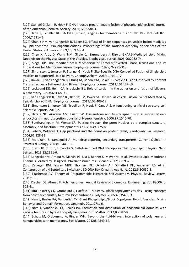
32
[122] Stengel G, Zahn R, Hook F. DNA-induced programmable fusion of phospholipid vesicles. Journal
of the American Chemical Society. 2007;129:9584-+.
[123] Jahn R, Scheller RH. SNAREs [mdash] engines for membrane fusion. Nat Rev Mol Cell Biol.
2006;7:631-43.
[124] Chan Y-HM, van Lengerich B, Boxer SG. Effects of linker sequences on vesicle fusion mediated
by lipid-anchored DNA oligonucleotides. Proceedings of the National Academy of Sciences of the
United States of America. 2009;106:979-84.
[125] Chen X, Araç D, Wang T-M, Gilpin CJ, Zimmerberg J, Rizo J. SNARE-Mediated Lipid Mixing
Depends on the Physical State of the Vesicles. Biophysical Journal. 2006;90:2062-74.
[126] Siegel DP. The Modified Stalk Mechanism of Lamellar/Inverted Phase Transitions and Its
Implications for Membrane Fusion. Biophysical Journal. 1999;76:291-313.
[127] Simonsson L, Jonsson P, Stengel G, Hook F. Site-Specific DNA-Controlled Fusion of Single Lipid
Vesicles to Supported Lipid Bilayers. Chemphyschem. 2010;11:1011-7.
[128] Rawle RJ, van Lengerich B, Chung M, Bendix PM, Boxer SG. Vesicle Fusion Observed by Content
Transfer across a Tethered Lipid Bilayer. Biophysical Journal. 2011;101:L37-L9.
[129] Leckband DE, Helm CA, Israelachvili J. Role of calcium in the adhesion and fusion of bilayers.
Biochemistry. 1993;32:1127-40.
[130] van Lengerich B, Rawle RJ, Bendix PM, Boxer SG. Individual Vesicle Fusion Events Mediated by
Lipid-Anchored DNA. Biophysical Journal. 2013;105:409-19.
[131] Simonsson L, Kurczy ME, Trouillon R, Hook F, Cans A-S. A functioning artificial secretory cell.
Scientific Reports. 2012;2.
[132] Harata NC, Aravanis AM, Tsien RW. Kiss-and-run and full-collapse fusion as modes of exo-
endocytosis in neurosecretion. Journal of Neurochemistry. 2006;97:1546-70.
[133] Suntharalingam M, Wente SR. Peering through the pore: Nuclear pore complex structure,
assembly, and function. Developmental Cell. 2003;4:775-89.
[134] Sohl G, Willecke K. Gap junctions and the connexin protein family. Cardiovascular Research.
2004;62:228-32.
[135] Murakami S, Yamaguchi A. Multidrug-exporting secondary transporters. Current Opinion in
Structural Biology. 2003;13:443-52.
[136] Burns JR, Stulz E, Howorka S. Self-Assembled DNA Nanopores That Span Lipid Bilayers. Nano
Letters. 2013;13:2351-6.
[137] Langecker M, Arnaut V, Martin TG, List J, Renner S, Mayer M, et al. Synthetic Lipid Membrane
Channels Formed by Designed DNA Nanostructures. Science. 2012;338:932-6.
[138] Zadegan RM, Jepsen MDE, Thomsen KE, Okholm AH, Schaffert DH, Andersen ES, et al.
Construction of a 4 Zeptoliters Switchable 3D DNA Box Origami. Acs Nano. 2012;6:10050-3.
[139] Tkachenko AV. Theory of Programmable Hierarchic Self-Assembly. Physical Review Letters.
2011;106.
[140] Discher DE, Ahmed F. Polymersomes. Annual Review of Biomedical Engineering. Vol. 82006. p.
323-41.
[141] Kita-Tokarczyk K, Grumelard J, Haefele T, Meier W. Block copolymer vesicles - using concepts
from polymer chemistry to mimic biomembranes. Polymer. 2005;46:3540-63.
[142] Nam J, Beales PA, Vanderlick TK. Giant Phospholipid/Block Copolymer Hybrid Vesicles: Mixing
Behavior and Domain Formation. Langmuir. 2011;27:1-6.
[143] Nam J, Vanderlick TK, Beales PA. Formation and dissolution of phospholipid domains with
varying textures in hybrid lipo-polymersomes. Soft Matter. 2012;8:7982-8.
[144] Schulz M, Olubummo A, Binder WH. Beyond the lipid-bilayer: interaction of polymers and
nanoparticles with membranes. Soft Matter. 2012;8:4849-64.

33
Figure Captions
Figure 1. Schematic cartoon depicting the concept of biomimetic chemical process control within a
network of liposomal modules. Each compartment has a different chemical environment and
catalytic function but is in communication with its environment and the other modules allowing
transport of chemical species and a cascade of sequential reactions within consecutive
compartments.
Figure 2. Schematic cartoon depicting the functionalization of different populations of liposomal
modules with DNA-amphiphiles and their subsequent assembly into higher-order composite
architectures [100] - Reproduced by permission of The Royal Society of Chemistry.
Figure 3. Structures of amphiphilic nucleic acids with example literature references. (a) Single
hydrocarbon chains: monoacyl functionalised PNA (left) and polypropylene oxide modifications
(right). (b) cholesterol anchors: single cholesterol modification (left), double cholesterol modification
(centre) and a schematic cartoon of double-anchored chol-DNA using two single cholesterol
modified DNAs (right; reprinted with permission from Pfeiffer I., Hook F., J. Am. Chem. Soc.
2004;126:10224-5 [55]. Copyright 2004 American Chemical Society). (c) Double hydrocarbon
anchors: structure of a dialkyl lipid-DNA conjugate ふノWaデぶ ;ミS ;ミ ü-tocopherol modified base which
can be inserted within the DNA chain to create two (or more) hydrophobic anchors (right). (d) Biotin-
avidin linkers for functionalisation of lipid bilayers with DNA: avidin binds to biotinylated lipids within
the membrane; subsequent addition of biotinylated DNA attaches the DNA to the avidin on the
membranes. Inset cartoon of biotin-avidin conjugation to lipid bilayers and subsequent membrane
adhesion reprinted with permission from reference [68].
Figure 4. Reversible assembly of BioNanoStacks formed from the DNA-mediated assembly of lipid
nanodiscs. (a) representative melting curves from UV-vis spectroscopy. (b) Melting temperatures as
a function of salt concentration for different lipid compositions separated by heating and cooling
directions. (c) Melting cooperativities represented by the full width half maximum (FWHM) of the
melting curves. (d,e) Negative staining TEM images of BioNanoStack morphologies. Reprinted with
permission from Beales P.A. et al., J. Am. Chem. Soc. 2013;135:3335-8 [83]. Copyright 2013 American
Chemical Society.
Figure 5. Assembly of DNA-a┌ミIデキラミ;ノキ┣WS さJ;ミ┌ゲざ ┗WゲキIノWゲく ふAぶ IラミaラI;ノ aノ┌ラヴWゲIWミIW マキIヴラゲIラヮ┞
sections of DNA-functionalized Janus vesicles with the liquid disordered domain labelled in red and
the DNA labelled in blue. (B) Fluorescence intensity line profiles of the liquid disordered dye (Rh-
DOPE) and DNA (A647-DNA) from the image in part A. (C) 3D-reconstructed z-stack of the

34
morphology of DNA-functionalized Janus vesicles. (D) Assembly of Janus vesicles into size limited
clusters with the liquid disordered phase labelled red and the liquid ordered phase labelled green;
the fluorescence intensity line profile demonstrates the presence of an unfused double membrane
in the adhesion plaques between liposomes [100] - Reproduced by permission of The Royal Society
of Chemistry.
Figure 6. Enzymatic-switching of DNA domain partitioning. (A) Hybridization of a palmitoylated PNA
and DNA tocopherol causes both molecules to partition into liquid disordered domains. (B) Cleavage
of the linking DNA strand by EcoR1-HF switches the palmitoylated PNA into the liquid ordered
domain. Reprinted with permission from Schade M. et al., J. Am. Chem. Soc. 2012;134:20490-7
[102]. Copyright 2012 American Chemical Society.Figure 7. Linker self-sorting by length between
DNA-tethered membranes. (A) cartoon showing a mixture of 24mer and 72mer DNAs separating a
silica supported bilayer from a second tethered bilayer. (B) Fluorescence interference contrast (FLIC)
image showing height differences of a tethered lipid bilayer labelled with a Texas Red lipid; brigher
areas represent higher membrane domains. (C) The same patch showing fluorescence from Alexa-
488 labelled 24mer DNA. The scale bars represent 10 µm [103] - Reproduced by permission of The
Royal Society of Chemistry.
Figure 8. Cartoon showing liposomes tethered to a planar bilayer by multiple DNA tethers deduced
from lateral mobility measurements. The DNA could form a concentrated patch of DNA at the
contact site (left) or move out in a crown-like ring that would increase the effective contact area
between the liposome and planar membrane. Reprinted with permission from Benkoski J.J., Hook F.,
J. Phys. Chem. B 2005;109:9773-9 [112]. Copyright 2005 American Chemical Society.Figure 9.
Competition between electroosmotic flow and electrophoretic flow of charged liposomes tethered
to (A) a neutral supported membrane and (B) a supported membrane containing charged lipids in
the presence of an applied electric field. (C) Separation of vesicles by charge due to their different
electrophoretic mobilities: egg PC + 1% Texas Red DHPE + 4% DPPS liposomes (red) and egg PC + 2%
Oregon Green DHPE liposomes (green) on a supported membrane of egg PC + 2% DPPS. Reprinted
with permission from Yoshina-Ishii C., Boxer S.G., Langmuir 2006;22:2384-91 [114]. Copyright 2006
American Chemical Society.Figure 10. Schematic cartoon of the steps of liposome fusion using chol-
DNAs that zip the liposome membranes into close apposition, stimulating lipid mixing and fusion in
an analogous mechanism to the natural SNARE fusion complex. Reprinted with permission from
Stengel G. et al., J. Phys. Chem. B 2008;112:8264-74 [57]. Copyright 2008 American Chemical
Society.Figure 11. DNA nanostructures as trans-membrane pores. (A-C) Cartoons illustrating the DNA
nanostructure designed by Langecker et al. along with (D) TEM images of the structure. From

35
Langecker M., Science 2012;338:932-6 [137]. Reprinted with permission from AAAS. (E, F) Illustrated
structure of the transmembrane pore designed by Burns et al. Reprinted with permission from Burns
J.R. et al., Nano Lett. 2013;13:2351-6 [136]. Copyright 2013 American Chemical Society.

36
Figure 1.
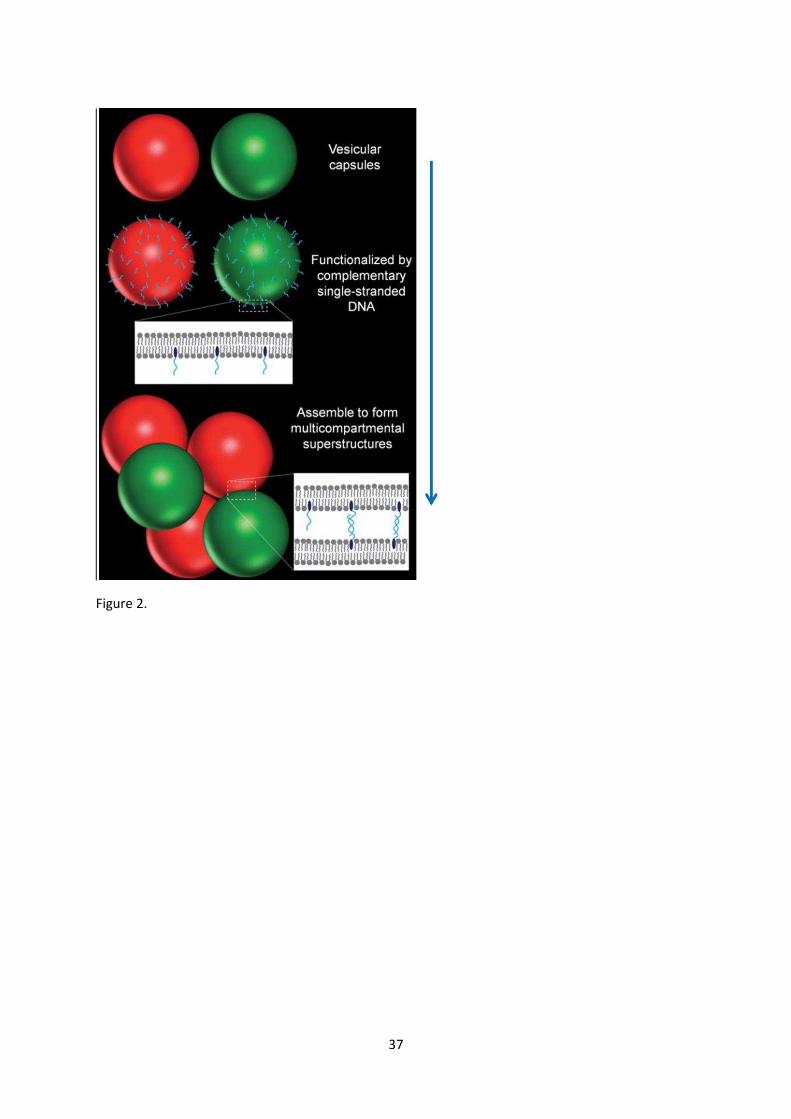
37
FF
Figure 2.

38
Figure 3

39
Figure 4.
40
Figure 5.
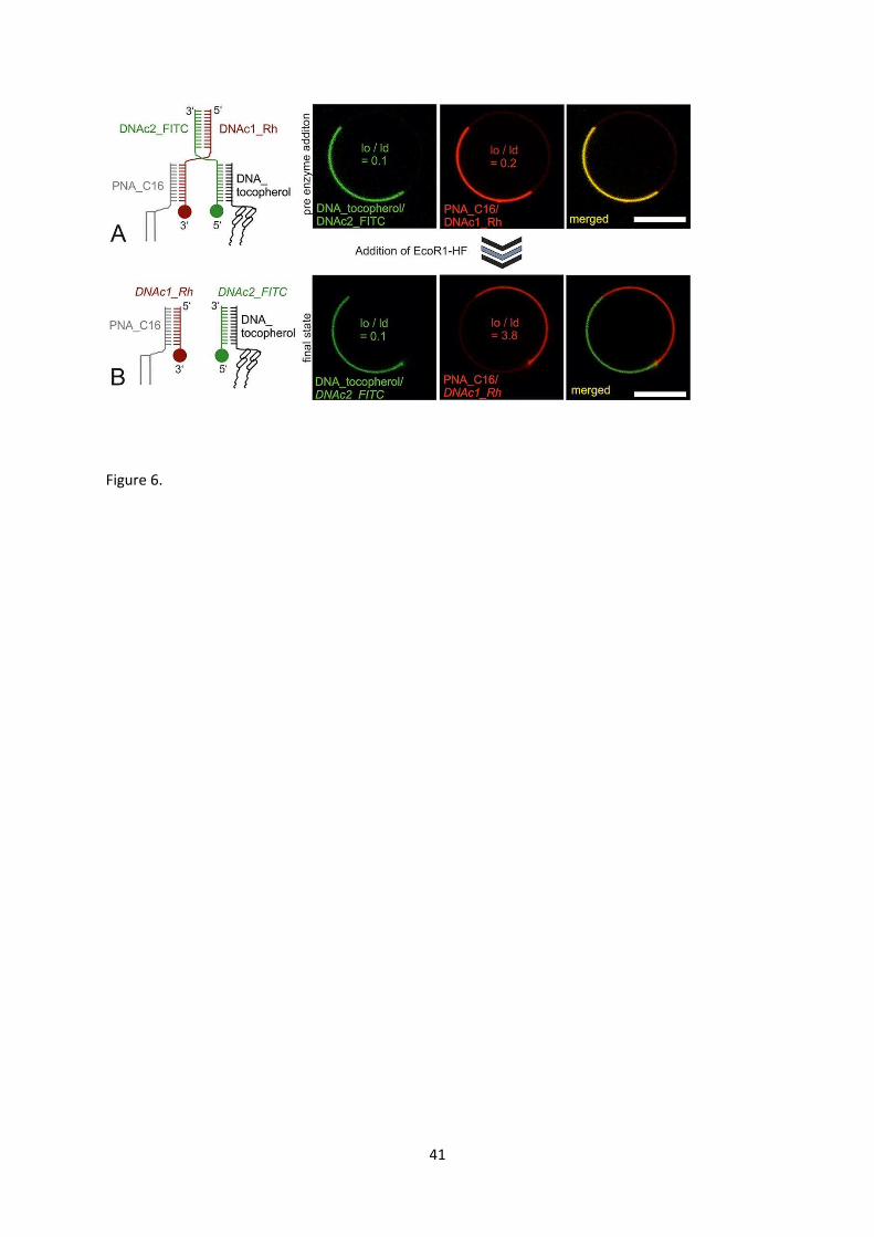
41
Figure 6.

42
Figure 7.

43
Figure 8.
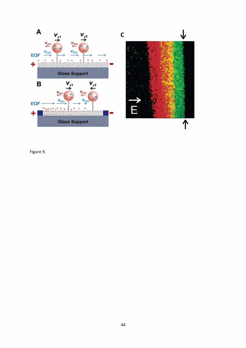
44
Figure 9.

45
Figure 10.
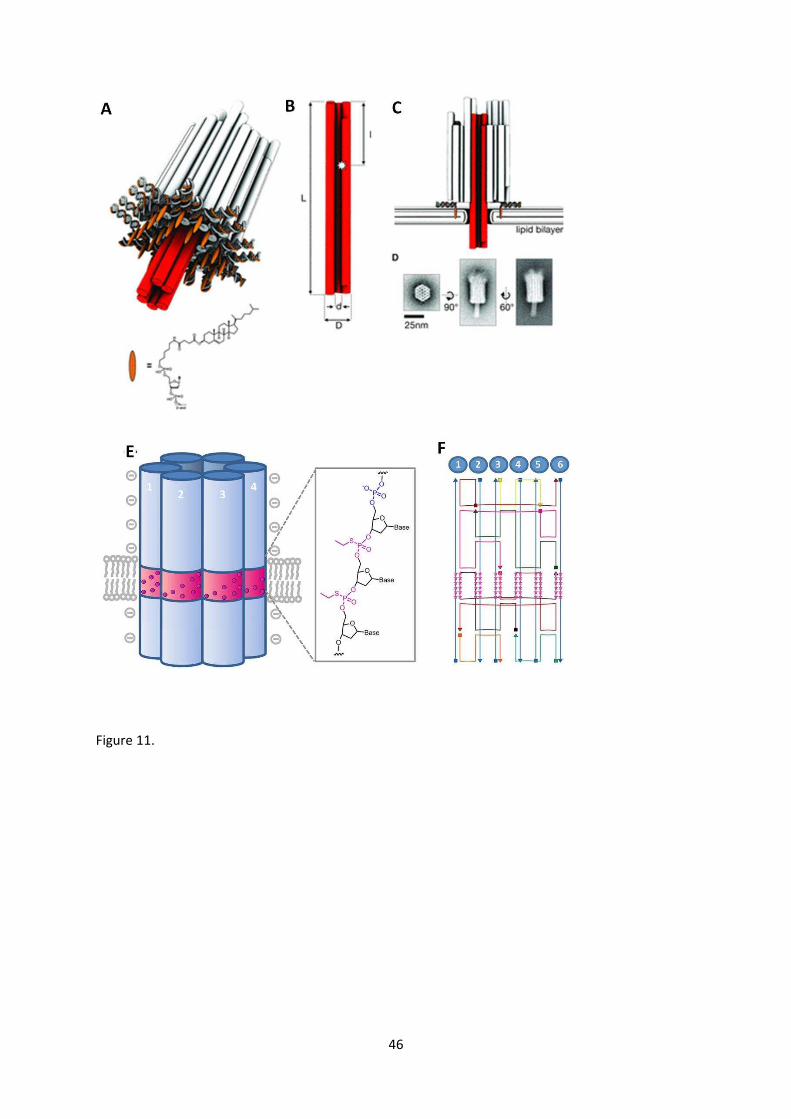
46
Figure 11.
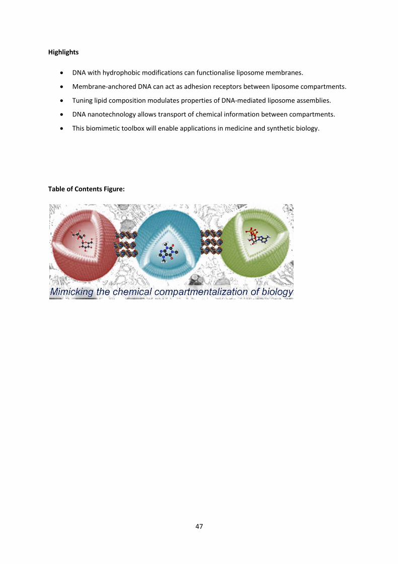
47
Highlights
DNA with hydrophobic modifications can functionalise liposome membranes.
Membrane-anchored DNA can act as adhesion receptors between liposome compartments.
Tuning lipid composition modulates properties of DNA-mediated liposome assemblies.
DNA nanotechnology allows transport of chemical information between compartments.
This biomimetic toolbox will enable applications in medicine and synthetic biology.
Table of Contents Figure: