application note - axion biosystems · axion biosystems tm predicting pro-arrhythmic potential...
TRANSCRIPT

Application Note
Predicting Pro-arrhythmic Potential using CytivaTM Plus Cardiomyocytes on MEAs
axionBioSystems
Trademarks
Axion BioSystems, Inc. and the logo are trademarks of Axion BioSystems, and may not be used without the express written permission of Axion BioSystems, Inc.
GE and GE monogram are trademarks of General Electric Company. Cytiva is a trademark of GE Healthcare companies. Spotfire is a trademark of Tibco Software Inc. All other brands, product names, company names, trademarks and service marks are the properties of their respective owners.
Restrictions and Liabilities
This document is provided “as is” and Axion BioSystems will assume no responsibility for any typographical, technical or other inaccuracies in this document. Axion BioSystems does not make any commitment to provide any changes, updates, enhancements to this document within any time frame or at all.
This document might contain references to third-party sources of information, hardware or software, products or services and/or third-party websites (collectively the “Third-Party Information”). Axion BioSystems has no control over and is not responsible for any Third-Party Information, including, without limitation the content, accuracy, copyright compliance, compatibility, performance, trustworthiness, legality, decency, links or any other aspect of Third-Party Information. The in-clusion of Third-Party Information in this document does not imply endorsement with the use of Axion BioSystems technol-ogy.
The Cytiva Plus product is for research use only – not for use in diagnostic procedures. GE Healthcare Cardiomyocytes are sold under licence from Geron Corporation and Wisconsin Alumni Research Foundation under US patent and pub-lication numbers : US 7,425,448, US 2009/0017465, US 6,800,480, US 5,843,780, US 6,200,806, US 7,029,913, US 7,582,479, US 7,413,902, US 7,297,539, US 2009/0047739 and US 2007/0010012 and equivalent patent and patent ap-plications in other countries.
Conditions of Use
The user is responsible for understanding and performing the protocols that are described within. Axion BioSystems makes no guarantee for any results. These protocols are provided as a recommendation by Axion BioSystems based on use and experience.
Origin
Axion BioSystems Microelectrode Arrays are manufactured in the United States of America.
Copyright Notice
© 2014 Axion BioSystems, Inc. All rights reserved. This document may not be reproduced, distributed, modified or publicly displayed without the express written permission of Axion BioSystems.
Acknowledgement
Axion BioSystems would like to thank GE Healthcare for providing their experience and resources toward the creation of this application note. In particular the individuals: Mike Clements, Hayley Tinkler, Angela Williams, and Nick Thomas.
GE Healthcare would like to thank Dr Hua Rong Lu and Janssen Pharmaceutica for the generous gift of JNJ282 and JNJ303.
axionBioSystems

Axion BioSystems Predicting Pro-arrhythmic Potential using CytivaTM Plus Cardiomyocytes on MEAs 3
Introduction
Drug-induced delayed cardiac repolarization, a rec-ognized risk factor for pro-arrhythmia, is the single most common cause for the withdrawal of prescrip-tion drugs. The vast majority of drugs known to prolong the repolarization of the cardiac membrane preferentially inhibit the delayed rectifier current (IKr) by binding to the hERG K+ channel. Consequently, functional in vitro assays for predicting a drug’s potential to delay cardiac repolarization typically include evaluating hERG K+ channel block in trans-genic cell lines, or action potential duration assays with primary canine or rabbit Purkinje fibers. How-ever, the predictive value of these existing assays is limited due to species differences and the lack of complex ion channel interactions in cell lines over-expressing the hERG K+ channel.1 Improved simu-lation of cardiac networks could allow for drug dis-
covery stage testing to elucidate off-target effects, including detrimental hERG K+ channel interactions. This ability will improve drug safety screening and reduce the cost and time of drug development.
A compelling approach to this issue uses assays performed on human embryonic stem cell-derived (hESC) cardiomyocytes. Cytiva Plus Cardiomyo-cytes are hESC-derived and exhibit the morphology and electrophysiological activity typical of human cardiomyocytes, while the Axion BioSystems Mae-stroTM Microelectrode Array (MEA) system provides a high throughput platform for the evaluation of extracellular field potentials. Combining these two technologies creates a robust platform for cardio-toxicity profiling.
To induce a cardiomyocyte phenotype, hESCs (H7 cell line) were subjected to a controlled differentia-tion process. Briefly, the hESCs were adapted to alternative growth conditions, subjected to growth factor induction, followed by a period of cardiomyo-cyte maturation. At the end point of differentiation, cardiomyocytes were harvested and cryo-pre-served at 1x10⁶ cardiomyocytes per vial.
Following the timeline presented in Figure 1, Cytiva Plus Cardiomyocytes (GE Healthcare) were seed-
ed directly from thaw onto multiwell MEA plates (Ax-ion BioSystems) at a density of 60,000 cells in a 4 μL dot over the electrode array using the optimized culture protocol. On day 4 post-thaw, half of the seeding medium was replaced with fresh medium. Treatment of the cells on the MEA on day 5 post-thaw followed the timeline outlined in Figure 2. For a step-by-step guide on the culture methods, please review the Full Culture Protocol.
Figure 1: Timeline depicting cell seeding and maintenance for MEA experiments
40 1
MEA experimentThaw cells, seed directly onto MEA
Days in Culture
Medium change
2 3 5
Data Acquisition
Experimental Phase
EquilibrationConc. #1
Conc. #2
Conc. #3
Conc. #4
Conc. #5Figure 2: Timeline depicting MEA experimental protocol. MEA recordings were made on day 5 post-thaw at 37°C following the compound addition protocol outlined here.
0 12 24 36 48 60 72 Time (min)
Materials and Methods

Axion BioSystems Predicting Pro-arrhythmic Potential using CytivaTM Plus Cardiomyocytes on MEAs 4
Results
Figure 3: A) Cytiva Plus Cardiomyocytes on day 5 post-thaw stained for DNA (hoechst; blue) and troponin I (green). B) Cytiva Plus Cardiomyocytes on day 5 post-thaw on a MEA plate. C) Cytiva Plus Cardiomyocytes on day 5 post-thaw showing the alternating troponin I and α-actinin bands.
Figure 4: Evaluation of Cytiva Plus Cardiomyocyte spontaneous beating characteristics using MEAs at 37°C. The cells exhibit regular uniform spike amplitudes and beat periods with strong beat regularity as demonstrated by the histograms above. Of the 144 wells (i.e. 3x 48-well plates) seeded with cardiomyo-cytes, 85% (122 wells) achieved a minimum level of activity sufficient for analysis. FDPcF = FPD/³√(Beat Period).
To characterize the Cytiva Plus Cardiomyocytes, the cells were stained on day 5 post-thaw for DNA (hoescht stain, blue) and troponin I (green), a protein critical for skeletal and cardiac muscle contraction (Fig. 3). The contractile nature of the cells was confirmed by the prevalence of troponin I. When grown on an MEA plate (Fig. 3), the car-diomyocytes formed a uniform monolayer on top of
the microelectrode array. This conformation pro-duced a functional syncytium of cells with a robust electrophysiological signature. The Maestro MEA system was used to record spike amplitude and timing of the field potential, allowing for calculation of beat period, beat regularity, field potential dura-tion (FPD) and field potential duration corrected by Fridericia’s formula (FPDcF).
100 mm50 mm
10 mm
A B
C
Axion BioSystems Predicting Pro-arrhythmic Potential using CytivaTM Plus Cardiomyocytes on MEAs 5
1 sec10%
ΔF/
F1 sec10
% Δ
F/F 0
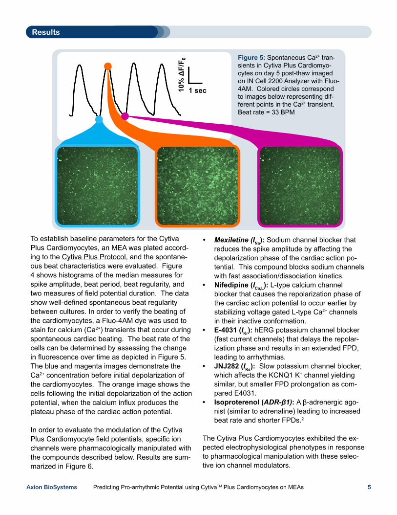
To establish baseline parameters for the Cytiva Plus Cardiomyocytes, an MEA was plated accord-ing to the Cytiva Plus Protocol, and the spontane-ous beat characteristics were evaluated. Figure 4 shows histograms of the median measures for spike amplitude, beat period, beat regularity, and two measures of field potential duration. The data show well-defined spontaneous beat regularity between cultures. In order to verify the beating of the cardiomyocytes, a Fluo-4AM dye was used to stain for calcium (Ca2+) transients that occur during spontaneous cardiac beating. The beat rate of the cells can be determined by assessing the change in fluorescence over time as depicted in Figure 5. The blue and magenta images demonstrate the Ca2+ concentration before initial depolarization of the cardiomyocytes. The orange image shows the cells following the initial depolarization of the action potential, when the calcium influx produces the plateau phase of the cardiac action potential.
In order to evaluate the modulation of the Cytiva Plus Cardiomyocyte field potentials, specific ion channels were pharmacologically manipulated with the compounds described below. Results are sum-marized in Figure 6.
Figure 5: Spontaneous Ca2+ tran-sients in Cytiva Plus Cardiomyo-cytes on day 5 post-thaw imaged on IN Cell 2200 Analyzer with Fluo-4AM. Colored circles correspond to images below representing dif-ferent points in the Ca2+ transient. Beat rate = 33 BPM
• Mexiletine (INa): Sodium channel blocker that reduces the spike amplitude by affecting the depolarization phase of the cardiac action po-tential. This compound blocks sodium channels with fast association/dissociation kinetics.
• Nifedipine (ICa,L): L-type calcium channel blocker that causes the repolarization phase of the cardiac action potential to occur earlier by stabilizing voltage gated L-type Ca2+ channels in their inactive conformation.
• E-4031 (IKr): hERG potassium channel blocker (fast current channels) that delays the repolar-ization phase and results in an extended FPD, leading to arrhythmias.
• JNJ282 (IKs): Slow potassium channel blocker, which affects the KCNQ1 K+ channel yielding similar, but smaller FPD prolongation as com-pared E4031.
• Isoproterenol (ADR-β1): A β-adrenergic ago-nist (similar to adrenaline) leading to increased beat rate and shorter FPDs.2
The Cytiva Plus Cardiomyocytes exhibited the ex-pected electrophysiological phenotypes in response to pharmacological manipulation with these selec-tive ion channel modulators.
Results

Axion BioSystems Predicting Pro-arrhythmic Potential using CytivaTM Plus Cardiomyocytes on MEAs 6
Figure 6: Many different ion channels contribute to the measured extracellular field potential. Blockade of a specific ion channel results in characteristic modulation of the Cytiva Plus Cardiomyocyte field potential waveform. For the mexiletine voltage waveform, traces are offset along the x-axis to high-light the change in peak amplitude. The cartoon in the lower right corner high-lights ion channel current profiles.
Results
Na+ channel block (INa)
L-type Ca2+ channel block (ICa,L)
hERG K+ channel block (IKr)
KCNQ1 K+ channel block (IKs)
β-adrenergic receptor agonism

Axion BioSystems Predicting Pro-arrhythmic Potential using CytivaTM Plus Cardiomyocytes on MEAs 7
Rank DrugConc where FPD
= +20% (nM)hERG IC50 (nM) Drug Class
1 Dofetilide < 3 10 Class III antiarrhythmic
2 Astemizole < 3 13 Antihistamine
3 E-4031 < 3 32 Class III antiarrhythmic (experimental)
4 Tolterodine 5 10 For bladder incontinence
5 Terfenadine 90 40 Antihistamine
6 Quinidine 150 750 Class Ia antiarrhythmic
7 Terodiline 230 380 For bladder incontinence
8 Alfuzosin 300 14,000 For benign prostatic hyperplasia
9 Sotalol 1,500 75,000 Class III antiarrhythmic
10 Ranolazine 5,000 12,000 Antianginal
11 Moxifloxacin 26,000 40,000 Antibacterial
12 Aspirin > 1,000,000 No Block Cyclooxygenase inhibitor
13 Nifedipine - 275 Antihypertensive/antianginal
14 Verapamil - 540 Class IV antiarrhythmic
IFPD20
When combined with MEAs, the Cytiva Plus Car-diomyocytes serve as a model system for evaluat-ing cardioactive compounds. To validate Cytiva Plus Cardiomyocytes as a potentially useful new in vitro test system for drug-induced delayed cardiac repolarization, multiple drugs were applied to the cardiomyocytes to determine their effect on the FPD (Fig. 7). Drugs that triggered a 20% or greater prolongation of the FPD were compiled and ranked based on the concentration required to elicit the FPD change. A positive relationship was found between this concentration and the hERG IC50 values across all drug classes (Table 1). In combi-nation with the field potential duration, the beat rate and spike amplitude of the field potential wave-
form establishes the electrophysiological profile of cardioactive compounds, as depicted in Figure 8, and provides information on the mechanism of action. For example, while hERG blockers Sotalol and Ranolazine have 2 different clinical uses, both show similar modulation of field potential metrics: increased beat period, prolongation of the FPD, and decreased spike amplitude. A hierarchical clustering analysis was performed on these elec-trophysiological profiles (Figure 9). Compounds acting through similar mechanisms clustered to-gether, suggesting that application of these metrics to unknown or novel drugs could be used to predict mechanism of action.
Results
Figure 7: Dose escalation studies were performed for drugs known to modify the QT interval
Table 1: For selective hERG K+ channel blockers, a similar rank-ordering of compounds was found for both the drug’s propensity to induce FPD prolongation and the drug’s hERG IC50 value.
Axion BioSystems Predicting Pro-arrhythmic Potential using CytivaTM Plus Cardiomyocytes on MEAs 8
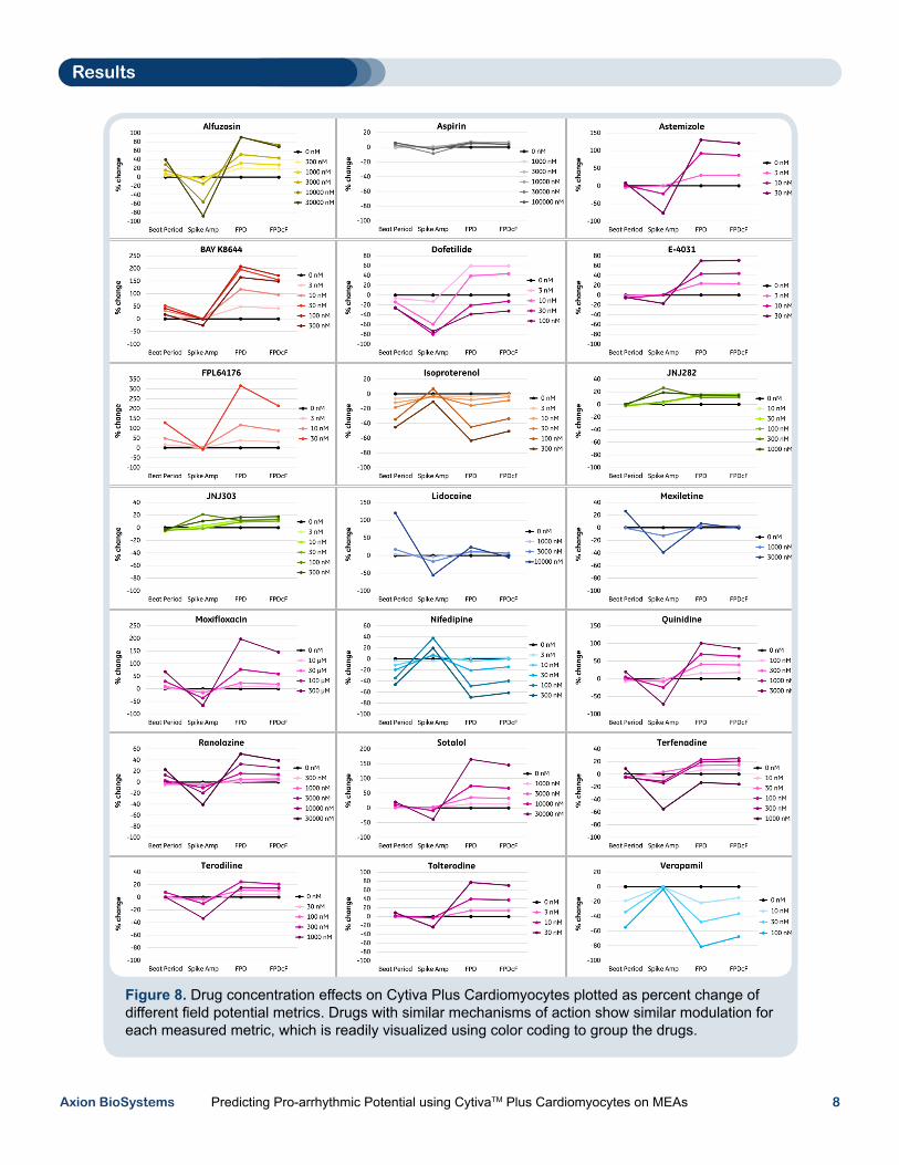
Figure 8. Drug concentration effects on Cytiva Plus Cardiomyocytes plotted as percent change of different field potential metrics. Drugs with similar mechanisms of action show similar modulation for each measured metric, which is readily visualized using color coding to group the drugs.
Results

Axion BioSystems Predicting Pro-arrhythmic Potential using CytivaTM Plus Cardiomyocytes on MEAs 9
Figure 9. Electrophysiological profiles for an array of validation compounds on the Cytiva Plus Cardiomyo-cytes. The series of traces within each plot indicates the change in the field potential metrics as a function of increasing drug concentration, with compounds of a common mechanism of action sharing the same color.
Cytiva Plus Cardiomyocytes are hESC-derived cardiomyocytes that exhibit morphology and elec-trophysiological responses consistent with human cells. The cultured cells form spontaneously beat-ing monolayers whose extracellular field potential characteristics can be readily examined using Axion BioSystems MEAs.
Together, the Cytiva Plus Cardiomyocytes and Maestro MEA system offer an in vitro system for evaluating the electrophysiological effects of car-dioactive compounds. The field potential waveform was sensitive to selective manipulations of multiple ion channels contributing to the cardiac action potential, including the hERG potassium channel. Notably, there was a positive relationship between the IC50 values of hERG channel blockers and the concentration that elicited a 20% increase in
FPD. Extension of the analysis to include multiple field potential metrics allowed the development of electrophysiological profiles of known cardioactive compounds. Compounds with shared mechanisms of action displayed quantitatively similar profiles in a hierarchical clustering analysis, providing support for mechanism prediction for novel pharmaceuti-cals.1
Based on these results, an MEA assay using hESC-derived cardiomyocytes could complement or replace a portion of the pre-clinical cardiac toxic-ity screening tests currently used for lead optimiza-tion and further development of new drugs.
Results
1. Clements & Thomas, High-Throughput Multi-Param-eter Profiling of Electrophysiological Drug Effects in Human Embryonic Stem Cell Derived Cardiomyo-cytes using Multi-Electrode Arrays. Toxicol Sci, 2014
2. Grant, A.O., Cardiac ion channels. Circ Arrhythm Electrophysiol, 2009. 2: p. 185-94.
- L-type Ca2+ channel block- Na+ channel block- KCNQ1 K+ channel block- hERG K+ channel block- L-type Ca2+ channel agonist
References
FPD Spike amp. Beat period Beat rate
ICa,L
INa IKs IKr ICa,L
Discussion and Conclusions