appendicular skeleton chapter 8 (9/30/08). the pectoral girdle
TRANSCRIPT

APPENDICULAR SKELETON
CHAPTER 8(9/30/08)
THE PECTORAL GIRDLE
Pectoral or Shoulder Girdle
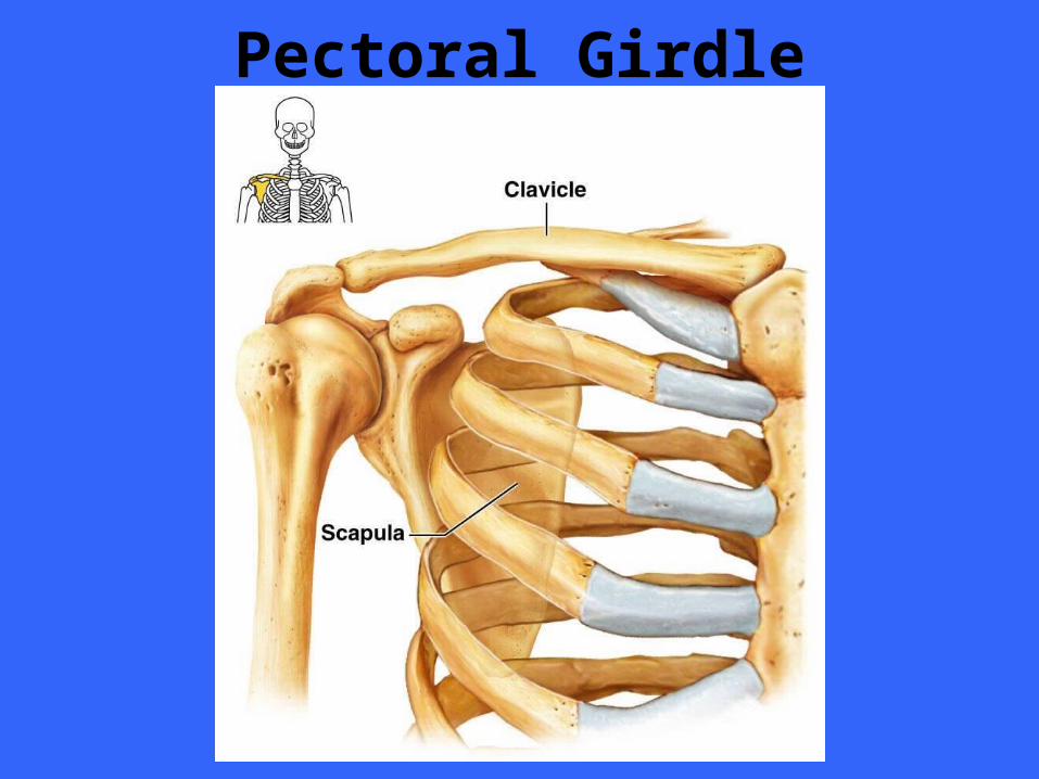
Consists of two bones, the anteriorly positioned clavicle and the posteriorly positioned scapula
Pectoral girdle is a loosely attached, held in place largely by musculature attached to the thorax and the vertebral column
Only direct ligament attachment exists at the sternoclavicular joint
Frees girdle to move over the thorax as the need arises
Pectoral Girdle
Flexible and Mobile Pectoral girdle is very light to allow the upper limb flexibility and mobility not allowed anywhere else in body
This is possible because only the sternal end of clavicle is attached to axial skeleton thus allowing the scapula to move across thorax and the arm with it
The socket of the shoulder joint is shallow and poorly reinforced
Although this arrangement does not restrict movement it is less stable
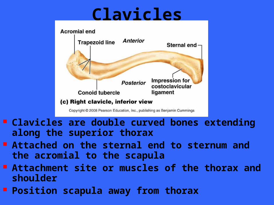
Clavicles
Clavicles are double curved bones extending along the superior thorax
Attached on the sternal end to sternum and the acromial to the scapula
Attachment site or muscles of the thorax and shoulder
Position scapula away from thorax
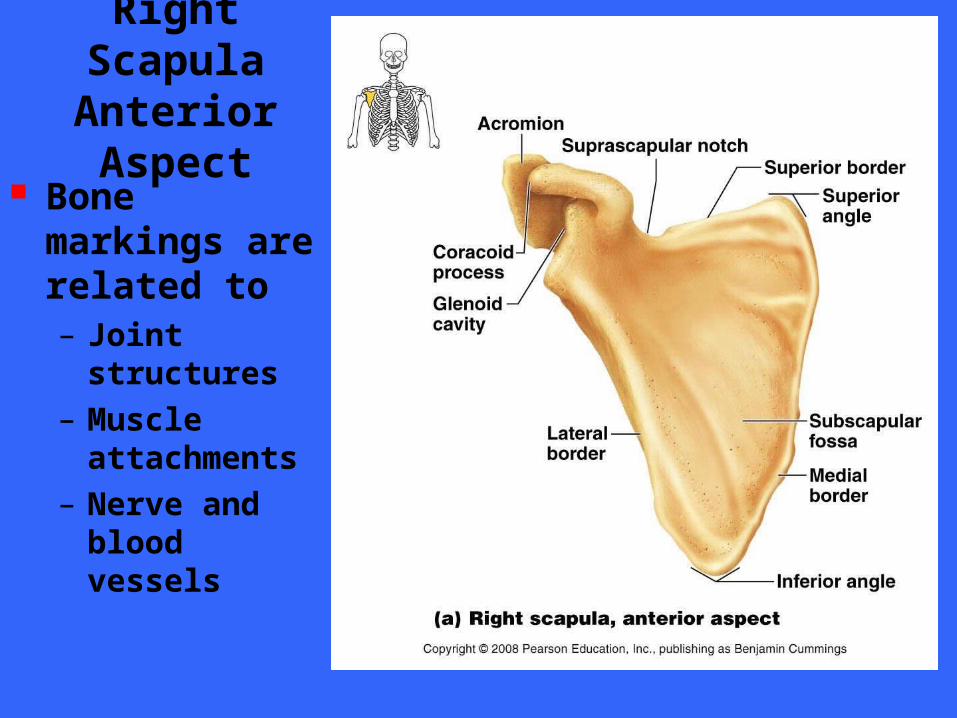
Right ScapulaAnterior Aspect
Bone markings are related to– Joint structures
– Muscle attachments
– Nerve and blood vessels
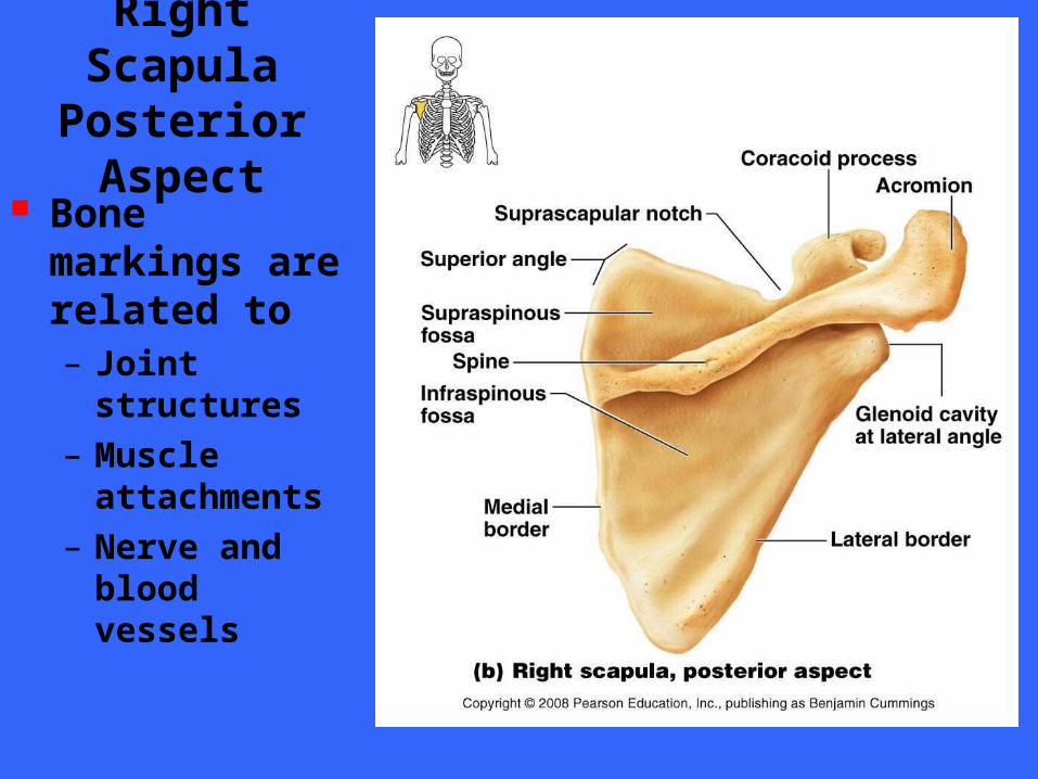
Right Scapula
Posterior Aspect
Bone markings are related to– Joint structures
– Muscle attachments
– Nerve and blood vessels
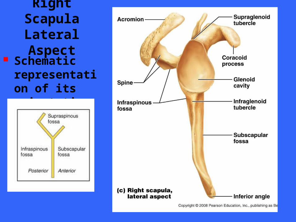
Right ScapulaLateral Aspect
Schematic representation of its orientation
THE UPPER LIMB
SECTION V
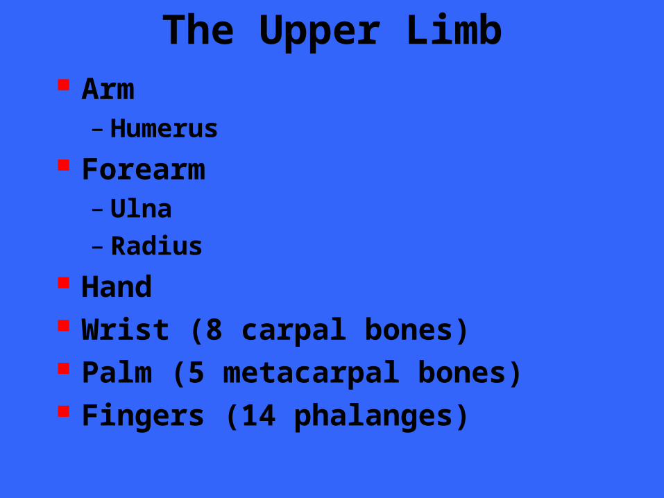
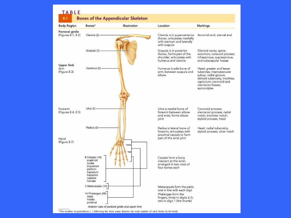
The Upper Limb Arm
– Humerus Forearm
– Ulna– Radius
Hand Wrist (8 carpal bones) Palm (5 metacarpal bones) Fingers (14 phalanges)
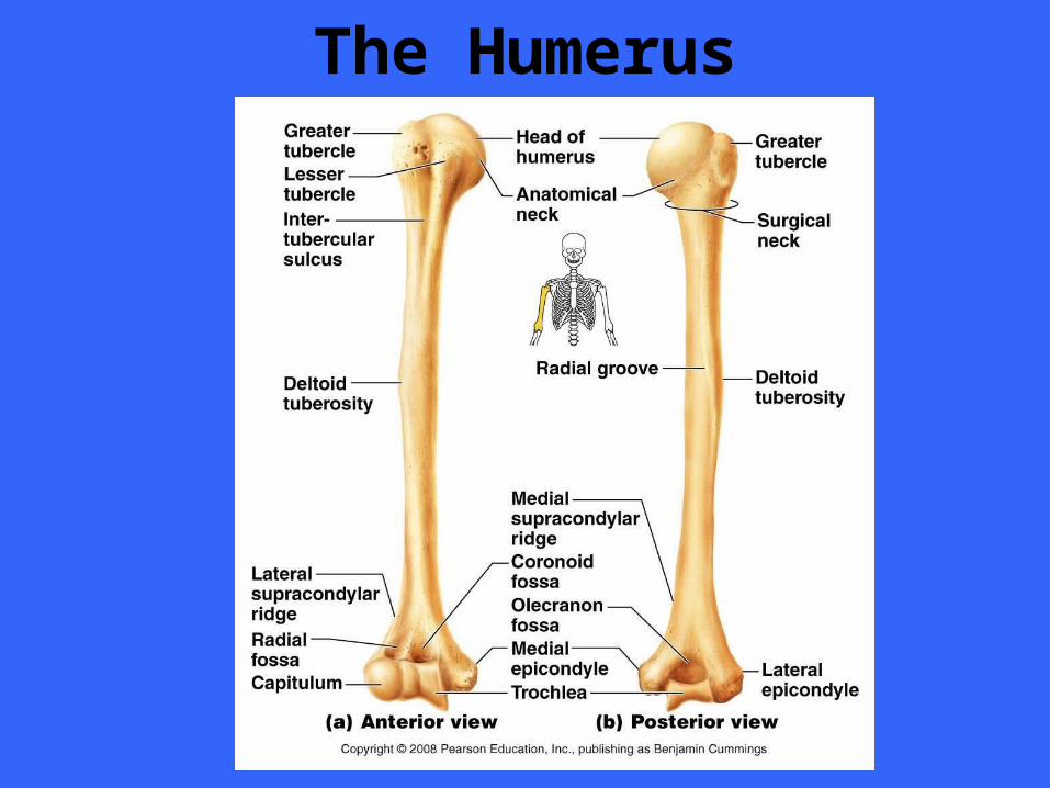
The Humerus
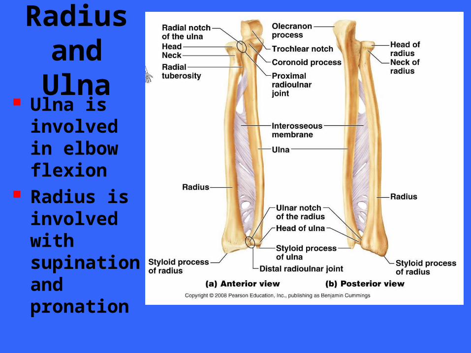
Radius and Ulna
Ulna is involved in elbow flexion
Radius is involved with supination and pronation
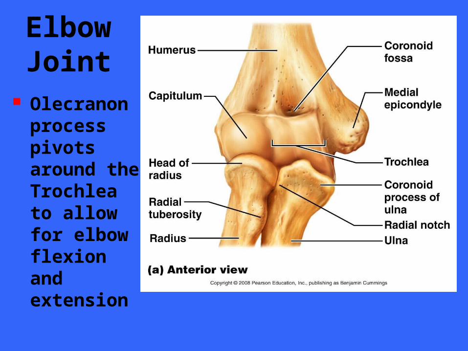
Elbow Joint
Olecranon process pivots around the Trochlea to allow for elbow flexion and extension
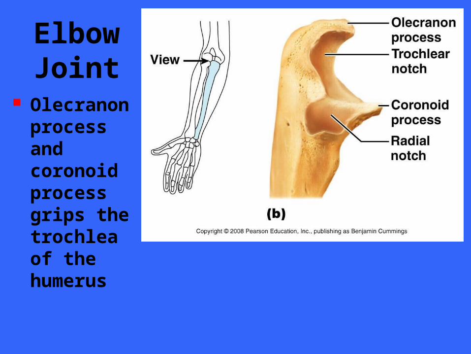
Elbow Joint
Olecranon process and coronoid process grips the trochlea of the humerus
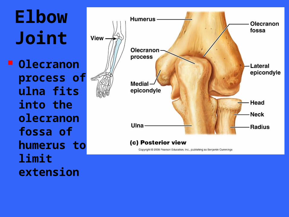
Elbow Joint
Olecranon process of ulna fits into the olecranon fossa of humerus to limit extension
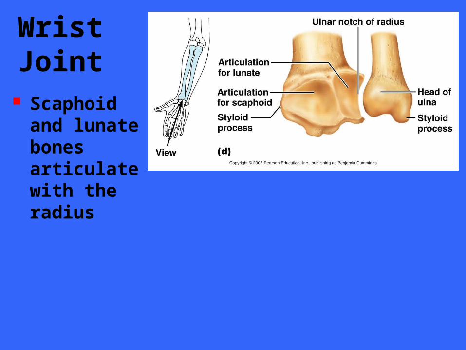
Wrist Joint
Scaphoid and lunate bones articulate with the radius
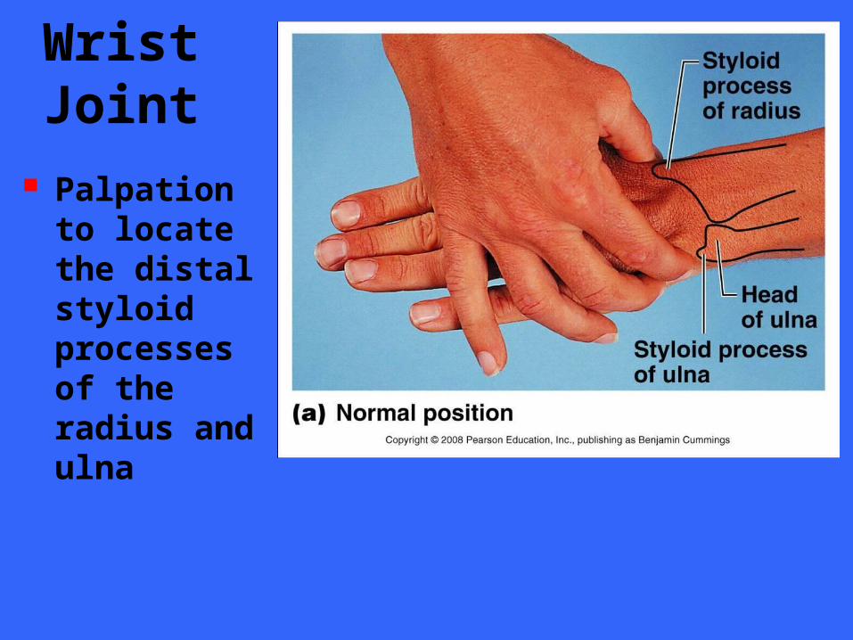
Wrist Joint
Palpation to locate the distal styloid processes of the radius and ulna
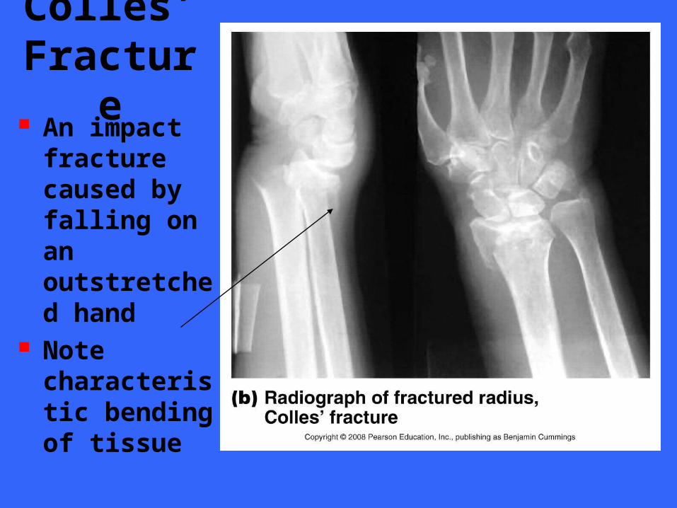
Colles’ Fractur
e An impact fracture caused by falling on an outstretched hand
Note characteristic bending of tissue
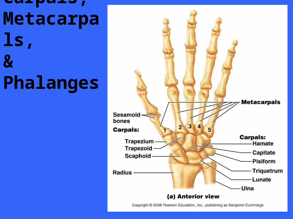
Carpals, Metacarpals, & Phalanges
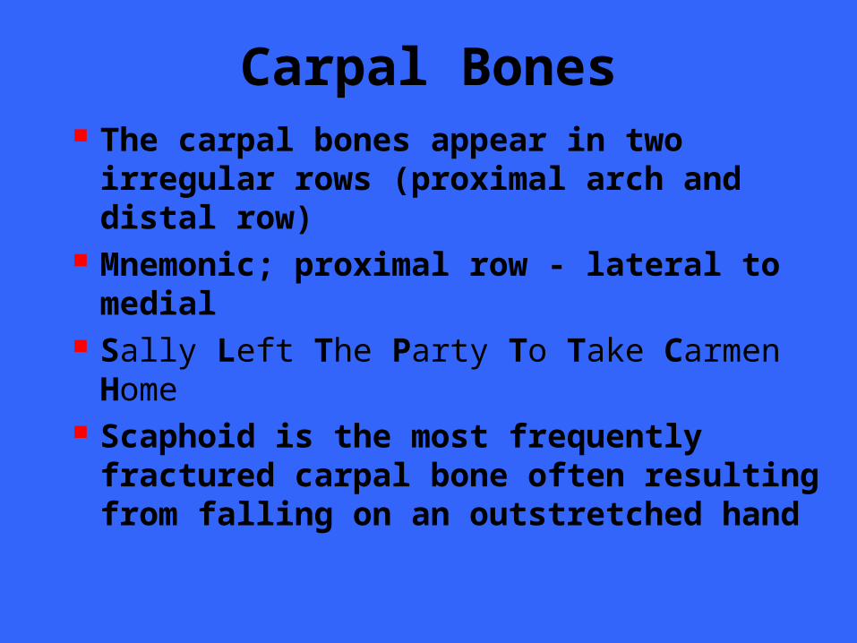
Carpal Bones The carpal bones appear in two irregular rows (proximal arch and distal row)
Mnemonic; proximal row - lateral to medial
Sally Left The Party To Take Carmen Home
Scaphoid is the most frequently fractured carpal bone often resulting from falling on an outstretched hand
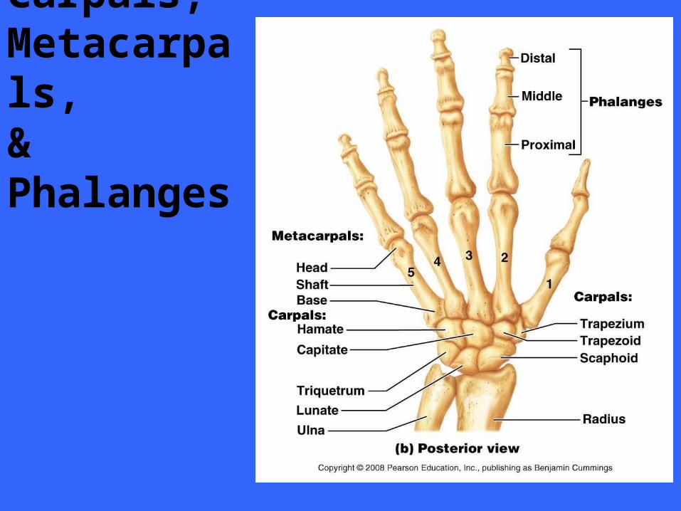
Carpals, Metacarpals, & Phalanges
THE PELVIC GIRDLE
SECTION VI
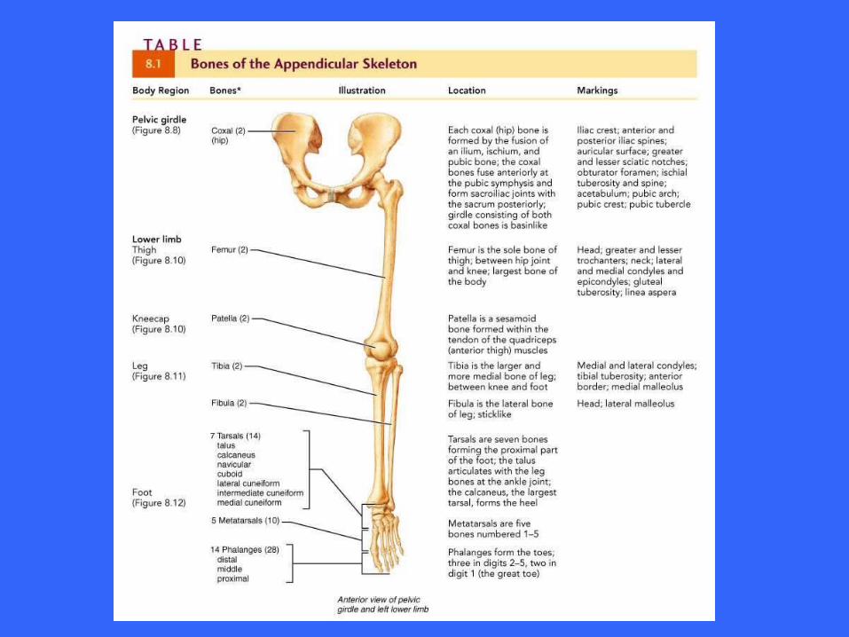
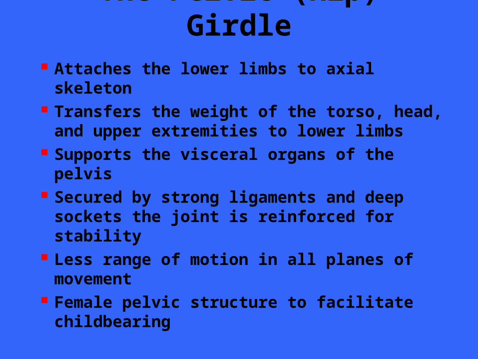
The Pelvic (Hip) Girdle
Attaches the lower limbs to axial skeleton
Transfers the weight of the torso, head, and upper extremities to lower limbs
Supports the visceral organs of the pelvis
Secured by strong ligaments and deep sockets the joint is reinforced for stability
Less range of motion in all planes of movement
Female pelvic structure to facilitate childbearing
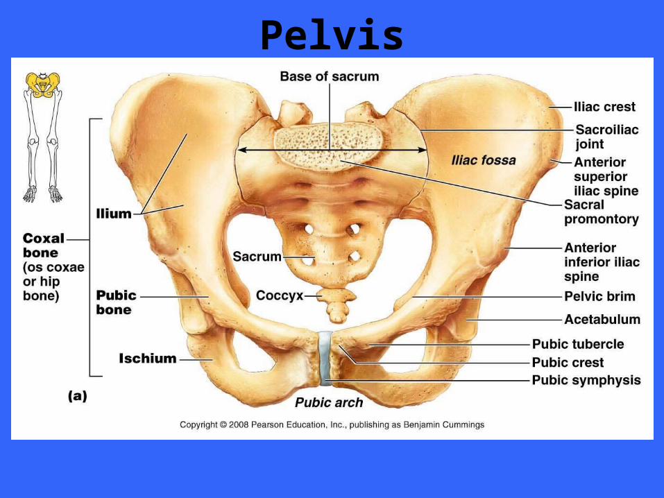
Pelvis
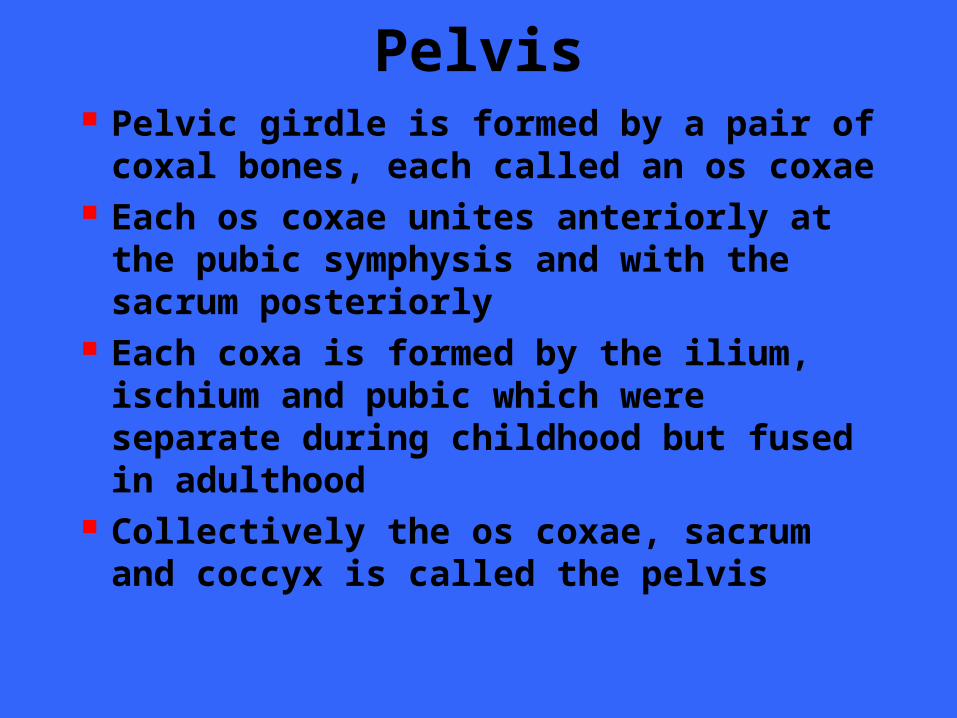
Pelvis Pelvic girdle is formed by a pair of coxal bones, each called an os coxae
Each os coxae unites anteriorly at the pubic symphysis and with the sacrum posteriorly
Each coxa is formed by the ilium, ischium and pubic which were separate during childhood but fused in adulthood
Collectively the os coxae, sacrum and coccyx is called the pelvis
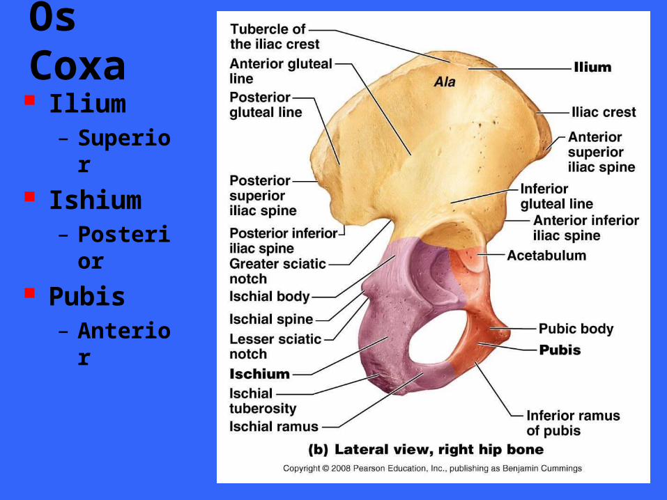
Os Coxa Ilium
– Superior
Ishium– Posterior
Pubis– Anterior
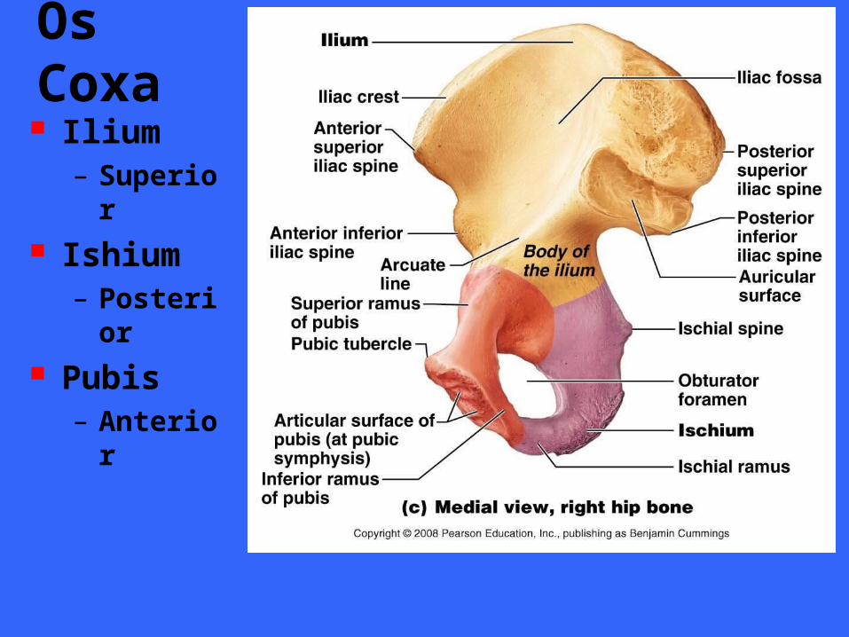
Os Coxa Ilium
– Superior
Ishium– Posterior
Pubis– Anterior
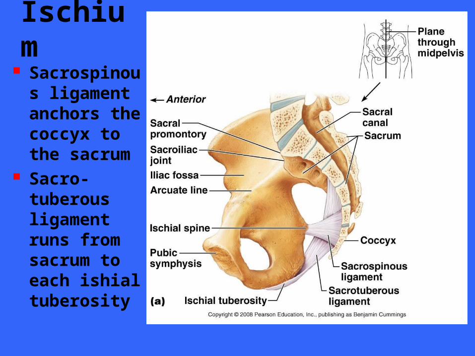
Ischium
Sacrospinous ligament anchors the coccyx to the sacrum
Sacro-tuberous ligament runs from sacrum to each ishial tuberosity

False Pelvis The false pelvis lies superior to the pelvic brim
The area is bounded by the alae of the iliac bones
Actually part of the abdomen and contains abdominal organs
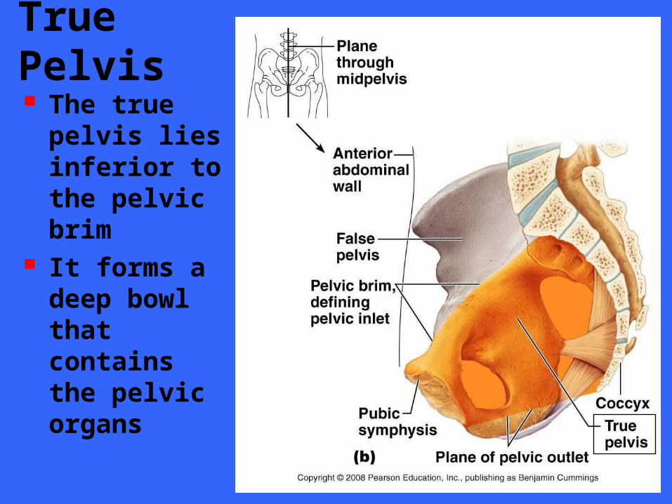
True Pelvis The true pelvis lies inferior to the pelvic brim
It forms a deep bowl that contains the pelvic organs
Pelvic Structure and Childbearing
The female pelvis reflects modifications for child bearing
It tends to be wider, shallower, lighter, and rounder than the male
Pelvic modifications accommodate the growing fetus as well as providing a birth canal wide enough to allow the infants head to exit at birth
Pelvic inlet and outlet are critical to delivery
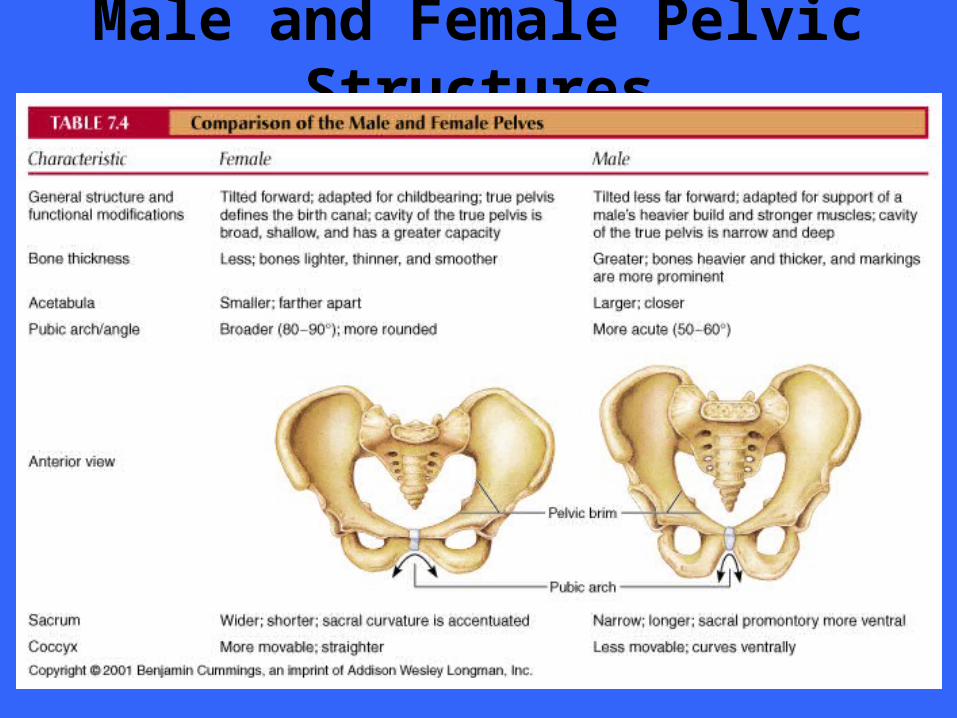
Male and Female Pelvic Structures
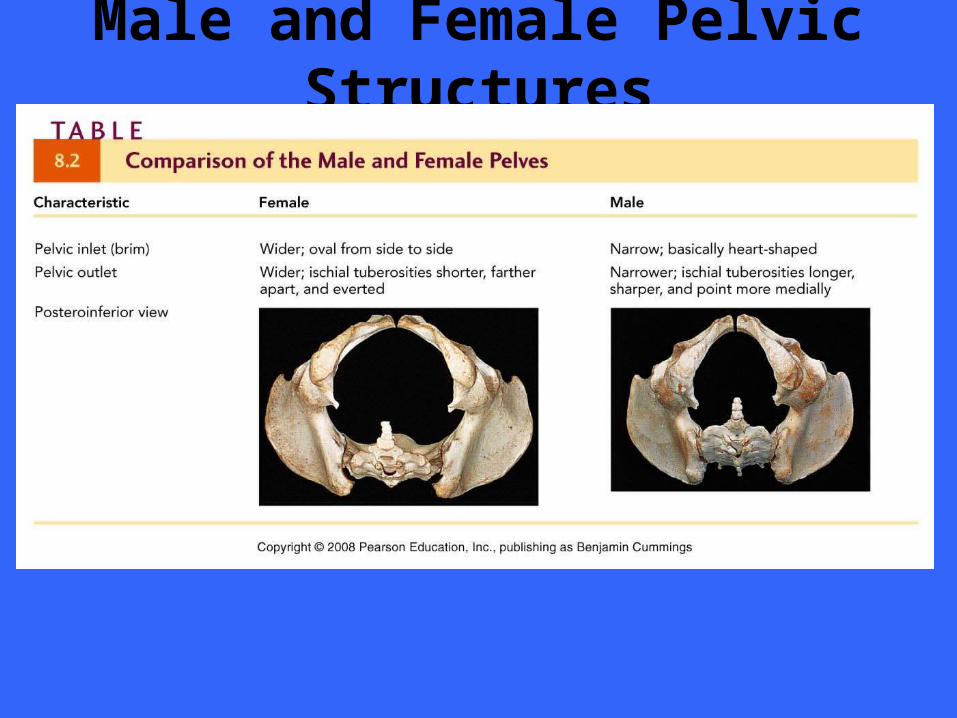
Male and Female Pelvic Structures
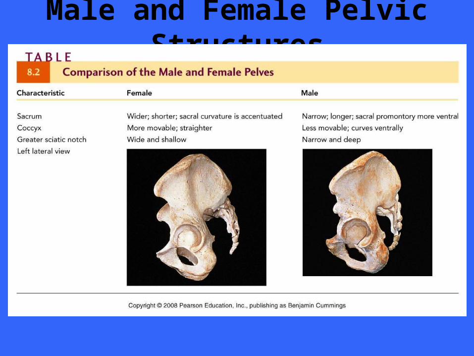
Male and Female Pelvic Structures
THE LOWER LIMB
SECTION VII
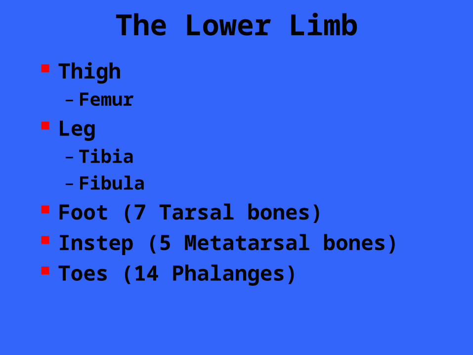
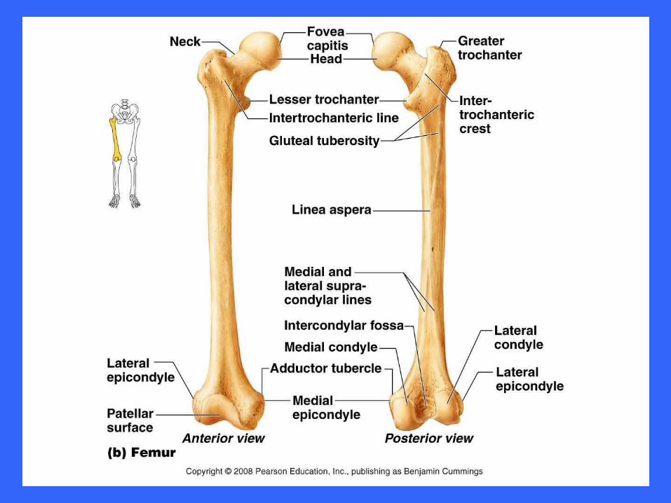
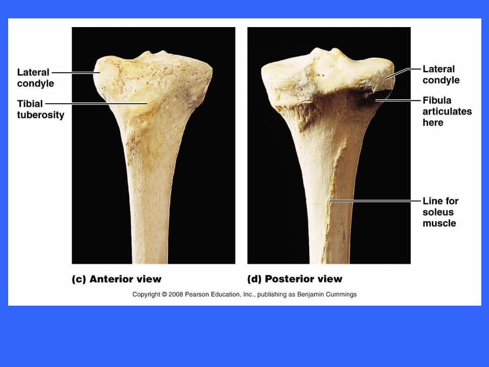
The Lower Limb Thigh
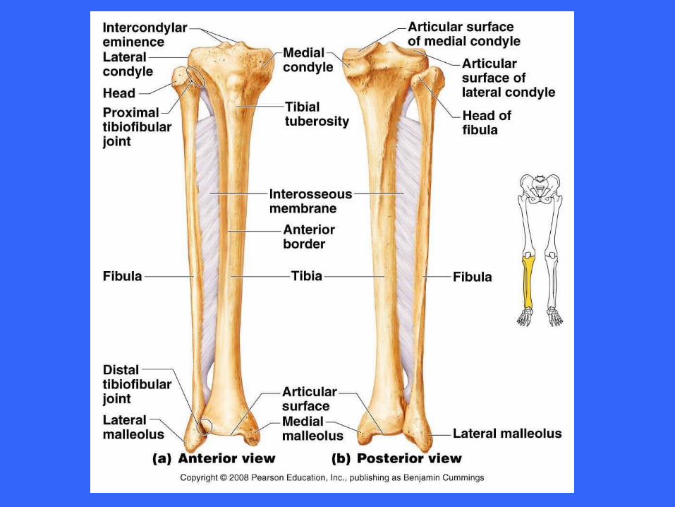
– Femur Leg
– Tibia– Fibula
Foot (7 Tarsal bones) Instep (5 Metatarsal bones) Toes (14 Phalanges)
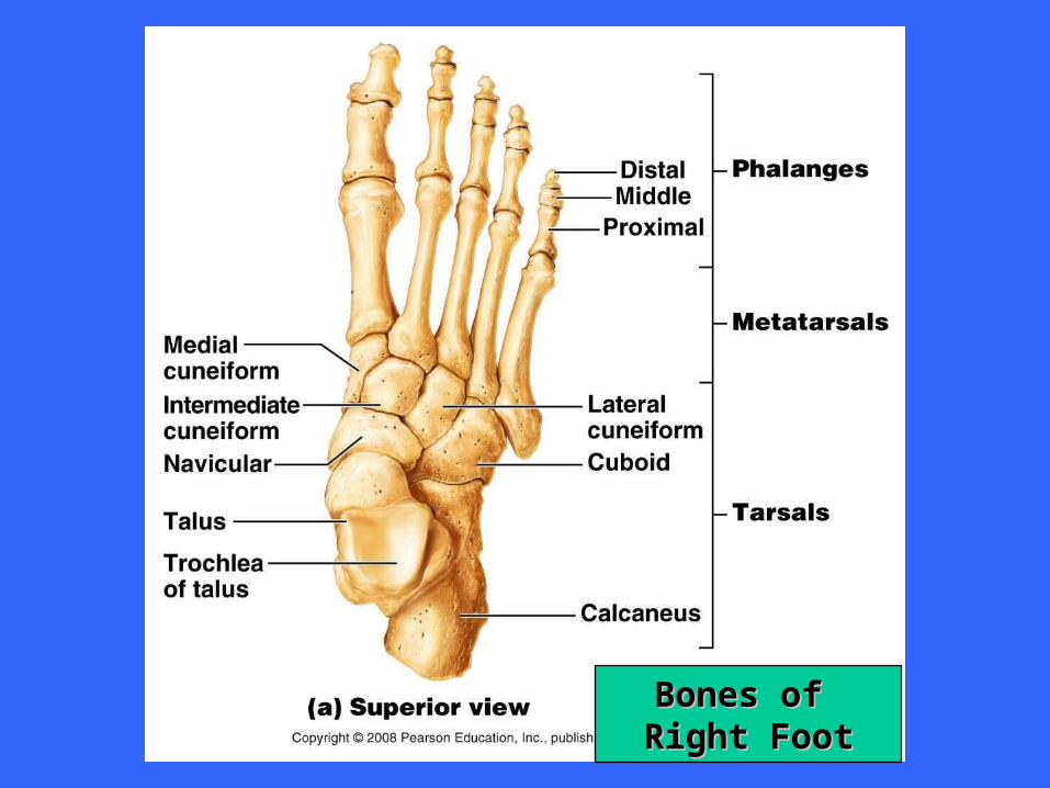
Bones of Bones of Right FootRight Foot
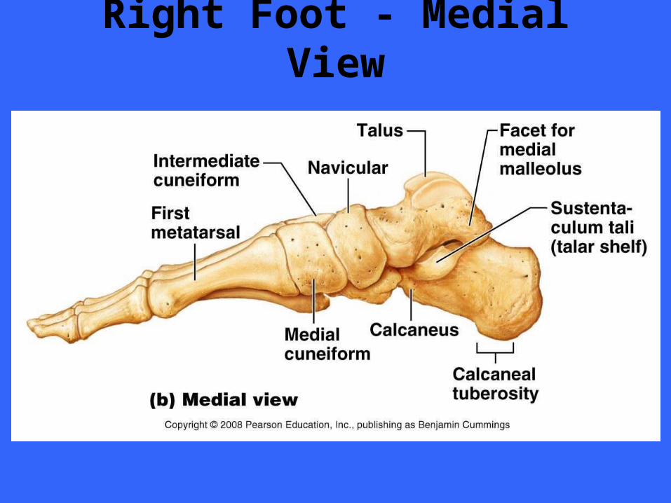
Right Foot - Medial View
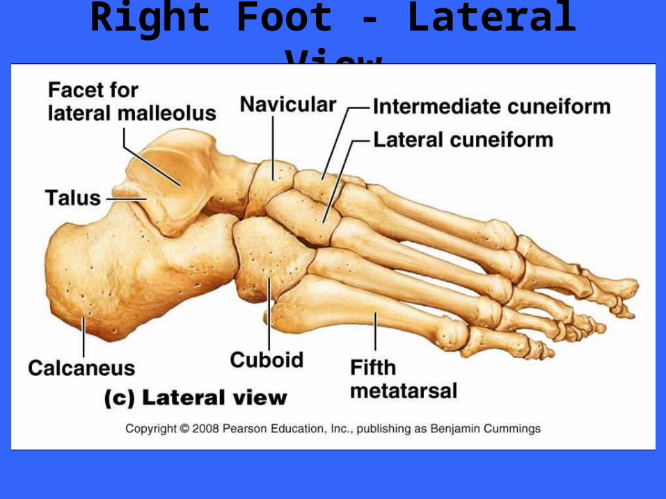
Right Foot - Lateral View
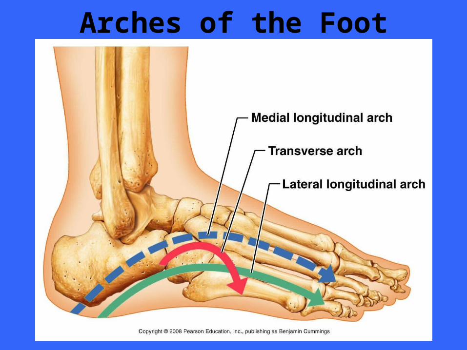
Arches of the Foot
DEVELOPMENTAL ASPECT OF THE SKELETON
SECTION VIII
The Appendicular Skeleton Throughout
Life Long Bone Ratio
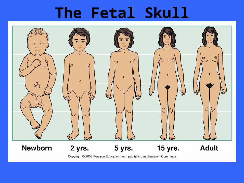
The Fetal Skull