apc by vicente kenyi saito-diaz -...
TRANSCRIPT
REGULATION OF WNT RECEPTOR ACTIVATION BY THE TUMOR SUPPRESSOR
APC
By
Vicente Kenyi Saito-Diaz
Dissertation
Submitted to the Faculty of the
Graduate School of Vanderbilt University
in partial fulfillment of the requirements
for the degree of
DOCTOR OF PHILOSOPHY
in
Cell and Developmental Biology
May, 2017
Nashville, Tennessee
Approved:
William Tansey, Ph.D.
Andrea Page-McCaw, Ph.D.
Jin Chen, M.D., Ph.D.
Scott Hiebert, Ph.D.
Ethan Lee, M.D., Ph.D.

ii
Copyright © 2017 by Vicente Kenyi Saito-Diaz All Rights Reserved

iii
For my mother.
Y para Mollie, por su apoyo incondicional.

iv
ACKNOWLEDGEMENTS
The first person I need to thank is my mentor, Dr. Ethan Lee. His patience,
guidance, and support has made this work possible. I will always be amazed by his
passion for science and his ability to think outside the box. Despite the inherent difficulties
of the scientific endeavors, he is always excited about his work. I hope I can follow his
example and share the excitement of science wherever I go. If I can call myself a scientist
today, it is mainly because of him.
Also, I would like to thank my committee members, Bill Tansey, Jin Chen,
Andrea Page-McCaw, and Scott Hiebert. Your guidance and insight helped me realize of
my true potential. It is thanks to your support and patience that I am writing these words
today. I want to give special thanks to my chairman, Bill. I am very thankful for your wise
words and your confidence in me.
I would like to thank my lab mates, former and current. They always helped
me, gave me words of advice, and kept me sane throughout this years. Specially, I want
to thank Dr. Curtis Thorne. Although we overlapped for a short period of time, he
introduced me to the awesomeness of the Lee lab and gave me one of the best pieces of
advice I received during graduate school. I would like to thank Leah and Aja who played
pivotal roles in this work. I would also want to thank to Dr. Laura Lee and her lab. For
many years, they were the voice of reason and provided the very needed outside
perspective which helped my project at many levels.
There is a person who played a fundamental role in my life, Dr. Abel Alcazar-
Roman. He selected me to participate in a little experiment called Research Experience

v
for Peruvian Undergraduates. His decision changed my life. I am honored to have met
such a generous person. His energy and desire to change our home country, Peru,
rubbed on me and allowed me to grow and do things I would never knew I could. I consider
him a friend and a brother.
I would like to thank Dr. Jose Luis Aguilar, Anita Arce, Pilar García, and Ivan
Lozada. They were my first mentors and guided my first steps as a scientist. I was lucky
to have worked with such an amazing group. Also, I would like to thank Dr. Daniela
Drummond-Barbosa. She had faith that a Peruvian undergrad with no experience working
in developmental biology would be an asset to her lab. I worked in her lab for one summer
and the experience showed me a whole new world. It was an incredible experience.
I would specially like to thank my parents and my sister whose unconditional
support have been invaluable throughout my life. My mother, who like water turning a
rock into a sculpture, very patiently shaped me into the person I am today. Although, she
is not with me anymore, she is still the driving force that pushes me to become a better
person every day. I also want to thank my sister, who supported me during many difficult
times of my life, and believes that I will win a Nobel Prize someday.
And of course, I want to thank my beautiful wife, Mollie. Every day with you is
a fun new adventure and I am thrilled to share many more with you. You take me to places
I don’t even dream about. Without you, I would have never run two half-marathons or
adopted our puppy, Nelson. You make me a better person. Thank you.
vi
TABLE OF CONTENTS
Page
DEDICATION .................................................................................................................. iii
ACKNOWLEDGEMENTS ............................................................................................... iv
LIST OF FIGURES .......................................................................................................... ix
LIST OF ABBREVIATIONS ............................................................................................ xi
Chapter
I. INTRODUCTION TO THE WNT SIGNALING PATHWAY .......................................... 1
Introduction ................................................................................................................. 1 The cell: the basic unit of life ....................................................................................... 1 History of the Wnt pathway ......................................................................................... 2 The current model of the Wnt pathway ....................................................................... 5 Surface receptor activation.......................................................................................... 6
Inhibition of the -catenin destruction complex ......................................................... 18 Activation of a Wnt-specific nuclear transcriptional complex ..................................... 28 The scaffold protein APC .......................................................................................... 32 The interaction between cells and their extracellular environment ............................ 35 Regulation of the Wnt pathway by endocytosis ......................................................... 43 II. MATERIALS AND METHODS .................................................................................. 47 Cell lines.................................................................................................................... 47 Drosophila RNAi lines, Gal4 driver, and progenitor cell reporter ............................... 47 Transfections ............................................................................................................. 48 Immunoblots and immunoprecipitation ...................................................................... 48 Generation of the mAb7E5 antibody ......................................................................... 49 Generation of RKO APCKO ........................................................................................ 49 Generation of stable cell lines ................................................................................... 50 Sucrose density gradients ......................................................................................... 50 Endocytosis assays ................................................................................................... 51 Microscopy ................................................................................................................ 51 Live cell imaging ........................................................................................................ 52 Line scan analysis ..................................................................................................... 53 Drosophila RNAi experiments ................................................................................... 53
vii
Drosophila immunohistochemistry ............................................................................ 54 Auxin-dependent degradation assay ......................................................................... 55 Reporter assay .......................................................................................................... 55 RNA isolation and qRT-PCR ..................................................................................... 56 Statistical analysis ..................................................................................................... 56 Colocalization analysis .............................................................................................. 57 III.INHIBITION OF THE WNT PATHWAY BY A MONOCLONAL ANTIBODY AGAINST THE CO-RECEPTOR LRP6 .......................................................................................... 58 Introduction ............................................................................................................... 58 Results ...................................................................................................................... 59 Development of an antibody that inhibits LRP6 ......................................................... 59 mAb7E5 inhibits ligand-mediated Wnt activation ...................................................... 60 mAb7E5 binds specifically to LRP6 ........................................................................... 61 mAb7E5 inhibits Wnt signaling in breast cancer cells ............................................... 69 Discussion ................................................................................................................. 72 IV. APC INHIBITS LIGAND-INDEPENDENT WNT SIGNALING BY THE CLATHRIN ENDOCYTIC PATHWAY .............................................................................................. 75 Introduction ............................................................................................................... 75 Results ...................................................................................................................... 76
LRP6 is required for Wnt pathway activation in APC mutant cells but not in cells with
mutant -catenin ....................................................................................................... 76 Requirement for surface receptor activation but not Wnt ligands for pathway activation in APC mutant cells ................................................................................... 77 LRP6 and Dishevelled are required for the aberrantly increased proliferation of Apc1 mutant adult intestinal stem cells in Drosophila ......................................................... 91
Rapid accumulation of phospho-LRP6 and -catenin stabilization occur upon immediate loss of APC .............................................................................................. 96
Endocytosis is required for Wnt signaling upon loss of APC ................................... 100 Wnt receptor activation in APC-deficient cells occurs via a clathrin-dependent mechanism. ............................................................................................................. 107
Discussion ............................................................................................................... 114 V. CONCLUSIONS AND FUTURE DIRECTIONS .................................................... 118 Biochemical characterization of mAb7E5 ................................................................ 118 Regulation of endocytosis by APC .......................................................................... 119 Targeting LRP6 in TNBC and CRC ......................................................................... 123 Concluding remarks ................................................................................................ 125

viii
BIBLIOGRAPHY ......................................................................................................... 126
ix
LIST OF FIGURES
Figure Page
1.1. Schematic of the canonical Wnt signaling pathway .................................................. 8 1.2. Synthesis and export of Wnt ligand ........................................................................ 11 1.3. Schematics of major endocytic pathways in the cell ............................................... 38 3.1. LRP6 domains selected as antigen for antibody development ............................... 62 3.2. The monoclonal antibody mAb7E5 is an inhibitor of the Wnt pathway ................... 64 3.3. mAb7E5 inhibits ligand mediated Wnt activation .................................................... 65 3.4. mAb7E5 binds to LRP6-2 and LRP6-3 domains in vitro ......................................... 67 3.5. mAb7E5 inhibits Wnt activation at the level of the co-receptor LRP6 ..................... 68
3.6. mAb7E5 downregulates -catenin levels in MDA-MB-231 cells ............................. 70
3.7. Minimum mAb7E5 concentration required to inhibit -catenin accumulation in MDA-MB-231 cells ................................................................................................................. 71 4.1. mAb7E5 inhibits Wnt signaling in APC-mutant CRC cells ...................................... 78 4.2. mAb7E5 blocks Wnt activation in APC-deficient cells ............................................ 79 4.3. LRP6 is required for Wnt signaling in APC-deficient cells ...................................... 81 4.4. Loss of APC promotes ligand-independent signalosome formation ....................... 83 4.5. Wnt receptor activation in APC-deficient cells is Wnt ligand independent. ............. 86 4.6. PORCN is not required for Wnt activation in the absence of APC ......................... 88 4.7. LRP6 is required for Wnt activation upon loss of APC in PORCN null cells ........... 90 4.8. Both ISC overproliferation and epithelial cell polarity defects in Drosophila Apc1 null mutant midguts are rescued by Arrow depletion. .......................................................... 92 4.9. Arrow and Dishevelled are required for overproliferation of midgut stem cells resulting from depletion of Apc1 in Drosophila. ............................................................. 94

x
4.10. LRP6 activation and -catenin accumulation occur rapidly upon loss of APC function. ......................................................................................................................... 97
4.11. Endocytosis inhibition induced by temperature shift prevents -catenin accumulation and LRP6 phosphorylation. ................................................................... 101
4.12. Endocytosis inhibition induced by temperature shift prevents -catenin accumulation in APC-mutant CRC cells. ..................................................................... 103 4.13. Dynamin is required for Wnt signaling in the absence of functional APC ........... 104 4.14. LRP6 is internalized in APC-deficient cells ......................................................... 105 4.15. Ligand-dependent and -independent Wnt activation requires distinct endocytic pathways. .................................................................................................................... 108 4.16. Clathrin-mediated endocytosis is required for Wnt activation upon loss of APC 110 4.17. APC forms a complex with clathrin ..................................................................... 112 4.18. Proposed model for APC functioning as a negative regulator of clathrin-mediated endocytosis of Wnt pathway membrane components ................................................. 115
xi
LIST OF ABBREVIATIONS
AID, Auxin-inducible degron AP-1, Adaptor protein 1 APC, Adenomatous Polyposis Coli Arr, arrow
P-E, -propellers and Epidermal Growth Factor-like domain Cav1, Caveolin-1 Cav2, Caveolin-2 Cav3, Caveolin-3 CCP, clathrin-coated pits CCV, clathrin-coated vesicles CK1, Casein Kinase 1 CME, clathrin-mediated endocytosis CMV, cytomegalovirus CRC, colorectal cancer CRD, Cystein-rich domain C-terminal, Carboxy terminal D, aspartic acid DKK, Dickopff dn, dominant-negative Dvl, dsh, Dishevelled ECD, extracellular domain EGFR, Epidermal Growth Factor Receptor

xii
EM, electron microscopy ER, Endoplasmic reticulum esg, escargot Fab, fragment antigen-binding FAP, familial adenomatous polyposis Fc, fragment crystallizable FL, Full-length Fz, Frizzled GFP, green fluorescent protein GPCR, G-protein coupled receptor gRNA, guide RNA Gro, Groucho GSK3, Glycogen Synthase Kinase 3 HA, hemagglutinin HEK, Human Embryonic kidney ICD, Intracellular domain IgG, Immunoglobulin G IgM, Immunoglobulin M int-1, integration-1 ISC, Intestinal stem cell KO, knock out LDL, low-density lipoprotein LRP5/6, low-density lipoprotein receptor related 5 or 6
xiii
mAb, monoclonal antibody MAP, mitogen-activated protein MBP, Maltose Binding Protein MEF, Mouse embryonic fibroblast MVB, multivesicular bodies MVE, multivesicular endosomes NDLB, non-denaturing lysis buffer NES, nuclear export signal NLS, nuclear localization signal N-terminal, Amino terminal P, Proline PIP2, phosphatidyl inositol-4,5-bisphosphate PIP3, phosphatidyl inositol-3,4,5-trisphosphate PORCN, Porcupine Pygo, Pygopus qRT-PCR, Quantitative Reverse trancriptase polymerase chain reaction RNAi, RNA interference RPE, Retinal Pigment Epitheilium SCF, Skp1-Cullin-F-box SDS-PAGE, Sodium Dodecyl Sulfate - Polyacrylamide Gel Electrophoresis Ser/S, Serine SHC, SH2 domain-containing transforming protein shRNA, short-hairpin RNA

xiv
siRNA, short interference RNA STF, Super TOPflash TCF, T-cell factor
TGF, Transforming Growth Factor Thr/T, Threonine TKO, triple knock out TLE, Transducing-like enhancer TLR4, Toll-like receptor 4 TNBC, Triple negative breast cancer ts, temperature sensitive W, tryptophan Wg, wingless WT, wild-type Y, tyrosine YFP, yellow fluorescent protein
1
CHAPTER I
INTRODUCTION TO THE WNT SIGNALING PATHWAY
Introduction
The canonical Wnt signaling pathway is an evolutionary conserved pathway
required for proper development of all metazoans. Wnt signaling plays a fundamental
role in the determination of cell fate, proliferation, polarity, and cell death during
embryonic development, as well as in tissue homeostasis in adults. Misregulation of the
Wnt pathway leads to disease including osteoporosis and multiple types of cancer
(Saito-Diaz et al. 2012). Understanding the mechanism of Wnt signal transduction is
critical for developing new therapeutic approaches. In this chapter, I establish the
background to understand my cell biology and biochemical studies of the Wnt pathway
as outlined in Chapters II, III and IV. I conclude my work with future directions (Chapter
V) based on the results presented in Chapters III and IV.
The cell: the basic unit of life
Signaling transduction is the process in which cells detect changes in their
environment and orchestrate their intracellular response (Gerhart 1999; Alberts et al.
2002; Gomperts et al. 2009). Signaling transduction pathways can be characterized as
a cascade of events triggered by binding of a ligand to a “receptor” (often embedded in
the plasma membrane). When the appropriate stimulus (e.g. chemically encoded
information) is detected and binds the receptor, a series of biochemical reactions occur
that, ultimately, result in a wide range of events that include changes in cell metabolism,

2
cell motility, regulation of gene transcription, or even initiation of cell death (Gomperts et
al. 2009). Signals sent to and from other cells activate signaling transduction pathways
which promote cell-cell communication and allow cells to become organized in a more
complex fashion, giving rise to tissues, organs and, ultimately multicellular organisms
(Alberts et al. 2002).
Despite the legion of metazoans and their significant diversity in morphology,
reproductive strategy, nutrient requirement, and the ecological niche they occupy, there
are only 18 known signaling pathways in metazoans as distinguished by their
transduction intermediates. These signaling pathways are required during early
development, late development, and in differentiated cells in adults (Gerhart 1999;
Meng et al. 2016). Nearly all of these pathways contribute to human disease when their
normal functions are disrupted. Because of this, it is necessary to understand how
signaling pathways work and how they interact with each other (cross-talk) during
animal developmental in order to develop better treatment in cases where they are
misregulated in human disease.
History of the Wnt pathway
The Wnt pathway, named for its ligands, the Wnt family of secreted
glycoproteins, was discovered more than 30 years ago, and the historical events that
led to the discovery and naming of Wnt ligands highlight its importance in development
and in human disease. In 1976, Sharma and Chopra described a Drosophila
melanogaster mutant that exhibited reduced or absent wings and halteres (Sharma and
3
Chopra 1976). Based on the mutant phenotype, they named this locus wingless (wg)
and suggested that it played an important role in development. A few years later, Nusse
and Varmus conducted a forward genetic screen to identify genes in mice that could
lead to tumorigenesis (Nusse et al. 1984). Using mouse mammary tumor virus (MMTV)
insertion sites, they identified a locus termed int-1, short for integration 1, which induced
mouse mammary tumors. Comparative genomic studies revealed that wg and int-1
were homologs, and the names were merged into the mnemonic Wnt (Nusse et al.
1991). Overexpression of int-1 in Xenopus embryos induced the formation of an ectopic
axis, demonstrating that it not only acts as an oncogene but also plays a critical role in
early axis specification (McMahon and Moon 1989). These studies collectively drew an
implicit connection between the physiological role for Wnts in development and a
potential pathophysiological role in carcinogenesis.
Forward genetic studies in Drosophila have been crucial in identifying Wnt
pathway components. In 1980, Eric Wieschaus and Christiane Nusslein-Volhard
identified a series of Drosophila mutants that controlled patterning of the early embryo
(Nüsslein-Volhard and Wieschaus 1980). This work was a watershed moment in
developmental biology, for which they were awarded a Nobel Prize in 1995. The 15-
year period after their initial publication produced a number of genetic and molecular
studies that elucidated the role of these mutants within various signaling pathways and
resulted in the discovery of key members of the Wnt pathway, including armadillo (the
vertebrate homolog of β-catenin), disheveled (Dvl/Dsh), shaggy (the vertebrate version
of glycogen synthase kinase 3 or GSK3), frizzled, and arrow (Riggleman et al. 1989;
4
Riggleman et al. 1990; Siegfried et al. 1992; Klingensmith et al. 1994; Bhanot et al.
1996; Wehrli et al. 2000).
The activation of the Wnt signaling pathway on the future dorsal side of the early
Xenopus embryo is a critical event in the formation of the Spemann organizer, a tissue-
organizing center found in vertebrates (Spemann and Mangold 1938). The role of Wnt
in organizer formation was uncovered when mRNA of Wnt-1 and Xwnt8 was injected
into Xenopus blastomeres. Ectopic activation of Wnt signaling on the future ventral side
of the embryo was shown to induce a second organizer that coordinates the formation
of a complete secondary body axis (Smith and Harland 1991; Sokol et al. 1991).
Embryonic axis duplication was also found to be induced by overexpression of positive
downstream components of the pathway (i.e. Dvl and β-catenin) or by inhibiting
negative components of the pathway (i.e. inhibiting GSK3 activity or overexpressing
dominant-negative Axin) (Dominguez et al. 1995; Guger and Gumbiner 1995; Fagotto
1999).
Numerous genetic and environmental perturbations of the Wnt pathway can lead
to a variety of human diseases, ranging from birth defects to cancers (MacDonald et al.
2009). One well-established connection between the Wnt pathway and human disease
is a genetic lesion that occurs early in the onset of colon cancer. In 1991, a germline
mutation in the Wnt pathway component adenomatous polyposis coli (APC) was
identified in patients with familial adenomatous polyposis (FAP), a form of hereditary
cancer (Kinzler et al. 1991; Nishisho et al. 1991). FAP patients inherit one defective
allele of APC, and upon stochastic loss of the second allele develop colon adenomas
(polyps) at an early age. These benign polyps frequently acquire other mutations and

5
develop into invasive colon carcinomas. Later studies showed that loss of both APC
alleles occurs in the large majority (>80%) of nonhereditary, sporadic colorectal cancers
as well (Kinzler and Vogelstein 1996). Following this work, inappropriate activation of
Wnt signaling was subsequently found in other cancers, including liver cancer, skin
cancer, lung cancer, Wilms’ tumor, prostate cancer, and breast cancer. A variety of
developmental genetic defects were also shown to occur as a result of Wnt pathway
misregulation, including defects in limb formation (tetra-amelia), bone ossification, eye
vascularization, and tooth development (Gong et al. 2001; Boyden et al. 2002; Niemann
et al. 2004; Xu et al. 2004). Understanding the basis of the numerous human diseases
resulting from misregulation of Wnt signaling and designing therapies for their treatment
obviously require a detailed understanding of the molecular mechanism of the Wnt
pathway.
The current model of the Wnt pathway
Wnt signals can direct a wide variety of cellular responses in development,
physiology, and disease. Originally, it was thought that a variety of cellular responses to
Wnt signaling were mediated by the different transcriptional targets modulated in
different cellular contexts. This original model, in which Wnt signaling alters
transcription, is referred to as “canonical” Wnt signaling. It is now widely accepted that
Wnt signaling can also activate distinct pathways that do not involve the nucleus or
transcription, but rather signals cytoplasmic changes involving the actin cytoskeleton
and intracellular calcium stores. These non-transcriptional Wnt pathways are loosely
known as “noncanonical” Wnt signaling. My work focuses exclusively on the “canonical”

6
Wnt signaling, so I will only discuss this pathway. For clarity, I will use the term Wnt/β-
catenin signaling to identify what has been commonly referred to as “canonical”
signaling. This nomenclature specifies the ligand (Wnt) and the essential downstream
transcriptional effector (β-catenin).
Wnt/β-catenin signal transduction, at its simplest, is a pathway that results in the
cytoplasmic protein β-catenin entering the nucleus to modulate transcription. When the
pathway is not activated, β-catenin is subject to a “futile cycle” of continual synthesis
and destruction by the β-catenin destruction complex, comprised of the scaffold proteins
Axin and APC and the kinases GSK3 and casein kinase 1 (CK1) (Figure 1.1). Wnt
signaling removes APC from the complex and relocalizes the other components to the
plasma membrane via the adaptor Dvl, thus stabilizing β-catenin which enters the
nucleus to mediate transcription (Figure 1.1). Thus, Wnt/β-catenin signaling can be
divided into three general molecular events: (1) surface receptor activation, (2) inhibition
of the β-catenin destruction complex, and (3) activation of a Wnt-specific nuclear
transcriptional complex. The next sections of this chapter consider each of these steps
more closely.
Surface receptor activation
Wnt ligands
Wnt proteins are cysteine-rich morphogens of ~350–400 amino acids that can act
in short- and long-range signaling. There are 19 vertebrate Wnts, and all appear to be
able to activate the pathway. The crystal structure of Xenopus Wnt8 (XWnt8) bound to

7
the cysteine-rich domain (CRD) of the mouse Frizzled (Fz) 8 receptor has been solved
to 3.25 Å. The Wnt8 structure is bilobular with an N-terminal helical domain and a C-
terminal extended β-hairpin stabilized by extensive disulfide bonds. Interactions
between XWnt8 and Fz8 occur via extensions from each lobe (“thumb” and “finger”) to
grasp the CRD of Fz8 on two distinct sites (Janda et al. 2012). All Wnts contain an N-
terminal signal peptide for secretion and are N-linked glycosylated (Smolich et al. 1993;
Willert et al. 2003; Takada et al. 2006). N-glycosylation of Wg (Drosophila Wnt
homolog) has been shown to be stimulated by lipid modifications (Tanaka et al. 2002).
Although an early study suggests that glycosylation is dispensable for Wnt secretion
and activity (Mason et al. 1992), more recent studies demonstrate that mutating the
glycosylation sites on Wnts blocks their secretion (Komekado et al. 2007; Kurayoshi et
al. 2007).
Wnts contain several polar amino acids and undergo a series of lipid
modifications that affect their activity and secretion (Bradley and Brown 1990). Wnts
have been shown to undergo acylation at Cys77 and Ser209 with palmitate and
palmitoleate, respectively (Willert et al. 2003; Takada et al. 2006). Interestingly, the co-
crystal structure of XWnt8–Fz8 CRD indicates that Cys77 is engaged in disulfide
bonding, whereas Ser209 is acylated (likely palmitoleic acid). The palmitoleic acid lipid
group was shown to dock within a hydrophobic groove on the CRD of Fz8 and, thus,
plays a direct role in Wnt–Fz interaction (Janda et al. 2012).

8
Figure 1.1. Schematic of the canonical Wnt signaling pathway
Left panel: In the absence of a Wnt ligand, -catenin associates with the -catenin
destruction complex composed by APC, Axin, GSK3, and CK1. Within this complex, -
catenin is sequentially phosphorylated by CK1 and GSK3. Phosphorylated -catenin is
recognized by the E3 ligase -TRCP and targeted for proteasome-mediated
degradation.
Right panel: Binding of a Wnt ligand to the co-receptors Fz and LRP6, promotes LRP6
phosphorylation by GSK3. Axin is recruited to the plasma membrane by Dvl and binds
phospho-LRP6 which results in the disassembly of the -catenin destruction complex. In
consequence, -catenin accumulates in the cytoplasm and translocates to the nucleus
where it binds to TCF to activate the transcription of Wnt target genes.

9
The endoplasmic reticulum (ER)-embedded, multi-pass transmembrane O-
acetyltransferase protein Porcupine (PORCN) is the enzyme that mediates lipid
modifications of Wnt (Port and Basler 2010). PORCN was initially identified in
Drosophila as a segment polarity gene and was the first gene shown to be required in
Wnt-secreting cells (van den Heuvel et al. 1993). Loss of PORCN function causes Wnts
to accumulate in the ER (van den Heuvel et al. 1993; Kadowaki et al. 1996), whereas
PORCN over-expression results in a larger fraction of Wnts that are modified by lipids
(Galli et al. 2007). PORCN is directly responsible for Ser209 palmitoylation of Wnts,
however, it is not known whether other acetyltransferases are involved (Gao and
Hannoush 2014). Modified Wnts are translocated from the ER to the Golgi apparatus in
a process mediated by the p24 protein family (Buechling et al. 2011; Port et al. 2011).
Once in the trans-Golgi network, the seven-pass transmembrane protein Wntless (Wls)
is thought to provide further transport of Wnts to the plasma membrane for release
outside the cell (Port and Basler 2010) . Consistent with the necessary role of Wls in
Wnt signaling, loss of Wls resembles a Wnt loss-of-function phenotype (Bänziger et al.
2006). Wls has been shown to bind Wnts at the conserved palmitoylated Ser209,
explaining the accumulation of Wnts in the ER of PORCN mutants (Herr and Basler
2012). Wls is recycled from the plasma membrane via a multiprotein complex called the
retromer. The retromer is responsible for routing Wls into a retrograde pathway that
transports transmembrane proteins from endosomes back to the trans-Golgi network
(Coudreuse et al. 2006; Port and Basler 2010). In the absence of the retromer complex,
Wls is trapped in endosomes and subsequently degraded (Yang et al. 2008). This
requirement of the retromer for Wnt signaling can be bypassed by providing additional
10
Wls (Franch-Marro et al. 2008; Port et al. 2008), further confirming the role of the
retromer in Wnt secretion via its regulation of Wls (Figure 1.2).
Wnt extracellular transport
Several mechanisms have been proposed for how Wnt ligands traverse the
extracellular space to bind their target cells. It is possible that these mechanisms are
tissue specific and influenced by the extracellular environment in which the cell resides
(Port and Basler 2010). The mechanisms underlying the graded distribution of
extracellular Wnt/Wg ligands have been best delineated in Drosophila wing discs, where
a gradient of Wg protein patterns the boundary between the developing dorsal and
ventral wing surfaces. These studies suggest a restricted diffusion model. In this model,
Wg diffuses across cells extracellularly while interacting with receptors and cell-surface
heparan sulfate proteoglycans (HSPGs), which act generally as positive regulators of
Wg signaling (Strigini and Cohen 2000; Baeg et al. 2004; Han et al. 2005; Yan and Lin
2009). HSPGs are comprised of a protein core decorated with long glycosaminoglycan
(GAG) chains (Häcker et al. 2005). The importance of GAG chains of HSPGs has been
demonstrated by studies showing that when enzymes involved in heparan sulfate
synthesis are mutated, extracellular Wg does not accumulate and Wg signaling is
reduced (Binari et al. 1997; Häcker et al. 1997; Haerry et al. 1997; Lin and Perrimon
1999; Lin 2004). The HSPGs most studied as modulators of morphogen activity are
glypicans, which are anchored to the cell surface by a glycosylphosphatidylinositol (GPI)
11
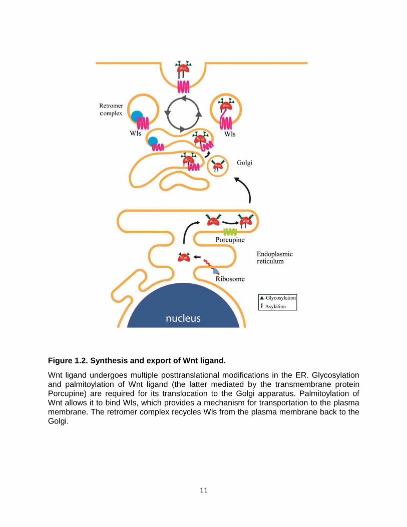
Figure 1.2. Synthesis and export of Wnt ligand.
Wnt ligand undergoes multiple posttranslational modifications in the ER. Glycosylation and palmitoylation of Wnt ligand (the latter mediated by the transmembrane protein Porcupine) are required for its translocation to the Golgi apparatus. Palmitoylation of Wnt allows it to bind Wls, which provides a mechanism for transportation to the plasma membrane. The retromer complex recycles Wls from the plasma membrane back to the Golgi.
12
anchor. There are two glypicans in Drosophila, known as division abnormally delayed
(Dally) and Dally-like protein (Dlp), and both bind Wg in cell culture (Franch-Marro et al.
2005). Dlp overexpression in wing discs leads to extracellular Wg accumulation,
whereas Dally has little effect (Franch-Marro et al. 2005; Han et al. 2005), suggesting
that Dlp binds Wg with higher affinity than Dally. Genetic studies suggest that Dally
mainly functions as a co-receptor to present Wg to the Fz2 receptor (Lin and Perrimon
1999).
In contrast to Dally, Dlp has a more complex influence on Wg signaling. Dlp
inhibits Wg signaling close to the Wg source (short-range Wg signaling) but promotes
the range of Wg signaling distant from the source (long-range Wg signaling) (Kirkpatrick
et al. 2004; Kreuger et al. 2004; Franch-Marro et al. 2005); this biphasic activity of Dlp
serves to reduce the morphogen gradient. Various models have been proposed to
explain Dlp’s biphasic activity. An early model focused on proteolytic cleavage of Dlp by
Notum, an α/β-hydrolase expressed at the Wg source, capable of shedding Dlp at its
GPI anchor and releasing it from the cell surface. One possibility to account for Dlp
biphasic activity is that Notum cleavage converts Dlp into a short-range Wg antagonist
(Kreuger et al. 2004). However, in the wing, neither the ectopic expression nor the loss
of Notum alters Dlp levels, suggesting that the biphasic activity of Dlp is independent of
Notum (Han et al. 2005; Gallet et al. 2008). A more recent model proposes that Dlp
mediates transcytosis of Wg from the apical cell surface to the basolateral surface,
where it is spread to the next distal cell, so that Dlp effectively siphons Wg away from
regions of high Wg expression toward distal regions (Gallet et al. 2008). Another model,
based on wing-disc and cell culture studies, suggests that competition between Dlp and
13
Fz2 for binding Wg at the cell surface is responsible for the biphasic activity of Dlp (Yan
et al. 2009), such that when Wg concentration is high (in short-range signaling), Dlp
sequesters Wg from Fz2, but when Wg concentration is lower (in long-range signaling),
Dlp concentrates Wg in the vicinity of Fz2 to promote signaling.
The Fz receptor family
The soluble Wnt ligands bind to members of the Fz (Fz) family of seven
transmembrane domain receptors, which have structural similarities to G-protein-
coupled receptors (GPCRs). Biochemical evidence indicates that Wnts bind to the CRD
of the Fz receptor, and that the affinity of Wnt for Fz is in the low nanomolar range
(Bhanot et al. 1996; Hsieh et al. 1999).
The topological similarities of Fz to GPCRs have led to the suggestion that
heterotrimeric G proteins may be required for Wnt signal transduction, and several
studies propose a link between G proteins and Wnt pathway activation. Genetic studies
in Drosophila suggest that Gαo transduces signaling from Fz, and that Gαo interacts
with the scaffold protein Axin to promote its localization to the plasma membrane
(Katanaev et al. 2005; Egger-Adam and Katanaev 2009). In cultured mammalian cells,
depletion of Gαo or Gαq has been shown to inhibit Wnt signaling, possibly via disruption
of GSK3β–Axin complexes (Liu et al. 2005). More direct evidence for a role of G
proteins in Wnt pathway activation comes from reconstitution studies indicating that
Gαo, Gαi2, Gαq, and Gβγ have the capacity to inhibit both β-catenin phosphorylation by
GSK3 and β-catenin turnover in Xenopus egg extract. In the case of the latter, it was
14
proposed that Gβγ promotes the recruitment of GSK3 to the plasma membrane to
enhance low-density lipoprotein receptor-related protein 6 (LRP6) phosphorylation and
activation (Jernigan et al. 2010). Whether heterotrimeric G proteins are bona fide
mediators of Wnt ligand-mediated signaling, or whether other pathways act through
them to modulate Wnt signaling, remains unclear.
The co-receptor LRP5/6
LRP5 and LRP6 are functionally redundant single-pass transmembrane
receptors that act as co-receptors for Wnt ligands (Pinson et al. 2000; Tamai et al.
2000; Wehrli et al. 2000). In Drosophila, there is only one family member, Arrow. In
some assays, LRP6 is more potent than LRP5. There are thought to be no qualitative
differences in their mechanism of action in mediating Wnt pathway activation, although
they likely play different roles during development (He et al. 2004; Mi and Johnson
2005). Biochemical studies of LRP6 indicate that different Wnts may bind to different
extracellular domains of the LRP5/6 protein (Bourhis et al. 2010). Specifically, the LRP6
extracellular domain contains four repeating sequences of β-propeller and epidermal
growth factor-like (βP–E) domains. The crystal structures of the extracellular LRP6
regions indicate that the βP–E repeats represent two discrete, compact, rigid structures,
each containing two βP–E pairs. Wnt9b binds the first two βP–E repeats on the
extracellular domain of LRP6, whereas Wnt3a binds the last two βP–E domains (Ahn et
al. 2011; Chen et al. 2011; Cheng et al. 2011).
15
Binding of Wnt ligands to Fz and LRP5/6 results in the production of
phosphatidylinositol (4,5)-bisphosphate (PIP2) (Pan et al. 2008). Increased PIP2
induces oligomerization and clustering of LRP5/6. Although hydrodynamic studies
suggest that Fz and LRP6 oligomerize and form clusters of “signalosomes” upon Wnt
signaling, the in vivo, physiological significance of such events in Wnt pathway
activation remains to be determined (Cong et al. 2004; Bilić et al. 2007).
Increased PIP2 also induces recruitment of Axin to LRP5/6. This recruitment may
be due, in part, to the action of Amer1/WTX (APC membrane recruitment 1 or Wilms
tumor gene on the X chromosome), a tumor suppressor mutated in Wilms’ tumor that
binds to Axin, CK1γ, and GSK3. Amer1/WTX is recruited to the plasma membrane in a
PIP2-dependent manner (Major et al. 2007; Tanneberger et al. 2011).
The interaction between LRP6 and Axin is critical for activation of the Wnt
pathway, and the recruitment of Axin and the associated destruction complex to the
plasma membrane upon Wnt ligand binding initiates a chain of events that leads to the
phosphorylation of the intracellular domain of LRP5/6. This initial recruitment of Axin to
LRP6 in a Wnt–Fz-dependent manner is referred to as the “initiation step” of Wnt
pathway activation (Baig-Lewis et al. 2007).
The LRP5/6 receptor contains five PPPSPxS motifs on its intracellular domain
that are required for signal transmission. Each of these five motifs alone can activate
the Wnt/β-catenin pathway: when transferred to heterologous receptors, the PPPSPxS
motif is sufficient for pathway activation (Tamai et al. 2004; Zeng et al. 2005). Mutational
analyses of these motifs indicate that they act in a cooperative manner to mediate
downstream signaling (MacDonald et al. 2008; Wolf et al. 2008).
16
The predominant kinases involved in PPPSPxS phosphorylation have been
identified as GSK3 and CK1 (Davidson et al. 2005; Zeng et al. 2005). Wnt binding to
LRP5/6 has been shown to induce PPPSP phosphorylation by GSK3, and this event
primes LRP6 for subsequent xS phosphorylation by CK1 (Zeng et al. 2005; Pan et al.
2008). Another study, however, suggests that CK1 phosphorylates conserved S/T
clusters outside the PPPSPxS motif, and this phosphorylation primes phosphorylation of
LRP6 by GSK3 (Davidson et al. 2005). Phosphorylated LRP6 has a high affinity for Axin
and promotes further recruitment of cytoplasmic Axin-bound GSK3 complexes to the
cell surface (Mao et al. 2001; Zeng et al. 2008). The recruitment of additional Axin-
bound GSK3 complexes further promotes the phosphorylation of additional LRP5/6
PPPSP motifs in a positive feedback mechanism and has been referred to as the
“amplification step” in Wnt pathway activation (Baig-Lewis et al. 2007). Once the Axin-
bound β-catenin destruction complex is recruited by LRP6, the phosphorylated
cytoplasmic domain of LRP6 is capable of directly inhibiting GSK3 activity, blocking β-
catenin phosphorylation and subsequent ubiquitin-mediated proteasomal degradation
(Cselenyi et al. 2008; Piao et al. 2008). The capacity of phosphorylated LRP6 to limit
GSK3 activity by direct inhibition appears to limit the capacity of Axin-bound GSK3 to
promote further LRP6 phosphorylation (amplification step). It is possible that these two
events are temporally regulated such that direct inhibition of GSK3 may be blocked
during the amplification step.

17
The cytoplasmic adaptor Dvl
Dishevelled (Dvl/Dsh) has long been known to be required genetically in the
Wnt/β-catenin pathway (Klingensmith et al. 1996). In vertebrates, there are three Dsh
isoforms encoded by distinct genes (Dvl1–3) (Sussman et al. 1994; Semënov and
Snyder 1997). Upon Wnt–receptor interaction, Dsh is phosphorylated and recruited to
the cytoplasmic side of the receptor complex (Yanagawa et al. 1995; Semënov and
Snyder 1997; Rothbächer et al. 2000). Several studies suggest that physical interaction
between the Fz receptor and Dvl is important for transduction of the Wnt signal. Dvl
contains three major domains: the DEP, the PDZ, and the DIX domains. Biochemical
and structural studies have implicated both the PDZ and the DIX domains of Dsh in
binding to the Fz receptor (Wong et al. 2000; Wong et al. 2003; Tauriello et al. 2012).
Dvl phosphorylation upon Wnt signaling appears to be independent of LRP6 activation
(González-Sancho et al. 2004). Dvl and Axin share DIX domains that can polymerize
and are required for receptor clustering (Schwarz-Romond et al. 2007). Loss-of-function
studies show that Dvl acts upstream of LRP6 (Tolwinski et al. 2003). Consistent with
this observation, Dsh has been shown to bind and activate PI4KIIa and PIP5KI to
promote the synthesis of PIP2, which is required to promote oligomerization and
clustering of LRP5/6 (Pan et al. 2008). In overexpression studies in Drosophila and
Xenopus egg extracts, however, Dsh has been shown to activate β-catenin signaling
independently of Arrow/LRP6 (Salic et al. 2000). The precise involvement of Dsh in
receptor activation and Axin recruitment remains to be determined.
Both the stability and the activity of Dsh appear to be regulated by ubiquitylation.
Three ubiquitin ligases, NEDL1 and ITCH of the HECT-type ligase and KLHL12 of the
18
Cullin3-type ligase, have been implicated in ubiquitylating Dvl to promote its degradation
(Miyazaki et al. 2004; Angers et al. 2006; Wei et al. 2012). One study implicates the
Naked2 protein as a necessary co-factor for Dvl ubiquitylation (Hu et al. 2010). Finally,
the deubiquitinating enzyme, CYLD (encoded by the familial cylindromatosis tumor
suppressor gene), has been shown to be a negative regulator of Wnt signaling
(Tauriello et al. 2010). CYLD was shown to remove a regulatory Lys63-linked ubiquitin
from Dvl. Thus, ubiquitylation of Dvl via Lys63 linkages appears to be necessary for
efficient activation of signaling by Dvl. The ubiquitin ligase that mediates Lys63-linked
ubiquitylation of Dvl, however, is still unknown.
Inhibition of the β-catenin destruction complex
The β-catenin destruction complex is a macromolecular machine that efficiently
acts to phosphorylate β-catenin, targeting it for degradation. We will first describe the
players involved in the formation of the β-catenin destruction complex (Figure 1.1) and
follow with our current understanding of the behavior of the pathway upon receptor
activation.
The transcriptional regulator β-catenin
β-Catenin is the primary effector of Wnt signaling. In the absence of signaling,
the destruction complex targets β-catenin for ubiquitin-mediated proteasome
degradation by SCFβ-TRCP, a member of the Skp1-Cullin-F-box (SCF) E3 ubiquitin ligase
complex. In the presence of signaling, β-catenin is spared destruction and translocates
19
from the cytoplasm to the nucleus to activate signaling. β-Catenin was first identified in
Drosophila as the segment polarity gene armadillo and also as a component of the
adherens junction in Xenopus (Nüsslein-Volhard and Wieschaus 1980; McCrea et al.
1991). The structure of β-catenin consists of a central core of 12 helical 42 amino acid
armadillo repeats that form a superhelical structure (Huber et al. 1997). Analysis of full-
length β-catenin protein indicates that the N- and C-terminal domains are unstructured
and form dynamic interactions with the armadillo repeats of the protein (Xing et al.
2008). Notably, the armadillo repeats form a positively charged groove that mediates
the interaction of β-catenin with other components of the Wnt pathway [e.g. APC, Axin,
and T-cell factor (TCF)/LEF] as well as with E-cadherin (Huber et al. 1997; Graham et
al. 2000; Xing et al. 2003; Xing et al. 2004).
The cellular factors that coordinate whether nascent β-catenin mediates Wnt
target gene transcription or plays a structural role in maintaining integrity of the
adherens junction are not completely understood. A large number of studies using a
variety of model systems have shown that overexpression of cadherins is sufficient to
inhibit Wnt target gene transcription and to promote relocalization of β-catenin to the
membrane (Heasman et al. 1994; Sadot et al. 1998; Gottardi et al. 2001; Stockinger et
al. 2001). Furthermore, evidence to support potential influence of cadherins on Wnt
signaling comes from studies demonstrating that proteolytic cleavage of cadherins by
proteases such as ADAM10 and presenilin-1/γ-secretase is sufficient to release bound
β-catenin, to increase soluble cytoplasmic β-catenin, and to activate Wnt target gene
transcription (Marambaud et al. 2002; Maretzky et al. 2005; Reiss et al. 2005). Multiple
studies in which E-cadherin is knocked down in cells with wild-type Wnt pathway
20
components, however, fail to demonstrate activation of Wnt signaling, suggesting a lack
of a significant interaction between cadherin-mediated cell adhesion and Wnt signaling,
compensatory regulation of β-catenin levels with E-cadherin, or that turnover of β-
catenin by the degradation complex in the wild-type situation is capable of
compensating for the increased flux of β-catenin (Kuphal and Behrens 2006; Herzig et
al. 2007). Support for the existence of distinct pools of β-catenin comes from a study
demonstrating that β-catenin can exist as a monomeric or dimeric form bound to α-
catenin (Gottardi and Gumbiner 2004). Biochemical studies indicate that the monomeric
form preferentially participates in Wnt signaling, whereas the dimeric form preferentially
binds cadherins. Surprisingly, little is known about the mechanism of β-catenin nuclear
translocation.
The scaffold protein Axin, the limiting component of the destruction complex
The scaffold protein Axin is a critical component of the β-catenin destruction
complex, acting as a limiting negative regulator of Wnt/β-catenin signaling. It was first
identified as the gene product of the locus fused in mice (Zeng et al. 1997). Axin plays a
role as a scaffold protein that directly binds to many of the other components of the
destruction complex and brings them within close proximity to each other (Figure 1.1).
The sites of interactions have been visualized in co-crystal structures of Axin and APC
proteins (Spink et al. 2000), Axin and β-catenin (Xing et al. 2003), and Axin and GSK3β
(Dajani et al. 2003). Studies of Axin in fly embryos suggest that Axin complexes may
form oligomers in vivo, and that Axin may also act as a cytoplasmic anchor to restrict
armadillo/β-catenin import into the nucleus (Tolwinski and Wieschaus 2001; Peterson-
21
Nedry et al. 2008). Axin was initially found to be present at low concentrations and is the
limiting component of the β-catenin degradation complex in Xenopus (Lee et al. 2003).
A long-standing puzzle about Wnt signaling is how it maintains specificity because
many Wnt components also play biological roles in other cellular processes; for
example, GSK3 is a node for many types of cell signaling (Forde and Dale 2007). The
low concentration of Axin has been proposed to isolate the Wnt pathway from affecting
other intracellular pathways (Lee et al. 2003). The low level of intracellular Axin is due,
in part, to its ubiquitin-mediated turnover that is promoted by LRP5/6 (Yamamoto et al.
1999; Cselenyi et al. 2008). Degradation of Axin has been shown to be regulated by
GSK3 phosphorylation, which inhibits its rate of degradation (Yamamoto et al. 1999). In
addition, the turnover of Axin requires the tumor suppressor APC, and studies in
Xenopus egg extract, as well as in flies, suggest that this may represent a mechanism
to compensate for fluctuations in levels of APC in order to maintain low levels of β-
catenin in cells (Lee et al. 2003).
Smad ubiquitin regulatory factor 2 (Smurf2) has been shown to be an E3
ubiquitin ligase that targets Axin for degradation (Kim and Jho 2010). Due to its key role
in Wnt signaling, it is likely that Axin is tightly regulated. Tankyrase has been shown to
promote poly ADP-ribosylation (PARsylation) and ubiquitylation and further degradation
of Axin through the addition of polyADP-ribose moieties onto proteins through
PARsylation (Huang et al. 2009a). The importance of Axin turnover is demonstrated by
the identification of tankyrase inhibitors IWR-1 and XAV939 that have been shown to
potently inhibit Wnt signaling by increasing the steady-state level of Axin (Chen et al.
2009; Huang et al. 2009). These tankyrase inhibitors prevent PARsylation of Axin and

22
thus reduce Axin turnover. RNF146 has been identified as the polyADP-ribose-directed
E3 ubiquitin ligase that ubiquitylates Axin (Callow et al. 2011; Zhang et al. 2011).
RNF146 binds directly to the covalently linked poly(ADP-ribose), targeting Axin for
degradation and maintaining low steady-state levels of Axin. A deubiquitinating enzyme,
ubiquitin-specific protease (USP) 34, has been identified to catalyze the deubiquitylation
of Axin and increase steady-state levels of Axin in cells (Lui et al. 2011). In contrast to
PARsylation, SUMOylation of Axin at its C-terminus has been shown to confer stability
by inhibiting Axin ubiquitylation (Kim et al. 2008). Recently, quantitative measurements
of Axin concentration in a variety of mammalian cells suggest that its levels vary
significantly to alter the dynamics of Wnt signaling (Tan et al. 2012). Thus, the
regulation of Axin levels and stability may be a major mechanism by which cells control
the response to Wnt signals.
The kinase GSK3
GSK3 is a ubiquitous serine/threonine protein kinase involved in numerous
cellular processes (Forde and Dale 2007). Antagonizing GSK3 activity is central to all
models of Wnt signaling mechanisms. The homolog of GSK3 in Drosophila is shaggy,
aka zeste white 3 (Siegfried et al. 1992). In mammals, there are two distinct genes, α
and β, that are likely to have redundant functions in the Wnt pathway (Doble et al.
2007). GSK3 was first identified for its role in the regulation of glucose metabolism,
targeting muscle glycogen synthase (Embi et al. 1980). GSK3 has both positive and
negative roles in Wnt signal transduction, which will be described in further detail later in
this review. GSK3 often recognizes substrates that have been previously

23
phosphorylated (primed), and thus GSK3 is often found to act in concert with other
kinases. β-Catenin phosphorylation by GSK3 (at Ser33, Ser37, and Thr41) leads to its
ubiquitin-mediated degradation (Peifer et al. 1994; Yost et al. 1996). The crystal
structure of GSKβ has been solved and conforms to a typical protein kinase bilobed
structure topology consisting of an amino-terminal β-sheet domain linked to a carboxy-
terminal α-helical domain. Similar to other activated kinases, the structure of
unphosphorylated GSK3β shows an activation loop that is responsible for its unique
priming mechanism (Haar et al. 2001). Furthermore, phosphorylation of Ser9, which has
been shown to inhibit GSK3 activity, is predicted to act in an auto-inhibitory fashion by
blocking access to the catalytic site (Dajani et al. 2001). In addition to β-catenin, other
major Wnt pathway substrates of GSK3 include APC, Axin, and LRP6 (Rubinfeld et al.
1996; Willert et al. 1999; Zeng et al. 2005).
The kinase CK1α
The CK1 family of kinases is comprised of a group of serine/threonine kinases
encoded by seven distinct genes in mammals (α, β, γ1, γ2, γ3, δ, and ε; although β was
identified in bovine and has not been found in humans) (Knippschild et al. 2005). As
with GSK3, CK1 is a widely-expressed family of kinases with a large number of
substrates. All CK1 members have highly similar catalytic domains, but differ
significantly in both the length and the sequence of their C-terminal non-catalytic
domains. CK1α, with its short (~24 amino acid) C-terminal domain, appears to be an
outlier compared with the other family members, which have much longer C-terminal
tails (~200 amino acids). CK1α, γ, δ, and ε have been implicated in positively regulating
24
the Wnt pathway by phosphorylating Dsh, LRP5, TCF/LEF, and Axin (Peters et al.
1999; Kishida et al. 2001; Cong et al. 2004; Zeng et al. 2005). In contrast, CK1 family
members have also been implicated as negative regulators of the Wnt pathway by
phosphorylating β-catenin, APC, Axin, and TCF/LEF (Kishida et al. 2001; Gao et al.
2002; Hämmerlein et al. 2005). CK1α has been proposed to be the in vivo priming
kinase for GSK3 and phosphorylates β-catenin at Ser45 (Liu et al. 2002). The activation
of CK1α has emerged as a potential therapeutic drug target, and a recent study
reported that the antihelminthic drug, pyrvinium, inhibits Wnt signaling by activating
CK1α to enhance β-catenin phosphorylation and degradation (Thorne et al. 2010).
β-catenin degradation cycle
The ubiquitous expression of β-catenin and other pathway members suggests
that all metazoan cells express the β-catenin destruction complex. Although commonly
described as having cytoplasmic localization, the core components can also be found in
the nucleus where the β-catenin destruction complex is likely to reside and function
(Cong and Varmus 2004; Wiechens et al. 2004; Sierra et al. 2006). This machine is in a
constitutively active state and contains a number of enzymes that target β-catenin for
degradation. Thus, in the absence of a Wnt signal, β-catenin is caught in a cycle of
synthesis followed by rapid destruction. Axin is the scaffold protein that nucleates the
formation of the β-catenin destruction complex. It binds with high affinity to the two
kinases GSK3 and CK1α. A well-defined α-helix in the central portion of Axin anchors
GSK3, whereas the site of CK1 binding has been mapped to a more C-terminal region
of Axin (Dajani et al. 2003; Sobrado et al. 2005). APC binds the RGS domain of Axin,
25
an N-terminal region that has structural homology to domains found in regulators of G-
protein signaling (Spink et al. 2000). β-Catenin enters the complex by binding both 15
amino acid repeats of APC and a single α-helix on Axin on the C-terminal side of the
GSK3 binding site. The exact order and kinetics of binding are unknown, and it is
unclear whether this binding is ordered or stochastic (Lee et al. 2003).
Evidence suggests that phosphorylation of Axin, likely by GSK3, increases its
affinity for β-catenin (Willert et al. 1999). Upon binding to Axin, the N-terminal region of
β-catenin becomes positioned for rapid phosphorylation by CK1 at serine 45. This
creates a priming site for subsequent and successive phosphorylation of β-catenin by
GSK3 at Thre41, Ser37, and Ser33 (Amit et al. 2002; Liu et al. 2002). Twenty amino
acid repeats on APC are also phosphorylated by CK1 and GSK3 (Ha et al. 2004). This
phosphorylation increases the affinity of the 20 amino acid repeats for β-catenin by 140-
fold and competes β-catenin off of the α-helix binding site on Axin. Based on these
observations, it has been proposed that APC phosphorylation triggers β-catenin
removal from Axin, allowing a new β-catenin species to enter the destruction complex
(Kimelman and Xu 2006). The action of APC is also thought to prevent the action of the
phosphatase, PP2A, from acting on the phosphorylated β-catenin (Su et al. 2008). Once
β-catenin is phosphorylated at Ser33 and Ser37, a destruction consensus sequence is
recognized by β-TRCP, a specificity subunit of the SCF ubiquitin ligase (Jiang and
Struhl 1998; Kitagawa et al. 1999; Liu et al. 1999). Binding of SCFβ-TRCP to β-catenin
catalyzes its polyubiquitylation (via K48 linkages) and subsequent degradation through
the proteasome. These events ensure that newly synthesized, free cytosolic levels of β-
catenin are kept below the threshold necessary for gene regulation.
26
Pathway behavior on activation
Elevation of β-catenin levels in response to the presence of Wnt is a hallmark of
the Wnt/β-catenin pathway. The precise mechanism of destruction complex inhibition is
under intense investigation and a number of proposed mechanisms exist. At the core of
all the current models is the inhibition of GSK3 anti-catenin activity. This has been
proposed to arise through several distinct mechanisms: (1) dissociation of components
of the β-catenin destruction complex, (2) phosphorylation of GSK3 at Ser9, which
inhibits its activity, (3) LRP6 binding and direct inhibition of GSK3 activity against β-
catenin, and (4) Axin degradation upon activation of signaling, which prevents formation
of the destruction complex necessary for GSK3 to phosphorylate β-catenin. We discuss
these models individually below.
Several studies have described Axin–GSK3 dissociation upon signaling (Liu et al.
2002; Luo et al. 2007). Early studies seem to suggest that Dvl recruits GSK3 binding
protein (GBP) to Axin where GSK3 is directly inhibited and dissociates from Axin (Yost
et al. 1998; Farr et al. 2000). One caveat to these studies is the apparent lack of
requirement for GBP in Wnt signaling in Drosophila and mice. Drosophila does not have
a GBP ortholog, and genetic knockout studies in mice show no requirement for GBP in
development or Wnt signaling (van Amerongen et al. 2005). In contrast to these studies,
other studies have shown that the complex remains intact and localizes to the Fz/LRP6
co-receptors rapidly after Wnt stimulation (Mao et al. 2001; Bilić et al. 2007).
GSK3α/β is phosphorylated at Ser9 and Ser21, and when phosphorylated at
these sites, kinase activity is greatly reduced (Sutherland et al. 1993; Cross et al. 1995).
Wnt-induced stimulation of Ser9/21 phosphorylation has been speculated about by
27
several groups (Ding et al. 2000; Yokoyama and Malbon 2007). However, studies in
mice in which non-phosphorylatable forms of GSK3α/β were knocked-in produced
animals with no developmental defects and no observable perturbation in Wnt pathway
activation (McManus et al. 2005).
A number of mammalian cell culture and biochemical studies suggest that the β-
catenin destruction complex moves to the cell surface upon Wnt stimulation, and this
migration is followed by the direct inhibition of GSK3 by LRP6 (Yamamoto et al. 2006;
Bilić et al. 2007; Cselenyi et al. 2008). The collective evidence from these studies
suggests a mechanism in which ligand-stimulated LRP6 phosphorylation by GSK3 and
CK1 creates a docking site for Axin. This phosphorylation event in turn recruits the
Axin–GSK3 complex to the receptor. Recruitment of Axin–GSK3 to phosphorylated
LRP6 has been proposed to be facilitated in part by Dsh, which binds directly to the Fz
receptor and Axin (Schwarz-Romond et al. 2007). A problem for this model, however, is
the requirement for GSK3 to phosphorylate the PPPSPxS motifs on LRP5/6 to create
docking sites for Axin–GSK3 (amplification step) and the fact that phosphorylated
PPPSPxS motifs can directly inhibit GSK3 activity, further limiting LRP6
phosphorylation. High-resolution structural data are essential to clarify this model and to
explain how PPPSPxS motifs are sufficient for both Axin docking and GSK3 inhibition.
A compendium of studies from cell culture, Xenopus embryos and egg extracts,
and Drosophila suggest that a conserved and critical event is the degradation of Axin
upon Wnt signaling (Yamamoto et al. 1999; Tolwinski et al. 2003; Cselenyi et al. 2008).
Because Axin is a concentration-limiting factor, regulation of Axin stability is likely to
have a dramatic effect on signaling (Lee et al. 2003). Genetic studies in Drosophila
28
designed to elucidate the requirement of Axin regulation show that flies genetically null
for GSK3 have elevated levels of β-catenin as expected, yet unexpectedly they still
have near wild-type Wg-based embryonic patterning that is dependent on dynamic
levels of Axin. These results indicate that regulation of Axin stability is sufficient to
pattern the embryo, as β-catenin nuclear translocation only occurs when Axin levels are
low, and Axin may act as a cytoplasmic anchor to regulate β-catenin-mediated
transcription (Tolwinski et al. 2003). Other work has shown that Axin degradation
appears to lag behind β-catenin stabilization and is not necessary for β-catenin
stabilization (Willert et al. 1999; Hino et al. 2005; Cselenyi et al. 2008). These studies
promote the model that the block in β-catenin destruction that occurs upon Wnt
signaling does not require Axin turnover, and that degradation of Axin is a distinct event
that may modulate the character of the response.
Activation of a Wnt-specific nuclear transcriptional complex
Nuclear translocation of β-catenin
β-catenin is normally constitutively transcribed and translated. Thus, a signal-
induced block in proteolysis leads to rapid rise in cytosolic β-catenin protein levels upon
receptor activation. In addition to increased cytosolic accumulation, β-catenin also
accumulates in the nucleus. Remarkably, studies indicate that it is not the absolute
increase in concentration of β-catenin that results in activation of a Wnt transcriptional
program (Goentoro and Kirschner 2009). Rather, it is the relative fold change (~2 ×) in
the concentration of β-catenin that is recognized by the Wnt transcriptional machinery.

29
Such a mechanism is expected to minimize cell–cell variation caused by fluctuations in
the basal level of β-catenin as well as allowing for activation of the transcriptional
program when the signal is sufficiently elevated in proportion to the background noise
(Goentoro et al. 2009).
β-catenin does not contain any recognizable nuclear localization signal (NLS) or
nuclear export signal (NES). Nuclear accumulation of β-catenin has been attributed to
its cytoplasmic retention, nuclear retention, and nuclear export. Nuclear entry of β-
catenin is thought to be independent of classic import factors (e.g. RanGTPase and
importins) (Fagotto et al. 1998; Yokoya et al. 1999). The armadillo repeats of β-catenin,
however, are structurally related to importin-β HEAT repeats and may interact directly
with the nuclear pore complex during nuclear entry (Kutay et al. 1997; Malik et al. 1997).
Surprisingly, deletion of β-catenin armadillo repeats 3–6 in Drosophila, which
presumably disrupts binding to major cytoplasmic Wnt components, results in
constitutive nuclear localization of the armadillo/β-catenin mutant (Orsulic and Peifer
1996). Based on this result, it has been suggested that β-catenin is normally regulated
by its cytoplasmic retention. Consistent with this data, both cadherins and Axin have
been shown to sequester β-catenin in the plasma membrane and cytoplasm,
respectively (Heasman et al. 1994; Sadot et al. 1998; Gottardi et al. 2001; Tolwinski and
Wieschaus 2001). The nuclear proteins TCF, Pygopus, and BCL9 (described below)
have been proposed to similarly act as anchors for β-catenin in the nucleus (Townsley
et al. 2004; Krieghoff et al. 2006). It has been proposed that nuclear export of β-catenin
may play a critical role in Wnt signaling, and Axin, APC, and RanBP3 have been
implicated in regulating the export of β-catenin from the nucleus (Henderson and
30
Fagotto 2002; Cong and Varmus 2004). Finally, Rac1 GTPase and Jun N-terminal
kinase 2 (JNK2) have also been shown to promote β-catenin nuclear localization upon
Wnt signaling, although the mechanism by which this is accomplished is unclear (Wu et
al. 2008).
β-catenin-mediated gene transcription
Wnt-induced nuclear β-catenin accumulation leads to an interaction with the
TCF/LEF family of DNA-bound transcription factors that are critical for Wnt-mediated
gene regulation (Behrens et al. 1996; Molenaar et al. 1996). Invertebrates appear to
have only one TCF gene, whereas mammals have four: TCF1, LEF1, TCF3, and TCF4.
In the absence of β-catenin, TCF interacts with the co-repressor Groucho/transducin-
like enhancer (Gro/TLE1–3 in vertebrates) to repress gene transcription. TCF binds at
the DNA consensus sequence CCTTTGWW (W can be either T or A), termed the Wnt-
responsive element (WRE). It has been predicted that there are greater than 6000 high-
confidence WREs in at least one studied colorectal cancer cell line, and these WREs
collectively regulate 300–400 genes (Hatzis et al. 2008).
Several studies suggest that TCF proteins are phosphorylated to regulate Wnt
signaling. CK1 has been shown to phosphorylate TCF to positively and negatively
regulate its interaction with β-catenin (Lee et al. 2001; Hämmerlein et al. 2005). The
Nemo-like kinase (NLK) has been shown to phosphorylate TCF and inhibit Wnt
signaling by reducing the affinity of β-catenin–TCF for DNA (Ishitani et al. 2003; Smit et
al. 2004). Finally, phosphorylation of LEF-1, TCF4, and TCF3 by the homeodomain-

31
interacting protein kinase 2 has been shown to promote their dissociation from Wnt
target gene promoter (Hikasa et al. 2010; Hikasa and Sokol 2011). TCF proteins are
also modified by ubiquitylation. The NLK-associated RING finger protein promotes the
ubiquitylation of TCF/LEF, targeting it for degradation in a manner dependent on NLK
activity (Yamada et al. 2006). Similarly, the deubiquitinase USP4, which deubiquitylates
K48 and K63 ubiquitin linkages, has been shown to act on TCF4 to inhibit Wnt signaling
(Zhao et al. 2009). In the canonical model of Wnt target gene activation, displacement
of Gro/TLE by β-catenin converts TCF/LEF into a transcriptional activator. The X-linked
inhibitor of apoptosis has been shown to monoubiquitinate Gro/TLE, decreasing its
affinity for TCF/LEF and allowing for unrestricted binding of β-catenin to TCF/LEF
(Hanson et al. 2012).
Two notable nuclear co-factors found in Drosophila and vertebrates, Pygopus
(Pygo) and BCL9, have been shown to facilitate Wnt pathway-mediated transcription
(Belenkaya et al. 2002; Thompson et al. 2002). BCL9 binds and bridges β-catenin with
the Pygo protein. Pygo has been shown to bind the multiprotein transcriptional co-
activator mediator complex and contains a plant homology domain that interacts with
dimethylated histone 3 lysine 4, an indicator of transcriptional activation (Fiedler et al.
2008). Studies in Drosophila indicate that TCF, β-catenin, BCL9, and Pygo represent a
core transcriptional complex that is necessary for Wnt-mediated gene transcription. In
mammals, the situation is more complex, and there appears to be functional
redundancy and cell type-specific roles for BCL9 and Pygo (Schwab et al. 2007;
Sustmann et al. 2008).

32
It is important to note an often-underappreciated role for TCF/β-catenin signaling
in transcriptional repression. Studies have shown a number of potential mechanisms of
repression, including competition of TCF/β-catenin with transcriptional activators,
recruitment of co-repressors to WREs, or TCF binding to a novel consensus sequence
that specifically mediates repression (Jamora et al. 2003; Theisen et al. 2007;
Blauwkamp et al. 2008). In addition, there are multiple studies showing that many DNA-
binding transcription factors (e.g. Smad3, AP-1, RXR, and Kaiso) bind to β-catenin to
activate or repress Wnt/β-catenin target genes. In light of the large number of TCF
binding sites and numerous transcriptional co-regulators, it is clear that the Wnt/β-
catenin gene expression program is vast and induces dramatic changes in the
physiological state of the cell, many of which are still not well understood (Cadigan
2012). Thus, Wnt signaling leads to a transcriptional program that includes
downregulation and upregulation, rather than to simply transcriptional activation.
The scaffold protein APC
APC is a scaffold protein consisting of 2843 amino acids with a mass of
approximately 310 kDa, and it acts as a negative regulator of Wnt/β-catenin signaling.
Vogelstein and colleagues first identified the gene in 1991 as the site of a mutation
found in FAP, a familial form of colon cancer (Kinzler et al. 1991; Nishisho et al. 1991).
APC plays diverse roles in cellular functions, including Wnt signaling, migration, mitotic
spindle alignment, and apoptosis, which are likely carried out through different APC
subpopulations (Faux et al. 2008). The C-terminal third of APC contains a region
involved in microtubule binding that interacts with the proteins EB1 and Discs large (Su
33
et al. 1995; Matsumine et al. 1996). These regions have been demonstrated to be
involved in microtubule dynamics in mitosis and cell migration and are thought to be
independent of the role of APC in Wnt signaling (Näthke et al. 1996). The connection
between APC and Wnt signaling was identified in a series of studies that showed that
APC binds to β-catenin, and that mutations in APC caused elevated levels of β-catenin
in cancer cells (Rubinfeld et al. 1993; Su et al. 1993). APC binds β-catenin, GSK3, and
Axin in several regions within the central portion of the protein (Rubinfeld et al. 1996;
Ikeda et al. 1998; Fagotto 1999). Despite numerous studies published on the function of
APC, a clear mechanistic picture of the role of APC in regulating the Wnt pathway
remains elusive. Indeed, loss of APC leading to elevated β-catenin levels and activation
of the Wnt pathway can be overcome by overexpression of Axin, which is the limiting
component of the β-catenin destruction complex (Lee et al. 2003). Furthermore,
overexpression of an Axin mutant lacking its APC binding domain (RGS) is capable of
promoting β-catenin degradation and inhibiting Wnt signaling to a similar extent as
overexpression of wild-type Axin (Hart et al. 1998).
How APC acts as a negative regulator of Wnt/β-catenin signaling is something of
a mystery. Several models have been proposed, including (1) exporting β-catenin from
the nucleus (Hamada and Bienz 2004), (2) repressing Wnt target genes (Sierra et al.
2006), (3) retaining β-catenin in the cytoplasm (Tolwinski and Wieschaus 2001), (4)
targeting the β-catenin destruction complex to the cell cortex, where its E3 ubiquitin
ligase (SCFβ-TRCP) resides (Näthke et al. 1996; McCartney et al. 1999), (5) coordinating
the phosphorylation and release of β-catenin from the destruction complex to allow its
ubiquitylation (Kimelman and Xu 2006), (6) blocking dephosphorylation of β-catenin by

34
protein phosphatase 2A (PP2A) (Su et al. 2008), and (7) shielding the β-catenin
degradation complex from the inhibitory action of Dvl (Mendoza-Topaz et al. 2011).
None of the proposed models for the role of APC in the Wnt pathway are
mutually exclusive, although the most compelling experimental evidence (4–6) strongly
supports APC’s role in negatively regulating steady-state levels of cytoplasmic β-
catenin. It is likely that APC, similar to other components of the Wnt pathway (e.g.
GSK3 and CK1), may participate in multiple events in the Wnt signaling pathway.
Several kinases, including CK1, protein kinase A (PKA), and GSK3, have been
shown to phosphorylate APC (Rubinfeld et al. 1996; Morin et al. 1997). Phosphorylation
of APC by GSK3 was shown to enhance binding of β-catenin by APC (Salic et al. 2000).
In addition to phosphorylation, the APC protein is ubiquitinated. In cells, APC is
stabilized by the COP9 signalosome-associated deubiquitinase, USP15, which binds to
the β-catenin degradation complex (Huang et al. 2009). The deubiquitinase Trabid,
which removes K63-linked regulatory ubiquitylation chains from APC, has been
identified as a positive regulator of Wnt signaling (Tran et al. 2008). Although it is not
clear if APC is the sole target of Trabid, this finding is consistent with the suggestion
that K63-linked ubiquitylation of APC antagonizes Wnt signaling, presumably by
potentiating APC activity. The E3 ligase that mediates ubiquitylation of APC and the
exact mechanism by which these K63-linked ubiquitin chains regulate APC activity are
unknown.
35
The interaction between cells and their extracellular environment
The outer layer of the plasma membrane is the surface from which cells
communicate with their environment. Among the many membrane components, there
are membrane receptors which are required to activate signaling pathways in response
to environmental perturbations. In order to maintain homeostasis, cells often control the
abundance of receptors at the surface of the plasma membrane via their internalization
(thereby removing them from participation in signaling) through a process called
“endocytosis” (Doherty and McMahon 2009; Sorkin and von Zastrow 2009).
Over 100 years ago, Ilya Metchnikoff made the first observation of cell
internalization. In 1882, he reported that specific cells used vesicles to engulf and
degrade pathogens. He called this process “phagocytosis” (Tauber 2003). In 1931,
Warren Lewis observed that cells used similar vesicles to uptake fluids in a process he
described as “pinocytosis”. In the following years, Christian de Duve discovered
lysosomes and while studying their function coined the term “endocytosis” (Sabatini and
Adesnik 2013). The development of electron microscopy (EM) allowed for further
discoveries in the process of endocytosis and greatly advanced the field. In 1953,
George Palade identified “cave-like” invaginations of the cell membrane, and named
them “caveolae” (Latin for little caves). For their work, both de Duve and Palade
received the Nobel Prize in 1974 (Nassoy and Lamaze 2012). Finally, in 1975 Joseph
Goldstein, Michael Brown, and Richard Anderson showed that the low-density
lipoprotein (LDL), binds to a cell surface receptor leading to formation of “coated pits”
(Goldstein and Brown 2009). These pits are then internalized and the LDL receptor is
endocytosed. Goldstein and Brown coined this process “receptor-mediated endocytosis”

36
and were awarded the Nobel Prize in 1985. During the same year, Barbara Pearse
isolated and identified the major component of those pits as clathrin, giving rise to the
term “clathrin-mediated endocytosis” (CME) (Robinson 2015).
Clathrin mediated endocytosis
Most of the early work performed to understand the process of endocytosis
focused on CME. The main player involved in CME is the clathrin protein. Clathrin is a
trimer composed of three 190kDa heavy chains bound to a 25kDa light chain which,
together assemble a cage-like structure and form invaginations on the cytoplasmic side
of the plasma membrane called clathrin-coated pits (CCP) (Figure 1.3) (Kirchhausen
2000; Takei and Haucke 2001; Schütze et al. 2008). Several adaptor proteins such as
AP-1 through AP-5, ARH, Dab2, Fe65, AP180, Epsin, and Snx17 interact with clathrin
and their specific cargo proteins (Traub and Bonifacino 2013). Of these clathrin-
associated proteins, the best studied are the AP family of adaptors. AP-2 is the major
AP family member required for trafficking at the plasma membrane and plays a pivotal
role in clathrin-mediated endocytosis. AP-2 links clathrin to the membrane and
coordinates the assembly of the clathrin coat. The AP-2 complex comprises two large
subunits, and 2, and two smaller subunits, 2 and 2 (Takei and Haucke 2001). The
subunit targets the AP-2 complex to the membrane by binding to the phospholipids,
phosphatidyl inositol-4,5-bisphosphate (PIP2) and phosphatidyl inositol-3,4,5-
trisphosphate (PIP3) (Chang et al. 1993; Gaidarov et al. 1996; Gaidarov and Keen
1999). The 2 and 2 subunits recognize specific motifs on the cytoplasmic domain of

37
transmembrane receptors and provide cargo specificity to the CCPs (Ohno et al. 1995;
Nesterov et al. 1999).
In CME, AP-2 associates with PIP2 at the plasma membrane and recruits clathrin
molecules resulting in the formation of clathrin-coated buds. These clathrin-coated buds
eventually form clathrin-coated vesicles (CCV), which are irreversibly released into the
cytoplasm by a large GTPase called Dynamin. Inhibition of dynamin function by small
molecules or a kinase-dead form of Dynamin prevents release of CCV and blocks
endocytosis (Damke et al. 1994; Doherty and McMahon 2009; Kirchhausen et al. 2014).
Clathrin is released from vesicles by Auxilin and Hsc70, and naked vesicles are
transported (along with their cargo) to their designated cellular compartment (Doherty
and McMahon 2009). Internalization of a variety of receptors are regulated by CME and
include G-protein coupled receptors (GPCRs), the transferrin receptor, and the LDL
receptor. Thus, CME plays a role in regulating homeostasis of a diverse number of
receptors.
Caveolin-mediated endocytosis
Among the many clathrin-independent endocytic pathways, endocytosis by caveolae is
the best characterized. Caveolae are pits 60-80 nm in diameter in the plasma
membrane that are enriched for caveolins, sphingolipids, and cholesterol (Figure 1.3).
Three caveolin isoforms are expressed in mammals, and expression of each one is cell
type dependent. Caveolin-1 (Cav1, the most common isoform) and caveolin-2 (Cav2)
are expressed in non-muscle cells, whereas caveolin-3 (Cav3) is present only in muscle

38
Dynamin
Clathrin
Caveolin
Lipid raft
Clathrin-dependent
endocytosis
Caveolin-dependent
endocytosis
CCV
Caveosome
Early Endosome
Late Endosome Lysosome
Figure 1.3. Schematics of major endocytic pathways in the cell.
39
cells (Nassoy and Lamaze 2012). Cav1 and Cav3 are required for caveolae formation
(Drab et al. 2001; Galbiati et al. 2001). The requirement of Cav2 in caveolae formation
is not clear because loss of Cav2 function has no observable effect in vivo (Razani et al.
2002; Sowa et al. 2003). All caveolin proteins encode cytoplasmic N- and C-terminal
domains connected by an intramembrane hairpin. Caveolin palmitoylation at Cys133,
Cys145, and Cys156 facilitate their binding to the plasma membrane (Dietzen et al.
1995; Parton and Simons 2007). Caveolins oligomerize and interact with cholesterol-
rich domains in the plasma membrane known as lipid rafts (Murata et al. 1995).
Interaction with lipid rafts is critical for caveolae formation, and it has been shown that
cholesterol depletion increases Cav1 motility and disrupts the function of caveolae
(Rothberg et al. 1992).
Once caveolae are released from the plasma membrane by the GTPase,
Dynamin (Henley et al. 1998), they can either fuse with the early endosome in a
process mediated by Rab5, or fuse with a large non-endosomal organelle (caveosome)
in a Rab5-independent manner. The caveolar vesicles are then recycled back to the
membrane for reuse (Razani et al. 2002; Pelkmans et al. 2004). Two early studies
identified a large number of proteins involved in signal transduction within caveolae
(Sargiacomo et al. 1993; Lisanti et al. 1994). This observation, led to formation of the
“caveolae/raft hypothesis”, that proposed that localization of signaling molecules in a
compartment could facilitate cross-talk between signaling pathways. Although the
molecules found in caveolae do not exhibit a common feature that specifically targets
them to caveolae, they all undergo lipid modification. Indeed, many signal transduction
proteins found in caveolae (e.g., the Epidermal Growth Factor Receptor (EGFR), H-

40
Ras, Src family of tyrosine kinases, and subunit of heterotrimeric G-proteins) have
one or more myristoyl, palmitoyl, or prenyl groups (Platta and Stenmark 2011).
Endocytosis and signaling
Although receptor internalization is often thought of being associated with a
decrease in sensitivity to stimulus by controlling the number of receptors available at the
plasma membrane, it also promotes signal propagation within the cell. Upon their
activation, receptors typically undergo post-translational modifications, which promote
recruitment of proteins required for signaling and/or endocytosis (Sorkin and von
Zastrow 2009). Internalized receptors are subsequently sorted to specific compartments
depending on the requirement of the cell. For example, if decreased signaling is
needed, receptors are sorted to late endosomes and lysosomes for degradation. If
further signaling is required, receptors are shuttled back to the plasma membrane
(Platta and Stenmark 2011).
The prevailing model for receptor endocytosis proposes that receptors of
essentially all major signaling pathways undergo CME. On other hand, certain receptors
(e.g., some GPCRs, Transforming Growth Factor (TGF) receptors, Wnt receptors,
and Notch) undergo clathrin-independent endocytosis such as caveolin-mediated
endocytosis (Sorkin and von Zastrow 2009). The concentration of ligands (e.g., EGF) at
the plasma membrane modulate whether the signaling is regulated by clathrin-or
caveolin-mediated endocytosis (Platta and Stenmark 2011). Because both clathrin- and
41
caveolin-mediated endocytosis have a direct impact on the qualitative behavior of the
output, preference for one or the other may depend on the type of response required.
Upon CME, receptors are sorted to early endosomes, where they continue to
interact with downstream molecules to propagate the response in order to maintain
proper signal duration and intensity (Sigismund et al. 2008). EGFR has been the most
extensively studied and exemplifies this mode of action. Stimulation of cells with EGF,
result in rapid clustering of EGFR in CCPs and CCVs. Recruitment of downstream
effectors such as GRB2, SH2 domain-containing transforming protein (SHC), Ras, and
MAP kinase, occur at the plasma membrane and maintain their interactions in early
endosomes (Vieira et al. 1996; Sorkin and von Zastrow 2009).
Some signaling complexes are exclusively sorted into early endosomes where
the signaling output is modulated. The lipid composition of the early endosomal
membrane, which is rich in phosphatidylinositol-3-phosphate (PIP3), define their
specificity (Funderburk et al. 2010). This phenomenon occurs in TGFsignaling where
the TGFreceptor associates with the PIP3-binding proteins, SARA and endofin. SARA
binds to internalized TGF-receptor and SMAD2 and facilitates the phosphorylation of
the latter. Phosphorylated SMAD2 interacts with SMAD4, and the resultant complex
translocates to the nucleus to promote gene transcription. Similarly, endofin interacts
with TGF-receptor to promote assembly of the SMAD2-SMAD4 complex (Chen et al.
2007).
Change in phospholipid composition in early endosomes can also result in the
disassembly of protein complexes. In the case of the Toll-like receptor 4 (TLR4),

42
activated TLR4 interacts with the PIP2-associated protein TIRAP and MyD88. After
translocation to the endosome, the low concentration of PIP2 results in complex
dissociation. TLR4 is subsequently free to associate with TRAM and TRIF, thereby
activating endosome-specific TLR4 signaling to promote the secretion of type I
interferons (Platta and Stenmark 2011).
Recycling of receptors from the early endosome back to the plasma membrane
can modulate the response of signaling pathways. For example, the β2-adrenergic
receptor is sorted into early endosomes that are shuttled back to the plasma membrane
following a fast recycling route mediated by Rab4-positive endosomes. Alternatively,
receptors are transported to recycling endosomes in a Rab11-dependent manner to
follow a slow recycling pathway (Puthenveedu et al. 2010). In the case of the transferrin
receptor, export to the plasma membrane can occur via bulk membrane recycling or
sequence-dependent recycling, which relies on local actin polymerization. Studies
indicate that the β2-adrenergic receptor is also recycled by this mechanism (Stenmark
2009; Puthenveedu et al. 2010).
Finally, receptors transported to late endosomes or multivesicular endosomes
(MVE, also known as multivesicular bodies (MVB)) are usually targeted to lysosomes
for degradation. One well-studied example is EGFR, which is ubiquitylated and targeted
for lysosomal degradation, thereby, downregulating the EGFR signaling cascade (Wiley
et al. 1985; Renfrew and Hubbard 1991). MVEs can also promote specific signaling
events. For instance, the adaptors p14 and MP1 recruit MEK1 to MVEs in order to
propagate ERK signaling (Teis et al. 2006). It is clear that signal transduction relies on
multiple cellular mechanisms to effectively activate and regulate downstream effectors.

43
For the Wnt pathway, there is a growing body of evidence supporting the necessary role
of endocytosis in Wnt signal transduction although our knowledge of the role of
endocytosis in Wnt signaling is currently limited.
Regulation of the Wnt pathway by endocytosis
The first evidence of the role of endocytosis in Wnt signaling was the observation
that Wg (Drosophila Wnt) localizes by electron microscopy in receiving cells in small
vesicles as well as multivesicular bodies (MVBs) (van den Heuvel et al. 1989). MVBs
are the last compartment receptors are sorted into prior to lysosomal degradation.
Furthermore, shibire mutants, the Drosophila homolog of dynamin, resulted in
accumulation of Wg at the extracellular surface of the plasma membrane (Bejsovec and
Wieschaus 1995; Strigini and Cohen 2000). Consistent with these findings, the co-
receptors Arrow and Fz mediate Wg internalization in Drosophila (Rives et al. 2006).
Initial studies suggested that Wg internalization resulted in downregulation of the
Wnt pathway in Drosophila embryos (Dubois et al. 2001). Thus, blocking internalization
should enhance Wnt signaling. However, further evidence showed that inhibition of
endocytosis by loss of dynamin or Rab5 function not only affected Wg endocytosis, but
also inhibited Wg signaling (Seto and Bellen 2006). These results were confirmed in
mammalian cells where loss of dynamin function and inhibition of clathrin- or caveolin-
mediated endocytosis blocked Wnt activation in the presence of Wnt ligands (Blitzer
and Nusse 2006; Yamamoto et al. 2006). The contradicting evidence highlights the
complexity behind the regulation of membrane components of the Wnt pathway.
44
Both clathrin and caveolin have been proposed to mediate LRP6 internalization.
LRP6 interacts with clathrin as well as the adaptor protein AP-2 (as assessed by
sucrose density gradient experiments). The interaction between LRP6 and AP-2 appear
to be strengthened by Wnt ligands. Reports show that the number of clathrin clusters
around LRP6 at the cell membrane significantly increases upon Wnt3a treatment (Kim
et al. 2013). Also, Wnt3a promotes PIP2 formation leading to LRP6 phosphorylation and
AP-2 recruitment (Pan et al. 2008). Furthermore, the subunit of AP-2 has been shown
to interact with Dvl (Yu et al. 2007). These studies agree with a recently proposed
model in which LRP6 resides in nascent clathrin-coated pits, and, in the presence of
Wnt3a, the Wnt signalosome is assembled in CCV in a process mediated by AP-2
(Gammons et al. 2016).
In contrast to the clathrin-based model, another model proposes that LRP6 is
internalized by caveolae upon Wnt activation. In this model, LRP6 resides in lipid rafts
that interact with caveolin in the presence of Wnt3a, allowing for Axin recruitment and -
catenin accumulation. LRP6 subsequently recycles back to the plasma membrane via
Rab5- and Rab11-positive vesicles (Yamamoto et al. 2006). Interestingly, LRP6
phosphorylation still occurs upon dynamin inhibition (blocks LRP6 internalization) in the
presence of Wnt, and LRP6 is internalized in the absence of Wnt. In the case of the
latter, LRP6 phosphorylation required the presence of Wnt ligand. -catenin
accumulation, however, required both LRP6 phosphorylation and internalization,
suggesting that caveolin-mediated endocytosis prevent spontaneous activation of the
Wnt pathway. The same investigators showed that the Wnt antagonist, DKK1, promotes
CME of LRP6, suggesting that different endocytic pathways control Wnt signaling

45
(Yamamoto et al. 2008). Fz has also been shown to internalize upon Wnt activation.
Wnt3a induces rapid Fz5 internalization into Rab5-dependent vesicles, and it is shuttled
back to the membrane by Rab11-positive endosomes (Yamamoto et al. 2006). Similar
to LRP6, the interaction between AP2 and Dvl is essential for clathrin-mediated Fz
endocytosis and Wnt activation (Yu et al. 2007). In summary, there is compelling
evidence to support a role for both clathrin and caveolin-mediated receptor endocytosis
in Wnt signaling. It remains unclear, however, how a cell chooses to undergo Wnt
signaling by utilizing either mechanisms, how Wnt signaling may lead to distinct outputs
depending on whether it is via the clathrin or caveolin pathways, and how signaling via
the clathrin or caveolin pathways are distinctly controlled.
In the current body of work, I address two major questions in the Wnt field and
demonstrate that they are linked: the role of APC in Wnt signaling and the role of the
endocytic pathway in Wnt receptor activation. In the latter case, because endocytosis
has been shown to be absolutely required for Wnt signaling and because its
requirement is evolutionarily conserved, understanding its role in receptor activation will
be extremely important for our understanding of the basic mechanism of Wnt pathway
activation. Because mutations in the tumor suppressor, APC, occur in 80% of all cases
of sporadic CRC (accounting for 600,000 deaths annually worldwide) our understanding
of the former case has obvious important clinical implications.
In Chapter III, I describe the initial characterization of an inhibitory monoclonal
antibody (mAb7E5) developed against the extracellular domain of LRP6, and
demonstrate that mAb7E5 potently inhibited Wnt signaling. In Chapter IV, using
mAb7E5, I show that mutations in APC lead to clathrin-dependent, ligand-independent

46
activation of the Wnt pathway. Furthermore, I demonstrate that whether Wnt signaling
occurs via the clathrin or via caveolin-mediated endocytosis is dictate by the cell type of
the responding cell. Finally, in Chapter V, I discuss the biological significance of my
findings and how it has revised our current model of the Wnt pathway.
47
CHAPTER II
MATERIALS AND METHODS
Cell lines
The following cells were generous gifts: HEK293 STF (J. Nathans, Johns Hopkins
University), RPE (I. Kaverina, Vanderbilt University), MEF and MEF PORCNKO (C.
Murtaugh, University of Utah), 293FT (D. Carbone, Ohio State University), HEK293T
Dvl TKO (S. Angers, University of Toronto), and HCT116 WTKO (B. Vogelstein, Johns
Hopkins University). HEK293, RKO, SW480, DLD1, L-Wnt3a, L-cells, and MEF
Caveolin-1KO were purchased from the American Type Culture Collection. HEK293
CMV-luc was reported previously (Thorne et al. 2010). Cell lines were maintained in
DMEM except for SW480 and DLD1 cells (RPMI) and HCT116 cells (McCoy’s) with
10% FBS and 1% penicillin/streptomycin. See also table 2.1.
Drosophila RNAi lines, Gal4 driver, and progenitor cell reporter
The following Drosophila strains were used: Apc1Q8 (Ahmed et al. 1998), Apc1 RNAi
line (Vienna Drosophila Resource Center [VDRC] #51469), four Arr RNAi lines (Arri1,
VDRC#4819; Arri2, VDRC #6707; Arri4, VDRC #6708; and Arri3, Bloomington Drosophila
Stock Center (BDSC) #53342), two dsh RNAi lines (dshi1, BDSC #31306; dshi2, VDRC
#101525), UAS-dicer2, VDRC #60010, and esgts (esg-gal4 tubgal80ts UAS-GFP/CyO)
(Micchelli and Perrimon 2006; Guo et al. 2013).
48
Transfections
Plasmid transfections were performed using CaCl2, Lipofectamine 3000 (Invitrogen), or
FuGene HD (PROMEGA). siRNA transfections were performed using Dharmafect1
(HEK293 and RKO cells), Dharmafect2 (SW480, DLD1, and HCT116 cells) (both from
Dharmacon), or HiPerfect (RPE cells) (Qiagen) according to manufacturer’s protocol.
siRNAs were transfected in a pool consisting of two distinct siRNAs in all experiments
except where noted.
Immunoblots and immunoprecipitation
Whole cell lysates were obtained using non-denaturing lysis buffer (NDLB) (50 mM Tris-
Cl (pH 7.4), 300 mM NaCl, 5 mM EDTA, 1% (w/v) Triton X-100, 1mM PMSF, 1 mM
sodium orthovandate, 10 mM NaF, and phosphatase inhibitor cocktail (Roche). Soluble
fractions were prepared for immunoblotting. Cytoplasmic fractionation was performed as
previously describe (Thorne et al. 2010). Cells were incubated on ice in lysis buffer (10
mM HEPES (pH 7.8), 10 mM KCl, 2 mM MgCl2, 0.1 mM EDTA, 1mM PMSF, 1 mM
sodium orthovandate, 10 mM NaF, and phosphatase inhibitor cocktail (Roche)),
scraped, transferred to microfuge tubes, and NP-40 was added to 0.5%. Lysates were
vortexed and sheared 8 times using a 23-gauge needle. Lysates were centrifuged at
16,000Xg for 2 minutes. Supernatants (cytoplasmic fractions) were recovered. For co-
immunoprecipitations, cell lysates were prepared using NDLB. Lysates were diluted to 1
mg/ml and incubated with antibody for 1 hour in a TOMY shaker at 4°C followed by
addition of Protein G magnetic beads (New England Biolabs) for 1 hour. Beads were
49
then washed 3 times with 10X volume of NDLB. Proteins were eluted from beads with
protein sample buffer and analyzed by SDS-PAGE and immunoblotting.
Chemiluminescence signal was detected using a C-DiGit blot scanner (LI-COR).
Obtained images and band intensity were analyzed using Image Studio (LI-COR).
Generation of the mAb7E5 antibody
A protein fragment corresponding to the fragments LRP6-1, LRP6-2, and LRP6-3 of the
extracellular domain of human LRP6 was used to inoculate mice for monoclonal
antibody production. Positive clones were initially identified based on their capacity to
inhibit Wnt3a-mediated signaling in HEK293 STF cells. Clones were then screened for
their capacity to inhibit Wnt signaling in the SW480 CRC cell line.
Generation of RKO APCKO
Two APC gRNA sequences (5’-CCCCCTATGTACGCCTCCC-3’ and 5’-
CTTTGACAAACTTGACTTT-3’) were cloned into pCAS-Guide-eGFP (Origene) using
the restriction enzymes BamHI and BsmBI. APC gRNA oligos were designed with a 5’-
GATCG overhang and an extra guanidine nucleotide on the 3’-end of the sequence
(Sigma). Oligonucleotides were annealed as per manufacturer’s protocol using a 4-step
PCR procedure. Digested pCAS-Guide-eGFP vector was ligated to the annealed oligos
using T4 ligase. Successful clones were sequence-verified. Plasmids encoding the two
APC gRNA sequences were transfected into RKO cells at 80% confluence using
Fugene HD. Transfected cells were sorted by flow cytometry (Vanderbilt Flow

50
Cytometry Core) as single cells into a 96-well plate. Single colonies were grown to
confluence and split into 24-well plates. Clones were propagated and screened by
TOPflash. Clones that showed activation as compared to a vector control were further
screened by immunofluorescence and stained for β-catenin. Clones with high levels of
β-catenin were further screened by immunoblotting.
Generation of stable cell lines
RKO, DLD1, HCT116 WTKO, and RPE cells were incubated with 1.5 mL of TOPflash or
APC shRNA lentivirus, and RKO APCKO cells were incubated with 1.5 mL TIR1-9myc
retrovirus with 12 g of polybrene (hexadimethrine bromide) for 4 hours. Media was
removed and cells were incubated for 48 hours prior to selection with puromycin or
G418.
Sucrose density gradients
Lysates from RKO, RKO incubated for 2 hours with 20ng recombinant Wnt3a (Times
Bioscience), and RKO APCKO were prepared using NDLB as previously described.
Lysates were added on top of sucrose gradients containing equal volumes of 5%, 10%,
20%, and 30% sucrose in PBS (137 mM NaCl, 7 mM Na2HPO4, and 3 mM NaH2PO4,
pH 7.2). Gradients were centrifuged for 4 hours at 46,000 rpm in a SW55Ti swinging-
bucket rotor (Beckman Coulter) along with molecular weight standards (Bio-Rad). After
centrifugation, fractions were collected and precipitated using a protein precipitation kit

51
(National Diagnostics) according to manufacturer’s protocol. Precipitated proteins were
resuspended in sample buffer and analyzed by immunoblotting.
Endocytosis assays
Cells were plated in four 35 mm dish and incubated at 37°C with L-cells conditioned
media (L CM), Wnt3a Conditioned Media (Wnt3a CM) or 20 ng/ml recombinant Wnt3a
(Time Bioscience) for 24 hours. Cells were then incubated at 4°C for 3 hours. Cells
were lysed in NDLB at 0, 1, 2, and 3 hours post temperature shift. For recovery assays,
dishes were then shifted back to 37°C, and lysates were taken 0, 1, 2, and 3 hours later.
For small molecule experiments, cells were pre-incubated for 30 minutes with specified
concentrations of chloroquine, pitstop-2, and nystatin. Cells were then incubated with 20
ng/ml recombinant Wnt3a (Time Bioscience) for 2 hours. Cells were then washed with
ice cold PBS and lysed with NDLB or analyzed by confocal microscopy.
Microscopy
For -catenin staining, cells were fixed in methanol at -20°C followed by washing with
TBS plus 0.1% Triton X-100. Cells were then blocked in Abdil buffer (TBS, 0.1% Triton
X-100, 2% BSA, and 0.1% sodium azide) overnight at 4°C. Cells were washed with TBS
plus 0.1% Triton X-100 and incubated with anti--catenin antibody (1:250, Vanderbilt
Protein and Antibody Resource). Cells were incubated with anti-mouse secondary
antibody conjugated to Alexa Fluor 488 in a 1:1000 dilution. Cells were mounted in
ProLong Gold Antifade Reagent with DAPI (Invitrogen). Images were acquired using a

52
CoolSNAP ES camera mounted on a Nikon Eclipse 80i fluorescence microscope using
40X and 60X objectives. For immunostaining with APC and clathrin antibodies, RPE
cells were seeded on 12 mm glass coverslips and cultured for 24 hours. Cells were then
either untreated or treated with Wnt Ligand (20 ng/ml) and incubated in tissue culture for
additional 2 hours. Cells were fixed at 37°C for 20 minutes in 4% paraformaldehyde in
PBS. The reaction was quenched by rinsing cells 3X with 100 mM glycine in PBS
followed by blocking and permeabilization for 1 hour with 0.1% TX-100 in PBS
containing 5% glycine and 5% goat serum for 1 hour at room temperature. Cells were
then incubated with mouse anti-APC antibody (Santa Cruz) and rabbit anti-clathrin
heavy chain monoclonal antibody (1:200, Cell Signaling) or rabbit anti-caveolin-1
polyclonal antibody (1:200, BD Transduction Laboratories) in blocking buffer with 0.05%
Tx-100 for 2 hours at room temperature. Cells were rinsed with PBS and incubated for 1
hour in a 1:300 dilution of Alexa Fluor 488- and Alexa Fluor 546-conjugated secondary
antibodies. After washing in PBS, the coverslips were mounted on glass slides using
Prolong Gold Antifade Reagent. Fluorescent images were acquired using a Zeiss LSM
510 confocal microscope (Carl Zeiss Microscopy, Inc.; Thornwood, NY) using a Plan-
Apochromat 100X/1.4 oil Neofluar oil immersion objective with Argon/2 30mW laser
(458, 488, 514 nm) or HeNe lasers (543 or 633 nm).
Live cell imaging
Cells were plated in 35 mm glass bottom dishes (MatTek Corporation) at ~50%
confluency and transfected with LRP6-eYFP for 6h. Media was then removed and
replaced with red phenol-free DMEM + 10% FBS. Cells were pre-incubated for 30
53
minutes with small molecules or mAb7E5 and/or incubated with 20ng recombinant
Wnt3a for 2 hours. Images were obtained using a Leica TCS SP5 confocal microscope
with 63X objective. For time-lapse live cell imaging one control stack was taken of 7-10
cells using “mark and find” (Leica Application Suite). Wnt3a was added to the MatTek
dish, refocusing was done, and subsequent z-stacks of the same cells were taken every
9 minutes for 3 hours. Imaging, was done on a Leica SP5 confocal microscope using
the Live Cell Controller by Live Cell at 37°C and 85-90% relative humidity. The 561nm
laser was used for excitation, and the laser power remained the same during acquisition
for each experiment. This eliminated cells not visible under a certain threshold of
brightness to account for heterogeneity in plasmid expression of transiently transfected
plasmids.
Line scans analysis
Images or maximum-intensity projections of z-stacks were analyzed using ImageJ
(National Institutes of Health, Bethesda, MD). Line scans analyses to quantify nuclear -
catenin or LRP6-eYFP signal intensity were performed using ImageJ. At least five
representative cells were measured per condition.
Drosophila RNAi experiments
To induce temporal knockdown in progenitor cells, RNAi-mediated knockdown was
performed using the temperature-sensitive progenitor cell driver esgts. For knockdown,
crosses with Arr RNAi lines were maintained at 18°C until late third instar, when larvae

54
were shifted to the restrictive temperature of 29°C to allow RNAi-mediated knockdown
during pupation. Crosses with the dsh RNAi lines, were maintained at 18°C and shifted
to 29°C during the second instar larval stage.
Genotype for Arrow knockdown in Apc1 null mutant: esg-Gal4 tubGal80ts UAS-GFP/
UAS-Arrow RNAi; Apc1Q8
Genotype for simultaneous knockdown of Arrow and Apc1: UAS-dicer2/+; esg-Gal4
tubGal80ts UAS-GFP/+ or UAS-Arrow RNAi; UAS-Apc1 RNAi/UAS-Arrow RNAi or +.
Genotype for simultaneous knockdown of Dsh and Apc1: UAS-dicer2/+; esg-Gal4
tubGal80ts UAS-GFP/+ or UAS-dsh1 RNAi; UAS-Apc1 RNAi/UAS-dsh1 RNAi or +.
Drosophila Immunohistochemistry
Two-day-old adult intestines were dissected in PBS and fixed in 4% paraformaldehyde
in PBS for 45 minutes. Tissues were washed with PBS, 0.1% Triton X-100, followed by
incubation in PBS, 0.1% Tween-20 and 10% BSA for 1 hour at room temperature. The
samples were incubated with chicken anti-GFP antibody (1:10000, Abcam) at 4°C
overnight in PBS, 0.5% Triton X-100. Samples were stained with secondary antibody
goat anti-chicken Alexa Fluor 488 in a 1:400 dilution for 2 hours at room temperature,
stained with DAPI (2 μg/ml), and mounted in Prolong Gold (Invitrogen). Confocal
images were obtained from the R4 region of the posterior midgut with a Nikon A1RSi
confocal microscope with 60x lens, and the total number of esg-positive cells in a field
of 0.051 mm2 was counted. For all quantification, more than 15 posterior midguts of

55
each genotype were analyzed. Images were processed using Adobe Photoshop
software.
Auxin-dependent degradation assay
RKO APCKO TIR1 cells were plated in 35 mm dishes, and transfected with pCS2 Myc-
APCT or pCS2 AID-Myc-mCherry-APCT. At 24 hours post-transfection, cells were
incubated with 500 M auxin analog 1-Naphthaleneacetic acid (Sigma). Timepoints
were taken every 30 minutes for 2 hours, and cells were lysed in NDLB and
immunoblotted.
Reporter assay
For cell-based luciferase assays, HEK293 STF, RKO STF, SW480 STF, DLD1 STF,
HCT116 STF, HCT116 WTKO STF, and HEK293 CMV-Luc cells were seeded into 24-
well plates at ~50% confluency. Cells were incubated with L CM, Wnt3a CM, 20 ng/ml
purified Wnt3a (Time Bioscience), or 30 mM LiCl prior to lysis with 1X Passive Lysis
buffer. Luciferase expression was measured using Steady-Glo Luciferase Assay
(Promega). For inhibitor studies, cells were incubated with mAb7E5, Wnt-C59 (Cellagen
Technology), IWP-2 (StemRD) or Fz8-Fc (R&D Systems) for 24 hours (with the
exception of 48 hours for SW480, DLD1, and HCT116 cells) prior to lysis. Luciferase
signal was normalized to viable cell number using CellTiter-Glo Assay (Promega).
Alternatively, TOPflash experiments in HEK293, HEK293 Dvl TKO, MEF, MEF
PORCNKO, MEF Caveolin-1KO, RKO, and RKO APCKO cells were performed as follows.
56
Cells were plated and co-transfected with TOPflash and Renilla expression plasmids 24
hours later. Cells were lysed using Dual-Glo Assay (Promega), and luciferase signal
was normalized to co-transfected Renilla expression. For each experiment, data was
normalized to the signal obtained from positive controls (plotted as 100% activation).
Assays were performed in quadruplicate and repeated at least 3X.
RNA isolation and qRT-PCR
HEK293 cells were treated with LRP6 siRNA and APC siRNA for 72 hours. Cells were
then harvested using STAT60 reagent (Amsbio) according to manufacturer’s protocol
and further purified using RNeasy purification kit (Qiagen). RNA (2 g) was reverse
transcribed to cDNA using High-Capacity cDNA Reverse Transcription Kit (Thermo
Scientific). qRT-PCR was performed on a Bio-Rad C1000 thermocycler using
predesigned and revalidated TaqMan probes that span exons (Thermo Scientific).
GAPDH was used as reference control. Technical duplicates were performed for each
gene.
Statistical analysis
All graphs were made using Prism (GraphPad Software, Inc.). Statistical analyses were
performed using a two-tailed, unpaired Student's t-test, except for colocalization
analyses. A value of p<0.05 is considered statistically significant.
57
Colocalization Analysis
For colocalization analysis, Intensity Correlation Analysis plugin in Fiji was used.
Pearson’s Correlation coefficients were calculated using 4X zoom images. 10-19
images were analyzed for each condition. Box and Whisker plot was plotted for
reporting Pearson’s Correlation Coefficients. A two-tailed, unpaired, non-parametric
Mann-Whitney U-test was used for calculating p-values.

58
CHAPTER III
INHIBITION OF THE WNT PATHWAY BY A MONOCLONAL ANTIBODY AGAINST
THE CO-RECEPTOR LRP6
Introduction
Given the fact that there are nineteen Wnt ligands, ten Frizzled receptors, and
only two LRPs, it would be logical to target LRP5 or 6 as the most efficient way to
globally inhibit Wnt signaling. LRP5 and LRP6 are 70% identical, however LRP6 has
been proposed to play a more significant role in Wnt signaling transduction (He et al.
2004). Indeed, LRP6 overexpression but not LRP5 results in Wnt pathway activation in
vivo and in mammalian cells (Mi and Johnson 2005; MacDonald et al. 2011). The
extracellular domain (ECD) of LRP6 contains four YWTD -propellers followed by four
Epidermal Growth Factor-like domains (E1 through E4). These domains bind different
Wnt ligands. For instance, Wnt1 binds E1 and E2, whereas Wnt3a bind E3 and E4. Wnt
antagonists such as DKK1 compete for the same domains that Wnt ligands bind to,
thereby supporting a model for Wnt inhibition by DKK1 through ligand competition
(Chen et al. 2011; Cheng et al. 2011). Wnt-binding to LRP6 induces a conformational
change in the ECD due to a hinge-like structure found between E2 and E3 (Cheng et al.
2011). This conformational change is thought to act as a regulatory step that prevents
constitutive Wnt activation. Agreeing with this model, expression of a truncated LRP6
missing the extracellular domain leads to higher levels of Wnt activation compared to
59
that of full-length LRP6, suggesting that the extracellular domain of LRP6 auto-inhibits
its activation (Liu et al. 2003).
Multiple antibodies targeting the LRP6 ECD that can inhibit or potentiate ligand-
dependent Wnt activation have been reported (Ettenberg et al. 2010; Gong et al. 2010).
Because Wnt ligands bind to the two distinct domains of LRP6 ECD (E1E2 and E3E4),
treatment with at least two distinct antibodies may be necessary to prevent all Wnt
activation. Using a bioinformatics approach, we identified three regions from the LRP6
ECD that are highly conserved across phyla. We developed an antibody (mAb7E5)
against those three regions, and we show that mAb7E5 inhibits ligand-dependent Wnt
activation of the Wnt1 and Wnt3a class. Furthermore, we found that mAb7E5 inhibits
Wnt activation in the triple-negative breast cancer cell line MDA-MB-231, which
expresses high levels of LRP6, suggesting that mAb7E5 can potentially be developed
as a therapeutic agent for the treatment of Wnt-driven cancers.
Results
Development of an antibody that inhibits LRP6
To identify LRP6 antibodies that regulate Wnt signaling we performed alignments
of DNA sequences of LRP6 across multiple species to find highly similar regions. We
identified three highly conserved regions (LRP6-1, LRP6-2, and LRP6-3) located
between E2 and E3 (Figure 3.1A and B). These regions were of particular interest
because the LRP6 hinge, which facilitates LRP6 activation, connects E2 and E3 (Chen
et al. 2011). Monoclonal antibodies against a fusion of LRP6-1, LRP6-2, and LRP6-3

60
(LRP6-1,2,3) fragments were generated and tested for their capacity to suppress Wnt
signaling in HEK293 cells stably expressing the Wnt reporter TOPflash (HEK293 STF)
(Thorne et al. 2010) (Thorne et al. 2010). We identified the clone 7E5 (herein referred
as mAb7E5) as the most potent Wnt inhibitor as measured by TOPflash (Figure 3.2A).
Interestingly, mAb7E5 was expressed as an IgM. To confirm that the observed inhibition
was not due to the multivalent nature of IgMs, we cloned the variable region of mAb7E5
into an IgG backbone. We found that both mAb7E5 IgM and IgG inhibited Wnt activation
in HEK293 cells (Figure 3.2B). Furthermore, we tested whether the Fab fragment of
mAb7E5 (mAb7E5Fab) could also inhibit Wnt signaling. Similar to mAb7E5 IgM, and
mAb7E5 IgG, mAb7E5Fab inhibited Wnt3a-mediated TOPflash activity and -catenin
accumulation (Figure 3.2C). These results confirm that mAb7E5 effectively inhibits Wnt
signaling.
mAb7E5 inhibits ligand-mediated Wnt activation
Activation of the Wnt pathway involves multiple coordinated events that occur
downstream of the Wnt receptors. Key steps are activation of LRP6 (measured by an
increase in LRP6 phosphorylation at Thr1572) and accumulation of cytoplasmic -
catenin (Saito-Diaz et al. 2012). Because mAb7E5 is a potent inhibitor of the Wnt
pathway, we further characterized its effects on the various steps required for Wnt
activation. Agreeing with our initial screening results, mAb7E5 blocked Wnt3a-
dependent TOPflash activity in the human cell lines HEK293 STF and RKO STF, which
correlated with decreased cytoplasmic -catenin levels and decreased Wnt-mediated
61
LRP6 phosphorylation (Figure 3.3A and B). These results were confirmed by
immunofluorescence in the Retinal Pigment Epithelium (RPE) cell line. mAb7E5
decreased the number of RPE cells with elevated levels of nuclear -catenin staining,
compared to RPE cells treated with Wnt3a alone (Figure 3.3C). Furthermore, mAb7E5
inhibited Wnt activation by Wnt3a and Wnt1, which bind different domains of LRP6
(Figure 3.1B and Figure 3.3D). Taken together, these results confirm that mAb7E5
inhibits Wnt signaling at the level of the co-receptor, LRP6.
mAb7E5 binds specifically to LRP6
To confirm that mAb7E5 interacts with the LRP6 ECD and to identify its binding
region on the LRP6 ECD, we added a Histidine (His) tag to the C-terminal domain of the
IgG heavy chain. We performed pull-down studies with the His-tagged mAb7E5 and
purified maltose binding protein (MBP)-tagged LRP6-1, LRP6-2, and LRP6-3 fragments.
We found that mAb7E5 binds to LRP6-2 and LRP6-3, as well as the fusion LRP6-1,2,3
(Figure 3.4A and B).
To confirm that mAb7E5 inhibited Wnt signaling via direct interaction with LRP6,
cells were transfected with full-length LRP6 (LRP6 FL) or a constitutively active
truncated-LRP6 missing the extracellular domain (LRP6N) (Liu et al. 2003). As
predicted, mAb7E5 inhibited Wnt activation by LRP6 FL overexpression but not by
LRP6N overexpression (Figure 3.5A). To rule out effects by mAb7E5 downstream of
LRP6, we inhibited GSK3 using lithium chloride in HEK293 STF. As predicted, mAb7E5
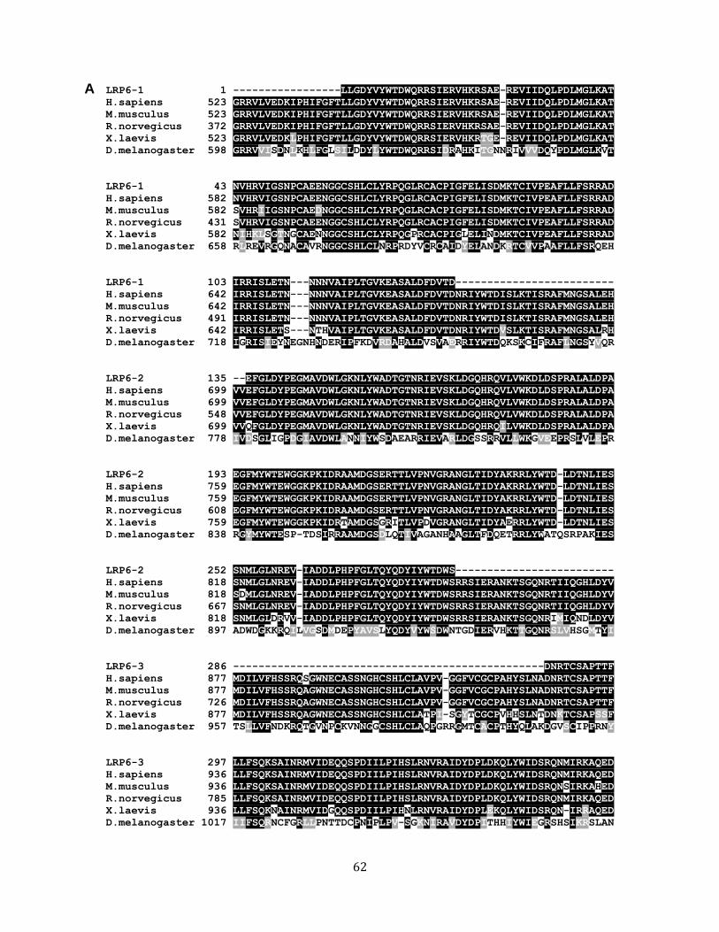
62
LRP6-1 1 -----------------LLGDYVYWTDWQRRSIERVHKRSAE-REVIIDQLPDLMGLKAT
H.sapiens 523 GRRVLVEDKIPHIFGFTLLGDYVYWTDWQRRSIERVHKRSAE-REVIIDQLPDLMGLKAT
M.musculus 523 GRRVLVEDKIPHIFGFTLLGDYVYWTDWQRRSIERVHKRSAE-REVIIDQLPDLMGLKAT
R.norvegicus 372 GRRVLVEDKIPHIFGFTLLGDYVYWTDWQRRSIERVHKRSAE-REVIIDQLPDLMGLKAT
X.laevis 523 GRRVLVEDKLPHIFGFTLLGDYVYWTDWQRRSIERVHKRTGE-REVIIDQLPDLMGLKAT
D.melanogaster 598 GRRVVISDNLKHLFGLSILDDYLYWTDWQRRSIDRAHKITGNNRIVVVDQYPDLMGLKVT
LRP6-1 43 NVHRVIGSNPCAEENGGCSHLCLYRPQGLRCACPIGFELISDMKTCIVPEAFLLFSRRAD
H.sapiens 582 NVHRVIGSNPCAEENGGCSHLCLYRPQGLRCACPIGFELISDMKTCIVPEAFLLFSRRAD
M.musculus 582 SVHRIIGSNPCAEDNGGCSHLCLYRPQGLRCACPIGFELISDMKTCIVPEAFLLFSRRAD
R.norvegicus 431 SVHRVIGSNPCAEENGGCSHLCLYRPQGLRCACPIGFELISDMKTCIVPEAFLLFSRRAD
X.laevis 582 NIHKLSGTNGCAENNGGCSHLCLYRPQGPRCACPIGLELINDMKTCIVPEAFLLFSRRAD
D.melanogaster 658 RLREVRGQNACAVRNGGCSHLCLNRPRDYVCRCAIDYELANDKRTCVVPAAFLLFSRQEH
LRP6-1 103 IRRISLETN---NNNVAIPLTGVKEASALDFDVTD-------------------------
H.sapiens 642 IRRISLETN---NNNVAIPLTGVKEASALDFDVTDNRIYWTDISLKTISRAFMNGSALEH
M.musculus 642 IRRISLETN---NNNVAIPLTGVKEASALDFDVTDNRIYWTDISLKTISRAFMNGSALEH
R.norvegicus 491 IRRISLETN---NNNVAIPLTGVKEASALDFDVTDNRIYWTDISLKTISRAFMNGSALEH
X.laevis 642 IRRISLETS---NTHVAIPLTGVKEASALDFDVTDNRIYWTDVSLKTISRAFMNGSALRH
D.melanogaster 718 IGRISIEYNEGNHNDERIPFKDVRDAHALDVSVAERRIYWTDQKSKCIFRAFLNGSYVQR
LRP6-2 135 --EFGLDYPEGMAVDWLGKNLYWADTGTNRIEVSKLDGQHRQVLVWKDLDSPRALALDPA
H.sapiens 699 VVEFGLDYPEGMAVDWLGKNLYWADTGTNRIEVSKLDGQHRQVLVWKDLDSPRALALDPA
M.musculus 699 VVEFGLDYPEGMAVDWLGKNLYWADTGTNRIEVSKLDGQHRQVLVWKDLDSPRALALDPA
R.norvegicus 548 VVEFGLDYPEGMAVDWLGKNLYWADTGTNRIEVSKLDGQHRQVLVWKDLDSPRALALDPA
X.laevis 699 VVQFGLDYPEGMAVDWLGKNLYWADTGTNRIEVSKLDGQHRQILVWKDLDSPRALALDPA
D.melanogaster 778 IVDSGLIGPDGIAVDWLANNIYWSDAEARRIEVARLDGSSRRVLLWKGVEEPRSLVLEPR
LRP6-2 193 EGFMYWTEWGGKPKIDRAAMDGSERTTLVPNVGRANGLTIDYAKRRLYWTD-LDTNLIES
H.sapiens 759 EGFMYWTEWGGKPKIDRAAMDGSERTTLVPNVGRANGLTIDYAKRRLYWTD-LDTNLIES
M.musculus 759 EGFMYWTEWGGKPKIDRAAMDGSERTTLVPNVGRANGLTIDYAKRRLYWTD-LDTNLIES
R.norvegicus 608 EGFMYWTEWGGKPKIDRAAMDGSERTTLVPNVGRANGLTIDYAKRRLYWTD-LDTNLIES
X.laevis 759 EGFMYWTEWGGKPKIDRTAMDGSGRITLVPDVGRANGLTIDYAERRLYWTD-LDTNLIES
D.melanogaster 838 RGYMYWTESP-TDSIRRAAMDGSDLQTIVAGANHAAGLTFDQETRRLYWATQSRPAKIES
LRP6-2 252 SNMLGLNREV-IADDLPHPFGLTQYQDYIYWTDWS-------------------------
H.sapiens 818 SNMLGLNREV-IADDLPHPFGLTQYQDYIYWTDWSRRSIERANKTSGQNRTIIQGHLDYV
M.musculus 818 SDMLGLNREV-IADDLPHPFGLTQYQDYIYWTDWSRRSIERANKTSGQNRTIIQGHLDYV
R.norvegicus 667 SNMLGLNREV-IADDLPHPFGLTQYQDYIYWTDWSRRSIERANKTSGQNRTIIQGHLDYV
X.laevis 818 SNMLGLDRVV-IADDLPHPFGLTQYQDYIYWTDWSRRSIERANKTSGQNRIMIQNDLDYV
D.melanogaster 897 ADWDGKKRQILVGSDMDEPYAVSLYQDYVYWSDWNTGDIERVHKTTGQNRSLVHSGMTYI
LRP6-3 286 -------------------------------------------------DNRTCSAPTTF
H.sapiens 877 MDILVFHSSRQSGWNECASSNGHCSHLCLAVPV-GGFVCGCPAHYSLNADNRTCSAPTTF
M.musculus 877 MDILVFHSSRQAGWNECASSNGHCSHLCLAVPV-GGFVCGCPAHYSLNADNRTCSAPTTF
R.norvegicus 726 MDILVFHSSRQAGWNECASSNGHCSHLCLAVPV-GGFVCGCPAHYSLNADNRTCSAPTTF
X.laevis 877 MDILVFHSSRQAGWNECASSNGHCSHLCLATPI-SGYTCGCPVHHSLNTDNKTCSAPSSF
D.melanogaster 957 TSLLVFNDKRQTGVNPCKVNNGGCSHLCLAQPGRRGMTCACPTHYQLAKDGVSCIPPRNY
LRP6-3 297 LLFSQKSAINRMVIDEQQSPDIILPIHSLRNVRAIDYDPLDKQLYWIDSRQNMIRKAQED
H.sapiens 936 LLFSQKSAINRMVIDEQQSPDIILPIHSLRNVRAIDYDPLDKQLYWIDSRQNMIRKAQED
M.musculus 936 LLFSQKSAINRMVIDEQQSPDIILPIHSLRNVRAIDYDPLDKQLYWIDSRQNSIRKAHED
R.norvegicus 785 LLFSQKSAINRMVIDEQQSPDIILPIHSLRNVRAIDYDPLDKQLYWIDSRQNMIRKAQED
X.laevis 936 LLFSQKNAINRMVIDGQQSPDIILPIHNLRNVRAIDYDPLEKQLYWIDSRQN-IRRAQED
D.melanogaster 1017 IIFSQRNCFGRLLPNTTDCPNIPLPV-SGKNIRAVDYDPITHHIYWIEGRSHSIKRSLAN
A
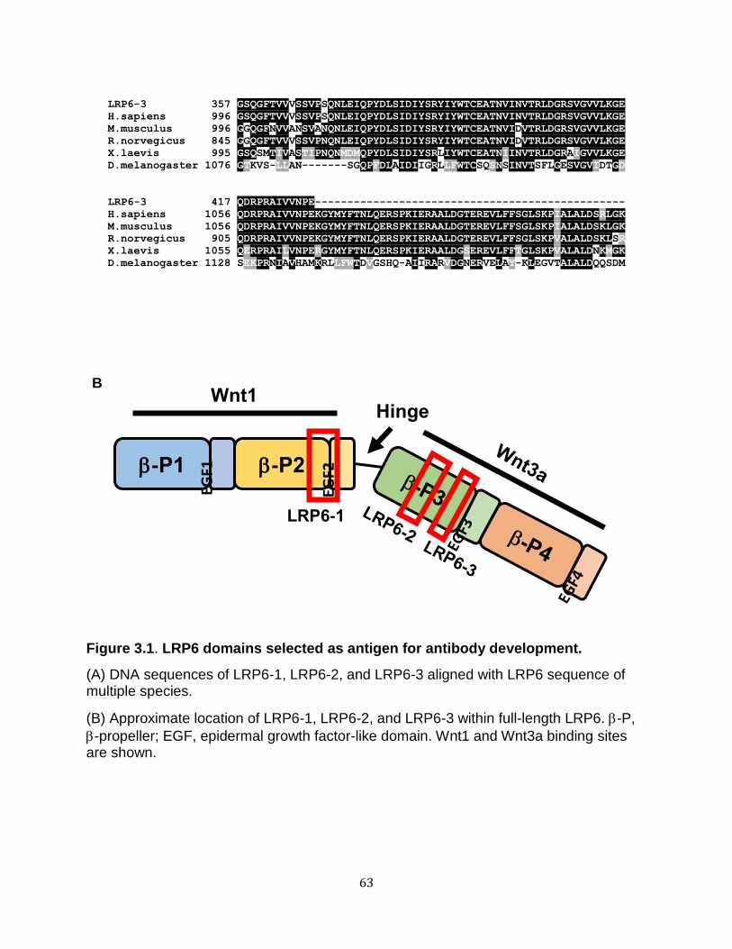
63
LRP6-3 357 GSQGFTVVVSSVPSQNLEIQPYDLSIDIYSRYIYWTCEATNVINVTRLDGRSVGVVLKGE
H.sapiens 996 GSQGFTVVVSSVPSQNLEIQPYDLSIDIYSRYIYWTCEATNVINVTRLDGRSVGVVLKGE
M.musculus 996 GGQGFNVVANSVANQNLEIQPYDLSIDIYSRYIYWTCEATNVIDVTRLDGRSVGVVLKGE
R.norvegicus 845 GGQGFTVVVSSVPNQNLEIQPYDLSIDIYSRYIYWTCEATNVIDVTRLDGRSVGVVLKGE
X.laevis 995 GSQSMTIVASTIPNQNMDMQPYDLSIDIYSRLIYWTCEATNIINVTRLDGRAIGVVLKGE
D.melanogaster 1076 GTKVS-LLAN-------SGQPFDLAIDIIGRLLFWTCSQSNSINVTSFLGESVGVIDTGD
LRP6-3 417 QDRPRAIVVNPE------------------------------------------------
H.sapiens 1056 QDRPRAIVVNPEKGYMYFTNLQERSPKIERAALDGTEREVLFFSGLSKPIALALDSRLGK
M.musculus 1056 QDRPRAIVVNPEKGYMYFTNLQERSPKIERAALDGTEREVLFFSGLSKPIALALDSKLGK
R.norvegicus 905 QDRPRAIVVNPEKGYMYFTNLQERSPKIERAALDGTEREVLFFSGLSKPVALALDSKLSR
X.laevis 1055 QERPRAILVNPERGYMYFTNLQERSPKIERAALDGSEREVLFFTGLSKPVALALDNKMGK
D.melanogaster 1128 SEKPRNIAVHAMKRLLFWTDVGSHQ-AIIRARVDGNERVELAY-KLEGVTALALDQQSDM
Figure 3.1. LRP6 domains selected as antigen for antibody development.
(A) DNA sequences of LRP6-1, LRP6-2, and LRP6-3 aligned with LRP6 sequence of multiple species.
(B) Approximate location of LRP6-1, LRP6-2, and LRP6-3 within full-length LRP6. -P,
-propeller; EGF, epidermal growth factor-like domain. Wnt1 and Wnt3a binding sites are shown.
B
-P1 EG
F
1
-P2 EG
F
2
Hinge
LRP6-1
Wnt1
EGF1
EGF2

64
Figure 3.2. The monoclonal antibody mAb7E5 is an inhibitor of the Wnt pathway.
(A) Screening of multiple clones of monoclonal antibodies. HEK293 STF cells were
incubated with Wnt3a and monoclonal antibodies.
(B) The monoclonal antibody mAb7E5 inhibits Wnt activation regardless of its isotype.
HEK293 STF cells were incubated with L (L CM) or Wnt3a (Wnt3a CM) conditioned
media and mAb7E5 IgG or mAb7E5 IgM.
(C) mAb7E5Fab can inhibit ligand-dependent Wnt signaling. HEK293 STF cells were
incubated with full-length or Fab mAb7E5 and Wnt3a CM.
B
A
C
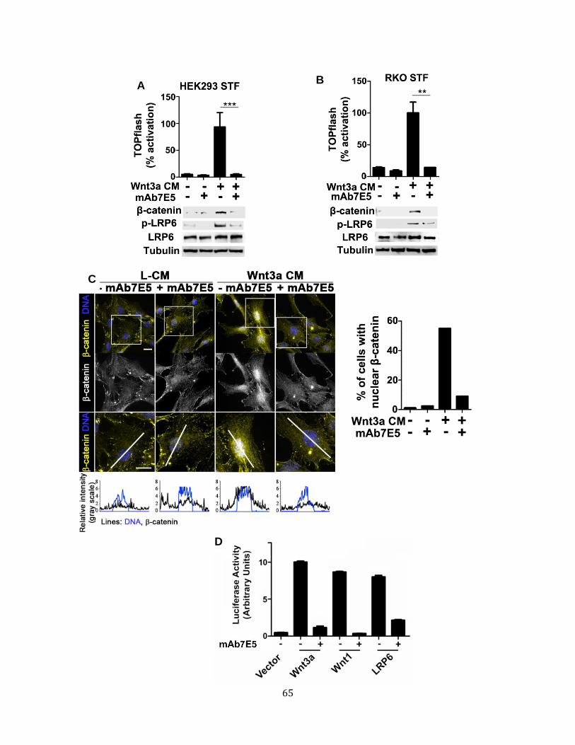
65
C
A B
D
66
Figure 3.3. mAb7E5 inhibits ligand mediated Wnt activation.
(A and B) mAb7E5 blocks Wnt ligand-mediated signaling (A) HEK293 STF and (B) RKO
STF cells were incubated with Wnt3a and mAb7E5.
(C) mAb7E5 prevents nuclear accumulation of -catenin. RPE cells were incubated with
Wnt3a and mAb7E5 followed by fixation and immunostaining. Scale bar: 5 m. Inset:
Line scans of selected cells. Scale bar: 10 m. Cells (>100/condition) showing nuclear
-catenin staining were quantified and plotted.
(D) mAb7E5, inhibits Wnt signaling mediated by the Wnt3a and Wnt1 classes of ligands
and by elevated LRP6. HEK293 STF were transfected with plasmids encoding Wnt3a,
Wnt1, LRP6 or vector control in the presence or absence of mAb7E5.
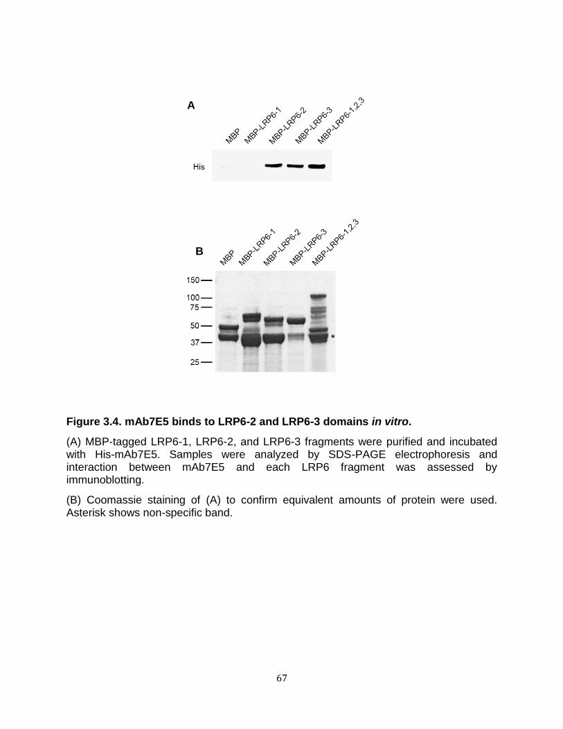
67
Figure 3.4. mAb7E5 binds to LRP6-2 and LRP6-3 domains in vitro.
(A) MBP-tagged LRP6-1, LRP6-2, and LRP6-3 fragments were purified and incubated with His-mAb7E5. Samples were analyzed by SDS-PAGE electrophoresis and interaction between mAb7E5 and each LRP6 fragment was assessed by immunoblotting.
(B) Coomassie staining of (A) to confirm equivalent amounts of protein were used. Asterisk shows non-specific band.
A
B

68
Figure 3.5. mAb7E5 inhibits Wnt activation at the level of the co-receptor LRP6
(A) The extracellular domain of LRP6 is required for mAb7E5 to inhibit LRP6-mediated
Wnt signaling. HEK293 STF cells were transfected with LRP6N or LRP6 FL and
incubated with mAb7E5.
(B) mAb7E5 blocks Wnt signaling upstream of the degradation complex. HEK293 STF
cells were incubated with mAb7E5 and 30 mM LiCl.
A B

69
did not block Wnt reporter activity or accumulation of cytoplasmic -catenin upon lithium
treatment (Figure 3.5B). These results provide strong evidence that mAb7E5 inhibits
Wnt signaling at the level of LRP6.
mAb7E5 inhibits Wnt signaling in breast cancer cells
Three major markers are currently used for guiding clinical diagnosis and
treatment of breast cancer: estrogen receptor expression, epidermal growth factor
receptor expression, and aberrant HER2 expression. Triple-negative breast cancer
(TNBC) is defined as the subtype of breast cancer lacking expression of all three
markers, and is considered highly aggressive and patients exhibit poor prognosis
(Banerjee et al. 2006; Bianchini et al. 2016).
MDA-MB-231 is a TNBC cell line, which exhibits constitutive Wnt signaling due to
an autocrine signaling loop. Incubation with the Wnt inhibitors DKK1 and SFRP1
prevent -catenin accumulation and Wnt activation in this cell line (Bafico et al. 2004).
MDA-MB-231 cells exhibit high levels of LRP6, and depletion of LRP6 by siRNA
decreases Wnt signaling, viability, and proliferation of MDA-MB-231 in vitro and in
mouse xenograft models (Bafico et al. 2004; Liu et al. 2010). Loss of Fz7 in MDA-MB-
231 cells also inhibits tumor growth in vivo (Yang et al. 2011). Because mAb7E5 targets
LRP6, we decided to test whether mAb7E5 inhibits Wnt signaling in MDA-MB-231 cells.
We found that both mAb7E5 and mAb7E5Fab decrease nuclear -catenin accumulation
in MDA-MB-231 cells (Figure 3.6). In titration studies, we found that mAb7E5 is effective
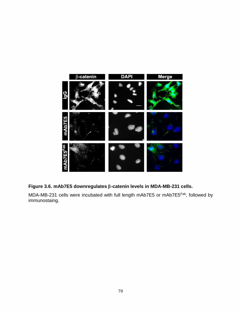
70
Figure 3.6. mAb7E5 downregulates -catenin levels in MDA-MB-231 cells.
MDA-MB-231 cells were incubated with full length mAb7E5 or mAb7E5Fab, followed by immunostaing.

71
Figure 3.7. Minimum mAb7E5 concentration required to inhibit -catenin accumulation in MDA-MB-231 cells.
MDA-MB-231 cells were incubated with indicated concentrations of mAb7E5 followed by immunostaining.

72
at a concentration of 30g/ml (Figure 3.7). These results suggest that mAb7E5 can
potentially be used as a therapeutic agent in treating TNBC.
Discussion
LRP6 is a very attractive target for developing inhibitors of the Wnt pathway for
several reasons: (1) LRP6 is located at the plasma membrane and binds to Wnt ligands,
(2) there are only two members of the LRP receptor family (LRP5 and LRP6), and (3)
LRP6 is only involved in the canonical Wnt pathway. Thus, any perturbation in LRP6
function would not have significant effects in other non-canonical functions of the Wnt
pathway, thereby limiting toxicity (Saito-Diaz et al. 2012).
Previously reported antibodies have been generated against the extracellular
domain of LRP6 using antibody libraries (Ettenberg et al. 2010) or by producing them
against purified E1-E2 and E3-E4 fragments of the LRP6 ECD (Gong et al. 2010). As a
result, these studies identified multiple antibodies that uncovered new properties of
LRP6. In contrast, our approach focused on bioinformatically identifying highly
conserved regions of LRP6 ECD important for LRP6 function and Wnt signal
transduction. Furthermore, in contrast to the reported anti-LRP6 antibodies, we found
that mAb7E5 inhibits both Wnt1 and Wnt3a class of ligand.
Interestingly, we found that mAb7E5 also inhibits ligand-independent Wnt
activation. Our data shows that mAb7E5 blocks Wnt activation upon LRP6
overexpression in human cells. This suggests that mAb7E5 may target a region of
LRP6 that is undergoes conformational change in response to Wnt ligand binding. It has
73
been reported that ECD of LRP6 prevents constitutive LRP6 activation and, upon Wnt
ligand binding, LRP6 undergoes a conformational change that relays the signal to
downstream effectors. It is possible that binding of mAb7E5 blocks this conformational
change to block LRP6 activation. Thus, the regions of LRP6-2 and LRP6-3 (targeted by
mAb7E5) may mediate structural changes in the coreceptor that are induced by Wnt
ligand binding.
The Wnt pathway is constitutively active in many diseases, including colorectal
and breast cancer. Thus, Wnt inhibitors would be potentially beneficial for the treatment
of these diseases. Multiple studies have reported several small molecules as candidate
therapeutics (Blagodatski et al. 2014). These small molecules, however, suffer from low
efficacy and selectivity, thereby limiting their potential use as therapeutics. In contrast,
monoclonal antibodies have multiple advantages as therapeutics as they are highly
selective and have longer half-lives than small molecules.
Antibodies against Fz have been developed as potent Wnt inhibitors. Two
antibodies, vantictumab and OTSA101, which targets Fz7 and Fz10, respectively, are
currently undergoing Phase II clinical trials (Blagodatski et al. 2014). Fz7 is up regulated
in a wide range of cancers, including CRC, TNBC, and hepatocellular carcinomas,
whereas Fz10 is highly expressed in synovial sarcomas (King et al. 2012). Although
vantictumab was initially identified as in inhibitor of Fz7, it has been shown to interact
with five other members of the Fz family (Gurney et al. 2012). As a result, patients
taking vantictumab demonstrate moderate-severe side effects, including osteoporosis (a
side of effect of significant Wnt inhibition) (Blagodatski et al. 2014).
74
Our results suggest that antibodies against LRP6 also have the potential for
being developed further for the treatment of Wnt-driven diseases. Just as importantly,
these antibodies can also be used as a tool to probe how LRP6 controls Wnt pathway
activation and possibly give us insight of unappreciated regulatory mechanisms. In the
case of the latter, the role of mAb7E5 in deciphering the mechanism of action of the
tumor suppressor, APC, in the Wnt pathway will be described in Chapter IV

75
CHAPTER IV
APC INHIBITS LIGAND-INDEPENDENT WNT SIGNALING BY THE CLATHRIN
ENDOCYTIC PATHWAY
Introduction
In the absence of a Wnt ligand, levels of cytoplasmic β-catenin are maintained at low
levels by its association with a destruction complex consisting of the scaffold protein
Axin, the tumor suppressor adenomatous polyposis coli (APC), and the kinases casein
kinase 1 (CK1) and glycogen synthase kinase (GSK3). Within this complex, β-
catenin is phosphorylated and targeted for degradation by the ubiquitin-proteasome
system (Saito-Diaz et al. 2012). Wnt ligand binding promotes the formation of a trimeric
complex of Wnt, the co-receptors Frizzled (Fz), and the low-density lipoprotein receptor
5/6 (LRP5/6) (Tamai et al. 2000). The formation of aggregated receptors (the
signalosome) promotes the recruitment of Axin in a process mediated by Dishevelled
(Dvl) (Tamai et al. 2004; Bilić et al. 2007; Zeng et al. 2008). Consequently, degradation
of β-catenin is inhibited, its cytoplasmic levels rise, and it is translocated to the nucleus.
Within the nucleus, β-catenin associates with the TCF family of transcription factors to
initiate a Wnt transcriptional program (Saito-Diaz et al., 2012).
In the classical model, the role of APC is limited to -catenin proteolysis. Herein,
we report that APC is also essential to inhibit Wnt receptor activation in the unstimulated
state. We have identified a constitutive, clathrin-mediated pathway that activates Wnt
signaling independent of Wnt ligands upon APC inactivation. These studies provide a

76
new model for the mechanism by which the Wnt pathway is aberrantly activated upon
APC loss and offer critical insight for the development of novel therapeutics targeting
Wnt-driven cancers.
Results
LRP6 is required for Wnt pathway activation in APC mutant cells but not in cells
with mutant -catenin
Loss of APC has been proposed to induce the formation of a Wnt autocrine loop
in colorectal cancer (CRC) (Bafico et al. 2004; Voloshanenko et al. 2013). We tested the
effects of mAb7E5 on the CRC lines SW480 and DLD1, which contain mutations in
APC, and HCT116, which expresses stabilized -catenin. mAb7E5 inhibited Wnt
signaling only in SW480 and DLD1 cells (Figure 4.1A and B). We found that all the CRC
cells tested exhibit elevated levels of phospho-LRP6 that were decreased by treatment
with mAb7E5 (Figure 4.1A). To confirm these results, we knocked down APC using
siRNA in HEK293 and RKO cells, which increased Wnt reporter activity, -catenin
accumulation, and phospho-LRP6 levels (Figure 4.2A-C). mAb7E5 treatment blocked all
of the effects of APC depletion (Figure 4.2B and C). As control, mAb7E5 had no effect
on a reporter line (HEK293-CMV) under the control of a non-Wnt regulated promoter
(Thorne et al., 2010) (Figure 4.2D). To rule out effects of residual APC activity, we used
CRISPR-Cas9 to generate a cell line (RKO APCKO) null for APC protein (Figure 4.3E).
RKO APCKO cells exhibited elevated levels of phospho-LRP6 and cytoplasmic -catenin
77
(Figure 4.2E), that was downregulated with mAb7E5 treatment (Figure 4.2F). These
findings indicate that the effects of APC loss on Wnt pathway activation are mediated
partly through LRP6.
To confirm that the constitutive activation of Wnt signaling resulting from APC
loss requires LRP6, we performed siRNA knockdown of LRP6 in HEK293 cells (Figure
4.3A). The activation of both the Wnt reporter and endogenous Wnt target genes
resulting from APC knockdown (on our Wnt reporter line and endogenous Wnt target
genes) was reversed by concomitant knockdown of LRP6 (Figures 4.3B-E). A similar
result was observed with either combined siRNA knockdown of APC in RKO cells or
siRNA knockdown of LRP6 in RKO APCKO cells (Figure 4.3F and G). Furthermore, in
SW480 and DLD1 cells, but not in HCT116 cells, knocking down LRP6 also inhibited
Wnt signaling (Figure 4.3H). Together, these findings provide further evidence that the
activation of Wnt signaling resulting from APC depletion is dependent on LRP6.
Requirement for surface receptor activation but not Wnt ligands for pathway
activation in APC mutant cells
We hypothesized that like LRP6, other components of the signalosome are
required for the constitutive activation of Wnt signaling upon APC loss. Thus, inhibiting
other membrane components (e.g., Fz and Dvl) should inhibit Wnt signaling in APC
mutant cells. To block multiple Fz and Dvl isoforms, we expressed their dominant-
negative forms, Fz1-ER and Xdd1, respectively (Wallingford et al. 2000; Kaykas et al.
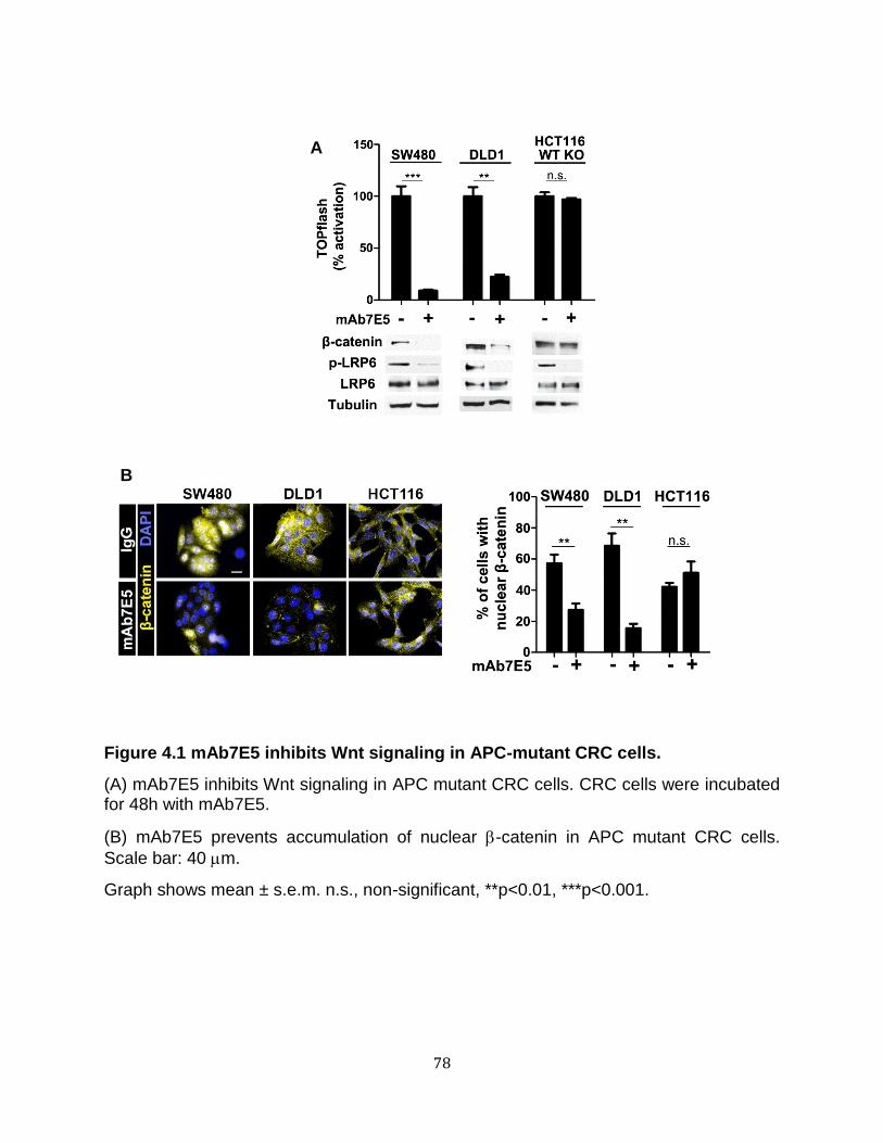
78
Figure 4.1 mAb7E5 inhibits Wnt signaling in APC-mutant CRC cells.
(A) mAb7E5 inhibits Wnt signaling in APC mutant CRC cells. CRC cells were incubated for 48h with mAb7E5.
(B) mAb7E5 prevents accumulation of nuclear -catenin in APC mutant CRC cells.
Scale bar: 40 m.
Graph shows mean ± s.e.m. n.s., non-significant, **p<0.01, ***p<0.001.
A
B
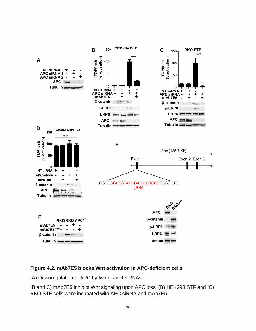
79
Figure 4.2. mAb7E5 blocks Wnt activation in APC-deficient cells
(A) Downregulation of APC by two distinct siRNAs.
(B and C) mAb7E5 inhibits Wnt signaling upon APC loss. (B) HEK293 STF and (C)
RKO STF cells were incubated with APC siRNA and mAb7E5.
D
E
F
A
B C

80
(D) Specificity of Wnt signaling inhibition by mAb7E5 in APC-deficient cells. A non-Wnt-
regulated reporter cell line, HEK293 CMV-luc, was incubated with APC siRNA and
mAb7E5.
(E) Generation of RKO APCKO. Schematics of selected gRNA and characterization of
RKO APCKO cells.
(F) RKO APCKO cells were incubated with mAb7E5 or mAb7E5Fab followed by
immunoblotting.
Graph shows mean ± s.e.m. n.s., non-significant, ***p<0.001.

81
H
A B C D
E
F G
82
Figure 4.3. LRP6 is required for Wnt signaling in APC-deficient cells.
(A) Downregulation of LRP6 by two distinct siRNAs.
(B and C) LRP6 is required for Wnt pathway activation upon loss of APC. (B) HEK293 STF and (C) RKO STF cells transfected with cells were transfected with APC and LRP6 siRNA.
(D) Specificity of Wnt signaling inhibition by downregulating LRP6 in APC-deficient cells. The non-Wnt-regulated reporter cell line, HEK293 CMV-luc, was incubated with APC and LRP6 siRNAs.
(E) Stimulation of endogenous Wnt target gene expression upon APC loss is inhibited by downregulating LRP6. HEK293 cells were transfected with APC and LRP6 siRNA, mRNAs isolated, and qRT-PCR performed.
(F) LRP6 is required nuclear -catenin accumulation upon loss of APC. RKO cells were treated with APC siRNA and either LRP6 siRNA or mAb7E5 followed by
immunostaining. Scale bar: 40 m.
(G) RKO APCKO cells were incubated with LRP6 siRNA. Lysates were collected and analyzed by immunoblotting.
(H) CRC cells were transfected with LRP6 siRNA followed by immunoblotting and TOPflash.
Graph shows mean ± s.e.m. n.s., non-significant, *p<0.05, **p<0.01, ***p<0.001.

83
A B
C
D
E
84
Figure 4.4 Loss of APC promotes ligand-independent signalosome formation.
(A) Fz and Dvl are required for ligand-mediated Wnt activation. HEK293 STF cells were
transfected with Xdd1 and Fz1-ER for 24 hours and then incubated with either L-cell (L
CM) or Wnt3a (Wnt3a CM) conditioned media.
(B) Dvl and Fz are required for Wnt activation in APC-deficient cells. HEK293 STF cells
were transfected with APC siRNA plus Xdd1 or Fz1-ER.
(C) APC depletion fails to activate the Wnt pathway in Dvl null cells. HEK293 and
HEK293 Dvl TKO were transfected with APC siRNA or treated with Wnt3a CM or 30
mM LiCl.
(D) Wnt signaling due to -catenin stabilization is not dependent on Fz or Dvl. HEK293
STF cells were transfected with Xdd1 or Fz1-ER and incubated with 30 mM LiCl.
(E) Similar to Wnt ligand-activated cells, LRP6 forms high molecular complexes in the
absence of APC on sucrose density gradient.
Graphs show mean ± s.e.m., n.s. non-significant, **p<0.01, ***p<0.001.

85
2004) (Figure 4.4A). Fz1-ER and Xdd1 expression inhibited Wnt reporter activity and
reduced phospho-LRP6 levels in APC-depleted cells (Figure 4.4B). Furthermore,
knocking down APC in a cell line null for all three Dvl genes (Dvl TKO) (Jiang et al.
2015) failed to activate Wnt signaling (Figure 4.4C). As control, inhibiting Fz and Dvl
function did not inhibit Wnt signaling induced by lithium treatment (Figure 4.4D).
These findings suggested that multiple signalosome components mediated the
aberrant signaling resulting from APC loss. We therefore tested whether loss of APC
induced signalosome formation. The receptor aggregates or signalosomes that form
upon Wnt ligand activation are detectable as multi-protein complexes in sucrose density
gradients (Bilić et al., 2007). As expected, we observed a shift in the migration of LRP6
into the denser fractions of lysates from RKO cells treated with Wnt3a compared to
untreated cells (Figure 4.4E). However, LRP6 was also detected in the denser fractions
of lysates from RKO APCKO cells even in the absence of Wnt3a, suggesting aggregate
formation (Figure 4.4E). Based on these findings, we conclude that APC loss induces
signalosome assembly.
We next tested if Wnt ligands were necessary for the aberrant activation of
signaling in APC mutant cells. A chimeric protein encoding the immunoglobulin Fc
domain fused to the cysteine-rich region of Fz8 (Fz8-Fc) sequesters Wnt ligands and
thereby blocks Wnt pathway activation (DeAlmeida et al. 2007). Fz8-Fc inhibited Wnt
reporter activity and -catenin accumulation in Wnt3a-treated cells (Figure 4.5A). Fz8-
Fc, however, had no effect on Wnt reporter activity in APC-deficient cells (Figures 4.5B-
D). Interestingly, APC-deficient cells were still responsive to exogenous Wnt3a
activation, as indicated by the increase of phospho-LRP6, which was inhibited by Fz8-

86
Figure 4.5. Wnt receptor activation in APC-deficient cells is Wnt ligand
independent.
(A-D) Fz8-Fc blocks Wnt ligand-mediated signaling but not signaling in APC-deficient
cells. (A) RKO STF cells were incubated with Wnt3a and Fz8-Fc at the indicated
concentrations. (B) HEK293 STF cells transfected with APC siRNA and (C) RKO APCKO
cells were incubated with Fz8-Fc. (D) Wnt activation in CRC cells is not dependent on
Wnt ligand. CRC cells were incubated with Fz8-Fc at the indicated concentrations for
48h.
(E) APC-depleted cells are still responsive to Wnt ligand. RKO APCKO cells were pre-
incubated for 30 min with Fz8-Fc followed by incubation with Wnt3a.
Graphs show mean ± s.e.m., n.s. non-significant, *p<0.05, **p<0.01.
A B C
D E
87
Fc (Figure 4.5E). We conclude that Wnt ligands are dispensable for pathway activation
resulting from APC loss.
To test the possible requirement for Wnt ligands using an independent approach,
we inhibited their secretion. Palmitoylation of Wnt ligands by the O-acyltransferase
Porcupine (PORCN), is required for their secretion and receptor binding (Gao and
Hannoush, 2014; Janda et al., 2012). As expected, two PORCN inhibitors, Wnt-C59
(IC50=0.1nM) and IWP-2 (IC50=27nM) (Chen et al. 2009; Proffitt et al. 2013), inhibited
Wnt signaling in cells transfected with Wnt3a (Figure 4.6A and B) but not when Wnt3a
was added exogenously (Figure 4.6C and D). Wnt-C59 and IWP-2 also had no
observable effect on Wnt signaling in APC-deficient cells (Figure 4.6E-H). In contrast to
APC mutant CRC cells, we observed a reduction in phospho-LRP6 in HCT116 cells
upon Fz8-Fc, Wnt-C59, and IWP-2 treatment, suggesting that an autocrine feedback
circuit occurs in HCT116 cells; reporter activity, however, was not noticeably impaired
(Figure 4.6I and J).
To rule out any residual PORCN activity, we knocked down APC in PORCN null
cells, which are non-responsive to transfected Wnt3a (Barrott et al. 2011) (Figure 4.7A).
In contrast to Dvl TKO cells, knocking down APC in PORCN null cells activated Wnt
signaling, which was inhibited by LRP6 knockdown (Figure 4.7B). These results are
consistent with a model in which loss of APC leads to ligand-independent activation of
the Wnt receptors and downstream signaling.

88
E
A B C D
F G H
I J
89
Figure 4.6. PORCN is not required for Wnt activation in the absence of APC.
(A and B) Inhibition of secreted Wnt3a by the PORCN inhibitors Wnt-C59 and IWP-2. HEK293 STF were transfected with Wnt3a and treated with (A) Wnt-C59 (0.3 nM) or (B) IWP-2 (30 nM).
(C and D) Wnt pathway activation by exogenously added Wnt3a is not blocked by PORCN inhibitors. HEK293 STF cells were pre-incubated with (C) Wnt-C59 or (D) IWP-2 for 30 minutes followed by incubation with Wnt3a.
(E-H) PORCN inhibition does not block Wnt activation upon loss of APC. (E and F) HEK293 STF cells transfected with APC siRNA and (G and H) RKO APCKO cells were incubated with (E and G) Wnt-C59 or (F and H) IWP-2.
(H and I) PORCN inhibition does not block Wnt activation in CRC cells. CRC cells were incubated with (H) Wnt-C59 or (I) IWP-2 for 48h.
Graphs show mean ± s.e.m., n.s. non-significant, *p<0.05.

90
Figure 4.7. LRP6 is required for Wnt activation upon loss of APC in PORCN null cells
(A) PORCNKO cells are responsive to exogenous Wnt3a but not transfected Wnt3a. PORCNKO MEF and wild-type MEF were transfected with Wnt3a or incubated with exogenous Wnt3a.
(B) LRP6 is required for Wnt activation upon loss of APC in the absence of PORCN. PORCNKO MEF and wild-type MEF were transfected with APC and LRP6 siRNA.
Graphs show mean ± s.e.m., n.s. non-significant, *p<0.05, **p<0.01, ***p<0.001.
A B
91
LRP6 and Dishevelled are required for the aberrantly increased proliferation of
Apc1 mutant adult intestinal stem cells in Drosophila
Similar to the consequences of APC loss in the mammalian intestine (Kinzler and
Vogelstein 1996; Korinek et al. 1997; Sansom et al. 2004), inactivation of Drosophila
Apc1 in adult intestinal stem cells (ISCs) results in their aberrantly increased
proliferation (Cordero et al. 2009; Lee et al. 2009). We tested whether the ISC
overproliferation and epithelial cell polarity defects resulting from Apc1 inactivation is
dependent on the Drosophila homolog of LRP6 (Arrow; Arr) (Wehrli et al., 2000). By
comparison with controls (Figure 4.8A, B, and M), a significantly increased number of
progenitor cells (ISCs and enteroblasts), marked by their expression of esg>GFP (esg-
gal4,UAS-GFP) (Micchelli and Perrimon 2006), was observed in the posterior midgut of
1- to 2-day-old Apc1 null mutant adults (Figure 4.8E, F, and M). In contrast, RNAi-
mediated Arrow knockdown in Apc1 null mutant intestinal progenitor cells during
development of the adult midgut resulted in a significant reduction in progenitor cell
number (Figure 4.8I, J, and M). Furthermore, the disrupted epithelial cell polarity in
Apc1 null mutants, as revealed by the aberrant subcellular localization of adherens
junction marker Armadillo (Arm) (compare Figures 4.8C and 4.8D with Figures 4.8G and
4.8H) was also rescued by Arrow depletion (Figures 4.8K and 4.8L). To rule out off-
target effects resulting from expression of the RNAi constructs, we tested two
independently-derived Arrow knockdown lines and obtained similar results (Figure
4.8M).
To further test these findings, we examined the effects RNAi-mediated
knockdown of Apc1 either singly or with concomitant knockdown of Arrow or Dvl

92
Figure 4.8. Both ISC overproliferation and epithelial cell polarity defects in Drosophila Apc1 null mutant midguts are rescued by Arrow depletion.
(A-H, and M) By comparison to controls (A-D, and M), loss of Apc1 leads to increased progenitor cell number (E, F, H, and M) and deregulated epithelial cell polarity in the

93
adult posterior midgut (G, H). Intestinal progenitor cells are marked with esg>GFP (green). Adherens junctions are marked with Arm (magenta) and nuclei with DAPI (blue). Enteroendocrine cells are marked with Prospero (Pros, magenta).
(I-L, and M) Expression of Arrow RNAi constructs in Apc1 mutants leads to a significant reduction in progenitor cell numbers (I, J, L, and M) and rescues defects in cell polarity (K and L).
(A, E, and I) Lower magnification view (scale bars: 50 m) (B-D, F-H, and J-L) higher
magnification view (scale bar: 10 m).
Graphs show mean ± s.e.m., ****p<0.0001, ***p<0.001.

94
Figure 4.9. Arrow and Dishevelled are required for overproliferation of midgut stem cells resulting from depletion of Apc1 in Drosophila.
(A-D, and G) By comparison to controls (A, B and G), RNAi-mediated knockdown of Apc1 during pupation in ISCs using esgts (temperature sensitive driver) leads to an increase in adult midgut progenitor cell number (C, D, and G). Progenitor cells are marked with esg>GFP (green), and nuclei are marked with DAPI (magenta). (E-G) Concomitant expression of the Apc1 RNAi construct with any one of three different arr RNAi constructs leads to a significant reduction in progenitor cell numbers (E-G). Scale bar: 30 μm
95
(H-K, and N) By comparison to controls (H, I, and N), RNAi-mediated knockdown of Apc1 in ISCs results in an increase in progenitor cell number (J, K, and N). Apc1 knockdown in progenitor cells during pupation was achieved by the temperature-sensitive ISC driver, esgts. Intestinal progenitor cells are marked with esg>GFP (green). Nuclei are marked with DAPI (magenta). (L-N) Concomitant expression of the Apc1 RNAi construct with either of two different dsh RNAi constructs results in a significant reduction in progenitor cell numbers. Scale bar: 30 μm.,
Graphs show mean ± s.e.m., *** p<0.001, **** p<0.0001.
96
(Dishevelled; dsh) (Klingensmith et al. 1994; Noordermeer et al. 1994; Siegfried et
al.1994). Apc1, Arrow, or dsh were knocked down in ISCs during formation of the adult
midgut using the temperature-sensitive progenitor cell driver esgts. Consistent with the
Apc1 null mutant phenotype, knockdown of Apc1 resulted in an increase in progenitor
cell number in the posterior midgut of 1- to 2-day-old adults (compare Figure 4.9A, B,
and G with Figure 4.9C, D, and G). In contrast, concomitant RNAi-mediated knockdown
of Apc1 and Arrow resulted in a significant reduction in this number (Figure 4.9E, F, and
G). Similarly, concomitant knockdown of dsh and Apc1 also resulted in a significant
reduction of ISC number as compared to knockdown of Apc1 alone (Figure 4.9H-N). To
rule out RNAi off-target effects, we tested multiple independently-derived arrow and dsh
knockdown lines and obtained similar results (Figure 4.9G and Figure 4.9N). These
findings provide in vivo evidence that the requirement for plasma membrane
components of the Wnt pathway for the aberrantly increased signaling in APC-deficient
cells is evolutionarily conserved.
Rapid accumulation of phospho-LRP6 and -catenin stabilization occur upon
immediate loss of APC
We asked if receptor activation following APC loss was a secondary
consequence of a simple positive feedback mechanism resulting from the regulation of
Wnt target genes. We found that lithium treatment stabilized -catenin and promoted
Wnt reporter activity, but, in contrast to loss of APC, did not promote phospho-LRP6
accumulation (Figure 4.10A). We also tested the effects of a dominant-negative form of
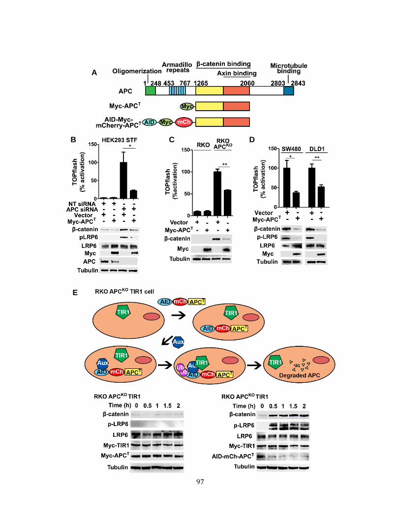
97
A
B C D
E

98
Figure 4.10. LRP6 activation and -catenin accumulation occur rapidly upon loss of APC function.
(A) Schematics of APC structure and truncations tested.
(B-D) -catenin and Axin binding domains of APC are sufficient to rescue loss-of-APC phenotype. (E) HEK293 STF treated with APC siRNA, (F) RKO APCKO, and (G) CRC cells were transfected with Myc-APCT followed by TOPflash analysis and immunoblotting.
(E) Loss of APC correlates with -catenin and phospho-LRP6 accumulation. RKO APCKO TIR1 cells were transfected with Myc-APCT or AID-Myc-mCherry-APCT followed by incubation with auxin. AID, auxin-inducible degron; mCh, mCherry; Aux, auxin.
Graphs show mean ± s.e.m., n.s. non-significant, *p<0.05, **p<0.01, ***p<0.001.

99
TCF4 (dnTCF4) (van de Wetering et al. 2002) that prevents TCF-mediated expression
of Wnt target genes. We found that dnTCF4 inhibited reporter activation, but not
accumulation of either -catenin or phospho-LRP6, upon APC knockdown (Figure
4.10B and C). Thus, stabilization of -catenin and activation of Wnt target genes are not
sufficient to activate Wnt receptors upon APC loss.
APC exhibits multiple activities in addition to its role in Wnt signaling (Saito-Diaz
et al., 2012). The aberrant activation of Wnt signaling in APC mutant cells is rescued by
an APC fragment (APCT) containing its -catenin and Axin binding domains (Li et al.
2012) (Figure 4.10A). We found that APCT suppresses LRP6 phosphorylation in APC-
deficient cells (Figure 4.10B-D), suggesting that LRP6 activation in APC mutant cells is
due to loss of the region of APC associated with its Wnt pathway function. If formation
of an active Wnt receptor complex is not due to a transcriptional/translational feedback
mechanism in APC-deficient cells, LRP6 phosphorylation and -catenin stabilization
upon APC loss should be temporally correlated. To test this hypothesis, we investigated
the timing of LRP6 phosphorylation and -catenin accumulation following APC loss. We
fused the APCT fragment to an auxin-inducible degron (AID) that allows for auxin-
induced turnover (Holland et al. 2012) by the plant E3 TIR1 protein (Figure 4.10A).
Transfection of AID-Myc-mCherry-APCT into RKO APCKO cells stably expressing E3
TIR1 suppresses Wnt signaling to a similar extent as the APCT fragment (Figure 4.10E,
compare left and right panels). The addition of auxin resulted in a rapid loss of AID-Myc-
mCherry-APCT (within 30 minutes) that paralleled the stabilization of -catenin.
Significantly, the accumulation of -catenin levels paralleled the appearance of
phospho-LRP6 (Figure 4.10E, right panel).

100
Endocytosis is required for Wnt signaling upon loss of APC
Endocytosis is required for receptor-mediated activation of the Wnt pathway
(Blitzer and Nusse 2006; Yamamoto et al. 2006; Gagliardi et al. 2014). As expected, we
found that blocking endocytosis by shifting Wnt3a-treated RKO or RKO APCKO cells to
4˚C decreased cytoplasmic -catenin and phospho-LRP6 levels; this effect was
reversed by shifting cells back to 37˚C. In contrast, activating RKO cells with lithium had
no effect on cytoplasmic -catenin levels when incubated at 4˚C (Figure 4.11A-D).
Similarly, we found that shifting SW480 (APC mutant), but not HC116 (-catenin
mutant) cells, to 4°C decreased cytoplasmic -catenin levels (Figure 4.12). To test the
requirement for endocytosis further, we used a dominant-negative Dynamin
(DynaminK44A), which inhibits both clathrin and caveolin-mediated endocytosis and has
been shown to inhibit Wnt signaling (Blitzer and Nusse 2006; Yamamoto et al. 2008;
Dutta and Donaldson 2012). DynaminK44A decreased -catenin levels in all APC-
deficient cell lines tested (Figure 4.13A-D).
Given that we found that Dynamin is required for Wnt pathway activation in APC-
deficient cells, we asked whether LRP6 is internalized upon loss of APC. Consistent
with previous reports (Yamamoto et al., 2006), we found that LRP6 was internalized
within two hours after addition of Wnt3a and could be blocked with mAb7E5 (Figure
4.14A). As expected, lithium had no effect on LRP6 internalization. Internalization of
LRP6 induced by APC knockdown was blocked with mAb7E5 but not with PORCN
inhibitors (Figure 4.14B and C). In RPE cells, fluorescence intensity profiles from an
axial scan showed two distinct peaks of LRP6-eYFP signal cells that corresponded to

101
A
B
C
D
102
Figure 4.11. Endocytosis inhibition induced by temperature shift prevents -catenin accumulation and LRP6 phosphorylation.
(A-D) RKO cells (treated with Wnt3a or 30 mM LiCl), and RKO APCKO cells were incubated at 4°C (A and B) and shifted back to 37°C (C and D). The average of three independent replicates were plotted.

103
Figure 4.12. Endocytosis inhibition induced by temperature shift prevents -catenin accumulation in APC-mutant CRC cells.
CRC cells were incubated at 4°C. Lysates were obtained at specified times and analyzed
by immunoblotting. The average of three independent replicates were plotted. Blot are
representative experiments.
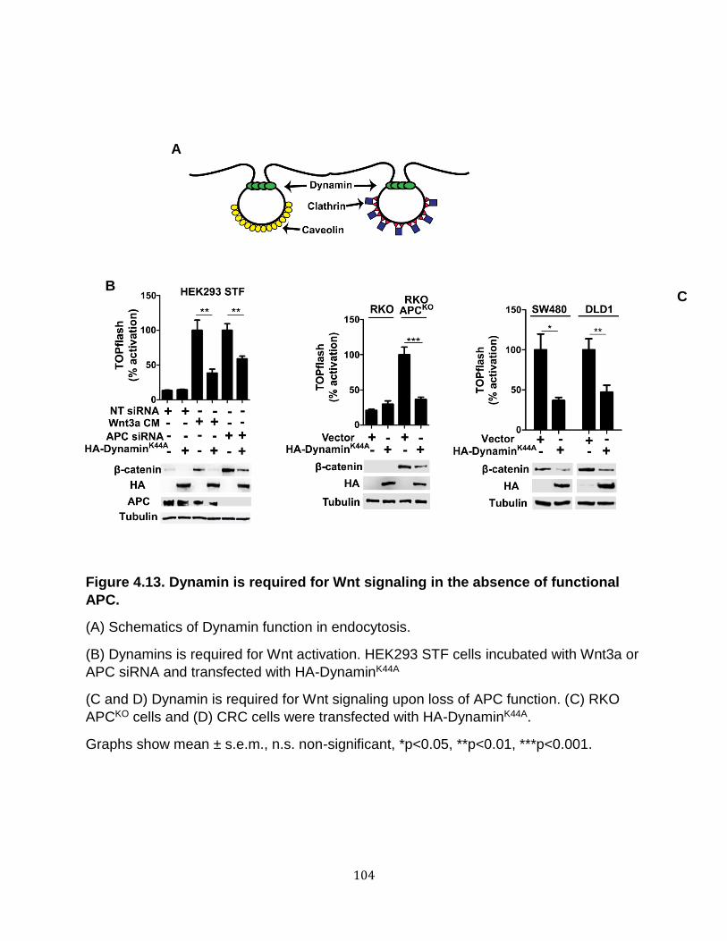
104
Figure 4.13. Dynamin is required for Wnt signaling in the absence of functional
APC.
(A) Schematics of Dynamin function in endocytosis.
(B) Dynamins is required for Wnt activation. HEK293 STF cells incubated with Wnt3a or
APC siRNA and transfected with HA-DynaminK44A
(C and D) Dynamin is required for Wnt signaling upon loss of APC function. (C) RKO
APCKO cells and (D) CRC cells were transfected with HA-DynaminK44A.
Graphs show mean ± s.e.m., n.s. non-significant, *p<0.05, **p<0.01, ***p<0.001.
A
B C

105
A
B
C
D
106
Figure 4.14. LRP6 is internalized in APC-deficient cells.
(A and B) LRP6 is internalized in Wnt3a treated and APC deficient cells. RPE cells were transfected with LRP6-eYFP and incubated with Wnt3a or 30mM LiCl or (B) APC siRNA and mAb7E5. Cells (>100/condition) demonstrating membrane or internalized LRP6 were quantified.
(C) PORCN inhibition does not block LRP6 internalization upon loss of APC. RPE shAPC cells were transfected with LRP6-eYFP and incubated with Wnt-C59 or IWP-2.
(D) Loss of APC promotes LRP6 internalization. RPE cells were transfected with LRP6-eYFP and APC siRNA in the absence or presence of mAb7E5. Line scans were performed to quantify changes in LRP6 intensity across cells.
Scale bars: 20 m.
107
high concentrations of LRP6 at the plasma membrane. In contrast, APC-depleted cells,
we observed multiple peaks corresponding to previously reported large, punctate,
LRP6-containing structures postulated to represent intracellular vesicles (Bilić et al.,
2007). Treatment of APC-depleted cells with mAb7E5 re-established the formation of
two predominant peaks of fluorescence signal (Figure 4.14D). These studies provide
further evidence that the endocytic pathway is required to activate the Wnt pathway in
APC-deficient cells.
Wnt receptor activation in APC-deficient cells occurs via a clathrin-dependent
mechanism.
Clathrin- and caveolin-mediated endocytosis have both been implicated in Wnt
signaling (Blitzer and Nusse 2006; Yamamoto et al. 2006; Bilić et al. 2007; Hanson et
al. 2013). Chloroquine and nystatin are small molecule inhibitors of clathrin- and
caveolin-mediated endocytosis, respectively (Yamamoto et al. 2006; Chen et al. 2009;
Dutta and Donaldson 2012). Consistent with a previous study (Yamamoto et al., 2006),
nystatin, but not chloroquine, inhibited Wnt3a-mediated accumulation of -catenin in
RKO cells (Figure 4.15A and B). In RKO APCKO cells, however, chloroquine and pitstop-
2 (another inhibitor of clathrin-mediated endocytosis) (Dutta et al. 2012) decreased -
catenin, whereas nystatin had no observable effect (Figure 4.15C-F). To confirm these
results, we knocked down clathrin and caveolin (Figure 4.16A and B). Knocking down
caveolin, but not clathrin, inhibited Wnt3a signaling in RKO cells (Figure 4.16C). In
contrast, knocking down clathrin, but not caveolin, inhibited Wnt signaling in RKO
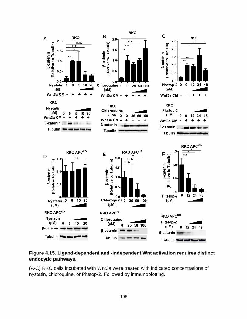
108
Figure 4.15. Ligand-dependent and -independent Wnt activation requires distinct
endocytic pathways.
(A-C) RKO cells incubated with Wnt3a were treated with indicated concentrations of
nystatin, chloroquine, or Pitstop-2. Followed by immunoblotting.
A B C
D E F
109
(D-F) RKO APCKO cells were treated with indicated concentrations of nystatin,
chloroquine, or Pitstop-2. Followed by immunoblotting.
The average of three independent replicates were plotted. Blot are representative experiments.
Graphs show mean ± s.e.m., n.s. non-significant, *p<0.05, **p<0.01, ***p<0.001.
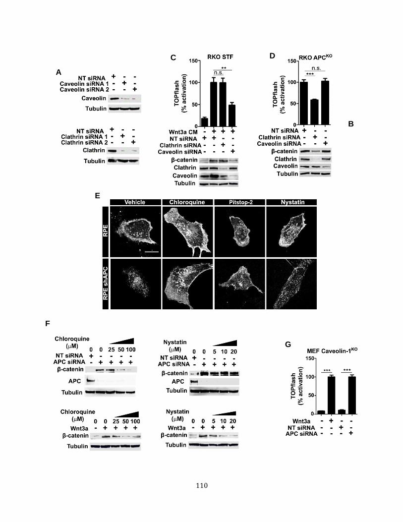
110
A
B
C D
E
F
G

111
Figure 4.16. Clathrin-mediated endocytosis is required for Wnt activation upon loss of APC.
(A) Downregulation of clathrin by siRNAs. RKO cells were incubated with two different siRNAs.
(B) Two distinct siRNAs decrease caveolin levels. RKO cells were incubated with two siRNAs.
(C) RKO cells incubated with Wnt3a were transfected with caveolin-1 or clathrin siRNA.
(D) RKO APCKO cells were transfected with clathrin or caveolin-1 siRNA.
(E) Inhibition of clathrin-mediated endocytosis blocks LRP6 endocytosis in APC-depleted cells. RPE and RPE shAPC were incubated with chloroquine, Pitstop-2, and nystatin, and live cells were imaged by confocal microscopy.
(F) Clathrin requirement for Wnt activation upon loss of APC is conserved. L-cells were incubated with Wnt3a or APC siRNA and treated with chloroquine or nystatin.
(G) Wnt activation upon loss of APC is caveolin-1-independent. Caveolin-1KO MEFs were incubated with Wnt3a or APC siRNA.
Graphs show mean ± s.e.m., *p<0.05, **p<0.01, ***p<0.001.
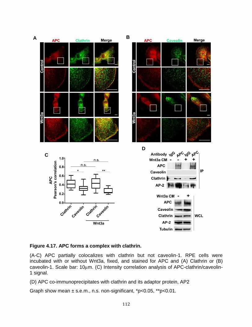
112
Figure 4.17. APC forms a complex with clathrin.
(A-C) APC partially colocalizes with clathrin but not caveolin-1. RPE cells were incubated with or without Wnt3a, fixed, and stained for APC and (A) Clathrin or (B)
caveolin-1. Scale bar: 10m. (C) Intensity correlation analysis of APC-clathrin/caveolin-1 signal.
(D) APC co-immunoprecipitates with clathrin and its adaptor protein, AP2
Graph show mean ± s.e.m., n.s. non-significant, *p<0.05, **p<0.01.
C
A B
D

113
APCKO cells (Figure 4.16D). We next assessed whether treatment with chloroquine,
pitstop-2, or nystatin altered LRP6-eYFP accumulation at the plasma membrane,
whereas nystatin treatment had no observable effect (Figure 4.16E). These studies
provide further evidence that Wnt signaling in APC-depleted cells occurs via a clathrin-
mediated mechanism.
A previous study showed that disruption of clathrin-mediated endocytosis in
mouse fibroblasts L-cells inhibits Wnt ligand-mediated activation (Blitzer and Nusse,
2006). We found that Wnt3a signaling in L-cells is mediated by both clathrin and
caveolin-pathways (Figure 4.16F). Wnt pathway activation upon APC knockdown,
however, is a clathrin-dependent process in both L-cells (Figure 4.16F), and in a MEF
cell line deficient for caveolin (Hanson et al., 2013) (Figure 4.16G). Thus, the
requirement for caveolae versus clathrin for Wnt ligand activation appears to be cell-
type dependent, whereas clathrin is specifically required for constitutive Wnt pathway
activation in APC-deficient cells regardless of cell type.
Finally, we tested whether endogenous APC forms a complex with endogenous
clathrin. Immunofluorescence microscopy studies revealed significant overlap of APC
with clathrin, but not caveolin, at the plasma membrane in RPE cells (Figure 4.17A-C).
To further test these results using a biochemical approach, we performed co-
immunoprecipitation studies and found that endogenous APC was pulled down with
clathrin and its adaptor protein, AP-2, but not caveolin (Figure 4.17D).

114
Discussion
Despite extensive investigation, the mechanistic basis for APC function in Wnt
signaling has remained an enigma (Saito-Diaz et al., 2012). Based on our studies, we
propose that a major role of APC is to prevent constitutive, ligand-independent, clathrin-
dependent signalosome formation and pathway activation (Figure 4.18). Our findings
are not inconsistent with the role for APC in -catenin phosphorylation and degradation
(Clevers and Nusse 2012). APC and APC2 are functionally redundant in some contexts
(Ahmed et al. 2002; Daly et al. 2016). Thus, in the absence of APC, APC2 could act as
the primary scaffold protein in the -catenin degradation complex. This could explain
why -catenin can still be degraded in cells upon loss of APC when the Wnt receptor
complex is disrupted; APC2 is able to associate with the degradation complex to
promote -catenin turnover
Previous studies provide evidence for both caveolin and clathrin-based
mechanisms in the activation of the Wnt pathway (Bilić et al., 2007; Blitzer and Nusse,
2006; Hanson et al., 2013; Yamamoto et al., 2006). Consistent with this possibility, we
observed that epithelial cells (e.g., HEK293, RKO and RPE cells) mediate Wnt signaling
via the caveolin pathway, whereas fibroblasts (e.g., L-cells and MEFs) mediate Wnt
signaling via the clathrin pathway. Because caveolin- and clathrin-dependent
endocytotic pathways differ in their dynamic association with the plasma membrane,
Wnt signaling could potentially be qualitatively different depending on the particular
pathway utilized. In addition, clathrin-dependent endocytosis has been linked to an early
response to stimuli, whereas caveolae play a later role in maintaining responses (Zhang
et al. 2014). The clathrin-mediated pathway can be promiscuous: a well-known example

115
Figure 4.18. Proposed model for APC functioning as a negative regulator of
clathrin-mediated endocytosis of Wnt pathway membrane components.
(A) In the absence of a Wnt ligand, LRP6 localizes in nascent Clathrin-coated pits. APC
forms a complex with clathrin to block spontaneous Wnt receptor aggregation and
activation.
(B) Wnt binds to its co-receptors, Fz and LRP6, to promote signalosome formation. The
Wnt signalosome is internalized via the caveolae or clathrin pathways.
A
B
C
116
(C) Loss of APC results in ligand-independent LRP6 oligomerization, Wnt signalosome
formation, and pathway activation via the clathrin endocytic pathway. This event drives
constitutive activation of the Wnt pathway.

117
is the constitutive internalization of the transferrin receptor. Thus, it is possible that APC
acts as a “brake” on receptor activation via the clathrin endocytic pathway in the
absence of Wnt ligand. Future studies will focus on the detailed mechanism by which
APC regulates this process.

118
CHAPTER V
CONCLUSIONS AND FUTURE DIRECTIONS
Since the discovery of the Wnt signaling pathway, much of the work has focused
on understanding the regulatory steps in the pathway as well as identifying targets for
developing therapeutics against Wnt-driven diseases. The work presented herein
provides insight into both areas of investigation. In this Chapter, I will briefly summarize
my findings and describe important questions that arose from my work.
Biochemical characterization of mAb7E5
We identified the monoclonal antibody mAb7E5 that targets the extracellular
domain of LRP6. Using biochemical analysis, we identified that mAb7E5 inhibits ligand-
dependent and independent Wnt activation. We found that mAb7E5 binds to the LRP6-
2 and LRP6-3 fragments of human LRP6. Our working hypothesis is that mAb7E5
blocks the LRP6 conformational change required to effectively transduce the Wnt signal
from the plasma membrane to downstream, cytoplasmic effectors. Further studies are
needed to more accurately identify the region(s) where mAb7E5 binds to LRP6. This
may be readily accomplished by performing binding assays with a series of LRP6
truncations and point mutations to further narrow the mAb7E5 binding domain on LRP6.
Ideally, these results will ultimately be confirmed by analyzing the X-ray crystal structure
of mAb7E5 bound to LRP6. Such a structure may provide important insight into the

119
mechanism by which Wnt ligand can induces the conformation of LRP6 to promote its
activation.
Regulation of endocytosis by APC
Loss of APC results in constitutive activation of the Wnt pathway. This was
thought to occur primarily by disrupting the assembly of the -catenin degradation
complex, thereby bypassing the need for activation of membrane receptors
components. Based on my studies, a new role for APC function in the Wnt pathway is
revealed that does not involve its role in the degradation complex. My studies indicate
that APC prevents inappropriate activation of the membrane components of the Wnt
pathway via the clathrin endocytic pathway. To my knowledge, this is the first time such
a phenomenon has been demonstrated to occur in the Wnt pathway. Significantly, loss
of APC resulting in Wnt pathway activation via the clathrin endocytic pathway is ligand-
independent and is, thus, cell autonomous. Although p53 and Ras mutant cancer cells
have been shown to exhibit high rates of receptor endocytosis (Mellman and Yarden
2013), the process that links the endocytic pathway and cell signaling remains poorly
understood.
Signaling pathways rely on exquisite mechanisms to effectively transduce
external cues to downstream effectors. Consistent with this, a growing body of evidence
links endocytosis to activation of the Wnt pathway however the mechanism is not clear.
Both clathrin and caveolin have been reported to be required for LRP6 endocytosis and
Wnt pathway activation (Blitzer and Nusse 2006; Yamamoto et al. 2006). I found that
whether the clathrin or the caveolin endocytic machinery is required for Wnt ligand-
mediated signaling is highly dependent on the particular cell type tested. I found,
120
however, that activation of the Wnt pathway upon APC loss is clathrin-mediated and is
independent of cell type. Why would a cell require two different endocytic pathways to
regulate Wnt signaling? It is possible that Wnt activation mediated by clathrin and
caveolin-dependent endocytosis is qualitatively different and may be required in
different biological contexts (e.g. during development). Future studies should focus on
understanding whether there are qualitative differences in Wnt signaling that occur via
the clathrin versus the caveolin pathway.
The evolutionary conservation between Drosophila and vertebrate in the
regulation of Wnt signaling by APC suggests that this mechanism may play a critical
role in early animal development. Interestingly, Drosophila do not express caveolin
suggesting that caveolin-mediated Wnt activation may be a more evolutionarily recent
development in vertebrates (Fischer et al. 2006). Although the Wnt pathway is highly
conserved, some differences in how Wnt signaling is regulated between Drosophila and
vertebrates are not unexpected, particularly when it involves regulation of Wnt receptor
activity. For example, secreted Wnt inhibitors (e.g. Dkk1 and Sfrp-1) and the Wnt
agonist, R-Spondin, are present in vertebrates but not in Drosophila. Wnt signaling has
been proposed to be critical for the evolution of epithelial morphogenesis (Lapébie et al.
2009). Interestingly, Wnt signaling via the caveolin pathway occur primarily in
mesodermal cells and may reflect an evolutionary requirement in vertebrate for Wnt
signaling in patterning mesenchymal tissues.
We found that APC associates with clathrin by coimmunoprecipitation. One
possible model is that LRP6 is present in nascent clathrin coated pits, and APC acts as
a brake to block the activity of the LRP6-bound clathrin. Upon Wnt stimulation, LRP6

121
subsequently translocates to mature clathrin vesicles where it is ultimately activated
upon internalization of the clathrin vesicles. Careful colocalization studies of APC,
clathrin, and LRP6 by electron microscopy may provide a better (high resolution)
understanding of their spatial and temporal regulation upon Wnt ligand activation.
My findings demonstrate that inhibiting the activities of LRP6, Fz, or Dvl result in
-catenin downregulation (and inhibition of Wnt signaling) in APC-deficient cells. These
studies suggest that the -catenin destruction complex remains fully functional upon
loss of APC. APC and APC2 have been proposed to exhibit redundant functions
(Ahmed et al. 2002; Daly et al. 2016). In Drosophila, overactivation of Wg signaling due
to inactivation of one of the APC proteins can be inhibited by overexpression of the
other Apc (Ahmed et al. 2002). Loss of both APC and APC2 disrupts mammary tissue
homeostasis and promotes mammary tumor formation in mice (Daly et al. 2016). This
study suggests that APC and APC2 may act in a redundant fashion to promote
tumorigenesis in certain tissues.
Despite their functional redundancy in human cells, APC2 shows lower affinity to
-catenin and is less efficient promoting its turnover compared to APC (Schneikert et al.
2013). Thus, I hypothesize that in the absence of APC, APC2 functions as the
predominant scaffold (along with Axin) in the -catenin degradation complex. One would
predict that inhibiting the function of LRP6, Fz, or Dvl would no longer be able to
promote -catenin downregulation in APC-APC2 double knockdowns. The vast majority
of human cancers occur due to disruption of APC (not APC2) function. Also, APC
mutant CRC cells express APC2. One possibility is that only APC can block Wnt-ligand
independent activation of the Wnt signaling by the clathrin-endocytic pathway. Thus,

122
loss of APC cannot be fully compensated by APC2. Further experiments should be
performed to test this possibility.
My work supports a role for APC regulating the function of the -catenin
destruction complex and the activation of plasma membrane components. It is unclear
which of these two activities represent the “the main function” of APC in the Wnt
pathway. I have shown that a fragment of APC encoding the -catenin and Axin-binding
domains (APCT) is sufficient to prevent LRP6 phosphorylation and -catenin turnover in
APC-depleted cells, suggesting that both events are dependent on APC function. One
possible explanation is that independent pools of APC regulate each event. However, if
this were true, APCT degradation would only affect -catenin degradation and not LRP6
activation because it only binds to Axin and -catenin. Another explanation is that both
functions are not mutually exclusive and APC regulates both the -catenin destruction
complex function and LRP6 activation. The multiple roles of APC in the Wnt pathway is
reminiscent of the role of GSK3 in the destruction complex and receptor activation.
Further experiments, are necessary to understand the precise mechanism of APC
action, particularly at the membrane.
Tight regulation of Wnt signal transduction is necessary for proper development
of all metazoans. In adults, APC inhibition is involved in early development of colorectal
adenocarcinomas. Because homozygous deletion of APC is embryonic lethal (Dow et
al. 2015), the effects caused by APC loss are typically studied in the context of disease
in fully developed organisms in which conditional knockouts can be performed. Indeed,
loss of APC function in adult mice deregulates the Wnt pathway and recapitulates the
molecular features of CRC. Furthermore, experimental APC restoration reestablishes
123
intestinal crypt homeostasis in APC-deficient mice (Chartier et al. 2016). Given the
seemingly elaborate mechanism by which APC regulates Wnt receptor activation, one
could hypothesize that regulating APC levels may represent another way by which the
pathway can be activated in a cell autonomous fashion (independent of Wnt ligands)
during certain normal developmental processes. This is not without precedence. Albeit,
via a distinct mechanism, Notch signaling can also occur via a ligand-independent
manner involving endocytosis of the Notch receptor (Palmer and Deng 2015). In
Drosophila, the hematopoietic crystal cells exhibit ligand-independent Notch pathway
activation at the endosome that promotes their survival during development (Mukherjee
et al. 2011). Thus, ligand-independent activation during development might be
advantageous in a context where there is no guaranteed ligand source. Further
experiments will be required to test our hypothesis that downregulating APC levels
represents a normal developmental mechanism to activate the Wnt pathway in a cell
autonomous manner.
Targeting LRP6 in TNBC and CRC
We found that mAb7E5 inhibits the Wnt pathway in triple negative breast cancer
(TNBC) and colorectal cancer (CRC) cells, suggesting that mAb7E5 could be developed
further as a therapeutic agent to treat Wnt-driven tumors. Our preliminary study
indicated that mAb7E5 inhibit Wnt signaling in the TNBC cell line, MDA-MB-231.
Previous reports have shown that LRP6 expression is upregulated in TNBC, and RNAi
knockdown of LRP6 results in inhibition of the Wnt pathway and decreased cellular
growth/proliferation (Liu et al. 2010).

124
Receptor activation in APC-mutant colorectal cancer cells has not been
previously studied in detail. Past studies suggested that constitutive activation of the
Wnt pathway in CRC may occur via an autocrine signal loop (Voloshanenko et al.
2013). In the current study, I found that inhibition of Porcupine (PORCN), the protein
required for Wnt secretion and function, was not sufficient to inhibit -catenin
accumulation and loss of APC in PORCN null cells did not inhibit transcriptional
activation of a Wnt reporter. These results suggest that an autocrine Wnt signal is
unlikely to play a major role in Wnt pathway activation upon loss of APC. Thus, future
therapeutic approaches to target APC mutant tumors should focus on disrupting the
receptors and other Wnt signaling components necessary for formation of active
receptor complexes (e.g., signalosomes) other than Wnt ligands. Our own studies with
mAb7E5 suggest that it may represent an attractive candidate to target CRC.
If the utility of mAb7E for treating TNBC and APC mutant CRC is to be fully
realized, further studies are required to test whether mAb7E5 is effective in in vivo
tumor models (either cancer cell line xenografts or patient derived xenografts). Other
tasks that will be required for its development as a therapeutic agent include the
development and testing of a humanized version of the antibody (mAb7E5 is a mouse
monoclonal) as well as assessing its bioavailability, in vivo half-life, and
pharmacodynamics/pharmacokinetics.
125
Concluding remarks
The work presented herein combines multiple approaches to understand the
mechanism of Wnt signaling transduction. We identified a new function of the tumor
suppressor, APC, that provides a link between a basic cell biological process
(endocytosis) with the Wnt signaling pathway. The current work highlights the
importance of studying signaling pathways as part of a complex system that is regulated
in a temporal and spatial fashion and not simply as an isolated biochemical cascade of
events. It is likely that every signaling pathway is ultimately interconnected to form
complex signaling networks that are tightly regulated by the cell in a similar manner.
Thus, to understand how cells respond to their environment, we must take into account
this level of complexity, which represents an exciting challenge for scientists studying
how diverse chemically encoded information is organized to regulate behavior at the
cellular and organismal level.

126
BIBLIOGRAPHY
Ahmed, Y., S. Hayashi, et al. (1998). "Regulation of Armadillo by a Drosophila APC Inhibits Neuronal Apoptosis during Retinal Development." Cell 93(7): 1171-1182
Ahmed, Y., A. Nouri, et al. (2002). "Drosophila Apc1 and Apc2 regulate Wingless transduction throughout development." Development 129(7): 1751-1762
Ahn, Victoria E., Matthew L.-H. Chu, et al. (2011). "Structural Basis of Wnt Signaling Inhibition by Dickkopf Binding to LRP5/6." Developmental Cell 21(5): 862-873.
Alberts, B., A. Johnson, et al. (2002). Molecular Biology of the Cell, Garland Science
Amit, S., A. Hatzubai, et al. (2002). "Axin-mediated CK1 phosphorylation of β-catenin at Ser 45: a molecular switch for the Wnt pathway." Genes & Development 16(9): 1066-1076
Angers, S., C. J. Thorpe, et al. (2006). "The KLHL12-Cullin-3 ubiquitin ligase negatively regulates the Wnt-beta-catenin pathway by targeting Dishevelled for degradation." Nature Cell Biology 8(4): 348-357.
Baeg, G.-H., E. M. Selva, et al. (2004). "The Wingless morphogen gradient is established by the cooperative action of Frizzled and Heparan Sulfate Proteoglycan receptors." Developmental Biology 276(1): 89-100.
Bafico, A., G. Liu, et al. (2004). "An autocrine mechanism for constitutive Wnt pathway activation in human cancer cells." Cancer Cell 6(5): 497-506.
Baig-Lewis, S., W. Peterson-Nedry, et al. (2007). "Wingless/Wnt signal transduction requires distinct initiation and amplification steps that both depend on Arrow/LRP." Developmental Biology 306(1): 94-111
Banerjee, S., J. S. Reis-Filho, et al. (2006). "Basal-like breast carcinomas: clinical outcome and response to chemotherapy." Journal of Clinical Pathology 59(7): 729-735
Bänziger, C., D. Soldini, et al. (2006). "Wntless, a Conserved Membrane Protein Dedicated to the Secretion of Wnt Proteins from Signaling Cells." Cell 125(3): 509-522.

127
Barrott, J. J., G. M. Cash, et al. (2011). "Deletion of mouse Porcn blocks Wnt ligand secretion and reveals an ectodermal etiology of human focal dermal hypoplasia/Goltz syndrome." Proceedings of the National Academy of Sciences 108(31): 12752-12757.
Behrens, J., J. P. von Kries, et al. (1996). "Functional interaction of β-catenin with the transcription factor LEF-1." Nature 382(6592): 638-642
Bejsovec, A. and E. Wieschaus (1995). "Signaling activities of the Drosophila wingless gene are separately mutable and appear to be transduced at the cell surface." Genetics 139(1): 309-320.
Belenkaya, T. Y., C. Han, et al. (2002). "pygopus encodes a nuclear protein essential for Wingless/Wnt signaling." Development 129(17): 4089-4101
Bhanot, P., M. Brink, et al. (1996). "A new member of the frizzled family from Drosophila functions as a Wingless receptor." Nature 382(6588): 225-230
Bianchini, G., J. M. Balko, et al. (2016). "Triple-negative breast cancer: challenges and opportunities of a heterogeneous disease." Nature Reviews Clinical Oncology 13(11): 674-690
Bilić, J., Y.-L. Huang, et al. (2007). "Wnt Induces LRP6 Signalosomes and Promotes Dishevelled-Dependent LRP6 Phosphorylation." Science 316(5831): 1619 -1622.
Binari, R. C., B. E. Staveley, et al. (1997). "Genetic evidence that heparin-like glycosaminoglycans are involved in wingless signaling." Development (Cambridge, England) 124(13): 2623-2632.
Blagodatski, A., D. Poteryaev, et al. (2014). "Targeting the Wnt pathways for therapies." Molecular and Cellular Therapies 2(1): 28
Blauwkamp, T. A., M. V. Chang, et al. (2008). "Novel TCF-binding sites specify transcriptional repression by Wnt signalling." The EMBO Journal 27(10): 1436-1446.
Blitzer, J. T. and R. Nusse (2006). "A critical role for endocytosis in Wnt signaling." BMC Cell Biology 7(1): 28.
Bourhis, E., C. Tam, et al. (2010). "Reconstitution of a Frizzled8·Wnt3a·LRP6 Signaling Complex Reveals Multiple Wnt and Dkk1 Binding Sites on LRP6." Journal of Biological Chemistry 285(12): 9172-9179

128
Boyden, L. M., J. Mao, et al. (2002). "High bone density due to a mutation in LDL-receptor-related protein 5." The New England Journal of Medicine 346(20): 1513-1521.
Bradley, R. S. and A. M. Brown (1990). "The proto-oncogene int-1 encodes a secreted protein associated with the extracellular matrix." The EMBO Journal 9(5): 1569.
Buechling, T., V. Chaudhary, et al. (2011). "p24 proteins are required for secretion of Wnt ligands." EMBO reports 12(12): 1265-1272.
Cadigan, K. M. (2012). Chapter one - TCFs and Wnt/β-catenin Signaling: More than One Way to Throw the Switch. Current Topics in Developmental Biology. S. P. Payre and François, Academic Press. 98: 1-34
Callow, M. G., H. Tran, et al. (2011). "Ubiquitin ligase RNF146 regulates tankyrase and Axin to promote Wnt signaling." PLOS ONE 6(7): e22595.
Chang, M. P., W. G. Mallet, et al. (1993). "Adaptor self-aggregation, adaptor-receptor recognition and binding of alpha-adaptin subunits to the plasma membrane contribute to recruitment of adaptor (AP2) components of clathrin-coated pits." The EMBO Journal 12(5): 2169
Chartier, C., J. Raval, et al. (2016). "Therapeutic Targeting of Tumor-Derived R-Spondin Attenuates β-Catenin Signaling and Tumorigenesis in Multiple Cancer Types." Cancer Research 76(3): 713-723
Chen, B., M. E. Dodge, et al. (2009). "Small molecule-mediated disruption of Wnt-dependent signaling in tissue regeneration and cancer." Nature Chemical Biology 5(2): 100-107.
Chen, C.-L., W.-H. Hou, et al. (2009). "Inhibitors of clathrin-dependent endocytosis enhance TGFβ signaling and responses." Journal of Cell Science 122(11): 1863-1871.
Chen, S., D. Bubeck, et al. (2011). "Structural and Functional Studies of LRP6 Ectodomain Reveal a Platform for Wnt Signaling." Developmental Cell 21(5): 848-861.
Chen, Y.-G., Z. Wang, et al. (2007). "Endofin, a FYVE Domain Protein, Interacts with Smad4 and Facilitates Transforming Growth Factor-β Signaling." Journal of Biological Chemistry 282(13): 9688-9695
129
Cheng, Z., T. Biechele, et al. (2011). "Crystal structures of the extracellular domain of LRP6 and its complex with DKK1." Nat Struct Mol Biol 18(11): 1204-1210.
Clevers, H. and R. Nusse (2012). "Wnt/β-Catenin Signaling and Disease." Cell 149(6): 1192-1205.
Cong, F., L. Schweizer, et al. (2004). "Casein Kinase Iɛ Modulates the Signaling Specificities of Dishevelled." Molecular and Cellular Biology 24(5): 2000-2011.
Cong, F., L. Schweizer, et al. (2004). "Wnt signals across the plasma membrane to activate the β-catenin pathway by forming oligomers containing its receptors, Frizzled and LRP." Development 131(20): 5103 -5115.
Cong, F. and H. Varmus (2004). "Nuclear-cytoplasmic shuttling of Axin regulates subcellular localization of β-catenin." Proceedings of the National Academy of Sciences of the United States of America 101(9): 2882-2887
Cordero, J., M. Vidal, et al. (2009). "APC as a master regulator of intestinal homeostasis and transformation: From flies to vertebrates." Cell Cycle 8(18): 2927-2932.
Coudreuse, D. Y. M., G. Roël, et al. (2006). "Wnt Gradient Formation Requires Retromer Function in Wnt-Producing Cells." Science 312(5775): 921-924.
Cross, D. A., D. R. Alessi, et al. (1995). "Inhibition of glycogen synthase kinase-3 by insulin mediated by protein kinase B." Nature 378(6559): 785-789.
Cselenyi, C. S., K. K. Jernigan, et al. (2008). "LRP6 transduces a canonical Wnt signal independently of Axin degradation by inhibiting GSK3's phosphorylation of β-catenin." Proceedings of the National Academy of Sciences 105(23): 8032 -8037.
Dajani, R., E. Fraser, et al. (2003). "Structural basis for recruitment of glycogen synthase kinase 3b to the axin-APC scaffold complex." The EMBO Journal 22(3): 494-501.
Daly, C. S., P. Shaw, et al. (2016). "Functional redundancy between Apc and Apc2 regulates tissue homeostasis and prevents tumorigenesis in murine mammary epithelium." Oncogene.
Damke, H., T. Baba, et al. (1994). "Induction of mutant dynamin specifically blocks endocytic coated vesicle formation." The Journal of Cell Biology 127(4): 915-934
130
Davidson, G., W. Wu, et al. (2005). "Casein kinase 1g couples Wnt receptor activation to cytoplasmic signal transduction." Nature 438(7069): 867-872.
DeAlmeida, V. I., L. Miao, et al. (2007). "The Soluble Wnt Receptor Frizzled8CRD-hFc Inhibits the Growth of Teratocarcinomas In vivo." Cancer Research 67(11): 5371-5379.
Dietzen, D. J., W. R. Hastings, et al. (1995). "Caveolin Is Palmitoylated on Multiple Cysteine Residues PALMITOYLATION IS NOT NECESSARY FOR LOCALIZATION OF CAVEOLIN TO CAVEOLAE." Journal of Biological Chemistry 270(12): 6838-6842
Ding, V. W., R. H. Chen, et al. (2000). "Differential regulation of glycogen synthase kinase 3beta by insulin and Wnt signaling." The Journal of Biological Chemistry 275(42): 32475-32481.
Doble, B. W., S. Patel, et al. (2007). "Functional Redundancy of GSK-3α and GSK-3β in Wnt/β-Catenin Signaling Shown by Using an Allelic Series of Embryonic Stem Cell Lines." Developmental Cell 12(6): 957-971
Doherty, G. J. and H. T. McMahon (2009). "Mechanisms of Endocytosis." Annual Review of Biochemistry 78(1): 857-902
Dominguez, I., K. Itoh, et al. (1995). "Role of glycogen synthase kinase 3b as a negative regulator of dorsoventral axis formation in Xenopus embryos." Proceedings of the National Academy of Sciences of the United States of America 92(18): 8498-8502.
Drab, M., P. Verkade, et al. (2001). "Loss of Caveolae, Vascular Dysfunction, and Pulmonary Defects in Caveolin-1 Gene-Disrupted Mice." Science 293(5539): 2449-2452
Dow, L. E., K. P. O'Rourke, et al. (2015). "Apc Restoration Promotes Cellular Differentiation and Reestablishes Crypt Homeostasis in Colorectal Cancer." Cell 161(7): 1539-1552.
Dubois, L., M. Lecourtois, et al. (2001). "Regulated Endocytic Routing Modulates Wingless Signaling in Drosophila Embryos." Cell 105(5): 613-624
Dutta, D. and J. G. Donaldson (2012). "Search for inhibitors of endocytosis." Cellular Logistics 2(4): 203-208.
131
Dutta, D., C. D. Williamson, et al. (2012). "Pitstop 2 Is a Potent Inhibitor of Clathrin-Independent Endocytosis." PLOS ONE 7(9): e45799.
Egger-Adam, D. and V. L. Katanaev (2009). "The trimeric G protein Go inflicts a double impact on axin in the Wnt/frizzled signaling pathway." Developmental Dynamics: NA-NA.
Embi, N., D. B. Rylatt, et al. (1980). "Glycogen synthase kinase-3 from rabbit skeletal muscle. Separation from cyclic-AMP-dependent protein kinase and phosphorylase kinase." European Journal of Biochemistry 107(2): 519-527 %G eng.
Ettenberg, S. A., O. Charlat, et al. (2010). "Inhibition of tumorigenesis driven by different Wnt proteins requires blockade of distinct ligand-binding regions by LRP6 antibodies." Proceedings of the National Academy of Sciences 107(35): 15473 -15478.
Fagotto, F. (1999). "Domains of Axin Involved in Protein-Protein Interactions, Wnt Pathway Inhibition, and Intracellular Localization." The Journal of Cell Biology 145(4): 741-756.
Fagotto, F., U. Glück, et al. (1998). "Nuclear localization signal-independent and importin/karyopherin-independent nuclear import of β-catenin." Current Biology 8(4): 181-190.
Farr, G. H., D. M. Ferkey, et al. (2000). "Interaction among GSK-3, GBP, axin, and APC in Xenopus axis specification." The Journal of Cell Biology 148(4): 691-702.
Faux, M. C., J. L. Coates, et al. (2008). "Recruitment of adenomatous polyposis coli and β-catenin to axin-puncta." Oncogene 27(44): 5808-5820.
Fiedler, M., M. J. Sánchez-Barrena, et al. (2008). "Decoding of methylated histone H3 tail by the Pygo-BCL9 Wnt signaling complex." Molecular Cell 30(4): 507-518.
Fischer, J. A., S. H. Eun, et al. (2006). "Endocytosis, endosome trafficking, and the regulation of Drosophila development." Annual Review of Cell and Developmental Biology 22: 181-206.
Forde, J. E. and T. C. Dale (2007). "Glycogen synthase kinase 3: A key regulator of cellular fate." Cellular and Molecular Life Sciences 64(15): 1930-1944

132
Franch-Marro, X., O. Marchand, et al. (2005). "Glypicans shunt the Wingless signal between local signalling and further transport." Development (Cambridge, England) 132(4): 659-666.
Franch-Marro, X., F. Wendler, et al. (2008). "Wingless secretion requires endosome-to-Golgi retrieval of Wntless/Evi/Sprinter by the retromer complex." Nature Cell Biology 10(2): 170.
Funderburk, S. F., Q. J. Wang, et al. (2010). "The Beclin 1–VPS34 complex – at the crossroads of autophagy and beyond." Trends in Cell Biology 20(6): 355-362
Gagliardi, M., A. Hernandez, et al. (2014). "Inhibitors of endocytosis prevent Wnt/Wingless signalling by reducing the level of basal β-catenin/Armadillo." J Cell Sci 127(22): 4918-4926.
Gaidarov, I., Q. Chen, et al. (1996). "A Functional Phosphatidylinositol 3,4,5-Trisphosphate/Phosphoinositide Binding Domain in the Clathrin Adaptor AP-2 α Subunit. IMPLICATIONS FOR THE ENDOCYTIC PATHWAY." Journal of Biological Chemistry 271(34): 20922-20929
Gaidarov, I. and J. H. Keen (1999). "Phosphoinositide–Ap-2 Interactions Required for Targeting to Plasma Membrane Clathrin-Coated Pits." The Journal of Cell Biology 146(4): 755-764
Galbiati, F., J. A. Engelman, et al. (2001). "Caveolin-3 Null Mice Show a Loss of Caveolae, Changes in the Microdomain Distribution of the Dystrophin-Glycoprotein Complex, and T-tubule Abnormalities." Journal of Biological Chemistry 276(24): 21425-21433
Gallet, A., L. Staccini-Lavenant, et al. (2008). "Cellular trafficking of the glypican Dally-like is required for full-strength Hedgehog signaling and wingless transcytosis." Developmental Cell 14(5): 712-725.
Galli, L. M., T. L. Barnes, et al. (2007). "Porcupine-mediated lipid-modification regulates the activity and distribution of Wnt proteins in the chick neural tube." Development 134(18): 3339-3348.
Gammons, Melissa V., M. Renko, et al. (2016). "Wnt Signalosome Assembly by DEP Domain Swapping of Dishevelled." Molecular Cell 64(1): 92-104.
133
Gao, X. and R. N. Hannoush (2014). "Single-cell imaging of Wnt palmitoylation by the acyltransferase porcupine." Nature Chemical Biology 10(1): 61-68.
Gao, Z.-H., J. M. Seeling, et al. (2002). "Casein kinase I phosphorylates and destabilizes the β-catenin degradation complex." Proceedings of the National Academy of Sciences 99(3): 1182-1187
Gerhart, J. (1999). "1998 warkany lecture: Signaling pathways in development." Teratology 60(4): 226-239
Goentoro, L. and M. W. Kirschner (2009). "Evidence that Fold-Change, and Not Absolute Level, of β-Catenin Dictates Wnt Signaling." Molecular Cell 36(5): 872-884
Goentoro, L., O. Shoval, et al. (2009). "The Incoherent Feedforward Loop Can Provide Fold-Change Detection in Gene Regulation." Molecular Cell 36(5): 894-899
Goldstein, J. L. and M. S. Brown (2009). "The LDL Receptor." Arteriosclerosis, Thrombosis, and Vascular Biology 29(4): 431-438
Gomperts, B. D., I. M. Kramer, et al. (2009). Signal transduction. Amsterdam, Elsevier, Acad. Press
Gong, Y., E. Bourhis, et al. (2010). "Wnt Isoform-Specific Interactions with Coreceptor Specify Inhibition or Potentiation of Signaling by LRP6 Antibodies." PLOS ONE 5(9): e12682.
Gong, Y., R. B. Slee, et al. (2001). "LDL receptor-related protein 5 (LRP5) affects bone accrual and eye development." Cell 107(4): 513-523.
González-Sancho, J. M., K. R. Brennan, et al. (2004). "Wnt proteins induce dishevelled phosphorylation via an LRP5/6- independent mechanism, irrespective of their ability to stabilize beta-catenin." Molecular and Cellular Biology 24(11): 4757-4768.
Gottardi, C. J. and B. M. Gumbiner (2004). "Distinct molecular forms of b-catenin are targeted to adhesive or transcriptional complexes." The Journal of Cell Biology 167(2): 339-349.
Gottardi, C. J., E. Wong, et al. (2001). "E-cadherin suppresses cellular transformation by inhibiting beta-catenin signaling in an adhesion-independent manner." The Journal of Cell Biology 153(5): 1049-1060.

134
Graham, T. A., C. Weaver, et al. (2000). "Crystal Structure of a β-Catenin/Tcf Complex." Cell 103(6): 885-896
Guger, K. A. and B. M. Gumbiner (1995). "β-Catenin Has Wnt-like Activity and Mimics the Nieuwkoop Signaling Center inXenopusDorsal–Ventral Patterning." Developmental Biology 172(1): 115-125
Guo, Z., I. Driver, et al. (2013). "Injury-induced BMP signaling negatively regulates Drosophila midgut homeostasis." J Cell Biol 201(6): 945-961.
Gurney, A., F. Axelrod, et al. (2012). "Wnt pathway inhibition via the targeting of Frizzled receptors results in decreased growth and tumorigenicity of human tumors." Proceedings of the National Academy of Sciences 109(29): 11717-11722
Ha, N.-C., T. Tonozuka, et al. (2004). "Mechanism of Phosphorylation-Dependent Binding of APC to b-Catenin and Its Role in b-Catenin Degradation." Molecular Cell 15(4): 511-521.
Haar, E. t., J. T. Coll, et al. (2001). "Structure of GSK3|[beta]| reveals a primed phosphorylation mechanism." Nature Structural & Molecular Biology 8(7): 593-596.
Häcker, U., X. Lin, et al. (1997). "The Drosophila sugarless gene modulates Wingless signaling and encodes an enzyme involved in polysaccharide biosynthesis." Development (Cambridge, England) 124(18): 3565-3573.
Häcker, U., K. Nybakken, et al. (2005). "Heparan sulphate proteoglycans: the sweet side of development." Nature Reviews. Molecular Cell Biology 6(7): 530-541.
Haerry, T. E., T. R. Heslip, et al. (1997). "Defects in glucuronate biosynthesis disrupt Wingless signaling in Drosophila." Development (Cambridge, England) 124(16): 3055-3064.
Hamada, F. and M. Bienz (2004). "The APC tumor suppressor binds to C-terminal binding protein to divert nuclear beta-catenin from TCF." Developmental Cell 7(5): 677-685.
Hämmerlein, A., J. Weiske, et al. (2005). "A second protein kinase CK1-mediated step negatively regulates Wnt signalling by disrupting the lymphocyte enhancer factor-1/β-catenin complex." Cellular and Molecular Life Sciences 62(5): 606
135
Han, C., D. Yan, et al. (2005). "Drosophila glypicans Dally and Dally-like shape the extracellular Wingless morphogen gradient in the wing disc." Development (Cambridge, England) 132(4): 667-679.
Hanson, A. J., H. A. Wallace, et al. (2012). "XIAP Monoubiquitylates Groucho/TLE to Promote Canonical Wnt Signaling." Molecular Cell.
Hanson, C. A., K. R. Drake, et al. (2013). "Overexpression of Caveolin-1 Is Sufficient to Phenocopy the Behavior of a Disease-Associated Mutant." Traffic 14(6): 663-677.
Hart, M. J., R. de los Santos, et al. (1998). "Downregulation of [beta]-catenin by human Axin and its association with the APC tumor suppressor, [beta]-catenin and GSK3[beta]." Current Biology 8(10): 573-581.
Hatzis, P., L. G. van der Flier, et al. (2008). "Genome-wide pattern of TCF7L2/TCF4 chromatin occupancy in colorectal cancer cells." Molecular and Cellular Biology 28(8): 2732-2744.
He, X., M. Semenov, et al. (2004). "LDL receptor-related proteins 5 and 6 in Wnt/β-catenin signaling: Arrows point the way." Development 131(8): 1663 -1677.
Heasman, J., A. Crawford, et al. (1994). "Overexpression of cadherins and underexpression of beta-catenin inhibit dorsal mesoderm induction in early Xenopus embryos." Cell 79(5): 791-803.
Henderson, B. R. and F. Fagotto (2002). "The ins and outs of APC and β‐catenin nuclear transport." EMBO reports 3(9): 834-839
Henley, J. R., E. W. A. Krueger, et al. (1998). "Dynamin-mediated Internalization of Caveolae." The Journal of Cell Biology 141(1): 85-99
Herr, P. and K. Basler (2012). "Porcupine-mediated lipidation is required for Wnt recognition by Wls." Developmental Biology 361(2): 392-402.
Herzig, M., F. Savarese, et al. (2007). "Tumor progression induced by the loss of E-cadherin independent of beta-catenin/Tcf-mediated Wnt signaling." Oncogene 26(16): 2290-2298.
136
Hikasa, H., J. Ezan, et al. (2010). "Regulation of TCF3 by Wnt-dependent phosphorylation during vertebrate axis specification." Developmental Cell 19(4): 521-532.
Hikasa, H. and S. Y. Sokol (2011). "Phosphorylation of TCF proteins by homeodomain-interacting protein kinase 2." The Journal of Biological Chemistry 286(14): 12093-12100.
Hino, S.-i., C. Tanji, et al. (2005). "Phosphorylation of b-catenin by cyclic AMP-dependent protein kinase stabilizes b-catenin through inhibition of its ubiquitination." Molecular and Cellular Biology 25(20): 9063-9072.
Holland, A. J., D. Fachinetti, et al. (2012). "Inducible, reversible system for the rapid and complete degradation of proteins in mammalian cells." Proceedings of the National Academy of Sciences 109(49): E3350-E3357.
Hsieh, J.-C., A. Rattner, et al. (1999). "Biochemical characterization of Wnt-Frizzled interactions using a soluble, biologically active vertebrate Wnt protein." Proceedings of the National Academy of Sciences 96(7): 3546-3551.
Hu, T., C. Li, et al. (2010). "Myristoylated Naked2 antagonizes Wnt-beta-catenin activity by degrading Dishevelled-1 at the plasma membrane." The Journal of Biological Chemistry 285(18): 13561-13568.
Huang, S.-M. A., Y. M. Mishina, et al. (2009). "Tankyrase inhibition stabilizes axin and antagonizes Wnt signalling." Nature 461(7264): 614-620.
Huang, X., C. Langelotz, et al. (2009). "The COP9 Signalosome Mediates β-Catenin Degradation by Deneddylation and Blocks Adenomatous Polyposis coli Destruction via USP15." Journal of Molecular Biology 391(4): 691-702.
Huber, A. H., W. J. Nelson, et al. (1997). "Three-Dimensional Structure of the Armadillo Repeat Region of β-Catenin." Cell 90(5): 871-882
Ikeda, S., S. Kishida, et al. (1998). "Axin, a negative regulator of the Wnt signaling pathway, forms a complex with GSK-3[beta] and [beta]-catenin and promotes GSK-3[beta]-dependent phosphorylation of [beta]-catenin." EMBO J 17(5): 1371-1384.
Ishitani, T., J. Ninomiya-Tsuji, et al. (2003). "Regulation of lymphoid enhancer factor 1/T-cell factor by mitogen-activated protein kinase-related Nemo-like kinase-dependent

137
phosphorylation in Wnt/beta-catenin signaling." Molecular and Cellular Biology 23(4): 1379-1389.
Jamora, C., R. DasGupta, et al. (2003). "Links between signal transduction, transcription and adhesion in epithelial bud development." Nature 422(6929): 317-322.
Janda, C. Y., D. Waghray, et al. (2012). "Structural Basis of Wnt Recognition by Frizzled." Science 337(6090): 59-64.
Jernigan, K. K., C. S. Cselenyi, et al. (2010). "Gbg activates GSK3 to promote LRP6-mediated b-catenin transcriptional activity." Science Signaling 3(121): ra37.
Jiang, J. and G. Struhl (1998). "Regulation of the Hedgehog and Wingless signalling pathways by the F-box/WD40-repeat protein Slimb." Nature 391(6666): 493-496
Jiang, X., O. Charlat, et al. (2015). "Dishevelled Promotes Wnt Receptor Degradation through Recruitment of ZNRF3/RNF43 E3 Ubiquitin Ligases." Molecular Cell 58(3): 522-533.
Kadowaki, T., E. Wilder, et al. (1996). "The segment polarity gene porcupine encodes a putative multitransmembrane protein involved in Wingless processing." Genes & Development 10(24): 3116-3128.
Katanaev, V. L., R. Ponzielli, et al. (2005). "Trimeric G Protein-Dependent Frizzled Signaling in Drosophila." Cell 120(1): 111-122.
Kaykas, A., J. Yang-Snyder, et al. (2004). "Mutant Frizzled 4 associated with vitreoretinopathy traps wild-type Frizzled in the endoplasmic reticulum by oligomerization." Nat Cell Biol 6(1): 52-58.
Kim, I., W. Pan, et al. (2013). "Clathrin and AP2 are required for PtdIns(4,5)P2-mediated formation of LRP6 signalosomes." The Journal of Cell Biology 200(4): 419-428.
Kim, M. J., I. V. Chia, et al. (2008). "SUMOylation target sites at the C terminus protect Axin from ubiquitination and confer protein stability." FASEB journal: official publication of the Federation of American Societies for Experimental Biology 22(11): 3785-3794.
Kimelman, D. and W. Xu (2006). "β-Catenin destruction complex: insights and questions from a structural perspective." Oncogene 25(57): 7482-7491

138
King, T. D., W. Zhang, et al. (2012). "Frizzled7 as an emerging target for cancer therapy." Cellular Signalling 24(4): 846-851
Kinzler, K. W., M. C. Nilbert, et al. (1991). "Identification of FAP locus genes from chromosome 5q21." Science 253(5020): 661-665
Kinzler, K. W. and B. Vogelstein (1996). "Lessons from Hereditary Colorectal Cancer." Cell 87(2): 159-170.
Kirchhausen, T. (2000). "Clathrin." Annual Review of Biochemistry 69(1): 699-727
Kirchhausen, T., D. Owen, et al. (2014). "Molecular Structure, Function, and Dynamics of Clathrin-Mediated Membrane Traffic." Cold Spring Harbor Perspectives in Biology 6(5 ).
Kirkpatrick, C. A., B. D. Dimitroff, et al. (2004). "Spatial regulation of Wingless morphogen distribution and signaling by Dally-like protein." Developmental Cell 7(4): 513-523.
Kishida, M., S.-i. Hino, et al. (2001). "Synergistic Activation of the Wnt Signaling Pathway by Dvl and Casein Kinase Iε." Journal of Biological Chemistry 276(35): 33147-33155
Kitagawa, M., S. Hatakeyama, et al. (1999). "An F‐box protein, FWD1, mediates ubiquitin‐dependent proteolysis of β‐catenin." The EMBO Journal 18(9): 2401-2410
Klingensmith, J., R. Nusse, et al. (1994). "The Drosophila segment polarity gene dishevelled encodes a novel protein required for response to the wingless signal." Genes & Development 8(1): 118-130
Klingensmith, J., Y. Yang, et al. (1996). "Conservation of dishevelled structure and function between flies and mice: isolation and characterization of Dvl2." Mechanisms of Development 58(1-2): 15-26.
Knippschild, U., A. Gocht, et al. (2005). "The casein kinase 1 family: participation in multiple cellular processes in eukaryotes." Cellular Signalling 17(6): 675-689
Komekado, H., H. Yamamoto, et al. (2007). "Glycosylation and palmitoylation of Wnt‐3a are coupled to produce an active form of Wnt‐3a." Genes to Cells 12(4): 521-534.
139
Korinek, V., N. Barker, et al. (1997). "Constitutive Transcriptional Activation by a β-Catenin-Tcf Complex in APC−/− Colon Carcinoma." Science 275(5307): 1784-1787.
Kreuger, J., L. Perez, et al. (2004). "Opposing activities of Dally-like glypican at high and low levels of Wingless morphogen activity." Developmental Cell 7(4): 503-512.
Krieghoff, E., J. Behrens, et al. (2006). "Nucleo-cytoplasmic distribution of beta-catenin is regulated by retention." Journal of Cell Science 119(Pt 7): 1453-1463.
Kuphal, F. and J. Behrens (2006). "E-cadherin modulates Wnt-dependent transcription in colorectal cancer cells but does not alter Wnt-independent gene expression in fibroblasts." Experimental Cell Research 312(4): 457-467.
Kurayoshi, M., H. Yamamoto, et al. (2007). "Post-translational palmitoylation and glycosylation of Wnt-5a are necessary for its signalling." Biochemical Journal 402(3): 515.
Kutay, U., E. Izaurralde, et al. (1997). "Dominant-negative mutants of importin-beta block multiple pathways of import and export through the nuclear pore complex." The EMBO Journal 16(6): 1153-1163.
Lapébie, P., E. Gazave, et al. (2009). "WNT/β-Catenin Signalling and Epithelial Patterning in the Homoscleromorph Sponge Oscarella." PLOS ONE 4(6): e5823.
Lee, E., A. Salic, et al. (2001). "Physiological regulation of [beta]-catenin stability by Tcf3 and CK1epsilon." The Journal of Cell Biology 154(5): 983-993.
Lee, E., A. Salic, et al. (2003). "The roles of APC and Axin derived from experimental and theoretical analysis of the Wnt pathway." PLoS Biology 1(1): E10.
Lee, W.-C., K. Beebe, et al. (2009). "Adenomatous polyposis coli regulates Drosophila intestinal stem cell proliferation." Development 136(13): 2255-2264.
Li, V. S. W., S. S. Ng, et al. (2012). "Wnt signaling through inhibition of β-catenin degradation in an intact Axin1 complex." Cell 149(6): 1245-1256.
Lin, X. (2004). "Functions of heparan sulfate proteoglycans in cell signaling during development." Development (Cambridge, England) 131(24): 6009-6021.

140
Lin, X. and N. Perrimon (1999). "Dally cooperates with Drosophila Frizzled 2 to transduce Wingless signalling." Nature 400(6741): 281-284.
Lisanti, M. P., P. E. Scherer, et al. (1994). "Characterization of caveolin-rich membrane domains isolated from an endothelial-rich source: implications for human disease." The Journal of Cell Biology 126(1): 111-126
Liu, C.-C., J. Prior, et al. (2010). "LRP6 overexpression defines a class of breast cancer subtype and is a target for therapy." Proceedings of the National Academy of Sciences 107(11): 5136-5141
Liu, C., Y. Kato, et al. (1999). "β-Trcp couples β-catenin phosphorylation-degradation and regulates Xenopus axis formation." Proceedings of the National Academy of Sciences 96(11): 6273-6278
Liu, C., Y. Li, et al. (2002). "Control of β-Catenin Phosphorylation/Degradation by a Dual-Kinase Mechanism." Cell 108(6): 837-847
Liu, G., A. Bafico, et al. (2003). "A Novel Mechanism for Wnt Activation of Canonical Signaling through the LRP6 Receptor." Mol. Cell. Biol. 23(16): 5825-5835.
Liu, X., J. S. Rubin, et al. (2005). "Rapid, Wnt-Induced Changes in GSK3β Associations that Regulate β-Catenin Stabilization Are Mediated by Gα Proteins." Current Biology 15(22): 1989-1997.
Lui, T. T. H., C. Lacroix, et al. (2011). "The Ubiquitin-Specific Protease USP34 Regulates Axin Stability and Wnt/β-Catenin Signaling." Molecular and Cellular Biology 31(10): 2053 -2065.
Luo, W., A. Peterson, et al. (2007). "Protein phosphatase 1 regulates assembly and function of the beta-catenin degradation complex." The EMBO Journal 26(6): 1511-1521.
MacDonald, B. T., M. V. Semenov, et al. (2011). "Dissecting Molecular Differences between Wnt Coreceptors LRP5 and LRP6." PLOS ONE 6(8): e23537.
MacDonald, B. T., K. Tamai, et al. (2009). "Wnt/β-Catenin Signaling: Components, Mechanisms, and Diseases." Developmental Cell 17(1): 9-26.

141
MacDonald, B. T., C. Yokota, et al. (2008). "Wnt Signal Amplification via Activity, Cooperativity, and Regulation of Multiple Intracellular PPPSP Motifs in the Wnt Co-receptor LRP6." Journal of Biological Chemistry 283(23): 16115-16123.
Major, M. B., N. D. Camp, et al. (2007). "Wilms tumor suppressor WTX negatively regulates WNT/beta-catenin signaling." Science (New York, N.Y.) 316(5827): 1043-1046.
Malik, H. S., T. H. Eickbush, et al. (1997). "Evolutionary specialization of the nuclear targeting apparatus." Proceedings of the National Academy of Sciences of the United States of America 94(25): 13738-13742.
Mao, J., J. Wang, et al. (2001). "Low-Density Lipoprotein Receptor-Related Protein-5 Binds to Axin and Regulates the Canonical Wnt Signaling Pathway." Molecular Cell 7(4): 801-809.
Marambaud, P., J. Shioi, et al. (2002). "A presenilin-1/gamma-secretase cleavage releases the E-cadherin intracellular domain and regulates disassembly of adherens junctions." The EMBO Journal 21(8): 1948-1956.
Maretzky, T., K. Reiss, et al. (2005). "ADAM10 mediates E-cadherin shedding and regulates epithelial cell-cell adhesion, migration, and beta-catenin translocation." Proceedings of the National Academy of Sciences of the United States of America 102(26): 9182-9187.
Mason, J. O., J. Kitajewski, et al. (1992). "Mutational analysis of mouse Wnt-1 identifies two temperature-sensitive alleles and attributes of Wnt-1 protein essential for transformation of a mammary cell line." Molecular Biology of the Cell 3(5): 521.
Matsumine, A., A. Ogai, et al. (1996). "Binding of APC to the human homolog of the Drosophila discs large tumor suppressor protein." Science 272(5264): 1020-1023.
McCartney, B. M., H. A. Dierick, et al. (1999). "Drosophila Apc2 Is a Cytoskeletally-Associated Protein That Regulates Wingless Signaling in the Embryonic Epidermis." The Journal of Cell Biology 146(6): 1303-1318.
McCrea, P. D., C. W. Turck, et al. (1991). "A homolog of the armadillo protein in Drosophila (plakoglobin) associated with E-cadherin." Science (New York, N.Y.) 254(5036): 1359-1361.
142
McMahon, A. P. and R. T. Moon (1989). "Ectopic expression of the proto-oncogene int-1 in Xenopus embryos leads to duplication of the embryonic axis." Cell 58(6): 1075-1084.
McManus, E. J., K. Sakamoto, et al. (2005). "Role that phosphorylation of GSK3 plays in insulin and Wnt signalling defined by knockin analysis." The EMBO Journal 24(8): 1571-1583.
Mendoza-Topaz, C., J. Mieszczanek, et al. (2011). "The Adenomatous polyposis coli tumour suppressor is essential for Axin complex assembly and function and opposes Axin's interaction with Dishevelled." Open biology 1(3): 110013.
Meng, Z., T. Moroishi, et al. (2016). "Mechanisms of Hippo pathway regulation." Genes & Development 30(1): 1-17
Mi, K. and G. V. W. Johnson (2005). "Role of the intracellular domains of LRP5 and LRP6 in activating the Wnt canonical pathway." Journal of Cellular Biochemistry 95(2): 328-338.
Micchelli, C. A. and N. Perrimon (2006). "Evidence that stem cells reside in the adult Drosophila midgut epithelium." Nature 439(7075): 475-479.
Miyazaki, K., T. Fujita, et al. (2004). "NEDL1, a novel ubiquitin-protein isopeptide ligase for dishevelled-1, targets mutant superoxide dismutase-1." The Journal of Biological Chemistry 279(12): 11327-11335.
Molenaar, M., M. van de Wetering, et al. (1996). "XTcf-3 Transcription Factor Mediates β-Catenin-Induced Axis Formation in Xenopus Embryos." Cell 86(3): 391-399
Morin, P. J., A. B. Sparks, et al. (1997). "Activation of β-Catenin-Tcf Signaling in Colon Cancer by Mutations in β-Catenin or APC." Science 275(5307): 1787-1790.
Mukherjee, T., W. S. Kim, et al. (2011). "Interaction between Notch and Hif-alpha in development and survival of Drosophila blood cells." Science (New York, N.Y.) 332(6034): 1210-1213.
Murata, M., J. Peränen, et al. (1995). "VIP21/caveolin is a cholesterol-binding protein." Proceedings of the National Academy of Sciences 92(22): 10339-10343
143
Nassoy, P. and C. Lamaze (2012). "Stressing caveolae new role in cell mechanics." Trends in Cell Biology 22(7): 381-389
Näthke, I. S., C. L. Adams, et al. (1996). "The adenomatous polyposis coli tumor suppressor protein localizes to plasma membrane sites involved in active cell migration." The Journal of Cell Biology 134(1): 165-179.
Nesterov, A., R. E. Carter, et al. (1999). "Inhibition of the receptor-binding function of clathrin adaptor protein AP-2 by dominant-negative mutant μ2 subunit and its effects on endocytosis." The EMBO Journal 18(9): 2489-2499
Niemann, S., C. Zhao, et al. (2004). "Homozygous WNT3 mutation causes tetra-amelia in a large consanguineous family." American Journal of Human Genetics 74(3): 558-563.
Nishisho, I., Y. Nakamura, et al. (1991). "Mutations of chromosome 5q21 genes in FAP and colorectal cancer patients." Science 253(5020): 665-669
Noordermeer, J., J. Klingensmith, et al. (1994). "dishevelled and armadillo act in the Wingless signalling pathway in Drosophila." Nature 367(6458): 80-83
Nusse, R., A. Brown, et al. (1991). "A new nomenclature for int-1 and related genes: The Wnt gene family." Cell 64(2): 231.
Nusse, R., A. van Ooyen, et al. (1984). "Mode of proviral activation of a putative mammary oncogene (int-1) on mouse chromosome 15." Nature 307(5947): 131-136.
Nüsslein-Volhard, C. and E. Wieschaus (1980). "Mutations affecting segment number and polarity in Drosophila." Nature 287(5785): 795-801
Ohno, H., J. Stewart, et al. (1995). "Interaction of tyrosine-based sorting signals with clathrin-associated proteins." Science 269(5232): 1872-1875
Orsulic, S. and M. Peifer (1996). "An in vivo structure-function study of armadillo, the beta-catenin homologue, reveals both separate and overlapping regions of the protein required for cell adhesion and for wingless signaling." The Journal of Cell Biology 134(5): 1283-1300
Palmer, W. H. and W.-M. Deng (2015). "Ligand-Independent Mechanisms of Notch Activity." Trends in Cell Biology 25(11): 697-707.
144
Pan, W., S.-C. Choi, et al. (2008). "Wnt3a-Mediated Formation of Phosphatidylinositol 4,5-Bisphosphate Regulates LRP6 Phosphorylation." Science 321(5894): 1350 -1353.
Parton, R. G. and K. Simons (2007). "The multiple faces of caveolae." Nature Reviews Molecular Cell Biology 8(3): 185-194
Peifer, M., L.-M. Pai, et al. (1994). "Phosphorylation of the Drosophila Adherens Junction Protein Armadillo: Roles for Wingless Signal and Zeste-white 3 Kinase." Developmental Biology 166(2): 543-556
Pelkmans, L., T. Bürli, et al. (2004). "Caveolin-Stabilized Membrane Domains as Multifunctional Transport and Sorting Devices in Endocytic Membrane Traffic." Cell 118(6): 767-780
Peters, J. M., R. M. McKay, et al. (1999). "Casein kinase I transduces Wnt signals." Nature 401(6751): 345-350
Peterson-Nedry, W., N. Erdeniz, et al. (2008). "Unexpectedly robust assembly of the Axin destruction complex regulates Wnt/Wg signaling in Drosophila as revealed by analysis in vivo." Developmental Biology 320(1): 226-241.
Piao, S., S.-H. Lee, et al. (2008). "Direct Inhibition of GSK3β by the Phosphorylated Cytoplasmic Domain of LRP6 in Wnt/β-Catenin Signaling." PLOS ONE 3(12): e4046.
Pinson, K. I., J. Brennan, et al. (2000). "An LDL-receptor-related protein mediates Wnt signalling in mice." Nature 407(6803): 535-538
Platta, H. W. and H. Stenmark (2011). "Endocytosis and signaling." Current Opinion in Cell Biology 23(4): 393-403
Port, F. and K. Basler (2010). "Wnt Trafficking: New Insights into Wnt Maturation, Secretion and Spreading." Traffic 11(10): 1265-1271.
Port, F., G. Hausmann, et al. (2011). "A genome-wide RNA interference screen uncovers two p24 proteins as regulators of Wingless secretion." EMBO reports 12(11): 1144-1152.
Port, F., M. Kuster, et al. (2008). "Wingless secretion promotes and requires retromer-dependent cycling of Wntless." Nature Cell Biology 10(2): 178.
145
Proffitt, K. D., B. Madan, et al. (2013). "Pharmacological Inhibition of the Wnt Acyltransferase PORCN Prevents Growth of WNT-Driven Mammary Cancer." Cancer Research 73(2): 502-507.
Puthenveedu, M. A., B. Lauffer, et al. (2010). "Sequence-Dependent Sorting of Recycling Proteins by Actin-Stabilized Endosomal Microdomains." Cell 143(5): 761-773
Razani, B., X. B. Wang, et al. (2002). "Caveolin-2-Deficient Mice Show Evidence of Severe Pulmonary Dysfunction without Disruption of Caveolae." Molecular and Cellular Biology 22(7): 2329-2344
Razani, B., S. E. Woodman, et al. (2002). "Caveolae: From Cell Biology to Animal Physiology." Pharmacological Reviews 54(3): 431-467
Reiss, K., T. Maretzky, et al. (2005). "ADAM10 cleavage of N-cadherin and regulation of cell-cell adhesion and beta-catenin nuclear signalling." The EMBO Journal 24(4): 742-752.
Renfrew, C. A. and A. L. Hubbard (1991). "Sequential processing of epidermal growth factor in early and late endosomes of rat liver." Journal of Biological Chemistry 266(7): 4348-4356
Riggleman, B., P. Schedl, et al. (1990). "Spatial expression of the Drosophila segment polarity gene armadillo is posttranscriptionally regulated by wingless." Cell 63(3): 549-560
Riggleman, B., E. Wieschaus, et al. (1989). "Molecular analysis of the armadillo locus: uniformly distributed transcripts and a protein with novel internal repeats are associated with a Drosophila segment polarity gene." Genes & Development 3(1): 96-113
Rives, A. F., K. M. Rochlin, et al. (2006). "Endocytic trafficking of Wingless and its receptors, Arrow and DFrizzled-2, in the Drosophila wing." Developmental Biology 293(1): 268-283
Robinson, M. S. (2015). "Forty Years of Clathrin-coated Vesicles." Traffic 16(12): 1210-1238
Rothbächer, U., M. N. Laurent, et al. (2000). "Dishevelled phosphorylation, subcellular localization and multimerization regulate its role in early embryogenesis." The EMBO Journal 19(5): 1010-1022.
146
Rothberg, K. G., J. E. Heuser, et al. (1992). "Caveolin, a protein component of caveolae membrane coats." Cell 68(4): 673-682
Rubinfeld, B., I. Albert, et al. (1996). "Binding of GSK3β to the APC-β-Catenin Complex and Regulation of Complex Assembly." Science 272(5264): 1023-1026.
Rubinfeld, B., B. Souza, et al. (1993). "Association of the APC gene product with beta-catenin." Science 262(5140): 1731-1734
Sabatini, D. D. and M. Adesnik (2013). "Christian de Duve: Explorer of the cell who discovered new organelles by using a centrifuge." Proceedings of the National Academy of Sciences of the United States of America 110(33): 13234-13235 %G eng.
Sadot, E., I. Simcha, et al. (1998). "Inhibition of beta-catenin-mediated transactivation by cadherin derivatives." Proceedings of the National Academy of Sciences of the United States of America 95(26): 15339-15344.
Saito-Diaz, K., T. W. Chen, et al. (2012). "The way Wnt works: Components and mechanism." Growth Factors: 121221001910005.
Salic, A., E. Lee, et al. (2000). "Control of b-catenin stability: reconstitution of the cytoplasmic steps of the wnt pathway in Xenopus egg extracts." Molecular Cell 5(3): 523-532.
Sansom, O. J., K. R. Reed, et al. (2004). "Loss of Apc in vivo immediately perturbs Wnt signaling, differentiation, and migration." Genes & Development 18(12): 1385-1390.
Sargiacomo, M., M. Sudol, et al. (1993). "Signal transducing molecules and glycosyl-phosphatidylinositol-linked proteins form a caveolin-rich insoluble complex in MDCK cells." The Journal of Cell Biology 122(4): 789-807
Schneikert, J., S. H. Vijaya Chandra, et al. (2013). "Functional Comparison of Human Adenomatous Polyposis Coli (APC) and APC-Like in Targeting Beta-Catenin for Degradation." PLOS ONE 8(7).
Schütze, S., V. Tchikov, et al. (2008). "Regulation of TNFR1 and CD95 signalling by receptor compartmentalization." Nature Reviews Molecular Cell Biology 9(8): 655-662
147
Schwab, K. R., L. T. Patterson, et al. (2007). "Pygo1 and Pygo2 roles in Wnt signaling in mammalian kidney development." BMC biology 5: 15.
Schwarz-Romond, T., M. Fiedler, et al. (2007). "The DIX domain of Dishevelled confers Wnt signaling by dynamic polymerization." Nature Structural & Molecular Biology 14(6): 484-492.
Semënov, M. V. and M. Snyder (1997). "Human Dishevelled Genes Constitute a DHR-Containing Multigene Family." Genomics 42(2): 302-310
Seto, E. S. and H. J. Bellen (2006). "Internalization is required for proper Wingless signaling in Drosophila melanogaster." The Journal of Cell Biology 173(1): 95-106.
Sharma, R. P. and V. L. Chopra (1976). "Effect of the wingless (wg1) mutation on wing and haltere development in Drosophila melanogaster." Developmental Biology 48(2): 461-465.
Siegfried, E., T.-B. Chou, et al. (1992). "wingless signaling acts through zeste-white 3, the drosophila homolog of glycogen synthase kinase-3, to regulate engrailed and establish cell fate." Cell 71(7): 1167-1179
Siegfried, E., E. L. Wilder, et al. (1994). "Components of wingless signalling in Drosophila." Nature 367(6458): 76-80
Sierra, J., T. Yoshida, et al. (2006). "The APC tumor suppressor counteracts b-catenin activation and H3K4 methylation at Wnt target genes." Genes & Development 20(5): 586-600.
Sigismund, S., E. Argenzio, et al. (2008). "Clathrin-Mediated Internalization Is Essential for Sustained EGFR Signaling but Dispensable for Degradation." Developmental Cell 15(2): 209-219
Smit, L., A. Baas, et al. (2004). "Wnt activates the Tak1/Nemo-like kinase pathway." The Journal of Biological Chemistry 279(17): 17232-17240.
Smith, W. C. and R. M. Harland (1991). "Injected Xwnt-8 RNA acts early in Xenopus embryos to promote formation of a vegetal dorsalizing center." Cell 67(4): 753-765.
Smolich, B. D., J. A. McMahon, et al. (1993). "Wnt family proteins are secreted and associated with the cell surface." Molecular Biology of the Cell 4(12): 1267.

148
Sobrado, P., A. Jedlicki, et al. (2005). "Basic region of residues 228–231 of protein kinase CK1α is involved in its interaction with axin: Binding to axin does not affect the kinase activity." Journal of Cellular Biochemistry 94(2): 217-224
Sokol, S., J. L. Christian, et al. (1991). "Injected Wnt RNA induces a complete body axis in Xenopus embryos." Cell 67(4): 741-752.
Sorkin, A. and M. von Zastrow (2009). "Endocytosis and signalling: intertwining molecular networks." Nature Reviews Molecular Cell Biology 10(9): 609-622.
Sowa, G., M. Pypaert, et al. (2003). "The phosphorylation of caveolin-2 on serines 23 and 36 modulates caveolin-1-dependent caveolae formation." Proceedings of the National Academy of Sciences 100(11): 6511-6516
Spink, K. E., P. Polakis, et al. (2000). "Structural basis of the Axin-adenomatous polyposis coli interaction." The EMBO Journal 19(10): 2270-2279.
Stenmark, H. (2009). "Rab GTPases as coordinators of vesicle traffic." Nature Reviews Molecular Cell Biology 10(8): 513-525
Stockinger, A., A. Eger, et al. (2001). "E-cadherin regulates cell growth by modulating proliferation-dependent beta-catenin transcriptional activity." The Journal of Cell Biology 154(6): 1185-1196.
Strigini, M. and S. M. Cohen (2000). "Wingless gradient formation in the Drosophila wing." Current Biology 10(6): 293-300
Su, L.-K., M. Burrell, et al. (1995). "APC Binds to the Novel Protein EB1." Cancer Research 55(14): 2972 -2977.
Su, L. K., B. Vogelstein, et al. (1993). "Association of the APC tumor suppressor protein with catenins." Science 262(5140): 1734-1737
Su, Y., C. Fu, et al. (2008). "APC Is Essential for Targeting Phosphorylated β-Catenin to the SCFβ-TrCP Ubiquitin Ligase." Molecular Cell 32(5): 652-661.
Sussman, D. J., J. Klingensmith, et al. (1994). "Isolation and characterization of a mouse homolog of the Drosophila segment polarity gene dishevelled." Developmental Biology 166(1): 73-86.

149
Sustmann, C., H. Flach, et al. (2008). "Cell-type-specific function of BCL9 involves a transcriptional activation domain that synergizes with beta-catenin." Molecular and Cellular Biology 28(10): 3526-3537.
Sutherland, C., I. A. Leighton, et al. (1993). "Inactivation of glycogen synthase kinase-3 beta by phosphorylation: new kinase connections in insulin and growth-factor signalling." The Biochemical Journal 296 ( Pt 1): 15-19.
Takada, R., Y. Satomi, et al. (2006). "Monounsaturated Fatty Acid Modification of Wnt Protein: Its Role in Wnt Secretion." Developmental Cell 11(6): 791-801
Takei, K. and V. Haucke (2001). "Clathrin-mediated endocytosis: membrane factors pull the trigger." Trends in Cell Biology 11(9): 385-391.
Tamai, K., M. Semenov, et al. (2000). "LDL-receptor-related proteins in Wnt signal transduction." Nature 407(6803): 530-535.
Tamai, K., X. Zeng, et al. (2004). "A Mechanism for Wnt Coreceptor Activation." Molecular Cell 13(1): 149-156.
Tan, C. W., B. S. Gardiner, et al. (2012). "Wnt signalling pathway parameters for mammalian cells." PLOS ONE 7(2): e31882.
Tanaka, K., Y. Kitagawa, et al. (2002). "Drosophila Segment Polarity Gene Product Porcupine Stimulates the Posttranslational N-Glycosylation of Wingless in the Endoplasmic Reticulum." Journal of Biological Chemistry 277(15): 12816-12823.
Tanneberger, K., A. S. Pfister, et al. (2011). "Amer1/WTX couples Wnt-induced formation of PtdIns(4,5)P2 to LRP6 phosphorylation." EMBO J 30(8): 1433-1443.
Tauber, A. I. (2003). "Metchnikoff and the phagocytosis theory." Nature Reviews. Molecular Cell Biology 4(11): 897-901 %G eng.
Tauriello, D. V. F., A. Haegebarth, et al. (2010). "Loss of the Tumor Suppressor CYLD Enhances Wnt/β-Catenin Signaling through K63-Linked Ubiquitination of Dvl." Molecular Cell 37(5): 607-619.
150
Tauriello, D. V. F., I. Jordens, et al. (2012). "Wnt/β-catenin signaling requires interaction of the Dishevelled DEP domain and C terminus with a discontinuous motif in Frizzled." Proceedings of the National Academy of Sciences 109(14): E812-E820
Teis, D., N. Taub, et al. (2006). "p14–MP1-MEK1 signaling regulates endosomal traffic and cellular proliferation during tissue homeostasis." J Cell Biol 175(6): 861-868
Theisen, H., A. Syed, et al. (2007). "Wingless directly represses DPP morphogen expression via an armadillo/TCF/Brinker complex." PLOS ONE 2(1): e142.
Thompson, B., F. Townsley, et al. (2002). "A new nuclear component of the Wnt signalling pathway." Nature Cell Biology 4(5): 367-373
Thorne, C. A., A. J. Hanson, et al. (2010). "Small-molecule inhibition of Wnt signaling through activation of casein kinase 1a." Nature Chemical Biology.
Tolwinski, N. S., M. Wehrli, et al. (2003). "Wg/Wnt signal can be transmitted through arrow/LRP5,6 and Axin independently of Zw3/Gsk3beta activity." Developmental Cell 4(3): 407-418.
Tolwinski, N. S. and E. Wieschaus (2001). "Armadillo nuclear import is regulated by cytoplasmic anchor Axin and nuclear anchor dTCF/Pan." Development 128(11): 2107-2117
Townsley, F. M., A. Cliffe, et al. (2004). "Pygopus and Legless target Armadillo/beta-catenin to the nucleus to enable its transcriptional co-activator function." Nature Cell Biology 6(7): 626-633.
Tran, H., F. Hamada, et al. (2008). "Trabid, a new positive regulator of Wnt-induced transcription with preference for binding and cleaving K63-linked ubiquitin chains." Genes & Development 22(4): 528 -542.
Traub, L. M. and J. S. Bonifacino (2013). "Cargo Recognition in Clathrin-Mediated Endocytosis." Cold Spring Harbor Perspectives in Biology 5(11): a016790
van Amerongen, R., M. Nawijn, et al. (2005). "Frat is dispensable for canonical Wnt signaling in mammals." Genes & Development 19(4): 425-430.
van de Wetering, M., E. Sancho, et al. (2002). "The β-Catenin/TCF-4 Complex Imposes a Crypt Progenitor Phenotype on Colorectal Cancer Cells." Cell 111(2): 241-250.
151
van den Heuvel, M., C. Harryman-Samos, et al. (1993). "Mutations in the segment polarity genes wingless and porcupine impair secretion of the wingless protein." The EMBO Journal 12(13): 5293-5302.
van den Heuvel, M., R. Nusse, et al. (1989). "Distribution of the wingless gene product in drosophila embryos: A protein involved in cell-cell communication." Cell 59(4): 739-749
Vieira, A. V., C. Lamaze, et al. (1996). "Control of EGF receptor signaling by clathrin-mediated endocytosis." Science 274(5295): 2086.
Voloshanenko, O., G. Erdmann, et al. (2013). "Wnt secretion is required to maintain high levels of Wnt activity in colon cancer cells." Nature Communications 4.
Wallingford, J. B., B. A. Rowning, et al. (2000). "Dishevelled controls cell polarity during Xenopus gastrulation." Nature 405(6782): 81-85.
Wehrli, M., S. T. Dougan, et al. (2000). "arrow encodes an LDL-receptor-related protein essential for Wingless signalling." Nature 407(6803): 527-530.
Wei, W., M. Li, et al. (2012). "The E3 ubiquitin ligase ITCH negatively regulates canonical Wnt signaling by targeting dishevelled protein." Molecular and Cellular Biology 32(19): 3903-3912.
Wiechens, N., K. Heinle, et al. (2004). "Nucleo-cytoplasmic shuttling of Axin, a negative regulator of the Wnt-beta-catenin Pathway." The Journal of Biological Chemistry 279(7): 5263-5267.
Wiley, H. S., W. VanNostrand, et al. (1985). "Intracellular processing of epidermal growth factor and its effect on ligand-receptor interactions." Journal of Biological Chemistry 260(9): 5290-5295
Willert, K., J. D. Brown, et al. (2003). "Wnt proteins are lipid-modified and can act as stem cell growth factors." Nature 423(6938): 448-452
Willert, K., S. Shibamoto, et al. (1999). "Wnt-induced dephosphorylation of Axin releases β-catenin from the Axin complex." Genes & Development 13(14): 1768 -1773.

152
Wolf, J., T. R. Palmby, et al. (2008). "Multiple PPPS/TP motifs act in a combinatorial fashion to transduce Wnt signaling through LRP6." FEBS letters 582(2): 255-261.
Wong, H.-C., A. Bourdelas, et al. (2003). "Direct Binding of the PDZ Domain of Dishevelled to a Conserved Internal Sequence in the C-Terminal Region of Frizzled." Molecular Cell 12(5): 1251-1260
Wong, H. C., J. Mao, et al. (2000). "Structural basis of the recognition of the dishevelled DEP domain in the Wnt signaling pathway." Nature Structural Biology 7(12): 1178-1184.
Wu, X., X. Tu, et al. (2008). "Rac1 activation controls nuclear localization of beta-catenin during canonical Wnt signaling." Cell 133(2): 340-353.
Xing, Y., W. K. Clements, et al. (2003). "Crystal structure of a beta-catenin/axin complex suggests a mechanism for the beta-catenin destruction complex." Genes & Development 17(22): 2753-2764.
Xing, Y., W. K. Clements, et al. (2004). "Crystal Structure of a β-Catenin/APC Complex Reveals a Critical Role for APC Phosphorylation in APC Function." Molecular Cell 15(4): 523-533
Xing, Y., K.-I. Takemaru, et al. (2008). "Crystal Structure of a Full-Length β-Catenin." Structure 16(3): 478-487.
Xu, Q., Y. Wang, et al. (2004). "Vascular development in the retina and inner ear: control by Norrin and Frizzled-4, a high-affinity ligand-receptor pair." Cell 116(6): 883-895.
Yamada, M., J. Ohnishi, et al. (2006). "NARF, an nemo-like kinase (NLK)-associated ring finger protein regulates the ubiquitylation and degradation of T cell factor/lymphoid enhancer factor (TCF/LEF)." The Journal of Biological Chemistry 281(30): 20749-20760.
Yamamoto, H., S. Kishida, et al. (1999). "Phosphorylation of Axin, a Wnt Signal Negative Regulator, by Glycogen Synthase Kinase-3β Regulates Its Stability." Journal of Biological Chemistry 274(16): 10681-10684
Yamamoto, H., H. Komekado, et al. (2006). "Caveolin is necessary for Wnt-3a-dependent internalization of LRP6 and accumulation of beta-catenin." Developmental Cell 11(2): 213-223.
153
Yamamoto, H., H. Sakane, et al. (2008). "Wnt3a and Dkk1 regulate distinct internalization pathways of LRP6 to tune the activation of beta-catenin signaling." Developmental Cell 15(1): 37-48.
Yan, D. and X. Lin (2009). "Shaping morphogen gradients by proteoglycans." Cold Spring Harbor Perspectives in Biology 1(3): a002493.
Yan, D., Y. Wu, et al. (2009). "The core protein of glypican Dally-like determines its biphasic activity in wingless morphogen signaling." Developmental Cell 17(4): 470-481.
Yanagawa, S., F. van Leeuwen, et al. (1995). "The dishevelled protein is modified by wingless signaling in Drosophila." Genes & Development 9(9): 1087-1097.
Yang, L., X. Wu, et al. (2011). "FZD7 has a critical role in cell proliferation in triple negative breast cancer." Oncogene 30(43): 4437-4446
Yang, P.-T., M. J. Lorenowicz, et al. (2008). "Wnt Signaling Requires Retromer-Dependent Recycling of MIG-14/Wntless in Wnt-Producing Cells." Developmental Cell 14(1): 140-147.
Yokoya, F., N. Imamoto, et al. (1999). "β-Catenin Can Be Transported into the Nucleus in a Ran-unassisted Manner." Molecular Biology of the Cell 10(4): 1119-1131
Yokoyama, N. and C. C. Malbon (2007). "Phosphoprotein phosphatase-2A docks to Dishevelled and counterregulates Wnt3a/beta-catenin signaling." Journal of Molecular Signaling 2: 12.
Yost, C., G. H. Farr, et al. (1998). "GBP, an inhibitor of GSK-3, is implicated in Xenopus development and oncogenesis." Cell 93(6): 1031-1041.
Yost, C., M. Torres, et al. (1996). "The axis-inducing activity, stability, and subcellular distribution of beta-catenin is regulated in Xenopus embryos by glycogen synthase kinase 3." Genes & Development 10(12): 1443-1454
Yu, A., J.-F. Rual, et al. (2007). "Association of Dishevelled with the Clathrin AP-2 Adaptor Is Required for Frizzled Endocytosis and Planar Cell Polarity Signaling." Developmental Cell 12(1): 129-141.

154
Zeng, L., F. Fagotto, et al. (1997). "The mouse Fused locus encodes Axin, an inhibitor of the Wnt signaling pathway that regulates embryonic axis formation." Cell 90(1): 181-192.
Zeng, X., H. Huang, et al. (2008). "Initiation of Wnt signaling: control of Wnt coreceptor Lrp6 phosphorylation/activation via frizzled, dishevelled and axin functions." Development 135(2): 367-375.
Zeng, X., K. Tamai, et al. (2005). "A dual-kinase mechanism for Wnt co-receptor phosphorylation and activation." Nature 438(7069): 873-877.
Zhang, Y., S. Liu, et al. (2011). "RNF146 is a poly(ADP-ribose)-directed E3 ligase that regulates axin degradation and Wnt signalling." Nature Cell Biology 13(5): 623-629.
Zhang, Y., L. Zhang, et al. (2014). "Different Contributions of Clathrin- and Caveolae-Mediated Endocytosis of Vascular Endothelial Cadherin to Lipopolysaccharide-Induced Vascular Hyperpermeability." PLOS ONE 9(9): e106328.
Zhao, B., C. Schlesiger, et al. (2009). "The ubiquitin specific protease 4 (USP4) is a new player in the Wnt signalling pathway." Journal of Cellular and Molecular Medicine 13(8b): 1886-1895.