anxa3/jnk signaling promotes self-renewal and...
TRANSCRIPT
TitleANXA3/JNK Signaling Promotes Self-Renewal and TumorGrowth, and Its Blockade Provides a Therapeutic Target forHepatocellular Carcinoma
Author(s)Tong, M; Fung, TM; Luk, TCS; Ng, KY; Lee, KW; Lin, C; Yam,JWP; Chan, KW; Ng, F; Zheng, B; Yuan, YF; Xie, D; Lo, CM; Man,K; Guan, X; Ma, SKY
Citation Stem Cell Reports, 2015, v. 5 n. 1, p. 45-59
Issued Date 2015
URL http://hdl.handle.net/10722/211613
Rights This work is licensed under a Creative Commons Attribution-NonCommercial-NoDerivatives 4.0 International License.
Stem Cell Reports
ArticleANXA3/JNK Signaling Promotes Self-Renewal and Tumor Growth, and ItsBlockade Provides a Therapeutic Target for Hepatocellular Carcinoma
Man Tong,1 Tsun-Ming Fung,1 Steve T. Luk,1 Kai-Yu Ng,1 Terence K. Lee,2,6 Chi-Ho Lin,7 Judy W. Yam,2,6
Kwok Wah Chan,2 Fai Ng,3 Bo-Jian Zheng,3 Yun-Fei Yuan,8 Dan Xie,8 Chung-Mau Lo,4,6 Kwan Man,4,6
Xin-Yuan Guan,5,6 and Stephanie Ma1,6,*1Department of Anatomy2Department of Pathology3Department of Microbiology4Department of Surgery5Department of Clinical Oncology6State Key Laboratory for Liver Research7Centre for Genomic Sciences
Li Ka Shing Faculty of Medicine, The University of Hong Kong, Hong Kong8State Key Laboratory of Oncology in Southern China, Sun Yat-Sen University Cancer Center, Guangzhou, China
*Correspondence: [email protected]
http://dx.doi.org/10.1016/j.stemcr.2015.05.013
This is an open access article under the CC BY-NC-ND license (http://creativecommons.org/licenses/by-nc-nd/4.0/).
SUMMARY
Frequent tumor relapse in hepatocellular carcinoma (HCC) has been commonly attributed to the presence of residual cancer stem cells
(CSCs) after conventional treatments. We have previously identified and characterized CD133 to mark a specific CSC subset in HCC. In
the present study, we found endogenous and secretory annexin A3 (ANXA3) to play pivotal roles in promoting cancer and stem cell-like
features in CD133+ liver CSCs through a dysregulated JNK pathway. Blockade of ANXA3 with an anti-ANXA3 monoclonal antibody
in vitro as well as in human HCC xenograft models resulted in a significant reduction in tumor growth and self-renewal. Clinically,
ANXA3 expression in HCC patient sera closely associated with aggressive clinical features. Our results suggest that ANXA3 can serve
as a novel diagnostic biomarker and that the inhibition of ANXA3 may be a viable therapeutic option for the treatment of CD133+
liver-CSC-driven HCC.
INTRODUCTION
Hepatocellular carcinoma (HCC) is themost common form
of liver cancer. Resection and liver transplantation is reme-
dial for early-stage HCC. Yet, since most patients are diag-
nosed at an advanced stage, therapy is rarely curative and
the prognosis for the disease is poor. Despite advances in
diagnosis and treatment, the disease remains a major
health concern due to the infiltrative nature of these
tumors, their resistance to chemotherapy, their high rate
of recurrence, and our limited understanding of the mech-
anisms underlying initiation and progression of the dis-
ease. This dismal situation motivates the search for new
therapies and better diagnostic biomarkers for detection
of the disease at an earlier stage.
The cancer stem cell (CSC) model has helped explain
why tumor eradication has not been achieved despite ad-
vances in treatment. The model suggests that a cellular hi-
erarchy exists in some cancers, with self-renewing CSCs
generating progeny constituting the tumor bulk. CSCs
possess both tumor and stem cell-like properties (Pardal
et al., 2003). Studies have shown that CSCs bear the exclu-
sive ability to regenerate tumors. Treatment of bulk cancer
cell populations within tumors with chemotherapy has
been shown to select for the outgrowth of therapy-resistant
cancer cells that are more tumorigenic, invasive, and stem-
like. Hence, cancer therapies may be rendered ineffective
because the bulk of cancer cells within a tumor may be
eliminated while leaving behind CSC-enriched cells that
proceed to regenerate tumors. This underscores the need
for a detailed understanding of the molecular differences
between CSCs and non-CSCs to discover cell-state-specific
features that may render CSCs susceptible to selective ther-
apeutic intervention.
The perpetuation of many cancer types has been sug-
gested to stem fromCSCs.Wehave foundHCC to be driven
by a liver CSC subset marked by the CD133 phenotype.
CD133+ HCC cells display sustained self-renewal, differen-
tiate toward multiple lineages, and phenocopy the original
tumor upon xenotransplantation (Ma et al., 2007, 2010).
These cells also possess an enhanced ability to resist
chemotherapy through activated AKT/BCL-2 (Ma et al.,
2008). CD133 is not simply a marker of liver CSCs; it also
plays a functional role in regulating HCC tumorigenesis
(Tang et al., 2012). Increased CD133 expression in HCC is
associated with worse overall survival and higher recur-
rence rates (Ma et al., 2010). Our results are consistent
with studies by other groups where CD133 was also found
to be an important risk factor for overall survival of the dis-
ease, demonstrating the prominence of CD133 in HCC.
Stem Cell Reports j Vol. 5 j 45–59 j July 14, 2015 j ª2015 The Authors 45
Despite our growing understanding of the importance of a
CD133+ liver CSC population, the functional paths by
which these cells promote hepatocarcinogenesis remains
limited.
Since the intrinsic molecular mechanisms by which
CSCs sustain tumor growth is believed to be inter-related
with its tumor microenvironment, our present study aims
at investigating the mechanism by which CD133+ liver
CSCs mediate tumor formation, self-renewal, and interac-
tion with its niche. Toward this goal, RNA sequencing
(RNA-seq) profiling was carried out to compare the differ-
ential gene expressions between CD133+ liver CSCs and
CD133� differentiated counterparts. Many of the differen-
tially expressed genes common to the two samples encoded
for secretory proteins, which we know represent major
means of communication between cancer cells and the
microenvironment. From our profiling, the most signifi-
cantly deregulated gene that encodes for a secretory pro-
tein is annexin A3 (ANXA3), a gene we now show to be
critical in promoting CSC-like properties in CD133+ liver-
CSC-driven HCC through both an autocrine and paracrine
manner. ANXA3 belongs to the annexin family of Ca2+-
dependent phospholipid-binding proteins (Raynal and
Pollard, 1994). It has been shown to possess the ability to
promote angiogenesis (Park et al., 2005) and rat liver regen-
eration (Harashima et al., 2008). Upregulation of ANXA3
expression is detected in various tumor types including
prostate, ovarian, and lung cancers (Kollermann et al.,
2008; Schostak et al., 2009; Liu et al., 2009; Yan et al.,
2010). In ovarian cancer, serum ANXA3 levels were signif-
icantly upregulated in diseased patients compared with
healthy individuals (Yin et al., 2012). Further, overexpres-
sion of ANXA3 was found to contribute to platinum resis-
tance in ovarian cancer (Yan et al., 2010). In HCC,
ANXA3 was also found to be overexpressed in 5-fluoro-
uracil (5-FU)-resistant cell lines (Yin et al., 2012) and to
play a role in promoting tumorigenesis and resistance to
chemotherapy (Pan et al., 2013). Nevertheless, the role of
endogenous and secretory ANXA3 in the context of
CD133+ liver CSCs or HCC and the mechanism by which
ANXA3 regulates CSC-like features has not been explored.
Here, we investigated the clinical significance, functional
role, and therapeutic implications of ANXA3 in CD133+
liver-CSC-driven HCC. We identified caveolin-1-depen-
dent endocytosis to mediate internalization of secretory
ANXA3 into HCC cells, thereby activating a dysregu-
lated JNK pathway to promote CSC-like properties. We
also developed a monoclonal antibody specific against
ANXA3 (anti-ANXA3 mAb) and showed in vivo that the
use of this antibody alone or in combination with cisplatin
could efficiently lead to a reduced ability of HCC cells to
initiate tumor growth and self-renewal, concomitant with
a decrease in liver CSC proportions.
46 Stem Cell Reports j Vol. 5 j 45–59 j July 14, 2015 j ª2015 The Authors
RESULTS
Transcriptome Sequencing Profiling Identifies ANXA3
to Be Preferentially Expressed in the CD133+ Liver CSC
Subset
To detect differential gene expression profiles between
CD133+ liver CSCs and their CD133� differentiated coun-
terparts, we applied RNA-seq to investigate the sorted sub-
sets isolated from two HCC cells, Huh7 and PLC8024.
95.61% of the reads mapped to the reference human
genome (GRCh37/hg19) (Table S1). Using a stringent
fold-change cutoff of >2 and <0.5 and a p value % 0.05,
38 genes were found to be commonly de-regulated (Table
S2). Pathway enrichment analysis identified critically
over-represented pathways related to cancer, focal adhe-
sion, extracellular matrix (ECM)-receptor interaction,
drug metabolism, and ATP-binding casette (ABC) trans-
porters in the deregulated gene set (Figure S1A). The same
gene set was also surveyed using GSEA where MAPK
signaling was found to be exclusively enriched in the
CD133+ liver CSC subset while CD133� cells was enriched
for genes associated with hepatocyte differentiation (Fig-
ure S1B). Of the commonly differentially expressed genes,
a good proportion of them (13/38; 34.2%) encode for secre-
tory proteins (Table S2, red). And of these 13, annexin A3
(ANXA3) was the most significantly upregulated in the
CD133+ subset (Figure S1C) and was thus chosen for
studies. Subsequent validation by qPCR confirmed prefer-
ential overexpression of ANXA3 in CD133+ liver CSCs iso-
lated from a larger cohort of HCC cell lines and clinical
samples (n = 8, Figure 1A). Endogenous and secretory pro-
teomic ANXA3 levels were also likewise found elevated in
the CD133+ liver CSC subset (Figures 1B and 1C). Dual-co-
lor immunofluorescence (IF) confirmed a high degree of
ANXA3 and CD133 co-localization in Huh7 (Figure 1D)
and PLC8024 (Figure S1D). Concordant with this finding,
expressions of ANXA3 and CD133 were also positively
correlated across a panel of HCC cell lines (Figure 1E;
Figure S1E).
Endogenous and Secretory ANXA3 Overexpression Is
Tightly Associated with HCC Pathogenesis
We investigated endogenous ANXA3 expression in 83
matched primary HCC and non-tumor liver tissues.
Approximately 50.6% (42/83) of the HCC specimens had
ANXA3 upregulated (>1.5-fold) compared to non-tumor
specimens. ANXA3 overexpression in HCC was signifi-
cantly associated with advanced tumor stages (p = 0.027;
Figure 1F; Table S3). Interestingly, secretory ANXA3 was
also found to be progressively elevated from non-HCC pa-
tients (healthy subjects, hepatitis B virus [HBV] carriers,
and patients with liver cirrhosis) to patients with early-
and advanced-stage HCC, with ANXA3 overexpression in
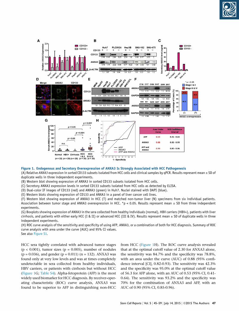
Figure 1. Endogenous and Secretory Overexpression of ANXA3 Is Strongly Associated with HCC Pathogenesis(A) Relative ANXA3 expression in sorted CD133 subsets isolated from HCC cells and clinical samples by qPCR. Results represent mean ± SD ofduplicate wells in three independent experiments.(B) Western blot showing expression of ANXA3 in sorted CD133 subsets isolated from HCC cells.(C) Secretory ANXA3 expression levels in sorted CD133 subsets isolated from HCC cells as detected by ELISA.(D) Dual-color IF images of CD133 (red) and ANXA3 (green) in Huh7. Nuclei stained with DAPI (blue).(E) Western blots showing expression of CD133 and ANXA3 in a panel of liver cancer cell lines.(F) Western blot showing expression of ANXA3 in HCC (T) and matched non-tumor liver (N) specimens from six individual patients.Association between tumor stage and ANXA3 overexpression in HCC. *p < 0.05. Results represent mean ± SD from three independentexperiments.(G) Boxplots showing expression of ANXA3 in the sera collected from healthy individuals (normal), HBV carriers (HBV+), patients with livercirrhosis, and patients with either early HCC (I & II) or advanced HCC (III & IV). Results represent mean ± SD of duplicate wells in threeindependent experiments.(H) ROC curve analysis of the sensitivity and specificity of using AFP, ANXA3, or a combination of both for HCC diagnosis. Summary of ROCcurve analysis with area under the curve (AUC) and 95% CI values.See also Figure S1.
HCC sera tightly correlated with advanced tumor stages
(p < 0.001), tumor sizes (p = 0.005), number of nodules
(p = 0.036), and gender (p = 0.011) (n = 132). ANXA3 was
found only at very low levels and was at times completely
undetectable in sera collected from healthy individuals,
HBV carriers, or patients with cirrhosis but without HCC
(Figure 1G; Table S4). Alpha-fetoprotein (AFP) is the most
widely used biomarker for HCCdiagnosis. By receiver-oper-
ating characteristic (ROC) curve analysis, ANXA3 was
found to be superior to AFP in distinguishing non-HCC
from HCC (Figure 1H). The ROC curve analysis revealed
that at the optimal cutoff value of 2.30 for ANXA3 alone,
the sensitivity was 84.7% and the specificity was 78.8%,
with an area under the curve (AUC) of 0.88 (95% confi-
dence interval [CI], 0.82-0.93). The sensitivity was 42.3%
and the specificity was 95.0% at the optimal cutoff value
of 56.3 for AFP alone, with an AUC of 0.53 (95% CI, 0.41-
0.64). The sensitivity was 93.2% and the specificity was
70% for the combination of ANXA3 and AFP, with an
AUC of 0.90 (95% CI, 0.85-0.96).
Stem Cell Reports j Vol. 5 j 45–59 j July 14, 2015 j ª2015 The Authors 47
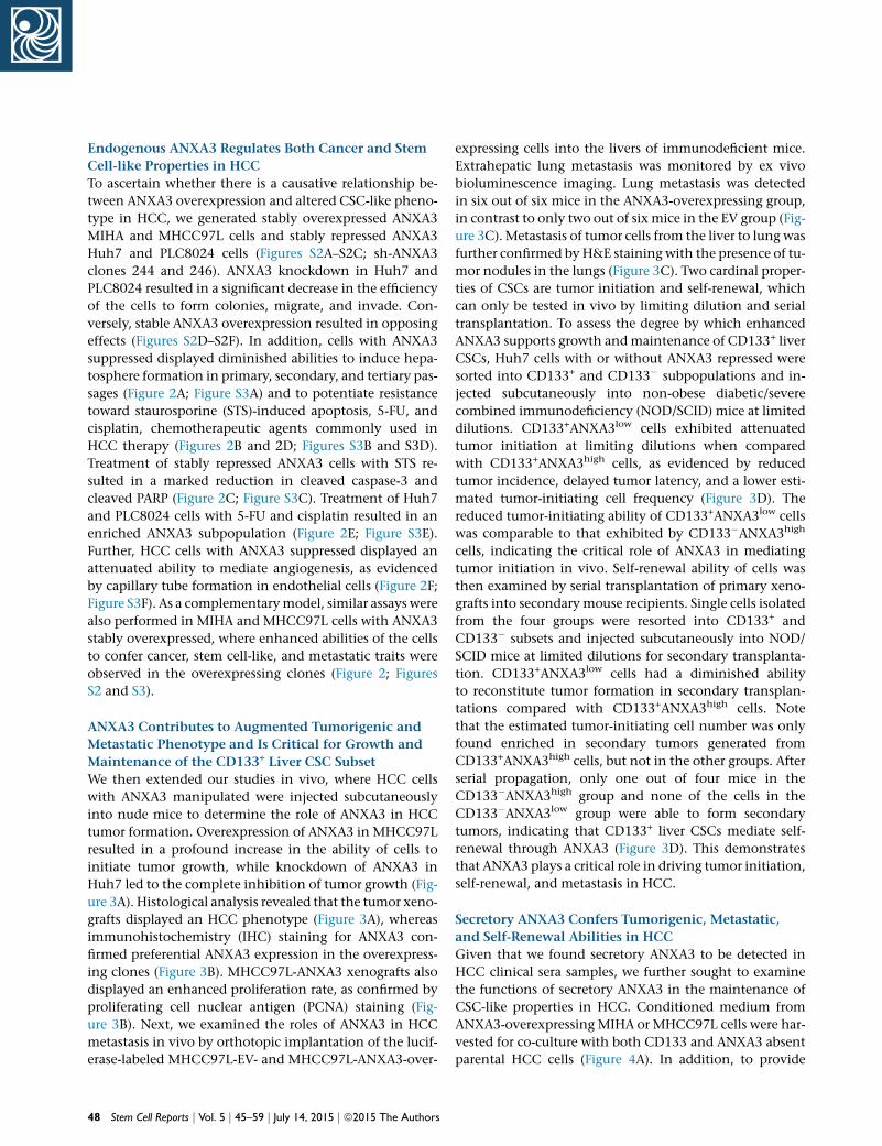
Endogenous ANXA3 Regulates Both Cancer and Stem
Cell-like Properties in HCC
To ascertain whether there is a causative relationship be-
tween ANXA3 overexpression and altered CSC-like pheno-
type in HCC, we generated stably overexpressed ANXA3
MIHA and MHCC97L cells and stably repressed ANXA3
Huh7 and PLC8024 cells (Figures S2A–S2C; sh-ANXA3
clones 244 and 246). ANXA3 knockdown in Huh7 and
PLC8024 resulted in a significant decrease in the efficiency
of the cells to form colonies, migrate, and invade. Con-
versely, stable ANXA3 overexpression resulted in opposing
effects (Figures S2D–S2F). In addition, cells with ANXA3
suppressed displayed diminished abilities to induce hepa-
tosphere formation in primary, secondary, and tertiary pas-
sages (Figure 2A; Figure S3A) and to potentiate resistance
toward staurosporine (STS)-induced apoptosis, 5-FU, and
cisplatin, chemotherapeutic agents commonly used in
HCC therapy (Figures 2B and 2D; Figures S3B and S3D).
Treatment of stably repressed ANXA3 cells with STS re-
sulted in a marked reduction in cleaved caspase-3 and
cleaved PARP (Figure 2C; Figure S3C). Treatment of Huh7
and PLC8024 cells with 5-FU and cisplatin resulted in an
enriched ANXA3 subpopulation (Figure 2E; Figure S3E).
Further, HCC cells with ANXA3 suppressed displayed an
attenuated ability to mediate angiogenesis, as evidenced
by capillary tube formation in endothelial cells (Figure 2F;
Figure S3F). As a complementarymodel, similar assays were
also performed in MIHA and MHCC97L cells with ANXA3
stably overexpressed, where enhanced abilities of the cells
to confer cancer, stem cell-like, and metastatic traits were
observed in the overexpressing clones (Figure 2; Figures
S2 and S3).
ANXA3 Contributes to Augmented Tumorigenic and
Metastatic Phenotype and Is Critical for Growth and
Maintenance of the CD133+ Liver CSC Subset
We then extended our studies in vivo, where HCC cells
with ANXA3 manipulated were injected subcutaneously
into nude mice to determine the role of ANXA3 in HCC
tumor formation. Overexpression of ANXA3 in MHCC97L
resulted in a profound increase in the ability of cells to
initiate tumor growth, while knockdown of ANXA3 in
Huh7 led to the complete inhibition of tumor growth (Fig-
ure 3A). Histological analysis revealed that the tumor xeno-
grafts displayed an HCC phenotype (Figure 3A), whereas
immunohistochemistry (IHC) staining for ANXA3 con-
firmed preferential ANXA3 expression in the overexpress-
ing clones (Figure 3B). MHCC97L-ANXA3 xenografts also
displayed an enhanced proliferation rate, as confirmed by
proliferating cell nuclear antigen (PCNA) staining (Fig-
ure 3B). Next, we examined the roles of ANXA3 in HCC
metastasis in vivo by orthotopic implantation of the lucif-
erase-labeled MHCC97L-EV- and MHCC97L-ANXA3-over-
48 Stem Cell Reports j Vol. 5 j 45–59 j July 14, 2015 j ª2015 The Authors
expressing cells into the livers of immunodeficient mice.
Extrahepatic lung metastasis was monitored by ex vivo
bioluminescence imaging. Lung metastasis was detected
in six out of six mice in the ANXA3-overexpressing group,
in contrast to only two out of six mice in the EV group (Fig-
ure 3C). Metastasis of tumor cells from the liver to lung was
further confirmed by H&E staining with the presence of tu-
mor nodules in the lungs (Figure 3C). Two cardinal proper-
ties of CSCs are tumor initiation and self-renewal, which
can only be tested in vivo by limiting dilution and serial
transplantation. To assess the degree by which enhanced
ANXA3 supports growth andmaintenance of CD133+ liver
CSCs, Huh7 cells with or without ANXA3 repressed were
sorted into CD133+ and CD133� subpopulations and in-
jected subcutaneously into non-obese diabetic/severe
combined immunodeficiency (NOD/SCID) mice at limited
dilutions. CD133+ANXA3low cells exhibited attenuated
tumor initiation at limiting dilutions when compared
with CD133+ANXA3high cells, as evidenced by reduced
tumor incidence, delayed tumor latency, and a lower esti-
mated tumor-initiating cell frequency (Figure 3D). The
reduced tumor-initiating ability of CD133+ANXA3low cells
was comparable to that exhibited by CD133�ANXA3high
cells, indicating the critical role of ANXA3 in mediating
tumor initiation in vivo. Self-renewal ability of cells was
then examined by serial transplantation of primary xeno-
grafts into secondary mouse recipients. Single cells isolated
from the four groups were resorted into CD133+ and
CD133� subsets and injected subcutaneously into NOD/
SCID mice at limited dilutions for secondary transplanta-
tion. CD133+ANXA3low cells had a diminished ability
to reconstitute tumor formation in secondary transplan-
tations compared with CD133+ANXA3high cells. Note
that the estimated tumor-initiating cell number was only
found enriched in secondary tumors generated from
CD133+ANXA3high cells, but not in the other groups. After
serial propagation, only one out of four mice in the
CD133�ANXA3high group and none of the cells in the
CD133�ANXA3low group were able to form secondary
tumors, indicating that CD133+ liver CSCs mediate self-
renewal through ANXA3 (Figure 3D). This demonstrates
that ANXA3 plays a critical role in driving tumor initiation,
self-renewal, and metastasis in HCC.
Secretory ANXA3 Confers Tumorigenic, Metastatic,
and Self-Renewal Abilities in HCC
Given that we found secretory ANXA3 to be detected in
HCC clinical sera samples, we further sought to examine
the functions of secretory ANXA3 in the maintenance of
CSC-like properties in HCC. Conditioned medium from
ANXA3-overexpressing MIHA or MHCC97L cells were har-
vested for co-culture with both CD133 and ANXA3 absent
parental HCC cells (Figure 4A). In addition, to provide
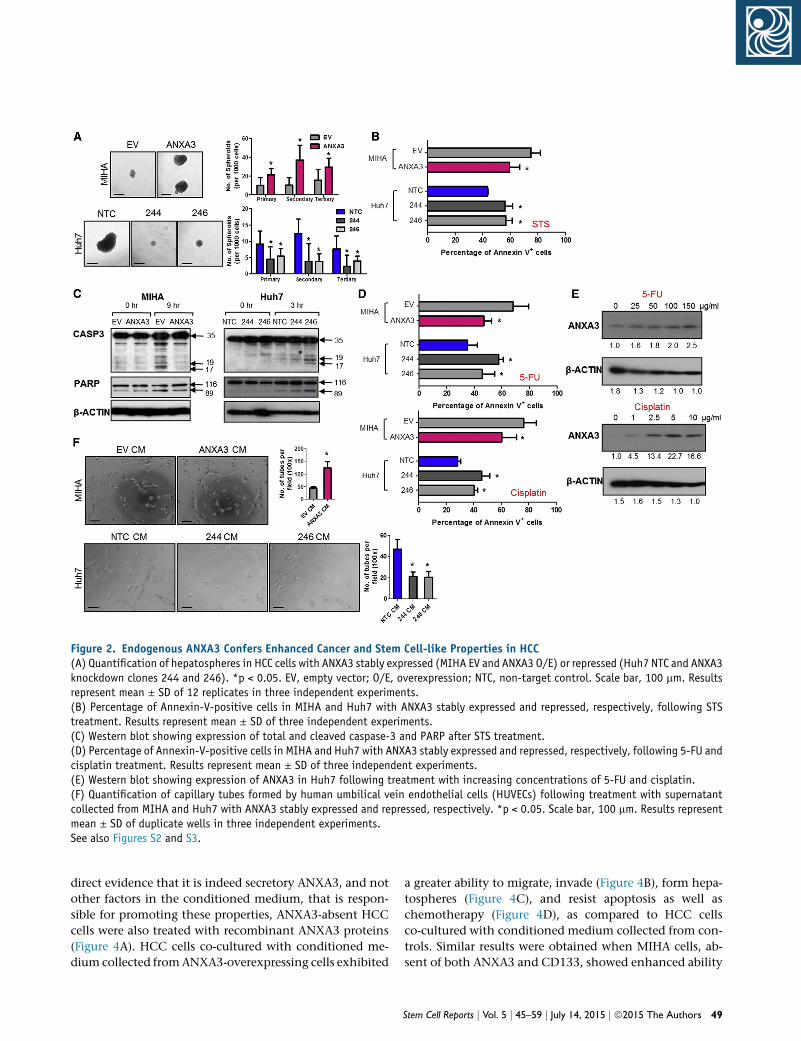
Figure 2. Endogenous ANXA3 Confers Enhanced Cancer and Stem Cell-like Properties in HCC(A) Quantification of hepatospheres in HCC cells with ANXA3 stably expressed (MIHA EV and ANXA3 O/E) or repressed (Huh7 NTC and ANXA3knockdown clones 244 and 246). *p < 0.05. EV, empty vector; O/E, overexpression; NTC, non-target control. Scale bar, 100 mm. Resultsrepresent mean ± SD of 12 replicates in three independent experiments.(B) Percentage of Annexin-V-positive cells in MIHA and Huh7 with ANXA3 stably expressed and repressed, respectively, following STStreatment. Results represent mean ± SD of three independent experiments.(C) Western blot showing expression of total and cleaved caspase-3 and PARP after STS treatment.(D) Percentage of Annexin-V-positive cells in MIHA and Huh7 with ANXA3 stably expressed and repressed, respectively, following 5-FU andcisplatin treatment. Results represent mean ± SD of three independent experiments.(E) Western blot showing expression of ANXA3 in Huh7 following treatment with increasing concentrations of 5-FU and cisplatin.(F) Quantification of capillary tubes formed by human umbilical vein endothelial cells (HUVECs) following treatment with supernatantcollected from MIHA and Huh7 with ANXA3 stably expressed and repressed, respectively. *p < 0.05. Scale bar, 100 mm. Results representmean ± SD of duplicate wells in three independent experiments.See also Figures S2 and S3.
direct evidence that it is indeed secretory ANXA3, and not
other factors in the conditioned medium, that is respon-
sible for promoting these properties, ANXA3-absent HCC
cells were also treated with recombinant ANXA3 proteins
(Figure 4A). HCC cells co-cultured with conditioned me-
dium collected fromANXA3-overexpressing cells exhibited
a greater ability to migrate, invade (Figure 4B), form hepa-
tospheres (Figure 4C), and resist apoptosis as well as
chemotherapy (Figure 4D), as compared to HCC cells
co-cultured with conditioned medium collected from con-
trols. Similar results were obtained when MIHA cells, ab-
sent of both ANXA3 and CD133, showed enhanced ability
Stem Cell Reports j Vol. 5 j 45–59 j July 14, 2015 j ª2015 The Authors 49
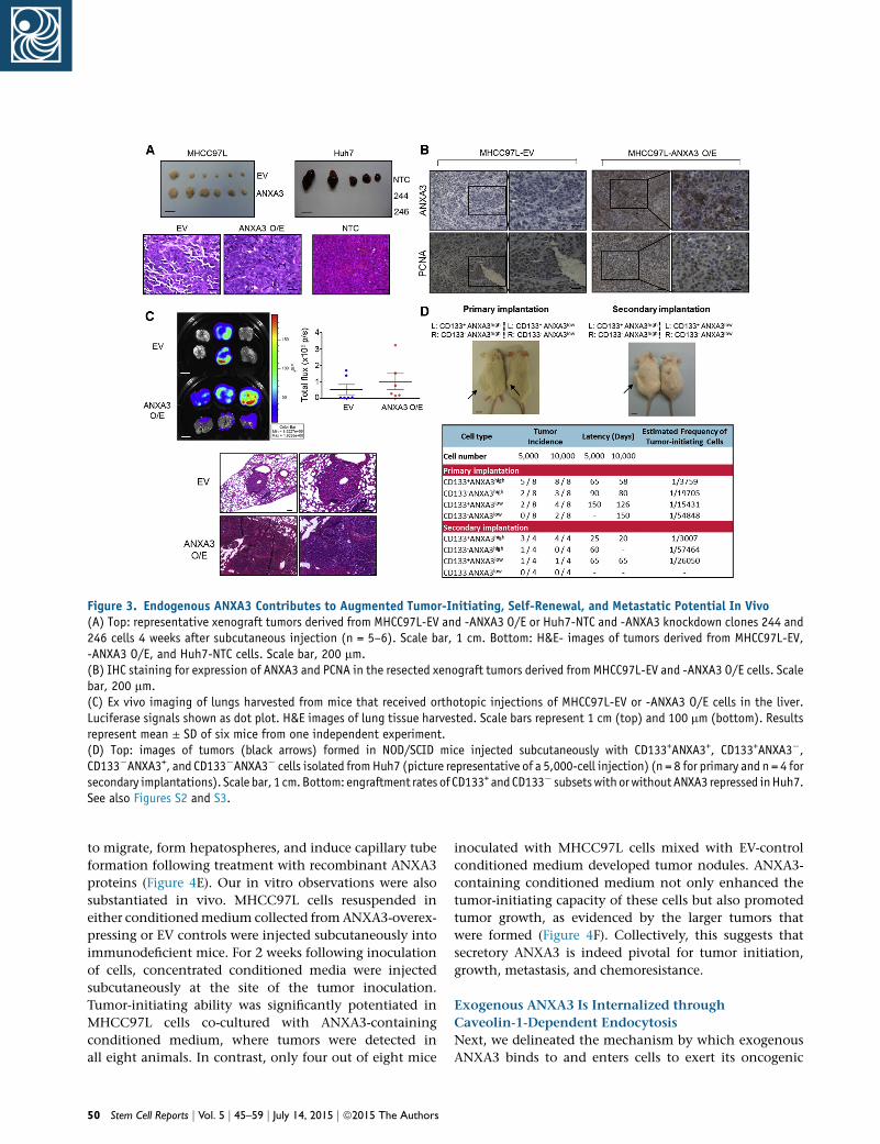
Figure 3. Endogenous ANXA3 Contributes to Augmented Tumor-Initiating, Self-Renewal, and Metastatic Potential In Vivo(A) Top: representative xenograft tumors derived from MHCC97L-EV and -ANXA3 O/E or Huh7-NTC and -ANXA3 knockdown clones 244 and246 cells 4 weeks after subcutaneous injection (n = 5–6). Scale bar, 1 cm. Bottom: H&E- images of tumors derived from MHCC97L-EV,-ANXA3 O/E, and Huh7-NTC cells. Scale bar, 200 mm.(B) IHC staining for expression of ANXA3 and PCNA in the resected xenograft tumors derived from MHCC97L-EV and -ANXA3 O/E cells. Scalebar, 200 mm.(C) Ex vivo imaging of lungs harvested from mice that received orthotopic injections of MHCC97L-EV or -ANXA3 O/E cells in the liver.Luciferase signals shown as dot plot. H&E images of lung tissue harvested. Scale bars represent 1 cm (top) and 100 mm (bottom). Resultsrepresent mean ± SD of six mice from one independent experiment.(D) Top: images of tumors (black arrows) formed in NOD/SCID mice injected subcutaneously with CD133+ANXA3+, CD133+ANXA3�,CD133�ANXA3+, and CD133�ANXA3� cells isolated fromHuh7 (picture representative of a 5,000-cell injection) (n = 8 for primary and n = 4 forsecondary implantations). Scale bar, 1 cm.Bottom: engraftment rates of CD133+ and CD133� subsetswithorwithoutANXA3 repressed inHuh7.See also Figures S2 and S3.
to migrate, form hepatospheres, and induce capillary tube
formation following treatment with recombinant ANXA3
proteins (Figure 4E). Our in vitro observations were also
substantiated in vivo. MHCC97L cells resuspended in
either conditionedmedium collected from ANXA3-overex-
pressing or EV controls were injected subcutaneously into
immunodeficient mice. For 2 weeks following inoculation
of cells, concentrated conditioned media were injected
subcutaneously at the site of the tumor inoculation.
Tumor-initiating ability was significantly potentiated in
MHCC97L cells co-cultured with ANXA3-containing
conditioned medium, where tumors were detected in
all eight animals. In contrast, only four out of eight mice
50 Stem Cell Reports j Vol. 5 j 45–59 j July 14, 2015 j ª2015 The Authors
inoculated with MHCC97L cells mixed with EV-control
conditioned medium developed tumor nodules. ANXA3-
containing conditioned medium not only enhanced the
tumor-initiating capacity of these cells but also promoted
tumor growth, as evidenced by the larger tumors that
were formed (Figure 4F). Collectively, this suggests that
secretory ANXA3 is indeed pivotal for tumor initiation,
growth, metastasis, and chemoresistance.
Exogenous ANXA3 Is Internalized through
Caveolin-1-Dependent Endocytosis
Next, we delineated the mechanism by which exogenous
ANXA3 binds to and enters cells to exert its oncogenic
Figure 4. Secretory ANXA3 Confers Enhanced Cancer and Stem Cell-like Properties in HCC In Vitro and In Vivo(A) Secretory ANXA3 overexpression and recombinant ANXA3 model systems.(B) Quantification of number of MIHA and MHCC97L cells that migrated or invaded following co-culture with empty vector control medium(EV CM) or ANXA3-containing conditioned medium (ANXA3 O/E CM). *p < 0.05. Scale bar, 200 mm. Results represent mean ± SD from threeindependent experiments.(C) Quantification of hepatospheres formed following co-culture with EV CM or ANXA3 O/E CM. ***p < 0.001. Scale bar, 100 mm. Resultsrepresent mean ± SD of 12 replicates in three independent experiments.(D) Percentage of Annexin-V-positive cells in MIHA and MHCC97L co-cultured with EV CM or ANXA3 O/E CM, following treatment with STS,5-FU, or cisplatin. Results represent mean ± SD of three independent experiments.(E) Top and bottom: quantification of number of MIHA that migrated and formed hepatospheres following treatment with recombinantANXA3 protein (rANXA3). *p < 0.05 and **p < 0.01. Results represent mean ± SD from three independent experiments. Middle: quanti-fication of capillary tubes formed by HUVECs following treatment with rANXA3. ***p < 0.001. Scale bars represent 200 mm (top) and100 mm (middle and bottom). Results represent mean ± SD of duplicate wells in three independent experiments.(F) Representative xenograft tumors derived from MHCC97L cells co-cultured with EV CM or ANXA3 O/E CM 4 weeks after subcutaneousinjection (n = 8). Dot plot shows the tumor weight of each xenograft. Results represent mean ± SD of eight mice from one independentexperiment.
effects to promote HCC.We first confirmed that exogenous
ANXA3 was indeed internalized. Recombinant SNAP-
tagged ANXA3 protein was first labeled with fluorophore
(red). PLC8024 HCC cells were also labeled with Vybrant
carboxyfluorescein diacetate (CFDA) succinimidyl ester
cell tracer dye (green). Following co-culture of fluoro-
phore-tagged recombinant ANXA3 protein with fluores-
cein-isothiocyanate-labeled PLC8024 cells, SNAP-tagged
ANXA3 proteins were clearly detected on both the mem-
brane and in the cytoplasm of PLC8024 cells by confocal
microscopy (Figure 5A). This observation was further sub-
stantiated by western blot where subcellular fractionation
of HCC cells co-cultured with recombinant ANXA3 protein
was analyzed. FLAG-tagged ANXA3 proteins present in
the two fractions were pulled down by immunoprecipita-
tion with FLAG-conjugated antibodies and subjected to
Stem Cell Reports j Vol. 5 j 45–59 j July 14, 2015 j ª2015 The Authors 51
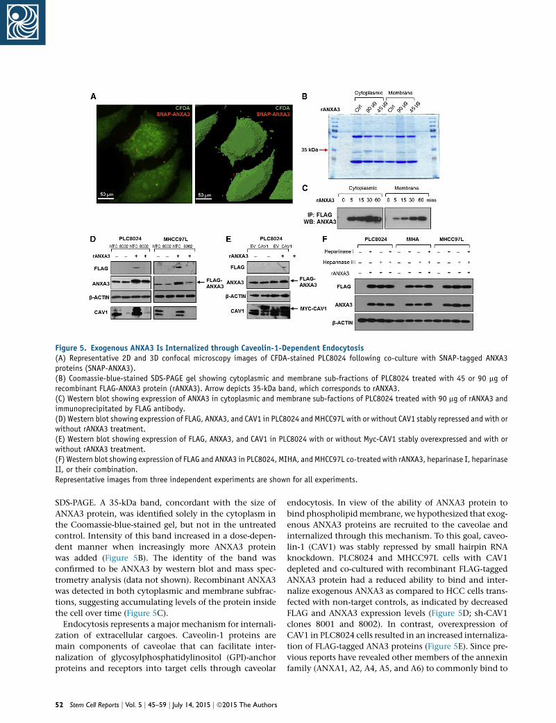
Figure 5. Exogenous ANXA3 Is Internalized through Caveolin-1-Dependent Endocytosis(A) Representative 2D and 3D confocal microscopy images of CFDA-stained PLC8024 following co-culture with SNAP-tagged ANXA3proteins (SNAP-ANXA3).(B) Coomassie-blue-stained SDS-PAGE gel showing cytoplasmic and membrane sub-fractions of PLC8024 treated with 45 or 90 mg ofrecombinant FLAG-ANXA3 protein (rANXA3). Arrow depicts 35-kDa band, which corresponds to rANXA3.(C) Western blot showing expression of ANXA3 in cytoplasmic and membrane sub-factions of PLC8024 treated with 90 mg of rANXA3 andimmunoprecipitated by FLAG antibody.(D) Western blot showing expression of FLAG, ANXA3, and CAV1 in PLC8024 and MHCC97L with or without CAV1 stably repressed and with orwithout rANXA3 treatment.(E) Western blot showing expression of FLAG, ANXA3, and CAV1 in PLC8024 with or without Myc-CAV1 stably overexpressed and with orwithout rANXA3 treatment.(F) Western blot showing expression of FLAG and ANXA3 in PLC8024, MIHA, and MHCC97L co-treated with rANXA3, heparinase I, heparinaseII, or their combination.Representative images from three independent experiments are shown for all experiments.
SDS-PAGE. A 35-kDa band, concordant with the size of
ANXA3 protein, was identified solely in the cytoplasm in
the Coomassie-blue-stained gel, but not in the untreated
control. Intensity of this band increased in a dose-depen-
dent manner when increasingly more ANXA3 protein
was added (Figure 5B). The identity of the band was
confirmed to be ANXA3 by western blot and mass spec-
trometry analysis (data not shown). Recombinant ANXA3
was detected in both cytoplasmic and membrane subfrac-
tions, suggesting accumulating levels of the protein inside
the cell over time (Figure 5C).
Endocytosis represents a major mechanism for internali-
zation of extracellular cargoes. Caveolin-1 proteins are
main components of caveolae that can facilitate inter-
nalization of glycosylphosphatidylinositol (GPI)-anchor
proteins and receptors into target cells through caveolar
52 Stem Cell Reports j Vol. 5 j 45–59 j July 14, 2015 j ª2015 The Authors
endocytosis. In view of the ability of ANXA3 protein to
bind phospholipidmembrane, we hypothesized that exog-
enous ANXA3 proteins are recruited to the caveolae and
internalized through this mechanism. To this goal, caveo-
lin-1 (CAV1) was stably repressed by small hairpin RNA
knockdown. PLC8024 and MHCC97L cells with CAV1
depleted and co-cultured with recombinant FLAG-tagged
ANXA3 protein had a reduced ability to bind and inter-
nalize exogenous ANXA3 as compared to HCC cells trans-
fected with non-target controls, as indicated by decreased
FLAG and ANXA3 expression levels (Figure 5D; sh-CAV1
clones 8001 and 8002). In contrast, overexpression of
CAV1 in PLC8024 cells resulted in an increased internaliza-
tion of FLAG-tagged ANA3 proteins (Figure 5E). Since pre-
vious reports have revealed other members of the annexin
family (ANXA1, A2, A4, A5, and A6) to commonly bind to
glycosaminoglycans (GAGs) such as heparin sulfate chains,
we also explored whether ANXA3 could also be internal-
ized via heparin sulfate proteoglycan (HSPG)-mediated
endocytosis. PLC8024, MIHA, and MHCC97L cells treated
with heparinase I, heparinase III, or their combination and
co-cultured with recombinant FLAG-tagged ANXA3 pro-
tein did not alter the amount of ANXA3 proteins internal-
ized (Figure 5F), suggesting that entry of exogenous of
ANXA3 in HCC is dependent on caveolin-1-mediated,
but not HSPG-mediated, endocytosis.
ANXA3 Induces a Feed-Forward Loop that Is Mediated
by the MKK4/JNK Signaling Cascade
In an effort to characterize the molecular mechanism by
which ANXA3 drives CSC-like properties in HCC, mRNA
expression profiling was performed to compare the gene
expression profiles of PLC8024 cells with or without
ANXA3 repressed (Figure S4A; NTC versus sh-ANXA3 clone
246). Using a fold-change cutoff of >3, 2,372 differentially
expressed genes were identified. Subsequent pathway anal-
ysis found many of the deregulated genes to be closely
associated with JNK/AP-1 and MAPK, pathways, which
have previously been shown to play a critical role in
HCC pathogenesis (Figure 6A; Figure S4B) (Hagiwara
et al., 2012; Jin et al., 2013). Deregulation of key players
of the JNK pathway, including p-MKK4, JNK kinase activ-
ity, c-MYC, and p21, was subsequently validated by west-
ern blot in HCC cells with ANXA3 repressed (Huh7 and
PLC8024) or overexpressed (MIHA) (Figure 6B). Similar to
results obtained from ANXA3-overexpressing cells, the
addition of exogenous ANXA3 proteins in CD133- and
ANXA3-absent MIHA cells or culturing of the cells with
conditioned medium collected from ANXA3-overexpress-
ing cells resulted in JNK pathway activation, as evidenced
by an increase in JNK activity, increased c-MYC expression,
and reduced p21 expression (Figure 6C). A luciferase-re-
porter construct consisting of a consensus sequence of
AP-1 binding site was transfected into Huh7 cells with or
without ANXA3 repressed. Activity of AP-1, a known
downstream target of JNK, was significantly reduced in
the ANXA3-knockdown clones, further suggestive of the
critical role of the ANXA3-JNK axis in CD133+ liver-CSC-
driven HCC (Figure 6D). Consistent with this, we also
found p-JNK to be preferentially expressed in CD133+
HCC cells (Figure S4C), while dual-color IF confirmed co-
localization of ANXA3 and p-JNK, as well as CD133 and
p-JNK in both Huh7 and PLC8024 cells (Figure 6E; Figures
S4D and S4E). To substantiate the importance of the JNK/
AP-1 pathway in ANXA3-driven HCC, we performed res-
cue experiments using the JNK-specific inhibitor (JNKi)
SP600125. JNKi suppressed the oncogenic properties con-
ferred by ANXA3 overexpression, as evidenced by the
diminished abilities of HCC cells to form colonies,migrate,
invade, induce angiogenesis, form hepatospheres, and
resist apoptosis and chemotherapy (Figures 6F–6J). Inter-
estingly, treatment of parental HCC cells or HCC cells over-
expressing ANXA3 with JNKi resulted in not only a reduc-
tion in JNK activity and modulation of downstream target
genes (c-MYC and p21) but also a marked decrease in
ANXA3 expression, suggesting that ANXA3 induces a
feed-forward loop that is mediated byMKK4/JNK signaling
(Figures 6K–6L).
ANXA3 Neutralization Suppresses Growth and
Self-Renewal of HCC In Vivo, Sensitizes HCC to
Chemotherapy, and Eradicates the CD133 Liver CSC
Subset
In light of the implications of ANXA3 in CD133+ liver
CSCs and in HCC, we subsequently developed a mono-
clonal antibody specific against ANXA3 (anti-ANXA3
mAb) and tested for its application as a therapeutic treat-
ment against HCC. Specificity of the antibody was con-
firmed by western blot on a panel of HCC cell lines where
a single 33-kDa band was detected, corresponding to
ANXA3 (Figure 7A). The expression levels of ANXA3 ob-
tained were concordant with findings using a commer-
cially validated ANXA3 antibody. Cell proliferation rates
of CD133- and ANXA3-positive Huh7 cells were signifi-
cantly inhibited with the addition of anti-ANXA3 mAb,
while the CD133- and ANXA3-absent immortalized
normal liver cell line MIHA remained unresponsive (Fig-
ure 7B). Treatment of Huh7 with anti-ANXA3 mAb re-
sulted in a marked increase in apoptotic cells, suggesting
that cell death was a factor causing the inhibition of cell
proliferation (Figure 7C). In addition, anti-ANXA3 mAb
also reduced the abilities of HCC cells to migrate, invade,
induce angiogenesis, and form hepatospheres (Figures
7D–7F). HCC cells treated with a combination of anti-
ANXA3 mAb and cisplatin synergistically inhibited cell
proliferation in vitro and sensitized HCC cells to cisplatin
(Figure 7G). Consistently, a similar trend was also
observed when anti-ANXA3 mAb was used for treatment
of secretory ANXA3. MIHA and MHCC97L cells, both
negative for CD133 and ANXA3, were stably transfected
with ANXA3 or EV control. Conditioned medium from
these cells were then collected and used for various func-
tional experiments. Addition of ANXA3 mAb in HCC
cells treated with conditioned medium collected from
ANXA3-overexpressing cells resulted in a reduced ability
of the cells to migrate, invade, induce angiogenesis,
form hepatospheres, and resist apoptosis and chemo-
therapy (Figures S5A and S5B). This observation was
confirmed in vivo when treatment of Huh7 xenografts
in immunodeficient mice with anti-ANXA3 mAb alone
or in combination with cisplatin resulted in a marked
reduction in tumor volume (Figure 7H; Figure S5C).
Stem Cell Reports j Vol. 5 j 45–59 j July 14, 2015 j ª2015 The Authors 53
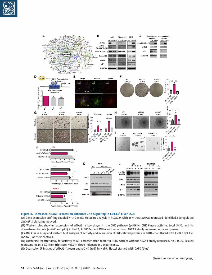
Figure 6. Increased ANXA3 Expression Enhances JNK Signaling in CD133+ Liver CSCs(A) Gene expression profiling coupled with GeneGo Metacore analysis in PLC8024 with or without ANXA3 repressed identified a deregulatedJNK/AP-1 signaling network.(B) Western blot showing expression of ANXA3, a key player in the JNK pathway (p-MKK4, JNK kinase activity, total JNK), and itsdownstream targets (c-MYC and p21) in Huh7, PLC8024, and MIHA with or without ANXA3 stably repressed or overexpressed.(C) JNK kinase assay and western blot analysis of activity and expression of JNK-related proteins in MIHA co-cultured with ANXA3 O/E CM,rANXA3, or their controls.(D) Luciferase reporter assay for activity of AP-1 transcription factor in Huh7 with or without ANXA3 stably repressed. *p < 0.05. Resultsrepresent mean ± SD from triplicate wells in three independent experiments.(E) Dual-color IF images of ANXA3 (green) and p-JNK (red) in Huh7. Nuclei stained with DAPI (blue).
(legend continued on next page)
54 Stem Cell Reports j Vol. 5 j 45–59 j July 14, 2015 j ª2015 The Authors
Tumor growth was markedly suppressed in mice treated
with increasing concentrations of anti-ANXA3 mAb.
When anti-ANXA3 mAb was administered in combina-
tion with cisplatin, the growth of the tumor engraft-
ments was inhibited by as much as 90% compared with
the controls (PBS or immunoglobulin G [IgG]). Although
cisplatin treatment alone led to a bigger reduction in
tumor volume as compared to anti-ANXA3 mAb (Fig-
ure 7H), the residual xenografts, when serially trans-
planted into secondary mouse recipients, formed the
largest tumors (Figure 7I). Residual xenografts from
anti-ANXA3 mAb or mAb and cisplatin treatment failed
to give rise to tumors in serial transplantations (Figure 7I),
suggesting that a subset of CSCs is enriched by chemo-
therapy treatment. Subsequent analysis of the residual
xenografts by flow cytometry for CD133 confirmed this
hypothesis, where proportion of CD133+ cells was found
enriched after chemotherapy, while the proportion of
CD133+ cells decreased following treatment with anti-
ANXA3 mAb alone or in combination with cisplatin (Fig-
ure 7J; Figure S5E). In addition to CD133, other liver CSC
markers known to have overlapping expression with
CD133, including CD24 (Lee et al., 2011) and EpCAM
(Yamashita et al., 2009), were also found to be dimin-
ished upon antibody treatment (Figure S5F). H&E stain-
ing and IHC were performed on tissue sections from
the resected tumor residuals (Figure 7K; Figure S5D).
Necrosis was only observed in the xenografts treated
with anti-ANXA3 mAb alone or in combination with
cisplatin. ANXA3 expression was increased in cisplatin-
treated xenografts but was markedly reduced in anti-
ANXA3 mAb-treated mice. A concomitant decrease in
PCNA was also observed in xenografts treated with anti-
ANXA3 mAb with or without cisplatin, indicative of
attenuated proliferative potential. None of the mice
showed signs of disability, behavior abnormalities, or sig-
nificant changes in body weight. Compared to untreated
control or IgG-treated mice, no additional tissue damage
was observed in other vital organs (Figure S5G). Mecha-
(F) Quantification of number of colonies formed in the indicated stab5 mm. Results represent mean ± SD from triplicate wells in three ind(G) Quantification of number of cells that migrated or invaded followinScale bar, 100 mm. Results represent mean ± SD from three independ(H) Quantification of capillary tubes formed by HUVECs following treatwithout JNK inhibitor. *p < 0.05. Scale bar, 100 mm. Results represen(I) Quantification of hepatospheres with or without JNK inhibitor. Scaindependent experiments.(J) Percentage of Annexin-V-positive cells in the indicated stable cel5-FU, or cisplatin. Results represent mean ± SD of three independent(K and L) Western blot showing expression of ANXA3, c-MYC, and p21(L) treated with 25 or 50 mM JNK inhibitor (SP600125).See also Figure S4.
nistically, treatment of HCC cells with anti-ANXA3
mAb in vitro and in vivo similarly led to a suppressed
JNK pathway (Figure 7L).
DISCUSSION
Frequent tumor relapse in multiple tumor types has now
been attributed to the presence of residual CSCs after con-
ventional treatments. We and others have previously iden-
tified CD133 to mark a liver CSC subpopulation in HCC
(Ma et al., 2007, 2010). Yet, the functional paths by which
these cells promote hepatocarcinogenesis remain limited,
significantly impeding our efforts in developing CSC-spe-
cific therapies. We characterized the mRNA transcriptome
of CD133+ and CD133� subpopulations in HCC by RNA-
seq. Pathway enrichment analysis found the CD133+ sub-
set to be tightly associated with an activated MAPK
pathway, which is in concordant with our previous study,
where we found CD133 to promote angiogenesis through
IL-8-activated ERK (Tang et al., 2012). In addition, our
CD133� non-CSC subset was also found to be enriched
for genes critical in hepatocyte differentiation. This result
is also consistent with our finding where CD133� cells
were found to be unable to differentiate into skeletal and
cardiac lineages, while in contrast, CD133+ liver CSCs
could efficiently differentiate into non-hepatocyte-like, an-
giomyogenic-like cells following cell-directed differentia-
tion in vitro (Ma et al., 2007), suggesting that CD133� cells
exist in a terminally differentiated state. Of interest, one-
third of the differentially expressed genes identified were
found to encode for secretory proteins. There is accumu-
lating data to show that maintenance of cancer and stem-
ness properties is dependent on the microenvironment in
which deregulated secreting factors can communicate
through paracrine or autocrine signaling. Given the impor-
tance of secretory factors in modulating CSC features and
that they can be found accessible in the conditionedmedia
of cells or serum in patients, thus making them prime
le cell lines with or without JNK inhibitor. ***p < 0.001. Scale bar,ependent experiments.g co-culture with or without JNK inhibitor. *p < 0.05 and *p < 0.01.ent experiments.ment with supernatant collected from the indicated cell lines with ort mean ± SD of duplicate wells in three independent experiments.le bar, 100 mm. Results represent mean ± SD of 12 replicates in three
l lines with or without JNK inhibitor, following treatment with STS,experiments.in ANXA3-overexpressing MIHA (K) and parental Huh7 and PLC8024
Stem Cell Reports j Vol. 5 j 45–59 j July 14, 2015 j ª2015 The Authors 55
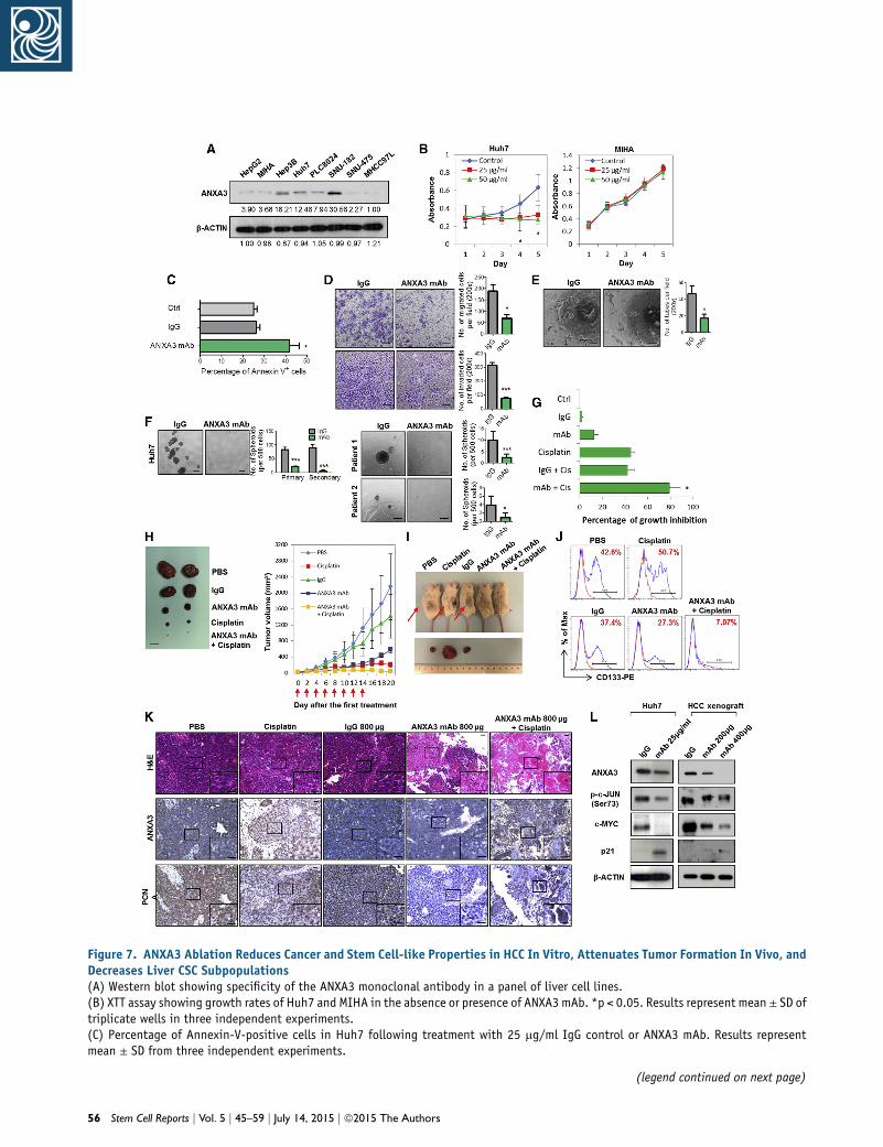
Figure 7. ANXA3 Ablation Reduces Cancer and Stem Cell-like Properties in HCC In Vitro, Attenuates Tumor Formation In Vivo, andDecreases Liver CSC Subpopulations(A) Western blot showing specificity of the ANXA3 monoclonal antibody in a panel of liver cell lines.(B) XTT assay showing growth rates of Huh7 and MIHA in the absence or presence of ANXA3 mAb. *p < 0.05. Results represent mean ± SD oftriplicate wells in three independent experiments.(C) Percentage of Annexin-V-positive cells in Huh7 following treatment with 25 mg/ml IgG control or ANXA3 mAb. Results representmean ± SD from three independent experiments.
(legend continued on next page)
56 Stem Cell Reports j Vol. 5 j 45–59 j July 14, 2015 j ª2015 The Authors
druggable targets for novel therapy development, we
focused our studies in this area.
The focus of ANXA3 research in the past has centered
on its expression in various diseases. A number of studies
have found ANXA3 to be frequently overexpressed in
ovarian, breast, colon, lung, gastric, gallbladder, testic-
ular, and urothelial cancers (Kollermann et al., 2008;
Schostak et al., 2009; Liu et al., 2009; Yan et al., 2010;
Wu et al., 2013). In contrast, ANXA3 was also found to
be downregulated in prostate and papillary thyroid can-
cers (Wu et al., 2013). In recent years, a small number
of studies have identified ANXA3 to be secreted, where
low levels of ANXA3 present in the urine were found to
have diagnostic significance for early prostate cancer
(Schostak et al., 2009; Yin et al., 2012) and secretion of
ANXA3 from ovarian cancer cells is associated with plat-
inum resistance. Here, we found ANXA3 to represent the
most significantly upregulated gene that encoded for a
secretory protein in the CD133+ liver CSC subset. Hara-
guchi et al. also likewise found ANXA3 to be preferen-
tially expressed in the side population (SP) isolated
from Huh7 cells (Haraguchi et al., 2006). SP cells from
HCC were subsequently characterized to also express
CD133. Specifically in HCC, Tong et al. found ANXA3
to be upregulated in 5-FU-resistant HCC cells and that
silencing of ANXA3 by RNAi resulted in enhanced sensi-
tivity of HCC cells to chemotherapy (Tong et al., 2012).
By mass-spectrometry-based profiling, Tsai et al. identi-
fied several altered proteins, including ANXA3, to be
highly expressed in Huh7 CD133+ liver CSCs (Tsai
et al., 2012). Pan et al. also reported the preferential
upregulation of endogenous ANXA3 in CD133+ cells
isolated from Huh7 and that ANXA3 is required for
sphere formation, tumor initiation, migration, invasion,
and chemoresistance in HCC cells via a deregulated
(D) Quantification of cells that migrated or invaded following treatmenbar, 100 mm. Results represent mean ± SD from three independent ex(E) Quantification of capillary tubes formed by HUVECs following trecontrol or ANXA3 mAb. *p < 0.05. Scale bar, 100 mm. Results represen(F) Quantification of hepatospheres in Huh7 and clinical samples fo***p < 0.01. Scale bar, 100 mm. Results represent mean ± SD of 12 r(G) XTT assay showing percentage growth inhibition in Huh7 followincontrols. *p < 0.05. Results represent mean ± SD of triplicate wells i(H) Representative xenograft tumors resected from mice treated withmAb and cisplatin (n = 5). Graph of average tumor volumes of mice alowas administered. Scale bar, 1 cm. Results represent mean ± SD of fi(I) Representative images of secondary tumors (black arrows) formed iresidual primary tumors shown in (A).(J) Flow cytometry for CD133 in residual xenografts of the indicated(K) H&E and IHC staining for expression of ANXA3 and PCNA in the r(L) JNK kinase assay and western blot analysis of activity and expressimice treated with IgG control or ANXA3 mAb. Images shown of dataSee also Figure S5.
HIF1a/NOTCH pathway (Pan et al., 2013, 2015). These
studies do suggest the importance of ANXA3 in CD133+
liver CSCs and in HCC. However, since the authors did
not perform in vivo transplantation and serial propaga-
tions at limited dilutions, which remains the gold stan-
dard to test tumorigenicity and self-renewal, the reports
are at best suggestive. Further, their work only looked
at cytoplasmic ANXA3. Our present study definitively re-
ports the functional role of ANXA3 in mediating CSC-
like properties in HCC. Our work also demonstrates the
clinical relevance and functional significance of secretory
ANXA3 in HCC. Whether ANXA3 can be developed as a
standalone biomarker or used in combination with AFP
for clinical use warrants further investigation in a larger
patient sample cohort. Our functional studies identified
the role of endogenous and exogenous ANXA3 in confer-
ring CSC-like properties.
Next, we unraveled a mechanism by which exogenous
ANXA3 proteins are internalized in HCC cells and charac-
terized the mechanism by which ANXA3 promotes CSC-
like properties following its internalization. There has
been no study regarding the route of entry of exogenous
ANXA3 in any cell type thus far. We found exogenous
ANXA3 to be internalized into HCC cells through caveo-
lin-1-mediated, but not HSPG-mediated, endocytosis.
Consistently, data obtained from our previous mRNA
expression profiles (Tang et al., 2012) also found caveo-
lin-1 to be upregulated in the CD133+ subsets, suggesting
an autocrine regulation through the secretion and internal-
ization of ANXA3. Through gene expression profiling
coupled with functional rescue experiments, we found
ANXA3 to activate the JNK pathway in CD133+ liver
CSCs.Multiple studies have found JNK to drive HCC (Hagi-
wara et al., 2012; Jin et al., 2013). One recent study also
found JNK activation to regulate self-renewal and tumor
t with IgG control or ANXA3 mAb. *p < 0.05 and ***p < 0.001. Scaleperiments.atment with supernatant collected from HCC cells treated with IgGt mean ± SD of duplicate wells in three independent experiments.llowing treatment with IgG control or ANXA3 mAb. *p < 0.05 andeplicates in three independent experiments.g treatment with ANXA3 mAb, cisplatin, their combination, or theirn three independent experiments.PBS, IgG control, ANXA3 mAb, cisplatin, or a combination of ANXA3ng treatment course. Red arrows indicate the days when treatmentve mice from one independent experiment.n NOD/SCID mice injected subcutaneously with cells harvested from
treatment groups.esected xenograft tumors. Scale bar, 200 mm.on of JNK-related proteins in Huh7 or HCC xenografts resected fromgathered from n = 5 mice for in vivo studies shown in (H)–(K).
Stem Cell Reports j Vol. 5 j 45–59 j July 14, 2015 j ª2015 The Authors 57
initiation in CD133+ glioblastoma stem cells (Yoon et al.,
2012). Through rescue experiments, we substantiated the
importance of JNK pathway in mediating ANXA3-driven
CSC-like features. Blockade of the JNK pathway in turn
caused a reduction of ANXA3 expression, suggesting the
existence of a positive feedback loop regulating ANXA3
expression in HCC. Our group has also initiated some
studies to clarify the mechanism by which ANXA3 is
secreted, and we have pilot data (not shown) to suggest
that ANXA3 is secreted from CD133-expressing HCC cells
as exosomes. However, more work is needed to validate
this observation.
The last part of our study centered on the therapeutic po-
tential of targeting secretory ANXA3 through the use of a
neutralizing antibody. Here, we provide data to show that
ANXA3 sequestration by our newly developed mAb re-
sulted in attenuation of CSC-like properties via the sup-
pression of JNK. Not only did the anti-ANA3mAb suppress
CSC properties, but also CSC content was depleted with a
marked reduction in the expression of CD133, CD24, and
EpCAM. We also found that when administered in combi-
nation with cisplatin, the mAb would exert a synergistic
inhibitory effect against HCC. Emerging studies have sug-
gested the possibility of CSC replenishment through dedif-
ferentiation of cancer cells. Therefore, combination treat-
ment therapy that targets both the CSC subsets and the
differentiated cancer cells represents an ideal therapeutic
regimen to attain complete eradication of cancer. Collec-
tively, findings presented in this study provide evidence
to show the clinical relevance, functional significance,
and therapeutic implication of both endogenous and secre-
tory ANXA3 in CD133+ liver CSCs and HCC. We believe
that ANXA3 can be used as a novel biomarker for the better
detection of HCC and that targeting ANXA3 can be poten-
tially developed as a novel treatment regime for this
disease.
EXPERIMENTAL PROCEDURES
Cell lines, patient samples, reagents, plasmids, in vitro and in vivo
assays, antibody production, microscopy, and statistical analyses
are described in Supplemental Experimental Procedures.
ACCESSION NUMBERS
The GEO accession number for the RNA-seq data reported in this
paper is GSE62905.
SUPPLEMENTAL INFORMATION
Supplemental Information includes Supplemental Experimental
Procedures, five figures, and four tables and can be found with
this article online at http://dx.doi.org/10.1016/j.stemcr.2015.05.
013.
58 Stem Cell Reports j Vol. 5 j 45–59 j July 14, 2015 j ª2015 The Authors
AUTHOR CONTRIBUTIONS
M.T. and S.M. conceived the project. M.T., K.-Y.N., S.T.L., T.K.L.,
and S.M. performed the experiments and analyzed the data.
M.T., T.-M.F., and C.-H.L. performed statistical analysis. J.W.Y.
provided reagents for caveolin-1 studies. K.W.C. provided advice
on histology. F.N. and B.-J.Z. provided help with sorting. C.M.L,
K.M., X.-Y.G., Y.-F.Y., and D.X. obtained consent from patients
and provided the clinical samples and patient information. M.T.
and S.M. wrote the manuscript.
ACKNOWLEDGMENTS
We thank the Faculty Core Facility at the Faculty ofMedicine, HKU
for providing and maintaining the equipment needed for flow
cytometry, sorting, animal imaging, and confocal microscopy.
We also thank Yuen-Piu Chan and Pak-Shing Kwan for their
assistance andhelpwith statistical analysis. This study is supported
by the RGC GRF (HKU_774513M, HKU_773412M) and CRF
(C7027-14G), HMRF (12110792), the NSFC Science Fund for
Young Scholars (81302171), and a Croucher Innovation Award
(to S.M.). A patent application has been filed for the anti-ANXA3
mAb (US14/485,206).
Received: December 7, 2014
Revised: May 19, 2015
Accepted: May 21, 2015
Published: June 18, 2015
REFERENCES
Hagiwara, S., Kudo, M., Nagai, T., Inoue, T., Ueshima, K., Nishida,
N., Watanabe, T., and Sakurai, T. (2012). Activation of JNK and
high expression level of CD133 predict a poor response to sorafe-
nib in hepatocellular carcinoma. Br. J. Cancer 106, 1997–2003.
Haraguchi, N., Utsunomiya, T., Inoue, H., Tanaka, F., Mimori, K.,
Barnard, G.F., and Mori, M. (2006). Characterization of a side pop-
ulation of cancer cells from human gastrointestinal system. Stem
Cells 24, 506–513.
Harashima, M., Harada, K., Ito, Y., Hyuga, M., Seki, T., Ariga, T., Ya-
maguchi, T., andNiimi, S. (2008). AnnexinA3 expression increases
in hepatocytes and is regulated by hepatocyte growth factor in rat
liver regeneration. J. Biochem. 143, 537–545.
Jin, Y., Mao, J., Wang, H., Hou, Z., Ma, W., Zhang, J., Wang, B.,
Huang, Y., Zang, S., Tang, J., and Li, L. (2013). Enhanced tumori-
genesis and lymphatic metastasis of CD133+ hepatocarcinoma as-
cites syngeneic cell lines mediated by JNK signaling pathway
in vitro and in vivo. Biomed. Pharmacother. 67, 337–345.
Kollermann, J., Schlomm, T., Bang, H., Schwall, G.P., von Eichel-
Streiber, C., Simon, R., Schostak, M., Huland, H., Berg, W., Sauter,
G., et al. (2008). Expression and prognostic relevance of annexin
A3 in prostate cancer. Eur. Urol. 54, 1314–1323.
Lee, T.K., Castilho, A., Cheung, V.C., Tang, K.H., Ma, S., and Ng,
I.O. (2011). CD24(+) liver tumor-initiating cells drive self-renewal
and tumor initiation through STAT3-mediated NANOG regula-
tion. Cell Stem Cell 9, 50–63.
Liu, Y.F., Xiao, Z.Q., Li, M.X., Li, M.Y., Zhang, P.F., Li, C., Li, F.,
Chen, Y.H., Yi, H., Yao, H.X., and Chen, Z.C. (2009). Quantitative
proteome analysis reveals annexin A3 as a novel biomarker in lung
adenocarcinoma. J. Pathol. 217, 54–64.
Ma, S., Chan, K.W., Hu, L., Lee, T.K., Wo, J.Y., Ng, I.O., Zheng, B.J.,
and Guan, X.Y. (2007). Identification and characterization of
tumorigenic liver cancer stem/progenitor cells. Gastroenterology
132, 2542–2556.
Ma, S., Lee, T.K., Zheng, B.J., Chan, K.W., and Guan, X.Y. (2008).
CD133+ HCC cancer stem cells confer chemoresistance by prefer-
ential expression of the Akt/PKB survival pathway. Oncogene 27,
1749–1758.
Ma, S., Tang, K.H., Chan, Y.P., Lee, T.K., Kwan, P.S., Castilho, A., Ng,
I., Man, K., Wong, N., To, K.F., et al. (2010). miR-130b Promotes
CD133(+) liver tumor-initiating cell growth and self-renewal via
tumor protein 53-induced nuclear protein 1. Cell Stem Cell 7,
694–707.
Pan, Q.Z., Pan, K., Weng, D.S., Zhao, J.J., Zhang, X.F., Wang, D.D.,
Lv, L., Jiang, S.S., Zheng, H.X., and Xia, J.C. (2013). Annexin A3
promotes tumorigenesis and resistance to chemotherapy in hepa-
tocellular carcinoma. Mol. Carcinog. Published online December
23, 2013. http://dx.doi.org/10.1002/mc.22126.
Pan, Q.Z., Pan, K., Wang, Q.J., Weng, D.S., Zhao, J.J., Zheng, H.X.,
Zhang, X.F., Jiang, S.S., Lv, L., Tang, Y., et al. (2015). Annexin A3 as
a potential target for immunotherapy of liver cancer stem-like
cells. Stem Cells 33, 354–366.
Pardal, R., Clarke, M.F., and Morrison, S.J. (2003). Applying
the principles of stem-cell biology to cancer. Nat. Rev. Cancer 3,
895–902.
Park, J.E., Lee, D.H., Lee, J.A., Park, S.G., Kim, N.S., Park, B.C., and
Cho, S. (2005). AnnexinA3 is a potential angiogenicmediator. Bio-
chem. Biophys. Res. Commun. 337, 1283–1287.
Raynal, P., and Pollard, H.B. (1994). Annexins: the problem of as-
sessing the biological role for a gene family of multifunctional cal-
cium- and phospholipid-binding proteins. Biochim. Biophys. Acta
1197, 63–93.
Schostak, M., Schwall, G.P., Poznanovi�c, S., Groebe, K., Muller, M.,
Messinger, D., Miller, K., Krause, H., Pelzer, A., Horninger, W., et al.
(2009). Annexin A3 in urine: a highly specific noninvasive marker
for prostate cancer early detection. J. Urol. 181, 343–353.
Tang, K.H., Ma, S., Lee, T.K., Chan, Y.P., Kwan, P.S., Tong, C.M., Ng,
I.O., Man, K., To, K.F., Lai, P.B., et al. (2012). CD133(+) liver tumor-
initiating cells promote tumor angiogenesis, growth, and self-
renewal through neurotensin/interleukin-8/CXCL1 signaling.
Hepatology 55, 807–820.
Tong, S.W., Yang, Y.X., Hu, H.D., An, X., Ye, F., Hu, P., Ren, H., Li,
S.L., and Zhang, D.Z. (2012). Proteomic investigation of 5-fluoro-
uracil resistance in a human hepatocellular carcinoma cell line.
J. Cell. Biochem. 113, 1671–1680.
Tsai, S.T., Tsou, C.C.,Mao,W.Y., Chang,W.C., Han,H.Y., Hsu,W.L.,
Li, C.L., Shen, C.N., andChen, C.H. (2012). Label-free quantitative
proteomics of CD133-positive liver cancer stemcells. Proteome Sci.
10, 69.
Wu, N., Liu, S., Guo, C., Hou, Z., and Sun, M.Z. (2013). The role of
annexin A3 playing in cancers. Clin. Transl. Oncol. 15, 106–110.
Yamashita, T., Ji, J., Budhu, A., Forgues, M., Yang, W., Wang, H.Y.,
Jia, H., Ye, Q., Qin, L.X.,Wauthier, E., et al. (2009). EpCAM-positive
hepatocellular carcinoma cells are tumor-initiating cells with
stem/progenitor cell features. Gastroenterology 136, 1012–1024.
Yan, X., Yin, J., Yao, H., Mao, N., Yang, Y., and Pan, L. (2010).
Increased expression of annexin A3 is a mechanism of platinum
resistance in ovarian cancer. Cancer Res. 70, 1616–1624.
Yin, J., Yan, X., Yao, X., Zhang, Y., Shan, Y., Mao, N., Yang, Y., and
Pan, L. (2012). Secretion of annexin A3 from ovarian cancer cells
and its association with platinum resistance in ovarian cancer pa-
tients. J. Cell. Mol. Med. 16, 337–348.
Yoon, C.H., Kim, M.J., Kim, R.K., Lim, E.J., Choi, K.S., An, S.,
Hwang, S.G., Kang, S.G., Suh, Y., Park, M.J., and Lee, S.J. (2012).
c-Jun N-terminal kinase has a pivotal role in the maintenance of
self-renewal and tumorigenicity in glioma stem-like cells. Onco-
gene 31, 4655–4666.
Stem Cell Reports j Vol. 5 j 45–59 j July 14, 2015 j ª2015 The Authors 59