antitumor activity of liposomal formulations of bis-naphthalimide ... · antitumor activity of...
TRANSCRIPT

Antitumor activity of liposomal
formulations of Bis-naphthalimide
derivatives and Erlotinib: in vitro study
in pancreatic cancer cell lines
Dissertation of the Master Degree in
Pharmaceutical Technology
Rita Isabel Henriques Sério Vidal
This work was performed under the supervision of
Prof. Doutora M. Helena Vasconcelos and co-supervision of
Doutora Sofia Costa Lima
- Outubro, 2013 -

Dissertation of the Master Degree in Pharmaceutical Technology
ii
Antitumor activity of liposomal formulations of Bis-naphthalimide derivatives and Erlotinib: in vitro study in pancreatic cancer cell lines
DECLARATION
ACCORDING TO THE PRESENT LEGISLATION, IT IS NOT
AUTHORIZED THE REPRODUCTION OF ANY PART OF THIS
DISSERTATION;
The author

Dissertation of the Master Degree in Pharmaceutical Technology
iii
Antitumor activity of liposomal formulations of Bis-naphthalimide derivatives and Erlotinib: in vitro study in pancreatic cancer cell lines
AGRADECIMENTOS
Chegado o fim desta jornada, resta-me distribuir agradecimentos por todos que me
acompanharam.
Em primeiro lugar quero agradecer à minha orientadora, Professora Doutora Helena M.
Vasconcelos, pela confiança depositada em mim, dando-me a oportunidade de
desenvolver a minha tese nesta área. Agradeço-lhe por todos os ensinamentos e apoio na
área de cultura e análise celular.
Agradeço à minha co-orientadora, Doutora Sofia Costa Lima, pelo grande apoio e
disponibilidade na partilha dos seus conhecimentos, tanto a nível de vetores lipossómicos
bem como a nível de cultura celular. Não posso deixar de agradecer pela sua paciência, e
por tentar sempre incutir-me espírito crítico e o entendimento do real porquê das coisas.
Gostaria de agradecer à Professora Doutora Anabela Cordeiro da Silva, por todo o apoio,
pela disponibilização de equipamento e do seu laboratório, mas com maior importância
devo agradecer pela confiança depositada e pelas palavras de incentivo e encorajamento.
Agradeço à Doutora Raquel Lima, que me iniciou no universo da cultura celular e que
sempre esteve disponível para todas e quaisquer questões levantadas. Agradeço por
todos os ensinamentos, pela disponibilidade nas análises de citometria (nem sempre
foram fáceis), e por todas as conversas de encorajamento e palavras de apoio quando as
coisas corriam menos bem.
Não posso deixar de agradecer a Diana Sousa que foi um apoio incondicional, sempre
com um sorriso e grande otimismo, fazendo-me sempre acreditar que tudo estaria a correr
bem.
À minha colega e amiga Vanessa Duarte agradeço-lhe por ser a melhor companheira que
poderia pedir para fazer esta travessia comigo. Durante estes 2 anos de Mestrado foi o
meu maior apoio e nunca me deixou pensar sequer em desistir. Agradeço-lhe pela sua
presença em todos os momentos, nos risos, nos choros e pela sua amizade. Não teria
sido a mesma coisa sem ti.
Agradeço ao Nuno, que desde o momento que pensei em ingressar no Mestrado se
manteve a meu lado, e acreditou sempre em mim (mais do que eu, por vezes). Agradeço-
lhe por toda a paciência nas minhas crises existenciais e ausências em prol do Mestrado,
e por toda a força que me dá todos os dias.
Por último, mas não menos importante gostaria de agradecer à minha família, em
particular à minha mãe que desde pequena me incentiva a ultrapassar e redefenir os
meus limites, acreditando sempre que serei mais uma vez capaz de me superar.

Dissertation of the Master Degree in Pharmaceutical Technology
iv
Antitumor activity of liposomal formulations of Bis-naphthalimide derivatives and Erlotinib: in vitro study in pancreatic cancer cell lines
ABSTRACT
Pancreatic cancer (PC) remains one of the most aggressive types of cancer, resulting in a
5 years survival rate of less than 5%, lacking in new therapies that present higher rates of
success. New therapeutic alternatives using nanotechnology can be an interesting
approach to novel or already commercialized drugs used for PC therapy. Liposomes are
nanocarriers able to enhance stability and half-life of the loaded compound, increase
solubility of poorly water soluble compounds, and improve bio-distribution and active
targeting to the required pathological area of interest.
The purpose of this work was to evaluate the in vitro pancreatic antitumor activity (in the
BxPC-3 cell line) of bis-naphthalimide derivatives BNIPDaCHM and NPA, and of a
commercial tyrosine kinase inhibitor, erlotinib (Tarceva®). In addition, the most potent
compounds were further evaluated in vitro, following their incorporation in liposomal non-
targeted formulations and within anti-EGFR immunoliposomes.
Cytotoxicity assays were carried out with the sulforhodamine B assay, in human
pancreatic cancer BxPC-3 cells. To understand the mechanisms of action of the
compounds/drug, the cell cycle profile of BxPC-3 cells treated with BNIPDaCHM and
erlotinib was analyzed by flow cytometry. In addition, the levels of apoptotic programmed
cell death in cells treated with the compounds was analyzed by flow cytometry, and
confirmed by immunodetection. Moreover, compound-loaded liposomes formulations were
obtained by lipid film hydration, and extruded. Anti-human EGFR antibody coupling to
liposomes was performed by a carbodiimide reaction, in order to obtain immunoliposomes.
Finally, the liposomes as well as immunoliposomes’ sizes, polydispersity index and zeta
potential measurements were determined by dynamic light scattering. Encapsulation
efficiency of the compounds in the liposomal formulations was determined by fluorimetry
and the liposomal formulations were observed by transmission electron microscopy.
From the three compounds tested, the most potent were BNIPDaCHM and erlotinib (IC50
concentrations of 1.3±0.05 µM and 1.7±0.06 µM, respectively). The cell cycle profile
evaluation indicated that treatment of cells with the IC50 concentration of BNIPDaCHM
caused a 13% decreased in the G1 phase of the cell cycle and treatment with 2xIC50
concentration of erlotinib caused a 15% decrease in the G1 phase of the cell cycle. Drug
and compound treatments also caused a slight increase in the % of programmed cell
death by apoptosis (erlotinib caused 18.9±1.7% and BNIPDaCHM caused 16.7±1.8 %
apoptosis, whereas the control levels were 11.7±1.8 % and 10.8±1.7%, respectively).
Liposome formulations presented a mean size > 200 nm, and encapsulation efficiencies of
40.6% for erlotinib-loaded liposomes and 28.5% for BNIPDaCHM-loaded liposomes.

Dissertation of the Master Degree in Pharmaceutical Technology
v
Antitumor activity of liposomal formulations of Bis-naphthalimide derivatives and Erlotinib: in vitro study in pancreatic cancer cell lines
The in vitro cytotoxic evaluation of compound loaded liposomes resulted in a minor
alteration of the IC50 values (erlotinib-loaded liposomes presented IC50 concentrations of
1.8±0.1 µM and BNIPDaCHM-loaded liposomes had IC50 concentrations of 1.5±0.1 µM).
Immunoliposomes coupling yields were ≤ 50% for all formulations. In vitro activity was
analyzed for the erlotinib-loaded immunoliposomes and BNIPDaCHM-loaded
immunoliposomes, resulting in a decrease of the IC50 concentration values (IC50 1.2 µM
and IC50 0.9 µM, respectively).
In conclusion, the preliminary results obtained in this work indicated that the proposed
compounds presented some potential for the treatment of pancreatic cancer, and results
may also suggested an enhancement in the anti-tumoral activity of the drug/compound
when combined with anti-EGFR immunoliposomes. However, further studies need to be
undertaken to confirm the enhanced anti-tumor activity of these immunoliposomes. In
addition, further work will need to be carried out in order to continue these studies, using
other cell lines and animal models, aiming to verify the mechanism of action of these
compounds.
Keywords: Lipossomes; immunoliposomes; bis-naphthalimide derivatives; erlotinib;
pancreatic cancer; BxPC-3 cells.

Dissertation of the Master Degree in Pharmaceutical Technology
vi
Antitumor activity of liposomal formulations of Bis-naphthalimide derivatives and Erlotinib: in vitro study in pancreatic cancer cell lines
RESUMO
O cancro pancreático continua a ser um dos tipos de cancro mais agressivos, resultando
numa taxa de sobrevivência menor a 5%, devido à falta de novas terapias que
apresentem maiores taxas de sucesso. Novas alternativas terapêuticas que utilizem a
nanotecnologia poderão ser uma abordagem interessante, quer para novos fármacos
como para fármacos que já se encontrem a ser comercializados para a terapia do cancro
pancreático. Os lipossomas são nanovetores capazes de melhorar a estabilidade do
composto encapsulado, aumentar a solubilidade de compostos pouco solúveis em
sistemas aquosos, e aumentar a biodistribuição e direcionamento ativo para a área
patológica de interesse.
O objetivo deste trabalho foi avaliar a atividade anti-tumoral in vitro (na linha celular
pancreática BxPC-3) de dois compostos derivados de bisnaftalimidas, BNIPDaCHM e
NPA, e de um fármaco inibidor da tirosina quinase já comercializado, erlotinib (Tarceva®).
Para além disso, os compostos mais potentes foram avaliados in vitro após terem sido
incorporados em lipossomas e em imunolipossomas anti-EGFR.
Os ensaios de citotoxicidade foram efetuados através do ensaio de sulforodamina B em
células de cancro pancreático, BxPC-3. De forma a compreender os mecanismos de ação
dos compostos/fármaco, o ciclo celular das células BxPC-3 após tratamento com o
compostos BNIPDaCHM e o fármaco erlotinib foi analisado através de citometria de fluxo.
Os níveis de morte apoptótica nas células tratadas com BNIPDaCHM e erlotinib também
foram analisados através de citometria de fluxo, e confirmados por imunodeteção. Por
outro lado, formulações lipossómicas com o composto/fármaco encapsulado foram
obtidas através da técnica de hidratação do filme lipídico e extrusão. O anticorpo anti-
humano EGFR foi ligado à superficíe lipossómica através de uma reação de carbodiimida,
de forma a se obterem imunolipossomas. Finalmente, as formulações de lipossomas bem
como de imunolipossomas foram caraterizadas em termos de tamanho, índice de
polidispersão e potencial zeta através de dispersão de luz dinâmica (DLS). A eficácia de
encapsulação do composto e fármaco nas formulações lipossómicas foi determinada por
fluorimetria e os lipossomas foram observados por microscopia de transmissão eletrónica
(TEM).
Dos três compostos testados, os mais potentes foram o BNIPDaCHM e o erlotinib
(concentrações de IC50 1.3±0.05 µM e 1.7±0.06 µM, respetivamente). A avaliação do perfil
do ciclo celular indicou que o tratamento das células com a concentração correspondente
ao IC50 do BNIPDaCHM causou uma diminuiçao de 13% na fase G1 do ciclo celular, e
que o tratamento com a concentração correspondente a 2xIC50 do erlotinib causou uma
diminuição de 15% na fase G1 do ciclo celular da linha BxPC-3.

Dissertation of the Master Degree in Pharmaceutical Technology
vii
Antitumor activity of liposomal formulations of Bis-naphthalimide derivatives and Erlotinib: in vitro study in pancreatic cancer cell lines
Os tratamento com o fármaco e o composto também causaram um ligeiro aumento na %
da morte celular por apoptose (erlotinib causou 18.9±1.7% e o BNIPDaCHM causou
16.7±1.8% de apoptose, considerando as percentagens de controlo 11.7±1.8% and
10.8±1.7%, respetivamente).
As formulações de lipossomas não funcionalizados apresentaram um tamanho médio
>200nm, e eficiências de encapsulação de 40.6% para os lipossomas com erlotinib
encapsulado e de 28.5% para os lipossomas com BNIPDaCHM encapsulado. A avaliação
da citotoxicidade in vitro dos lipossomas com composto/fármaco incorporado resultou
numa ligeira alteração dos valores de IC50 (lipossomas com erlotinib apresentaram
concentrações de IC50 de 1.8±0.1 µM e lipossomas com BNIPDaCHM obtiveram
concentrações de IC50 de 1.5±0.1 µM).
O rendimento da reação de ligação dos anticorpos anti-EGFR para produção dos
imunolipossomas foi ≤50% para todas as formulações. A atividade anti-tumoral foi
analisada in vitro para os imunolipossomas com erlotinib e BNIPDaCHM incorporados,
resultando num decréscimo das concentrações de IC50 (IC50 1.2 µM e IC50 0.9 µM,
respetivamente).
Em conclusão, os resultados preliminares obtidos neste trabalho indicaram que os
compostos propostos apresentam algum potencial para o tratamento do cancro
pancreático, e os resultados podem também sugerir que há um melhoramento na
atividade anti-tumoral do fármaco/composto quando combinados com imunolipossomas
anti-EGFR. No entanto, estudos adicionais precisam de ser realizados, de modo a
confirmar a atividade anti-tumoral melhorada com os imunolipossomas. Além disso,
estudos adicionais precisam de ser realizados utilizando outras linhas celulares e modelos
animais, visando a verificação do mecanismo ação destes compostos.
Palavras-chave: Lipossomas; immunoliposomas; derivados de bisnaftalimidas; erlotinib;
cancro pancreático; células BxPC-3.

Dissertation of the Master Degree in Pharmaceutical Technology
viii
Antitumor activity of liposomal formulations of Bis-naphthalimide derivatives and Erlotinib: in vitro study in pancreatic cancer cell lines
Table of Contents
DECLARATION………………………………………………………… ii
AGRADECIMENTOS………………………………………………….. iii
ABSTRACT……………………………………………………………...
RESUMO…………………………………………………………………
iv
vi
INDEX OF FIGURES…………………………………………………... xi
INDEX OF TABLES……………………………………………………. xiii
ABBREVIATIONS……………………………………………………… xiv
I. Introduction……………………………………………………... 1
1.1. Nanotechnology………………………………………………. 1
1.2. Drug delivery systems……………………………………….. 2
1.2.1. Liposomes as drug carriers…………………………………. 3
1.3. Pancreatic cancer……………………………………………. 5
1.3.1. Clinical approaches for the treatment of pancreatic cancer………………………………………………………….
6
1.3.1.1. Bisnaphthalimides as potential anticancer drugs……… 7
1.3.2. Targeted therapy with tyrosine kinase inhibitors…………. 9
1.3.3. Targeted therapy with immunoliposomes…………………. 12
II. Aims of project………………………………………………….
15

Dissertation of the Master Degree in Pharmaceutical Technology
ix
Antitumor activity of liposomal formulations of Bis-naphthalimide derivatives and Erlotinib: in vitro study in pancreatic cancer cell lines
III. Materials and methods………………………………………... 16
3.1. Materials……………………………………………………….
16
3.1.1. Compounds and lipids………………………………………. 16
3.1.2. Cell culture……………………………………………………. 17
3.2. Methods………………………………………………………. 17
3.2.1. In vitro cell growth inhibition analysis using Sulforhodamine B assay…………………………………….
17
3.2.2. Analysis of cell cycle profile, apoptosis and protein expression…………………………………………………….
18
3.2.2.1. Cell cycle analysis………………………………………... 18
3.2.2.2. Apoptosis analysis………………………………………. 19
3.2.3. Protein expression analysis by Western Blot…………….. 19
3.2.4. Liposomes preparation……………………………………… 20
3.2.4.1. Extrusion of the MLVs…………………………………… 20
3.2.5. Immunoliposomes preparations…………………………… 20
3.2.6. Anti-EGFR antibody quantifications………………………. 22
3.2.7. Liposomes characterization………………………………... 22
3.2.7.1. Particle size, polydispersity index and zeta potential… 22
3.2.7.2. Encapsulation efficiency………………………………… 22
3.2.7.3. Transmission electron microscopy…………………….. 23
3.3. Statistical analysis………………………………………….. 23

Dissertation of the Master Degree in Pharmaceutical Technology
x
Antitumor activity of liposomal formulations of Bis-naphthalimide derivatives and Erlotinib: in vitro study in pancreatic cancer cell lines
IV. Results……………………………………………………………
24
4.1. In vitro cell growth inhibition analysis with BNIPDaCHM,
NPA and erlotinib…………………………………………….
24
4.2. Effect of erlotinib and BNIPDaCHM in cell cycle profile of
BxPC-3 cells…………………………………………………..
26
4.3. Effect of erlotinib and BNIPDaCHM in apoptosis of BxPC-
3 cells……………………………………………………........
27
4.4. Effect of erlotinib and BNIPDaCHM in cellular protein
levels……..........................................................................
27
4.5. Liposomes characterization………………………………… 28
4.6. In vitro cell growth inhibition analysis for empty liposomes
and erlotinib and BNIPDaCHM loaded liposomes on
BxPC-3 cells………………………………………………….
30
4.7. Immunoliposomes characterization……………………….. 31
4.8. In vitro cell growth inhibition analysis for empty
immunoliposomes and erlotinib and BNIPDaCHM loaded
immunoliposomes on BxPC-3 cells………………………..
33
V. Discussion……………………………………………………… 35
VI. Conclusions……………………………………………………. 39
Bibliography……………………………………………………………
40

Dissertation of the Master Degree in Pharmaceutical Technology
xi
Antitumor activity of liposomal formulations of Bis-naphthalimide derivatives and Erlotinib: in vitro study in pancreatic cancer cell lines
INDEX OF FIGURES
Figure 1- Schematic representation of different nanotechnology-based drug
delivery systems....................................................................................................... 3
Figure 2- Representation of the human pancreas. ……………………………..... 5
Figure 3- Molecular structure of naphthalimide monosubstitued.................... 7
Figure 4- Representation of the standard molecular structure of bis-
naphthalimide, where the “point of diversity” represents the linker chain……. 8
Figure 5 - Molecular structure bis-naphthalimide elinafide............................... 8
Figure 6 – Molecular structure of (A) Spermidine, (B) Spermine and (C)
Putrescine............................................................................................................... 9
Figure 7 - Representation of the signal transduction through the epidermal
growth factor receptor (EGFR). ……………………………………………………….. 11
Figure 8 - Molecular structure of erlotinib. ………………………………………… 12
Figure 9- Structures of (A) erlotinib, (B) bisnaphtalimidopropyl
diaminodicyclohexylmethane (BNIPDaCHM) and (C)
mononaphthalimidopropylamine (NPA)…………………………………………….. 16
Figure 10 - Representation of the chemical reaction EDC/NHS antibody
activation…………………………………………………………………………………… 21
Figure 11 - Effect of (A) erlotinib, (B) BNIPDaCHM and (C) NPA in BxPC3
cells: cell growth determined by the sulforhodamine B assay. ………………… 24
Figure 12 - Effect of erlotinib and BNIPDaCHM on BxPC-3 cells: viable cell
number determined by the trypan blue exclusion assay. ……………………….. 25
Figure 13 - Cell cycle profile of BxPC-3 cells treated with erlotinib and
BNIPDaCHM............................................................................................................ 26
Figure 14 - Expression of PARP and Bcl-2 in BxPC-3 cells following treatment
with erlotinib and BNIPDaCHM………………………………………………………… 28

Dissertation of the Master Degree in Pharmaceutical Technology
xii
Antitumor activity of liposomal formulations of Bis-naphthalimide derivatives and Erlotinib: in vitro study in pancreatic cancer cell lines
Figure 15 - Images of different liposomes obtained by TEM. Empty liposomes
(control) (A), liposomes with BNIPDaCHM (B) and liposomes with Erlotinib
(C). ………………………………………………………………………………………….. 30
Figure 16 - Effect of empty liposomes in BxPC3 cell growth determined by
the sulforhodamine B assay. ………………………………………………………….. 30
Figure 17 - Imaging of immunoliposomes obtained by TEM. Empty
immunoliposomes (A), immunoliposomes with BNIPDaCHM (B) and
immunoliposomes with erlotinib (C)…………………………………………………. 33
Figure 18 - Effect of empty immunoliposomes in BxPC3 viable cells number
determined by the sulforhodamine B assay. ……………………………………….. 34
Dissertation of the Master Degree in Pharmaceutical Technology
xiii
Antitumor activity of liposomal formulations of Bis-naphthalimide derivatives and Erlotinib: in vitro study in pancreatic cancer cell lines
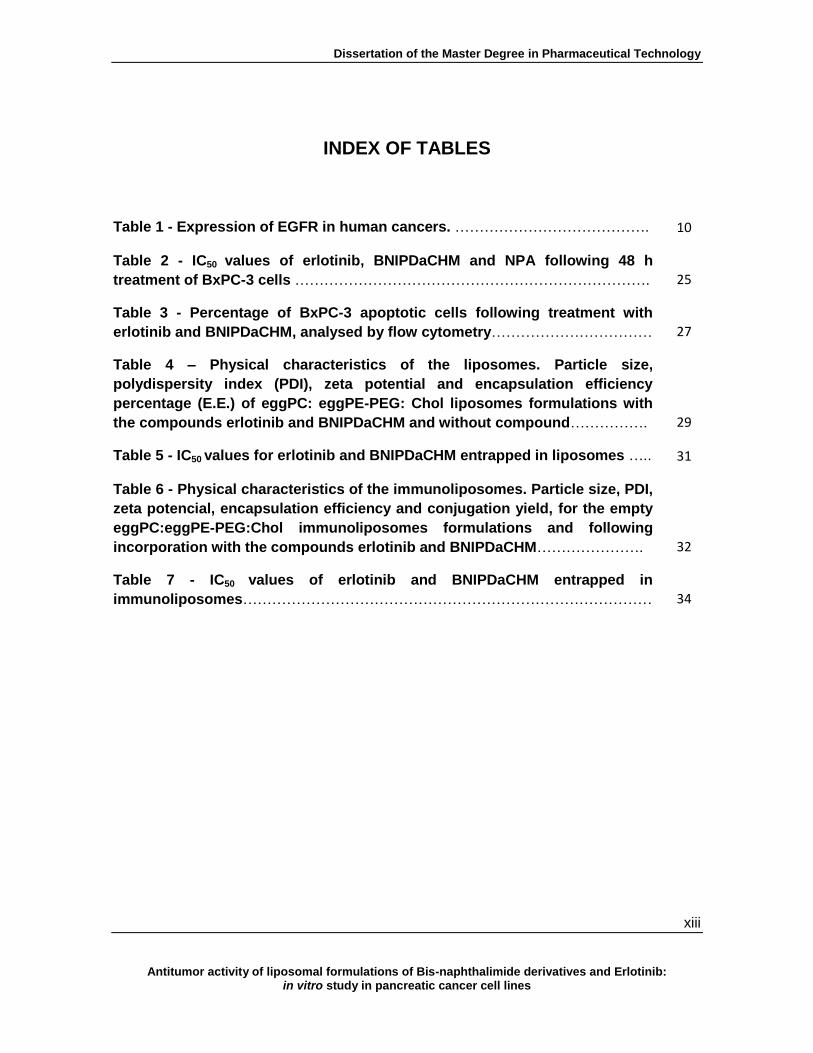
INDEX OF TABLES
Table 1 - Expression of EGFR in human cancers. …………………………………. 10
Table 2 - IC50 values of erlotinib, BNIPDaCHM and NPA following 48 h
treatment of BxPC-3 cells ………………………………………………………………. 25
Table 3 - Percentage of BxPC-3 apoptotic cells following treatment with
erlotinib and BNIPDaCHM, analysed by flow cytometry…………………………… 27
Table 4 – Physical characteristics of the liposomes. Particle size,
polydispersity index (PDI), zeta potential and encapsulation efficiency
percentage (E.E.) of eggPC: eggPE-PEG: Chol liposomes formulations with
the compounds erlotinib and BNIPDaCHM and without compound……………. 29
Table 5 - IC50 values for erlotinib and BNIPDaCHM entrapped in liposomes ….. 31
Table 6 - Physical characteristics of the immunoliposomes. Particle size, PDI,
zeta potencial, encapsulation efficiency and conjugation yield, for the empty
eggPC:eggPE-PEG:Chol immunoliposomes formulations and following
incorporation with the compounds erlotinib and BNIPDaCHM…………………. 32
Table 7 - IC50 values of erlotinib and BNIPDaCHM entrapped in
immunoliposomes………………………………………………………………………… 34

Dissertation of the Master Degree in Pharmaceutical Technology
xiv
Antitumor activity of liposomal formulations of Bis-naphthalimide derivatives and Erlotinib: in vitro study in pancreatic cancer cell lines
ABBREVIATIONS
Anti-EGFR Human anti-human epidermal growth factor receptor antibody
BNIP Bisnaphthalimidopropyl polyamine
BNIPDaCHM Bisnaphthalimidopropyl diaminodicyclohexylmethane
Chol Cholesterol
DDS Delivery drug system
DLS Dinamic light scattering
E.E. Encapsulation efficiency
EDC 1-ethyl-3-(3-dimethylaminopropyl) carbodiimide hydrochloride
EGFR Epidermal growth factor receptor
EGFR-TK Epidermal growth factor of the tyrosine kinase
EggPE-PEG Polyethylene glycol conjugated-L-α-phosphatidylethanolamine
EggPC L-α-Phosphatidylcholine from egg yolk
FBS Fetal bovine serum
FDA Food and drug administration
5-FU Fluorouracil
LUV Large unilamellar vesicle
mAb Monoclonal antibody
MLV Multi-lamellar vesicle
MPS Mononuclear phagocytic system
NHS N-hydroxysulfosuccinimide
Dissertation of the Master Degree in Pharmaceutical Technology
xv
Antitumor activity of liposomal formulations of Bis-naphthalimide derivatives and Erlotinib: in vitro study in pancreatic cancer cell lines
NPA Mononaphthalimidopropylamine
NSCLC Non-small cell lung cancer
OD Optical density
PBS Phosphate buffer saline
PC Pancreatic cancer
PDI Polydispersity index
PEG Polyethylene glycol
SRB Sulforhodamine B
SUV Small unilamellar vesicle
TCA Trichloroacetic acid
TEM Transmission electron microscopy
TfR Transferrin receptor
TK Tyrosine kinase
TKI Tyrosine kinase inhibitor

Dissertation of the Master Degree in Pharmaceutical Technology
1
Antitumor activity of liposomal formulations of Bis-naphthalimide derivatives and Erlotinib: in vitro study in pancreatic cancer cell lines
I. Introduction
1.1. Nanotechnology
Nanotechnology involves the creation and utilization of materials, devices or systems on
the nanometer scale. This field of science is currently experiencing growing developments
being expected to create innovation and to play a critical role in various biomedical
applications (not only in drug delivery, but also in molecular imaging, biomarkers and
biosensors) (1).
For pharmaceutical industry, novel technologies, such as nanotechnology, represent a
strategic tool for expanding drug markets. Interestingly, it often costs substantially less to
develop new methods of administration for an existing drug (resulting in improved efficacy
and bioavailability, together with reduced dosing frequency and lower side effects) than to
develop novel drugs. These new ways of administration may be accomplished by
incorporating the existing drug into a drug delivery system (DDS) (2).
As result of the nanotechnology developments, the pharmaceutical market is changing.
Those changes are due to the several benefits that the new formulations of DDS offer,
such as a competitive edge after patent expiry, market extension, and reduction of the
drug development budget (2).
However, many DDS have not been successful in delivering drugs because of their limited
ability to specifically reach the target tissue. For example, in cancer chemotherapy, several
drugs affect not only tumor cells but also the normal cells. Other problems include
premature drug loss through rapid clearance from the blood stream and metabolism.
Therefore, a drug delivery strategy which allows to selectively target the tumor and offers a
long-circulating period in the blood stream is very much needed (3).
New technological approaches, such as the development of new DDS, are being studied
and tested not only by pharmaceutical industry but also by academic researchers.
Moreover, the development of new DDS is usually a continuum process, from early
research to product development, passing through all stages of clinical trials and finally
resulting in commercial exploitation (1).

Dissertation of the Master Degree in Pharmaceutical Technology
2
Antitumor activity of liposomal formulations of Bis-naphthalimide derivatives and Erlotinib: in vitro study in pancreatic cancer cell lines
1.2. Drug delivery systems
Over the past 30 years, nanosized vehicles have received considerable attention as
pharmaceutical carriers with a wide range of applications, including: i) vaccine adjuvants,
ii) signal enhancers/carriers in medical diagnostics and analytical biochemistry, iii) support
matrix for chemical ingredients and iv) solubilize various materials. Moreover, particular
attention has been given to their role as penetration enhancers in cosmetic products and
as drug delivery vehicles (1).
A DDS is defined as an interface between the patient and the drug, and varies according
to the administration route (oral, inhalation, transdermal or intravenous), the formulation
(including capsules, gels or nanocarriers such as liposomes) or even with the device used
(pumps, delivery conduits, reservoirs, patches or transdermal implants) (2, 3). Each year
new delivery approaches are exploited and almost every body part has already been
studied as a potential route for administrating both classical and novel medicines,
potentiating drug delivery to become an interdisciplinary and independent field of research.
A targeted and safe drug delivery can improve the performance of classical drugs (which
are already commercialized) and have implications in the development and success of
new therapeutic drugs.
The use of various DDS to enhance the in vivo efficiency of many drugs (and drug
administration protocols) has been well established during the last decade (1). Novel DDS
have been developed, promising new ways for delivering poorly soluble drugs, unstable
drugs, peptides, proteins, glycoproteins, genes, among others (4).
Nanocarriers are among the most studied DDS, being mainly associated with drugs which
are pharmacologically potent but may not be used as a “free” drug due to: side effects
inherent to the high toxicity or less efficacy inherent to the low bioavailability. Nanocarrier
systems have demonstrated increased drug bioavailability and stability in the blood
stream. In addition, they are also able to deliver the drug into the target tissue, using lower
drug doses in order to reduce side effects (1, 5, 6). Nanocarriers able to transport active
compounds include: micelles, nanospheres, nanocapsules, niosomes, dendrimers,
polymeric nanoparticles, solid lipid particles and liposomes (Figure 1) (7).

Dissertation of the Master Degree in Pharmaceutical Technology
3
Antitumor activity of liposomal formulations of Bis-naphthalimide derivatives and Erlotinib: in vitro study in pancreatic cancer cell lines
Figure 1- Schematic representation of different nanotechnology-based drug delivery systems. Adapted
from (8).
The surface modification of pharmaceutical nanocarriers is usually performed to control
biological properties of DDS and to simultaneously allow the nanocarriers to perform
various therapeutical or diagnostic functions. The most important advantages of such
modifications in the nanocarriers include: increased stability and half-life of the drug
nanocarriers in circulation, enhanced bio-distribution and active targeting into the required
pathological area, among others (9).
1.2.1. Liposomes as drug carriers
Alec Bangham discovered liposomes in the 1960’s and, since then, these nanoparticles
have been used as a versatile tool in biology, biochemistry and medicine (10). Liposomes
are small artificial vesicles of spherical shape, formed with one or more lipid bilayers
constituted by amphiphilic lipids (phospholipids) and may include cholesterol. Because of
their size, hydrophobic and hydrophilic character, as well as their biocompatibility,
liposomes can be efficient systems for drug delivery (3).

Dissertation of the Master Degree in Pharmaceutical Technology
4
Antitumor activity of liposomal formulations of Bis-naphthalimide derivatives and Erlotinib: in vitro study in pancreatic cancer cell lines
Liposome properties vary substantially with lipid composition, size, surface charge and the
method of preparation. Depending on their size and number of bilayers, they may be
classified into three classes : i) Small Unilamellar Vesicles (SUV), which are surrounded by
a single lipid layer and are 40-100 nm in diameter, ii) Large Unilamellar Vesicles (LUV),
which are a heterogeneous group of vesicles similar to SUVs and are surrounded by a
single lipid layer, with sizes ranging from 100 nm to 1 µm and iii) Multi-Lamellar Vesicles
(MLV), which consist of several lipid layers separated from one another by a layer of
aqueous solution, with sizes >1 µm (11).
The choice of bilayer components allows determining the fluidity and the charge of the lipid
bilayer. For example, saturated phospholipids with long acyl chains form a rigid and
impermeable bilayer structure, whereas the unsaturated phosphatidylcholine organic
species give much more permeable and less stable lipid bilayers (12). In addition, the
presence of cholesterol increases the saturation of the liposomal vesicles, and decreases
the fluidity of the lipid bilayer, reducing drug loss. Therefore, cholesterol acts in the lipid
bilayer as a modulator, resulting in less permeable liposomal vesicles (11). Moreover, the
inclusion of positively or negatively charged lipids into the formulations provides the
liposomes a surface charge, important to guarantee the colloidal stability of the
formulation. The surface charge of liposomes is determined by measuring the zeta
potential, and values higher than ± 30 mV guarantee a stable formulation (13).
Recently, developments have been reported on the use of liposomes as drug carriers,
particularly for cancer therapy (14, 15). An example of a pegylated liposomal delivery
system applied to an approved cancer therapy is Doxil®, a doxorubicin-loaded pegylated
liposomal formulation which resulted in an therapeutic option for various human cancers,
such as breast, lung or ovarian cancer (16).
Indeed, liposomes can be used to improve current cancer treatment regimens due to their
capacity to entrap poorly water soluble antitumor drugs (4, 14, 15). Moreover, when an
inert polymer (such as polyethylene glycol, PEG) is conjugated to the nanocarrier surface,
this conjugation causes a decrease on their uptake by the mononuclear phagocytic system
(MPS). This results in long-circulation and stability of the nanocarriers and consequently
promotes a passive targeting towards the tumor region (15, 17).
Importantly, the surface of liposomes can also be readily modified to improve its specificity
for certain organs or tissues, preventing their uptake by healthy tissues. Liposomes have
been surface functionalized, by binding specific ligands such as: antibodies and antibody
fragments (immunoliposomes), lectins, glycoproteins, oligosaccharides, vitamins or
peptides. The ligand can be attached to the liposome by covalent binding to the carrier
surface or by electrostatic and hydrophobic insertion into the liposomal membrane (1).
These ligands are then capable of directing the liposomes to the region of interest by an
“active targeting” process.

Dissertation of the Master Degree in Pharmaceutical Technology
5
Antitumor activity of liposomal formulations of Bis-naphthalimide derivatives and Erlotinib: in vitro study in pancreatic cancer cell lines
The drugs within surface-modified liposomes have altered characteristics, increasing the
bioavailability of the drug and the fraction of drug delivered within the pathological area,
thus improving efficacy and/or minimizing drug toxicity (3, 12, 15).
1.3. Pancreatic cancer
Pancreas is an organ of approximately 15 cm in length, with a similar shape to a flattened
pear (Figure 2), responsible for the secretion of enzymes that help in food digestion, and
production of hormones such as insulin.
Figure 2- Representation of the human pancreas. The human pancreas is constituted by a head, the body
and a tail, and is placed behind the stomach, surrounded by the liver, intestines and spleen. Adapted from (18).
Pancreatic cancer (PC) is one of most malignant gastrointestinal cancers and, in its
advanced stage, a deadly disease for nearly all affected patients (19). In Portugal, PC
accounts for 4% of the mortality associated with cancer, having been responsible for the
death of a total of 4.558 patients (70.8 % men and 29.2 % women) during the year of 2010
(18, 20). One of the major factors contributing to the high mortality rate observed in PC
patients is the lack of specific markers which allow its early detection. Approximately 80%
of PC cases are diagnosed at an advanced stage of the disease and in these cases
patients present an intrinsic resistance to radio- and chemotherapy (21). In addition, due to
its late detection only a small percentage of patients (15 - 20%) present a resectable tumor
(19, 22).

Dissertation of the Master Degree in Pharmaceutical Technology
6
Antitumor activity of liposomal formulations of Bis-naphthalimide derivatives and Erlotinib: in vitro study in pancreatic cancer cell lines
1.3.1. Clinical approaches for the treatment of pancreatic cancer
Surgical resection is considered the strategy that gives significantly increased survival
rates in PC. However, it may only be applied in very few cases, namely those in which the
tumor appears to be localized in the pancreas without invasion of important surrounding
structures (such as the mesenteric blood vessels located adjacent to the head area of the
pancreas). In addition, there should be no evidence of metastatic spread to the liver, lining
of the intestines, and other vital organs of the peritoneal cavity (19). Nevertheless,
surgical resection alone still results in a very low median overall survival, of approximately
20 months, due to the high rate of relapse. In fact, studies on long-term survival after
surgery indicate low 5 to 10 year survival rates (18% and 13 %, respectively). Therefore,
surgery without additional complementary treatment is not able to improve survival of
patients with PC (19, 21, 23). Improvements have been described in PC’s therapeutic
approaches with the use of adjuvant systemic therapies following surgical resection.
Adjuvant therapy may be achieved with chemotherapeutic agents only or in combination
with radiotherapy, following pancreatic resection (22).
Over the past decade, gemcitabine has been considered the reference chemotherapeutic
for advanced PC, along with fluorouracil (5-FU) and platinum agents (16). However, tumor
recurrence and progression after chemotherapy with gemcitabine have been reported to
be as high as 58% (4, 24). Recently, the combination of gemcitabine with paclitaxel or the
use of FOLFIRINOX (a combination regimen of oxaliplatin, irinotecan, 5-FU and
leucovorin) without gemcitabine, have resulted in an improvement of survival rates
compared to treatment with gemcitabine alone (22, 24, 25). Despite the referred
improvements in the survival rates with chemo- and/or radiotherapy application after tumor
resection, the survival rates are still relatively low (26). Moreover, during treatment,
approximately 80% of PC patients become resistant to chemotherapeutic regimens
applied to them, resulting in a median survival lower than two years (21, 27).
Therefore, new therapeutic strategies are needed to overcome these problems. Currently,
the association of conventional cytotoxic drugs with novel nanotechnology-based
approaches has allowed enhancing the efficiency of those drugs. An example is the use of
gemcitabine entrapped within pegylated liposomes, which has been tested in human PC
and interestingly resulted in a significant increased inhibition of tumor growth , both in vitro
and in vivo when compared to “free” gemcitabine treatment (16, 28, 29).
Dissertation of the Master Degree in Pharmaceutical Technology
7
Antitumor activity of liposomal formulations of Bis-naphthalimide derivatives and Erlotinib: in vitro study in pancreatic cancer cell lines
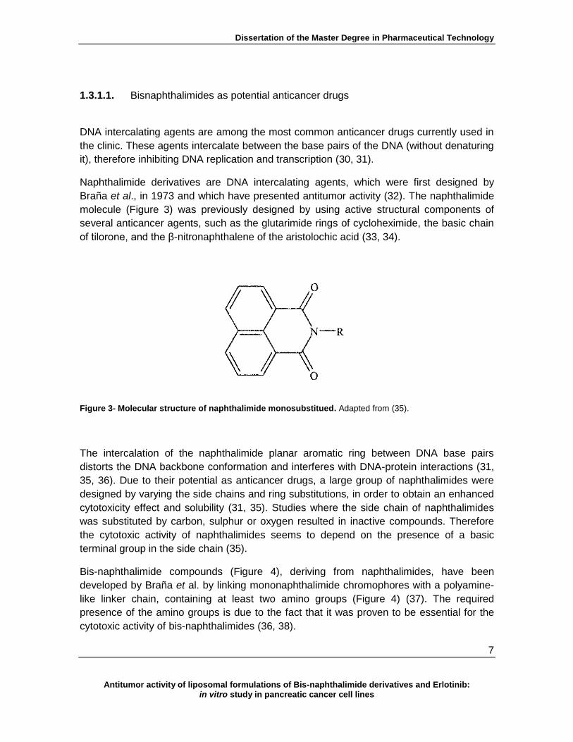
1.3.1.1. Bisnaphthalimides as potential anticancer drugs
DNA intercalating agents are among the most common anticancer drugs currently used in
the clinic. These agents intercalate between the base pairs of the DNA (without denaturing
it), therefore inhibiting DNA replication and transcription (30, 31).
Naphthalimide derivatives are DNA intercalating agents, which were first designed by
Braña et al., in 1973 and which have presented antitumor activity (32). The naphthalimide
molecule (Figure 3) was previously designed by using active structural components of
several anticancer agents, such as the glutarimide rings of cycloheximide, the basic chain
of tilorone, and the β-nitronaphthalene of the aristolochic acid (33, 34).
Figure 3- Molecular structure of naphthalimide monosubstitued. Adapted from (35).
The intercalation of the naphthalimide planar aromatic ring between DNA base pairs
distorts the DNA backbone conformation and interferes with DNA-protein interactions (31,
35, 36). Due to their potential as anticancer drugs, a large group of naphthalimides were
designed by varying the side chains and ring substitutions, in order to obtain an enhanced
cytotoxicity effect and solubility (31, 35). Studies where the side chain of naphthalimides
was substituted by carbon, sulphur or oxygen resulted in inactive compounds. Therefore
the cytotoxic activity of naphthalimides seems to depend on the presence of a basic
terminal group in the side chain (35).
Bis-naphthalimide compounds (Figure 4), deriving from naphthalimides, have been
developed by Braña et al. by linking mononaphthalimide chromophores with a polyamine-
like linker chain, containing at least two amino groups (Figure 4) (37). The required
presence of the amino groups is due to the fact that it was proven to be essential for the
cytotoxic activity of bis-naphthalimides (36, 38).

Dissertation of the Master Degree in Pharmaceutical Technology
8
Antitumor activity of liposomal formulations of Bis-naphthalimide derivatives and Erlotinib: in vitro study in pancreatic cancer cell lines
Despite the importance of the presence of the amino group in the linker chain for bis-
naphthalimides’ cytotoxicity, several of these agents (with or without the amino substituent)
were tested in vitro for their cytotoxicity towards colon cancer cell lines. Results indicated
that bis-napthalimides can be more potent than the most active naphthalimide, even
without the crucial presence of the amino group. This is due to the own structure of bis-
naphthalimides, which per se leads to improvements in the DNA binding capacity,
increasing their inherent cytotoxicity (34).
Figure 4 - Representation of the standard molecular structure of bis-naphthalimide, where the “point of
diversity” represents the linker chain. Adapted from (39).
A series of bis-napthalimide compounds, with different substitutions in the chromophore,
as well as differences in the length of the linker chain, were designed and synthesized.
Several studies of novel bis-naphthalimides showed promising results for cancer therapy,
by intercalation into the DNA‘s double helix via the major groove, therefore inhibiting the
action of topoisomerase II (31, 34). An example of one of the most cytotoxic bis-
naphthalimides, selected for Phase I and II clinical trials in Europe and USA, is elinafide
(Figure 5) (34).
Figure 5 - Molecular structure bis-naphthalimide elinafide. Adapted from (34).

Dissertation of the Master Degree in Pharmaceutical Technology
9
Antitumor activity of liposomal formulations of Bis-naphthalimide derivatives and Erlotinib: in vitro study in pancreatic cancer cell lines
The structural features of bis-naphthalimides which have been described as important for
the enhanced activity (in human colon cancer cells), are: i) a nitro substitution in the
chromophore rings, ii) two internal nitrogen atoms, or iii) an alkyl linker chain of at least
three methylene groups (31, 34, 36).
Moreover, the development of a novel group of derivatives, the bisnaphthalimidopropyl
polyamines (BNIPs) have been primarily described by Lin and Pavlov (40). In these
compounds, the natural polyamines, putrescine, spermidine and spermine (Figure 6), as
well as oxa-polyamines were incorporated into the linker chain (36, 40, 41). Because
BNIPs are also derivatives of naphthalimides, they also present high insolubility in
aqueous solutions (41, 42). However, an increase in the number of nitrogen atoms in the
linker chain has been proposed to result in an enhanced solubility (40).The incorporation
of heteroatoms into the linker chain of the BNIPs has been described to increase the
solubility of these compounds, therefore improving their activity and their potential as
anticancer agents (40, 41). In addition, the heteroatoms incorporation in the linker chain
also strongly stabilized DNA duplexes (40).
Figure 6 – Molecular structure of (A) Spermidine, (B) Spermine and (C) Putrescine. Adapted from (43).
An example that showed the potential of BNIPs as antimour agents in PC is the study in
which BNIPDaoct (a BNIP derivative) was loaded into polyallylamine and cholesteryl (CH5-
PAA) sell-assembling polymers and showed potent anticancer activity both in vitro (in the
BxPC-3 cell line) and in vivo (using xenograft mice) (44).

Dissertation of the Master Degree in Pharmaceutical Technology
10
Antitumor activity of liposomal formulations of Bis-naphthalimide derivatives and Erlotinib: in vitro study in pancreatic cancer cell lines
1.3.2. Targeted therapy with tyrosine kinase inhibitors
Active and passive targeting can bring selectivity and specificity to drug delivery. The ideal
anticancer strategy would selectively kill tumor cells without affecting normal cells.
Cancer therapy has been evolving towards this ideal, through the introduction of targeted
therapies, which focus on unique molecules present in tumors or proteins whose
expression or function are enriched within the neoplastic tissue.
These molecules are commonly designated by molecular targets (1). Molecular-targeted
therapy is regarded as an exciting research hotspot in the treatment of tumors because of
its high specificity and minimal adverse effects. In the case of PC, new targeted
therapeutic strategies have been recently developed. These strategies include the use of
small inhibitor molecules or monoclonal antibodies (mAbs) (or their fragments), which are
capable of inhibiting the tumor progression (16, 45).
Tyrosine kinase inhibitors (TKIs) are small molecules that compete with the ATP binding
site of the catalytic domain of several tyrosine kinases (TKs) (46). Since the early 1980’s,
epidermal growth factor receptor (EGFR) aberrant expression and activation has been
reported in a wide range of human malignancies, such as PC (Table 1) (47, 48). This 170
kDa transmembrane glycoprotein has ligand-dependent intracellular TK activity, being one
of the four members of the receptor tyrosine kinase family (ErbB /HER family) constituted
by: i) EGFR (ErbB-1), ii) HER-2/neu (ErbB-2), iii) HER-3 (ErbB-3) and iv) HER-4 (ErbB-4).
It is present in most cell types, though not hematopoietic cells (49, 50).
Table 1 - Expression of EGFR in human cancers. Adapted from (48).
Type of tumor % EGFR expression
Head and neck 40 - 80
Colorectal 25 - 100
Gastric 33 - 81
Pancreatic 30 - 50
Ovarian 35 - 70
Breast 15 - 37
Prostate 40 - 90
Activation of the EGFR-TK enzyme (epidermal growth factor receptor of the tyrosine
kinase) results in its auto-phosphorylation, which drives signal transduction pathways
leading to tumor growth and malignant progression. EGFR promotes the activation of TKs
resulting in uncontrolled tissue growth, cellular migration and differentiation, as well as

Dissertation of the Master Degree in Pharmaceutical Technology
11
Antitumor activity of liposomal formulations of Bis-naphthalimide derivatives and Erlotinib: in vitro study in pancreatic cancer cell lines
angiogenesis and metastasis (Figure 7) (51). Oncogenic activation of EGFR-TK can occur
by multiple mechanisms such as: i) excess of ligand expression or high expression of
EGFR, ii) activating mutation, iii) failure of inactivation mechanisms, or iv) transactivation
through receptor dimerization (52).
Figure 7 - Representation of the signal transduction through the epidermal growth factor receptor
(EGFR). Ligand biding leads to receptor dimerization. This results in receptor autophosphorylation. Adapted
from (8).
The development of anticancer therapies that selectively modulate the EGFR signaling
pathways activated in pre-tumor and tumor conditions has involved several approaches
(49). In particular, anti-EGFR mAbs have been used to block the extracellular ligand-
binding region of the receptor, thereby interfering with its activation and modulating the
resultant intracellular signal cascade (53). Combination of anti-EGFR antibodies with
gemcitabine or radiotherapy is an example of combined treatments which resulted in
significant growth inhibition of pancreatic cancer cells, compared with the gemcitabine
therapy alone (16).
In addition, TKIs have been developed which block phosphorylation of the intracellular
region of the receptor.

Dissertation of the Master Degree in Pharmaceutical Technology
12
Antitumor activity of liposomal formulations of Bis-naphthalimide derivatives and Erlotinib: in vitro study in pancreatic cancer cell lines
These agents interfere with ligand-induced EGFR activity by inhibiting tyrosine
phosphorylation, thereby blocking the associated signaling pathway (49).Erlotinib
(Tarceva®;N-(3-ethynylphenyl)-6,7-bis(2-methoxyethoxy)-4-quinazolinamine) (Figure 8),
was considered to be the first effective biological drug approved for the treatment of PC,
licensed by FDA in 2006 (28). This orally available low-molecular-weight quinazolinamine,
has been shown to act as a potent and reversible EGFR-TKI (ErbB1 inhibitor), that
suppresses the growth of various tumors, such as non-small cell lung cancer (NSCLC), as
well as breast and pancreatic cancers (50).
Erlotinib has been tested in combination with gemcitabine in patients with advanced PC,
resulting in an improved survival rate of 24 %, in comparison with the 17 % observed for
patients treated with gemcitabine alone (54).
Figure 8 - Molecular structure of erlotinib. Adapted from (8).
Cancer therapeutic strategies involving TKIs differ from the antibody-based ones, since
TKIs are small molecules capable of entering tumor cells and directly interfere with TK
enzymes (48). In comparison, anti-EGFR mABs are more selective towards the
ectodomain target than the TKIs small molecules. This is due to the fact that TKIs may
present affinity to several TKs, decreasing their specificity to the EGFR target (55).
However, this “imperfect” selectivity of TKIs for EGFR (such as erlotinib) may play a
favorable role by allowing partial inhibition of the adjacent ErbB (or other TKs), blocking
the signaling pathway in mAb-resistant cells (53).
1.3.3. Targeted therapy with immunoliposomes
Antibody based targeted therapies are promising strategies to improve the selectivity and
potency of current cancer treatments. The coating, or surface conjugation, of the
nanoparticles with antibodies or antibody fragments (immunoliposomes) that specifically
bind to overexpressed receptors can be used to target them to tumor sites (1).

Dissertation of the Master Degree in Pharmaceutical Technology
13
Antitumor activity of liposomal formulations of Bis-naphthalimide derivatives and Erlotinib: in vitro study in pancreatic cancer cell lines
The first description on immunoliposomes came from Torchilin et al. in the 1980’s, in which
the ability of immunoliposomes to bind to an antigen expressed on the target cells has
been referred (56). Immunoliposomes are the result of the conjugation of a liposomal
formulation with a specific ligand, which may be antibodies or their fragments, vitamins,
glycoproteins, peptides and oligonucleotide aptamers (57). Immunoliposomes have a great
deal of interest because of their potential as targeted drug delivery systems and also their
potential as diagnostic applications.
In fact, immunoliposomes can provide highly concentrated delivery of potent anticancer
agents, which can possess their own inherent selectivity against the target tissue, thus
minimizing the potential for cytotoxicity against healthy tissues with low antigen expression
(58, 59). In addition, the selectivity provided by the use of antibodies is not only used to
target anticancer drugs to cancer cells but it can also act as a direct treatment, when the
ligand is specific to an oncogenic target. An example for immunoliposomes used as
therapy is anti-HER2 immunoliposomes that selectively bind to and internalize in HER2-
overexpressing cancer cells in vitro, inhibiting tumor proliferation. Other example is
doxorubicin-loaded anti-HER2 immunoliposomes that show enhanced therapeutics
doxorubicin effects in HER2-overexpressing xenograft mices (58, 60).
The main problem when “designing” selective immunoliposomes is the lack of knowledge
on specific molecules expressed by tumor cells which allow their recognition. Therefore,
the target, when not totally tumor specific, may be defined by using a protein which is
overexpressed in the tumor, in order to reduce side effects and to increase the potency of
the formulation. Usually, tumor overexpressed proteins have expression levels up to one
hundred times, in comparison to normal cells (59, 61). The most used and characterized
antigens (as cellular targets) belong to the HER family (57).
Through the years several coupling techniques have been described for conjugating
antibodies or their fragments to liposomes, and reports of in vitro experiments have
demonstrated highly specific binding of immunoliposomes to target cells (1, 62). A
practical example is the conjugation of lipid nanocapsules with an antibody fragment
against the transferrin receptor (TfR), by thiolation, obtaining promising in vitro results for
the active transport of drugs to the brain (63). Also, doxorubicin-loaded anti-HER2
immunoliposomes against breast cancer presented advantages in comparison with the
non-targeted Doxil® therapy (58). The unique structural characteristics of the antibody
molecules allow for a number of possibilities for modification and conjugation, possessing
a number of functional groups suitable for modification or conjugation purposes, such as
N-terminal α-amine or C-terminal carboxylate groups (62, 64). The chemistry used for the
conjugate formation should be chosen to yield the best possible retention of antigen
binding activity.

Dissertation of the Master Degree in Pharmaceutical Technology
14
Antitumor activity of liposomal formulations of Bis-naphthalimide derivatives and Erlotinib: in vitro study in pancreatic cancer cell lines
Carbodiimines are an example of reagents for bioconjugation procedures and can be used
to mediate formation of amido linkages between a carboxylate and an amine groups, being
able to form conjugates between two protein molecules, or proteins conjugated with
peptides, oligonucleotides or phospholipids, among other small molecules (62, 65).
Interactions between ligand-receptor only occur when the distance between both is < 0.5
nm. In vitro this condition is not an issue, and good results have been reported (60, 66).
However, the in vivo targeting of tumor tissue with immunoliposomes is not that linear,
since the tumor tissues may be located at sites less accessible, preventing ligand-receptor
interactions (59, 67).

Dissertation of the Master Degree in Pharmaceutical Technology
15
Antitumor activity of liposomal formulations of Bis-naphthalimide derivatives and Erlotinib: in vitro study in pancreatic cancer cell lines
II. Aims
Pancreatic cancer remains one of the most aggressive types of human cancers, and new
alternatives to conventional therapy are much needed.
Therefore, one of the objectives of this work was to study the activity of two bis-
naphthalimides derivatives, BNIPDaCHM and NPA, and a commercial TKI, erlotinib
(Tarceva®), in liposomal formulations.
In particular, the specific aims of this project were to:
A) Analyze the of effect of the “free” BNIPDaCHM and erlotinib, in the BxPC-3 tumor
cell line, particularly regarding their effect on:
Cell number and viability;
Cell cycle profile;
Apoptosis;
Apoptotic protein expression levels.
B) Incorporate the compounds into liposomes by:
Preparation of the liposomes, through the lipid film hydration technique, and
extrusion;
Physicochemical characterization of the liposome formulations.
C) Prepare compound-loaded immunoliposomes using a human anti-EGFR antibody
coating.
D) Evaluate the cytotoxicity of the compound-loaded liposomes and immunoliposomes,
in BxPC-3 pancreatic tumour cells.

Dissertation of the Master Degree in Pharmaceutical Technology
16
Antitumor activity of liposomal formulations of Bis-naphthalimide derivatives and Erlotinib: in vitro study in pancreatic cancer cell lines
III. Materials and methods
3.1. Materials
3.1.1. Compounds and lipids
Erlotinib HCl(OSI-744, CP358774, NSC7188782) (Figure 9 A) was purchased from
SelecktChemicals. A stock solution (27 mM) was prepared in dimethyl sulfoxide (DMSO;
Applichem, Germany), and stored at 4ºC, for a period no longer than 6 months. The
bisnaphthalimidopropyl polyamine derivatives compounds, bisnaphthalimidopropyl
diaminodicyclohexylmethane (BNIPDaCHM) and mononaphthalimidopropylamine (NPA)
(Figure 9 B and C, respectively) were kindly given by Prof. Paul Kong, from the Robert
Gordon University, UK. BNIPDaCHM and NPA stock solutions, both at 20 mM in DMSO,
were stored at 4ºC. L-α-Phosphatidylcholine from egg yolk (eggPC) (100 mg/mL in
chloroform), PEG-conjugated-L-α-phosphatidylethanolamine (eggPE-PEG), from egg yolk
and cholesterol (Chol) were purchased from Sigma and stored at -20ºC.
(A)
(B)
(C)
Figure 9 - Structures of (A) erlotinib, (B) bisnaphtalimidopropyl diaminodicyclohexylmethane
(BNIPDaCHM) and (C) mononaphthalimidopropylamine (NPA). Adapted from (42, 68)
.HCl

Dissertation of the Master Degree in Pharmaceutical Technology
17
Antitumor activity of liposomal formulations of Bis-naphthalimide derivatives and Erlotinib: in vitro study in pancreatic cancer cell lines
3.1.2. Cell culture
The human pancreatic cancer cell line, BxPC-3 (American Type Culture Collection –
ATCC® CRL-1687™, USA) was cultured and maintained as adherent cell culture in RPMI-
1640 with Ultraglutamine I medium (Lonza, Belgium), supplemented with 5% (v/v) heat
inactivated fetal bovine serum (FBS, Gibco, USA) for sulforhodamine B assay or 10% (v/v)
FBS for the remaining assays. Cells were incubated at 37 ºC, in a humidified 5% CO2
atmosphere within a CB 210 incubator (Binder, Germany). Once 80 % confluence was
reached, cells were trypsinized. For this, cell medium was discarded, cells were washed
and further incubated with trypsin (TrypLE™ Express; Gibco, USA), for 10 min at 37 ºC.
Trypsin was inactivated with medium supplemented with FBS. Cell suspension was
centrifuged for 5 min at 134 x g in a High Speed 350-R centrifuge (MPW, Poland). Cell
pellets were re-suspended in fresh medium. Cells were diluted (1:10), transferred into a
new flask and further incubated. For the experiments, cells were plated into 96- or 6-well
plates at different densities, according to the experiment (which will be explained later on
in section 3.2.). Cell number and viability were routinely assessed by counting number of
cells in a Neubauer chamber (Haemocytometer; Marienfeld, Germany) under the TS100
(Nikon, USA) inverted microscope. For this, cell suspension was diluted (1:1) with trypan
blue solution 0.4 % (Sigma-Aldrich, UK).
3.2. Methods
3.2.1. In vitro cell growth inhibition analysis using Sulforhodamine B assay
The Sulforhodamine B (SRB) assay was carried out as previously described (69, 70).
Briefly, cells were plated at 1 x 10⁵ cells per well in 96-wells plate and incubated for 24 h.
Cells were then treated with appropriate dilutions of compound stocks, prepared in
medium with 5% FBS just prior to the assay. Erlotinib was diluted within a concentration
range of 10-0.625 μM, BNIPDaCHM was diluted in a concentration range from 2-0.125 μM
and NPA within a concentration range of 100-6.25 μM. The effect of DMSO was also
evaluated in the growth of this cell line by treating cells with the maximum concentration of
DMSO used (0.07 % for erlotinib, 0.03 % for BNIPDaCHM and 1.25 % for NPA). Erlotinib
and BNIPDaCHM incorporated in liposomes were diluted within an adequate concentration
range from 2 - 0.125 µM were added to BxPC-3 cells (final lipid concentration range from
249 to 16 µg/mL for erlotinib-loaded liposomes and final lipid concentration range 350 to
22 µg/mL for BNIPDaCHM-loaded liposomes). After 48h treatment, cells were fixed in situ
with 10% ice-cold trichloroacetic acid (TCA,VWR, Belgium) and incubated on ice for 1 h.

Dissertation of the Master Degree in Pharmaceutical Technology
18
Antitumor activity of liposomal formulations of Bis-naphthalimide derivatives and Erlotinib: in vitro study in pancreatic cancer cell lines
Wells were then washed 3 times with distilled water and allowed to air-dry at room
temperature. SRB solution (0.4% wt/vol in 1% acetic acid) was added to the wells and
incubated for 30 min.
Plates were then rinsed four times with 1% (vol/vol) acetic acid to remove the unbound dye
and left to air-dry at room temperature. Tris-base solution 10 mM (pH 10.5) was added to
solubilize the bounded SRB. Following a minimum period of 15 min, optical density was
measured at 510 nm in a Synergy HT microplate reader (BioTek, USA). Dose-response
curves were generated from the plotted results and the IC50 (corresponding to half maximal
inhibitory concentration) was calculated. OD values were plotted against the concentration
values and the IC50 determined by linear regression, using the
equation .
3.2.2. Analysis of cell cycle profile, apoptosis and protein expression
Cells were plated at 3 x 10⁵ cells per well (in 6 well plates) in 10% FBS supplemented
medium and incubated for 24 h. Cells were then treated with medium only (blank cells),
with compounds/drug:BNIPDaCHM [at its IC50 (1.3 ± 0.05 µM)] and erlotinib [at its IC50 (1.7
± 0.06) and twice this concentration (2xIC50; 3.4 µM)]. The effect of DMSO on the growth
of the cell line was evaluated by treating cells with the maximum concentration of DMSO
used (0.05 % for erlotinib and 0.01 % for BNIPDaCHM). Following 48 h, cell pellets were
then further processed according to the following procedures.
3.2.2.1. Cell cycle analysis
Cell pellets were fixed in 70% ice-cold ethanol for at least 12 hours at 4 ºC. Prior to
analysis, pellets were re-suspended in PBS containing 0.1 mg/mL RNase A and 5 µg/mL
propidium iodide solution (Sigma-Aldrich, Germany). Cellular DNA content was analyzed
by flow cytometry using a FACScalibur (BD Biosciences, USA) flow cytometer plotting at
least 10.000 events per sample. The analysis of cell cycle distribution was subsequently
carried out using the FlowJo 7.6.5 software (Tree Star, Inc., Ashland, USA) after cell
debris and aggregates exclusion.
Dissertation of the Master Degree in Pharmaceutical Technology
19
Antitumor activity of liposomal formulations of Bis-naphthalimide derivatives and Erlotinib: in vitro study in pancreatic cancer cell lines
3.2.2.2. Apoptosis analysis
For apoptosis analysis, the Human Annexin V-FITC/PI apoptosis detection kit (Bender
MedSystems, Austria) was used according to manufacturer’s instructions. Briefly, cell
pellets were re-suspended using 400 µL of binding buffer (previously diluted in water) and
kept on ice. Annexin V-FITC (5 µL) was added to 195 µL of cell suspension which was
then incubated in the dark for 10 min.
Propidium iodide (10 µL) was then added to the samples and further incubated on ice for 2
min. Annexin V-FITC/ propidium iodide staining was analysed by flow cytometry, using a
FACScalibur (BD Biosciences, USA) flow cytometer plotting at least 10.000 events per
sample. Data was analysed using FlowJo 7.6.5 software (Tree Star, Inc., Ashland, USA).
3.2.3. Protein expression analysis by Western Blot
For protein expression analysis, cells were lysed with Winman’s buffer (1% NP-40, 0.1 M
Tris-HCl pH 8.0, 0.15 M NaCl and 5 mM EDTA) complemented with protease inhibitor, for
45 min at 4ºC, with agitation. The lysates were centrifuged for 10 min at 18188 x g at 4 ºC,
to exclude cellular debris. Protein content was quantified using the “DC protein assay kit”
(BioRad, USA), according to the manufacturer. Total protein extracts (20 µg) were
subjected to SDS-PAGE electrophoresis in 12% Bis-Tris gel and then transferred
electrophoretically to a Hybond-C Extra nitrocellulose membrane (Amersham Biosciences,
UK). The membrane was stained with Ponceau solution (BioRad, USA), dried and stored
at room temperature until analysis. Following incubation in blocking solution [5% non-fat
dry, in 0.05% Tween 20 in Tris buffered saline solution (TBS-T)], the membrane was
incubated with the following antibodies: goat anti-actin (1:2000, Santa Cruz
Biotechnology), PARP-1 (1:2000, Santa Cruz Biotechnology) or Bcl-2 (1:100, Dako).
Membranes were then washed and further incubated with one of the following secondary
antibodies from Santa Cruz Biotechnology diluted 1:2000: anti-goat IgG, anti-rabbit IgG or
anti-mouse IgG. Signal was detected using Amersham™ ECL Western Blotting Detection
Reagents (GE Healthcare), the Amersham Hyperfilm ECL (GE Healthcare) and the Kodak
GBX developer and fixer (Sigma).

Dissertation of the Master Degree in Pharmaceutical Technology
20
Antitumor activity of liposomal formulations of Bis-naphthalimide derivatives and Erlotinib: in vitro study in pancreatic cancer cell lines
3.2.4. Liposomes preparation
Liposomes were prepared by the film hydration method (10). Lipids eggPC in chloroform
(100 mg/mL) and eggPE-PEG and Chol were dissolved in chloroform at a 60:20:20 molar
ratio. Empty liposomes and liposomes loaded with BNIPDaCHM or erlotinib had a final
lipid concentration of 5 mg/mL. Briefly, eggPE-PEG and Chol, were weighted in a glass
vial, using an ABS 80-4 (Kern & Sohn GmbH, Germany) analytical balance. EggPC in
chloroform was added to the glass vial, to obtain a final lipid concentration of 5 mg/mL.
Erlotinib or BNIPDaCHM at 100 µM final concentration were added to the corresponding
vials. Empty liposomes were prepared using the same procedure, without the addition of
any compound.
The solvent was slowly evaporated in a heating block at 37ºC under a gentle stream of
nitrogen in a hood, for approximately 30 min. Removal of the solvent produced the
deposition of lipids as a film on the bottom and walls of the glass vial. The lipidic film was
hydrated with PBS pH 7.4, under vortex, in order to have a 5 mg/mL final lipid
concentration.
3.2.4.1. Extrusion of the MLVs
The extruder LiposoFast (Avestin,Germany) was assembled as specified by the
manufacturer initially with the polycarbonate membrane with the highest diameter pore
(400 nm), followed by the 200 nm. Hydrated liposomes were passed 20 times through
each filter.
3.2.5. Immunoliposomes preparation
Carbodiimide conjugation technique reacts with amine and carboxylate groups that are
present in antibody molecules with abundance. 1-ethyl-3-(3-dimethylaminopropyl)
carbodiimide hydrochloride (EDC), is a water soluble carbodiimide and may form active
ester functional groups with carboxylate groups using the compound N-
hydroxysulfosuccinimide (NHS). NHS esters are hydrophilic active groups that react
rapidly with amines on the target molecules (Figure 10) (56, 64). EDC/NHS-coupled
reactions are highly efficient and usually increase the yield of conjugation, in comparison
with conjugations using solely EDC (65).
Dissertation of the Master Degree in Pharmaceutical Technology
21
Antitumor activity of liposomal formulations of Bis-naphthalimide derivatives and Erlotinib: in vitro study in pancreatic cancer cell lines
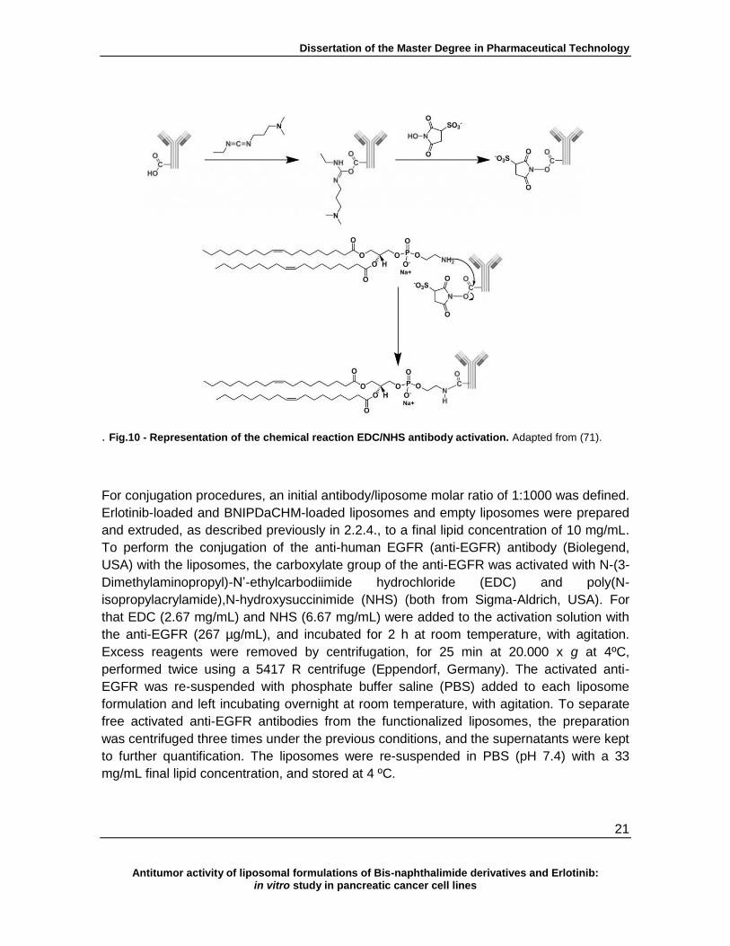
. Fig.10 - Representation of the chemical reaction EDC/NHS antibody activation. Adapted from (71).
For conjugation procedures, an initial antibody/liposome molar ratio of 1:1000 was defined.
Erlotinib-loaded and BNIPDaCHM-loaded liposomes and empty liposomes were prepared
and extruded, as described previously in 2.2.4., to a final lipid concentration of 10 mg/mL.
To perform the conjugation of the anti-human EGFR (anti-EGFR) antibody (Biolegend,
USA) with the liposomes, the carboxylate group of the anti-EGFR was activated with N-(3-
Dimethylaminopropyl)-N’-ethylcarbodiimide hydrochloride (EDC) and poly(N-
isopropylacrylamide),N-hydroxysuccinimide (NHS) (both from Sigma-Aldrich, USA). For
that EDC (2.67 mg/mL) and NHS (6.67 mg/mL) were added to the activation solution with
the anti-EGFR (267 µg/mL), and incubated for 2 h at room temperature, with agitation.
Excess reagents were removed by centrifugation, for 25 min at 20.000 x g at 4ºC,
performed twice using a 5417 R centrifuge (Eppendorf, Germany). The activated anti-
EGFR was re-suspended with phosphate buffer saline (PBS) added to each liposome
formulation and left incubating overnight at room temperature, with agitation. To separate
free activated anti-EGFR antibodies from the functionalized liposomes, the preparation
was centrifuged three times under the previous conditions, and the supernatants were kept
to further quantification. The liposomes were re-suspended in PBS (pH 7.4) with a 33
mg/mL final lipid concentration, and stored at 4 ºC.

Dissertation of the Master Degree in Pharmaceutical Technology
22
Antitumor activity of liposomal formulations of Bis-naphthalimide derivatives and Erlotinib: in vitro study in pancreatic cancer cell lines
3.2.6. Anti-EGFR antibody quantification
Quantification of the antibody was performed using a Bradford assay (BioRad, USA),
accordingly to the manufacturer’s instructions. Samples were read at OD690nm and, by
interpolation in the standard curve, the amount of protein was determined.
3.2.7. Liposomes characterization
3.2.7.1. Particle size, polydispersity index and zeta potential
Liposome formulations were diluted 1:10 in distilled water. Each liposome formulation was
analyzed by dynamic light scattering (DLS) using a Zetasyzer Nano ZS (Malvern, UK) to
determine the particle size, polydispersity index and zeta potential. Measurements were
made in triplicate at 25 ºC.
3.2.7.2. Encapsulation efficiency
Erlotinib stock solution (2150 µg/mL) was diluted with acetonitrile to obtain standard
solutions of concentrations ranging from 1000 to 31.25 ng/mL. The fluorescence intensity
of the standard solutions was measured using a FluoroMax-4 spectrofluorometer (Horiba
Scientific, France), at 470 nm following an excitation at 247 nm, with a slit of 5 nm.
The intensity of the blank solution (acetonitrile) was also measured. BNIPDaCHM stock
solution (10 mM) was diluted with 0.01 % Triton X-100 in PBS (pH 7.4) to obtain standard
solutions of concentrations ranging from 0.5 - 0.05 μM. The fluorescence intensity of the
standard solutions was measured on the spectrofluorometer at 397 nm following an
excitation at 350 nm, with a slit of 10 nm. The intensity of the blank solution (0.01 % Triton
X-100 in PBS (pH 7.4)) was also measured. After the standard curves were determined for
both compounds, liposome formulations with erlotinib were diluted 1:200 with acetonitrile
and liposome formulations entrapping BNIPDaCHM were diluted 1:200 with the surfactant
[0.01%Triton X-100 in PBS (pH 7.4)], and the samples were read in the
spectrofluorometer, under the previous described conditions. By correlation with the
standard curves obtained, the encapsulation efficiency of each formulation read was
determined.
Dissertation of the Master Degree in Pharmaceutical Technology
23
Antitumor activity of liposomal formulations of Bis-naphthalimide derivatives and Erlotinib: in vitro study in pancreatic cancer cell lines
3.2.7.3. Transmission electron microscopy
Liposome formulations were observed by transmission electron microscopy (TEM, JEM-
1400 (Jeol, Japan). For this, 20-30 μL of liposomes formulation were placed on carbon
coated copper grid, for 1 min, and removed with the help of filter paper. The sample was
stained with a drop of uranyl acetate, for 1 min at room temperature. Uranyl acetate
excess was removed with filter paper. Samples on the carbon coated copper grid were
observed in transmission electron microscope, with 50.000x and 80.000x magnifications.
3.3. Statistical analysis
All experimental data are presented as means ± SE. Statistical analysis was conducted
using the t-student test. Analysis were performed by comparing treatment with the
compounds with blanks, except for the Annexin V/FITC assay in which the analysis was
performed by comparing treatment with compounds with the controls. Data was
considered statistically significant when P < 0.05.

Dissertation of the Master Degree in Pharmaceutical Technology
24
Antitumor activity of liposomal formulations of Bis-naphthalimide derivatives and Erlotinib: in vitro study in pancreatic cancer cell lines
IV. Results
4.1. Effect of BNIPDaCHM, NPA and erlotinib on growth inhibition of human
pancreatic cells
In order to assess the effect of the compounds/drug studied (BNIPDaCHM, NPA and
erlotinib) in the in vitro growth of the BxPC-3 cells, the SRB assay was used. This assay
measures the amount of protein in the cells, allowing to indirectly analyze cell number.
Results after 48 h of treatment (Figure 11), clearly indicate that both BNIP compounds,
BNIPDaCHM and NPA, inhibited cell growth, in a dose-dependent effect. Interestingly,
regarding erlotinib, a dose-dependent decrease in cell growth is only observed up to 4 µM
concentrations, above which a plateau is reached.
Figure 11 - Analysis of the effect of (A) erlotinib, (B) BNIPDaCHM and (C) NPA on BxPC3 cell growth
determined by sulforhodamine B assay. Results are presented as a percentage of cell growth in relation to
blank cells (treated with medium only) and are mean ± SE of at least three independent experiments in
quadruplicate.
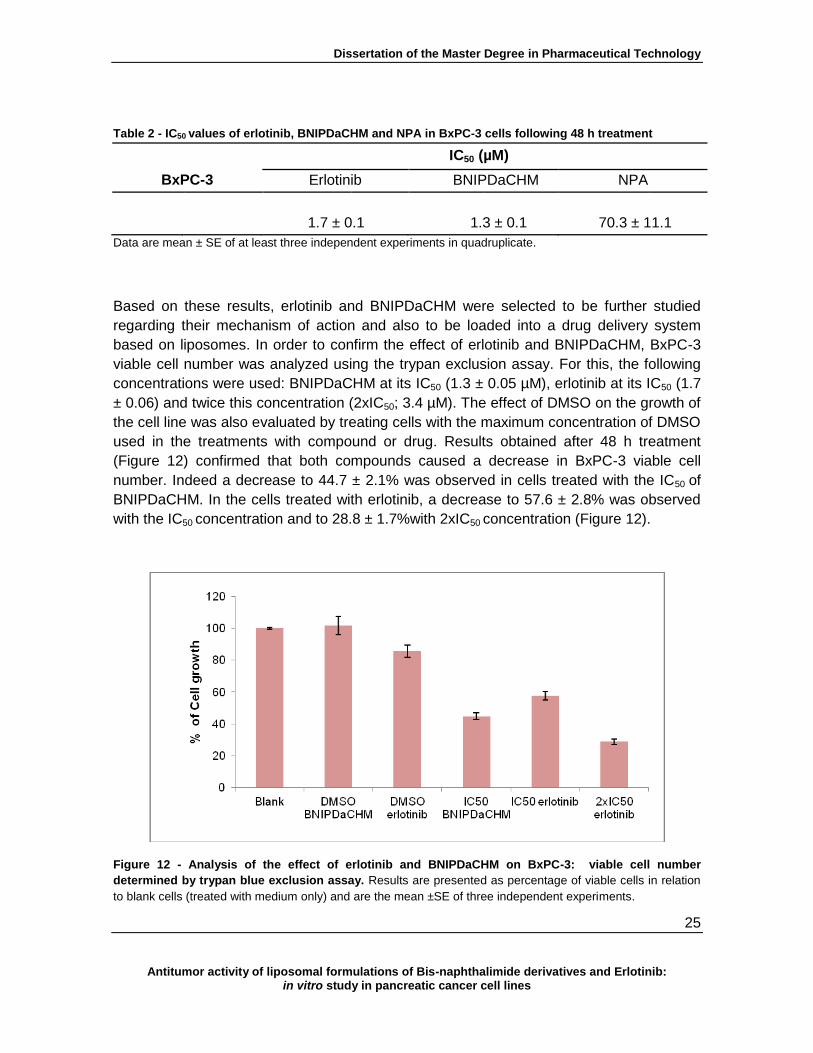
The determined IC50 values (Table 1) showed that, from the compounds studied,
BNIPDaCHM was the most potent with an IC50 value of 1.3 ± 0.1 µM, followed by the drug
erlotinib with an IC50 value of 1.7 ± 0.1 µM. NPA was found to be the compound that
presented less activity, with an IC50 of 70.3 ± 11.1µM.
B A
A
Dissertation of the Master Degree in Pharmaceutical Technology
25
Antitumor activity of liposomal formulations of Bis-naphthalimide derivatives and Erlotinib: in vitro study in pancreatic cancer cell lines
Table 2 - IC50 values of erlotinib, BNIPDaCHM and NPA in BxPC-3 cells following 48 h treatment
IC50 (µM)
BxPC-3 Erlotinib BNIPDaCHM NPA
1.7 ± 0.1 1.3 ± 0.1 70.3 ± 11.1
Data are mean ± SE of at least three independent experiments in quadruplicate.
Based on these results, erlotinib and BNIPDaCHM were selected to be further studied
regarding their mechanism of action and also to be loaded into a drug delivery system
based on liposomes. In order to confirm the effect of erlotinib and BNIPDaCHM, BxPC-3
viable cell number was analyzed using the trypan exclusion assay. For this, the following
concentrations were used: BNIPDaCHM at its IC50 (1.3 ± 0.05 µM), erlotinib at its IC50 (1.7
± 0.06) and twice this concentration (2xIC50; 3.4 µM). The effect of DMSO on the growth of
the cell line was also evaluated by treating cells with the maximum concentration of DMSO
used in the treatments with compound or drug. Results obtained after 48 h treatment
(Figure 12) confirmed that both compounds caused a decrease in BxPC-3 viable cell
number. Indeed a decrease to 44.7 ± 2.1% was observed in cells treated with the IC50 of
BNIPDaCHM. In the cells treated with erlotinib, a decrease to 57.6 ± 2.8% was observed
with the IC50 concentration and to 28.8 ± 1.7%with 2xIC50 concentration (Figure 12).
Figure 12 - Analysis of the effect of erlotinib and BNIPDaCHM on BxPC-3: viable cell number
determined by trypan blue exclusion assay. Results are presented as percentage of viable cells in relation
to blank cells (treated with medium only) and are the mean ±SE of three independent experiments.

Dissertation of the Master Degree in Pharmaceutical Technology
26
Antitumor activity of liposomal formulations of Bis-naphthalimide derivatives and Erlotinib: in vitro study in pancreatic cancer cell lines
4.2. Effect of erlotinib and BNIPDaCHM on the BxPC-3 cell cycle profile
To initiate the elucidation of the mechanism of action of erlotinib and BNIPDaCHM, the cell
cycle profile of BxPC-3 cells was analyzed by flow cytometry 48 h after treatment with the
compound or drug.
Results showed that treatment with the IC50 concentration of BNIPDaCHM (1.3 ± 0.1 µM)
caused a statistically significant decrease (of approximately 13%) in the percentage of
cells in the G1 phase of the cell cycle (P < 0.05) (Figure 13). The effect of the IC50 of
erlotinib (1.7 ± 0.1µM) was less pronounced, with a smaller decrease (of only 10%) in the
percentage of cells in G1 phase being observed following 48 h of treatment. However,
when increasing the concentration of erlotinib to 2xIC50, a statistically significant decrease
(of approximately 15%) in the G1 population was observed (P <0.05).
Figure 13 - Cell cycle profile of BxPC-3 cells treated with erlotinib and BNIPDaCHM. Results are the
mean±SE of four independent experiments (except for the DMSO of erlotinib and 2xIC50 of erlotinib treatments
which are the mean of 3 experiments). * P<0.05 compared to blank values
In addition, following 48h treatment of cells with the IC50 concentration of BNIPDaCHM and
with 2xIC50 concentration of erlotinib, a slight increase in the sub-G1 peak (of 3% and 5%,
respectively) was observed, which might be suggestive of induction of apoptosis.
* *
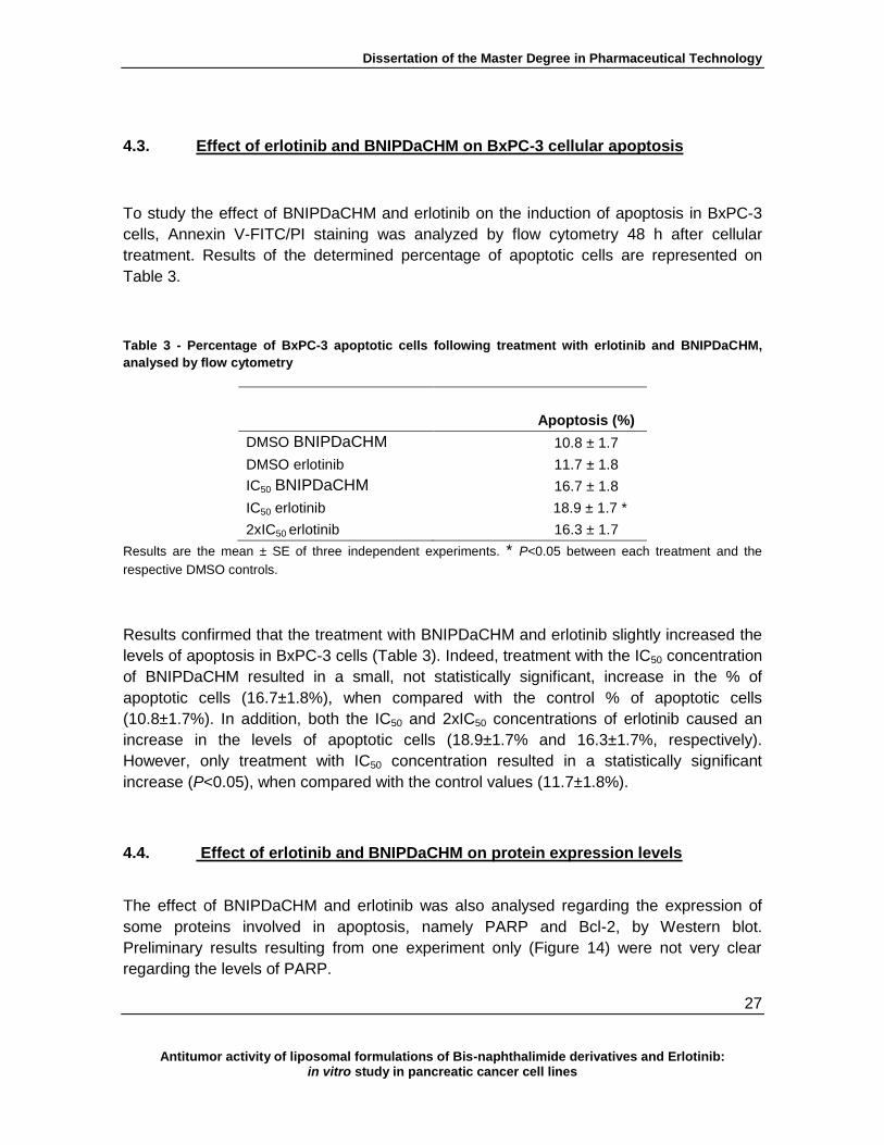
Dissertation of the Master Degree in Pharmaceutical Technology
27
Antitumor activity of liposomal formulations of Bis-naphthalimide derivatives and Erlotinib: in vitro study in pancreatic cancer cell lines
4.3. Effect of erlotinib and BNIPDaCHM on BxPC-3 cellular apoptosis
To study the effect of BNIPDaCHM and erlotinib on the induction of apoptosis in BxPC-3
cells, Annexin V-FITC/PI staining was analyzed by flow cytometry 48 h after cellular
treatment. Results of the determined percentage of apoptotic cells are represented on
Table 3.
Table 3 - Percentage of BxPC-3 apoptotic cells following treatment with erlotinib and BNIPDaCHM,
analysed by flow cytometry
Apoptosis (%)
DMSO BNIPDaCHM 10.8 ± 1.7
DMSO erlotinib 11.7 ± 1.8
IC50 BNIPDaCHM 16.7 ± 1.8
IC50 erlotinib 18.9 ± 1.7 *
2xIC50 erlotinib 16.3 ± 1.7
Results are the mean ± SE of three independent experiments. * P<0.05 between each treatment and the
respective DMSO controls.
Results confirmed that the treatment with BNIPDaCHM and erlotinib slightly increased the
levels of apoptosis in BxPC-3 cells (Table 3). Indeed, treatment with the IC50 concentration
of BNIPDaCHM resulted in a small, not statistically significant, increase in the % of
apoptotic cells (16.7±1.8%), when compared with the control % of apoptotic cells
(10.8±1.7%). In addition, both the IC50 and 2xIC50 concentrations of erlotinib caused an
increase in the levels of apoptotic cells (18.9±1.7% and 16.3±1.7%, respectively).
However, only treatment with IC50 concentration resulted in a statistically significant
increase (P<0.05), when compared with the control values (11.7±1.8%).
4.4. Effect of erlotinib and BNIPDaCHM on protein expression levels
The effect of BNIPDaCHM and erlotinib was also analysed regarding the expression of
some proteins involved in apoptosis, namely PARP and Bcl-2, by Western blot.
Preliminary results resulting from one experiment only (Figure 14) were not very clear
regarding the levels of PARP.
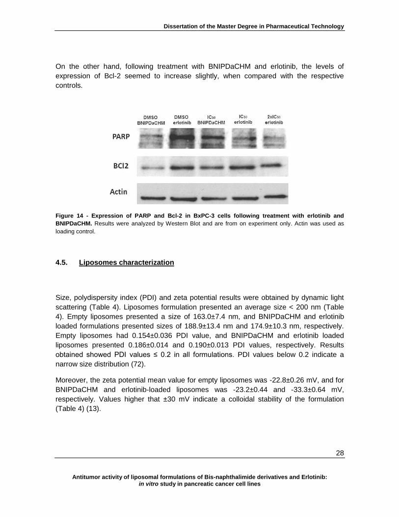
Dissertation of the Master Degree in Pharmaceutical Technology
28
Antitumor activity of liposomal formulations of Bis-naphthalimide derivatives and Erlotinib: in vitro study in pancreatic cancer cell lines
On the other hand, following treatment with BNIPDaCHM and erlotinib, the levels of
expression of Bcl-2 seemed to increase slightly, when compared with the respective
controls.
Figure 14 - Expression of PARP and Bcl-2 in BxPC-3 cells following treatment with erlotinib and
BNIPDaCHM. Results were analyzed by Western Blot and are from on experiment only. Actin was used as
loading control.
4.5. Liposomes characterization
Size, polydispersity index (PDI) and zeta potential results were obtained by dynamic light
scattering (Table 4). Liposomes formulation presented an average size < 200 nm (Table
4). Empty liposomes presented a size of 163.0±7.4 nm, and BNIPDaCHM and erlotinib
loaded formulations presented sizes of 188.9±13.4 nm and 174.9±10.3 nm, respectively.
Empty liposomes had 0.154±0.036 PDI value, and BNIPDaCHM and erlotinib loaded
liposomes presented 0.186±0.014 and 0.190±0.013 PDI values, respectively. Results
obtained showed PDI values ≤ 0.2 in all formulations. PDI values below 0.2 indicate a
narrow size distribution (72).
Moreover, the zeta potential mean value for empty liposomes was -22.8±0.26 mV, and for
BNIPDaCHM and erlotinib-loaded liposomes was -23.2±0.44 and -33.3±0.64 mV,
respectively. Values higher that ±30 mV indicate a colloidal stability of the formulation
(Table 4) (13).
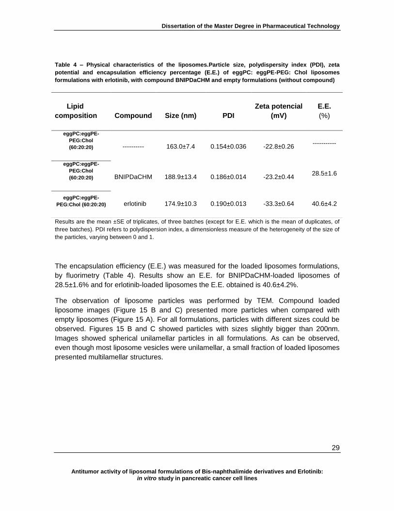
Dissertation of the Master Degree in Pharmaceutical Technology
29
Antitumor activity of liposomal formulations of Bis-naphthalimide derivatives and Erlotinib: in vitro study in pancreatic cancer cell lines
Table 4 – Physical characteristics of the liposomes.Particle size, polydispersity index (PDI), zeta
potential and encapsulation efficiency percentage (E.E.) of eggPC: eggPE-PEG: Chol liposomes
formulations with erlotinib, with compound BNIPDaCHM and empty formulations (without compound)
Results are the mean ±SE of triplicates, of three batches (except for E.E. which is the mean of duplicates, of
three batches). PDI refers to polydispersion index, a dimensionless measure of the heterogeneity of the size of
the particles, varying between 0 and 1.
The encapsulation efficiency (E.E.) was measured for the loaded liposomes formulations,
by fluorimetry (Table 4). Results show an E.E. for BNIPDaCHM-loaded liposomes of
28.5±1.6% and for erlotinib-loaded liposomes the E.E. obtained is 40.6±4.2%.
The observation of liposome particles was performed by TEM. Compound loaded
liposome images (Figure 15 B and C) presented more particles when compared with
empty liposomes (Figure 15 A). For all formulations, particles with different sizes could be
observed. Figures 15 B and C showed particles with sizes slightly bigger than 200nm.
Images showed spherical unilamellar particles in all formulations. As can be observed,
even though most liposome vesicles were unilamellar, a small fraction of loaded liposomes
presented multilamellar structures.
Lipid
composition
Compound Size (nm) PDI
Zeta potencial
(mV)
E.E.
(%)
eggPC:eggPE-
PEG:Chol
(60:20:20) ---------- 163.0±7.4 0.154±0.036 -22.8±0.26
-----------
eggPC:eggPE-
PEG:Chol
(60:20:20)
BNIPDaCHM 188.9±13.4 0.186±0.014 -23.2±0.44
28.5±1.6
eggPC:eggPE-
PEG:Chol (60:20:20) erlotinib 174.9±10.3 0.190±0.013 -33.3±0.64
40.6±4.2

Dissertation of the Master Degree in Pharmaceutical Technology
30
Antitumor activity of liposomal formulations of Bis-naphthalimide derivatives and Erlotinib: in vitro study in pancreatic cancer cell lines
Figure 15 - Images of different liposomes obtained by TEM. Empty liposomes (control) (A), liposomes
with BNIPDaCHM (B) and liposomes with erlotinib (C). (Magnification x 80 000; 200 nm scale).
4.6. In vitro cell growth inhibition analysis for empty liposomes and BNIPDaCHM
or erlotinib loaded liposomes on BxPC-3 cells
In order to assess the effect of the compounds/drug loaded into liposomes on BxPC-3
cells, the SRB assay was used. Empty liposomes were initially tested, to certify that the
carriers were non-toxic (at least at the quantities used in this study) for this line (Figure
16).
Figure 16 - Effect of empty liposomes in BxPC-3 cell growth determined by the sulforhodamine B
assay. Results are presented as a percentage of cell growth in relation to blank cells (treated with medium
only) and are shown as mean ±SE three independent experiments in quadruplicate.

Dissertation of the Master Degree in Pharmaceutical Technology
31
Antitumor activity of liposomal formulations of Bis-naphthalimide derivatives and Erlotinib: in vitro study in pancreatic cancer cell lines
Results demonstrated that liposomes did not affect cell growth. In fact, for the highest
concentration of lipidic concentration used (4 mg/mL) the mean percentage of cell growth
obtained was 96.8±7.12% in relation to blank cells.
The effect of the erlotinib and BNIPDaCHM-loaded liposomes in BxPC-3 cell line was then
analyzed by the SRB assay.
Table 5 - IC50 values for erlotinib and BNIPDaCHM entrapped in liposomes
IC50 (µM)
Erlotinib in
liposomes
BNIPDaCHM in
liposomes
1.8 ± 0.1 1.5 ± 0.1*
Data are mean ± SE of three independent experiments in quadruplicate. * P<0.05 between treatment with
BNIPDaCHM-loaded liposomes and IC50 values for BxPC-3 treated with “free” BNIPDaCHM
Results (Table 5) from the determination of the IC50 concentrartions of erlotinib-loaded and
BNIPDaCHM-loaded liposomes were found to be 1.8±0.1 µM (for a final lipid concentration
of 186 µg/mL) and IC50 1.5±0.1 µM (for a final lipid concentration of 315 µg/mL),
respectively. Both these values show a slight increase in comparison with the IC50 values
previously obtained (Table 5), for “free” compounds (IC50 1.7±0.06 µM for erlotinib and
IC50 1.3±0.05 µM for BNIPDaCHM). However, only the IC50 obtained for BNIPDaCHM-
loaded liposomes was considered to be statistically significantly increased when compared
with the IC50 obtained with the “free” compound treatment (P<0.05).
4.7. Immunoliposomes characterization
In addition to formulations of non-targeted liposomes entrapping erlotinib or BNIPDaCHM,
functionalized liposomes with human anti-EGFR antibody were also prepared. The result
(Table 6) for the coupling yield analysis of the empty immunoliposomes was 50.8%, while
for loaded immunoliposomes with erlotinib and with BNIPDaCHM the yields were 59.7%
and 47.8%, respectively.
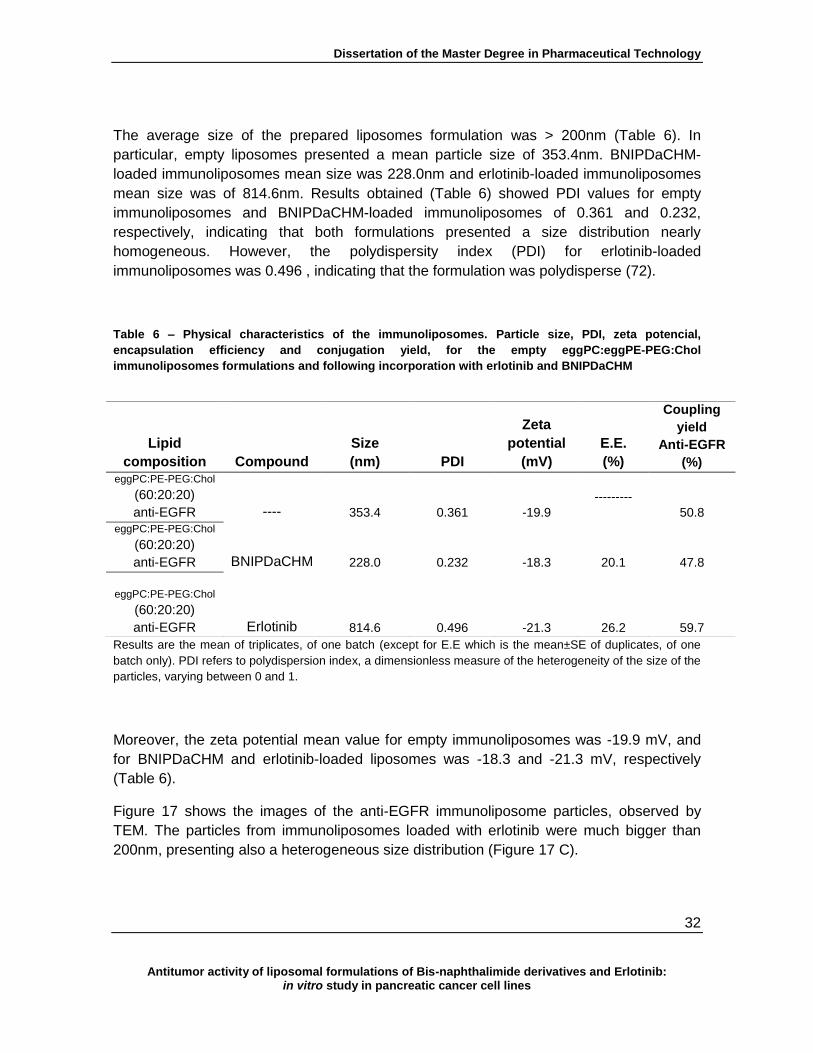
Dissertation of the Master Degree in Pharmaceutical Technology
32
Antitumor activity of liposomal formulations of Bis-naphthalimide derivatives and Erlotinib: in vitro study in pancreatic cancer cell lines
The average size of the prepared liposomes formulation was > 200nm (Table 6). In
particular, empty liposomes presented a mean particle size of 353.4nm. BNIPDaCHM-
loaded immunoliposomes mean size was 228.0nm and erlotinib-loaded immunoliposomes
mean size was of 814.6nm. Results obtained (Table 6) showed PDI values for empty
immunoliposomes and BNIPDaCHM-loaded immunoliposomes of 0.361 and 0.232,
respectively, indicating that both formulations presented a size distribution nearly
homogeneous. However, the polydispersity index (PDI) for erlotinib-loaded
immunoliposomes was 0.496 , indicating that the formulation was polydisperse (72).
Table 6 – Physical characteristics of the immunoliposomes. Particle size, PDI, zeta potencial,
encapsulation efficiency and conjugation yield, for the empty eggPC:eggPE-PEG:Chol
immunoliposomes formulations and following incorporation with erlotinib and BNIPDaCHM
Results are the mean of triplicates, of one batch (except for E.E which is the mean±SE of duplicates, of one
batch only). PDI refers to polydispersion index, a dimensionless measure of the heterogeneity of the size of the
particles, varying between 0 and 1.
Moreover, the zeta potential mean value for empty immunoliposomes was -19.9 mV, and
for BNIPDaCHM and erlotinib-loaded liposomes was -18.3 and -21.3 mV, respectively
(Table 6).
Figure 17 shows the images of the anti-EGFR immunoliposome particles, observed by
TEM. The particles from immunoliposomes loaded with erlotinib were much bigger than
200nm, presenting also a heterogeneous size distribution (Figure 17 C).
Lipid
composition Compound
Size
(nm) PDI
Zeta
potential
(mV)
E.E.
(%)
Coupling
yield
Anti-EGFR
(%) eggPC:PE-PEG:Chol
(60:20:20)
anti-EGFR ---- 353.4 0.361 -19.9
---------
50.8
eggPC:PE-PEG:Chol
(60:20:20)
anti-EGFR
BNIPDaCHM 228.0 0.232 -18.3
20.1 47.8
eggPC:PE-PEG:Chol
(60:20:20)
anti-EGFR Erlotinib 814.6 0.496 -21.3
26.2 59.7

Dissertation of the Master Degree in Pharmaceutical Technology
33
Antitumor activity of liposomal formulations of Bis-naphthalimide derivatives and Erlotinib: in vitro study in pancreatic cancer cell lines
In addition, immunoliposome particles presented spherical unilamellar morphology. A ring
around the liposomal surface of all formulations could be observed, showing the anti-
EGFR coating.
Figure 17 – Imaginga of immunoliposomes obtained by TEM. Empty immunoliposomes (A),
immunoliposomes with BNIPDaCHM (B) and immunoliposomes with erlotinib (C).
4.8. In vitro cell growth inhibition analysis for empty immunoliposomes and
erlotinib and BNIPDaCHM loaded immunoliposomes on BxPC-3 cells
In order to assess the effect of the compound/drug loaded in anti-EGFR immunoliposomes
on BxPC-3 cells, the SRB assay was performed. Empty immunoliposomes were tested, to
certify the non-toxicity of the carriers with the cell line BxPC-3 (Figure 18). Results
demonstrated that immunoliposomes when tested up to 1 mg/mL did not affect cell growth
(observing 97.3% of cell growth in relation to blank). Nevertheless higher concentrations
caused a slight decrease in the percentage of cell growth. In fact, treatment with 2 mg/mL
reduced the cell growth to 87.1%, while for the highest concentration (4 mg/mL) the mean
percentage of cell growth obtained was 70.1 ± 2.1%.

Dissertation of the Master Degree in Pharmaceutical Technology
34
Antitumor activity of liposomal formulations of Bis-naphthalimide derivatives and Erlotinib: in vitro study in pancreatic cancer cell lines
Figure 18 - Effect of empty immunoliposomes on BxPC-3 viable cells number determined by the
sulforhodamine B assay. Results are presented as a percentage of cell growth in relation to blank cells
(treated with medium only) and are from one experiment in quadruplicate.
The activity of erlotinib and BNIPDaCHM loaded on anti-EGFR immunoliposomes was
also analyzed by SRB assay. Results from one preliminary experiment (Table 7) showed a
decrease in the IC50 values for both entrapped erlotinib and BNIPDaCHM. BNIPDaCHM-
loaded immunoliposomes presented an IC50 concentration of 0.9 µM and erlotinib-loaded
immunoliposomes presented an IC50 of 1.2 µM, both of which are lower than the values
obtained with non-functionalized liposomes (non-targeted liposomes with BNIPDaCHM
presented IC50 of 1.5 ± 0.1 µM and liposomes with erlotinib presented IC50 of 1.8 ± 0.1
µM). These experiments need to be repeated in order to confirm the results.
Table 7 - IC50 values of erlotinib and BNIPDaCHM entrapped immunoliposomes
IC50 (µM)
Erlotinib
BNIPDaCHM
eggPC:PE-PEG:Chol
(60:20:20)
anti-EGFR
1.2 0.9
Data is mean of IC50 quadruplicates, from one experiment only.
Dissertation of the Master Degree in Pharmaceutical Technology
35
Antitumor activity of liposomal formulations of Bis-naphthalimide derivatives and Erlotinib: in vitro study in pancreatic cancer cell lines
IV. Discussion
In the present work the effect of two BNIP derivative compounds, BNIPDaCHM and NPA,
and of the TKI, erlotinib, were studied on the BxPC-3 pancreatic cancer cell line.
BNIPs have been reported as DNA-binding compounds, with potential for cancer therapy,
in particular for NSCLC and breast cancer (38, 73, 74). Previous studies reported that in
vitro exposure of a pancreatic cell line to a BNIP derivative (BNIPDaoct) was more
effective than gemcitabine (44).
The cytotoxicity assays carried out for the BNIP derivatives used in this study clearly
indicated that NPA was the less active compound (IC50 of 70.3 ± 11.1 µM), in comparison
with BNIPDaCHM (IC50 of 1.3 ± 0.1 µM). These results are in agreement with previous
observations revealing that the presence of two naphthalimidopropyl moieties influences
the biological activity of BNIP compounds in cancer cells (42, 75). NPA has only one
naphthalimidopropyl moiety and consequently is less cytotoxic, as previously described for
NSCLC cell lines (42, 75). Structural derivatives features on BNIPs are responsible for
enhanced biological activity and solubility, such as the bisnaphthalimidopropyl
functionality, an alkyl linker chain length ideally between 8-10 carbons, and the presence
of heteroatoms (more than 2 nitrogen atoms) (40-42, 76).
Erlotinib was the first biological drug approved by FDA (Food and Drug Administration) for
the treatment of advanced pancreatic cancer, combined with the conventional
chemotherapeutic agent gemcitabine, presenting improvements on the survival rates of
patients (16, 54). This drug is a tyrosine kinase inhibitor, being an EGFR inhibitor, and is
also used for other types of cancer such as ovarian, breast and NSCLC (77-79). In vitro
assays of erlotinib on the BxPC-3 cell line confirmed its biological activity in this cell line
(IC50 of 1.7 ± 0.06 µM). Others have obtained similar results for the same cell line (IC50 =
1.26 µM) (21).
The more active of the two tested compound/drug on BxPC-3 cells, BNIPDaCHM and the
EGFR inhibitor erlotinib, respectively, were selected to further study their cellular effect.
Both affected the cell cycle profile of BxPC-3 cells. BNIPDaCHM and erlotinib elicited a
significant (P < 0.05) decrease of 13 and 15%, respectively, in the G1 phase of BxPC-3, in
agreement to what has been previously published, in the same cell line for erlotinib and in
breast cancer cell line in the case of BNIPDaCHM (21, 75). Results also showed a very
small increase in the sub-G1 peak following treatment with BNIPDaCHM at its IC50 and
with erlotinib at its 2xIC50 (3 and 5%, respectively), which was suggestive of cell death by
apoptosis. Interestingly, previous reports indicated that both erlotinib and BNIPDaCHM
increased the sub-G1 peak (21, 75).

Dissertation of the Master Degree in Pharmaceutical Technology
36
Antitumor activity of liposomal formulations of Bis-naphthalimide derivatives and Erlotinib: in vitro study in pancreatic cancer cell lines
Therefore, the levels of apoptotic cell death were verified for both compounds, by flow
cytometry analysis following Annexin V/PI staining. Results showed a slight increase in cell
death following treatment with BNIPDaCHM (16.7 ± 1.8%), in comparison with the control
(10.8 ± 1.7%).
This is in agreement with previous reports, where BNIPDaCHM clearly increased the level
of apoptosis in breast cancer cell line (75). In addition, a slight increase in cell death was
observed following treatment with erlotinib (IC50 to 18.9 ± 1.7% and for 2xIC50 to 16.3 ±
1.7%) comparatively with the control (11.7 ± 1.8%). However, this effect was only
statistical significant for treatment with the IC50 concentration of erlotinib (P < 0.05).
Previous reports from other groups have also indicated that erlotinib induced cell death by
apoptosis in these cells and also in a hepatocellular cancer cell line (21, 50).
The expression levels of apoptosis-related proteins, PARP and Bcl-2 were also analyzed
following treatment with both the compounds studied. A slight decrease on the expression
of PARP was observed in cells treated with BNIPDaCHM and erlotinib, however these
results were not very clear. Regarding the expression of Bcl-2 protein, preliminary results
from this study seem to indicate a very small increase in the levels of this anti-apoptotic
protein following treatment with both compounds in relation to control cells. Nevertheless,
these results need to be further confirmed.
In this work, erlotinib and BNIPDaCHM (the most active of both compounds tested), were
further incorporated into liposomes. Entrapping the compound/drug in liposomal carriers
presents advantages in vitro as well as in vivo, due to the ability of liposomes to retain the
entrapped compound while promoting a sustained release of the active substance (1). In
addition, because both BNIPDaCHM and erlotinib are lipophilic, liposome entrapment
would help their administration in vivo and enhance the drug stability after administration
(80). The average size of all produced liposomal formulations was below 200nm [empty
liposomes (163.0 ± 0.74nm) < liposomes with erlotinib (174.9 ± 10.3nm) < liposomes with
BNIPDaCHM (188.9 ± 13.4nm)]. It is described that nanoparticles with sizes up to 200nm
are more stable in vivo, as MPS does not uptake them so easily from the blood stream
(11).
The determined PDI for the produced liposomal formulations were in average below 0.2
and it is described that PDI values between 0.1-0.2 indicate the presence of a
homogenous size distribution (3, 72). Low PDI values are important to ascertain the
colloidal stability of the liposomal formulations, since higher PDI values (≥0.5) indicate
polydisperse formulations, which may result in particle aggregates (81).

Dissertation of the Master Degree in Pharmaceutical Technology
37
Antitumor activity of liposomal formulations of Bis-naphthalimide derivatives and Erlotinib: in vitro study in pancreatic cancer cell lines
The encapsulation efficiency obtained for the produced liposomal formulations with
BNIPDaCHM and erlotinib were 28.5 ± 1.6 % and 40.6 ± 4.2 %, respectively. The results
were promising, particularly for erlotinib, even though not as high as it was expected.
Other preparation techniques should be tested in order to enhance the encapsulation
efficiency.
The liposomes observation by TEM showed spherical unilamellar vesicles with sizes below
200 nm, which are in agreement with the results obtained by DLS.
The in vitro cytotoxic activity of BNIPDaCHM and erlotinib incorporated in the liposomes
was assessed on BxPC-3 cells. Results showed that the IC50 concentrations obtained with
BNIPDaCHM-loaded liposomes (IC50 1.5 ± 0.1) increased slightly in comparison with the
“free” compound (1.3 ± 0.05 µM).
Even though the IC50 values for BNIPDaCHM-loaded liposomes and for “free”
BNIPDaCHM were rather similar, they were considered statistically significant (P <0.05).
Erlotinib-loaded liposomes presented a slight increase in the IC50 concentration, with no
statistical significance (IC50 1.8 ± 0.1). Literature refers to liposomes as a drug delivery
vehicle that can allow a sustained release of entrapped compounds (1, 82). The slight
increase in the IC50 of the BNIPDaCHM-loaded liposomes may suggest a more sustained
release of the compound into the BxPC-3 cells, allowed by the liposomes; however more
assays would need to be undertaken to verify this hypothesis.
Immunoliposomes are an attractive strategy to enhance therapeutic approaches, by
targeting specifically tumor tissues for delivery of entrapped drugs, and thereby not
affecting the normal tissues which are sensitive to the toxic effects of the compounds. The
coupling yield of the human anti-EGFR antibody to the drug-loaded liposomes was in
average 50%, in agreement with previously published studies using the carbodiimide
coupling technique (56).
The immunoliposomes were characterized in terms of size, PDI, morphology and
encapsulation efficiency. In general, for all formulations, it was observed an increase on
the size and PDI values with the functionalization of the liposomes with the EGFR
antibody.
Preliminary experiments indicate that the sizes of the immunoliposomes with erlotinib were
increased, in comparison with the other immune formulations. In addition, it was the only
formulation that presented a PDI value ≥ 0.5, indicating a polydisperse formulation. For the
immune formulations without any compound or incorporating BNIPDaCHM, a slight
increase in their sizes was observed in comparison with the non-functionalized liposomes
(BNIPDaCHM-loaded immunoliposomes 228.0 nm and empty immunoliposomes 353.4
nm).

Dissertation of the Master Degree in Pharmaceutical Technology
38
Antitumor activity of liposomal formulations of Bis-naphthalimide derivatives and Erlotinib: in vitro study in pancreatic cancer cell lines
Previous reports indicated that the conjugation of antibodies in the surface of liposomes
can result in an increase of the liposomal size to sizes higher than 200 nm (83). The
observation of immunoliposomes by TEM showed spherical unilamellar morphology and
the coupling of the antibody on the liposomal surface. TEM imaging also confirmed that
immunoliposomes with erlotinib were a polydisperse formulation with particles bigger than
200nm.
The in vitro cytotoxicity of the BNIPDaCHM and erlotinib incorporated in the
immunoliposomes was assessed on BxPC-3. Preliminary results showed a decrease in
the IC50 values, for both BNIPDaCHM and erlotinib following incorporation in the
immunoliposomes (IC50 1.2 µM for erlotinib-loaded immunoliposomes and IC50 0.9 µM for
BNIPDaCHM-loaded immunoliposomes) in relation to the non-functionalized liposomes.
This decrease on the IC50 concentration for both erlotinib and BNIPDaCHM entrapped into
immunoliposomes may be due to combination of the compounds with an anti-EGFR
antibody. Indeed, other authors have published the advantages of combining two distinct
classes of EGFR inhibitors (small molecules TKIs and antibodies anti-EGFR), increasing
their antitumor activity and overcoming the tumor resistance to TKIs. These previously
published studies were undertaken in vitro using head and neck, prostate and NSCLC
cancer cell lines (53).
Previous in vivo reports indicate that the association of a DNA-intercalating drug
(Doxorubicin) with anti-EGFR immunoliposomes increased its antitumor activity in human
breast cancer xenograft mice (58, 60). In the present study, further studies for both
BNIPDaCHM and erlotinib are needed, to assess possible alterations to their activity due
to the anti-EGFR antibody presence.

Dissertation of the Master Degree in Pharmaceutical Technology
39
Antitumor activity of liposomal formulations of Bis-naphthalimide derivatives and Erlotinib: in vitro study in pancreatic cancer cell lines
V. Conclusions
In the present work, BNIPDaCHM (a BNIP derivative compound) and erlotinib (Tarceva®)
were shown to significantly affect the viability of BxPC-3 cells. Both compound and drug
were shown to affect the cell cycle profile of these cells, decreasing the percentage of cells
in the G1 phase of the cell cycle. In addition, a slight increase in apoptosis was observed
in BxPC-3 cells following treatment with BNIPDaCHM or erlotinib.
Liposomes prepared by the lipid film hydration method resulted in acceptable size of
particles for further in vivo testing; the encapsulation efficiency obtained through this
preparation technique was also promising even though not good, having been in average
higher than 20 %.
Preliminary experiments indicate that erlotinib and BNIPDaCHM incorporation in anti-
EGFR immunoliposomes resulted in a decrease of the IC50 values for both erlotinib and
BNIPDaCHM. These results suggest that the combination of an anti-EGFR antibody with a
small molecule EGFR inhibitor or a DNA-intercalating compound enhance the biological
activity for both compounds. However further studies need to be undertaken to confirm the
enhanced anti-tumor activity of these immunoliposomes.

Dissertation of the Master Degree in Pharmaceutical Technology
40
Antitumor activity of liposomal formulations of Bis-naphthalimide derivatives and Erlotinib: in vitro study in pancreatic cancer cell lines
Bibliography
1. Torchilin VP. Multifunctional Pharmaceutical Nanocarriers: Promises and Problems. Nanotechnologies for the Life Sciences: Wiley-VCH Verlag GmbH & Co. KGaA; 2007. 2. Martins Ochubiojo E, Ifeoma Chinwude O, Ekaete Ibanga A, Sabinus Ifeanyi O. Nanotechnology in Drug Delivery. 2012. 3. Lasic DD. Liposomes: from physics to applications: Elsevier; 1993. 4. Ana Catarina Pinto. Combination Chemotherapy in Cancer: Principles, Evaluation and Drug Delivery Strategies, Current Cancer Treatment - Novel Beyond Conventional Approaches. In: InTech, editor.2011. 5. Vega-Villa KR, Takemoto JK, Yáñez JA, Remsberg CM, Forrest ML, Davies NM. Clinical toxicities of nanocarrier systems. Advanced Drug Delivery Reviews. 2008;60(8):929-38. 6. Alexis F, Rhee J-W, Richie JP, Radovic-Moreno AF, Langer R, Farokhzad OC. New frontiers in nanotechnology for cancer treatment. Urologic Oncology: Seminars and Original Investigations. 2008;26(1):74-85. 7. Torchlin VP. Liposomes: Practical approach. 2nd edition ed. Edition n, editor: Oxford University Press; 2003. 380 p. 8. Nature. [cited 2013 22-08]; Available from: www.nature.com. 9. Mayer LD, Cullis PR, Bally MB. Chapter 4.2 - Designing therapeutically optimized liposomal anticancer delivery systems: Lessons from conventional liposomes. In: Lasic DD, Papahadjopoulos D, editors. Medical Applications of Liposomes. Amsterdam: Elsevier Science B.V.; 1998. p. 231-57. 10. Bangham AD, Standish MM, Watkins JC. Diffusion of univalent ions across the lamellae of swollen phospholipids. Journal of Molecular Biology. 1965;13(1):238-IN27. 11. A.J. Domb YT. Particles for Pharmaceutical Applications. Ranch tS, editor: American Scientific Publishers; 2007 2007. 12. Sahoo SK, Labhasetwar V. Nanotech approaches to drug delivery and imaging. Drug Discov Today. 2003;8(24):1112-20. Epub 2003/12/18. 13. Kirby BJ, Hasselbrink EF. Zeta potential of microfluidic substrates: 1. Theory, experimental techniques, and effects on separations. ELECTROPHORESIS. 2004;25(2):187-202. 14. Torchilin VP. Recent advances with liposomes as pharmaceutical carriers. Nature reviews Drug discovery. 2005;4(2):145-60. Epub 2005/02/03. 15. Sávia Caldeira de Araújo L, Cristiane dos Santos G, Talita Guieiro Ribeiro R, Diêgo dos Santos F, Elaine Amaral L, Mônica Cristina O. Liposomes as Carriers of Anticancer Drugs. 2013. 16. Yu X, Zhang Y, Chen C, Yao Q, Li M. Targeted drug delivery in pancreatic cancer. Biochimica et biophysica acta. 2010;1805(1):97-104. Epub 2009/10/27. 17. Lasic DD, Martin FJ, Gabizon A, Huang SK, Papahadjopoulos D. Sterically stabilized liposomes: a hypothesis on the molecular origin of the extended circulation times. Biochimica et Biophysica Acta (BBA) - Biomembranes. 1991;1070(1):187-92. 18. POP. Portal de Oncologia Português. [cited 2013 24-08]; Available from: http://www.pop.eu.com/portal.

Dissertation of the Master Degree in Pharmaceutical Technology
41
Antitumor activity of liposomal formulations of Bis-naphthalimide derivatives and Erlotinib: in vitro study in pancreatic cancer cell lines
19. Heinemann V, Haas M, Boeck S. Systemic treatment of advanced pancreatic cancer. Cancer Treat Rev. 2012;38(7):843-53. Epub 2012/01/10. 20. INE. Instituto Nacional de Estatística [cited 2013 20-07]; Available from: http://www.ine.pt/xportal. 21. Ioannou N, Dalgleish AG, Seddon AM, Mackintosh D, Guertler U, Solca F, et al. Anti-tumour activity of afatinib, an irreversible ErbB family blocker, in human pancreatic tumour cells. British journal of cancer. 2011;105(10):1554-62. Epub 2011/10/06. 22. Tamburrino A, Piro G, Carbone C, Tortora G, MELISI D. Mechanisms of resistance to chemotherapeutic and antiangiogenic drugs as novel targets for pancreatic cancer therapy. Frontiers in Pharmacology. 2013;4. 23. Belli C, Cereda S, Anand S, Reni M. Neoadjuvant therapy in resectable pancreatic cancer: a critical review. Cancer Treat Rev. 2013;39(5):518-24. Epub 2012/11/06. 24. Giuliani F, Di Maio M, Colucci G, Perrone F. Conventional chemotherapy of advanced pancreatic cancer. Current drug targets. 2012;13(6):795-801. Epub 2012/03/31. 25. Conroy T, Desseigne F, Ychou M, Bouche O, Guimbaud R, Becouarn Y, et al. FOLFIRINOX versus gemcitabine for metastatic pancreatic cancer. The New England journal of medicine. 2011;364(19):1817-25. Epub 2011/05/13. 26. Michael ELM. Medical Therapy of Pancreatic Cancer: Current Status and Future Targets, Pancreatic Cancer - Clinical Management, Prof. Sanjay Srivastava (Ed.)2012. 27. Vikas Bhardwaj SMT, James C.K. Lai and Alok Bhushan. Failure of Pancreatic Cancer Chemotherapy: Consequences of Drug Resistance Mechanisms. In: Srivastava SK, editor.: InTech; 2012. 28. Yu X, Zhang Y, Chen C, Yao Q, Li M. Targeted drug delivery in pancreatic cancer. Biochimica et Biophysica Acta (BBA) - Reviews on Cancer. 2010;1805(1):97-104. 29. Paolino D, Cosco D, Racanicchi L, Trapasso E, Celia C, Iannone M, et al. Gemcitabine-loaded PEGylated unilamellar liposomes vs GEMZAR®: Biodistribution, pharmacokinetic features and in vivo antitumor activity. Journal of Controlled Release. 2010;144(2):144-50. 30. Palchaudhuri R, Hergenrother PJ. DNA as a target for anticancer compounds: methods to determine the mode of binding and the mechanism of action. Current Opinion in Biotechnology. 2007;18(6):497-503. 31. antitumour activity of new dendritic polyamines–(imide–DNA-intercalator) conjugates: potent Lck inhibitors. European Journal of Medicinal Chemistry. 2002;37(7):541-51. 32. Brana MF, Castellano JM, Roldan CM, Santos A, Vazquez D, Jimenez A. Synthesis and mode(s) of action of a new series of imide derivatives of 3-nitro-1,8 naphthalic acid. Cancer chemotherapy and pharmacology. 1980;4(1):61-6. Epub 1980/01/01. 33. Tan S, Yin H, Chen Z, Qian X, Xu Y. Oxo-heterocyclic fused naphthalimides as antitumor agents: Synthesis and biological evaluation. European Journal of Medicinal Chemistry. 2013;62(0):130-8. 34. Bailly C, Braña M, Waring MJ. Sequence-Selective Intercalation of Antitumour Bis-Naphthalimides into DNA. European Journal of Biochemistry. 1996;240(1):195-208. 35. Brana MF, Ramos A. Naphthalimides as anti-cancer agents: synthesis and biological activity. Current medicinal chemistry Anti-cancer agents. 2001;1(3):237-55. Epub 2003/04/08.
Dissertation of the Master Degree in Pharmaceutical Technology
42
Antitumor activity of liposomal formulations of Bis-naphthalimide derivatives and Erlotinib: in vitro study in pancreatic cancer cell lines
36. Bestwick CS, Ralton LD, Milne L, Kong Thoo Lin P, Duthie SJ. The influence of bisnaphthalimidopropyl polyamines on DNA instability and repair in Caco-2 colon epithelial cells. Cell biology and toxicology. 2011;27(6):455-63. Epub 2011/08/16. 37. Brana MF, Castellano JM, Moran M, Perez de Vega MJ, Romerdahl CR, Qian XD, et al. Bis-naphthalimides: a new class of antitumor agents. Anti-cancer drug design. 1993;8(4):257-68. Epub 1993/08/01. 38. Ralton LD, Bestwick CS, Milne L, Duthie S, Kong Thoo Lin P. Bisnaphthalimidopropyl spermidine induces apoptosis within colon carcinoma cells. Chemico-Biological Interactions. 2009;177(1):1-6. 39. IPA. Intellectual Property in Australia. [cited 2013 18-09]; Available from: http://www.ipaustralia.com.au/applicant/universidade-do-porto/patents/AU2007273886/. 40. Rodilla V, Pavlov V, Hion D, Lin PKT. Cytotoxic and DNA binding properties of new bis-naphthalimido polyamine derivatives. Toxicology. 2000;148(1):72-3. 41. Dance A-M, Ralton L, Fuller Z, Milne L, Duthie S, Bestwick CS, et al. Synthesis and biological activities of bisnaphthalimido polyamines derivatives: cytotoxicity, DNA binding, DNA damage and drug localization in breast cancer MCF 7 cells. Biochemical Pharmacology. 2005;69(1):19-27. 42. Barron GA, Bermano G, Gordon A, Kong Thoo Lin P. Synthesis, cytotoxicity and DNA-binding of novel bisnaphthalimidopropyl derivatives in breast cancer MDA-MB-231 cells. European Journal of Medicinal Chemistry. 2010;45(4):1430-7. 43. Pavlov V, Kong Thoo Lin P, Rodilla V. Cytotoxicity, DNA binding and localisation of novel bis-naphthalimidopropyl polyamine derivatives. Chemico-Biological Interactions. 2001;137(1):15-24. 44. Hoskins C, Ouaissi M, Lima SC, Cheng WP, Loureirio I, Mas E, et al. In vitro and in vivo anticancer activity of a novel nano-sized formulation based on self-assembling polymers against pancreatic cancer. Pharm Res. 2010;27(12):2694-703. Epub 2010/09/28. 45. Taciane Ladislau KPM, Renata D Daltoé, Isabella S Guimarães,Rangel and Alice L Herlinger. Target Cancer Therapy, Cancer Treatment - Conventional and Innovative Approaches. Rangel PL, editor: InTech; ,2013. 46. Arora A, Scholar EM. Role of Tyrosine Kinase Inhibitors in Cancer Therapy. Journal of Pharmacology and Experimental Therapeutics. 2005. 47. Tzeng C-WD, Frolov A, Frolova N, Jhala NC, Howard JH, Buchsbaum DJ, et al. Epidermal growth factor receptor (EGFR) is highly conserved in pancreatic cancer. Surgery. 2007;141(4):464-9. 48. Vlahovic G, Crawford J. Activation of Tyrosine Kinases in Cancer. The Oncologist. 2003;8(6):531-8. 49. Raymond E, Faivre S, Armand JP. Epidermal Growth Factor Receptor Tyrosine Kinase as a Target for Anticancer Therapy. Drugs. 2000;60(2):15-23. 50. Huether A, Höpfner M, Sutter AP, Schuppan D, Scherübl H. Erlotinib induces cell cycle arrest and apoptosis in hepatocellular cancer cells and enhances chemosensitivity towards cytostatics. Journal of Hepatology. 2005;43(4):661-9. 51. Tzeng C-WD, Frolov A, Frolova N, Jhala NC, Howard JH, Vickers SM, et al. EGFR Genomic Gain and Aberrant Pathway Signaling in Pancreatic Cancer Patients. Journal of Surgical Research. 2007;143(1):20-6.

Dissertation of the Master Degree in Pharmaceutical Technology
43
Antitumor activity of liposomal formulations of Bis-naphthalimide derivatives and Erlotinib: in vitro study in pancreatic cancer cell lines
52. Liao C, Sun Q, Liang B, Shen J, Shuai X. Targeting EGFR-overexpressing tumor cells using Cetuximab-immunomicelles loaded with doxorubicin and superparamagnetic iron oxide. European Journal of Radiology. 2011;80(3):699-705. 53. Huang S, Armstrong EA, Benavente S, Chinnaiyan P, Harari PM. Dual-agent molecular targeting of the epidermal growth factor receptor (EGFR): combining anti-EGFR antibody with tyrosine kinase inhibitor. Cancer research. 2004;64(15):5355-62. Epub 2004/08/04. 54. Philip PA. Targeted therapies for pancreatic cancer. Gastrointestinal cancer research : GCR. 2008;2(4 Suppl):S16-9. Epub 2009/04/04. 55. Traxler P, Bold G, Buchdunger E, Caravatti G, Furet P, Manley P, et al. Tyrosine kinase inhibitors: from rational design to clinical trials. Medicinal research reviews. 2001;21(6):499-512. Epub 2001/10/19. 56. Torchilin VP, Khaw BA, Smirnov VN, Haber E. Preservation of antimyosin antibody activity after covalent coupling to liposomes. Biochemical and Biophysical Research Communications. 1979;89(4):1114-9. 57. Mastrobattista E, Koning GA, Storm G. Immunoliposomes for the targeted delivery of antitumor drugs. Advanced Drug Delivery Reviews. 1999;40(1–2):103-27. 58. Park JW, Kirpotin DB, Hong K, Shalaby R, Shao Y, Nielsen UB, et al. Tumor targeting using anti-her2 immunoliposomes. Journal of Controlled Release. 2001;74(1–3):95-113. 59. Park JW, Hong K, Kirpotin DB, Papahadjopoulos D, Benz CC. Immunoliposomes for Cancer Treatment. In: August JT, editor. Advances in Pharmacology: Academic Press; 1997. p. 399-435. 60. Park JW, Hong K, Kirpotin DB, Colbern G, Shalaby R, Baselga J, et al. Anti-HER2 immunoliposomes: enhanced efficacy attributable to targeted delivery. Clinical cancer research : an official journal of the American Association for Cancer Research. 2002;8(4):1172-81. Epub 2002/04/12. 61. Barrajón-Catalán E, Menéndez-Gutiérrez MP, Falco A, Carrato A, Saceda M, Micol V. Selective death of human breast cancer cells by lytic immunoliposomes: Correlation with their HER2 expression level. Cancer Letters. 2010;290(2):192-203. 62. Manjappa AS, Chaudhari KR, Venkataraju MP, Dantuluri P, Nanda B, Sidda C, et al. Antibody derivatization and conjugation strategies: Application in preparation of stealth immunoliposome to target chemotherapeutics to tumor. Journal of Controlled Release. 2011;150(1):2-22. 63. Béduneau A, Saulnier P, Hindré F, Clavreul A, Leroux J-C, Benoit J-P. Design of targeted p c p c j f w ’ f 2007;28(33):4978-90. 64. Hermanson GT. Chapter 2 - The Chemistry of Reactive Groups. Bioconjugate Techniques (Second Edition). New York: Academic Press; 2008. p. 169-212. 65. Hermanson GT. Chapter 3 - Zero-Length Crosslinkers. Bioconjugate Techniques (Second Edition). New York: Academic Press; 2008. p. 213-33. 66. Cheng WWK, Allen TM. Targeted delivery of anti-CD19 liposomal doxorubicin in B-cell p : A c p f w c ′ f c v Journal of Controlled Release. 2008;126(1):50-8. 67. Bae YH, Park K. Targeted drug delivery to tumors: myths, reality and possibility. Journal of controlled release : official journal of the Controlled Release Society. 2011;153(3):198-205. Epub 2011/06/15.

Dissertation of the Master Degree in Pharmaceutical Technology
44
Antitumor activity of liposomal formulations of Bis-naphthalimide derivatives and Erlotinib: in vitro study in pancreatic cancer cell lines
68. Jiangsy Sihuan Bioengineering Co. L. Jiangsy Sihuan Bioengineering Co.,LTD. [cited 2013 19-09]; Available from: http://www.sihuanpharma.com/product/erlotinib-hydrochloride.html#sthash.iIGonH5o.dpbs. 69. Monks A, Scudiero D, Skehan P, Shoemaker R, Paull K, Vistica D, et al. Feasibility of a high-flux anticancer drug screen using a diverse panel of cultured human tumor cell lines. Journal of the National Cancer Institute. 1991;83(11):757-66. Epub 1991/06/05. 70. Vichai V, Kirtikara K. Sulforhodamine B colorimetric assay for cytotoxicity screening. Nature protocols. 2006;1(3):1112-6. Epub 2007/04/05. 71. Nanosciences. [cited 2013 24-08]; Available from: http://www.immunosome.com/conjugations/conjugation-through-c-on-antibody/pe/. 72. Vijaykumar Nekkanti VVaRP. Drug Nanoparticles - an overview. In: Hashim DAA, editor. "The Delivery of Nanoparticles". InTech2012. 73. Filosa R, Peduto A, Micco SD, Caprariis Pd, Festa M, Petrella A, et al. Molecular modelling studies, synthesis and biological activity of a series of novel bisnaphthalimides and their development as new DNA topoisomerase II inhibitors. Bioorganic & Medicinal Chemistry. 2009;17(1):13-24. 74. Lima RT, Barron G, Grabowska JA, Bermano G, Kaur S, Roy N, et al. 1005 Bisnaphthalimidopropyl Derivatives as Antitumor Agents –Targeting SIRT2. European Journal of Cancer. 2012;48, Supplement 5(0):S243. 75. Lima RT, Barron GA, Grabowska JA, Bermano G, Kaur S, Roy N, et al. Cytotoxicity and cell death mechanisms induced by a novel bisnaphthalimidopropyl derivative against the NCI-H460 non-small lung cancer cell line. Anti-cancer agents in medicinal chemistry. 2013;13(3):414-21. Epub 2012/10/25. 76. Costa Lima S, Rodrigues V, Garrido J, Borges F, Kong Thoo Lin P, Cordeiro da Silva A. In vitro evaluation of bisnaphthalimidopropyl derivatives loaded into pegylated nanoparticles against Leishmania infantum protozoa. International Journal of Antimicrobial Agents. 2012;39(5):424-30. 77. Muir VJ, Dhillon S. Erlotinib: as maintenance monotherapy in non-small-cell lung cancer. BioDrugs : clinical immunotherapeutics, biopharmaceuticals and gene therapy. 2011;25(3):139-46. Epub 2011/04/26. 78. Marth C, Egle D, Auer D, Rössler J, Zeimet AG, Vergote I, et al. Modulation of CA-125 tumor marker shedding in ovarian cancer cells by erlotinib or cetuximab. Gynecologic Oncology. 2007;105(3):716-21. 79. Twelves C, Trigo JM, Jones R, De Rosa F, Rakhit A, Fettner S, et al. Erlotinib in combination with capecitabine and docetaxel in patients with metastatic breast cancer: A dose-escalation study. European Journal of Cancer. 2008;44(3):419-26. 80. Gulati M, Grover M, Singh S, Singh M. Lipophilic drug derivatives in liposomes. International journal of pharmaceutics. 1998;165(2):129-68. 81. Franzen U, Ostergaard J. Physico-chemical characterization of liposomes and drug substance-liposome interactions in pharmaceutics using capillary electrophoresis and electrokinetic chromatography. Journal of chromatography A. 2012. Epub 2012/07/25. 82. D.D.Lasic. Handbook of Biological Physics1995. p. pag. 493-516. 83. Gao J, Liu W, Xia Y, Li W, Sun J, Chen H, et al. The promotion of siRNA delivery to breast cancer overexpressing epidermal growth factor receptor through anti-EGFR antibody conjugation by immunoliposomes. Biomaterials. 2011;32(13):3459-70.

Dissertation of the Master Degree in Pharmaceutical Technology
45
Antitumor activity of liposomal formulations of Bis-naphthalimide derivatives and Erlotinib: in vitro study in pancreatic cancer cell lines