antimicrobial surfaces for craniofacial implants: state of ... · antimicrobial surfaces for...
TRANSCRIPT

43
Antimicrobial surfaces for craniofacial implants: state of the art
Lisa Actis, Laura Gaviria, Teja Guda, Joo L. Ong
Department of Biomedical Engineering, University of Texas at San Antonio, San Antonio, TX, USA
Abstract (J Korean Assoc Oral Maxillofac Surg 2013;39:43-54)
In an attempt to regain function and aesthetics in the craniofacial region, different biomaterials, including titanium, hydroxyapatite, biodegradable polymers and composites, have been widely used as a result of the loss of craniofacial bone. Although these materials presented favorable success rates, osseointegration and antibacterial properties are often hard to achieve. Although bone-implant interactions are highly dependent on the implant’s surface characteristics, infections following traumatic craniofacial injuries are common. As such, poor osseointegration and infections are two of the many causes of implant failure. Further, as increasingly complex dental repairs are attempted, the likelihood of infection in these implants has also been on the rise. For these reasons, the treatment of craniofacial bone defects and dental repairs for long-term success remains a challenge. Various approaches to reduce the rate of infection and improve osseointegration have been investigated. Furthermore, recent and planned tissue engineering developments are aimed at improving the implants’ physical and biological properties by improving their surfaces in order to develop craniofacial bone substitutes that will restore, maintain and improve tissue function. In this review, the commonly used biomaterials for craniofacial bone restoration and dental repair, as well as surface modification techniques, antibacterial surfaces and coatings are discussed.
Key words: Dental implants, Osseointegration, Antimicrobial agents, Surface-coated materials, Bone regeneration[paper submitted 2013. 4. 1 / accepted 2013. 4. 2]
the placement of extra-oral implants can only be done in an
operating room, and requires trained oral and maxillofacial
surgeons. Additionally, the fabrication of these prostheses has
to be performed by trained prosthodontists resulting in extra-
oral implants being more complex and costly than the intra-
oral counterparts2.
The loss of craniofacial tissues can result from neoplasm,
trauma, tumor or cyst resection, infectious diseases, nonunion
fractures, and congenital or developmental conditions (i.e.,
cleft palate defects); which results in serious functional,
aesthetic and psychological sequelae. In these situations,
the absence of hard and soft tissues can be disfiguring,
and in many cases, it compromises basic functions such as
mastication, speech, swallowing, leading to limited thermal
and physical protection of important anatomical structures
(i.e., brain, nerves, arteries, veins)3-6. In the United States,
there is a clinical need for craniofacial bone regeneration, and
more than 30,000 surgical procedures are performed each
year to repair craniofacial bone defects7. Data also revealed
that over 1 million skeletal-related craniofacial procedures
were performed in 2002, including 16,338 craniotomies and
32,043 post-traumatic facial reconstructions3. The treatment
I. Introduction
The term ‘cranio-facial implants’ has been used to describe
endosseous implants inserted in the mastoid, orbital and
nasal regions; and although some authors exclude the upper
and lower jaws, both are parts of the craniofacial skeleton1.
Cranio-facial implants have been classified in two groups:
intra-oral dental implants, which are well-developed and
extensively studied; and extra-oral implants, which are not as
developed or studied as the intra-oral ones. The development
of extra-oral implants has been slower in terms of design
and applications because this type of implants has limited
demand compared to the intra-oral implants. Furthermore,
REVIEW ARTICLEhttp://dx.doi.org/10.5125/jkaoms.2013.39.2.43
pISSN 2234-7550·eISSN 2234-5930
Joo L. OngDepartment of Biomedical Engineering, University of Texas at San Antonio, One UTSA Circle, AET 1.102, San Antonio, Texas 78249, USATEL: +1-210-458-7084 FAX: +1-210-458-7007E-mail: [email protected]
This is an open-access article distributed under the terms of the Creative Commons Attribution Non-Commercial License (http://creativecommons.org/licenses/by-nc/3.0/), which permits unrestricted non-commercial use, distribution, and reproduction in any medium, provided the original work is properly cited.
CC
Copyright Ⓒ 2013 The Korean Association of Oral and Maxillofacial Surgeons. All rights reserved.

J Korean Assoc Oral Maxillofac Surg 2013;39:43-54
44
formation of woven bone13. Woven bone is then remodeled
resulting in the formation of mature bone, the desired end
result13.
When proper bone healing around the implant does not
occur, implant failure results. There are many causes of implant
failure, but most can be broken up into two main categories:
aseptic loosening and infection14. Aseptic loosening, not
associated with infection, can result from the inability of
the bone to integrate with the adjacent implant surface15,16.
Inadequate bone integration with the implant can lead to
implant migration which stimulates the foreign body reaction
and can lead to infection and tissue necrosis. Modifying the
surface roughness and chemistry of the implant can affect
the ability of the implant to induce strong osseointegration15.
Roughening treatments have been employed in attempts to
increase cell attachment and cell proliferation17. Moreover,
bioactive coatings have been added to implants to further
improve cell attachment and differentiation while reducing the
likelihood of loosening15.
While prosthetic implant infection (PII) can arise periope-
ratively or postoperatively, the majority are due to the intro-
duction of bacteria directly into the patient during or soon
after the surgery (perioperatively) and occur within 3 months
of implantation18. The bacteria can originate from skin flora
present around the site of surgery, from the bacteria present
in the mouth, or from the surgeon. Furthermore, the condi-
tions of the surgical wound, such as clotted blood and
compromised soft tissue, make the site ideal for bacterial
colonization. In the cases of acute infection, local cellulitis is
produced which leads to the death of leukocytes, an increase
in bone pressure, a decrease in pH, and a decrease in oxygen
tension. Blood circulation is thus compromised which
ultimately leads to the necrosis of large segments of bone19.
Chronic PII extending from the bone-implant interface
into the surrounding native bone and marrow is termed
osteomyelitis and can result in significant bone loss and
implant damage in severe cases20. Postoperative PII is usually
caused by a single bacterial species (monomicrobiotic), the
most common of which is Staphylococcus aureus. Infections
caused by these microorganisms are becoming more
worrisome due to the increase in multiple-antibiotic-resistant
strains, such as methicillin-resistant S. aureus, which is now
the most commonly isolated nosoco-mial bacterial pathogen
in most of the world. While the immature or compromised
immune system of the host is the primary cause of initial
infection, the development of the infection into a persistent
and chronic one is generally caused by other species of
of craniofacial bone defects remains challenging in terms of
providing protection to the brain, preventing infection and
maintaining adequate appearance. Consequently, the outcome
of craniofacial bone reconstruction is thought to be dependent
on surgical skills, the quality of adjacent soft tissues, the size
and location of the defect and the choice of repair method4,7,8.
The long-term success of dental, facial, orbital, or auricular
prostheses beyond primary reconstruction is dependent
on the maintenance of its anchorage function. Providing
adequate retention and support of the implant has been a
constant challenge; as the inherent mechanical retention
within the defect or the use of adhesive systems has proven
to be either problematic or unacceptable3,4,7-10. Additional
hurdles in the treatment of craniofacial bone defects include
the presence of bacteria from the oral and sinus cavities, the
ability of the implant to withstand mechanical stresses from
the masticatory function, and the challenge on finding a cost-
effective solution2-4,7,10.
II. Bone Response
In order to understand the causes of cranio-facial and
dental implant failure it is first important to understand the
process of implantation and the healing of the bone around
the implant when the process is successful. Bone healing aro-
und an implant follows a similar process to fracture healing
but is highly dependent on the surface characteristics of the
implant. Blood at the implant surface supports the deposition
of proteins, which is followed by coagulation, inflammation
and tissue formation, all of which are regulated by the surface
chemistry and topography of the implant11. Within seconds
of blood contact with the implant, proteins adsorb to the
implant surface which then allows for platelets to become
activated and bind to the adsorbed protein. A clot then forms,
which contains many signaling molecules which influence
the migration of monocytes, neutrophils (both involved
in inflammation), and mesenchymal cells (cells that can
differentiate into osteoblasts) towards the implant surface12.
When neutrophils and macrophages are activated they
migrate to the implant site from nearby capillary beds release
inflammatory mediators which are necessary for the initiation
of bone formation. Members of the tissue growth factor β
(TGF-β) superfamily are also expressed within 24 hours of
implantation, including bone morphogenetic protiens (BMPs)
and growth and differentiation factors (GDFs). These
signaling molecules allow for the recruitment, migration, and
differentiation of mesenchymal cells, which take part in the

Antimicrobial surfaces for craniofacial implants: state of the art
45
properties such as corrosion resistance. Furthermore, com-
pared to other metals, titanium has a relatively low modulus,
reducing the potential for stress shielding, as well as good
fatigue strength15,24,25. It has been shown to produce very
little fibrous tissue which allows for bone to easily grow on
its surface26. Moreover, it has been shown to have a high
capacity to join with bone. However, simple machined
surfaces require several months of healing before bone
integration occurs which means that there is a latency period
of several months before the implant can undergo mechanical
loading. To this end, several surface modification techniques
have been employed to shorten the time between implantation
and use of the site, summarized in Table 127.
Grit-blasting and acid etching among the most commonly
employed surface modification techniques used in commer-
cially available implants. Sand blasting increases the surface
area of the implant compared to machined surfaces. This
increase in surface area has been shown to improve cell attach-
ment and proliferation which results in increased implant
stability15,24,28. SLActive (Institut Staumann AG, Basel,
Switzerland), for example, has been shown to increase wound-
healing rate when compared to SLA (Institut Staumann AG)
which may be attributed to its greater hydrophilic surface which
results from its thicker oxide layer29-31. In these animal studies,
a healing chamber model was used as opposed to appositional
bone formation which is what is typically seen for screw
root form implants. Electrochemical anodization is another
chemical surface modification method that has been employed.
It increases the surface microtexture and changes the surface
chemistry of the implant resulting in a TiO2 layer that is several
orders of magnitude thicker than a passivated surfaces32.
This surface modification, seen in the TiUnite implant, has
been shown to increase the host/implant response at early
implantation times relative to other surfaces33-36.
The addition of a ceramic coating to the roughened sur-
face has gained much popularity due to their increased
osseoconductivity. Integra-CP (Bicon Dental Implants,
Boston, MA, USA), for example, employs a plasma sprayed
hydroxyapatite (HA) coating which results in an irregular
surface. The process involves blasting the surface with
HA particles at the implant surface at high temperatures,
resulting in a cracked coating as the coating undergoes rapid
cooling. While this coating has shown enhanced bone-to-
implant contact magnitudes at early implant times in vivo,
the technique has compromised bone-coating interface
mechanical properties in addition to non-uniformity in
degradation after long periods in function37-43. For these
bacteria, such as Enterococcus spp., Strepto-coccus spp.,
Pseudomonas aeruginosa, Enterobacter spp., Mycobacterium
spp., and Candida spp19. Bacteria present at the implant can
also lead to biofilm formation which is of much concern
due to the difficulty in eradicating them. Several strains of
bacteria, especially those found in the oral cavity, are capable
of forming biofilms. Biofilms form when they attach to the
material surface and then begin to grow in multilayered
cell cluster which are then surrounded by a slimy matrix
produced by the bacterial cells21. Biofilms can be a thin
single layer of cells, or they can be thick with complex
architecture in which the microcolonies form distinct pillars
or mushroom-shaped structures. Between these pillars runs
an intricate channel network through which nutrients can
be transported, even to the deepest areas of the biofilm22.
One benefit for bacteria that exist in biofilms is that the
extracellular matrix is able to seize and concentrate several
environmental nutrients. Furthermore, the bacteria that grow
in biofilms are more resistant to several removal tactics,
such as elimination by antimicrobial or antifouling agents
(mediated by low metabolic levels and downregulated rates
of cell division), shear stress, host phagocytic clearance, and
host oxygen radical and protease defense23. Biofilms are also
able to slow the infiltration of some antimicrobial agents, and
in many cases, inactivate them in the process. The biofilm
can also prevent the host inflammatory molecules from
entering the biofilm, thus leading to a resistance to the host
response. The host response itself can lead to host cell lysis
and subsequent damage to the host tissue, resulting in the
release of host cell components which act as nutrients for the
bacteria. Finally, the biofilm has the potential for dispersion
by way of detachment. This means that the microcolonies
that exist in the pillars can detach under the direction of
mechanical fluid shear or through a genetically programmed
response that mediates the detachment process. The detached
microcolonies can then travel under the direction of the fluid
flow and attach and promote biofilm formation to other areas
in the host that were previously uninfected19.
III. Current Clinical Solutions-Dental Implants
Titanium is the most commonly used material for bone-
contacting dental implants due to its high biocompatibility
and good mechanical properties. Titanium and its alloys
spontaneously form a titanium oxide (TiO2) layer on
their surfaces which contribute to many of their excellent
J Korean Assoc Oral Maxillofac Surg 2013;39:43-54
46
infection to the best of our knowledge. While some claim to
be bacteria-proof due to their tight interlocking, the implant
itself does not prevent bacterial attachment which can lead to
biofilm formation and finally implant failure.
IV. Current Clinical Solutions: Other Cranio-facial Implants
1. Metals
Fixation plates molded from various metal alloys have
been used as materials for craniofacial implants, and have
been quite popular owing to their relative ease of use and
versatility48. As an example, titanium-based meshes have
been widely used in adult cranioplasty, showing almost
non-allergic reactions. However, the major drawbacks are
the cost, the tendency to corrode with time, and the risk of
migration48,49. Titanium and/or polymer/titanium meshes
like M-TAM (Stryker, Kalamazoo, MI, USA) and TiMesh
(BioMet, Warsaw, IN, USA) are examples of commercially
used implants, and are very popular since they can be
formed and shaped individually and easily cut with scissors
by surgeons in the operating theatre. They are fixated with
reasons, these types of coatings have fallen out of favor
in dentistry. Alternatives have been recently employed in
commercially available implants. This includes the discrete
crystalline deposition of nano-sized calcium phosphate as
seen in the NanoTite implant. In this surface modification,
the titanium surface undergoes a dual acid-etched treatment
followed by nano-HA deposition. A clincial pilot study
showed higher bone implant contact (BIC) after two
months44. The addition of a fluoride treatment to roughened
titanium surfaces has also been employed as seen in
Osseospeed. Fluoride treatment has been shown to enhance
gene expression in cell arrays and enhance host-to-implant
response at early implant times45. An in vitro study showed
that after 14 days of culture with osteoblast cells, the cells
grown on Osseospeed expressed increased levels of alkaline
phosphatase activity and collagen I production compared to
TiOblast and tissue culture plastic. Further, a higher number of
calcium crystals were evident on the Osseospeed substrates46.
An in vivo animal study conducted in canine mandibles,
the Osseospeed implant was shown to have a higher BIC
compared to the TiOblast implant47. While several advances
in surface modification have been made in order to improve
implant osseointegration, no treatments address the issue of
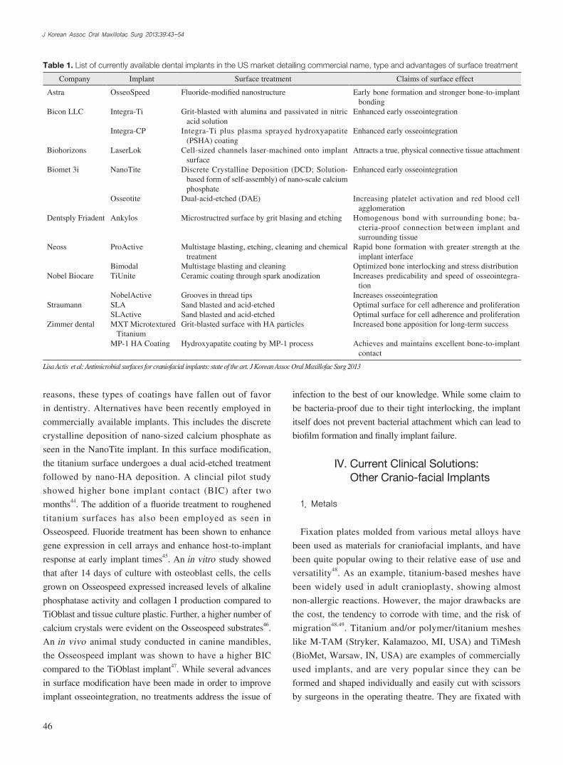
Table 1. List of currently available dental implants in the US market detailing commercial name, type and advantages of surface treatment
Company Implant Surface treatment Claims of surface effect
Astra
Bicon LLC
Biohorizons
Biomet 3i
Dentsply Friadent
Neoss
Nobel Biocare
Straumann
Zimmer dental
OsseoSpeed
Integra-Ti
Integra-CP
LaserLok
NanoTite
Osseotite
Ankylos
ProActive
BimodalTiUnite
NobelActiveSLASLActiveMXT Microtextured TitaniumMP-1 HA Coating
Fluoride-modified nanostructure
Grit-blasted with alumina and passivated in nitric acid solution
Integra-Ti plus plasma sprayed hydroxyapatite (PSHA) coating
Cell-sized channels laser-machined onto implant surface
Discrete Crystalline Deposition (DCD; Solution-based form of self-assembly) of nano-scale calcium phosphate
Dual-acid-etched (DAE)
Microstructred surface by grit blasing and etching
Multistage blasting, etching, cleaning and chemical treatment
Multistage blasting and cleaningCeramic coating through spark anodization
Grooves in thread tipsSand blasted and acid-etchedSand blasted and acid-etchedGrit-blasted surface with HA particles
Hydroxyapatite coating by MP-1 process
Early bone formation and stronger bone-to-implant bonding
Enhanced early osseointegration
Enhanced early osseointegration
Attracts a true, physical connective tissue attachment
Enhanced early osseointegration
Increasing platelet activation and red blood cell agglomeration
Homogenous bond with surrounding bone; ba-cteria-proof connection between implant and surrounding tissue
Rapid bone formation with greater strength at the implant interface
Optimized bone interlocking and stress distributionIncreases predicability and speed of osseointegra-
tionIncreases osseointegrationOptimal surface for cell adherence and proliferationOptimal surface for cell adherence and proliferationIncreased bone apposition for long-term success
Achieves and maintains excellent bone-to-implant contact
Lisa Actis et al: Antimicrobial surfaces for craniofacial implants: state of the art. J Korean Assoc Oral Maxillofac Surg 2013

Antimicrobial surfaces for craniofacial implants: state of the art
47
associated with the use of these products, such as rate of
degradation, and their mechanical profile, because they
tend to become brittle over time and can revert to a crystal
powder48-50. Examples of HA cements include BoneSource
(Stryker), Norian SRS (DePuy Synthes, West Chester, PA,
USA) and Mimix (Walter Lorenz Surgical, Jacksonville,
FL, USA). In general, these cements can be easily handled
and have good clinical performance, but one of the major
drawbacks of BoneSource (Stryker) in particular, is that
during the curing process the cement must not gain contact to
any fluids (blood); conditions that are practically unachievable
in craniofacial surgery49,50,56-63. However, case studies of the
different products are documented for different uses64-67.
One interesting commercially available material is Palacos
(Zimmer, Warsaw, IN, USA), a bone cement with the addition
of Gentamicin, which has demonstrated antibiotic release with
a broad spectrum of kill. It also has a proven clinical history
of low revision risk68. In general terms, while CaP cements
have been successfully used for clinical applications such as
vertebroplasty and cranial defect repair, they are brittle and
contraindicated for use in areas of mobility, active infection, or
in situations where they directly contact the sinuses or dura50.
Bioactive glasses have been shown to form a surface apatite
layer in vivo that enhances the formation and attachment of
bone, minimizing the formation of a fibrous capsule around
the implant49. NovaBone (NovaBone) is a bioglass composite
which has been used as a bioactive dental and orthopedic
filler. It has been shown to influence the formation of new
bone, promote intensive bond between bone and implant,
and induce accelerated bone formation. One of the major
drawbacks is that substitution of the material does not occur.
Also the main fields of application of NovaBone are dental
surgery and reconstruction of the calvarium and the floor of
the orbit because it is considered too fragile for load-bearing
applications49,50,69. Bioverit (G+W Implan tate, Łomianki,
Poland), is another bioactive glass–ceramic used for bone
sub stitutions in several fields of human surgery, as well as
implant craniofacial reconstruction, showing good clinical
results. These implants allow intraoperative remodeling,
adjustment and, as opposed to titanium implants, do not show
thermosensitivity49,50,70. Complications associated with the
use of this type of implants include extrusion, which requires
reoperation49.
3. Polymers
Porous polyethylene (PPE) implants are produced from
titanium screws and are convenient for primary fractures
where there is bone loss49,50. In particular, TiMesh is a
monofilament, composite mesh combining polypropylene
with a covalent bonded titanized surface. TiMesh titanized
polymers were designed specifically to have the following
properties: inertness, molecular permeability, pliability,
transparency, mechanical integrity and biocompatibility.
Studies have demonstrated that TiMesh conforms to the local
anatomy and has a high degree of biocompatibility. This is
because of the titanium surface as well as the reduced amount
of material51-53.
2. Ceramics
Calcium phosphates (CaP) are widely used in cranio-facial
and orthopaedic applications due to their biochemical similarity
to the mineral component of bone. HA is a biocompatible
CaP compound, which promotes osteoconduction, i.e., bony
ingrowth from adjacent surfaces as well as osteoinduction, i.e.,
bone formation with a high successs rate. On the other hand, it
does not promote toxic or allergic reactions48,49. HA has been
made available for clinical use as a bulk material, in granular
form, in pastes, and also as a coating material for metallic
implants in order to facilitate the growth of bone because of
its excellent bioactivity. Fluoridated HA (FHA), a variation
of HA has been shown to exhibit a better stability than HA
in physiological environments; and released fluorine ions can
affect bacterial metabolism as an enzyme inhibitor and act as an
antibacterial agent. However, few reports quantitatively study
the effect of FHA antibacterial activity49,54. A privately held
biotechnology company, PolyPid Ltd. (PetachTikva, Israel) has
developed BonyPid which is a fully biodegradable synthetic
bone filler that consists of beta-tricalcium phosphate (β-TCP)
particles, micro-coated with PolyPid, which is a controlled
release formulation technology of antibiotics. Preliminary
clinical data have clearly demonstrated BonyPid’s safety and
efficacy in early bone formation and anti-infective effects55.
There are also a number of CaP injectable materials that
are currently used and regulated in clinical applications.
CaP cements combine a dry CaP powder and a liquid
component (i.e., an inorganic or organic acid, or sodium
phosphate solutions) in a setting reaction that occurs under
physiologic pH and temperatures. The major purpose
of the pastes has been to allow the surgeon to easily fill
irregular defects and shape the material during surgery into
reasonable aesthetic contours. However, further surgical
experience has demonstrated that there are several problems
J Korean Assoc Oral Maxillofac Surg 2013;39:43-54
48
they have degraded. Major disadvantages of these materials
include screw breakage and inflammatory reactions due to their
degradation products49,50. LactoSorb (Biomet Microfixation,
Jacksonville, FL, USA) is a resorbable plating system of
plates and screws composed of 82% poly L-lactic acid and
18% poly glycolic acid. Lactosorb systems have been used
successfully in more than 50,000 craniomaxillofacial cases,
showing reduced risk of inflammation as well as lower risk
of implant migration, as it resorbs during the first 12 months,
approximately74.
4. Composites
PLGA microparticles incorporated within an injectable
CaP formulation can, upon degradation of the PLGA, yield
macroporosity for tissue ingrowth and, possibly through
a lowered local pH upon degradation of the PLGA, can
accelerate degradation of the CaP phase. The incorporation of
other degradable particles such as poly(trimethyl carbonate)
and gelatin microparticles has yielded similar favorable
results within injectable formulations, and the potential
for drug or growth factor release from these systems has
been well demonstrated50. In this category, a commercially
available implant BonAlive (BonAlive Biomaterials Ltd.,
Turku, Finland) is a 100% synthetic, osteo conductive,
osteostimulative silica-based bone graft substitute that is used
for bone cavity filling in orthopaedic and cranio-maxillofacial
surgery including jaw surgery. It has been shown that
BonAlive bonds firmly to bone and several clinical studies
have shown bacterial growth inhibition75.
V. Translational Studies with Antibacterial Surfaces
Several synthetic polymers and chitosan, have exhibited
antibacterial properties and can be used as coatings on dental
implants to prevent infection at the implant site. Martin
et. al.57, developed a poly (dimethylaminomethyl styrene)
coating, deposited by chemical vapor deposition, that was
effective in killing 99.9999% of Escherichia coli and Bacillus subtilis bacteria. While the coating was deposited on a
nylon substrate, the authors suggest that the coating could
be used for other biomedical implants, including dental
and other craniofacial implants. Another polymer coating,
a poly(L-lysine)-grafted-poly(ethylene glycol) copolymer
was shown by Harris et al.76, to reduce S. aureus adhesion
as well as osteoblast adhesion. However, the addition of
high-density polyethylene microspheres, that form a porous
matrix which is commonly used for facial augmentation
and to restore continuity to craniofacial skeletal defects,
and it is designed to allow for ingrowth of the host tissue
including both osteogenic and angiogenic material. The
major advantages of PPE are that it appears to have a low
infection rate and that it can be cut and contoured easily48,71.
Medpor (Stryker) is a commercial implant made of PPE,
characterized for being inert, biocompatible and porous: It
is mainly used for facial augmentation in post-traumatic or
tumor resection defects of the calvarium, orbit, mandible
and also aesthetic augmentation (e.g., chin). More than
400,000 procedures have been performed with Medpor,
and there are over 350 published clinical reports in cranial,
reconstructive, oculoplastic and cosmetic applications49,50,72.
There are almost no reported cases of extrusion, migration,
or capsule formation. Reported reoperation rates are about
10%, consisting of implant removal for infection (3%) or
displeasing contour (2%), and implant revision/replacement
for improvement of contour (6%)49.
Methyl methacrylate (MMA) is an acrylic-based resin that
is commonly employed together with titanium wire mesh to
contour and fill large cranial defects, having acceptable low
infection rates. Although MMA is resistant to absorption,
it has several advantages such as its low cost, predictable
resultant shape and suitability for complex defects. However,
complications include exothermic reactions with the release
of potential toxic monomers, causing local inflammation. In
pediatric patients, some common complications are: infection,
extrusion, migration, bone loss around the implant, undesirable
thermal sensitivity. Another complication in growing children
is that the implant can become isolated with time, forming a
fibrotic tissue with no attachment to bone48-50,71. Commercially
available MMAs include Clearshield (OsteoSymbionics,
Cleveland, OH, USA) which has been proved in hard tissue
replacement in the craniofacial reigion73.
Resorbable polymeric systems such as poly-lactic acid
and poly-lactide-go-glycolide (PLGA) are biologically
compatible, but do not possess osteogenic, osteoconductive,
or osteoinductive properties. However, properties such as
their rate of degradation, pore size, porosity, inter connec ti-
vity, hydrophobicity/hydrophilicity, ability for cell attach-
ment, morphology, and handling properties have made
them attractive materials for investigation and implantation.
Advantages of these devices over traditional titanium plates
and screws include elimination of long-term palpable devices
and continued skull growth in the pediatric population once
Antimicrobial surfaces for craniofacial implants: state of the art
49
using pulsed unbalanced magnetron sputtering using high-
purity Zr, Ag and Cu targets. When tested against S. aures
and Actinobacillus actinomycetemcomitans, ZrO2 surfaces
showed less bacterial attachment and proliferation than
the ZnO2-Cu surfaces, which in turn, showed less bacterial
proliferation than ZrO2 coatings82. Titanium oxide (TiO2) and
zinc-doped TiO2 (Ti(Zn)O2) and ZnO coatings deposited by
cathodic arc deposition have also been developed. The ZnO
coatings had the least bacterial attach ment and the Ti(Zn)O2
coating had less than the TiO2 coating. However, when tested
for osteoblast attachment, ZnO showed the least number of
osteoblasts while TiO2 showed the most.
VI. Antibacterial Surfaces
The presence of infection is an important parameter that
must be considered for nearly any reconstructive technique.
Infections following traumatic craniofacial injuries are
common, and although success rate of dental implants
are high, biomaterials for the restoration of oral function
are prone to biofilm formation, and failure is commonly
associated with bacterial infections. The consequences of
implant associated infection are significant and usually
require revision surgery, with removal of the implant,
prolonged antibiotic treatment, impaired oral function, and in
extreme cases even death. Furthermore, bacterial colonization
of dental implants can lead to inflammatory reactions which
prevent or result in loss of osseointegration. For those
reasons, various approaches to reduce the rate of infection
have been investigated. Therefore, antibiotic delivery and
antibacterial surfaces may thus be important aspects of tissue
engineering strategies in the craniofacial complex18,50,83-86.
In this sense, the purpose of these bioactive surfaces would
be to disrupt the metabolic machinery of the microbes or to
prevent bacterial adhesion to the implant and, consequently,
the development of biofilms. Different surface modification
approaches and techniques have been developed for this
purpose. Materials which promote the colonization of host
tissue and suppress the colonization of bacterial species are
often studied. These materials have many different mechanisms
of action; some can interfere with bacterial adhesion by
modifying surface energy, have surface immobilized molecules
that are bactericidal, are photocatalytic, or more commonly,
release metal ions or antibiotics18,83.
Many drugs and coatings have been developed to create
antibacterial surfaces that either kill bacteria or prevent their
attachment to implant surfaces. A handful of polymers are
an arginine-glycine-aspartate peptide showed improved
osteoblast attachment without inhibiting its antibacterial
activity. An antibacterial polymer, Poly(N,N-dimethyl-
N-(ethoxycarbonylmethyl)-N-(2’-(meth acry loyloxy)ethyl)-
ammonium bromide), or (pCBMA-1 C2), was attached as
a coating by a grafting technique known as surface-initiated
atom transfer radical polymerization. The coating was shown
to kill 99.9% E. coli bacteria followed by a release of the
bacterial cells upon hydrolysis77. The coating shows promise,
but bacteria more relevant to dental and craniofacial implants
would need to be tested.
Polymers have also been combined with antibiotics to develop
antimicrobial polymeric coatings. Al-Deyab, for example,
soaked electrospun nylon-6/chitosan (nylon-6/Ch) nanofibers
in an aqueous solution of glycidyltrimethylammonium
chloride, an antibacterial agent, to make make nylon-6/N-[(2-
hydroxy-3-trimethylammonium) propyl] chitosan chloride.
The antibacterial efficacy of the fibers were tested against E. coli, P. aeruginosa and S. aureus and showed to negatively
affect bacterial replication and induce cell damage. Significant
zones of inhibition were also observed78. Grafted Allylamine,
N-allylmethmylamine (AMA) and N,N-dimethylamine
(DMAA) monomers with the addition of the antibiotic triclosan
(TC) were tested for their antibacterial activity against S. aureus and E. coli. Bílek et al.79, found that the grafted AMA
and DMAA with TC showed antibacterial activity against
both bacterial strains and that they actually exhibited greater
antibacterial activity against the gram-positive S. aureus. Zhao
et al.80, developed a Poly(N-hydroxyethylacrylamide)/Salicylate
hydrogel (polyHEAA/SA) coating that released antibacterial
SA compounds resulting in a polymeric coating that resisted
the attachment of S. epidermis and E. coli after 24 hours. The
coating also inhibited bacterial growth which was determined
by measuring the optical density of the bacteria.
Alternative antibacterial agents, such as zinc oxide
(ZnO) and silver (Ag), have been combined with polymers
as well. Liu and Kim81, added ZnO and Ag to genipin-
crosslinked chitosan/poly(ethylene glycol) (GC/PEG)
hydrogel matrix. The nanocomposites showed enhanced
antibacterial activity against gram-negative E. coli and
P. aeruginosa as well as gram-positive S. aureus and B. subtilis over GC/PEG alone. Adding ZnO increased the
zone of inhibition of the copolymer, which was further
enhanced by the addition of Ag81. Antibacterial metals have
also been deposited directly onto implant surfaces. In one
study, zirconium oxid (ZrO2), ZrO2-copper (ZrO2-Cu) and
ZrO2-Ag coating were deposited onto pure-Ti substrates
J Korean Assoc Oral Maxillofac Surg 2013;39:43-54
50
bacterial infections was minimized. Yet, with the emergence
of antibiotic-resistant strains of bacteria, interest has returned
to the use of silver as metallic silver, silver ions, and silver
nanoparticles93. Silver’s antimicrobial properties are related
to the amount of silver present and its rate of release. In
its metallic state, silver is inert, but when it is exposed to
moisture in the skin or the fluid in a wound, it becomes
ionized and thus highly reactive. It binds to proteins in tissue
and causes structural changes the cell wall of bacteria and
their nuclear membrane leading to cell distortion and death93.
It also binds to bacterial DNA and RNA, denaturing it and
inhibiting bacterial replication93. Silver nanoparticles in
particular have been receiving a lot of attention due to their
enhanced antimicrobial properties especially in light of the
growing antimicrobial resistance against metal ions94. This
improved antibacterial activity is due in part to their large
surface area to volume ratio95. They have been shown to have
high antimicrobial and bactericidal activity on Gram-negative
and Gram-positive bacteria, including multi-resistant strains
such as methicillin resistant S. aureus 96. Studies on both
Gram-negative and Gram-positive bacteria have shown
that silver nanoparticles show more efficient antibacterial
properties (1.4-1.9×stronger) compared to silver ions93.
The mechanism of action of silver is related with its
interaction with thiol group compounds which are found
in the respiratory enzymes of bacteria. Silver can also bind
to the bacterial cell wall and cell membrane and inhibit the
respiratory process. In order for DNA molecules to replicate
themselves, they must be in a relaxed state. It has been
suggested that when silver ions penetrate into the bacterial
cell, the DNA molecule turns into a condensed form and
loses its replication ability which ultimately leads to cell
death97. Furthermore, proteins get inactivated when the silver
ions attach to their thiol groups. Silver nanoparticles get
attached to the bacterial membrane and can also penetrate
inside the bacteria. The silver nanoparticles interact with
the sulfur-containing proteins in the bacteria as well as with
the phosphorus containing compounds, such as DNA97.
When the silver nanoparticles enter the cell, they form a low
molecular weight region in the cell which causes the DNA
to condense so as to protect the DNA from the silver ions98.
Furthermore, silver nanoparticles attack the respiratory
chain and cell division which lead to bacterial death99. Silver
nanoparticles also release silver ions which further increase
their bactericidal activity93. Size and shape also seem to
affect the antimicrobial activity of silver nanoparticles; silver
nanoparticles with a smaller size have an increased surface
known to kill bacteria or prevent them from attaching and
can therefore be used as coatings for antibacterial purposes87.
There are also several low molecular weight molecules and
inorganic ions that are known to also be antibacterial in
solution and can either be released in a controlled manner
or be grafted by covalent immobilization onto the implant
surface87. The disadvantage to the controlled release method
is that the duration of the antibacterial action is limited by
loading and release kinetics88. Polymers can be coated onto
medical implants by many methods including dip coating,
spin coating, and layer-by-layer plasma polymerization
which allows for a great variety of polymers to be applied
onto material surfaces for the purposes of antibacterial
action87. The majority of synthetic polymers, and the natural
polymer chitosan, that have been reported to be antibacterial
are cationic. The release of antibiotics from polymer coatings
has also been extensively investigated. By releasing the
drugs locally, as opposed to systemically, higher local
doses can be administered without the risk of exceeding the
systemic toxicity levels of the drug which could result in
renal and liver complications89. When considering the release
mechanism for these systems, it is important to consider
the release kinetics of the drug; a fast release allows for a
high dose but only for a short period of action whereas a
slow release may not reach the required therapeutic levels
and could also result in bacterial resistance87. According to
Vasilev et al.87 the ideal release coating should provide a fast
initial release within the first 6 hours, that will protect the
site while the immune system is weakened, followed by a
slow release. The use of a polymer matrix that degrades in
the body allows for a combination delivery by diffusion and
polymer matrix erosion90. Some common antibiotics used
in these polymeric coatings are gentamicin, norfloxacin,
cefazolin, amikacin, and vancomycin87. Nitric oxide has also
been used in release systems as an antibacterial agent and
has been shown to reduce the adhesion of P. aeruginosa, S. aureus, and S. epidermis87.
1. Silver nanoparticles
Due to the emergence of multiple drug-resistant bacterial
strains, alternatives to traditional antimicrobials has been
sought through inorganic agents91. Among these agents,
silver ions and silver nanoparticles have been studied most
extensively91. Silver has been used for centuries for the
treatment of burns and chronic wounds92. However, with
the advent of penicillin in the 1940s, the use of silver in

Antimicrobial surfaces for craniofacial implants: state of the art
51
3. Other surface modifications
Covalent surface modification consists of immobilizing
active antibiotics on metal surfaces. One example is use of
aminopropylsilane to immobilize vancomycin on titanium
surfaces. These surfaces have shown to have strong bactericidal
activity and remain active over long periods of time (up to 1
month) in vitro. However, further testing is needed to prove
the validity of this approach because permanent modification
of the implant surface may lead to unfavorable tissue
reactions18. The purpose of another generation of bioactive
implants is the design of surfaces that are permanently
rendered antimicrobial by covalent attachment of antibiotics
or custom designed bactericidal peptides. As such, the active
molecules are not allowed to elude off the surface of the
implant, thus decreasing possible local and systemic toxicity
and circumventing the problem of inconsistent elution
characteristics while providing long-lasting protection83.
Photocatalytic surfaces are thin films or coatings of TiO2
that can be constructed on metal implants. They become
bactericidal under near ultraviolet light and require up to 80
min of ultra violet exposure to eliminate 75-95% of bacteria.
These surfaces can also be nitrogen-doped, which grant
bactericidal activity under visible light and do not require
long exposure times18.
4. Antibiotic releasing coatings
Devices which rely on the release of antimicrobial agents
enjoy only a finite duration of antimicrobial activity; and one
of the major problems is the inability to discriminate between
normal and pathogenic microflora of the mouth. There have
been a variety of strategies to deliver antibiotics from implant
materials. Some include coating biomedical alloys with
degradable materials, such as poly-lactic acid, silica sol-gel,
and chitosan. In theory, as the coating degrades, the infection
is eradicated and the implant surface is left to achieve
osseointegration. Additionally, the antimicrobial coatings
can be effective against a wide spectrum of bacterial species
and eliminate infection without the development of resistant
strains18,85.
Although antibiotics can and have been incorporated
into many commercially available bone cements, poor
release kinetics and the sensitivity of many antibiotics to the
high curing temperatures associated with cements such as
polymethyl methacrylate make incorporation into the bulk
material an inefficient and in some cases ineffective strategy.
area to volume ratio and therefore are more effective in their
actions against bacteria, and triangular nanoparticles were
shown to inhibit bacterial growth at lower concentrations than
spherical nanoparticles, and silver nanorods were shown to
need the highest concentration100. Another advantage to silver
is that there have been no regular reports of silver allergy,
which can be of concern with other administered antibiotics93.
Yet, while studies suggest that silver nanoparticles are
nontoxic, some studies that have been conducted to this effect
have shown that silver nanoparticles had negative effects on
mitochondrial activity with increased concentrations101. Thus,
for dental applications, caution should be taken to only use
the maximal concentration necessary to prevent bacterial
growth while maintaining host cell viability.
2. Anti-bioadhesion coatings
It has been shown that bioadhesion can be regulated by
changing the surface hydrophilicity–hydrophobicity18,85.
Strongly hydrophilic surfaces spontaneously form a monolayer
of mobile water molecules which are not displaced by proteins
and cells. Thus, the bioadhesion processes of both cells and
bacteria are disrupted. However, in biomaterial applications
which require cell attachment, such as osseointegration,
these surfaces may not be useful because cellular adhesion is
interrupted. Plasma deposition technique has been use for this
purpose to coat materials used for fixed dental prostheses with
hydrophilic molecules such as polyvinylpyrollidone18.
Quaternary ammonium compounds (QACs) are widely
used as antimicrobial agents to inhibit microbial growth. The
antimicrobial activity provided by QACs results from both
ionic and hydrophobic interactions between the QAC and
components of the microbial cell wall that leads to cell death
or malfunction of cellular processes85.
Other examples are polymer-brush coatings which are
currently some of the most promising nonadhesive coatings,
since they reduce the initial adhesion of various bacterial
strains and yeasts by several log-units, both in terms of
adhesion numbers as well as in terms of adhesion forces. A
polymer brush is formed when hydrophilic polymer chains
are end-grafted to a surface in high density, forcing the
polymer chains to stretch away from the surface into the
adjacent medium. Compression of such a structure upon
microbial approach gives rise to an osmotic pressure and
decreased mobility (conformational entropy) of the polymer
chains in the brush, which causes repulsion of approaching
micro-organisms85.

J Korean Assoc Oral Maxillofac Surg 2013;39:43-54
52
optimization and further investigation of surface manipulation
and coating techniques is necessary in order to develop
craniofacial bone substitutes that will restore, maintain and
improve tissue function while combating infection.
References
1. Abu-Serriah MM, McGowan DA, Moos KF, Bagg J. Extra-oral endosseous craniofacial implants: current status and future developments. Int J Oral Maxillofac Surg 2003;32:452-8.
2. Bencharit S. Challenges and prospective applications of extra-oral implants for maxilloracial rehabilitation. Anaplastology 2012;1:e103.
3. Wan DC, Nacamuli RP, Longaker MT. Craniofacial bone tissue engineering. Dent Clin North Am 2006;50:175-90.
4. Dumas JE, BrownBaer PB, Prieto EM, Guda T, Hale RG, Wenke JC, et al. Injectable reactive biocomposites for bone healing in critical-size rabbit calvarial defects. Biomed Mater 2012;7:024112.
5. Kretlow JD. Biomaterial-based strategies for craniofacial tissue engineering [PhD thesis]. Houston: Department of Bioengineering, Rice University; 2010. p. 416.
6. Pagni G, Kaigler D, Rasperini G, Avila-Ortiz G, Bartel R, Giannobile WV. Bone repair cells for craniofacial regeneration. Adv Drug Deliv Rev 2012;64:1310-9.
7. Kim J, McBride S, Fulmer M, Harten R, Garza Z, Dean DD, et al. Fiber-reinforced calcium phosphate cement formulations for cranioplasty applications: a 52-week duration preclinical rabbit calvaria study. J Biomed Mater Res B Appl Biomater 2012;100:1170-8.
8. Thimmappa B, Girod SC. Principles of implant-based reconstruction and rehabilitation of craniofacial defects. Craniomaxillofac Trauma Reconstr 2010;3:33-40.
9. Wolfaardt JF, Wilkes GH, Parel SM, Tjellström A. Craniofacial osseointegration: the Canadian experience. Int J Oral Maxillofac Implants 1993;8:197-204.
10. Kretlow JD, Young S, Klouda L, Wong M, Mikos AG. Injectable biomaterials for regenerating complex craniofacial tissues. Adv Mater 2009;21:3368-93.
11. Stanford CM. Surface modifications of dental implants. Aust Dent J 2008;53(Suppl 1):S26-33.
12. Davies JE. Understanding peri-implant endosseous healing. Dent Educ 2003;67:932-49.
13. Kuzyk PR, Schemitsch EH. The basic science of peri-implant bone healing. Indian J Orthop 2011;45:108-15.
14. Wang W, Ouyang Y, Poh CK. Orthopaedic implant technology: biomaterials from past to future. Ann Acad Med Singapore 2011;40:237-44.
15. Geetha M, Singh AK, Asokamani R, Gogia AK. Ti based biomaterials, the ultimate choice for orthopaedic implants-A review. Prog Mater Sci 2009;54:397-425.
16. Black J, Hastings GW. Handbook of biomaterial properties. London, New York: Chapman & Hall; 1998.
17. Le Guehennec L, Lopez-Heredia MA, Enkel B, Weiss P, Amouriq Y, Layrolle P. Osteoblastic cell behaviour on different titanium implant surfaces. Acta Biomater 2008;4:535-43.
18. Norowski PA Jr, Bumgardner JD. Biomaterial and antibiotic strategies for peri-implantitis: a review. J Biomed Mater Res B Appl Biomater 2009;88:530-43.
19. Shirtliff M, Leid JG. The role of biofilms in device-related infections. Springer series on biofilms, 3. Berlin: Springer; 2009.
20. Piattelli A, Cosci F, Scarano A, Trisi P. Localized chronic suppurative bone infection as a sequel of peri-implantitis in a hydroxyapatite-coated dental implant. Biomaterials 1995;16:917-20.
21. Götz F. Staphylococcus and biofilms. Mol Microbiol 2002;43:
Many drug delivery systems for antibiotic and other bioactive
factors utilize drug-loaded microspheres or microparticles.
At small particle or sphere sizes, these systems are easily
injectable, have well characterized and tunable release
kinetics, and can be fabricated from biocompatible,
biodegradable materials such as PLGA or gelatin50.
Most coatings for biomaterial implants and devices are
monofunctional, i.e., aimed solely at discouraging biofilm
formation or enhancing tissue integration. New approaches
include bifunctional coatings containing anti-adhesive
functionalities, such as a polyethylene glycol polymer brush
to discourage biofilm formation, while at the same time
possessing functionalities like arginine-glycine-aspartic acid
sequences to support tissue integration85.
VII. Summary
Although current implant materials for the reconstruction
of craniofacial bone defects have shown favorable results
in most craniofacial and dental applications, the presence of
complications related with infection and poor osseointegration
still represent a challenge in the biomedical field. Different
clinical circumstances may present different challenges;
however, the multitude of dissimilar solutions for the repair
of bone defects that have been proposed during the last few
decades highlights the fact that an ideal solution has yet to be
defined. There remains a need to develop strategies that will
further reduce implant failure while simultaneously addressing
different problems and causes of complications in a cost-
effective manner.
Biomaterials for craniofacial bone repair and dental
applications such as titanium, HA, bioactive glass, biocom-
patible and biodegradable polymers, and composites have
been widely studied and used in clinical applications.
Ongoing developments indicate that the tissue engineering
field is moving towards the development of biomaterials
with improved surfaces that will stimulate bone formation
and avoid infections though the incorporation of surface
modification techniques and antibacterial coatings and
agents, as well as the incorporation of growth factors, stem
cells and other pharmacological drugs. Scientists are paying
far closer attention to the biological interface in terms of both
the specific cellular and vascular responses necessary for
stable osseointegration as well as the unique microbial strains
in the oral space and the necessary steps to prevent biofilm
formation to avoid infection related complications. Through
the application of principles of engineering and biology,

Antimicrobial surfaces for craniofacial implants: state of the art
53
41. Lacefield WR. Current status of ceramic coatings for dental implants. Implant Dent 1998;7:315-22.
42. Kay JF. Calcium phosphate coatings for dental implants. Current status and future potential. Dent Clin North Am 1992;36:1-18.
43. Lacefield WR. Hydroxyapatite coatings. Ann N Y Acad Sci 1988; 523:72-80.
44. Goené RJ, Testori T, Trisi P. Influence of a nanometer-scale surface enhancement on de novo bone formation on titanium implants: a histomorphometric study in human maxillae. Int J Periodontics Restorative Dent 2007;27:211-9.
45. Berglundh T, Abrahamsson I, Albouy JP, Lindhe J. Bone healing at implants with a fluoride-modified surface: an experimental study in dogs. Clin Oral Implants Res 2007;18:147-52.
46. Monjo M, Petzold C, Ramis JM, Lyngstadaas SP, Ellingsen JE. In vitro osteogenic properties of two dental implant surfaces. Int J Biomater 2012;2012:181024.
47. Abrahamsson I, Albouy JP, Berglundh T. Healing at fluoride-modified implants placed in wide marginal defects: an experimental study in dogs. Clin Oral Implants Res 2008;19:153-9.
48. Goodrich JT, Sandler AL, Tepper O. A review of reconstructive materials for use in craniofacial surgery bone fixation materials, bone substitutes, and distractors. Childs Nerv Syst 2012;28:1577-88.
49. Cho YR, Gosain AK. Biomaterials in craniofacial reconstruction. Clin Plast Surg 2004;31:377-85.
50. Kretlow JD, Young S, Klouda L, Wong M, Mikos AG. Injectable biomaterials for regenerating complex craniofacial tissues. Adv Mater 2009;21:3368-93.
51. BioMet. TiMesh®, Titanized polymers. 2013 [cited 2013 Feb 26]. Available from: http://www.biomet.com/biologics/timesh.cfm.
52. Schug-Pass C, Tamme C, Tannapfel A, Köckerling F. A lightweight polypropylene mesh (TiMesh) for laparoscopic intraperitoneal repair of abdominal wall hernias: comparison of biocompatibility with the DualMesh in an experimental study using the porcine model. Surg Endosc 2006;20:402-9.
53. Hollinsky C, Sandberg S, Koch T, Seidler S. Biomechanical properties of lightweight versus heavyweight meshes for laparo-scopic inguinal hernia repair and their impact on recurrence rates. Surg Endosc 2008;22:2679-85.
54. Ge X, Leng Y, Bao C, Xu SL, Wang R, Ren F. Antibacterial coatings of fluoridated hydroxyapatite for percutaneous implants. J Biomed Mater Res A 2010;95:588-99.
55. Polypid. Stretching the limits of effective long term drug delivery. 2013 [cited 2013 Feb 28]. Available from: http://www.polypid.com/.
56. Miyamoto Y, Ishikawa K, Fukao H, Sawada M, Nagayama M, Kon M, et al. In vivo setting behaviour of fast-setting calcium phosphate cement. Biomaterials 1995;16:855-60.
57. Martin TP, Kooi SE, Chang SH, Sedransk KL, Gleason KK. Initiated chemical vapor deposition of antimicrobial polymer coatings. Biomaterials 2007;28:909-15.
58. Crawford K, Berrey BH, Pierce WA, Welch RD. In vitro strength comparison of hydroxyapatite cement and polymethylmethacrylate in subchondral defects in caprine femora. J Orthop Res 1998;16: 715-9.
59. Dickson KF, Friedman J, Buchholz JG, Flandry FD. The use of BoneSource hydroxyapatite cement for traumatic metaphyseal bone void filling. J Trauma 2002;53:1103-8.
60. Belkoff SM, Mathis JM, Jasper LE, Deramond H. An ex vivo biomechanical evaluation of a hydroxyapatite cement for use with vertebroplasty. Spine (Phila Pa 1976) 2001;26:1542-6.
61. Stryker. BoneSource: Ostoconductive HA bone paste. 2004 [cited 2013 Mar 1]. Available from: http://www.stryker.com/en-us/GSDAMRetirement/index.htmstellent/groups/public/documents/web_prod/023526.pdf.
62. Spies CK, Schnürer S, Gotterbarm T, Breusch SJ. Efficacy of Bone SourceTM and CementekTM in comparison with EndobonTM
1367-78.22. Archer NK, Mazaitis MJ, Costerton JW, Leid JG, Powers
ME, Shirtliff ME. Staphylococcus aureus biofilms: properties, regulation, and roles in human disease. Virulence 2011;2:445-59.
23. Richter WS, Ivancevic V, Meller J, Lang O, Le Guludec D, Szilvazi I, et al. 99mTc-besilesomab (Scintimun) in peripheral osteomyelitis: comparison with 99mTc-labelled white blood cells. Eur J Nucl Med Mol Imaging 2011;38:899-910.
24. Yaszemski MJ, Trantolo DJ, Lewandrowski KU, Hasirci V, Altobelli DE, Wise DL. Biomaterials in Orthopedics. New York: Marcel Dekker; 2004.
25. Ramaswamy Y, Wu C, Zreiqat H. Orthopedic coating materials: considerations and applications. Expert Rev Med Devices 2009;6:423-30.
26. Özcan M, Hämmerle C. Titanium as a reconstruction and implant material in dentistry: advantages and Pitfalls. Materials 2012;5:1528-45.
27. Coelho PG, Granjeiro JM, Romanos GE, Suzuki M, Silva NR, Cardaropoli G, et al. Basic research methods and current trends of dental implant surfaces. J Biomed Mater Res B Appl Biomater 2009;88:579-96.
28. Jackson MJ, Ahmed W. Surface engineered surgical tools and medical devices. New York: Springer; 2007.
29. Buser D, Broggini N, Wieland M, Schenk RK, Denzer AJ, Cochran DL, et al. Enhanced bone apposition to a chemically modified SLA titanium surface. J Dent Res 2004;83:529-33.
30. Schwarz F, Ferrari D, Herten M, Mihatovic I, Wieland M, Sager M, et al. Effects of surface hydrophilicity and microtopography on early stages of soft and hard tissue integration at non-submerged titanium implants: an immunohistochemical study in dogs. Periodontol 2007;78:2171-84.
31. Schwarz F, Herten M, Sager M, Wieland M, Dard M, Becker J. Histological and immunohistochemical analysis of initial and early osseous integration at chemically modified and conventional SLA titanium implants: preliminary results of a pilot study in dogs. Clin Oral Implants Res 2007;18:481-8.
32. Sul YT, Johansson CB, Röser K, Albrektsson T. Qualitative and quantitative observations of bone tissue reactions to anodised implants. Biomaterials 2002;23:1809-17.
33. Sul YT, Johansson C, Albrektsson T. Which surface properties enhance bone response to implants? Comparison of oxidized magnesium, TiUnite, and Osseotite implant surfaces. Int J Prosthodont 2006;19:319-28.
34. Al-Nawas B, Groetz KA, Goetz H, Duschner H, Wagner W. Comparative histomorphometry and resonance frequency analysis of implants with moderately rough surfaces in a loaded animal model. Clin Oral Implants Res 2008;19:1-8.
35. Sul YT, Johansson C, Byon E, Albrektsson T. The bone response of oxidized bioactive and non-bioactive titanium implants. Biomaterials 2005;26:6720-30.
36. Sul YT, Johansson CB, Jeong Y, Wennerberg A, Albrektsson T. Resonance frequency and removal torque analysis of implants with turned and anodized surface oxides. Clin Oral Implants Res 2002;13:252-9.
37. Yang Y, Kim KH, Ong JL. A review on calcium phosphate coatings produced using a sputtering process--an alternative to plasma spraying. Biomaterials 2005;26:327-37.
38. de Groot k, Klein COAT, Wolke JGC, de Blieck-Hogervorst JMA. Plasma-sprayed coating of calcium phosphate. In: Yamamuro T, Hench LL, Wilson J, eds. Handbook of bioactive ceramics, Vol. II: Calcium phosphate and Hydroxyapatite Ceramics. Boca Raton: CRC Press; 1990:133-42.
39. Ong JL, Carnes DL, Bessho K. Evaluation of titanium plasma-sprayed and plasma-sprayed hydroxyapatite implants in vivo. Biomaterials 2004;25:4601-6.
40. Lemons J. Biomaterials for dental implants. In: Misch CE, ed. Contemporary implant dentistry. St. Louis: Mosby; 1999.
J Korean Assoc Oral Maxillofac Surg 2013;39:43-54
54
salicylate hydrogels. Langmuir 2013;29:1517-24. 81. Liu Y, Kim HI. Characterization and antibacterial properties
of genipin-crosslinked chitosan/poly(ethylene glycol)/ZnO/Ag nanocomposites. Carbohydrate Polymers 2012;89:111-6.
82. Tsai MT, Chang YY, Huang HL, Hsu JT, Chen YC, Wu AY. Characterization and antibacterial performance of bioactive Ti-Zn-O coatings deposited on titanium implants. Thin Solid Films 2013;528:143-50.
83. Ketonis C, Parvizi J, Jones LC. Evolving strategies to prevent implant-associated infections. J Am Acad Orthop Surg 2012;20:478-80.
84. Marsich E, Travan A, Donati I, Turco G, Kulkova J, Moritz N, et al. Biological responses of silver-coated thermosets: an in vitro and in vivo study. Acta Biomater 2013;9:5088-99.
85. Busscher HJ, Rinastiti M, Siswomihardjo W, van der Mei HC. Biofilm formation on dental restorative and implant materials. J Dent Res 2010;89:657-65.
86. Li L, Finnegan MB, Özkan S, Kim Y, Lillehoj PB, Ho CM, et al. In vitro study of biofilm formation and effectiveness of antimicrobial treatment on various dental material surfaces. Mol Oral Microbiol 2010;25:384-90.
87. Vasilev K, Cook J, Griesser HJ. Antibacterial surfaces for biomedical devices. Expert Rev Med Devices 2009;6:553-67.
88. Li Z, Lee D, Sheng X, Cohen RE, Rubner MF. Two-level antibacterial coating with both release-killing and contact-killing capabilities. Langmuir 2006;22:9820-3.
89. Zilberman M, Elsner JJ. Antibiotic-eluting medical devices for various applications. J Control Release 2008;130:202-15.
90. Langer R. Polymer-controlled drug delivery systems. Acc Chem Res 1993;26:537-42.
91. Potara M, Jakab E, Damert A, Popescu O, Canpean V, Astilean S. Synergistic antibacterial activity of chitosan-silver nanocomposites on Staphylococcus aureus. Nanotechnology 2011;22:135101.
92. White RJ. An historical overview of the use of silver in wound management. Br J Community Nurs 2001;6(Silver Suppl 1):3-8.
93. Rai M, Yadav A, Gade A. Silver nanoparticles as a new generation of antimicrobials. Biotechnol Adv 2009;27:76-83.
94. Lee D, Cohen RE, Rubner MF. Antibacterial properties of Ag nanoparticle loaded multilayers and formation of magnetically directed antibacterial microparticles. Langmuir 2005;21:9651-9.
95. Nair LS, Laurencin CT. Silver nanoparticles: synthesis and therapeutic applications. J Biomed Nanotechnol 2007;3:301-16.
96. Panacek A, Kvítek L, Prucek R, Kolar M, Vecerova R, Pizúrova N, et al. Silver colloid nanoparticles: synthesis, characterization, and their antibacterial activity. J Phys Chem B 2006;110:16248-53.
97. Morones JR, Elechiguerra JL, Camacho A, Holt K, Kouri JB, Ramírez JT, et al. The bactericidal effect of silver nanoparticles. Nanotechnology 2005;16:2346-53.
98. Eom HJ, Choi J. p38 MAPK activation, DNA damage, cell cycle arrest and apoptosis as mechanisms of toxicity of silver nanoparticles in Jurkat T cells. Environ Sci Technol 2010;44:8337-42.
99. Li WR, Xie XB, Shi QS, Zeng HY, Ou-Yang YS, Chen YB. Antibacterial activity and mechanism of silver nanoparticles on Escherichia coli. Appl Microbiol Biotechnol 2010;85:1115-22.
100. Pal S, Tak YK, Song JM. Does the antibacterial activity of silver nanoparticles depend on the shape of the nanoparticle? A study of the Gram-negative bacterium Escherichia coli. Appl Environ Microbiol 2007;73:1712-20.
101. de Lima R, Seabra AB, Durán N. Silver nanoparticles: a brief review of cytotoxicity and genotoxicity of chemically and biogenically synthesized nanoparticles. Appl Toxicol 2012;32:867-79.
in critical size metaphyseal defects, using a minipig model. J Appl Biomater Biomech 2010;8:175-85.
63. DePuy Synthes. Norian SRS. 2012 [cited 2013 Mar 1]. Available from: http://www.synthes.com/sites/intl/Products/Biomaterials/Trauma/Pages/Norian_SRS.aspx.
64. DePuy Synthes. Norian SRS. Distal radius-impacted intra-articular fracture. 2007 [cited 2013 Mar 1]. Available from: http://www.synthes.com/MediaBin/International%20DATA/036.000.883.pdf.
65. DePuy Synthes. Norian SRS. Cystic lesion (pelvis) - curettage of a cystic lesion. 2006 [cited 2013 Mar 1]. Available from: http://www.synthes.com/MediaBin/International%20DATA/036.000.886.pdf.
66. DePuy Synthes. Norian SRS. Calcaneus. 2006 [cited 2013 Mar 1]. Available from: http://www.synthes.com/MediaBin/International%20DATA/036.000.886.pdf.
67. BioMet Microfixation. Biomet Microfixation Mimix® and Mimix® QS Bone Replacement Systems. 2012 [cited 2013 Mar 4]. Available from: http://www.lorenzsurgical.com/product.php?item=24&cat=9;%20http://www.lorenzsurgical.com/downloads/LOR-7013-MimixBro%20(m)-FINAL.pdf.
68. Zimmer. Palacos® Bone Cements. 2013 [cited 2013 Mar 4]. Available from: http://www.zimmer.com/en-US/hcp/surgical/product/palacos-bone-cements.jspx.
69. NovaBone. NovaBone: bioactive synthetic bone graft. 2009 [cited 2013 Mar 4]. Available from: http://www.novabone.com/NB/novabone_works.html.
70. Verné E, Ferraris M, Jana C, Paracchini L. Bioverit® I base glass/Ti particulate biocomposite: “in situ” vacuum plasma spray deposition. J Eur Ceram Soc 2000;20:473-9.
71. Neovius E, Engstrand T. Craniofacial reconstruction with bone and biomaterials: review over the last 11 years. J Plast Reconstr Aesthet Surg 2010;63:1615-23.
72. Stryker. Medpor®. 2013 [cited 2013 Mar 4]. Available from: http://www.stryker.com/en-us/products/Craniomaxillofacial/MEDPOR/index.htm.
73. OsteoSymbionicsTM. CLEARSHIELDTM Craniofacial Implant. 2011 [cited 2013 Mar 4]. Available from: http://www.osteosymbionics.com/implants/.
74. BioMet Microfixation. LactoSorb® SE: The leader in resorbable technology. 2013 [cited 2013 Mar 4]. Available from: http://www.lorenzsurgical.com/product.php?item=17.
75. BonAlive Biomaterials Ltd. BonAlive®. 2012 [cited 2013 Mar 4]. Available from: http://www.bonalive.com/.
76. Harris LG, Tosatti S, Wieland M, Textor M, Richards RG. Staphylococcus aureus adhesion to titanium oxide surfaces coated with non-functionalized and peptide-functionalized poly(L-lysine)-grafted-poly(ethylene glycol) copolymers. Biomaterials 2004;25:4135-48.
77. Cheng G, Xue H, Zhang Z, Chen S, Jiang S. A switchable biocompatible polymer surface with self-sterilizing and nonfouling capabilities. Angew Chem Int Ed Engl 2008;47:8831-4.
78. Al-Deyab SS, El-Newehy MH, Nirmala R, Abdel-Megeed A, Kim HY. Preparation of nylon-6/chitosan composites by nanospider technology and their use as candidate for antibacterial agents. Korean J Chem Eng 2013;30:422-8.
79. Bílek F, Sulovská K, Lehocký M, Sáha P, Humpolíček P, Mozetič M, et al. Preparation of active antibacterial LDPE surface through multistep physicochemical approach II: graft type effect on antibacterial properties. Colloids Surf B Biointerfaces 2013;102:842-8.
80. Zhao C, Li X, Li L, Cheng G, Gong X, Zheng J. Dual functionality of antimicrobial and antifouling of poly(N-hydroxyethylacrylamide)/