antimicrobial materials, coatings and biomimetic … vol. 7/01. hed/02. frank moerman.pdf ·...
TRANSCRIPT

Journal of Hygienic Engineering and Design
8
Review paperUDC 664.013.8:614.31
ANTIMICROBIAL MATERIALS, COATINGS AND BIOMIMETIC SURFACES WITH MODIFIED MICROTOGRAPHY TO CONTROL
MICROBIAL FOULING OF PRODUCT CONTACT SURFACES WITHIN FOOD PROCESSING EQUIPMENT: LEGISLATION, REQUIREMENTS,
EFFECTIVENESS AND CHALLENGES
Frank Moerman1*
1EHEDG Belgium & Catholic University of Leuven - KU Leuven, Belgium,Naamsestraat 22 - bus 5000, B-3000 Leuven
*e-mail: [email protected]
Abstract
Pathogens and spoiling microorganisms which adhere to interior equipment surfaces may be transferred to food products during their processing. To improve the hygiene during food processing operations, either in-organic or organic antimicrobials are added to various materials of construction used in the manufacturing of food processing equipment.
In the USA, antimicrobials used in materials of con-struction must be registered with EPA under the Fed-eral Insecticide, Fungicide, and Rodenticide Act, while in Europe biocidal products, included antimicrobial food contact materials, are subjected to Regulation (EU) No 528/2012. However, residual limits of antimi-crobials released from food contact materials in food are still to be set via the existing European food con-tact material regulations. An optimal antimicrobial component for food contact applications should have the following properties: broad-spectrum antimicro-bial activity towards undesirable microorganisms and without killing of beneficial microorganisms, fast-act-ing at low concentration, long-lasting effect or possi-bility to regenerate antimicrobial qualities upon loss of activity, no release of non-food grade substances, no development of resistance, and low cost. However, the majority of antimicrobial materials do not fulfil all these criteria, and quite often, in practical conditions, their hygienic effect is insignificant, due to the deple-tion of the antimicrobial substance within the material surface and because food residues, biofilms and scale deposits may exert a protective effect by prohibiting intimate contact between the microbes and antimicro-bial surface, or by forming an obstacle for the passage of antimicrobials released from the surface.
Therefore, the European Hygienic Engineering & De-sign Group clearly states that materials modified with
antimicrobials may not be considered as a substitute for hygienic design, and certainly not for proper clean-ing and disinfection practices. As an alternative, bio-film anti-adhesive coatings are developed to reduce adhesion of microorganisms on the product contact surfaces of food processing equipment.
Key words: Antimicrobial, Nano, Legislation, Hygiene re-quirements, Coating, Toxicity.
1. Introduction
Pathogens and spoiling microorganisms which adhere to interior equipment surfaces may be transferred to food products during their processing. To improve the hygiene during food processing operations, either in-organic or organic antimicrobials are added to various materials of construction used in the manufacturing of food processing equipment. Both in the USA and Eu-rope, these anti-microbial materials must comply with several food contact regulations. An optimal antimi-crobial component for food contact applications must have broad-spectrum antimicrobial activity towards undesirable microorganisms and without killing of ben-eficial microorganisms, long-lasting effect or the possi-bility to regenerate antimicrobial qualities upon loss of activity, no release of non-food grade substances, no development of resistance, and must be fast-acting at low concentration and low cost. However, the majority of antimicrobial materials do not fulfill all these criteria, and quite often in practical conditions their hygienic effect is insignificant. As an alternative, coatings and bioinspired surfaces with modified microtopography are developed to reduce microbial fouling of product

Journal of Hygienic Engineering and Design
9
contact surfaces within food processing equipment. This text gives an overview of the regulations and hy-gienic requirements that must be met, describes the effectiveness of several contemporary methodologies intended to control microbial fouling of food contact surfaces and the specific problems accompanied with the use of such materials of construction in the food environment.
2. Why should we use antimicrobials, antifouling coatings and surfaces with modified microto-pography?
The mentioned methodologies should be used:• to prevent the formation of biofilms, which are more
difficult to remove by cleaning and disinfection.• to prevent (cross-) contamination.• to have beneficial effect on food safety when stand-
ard hygiene procedures are not employed correctly.• to prevent biodegradation of materials of construc-
tion (microbial corrosion of metals, steels and alloys; and microbiological degradation of plastics, etc).
Antimicrobial materials and antifouling coatings are already widely used to reduce fouling and bacterial in-fection of implanted materials and devices (catheters, pacemaker leads, knee and hip implants).
2.1. Antimicrobial materials, coatings and surfaces
2.1.1 Legislation and regulations
United States
Biocides used as additives for materials must be regis-tered with the Environmental Protection Agency (EPA) under the Federal Insecticide, Fungicide, and Rodenticide Act (FIFRA). Treated articles are exempted from regis-tration, with the exception of treated articles for med-ical purposes, which are regulated by the FDA. Explicit or implicit health benefit claims cannot be made about unregistered products. For products not claiming pub-lic health effects (e.g., “control of odour-causing bacte-ria”), no efficacy data are needed for the registration.
European Union (EU)
In the EU, we have the Regulation on Biocidal products (EU) No. 528/2012, that repeals and replaces the Bioc-idal Products Directive (98/8/EC). Regulation (EU) No. 528/2012 also covers food contact materials (including nanomaterials). The old Biocidal Products Directive 98/8/EC excluded food contact materials and only reg-ulated treated articles or materials when the biocide is released to obtain an external effect. However, it does not regulate treated articles to which biocides are added for its own protection against biodegradation (internal effect).
The main principles of the Regulation on Biocidal prod-ucts (EU) No. 528/2012 are the following:• Active substances contained in the biocidal articles
must be approved for the relevant product-type.• Active substances also must be mentioned in the
list of Article 9(2) of Regulation (EU) No. 528/2012, for the relevant product-type and use.
• A distinction is made between “Articles with biocid-al properties” and “Articles with biocidal function”.
• Articles which contain biocides for their own pro-tection are considered as having “biocidal proper-ties”; while articles in which a particular or particular biocide(s) is/are integrated to exert an external anti-microbial effect are considered as having a “biocidal function”.
• Labeling the article with the quotation “biocid-al function” or “biocidal property” is required, and where applicable the term “nano” should be used.
• Precautions with respect to the application of the “biocide” are described. Approval for 1 EU country = approval for all countries in the EU.
• European Chemicals Agency (ECHA) must facilitate and coordinate the approval process, and provide scientific and technical backup to the EU commis-sion and EU countries.
• Companies must provide efficacy data, and compa-nies must share data.
• Costs of the assessment of active biocidal substanc-es must be equally shared by applicants.
Limits are set by Framework Regulation (EC) No. 1935/2004, Regulation EU No. 1183/2012 and Regulation EU No.10/2011.
2.1.2 Definition of antimicrobial materials
Substances and food contact materials are antimicro-bial materials, if they realize “a reduction of Colony Forming Units (CFU) > 2-log“. The problem is a lack of internationally recognized methodologies for testing treated articles. Available test methods are defined in the ISO 22196, ASTM E2149-10 and ASTM E2180-07 standards. Further, the Economic Cooperation and De-velopment (OECD) have developed a ‘Guidance docu-ment on the evaluation of the efficacy of antimicrobial treated articles with claims for external effects’.
2.1.3 Application of antimicrobials
Antimicrobials are already largely used in cutting boards, knives, bowls, storage containers, counter-tops, kitchen utensils, refrigerators and conveyor belts. The antimicrobial compound may be incorporated throughout the materials or added as a coating on the surface of the material. However, the most common concept is to add the antimicrobial as a thin coating on the outside of the material.
Journal of Hygienic Engineering and Design
10
Independent whether the antimicrobial compound is incorporated or applied as a coating, there are two different approaches to obtain an effect against micro-organisms [1]: • For polymers, the antimicrobial agent may be added
before the polymerization process. Present through-out the material, the antimicrobial can migrate from the bulk to the surface of the plastic by migration. Once diffused to the surface, the antimicrobial can finally be released from that surface under humid conditions, with as advantage that microorganisms not in direct contact with the material can be inhib-ited. Also some surface-coating products exert dis-infection action by allowing biocides to leach out of the polymer film on the surface. However, a leacha-ble antimicrobial has as result that the antimicrobial activity of the material is time-limited, that it is con-ducive to development of bacterial resistance and that it may be transferred to food.
• The antimicrobial agent also can be immobilized to the surface, often by covalent binding, and the bacteriostatic or bactericidal activity is dependent on contact between the microorganisms and the material. The effect may be inhibition of growth, loss of viability, or prevention of attachment of mi-croorganisms. Permanent, non-leaching materials are more desirable because they provide stability without releasing low molecular weight toxic prod-ucts to the environment, don’t cause problems of residual toxicity, prevent the transfer of the antimi-crobial compound to the food, limit antimicrobial resistance development, and provide long-term effectiveness. However, immobilizing antimicrobial molecules may undermine their bacteriocidal ac-tivity and limit their access to microbes in the sur-rounding environment.
2.1.4 Requirements to be met by antimicrobials
If used, antimicrobial materials must meet the follow-ing requirements [1, 2]:• Broad-spectrum activity against microorganisms;
although selective activity especially towards path-ogens, without killing of beneficial microorganisms.
• Active at low concentration and fast-acting.• Select an antimicrobial in function of its conditions
of service (e.g., wet versus dry conditions).• Antimicrobial activity may not be too much inhibit-
ed in the presence of food • Antimicrobials must resist the expected drastic en-
vironmental conditions encountered in the food and beverage industries, such as mechanical and chemical wear and tear.
• No resistance build-up over a period of time.• Non-toxic for humans & food grade.
• Only released in very limited quantities into the food to eliminate any risk for consumers.
• Antimicrobials for contact with food must meet na-tional or international legislation/regulations.
• Antimicrobial substances also must fulfil the regula-tions concerning maximal permitted intake.
• May not cause harm to the environment and wildlife.• Must be compatible with the materials, plastics and
additives in which they must be incorporated.• May not change the properties and appearance of
a material (e.g., plastic) (Ag+-ions are highly reactive and, if not sufficiently stabilized, cause discoloration of plastics).
• Must be heat resistant when they have to be incor-porated in plastic (they must retain activities after polymerization and extrusion).
• Good light stability.• Long-lasting antimicrobial effect: controlled release
of antimicrobial substances in sufficient concentra-tion to prevent fast depletion. The user must avoid that the antimicrobial coating peel off.
• The cost of the antimicrobial compound must be low (e.g., triclosan is less expensive than Ag).
• Must be easy to apply or re-apply on a given surface (incorporation versus coating).
• Preferentially the antimicrobial qualities can be re-generated upon loss of activity (e.g., N-halamine).
2.1.5 Inorganic antimicrobial agents
In the past, organic antimicrobial coatings were ap-plied to metals and steel to provide them with anti-bacterial properties. However, their antibacterial effect was not long lasting. Therefore inorganic antimicrobi-als were used as alternative. Inorganic antimicrobial compounds have as advantage that microorganism-re-action mechanisms are highly non-specific, making it rare for microorganisms to develop resistance. Al-though some microorganisms are capable of develop-ing resistance to metals through natural selection or horizontal gene transfer. Inorganic antimicrobial com-pounds also have higher stability in harsh conditions (high temperatures, high pressure, cleaning chemicals, etc.), superior durability and longevity, lower toxicity and greater selectivity.
2.1.5.1 Silver-containing antimicrobial materials
Silver ions
Metallic (non-ionic, Ag°) silver has no antibacterial effect. The activity of silver relies on Ag+-ions that dif-fuse from the substrate material and exert strong in-hibitory effects (already at levels of 0.001 ppm) on a broad spectrum of microorganisms, such as bacteria, moulds, and viruses. It tends to be more active against
Journal of Hygienic Engineering and Design
11
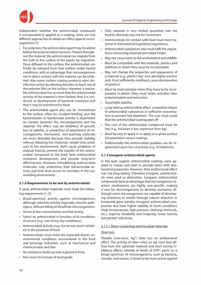
Gram-negative bacteria than against Gram-positive bacteria (Fig. 1). Higher Ag+-ion concentrations are re-quired for yeasts and moulds, because their cell wall is thicker. Dormant spores are not or less affected by Ag+-ions due to the presence of different spore coat layers [1, 3].
Figure 1. Effect of the silver ion solution on Staphylococcus aureus and Escherichia coli was investigated by conventional plate counting.
The tested silver ion concentrations were 0.2 ppm, 0.1 ppm, and 0.05 ppm, and PBS was used as a control [3]
Antibacterial stainless steel can be produced by add-ing an antimicrobial element to the alloy. Doping of stainless steel with Ag+-ions can be achieved without loss of its physical properties. However, silver coatings on stainless steel are non-permanent, because the modified surface layer is very thin, and once worn off, the base material loses its antibacterial ability [5].
Chiang et al. [6] demonstrated that silver-alloyed 316 stainless steels could be used in areas where hygiene is a major requirement, but that leaching of silver, which is quite expensive, out of the stainless steel could be a problem. The impact of repeated cleaning and disin-fection on the silver-bearing surface must be assessed.
Silver nanoparticles
A plenty of studies have demonstrated that nanosized silver particles exhibit antimicrobial properties. Nano-particles of silver may be incorporated within polymeric matrices (e.g., antimicrobial Makrolon® polycarbonate - AM1825, AM24281 and AM2520, Bayer Material Sci-ence AG) or applied in coatings on materials. Also, the antibacterial effect of nano-silver is based on the re-lease of silver ions, and nano-formulations of silver may cross biological barriers into the cell causing damage to the cell constituents. However, studies of silver nan-oparticles revealed that silver nanoparticle aggregates are more toxic than asbestos [7]. Das Bundesinstitut für Risikobewertung [8] has recommended manufacturers to avoid the use of nanoscale silver or nanoscale silver compounds in foods and everyday products until the time that the data are comprehensive enough to allow a conclusive risk assessment which would ensure that products are safe for consumer health. In the EU and Australia, for a long time, nano-silver was treated just like silver, but as in the US now documentation is re-quired on the safety of nano-silver products. At pres-ent, nanoscale silver has not yet been authorized to be used in plastic for direct food contact at EU level. The only authorized nanomaterial for direct food con-tact by the EU is nanoscale titanium nitride which was shown to be chemically inert and would not migrate. It was restricted to be used in PET bottles up to 20 mg/kg.
2.1.5.2 Copper-containing antimicrobial materials
Copper ions
The antibacterial and antifungal effects of copper ions have been well known for a long time. As early as in 1885, Milharde successfully developed the Bordeaux mixture (contains CuSO4) to kill bacteria and fungi in vineyards. Microorganisms are extremely sensitive to the toxic effects of copper but copper ions have lower efficacy than silver.
For many organisms, copper is an essential trace ele-ment involved in numerous physiological and met-abolic processes. Human tissue has low sensitivity to
To avoid that treated articles are too fast depleted of Ag+, silver is incorporated in a matrix of zeolite (e.g., Zeomic, Sinanen Zeomic Co., Ltd.) or zirconium phos-phate (e.g., Alphasan®, Milliken Chemical), that act as ion-exchange resins, exchanging Ag+-ions for cations in the environment [2]. Silver-exchanged zeolites, which are nontoxic and non-carcinogenic, have been used as coatings and in composites, plastic products, stainless steel and ceramics to confer long-lasting antimicrobi-al properties. The release of Ag+-ions from zeolites to the environment is based on an ion-exchange process, where Ag+-ions are exchanged to Na+-ions or other cations that can be found in the environment. Strain-, species- and environment-specific differences in the inactivation rates of various bacteria exposed to silver zeolites has been reported. Environmental conditions may change with respect to temperatures, humidity, pH and ion strength. Some zeolite-based inorganic antimicrobials, such as AgION™ (AgION Technologies, Inc., AK steel) contain both Ag+ and Zn2+-ions within the same matrix. With regards to AgION™, it contains 2.5% (w/w) silver and 14% (w/w) zinc. Zirconium phos-phate releases silver under humid conditions [4].
Silver-bearing stainless steel
The metal of choice for food preparation and handling is stainless steel (types 304 and 316) due to its mechan-ical strength, corrosion resistance, longevity and ease of fabrication. However, it has been shown that even with cleaning and sanitation procedures consistent with good manufacturing practices, microorganisms can remain in a viable state on stainless steel equip-ment surfaces. In addition, this alloy has been shown to be ineffective at reducing microbial load once it is contaminated.

Journal of Hygienic Engineering and Design
12
copper, although toxicity in humans can occur at high concentrations. In general, exposure to copper is con-sidered safe. The joint FAO/WHO expert Committee on Food Additives suggests a maximum daily intake value for copper of 0.05 mg/kg [9].
Metallic copper
Santo et al. [10] have studied the inactivation of bac-teria when in contact with dry copper, and they found killing rates of within minutes (e.g., Escherichia coli cells were killed after 1 min contact with dry copper), much faster than it was on moist copper (Fig. 2). For bacteria in contact with moist metallic copper surfaces, there is a clear correlation between release of copper from copper surfaces in the medium and its accumulation in the cells. The antibacterial effect of wet copper is similar to that of copper ions in media, being chronic in nature as the consequence of increasing concentra-tions of copper ions in the bacterial cells with time. In contrary, contact with dry metallic copper rather caus-es acute effects on bacterial cells (copper shock). Wilks et al. [11, 12] studied the survival of Escherichia coli and Listeria monocytogenes on several moist coppers hav-ing 99 or 100 % copper content. In all cases a 7-log re-duction of Escherichia coli and a 5-log reduction of Lis-teria monocytogenes were observed after respectively 75 - 90 min. and 60 min.
(a)
(b)
Figure 2. Number of survivors of Escherichia coli counted as Colony Forming Units (CFU) and copper
uptake into Escherichia coli cells after exposure to dry (graphic above) and moist copper (graphic below)
for the indicated time [10]
The best known applications of copper are vessels, traditionally used in many breweries and distilleries. Copper is largely applied in the non-product contact area, with as main application the tubes in evaporators installed in refrigerators and freezers, water pipes, etc. However, copper itself would not be a suitable alter-native for stainless as food contact material, as it may cause unacceptable organoleptic effects. Moreover, it is quickly and severely affected by strong alkaline de-tergents, sodium hypochlorite (NaOCl), acidic and salty food. The rate of attack is slow enough that alkaline de-tergents can be used for the cleaning of copper vessels, but NaOCl renders copper surfaces less hygienic with time. As copper ions may leach from the copper met-al, its surface roughness may increase. So even though it has such strong antibacterial properties, copper is a soft, not durable material that tarnishes easily. As cop-per prices are on the raise, cost considerations favor copper coatings over solid structural copper [13, 14].
Copper alloys
Wilks et al. [11] studied the survival of Escherichia coli on several moist copper-containing surfaces (Fig. 3). The inhibitory effect of the brasses with 78 or 95% cop-per content was less pronounced as compared with the coppers, requiring respectively 120 min. and 90 min. to obtain a 7-log reduction. The zinc content in the brasses seems not to have any impact on the sur-vival rate. In the group of the bronzes, 7-log reductions are only obtained after 65 min, 90-100 min, 180 min and 270 min for bronzes having respectively 97%, 95%, 90% and 74% copper. Hence, the percentage copper required for significant biocidal effect must range be-tween 55 and 100%. The greatest efficiency is seen in alloys with high copper content.
Brass (60 - 70% copper, 30 - 40% zinc) and bronze (80 - 95% copper, 5 - 20% tin) are more prone to corro-sion by alkaline and acidic detergents, salty and acid-ic food than ferrous steels. Brass is also susceptible to
Figure 3. Number of survivors of Listeria monocytogenes counted as Colony
Forming Units (CFU) on different copper alloys at 20 0C [12]
Journal of Hygienic Engineering and Design
13
de-zincification (in e.g., steam), and because cadmium and lead are co-elements to zinc, brass shall never be used in the food contact area. The use of bronze in the food contact zone should be avoided because it quick-ly becomes porous in contact with acidic foodstuffs, cleaning agents and steam [14].
Copper-bearing stainless steel
Besides bronze and braze, copper-alloyed stainless steels (e.g., 430Cu ferrite antibacterial stainless steel with 1.5% copper of Baosteel) have shown to possess antimicrobial properties. The basic principle of pro-ducing antibacterial stainless steels is adding a proper amount of antibacterial element (e.g., Cu) to alloy, and adopting a special heat treatment composed of solid solutioning and succedent ageing to form the active antibacterial phase in the matrix. Released copper ions can be absorbed on the bacterial cell membrane, where denaturation of proteins and oxidation of lipids may take place. The copper ions in the copper-bearing stainless steel are also strongly reductive, which means that they extract electrons from bacteria [9].
Notice however that in production environments, the efficacy of copper-alloyed stainless steels remains low-er than that of silver-alloyed stainless steels, and also differs for various bacterial strains [2]. In the opinion of Robine et al. [15], copper-bearing stainless steels dis-play poor and limited hygienic efficiency and they also put the long-lasting effect of biocide-based stainless steels on microbial ecology in doubt. Moreover, foul-ing may reduce the efficacy, as it provides a protective matrix for the bacterial cells to ‘hide in’ and copper ions released from the metallic surface may immediately be trapped by residues adsorbed on the surface.
Copper oxide nanoparticles
Copper oxide nanoparticles have been physically and chemically characterized and investigated with respect to potential antimicrobial applications. It was found that CuO nanoparticles in suspension, having particle sizes in the 20 to 95 nm range with a mean surface area of 15.7 m2/g, show activity against a range of bacterial pathogens, including MRSA and Escherichia coli at min-imum bactericidal concentrations ranging from 100 to 5000 µg/ml. As with silver, studies of CuO nanoparti-cles incorporated into polymers suggest that release of ions may be required for optimum killing. Azam et al. [16] have reported that the antimicrobial activity of CuO nanoparticles is lower than that of ZnO nanoparti-cles, but higher than that of Fe2O3 nanoparticles.
2.1.5.3 Zinc-containing antimicrobial materials
Zinc ions
Zinc has the ability to restrict bacterial growth, but the antimicrobial action of the Zn2+-ion is less extensive
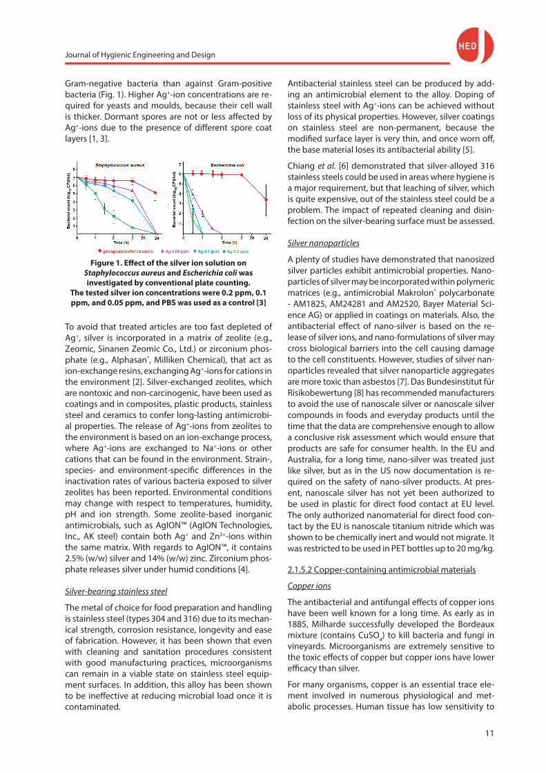
than that of Ag+. Gram-positive bacteria are the most acceptable bacterial group to Zn2+, while Gram-neg-ative aerobic bacteria are not inhibited even at the highest concentration. Like silver, to avoid that treated articles are too fast depleted of Zn, zinc is quite often incorporated in a zeolite which again acts as ion-ex-change resin. Coleman et al. [17] reported for Zn2+-ions incorporated in tober-mites, which behave like zeo-lites, a 5-log reduction in Staphylococcus aureus and Pseudomas aeruginosa cell counts, with Staphylococcus aureus being more sensitive to Zn2+ than Pseudomonas aeruginosa.
Figure 4. Suspension was inoculated onto the surface of a polyether polyurethane composite incorporated with
ion-exchange zeolites, and the cells were allowed to grow on the surface for 24 h at 37 0C and 98%
relative humidity [18]
Kaali et al. [18] observed the effect that zeolites con-taining Ag+, Cu2+ and Zn2+ have on Pseudomonas aerug-inosa, Candida tropicalis and MRSA. In Fig. 4, we can deduce the following order of activity: Ag+ > AgZn > ZnCu > AgCu > AgZnCu > Zn2+
> Cu2+. Zeolites con-
taining Zn2+ or Cu2+ exert little antimicrobial effect on Pseudomonas aeruginosa as compared to zeolites containing both Zn2+ and Cu2+. It can be assumed that each of these ions initiate different toxic mechanisms within the cell, and therefore duplex systems are prob-ably more successful than single ionic systems. These authors also argued that different ions in binary and ternary zeolite systems may block each others’ way, and therefore highly influence each others’ release.
Zinc oxide nanoparticles
Nanocrystalline zinc oxide, which is safe under harsh processing conditions and can be fabricated at ambi-ent temperatures, has more pronounced antimicrobial activities than micron sized zinc oxide particles. ZnO nanoparticles display photocatalytic and oxidizing ca-pabilities against biological and chemical species. Their small size (less than 100 nm) and high surface-to-vol-ume ratio allow for better interaction with bacteria. Hence, the antibacterial activity of ZnO nanoparticles
Journal of Hygienic Engineering and Design
14
increases with reduction in particle size, and further with higher concentrations. However, crystalline struc-ture and particle shape have little effect [19].
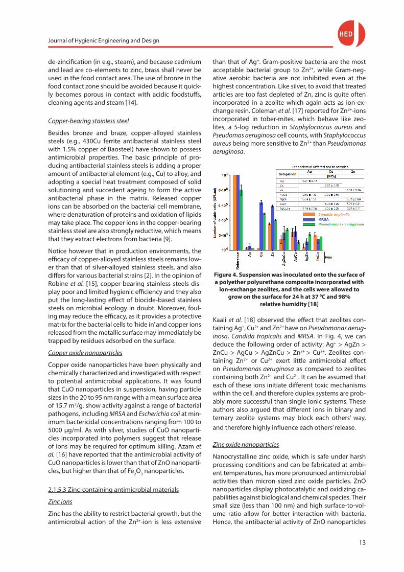
ZnO nanoparticles have been shown to have a wide range of antibacterial activities against both Gram-pos-itive and Gram-negative bacteria, including against spores. Growth inhibition and in-activation of food-borne pathogens like Escherichia coli O157:H7, Salmo-nella, Listeria monocytogenes, Staphylococcus aureus and Campylobacter jejuni is observed in many studies. ZnO nanoparticles have selective toxicity to bacteria, with some microorganisms being more sensitive than others. In their study, Xie et al. [19] found that ZnO nan-oparticles are more effective against Campylobacter je-juni than Escherichia coli, while the latter is more sensi-tive than Salmonella. An increasing antibacterial effect of ZnO nanoparticles (Fig. 5) is observed to the follow-ing microorganisms, and hence according to sensitivi-ty: Aspergillus niger < Candida albicans < Pseudomonas aeruginosa (G-) < Escherichia coli (G-) < Staphylococcus aureus (G+) < Bacillus subtilis (G+) [16].
Azam et al. [16] demonstrated that ZnO nanoparticles have excellent bactericidal potential, while Fe2O3 nan-oparticles exhibited the least bactericidal activity. The order of antibacterial activity was demonstrated to be the following: ZnO > CuO > Fe2O3.
metallic substrate, is scarcely removed at high temper-atures even in contact with hypochlorites and bleach chemicals, and highly concentrated salt and acid solu-tions. Further the TiO2 in that surface layer of titanium dioxide covering the titanium substrate is amorphous or crystalline in nature; from which crystalline titani-um dioxide in particular shows three microstructures: rutile, anatase, brookite. An intriguing role is played by anatase, because of its photocatalytic and antibac-terial properties. Anodization plus heating treatment of titanium results in the conversion of titanium oxide film from amorphous to crystalline and, in particular, the antibacterial anatase type of TiO2. Titanium doesn’t cause health problems, as it is generally considered to be poorly absorbed upon ingestion [13].
Titanium dioxide
Due to its brightness, high refractive index and resist-ance to discolouration, titanium dioxide is widely used as white pigment in paints, lacquers, enamels, coatings and plastics. For the same reasons, food grade TiO2 is also approved in the EU as an additive (E171) in foods, in e.g. candies, chewing gum, products with white ic-ing or powdered sugar toppings. Many products con-tain 0.01 to 1 mg Ti per serving.
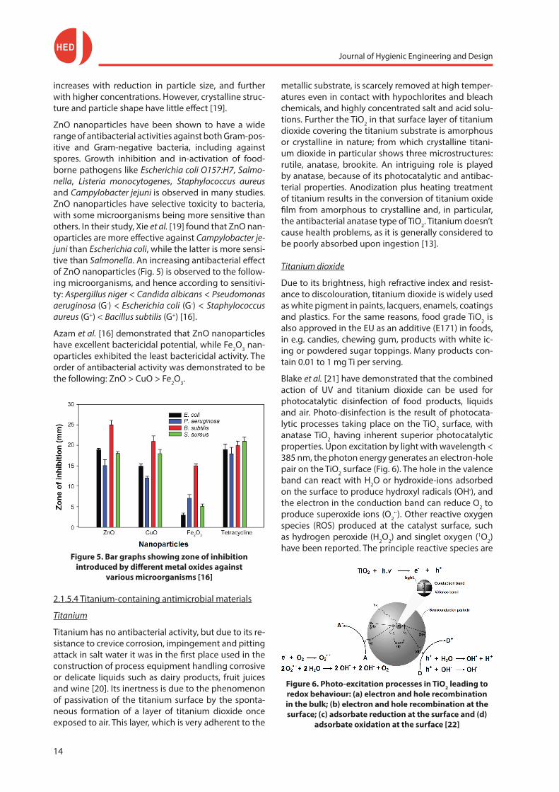
Blake et al. [21] have demonstrated that the combined action of UV and titanium dioxide can be used for photocatalytic disinfection of food products, liquids and air. Photo-disinfection is the result of photocata-lytic processes taking place on the TiO2 surface, with anatase TiO2 having inherent superior photocatalytic properties. Upon excitation by light with wavelength < 385 nm, the photon energy generates an electron-hole pair on the TiO2 surface (Fig. 6). The hole in the valence band can react with H2O or hydroxide-ions adsorbed on the surface to produce hydroxyl radicals (OH•), and the electron in the conduction band can reduce O2 to produce superoxide ions (O2
•-). Other reactive oxygen species (ROS) produced at the catalyst surface, such as hydrogen peroxide (H2O2) and singlet oxygen (1O2) have been reported. The principle reactive species are
2.1.5.4 Titanium-containing antimicrobial materials
Titanium
Titanium has no antibacterial activity, but due to its re-sistance to crevice corrosion, impingement and pitting attack in salt water it was in the first place used in the construction of process equipment handling corrosive or delicate liquids such as dairy products, fruit juices and wine [20]. Its inertness is due to the phenomenon of passivation of the titanium surface by the sponta-neous formation of a layer of titanium dioxide once exposed to air. This layer, which is very adherent to the
Figure 5. Bar graphs showing zone of inhibition introduced by different metal oxides against
various microorganisms [16]
Figure 6. Photo-excitation processes in TiO2 leading to redox behaviour: (a) electron and hole recombination in the bulk; (b) electron and hole recombination at the surface; (c) adsorbate reduction at the surface and (d)
adsorbate oxidation at the surface [22]

Journal of Hygienic Engineering and Design
15
the short lived hydroxyl radicals (OH•) which is a potent non-selective biocide with the ability to oxidize most organic compounds. When irradiated TiO2 particles are in direct contact with or close to microbes, and the mi-crobial surface is the primary target of the initial oxida-tive attack [22].
Specific problems that prohibit the use of titanium di-oxide as antimicrobial on a large scale are: the need of UV light to be effective, restricting its use to open equip-ment surfaces, and the limited long-term bactericidal effect due to mechanical and chemical wear. Moreover, fouling and scale deposition may decrease the photo-catalytic effect of titanium dioxide [11, 12, 22, 2].
Lu et al. [23] studied the photocatalytic inactivation of Escherichia coli Fig. 7 (on the left) shows the effect of the photocatalytic reaction by TiO2 thin films on cell vi-ability, while Figure 7 (on the right) shows the leakage of K+-ions from Escherichia coli cells illuminated by UV light with λ < 385 nm.
Figure 7. Effect of photocatalytic reaction by TiO2 thin films on cell viability. The survival curves were obtained by colony counting of Escherichia coli cells illuminated in the absence (green) and presence (blue) of TiO2 thin films at different time intervals (left figure). Leakage of
K+-ions from Escherichia coli cells illuminated by UV light in the presence (blue) and absence (green) of TiO2 thin
films at different time intervals (right figure). At point of time 0 min, Escherichia coli is intact; at point of time 10
min., the cell wall is decomposed and the cell membrane is exposed with loss of semi-permeability and leakage of K+-ions as result, finally leading to only 30% survivors; at point of time 40 min., there are holes in the cytoplasmic membrane, with more leakage of K+-ions and only 20%
survivors; at point of time 60 min, there is a large hole in the cytoplasmic membrane with only 10% survivors [23]
Titanium dioxide nanoparticles
Vargas-Reus et al. [24] investigated the antimicrobial activity of nanoparticulate metals and metal oxides un-der anaerobic conditions against four Gram-negative species of bacteria: Porphyromonas gingivalis, Prevotel-la intermedia, Fusobacterium nucleatum and Aggregati-bacter actinomycetemcomitans. Table 1 shows the Min-imum Inhibitory Concentration (MIC) and Minimum Bactericidal Concentration (MBC) for the four bacteria tested. Minimum Inhibitory Concentration (MIC) and Minimum Bactericidal Concentration (MBC) values were in the range of < 100 µg/mL to 2500 µg/mL and < 100 µg/mL to > 2500 µg/mL, respectively. Based upon mean values, the activity of the 5 nanoparticles against the four species tested in descending order was Ag > ZnO > CuO > TiO2 > WO3. TiO2 and WO3 showed poor or no antimicrobial activity at the concentrations tested. TiO2 and WO3 showed poor or no activity. However, in the case of TiO2, no ultraviolet light was used and it is well documented that in the presence of such ioniz-ing radiation TiO2 nanoparticles demonstrate bacte-ricidal properties. The anatase form of nano TiO2 and UV light excitation are required to ensure maximum antimicrobial activity.
Table 1. Minimum Inhibitory Concentration (MIC) and Minimum Bactericidal Concentration (MBC) values (in µg/ml) of the tested bacterial species with metal and metal oxide nanoparticles (n = 3 replicate determinations) [24]
Bacterial speciesAg ZnO CuO TiO2 WO3
MIC MBC MIC MBC MIC MBC MIC MBC MIC MBC
Porphyromonas gingivalis 250 250 250 250 500 2500 2500 >2500 2500 2500
Prevotella intermedia 100 100 1000 1000 250 250 1000 >2500 2500 >2500
Fusobacterium nucleatum 100 100 250 500 250 250 1000 >2500 2500 >2500
Aggregatibacter actinomycetemcomitans 100 100 250 250 250 250 250 >2500 2500 >2500
Mean 137.5 137.5 437.5 500 312.5 812.5 1187.5 >2500 2500 >2500
Figure 8. Process come-up time logarithmic red uction of the microorganism population suspended in
Luria-Bertani medium. Survival curves for Pseudomonas aeruginosa and Enterococcus faecalis (figure left) as a
function of the irradiation time for EVOH-TiO2 and control samples [25]
Journal of Hygienic Engineering and Design
16
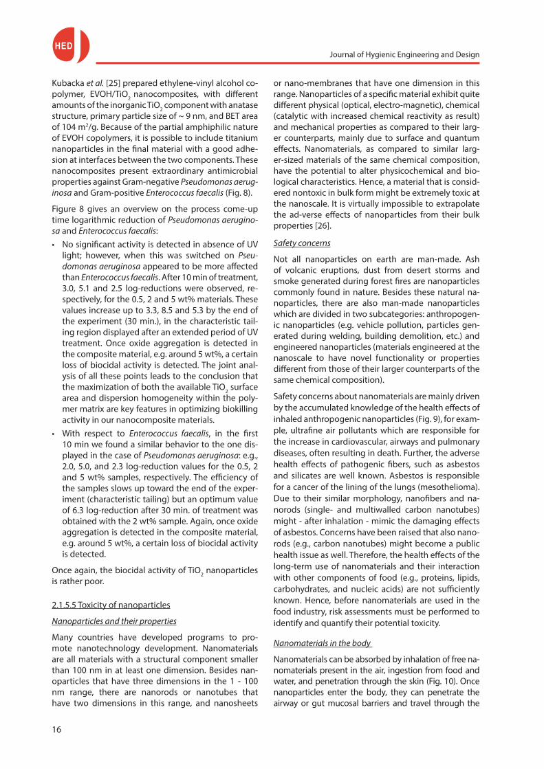
Kubacka et al. [25] prepared ethylene-vinyl alcohol co-polymer, EVOH/TiO2 nanocomposites, with different amounts of the inorganic TiO2 component with anatase structure, primary particle size of ~ 9 nm, and BET area of 104 m2/g. Because of the partial amphiphilic nature of EVOH copolymers, it is possible to include titanium nanoparticles in the final material with a good adhe-sion at interfaces between the two components. These nanocomposites present extraordinary antimicrobial properties against Gram-negative Pseudomonas aerug-inosa and Gram-positive Enterococcus faecalis (Fig. 8).
Figure 8 gives an overview on the process come-up time logarithmic reduction of Pseudomonas aerugino-sa and Enterococcus faecalis:• No significant activity is detected in absence of UV
light; however, when this was switched on Pseu-domonas aeruginosa appeared to be more affected than Enterococcus faecalis. After 10 min of treatment, 3.0, 5.1 and 2.5 log-reductions were observed, re-spectively, for the 0.5, 2 and 5 wt% materials. These values increase up to 3.3, 8.5 and 5.3 by the end of the experiment (30 min.), in the characteristic tail-ing region displayed after an extended period of UV treatment. Once oxide aggregation is detected in the composite material, e.g. around 5 wt%, a certain loss of biocidal activity is detected. The joint anal-ysis of all these points leads to the conclusion that the maximization of both the available TiO2 surface area and dispersion homogeneity within the poly-mer matrix are key features in optimizing biokilling activity in our nanocomposite materials.
• With respect to Enterococcus faecalis, in the first 10 min we found a similar behavior to the one dis-played in the case of Pseudomonas aeruginosa: e.g., 2.0, 5.0, and 2.3 log-reduction values for the 0.5, 2 and 5 wt% samples, respectively. The efficiency of the samples slows up toward the end of the exper-iment (characteristic tailing) but an optimum value of 6.3 log-reduction after 30 min. of treatment was obtained with the 2 wt% sample. Again, once oxide aggregation is detected in the composite material, e.g. around 5 wt%, a certain loss of biocidal activity is detected.
Once again, the biocidal activity of TiO2 nanoparticles is rather poor.
2.1.5.5 Toxicity of nanoparticles
Nanoparticles and their properties
Many countries have developed programs to pro-mote nanotechnology development. Nanomaterials are all materials with a structural component smaller than 100 nm in at least one dimension. Besides nan-oparticles that have three dimensions in the 1 - 100 nm range, there are nanorods or nanotubes that have two dimensions in this range, and nanosheets
or nano-membranes that have one dimension in this range. Nanoparticles of a specific material exhibit quite different physical (optical, electro-magnetic), chemical (catalytic with increased chemical reactivity as result) and mechanical properties as compared to their larg-er counterparts, mainly due to surface and quantum effects. Nanomaterials, as compared to similar larg-er-sized materials of the same chemical composition, have the potential to alter physicochemical and bio-logical characteristics. Hence, a material that is consid-ered nontoxic in bulk form might be extremely toxic at the nanoscale. It is virtually impossible to extrapolate the ad-verse effects of nanoparticles from their bulk properties [26].
Safety concerns
Not all nanoparticles on earth are man-made. Ash of volcanic eruptions, dust from desert storms and smoke generated during forest fires are nanoparticles commonly found in nature. Besides these natural na-noparticles, there are also man-made nanoparticles which are divided in two subcategories: anthropogen-ic nanoparticles (e.g. vehicle pollution, particles gen-erated during welding, building demolition, etc.) and engineered nanoparticles (materials engineered at the nanoscale to have novel functionality or properties different from those of their larger counterparts of the same chemical composition).
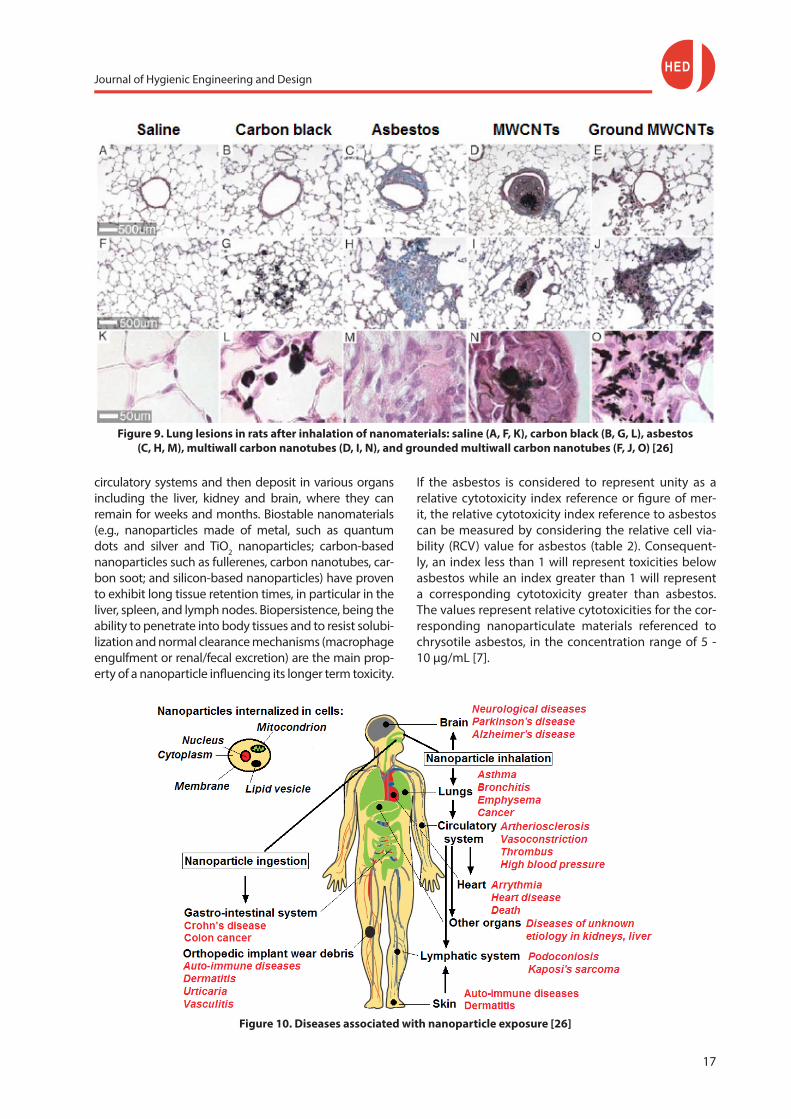
Safety concerns about nanomaterials are mainly driven by the accumulated knowledge of the health effects of inhaled anthropogenic nanoparticles (Fig. 9), for exam-ple, ultrafine air pollutants which are responsible for the increase in cardiovascular, airways and pulmonary diseases, often resulting in death. Further, the adverse health effects of pathogenic fibers, such as asbestos and silicates are well known. Asbestos is responsible for a cancer of the lining of the lungs (mesothelioma). Due to their similar morphology, nanofibers and na-norods (single- and multiwalled carbon nanotubes) might - after inhalation - mimic the damaging effects of asbestos. Concerns have been raised that also nano-rods (e.g., carbon nanotubes) might become a public health issue as well. Therefore, the health effects of the long-term use of nanomaterials and their interaction with other components of food (e.g., proteins, lipids, carbohydrates, and nucleic acids) are not sufficiently known. Hence, before nanomaterials are used in the food industry, risk assessments must be performed to identify and quantify their potential toxicity.
Nanomaterials in the body
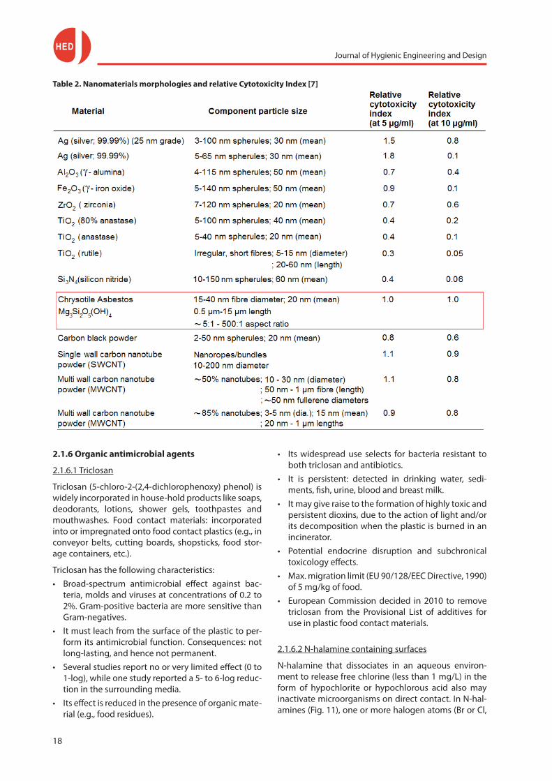
Nanomaterials can be absorbed by inhalation of free na-nomaterials present in the air, ingestion from food and water, and penetration through the skin (Fig. 10). Once nanoparticles enter the body, they can penetrate the airway or gut mucosal barriers and travel through the
Journal of Hygienic Engineering and Design
17
Figure 9. Lung lesions in rats after inhalation of nanomaterials: saline (A, F, K), carbon black (B, G, L), asbestos (C, H, M), multiwall carbon nanotubes (D, I, N), and grounded multiwall carbon nanotubes (F, J, O) [26]
circulatory systems and then deposit in various organs including the liver, kidney and brain, where they can remain for weeks and months. Biostable nanomaterials (e.g., nanoparticles made of metal, such as quantum dots and silver and TiO2 nanoparticles; carbon-based nanoparticles such as fullerenes, carbon nanotubes, car-bon soot; and silicon-based nanoparticles) have proven to exhibit long tissue retention times, in particular in the liver, spleen, and lymph nodes. Biopersistence, being the ability to penetrate into body tissues and to resist solubi-lization and normal clearance mechanisms (macrophage engulfment or renal/fecal excretion) are the main prop-erty of a nanoparticle influencing its longer term toxicity.
Figure 10. Diseases associated with nanoparticle exposure [26]
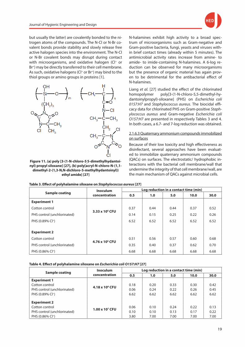
If the asbestos is considered to represent unity as a relative cytotoxicity index reference or figure of mer-it, the relative cytotoxicity index reference to asbestos can be measured by considering the relative cell via-bility (RCV) value for asbestos (table 2). Consequent-ly, an index less than 1 will represent toxicities below asbestos while an index greater than 1 will represent a corresponding cytotoxicity greater than asbestos. The values represent relative cytotoxicities for the cor-responding nanoparticulate materials referenced to chrysotile asbestos, in the concentration range of 5 - 10 μg/mL [7].
Journal of Hygienic Engineering and Design
18
Table 2. Nanomaterials morphologies and relative Cytotoxicity Index [7]
2.1.6 Organic antimicrobial agents
2.1.6.1 Triclosan
Triclosan (5-chloro-2-(2,4-dichlorophenoxy) phenol) is widely incorporated in house-hold products like soaps, deodorants, lotions, shower gels, toothpastes and mouthwashes. Food contact materials: incorporated into or impregnated onto food contact plastics (e.g., in conveyor belts, cutting boards, shopsticks, food stor-age containers, etc.).
Triclosan has the following characteristics:• Broad-spectrum antimicrobial effect against bac-
teria, molds and viruses at concentrations of 0.2 to 2%. Gram-positive bacteria are more sensitive than Gram-negatives.
• It must leach from the surface of the plastic to per-form its antimicrobial function. Consequences: not long-lasting, and hence not permanent.
• Several studies report no or very limited effect (0 to 1-log), while one study reported a 5- to 6-log reduc-tion in the surrounding media.
• Its effect is reduced in the presence of organic mate-rial (e.g., food residues).
• Its widespread use selects for bacteria resistant to both triclosan and antibiotics.
• It is persistent: detected in drinking water, sedi-ments, fish, urine, blood and breast milk.
• It may give raise to the formation of highly toxic and persistent dioxins, due to the action of light and/or its decomposition when the plastic is burned in an incinerator.
• Potential endocrine disruption and subchronical toxicology effects.
• Max. migration limit (EU 90/128/EEC Directive, 1990) of 5 mg/kg of food.
• European Commission decided in 2010 to remove triclosan from the Provisional List of additives for use in plastic food contact materials.
2.1.6.2 N-halamine containing surfaces
N-halamine that dissociates in an aqueous environ-ment to release free chlorine (less than 1 mg/L) in the form of hypochlorite or hypochlorous acid also may inactivate microorganisms on direct contact. In N-hal-amines (Fig. 11), one or more halogen atoms (Br or Cl,
Journal of Hygienic Engineering and Design
19
but usually the latter) are covalently bonded to the ni-trogen atoms of the compounds. The N-Cl or N-Br co-valent bonds provide stability and slowly release free active halogen species into the environment. The N-Cl or N-Br covalent bonds may disrupt during contact with microorganisms, and oxidative halogen (Cl+ or Br+) may be directly transferred to their cell membrane. As such, oxidative halogens (Cl+ or Br+) may bind to the thiol groups or amino groups in proteins [1].
Table 3. Effect of polyhalamine siloxane on Staphylococcus aureus [27]
Sample coating Inoculum concentration
Log reduction in a contact time (min)0.5 1.0 5.0 10.0 30.0
Experiment 1
3.33 x 10⁶ CFUCotton control 0.37 0.44 0.44 0.37 0.52
PHS control (unchlorinated) 0.14 0.15 0.25 0.22 0.26
PHS (0.89% Cl+) 6.52 6.52 6.52 6.52 6.52
Experiment 2
4.76 x 10⁶ CFUCotton control 0.51 0.56 0.57 0.60 0.68
PHS control (unchlorinated) 0.35 0.40 0.37 0.62 0.70
PHS (0.86% Cl+) 6.68 6.68 6.68 6.68 6.68
Figure 11. (a) poly [3-(1-N-chloro-5,5-dimethylhydantoi-nyl) propyl siloxane] [27], (b) poly[acryl-N-chloro-N- (1,1-
dimethyl-2-(1,3-N,N-dichloro-5-methylhydantoinyl)) ethyl amide] [27]
N-halamines exhibit high activity to a broad spec-trum of microorganisms such as Gram-negative and Gram-positive bacteria, fungi, yeasts and viruses with-in brief contact times (already within 5 minutes). The antimicrobial activity rates increase from amine- to amide- to imide-containing N-halamines. A 6-log re-duction can be observed for many microorganisms but the presence of organic material has again prov-en to be detrimental for the antibacterial effect of N-halamines.
Liang et al. [27] studied the effect of the chlorinated homopolymer poly[3-(1-N-chloro-5,5-dimethyl-hy-dantoinylpropyl)-siloxane] (PHS) on Escherichia coli 0157:H7 and Staphylococcus aureus. The biocidal effi-cacy data for chlorinated PHS on Gram-positive Staph-ylococcus aureus and Gram-negative Escherichia coli O157:H7 are presented in respectively Tables 3 and 4. In both cases, a 6.7- and 7-log reduction was obtained.
2.1.6.3 Quaternary ammonium compounds immobilized on surfacesBecause of their low toxicity and high effectiveness as disinfectant, several approaches have been evaluat-ed to immobilize quaternary ammonium compounds (QACs) on surfaces. The electrostatic/ hydrophobic in-teractions with the bacterial cell membrane/wall that undermine the integrity of that cell membrane/wall, are the main mechanism of QACs against microbial cells.
Table 4. Effect of polyhalamine siloxane on Escherichia coli O157:H7 [27]
Sample coating Inoculum concentration
Log reduction in a contact time (min)0.5 1.0 5.0 10.0 30.0
Experiment 1
4.18 x 10⁶ CFUCotton control 0.18 0.20 0.33 0.30 0.42PHS control (unchlorinated) 0.06 0.24 0.22 0.26 0.45PHS (0.89% Cl+) 6.62 6.62 6.62 6.62 6.62
Experiment 2
1.00 x 10⁷ CFUCotton control 0.06 0.10 0.24 0.22 0.13PHS control (unchlorinated) 0.10 0.10 0.13 0.17 0.22PHS (0.86% Cl+) 3.80 7.00 7.00 7.00 7.00
Journal of Hygienic Engineering and Design
20
Organosilane quaternary ammonium compounds
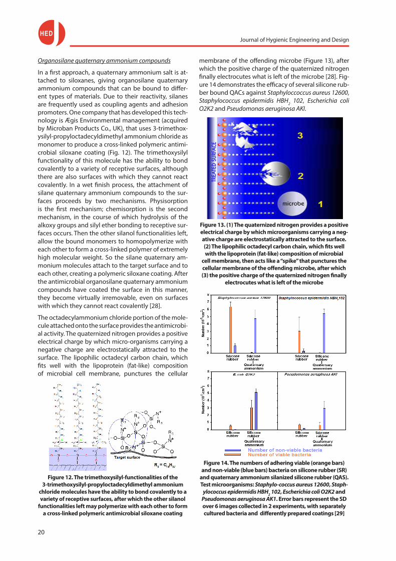
In a first approach, a quaternary ammonium salt is at-tached to siloxanes, giving organosilane quaternary ammonium compounds that can be bound to differ-ent types of materials. Due to their reactivity, silanes are frequently used as coupling agents and adhesion promoters. One company that has developed this tech-nology is Ægis Environmental management (acquired by Microban Products Co., UK), that uses 3-trimethox-ysilyl-propyloctadecyldimethyl ammonium chloride as monomer to produce a cross-linked polymeric antimi-crobial siloxane coating (Fig. 12). The trimethoxysilyl functionality of this molecule has the ability to bond covalently to a variety of receptive surfaces, although there are also surfaces with which they cannot react covalently. In a wet finish process, the attachment of silane quaternary ammonium compounds to the sur-faces proceeds by two mechanisms. Physisorption is the first mechanism; chemisorption is the second mechanism, in the course of which hydrolysis of the alkoxy groups and silyl ether bonding to receptive sur-faces occurs. Then the other silanol functionalities left, allow the bound monomers to homopolymerize with each other to form a cross-linked polymer of extremely high molecular weight. So the silane quaternary am-monium molecules attach to the target surface and to each other, creating a polymeric siloxane coating. After the antimicrobial organosilane quaternary ammonium compounds have coated the surface in this manner, they become virtually irremovable, even on surfaces with which they cannot react covalently [28].
The octadecylammonium chloride portion of the mole-cule attached onto the surface provides the antimicrobi-al activity. The quaternized nitrogen provides a positive electrical charge by which micro-organisms carrying a negative charge are electrostatically attracted to the surface. The lipophilic octadecyl carbon chain, which fits well with the lipoprotein (fat-like) composition of microbial cell membrane, punctures the cellular
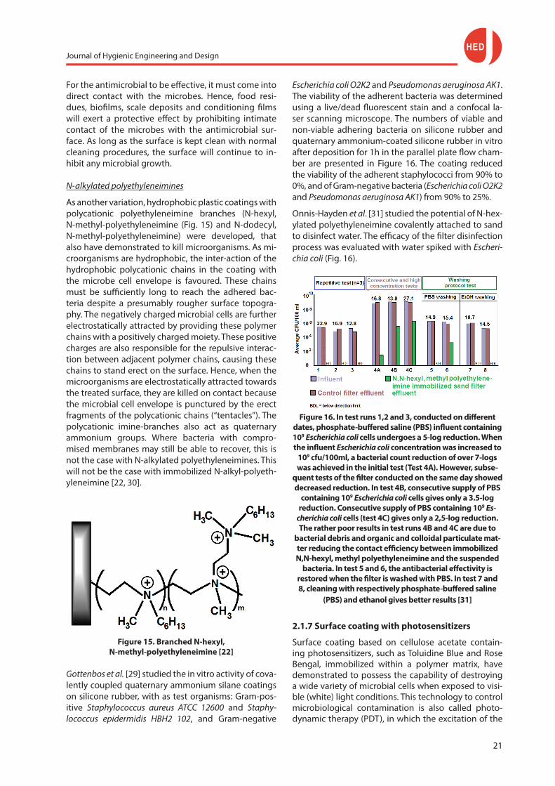
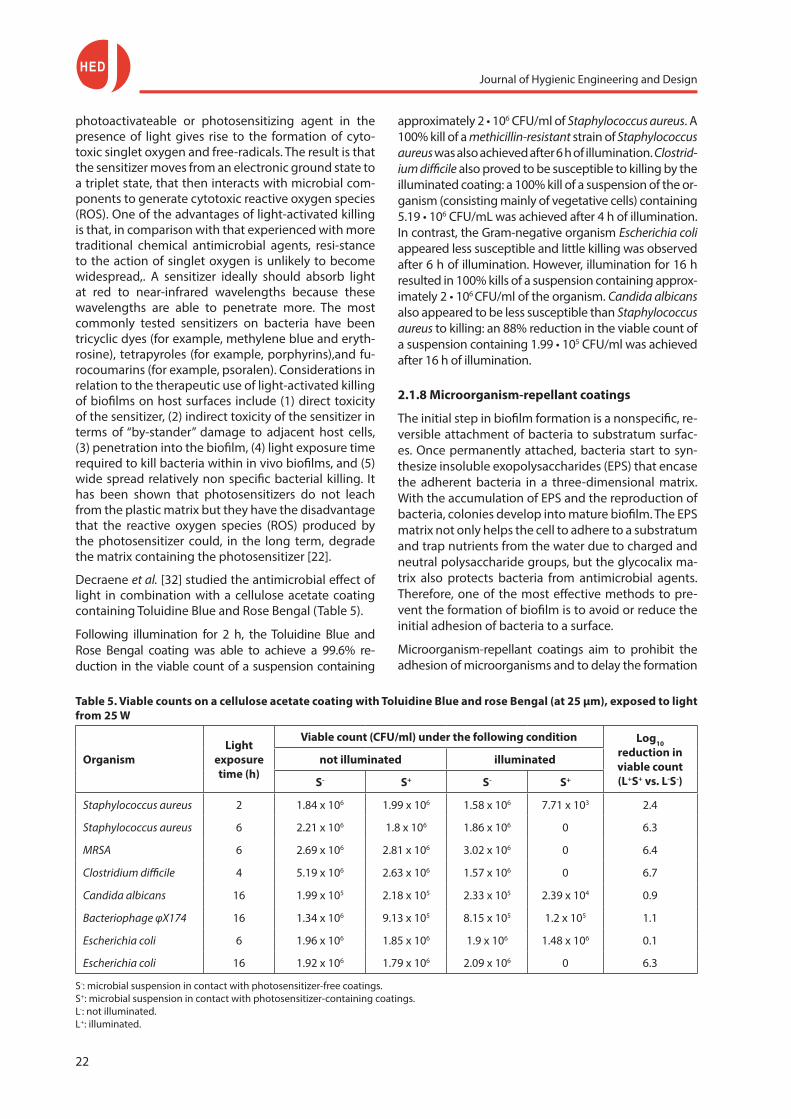
membrane of the offending microbe (Figure 13), after which the positive charge of the quaternized nitrogen finally electrocutes what is left of the microbe [28]. Fig-ure 14 demonstrates the efficacy of several silicone rub-ber bound QACs against Staphyloccoccus aureus 12600, Staphylococcus epidermidis HBH2 102, Escherichia coli O2K2 and Pseudomonas aeruginosa AKI.
Figure 13. (1) The quaternized nitrogen provides a positive electrical charge by which microorganisms carrying a neg-ative charge are electrostatically attracted to the surface. (2) The lipophilic octadecyl carbon chain, which fits well with the lipoprotein (fat-like) composition of microbial
cell membrane, then acts like a “spike” that punctures the cellular membrane of the offending microbe, after which (3) the positive charge of the quaternized nitrogen finally
electrocutes what is left of the microbe
Figure 14. The numbers of adhering viable (orange bars) and non-viable (blue bars) bacteria on silicone rubber (SR)
and quaternary ammonium silanized silicone rubber (QAS). Test microorganisms: Staphylo-coccus aureus 12600, Staph-
ylococcus epidermidis HBH2 102, Escherichia coli O2K2 and Pseudomonas aeruginosa AK1. Error bars represent the SD over 6 images collected in 2 experiments, with separately cultured bacteria and differently prepared coatings [29]
Figure 12. The trimethoxysilyl-functionalities of the 3-trimethoxysilyl-propyloctadecyldimethyl ammonium
chloride molecules have the ability to bond covalently to a variety of receptive surfaces, after which the other silanol
functionalities left may polymerize with each other to form a cross-linked polymeric antimicrobial siloxane coating
Journal of Hygienic Engineering and Design
21
For the antimicrobial to be effective, it must come into direct contact with the microbes. Hence, food resi-dues, biofilms, scale deposits and conditioning films will exert a protective effect by prohibiting intimate contact of the microbes with the antimicrobial sur-face. As long as the surface is kept clean with normal cleaning procedures, the surface will continue to in-hibit any microbial growth.
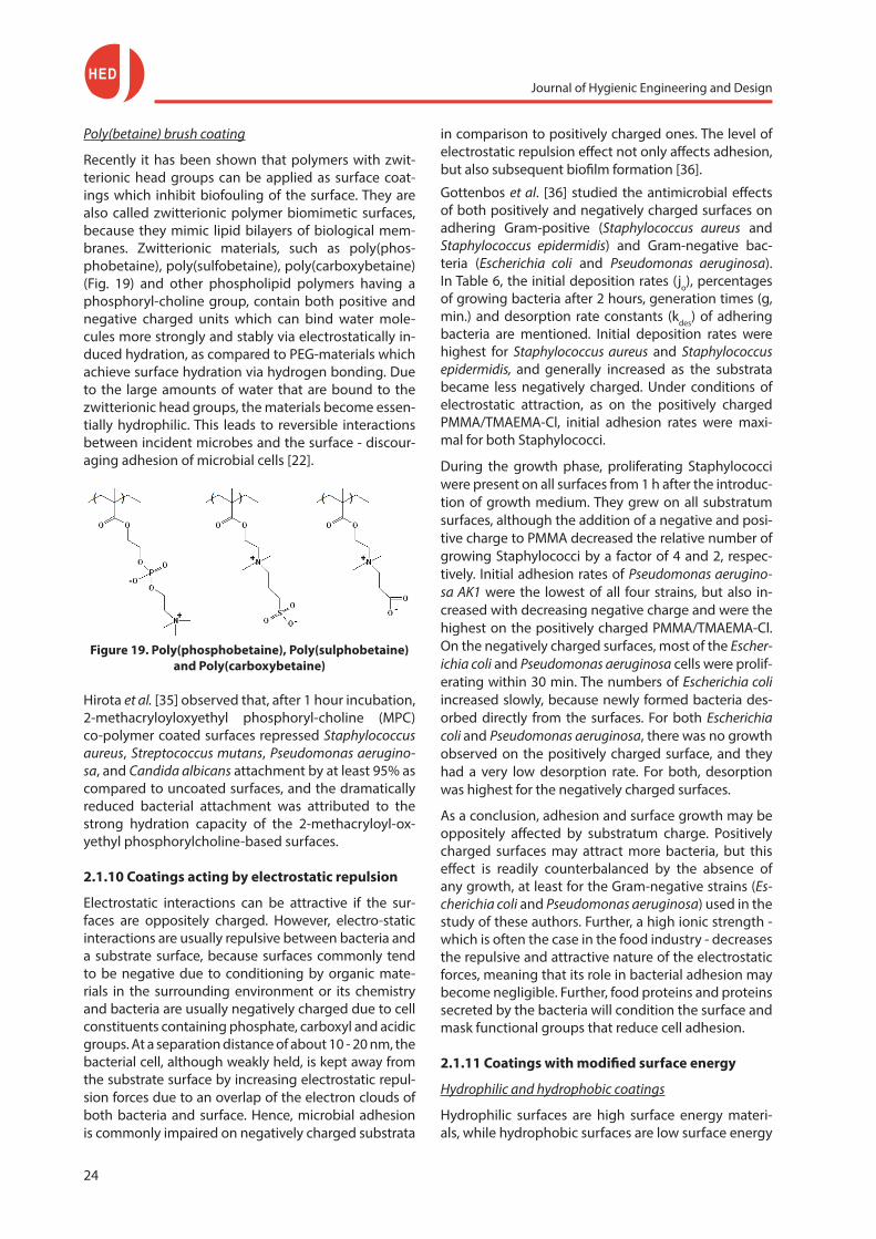
N-alkylated polyethyleneimines
As another variation, hydrophobic plastic coatings with polycationic polyethyleneimine branches (N-hexyl, N-methyl-polyethyleneimine (Fig. 15) and N-dodecyl, N-methyl-polyethyleneimine) were developed, that also have demonstrated to kill microorganisms. As mi-croorganisms are hydrophobic, the inter-action of the hydrophobic polycationic chains in the coating with the microbe cell envelope is favoured. These chains must be sufficiently long to reach the adhered bac-teria despite a presumably rougher surface topogra-phy. The negatively charged microbial cells are further electrostatically attracted by providing these polymer chains with a positively charged moiety. These positive charges are also responsible for the repulsive interac-tion between adjacent polymer chains, causing these chains to stand erect on the surface. Hence, when the microorganisms are electrostatically attracted towards the treated surface, they are killed on contact because the microbial cell envelope is punctured by the erect fragments of the polycationic chains (“tentacles”). The polycationic imine-branches also act as quaternary ammonium groups. Where bacteria with compro-mised membranes may still be able to recover, this is not the case with N-alkylated polyethyleneimines. This will not be the case with immobilized N-alkyl-polyeth-yleneimine [22, 30].
Escherichia coli O2K2 and Pseudomonas aeruginosa AK1. The viability of the adherent bacteria was determined using a live/dead fluorescent stain and a confocal la-ser scanning microscope. The numbers of viable and non-viable adhering bacteria on silicone rubber and quaternary ammonium-coated silicone rubber in vitro after deposition for 1h in the parallel plate flow cham-ber are presented in Figure 16. The coating reduced the viability of the adherent staphylococci from 90% to 0%, and of Gram-negative bacteria (Escherichia coli O2K2 and Pseudomonas aeruginosa AK1) from 90% to 25%.
Onnis-Hayden et al. [31] studied the potential of N-hex-ylated polyethyleneimine covalently attached to sand to disinfect water. The efficacy of the filter disinfection process was evaluated with water spiked with Escheri-chia coli (Fig. 16).
Figure 15. Branched N-hexyl, N-methyl-polyethyleneimine [22]
Gottenbos et al. [29] studied the in vitro activity of cova-lently coupled quaternary ammonium silane coatings on silicone rubber, with as test organisms: Gram-pos-itive Staphylococcus aureus ATCC 12600 and Staphy-lococcus epidermidis HBH2 102, and Gram-negative
Figure 16. In test runs 1,2 and 3, conducted on different dates, phosphate-buffered saline (PBS) influent containing 109 Escherichia coli cells undergoes a 5-log reduction. When the influent Escherichia coli concentration was increased to
109 cfu/100ml, a bacterial count reduction of over 7-logs was achieved in the initial test (Test 4A). However, subse-
quent tests of the filter conducted on the same day showed decreased reduction. In test 4B, consecutive supply of PBS
containing 109 Escherichia coli cells gives only a 3.5-log reduction. Consecutive supply of PBS containing 109 Es-
cherichia coli cells (test 4C) gives only a 2,5-log reduction. The rather poor results in test runs 4B and 4C are due to
bacterial debris and organic and colloidal particulate mat-ter reducing the contact efficiency between immobilized N,N-hexyl, methyl polyethyleneimine and the suspended
bacteria. In test 5 and 6, the antibacterial effectivity is restored when the filter is washed with PBS. In test 7 and 8, cleaning with respectively phosphate-buffered saline
(PBS) and ethanol gives better results [31]
2.1.7 Surface coating with photosensitizers
Surface coating based on cellulose acetate contain-ing photosensitizers, such as Toluidine Blue and Rose Bengal, immobilized within a polymer matrix, have demonstrated to possess the capability of destroying a wide variety of microbial cells when exposed to visi-ble (white) light conditions. This technology to control microbiological contamination is also called photo-dynamic therapy (PDT), in which the excitation of the
Journal of Hygienic Engineering and Design
22
photoactivateable or photosensitizing agent in the presence of light gives rise to the formation of cyto-toxic singlet oxygen and free-radicals. The result is that the sensitizer moves from an electronic ground state to a triplet state, that then interacts with microbial com-ponents to generate cytotoxic reactive oxygen species (ROS). One of the advantages of light-activated killing is that, in comparison with that experienced with more traditional chemical antimicrobial agents, resi-stance to the action of singlet oxygen is unlikely to become widespread,. A sensitizer ideally should absorb light at red to near-infrared wavelengths because these wavelengths are able to penetrate more. The most commonly tested sensitizers on bacteria have been tricyclic dyes (for example, methylene blue and eryth-rosine), tetrapyroles (for example, porphyrins),and fu-rocoumarins (for example, psoralen). Considerations in relation to the therapeutic use of light-activated killing of biofilms on host surfaces include (1) direct toxicity of the sensitizer, (2) indirect toxicity of the sensitizer in terms of “by-stander” damage to adjacent host cells, (3) penetration into the biofilm, (4) light exposure time required to kill bacteria within in vivo biofilms, and (5) wide spread relatively non specific bacterial killing. It has been shown that photosensitizers do not leach from the plastic matrix but they have the disadvantage that the reactive oxygen species (ROS) produced by the photosensitizer could, in the long term, degrade the matrix containing the photosensitizer [22].
Decraene et al. [32] studied the antimicrobial effect of light in combination with a cellulose acetate coating containing Toluidine Blue and Rose Bengal (Table 5).
Following illumination for 2 h, the Toluidine Blue and Rose Bengal coating was able to achieve a 99.6% re-duction in the viable count of a suspension containing
Table 5. Viable counts on a cellulose acetate coating with Toluidine Blue and rose Bengal (at 25 µm), exposed to light from 25 W
OrganismLight
exposuretime (h)
Viable count (CFU/ml) under the following condition Log10 reduction in viable count (L+S+ vs. L-S-)
not illuminated illuminated
S- S+ S- S+
Staphylococcus aureus 2 1.84 x 10⁶ 1.99 x 10⁶ 1.58 x 10⁶ 7.71 x 10³ 2.4
Staphylococcus aureus 6 2.21 x 10⁶ 1.8 x 10⁶ 1.86 x 10⁶ 0 6.3
MRSA 6 2.69 x 10⁶ 2.81 x 10⁶ 3.02 x 10⁶ 0 6.4
Clostridium difficile 4 5.19 x 10⁶ 2.63 x 10⁶ 1.57 x 10⁶ 0 6.7
Candida albicans 16 1.99 x 10⁵ 2.18 x 10⁵ 2.33 x 10⁵ 2.39 x 10⁴ 0.9
Bacteriophage φX174 16 1.34 x 10⁶ 9.13 x 10⁵ 8.15 x 10⁵ 1.2 x 10⁵ 1.1
Escherichia coli 6 1.96 x 10⁶ 1.85 x 10⁶ 1.9 x 10⁶ 1.48 x 10⁶ 0.1
Escherichia coli 16 1.92 x 10⁶ 1.79 x 10⁶ 2.09 x 10⁶ 0 6.3
S-: microbial suspension in contact with photosensitizer-free coatings. S+: microbial suspension in contact with photosensitizer-containing coatings. L-: not illuminated. L+: illuminated.
approximately 2 • 106 CFU/ml of Staphylococcus aureus. A 100% kill of a methicillin-resistant strain of Staphylococcus aureus was also achieved after 6 h of illumination. Clostrid-ium difficile also proved to be susceptible to killing by the illuminated coating: a 100% kill of a suspension of the or-ganism (consisting mainly of vegetative cells) containing 5.19 • 106 CFU/mL was achieved after 4 h of illumination. In contrast, the Gram-negative organism Escherichia coli appeared less susceptible and little killing was observed after 6 h of illumination. However, illumination for 16 h resulted in 100% kills of a suspension containing approx-imately 2 • 106 CFU/ml of the organism. Candida albicans also appeared to be less susceptible than Staphylococcus aureus to killing: an 88% reduction in the viable count of a suspension containing 1.99 • 105 CFU/ml was achieved after 16 h of illumination.
2.1.8 Microorganism-repellant coatings
The initial step in biofilm formation is a nonspecific, re-versible attachment of bacteria to substratum surfac-es. Once permanently attached, bacteria start to syn-thesize insoluble exopolysaccharides (EPS) that encase the adherent bacteria in a three-dimensional matrix. With the accumulation of EPS and the reproduction of bacteria, colonies develop into mature biofilm. The EPS matrix not only helps the cell to adhere to a substratum and trap nutrients from the water due to charged and neutral polysaccharide groups, but the glycocalix ma-trix also protects bacteria from antimicrobial agents. Therefore, one of the most effective methods to pre-vent the formation of biofilm is to avoid or reduce the initial adhesion of bacteria to a surface.
Microorganism-repellant coatings aim to prohibit the adhesion of microorganisms and to delay the formation

Journal of Hygienic Engineering and Design
23
of biofilms on product contact surfaces. These biopas-sive polymer coatings (Fig. 17) were initially used to give minimal protein adsorption on the surface, as proteins promote the adhesion of bacteria. In contrary to bioac-tive polymers, biopassive surfaces do not actively inter-act or kill bacteria. A wide variety of polymers, including poly(ethylene glycol) (PEG) and zwitterionic structures have been examined as biopassive surfaces [33].
2.1.9 Coatings acting by steric repulsion and their “superhydrophilic” properties
Surfaces can be modified by covalent or non-covalent (physisorption) binding of hydrophilic polymers or block copolymers consisting of highly hydrated, flex-ible chains with low polymer–water interfacial ener-gies in a high density, giving so called polymer brush-es. These end-tethered polymer chains are forced to stretch away from a surface into the adjacent solution due to that high density of chains per unit surface area. By using polymer brushes that bind a lot of water, the brush-coating becomes highly hydrophilic. Microor-ganisms encountering the brush surface are repelled by steric hindrance due to the bound water in the brush (microbial cells are hydrophobic, and microbial attachment to the surface is not favoured due to the lack of hydrophobic binding sites) and the elasticity of the polymer chains [33, 34].
polypropylene oxide (PPO). They observed that the adhesion of Staphylococci aureus and Staphylococcus epidermidis, as compared to pristine silicone rubber, is reduced on PEO brush-coated silicone rubber (Fig. 18). Yet, Staphylococcal biofilms grew on both brush-coat-ed and pristine silicone rubber, while the viability of biofilms on brush-coatings was higher than on pris-tine silicone rubber. However, Staphylococcal biofilms developed more slowly on brush-coatings and when exposed to high fluid shear, they almost fully detached from the brush-coating in contrast to detachment from pristine silicone rubber. Adhesion and growth of Pseu-domonas aeruginosa were not significantly affected by the presence of the brush on silicone rubber, although here too the viability of biofilms on brush-coatings was higher. The slow growth of Staphylococci on polymer brush-coatings may allow more time for treatment with cleaning agents and disinfectants before a ma-ture, more resistant biofilm can develop.
Figure 17. Passive protection of the surface by covalent immobilization or physisorption of hydrophilic
well- hydrated polymers [33, 34]
PEG-brush coating
Polymer brushes are quite often poly(ethylene glycol) (= poly(ethyleneoxide)) systems, which are hydrophilic, meaning that water will be attracted into the brush lay-er and form a repellent layer close to the surface. These polymer brushes may inhibit the adhesion of microbes by up to a 3-log unit reduction in attached microbes. Sometimes low molecular weight proteins and pep-tides still can penetrate through the PEG (PEO)-layer, providing the bacteria an attachment platform [33, 34].
Nedjadnik et al. [34] studied brush-coatings made of a tri-block copolymer of polyethylene oxide (PEO) and
Figure 18. Surface coverage by biofilms of Staphylococci aureus, Staphylococcus epidermidis and Pseudomonas aeruginosa as a function of time after initial adhesion and subsequent bacterial growth on pristine (∆) and
brush-coated ( ) silicone rubber. Error bars denote the SD over four separate experiments with different bacterial cultures and silicone rubber sheets with and without a
brush-coating [34]
Journal of Hygienic Engineering and Design
24
Poly(betaine) brush coating
Recently it has been shown that polymers with zwit-terionic head groups can be applied as surface coat-ings which inhibit biofouling of the surface. They are also called zwitterionic polymer biomimetic surfaces, because they mimic lipid bilayers of biological mem-branes. Zwitterionic materials, such as poly(phos-phobetaine), poly(sulfobetaine), poly(carboxybetaine) (Fig. 19) and other phospholipid polymers having a phosphoryl-choline group, contain both positive and negative charged units which can bind water mole-cules more strongly and stably via electrostatically in-duced hydration, as compared to PEG-materials which achieve surface hydration via hydrogen bonding. Due to the large amounts of water that are bound to the zwitterionic head groups, the materials become essen-tially hydrophilic. This leads to reversible interactions between incident microbes and the surface - discour-aging adhesion of microbial cells [22].
in comparison to positively charged ones. The level of electrostatic repulsion effect not only affects adhesion, but also subsequent biofilm formation [36].
Gottenbos et al. [36] studied the antimicrobial effects of both positively and negatively charged surfaces on adhering Gram-positive (Staphylococcus aureus and Staphylococcus epidermidis) and Gram-negative bac-teria (Escherichia coli and Pseudomonas aeruginosa). In Table 6, the initial deposition rates (jo), percentages of growing bacteria after 2 hours, generation times (g, min.) and desorption rate constants (kdes) of adhering bacteria are mentioned. Initial deposition rates were highest for Staphylococcus aureus and Staphylococcus epidermidis, and generally increased as the substrata became less negatively charged. Under conditions of electrostatic attraction, as on the positively charged PMMA/TMAEMA-Cl, initial adhesion rates were maxi-mal for both Staphylococci.
During the growth phase, proliferating Staphylococci were present on all surfaces from 1 h after the introduc-tion of growth medium. They grew on all substratum surfaces, although the addition of a negative and posi-tive charge to PMMA decreased the relative number of growing Staphylococci by a factor of 4 and 2, respec-tively. Initial adhesion rates of Pseudomonas aerugino-sa AK1 were the lowest of all four strains, but also in-creased with decreasing negative charge and were the highest on the positively charged PMMA/TMAEMA-Cl. On the negatively charged surfaces, most of the Escher-ichia coli and Pseudomonas aeruginosa cells were prolif-erating within 30 min. The numbers of Escherichia coli increased slowly, because newly formed bacteria des-orbed directly from the surfaces. For both Escherichia coli and Pseudomonas aeruginosa, there was no growth observed on the positively charged surface, and they had a very low desorption rate. For both, desorption was highest for the negatively charged surfaces.
As a conclusion, adhesion and surface growth may be oppositely affected by substratum charge. Positively charged surfaces may attract more bacteria, but this effect is readily counterbalanced by the absence of any growth, at least for the Gram-negative strains (Es-cherichia coli and Pseudomonas aeruginosa) used in the study of these authors. Further, a high ionic strength - which is often the case in the food industry - decreases the repulsive and attractive nature of the electrostatic forces, meaning that its role in bacterial adhesion may become negligible. Further, food proteins and proteins secreted by the bacteria will condition the surface and mask functional groups that reduce cell adhesion.
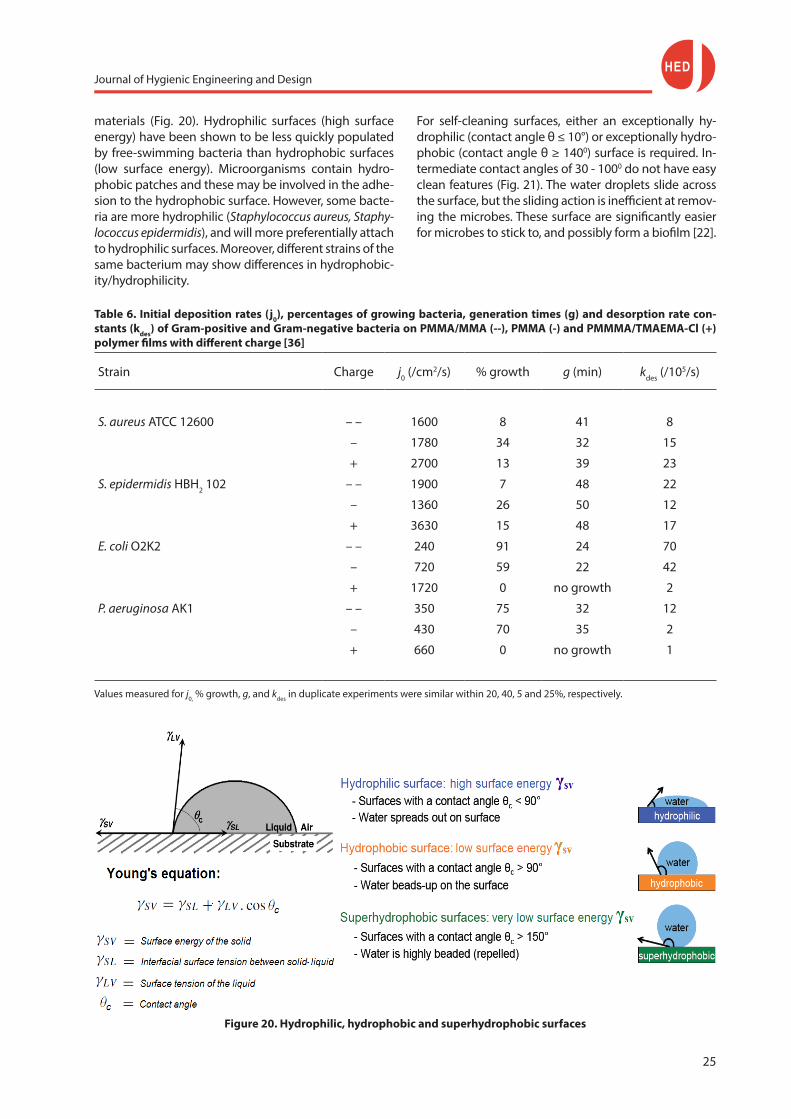
2.1.11 Coatings with modified surface energy
Hydrophilic and hydrophobic coatings
Hydrophilic surfaces are high surface energy materi-als, while hydrophobic surfaces are low surface energy
Figure 19. Poly(phosphobetaine), Poly(sulphobetaine) and Poly(carboxybetaine)
Hirota et al. [35] observed that, after 1 hour incubation, 2-methacryloyloxyethyl phosphoryl-choline (MPC) co-polymer coated surfaces repressed Staphylococcus aureus, Streptococcus mutans, Pseudomonas aerugino-sa, and Candida albicans attachment by at least 95% as compared to uncoated surfaces, and the dramatically reduced bacterial attachment was attributed to the strong hydration capacity of the 2-methacryloyl-ox-yethyl phosphorylcholine-based surfaces.
2.1.10 Coatings acting by electrostatic repulsion
Electrostatic interactions can be attractive if the sur-faces are oppositely charged. However, electro-static interactions are usually repulsive between bacteria and a substrate surface, because surfaces commonly tend to be negative due to conditioning by organic mate-rials in the surrounding environment or its chemistry and bacteria are usually negatively charged due to cell constituents containing phosphate, carboxyl and acidic groups. At a separation distance of about 10 - 20 nm, the bacterial cell, although weakly held, is kept away from the substrate surface by increasing electrostatic repul-sion forces due to an overlap of the electron clouds of both bacteria and surface. Hence, microbial adhesion is commonly impaired on negatively charged substrata
Journal of Hygienic Engineering and Design
25
materials (Fig. 20). Hydrophilic surfaces (high surface energy) have been shown to be less quickly populated by free-swimming bacteria than hydrophobic surfaces (low surface energy). Microorganisms contain hydro-phobic patches and these may be involved in the adhe-sion to the hydrophobic surface. However, some bacte-ria are more hydrophilic (Staphylococcus aureus, Staphy-lococcus epidermidis), and will more preferentially attach to hydrophilic surfaces. Moreover, different strains of the same bacterium may show differences in hydrophobic-ity/hydrophilicity.
Table 6. Initial deposition rates (j0), percentages of growing bacteria, generation times (g) and desorption rate con-stants (kdes) of Gram-positive and Gram-negative bacteria on PMMA/MMA (--), PMMA (-) and PMMMA/TMAEMA-Cl (+) polymer films with different charge [36]
Strain Charge j0 (/cm2/s) % growth g (min) kdes (/105/s)
S. aureus ATCC 12600 – – 1600 8 41 8
– 1780 34 32 15
+ 2700 13 39 23
S. epidermidis HBH2 102 – – 1900 7 48 22
– 1360 26 50 12
+ 3630 15 48 17
E. coli O2K2 – – 240 91 24 70
– 720 59 22 42
+ 1720 0 no growth 2
P. aeruginosa AK1 – – 350 75 32 12
– 430 70 35 2
+ 660 0 no growth 1
Values measured for j0, % growth, g, and kdes in duplicate experiments were similar within 20, 40, 5 and 25%, respectively.
Figure 20. Hydrophilic, hydrophobic and superhydrophobic surfaces
For self-cleaning surfaces, either an exceptionally hy-drophilic (contact angle θ ≤ 10°) or exceptionally hydro-phobic (contact angle θ ≥ 1400) surface is required. In-termediate contact angles of 30 - 1000 do not have easy clean features (Fig. 21). The water droplets slide across the surface, but the sliding action is inefficient at remov-ing the microbes. These surface are significantly easier for microbes to stick to, and possibly form a biofilm [22].

Journal of Hygienic Engineering and Design
26
Figure 21. As illustrated in the Baier-curve, surfaces with intermediate contact angles of 30-100° are more prone to microbial fouling, while superhydrophobic, hydro-
philic and superhydrophilic surfaces show lower micro-bial fouling retention [37]
Superhydrophilic and superhydrophobic coatings/ materials
Microstructuring a surface - in other words adding in unevenness or asperities - amplifies the natural ten-dency of the surface: hydrophilic surfaces become “superhydrophilic” (contact angle θ ≤ 100) and hydro-phobic surfaces become “superhydrophobic” (contact angle θ ≥ 1400)
Superhydrophilicity can be created by irradiating TiO2 coatings. The holes produced by photoexcitation pro-voke the apparition of oxygen vacancies which can be filled by adsorbed water to form surface hydroxide groups. The formation of these groups will increase the affinity of water toward the surface and result in complete wetting (water contact angle θc → 00) after UV irradiation (typical application: photocatalytically self-cleaning glasses). Due to the photocatalytic effect, germs also can be destroyed. Superhydrophobic mate-rials can be made in several ways - by coating a surface with a superhydrophobic material, by nanostructuring a surface, by applying nanoparticles to a surface or by a combination of these.
Superhydrophobic coatings are very low surface ener-gy coatings such as special waxes.
Notice, however, that hydrophobic easy to clean mate-rials reduce microbial contamination in the area treat-ed, but does not address the problem of pathogenic microbes which are incident upon the surface. It mere-ly moves them elsewhere, where they will have to be dealt with by other microbiocidal techniques.
2.1.12 Bio-inspired surfaces with modified surface microtopography and chemistry
The surface chemistry and physical properties of a substratum are both crucial to prevent the recruitment
of biofouling organisms. The idea to change surface topography and chemistry as tools to control fouling was taken from marine biology, because the natural surfaces of many marine organisms resist biofouling in these environments. These natural antifouling surfaces use a combination of chemical and physical structures to inhibit biofouling. Hence, surface modification tech-niques to tailor the surface energy via surface chemis-try and surface topography have been developed to study the effects of changes in these surface proper-ties on biofilm formation.
Natural antifouling surfaces
The skin of the approximately 900 species of Elasmo-branchii, which include sharks, skates, and rays is em-bedded with placoid scales. These scales serve several functions including reduction of mechanical abrasion, reduced hydrodynamic resistance and most interest-ingly protection from ectoparasites. The skin of two members of the porpoise family, i.e., the bottlenose dolphin Tursiops truncatus and the killer whale Orcinus orca (Fig. 22) forms a system of ridges and grooves ori-ented transversely to the direction of flow. The natural wavelength of the ridges and grooves is 0.3 to 0.4 mm with a trough to crest wave height of about 10 µm. These topographic features and a mucosal coating se-creted by epidermal cells contribute to the antifouling properties of these marine animals.
Figure 22. Scanning electron micrographs of natural textured surfaces: spinner shark skin (photo left),
Galapagos shark skin (photo right) [38]
Journal of Hygienic Engineering and Design
27
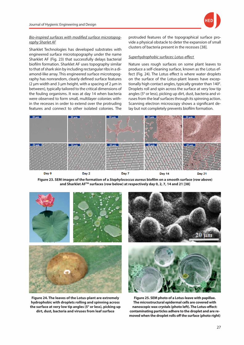
Bio-inspired surfaces with modified surface microtopog-raphy Sharlet AF
Sharklet Technologies has developed substrates with engineered surface microtopography under the name Sharklet AF (Fig. 23) that successfully delays bacterial biofilm formation. Sharklet AF uses topography similar to that of shark skin by including rectangular ribs in a di-amond-like array. This engineered surface microtopog-raphy has nonrandom, clearly defined surface features (2 µm width and 3 µm height, with a spacing of 2 µm in between), typically tailored to the critical dimensions of the fouling organisms. It was at day 14 when bacteria were observed to form small, multilayer colonies with-in the recesses in order to extend over the protruding features and connect to other isolated colonies. The
Figure 24. The leaves of the Lotus-plant are extremely hydrophobic with droplets rolling and spinning across
the surface at very low tip angles (5° or less), picking up dirt, dust, bacteria and viruses from leaf surface
Figure 23. SEM images of the formation of a Staphylococcus aureus biofilm on a smooth surface (row above) and Sharklet AFTM surfaces (row below) at respectively day 0, 2, 7, 14 and 21 [38]
protruded features of the topographical surface pro-vide a physical obstacle to deter the expansion of small clusters of bacteria present in the recesses [38].
Superhydrophobic surfaces: Lotus-effect
Nature uses rough surfaces on some plant leaves to produce a self-cleaning surface, known as the Lotus ef-fect (Fig. 24). The Lotus effect is where water droplets on the surface of the Lotus-plant leaves have excep-tionally high contact angles, typically greater than 1400. Droplets roll and spin across the surface at very low tip angles (50 or less), picking up dirt, dust, bacteria and vi-ruses from the leaf surfaces through its spinning action. Scanning electron microscopy shows a significant de-lay but not completely prevents biofilm formation.
Figure 25. SEM photo of a Lotus-leave with papillae. The microstructural epidermal cells are covered with
nanoscopic wax crystals (photo left). The Lotus-effect: contaminating particles adhere to the droplet and are re-moved when the droplet rolls off the surface (photo right)

Journal of Hygienic Engineering and Design
28
antimicrobial components. They also will never be a substitute for hygienic design.
4. References [1] Møretrø T., and Langsrud S. (2011). Effects of materials
containing antimicrobial compounds on food hygiene. Journal of Food Protection, 74 (7), pp. 1200-1211.
[2] Faille C., & Carpentier B. (2009). Food contact surfaces, surface soiling and biofilm formation. In: Fratamico P. M., Annous B. A.. and Gunther IV. N. W. (Eds.), Biofilms in the Food and Beverage Industries, Cambridge, United King-dom, Woodhead Publishing, pp. 303-330.
[3] Jung W. K., Koo H. C., Kim K. W., Shin S., Kim S. H., and Park Y. H. (2008). Antibacterial activity and mechanism of action of the silver ion in Staphylococcus aureus an Es-cherichia coli. Applied and Environmental Microbiology, 74 (7), pp. 2171-2178.
[4] Cowan M. M., Abshire K. Z., Houk S. L., and Evans S. M. (2003). Antimicrobial efficacy of a silver-zeolite matrix coating on stainless steel. Journal of Industrial Microbiol-ogy and Biotechnology, 30, (2), pp. 102-106.
[5] Junping Y., and Wei L. (2013). Antibacterial 316L stainless steel containing silver and niobium. Rare Metal Materials and Engineering, 42, (10), pp. 2004-2008.
[6] Chiang I. S., Møller P., Hilbert L. R., Tolker-Nielsen T., and Wu J. K. (2010). Influence of silver additions to type 316 stainless steels on bacterial inhibition, mechanical prop-erties, and corrosion resistance. Materials Chemistry and Physics, 119 (1-2), pp. 123-130.
[7] Soto K. F., Carrasco A., Powell T. G., Garza K. M., and Murr L. E. (2005). Comparative in vitro toxicity assessment of some manufactured nanoparticulate materials charac-terized by transmission electron microscopy. Journal of Nanoparticle Research, 7, (2-3), pp. 145-169.
[8] BfR (2010). Recommendation not to use nanosilver in foods and everyday products. BfR Opinion Nr. 024/2010, Das Bundesinstitut für Risikobewertung (BfR), Berlin, Germany, pp. 5.
[9] Ren L., Yang K., Guo L., and Chai H. W. (2012). Prelimnary study of anti-effective function of a copper-bearing stain-less steel. Materials Science and Engineering C, 32, (5), pp. 1204-1209.
[10] Santo C. E., Lam E. W., Elowsky C. G., Quaranta D., Do-maille D. W., Chang C. J., and Grass G. (2011). Bacterial killing by dry metallic copper surfaces. Applied and Envi-ronmental Microbiology, 77, (3), pp. 794-802.
[11] Wilks S. A., Michels H. T., and Keevil C. W. (2005). Survival of E. coli O157 on a range of metal surfaces: implications for cross-contamination. International Journal of Food Microbiology, 105, (2), pp. 445-454.
[12] Wilks S. A., Michels H. T., and Keevil C. W. (2006). Survival of Listeria monocytogenes Scott A on metal surfaces: im-plications for cross-contamination. International Journal of Food Microbiology, 111, (2), pp. 93-98.
[13] Council of Europe (2002). Guidelines on metals and al-loys used as food contact materials, policy statement concerning metals and alloys. Partial Agreement Department in the Social and Public Health Field, Strasbourg, France, pp. 88.
Figure 26. (a) Droplet running down on a flat hydropho-bic surface: redistribution of adhering particles by a
sliding drop of water on an declining solid surface; (b) Removal of adhering particles by a rolling droplet on a
rough superhydrophobic surface [39]
The leaves of the Lotus consist (Fig. 25) of micro- and nanoscale papillae that are coated in a hydrophobic wax. This double structure makes the leaves super-hydrophobic and water makes a contact angle of up to 1700. A hierarchical structure in which the micros-cale asperities themselves have nanoscale roughness is found to give better superhydrophobic properties, allowing water droplets to remove adhering particles from the surface (Fig. 26). Several methods are availa-ble for artificial synthesis of the Lotus effect.
3. Conclusions
- In addition to routine cleaning and disinfection, oth-er strategies to reduce microbial cross-contamination can be considered. Materials with antibacterial proper-ties and coatings reducing adhesion have a beneficial effect on food safety when standard hygiene proce-dures are not employed correctly.
- However, in contrary to in vitro test results, effects in practical conditions are rather low (≤ 2 - 3 log).
- The antibacterial effect is further reduced once de-pletion of the antimicrobial component within the material surface occurs. Food residues, scale deposits, bio- and conditioning films may exert a protective ef-fect by prohibiting intimate contact of the microbes with the antimicrobial surface, by forming an obstacle for the passage of the antimicrobials released from the surface and because dead microbial cells accumulat-ing on antimicrobial surfaces may generate debris that neutralizes the antimicrobial effect and allows subse-quent bacterial attachment.
- The hygienic effect of antimicrobial materials and coatings reducing adhesion is often insignificant. They rather delay but not exclude the formation of biofilms. They are supplement to, but not a substitute for clean-ing procedures and standard infection control prac-tices. Insufficient cleaning just impairs the effects of
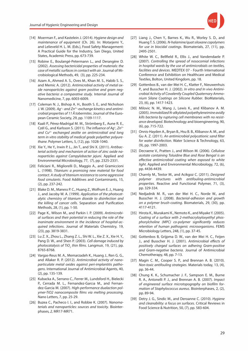
Journal of Hygienic Engineering and Design
29
[14] Moerman F., and Kastelein J. (2014). Hygiene design and maintenance of equipment (Ch. 26). In: Motarjemi Y., and Lelieveld H. L. M. (Eds.), Food Safety Management: A Practical Guide for the Industry, San Diego, United States, Academic Press, pp. 673-739.
[15] Robine E., Boulangé-Petermann L., and Derangère D. (2002). Assessing bactericidal properties of materials: the case of metallic surfaces in contact with air. Journal of Mi-crobiological Methods, 49, (3), pp. 225-234.
[16] Azam A., Ahmed A. S., Oves M., Khan M. S., Habib S. S., and Memic A. (2012). Antimicrobial activity of metal ox-ide nanoparticles against gram positive and gram neg-ative bacteria: a comparative study. Internal Journal of Nanomedicine, 7, pp. 6003-6009.
[17] Coleman N. J., Bishop A. H., Booth S. E., and Nicholson J. W. (2009). Ag+- and Zn2+-exchange kinetics and antimi-crobial properties of 11 Å tobermites. Journal of the Euro-pean Ceramic Society, 29, pp. 1109-1117.
[18] Kaali P., Pérez-Madrigal M. M., Strömberg E., Aune R. E., Czél G., and Karlsson S. (2011). The influence of Ag+, Zn2+ and Cu2+ exchanged zeolite on antimicrobial and long term in vitro stability of medical grade polyether polyure-thane. Polymer Letters, 5, (12), pp. 1028-1040.
[19] Xie Y., He Y., Irwin P. L., Jin T., and Shi X. (2011). Antibac-terial activity and mechanism of action of zinc oxide na-noparticles against Campylobacter jejuni. Applied and Environmental Microbiology, 77, (7), pp. 2325-2331.
[20] Feliciani R., Migliorelli D., Maggio A., and Gramiccioni L. (1998). Titanium: a promising new material for food contact. A study of titanium resistance to some aggressive food simulants. Food Additives and Contaminants, 15, (2), pp. 237-242.
[21] Blake D. M., Maness P. C., Huang Z., Wolfrum E. J., Huang J., and Jacoby W. A. (1999). Application of the photocat-alytic chemistry of titanium dioxide to disinfection and the killing of cancer cells. Separation and Purification Methods, 28, (1), pp. 1-50.
[22] Page K., Wilson M., and Parkin I. P. (2009). Antimicrobi-al surfaces and their potential in reducing the role of the inanimate environment in the incidence of hospital-ac-quired infections. Journal of Materials Chemistry, 19, (23), pp. 3819-3831.
[23] Lu Z. X., Zhou L., Zhang Z. L., Shi W. L., Xie Z. X., Xie H. Y., Pang D. W., and Shen P. (2003). Cell damage induced by photocatalysis of TiO2 thin films. Langmuir, 19, (21), pp. 8765-8768.
[24] Vargas-Reus M. A., Memarzadeh K., Huang J., Ren G. G., and Allaker R. P. (2012). Antimicrobial activity of nano-particulate metal oxides against peri-implantitis patho-gens. International Journal of Antimicrobial Agents, 40, (2), pp. 135-139.
[25] Kubacka A., Serrano C., Ferrer M., Lundsford H., Bielecki P., Cerrada M. L., Fernandez-Garcıa M., and Fernan-dez-Garcia M. (2007). High-performance dualaction pol-ymer-TiO2 nanocomposite films via melting processing. Nano Letters, 7, pp. 25-29.
[26] Buzea C., Pacheco I. I., and Robbie K. (2007). Nanoma-terials and nanoparticles: sources and toxicity. Biointer-phases, 2, MR17-MR71.
[27] Liang J., Chen Y., Barnes K., Wu R., Worley S. D., and Huang T. S. (2006). N-halamine/quat siloxane copolymers for use in biocidal coatings. Biomaterials, 27, (11), pp. 2495-2501.
[28] White W. C., Bellfield R., Ellis J., and Vandendaele P. (2007). Controlling the spread of nosocomial infections in hospital wards by the use of antimicrobials on textiles, facilities and devices. MEDTEX 07 - Fourth International Conference and Exhibition on Healthcare and Medical Textiles, Bolton, United Kingdom, pp. 18.
[29] Gottenbos B., van der Mei H. C., Klatter F., Nieuwenhuis P., and Busscher H. J. (2002). In vitro and In vivo Antimi-crobial Activity of Covalently Coupled Quaternary Ammo-nium Silane Coatings on Silicone Rubber. BioMaterials, 23, (6), pp. 1417-1423.
[30] Milovic N. M., Wang J., Lewis K., and Klibanov A. M. (2005). Immobilized N-alkylated polyethyleneimine avidly kills bacteria by rupturing cell membranes with no resist-ance developed. Biotechnology and bioengineering, 90, (6), pp. 715-722.
[31] Onnis-Hayden A., Bryan B., Hsu B. B., Klibanov A. M., and Gu A. Z. (2011). An antimicrobial polycationic sand filter for water disinfection. Water Science & Technology, 63, (9), pp. 1997-2003.
[32] Decraene V., Pratten J., and Wilson M. (2006). Cellulose acetate containing Toluidine Blue and Rose Bengal is an effective antimicrobial coating when exposed to white light. Applied and Environmental Microbiology, 72, (6), pp. 4436-4439.
[33] Charnly M., Textor M., and Acikgoz C. (2011). Designed polymer structures with antifouling-antimicrobial properties. Reactive and Functional Polymer, 71, (3), pp. 329-334.
[34] Nedjadnik M. R., van der Mei H. C., Norde W., and Busscher H. J. (2008). Bacterial-adhesion and growth on a polymer brush-coating. Biomaterials, 29, (30), pp. 4117-4121.
[35] Hirota K., Murakami K., Nemoto K., and Miyake Y. (2005). Coating of a surface with 2-methacryloyloxyethyl phos-phorylcholine (MPC) co-polymer significantly reduces retention of human pathogenic microorganisms. FEMS Microbiology Letters, 248, (1), pp. 37-45.
[36] Gottenbos B, Grijpma D. W., van der Mei H. C., Feijen J., and Busscher H. J. (2001). Antimicrobial effects of positively charged surfaces on adhering Gram-positive and Gram-negative bacteria. Journal of Antimicrobial Chemotherapy, 48, pp. 7-13.
[37] Magin C. M., Cooper S. P., and Brennan A. B. (2010). Non-toxic antifouling strategies. Materials today, 13, (4), pp. 36-44.
[38] Chung K. K., Schumacher J. F., Sampson E. M., Burne R. A., Antonelli P. J., and Brennan A. B. (2007). Impact of engineered surface microtopography on biofilm for-mation of Staphylococcus aureus. Biointerphases, 2, (2), pp. 89-94.
[39] Detry J. G., Sindic M., and Deroanne C. (2010). Hygiene and cleanability: a focus on surfaces. Critical Reviews in Food Science & Nutrition, 50, (7), pp. 583-604.