antileishmanial activity of meroditerpenoids from the...
TRANSCRIPT
lable at ScienceDirect
Experimental Parasitology 174 (2017) 1e9
Contents lists avai
Experimental Parasitology
journal homepage: www.elsevier .com/locate /yexpr
Full length article
Antileishmanial activity of meroditerpenoids from the macroalgaeCystoseira baccata
Carolina Bruno de Sousa a, Katkam N. Gangadhar a, b, Thiago R. Morais c,Geanne A.A. Conserva c, Catarina Vizetto-Duarte a, Hugo Pereira a, M�arcia D. Laurenti d,Lenea Campino e, f, Debora Levy g, Miriam Uemi c, Luísa Barreira a, Luísa Cust�odio a,Luiz Felipe D. Passero h, Jo~ao Henrique G. Lago c, **, Jo~ao Varela a, *
a Centro de Ciencias do Mar, Universidade do Algarve, Campus de Gambelas, Faro, Portugalb Instituto de Tecnologia Química e Biol�ogica, Universidade Nova de Lisboa, Oeiras, Portugalc Departamento de Ciencias Exatas e da Terra, Instituto de Ciencias Ambientais, Químicas e Farmaceuticas, Universidade Federal de S~ao Paulo, Diadema, SP,Brazild Laborat�orio de Patologia das Mol�estias Infecciosas (LIM-50), Departamento de Patologia, Faculdade de Medicina, Universidade de S~ao Paulo, S~ao Paulo,Brazile Global Health and Tropical Medicine Centre, Instituto de Higiene e Medicina Tropical, Universidade Nova de Lisboa, Lisboa, Portugalf Departamento de Ciencias Biom�edicas e Medicina, Universidade do Algarve, Campus de Gambelas, Faro, Portugalg Laborat�orio de Gen�etica e Hematologia Molecular (LIM-31), Departamento de Clinica M�edica, Faculdade de Medicina, Universidade de S~ao Paulo, S~aoPaulo, Brazilh S~ao Paulo State University (UNESP), Institute of Biosciences, S~ao Vicente, Praça Infante Dom Henrique, s/n, 11330-900 S~ao Vicente, SP, Brazil
h i g h l i g h t s
* Corresponding author. Centre of Marine Sciences,** Corresponding author. Departamento de Ciencias09972-270, Diadema, SP, Brazil.
E-mail addresses: [email protected] (J.H.G. Lago
http://dx.doi.org/10.1016/j.exppara.2017.01.0020014-4894/© 2017 Elsevier Inc. All rights reserved.
g r a p h i c a l a b s t r a c t
� Tetraprenyltoluquinols and tetrapre-nylquinones from Cystoseira baccata.
� Tetraprenyltoluquinols displayedantileishmanial activity.
� Tetraprenyltoluquinols induce alter-ations on promastigotes morphology.
� Tetraprenyltoluquinol disrupt theLeishmania mitochondrial membranepotential.
a r t i c l e i n f o
Article history:Received 25 May 2016Received in revised form10 November 2016Accepted 22 January 2017Available online 24 January 2017
Keywords:Leishmania infantumMacroalgaeCystoseira baccataMeroterpenoidsTetraprenyltoluquinolTetraprenyltoluquinone
a b s t r a c t
The development of novel drugs for the treatment of leishmaniases continues to be crucial to overcomethe severe impacts of these diseases on human and animal health. Several bioactivities have beendescribed in extracts from macroalgae belonging to the Cystoseira genus. However, none of the studieshas reported the chemical compounds responsible for the antileishmanial activity observed upon in-cubation of the parasite with the aforementioned extracts. Thus, this work aimed to isolate and char-acterize the molecules present in a hexane extract of Cystoseira baccata that was found to be bioactiveagainst Leishmania infantum in a previous screening effort. A bioactivity-guided fractionation of theC. baccata extract was carried out and the inhibitory potential of the isolated compounds was evaluatedvia the MTT assay against promastigotes and murine macrophages as well as direct counting againstintracellular amastigotes. Moreover, the promastigote ultrastructure, DNA fragmentation and changes inthe mitochondrial potential were assessed to unravel their mechanism of action. In this process, twoantileishmanial meroditerpenoids, (3R)- and (3S)-tetraprenyltoluquinol (1a/1b) and (3R)- and (3S)-
University of Algarve, Campus de Gambelas, 8005-139 Faro, Portugal.Exatas e da Terra, Instituto de Ciencias Ambientais, Químicas e Farmaceuticas, Universidade Federal de S~ao Paulo,
), [email protected] (J. Varela).
C. Bruno de Sousa et al. / Experimental Parasitology 174 (2017) 1e92
Abbreviations
BALB/c albino mouse laboratory-bredmouse
CC50 cytotoxic concentration that caof the viable cells
COSY correlation spectroscopyDEPT distortionless enhancement by
spectrometryFBS fetal bovine serumHMBC heteronuclear multiple-bond c
spectroscopyHRESIMShigh-resolution electrospray io
spectrometryHSQC heteronuclear single-quantum
spectroscopyIC50 half-maximal inhibitory conceIR infraredLRESIMS low-resolution electrospray ion
spectrometryMTT 3-(4,5-dimethylthiazol-2-yl)-2
diphenyltetrazolium bromideNMR nuclear magnetic resonance spNOESY nuclear Overhauser effect specRCF relative centrifugal forceSDS sodium dodecyl sulfateTLC thin-layer chromatographyTMS tetramethylsilaneUV ultravioletDJm mitochondrial membrane pote
tetraprenyltoluquinone (2a/2b), were isolated. Compounds 1 and 2 inhibited the growth of theL. infantum promastigotes (IC50 ¼ 44.9 ± 4.3 and 94.4 ± 10.1 mM, respectively), inducing cytoplasmicvacuolization and the presence of coiled multilamellar structures in mitochondria as well as an intensedisruption of the mitochondrial membrane potential. Compound 1 decreased the intracellular infectionindex (IC50 ¼ 25.0 ± 4.1 mM), while compound 2 eliminated 50% of the intracellular amastigotes at aconcentration > 88.0 mM. This work identified compound 2 as a novel metabolite and compound 1 as abiochemical isolated from Cystoseira algae displaying antileishmanial activity. Compound 1 can thus bean interesting scaffold for the development of novel chemotherapeutic molecules for canine and humanvisceral leishmaniases studies. This work reinforces the evidence of the marine environment as source ofnovel molecules.
© 2017 Elsevier Inc. All rights reserved.
strain of the house
uses the death of 50%
polarization transfer
orrelation
nisation mass
correlation
ntration
isation mass
,5-
ectroscopytroscopy
ntial
1. Introduction
Leishmaniases are a group of infectious diseases caused byobligate intracellular protozoa of the Leishmania genus. Endemic in98 tropical and subtropical countries and affecting 12 millionpeople, leishmaniases may entail cutaneous, mucocutaneous anddiffuse forms as well as the potentially fatal visceral form (Alvaret al., 2012). Visceral leishmaniasis causes considerable morbidityin 200e400 thousand individuals every year, with extremesuffering and financial loss, especially in the poorest populations ofthe Indian subcontinent (Mondal et al., 2014). Currently, leish-maniases are among the most neglected tropical diseases, facingproblems of resistance of the parasite to the available therapeuticmolecules. The need for the discovery and development of alter-native drugs allowing more efficient and effective treatments is
thus quite urgent (Freitas-Junior et al., 2012).Nowadays, marine natural products are recognized as powerful
reservoirs of novel, chemically diverse molecules with wideapplicability to health sciences (Tempone et al., 2011). Occurringworldwide, mainly in the rocky substrates of the MediterraneanSea and the adjoining Atlantic coasts, Cystoseira C. Agardh (1820)genus encompasses approximately 40 species of brownmacroalgae(Guiry and Guiry, 2015). Several bioactivities such as anti-inflammatory, antiproliferative, antioxidant (Mhadhebi et al.,2011), enzyme inhibitory (Ghannadi et al., 2013), cytotoxic(Khanavi et al., 2010), antifungal (Lopes et al., 2013), antiviral(Ibraheem et al., 2012), antibacterial (Tajbakhsh et al., 2011) andantiprotozoal (Spavieri et al., 2010) have been detected in this algalgenus. Despite the extensive chemical studies available for theCystoseira genus, there have been only a few reports describing theantileishmanial potential effects of its crude extracts, and no in-formation was found on the compounds responsible for theinhibitory effects on the Leishmania parasites (Amico, 1995; de LosReyes et al., 2012). As part of ongoing research on the identificationof antileishmanial compounds from the Cystoseira genus, this workdescribes the bioactivity-guided fractionation of the hexane extractfrom Cystoseira baccata and the effect of the extract, fractions andisolated compounds on the promastigote and amastigote forms ofLeishmania infantum.
2. Material and methods
2.1. General experimental procedures
Optical rotations were measured in a JASCO DIP-370 digitalpolarimeter (Na filter, l¼ 588 nm). UV spectrawere recorded usinga UV/visible Shimadzu 1650-PC spectrophotometer. IR spectrawereobtained with a Shimadzu IR Prestige-21 spectrophotometer. 1H,13C, DEPT, COSY, HSQC, HMBC and NOESY NMR spectra wererecorded in a Bruker Avance III 500 spectrometer, operating at 500and 125MHz, to 1H and 13C nuclei, respectively. CDCl3 (Aldrich) wasused as the solvent with TMS as the internal standard. HRESIMSspectra were measured with a Bruker Daltonics MicroTOF QIIspectrometer while LRESIMS spectra were recorded on a VG Plat-form II spectrometer. Silica gel (Merck, 230e400 mesh) andSephadex LH-20 (Amersham Biosciences) were used for columnchromatographic separation, while silica gel 60 PF254 (Merck) wasused for analytical (0.25 mm) and preparative TLC (1.0 mm).
2.2. Algal material
Cystoseira baccata biomass was collected in July 2012 in Areosa,Viana do Castelo, Portugal (41�42027.6000N, 8�51044.9000W). Aftercollection, biomass was cleaned and cryodesiccated. Voucherspecimen (MB-1) was deposited within the Laboratory of the
C. Bruno de Sousa et al. / Experimental Parasitology 174 (2017) 1e9 3
Marine Biotechnology Group - MarBiotech at the Centre of theMarine Sciences of the University of Algarve (Faro, Portugal).
2.3. Extraction and isolation of compounds
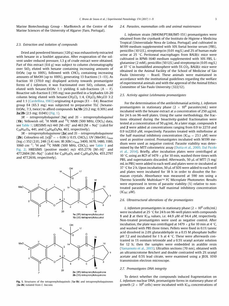
Dried and powdered biomass (120 g)was exhaustively extractedwith hexane in a Soxhlet apparatus. After evaporation of the sol-vent under reduced pressure, 1.3 g of crude extract were obtained.Part of this extract (0.6 g) was subject to column chromatographyover SiO2 eluted with hexane containing increasing amounts ofEtOAc (up to 100%), followed with CHCl3 containing increasingamounts of MeOH (up to 100%), generating 13 fractions (1e13). Asfraction 10 (370.0 mg) displayed activity towards promastigoteforms of L infantum, it was fractionated over SiO2 column, andeluted with hexane:EtOAc 1:1 yielding 6 sub-fractions (A e F).Bioactive sub-fraction E (195 mg) was purified in a Sephadex LH-20column being eluted with hexane:CH2Cl2 1:4, CH2Cl2:Me2CO 3:2and 1:1 (Cardellina, 1983) originating 4 groups (E1 e E4). Bioactivegroup E4 (65.3 mg) was subjected to preparative TLC (hexane-EtOAc, 7:3, twice) to afford compounds 1a/1b (23.2 mg; 0.30%) and2a/2b (2.5 mg; 0.04%) (Fig. 1).
3R e tetraprenyltoluquinol (1a) and 3S e tetraprenyltoluquinol(1b). Yellowish oil; 1H NMR and 13C NMR (500 MHz, CDCl3) data,see Table 1; LRESIMSm/z 441 [MþH]þ and 463 [Mþ Na]þ (calcd forC28H41O4, 441, and C28H40O4Na, 463, respectively).
3Re tetraprenyltoluquinone (2a) and 3Se tetraprenyltoluquinone(2b). Colourless oil; [a]D25 ¼ þ 0.06 (c 0.15, CHCl3); UV (MeOH) lmax(log ε) 352 (2.0), 248 (3.4) nm; IR (KBr) nmax 3400, 1670, 1480, 1180,1060 cm�1; 1H and 13C NMR (500 MHz, CDCl3), see Table 1 andFig. 2; HRESIMS (positive mode) m/z 455.2776 [MþH]þ and477.2604 [MþNa]þ (calcd for C28H39O5 and C28H38O5Na, 455.2797and 477.2616, respectively).
Fig. 1. Structures of the tetraprenyltoluquinols (1a-1b) and tetraprenyltoluquinones(2a-2b) isolated from C. baccata.
2.4. Parasites, mammalian cells and animal maintenance
L. infantum strain (MHOM/PT/88/IMT-151) promastigotes wereobtained from the cryobank of the Instituto de Higiene e MedicinaTropical (Universidade Nova de Lisboa, Portugal) and cultivated inM199 medium supplemented with 10% foetal bovine serum (FBS),penicillin (10 U/L), streptomycin (0.01 mg/L) and 2% of humanmaleurine at 25 �C. Peritoneal macrophages from BALB/c mice werecultivated in RPMI-1640 medium supplemented with 10% FBS, L-glutamine (2 mM), penicillin (50 U/L) and streptomycin (0.05 mg/L)at 37 �C in humidified atmosphere with 5% CO2. BALB/c mice wereobtained in the Animal Facility of the School of Medicine of S~aoPaulo University e Brazil. These animals were maintained inaccordance with the institutional guidelines regarding the welfareof experimental animals andwith the approval of the Animal EthicsCommittee of S~ao Paulo University (322/12).
2.5. Activity against Leishmania promastigotes
For the determination of the antileishmanial activity, L. infantumpromastigotes in stationary phase (2 � 106 parasites/mL) wereincubated with the hexane extract at a concentration of 250 mg/mLfor 24 h on 96-well plates. Using the same methodology, the frac-tions obtained during the bioactivity-guided fractionation weretested at a concentration of 50 mg/mL. At a later stage, compounds 1and 2 were added at concentrations ranging from 0.9 to 227.0 and0.9 to220.0 mM, respectively. Parasites treated with miltefosine atthe half maximal inhibitory concentration (IC50 ¼ 23.1 mM) wereused as positive control. Promastigotes incubated with M199 me-dium were used as negative control. Parasite viability was deter-mined by the MTT colorimetric assay (Dutta et al., 2005; Dal Picoloet al., 2014). Briefly, after incubation plates were centrifuged at10 �C, using an RCF of 1479� g for 10 min, washed three times withPBS, and supernatants discarded. Afterwards, 50 mL of MTT (5 mg/mL in PBS) were added to eachwell and plates were re-incubated at37 �C for 2 h. Upon incubation, 50 mL of SDSwere added to eachwelland plates were incubated for 18 h in order to dissolve the for-mazan crystals. Absorbance was measured at 590 nm using aThermo Scientific Multiskan™ FC Microplate Photometer. Resultswere expressed in terms of parasite viability (%) relative to non-treated parasites and the half maximal inhibitory concentration(IC50; mM).
2.6. Ultrastructural alterations of the promastigotes
L. infantum promastigotes in stationary phase (2 � 106 cells/mL)were incubated at 25 �C for 24 h on 96-well plates with compounds1 and 2 at their IC50 values, i.e. 44.9 mM of 94.4 mM, respectively.Non-treated promastigotes were used as negative control. Afterincubation, the plate was centrifuged at 1479 � g for 10 min at 4 �C,and washed with PBS three times. Pellets were fixed in 0.1% tannicacid dissolved in 2.0% glutaraldehyde in a 0.15 M phosphate bufferpH 7.2 and incubated for 1 h at 4 �C. These were afterwards con-trasted in 1% osmium tetroxide and a 0.5% uranyl acetate solutionfor 12 h; then the samples were embedded in araldite resin(Yamamoto et al., 2015). Ultrathin sections (70 nm), obtained withan ultramicrotome Reichert and double contrasted with 2% uranylacetate and 0.5% lead citrate, were examined using a JEOL 1010transmission electron microscope.
2.7. Promastigotes DNA integrity
To detect whether the compounds induced fragmentation onL. infantum nuclear DNA, promastigote forms in stationary phase ofgrowth (2 � 108 cells) were incubated with IC50 concentrations of

Table 11H and 13C NMR data (500 and 125 MHz, CDCl3, d/ppm) for compounds 1 (a/b) and 2 (a/b).
Compound 1a 1b 2a 2b
Position dC, type dH (J in Hz) dC, type dH (J in Hz) dC, type dH (J in Hz) dC, type dH (J in Hz)
1 22.7, CH2 2.79, (m) 22.6, CH2 2.79, (m) 192.2, C e 192.1, C e
2 32.5, CH2 1.80, (m) 33.6, CH2 1.80, (m) 47.8, CH2 2.56, (m) 48.6, CH2 2.57, (m)3 76.4, C e 76.2, C e 81.3, C e 81.2, C e
4 43.6, CH2 2.66, (s) 45.2, CH2 2.66, (s) 44.1, CH2 2.70, (s) 44.8, CH2 2.70, (s)5 153.7, C e 154.5, C e 153.3, C e 154.3, C e
6 44.3, CH2 2.57, (m) 44.7, CH2 2.57, (m) 44.1, CH2 2.60, (d, 4.0) 44.1, CH2 2.63, (d, 4.0)2.60, (m) 2.60, (m) 2.68, (d, 4.0) 2.68, (d, 4.0)
7 44.8, C e 44.8, C e 44.9, C e 44.9, C e
8 35.0, CH2 1.54, (m) 35.0, CH2 1.54, (m) 35.0, CH2 1.54, (m) 35.0, CH2 1.54, (m)1.73, (m) 1.73, (m) 1.73, (m) 1.73, (m)
9 18.8, CH2 1.74, (m) 18.8, CH2 1.74, (m) 18.8, CH2 1.74, (m) 18.8, CH2 1.74, (m)10 29.3, CH2 1.46, (m) 29.3, CH2 1.46, (m) 29.4, CH2 1.40, (d, 13.0) 29.7, CH2 1.42, (d, 13.0)11 54.9, C e 54.9, C e 54.9, C e 54.9, C e
12 208.5, C e 208.9, C e 208.0, C e 208.1, C e
13 132.9, C e 133.3, C e 133.5, C e 134.0, C e
14 39.4, CH2 2.45, (d, 15.0) 39.9, CH2 2.45, (d, 15.0) 39.4, CH2 2.54, (d, 15.0) 39.6, CH2 2.54, (d, 15.0)2.73, (d, 15.0) 2.73, (d, 15.0) 2.59, (d, 15.0) 2.59, (d, 15.0)
15 70.8, C e 71.1, C e 71.0, C e 71.0, C e
16 28.8, CH3 1.12, (s) 28.8, CH3 1.14, (s) 29.1, CH3 1.11, (s) 29.1, CH3 1.13, (s)17 30.5, CH3 1.24, (s) 31.6, CH3 1.19, (s) 30.8, CH3 1.25, (s) 31.3, CH3 1.26, (s)18 21.1, CH3 1.09, (s) 21.1, CH3 1.03, (s) 21.1, CH3 1.04, (s) 21.1, CH3 1.09, (s)19 22.4, CH3 0.91, (s) 22.5, CH3 0.83, (s) 22.4, CH3 0.83, (s) 22.5, CH3 0.91, (s)20 24.1, CH3 1.28, (s) 24.5, CH3 1.28, (s) 23.9, CH3 1.20, (s) 24.2, CH3 1.20, (s)10 145.2, C e 145.3, C e 167.8, C e 167.8, C e
20 120.4, C e 120.4, C e 119.6, C e 119.6, C e
30 111.1, CH 6.45, (d, 3.0) 111.2, CH 6.46, (d, 3.0) 114.6, CH 7.15, (d, 3.0) 114.6, CH 7.16, (d, 3.0)40 152.6, C e 152.6, C e 151.9, C e 151.9, C e
50 115.2, CH 6.59, (d, 3.0) 115.3, CH 6.60, (d, 3.0) 104.5, CH 7.00, (d, 3.0) 104.5, C 7.01, (d, 3.0)60 127.0, C e 127.2, C e 126.5, C e 126.5, C e
Me-60 16.6, CH3 2.16, (s) 16.8, CH 2.17, (s) 16.2, CH3 2.21, (s) 16.4, CH3 2.23, (s)OMe-40 55.6, CH3 3.73, (s) 55.6, CH 3.74, (s) 55.7, CH3 3.78, (s) 55.7, CH3 3.78, (s)
C. Bruno de Sousa et al. / Experimental Parasitology 174 (2017) 1e94
compounds 1 (44.9 mM), 2 (94.4 mM) and hydrogen peroxide(6.2 mM) as an inductor of DNA damage in parasites (Das et al.,2001) for 24 h at 25 �C. Non-treated cells were used as control.After incubation, plates were centrifuged at 1479 � g for 10 min at4 �C, and the supernatants discarded. Parasites pellets wereextracted with a Macherey-Nagel nucleoSpin® Blood kit accordingwith the manufacturer recommendations and ran on a 2% agarosegel, 100 V for 90 min.
2.8. Promastigote transmembrane mitochondrial potential
In order to evaluate the influence of compound 1 on the pro-mastigote mitochondrial membrane potential (DJm), parasites inthe stationary phase (2 � 106 parasites/mL) were incubated withcompound 1 and miltefosine at their IC50 values (44.9 and 23.1 mM,
Fig. 2. HMBC of the tetraprenyltoluquinones (2a-2b) isolated from C. baccata.
respectively) for 24 h on 96-well plates. Mitochondrial membranepotential was evaluated using the widefield automated microscopeMitoscreen Kit (BD Biosciences) according to the manufacturer'srecommendations (Levy et al., 2014; Yamamoto et al., 2015). Briefly,cells were incubated with working solution, containing the JC-1(5,5,6,6-tetrachloro-1,1,3,3-tetraethylbenzimidazolylcarbocyanineiodide) fluorochrome, for 15 min at 37 �C in an atmosphere of 5%CO2. DJm induces the uptake of JC-1monomers into the functionalmitochondria. Once inside the organelle, JC-1monomers aggregate,exhibiting high levels of red fluorescence and DJm is assessedthrough the determination of the presence of JC-1 fluorochromeinside the mitochondria. ImageXpress® Micro XLS Widefield High-Content Analysis System and transfluor MetaXpress software wereused to determine the presence of J-aggregates in nine sites perwell and three wells per treatment. DJm was expressed as a per-centage of J-aggregates per cell.
2.9. Cytotoxicity against murine macrophages
To determine the compounds toxicity in vitro, murine peritonealmacrophages, were seeded in RPMI-1640 at a density of 106 cells/mL and incubated overnight at 37 �C in humidified atmospherewith 5% CO2, allowing the cells to adhere to the plate background.Compounds 1 and 2 were tested for 24 h at concentrations rangingfrom 0.9 to 227.0 and 0.9e220.0 mM, respectively. Miltefosinecontrol cells were incubated with RPMI-1640 medium at concen-trations from 3.8 up to 490.7 mM. Cell viability was evaluated by theMTT colorimetric assay (Ferrari et al., 1990; Dal Picolo et al., 2014),as described above, for the determination of the activity againstLeishmania promastigotes. Absorbance was measured at 590 nmusing a Thermo Scientific Multiskan™ FC Microplate Photometer.Results were expressed in terms of the cytotoxic concentration

Table 2Effect of the compounds 1 and 2 against L. infantum promastigotes and intracellular amastigotes and mouse peritoneal macrophages.
Compounds Promastigotesa Intracellular amastigotesa Peritoneal macrophagesb SIc
1 44.9 ± 4.3 25.0 ± 4.1 126.6 ± 21.1 5.042 94.4 ± 10.1 >88.0 84.5 ± 12.5 <0.96Miltefosine 23.1 ± 0.0 20.3 ± 1.3 130.3 ± 17.2 6.42
a IC50 - Half maximal inhibitory concentration in mM.b CC50 - Cytotoxic concentration that causes the death of 50% of the viable cells in mM.c SI e Selectivity index concerning the activity against the intracellular amastigotes.
C. Bruno de Sousa et al. / Experimental Parasitology 174 (2017) 1e9 5
causing a 50% decrease in cell viability (CC50; mM) relative to non-treated cells (100%).
2.10. Activity against Leishmania intracellular amastigotes and NOproduction
Peritoneal macrophages of BALB/c mice were collected byintraperitoneal lavage, seeded on 24-well plates (105 cells/mL) andincubated at 37 �C with 5% CO2 during 2 h for cell attachment.Afterwards, L. infantum promastigotes in stationary phase wereadded to each well at an infection ratio of 10 promastigotes per cell,being further incubated at 37 �C for 24 h. Infected macrophageswere treated with compounds 1 and 2 at concentrations rangingfrom 7 to 90 mM to determine the corresponding IC50. Supernatantswere collected for nitric oxide (NO) determination after 24 h andintracellular amastigote burdenwas microscopically assessed uponGiemsa staining for determination of the infection index [% ofinfected macrophages � internalized amastigote forms/macro-phage)] (Passero et al., 2015) and the inhibitory concentrationallowing 50% reduction of the infection index (IC50) was estimated.Miltefosine was used as positive control. Culture supernatants oftreated and control macrophages were used for NO determinationthat was performed using theMeasure-iT™High-Sensitivity NitriteAssay Kit in accordance with the manufacturer's recommendations(Life Technologies). The NO concentration was determined using acalibration curve prepared with several known concentrations(2.75, 5.5, 11, 22, 33, 44 and 55 mM) of nitrite as standard. Resultswere expressed as NO production (mM) and compared with un-treated infected and non-infected macrophages. The selectivityindex (SI) was obtained by calculating the ratio of the CC50 of themacrophage by the IC50 of the intracellular amastigotes.
2.11. Statistical analysis
Bioassays results were expressed as mean ± standard error ofthe mean (SEM) of replicates samples from at least two indepen-dent assays. The IC50 values were calculated fitting the data as anon-linear regression using a dose-response inhibitory model, inthe GraphPad Prism V 5.0 program. Student's t-test was used todetermine whether differences between means were significant atdifferent levels (p < 0.05 and p < 0.01).
3. Results and discussion
The hexane extract from the C. baccata was incubated withpromastigote forms of L. infantum for 24 h, and cell viability wasdetermined by means of the MTT assay. As this extract decreasedthe viability of the parasite by 74% at a concentration of 250 mg/mL,it was selected for further study. Bioactivity-guided fractionationafforded compounds 1 and 2 (Fig. 1).
Compound 1 was obtained as an optically active oil [a]D ¼ þ17.8� (CHCl3, c 2.7). Structural evidence was obtained by analysis ofNMR (1H, 13C and DEPT 135�), HREIMS spectra and comparisonwiththose data previously reported in the literature to (3R)-(1a) and
(3S)-(1b) tetraprenyltoluquinol, previously isolated from C. baccata(Valls et al., 1993). In addition, some corrections in the attributionsof chemical shifts of C-18 and C-19 in 13C NMR spectrum werecarried out, based on the HMBC spectral analysis (Table 1). Com-pound 2, also obtained as an optically active colourless oil [a]D ¼ þ0.06� (CHCl3, c 0.15), appeared to be homogeneous on the TLCchromatograms, revealing that it is a mixture of closely relatedderivatives. The 1H NMR spectrum of compound 2 revealed somesimilarities with compound 1 - two peaks assigned to hydrogens ofaromatic ring at dH 7.15 (d, J ¼ 3.0 Hz, H-30) and 7.00 (d, J ¼ 3.0 Hz,H-50), one methoxyl group at dH 3.78 (s) as well as five singletsassigned to methyl groups at dH 1.20 (H-20), 1.25/1.26 (H-17), 1.13/1.11 (H-16), 1.09/1.04 (H-18), and 0.91/0.83 (H-19). 13C and DEPT135� NMR spectra confirmed the presence of aromatic ring due thepeaks at range dC 151.9e114.6 (C-1’ e C-60), and one methoxylgroup at dC 55.7. Additionally, peaks assigned to a carbonyl group atdC 192.2/192.1 (C-1), to carbinolic carbons at dC 81.3/81.2 (C-3) and71.0 (C-15) as well as an a,b-unsaturated carbonyl carbon at dC153.3/154.3 (C-5), 133.5/134.0 (C-13) and 208.0/208.1 (C-12) wereobserved. Finally, HRESIMS showed the [MþH]þ and [M þ Na]þ
quasi-molecular ion peaks at m/z 455.2776 and 477.2604, respec-tively, indicating the molecular formula C28H38O5. The connectivitybetween hydrogens and carbon atoms was revealed by analysis ofthe HMBC spectrum as showed in Fig. 2. The correlations betweensignals at dH 7.15 (H-3’) and 2.56/2.57 (H-2) with dC 192.2/192.1 (C-1) as well as between dH 2.70 (H-4) with dC 81.3/81.2 (C-3) and133.5/134.0 (C-13) indicated that compound 2 contained oneadditional carbonyl group at C-1. Based on these results, it waspossible to identify 2 as epimers of (3R)-(2a) and (3S)-(2b)tetraprenyltoluquinones.
In vitro antiparasitic activity and cytotoxic studies of the com-pounds 1 and 2 were evaluated by the colorimetric MTT methodagainst promastigote forms of L. infantum and murine macro-phages, respectively (Table 2). Compound 1 displayed an IC50 valueof 44.9 ± 4.3 mM against promastigote forms of L. infantum. Thecytotoxicity against mouse peritoneal macrophages(CC50 ¼ 126.6 ± 21.1 mM) was similar to that of the reference drug,miltefosine (130.3 ± 17.2 mM). Compound 2 showed lower activityagainst the promastigote forms (IC50 ¼ 94.4 ± 10.1 mM), and highertoxicity to the mouse peritoneal macrophages(CC50 ¼ 84.5 ± 12.5 mM).
To assess the alterations induced by the compounds on thepromastigotes forms of L. infantum, transmission electron micro-scopy images were acquired (Fig. 3). Important changes wereobserved with both treatments, including loss of the typical fusi-form shape (Fig. 3A). Ultrastructural analysis revealed morphologicchanges in parasites treated with the IC50 concentrations of bothcompounds 1 (Fig. 3B and C) and 2 (Fig. 3D and E). Moreover,cellular vacuolizationwas observed, whichmight be a consequenceof cytoplasmic organelle disruption (Fig. 3B and D). When treatedwith compound 1, parasites presented coiled multilamellar struc-tures within the mitochondria (Fig. 3C). These structures have beenshown to be a consequence of starvation processes caused bydeficient mitochondrial activity or autophagic mechanisms caused
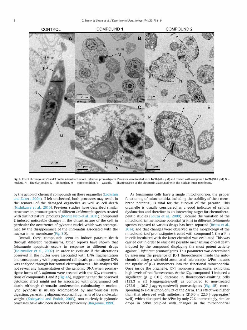
Fig. 3. Effect of compounds 1 and 2 on the ultrastructure of L. infantum promastigotes. Parasites were treated with 1a/1b (44.9 mM) and treated with compound 2a/2b (94.4 mM). N e
nucleus, FP - flagellar pocket, K e kinetoplast, M e mitochondrion, V e vacuole, * - disappearance of the chromatin associated with the nuclear inner membrane.
C. Bruno de Sousa et al. / Experimental Parasitology 174 (2017) 1e96
by the action of chemical compounds on these organelles (Lockshinand Zakeri, 2004). If left unchecked, both processes may result inthe removal of the damaged organelles as well as cell death(Nishikawa et al., 2010). Previous studies have described similarstructures in promastigotes of different Leishmania species treatedwith distinct natural products (Monte Neto et al., 2011). Compound2 induced noticeable changes in the ultrastructure of the cell, inparticular the occurrence of pyknotic nuclei, which was accompa-nied by the disappearance of the chromatin associated with thenuclear inner membrane (Fig. 3D).
Overall, these compounds seem to induce parasite deaththrough different mechanisms. Other reports have shown thatLeishmania apoptosis occurs in response to different drugs(Holzmuller et al., 2002). In order to evaluate if the alterationsobserved in the nuclei were associated with DNA fragmentationand consequently with programmed cell death, promastigote DNAwas analysed through horizontal electrophoresis. This analysis didnot reveal any fragmentation of the genomic DNA when promas-tigote forms of L. infantum were treated with the IC50 concentra-tions of compounds 1 and 2 (Fig. 4A), suggesting that the observedcytotoxic effect might not be associated with programmed celldeath. Although chromatin condensation culminating in nucleo-lytic pyknosis is usually accompanied by macronuclear DNAdigestion, generating oligonucleosomal fragments of lowmolecularweight (Kobayashi and Endoh, 2003), non-nucleolytic pyknoticprocesses have also been described previously (Burgoyne, 1999).
As Leishmania cells have a single mitochondrion, the properfunctioning of mitochondria, including the stability of their mem-brane potential, is vital for the survival of the parasite. Thisorganelle is usually considered as a good indicator of cellulardysfunction and therefore is an interesting target for chemothera-peutic studies (Souza et al., 2009). Because the variation of themitochondrial membrane potential (DJm) in different Leishmaniaspecies exposed to various drugs has been reported (Britta et al.,2014) and that changes were observed in the morphology of themitochondria of promastigotes treated with compound 1, the DJmin cells incubated with the latter chemical was evaluated. This wascarried out in order to elucidate possible mechanisms of cell deathinduced by the compound displaying the most potent activityagainst L. infantum promastigotes. This parameter was determinedby assessing the presence of JC-1 fluorochrome inside the mito-chondria using a widefield automated microscope. DJm inducesthe uptake of JC-1 monomers into the functional mitochondria.Once inside the organelle, JC-1 monomers aggregate, exhibitinghigh levels of red fluorescence. At the IC50, compound 1 induced asignificant (p � 0.01) decrease in fluorescence-emitting cells(133.3 ± 8.5 J-aggregates/well) as compared to non-treated(762.5 ± 36.7 J-aggregates/well) promastigotes (Fig. 4B), corre-sponding to a disruption of 83% of the DJm. This effect was higherthan that observed with miltefosine (216.0 ± 22.6 J-aggregates/well), which disrupted the DJm by only 72%. Interestingly, similardrops in DJm coupled with changes in the mitochondrial
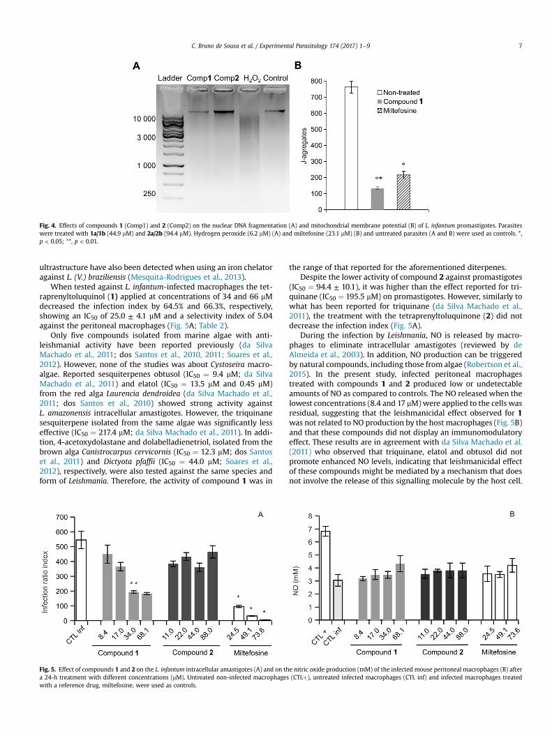
Fig. 4. Effects of compounds 1 (Comp1) and 2 (Comp2) on the nuclear DNA fragmentation (A) and mitochondrial membrane potential (B) of L. infantum promastigotes. Parasiteswere treated with 1a/1b (44.9 mM) and 2a/2b (94.4 mM). Hydrogen peroxide (6.2 mM) (A) and miltefosine (23.1 mM) (B) and untreated parasites (A and B) were used as controls. *,p < 0.05; **, p < 0.01.
C. Bruno de Sousa et al. / Experimental Parasitology 174 (2017) 1e9 7
ultrastructure have also been detected when using an iron chelatoragainst L. (V.) braziliensis (Mesquita-Rodrigues et al., 2013).
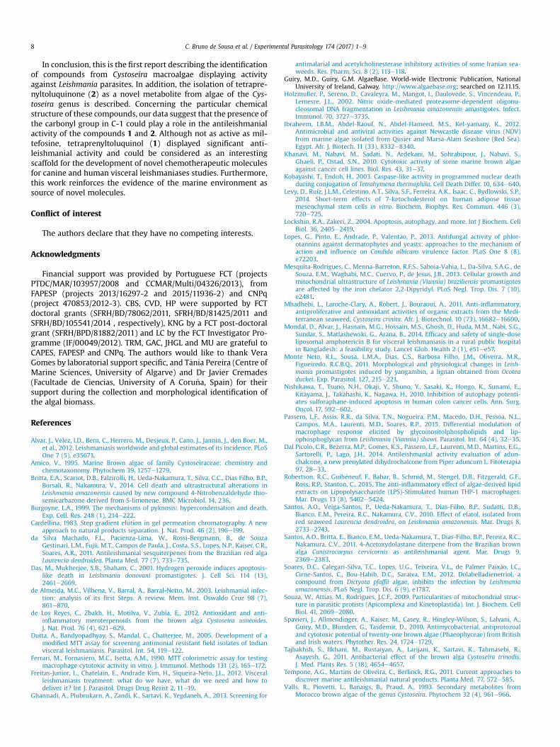
When tested against L. infantum-infected macrophages the tet-raprenyltoluquinol (1) applied at concentrations of 34 and 66 mMdecreased the infection index by 64.5% and 66.3%, respectively,showing an IC50 of 25.0 ± 4.1 mM and a selectivity index of 5.04against the peritoneal macrophages (Fig. 5A; Table 2).
Only five compounds isolated from marine algae with anti-leishmanial activity have been reported previously (da SilvaMachado et al., 2011; dos Santos et al., 2010, 2011; Soares et al.,2012). However, none of the studies was about Cystoseira macro-algae. Reported sesquiterpenes obtusol (IC50 ¼ 9.4 mM; da SilvaMachado et al., 2011) and elatol (IC50 ¼ 13.5 mM and 0.45 mM)from the red alga Laurencia dendroidea (da Silva Machado et al.,2011; dos Santos et al., 2010) showed strong activity againstL. amazonensis intracellular amastigotes. However, the triquinanesesquiterpene isolated from the same algae was significantly lesseffective (IC50 ¼ 217.4 mM; da Silva Machado et al., 2011). In addi-tion, 4-acetoxydolastane and dolabelladienetriol, isolated from thebrown alga Canistrocarpus cervicornis (IC50 ¼ 12.3 mM; dos Santoset al., 2011) and Dictyota pfaffii (IC50 ¼ 44.0 mM; Soares et al.,2012), respectively, were also tested against the same species andform of Leishmania. Therefore, the activity of compound 1 was in
Fig. 5. Effect of compounds 1 and 2 on the L. infantum intracellular amastigotes (A) and on tha 24-h treatment with different concentrations (mM). Untreated non-infected macrophageswith a reference drug, miltefosine, were used as controls.
the range of that reported for the aforementioned diterpenes.Despite the lower activity of compound 2 against promastigotes
(IC50 ¼ 94.4 ± 10.1), it was higher than the effect reported for tri-quinane (IC50 ¼ 195.5 mM) on promastigotes. However, similarly towhat has been reported for triquinane (da Silva Machado et al.,2011), the treatment with the tetraprenyltoluquinone (2) did notdecrease the infection index (Fig. 5A).
During the infection by Leishmania, NO is released by macro-phages to eliminate intracellular amastigotes (reviewed by deAlmeida et al., 2003). In addition, NO production can be triggeredby natural compounds, including those from algae (Robertson et al.,2015). In the present study, infected peritoneal macrophagestreated with compounds 1 and 2 produced low or undetectableamounts of NO as compared to controls. The NO released when thelowest concentrations (8.4 and 17 mM)were applied to the cells wasresidual, suggesting that the leishmanicidal effect observed for 1was not related to NO production by the host macrophages (Fig. 5B)and that these compounds did not display an immunomodulatoryeffect. These results are in agreement with da Silva Machado et al.(2011) who observed that triquinane, elatol and obtusol did notpromote enhanced NO levels, indicating that leishmanicidal effectof these compounds might be mediated by a mechanism that doesnot involve the release of this signalling molecule by the host cell.
e nitric oxide production (mM) of the infected mouse peritoneal macrophages (B) after(CTLþ), untreated infected macrophages (CTL inf) and infected macrophages treated
C. Bruno de Sousa et al. / Experimental Parasitology 174 (2017) 1e98
In conclusion, this is the first report describing the identificationof compounds from Cystoseira macroalgae displaying activityagainst Leishmania parasites. In addition, the isolation of tetrapre-nyltoluquinone (2) as a novel metabolite from algae of the Cys-toseira genus is described. Concerning the particular chemicalstructure of these compounds, our data suggest that the presence ofthe carbonyl group in C-1 could play a role in the antileishmanialactivity of the compounds 1 and 2. Although not as active as mil-tefosine, tetraprenyltoluquinol (1) displayed significant anti-leishmanial activity and could be considered as an interestingscaffold for the development of novel chemotherapeutic moleculesfor canine and human visceral leishmaniases studies. Furthermore,this work reinforces the evidence of the marine environment assource of novel molecules.
Conflict of interest
The authors declare that they have no competing interests.
Acknowledgments
Financial support was provided by Portuguese FCT (projectsPTDC/MAR/103957/2008 and CCMAR/Multi/04326/2013), fromFAPESP (projects 2013/16297-2 and 2015/11936-2) and CNPq(project 470853/2012-3). CBS, CVD, HP were supported by FCTdoctoral grants (SFRH/BD/78062/2011, SFRH/BD/81425/2011 andSFRH/BD/105541/2014 , respectively), KNG by a FCT post-doctoralgrant (SFRH/BPD/81882/2011) and LC by the FCT Investigator Pro-gramme (IF/00049/2012). TRM, GAC, JHGL and MU are grateful toCAPES, FAPESP and CNPq. The authors would like to thank VeraGomes by laboratorial support specific, and Tania Pereira (Centre ofMarine Sciences, University of Algarve) and Dr Javier Cremades(Facultade de Ciencias, University of A Coru~na, Spain) for theirsupport during the collection and morphological identification ofthe algal biomass.
References
Alvar, J., Velez, I.D., Bern, C., Herrero, M., Desjeux, P., Cano, J., Jannin, J., den Boer, M.,et al., 2012. Leishmaniasis worldwide and global estimates of its incidence. PLoSOne 7 (5), e35671.
Amico, V., 1995. Marine Brown algae of family Cystoseiraceae: chemistry andchemotaxonomy. Phytochem 39, 1257e1279.
Britta, E.A., Scariot, D.B., Falzirolli, H., Ueda-Nakamura, T., Silva, C.C., Dias Filho, B.P.,Borsali, R., Nakamura, V., 2014. Cell death and ultrastructural alterations inLeishmania amazonensis caused by new compound 4-Nitrobenzaldehyde thio-semicarbazone derived from S-limonene. BMC Microbiol. 14, 236.
Burgoyne, L.A., 1999. The mechanisms of pyknosis: hypercondensation and death.Exp. Cell. Res. 248 (1), 214e222.
Cardellina, 1983. Step gradient elution in gel permeation chromatography. A newapproach to natural products separation. J. Nat. Prod. 46 (2), 196e199.
da Silva Machado, F.L., Pacienza-Lima, W., Rossi-Bergmann, B., de SouzaGestinari, L.M., Fujii, M.T., Campos de Paula, J., Costa, S.S., Lopes, N.P., Kaiser, C.R.,Soares, A.R., 2011. Antileishmanial sesquiterpenes from the Brazilian red algaLaurencia dendroidea. Planta Med. 77 (7), 733e735.
Das, M., Mukherjee, S.B., Shaham, C., 2001. Hydrogen peroxide induces apoptosis-like death in Leishmania donovani promastigotes. J. Cell Sci. 114 (13),2461e2669.
de Almeida, M.C., Vilhena, V., Barral, A., Barral-Netto, M., 2003. Leishmanial infec-tion: analysis of its first Steps. A review. Mem. Inst. Oswaldo Cruz 98 (7),861e870.
de Los Reyes, C., Zbakh, H., Motilva, V., Zubía, E., 2012. Antioxidant and anti-inflammatory meroterpenoids from the brown alga Cystoseira usneoides.J. Nat. Prod. 76 (4), 621e629.
Dutta, A., Bandyopadhyay, S., Mandal, C., Chatterjee, M., 2005. Development of amodified MTT assay for screening antimonial resistant field isolates of Indianvisceral leishmaniasis. Parasitol. Int. 54, 119e122.
Ferrari, M., Fornasiero, M.C., Isetta, A.M., 1990. MTT colorimetric assay for testingmacrophage cytotoxic activity in vitro. J. Immunol. Methods 131 (2), 165e172.
Freitas-Junior, L., Chatelain, E., Andrade Kim, H., Siqueira-Neto, J.L., 2012. Visceralleishmaniasis treatment: what do we have, what do we need and how todeliver it? Int J. Parasitol. Drugs Drug Resist 2, 11e19.
Ghannadi, A., Plubrukarn, A., Zandi, K., Sartavi, K., Yegdaneh, A., 2013. Screening for
antimalarial and acetylcholinesterase inhibitory activities of some Iranian sea-weeds. Res. Pharm. Sci. 8 (2), 113e118.
Guiry, M.D., Guiry, G.M. AlgaeBase. World-wide Electronic Publication, NationalUniversity of Ireland, Galway. http://www.algaebase.org; searched on 12.11.15.
Holzmuller, P., Sereno, D., Cavaleyra, M., Mangot, I., Daulovede, S., Vincendeau, P.,Lemesre, J.L., 2002. Nitric oxide-mediated proteasome-dependent oligonu-cleosomal DNA fragmentation in Leishmania amazonensis amastigotes. Infect.Immunol. 70, 3727e3735.
Ibraheem, I.B.M., Abdel-Raouf, N., Abdel-Hameed, M.S., Kel-yamany, K., 2012.Antimicrobial and antiviral activities against Newcastle disease virus (NDV)from marine algae isolated from Qusier and Marsa-Alam Seashore (Red Sea).Egypt. Afr. J. Biotech. 11 (33), 8332e8340.
Khanavi, M., Nabavi, M., Sadati, N., Ardekani, M., Sohrabipour, J., Nabavi, S.,Ghaeli, P., Ostad, S.N., 2010. Cytotoxic activity of some marine brown algaeagainst cancer cell lines. Biol. Res. 43, 31e37.
Kobayashi, T., Endoh, H., 2003. Caspase-like activity in programmed nuclear deathduring conjugation of Tetrahymena thermophila. Cell Death Differ. 10, 634e640.
Levy, D., Ruiz, J.L.M., Celestino, A.T., Silva, S.F., Ferreira, A.K., Isaac, C., Bydlowski, S.P.,2014. Short-term effects of 7-ketocholesterol on human adipose tissuemesenchymal stem cells in vitro. Biochem. Biophys. Res. Commun. 446 (3),720e725.
Lockshin, R.A., Zakeri, Z., 2004. Apoptosis, autophagy, and more. Int J Biochem. CellBiol. 36, 2405e2419.
Lopes, G., Pinto, E., Andrade, P., Valent~ao, P., 2013. Antifungal activity of phlor-otannins against dermatophytes and yeasts: approaches to the mechanism ofaction and influence on Candida albicans virulence factor. PLoS One 8 (8),e72203.
Mesquita-Rodrigues, C., Menna-Barreton, R.F.S., Saboia-Vahia, L., Da-Silva, S.A.G., deSouza, E.M., Waghabi, M.C., Cuervo, P., de Jesus, J.B., 2013. Cellular growth andmitochondrial ultrastructure of Leishmania (Viannia) braziliensis promastigotesare affected by the iron chelator 2,2-Dipyridyl. PLoS Negl. Trop. Dis. 7 (10),e2481.
Mhadhebi, L., Laroche-Clary, A., Robert, J., Bouraoui, A., 2011. Anti-inflammatory,antiproliferative and antioxidant activities of organic extracts from the Medi-terranean seaweed, Cystoseira crinita. Afr. J. Biotechnol. 10 (73), 16682e16690.
Mondal, D., Alvar, J., Hasnain, M.G., Hossain, M.S., Ghosh, D., Huda, M.M., Nabi, S.G.,Sundar, S., Matlashewski, G., Arana, B., 2014. Efficacy and safety of single-doseliposomal amphotericin B for visceral leishmaniasis in a rural public hospitalin Bangladesh: a feasibility study. Lancet Glob. Health 2 (1), e51ee57.
Monte Neto, R.L., Sousa, L.M.A., Dias, C.S., Barbosa Filho, J.M., Oliveira, M.R.,Figueiredo, R.C.B.Q., 2011. Morphological and physiological changes in Leish-mania promastigotes induced by yangambin, a lignan obtained from Ocoteaduckei. Exp. Parasitol. 127, 215e221.
Nishikawa, T., Tsuno, N.H., Okaji, Y., Shuno, Y., Sasaki, K., Hongo, K., Sunami, E.,Kitayama, J., Takahashi, K., Nagawa, H., 2010. Inhibition of autophagy potenti-ates sulforaphane-induced apoptosis in human colon cancer cells. Ann. Surg.Oncol. 17, 592e602.
Passero, L.F., Assis, R.R., da Silva, T.N., Nogueira, P.M., Macedo, D.H., Pessoa, N.L.,Campos, M.A., Laurenti, M.D., Soares, R.P., 2015. Differential modulation ofmacrophage response elicited by glycoinositolphospholipids and lip-ophosphoglycan from Leishmania (Viannia) shawi. Parasitol. Int. 64 (4), 32e35.
Dal Picolo, C.R., Bezerra, M.P., Gomes, K.S., Passero, L.F., Laurenti, M.D., Martins, E.G.,Sartorelli, P., Lago, J.H., 2014. Antileishmanial activity evaluation of adun-chalcone, a new prenylated dihydrochalcone from Piper aduncum L. Fitoterapia97, 28e33.
Robertson, R.C., Guih�eneuf, F., Bahar, B., Schmid, M., Stengel, D.B., Fitzgerald, G.F.,Ross, R.P., Stanton, C., 2015. The anti-inflammatory effect of algae-derived lipidextracts on Lipopolysaccharide (LPS)-Stimulated human THP-1 macrophages.Mar. Drugs 13 (8), 5402e5424.
Santos, A.O., Veiga-Santos, P., Ueda-Nakamura, T., Dias-Filho, B.P., Sudatti, D.B.,Bianco, E.M., Pereira, R.C., Nakamura, C.V., 2010. Effect of elatol, isolated fromred seaweed Laurencia dendroidea, on Leishmania amazonensis. Mar. Drugs 8,2733e2743.
Santos, A.O., Britta, E., Bianco, E.M., Ueda-Nakamura, T., Dias-Filho, B.P., Pereira, R.C.,Nakamura, C.V., 2011. 4-Acetoxydolastane diterpene from the Brazilian brownalga Canistrocarpus cervicornis as antileishmanial agent. Mar. Drugs 9,2369e2383.
Soares, D.C., Calegari-Silva, T.C., Lopes, U.G., Teixeira, V.L., de Palmer Paix~ao, I.C.,Cirne-Santos, C., Bou-Habib, D.C., Saraiva, E.M., 2012. Dolabelladienetriol, acompound from Dictyota pfaffii algae, inhibits the infection by Leishmaniaamazonensis. PLoS Negl. Trop. Dis. 6 (9), e1787.
Souza, W., Attias, M., Rodrigues, J.C.F., 2009. Particularities of mitochondrial struc-ture in parasitic protists (Apicomplexa and Kinetoplastida). Int. J. Biochem. CellBiol. 41, 2069e2080.
Spavieri, J., Allmendinger, A., Kaiser, M., Casey, R., Hingley-Wilson, S., Lalvani, A.,Guiry, M.D., Blunden, G., Tasdemir, D., 2010. Antimycobacterial, antiprotozoaland cytotoxic potential of twenty-one brown algae (Phaeophyceae) from Britishand Irish waters. Phytother. Res. 24, 1724e1729.
Tajbakhsh, S., Ilkhani, M., Rustaiyan, A., Larijani, K., Sartavi, K., Tahmasebi, R.,Asayesh, G., 2011. Antibacterial effect of the brown alga Cystoseira trinodis.J. Med. Plants Res. 5 (18), 4654e4657.
Tempone, A.G., Martins de Oliveira, C., Berlinck, R.G., 2011. Current approaches todiscover marine antileishmanial natural products. Planta Med. 77, 572e585.
Valls, R., Piovetti, L., Banaigs, B., Praud, A., 1993. Secondary metabolites fromMorocco brown algae of the genus Cystoseira. Phytochem 32 (4), 961e966.

C. Bruno de Sousa et al. / Experimental Parasitology 174 (2017) 1e9 9
Yamamoto, E.S., Campos, B.L., Jesus, J.A., Laurenti, M.D., Ribeiro, S.P., Kall�as, E.G.,Rafael-Fernandes, M., Santos-Gomes, G., Silva, M.S., Sessa, D.P., Lago, J.H.,Levy, D., Passero, L.F., 2015. The effect of ursolic acid on Leishmania (Leishmania)amazonensis is related to programed cell death and presents therapeutic po-tential in experimental cutaneous leishmaniasis. PLoS One 10 (12), e0144946.
Carolina Bruno-de-Sousa - MSc in Animal Production and Post-graduated in MedicalParasitology. As PhD student of the Centre of Marine Sciences at the Algarve University,is currently studding marine algae as source of bioactive molecules against Leishmaniaparasites. Main interests include animal parasitological studies and genetic character-ization of domestic animals and algae populations. Also collaborated in studies of para-sitic diseases with public health significance.
Katkam N. Gangadhar - Post-doctoral Research Fellow at CCMAR, University ofAlgarve. Works in synthetic/organic lipid medicinal chemistry and pharmaceutical ap-plications: (i) isolation of wound healing and anti-cancer bioactive compounds fromnatural products; (ii) synthesis of lipid carriers as drug delivery materials for anti-tuberculosis drug and Amphotericin-B; (iii) chemo-enzymatic synthesis of cetyl myris-toleates and diacylglycerol and evaluation of their anti-inflammatory, anti-arthriticand nutritional properties; (iv) development of carbon-based solid acid catalyst fromcrude glycerol for biodiesel production from microalgae and non-edible oils and (v)its application in organic methodologies.
Thiago R. Morais - PhD student of Chemical Biology at UNIFESP, working with NaturalProducts chemistry, especially with isolation and characterization of micromoleculesusing NMR and MS data analysis.
Geanne A. A. Conserva - MSc student at the Federal University of S~ao Paulo e UNIFESPe working with Chemistry of Natural Products, mainly in the search and character-ization of bioactive derivatives in plant species, particularly those with antitumoralactivity.
Catarina Vizetto-Duarte - PhD student at Centre of Marine Sciences (CCMAR) at theUniversity of Algarve. She has an MSc in Molecular Genetics and Biomedicine from theUniversity of Lisbon in 2009. As a PhD student she is evaluating the biomedical ap-plications (especially antioxidant and antitumoral properties) of brown algae, focusingon finding novel bioactive molecules and studying the molecular mechanismsresponsible for the said activities in terms of cellular responses to drug exposure,inflammation, cell death (apoptosis/necrosis) versus cell survival.
Hugo Pereira - MSc on Aquaculture and Fisheries, where he worked on the optimi-zation of a novel culture medium for large-scale production of microalgae in photo-bioreactors at Necton S.A. (Portugal). He is currently a PhD student aiming thedevelopment of an algal biorefinery for different biotechnological applications,including the determination of bioactivities to improve the added-value of algalbiomass
M�arcia D. Laurenti - PhD in Veterinary Pathology; full professor and head chief ofLaboratory of Pathology of Infectious Diseases, Department of Pathology, MedicalSchool, University of S~ao Paulo; with experience in the immunopathology of human,canine and experimental cutaneous and visceral leishmaniasis.
Lenea Campino - Full Professor in Medical Parasitology, at the Institute of Hygiene andTropical Medicine, Universidade Nova de Lisboa (IHMT/UNL). Main areas of interestare: leishmaniasis and Leishmania-HIV co-infections; molecular epidemiology, parasitediversity, vector/host-parasite interactions; immunology of the infection; natural andexperimental leishmaniasis models; vaccine and drug candidates; diagnostics onvisceral and cutaneous leishmaniasis; environmental changes and emerging parasiticdiseases. She led national and International research projects in those areas, super-vised several postgraduate degrees, and acted as a consultant for the Portuguese Na-tional Directorate of Health.
Debora Levy - PhD in medical science at Medical School of Sao Paulo University.Currently is scientific researcher at Laboratory of Genetics and Molecular Hematology,and has experience in hematology, genetics and drug development.
Miriam Uemi - Associate Professor at the Federal University of the State of S~ao Paulo -has experience in molecular characterization by nuclear magnetic resonance and massspectrometry.
Luísa Barreira - Assistant Professor of the Chemistry and Pharmacy Department of theFaculty of Sciences and Technology of the University of Algarve since 2007. She has PhDin Environmental Sciences and Technologies and is currently a senior researcher inMarBiotech for I þ D þ I of biotechnological applications of marine organisms, from theproduction of biodiesel and other bioproducts (e.g. phospholipids) from microalgae tothe search of natural products with biological activities in marine organisms.
Luísa Cust�odio - PhD in Biotechnological Sciences and carried out her post-doctoralresearch at the University of Algarve and CCMAR. Presently she is a research assistanthired by CCMAR under the frame of the FCT investigator programme and her researchhas focused on the search for bioactive compounds in marine organisms and halophytespecies, and the evaluation of the nutritional profile of edible organisms (e.g. algae,halophytes and sea cucumbers).
Felipe Passero - He got PhD in physiopathology at Medical School of Sao Paulo Uni-versity. Currently is full professor at S~ao Paulo State University, and has experience inParasitology, mainly with leishmaniasis.
Joao Lago - Full professor at Federal University of Sao Paulo - has experience inChemistry of Natural Products, mainly in the search and characterization of bioactivederivatives in plant species, including those with antiparasitic anti-inflammatory, anti-microbial and antitumoral activities.
Jo~ao Varela - Assistant Professor at the University of Algarve and Group Leader of theMarBiotech (Marine Biotechnology) research group at the Centre of Marine Sciences(CCMAR). MarBiotech, which has the following research lines i) search for novelbioactive compounds in marine organisms, with particular emphasis on microalgae,macroalgae and halophytes; ii) design and implementation of biorefineries for theupgrade of algal biomass for biofuel, food and feed production; and iii) marine or-ganisms (e.g. sea cucumbers and halophytes) as innovative gourmet food.