antibody testing for use in immunohistochemistry and the
TRANSCRIPT

Antibody Testing for use in
Immunohistochemistry and the
Investigation of Precociously
Induced Maturation of the
Gastrointestinal Tract in Young
Nude Rats
Catherine Gidlund
Advisor: Ester Arévalo Sureda and Björn Weström
Master Degree project 60 credits in Medical Biology 2015/2016
Department of Biology, Lund University

1
Abstract
Background: Rat pups are born with an immature intestine adapted for the effective uptake of milk nutrients and
with high permeability for bioactive molecules from the maternal milk until weaning. During weaning the immune
system is activated after stimulation with e.g. dietary antigens, leading to an inflammatory response in the gut. This
response is believed to be important for progress of gut maturation which involves changes in the small intestine
from a foetal- to adult-like state. Recent studies indicate that immune cells, especially T cells, could play a key role
in this and that treatment with immunosuppressive drugs delay gut maturation. The aim of this project was to
further investigate the role of the immune system in the maturation of the gastrointestinal (GI) tract using
immunodeficient nude young rats as the model.
Methods: At first different antibodies were tested with immunohistochemistry (IHC) to see if they could be used
in the investigation of GI maturation. Thymus deficient (NIH-Foxn1rnu, nude) suckling rats were treated with a
dietary antigen (PHA) or a protease (trypsin) known to induce precocious GI maturation. Whereafter, the maturity
of the intestines was investigated, i.e., organ growth and the proportion of adult-like epithelium was studied by
morphometric methods and the presence of CD3+ cells was studied using IHC techniques.
Results: The α-CD3 antibody was found to be useful for the further IHC studies of GI maturation. My
combined results revealed that the treated nude rats showed intestinal maturation, similar to that seen at weaning
in euthymic suckling rats. This included increased length of the proximal villi, tendency of increased crypt depth
and change from foetal-like vacuolated cells to adult-like non-vacuolated cells in the distal epithelium. Surprisingly,
CD3+ cells could be observed in the small intestine of the nude athymic young rats, however, in a lower amount
than in the euthymic young rats.
Conclusion: This study showed that the intestinal maturation process could be induced in nude young rats.
However, since CD3+ cells could be detected in the nude rats this indicate that thymus-independent T cells might
be involved in maturation of the gut after birth. These discoveries might lead to a better understanding of
maturation of the GI tract and hopefully help in the search for treatment of Necrotizing Enterocolitis (NEC) in
preterm humans born too early with an immature gut.

2
Abbreviations
GI – Gastrointestinal
LP – Lamina propria
FcRn – Neonatal Fc Receptor
PHA – Phytohemagglutinin A
MLN – Mesenteric lymph nodes
CyA – Cyclosporin A
IL-2R – Interleukin-2 Receptor
NEC – Necrotizing Enterocolitis
dH2O – Distilled water
IHC – Immunohistochemistry
PBS – Phosphate Buffer Saline
MCT – Mast Cell Tryptase
Iβ7 – Integrin β7
SI – Sucrase-Isomaltase
Thy-1 – Thymocyte differentiation antigen 1
PCNA – Proliferating Cell Nuclear Antigen
SD – Sprague-Dawley strain
Nude_SD – Nude neonates nurtured by their natural dam
Nude_Nude – Nude neonates nurtured by a euthymic dam
TCR – T cell receptor
BIgG – Bovine immunoglobulin G
BSA – Bovine serum albumin
CCK – Cholecystokinin
PAR-2 – Proteinase-activated receptor 2
IEL – Intraepithelial lymphocytes

3
Introduction
The organization of the small intestine
The small intestine, a part of the gastrointestinal (GI) tract, is divided into three parts: the duodenum,
the jejunum and the ileum. The duodenum is the proximal part and is mainly responsible for the
digestion of nutrients, using enzymes from the exocrine pancreas and bile from the gallbladder. Jejunum
and ileum are the two distal parts of the small intestine and they are mainly responsible for the
absorption of nutrients 1, 2. The wall of the small intestine consists of three layers of tissue: the mucosa,
submucosa and muscularis propria, figure 1. The mucosa is the layer facing the lumen and it consists of
an outer layer of epithelium that provides the selective uptake of nutrients and rejection of harmful
solutes. Beneath the epithelium is a layer of loose connective tissue called the lamina propria (LP)
containing blood vessels, nerve endings and lymphatic capillaries, as well as a rich variety of immune and
inflammatory cells that contribute to the host defense and normal gut physiology. Finally, a thin layer of
smooth muscle, called muscularis mucosae, ends the mucosa 1, 2.
The epithelium is organized into villi and crypts to increase the absorptive area and in the base of the
crypts there are permanently anchored stem cells that undergo cell division originating new cells for
epithelial cell turnover. The epithelial lineages that emerge from the immature primitive cells are
enterocytes, goblet cells and enteroendocrine cells that migrate from the crypts to the villi, and in the villi
tips the old cells are shed. There are a few cells that migrate downwards into the bottom of the crypts
where they differentiate into long-lived Paneth cells that synthesize and release antimicrobial peptides as
a first line of defense against harmful microbes 1-3.
Figure 1. Schematic drawing of the organization of the intestinal wall. The small intestinal wall is organized into three main
layers, Mucosa, Submucosa and Muscularis propria. The Mucosa is the layer closest to the lumen and the epithelial layer is
divided into villi and crypts. Beneath the epithelium is a layer of loose connective tissue called Lamina propria (LP). A thin muscle
layer, Muscularis Mucosae, ends the Mucosa. (https://upload.wikimedia.org/wikipedia/commons/a/a2/Mucosa.jpg)
Villi
CryptLamina Propria
Muscularis Mucosa
SUBMUCOSA
MUCOSA
MUSCULARIS PROPRIA

4
The immune system of the gut The protection against harmful agents starts with the first line of defense, which are the physical and
chemical barriers. In the gut these are, for example, the mucosal barrier and the antimicrobial peptides
secreted by the Paneth cells in the crypts4. After the mucosal barrier the immune system is the next
defense against harmful antigens reaching the gut. As mentioned the LP is rich in a variety of immune
cells and among them the T cells. T cells are immune cells of the adaptive immune system that recognize
antigens via T cell receptors (TCR) that will bind to the antigen which lead to the activation of the cell.
Each T cell has a unique TCR on their surface as a result of the developmental selection during their
maturation in the thymus, where they go through positive and negative selection5. The TCR are non-
covalently associated with CD3 molecules that are involved in intracellular signaling and important for T
cell activation6. Upon activation T cells will, among other things, secret IL-2 which in turn will bind to
IL-2 receptors (IL-2R) which will lead to the activation and proliferation of more antigen specific T cells
and other immune cells4, 7.
Development of the gastrointestinal tract
The development of the mammalian GI tract starts in the safe environment of the mother’s womb. This
highly organized process result is a specialized intestinal epithelium that has full digestive and absorptive
functions, which also includes some endocrine and immunological functions. The ontogenetic
development can be divided into different phases, where the earliest phases happen during gestation.
This includes morphogenesis, cytodifferentiation and the preparation of the epithelium for the postnatal
life. The phases after birth include the suckling period, where the intestine has to adapt to the new
environment and be fully responsible for the absorption of nutrients. The last phase is the weaning
period when the offspring changes from maternal milk to solid food, and the intestine then has to
modify the systems for digestion and transport into more mature properties 1, 3. Different mammalian
species are born at different intestinal maturation stages and this is dependent on the length of the
gestation. The longer the gestation period the more mature the foetus is at birth. The precocious species
like sheep and pig have long gestational periods and are therefore more mature at birth, which means
that their intestine is more developed than altricial species, like mouse and rat. The altricial species have a
short gestation period and are born with a high dependence of their dam for thermoregulation, nutrition
and evacuation of the bowels 3.
In early postnatal life the absorption of bioactive macromolecules, such as immunoglobulin G (IgG) and
growth factors, from the maternal colostrum and milk is very important. Some mammals, like the mouse
and rat, are born more or less hypoglobulinemic, meaning that they have few or no immunoglobulins in
their blood plasma. They are therefore dependent on the passive transport of IgG from the maternal
milk thorough the small intestine. There are two different pathways for transport of macromolecules.
The first one is specific receptor-mediated transcytosis, where macromolecules bind to specific
receptors, like IgG to the FcRn receptor that shuttles them across the intestinal epithelium. The second
pathway is the non-specific transcytosis, where the macromolecules are transported across the intestinal
epithelium by vesicular transport. The ability of the epithelium to transport macromolecules by this
pathway is related to the presence of large cellular supranuclear vacuoles. The epithelium of the distal
part of the small intestine of the altricial species at birth consists of vacuolated foetal-type enterocytes,
they have a so called open intestine, which are replaced by mature non-vacuolated cells during weaning
(21 days after birth in rats)3. In suckling animals the intestinal degradation is low due to low gastric
secretion and even though the pancreas appears anatomically and morphologically developed already at

5
birth, it has a low secretion of enzymes. This leads to incomplete proteolytic digestion letting the intact
macromolecules reach the small intestine for absorption. The process leading to the decrease in uptake
of macromolecules at weaning is called gut closure and is dependent on increased intraluminal
degradation and epithelial maturation with decreased endocytic activity 1, 3, 8, 9.
Animal model: Precociously induced maturation of the gastrointestinal tract
Earlier studies show that the maturation of the GI tract can be precociously induced by a lectin from red
kidney beans, phytohemagglutinin A (PHA), and by exogenous proteases in suckling rats. Feeding these
exogenous substances will have strong growth promoting effects on the GI tract and its accessory
organs and induce gut closure, similar to normal weaning effects. PHA and protease stimulate the
replacement of immature vacuolated cells in the distal small intestine to adult-type non-vacuolated
epithelial cells, switch the intestinal disaccharides; decrease lactase and increase maltase and sucrase, and
also pancreatic growth and increased production and secretion of pancreatic enzymes; amylase and
trypsin 10-12.
Connection between gut maturation and immune system
Exposure to PHA and protease may provoke an inflammatory response in the gut, which can also be
seen at natural weaning, the so called ‘physiological inflammation’ 11, 13, 14. Both normal weaning and
induced maturation by PHA are accompanied by the activation of T cells, through the production and
secretion of pro-inflammatory cytokines and chemokines, like IL-1, IL-2 and TNF-α, that activate and
attract T cells from thymus and peripheral lymphoid organs to the small intestine 11, 15, 16. At normal
weaning an increase in activated T-cells, an increase in the weight of mesenteric lymph nodes (MLN) and
an overall increase in immune cells in the LP can be seen. These immunological changes accompanies
the morphological changes seen during the maturation of the small intestine; the villous area increases,
the crypts get deeper and the epithelium of the distal part of the small intestine changes from the
immature type to mature type 13, 17.
The association between activated T-cells and the development of the intestinal epithelium seems to be
essential for the maturation process, since treatment with cyclosporine A (CyA), an immunosuppressant
drug, leads to delayed development and maturation of the small intestine; reduced intestinal growth
including villus area, crypt depth and crypt proliferation 18. In another experiment it has also been shown
that the maturation of the small intestine is associated with the activation of T-cells, since a blockage of
interleukin-2-receptor (IL-2R) leads to reduced intestinal growth. Due to this, a theory has been
proposed that the T-cells are one of the main factors for the inducement of maturation of the small
intestine 13, 17.
Motivation and significance of the project
There is an intense interest in the process of maturation and developmental stages of the GI tract since
there is an increase in human preterm birth, resulting in infants with an immature GI tract. This is
believed to cause a lot of unwanted passage of antigens which may influence health in later life. In many
cases prematurity may lead to necrotizing enterocolitis (NEC), which is a multifactorial disease and it is
the most common cause of GI related morbidity and mortality in premature infants 19. The intestine of
preterm birth resembles that of an open intestine of the suckling rat pup. That is the reason why rat
pups are a good model for investigating the development of the GI tract 20.

6
The aim of this master project was to investigate the role of the immune system, especially the role of T
cells, in the maturation of the GI tract. The investigation was done in athymic (nude) rat pups that are
deficient in T lymphocytes and therefore have impaired T cell functions since they lack a functional
thymus 21. The nude rat pups were treated with PHA or a protease, trypsin, to see if it was possible to
induce maturation precociously and the presence of mature T cells was investigated. According to
previous data and the theory that T cells have an essential role in the maturation process it was
hypothesized that induced maturation would be impaired, and no mature T cells, expressing CD3, would
be found in the small intestine. However, to be able to do this the starting point of this project was to
test different antibodies and investigate their optimal dilution for the target tissue for further use in the
study. Additional antibodies were bought and they also needed to be tested for their specific antigen and
be optimized for the target tissue, in this case the small intestine.
Material and methods
Animals
The study was approved by the local Malmö-Lund Ethical Review Committee for Animal
Experimentation and conducted in accordance with the European Community regulation concerning the
protection of experimental animals. The study were carried out using immunodeficient athymic rats
(NIH-Foxn1rnu, Charles River Laboratories International Inc., nude), meaning that they lack a functional
thymus and are therefore T cell deficient, and euthymic rats of the Sprague-Dawley strain (SD) (Mol:
SPRD Han; Taconic M&B, Denmark). They were bred and kept under specific pathogen-free conditions
in the department animal facility at Lund University (20±1°C, 50±10 RH%, 12:12 h light-dark cycle).
Before parturition, the pregnant dams were moved to separate cages (polycarbonate) with aspen wood
bedding (Beekay B & K Universal AB, Solletuna, Sweden), enriched with paper-nesting material (Sizzle-
pet, Lillicobiotech). Parturition date was denominated as day 0 and litters were restricted to ≤12 pups for
the study. The restriction was kept as a standardized model because if the litter size is too big the rat
pups will not get enough maternal milk and they will get deficient and small, and if the litter size is small
the rat pups will get too much milk and they will get bigger and the results would be compromised. All
rat pups were kept with their dams during the experiments. The rat dams had free access to water and
rodent laboratory chow (RM1, SDS, Essex, England) placed on the lid of cages. In order to prevent the
pups from eating the solid chow, the cage height was increased using a 7 cm wall extender.
Experimental design
To investigate the implication of T cells on induced maturation during the postnatal development of the
small intestine of young suckling rats, experiments were performed in a split-litter manner where the
nude rat pups were randomly divided into different treatment groups. There were two sets of
experimental animals were the first set consisted of nude rat pups nurtured by their natural dam, 7
individuals from one litter. The second set consisted of nude rat pups nurtured by euthymic SD dams
since three to five days after birth, 34 individuals from 3 litters. This transfer could be stressful for the
young animals, but the second experimental set was included due to the difficulties experienced to get
normal size litters (n=10-12) for the nude pups reared by their nude dams.
The first experimental set, nude rat pups nurtured by their natural dams, were divided into one treatment
group and one control group. The treatment group were gavaged with a purified lectin, PHA from red
kidney bean (Phaseolus vulgaris), at fourteen days of age, with a single dose 0.1 mg/g.bwt (4 individuals).
The control group received distilled water (dH2O) at fourteen days of age and the administration
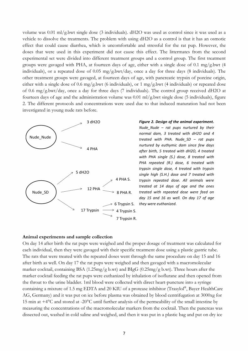
7
volume was 0.01 ml/g.bwt single dose (3 individuals). dH2O was used as control since it was used as a
vehicle to dissolve the treatments. The problem with using dH2O as a control is that it has an osmotic
effect that could cause diarrhea, which is uncomfortable and stressful for the rat pup. However, the
doses that were used in this experiment did not cause this effect. The littermates from the second
experimental set were divided into different treatment groups and a control group. The first treatment
groups were gavaged with PHA, at fourteen days of age, either with a single dose of 0.1 mg/g.bwt (4
individuals), or a repeated dose of 0.05 mg/g.bwt/day, once a day for three days (8 individuals). The
other treatment groups were gavaged, at fourteen days of age, with pancreatic trypsin of porcine origin,
either with a single dose of 0.6 mg/g.bwt (6 individuals), or 1 mg/g.bwt (4 individuals) or repeated dose
of 0.6 mg/g.bwt/day, once a day for three days (7 individuals). The control group received dH2O at
fourteen days of age and the administration volume was 0.01 ml/g.bwt single dose (5 individuals), figure
2. The different protocols and concentrations were used due to that induced maturation had not been
investigated in young nude rats before.
Animal experiments and sample collection
On day 14 after birth the rat pups were weighed and the proper dosage of treatment was calculated for
each individual, then they were gavaged with their specific treatment dose using a plastic gastric tube.
The rats that were treated with the repeated doses went through the same procedure on day 15 and 16
after birth as well. On day 17 the rat pups were weighed and then gavaged with a macromolecular
marker cocktail, containing BSA (1.25mg/g b.wt) and BIgG (0.25mg/g b.wt). Three hours after the
marker cocktail feeding the rat pups were euthanized by inhalation of isoflurane and then opened from
the throat to the urine bladder. 1ml blood were collected with direct heart-puncture into a syringe
containing a mixture of 1.5 mg EDTA and 20 KIU of a protease inhibitor (Trasylol®, Bayer HealthCare
AG, Germany) and it was put on ice before plasma was obtained by blood centrifugation at 3000xg for
15 min at +4°C and stored at -20°C until further analysis of the permeability of the small intestine by
measuring the concentrations of the macromolecular markers from the cocktail. Then the pancreas was
dissected out, washed in cold saline and weighed, and then it was put in a plastic bag and put on dry ice
Nude_Nude
Nude_SD
3 dH2O
5 dH2O
4 PHA
8 PHA R.
4 PHA S.
6 Trypsin S.
4 Trypsin S.H
7 Trypsin R.
12 PHA
17 Trypsin
Figure 2. Design of the animal experiment.
Nude_Nude – rat pups nurtured by their
normal dam, 3 treated with dH2O and 4
treated with PHA. Nude_SD – rat pups
nurtured by euthymic dam since few days
after birth, 5 treated with dH2O, 4 treated
with PHA single (S.) dose, 8 treated with
PHA repeated (R.) dose, 6 treated with
trypsin single dose, 4 treated with trypsin
single high (S.H.) dose and 7 treated with
trypsin repeated dose. All animals were
treated at 14 days of age and the ones
treated with repeated dose were feed on
day 15 and 16 as well. On day 17 of age
they were euthanized.

8
and later stored in -70°C until further analysis of the trypsin activity. The small intestine were dissected
from the pylorus to the ileo-cecal junction, measured by a ruler and split into equal halves as a proximal
and a distal part of the small intestine. The parts were flushed with cold saline, weighed and a piece of
approximately 1 cm of each part were taken from the middle and fixed in 10% neutral buffered
formalin, which cross-links the protein so the tissue does not degrade, for 24 hours at room temperature
and then stored in 70% ethanol, room temperature, until paraffin embedding. After the dissection of the
small intestine the cecum, the liver, the spleen and the stomach were dissected out, washed with cold
saline and weighed, but not stored. In the euthymic rat pups the thymus was also dissected out washed
in cold saline and weighed. When the sample collection were complete the heart artery were cut to
ensure that the rats were completely dead.
I started my master project by taking an animal experimental course, Laboratory Animal Science for
Researcher –Rodents and Lagomorphs, to be able to work with the animals. I did not participate in treatment
and sample collection of the animals used in my study. The tissue had already been collected and fixed in
formalin and was kept in 70% ethanol in room temperature and from there I started my experiments.
However, I was assisting in animal experiments on euthymic rat pups that were performed in the same
manner as the experiments on the nude rat pups.
Sample preparation
The small intestinal tissue from nude and euthymic rat pups, and small intestine, thymus and spleen
from adult euthymic rats kept in ethanol was embedded in paraffin according to standard procedures. A
small piece were cut from the tissue in the 70% ethanol and put into cassettes that were put in ethanol
80% over night. The next day they were incubated in ethanol 95% and 99%, after that they were
incubated in xylene (x3), exchanging the ethanol in the tissue with xylene, they were kept in xylene
overnight in room temperature. The third day the cassettes with the tissue were then embedded in
paraffin with a melting point at 52-54°C to be able to embed the paraffin into the tissue. After that the
tissue samples were embedded in embedding paraffin with a melting point of 56-58°C to not melt in
hands. The paraffin embedded tissue was then cut in a microtome into 5 μm thick sections that were
fixed on microscope slides (Thermo scientific, Polysine Slides), 37°C overnight.
Morphometry
Histology samples of 5 μm thickness were deparaffinised in xylene, ethanol and dH2O and stained with
haematoxylin Harris and eosin (H & E) according to standard procedures. Haematoxylin is a positively
charged compound that binds to acidic compound containing negative charge, like DNA, thus stain the
nuclei in a violet color. Eosin is an acidic compound that binds to positively charged proteins in the
cytoplasm and stains them pink. The slides were then dehydrated using ethanol 99% and xylene and
mounted under cover slip using Eukitt (Sigma-Aldrich). From each individual pictures of 20 complete
villous and 20 crypts both for the proximal and distal parts of the small intestine were taken using an
Olympus PROVIS microscope connected to a camera. The pictures were then analysed by measuring
the villi width and length and crypt depth of both proximal and distal small intestine. In the distal part of
the small intestine the length of the non-vacoulated cells were also measured and put in proportion to
the total villi length and estimated in percentage (%), and used as a measurement of maturity.

9
Immunohistochemistry
Literature search for extrathymic T cell markers
To find good markers for extrathymic T cells a quite extensive literature search was made. Many
different markers were considered but in the end the decision fell on Thy-1, which is a marker that is
found on thymectocytes and mature T cells in mice, but also on mature T cells in nude mice22. The other
marker that was chosen was CD3 which is a co-receptor to the TCR, and it is non-covalently associated
but important for the activation of the T cells6. CD3+ cells in nude rats and mice accumulate with age23-
25, and even if it is a defining feature of the T cell linage CD3+ cells in nude animals seems to have some
different features than CD3+ cells from the thymus-dependent pathway21.
Antibody testing
Antibodies, α-MCT, -MAdCAM, -Iβ7, -IL-2Rα and -CD45 (table 1), available in the lab had to be
analysed to see if they were suitable for immunohistochemistry (IHC) analysis and could be further used
in this study. They were tested in different dilutions, 1:50, 1:100, 1:200 and 1:400, mixed in 1% BSA in
0.02M phosphate buffer saline (PBS). This was also done to learn the technique of the IHC method and
therefore antibodies that were known to work for the target tissue were used as technical controls, table
2. The antibodies were tested on small intestine from young euthymic SD rat pups and on each slide one
or two dilutions of the antibody, the technical control and a negative reagent control were tested. The
negative reagent control was 1% BSA in 0.02M PBS, without antibody, and it was used to exclude
background staining from the detection kit.
New antibodies were bought, α-Thy-1 and α-CD3 (table 1), for the detection of extrathymic T cells in
the nude rat tissue. These antibodies had to be tested and optimized for the IHC method and the target
tissue. They were first tested on adult euthymic SD rat small intestine and as positive tissue controls
thymus and spleen from the same rats were used since this tissue contains T cells for sure. Then the
antibodies were tested on the target tissue, nude small intestine, using IHC analysis. The α-CD3 were
also tested on tissue from small intestine of euthymic young rat pups from 7 days of age up to 28 days of
age with a 7 day interval, to quantify the amount of CD3+ cells during normal development. The
quantification was estimated as cells/villi, table 3.

10
Table 1. Schedule of the antibody targets, the antibody and company of antibodies used for optimisation.
Antibody target Antibody Company
Mast Cell Tryptase (MCT) rabbit polyclonal anti-Mast Cell Tryptase (FL-275):sc-32889
Santa Cruz Biotechnology
Mucosal vascular addressin cell adhesion molecule (MAdCAM)
mouse monoclonal anti-MAdCAM-1 (F-6): sc-374398
Santa Cruz Biotechnology
Integrin β7 (Iβ7) rabbit polyclonal anti-Integrin β7 (H-120): sc-15330
Santa Cruz Biotechnology
Interleukin-2 receptor α (IL-2Rα)
rabbit polyclonal anti-IL-2R α (M-19): sc-666
Santa Cruz Biotechnology
leukocyte common antigen (CD45)
rabbit polyclonal anti-CD45 (H-230): sc-25590
Santa Cruz Biotechnology
Sucrase-Isomaltase (SI) rabbit polyclonal anti-Sucrase-Isomaltase (R-125): sc-99174
Santa Cruz Biotechnology
Thymocyte differentiation antigen 1 (Thy-1)
mouse monoclonal anti-CD90/Thy-1 [MRC OX-7] (ab225)
Abcam
Cluster of differentiation 3 (CD3)
rabbit monoclonal anti-CD3 [SP7] (ab16669)
Abcam
Table 2. Schedule of the antibody targets, the antibody, company and dilution of antibodies used as positive controls.
Antibody target Antibody Company Dilution
Neonatal-Fc-receptor (FcRn)
rabbit polyclonal anti-FcRn (M-255): sc-66893
Santa Cruz Biotechnology 1: 600
Proliferating cell nuclear antigen (PCNA)
mouse monoclonal anti-PCNA clone PC10
DakoCytomation 1:1200
Table 3. The quantitative estimation of CD3+ cells per villi expressed in plus signs. A higher number of plus means a
larger amount of CD3+ cells.
+ <1 cell/villi
++ 1-2 cells/villi
+++ ~5 cells/villi
++++ ~10 cells/villi
+++++ >10 cells/villi
IHC method First the tissue slides were deparaffinised, using xylene, ethanol (99%, 95%, 70%) and 0.01M PBS, and
then pap pen circles were drawn on the sample slides, 3-4 per slide each containing two tissue sections.
IHC analysis was performed by endogenous peroxidase blocking of the tissue samples, followed by
incubation with a blocking reagent, Background sniper or Rodent Blocker R (MACH 1/MACH 4
Universal; Biocare Medical, Llc.; USA) to reduce background staining by blocking the immunoglobulins
in the tissue. The tissue was then incubated with primary antibodies, diluted in 1% BSA in 0.02M PBS
overnight at +4°C, see table 1 and 2. The second day, the samples were treated with a detection system,

11
HRP-Polymer Detection kit (MACH 1/MACH 4 Universal; Biocare Medical, Llc.; USA) and the
procedure was performed according to the manufacturer´s specifications using DAB as the chromogen
substrate. The HRP-polymer will bind to the primary antibody and then the DAB chromogen will react
with the peroxides and give an orange/brow colour. Then the tissue samples were counterstained with
Mayer’s haematoxylin, which gives a weaker staining than Harris haematoxylin due to that Mayer’s
contain alcohol and Harris does not26. The weaker staining was used in IHC to give a clear chromogen
staining of the target. Whereafter the sections were dehydrated in ethanol and xylene, and mounted
under cover slip using Eukitt (Sigma-Aldrich). To exclude unspecific binding of the HRP-polymer
detection kit a negative control were included on all slides, were the primary antibody had been replaced
with only 1% BSA in 0.02M PBS.
Antigen retrieval was used to break the cross-linking between the proteins that forms during fixation with
formalin27. This procedure was done after deparaffination for α-CD3, where the slides were boiled in
sodium citrate buffer (10 mM sodium citrate, 0.05% Tween 20, pH 6.0), to uncover hidden antigenic
sites, according to abcam IHC antigen retrieval protocol for microwave procedure. This procedure was not
made for all antibodies because they came with specifications, the kit (MACH 4) is supposed to
minimize the background staining and since the antibodies were old and had been used before the time
it takes to test all the antibodies with antigen retrieval was not worth it, due to the time limit of the
project.
Statistics and analyzing methods All data showing maturation are presented as mean values ± standard deviations (SD). ANOVA
analyses, both one-way and two-way, were used to statistically compare the data between the different
treatment groups. Dunnett’s multiple comparison test was used to compare the treatment groups with
the control group. Unpaired T-test was performed when the parameters were too few. The analyses were
considered significant when P<0.05, expressed as *:P<0.05, **:P<0.01, ***:P<0.001 and ****:P<0.0001.
All calculations were done using Prism v7.0 (GraphPad Software, La Jolla, CA, USA,
www.graphpad.com). Quantitative analyses were performed using Olympus PROVIS microscope to
estimate the differences in expression patterns of the different immune targets. ImageJ software
(National Institute of Health USA) was used for morphometric evaluation of the images.
Results
Antibody testing
Different antibodies (table 1) where first analysed to identify which were suitable for
immunohistochemistry and their optimal dilution for the target tissue. This was done to decide which
antibodies were suitable for further use in the study of the precociously induced maturation of the nude
suckling rats. The antibodies were tested in different dilutions, 1:50, 1:100, 1:200 and 1:400, mixed in 1%
BSA in 0.02M PBS. The antibodies against Thy-1 and CD3 were newly purchased and therefore tested
on small intestine from euthymic adult SD rat, and as positive tissue controls spleen and thymus from
the same SD rats were used. The other antibodies had been used before, thus tested on small intestine of
euthymic young SD rats. As technical controls FcRn and PCNA (table 2), and as negative reagent
control 1% BSA in 0.02M PBS were used.
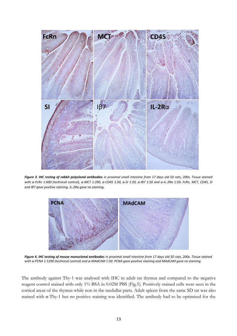
Figure 3 show representative pictures from the IHC analysis with the rabbit polyclonal antibodies on
tissue from proximal small intestine of euthymic young rats. The technical control stained with α- FcRn

12
show positive staining on the epithelial cells lining the villi, indicating that the staining method worked as
expected. The intense staining in the villi tips, can be seen in many of the pictures, is background
staining probably due to apoptotic cells that give unspecific binding. The antibodies against MCT, CD45
and SI showed positive staining for all dilutions. The positive staining of MCT showed the clearest
staining with dilution 1:200 while the chromogen reaction for dilutions of 1:50 and 1:100 overstained the
tissue in a very short time giving an unclear result. The antibodies against CD45 and SI showed clearest
result for the dilution 1:50, while the other dilutions gave very weak staining. α - Iβ7 showed unclear and
unspecific positive staining for all the dilutions, indicating that it might not work properly. The antibody
against IL-2R α showed no positive staining for any dilution.
The results from the IHC with mouse monoclonal antibodies can be seen in figure 4, which shows
representative pictures from α -PCNA, the technical control, and α -MAdCAM on proximal small
intestine from young SD rat. The technical control showed a positive staining of the antibody indicating
that the IHC method worked. The α -MAdCAM showed no positive staining for any of the dilutions.
The old antibodies were first tested with the MACH 1 system and it was then realized that a new
detection kit was needed that is more specific for the target tissue, in this case the small intestine of
young rats. The new detection kit was bought, MACH 4, and it contain a more specific background
sniper, the rodent blocker R. This detection kit was used on the new antibodies, α-Thy-1 and α-CD3.
The old antibodies were not tested again with the new detection kit due to time limits.
13
The antibody against Thy-1 was analysed with IHC in adult rat thymus and compared to the negative
reagent control stained with only 1% BSA in 0.02M PBS (Fig.5). Positively stained cells were seen in the
cortical areas of the thymus while non in the medullar parts. Adult spleen from the same SD rat was also
stained with α-Thy-1 but no positive staining was identified. The antibody had to be optimised for the
Figure 3. IHC testing of rabbit polyclonal antibodies in proximal small intestine from 17 days old SD rats, 200x. Tissue stained
with α-FcRn 1:600 (technical control), α-MCT 1:200, α-CD45 1:50, α-SI 1:50, α-Iβ7 1:50 and α-IL-2Rα 1:50. FcRn, MCT, CD45, SI
and Iβ7 gave positive staining. IL-2Rα gave no staining.
FcRn
IL-2Rα
CD45MCT
Iβ7SI
Figure 4. IHC testing of mouse monoclonal antibodies in proximal small intestine from 17 days old SD rats, 200x. Tissue stained with α-PCNA 1:1200 (technical control) and α-MAdCAM 1:50. PCNA gave positive staining and MAdCAM gave no staining.
PCNA MAdCAM

14
target tissue, therefore the proximal small intestine of adult euthymic SD rats were also stained.
Positively stained cells could be observed in the LP (Fig.6) and the dilution 1:50 gave the clearest result.
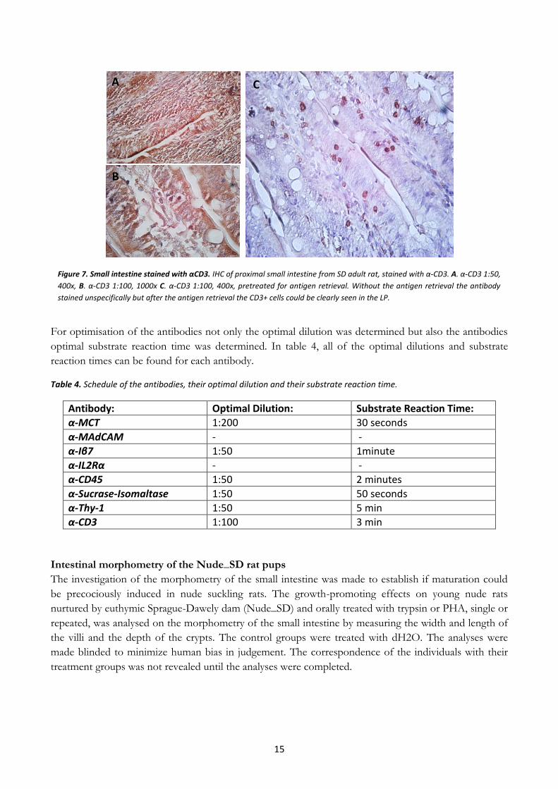
To investigate the antibody against CD3, it was first used in analysis of the thymus and spleen from adult
euthymic SD rat and positive staining could be observed in both tissues compared to the negative
reagent control. In the thymus, the medullar parts where positively stained, and in the spleen, the
positively stained areas where placed inside the white pulp, result not shown.
Proximal small intestine of normal adult rat was also stained with α-CD3 to optimise the antibody, but
the IHC analysis was not successful (Fig.7 A and B). There was a lot of unspecific staining, and it was
impossible to distinguish between positively stained cells and background staining. The α-CD3 antibody
was therefore tested after antigen retrieval, which gave clear staining (Fig.7 C). The CD3+ cells were easy
to distinguish and the cells could be observed in the tissue.
Figure 5. Thymus of adult SD rat stained with α-Thy-1. Picture to the left stained with α-Thy-1 and picture to the
right negative control (1% BSA in 0.02M PBS), 400x. Positive staining of Thy-1 can be seen in the cortical areas of
the thymus indicating that it stains T cells in the early developmental process.
Figure 6. Small intestine stained with αThy-1. IHC of adult small intestine from SD rats, stained with α-Thy-1, dilution 1:50, 400x.
Positive staining could be observed in the LP.
15
For optimisation of the antibodies not only the optimal dilution was determined but also the antibodies
optimal substrate reaction time was determined. In table 4, all of the optimal dilutions and substrate
reaction times can be found for each antibody.
Table 4. Schedule of the antibodies, their optimal dilution and their substrate reaction time.
Antibody: Optimal Dilution: Substrate Reaction Time:
α-MCT 1:200 30 seconds
α-MAdCAM - -
α-Iβ7 1:50 1minute
α-IL2Rα - -
α-CD45 1:50 2 minutes
α-Sucrase-Isomaltase 1:50 50 seconds
α-Thy-1 1:50 5 min
α-CD3 1:100 3 min
Intestinal morphometry of the Nude_SD rat pups
The investigation of the morphometry of the small intestine was made to establish if maturation could
be precociously induced in nude suckling rats. The growth-promoting effects on young nude rats
nurtured by euthymic Sprague-Dawely dam (Nude_SD) and orally treated with trypsin or PHA, single or
repeated, was analysed on the morphometry of the small intestine by measuring the width and length of
the villi and the depth of the crypts. The control groups were treated with dH2O. The analyses were
made blinded to minimize human bias in judgement. The correspondence of the individuals with their
treatment groups was not revealed until the analyses were completed.
Figure 7. Small intestine stained with αCD3. IHC of proximal small intestine from SD adult rat, stained with α-CD3. A. α-CD3 1:50,
400x, B. α-CD3 1:100, 1000x C. α-CD3 1:100, 400x, pretreated for antigen retrieval. Without the antigen retrieval the antibody
stained unspecifically but after the antigen retrieval the CD3+ cells could be clearly seen in the LP.
A
B
C

16
Proximal small intestine
A small increase in the villi length of the proximal small intestine can be observed between the treated
groups compared to the control in the H&E stained samples seen in figure 8. It is difficult to know if
this increase is due to the effects of the treatment or by stretching of the material when handling it.
There were no significant differences in the width of the villous for the PHA treatments or trypsin
treatments compared to the control group of the proximal intestine (Fig. 9 and 10). There were no
significant differences in the crypt depth of any of the trypsin treated groups compared to the control.
However, the PHA repeated dose had a small significant difference in the crypt depth compared to the
control. No difference could be seen for the PHA single group for the crypt depth. There was a clear
increase in the villi length in both of the PHA treated groups compared to the control. The villi length of
the trypsin treated single high and repeated groups had no significant increase, but trypsin single group
had a small significant increase of the villi length. However, this small increase could also be due to the
handling of the tissue and the SD for the trypsin treatments are high meaning that there are variations in
the treatment groups.
Figure 8. H&E stained small intestine. The proximal small intestine of Nude_SD rat pups, 17 days of age treated at 14 days of
age, stained with H&E, 200x. Picture to the left represents treated group. Picture to the right represent control group treated
with dH2O. A small difference of the villi length can be seen between the treated and the control. Scale bar in the right down
corner showing 0.1mm.

17
Vill i W
idth
Cry
pt
dep
th
Vill i len
gth
0 .0 0
0 .0 5
0 .1 0
0 .1 5
0 .4
0 .5
P H A P ro x im a l S m a ll In te s tin e
mm
P H A S in g le
P H A R e p e a te d
C o n tro l d H 2 O
********
*
Figure 9. PHA effects on the morphology of the proximal small intestine. Width and length of villi and crypt depth, measured in mm, of 17 days old Nude_SD rat pups proximal small intestine treated at 14 days of age with PHA single dose 0.1mg/g.bwt (4 individuals), PHA repeated dose 0.05mg/g.bwt/day (8 individuals) for 3 days and a control group treated with dH2O 0.01ml/g.bwt (5 individuals). The results are expressed as means ±SD. Two-way ANOVA test was performed, *= p<0.05, ****=p<0.0001. The villi length of the proximal small intestine had significantly increased in young rats treated with PHA.
Vill i W
idth
Cry
pt
dep
th
Vill i len
gth
0 .0 0
0 .0 5
0 .1 0
0 .1 5
0 .4
0 .5
T R Y P S IN P ro x im a l S m a ll In te s t in e
mm
T r y p s in s in g le
T r y p s in S in g le H ig h
T r y p s in R e p e a te d
C o n tr o l d H 2 O
*
Figure 10. Trypsin effects on the morphology of the proximal small intestine. Width and length of villi and crypt depth, measured in mm, of 17 days old Nude_SD rat pups proximal small intestine treated at 14 days of age with Trypsin single dose 0.6mg/g.bwt (6 individuals), Trypsin single high dose 1mg/g.bwt (4 individuals), Trypsin repeated dose 0.6mg/g.bwt/day for 3 days (7 individuals) and a control group treated with dH2O 0.01ml/g.bwt (5 individuals). The results are expressed as means ±SD. Two-way ANOVA test was performed, *= p<0.05.A significant increase could only be seen in villi length in young rats treated with trypsin single dose.

18
Distal small intestine
The distal part of the small intestine, of the Nude_SD rat pups treated with PHA or trypsin was analysed
for the same morphometric parameters as the proximal part. Figure 11, a representative picture of the
distal small intestine of treated Nude_SD rat pups compared with a control, stained with H&E, can be
seen. The treated rat pup only has vacuolated cells left in the villi tip while the control has vacuolated
cells along the whole villi.
No significant differences could be observed for the villi width, length or the depth of the crypts in any
of the treatment groups, PHA or trypsin, compared to the control group (Fig.12 and 13). In addition,
the proportion of non-vacuolated cells of the total villi length was calculated in percentage as a measure
of maturity. Trypsin single dose and trypsin single high dose had a significantly more mature distal small
intestine, around 50% and over 80% respectively, compared to the control rat pups, which had less than
20% maturity. While the trypsin repeated dose did not have a significant increase, even though there is
an indication of a growth-promoting effect. The PHA treatment both groups had significant increase in
maturity, over 50%, compared to the control (Fig. 14). However, the SD for the PHA treatments is high
so the result is probably not reliable. Trypsin single high dose had the highest increase in maturation.
Figure 11. H&E stained distal small intestine. Pictures of the distal small intestine of Nude_SD rat pups, 17 days of age treated at
14 days of age, stained with H&E illustrating length to vacuolated cells, 200x. Picture to the left represent treated groups. Picture
to the right represent control group treated with dH2O. The length of non-vacuolated cells is longer for the treatment groups
compared to the control group. Scale bar in the right down corner showing 0.1mm.

19
Vill i W
idth
Cry
pt
dep
th
Vill i len
gth
0 .0
0 .1
0 .2
0 .3
0 .4
0 .5
P H A D is ta l S m a ll In te s tin em
m
P H A R e p e a te d
C o n tro l d H 2 O
P H A S in g le
Figure 12. PHA effects on the morphology of the distal small intestine. Width and length of villi and crypt depth, measured in mm, of 17 days old Nude_SD rat pups distal small intestine treated at 14 days of age with PHA single dose 0.1mg/g.bwt (4 individuals), PHA repeated dose 0.05mg/g.bwt/day for 3 days (8 individuals) and a control group treated with dH2O 0.01ml/g.bwt (5 individuals). The results are expressed as means ±SD. Two-way ANOVA test was performed. No significant differences could be observed for villi width, crypt depth or villi length in distal small intestine of young rats treated with PHA.
Vill i W
idth
Cry
pt
dep
th
Vill i len
gth
0 .0
0 .1
0 .2
0 .3
0 .4
0 .5
T R Y P S IN D is ta l S m a ll In te s t in e
mm
T r y p s in s in g le
T r y p s in S in g le H ig h
T r y p s in R e p e a te d
C o n tr o l d H 2 O
Figure 13. Trypsin effects on the morphology of the distal small intestine. Width and length of villi and crypt depth, measured in
mm, of 17 days old Nude_SD rat pups distal small intestine treated at 14 days of age with Trypsin single dose 0.6mg/g.bwt (6
individuals), Trypsin single high dose 1mg/g.bwt (4 individuals), Trypsin repeated dose 0.6mg/g.bwt/day for 3 days (7 individuals)
and a control group treated with dH2O 0.01ml/g.bwt (5 individuals). The results are expressed as means ±SD. Two-way ANOVA test
was performed. No significant differences could be observed for villi width, crypt depth or villi length in distal small intestine of
young rats treated with trypsin.

20
Intestinal morphometry of the Nude_Nude rat pups The other experimental set with the young nude rat pups nurtured by their natural dam (Nude_Nude)
were only orally treated with PHA single dose, but analysed in the same way as the Nude_SD rat pups.
The PHA treatment had no effect on the width of the villi or on the crypt depth in either the proximal
or the distal parts of the small intestine (Fig. 15 and 16). The treatment had a small significant increase in
the length of the villi in the proximal part, but no increase in the distal part.
The maturity of the distal small intestine was calculated as the proportion of non-vacuolated cells of the
total villi length showed in percentage. PHA has a clear effect on the inducement of the maturity of the
distal small intestine, over 70% maturity compared to less than 20% for control (Fig.17).
Try
psin
Sin
gle
Typ
sin
Sin
gle
Hig
h
Try
psin
Rep
eate
d
PH
A S
ing
le
PH
A R
ep
eate
d
Co
ntr
ol d
H2O
0
2 0
4 0
6 0
8 0
1 0 0
% m
atu
re
-ty
pe
ep
ith
eli
al
ce
lls
*
* * * *
* * *
M a tu r ity
Figure 14. Maturation of the distal small intestine of treated young rats. Maturity of the epithelium of the distal small intestine estimated in percentage (%) as the length of non-vacuolated cells in proportion to the total villi length, for the different treatment groups. 17 days old Nude_SD rat pups distal small intestine treated at 14 days of age with Trypsin single dose 0.6mg/g.bwt (6 individuals), Trypsin single high dose 1mg/g.bwt (4 individuals), Trypsin repeated dose 0.6mg/g.bwt/day for 3 days (7 individuals), PHA single dose 0.1mg/g.bwt (4 individuals), PHA repeated dose 0.05mg/g.bwt/day for 3 days (8 individuals) and a control group treated with dH2O 0.01ml/g.bwt. The results are expressed as means ±SD. One-way ANOVA test was performed, *= p<0.05, **= p<0.01, ****=p<0.0001. All treatment groups, except trypsin repeated, showed significant increase in maturity of the epithelial cells of the distal small intestine.

21
Vill i W
idth
Cry
pt
Dep
th
Vill i L
en
gh
t
0 .0
0 .1
0 .2
0 .3
0 .4
0 .5
N u d e _ N u d e P H A P ro x im a l S m a ll In te s t in e
mm
P H A
C o n tro l d H 2 O
*
Figure 15. PHA effects on the morphology of the proximal small intestine. Width and length of villi and crypt depth, measured in mm, of 17 days old Nude_Nude rat pups proximal small intestine treated at 14 days of age with PHA single dose 0.1mg/g.bwt (4 individuals) and a control group treated with dH2O 0.01ml/g.bwt (3 individuals). The results are expressed as means ±SD. Two-way ANOVA test was performed, *= p<0.05. Significant difference could only be observed for villi length of the proximal small intestine of young Nude_Nude rats treated with PHA.
Vill i W
idth
Cry
pt
Dep
th
Vill i L
en
gh
t
0 .0
0 .1
0 .2
0 .3
0 .4
0 .5
N u d e _ N u d e P H A D is ta l S m a ll In te s t in e
mm
P H A
C o n tro l d H 2 O
Figure 16. PHA effects on the morphology of the distal small intestine. Width and length of villi and crypt depth, measured in mm, of 17 days old Nude_Nude rat pups distal small intestine treated at 14 days of age with PHA single dose 0.1mg/g.bwt (4 individuals) and a control group treated with dH2O 0.01ml/g.bwt (3 individuals). The results are expressed as means ±SD. Two-way ANOVA test was performed. No significant differences could be observed for the distal small intestine of young Nude_Nude rats treated with PHA.

22
Organ weight, trypsin activity and intestinal permeability of nude young rats The proximal and distal part of the small intestine and the pancreas were weighed, the trypsin activity of the pancreas and the permeability of the small intestine in both Nude_SD and Nude_Nude rat pups was analysed by others in the lab. Organ weight The weight of the proximal and distal small intestine of the young nude rats had significantly increased for all treatment groups compared to the control for both Nude_SD and Nude_Nude rat pups, figure 18 and 19. The pancreas weight was also measured during the sample collection and no difference can be seen for the weight between the treatment groups compared to the control group for neither the Nude_SD nor the Nude_Nude young rats, figure 18 and 19. Trypsin activity The trypsin activity was analysed in the pancreas for both Nude_SD and Nude_Nude and measured as trypsin units (U) per gram body weight (Fig. 20 and 21). The PHA treatments of both Nude_SD and Nude_Nude young rats had significant increase of the activity compared to the control group. Trypsin single dose and trypsin repeated dose had no significant increase in the trypsin activity of the pancreas in the Nude_SD young rats. However, trypsin single high dose had significant increase in trypsin activity in the pancreas and this might indicate that the effect seen by trypsin treatment was dose related. Permeability of the small intestine The concentration of BIgG and BSA was measured from the blood plasma obtained during sample collection and in Nude_SD the concentration of BIgG from all the treatment groups had significantly decreased compared to the control group, indicating a decreased BIgG uptake, figure 22. The concentration of BSA in the plasma had significantly decreased in almost all treatment groups, except for trypsin repeated dose, compared to the control group for Nude_SD rats, indicating a lowered uptake of the macromolecule. The permeability of BIgG and BSA had also significantly decreased in the PHA treated Nude_Nude young rats compared to the control treated rats, figure 23. The low significance of the permeability of BSA could be due to the high variations of the control group. If more individuals were included the variations would probably be smaller.
PH
A
Co
ntr
ol d
H2O
0
2 0
4 0
6 0
8 0
1 0 0
M a tu r ity N u d e _ N u d e
% m
atu
re
-ty
pe
ep
ith
eli
al
ce
lls
* * *
Figure 17. Maturation of the distal small intestine of PHA treated young rats. Maturity of the epithelium of the distal small intestine estimated in percentage (%) as the length of non-vacuolated cells in proportion to the total villi length, for the different treatment groups. 17 days old Nude_Nude rat pups distal small intestine treated at 14 days of age with PHA single dose 0.1mg/g.bwt (4 individuals) and a control group treated with dH2O 0.01ml/g.bwt (3 individuals). The results are expressed as means ±SD. Unpaired T-test was performed, ***=p<0.001. There is a significant increase in the maturity of the epithelium of the distal small intestine of young Nude_Nude rats treated with PHA.

23
SI P
roxim
al
SI D
ista
l
Pan
cre
as
0
5
1 0
1 5
2 0
2 5
O rg a n w e ig h t N u d e _ S D
mg
/gb
wt
T ry p s in S in g le
T ry p s in S in g le H ig h
T ry p s in R e p e a te d
P H A S in g le
P H A R e p e a te d
C o n tro l d H 2 O
****
****
********
****
************
****
***
Figure 18. The organ weight of small intestine and pancreas of Nude_SD young rats. The weight of proximal and distal small
intestine and the pancreas of Nude_SD rat pups treated with trypsin single dose 0.6mg/g.bwt (6 individuals), trypsin single high
dose 1mg/g.bwt (4 individuals), trypsin repeated dose 0.6mg/g.bwt/day for three days (7 individuals), PHA single dose 0.1mg/g
bwt (4 individuals), PHA repeated dose 0.05mg/g.bwt/day for three days (8 individuals) and a control group treated with dH2O
0.01ml/g.bwt (5 individuals) measured at 17 days of age. The results are expressed as means ±SD. Two-way ANOVA was
performed, ***=p<0.001, ****=p<0.0001. The weight if the small intestine, both proximal and distal, had significantly increased
for all treatment groups, while no significant difference could be observed of the pancreas.
SI P
roxim
al
SI D
ista
l
Pan
cre
as
0
5
1 0
1 5
2 0
2 5
O rg a n w e ig h t N u d e _ N u d e
mg
/gb
wt
P H A
C o n tro l d H 2 O
****
****
Figure 19. The organ weight of small intestine and pancreas of Nude_Nude young rats. The weight of proximal and distal small
intestine and the pancreas of Nude_Nude rat pups treated PHA single dose 0.1mg/g bwt (4 individuals) and a control group
treated with dH2O 0.01ml/g.bwt (3 individuals) measured at 17 days of age. The results are expressed as means ±SD. Two-way
ANOVA was performed, ****=p<0.0001. Significant increase of the weight of the small intestine, both proximal and distal, could
be obsereved in PHA treated rats, while no significant difference could be seen in the weight of the pancreas.

24
Try
psin
Sin
gle
Try
psin
Sin
gle
Hig
h
Try
psin
Rep
eate
d
PH
A S
ing
le
PH
A R
ep
eate
d
Co
ntr
ol d
H2O
0
1 0
2 0
3 0
4 0
N u d e _ S D try p s in a c tiv ity
Try
ps
in U
/g b
wt
* * * *
* * * *
* * * *
Figure 20. Trypsin activity of the pancreas in Nude_SD young rats. The trypsin activity of the pancreas was measured at 17 days
of age in Nude_SD rat pups treated with trypsin single dose 0.6mg/g.bwt (6 individuals), trypsin single high dose 1mg/g.bwt
(4 individuals), trypsin repeated dose 0.6mg/g.bwt/day for three days (7 individuals), PHA single dose 0.1mg/g.bwt
(4 individuals), PHA repeated dose 0.05mg/g.bwt/day for three days (8 individuals) and a control group treated with dH2O
0.01ml/g.bwt (5 individuals). The results are expressed as means ±SD. One-way ANOVA was performed,****=p<0.0001.
Significant increase in the pancreas trypsin activity could be observed for both PHA treatments, but only for single high dose of
the trypsin treated groups.
PH
A
Co
ntr
ol d
H2O
0
5
1 0
1 5
2 0
2 5
N u d e _ N u d e T ry p s in
Try
ps
in U
/g b
wt
* * *
Figure 21. Trypsin activity of the pancreas in Nude_Nude young rats. The trypsin activity of the pancreas was measured at 17
days of age in Nude_Nude rat pups treated with PHA single dose 0.1mg/g.bwt (4 individuals) and a control group treated with
dH2O 0.01ml/g.bwt (3 individuals). The results are expressed as means ±SD. Unpaired t-test was performed,***=p<0.001.
Significant increase of the pancreas trypsin activity could be observed in Nude_Nude rats treated with PHA.

25
Try
psin
Sin
gle
Try
psin
Sin
gle
H
igh
Try
psin
Rep
eate
d
PH
A S
ing
le
PH
A R
ep
eate
d
Co
ntr
ol d
H2O
0
1 0 0
2 0 0
3 0 0
4 0 0
5 0 0
N u d e _ S D B Ig G
g
/ml
* * * *
* * * * * * * ** * * *
* * * *
Try
psin
Sin
gle
Try
psin
Sin
gle
Hig
h
Try
psin
Rep
eate
d
PH
A S
ing
le
PH
A R
ep
eate
d
Co
ntr
ol d
H2O
0
1 0
2 0
3 0
4 0
5 0
N u d e _ S D B S A
mg
/ml
* * *
* * * ** * * * * * * *
Figure 22. Permeability of BIgG and BSA in the small intestine of Nude_SD young rats. The plasma concentration of BIgG (μg/ml)
and BSA (mg/ml) in Nude_SD treated with trypsin single dose 0.6mg/g.bwt (6 individuals), trypsin single high dose 1mg/g.bwt (4
individuals), trypsin repeated dose 0.6mg/g.bwt/day for three days (7 individuals), PHA single dose 0.1mg/g.bwt (4 individuals),
PHA repeated dose 0.05mg/g.bwt/day for three days (8 individuals) and a control group treated with dH2O 0.01ml/g.bwt (5
individuals). The results are expressed as means ±SD. One-way ANOVA was performed***=p<0.001,****=p<0.0001. Significant
decrease of plasma BIgG could be observed in all treatment groups and for BSA, except trypsin repeated treatment.
PH
A
Co
ntr
ol d
H2O
0
1 0 0
2 0 0
3 0 0
4 0 0
5 0 0
N u d e _ N u d e B Ig G
g
/ml
* * * *
PH
A
Co
ntr
ol d
H2O
0
5
1 0
1 5
2 0
2 5
N u d e _ N u d e B S A
mg
/ml
*
Figure 23. Permeability of BIgG and BSA in the small intestine of Nude_Nude young rats. The concentration of BIgG in the
plasma measured in mg/ml and the concentration of BSA measured mg/ml from Nude_SD treated PHA single dose 0.1mg/g.bwt
(4 individuals) and a control group treated with dH2O 0.01ml/g.bwt (3 individuals). The results are expressed as means ±SD.
Unpaired t-test was performed,*=p<0.1, ****=p<0.0001. Significant decrease of BIgG and BSA could be observed in PHA treated
Nude_Nude rats. PHA induced total closure of the small intestine, no marker molecules were detected in blood after treatment.

26
Figure 24. CD3+ cells in natural development. Proximal and distal small intestine of natural development of SD rats, 7 to 28 days of age, analysed
with immunostaining, α-CD3, and counterstained with haematoxylin Mayers, 400x. CD3+ cells could be observed at all ages and the amount of CD3+
cells were estimated to approximately 5 cells/villi (+++) for 7 and 14 days of age, 10 cells/villi (++++) for 21 days of age and >10 cells/villi (+++++) for
28 days of age.
7 Days 14 Days 21 Days 28 Days
Dis
tal
Pro
xim
al
Immunohistochemistry analysis for detection of T cells The Nude_SD small intestine was stained with α-Thy-1 to investigate the presence of immature T cells.
Few Thy-1 positive cells were found distributed between all treatment groups and also in the control
group treated with dH2O (result not shown).
The small intestine from Nude_SD, Nude_Nude and euthymic rat pups, during natural development
from 7 days to 28 days after birth, were stained with α-CD3 to investigate the presence of mature T
cells. In euthymic young rats CD3+ cell could be found in all ages (Fig. 24). In 7 days of age and in 14
days of age the amount of CD3+ cells were low (estimated as +++, table 3), but a small increase of
CD3+ cells could be seen from 7 days of age to 14 days of age. Around weaning, 21 days of age, the
CD3+ cells increased (++++) until day 28 (+++++).
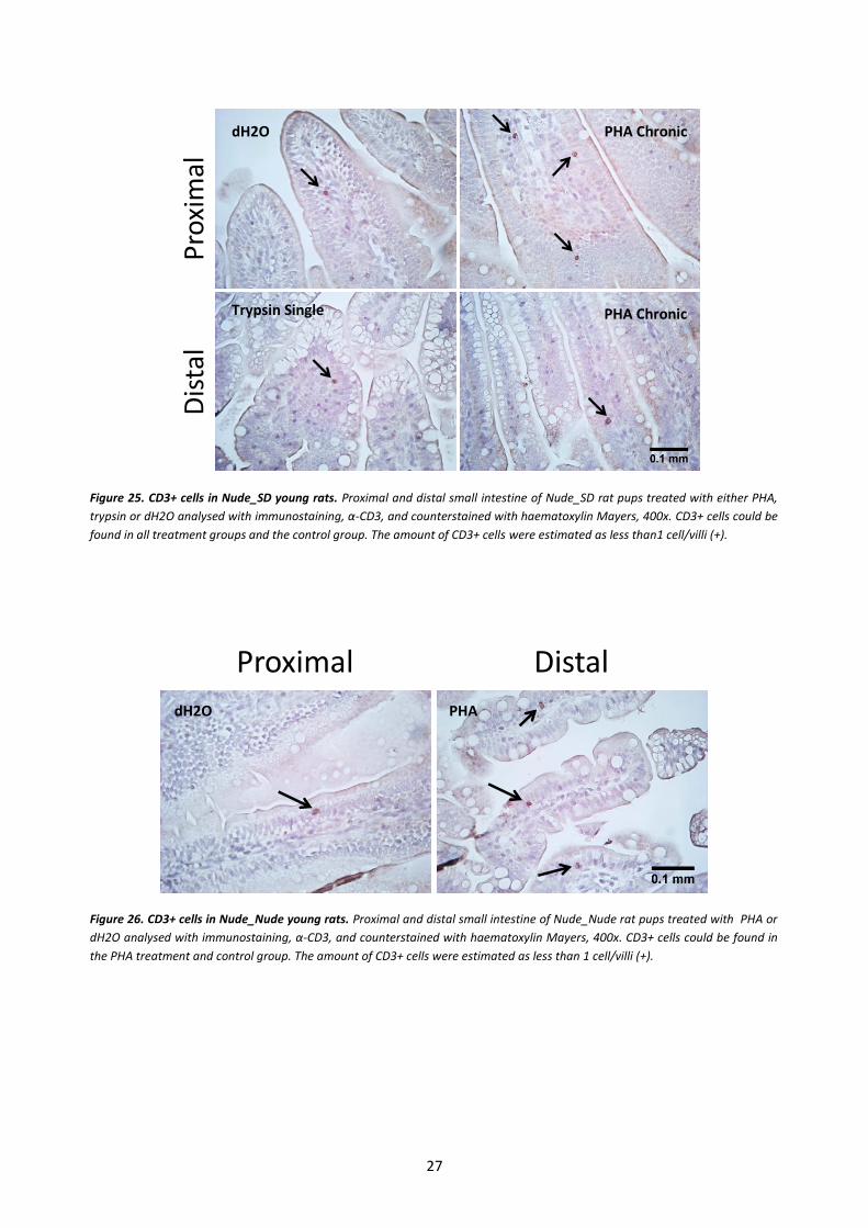
In the Nude_SD rat pups a small amount of CD3+ cells, less than one positive cell per villi, could be
seen in the small intestine in all treatment groups, even in the control group treated with dH2O (Fig. 25).
The colored lining of the villi is unspecific reactivity due to the secondary system. The CD3+ cells were
also found in the Nude_Nude small intestine of both PHA treated and control (Fig. 26). The nude rat
tissue had very few T cells in the small intestine compared to the euthymic rats, even the 7 days old
normal rat pup tissue (+++) contained a lot more positive cells than the 17 days nude rat tissue (+). The
amount of CD3+ cells in the nude tissue were estimated as less than 1 cell/villi, and no different pattern
could be seen between the Nude_SD and Nude_Nude or between the different treatment groups or
between proximal and distal parts. The analysis of the CD3+ cells were only made with qualitative
analysis due to time limits, to obtain a more specific result a quantification of cells/area could be done.
27
Figure 25. CD3+ cells in Nude_SD young rats. Proximal and distal small intestine of Nude_SD rat pups treated with either PHA,
trypsin or dH2O analysed with immunostaining, α-CD3, and counterstained with haematoxylin Mayers, 400x. CD3+ cells could be
found in all treatment groups and the control group. The amount of CD3+ cells were estimated as less than1 cell/villi (+).
Pro
xim
alD
ista
l
Figure 26. CD3+ cells in Nude_Nude young rats. Proximal and distal small intestine of Nude_Nude rat pups treated with PHA or
dH2O analysed with immunostaining, α-CD3, and counterstained with haematoxylin Mayers, 400x. CD3+ cells could be found in
the PHA treatment and control group. The amount of CD3+ cells were estimated as less than 1 cell/villi (+).
Proximal Distal
dH2O PHA Chronic
PHA Chronic Trypsin Single
dH2O PHA

28
Discussion
Antibody testing for IHC
At first I tested different antibodies to find their optimal dilution (table 4) for the IHC and target tissue.
To be sure that the method worked properly previously tested, optimised and established antibodies in
the lab, rabbit polyclonal antibody α -FcRn and mouse monoclonal antibody α – PCNA, were used as
technical controls of the method. FcRn is an essential receptor for the transfer of maternal IgG to the
infant and it is mainly expressed by epithelial cells in the small intestine of suckling rats 28. PCNA is a
protein that acts as a cofactor of DNA polymerase delta, thus it is found in the nucleus of proliferating
cells, mainly localised in the small intestinal crypts 29.
The α-MCT, α-CD45 and α-SI antibodies stained clearly without a lot of unspecific staining. MCT and
CD45 are targets of the immune system, MCT is a protease stored and secreted by mast cells as an
inflammatory response while CD45 is a transmembrane glycoprotein expressed on all leucocytes 30, 31.
Sucrase-isomaltase is a brush border membrane protein which is important for the final stages of
carbohydrate digestion and is localized on the apical side of the enterocytes mainly in the proximal small
intestine 32. MAdCAM is a mucosal vascular addressin that acts as a homing receptor for recirculating
lymphocytes to the Peyer´s patches and intestinal LP and IL-2Rα is a subunit of the IL-2 receptor, which
is a surface receptor with multiple biological processes, like cell-growth and proliferation of activated T-
and B-cells 7, 33. α-MAdCAM and α -IL-2Rα gave no staining in the suckling rat tissue indicating that the
antibodies did not function in IHC and were possibly too old to be used. The α-Iβ7 gave positive
staining but it was unclear and unspecific and gave a lot of background staining in the target tissue. Iβ7 is
a receptor mainly involved in trafficking and retention of lymphocytes, hence expected to be found
around blood vessels in the LP 34 but it was not. To get a better result with α-Iβ7 and the other
antibodies tested antigen retrieval could have been done before the staining procedure to reveal the
hidden antigens. If there would have been more time a positive and a negative control for the antibodies
could have been used. As a positive control a tissue that is known to express the wanted antigen could
be used and as a negative control the tissue could be treated with peptide blocking of the antigen. If the
antibody gives positive staining after peptide blocking the antibody stain unspecifically35.
Thy-1 is a glycoprotein that is expressed by many different cell types like neurons and thymocytes. It
promotes T cell activation and is involved in cell death, migration, cellular adhesion etc. (https://www.bio-
rad-antibodies.com/cd90-glycosylphosphatidylinositol-anchored-glycoprotein.html; 10-06-2016). Thy-1 is expressed
by T cells in mice and it has been reported that nude mice also express Thy-1 on their mature T cells 22.
In this study the investigation for the optimal dilution for the antibody against Thy-1 observed that Thy-
1 is only expressed by precursor T cells in the rat. This was realized since the positive staining of the
antibody was seen in the cortical areas of the thymus, which is the location for the earliest events in T
cell development 36, in adult euthymic rats. There was no positive staining in the spleen of the adult rat,
where there are a lot of mature T cells. These results indicate that Thy-1 is only expressed on precursor
T cells in the rat tissue, and it agrees with previous results 37. The result of the present study showed that
Thy-1+ cells could be observed in the small intestine of the adult euthymic rat and this could possibly be
precursor T cells that will mature in the gut independent of the thymus.
CD3 is a co-receptor of the T cell receptor (TCR), non-covalently associated, and is important in T cell
activation. The CD3 complex is a defining feature of the T cell lineage and thus a marker for T cells
(https://www.bio-rad-antibodies.com/minireview-cd3-antibody.html; 10-06-2016). It was found in the medullar

29
parts of the thymus, which is the location for the later events of the T cell maturation 36, indicating that
the antibody properly stains mature T cells. It was also found in the white pulp of the spleen, assumable
the T cell rich regions, which confirms that the antibody stains specifically for CD3+ cells.
Mechanisms of action of PHA and trypsin
Some differences can be seen between the treatment with PHA and trypsin, even though no specific
pattern has been evident more than the fact that trypsin needs a higher concentration to induce the same
maturation state, the differences might be due to different mechanisms of action for the two treatment
substances.
After feeding with PHA it will bind to the epithelium and line the whole villi, from the tip to the
beginning of the crypts in the small intestine38. This will result in the disturbance of the morphology of
the small intestine leading to villi shortening, which will later be restored through the increase in crypt
cell proliferation 11, 38. The direct binding of PHA stimulated crypt hyperplasia 11, which might be
through the activation of the mitogen-activated protein kinase cascade known to be involved in cell
growth and differentiation. It has been shown that PHA activated this cascade in both human and
rodent intestinal cell-lines 39. The crypt hyperplasia lead to the increase in villi length, deeper crypts and
the change from vacuolated cells to adult-like non-vacuolated cells in the distal epithelium 10, 11, 38, that
could be seen in the treated nude rats. The binding of PHA to enteroendocrine cells have been shown to
lead to the release of cholecystokinin (CCK) 40, which is a GI hormone that stimulates the secretion of
digestive enzymes from the exocrine pancreas 41, thus the increase of pancreatic trypsin seen in the nude
suckling rats might be stimulated by the release of CCK. It has been shown that precociously induced
maturation in euthymic suckling rats is associated with the activation of the immune system and an
increase in the numbers of intestinal T and B cells 11. There are theories that binding of PHA to the
intestinal epithelium will induce the secretion of cytokines and growth factors, which will stimulate the
recruitment of immune cells and their activation. The immune cells will then in turn be involved in the
stimulation of growth and the development of the GI tract 11, 15, 42.
During normal weaning there is an increase in the secretion of enzymes from the pancreatic acinar cells,
including trypsin 1, and feeding with exogenous trypsin lead to precociously induced maturation of the
GI tract of the suckling rat indicating that trypsin is involved in the process of maturation. Studies have
shown that trypsin might be a signaling molecule that specifically regulates cells by cleaving and
triggering proteinase-activated receptor 2 (PAR-2), which is a G-protein-coupled receptor. PAR-2
receptors can be found in the gastrointestinal tract on the apical and basolateral side of enterocytes in
both crypts and villi of the small intestine, on the pancreas and even on T cell lines 43-45. At
concentrations that are normally present in the intestinal lumen trypsin activates PAR-2 at the apical side
of the enterocytes which will stimulate eicosanoid secretion, which act in a paracrine and autocrine
manner and regulate multiple processes in the intestine 45. It is possible that the exogenous trypsin given
to the suckling rat pups activates PAR-2 signaling in the small intestine that provoke a pro-inflammatory
response by the secretion of eicosanoids and thereby stimulate the induced maturation. It has been
shown that eicosanoids act locally in the intestine to regulate growth and mediate inflammation 46.
Effects of PHA and trypsin on the small intestine in nude rat pups
The gut of the young rat undergoes the final remodeling process during the third postnatal week, the
weaning period. This is the time when the GI tract has to be fully adapted and able to digest an adult
diet. The process includes gut growth, change in epithelial cell kinetics and a decrease in gut permeability

30
3, 16. It has been established in previous studies that maturation can be precociously induced in suckling
rat pups by oral feeding PHA or protease 10-12. In this study the precociously induced maturation was
investigated in nude rat pups due to their lack in a functional thymus leading to a T cell deficiency and
the study aimed to examine the importance of T cells during the maturation process of the GI tract. The
direct effects seen in the nude rat pups in this study by PHA treatment were the increase in proximal villi
length and the change from vacuolated cells, in the distal small intestine, to an adult-like non-vacuolated
epithelium. This coincides with the effects seen in the small intestine of euthymic suckling rat pups
treated with PHA and at normal weaning 3, 10, 11, 16. The trypsin treated nude rat pups showed a similar
maturation pattern for the distal part of the small intestine in the change into adult-like epithelium,
which was also observed in euthymic suckling rats treated with protease 12. The increase in villi length in
the proximal small intestine was only observed for treatment with trypsin single dose and not for any of
the other trypsin treatments compared to the control. In the euthymic rat pups treated with protease the
increase in villi length could be seen 12.
The change into an adult functional phenotype of the distal small intestine was in this study measured as
maturity of the epithelium, the proportion of the non-vacuolated cells to the total villi length presented
in percentage. The results obtained showed a large range between the treatment groups in the maturity,
from non-significant to almost 100% maturity (Fig. 14 and 17). Repeated trypsin treatment had no
significant increase in the maturation of the epithelium. Trypsin single, PHA single and PHA repeated
treatments had a small significant increase, while trypsin single high treatment had a high increase in the
maturity. The exogenous substances will temporarily cause a mucosal disturbance that will lead to an
accelerated crypt cell proliferation and later the change into a mature epithelium 10, 38. The trypsin single
high dose might have severe effects on the epithelium, leading to severe disturbance and the renewal has
to arise quickly. This could be the explanation to the high significance in only this parameter; it is a direct
and acute effect of the damage.
In natural weaning and in precociously induced maturation the crypts get significantly deeper due to the
accelerated cell proliferation 3, 11, 12, 16. In the present study only treatment with PHA chronic dose showed
increase in crypt depth in the nude rat pups (Fig. 9.). Previous studies have shown that mucosal T cells
promote the crypt hyperplasia and regulate enterocyte growth under normal conditions and during
weaning 13, 47-49. It was proven in this study that the nude rat pups had CD3+ lymphocytes in their small
intestine, even if the amount observed is lower than euthymic rat pups (Fig. 25 and 26). It might be that
this small amount of T cells is enough to stimulate cell proliferation and promote exchange of
vacuolated epithelial cells to adult-like non-vacuolated cells, but might not be enough to provoke the rate
of cell proliferation that is needed for expanding the crypt depth.
The small intestinal weight of the Nude_SD and Nude_Nude rat pups was measured and analysed (by
others) showing that both PHA and trypsin had growth promoting effects on the proximal and distal
small intestine compared to the control (Fig. 18 and 19). There were no visible differences between the
Nude_SD and Nude_Nude of the PHA single treatment. There are some small differences between the
treatment groups in the Nude_SD rat pups, where the PHA treatment and the trypsin single high dose
have the highest increase. The results coincide with earlier studies made on euthymic rat pups treated
with either PHA or protease 10-12. Even though the rat pups in this study were immunodeficient they
show similarities in the pattern for precociously induced maturation as euthymic rat pups and normal
weaning.

31
In addition to the organ weight, the permeability of the small intestine of the nude rat pups was
measured by feeding a marker solution cocktail, containing BIgG and BSA, to the pups three hours
before euthanizing. The concentration of the markers was measured in plasma (analysed by others)
obtained during sample collection. The results obtained showed decrease in the uptake of
macromolecules for almost all treatment groups compared to the control for both BSA and BIgG (Fig.
22 and 23). Trypsin repeated had no significant decrease in the uptake of BSA. This result indicates that
the nude rat pups have a similar gut closure as the one seen at weaning. This effect can also be seen in
induced maturation in euthymic rat pups 10-12. The decrease in BSA coincides with the decrease in
vacuolated cells in the distal small intestine of the treated nude rat pups, which is responsible for the
unspecific transport of macromolecules over the epithelium 3. The decrease in the uptake of BIgG might
be a consequence of decrease in the receptor FcRn, which is a specific receptor mainly for the uptake of
IgGs from the colostrum and maternal milk 3, 50. The presence of FcRn was not considered in this study
but according to previous studies the FcRn receptors decrease during precocious maturation in euthymic
rat pups 10-12. Since many similarities were observed between the euthymic and nude rat pups during
precocious induced maturation this could be a possible explanation.
During normal weaning the pancreas also undergoes a process of maturation parallel to that of the GI
tract, which includes weight gain and increase in enzyme production and secretion 1. The pancreas of the
nude rat pups was investigated by others and no significant increase of the pancreas weight could be
observed (Fig. 18 and 19). However, all of the PHA treated groups, including the Nude_Nude rat pups,
showed increase in trypsin activity in the pancreas (Fig.20 and 21). This agrees with the studies made on
euthymic rat pups treated with PHA, except that the euthymic rat pups had an increase in pancreatic
weight 10, 11. The trypsin treated groups only had significant increase for trypsin activity in trypsin single
high group compared to the control group. The results coincide with the previous study made on
euthymic rat pups treated with microbial-like pancreatic enzymes, where the protease treated rats
showed increase in trypsin activity. The increase in enzyme production of the exocrine pancreas seems
to be associated with postnatal changes of the hormone levels, such as glucocorticoids, and the increase
of such receptors in young rats at weaning 1, 51.
The present study showed that maturation of the GI tract can be precociously induced in
immunodeficient suckling rats, with an impaired T cell function, by feeding with a red kidney bean lectin
or a pancreatic protease. These substances stimulated the proliferation of crypt cells leading to the
lengthening of the proximal villus and change from vacuolated cells to adult-like non-vacuolated cells in
the distal small intestine. The effects of trypsin treatment seem to be dose related since most effects are
seen for only trypsin single high dose. Additionally it was shown that there are mature T cells present in
the small intestine in nude suckling rats at 17 days of age, despite their dysfunctional thymus. No
differences in the maturation pattern could be observed between the Nude_SD and Nude_Nude rat
pups treated with PHA.
Thy-1 + and CD3+ cells in the small intestine of nude rats
Normal weaning and precociously induced maturation in euthymic young rats result in an increase in
immune cells, especially T cells, while the inhibition of T cells with CyA has shown an impaired and
delayed maturation. Blocking of IL-2R also leads to the same outcome 13 all indicating that T cells have
an important role in the maturation and development of the GI tract. The intestine of the euthymic rat
has CD3+ intraepithelial lymphocytes (IEL) already the first day after birth, which will increase
progressively throughout the suckling period 52, this might suggest that CD3+ IELs are not only

32
important for the immune response during weaning but might also be important for the protection of
the neonatal rat from the first day after birth.
It has been proven that adult nude mice and rats have T cells expressing CD3, and they accumulate with
age 23-25. Nude mice at 8 weeks of age did not express any CD3+ cells in the spleen or lymph nodes, but
after 8 weeks they started to increase in numbers 24. The maturation of extrathymic T cells mostly takes
place in the MLN and then the T cells migrate to the intestine where they accumulate, mostly as IELs 23.
A study made on nude neonatal rats showed that mature T cells could not be developed and that CD3+
IELs did not appear until four to six month of age 53. These results lead to the speculation that nude rats
would have a delayed maturation during weaning, due to the decrease in mature T cells. The present
study did not agree with this speculation, and the presence of CD3+ T cells was observed in the nude
suckling rat small intestine, albeit in lower amounts. The nude rat tissue of the small intestine were
stained with α-CD3 antibodies and the presence of CD3+ cells could be observed in all treatment
groups even the control, indicating that extrathymic T cell maturation takes place in the gut. There was
clearly less CD3+ cells in the nude small intestine of 17 days old suckling rats compared to the euthymic
suckling rats from 7 days of age up to 28 days after birth. No pattern between the different treatment
groups could be observed; they all had a very small amount of CD3+ cells. The CD3+ cells were found
in the LP of the villi and close to the crypts. They were also found in the epithelium indicating that these
cells are CD3+ IELs. This agrees with another study that also showed the presence of mature T cells in
nude young rats 17. The study investigated mature T cells expressing IL-2R and CD5 in the MLNs during
weaning, which showed that there was approximately the same numbers of these positive cells in both
euthymic and nude rat pups. Even though they found that the total number of T cells, in the T-
dependent interfollicular areas of the MLN, was decreased in the nude rat pups. Due to these findings it
is not possible to exclude the role of T-cells in the maturation process in suckling rats during weaning17.
The presence of mature T cells in the nude rat pups might be explained by the theory that maternal-milk
derived T cells can be transported to the neonatal gut mucosa after birth, where they can get activated by
antigens from the lumen 17, 54, 55. The present study investigated both nude rat pups nurtured by euthymic
dams and nude rat pups nurtured by their natural dam, no difference in the presence or numbers of
CD3+ cells could be observed. This suggests that there is no significant transport of maternal T cells to
the offspring from the maternal milk. This agrees with a previous study made on euthymic mice
indicating that there is no transport of maternal T cells to the gut after birth 56. The presence of Thy-1+
cells in the Nude_SD small intestine was also investigated in this study. Few positive cells could be
found and they were distributed between the treatment groups including the control group. Too few
samples were included in this investigation, thus no significant results can be observed. It can be
hypothesized that there are very few Thy-1+ cells because they have differentiated into mature T cells.
The increase in pancreas growth, villi width and crypt depth were not significantly clear in the nude
suckling rats treated with PHA or trypsin, as it was for euthymic suckling rats treated the same way. The
decrease in mature T cells might be the explanation, since an increase in the crypt depth could be seen in
all treatment groups even if it was not significant for more than treatment with PHA chronic, and a
reduced number of T cells might delay the precociously induced maturation in nude rats.
Conclusion
As can be understood there are many factors playing a part in the complex development of the small
intestine including exogenous and endogenous molecules, intracellular communication, hormonal

33
secretion and physiological inflammation. The immune response seen at natural weaning and during
precociously induced maturation is an important feature for the maturation and development of the GI
tract, were the effect of T cells seems to play a key role. T cells could be detected in nude suckling rats
indicating that the thymus-independent T cell maturation in the gut is stimulated shortly after birth.
Even a small amount of T cells seems to be important for the maturation process. More research is
needed in many aspects of the induced maturation, the maturation process of the extrathymic T cells and
the mechanism of action of PHA and trypsin needs to be further studied to get a clearer picture of the
maturation and development process. The discoveries of the presence of T cell in nude suckling rats
might lead to better understanding of the key players of the maturation of the GI tract and hopefully
help in the search for treatment of NEC in preterm birth infants.
References 1. Walthall K, Cappon GD, Hurtt ME, et al. Postnatal development of the gastrointestinal system: A
species comparison. Birth defects research. Part B, Developmental and reproductive toxicology 2005;74:132-56.
2. Barrett KE. In: M.Thomas MWaC, ed. Gastrointestinal Physiology. 2nd ed. United States of America: McGraw-Hill Education, 2014:3-7.
3. Pacha J. Development of intestinal transport function in mammals. Physiological Reviews 2000;80:1633-1667.
4. Abbas AK, Lichtman AHH, Pillai S. Basic Immunology: Functions and Disorders of the Immune System: Elsevier Health Sciences, 2012.
5. Femke Broere SGA, Michail V. Sitkovsky and Willem van Eden. T cell subsets and T cell-mediated immunity. In: Parnham FPNaMJ, ed. Principles of Immunopharmacology. Volume 3rd Springer, 2011:15-27.
6. Smith-Garvin JE, Koretzky GA, Jordan MS. T cell activation. Annu Rev Immunol 2009;27:591-619. 7. Miniami Y, Kono T, Miyazaki T, et al. The il-2 receptor complex: Its structure, function, and target
genes. In: Paul WE, ed. Annual Review of Immunology. Volume 11: Annual Reviews Inc. {a}, P.O. Box 10139, 4139 El Camino Way, Palo Alto, California 94306, USA, 1993:245-268.
8. Pierzynowski SG, Westrom BR, Svendsen J, et al. Development of exocrine pancreas function in chronically cannulated pigs during 1-13 weeks of postnatal life. Journal of Pediatric Gastroenterology and Nutrition 1990;10:206-212.
9. Pierzynowski SG, Westrom BR, Erlanson Albertsson C, et al. Induction of exocrine pancreas maturation at weaning in young developing pigs. Journal of Pediatric Gastroenterology and Nutrition 1993;16:287-293.
10. Linderoth A, Biernat T, Prykhodko O, et al. Induced growth and maturation of the gastrointestinal tract after phaseolus vulgaris lectin exposure in suckling rats. Journal of Pediatric Gastroenterology and Nutrition 2005;41:195-203.
11. Prykhod'ko O, Fed'kiv O, Linderoth A, et al. Precocious gut maturation and immune cell expansion by single dose feeding the lectin phytohaemagglutinin to suckling rats. British Journal of Nutrition 2009;101:735-742.
12. Prykhodko O, Pierzynowski SG, Nikpey E, et al. Pancreatic and pancreatic-like microbial proteases accelerate gut maturation in neonatal rats. Plos One 2015;10:1-14.
13. Thompson FM, Mayrhofer G, Cummins AG. Dependence of epithelial growth of the small intestine on t-cell activation during weaning in the rat. Gastroenterology 1996;111:37-44.
14. Cummins AG, Thompson FM. Effect of breast milk and weaning on epithelial growth of the small intestine in humans. Gut 2002;51:748-754.

34
15. Pie S, Lalles JP, Blazy F, et al. Weaning is associated with an upregulation of expression of inflammatory cytokines in the intestine of piglets. Journal of Nutrition 2004;134:641-647.
16. Cummins AG, Steele TW, Labrooy JT, et al. Maturation of the rat small-intestine at weaning - changes in epithelial-cell kinetics, bacterial-flora, and mucosal immune activity. Gut 1988;29:1672-1679.
17. Cummins AG, Thompson FM, Mayrhofer G. Mucosal immune activation and maturation of the small-intestine at weaning in the hypothymic (nude) rat. Journal of Pediatric Gastroenterology and Nutrition 1991;12:361-368.
18. Cummins AG, Labrooy JT, Shearman DJC. The effect of cyclosporine-a in delaying maturation of the small-intestine during weaning in the rat. Clinical and Experimental Immunology 1989;75:451-456.
19. Gephart SM, McGrath JM, Effken JA, et al. Necrotizing enterocolitis risk state of the science. Advances in Neonatal Care 2012;12:77-87.
20. Puiman P, Stoll B. Animal models to study neonatal nutrition in humans. Current Opinion in Clinical Nutrition and Metabolic Care 2008;11:601-606.
21. Rolstad B. The athymic nude rat: An animal experimental model to reveal novel aspects of innate immune responses? Immunological Reviews 2001;184:136-144.
22. Hunig T. T-cell function and specificity in athymic mice. Immunology Today 1983;4:84-87. 23. Guy-Grand D, Azogui O, Celli S, et al. Extrathymic t cell lymphopoiesis: Ontogeny and contribution to
gut intraepithelial lymphocytes in athymic and euthymic mice. Journal of Experimental Medicine 2003;197:333-341.
24. Kennedy JD, Pierce CW, Lake JP. Extrathymic t-cell maturation - phenotypic analysis of t-cell subsets in nude-mice as a function of age. Journal of Immunology 1992;148:1620-1629.
25. Schwinzer R, Hedrich HJ, Wonigeit K. T cell differentiation in athymic nude rats (rnu/rnu): Demonstration of a distorted t cell subset structure by flow cytometry analysis. European Journal of Immunology 1989;19:1841-7.
26. Avwioro G. Histochemical uses of heamatoxylin - a review. Journal of Pharmacy and Clinical Sciences 2011;1:24-34.
27. Shi SR, Cote RJ, Taylor CR. Antigen retrieval immunohistochemistry: Past, present, and future. Journal of Histochemistry & Cytochemistry 1997;45:327-343.
28. Roopenian DC, Akilesh S. Fcrn: The neonatal fc receptor comes of age. Nature Reviews Immunology 2007;7:715-725.
29. Strzalka W, Ziemienowicz A. Proliferating cell nuclear antigen (pcna): A key factor in DNA replication and cell cycle regulation. Annals of Botany 2011;107:1127-1140.
30. Payne V, Kam PCA. Mast cell tryptase: A review of its physiology and clinical significance. Anaesthesia 2004;59:695-703.
31. Altin JG, Sloan EK. The role of cd45 and cd45-associated molecules in t cell activation. Immunology and Cell Biology 1997;75:430-445.
32. Sim L, Willemsma C, Mohan S, et al. Structural basis for substrate selectivity in human maltase-glucoamylase and sucrase-isomaltase n-terminal domains. Journal of Biological Chemistry 2010;285:17763-17770.
33. Shyjan AM, Bertagnolli M, Kenney CJ, et al. Human mucosal addressin cell adhesion molecule-1 (madcam-1) demonstrates structural and functional similarities to the alpha 4 beta 7-integrin binding domains of murine madcam-1, but extreme divergence of mucin-like sequences. Journal of Immunology 1996;156:2851-7.
34. Gorfu G, Rivera-Nieves J, Ley K. Role of beta(7) integrins in intestinal lymphocyte homing and retention. Current Molecular Medicine 2009;9:836-850.
35. Bordeaux J, Welsh A, Agarwal S, et al. Antibody validation. Biotechniques 2010;48:197-209. 36. Zuniga-Pflucker JC. Innovation - t-cell development made simple. Nature Reviews Immunology
2004;4:67-72. 37. Crawford JM, Barton RW. Thy-1 glycoprotein - structure, distribution, and ontogeny. Laboratory
Investigation 1986;54:122-135.

35
38. Linderoth A, Prykhod'ko O, Ahren B, et al. Binding and the effect of the red kidney bean lectin, phytohaemagglutinin, in the gastrointestinal tract of suckling rats. British Journal of Nutrition 2006;95:105-115.
39. Otte JM, Chen CX, Brunke G, et al. Mechanisms of lectin (phytohemagglutinin)-induced growth in small intestinal epithelial cells. Digestion 2001;64:169-178.
40. Herzig KH, Bardocz S, Grant G, et al. Red kidney bean lectin is a potent cholecystokinin releasing stimulus in the rat inducing pancreatic growth. Gut 1997;41:333-338.
41. Liddle RA. Cholecystokinin cells. Annual Review of Physiology 1997;59:221-42. 42. Schaeffer C, Diab-Assef M, Plateroti M, et al. Cytokine gene expression during postnatal small
intestinal development: Regulation by glucocorticoids. Gut 2000;47:192-198. 43. Nystedt S, Emilsson IE, Wahlestedt C, et al. Molecular-cloning of a potential proteinase activated
receptor. Proceedings of the National Academy of Sciences of the United States of America 1994;91:9208-9212.
44. Bohm SK, Kong WY, Bromme D, et al. Molecular cloning, expression and potential functions of the human proteinase-activated receptor-2. Biochemical Journal 1996;314:1009-1016.
45. Kong WY, McConalogue K, Khitin LM, et al. Luminal trypsin may regulate enterocytes through proteinase-activated receptor 2. Proceedings of the National Academy of Sciences of the United States of America 1997;94:8884-8889.
46. Eberhart CE, Dubois RN. Eicosanoids and the gastrointestinal tract. Gastroenterology 1995;109:285-301.
47. Mowat AM, Felstein MV, Baca ME. Experimental studies of immunologically mediated enteropathy .III. Severe and progressive enteropathy during a graft-versus-host reaction in athymic mice. Immunology 1987;61:185-188.
48. MacDonald TT, Spencer J. Evidence that activated mucosal t cells play a role in the pathogenesis of enteropathy in human small intestine. Journal of Experimental Medicine 1988;167:1341-9.
49. Ferreira RC, Forsyth LE, Richman PI, et al. Changes in the rate of crypt epithelial cell proliferation and mucosal morphology induced by a t-cell-mediated response in human small intestine. Gastroenterology 1990;98:1255-63.
50. Menard S, Cerf-Bensussan N, Heyman M. Multiple facets of intestinal permeability and epithelial handling of dietary antigens. Mucosal Immunology 2010;3:247-59.
51. Sangild PT, Westrom BR, Fowden AL, et al. Developmental regulation of the porcine exocrine pancreas by glucocorticoids. Journal of Pediatric Gastroenterology and Nutrition 1994;19:204-212.
52. Perez-Cano FJ, Castellote C, Gonzalez-Castro AM, et al. Developmental changes in intraepithelial t lymphocytes and nk cells in the small intestine of neonatal rats. Pediatric Research 2005;58:885-91.
53. Helgeland L, Brandtzaeg P, Rolstad B, et al. Sequential development of intraepithelial gamma delta and alpha beta t lymphocytes expressing cd8 alpha beta in neonatal rat intestine: Requirement for the thymus. Immunology 1997;92:447-56.
54. Hale ML, Hanna EE, Hansen CT. Nude mice from homozygous nude parents show smaller pfc responses to sheep erythrocytes than nude mice from heterozygous mothers. Nature 1976;260:44-45.
55. Cabinian A, Sinsimer D, Tang M, et al. Transfer of maternal immune cells by breastfeeding: Maternal cytotoxic T lymphocytes present in breast milk localize in the peyer's patches of the nursed infant. PLoS One 2016;11:e0156762.
56. Torow N, Yu K, Hassani K, et al. Active suppression of intestinal cd4(+)tcralphabeta(+) t-lymphocyte maturation during the postnatal period. Nature Communications 2015;6:7725.