antibody enhanced viral growth in macrophages
TRANSCRIPT
Immunology Letters, 11 (1985) 213-217 ELsevier
Imlet 674
A N T I B O D Y E N H A N C E D V I R A L G R O W T H IN M A C R O P H A G E S
James S. PORTERFIELD Sir William Dunn School of Pathology, University of Oxford, Oxford OX1 3RE, U.K.
(Received 5 June 1985) (Accepted 25 July 1985)
1. Summary
Antiviral antibody can promote viral entry into macrophages by pathways involving cellular receptors for the Fc portion of immunoglobulin or for complement components. Whether virus taken up through these routes is restricted or results in productive infection depends upon a balance between a number of variables. These include the virus strain and dose, the macro- phage source and state of activation, the concen- tration, class and viral specificity of the anti- body, and environmental factors such as time and temperature. Under appropriate conditions viral replication can be enhanced by antiviral an- tibodies.
2. Introduction
Macrophages are professional phagocytes which play a major role in defending the body from the effects of foreign substances, including viruses. A second arm of the body's defence against viruses is provided by antibodies. One might reasonably expect that these two defence mechanisms, one cellular and the other humoral, would reinforce each other. In the majority of viral infections this does occur, but in some cir- cumstances antiviral antibodies can promote viral replication within macrophages, with conse- quences that can be detrimental to the host. This
Key words." antibody - enhancement - Flaviviridae - mac- rophages - viruses
review of antibody-dependent enhancement (ADE) of viral replication in macrophages will first describe the phenomenon of ADE of viral replication, and. will then consider the mecha- nisms involved. Finally, the possible implication of ADE in the intact host will be discussed.
3. The phenomenon
The first reports of ADE with animal viruses appeared over 20 yr ago [1]. Plaque counts of Murray Valley encephalitis virus (family Flaviviri- dae) in chick embryo monolayer cultures were in- creased about five-fold if virus was first mixed with certain concentrations of antiviral antibody prepared in avian species, whereas no such en- hanced infectivity was seen when virus was mixed with antibody prepared in mammalian species. Essentially similar findings were ob- served with rabbitpox virus (family Poxviridae) [2]. These effects were reproducible, but were un- explained.
Independently, Halstead and his associates, in exploring the pathogenesis of dengue haemor- rhagic fever, an important health problem affect- ing children in South East Asia, observed that the replication of dengue virus, another member of the family Flaviviridae, in primary prepara- tions of human or simian peripheral blood mononuclear cells, was enhanced 50-100-fold by sub-neutralizing concentrations of antiviral anti- body [3]. They showed that enhancement was de- pendent upon the presence of cells bearing recep- tors for the Fc portion of immunoglobulin
0165-2478 / 85 / $ 3.30 © 1985 Elsevier Science Publishers B.V. (Biomedical Division) 213
molecules (IgG), tha t it occurred in the presence o f ant iv i ra l IgG, bu t not IgM nor F ( a b ' ) 2 frag-
ments o f an t ibody. Some o f this work was done when Ha l s t ead and O ' R o u r k e were in Eng l and and I co l l abora ted with them in explor ing the a n t i b o d y specif ic i ty o f the p h e n o m e n o n . Togeth- er we showed tha t an t ibod ies agains t a wide range o f f laviviruses would enhance dengue virus repl ica t ion , whereas an t ibod ies agains t unre la ted viruses were wi thout effect [4]. One d i f f icu l ty with these exper iments was tha t p r i m a r y per iphera l b l o o d cells are heterogeneous , and the magn i tude o f the enhancemen t seen var ied be- tween exper iments , even when cells f rom a single d o n o r were used. It seemed poss ible tha t some
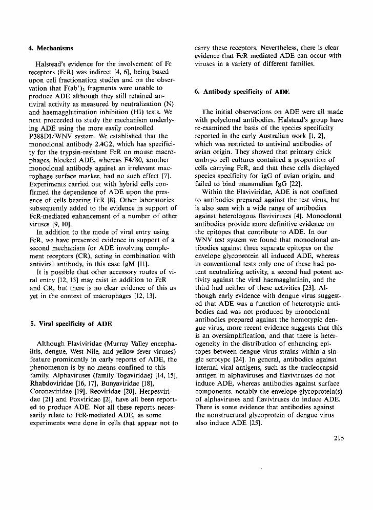
o f these diff icul t ies could be avoided by the use o f cell lines o f macrophages , or o f mac rophage - like cells, which were s tar t ing to become avail- able at abou t that t ime. Accordingly, Peiris and I looked at a number o f such cell lines and we were able to show enhancemen t o f the f lavivirus West Nile virus (WNV) in two mouse cell lines, P388D1 and J.774.1, and the h u m a n cell line U937 [5]. One m a j o r advan tage o f the P388D1 cell system was that W N V and several o ther viruses were lytic in these cells, so tha t it was poss ible to ob ta in a direct measurement o f A D E by count ing viral p laques tha t developed with and wi thout ant iviral ant ibody. (Fig. 1).
Fig. 1. Antibody dependent enhancement of West Nile virus. P388D1 cells were infected with mixtures of serial ten-fold dilutions of West Nile virus with serial ten-fold dilutions of a rabbit anti-West Nile virus antibody. The four wells in the right hand column show the plaques that developed when virus without antibody was added to the cells. High antibody concentrations neutralize the virus, whereas intermediate concentrations (10 -3 and 10 -4 ) show enhancement.
214
4. Mechanisms
Halstead's evidence for the involvement of Fc receptors (FcR) was indirect [4, 6], being based upon cell fractionation studies and on the obser- vation that F(ab')2 fragments were unable to produce ADE although they still retained an- tiviral activity as measured by neutralization (N) and haemagglutination inhibition (HI) tests. We next proceeded to study the mechanism underly- ing ADE using the more easily controlled P388D1/WNV system. We established that the monoclonal antibody 2.4G2, which has specifici- ty for the trypsin-resistant FcR on mouse macro- phages, blocked ADE, whereas F4/80, another monoclonal antibody against an irrelevant mac- rophage surface marker, had no such effect [7]. Experiments carried out with hybrid cells con- firmed the dependence of ADE upon the pres- ence of cells bearing FcR [8]. Other laboratories subsequently added to the evidence in support of FcR-mediated enhancement of a number of other viruses [9, 10].
In addition to the mode of viral entry using FcR, we have presented evidence in support of a second mechanism for ADE involving comple- ment receptors (CR), acting in combination with antiviral antibody, in this case IgM [11].
It is possible that other accessory routes of vi- ral entry [12, 13] may exist in addition to FcR and CR, but there is no clear evidence of this as yet in the context of macrophages [12, 13].
5. Viral specificity of ADE
Although Flaviviridae (Murray Valley encepha- litis, dengue, West Nile, and yellow fever viruses) feature prominently in early reports of ADE, the phenomenon is by no means confined to this family. Alphaviruses (family Togaviridae) [14, 15], Rhabdoviridae [16, 17], Bunyaviridae [18], Coronaviridae [19], Reoviridae [201, Herpesviri- dae [21] and Poxviridae [2], have all been report- ed to produce ADE. Not all these reports neces- sarily relate to FcR-mediated ADE, as some experiments were done in cells that appear not to
carry these receptors. Nevertheless, there is clear evidence that FcR mediated ADE can occur with viruses in a variety of different families.
6. Antibody specificity of ADE
The initial observations on ADE were all made with polyclonal antibodies. Halstead's group have re-examined the basis of the species specificity reported in the early Australian work [1, 2], which was restricted to antiviral antibodies of avian origin. They showed that primary chick embryo cell cultures contained a proportion of cells carrying FcR, and that these cells displayed species specificity for IgG of avian origin, and failed to bind mammalian IgG [22].
Within the Flaviviridae, ADE is not confined to antibodies prepared against the test virus, but is also seen with a wide range of antibodies against heterologous flaviviruses [4]. Monoclonal antibodies provide more definitive evidence on the epitopes that contribute to ADE. In our WNV test system we found that monoclonal an- tibodies against three separate epitopes on the envelope glycoprotein all induced ADE, whereas in conventional tests only one of these had po- tent neutralizing activity, a second had potent ac- tivity against the viral haemagglutinin, and the third had neither of these activities [23]. Al- though early evidence with dengue virus suggest- ed that ADE was a function of heterotypic anti- bodies and was not produced by monoclonal antibodies prepared against the homotypic den- gue virus, more recent evidence suggests that this is an oversimplification, and that there is heter- ogeneity in the distribution of enhancing epi- topes between dengue virus strains within a sin- gle serotype [24]. In general, antibodies against internal viral antigens, such as the nucleocapsid antigen in alphaviruses and flaviviruses do not induce ADE, whereas antibodies against surface components, notably the envelope glycoprotein(s) of alphaviruses and flaviviruses do induce ADE. There is some evidence that antibodies against the nonstructural glycoprotein of dengue virus also induce ADE [25].
215
7. Macrophage heterogeneity
Macrophage cell lines offer many advantages in the laboratory, but their use could be criti- cised as being removed from in vivo conditions. Primary macrophage preparations could be regarded as being more physiological, and as offering a closer approximation to the in vivo state. In some early studies, Peiris had had diffi- culty in demonstrating ADE in primary mouse peritoneal macrophages. Cardosa examined this problem in more detail and made some interest- ing observations on the effect which the state of activation of macrophages has upon their ability to show ADE. Resident peritoneal macrophages (RPM), harvested from untreated mice, thioglycollate elicited peritoneal macrophages (TPM), and BCG-activated peritoneal macro- phages (BCG-PM), were used in comparison with P388D1 cells and mouse L929 or pig kidney (PS) cells, which do not have Fc receptors. The well-characterised West Nile virus experimental system was used, exploring both FcR and CR mediated pathways. WNV replicates in all the cells studied. In the absence of antibody, non- macrophage cell lines were the most sensitive to viral infection, followed by macrophage cell lines, with primary macrophages the least sensitive. When infection was carried out in the presence of polyclonal antiviral IgG, ADE was greatest in BCG-PM, whether the end point was measured by peak enhancement ratio (virus yield in cul- tures with Ab over virus yield in cultures without Ab), or enhancement titre (greatest dilution of Ab showing 3-fold or greater ADE). RPM and TPM showed lower degrees of ADE. By con- trast, when complement dependent ADE was studied, TPM showed the greatest ADE, and BCG-PM were restrictive towards viral growth. Other variables which affected the outcome of cell/virus/antibody interactions were the strain and age of the mice used. TPM and BCG-PM prepared from CBAT6T6 mice produced substan- tially greater yields of virus than cells from C57B1/6 mice, whereas RPM from both strains of mice were equally sensitive. Surprisingly, al- though weanling mice are appreciably more sus- ceptible to WNV than adult mice, macrophages from weanling mice are relatively restrictive [26].
216
8. Flavivirus entry into macrophages
More recently we have been taking a closer look at the mechanisms of ADE using radio- labelled virus and electron microscopy to follow the course of viral entry into cells, including macrophages. The following points emerge from this study, which has been carried out by Gol- lins, working in my laboratory [27].
(1) Viral attachment to macrophages (studied at 4 °C) is substantially greater in the presence of an appropriate concentration of antiviral anti- body than in its absence.
(2) The infectivity of the virions attached to the P388D1 cell membrane could be measured by means of infectious centre assays using various dilutions of infected P388D1 cells on L929 monolayers as indicator cells. These showed that the specific infectivity of attached virus (ex- pressed as infectious centres per count per min- ute) was increased in the presence of sub- neutralizing concentrations of antiviral antibody. This was apparently because the FcR route of vi- ral entry was more efficient at mediating viral in- ternalization into macrophages (studied at 37 °C) than was the route of entry involving cellular membrane proteins to which WNV attached in the absence of antibody. It was also shown that virus particles which had interacted with anti- body and subsequently bound to the cell surface at 0 °C, went sequentially through a phase when they were optimally infectious for P388D1 cells, followed temporally by neutralization of the vi- rus.
Electron microscopic studies (submitted for publ.) have shown that the viral internalization pathway into macrophages is identical, both in the presence and absence of antiviral antibody, and that clathrin-coated vesicles mediate the ini- tial uptake of virions. Virions are then trans- ferred to endosomal, prelysosomal vacuoles, and finally into lysosomes.
How, then, does virus infect cells and escape degradation by lysosomal hydrolases?
More recent studies by Gollins have shown that much of the RNA from internalized virions escapes into the cytoplasm before the lysosomal compartment is reached, and that this occurs from the endosomal, prelysosomal vacuoles.
9. Enhancement in vivo
T h e ev idence tha t A D E o f viral r e p l i c a t i o n
c o n t r i b u t e s to d i sease in m a n is p r o b a b l y s t ron-
gest in the case o f d e n g u e h a e m o r r h a g i c fever
a n d the d e n g u e s h o c k syndrome . N o sa t i s f ac to ry
a n i m a l m o d e l for severe d e n g u e d isease exists, al-
t h o u g h e x p e r i m e n t s in m o n k e y s have s h o w n in-
c reased v i r a e m i a in a n i m a l s g iven d e n g u e v i rus in
c o m b i n a t i o n wi th pass ive a n t i b o d y [28]. T h e
"ea r ly d e a t h " p h e n o m e n o n wi th s o m e rabies vi-
rus in fec t ions in m a n a n d in a n i m a l s m a y be an
e x a m p l e o f A D E [29], as m a y a s imi la r ear ly
d e a t h in cats i n o c u l a t e d wi th fe l ine in fec t ious
panc rea t i t i s v i rus t o g e t h e r wi th a n t i b o d y aga ins t
t ha t v i rus [19]. A n awareness o f the poss ib i l i ty t ha t A D E m a y
c o n t r i b u t e to d i sease m a y reveal f u r t h e r exam-
ples.
References
[1] Hawkes, R. A. (1964) Austral. J. Exp. Biol. Med. Sci. 42, 465.
[2] Hawkes, R. A. and Lafferty, K. J. (1967) Virology 33, 250.
[3] Halstead, S. B. and O'Rourke, E. J. (1977) J. Exp. Med. 146, 201.
[4] Halstead, S. B., Porterfield, J. S. and O'Rourke, E. J. (1980) Am. J. Trop. Med. Hyg. 29, 638.
[5] Peiris, J. S. M. and Porterfield, J. S. (1979) Nature (Lon- don) 282, 509.
[6] Halstead, S. B., O'Rourke, E. J. and Allison, A. C. (1977) J. Exp. Med. 146, 218.
[7] Peiris, J. S. M., Gordon, S., Unkeless, J. C. and Porter- field, J. S. (1981) Nature (London) 289, 189.
[8] Peiris, J. S. M., Gordon, S., Porterfield, J. S. and Unke- less, J. C. (1981) in: Heterogeneity of Mononuclear
Phagocytes (O. Forster and M. Landy, Eds.) pp. 469-476, Academic Press, New York.
[9] Daughaday, C. C., Brandt, W. E., McCown, J. M. and Russell, P. K. (1981) Infect. Immun. 32, 469.
[10] Schlesinger, J. J. and Brandriss, M. W. (1981) J. Im- munol. 127, 659.
[11] Cardosa, M. J., Porterfield, J. S. and Gordon, S. (1983) J. Exp. Med. 158, 258.
[12] Marsh, M. (1984) Biochem. J. 218, 1. [13] Markwell, M. A. K., Portner, A. and Schwartz, A. L.
(1985) Proc. Natl. Acad. Sci. U.S.A. 82, 978. [14] Kimura, T., Ueha, N. and Minekawa, Y. (1981) Biken J.
24, 39. [15] Chanas, A. C., Gould, E. A., Clegg, J. C. S. and Var-
ma, M. G. R. (1982) J. Gen. Virol. 58, 37. [16] Clerx, J. E M., Horzinek, M. C. and Osterhaus, A. D.
M. E. (1978) J. Gen. Virol. 40, 297. [17] King, A. A., Sands, J. J. and Porterfield, J. S. (1984) J.
Gen. Virol. 65, 1091. [18] Millican, D. and Porterfield, J. S. (1982) J. Gen. Virol.
63, 233. [19] Weiss, R. C. and Scott, E W. (1981) Comp. Immun.
Microbiol. Infect. Dis. 4, 175. [20] Burstin, S. J., Brandriss, M. W. and Schlesinger, J. J.
(1983) J. Immunol. 130, 2915. [21] Inada, T., Chong, K. T. and Mires, C. A. (1985) J. Gen.
Virol. 66, 879. [22] Kliks, S. C. and Halstead, S. B. (1980) Nature (London)
285, 504. [23] Peiris, J. S. M., Porterfield, J. S. and Roehrig, J. T.
(1982) J. Gen. Virol. 58, 283. [24] Halstead, S. B., Venkateshan, C. N., Gentry, M. K. and
Larsen, L. K. (1984) J. Immunol. 132, 1529. [25] Henchal, E. A., McCown, J. M., Burke, D. S., Seguin,
M. C. and Brandt, W. E. 0985) Am. J. Trop. Med. Hyg. 34, 162.
[26] Cardosa, M. J., Gordon, S., Hirsch, S., Springer, T. A. and Porterfield, J. S. (1985) J. Virol., in press.
[27] Gollins, S. W. and Porterfield, J. S. (1984) J. Gen. Virol. 65, 1261.
[28] Halstead, S. B. (1979) J. Infect. Dis. 140, 527. [29] Prahhakar, B. S. and Nathanson, N. (1981) Nature (Lon-
don) 290, 590.
217