antibodies
DESCRIPTION
antibodiesTRANSCRIPT

Antibody
Dr. Wai Mar Linn
M.B.,B.S, DTM&H , MSc

Learning Outcomes
At the end of the lecture , the student must be able to describe
the followings
• Describe the origin, types and classes of antibodies.
• Detail the structure of an immunoglobulin and describe the
roles of various immunoglobulins.
• Explain the Function of antibodies , Antigen-Antibody
Binding and Its Results.
• Describe the genetic recombination leading to the diversity
among immunoglobulins. (Class switching)

Outlines
• Antibody ( Immunoglobulin)
• Antibody structure
• Classes of antibodies
• Function of antibodies
• Antigen-Antibody Binding and Its Results
• Affinity and Avidity

Antibody (Ab) (Immunoglobulins [Ig])
• Antibodies are gamma globulin proteins that react
specifically with the antigen that stimulated their
production.

Antibody
• Antibodies are proteins (immunoglobulins or gamma
globulins) produced by plasma cells in response to
stimulation by a specific antigen, capable of binding to the
antigen that stimulated their production.


• B-cells mature in the bone marrow.
• After the “education” process, immunocompetent but naïve B
lymphocytes leave the bone marrow to populate lymphoid tissues
and circulate through body fluids.
• The activated B-cell clonally proliferates to produce a population of
plasma cells and memory cells, which all recognize the same
antigen.
• The effector cells produced by B-cells are plasma cells, which
secrete antibodies.
• This is the humoral arm of the immune system.
• Memory cells, which are primed to deal with subsequent encounters
with the same antigen.
• Memory cells respond much more quickly and vigorously and form
the basis for the secondary or memory response of the immune
system.


Antibody
• They make up about 20% of the protein in blood plasma.
• Blood contains three types of globulins, alpha, beta, and
gamma, based on their electrophoretic migration rate.
• Antibodies are gamma globulins.

Antibody Structure
• Immunoglobulins made up of light (L) and heavy (H)
polypeptide chains.
• The simplest antibody molecule has a Y shape and consists
of four polypeptide chains: 2 H chains and 2 L chains.
• The four chains are linked by disulphide bonds.
• The terms light and heavy refer to molecular weight; light
chains have a molecular weight of about 25,000, whereas
heavy chains have a molecular weight of 50,000 to 70,000.
• An individual antibody molecule always consists of
2 identical H chains and 2 identical L chains.

Immunoglobulin

Antibody Structure
• L and H chains are subdivided into variable(V) and
constant(C )regions.
• The regions are composed of three-dimensionally folded,
repeating segments called domains.
• A L chain consists of one variable (VL) and one constant
(CL) domain.
• Most H chains consist of one variable (VH) and three
constant (CH) domains.

Immunoglobulin monomer

Antibody Structure
• The variable regions of both the light and heavy chain are
responsible for antigen-binding.
• The constant region of the heavy chain is responsible for
various biologic functions
(e.g.,complement activation and binding to cell surface receptors)
• The complement binding site is in the CH2 domain.
• The constant region of the light chain has no known biologic
function.
• The constant portion of the heavy chains forms the base and
the stem.


Antibody Structure
• Digestion with proteolytic enzyme (papin) cleaves on the
amino-terminal side of the inter-heavy chain disulphide
bonds, yielding two Fab fragments. (Fragment antigen
binding) they retain the ability to recognise the antigen .
• Other fragment can be easily crystallised and was termed
Fc (Fragment crystallisable)


Antibody Structure
• Antibodies have at least two identical antigen binding
(valence) sites.
• A monomer is a single bivalent antibody unit.
• Monomers consist of a combination of two heavy chains
and two light chains.
• Multivalent antibodies are composed of two or more
monomers.


Antibody Structure
• The variable regions of both L and H chains have three extremely
variable (hypervariable) amino acid sequences at the amino-
terminal end that form the antigen-binding site.
• Only 5 to 10 amino acids in each hypervariable region form the
antigen-binding site.
• Antigen–antibody binding involves electrostatic and van der
Waals’ forces and hydrogen and hydrophobic bonds rather than
covalent bonds.
• The remarkable specificity of antibodies is due to these
hypervariable regions.

Antibody Structure
• L chains belong to one of two types, κ (kappa) or λ
(lambda), on the basis of amino acid differences in their
constant regions.
• Both types occur in all classes of immunoglobulins (IgG,
IgM, etc.), but any one immunoglobulin molecule contains
only one type of L chain.
• The amino-terminal portion of each L chain participates in
the antigen-binding site.
• H chains are distinct for each of the five immunoglobulin
classes and are designated γ, α, μ, ε and δ .
• The amino-terminal portion of each H chain participates in
the antigen-binding site; the carboxy terminal forms the Fc
fragment

Antibody Structure
• The variable portion will always be the same on antibodies
produced by the same plasma cell, and will always
recognize the antigen that originally stimulated its
production.
• The structure of the stem, or Fc region determines the class
of an antibody and can be changed by an activated B-cell
(class switching) without the antigen specificity of the
antibody changing.
• Depending on the structure, the Fc region may bind and
activate complement, and may be recognized by Fc
receptors on macrophages, dendritic cells, neutrophils,
eosinophils, mast cells, and NK (natural killer) cells.

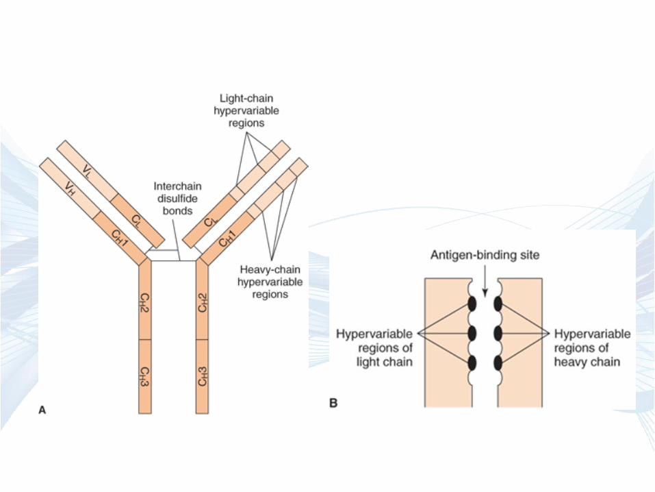
Structure of immunoglobulin G (IgG).
The Y-shaped IgG molecule consists of
two light chains and two heavy chains.
Each light chain consists of a variable
region and a constant region. Each
heavy chain consists of a variable region
and a constant region that is divided into
three domains: CH1, CH2, and CH3.
The CH2 domain contains the
complement-binding site, and the CH3
domain is the site of attachment of IgG
to receptors on neutrophils and
macrophages. The antigen-binding site
is formed by the variable regions of both
the light and heavy chains. The
specificity of the antigen-binding site is a
function of the amino acid sequence of
the hypervariable regions
The antigen-binding site is formed by the
hypervariable regions. A: Hypervariable
regions on immunoglobulin G (IgG). B:
Magnified view of antigen-binding site.
Antibody
The most important functions of antibodies are
• to neutralize toxins and viruses
• to opsonize microbes so they are more easily phagocytosed
• to activate complement and
• to prevent the attachment of microbes to mucosal surfaces.


Antigen-Antibody Binding and Its Results
• Antigen-antibody complex formation
• Inactivation of viruses and neutralization of bacterial toxins
• Agglutination of cellular antigens (causing cells to clump,
or agglutinate)
• Opsonization (macrophages have Fc receptors)
• Complement fixation


Antibody
• There are five classes of antibodies: IgG, IgA, IgM, IgE,
and IgD. (GAMED)
• Antibodies are subdivided into these five classes based on
differences in their heavy chains.

IMMUNOGLOBULIN CLASSES


IgG
• Each IgG molecule consists of two L chains and two H
chains linked by disulfide bonds .
• It has two identical antigen-binding sites, it is said to be
divalent.
• IgG has γ Heavy chain
IgG
• IgG is the predominant antibody in the secondary response
and constitutes an important defense against bacteria and
viruses
• IgG is the only antibody to cross the placenta; only its Fc
portion binds to receptors on the surface of placental cells.
It is therefore the most abundant immunoglobulin in
newborns.
• IgG is one of the two immunoglobulins that can activate
complement; IgM is the other
• IgG is the immunoglobulin that opsonizes. It can opsonize
(i.e., enhance phagocytosis) because there are receptors for
the γH chain on the surface of phagocytes.

IgG
• Monomer
• Most prevalent in serum (75% of the total)
• Major antibody of the secondary response
• Has the longest half life
• Pass through the placenta, most abundant Ig in newborn.
• Neutralizes bacterial toxins
• Agglutination
• Participates in complement fixation
• Enhances phagocytosis (opsonization)
• ADCC

IgA
• IgA is the main immunoglobulin in secretions such as
colostrum, saliva, tears, and respiratory, intestinal, and
genital tract secretions.
• serum IgA is a monomer and secretory IgA is a dimer
• IgA has α heavychain
• the dimer is held together by a J chain which is produced
by the antibody producing plasma cells.
• The secretory IgA(sIgA) is always in the dimeric form and
is composed of two basic four chain unit, a J chain and the
secretory component.
• The secretory component is part of the molecule that
transports the dimer produced by a submucosal plasma cell
to the mucosal surface.

IgA
• protects mucosal surfaces.
• It prevents attachment of microorganisms (e.g., bacteria and
viruses) to mucous membranes.

IgM
• present on the surface of both mature and immature B-cells
• As a monomer acting as an antigen receptor
• IgM has μ Heavy chain
• First antibody class secreted in the primary immune
response.
• Secreted as a pentamer, involved in agglutination and
complement fixation.

IgM • In serum, it is a pentamer composed of five H2L2 units
plus one molecule of J (joining) chain
• Pentamer has ten identical Ag binding sites and thus a
valence of 10

IgM
• It has the highest avidity of the immunoglobulins; its
interaction with antigen can involve all 10 of its binding
sites.
• It is the most efficient immunoglobulin in agglutination,
complement fixation (activation), and other antibody
reactions because the pentamer has 10 antigen-binding
sites.
• It and is important in defence against bacteria and viruses.
• It can be produced by the fetus in certain infections.

IgE
• Monomer
• Involved in allergic reactions.
• IgE has Ԑ Heavy chain
• It mediates immediate (anaphylactic) hypersensitivity
• It participates in host defenses against certain parasites
(e.g., helminths [worms])

IgE
• The Fc region of IgE binds to the surface of mast cells and
basophils.
• Bound IgE serves as a receptor for antigen (allergen).
• When the antigen-binding sites of adjacent IgEs are cross-
linked by allergens, several mediators are released by the
cells, and immediate (anaphylactic) hypersensitivity
reactions occur.
• Although IgE is present in trace amounts in normal serum
(approximately 0.004%), persons with allergic reactivity
have greatly increased amounts, and IgE may appear in
external secretions.
• IgE does not fix complement and does not cross the
placenta.

IgE
• worms are too large to be ingested by phagocytes, they are
killed by eosinophils that release worm-destroying
enzymes.
• IgE specific for worm proteins binds to receptors on
eosinophils, triggering the antibody-dependent cellular
cytotoxicity (ADCC) response

IgD
• Monomer
• Present on surface of mature B-cells
especially memory cells.
• IgD has ‘ δ ’ Heavy chain
• Function as an antigen receptor
• present in small amounts in serum.(< 1%)
• Have a very short half - life
• May be important for the secondary immune response
• May be important in regulating the immune response.


Immunoglobulin Isotypes
They are defined by difference in their heavy chain constant
region. Eg: class and sub classes
These determine their biological characteristics.
Eg . Complement activation ,cellular association via Fc
receptor and tissue distribution.

Class Switching
• Gene rearrangement to produce different immunoglobulin
(Ig) classes.
• IgM is formed first because the μ constant region is closest
to the VDJ DNA.
• Later the μ constant region can be switched with a γ, ε, or
α constant region to form the heavy chain of IgG, IgE, or
IgA, respectively.
• Note that the antigenic specificity of the B cell remains the
same because the VDJ DNA remains the same.
V : variable regions; D:diversity segments; J: joining
segments; C :constant regions; S : switch sites.


Affinity
Affinity defines the strength of the interaction between a
single antigen combining site and an epitope.
Avidity
Avidity defines the cumulative interactions of several
combining sites with an antigen.

