antibiotic resistance mechanism of esbl producing ... volume 2, issue 3/ijpab... · antibiotic...
TRANSCRIPT

www.ijpab.com 207
Antibiotic Resistance Mechanism of ESBL Producing Enterobacteriaceae in
Clinical Field: A Review
S. Thenmozhi1*, K. Moorthy 1, B. T. Sureshkumar1, M. Suresh2
1Department of Microbiology, Vivekanandha College of Arts and Sciences for Women (Autonomous), Elayampalayam- 637 205, Tiruchengode, Namakkal District, Tamil Nadu (India)
2Head-Microbiology and Molecular biology, Doctor’s Diagnostic Centre, Woraiyur, Trichy-620 003
*Corresponding Author E-mail: [email protected]
ABSTRACT
In modern medical practice, the development of antimicrobial resistance by Enterobacterriaceae is inevitable and is considered as a major problem in the treatment of bacterial infections in the hospital and in the community. Beta-Lactamase-Producing bacteria (BLPB) can play an important role in polymicrobial infections. They can have a direct pathogenic impact in causing the infection as well as an indirect effect through their ability to produce the enzyme beta-lactamase.These enzyme producing Enterobacteriaceae families are associated with a higher morbidity, mortality and fiscal burden.Extended spectrum β-lactamases (ESBL) are enzymes produced by a variety of Gram negative bacteria which confer an increased resistance to commonly used antibiotics.Extended-spectrum β-lactamases (ESBLs) are usually plasmid-mediated enzymes that confer resistance to a broad range of β-lactams. Initially, resistance to third-generation cephalosporins in Gram-negative rods was mainly due to the dissemination of TEM- and SHV-type ESBLs, which are point mutants of the classic TEM, SHV and CTX and enzymes with extended substrate specificity. Treatment of extended spectrum beta-lactamase (ESBL) producing strains of Enterobacteriaceae has emerged as a major challenge in hospitalised as well as community based patients. This article aims to give an overview of the current situation regarding ESBL producing Enterobacteriaceae with focus on the antibiotic resistance mechanism and management of such infection.
Keywords: Enterobacteriaceae, β-Lactamase, ESBL, Types of ESBL, β-Lactam antibiotics, Antibiotic resistance mechanism.
INTRODUCTION
We have been forced to fight against the newly acquired antibiotic resistance of various bacteria. Enterobacteriaceae, e.g. Escherichia coli (E.coli), Klebsiella, Enterobacter, Proteus, Pseudomonas produce many different beta-lactamase enzymes. Some have activity against only Penicillins and 1st, 2nd and 3rdgeneration Cephalosporins. However in recent year’s beta-lactamase enzymes capable of hydrolyzing extended-Spectrum Cephalosporins, e.g. Cefotaxime, Ceftriaxone, Ceptazidime and the monobactam, Aztreonam have been detected in numerous countries. These organisms frequently carry genes encoding resistance to other classes of antibiotics, e.g.aminoglycosides, quinolones and Co-trimoxazole. These are classified as multi-drug-resistance organisms (MDROs). Extended-spectrum ß-lactamases (ESBLs) have become increasingly common worldwide and have emerged as a major source of antimicrobial resistance in gram-negative pathogens. Except for Escherichia coli and Klebsiella pneumoniae, Proteus mirabilis is another common ESBL-producing gram-negative pathogen. It has been found that most ESBLs were derivatives of TEM-1 type, TEM-2 type and SHV-1 type of β-lactamase, which is composed of one or several point code gene mutation. In recent years, in addition to the TEM-type ESBLs, there also were increasing reports of CTX-M-type ESBLs produced by Proteus Mirabilis1, 2.
Available online at www.ijpab.com
ISSN: 2320 – 7051 Int. J. Pure App. Biosci. 2 (3): 207-226 (2014)
IINNTTEERRNNAATTIIOONNAALL JJOOUURRNNAALL OOFF PPUURREE && AAPPPPLLIIEEDD BBIIOOSSCCIIEENNCCEE
Review Article
www.ijpab.com 208
Thenmozhi, S. et al Int. J. Pure App. Biosci. 2 (3): 207-226 (2014) ISSN: 2320 – 7051
ESBLs confer resistance to Penicillins, Cephalosporins, Aztreonam, and also associated with resistance to other classes of non penicillin antibiotics, including Fluoroquinolones, Aminoglycosides, Trimethoprim sulfamethoxazole, and ß-lactam/ß-lactamase inhibitor combinations. Thus, ESBL-producing organisms often possess a multidrug resistance phenotype. Detection and susceptibility results of ESBL-producing Proteus mirabilis play an essential role in the treatment of infections caused by this pathogen and also in controlling the spread of ESBLs. Most ESBLs can be divided into three groups: TEM, SHV, and CTX-M types. Klebsiella pneumonia and Escherichia coli remain the major ESBL-producing organisms isolated worldwide, but these enzymes have also been identified in several other members of the Enterobacteriaceae family and in certain non-fermentors3. Resistance of bacteria to antibiotics has been noted ever since the discovery of penicillin by Fleming and its subsequent production by Florey. Antibiotic resistance may be intrinsic: This implies that the antibiotic is unable to have an effect on "wild-type" bacteria previously unexposed to the antibiotic in question. For example, Vancomycin is not active against gram-negative bacteria, nor has it ever been. Antibiotic resistance may also be either mutational or acquired. This implies changes in the bacteria that prevent the antibiotic from exerting its effect on the bacterial target, which may have resulted from either (1) mutation of existing genetic material within the bacteria or (2) acquisition of new genetic material from other bacteria. For example, Escherichia coli in its natural state may be susceptible to both Ampicillin and Ciprofloxacin. However, the mutation of existing genetic material may lead to Ciprofloxacin resistance, and the acquisition of genes that encode for beta-lactamase production may lead to resistance of E. coli to Ampicillin. The problems of antibiotic resistance are typically magnified in a hospital setting. Exposure to antibiotics while a patient is in the hospital may lead to genetic mutations that contribute to antibiotic resistance. Patients may be inadvertently exposed to the bacterial flora of other patients (usually due to a breakdown in basic infection control precautions). As a result, antibiotic-resistant bacteria may colonize multiple patients: Exposure of these patients to antibiotics may eliminate all but the most resistant bacteria. These resistant organisms may transfer antibiotic resistance genes to other bacteria, there by multiplying the problem4.Antimicrobial resistance is the ability of microbes, such as bacteria, viruses, parasites, or fungi, to grow in the presence of a chemical (drug) that would normally kill it or limit its growth.
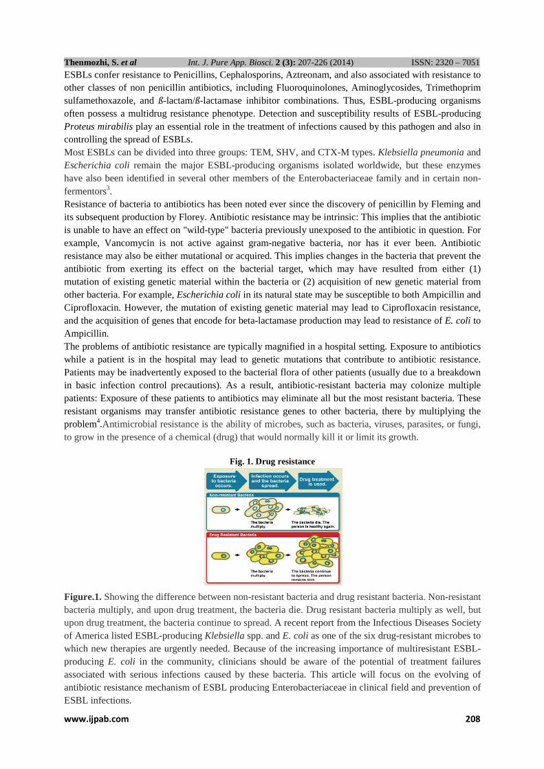
Fig. 1. Drug resistance
Figure.1. Showing the difference between non-resistant bacteria and drug resistant bacteria. Non-resistant bacteria multiply, and upon drug treatment, the bacteria die. Drug resistant bacteria multiply as well, but upon drug treatment, the bacteria continue to spread. A recent report from the Infectious Diseases Society of America listed ESBL-producing Klebsiella spp. and E. coli as one of the six drug-resistant microbes to which new therapies are urgently needed. Because of the increasing importance of multiresistant ESBL-producing E. coli in the community, clinicians should be aware of the potential of treatment failures associated with serious infections caused by these bacteria. This article will focus on the evolving of antibiotic resistance mechanism of ESBL producing Enterobacteriaceae in clinical field and prevention of ESBL infections.

www.ijpab.com 209
Thenmozhi, S. et al Int. J. Pure App. Biosci. 2 (3): 207-226 (2014) ISSN: 2320 – 7051
Enterobacteriaceae The members of the Enterobacteriaceae are gram-negative bacilli, which are usually resident in the gastrointestinal tract. Examples of such organisms include E. coli, K. pneumoniae, E. cloacae, Proteus mirabilis, and Citrobacter freundii. In patients hospitalized in ICUs, the Enterobacteriaceae account for approximately one third of all cases of ICU-acquired pneumonia, one third of all cases of ICU-acquired urinary tract infection, and 10% to 15% of ICU-acquired bloodstream infections. Options for treatment of the Enterobacteriaceae include beta-lactam antibiotics (Penicillin; Cephalosporins; Carbapenems; and the Monobactam, Aztreonam), beta-lactam antibiotics combined with beta-lactamase inhibitors, quinolones, TMP-SMX, Aminoglycosides, and Tigecycline5. Beta-lactamase production is the most common mechanism of resistance of the Enterobacteriaceae to Penicillins, Cephalosporins, or Aztreonam. The beta lactamases inactivate these antibiotics by splitting the amide bond of the antibiotic's beta-lactam ring. Over 300 different beta lactamases have been described. As mentioned previously, most strains of Enterobacter species, Citrobacter species, Providencia species, Morganella morganii, and Serratia species that are resistant to third-generation Cephalosporins produce an AmpC beta lactamase. Characteristically, AmpC beta lactamases can inactivate first- and second-generation cephalosporins (including the Cephamycins, such as Cefoxitin and Cefotetan) and third-generation Cephalosporins. These beta lactamases are not inhibited by beta-lactamase inhibitors, such as clavulanic acid. An important characteristic of AmpC beta lactamases is that their production can be increased by exposure of the bacteria to certain antibiotics. This phenomenon is known as induction. The amount of beta-lactamase production depends on the concentration of the antibiotic and the time of exposure. Penicillin, Ampicillin, most first-generation cephalosporins, cefoxitin, and imipenem are strong inducers of AmpC beta lactamases. However, all of these antibiotics except the carbapenem will be inactivated by the AmpC beta lactamase induced. In most populations of organisms, such as Enterobacter species, mutants exist that permanently hyper produce the AmpC type 1 beta lactamase. Organisms, such as K. pneumoniae, E. coli, or P. mirabilis, do not characteristically hyper produce the AmpC type 1 beta lactamase. Occasionally, they may acquire plasmid-mediated AmpC beta lactamases. However, much more commonly they may acquire ESBLs. These beta lactamases have important differences compared with AmpC beta lactamases. It is not appropriate to designate the AmpC beta lactamases as ESBLs because they are not derivatives of a parent beta lactamase with more limited spectra of activity (for example, TEM or SHV beta lactamases). The ESBLs also differ from the AmpC beta lactamases in that they are not able to inactivate the Cephamycins (for example, Cefoxitin or Cefotetan) and are inactivated in vitro by the beta-lactamase inhibitor clavulanic acid6. Unfortunately, multiple mechanisms of resistance may work together in producing resistance to a given class of antibiotics. The entry of any beta-lactam antibiotic into the bacterial cell is via outer membrane proteins, which function as doors through which the antibiotics pass. These proteins may be lost, contributing to decreased entry of the antibiotic and reduced antimicrobial activity. In some sites of infection with high organism load (for example, intra-abdominal abscesses or severe cases of ventilator-associated pneumonia), the huge amounts of beta-lactamase production by a high inoculum of organisms may overcome the effects of beta-lactamase inhibitors. Finally, as noted above, the AmpC beta lactamase (produced by organisms, such as Enterobacter species) is not susceptible to the effects of beta-lactamase inhibitors, and therefore may be inherently resistant to beta-lactam/beta-lactamase inhibitor combinations.
Quinolone antibiotics, such as Ciprofloxacin and Levofloxacin, are usually highly active in vitro against the Enterobacteriaceae family. However, the rates of resistance appear to be rising. There is an increased probability of resistance of ESBL-producing organisms to quinolones compared with non-ESBL-producing organisms of the same species7. The reasons for this co resistance are not entirely clear. Plasmid-mediated quinolone resistance has been reported and may contribute in some cases8.The usual mechanism of resistance to quinolones is mutation of the genes that encode the target enzymes (DNA gyrase and topoisomerase IV) for quinolones. Stepwise increases in resistance occur if there are mutations

www.ijpab.com 210
Thenmozhi, S. et al Int. J. Pure App. Biosci. 2 (3): 207-226 (2014) ISSN: 2320 – 7051
in one and then two of the genes encoding these enzymes. Although not yet fully characterized, alterations in outer membrane proteins coupled with active efflux pumps (which pump antibiotics out of the bacterial cell) appear to be important additional mechanisms of bacterial resistance to quinolones. E.coli and Klebsiella species Carbapenems are often used as drugs of last resort in the treatment of serious infections due to gram-negative bacilli. Resistance of the Enterobacteriaceae to carbapenems is generally rare, but may be mediated by the combination of outer membrane protein deficiency coupled with the production of beta lactamases. Rates of resistance to Ertapenem are generally higher than to Imipenem or Meropenem. In view of the comments above, serious infections due to the Enterobacteriaceae family should be managed with a beta lactam or quinolone antibiotic, which is active in vitro against the infecting organism. An important caveat is that ESBL-producing organisms may appear susceptible to third-generation cephalosporins (Ceftazidime, Cefotaxime, or Ceftriaxone) or Cefepime, yet be functionally resistant to these agents9.
High clinical failure rates are observed when Cephalosporins are used to treat bacteremia or pneumonia due to ESBL-producing Klebsiella species or, more rarely, ESBL-producing E. coli. Carbapenems (imipenem or meropenem) are the antibiotics of choice for serious infections due to ESBL producers10.
Polymyxins or tigecycline may be useful for infections that are resistant to carbapenems. A second important finding to reemphasize is that third-generation cephalosporins are associated with a significant risk for relapse of infection when used to treat AmpC-producing organisms, such as Enterobacter species.
Again, carbapenems or quinolones are the most reliable agents against Enterobacter species, Serratia species, or Citrobacter species. There is no evidence that combination therapy improves outcome or reduces the advent of resistance of the Enterobacteriaceae family11. Pseudomonas aeruginosa P.aeruginosa is a frequent and often the most troublesome of the gram-negative bacilli. It is a particular problem as a cause of ventilator-associated pneumonia. P.aeruginosa is also a common cause of both bloodstream infection and cholangitis. Antibiotic resistance is a major problem posed by P.aeruginosa; the organism displays a diverse array of antibiotic resistance mechanisms12.
Resistance to beta-lactam antibiotics is usually, but not exclusively, mediated by beta lactamases. P. aeruginosa produces a chromosomally encoded AmpC beta lactamase, which can hydrolyze antipseudomonal penicillins, Aztreonam, and third-generation cephalosporins. Derepressed mutants grossly hyperproduce this beta lactamase. A number of acquired beta lactamases can also be produced. The efflux pumps are an important mechanism of multidrug resistance, because they may confer resistance to quinolones, antipseudomonal penicillins, cephalosporins, and sometimes aminoglycosides. Tigecycline is ineffective against P. aeruginosa because of the presence of efflux pumps. Quinolone resistance in P.aeruginosa may also be mediated by mutations to the chromosomally mediated topoisomerases II and IV, whereas aminoglycoside resistance may be mediated by outer membrane impermeability or by aminoglycoside-modifying enzymes. Acinetobacter Species Acinetobacter species may also be capable of virtually complete antibiotic resistance. As is the case with P.aeruginosa, resistance of Acinetobacter species may be mediated by a combination of beta lactamases and outer membrane protein deficiencies. The role of efflux pumps is largely unexplored in Acinetobacter species, but may be important. A clinically useful observation has been the in vitro efficacy of Ampicillin-sulbactam in the face of resistance to almost all other drug classes. Sulbactam is able to bind to penicillin-binding protein 2 and therefore can impart direct antimicrobial activity against Acinetobacter species13.Carbapenems (for example, Imipenem or Meropenem) are often potent agents in the treatment of severe infections due to Acinetobacter species. This has been borne out in studies of Acinetobacter bacteremia14.
Ampicillin-sulbactam may represent a viable option to carbapenems15.In patients with Acinetobacter strains that are resistant to virtually all currently available antibiotics, Tigecycline or Colistin may be the only viable option16.As noted above, colistin has been combined with rifampin and other antibiotics, but reports of the success of these regimens are anecdotal only at this stage.

www.ijpab.com 211
Thenmozhi, S. et al Int. J. Pure App. Biosci. 2 (3): 207-226 (2014) ISSN: 2320 – 7051
I. Antibiotics: Definition: Antibiotics are molecules that kill, or stop the growth of microorganisms, including both bacteria and fungi. Antibiotics that kill bacteria are called "bactericidal". Antibiotics that stop the growth of bacteria are called "bacteriostatic" Fig. 1.1.
II. Origins of antibiotics 1. Most classes of antibiotics, including the β-lactam antibiotics, Tetracyclines, Aminoglycosides, and Macrolides. Originally derived from natural sources, and were then further chemically modified to confer better properties on the drug. 2. However, some important classes of antibiotics (including the sulfa antibiotics, the quinolones, and the oxazolidinones) are man-made, originating totally from synthetic chemical operations. Classification according to Spectrum of activity Depending on the range of bacterial species susceptible to these agents, antibacterials are classified as broad-spectrum, intermediate-spectrum, or narrow- spectrum. Note that the spectra of activity may change with acquisition of resistance genes, as will be discussed in the next module.
1. Broad spectrum antibacterials are active against both Gram-positive and Gram-negative organisms. Examples include: tetracyclines, phenicols, fluoroquinolones, “third-generation” and “fourth-generation” cephalosporins.
2. Narrow spectrum antibacterials have limited activity and are primarily only useful against particular species of microorganisms. For example, glycopeptides and bacitracin are only effective against Gram-positive bacteria, whereas polymixins are usually only effective against Gram negative bacteria. Aminoglycosides and sulfonamides are only effective against aerobic organisms, while nitroimidazoles are generally only effective for anaerobes.
β-Lactam antibiotics Beta-Lactams – are a group of antibiotics that include Penicillins and Cephalosporins. Beta-lactam antibiotics, including penicillins and the non-penicillin classes, share a basic chemical structure that includes a three-carbon, one-nitrogen cyclic amine structure known as the beta-lactam ring. The side chain associated with the beta-lactam ring is a variable group attached to the core structure by a peptide bond; the side chain variability contributes to antibacterial activity. As of the date of this publication, FDA has approved over 34 beta-lactam compounds as active ingredients in drugs for human use17. Beta-lactam antibiotics include the following five classes18.

www.ijpab.com 212
Thenmozhi, S. et al Int. J. Pure App. Biosci. 2 (3): 207-226 (2014) ISSN: 2320 – 7051
� Penicillins (e.g., ampicillin, oxacillin) � Cephalosporins (e.g., cephalexin, cefaclor) � Penems (e.g., imipenem, meropenem) � Carbacephems (e.g., loracarbef) � Monobactams (e.g., aztreonam) I. Penicillins A. History 1. 1928, Alexander Fleming noticed killing effect of mold accidentally blown onto his agar plate. After
attempt at isolation of compound responsible, judged to be too unstable for use as antibiotic. 2. 1938, Problem of isolating penicillin solved by Florey and Chain using a process called "Freeze
drying" now called lyophilization. 3. 1941, first clinical trial of penicillin were successful. 4. 1944, used against casualties in D-day landing. 5. 1945, structure of penicillin finally solved. B. Show structure of Benzylpenicillin (Penicillin G)
1. Structure was solved by x-ray crystallography by Dorothy Hodgkins 2. Previous to this, such a structure was proposed but was said to be "impossiblystrained"
C. Key features of structure 1. β-lactam ring
a. "Lactam" is a word for any cyclic amide (the word "lactone" is used for a cyclic ester) b. A β-lactam means that the nitrogen is joined to the carbon which is beta to the Carbonyl. c. this creates strain in the ring, since it is a four membered ring. d. b-lactam becomes good acylating agent for active site serine of penicillin binding Protein.
2. Carboxylate a. Negatively charged at neutral pH b. Anchors drug in active site pocket (positively charged)
3. Acylamido side chain a. Necessary for biological potency b. Proper stereochemistry of attachment to ring essential for activity c. Variation at side chain can dramatically affect biological activity against various strains of bacteria
D. Common Early Penicillins 1. Penicillin G had to be administered parenterally, since it is not acid stable 2. Penicillin V has more acid stability, and can be administered orally.

www.ijpab.com 213
Thenmozhi, S. et al Int. J. Pure App. Biosci. 2 (3): 207-226 (2014) ISSN: 2320 – 7051
II. Cephalosporins: Cephalosporins – include cefotaxine, ceftazidime and ceftriaxone
A. History 1. First isolated from fungus found in sewer line on island of Sardina in 1948. 2. Structure wasn't elucidated until 1961.
B. Prototypical early Cephalosporin: Cephalothin Biological source it is a semi-synthetic cephalosporin antibiotic derived from Cephalosporium acremonium.
1. Less antibiotic activity than Penicillin G against Gram positive bacteria. 2. More activity than Pen G against Gram negative bacteria. 3. Can be used on patients who are allergic to penicillin. 4. Side chain acetoxy group is hot point for metabolic inactivation.
C. Cephaloridine 1. Better leaving group in form of positively charged pyridinium group will "activate" system. 2. Avoids metabolic inactivation. 3. Note that compound is "zwitterion".
D. Ceftazidime 1. Combines activation of ceftazidime with steric shielding of b-lactam to protect it from
hydrolysis by β-lactamase. 2. Note additional hydrophilic groups on side chain further improve activity against gram negative
strains.

www.ijpab.com 214
Thenmozhi, S. et al Int. J. Pure App. Biosci. 2 (3): 207-226 (2014) ISSN: 2320 – 7051
Generation of Cephalosporins The cephalosporin nucleus can be modified to gain different properties. Cephalosporins are sometimes grouped into "generations" by their antimicrobial properties. The first cephalosporins were designated first-generation cephalosporins, whereas, later, more extended-spectrum cephalosporins were classified as second-generation cephalosporins. Each newer generation has significantly greater gram-negative antimicrobial properties than the preceding generation, in most cases with decreased activity against gram-positiveorganisms. Fourth-generation cephalosporins, however, have true broad-spectrum activity Table 1.
Table 1. Generation of Cephalosporin
In order for bacteria to grow, then, they need to make all the parts necessary for building new bacterial cells. DNA must be copied. New RNA, ribosome, and proteins must be made. Cell walls must be built. Membranes have to be synthesized. And, then, of course the cells must divide.Many, if not most, antibiotics act by inhibiting the events necessary for bacterial growth. Some inhibit DNA replication, some, transcription, some antibiotics prevent bacteria from making proteins, some prevent the synthesis of cell walls, and so on. In general, antibiotics keep bacteria from building the parts that are needed for growth. There are some antibiotics that act by attacking plasma membranes. Most antibiotics, though, work by holding bacterial populations in check until the immune system can take over Figure 2.
Fig. 2. Antibiotics working parts in the cell

www.ijpab.com 215
Thenmozhi, S. et al Int. J. Pure App. Biosci. 2 (3): 207-226 (2014) ISSN: 2320 – 7051
Different antibiotics have different modes of action, owing to the nature of their structure and degree of affinity to certain target sites within bacterial cells. 1. Inhibitors of cell wall synthesis. While the cells of humans and animals do not have cell walls, this
structure is critical for the life and survival of bacterial species. A drug that targets cell walls can therefore selectively kill or inhibit bacterial organisms. Examples: penicllins, cephalosporins, bacitracin and vancomycin.
2. Inhibitors of cell membrane function. Cell membranes are important barriers that segregate and regulate the intra- and extracellular flow of substances. A disruption or damage to this structure could result in leakage of important solutes essential for the cell’s survival. Because this structure is found in both eukaryotic and prokaryotic cells, the action of this class of antibiotic are often poorly selective and can often be toxic for systemic use in the mammalian host. Most clinical usage is therefore limited to topical applications. Examples: polymixin B and colistin.
3. Inhibitors of protein synthesis. Enzymes and cellular structures are primarily made of proteins. Protein synthesis is an essential process necessary for the multiplication and survival of all bacterial cells. Several types of antibacterial agents target bacterial protein synthesis by binding to either the 30S or 50S subunits of the intracellular ribosomes. This activity then results in the disruption of the normal cellular metabolism of the bacteria, and consequently leads to the death of the organism or the inhibition of its growth and multiplication. Examples: Aminoglycosides, macrolides, lincosamides, streptogramins, chloramphenicol, tetracyclines.
4. Inhibitors of nucleic acid synthesis. DNA and RNA are keys to the replication of all living forms, including bacteria. Some antibiotics work by binding to components involved in the process of DNA or RNA synthesis, which causes interference of the normal cellular processes which will ultimately compromise bacterial multiplication and survival. Examples: quinolones, metronidazole, and rifampin.
5. Inhibitors of other metabolic processes. Other antibiotics act on selected cellular processes essential for the survival of the bacterial pathogens. For example, both sulfonamides and trimethoprim disrupt the folic acid pathway, which is a necessary step for bacteria to produce precursors important for DNA synthesis. Sulfonamides target and bind to dihydropteroate synthase, trimethophrim inhibit dihydrofolate reductase; both of these enzymes are essential for the production of folic acid, a vitamin synthesized by bacteria, but not humans (Figure 3, Figure 4)
Fig. 3. Sites of antibacterial action Fig.4. Major drug sites

www.ijpab.com 216
Thenmozhi, S. et al Int. J. Pure App. Biosci. 2 (3): 207-226 (2014) ISSN: 2320 – 7051
ESBL (Extended Spectrum Beta-Lactamase) Spectrum→ Good effect against Enterobacteriaceae (E.coli, Klebsiella, Enterobacter), excellent effect against Enterococci. Beta-Lactamase→ is an enzyme produced by an organism that breaks down beta-lactams.ESBL stands for Extended Spectrum Beta-Lactamase. They are a group of bacteria usually associated with the bowel. A Beta-Lactamase is an enzyme produced by bacteria which breaks down certain types of antibiotics. ESBL producing bacteria are resistant to some of the antibiotics used to treat infection when it occurs. This resistance makes infection more difficult to treat. Enterobacteriaceae E.coli and Klebsiella pneumonia are common producers of ESBL, and they usually cause urinary tract infections and bacteraemia. People who carry ESBL producing bacteria without any sign or symptom of infection are “colonized”. Classification of β-Lactamase There are two system used to categorize ESBLs: I .The original method of β-lactamase categorisation is the Ambler classification which orders the enzymes into 4 classes (A, B, C, and D) based on molecular structure. ESBLs are Class A β-lactamases and may be defined as plasmid-mediated enzymes that hydrolyse Oxyimino-Cephalosporins, and Monobactams but not Cephamycins or Carbapenems. They are inhibited in vitro by Clavulanate. There are various genotypes of ESBLs. Of these, the most common are the SHV, TEM, and CTX-M types19. Other clinically important types include VEB, PER, BEL-1, BES-1, SFO-1, TLA, and IBC. II . In 1995, Bush et al., devised a classification of β-lactamases based upon their functional characteristics and substrate profile, a classification which is widely used. The enzymes are divided into three major groups: group 1 cephalosporinases which are not inhibited by clavulanic acid, the larger group 2 broad spectrum enzymes which are generally inhibited by clavulanic acid (except for the 2d and 2f groups) and the group 3 metallo-β-lactamases. The main points are illustrated in Table 2,3. Most ESBLs are assigned to group 2be, that is, hydrolyse penicillins, cephalosproins, and Monobactams, and inhibited by clavulanic acid (as per the Ambler classification). It should be noted that the CTX-M genotype was not classified in this original schemata but still fulfils the above criteria for group 2be enzymes20.
Table2.Classification of β – lactamaes according to Amber molecular scheme Class β – lactamases Examples A Broad spectrum β – lactamases
ESBL TEM – type ESBL SHV – type ESBL CTX – type Carbapenemases
TEM-1,TEM-2.SHV-1 TEM-3 SHV-5 CTX-M1,CTX-M9 KPC
Serine β - lactamases C AmpC Cephamycinases (chromosomal encode) AmpC cephamycinase (plasmid encode)
AmpC CMY,DHA
D Broad spectrum β – lactamases ESBL OXA–type carbapenemases
OXA-1,OXA-9,OXA-2,OXA-10,OXA-48,OXA-23
Metallo β - lactamases B Metallo β – lactamases VIM, IMP
Table 3: Amended from original Bush-Jacoby-Medeiros classification scheme for bacterial Bush-Jacoby
Group Molecular
class Preferred substrates Representative enzymes Resistance or
Susceptibility to β-lactamase inhibitors
1 C Cephalosporins AmpC Resistant 2b A Penicillins,Cephalosporins TEM,SHV Susceptible 2be A Penicillins,extended-spectrum
cephalosporins, Monobactams
TEM,SHV
Susceptible
2d D Penicillins,cloxacillin OXA Resistant 2e A Cephalosporins Inducible cephalosporinase
from Proteus vulgaris
Susceptible 2f A Penicillins,cephalosporins,carba-
penems NMC-A from Enterobacter
cloacae
Resistant 3 B Most β-lactams including
carbapenems L1 from Stenotrophomonas
maltophilia
Resistant

www.ijpab.com 217
Thenmozhi, S. et al Int. J. Pure App. Biosci. 2 (3): 207-226 (2014) ISSN: 2320 – 7051
1. Functional Classification Group 1 CEPHALOSPORINASE, Molecular Class C (not inhibited by clavulanic acid).Group 1 are Cephalosporinases not inhibited by Clavulanic acid, belonging to the molecular class C. Group 2 Penicillinases, Cephalosporinases, or both inhibited by clavulanic acid,corresponding to the molecular classes A and D reflecting the original TEM and SHV genes. However, because of the increasing number of TEM and SHV-derived β-lactumases, they were divided into two subclasses, 2a and 2b. 2.1. Group 2be: Extented-Spectrum, Molecular Class A. Subgroup 2be, with the letter “e” for extended spectrum of activity,represents the ESBLs, which are capable of inactivating third-generation cephalosporins (Ceftazidime,Cefotaxime, and Cefpodoxime) as well as monobactams (aztreonam). 2.2. Group 2br: Inhibitor-Resistant , Molecular Class A (diminished inhibition by clavulanic acid).The 2br enzymes, with the letter "r" denoting reduced binding to clavulanic acid and sulbactam, are also called inhibitor-resistant TEM – derivative enzymes; nevertheless, they are commonly still susceptible to tazobactam, except where an amino acid replacement exists at position met69. Group 2c: Carbenicillinase, Molecular Class A. Later subgroup 2c was segregated from group 2 because these enzymes inactivate Carbenicillin more than benzylpenicillin, with some effect on Cloxacillin. Group 2d: Cloxacilanase, Molecular Class D or A. Subgroup 2d enzymes inactivate Cloxacillin more than benzylpenicillin, with some activity against Carbenicillin; these enzymes are poorly inhibited by clavulanic acid, and some of them are ESBLs.The correct term is "OXACILLINASE". These enzymes are able to inactivate the oxazolyl penicillins like oxacillin, cloxacillin and dicloxacillin.The enzymes belong to the molecular class D not molecular class A. Group 2e: Cephalosporinase,Molecular Class A.Subgroup 2e enzymes are cephalosporinases that can also hydrolyse monobactams, and they are inhibited by clavulanic acid. Group 2f: Carbapenamase, Molecular Class A.Subgroup 2f was added because these are serine-based carbapenemases, in contrast to the zinc-based Carbapenemases included in group.3 Group 3 Metalloenzyme, Molecular Class B (not inhibited by clavulanic acid).Group 3 are the zinc-based or metallo β- lactumases, corresponding to the molecular class B, which are the only enzymes acting by the metal ion zinc. Metallo B-lactumases is able to hydrolyse penicillins, cephalosporins, and carbapenems. Thus, carbapenems are inhibited by both group 2f (serine-based mechanism) and group 3 (zinc-based mechanism). Group 4 Penicillinase, No Molecular Class (not inhibited by clavulanic acid) .Group 4 is penicillinases that are not inhibited by clavulanic acid, and they do not yet have a corresponding molecular class21. 2. Molecular Classification The molecular classification of β-lactumases is based on the nucleotide and amino acid sequences in these enzymes.To date, four classes are recognised (A-D), correlating with the functional classification. Classes A, C, and D act by a serine-based mechanism, whereas class B or metallo-β-lactumases need zinc for their action. 3. Extended-spectrum β-lactumase (ESBL)22-28 Members of the family Enterobacteriaceae commonly express plasmid-encoded β-lactumases (e.g., TEM-1, TEM-2, and SHV-1) which confer resistance to penicillins but not to expanded-spectrum cephalosporins. In the mid-1980s, a new group of enzymes, the extended-spectrum β-lactumases (ESBLs), was detected. (First detected in Germany in 1983). ESBLs are β-lactumases that hydrolyze extended spectrum Cephalosporins with an oxyimino side chain. These Cephalosporins include Cefotaxime, Ceftriaxone and Ceftazidime, as well as the oxyimino-monobactam Aztreonam. Thus ESBLs confer resistance to these antibiotics and related oxyimino-β lactums. In typical circumstances, they derive from genes for TEM-1, TEM-2, or SHV-1 by mutations that alter the amino

www.ijpab.com 218
Thenmozhi, S. et al Int. J. Pure App. Biosci. 2 (3): 207-226 (2014) ISSN: 2320 – 7051
acid configuration around the active site of this β-lactumases.This extends the spectrum of β-lactum antibiotics susceptible to hydrolysis by these enzymes. An increasing number of ESBLs not of TEM or SHV lineage have recently been described. The ESBLs are frequently plasmid encoded. Plasmids responsible for ESBL production frequently carry genes encoding resistance to other drug classes (for example, amino glycosides). Therefore, antibiotic options in the treatment of ESBL-producing organisms are extremely limited. Carbapenems are the treatment of choice for serious infections due to ESBL-producing organisms, yet carbapenem-resistant isolates have recently been reported. ESBL-producing organisms may appear susceptible to some extended-spectrum Cephalosporins. However, treatment with such antibiotics has been associated with high failure rates. Types TEM β-lactumases (class A) TEM-1 is the most commonly-encountered β-lactumase in Gram-negative bacteria. Up to 90% of ampicillin resistance in E.coli is due to the production of TEM-1. Also responsible for the ampicillin and penicillin resistance that is seen in H.influenzae and N.gonorrhoeae in increasing numbers. Although TEM-type β-lactumases are most often found in E.coli and K.pneumoniae, they are also found in other species of Gram-negative bacteria with increasing frequency. The amino acid substitutions responsible for the ESBL phenotype cluster around the active site of the enzyme and change its configuration, allowing access to oxyimino-β-lactum substrates. Opening the active site to β-lactum substrates also typically enhances the susceptibility of the enzyme to b-lactumase inhibitors, such as clavulanic acid. Single amino acid substitutions at positions 104, 164, 238, and 240 produce the ESBL.Phenotype, but ESBLs with the broadest spectrum usually have more than a single amino acid substitution. Based upon different combinations of changes, currently 140 TEM-type enzymes have been described. TEM-10, TEM-12, and TEM-26 are among the most common in the United States. SHV β-lactumases (class A) SHV-1 shares 68 percent of its amino acids with TEM-1 and has a similar overall structure. The SHV-1 β-lactumase is most commonly found in K. pneumoniae and is responsible for up to 20% of the plasmid-mediated ampicillin resistance in this species. ESBLs in this family also have amino acid changes around the active site, most commonly at positions 238 or 238 and 240. More than 60 SHV varieties are known. They are the predominant ESBL type in Europe and the United States and are found worldwide. SHV-5 and SHV-12 are among the most common. CTX-M β-lactumases (class A) These enzymes were named for their greater activity against cefotaxime than other oxyimino-β-lactum substrates (e.g., ceftazidime, ceftriaxone, or cefepime). Rather than arising by mutation, they represent examples of plasmid acquisition of β-lactumase genes normally found on the chromosome of Kluyvera species, a group of rarely pathogenic commensal organisms. These enzymes are not very closely related to TEM or SHV β-lactumases in that they show only approximately 40% identity with these two commonly isolated β-lactumases. More than 80 CTX-M enzymes are currently known. Despite their name, a few are more active on ceftazidime than cefotaxime. They have mainly been found in strains of Salmonella enterica serovar Typhimurium and E. coli, but have also been described in other species of Enterobacteriaceae and are the predominant ESBL type in parts of South America. (They are also seen in Eastern Europe) CTX-M-14, CTX-M-3, and CTX-M-2 are the most widespread. CTX-M-15 is currently (2006) the most widespread type in E. coli the UK and is widely prevalent in the community. OXA β-lactumases (class D) OXA β-lactumases were long recognized as a less common but also plasmid-mediated β-lactumase variety that could hydrolyze oxacillin and related anti-staphylococcal Penicillins. These β-lactumases differ from the TEM and SHV enzymes in that they belong to molecular class D and functional group 2d. The OXA-type β-lactumases confer resistance to Ampicillin and Cephalothin and are characterized by their high hydrolytic activity against Oxacillin and Cloxacillin and the fact that they are poorly inhibited by clavulanic acid. Aminoacid substitutions in OXA enzymes can also give the ESBL phenotype.
www.ijpab.com 219
Thenmozhi, S. et al Int. J. Pure App. Biosci. 2 (3): 207-226 (2014) ISSN: 2320 – 7051
While most ESBLs have been found in E. coli, K.pneumoniae, and other Enterobacteriaceae, the OXA-type ESBLs have been found mainly in P.aeruginosa. OXA-type ESBLs have been found mainly in Pseudomonas aeruginosa isolates from Turkey and France. The OXA β-lactumase family was originally created as a phenotypic rather than a genotypic group for a few β-lactumases that had a specific hydrolysis profile. Therefore, there is as little as 20% sequence homology among some of the members of this family. However, recent additions to this family show some degree of homology to one or more of the existing members of the OXA β-lactumase family.Some confer resistance predominantly to Ceftazidime, but OXA-17 confers greater resistance to Cefotaxime and Cefepime than it does resistance to Ceftazidime. Others Other plasmid-mediated ESBLs, such as PER, VEB, GES, and IBC β-lactumases, have been described but are uncommon and have been found mainly in P.aeruginosa and at a limited number of geographic sites.PER-1 in isolates in Turkey, France, and Italy; VEB-1 and VEB-2 in strains from Southeast Asia; and GES-1, GES-2, and IBC-2 in isolates from South Africa, France, and Greece. PER-1 is also common in multiresistant Acinetobacter species in Korea and Turkey. Some of these enzymes are found in Enterobacteriaceae as well, whereas other uncommon ESBLs (such as BES-1, IBC-1, SFO-1, and TLA-1) have been found only in Enterobacteriaceae. 4. Inhibitor-resistant β-lactumases Although the inhibitor-resistant β-lactamases are not ESBLs, they are often discussed with ESBLs because they are also derivatives of the classical TEM- or SHV-type enzymes. These enzymes were at first given the designation IRT for inhibitor-resistant TEM β-lactamase; however, all have subsequently been renamed with numerical TEM designations. There are at least 19 distinct inhibitor-resistant TEM β-lactamases. Inhibitor-resistant TEM β- lactamases have been found mainly in clinical isolates of E. coli, but also some strains of K. pneumoniae, Klebsiella oxytoca, P. mirabilis, and Citrobacter freundii .Although the inhibitor-resistant TEM variants are resistant to inhibition by clavulanic acid and sulbactam, thereby showing clinical resistance to the β-lactum lactumase inhibitor combinations of Amoxicillin-Clavulanate (Co-amoxiclav), Ticarcillin-clavulanate, and Ampicillin/sulbactam, they normally remain susceptible to inhibition by Tazobactam and subsequently the combination of Piperacillin/tazobactam, although resistance has been described. To date, these β-lactumases have primarily been detected in France and a few other locations within Europe. 5. AmpC-type β-lactumases (Class C) AmpC type β-lactumases are commonly isolated from extended-spectrum cephalosporin-resistant Gram-negative bacteria. AmpC β-lactumases (also termed class C or group 1) are typically encoded on the chromosome of many Gram-negative bacteria including Citrobacter, Serratia and Enterobacter species where its expression is usually inducible; it may also occur on Escherichia coli but is not usually inducible, although it can be hyperexpressed. AmpC type β-lactumases may also be carried on plasmids. AmpC β-lactumases, in contrast to ESBLs, hydrolyse broad and extended-spectrum Cephalosporins (Cephamycins as well as to oxyimino-β-lactums) but are not inhibited by β-lactumase inhibitors such as clavulanic acid. Carbapenemases Carbapenems are famously stable to AmpC β-lactumases and extended-spectrum-β-lactumases. Carbapenemases are a diverse group of β-lactumases that are active not only against the oxyimino-cephalosporins and Cephamycins but also against the carbapenems. Aztreonam is stable to the metallo-β-lactumases, but many IMP and VIM producers are resistant, owing to other mechanisms. Carbapenemases were formerly believed to derive only from classes A, B, and D, but a class C Carbapenemase has been described. IMP-type Carbapenemases (one of the metallo-β-lactumases) Plasmid-mediated IMP-type Carbapenemases, 17 varieties of which are currently known, became established in Japan in the 1990s both in enteric Gram-negative organisms and in Pseudomonas and Acinetobacter species. IMP enzymes spread slowly to other countries in the Far East, were reported from Europe in 1997, and have been found in Canada and Brazil.

www.ijpab.com 220
Thenmozhi, S. et al Int. J. Pure App. Biosci. 2 (3): 207-226 (2014) ISSN: 2320 – 7051
VIM (Verona integron-encoded metallo-β-lactumase) A second growing family of carbapenemases, the VIM family, was reported from Italy in 1999 and now includes 10 members, which have a wide geographic distribution in Europe, South America, and the Far East and have been found in the United States. VIM-1 was discovered in P. aeruginosa in Italy in 1996; since then, VIM-2 - now the predominant variant - was found repeatedly in Europe and the Far East; VIM-3 and -4 are minor variants of VIM-2 and -1, respectively.VIM enzymes occur mostly in P.aeruginosa, also P.putidaand, very rarely, Enterobacteriaceae. Amino acid sequence diversity is up to 10% in the VIM family, 15% in the IMP family, and 70% between VIM and IMP. Enzymes of both the families, nevertheless, are similar. Both are integron-associated, sometimes within plasmids. Both hydrolyse all β-lactums except monobactams, and evade all β-lactum inhibitors. OXA (oxacillinase) group of β-lactumases (Class D) The OXA group of β-lactumases occurs mainly in Acinetobacter species and is divided into two clusters. OXA carbapenemases hydrolyse carbapenems very slowly in vitro, and the high MICs seen for some Acinetobacter hosts (>64 mg/L) may reflect secondary mechanisms. They are sometimes augmented in clinical isolates by additional resistance mechanisms, such as impermeability or efflux. OXA carbapenemases also tend to have a reduced hydrolytic efficiency towards penicillins and cephalosporins. KPC (K. pneumoniae Carbapenemase) (Class A) A few class A enzymes, most noted the plasmid-mediated KPC enzymes, are effective Carbapenemases as well. Ten variants, KPC-2 through KPC-11 are known, and they are distinguished by one or two amino-acid substitutions (KPC-1 was re-sequenced in 2008 and found to be 100% homologous to published sequences of KPC-2). KPC-1 was found in North Carolina, KPC-2 in Baltimore and KPC-3 in New York. They have only 45% homology with SME and NMC/IMI enzymes and, unlike them, can be encoded by self-transmissible plasmids. The class A Klebsiella pneumoniae carbapenemase (KPC) is currently the most common carbapenemase, which was first detected in North Carolina, US, in 1996 and has since spread worldwide. A later publication indicated that Enterobacteriaceae that produce KPC were becoming common in the United States. CMY (Class C) The first class C carbapenemase was described in 2006 and was isolated from a virulent strain of Enterobacter aerogenes. It is carried on a plasmid, pYMG-1, and is therefore transmissible to other bacterial strains. Antibiotic Resistance Mechanisms Antibiotic resistance can be a result of horizontal gene transfer, and also of unlinked point mutations in the pathogen genome at a rate of about 1 in 108 per chromosomal replication. The antibiotic action against the pathogen can be seen as environmental pressure.Those bacteria with a mutation that allows them to survive live to reproduce. They then pass this trait to their offspring, which leads to the evolution of a fully resistant colony. Why does resistance develop? The large numbers of bacterial cells, combined with the short generation times facilitate the development of mutants. In a typical bacterial population of 1011 bacterial cells (e.g. in an infected patient) there can easily be 1000 mutants. If a mutant confers a selective advantage upon the bacterium (e.g. the ability to survive in the presence of an antibiotic) then that resistant bacterium will be selected and continue to grow while its neighbors perish. This can happen in a matter of days in patients being treated with antibiotics. Mode of Resistance Resistance to Penicillins and other β – lactams is due to one of four general mechanisms like29; 1. Inactivation of antibiotics by β – lactamase 2. Modification of target Penicillin- Binding Protein (PBP) 3. Impaired penetration of drug to target PBPs 4. Efflux

www.ijpab.com 221
Thenmozhi, S. et al Int. J. Pure App. Biosci. 2 (3): 207-226 (2014) ISSN: 2320 – 7051
Genetics of Antibiotic Resistance Bacterial resistance to antibiotics can be intrinsic or innate, which is characteristic of a particular bacterium and depends on biology of a microorganism (E.coli has innate resistance to vancomycin), and acquired resistance. Acquired resistance occurs from (i) acquisition of exogenous genes by plasmids (Conjugation or Transformation), Transposons (conjugation), Integrons and Bacteriophages (Transduction), (ii) mutation of cellular genes, and (iii) a combination of these mechanisms30. Spontaneous Mutations: Chromosomal mutations are quite rare (one in a population of 106–108 microorganisms) and commonly determine resistance to structurally related compounds. These mutations occur as errors of replication or an incorrect repair of damaged DNA. They are called spontaneous mutations or growth-dependent mutations. Resistance to quinolones in E. coli is caused by changes in at least seven amino acids in the gyrA gene or three amino acids in the parC gene whereas only a single point mutation in the rpoB gene is associated with a complete resistance to rifampin31. A chromosomal mutation in dihydropteroate synthetase results in a reduced affinity for sulfonamides). Some biochemical resistance mechanisms are the result of mutations. Antibiotic uptake or efflux system can be modified by mutations. Hypermutators: According to the “hypermutable state” model, a small bacterial population during a prolonged nonlethal selection of microorganisms may achieve a short-term state when the population mutates at a very high rate (hypermutable strains or mutators).These cells can increase the rate of mutations from 10 to 50 up to 10 000 times. Most hypermutators are found in populations of E. coli, S. enterica, Neisseria meningitides (N. meningitides), H. influenzae, S. aureus, Helicobacter pylori (H. pylori), Streptococcus pneumoniae (S.pneumoniae), and P. aeruginosa32. Adaptive Mutagenesis: Most mutations occur in dividing cells. However, they can also arise in nondividing or slowly dividing cells. Mutations occur only during nonlethal selection of microorganisms and are called “adaptive mutations.” This adaptive process is the only and main source of the antibiotic- resistant mutants to originate under normal conditions. Streptomycin causes a hypermutable phenotype in E. coli, and some antibiotics (quinolones) can induce the SOS mutagenic response and increase the rate of emergence of resistance to antibiotics33. Horizontal Gene Transfer:A transfer of resistance genes from one bacterium to another is called a horizontal gene transfer. The main mechanisms of resistance gene transfer in a bacterium are plasmid transfer, transfer by viral delivery, and transfer of free DNA Figure 5. Genes can be transferred by three main ways: transduction (via bacteriophages and integrons), conjugation (via plasmids and conjugative transposons), and transformation (via incorporation of chromosomal DNA, plasmids into a chromosome). Then genes are incorporated into the recipient chromosome by recombination or transposition and may have one or several changes in gene sequence.Most plasmids are double-stranded circular DNA whose size may vary from 2–3 kb to plasmids, which encode up to 10% of the host cell chromosome. The transfer of resistance genes is more effective than chromosomal mutation. Plasmids encode genes that confer resistance to main classes of antimicrobial agents (cephalosporins, fluoroquinolones, and aminoglycosides), toxic heavy metals (mercury, cadmium, silver), and virulence determinants that help a cell to survive in the environment of lethal antibiotic doses34. MDR genes are located in a DNA sequence that is transferred from one plasmid to another or to the genomes, which are called transposons or “jumping gene systems”35. Transposons can be integrated into plasmids or the host’s chromosome, encompass small elements called insertion sequences (IS elements), transposons, and transposing bacteriophages. They have terminal repeat sequences that play a role in recombination and recognize a protein for example, (transposase or recombinase) that is necessary to insert or remove a transposon from specifi c genome regions. Transposons are transferred by conjugation, transformation, or transduction (e.g., mecA gene is found in MRSA) and spread quicker than genes in chromosomes. Conjugative transposons have characteristic features of plasmids and can help to transfer endogenic plasmids from one microorganism to another36.

www.ijpab.com 222
Thenmozhi, S. et al Int. J. Pure App. Biosci. 2 (3): 207-226 (2014) ISSN: 2320 – 7051 Fig.5. Three main mechanisms of resistance gene transfer in a bacterium
(a) Plasmid transfer (Conjugation) (b) Transfer by viral delivery (Transduction) (c) Transfer of free
DNA (Transformation)
1. Antibiotic inactivation or modification: There are three main enzymes that inactivate antibiotics: β-lactamases, aminoglycoside-modifying enzymes, and chloramphenicol acetyltransferases. Antibiotic Modification by Hydrolysis: β-Lactamases are broadly prevalent enzymes that are classified using two main classification systems: Ambler and Bush-Jacoby-Medeiros. It is known about 300 different β-lactamases. The most clinically importantare produced by gram-negative bacteria and are coded on chromosomes and plasmids. Genes that encode β-lactamases are transferred by transposons but also they may be found in the composition of integrons . β-Lactamases hydrolyze nearly all β-lactams that have ester and amide bond, e.g., Penicillins, Cephalosporins, Monobactams, and Carbapenems. Serine β-lactamases – cephalosporinases,e.g. AmpC enzyme – are found in Enterobacter spp.and P. aeruginosa and penicillases in S. aureus. Metallo-β-lactamases (MBLs) found in P.aeruginosa, K. pneumoniae, E. coli, Proteus mirabilis(P. mirabilis), Enterobacter spp. have the same role as serine β-lactamases and are responsible for resistance to imipenem, new-generation cephalosporins and penicillins. MBLs are resistant to inhibitors of β-lactamases but sensitive to aztreonam37. Specific A. baumannii carbapenem - hydrolyzing oxacillinase (OXA) enzymes that have low catalytic efficiency together with porin deletion and other antibiotic resistance mechanisms can cause high resistance to carbapenems. The resistance of K. pneumoniae carbapenamases (KPC-1) toimipenem, meropenem, amoxicillin/clavulanate, Piperacillin/tazobactam, ceftazidime, aztreonam, and ceftriaxone is associated with the non conjugative plasmid coded bla gene. Extended-spectrum β-lactamases (ESBL) – TEM, SHV, OXA, PER, VEB-1, BES-1, GES, IBC, SFO and CTX – mainly are encoded in large plasmids. They can be transferred in connection of two plasmids or by transposon insertion. ESBL are resistant to penicillins (except temocillin), third-generation oxyimino-cephalosporins (e.g., Ceftazidime, Cefotoxime, Ceftriaxone), Aztreonam, cefamandole, cefoperazone, but they are sensitive to methoxy-cephalosporins,e.g., cephamycins and carbapenems,and can be inhibited by inhibitors of β-lactamases, e.g., clavulanic acid, sulbactam, or tazobactam. Strains producing ESBL are commonly resistant to quinolones but their resistance depends not on multiple resistance plasmids but on mutations in gyrA and parC genes38. Such strains are found among E. coli, K. pneumoniae, and P. mirabilis. The number of known ESBLs reaches 200.Hydrolysis of antibiotics can be run by other enzymes, e.g., esterases. E. coli gene ereB encodes erythromycin esterase II that hydrolyzes a lactone ring of erythromycin A and oleandomycin. ereB gene is prevalent in Enterobacteriaceae strain and is responsible for resistance to erythromycin A and oleandomycin. Ring-opening epoxidases cause resistance of bacteria to fosfomycin.

www.ijpab.com 223
Thenmozhi, S. et al Int. J. Pure App. Biosci. 2 (3): 207-226 (2014) ISSN: 2320 – 7051
2. Target Modification: An interaction between an antibiotic and a target molecule is very specific so even small changes in a target molecule can influence antibiotic binding to a target. Sometimes, in the presence of a modification in a target, other changes in the cell are needed to compensate an altered target. 3. Peptidoglycan Structure Alteration: Inhibition of cell wall synthesis is performed by β-lactams, e.g., penicillins, cephalosporins, carbapenems, monobactams,and glycopeptides, e.g., vancomycin and teicoplanin. The presence of mutation in PBPs leads to a reduced affinity to β-lactam antibiotics. It results in resistance of E. faecium to ampicillin and S. pneumoniae to penicillin. S. aureus resistance to methicillin and oxacillin is associated with integration of a mobile genetic element – “staphylococcal cassette chromosome mec” (SCCmec) – into the chromosome of S.aureus t at contains resistance gene mecA. mecA gene encodes PBP2a protein, a new penicillin-binding protein, that is required to change a native staphylococcal PBP. PBP2a shows a high resistance to β-lactam antibiotics (they do not bind to β-lactams) and ensures cell wall synthesis at lethal β-lactam concentrations. S. aureus strains resistant to methicillin can be cross resistant to all β-lactam antibiotics, streptomycin, and tetracycline and in some cases to erythromycin.When lesions in membrane proteins are present, cross-resistance between β-lactam antibiotics and fluoroquinolones is possible39. 4. Efflux Pumps and Outer Membrane Permeability: Membrane proteins that export antibiotics from the cell and maintain their low intracellular concentrations are called efflux pumps. Reduced outer membrane (OM) permeability results in reduced uptake of antibiotics Efflux Pumps. In analyzing resistance to antibiotics, identification and characterization of efflux pumps is one of the most actual problems. Single component efflux systems transfer their substrates across the cytoplasmic membrane. Multi-component pumps found in gram-negative bacteria and together with a periplasmic membrane synthesis protein (MFP) component and an OM protein (OMP) component transfer substrates across the cell envelope Figure 6. Antibiotics of all classes except Polymyxins are susceptible to the activation of efflux systems. Efflux pumps can be specific to antibiotics. Most of them are multidrug transporters that are capable to pump a wide range of unrelated antibiotics – Macrolides, Tetracyclines, Fluoroquinolones – and thus significantly contribute to MDR. Bacteria resistant to Tetracyclines often produce increased amounts of membrane proteins that are used as export or efflux pumps of antimicrobial drugs. To eliminate toxic compounds from the cytoplasm and periplasm, P. aeruginosa uses more than four powerful MDR efflux pumps40 (Mex).
Fig.6. A. System for antibiotic pumping out of the cell: B, Antibiotic interfering with
Ribosome in protein biosynthesis
A B

www.ijpab.com 224
Thenmozhi, S. et al Int. J. Pure App. Biosci. 2 (3): 207-226 (2014) ISSN: 2320 – 7051
PREVENTION AND CONTROL: MANAGEMENT OF INFECTION: 1. Health care workers (both hospital and community based) will undertake practices known to reduce the spread of ESBLs. These fall into two broad groups.
� Good hand hygiene and cleanliness � A restrictive approach to antibiotic prescribing, especially in the limitation of third generation
cephalosporin and quinolone use. These simple interventions can have a major influence on the impact of ESBLs in health care setting. 2. Appropriate use of antibiotics will greatly reduce the selection pressure for colonisation and infection with ESBLs:
� Antibiotics must be prescribed according to the Antimicrobial Policy and detailed Antimicrobial Prescribing Guidelines.
� Where there is more than one case on a ward, the prescriber should consider avoiding cephalosporin use altogether in other patients on the ward.
� In an outbreak situation, the Infection Control Doctor (ICD), a Consultant Medical Microbiologist and the Antimicrobial Pharmacist will suggest interim alternative antibiotic prescribing guidelines on a ward / unit.
CONCLUSION Antibiotic resistance is an important issue affecting public health, and rapid detection in clinical laboratories is essential for the prompt recognition of antimicrobial-resistant organisms. Infection-control practitioners and clinicians need the clinical laboratory to rapidly identify and characterise different types of resistant bacteria efficiently to minimise the spread of these bacteria and help to select more appropriate antibiotics. This is particularly true for ESBL-producing bacteria. The epidemiology of ESBL-producing bacteria is becoming more complex with increasingly blurred boundaries between hospitals and the community. E. coli that produce CTX-M β lactamases seem to be true community ESBL producers with different behaviours from Klebsiella spp, which produce TEM-derived and SHV-derived ESBLs. These bacteria have become widely prevalent in the community setting in certain areas of the world and they are most likely being imported into the hospital setting. A recent trend is the emergence of community-onset bloodstream infections caused by ESBL-producing bacteria, especially CTX-M-producing E. coli. These infections are currently rare, but it is possible that, in the near future, clinicians will be regularly confronted with hospital types of bacteria causing infections in patients from the community.β –lactums contribute a measure class of safer antibiotics. They are widely used as broad spectrum antibiotics for all the type of infections. New generation of antibiotics is predominantly preffered in clinical use. Many more new β- lactums are expected for the clinical use and many new β- lactums are expected in future. There is a better scope, prosperity for the discovery and development of new and safer β- lactums.The structure of β- lactams,their nature, classification, chemistry to be well studied. β - lactums, their mode of action, their bacteriocidal properties and their future growth is seen with new hopes.
REFERENCES 1. Luzzaro, F. Mezzatesta, M. Mugnaioli, C. Perilli, M. Stefani, S. Amicosante, G. et al., Trends in
Production of Extended-spectrum ß-Lactamases among Enterobacteriaceae of Medical Interest:Report of the Second Italian Nationwide Survey.Journalof Clinical Microbiology, 44:1659-1664 (2006)
2. Song, W. Kim, J. Bae, I.K. Jeong, S.H. Seo, Y.H. Shin, J.H. et al.,Chromosome-Encoded AmpC and CTXM Extended-Spectrum ß-Lactamases in Clinical Isolates of Proteus Mirabilis from Korea. Antimicrobial Agents and Chemotherapy, 55:1414-1419 (2011)
3. Johann, D.D. Pitout Kevin, B. Laupland. Extended Spectrum β- Lactamase- Producing Enterobacteriaceae: An emerging public-health concern. Lancet Infect.Dis.8:159-66 (2008)
4. David, L. Paterson, Maximizing Therapeutic Success in an Era of Increasing Antibiotic Drug Resistance. (2007)

www.ijpab.com 225
Thenmozhi, S. et al Int. J. Pure App. Biosci. 2 (3): 207-226 (2014) ISSN: 2320 – 7051
5. Richards, M.J. Edwards, J.R. Culver, D.H. Gaynes, R.P., Nosocomial infections in combined medical-surgical intensive care units in the United States. Infect Control Hosp Epidemiol. 21: 510-515 (2000)
6. Livermore, D.M. beta-Lactamases in laboratory and clinical resistance. Clin Microbiol Rev. 8: 557-584 (1995)
7. Paterson, D.L. Mulazimoglu, L. Casellas, J.M., et al. Epidemiology of ciprofloxacin resistance and its relationship to extended-spectrum beta-lactamase production in Klebsiella pneumoniae isolates causing bacteremia. Clin Infect Dis. 30: 473-478 (2000)
8. Martinez-Martinez, L. Pascual, A. Jacoby, G.A., Quinolone resistance from a transferable plasmid. Lancet. 351: 797-799 (1998)
9. Paterson, D.L. Ko, W.C. Von Gottberg, A. et al., Outcome of cephalosporin treatment for serious infections due to apparently susceptible organisms producing extended-spectrum beta-lactamases: implications for the clinical microbiology laboratory. J. Clin. Microbiol. 39: 2206-2212 (2001)
10. Paterson, D.L., Recommendation for treatment of severe infections caused by Enterobacteriaceae producing extended-spectrum beta-lactamases (ESBLs).Clin Microbiol Infect. 6: 460-463 (2000)
11. Chow, J.W. Fine, M.J. Shlaes, D.M. et al., Enterobacter bacteremia: clinical features and emergence of antibiotic resistance during therapy. Ann Intern Med. 115: 585-590 (1991)
12. Livermore, D.M., Multiple mechanisms of antimicrobial resistance in Pseudomonas aeruginosa: our worst nightmare? Clin. Infect. Dis. 34: 634-640 (2002)
13. Levin, AS., Multiresistant Acinetobacter infections: a role for sulbactam combinations in overcoming an emerging worldwide problem. Clin. Microbiol. Infect. 8: 144-153 (2002)
14. Cisneros, J.M. Reyes, M.J. Pachon, J., Bacteremia due to Acinetobacter baumannii: epidemiology, clinical findings, and prognostic features. Clin Infect Dis. 22: 1026-1032 (1996)
15. Wood, G.C. Hanes, S.D. Croce, M.A. et al., Comparison of Ampicillin-sulbactam and imipenem-cilastatin for the treatment of Acinetobacter ventilator-associated pneumonia. Clin. Infect. Dis. 34: 1425-1430 (2002)
16. Levin, A.S. Barone, A.A. Penco, J. et al., Intravenous colistin as therapy for nosocomial infections caused by multidrug-resistant Pseudomonas aeruginosa and Acinetobacter baumannii.Clin. Infect. Dis. 28: 1008-1011 (1999)
17. Approved beta-lactam antibiotics are listed in FDA’s Approved Drug Products with Therapeutic Equivalence Evaluations, generally known as the Orange Book (available on the Internet at http://www.accessdata.fda.gov/scripts/cder/ob/default.cfm).
18. Yao, J.D.C. and RC Moellering, Jr., Antibacterial agents, in Manual of Clinical Microbiology, 9th edition, edited by PR Murray et al., Washington D.C., ASM Press, (2007)
19. M. E. Rupp and P. D. Fey, “Extended spectrum β-lactamase (ESBL)-producing Enterobacteriaceae: considerations for diagnosis, prevention and drug treatment,” Drugs, 63(4): 353–365 (2003)
20. Rishi H.-P.Dhillon and John Clark. ESBLs: A Clear and Danger? Critical Care Research and Practice. 2012.
21. Siddheshwar, S.S. Pattan, S.R. Wabale, N.B. Pattan, J.S. Dighe, S.B. Bhavar, S.B. Gude, R.S., Need and Scope of Development of β–Lactums. Int. J. Pharm. Natural.Med., 1(1): 52-62 (2013)
22. Emery, C.L. Weymouth, L.A., "Detection and clinical significance of extended-spectrum β- lactumases in a tertiary-care medical center". J.Clin.Microbiol. 35(8): 2061–7(1997). PMC 229903. PMID 9230382.
23. Paterson, D.L. Hujer, K.M. Hujer, A.M. et al.,"Extended-spectrum b-lactumases in Klebsiella pneumoniae bloodstream isolates from seven countries: dominance and widespread prevalence of SHV and CTX-M-type beta-lactamases". Antimicrob Agents Chemother. 11: 3554–3560 (2003)
24. BradfordPA.,"Extended-spectrumβ-lactumases in the 21st century: characterization, epidemiology, and detection of this important resistance threat." Clin.Microbiol.Rev., 48: 933-51(2001)

www.ijpab.com 226
Thenmozhi, S. et al Int. J. Pure App. Biosci. 2 (3): 207-226 (2014) ISSN: 2320 – 7051
25. Jacoby George, A., Luisa Silvia Munoz-Price."Mechanisms of disease: The New β-Lactumases". N Engl .J .Med . 352 (4): 380–391(2005)
26. Paterson, D.L. Hujer, K.M. Hujer, A.M. et al.,"Extended-spectrum b-lactumases in Klebsiella pneumoniae bloodstream isolates from seven countries: dominance and widespread prevalence of SHVand CTX-M-type beta-lactumases". Antimicrob Agents Chemother, 11: 3554–3560 (2003)
27. Woodford, N. Ward, E. Kaufmann, M.E. et al., "Molecular characterisation of Escherichia coli isolates producing CTX-M-15 extended-spectrum β-lactumase (ESBL) in the United Kingdom" (PDF). Health Protection Agency.
28. Woodford N, Ward E, Kaufmann ME, et al., "Molecular characterisation of Escherichia coli isolates producing CTX-M-15 extended-spectrum β lactumase (ESBL) in the United Kingdom" (PDF).
29. Lakshmi R, Nusrin K.S., Georgy Sharon Ann, Sreelakshmi K.S., Role of beta lactamases in antibiotic resistance: A review. Int.Res.J.Pharm. 5(2): (2014)
30. Raghunath, D., Emerging antibiotic resistance in bacteria with special reference to India. J Biosci. 33(4): 593-603 (2008)
31. Rice, L.B. Sahm, D. Binomo, RA., Mechanisms of resistance to antibacterial agents. In: Murray, P.R. Baron, E.J. Jorgensen, J.H. Phaller, M.A. Yolken, R.H., editors.Manual of clinical microbiology. Washington: ASM Press; 1074-101 (2003)
32. Dzidic, S. Suskovic, J. Kos, B. Antibiotic resistance mechanisms in bacteria: biochemical and genetic aspects. Food Technol Biotechnol. 46: 11-21 (2008)
33. Guerin, E. Cambray, G. Sanchez-Alberola, N. Campoy, S. Erill, I. Da Re S, et al., The SOS response controls integron recombination. Science, 324: 1034-7(2009)
34. Hawkey, P. Molecular epidemiology of clinical significant antibiotic resistance genes. Br. J. Pharmacol. 153: 406-13 (2008)
35. Hawkey, PM., The origins and molecular basis of antibiotic resistance. BMJ, 317: 657-60 (1998) 36. Bennett, P.M., Plasmid encoded antibiotic resistance: acquisition and transfer of antibiotic resistance
genes in bacteria. Br .J .Pharmacol, 153: 347-57 (2008) 37. Thomson, J.M. Bonomo, R., The threat of antibiotic resistance in Gram-negative pathogenic bacteria:
β-lactams in peril! Curr. Opinion. Microbiol.,8: 518-24 (2005) 38. Vitkauskiene, A. Dudzevicius, V. Ryskus, L. Adukauskiene, D. Sakalauskas, R., Klebsiella
pneumoniae, gaminancių plateaus spektro veikimo beta laktamazes, isskyrimo is bronchų sekreto daznisir atsparumas antibiotikams.(The rate of isolation of Klebsiella pneumoniae producing extended spectrum beta-lactamases and resistance to antibiotics.) Medicina (Kaunas) 42(4): 288-93 (2006)
39. Lencastre, H. Oliveira, D. Tomasz, A., Antibiotic resistant Staphylococcus aureus: a paradigm of adaptive power. Curr .Opin. Microbiol., 10: 428-35 (2007)
40. Schweizer, H.P., Efflux as a mechanism of resistance to antimicrobials in Pseudomonas aeruginosa and related: unanswered questions.Genet Mol Res., 2(1): 48-62(2003)