antibiotic- and metal-resistant aeromonas isolated from
TRANSCRIPT

ANTIBIOTIC- AND METAL-RESISTANT Aeromonas
ISOLATED FROM ENVIRONMENTAL SOURCES
by
JENNIFER R. HUDDLESTON, B.S.
A THESIS
IN
BIOLOGY
Submitted to the Graduate Faculty of Texas Tech University in
Partial Fulfillment of the Requirements for
the Degree of
MASTER OF SCIENCE
Approved
May, 2003

ACKNOWLEDGEMENTS
My deepest gratitude goes to Dr. Randall Jeter for his patience, guidance,
suggestions, and helpful criticisms. I am also in debt to Dr. Michael San Francisco for
his assistance and Dr. John Zak for providing lab supplies.
Thanks go to Andrew Huddleston who sacrificed his time to help with the
sampling. Thanks also go to Jim Campbell for helping with the statistical analysis. I
would also like to show my appreciation to the science faculty at Hardin-Simmons
University who helped make the dream of graduate school a reality.
Finally, I would like to thank Andrew and my family for always being there and
supporting me. Without them, I would not be where I am today.
11

TABLE OF CONTENTS
ACKNOWLEDGEMENTS ii
ABSTRACT v
LIST OF TABLES vi
LIST OF FIGURES ix
CHAPTER
I. INTRODUCTION 1
The GenvLS Aeromonas 1
Antibiotic Resistance 5
Metal Resistance 8
Linked Antibiotic and Metal Resistance 10
Research Objectives 13
IL METHODS 15
Locations of Sampling 15
Sampling 16
Culture Media 17
Confirming the Identity of the Isolates 19
Antibiotics and Metals Susceptibility Testing 21
Biolog Identification of Isolates 23
Plasmid Purification 24
Preparation and Transformation of Competent Cells 24
i l l

Data Analysis 25
III. RESULTS 32
Sampling Dates, pH, and Temperature of the Water Bodies 32
Aeromonas Plate Coimts 33
Biolog Identification 36
Antibiotic, Drug, and Metal Resistance 38
Plasmid Isolations and Transformations 41
IV. DISCUSSION 76
Biolog Identification 76
Antibiotic and Drug Resistance 77
Metal Resistance 80
Resistant Isolates from Sediment 82
Plasmid Isolations and Transformations 84
The Absence of Linked Antibiotic and Metal Resistance 86
LITERATURE CITED 88
IV

ABSTRACT
Aeromonas is a ubiquitous aquatic bacterium that causes serious infections in both
cold- and warm-blooded animals, including humans. Clinical isolates of the organism
have shown an increasing incidence of antibiotic and antimicrobial drug resistances since
the widespread use of antibiotics began. The genes for antibiotic resistance and metal
resistance are frequently carried on the same plasmids, imparting both characteristics to
the host bacterium. When there are either antibiotics or metals present in the
environment, both markers are co-selected. Two hundred eighty-three Aeromonas
isolates belonging to eleven different species were isolated from several streams and both
urban and rural playa lakes in Lubbock, TX and New Mexico. The minimal inhibitory
concentrations of seven metals, six antibiotics, and two synthetic drugs were determined.
Low incidences of trimethoprim resistance, mercury resistance, and arsenite resistance
were found. Antibiotic and metal resistances were not linked in almost all of the
Aeromonas isolates. Plasmids were isolated from selected strains of the arsenite- and
mercury-resistant organisms and transformed into Escherichia coli XLl-Blue MRP',
showing that the resistance genes were carried on plasmids. From the data, it was
concluded that mercury and arsenite resistance could be transferred to other orgarisms in
natural environments.

LIST OF TABLES
2.1 Locations of sampling sites 27
2.2 Antibiotic and drug concentrations used in susceptibility testing 28
2.3 Metals concentrations used in susceptibility testing 29
2.4 Minimal inhibitory concentrations of antibiotics and drugs for Gram-negative, non-Enterobacteriaceae 30
2.5 Minimal inhibitory concentrations of metals for Gram-positive and Gram-negative bacteria 31
3.1 Average temperatures and pHs of sampling sites on particular dates 42
3.2 Total numbers of culturable bacteria (CFU/mL) and presumptive Aeromonas (CFU/mL) from all water sources 43
3.3 Total numbers of culturable bacteria (CFU/mL) and presumptive Aeromonas (CFU/mL) from the Brazos River, Lubbock, TX 46
3.4 Total numbers of culturable bacteria (CFU/mL) and presumptive Aeromonas (CFU/mL) from the playa at Jack Stevens Park, Lubbock, TX 46
3.5 Total numbers of culturable bacteria (CFU/mL) and presumptive Aeromonas (CFU/mL) from the playa at K.N. Clapp Park, Lubbock, TX 47
3.6 Total numbers of culturable bacteria (CFU/mL) and presumptive Aeromonas (CFU/mL) from the playa at Maxey Park, Lubbock, TX 47
3.7 Total numbers of culturable bacteria (CFU/mL) and presumptive Aeromonas (CFU/mL) from the playa at Higinbotham Park, Lubbock, TX 48
3.8 Total numbers of culturable bacteria (CFU/mL) and presumptive Aeromonas (CFU/mL) from the Rural Playa 1, Shallowater, TX 48
3.9 Total numbers of culturable bacteria (CFU/mL) and presumptive Aeromonas (CFU/mL) from the Rural Playa 2, Shallowater, TX 49
3.10 Total numbers of culturable bacteria (CFU/mL) and presumptive Aeromonas (CFU/mL) from the Rio Hondo, Taos Ski Valley, NM 49
VI

3.11 Total numbers of culturable bacteria (CFU/mL) and presumptive Aeromonas (CFU/mL) from the Pecos River, Carlsbad, NM 50
3.12 Biolog identification of 58 selected isolates, their sources, and number isolated 51
3.13 Comparison of numbers of Aeromonas isolated and identified from the Brazos River, Lubbock, TX, in March and July 2002 52
3.14 Comparison of numbers of Aeromonas isolated and identified from K. N. Clapp Park, Lubbock, TX, in March and July 2002 53
3.15 Comparison of numbers of Aeromonas isolated and identified from Frank Higinbotham Park, Lubbock, TX, in March and July 2002 54
3.16 Comparison of numbers of Aeromonas isolated and identified from Maxey Park, Lubbock, TX, in March and July 2002 55
3.17 Comparison of numbers of Aeromonas isolated and identified from Jack Stevens Park, Lubbock, TX, in March and July 2002 56
3.18 Accuracy of Aeromonas species identified by the Biolog identification
system across all sampling times and locations 57
3.19 Carbon source utilization of the isolates identified by Biolog 58
3.20 Percentages of identified Aeromonas isolates showing significant (>10%) anomalous carbon utilization 63
3.21 Minimal inhibitory concentrations of metals (mM) 64
3.22 Isolates resistant to 6 mMNaAs02, their sources, identification, and ratio of cell mass grown in 40% nutrient broth with 6 mMNaAs02to cell mass grown in 40% nutrient broth without NaAs02 65
3.23 Isolates resistant to 0.025 mMHgCh, their sources, and ratio of cell mass grown in 40% nutrient broth with 0.025 mMHgCla to cell mass grown in 40% nutrient broth without HgCh 66
3.24 Minimal inhibitory concentrations of antibiotics and drugs (\ig/mL) 67
3.25 Ampicillin-sensitive aeromonads 69
Vll

3.26 Isolates resistant to 8 ^ig/mL trimethoprim, their sources, and ratio of cell mass grown in 40% nutrient broth with 8 )j.g/mL trimethoprim to cell mass grown in 40%) nutrient broth without trimethoprim 70
3.27 Isolates resistant to 4 i g/mL trimethoprim, their sources, identification, and ratio of cell mass grown in 40%) nutrient broth with 4 )j,g/mL trimethoprim to cell mass grown in 40% nutrient broth without tiimethoprim 71
3.28 Isolates with trimethoprim resistance and a metal resistance 73
3.29 Isolates with one resistance and ampicillin sensitivity 73
3.30 Number of resistant isolates, their isolation sample types and sources 74
3.31 Plasmid isolations from arsenite- and mercury-resistant isolates 75
Vll l

LIST OF FIGURES
2.1 Map of rural playas 26
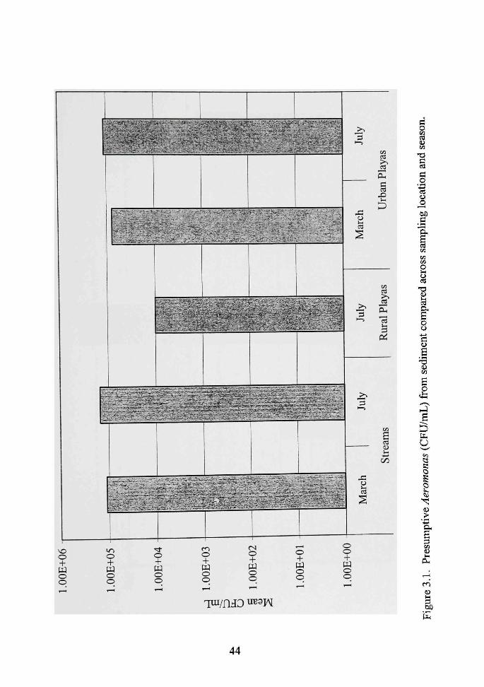
3.1 Presumptive Aeromonas (CFU/mL) from sediment compared across sampling location and season 44
3.2 Presumptive Aeromonas (CFU/mL) from water compared across
sampling location and season 45
3.3 Cumulative percentage plot for the NaAsOa MICs of the 283 isolates 64
3.4 Cumulative percentage plot for the trimethoprim MICs of the 283 isolates 68
3.5 Cumulative percentage plot for the cefuroxime MICs of the 283 isolates 68
IX

CHAPTER I
INTRODUCTION
The Genus Aeromonas
Members of the genus Aeromonas are ubiquitous aquatic bacteria. They are
Gram-negative rods that are oxidase positive and facuUatively anaerobic (Holt et al.,
1994). They are also resistant to vibriostatic agent 0/129 and do not require NaCl for
growth (Pemberton et al., 1997). They now belong to the family Aeromonadaceae.
Aeromonas was considered to be in the family Vibrionaceae until 1986. The
genera Vibrio and Aeromonas share many phenotypic characteristics (Overman et al.,
1985). They are so similar that, even now in the clinical setting, Aeromonas is sometimes
still misidentified as Vibrio (Abbott et al., 1998). However, despite their similarities,
Aeromonas and Vibrio each have different phylogenetic histories. This conclusion was
based on rRNA-DNA binding competition studies. As a consequence, Aeromonas was
removed from the family Vibrionaceae and a new family, Aeromonadaceae, was created
(Colwell et al., 1986).
The sixteen species in the genus Aeromonas can be divided either into phenotypic
groups or DNA hybridization groups. The members of the genus are phenotypically
similar and are easily confused (Valera and Esteve, 2002). There are four phenotypic
groups or phenons. These groups contain the phenospecies Aeromonas hydrophila, A.
caviae, A. sobria, and A. salmonicida. The Aeromonas genus is further broken down into
as many as fourteen DNA hybridization groups (Camahan and Altwegg, 1996).
Mesophilic aeromonads can be found in almost every aquatic environment. They
are found in both polluted and unpolluted environments as well as in chlorinated drinking
water distiibution systems around the world (Fernandez et al., 2000; Fox et al., 1990;
Huys et al., 1997; Kiihn et al., 1997a), raw sewage (Holmes et al., 1996), and in the
oceans at the freshwater/marine interfaces (Hazen et al., 1978). Aeromonads can also be
found thriving in deep groundwater (McKeon et al., 1995).
Since Aeromonas species are ubiquitous in aquatic environments, it is common
for fish, amphibians, and reptiles to come into direct contact with these bacteria. These
cold-blooded organisms can become infected with Aeromonas under certain
circumstances. Aeromonas causes, among many other diseases, hemorrhagic septicemia
in fish, red-leg disease in frogs, and ulcerative or necrotic stomatitis in snakes (Gosling,
1996). Aeromonas infections in fish are of major concem in the fish-farming industry.
A. hydrophila, A. sobria, A. allosaccharophila, A. salmonicida, and A. veronii are
the major causative agents offish infections. There is some debate as to whether v4.
hydrophila is a primary pathogen of fish or just an opportunistic pathogen of weakened
fish. Whatever the case may be, A. hydrophila plays a major role in the development of
disease in fish. A. hydrophila causes redsore disease, tail and fin rot, and hemorrhagic
septicemias. In cultivated fish, A. salmonicida causes furunculosis, which are boil-like
lesions in the musculature, and is particularly devastating in aquaculture. Since
Aeromonas infections can have such devastating consequences on aquaculture,
chemotherapy has been extensively used to prevent and treat the infections. However,

antibiotic-resistant strains of Aeromonas have emerged, making freatment more difficult
(Austin and Adams, 1996).
The range of susceptible hosts for Aeromonas infections does not stop with fish
and other cold-blooded organisms. Warm-blooded animals such as dogs, birds, and
humans can also become infected with Aeromonas. These infections can be in the form
of septicemia, pneumonia, or wound infections (Gosling, 1996). In humans, Aeromonas
is also a probable causal agent of gasfrointestinal disease. Although it has never been
proven as a true causal agent, high numbers of Aeromonas have been isolated from the
gastrointestinal tract of those presenting with the symptoms of human diarrheal disease
(Joseph, 1996; Kiihn et al., 1997b). Recently, the enterotoxin genes ah, act, and ast have
been found in aeromonads associated with diarrhea (Albert et al., 2000; Shat et al., 2002).
The most common Aeromonas infections in humans result in gastrointestinal distress
(Janda and Abbott, 1996). Most of the diarrheal diseases associated with. Aeromonas are
self-limiting and do not require antibiotics (Khardori and Fainstein, 1988).
In a severe Aeromonas infection, A. sobria was associated with acute renal failure
in an infant. The symptoms the child displayed were very similar to infections caused by
Escherichia coli 0157:H7. She had watery, bloody diarrhea and renal failure. However,
further investigation of stool cultures showed Aeromonas as the only probable pathogen.
The infant had been bathed in the same bathtub as had been used to clean an aquarium.
The suspected organism was also found in the fish tank (Filler et al., 2000).
Extraintestinal infections commonly occur when a woimd is exposed to
Aeromonas-contamimted water. The most common Aeromonas wound infections in

humans are attributed to A. hydrophila, A. veronii, A.jandaei, A. trota, and A. schubertii
(Janda and Abbott, 1996). Woimd infections can progress quickly and may ultimately be
fatal if the infection becomes systemic. Cellulitis, myonecrosis, and ecthyma
gangrenosum are the results of wound infections and are treated with antibiotic therapy.
The latter two types of infections may progress seriously enough for amputation to be
required. Swimming accidents, boating accidents, alligator bites, and fishing hook
accidents are ways people that have been wounded become infected with Aeromonas
(Janda and Abbott, 1996).
Nosocomial infections from leeches are another example of opportunistic
Aeromonas infection. Leeches are still used medicinally after some surgeries such as
replants, grafts, and reconstructive surgery (DeChalain, 1996). Leeches and^.
hydrophila are symbionts. The presence of Aeromonas is critical to the leeches.
Aeromonads provide the hemolytic enzymes necessary to break down blood for the
leech's nutrition. A. hydrophila from the gut of leeches has caused opportunistic
infections in the treated patients (Sartor, 2002).
Not only are aeromonads carried in leeches, but they can also survive within
houseflies. A. caviae can persist in the housefly gut for up to eight days and can be
transferred to other flies and food. Houseflies can serve as mechanical vectors, spreading
the organism to various environments where they may eventually act as pathogens
(Nayduch et al., 2002).
Because of the known and suspected infections aeromonads cause, the National
Academy of Sciences considers the organism to be an emerging microbial threat to health

(Lederberg et al., 1992). Aeromonas may eventually become a greater problem by
causing nosocomial infections (other than from leeches) (Ko et al., 1998). Aeromonas
may be a greater threat to human health when resistance to antimicrobial agents is
considered.
Antibiotic Resistance
Antibiotics and metals normally inhibit the growth of Aeromonas and other
microorganisms. However, these organisms can develop resistance to one or both of
these groups of antimicrobial agents. Under natural conditions, antibiotic resistance may
not be very important. In imcontaminated environments, there is only a low incidence of
antibiotic resistance (McArthur and Tuckfield, 2000).
Antibiotics are chemicals produced by microorganisms that kill or inhibit other
microorganisms. Different antibiotics have different cellular targets. For example, |3-
lactam antibiotics inhibit cell wall synthesis, kanamycin targets 16S rRNA, and
tetracycline attacks a site of the ribosome and inhibits protein translation. Microbes can
develop resistance to antibiotics through several different avenues as discussed later
(Snyder and Champness, 1997).
With the widespread use of antibiotics since the 1950s, populations of multi-
resistant microorganisms—microorganisms resistant to more than one antimicrobial
agent—and multi-resistance genes have emerged (Davies, 1994). Antibiotics act as a
selective agent. They kill or inhibit susceptible sti-ains, allowing sfrains that harbor
resistance genes to propagate. The resistant bacteria then comprise the majority of the

bacterial population (Levy, 1997). These resistant microorganisms are continuing to
increase in frequency for clinical isolations. A relevant example is that clinical
Aeromonas isolates in Taiwan are showing increased incidence of resistance to
teti-acycline, trimethoprim-sulfamethoxazole, some extended spectrum cephalosporins,
and aminoglycosides compared to aeromonads isolated from the United States and
Austraha (Ko et al., 1996).
Bacterial resistance to antibiotics and drugs comes from a variety of different
mechanisms. These mechanisms are encoded by genes on the microorganism's
chromosome or by a fransferable element and include the following. (1) The uptake of
the antibiotic into the cell is reduced. Chloramphenicol resistance occurs in this manner
(Davies, 1994). (2) The antibiotic is actively effluxed out of the cell as with P-lactams
(Zhang et al., 2000). (3) The antibiotic's cellular target is modified to inhibit the binding
of the antibiotic. One of the mechanisms of tetracycline resistance occurs through
ribosomal protection proteins (Chopra and Roberts, 2001). (4) Enzymes inactivate the
antibiotic, as is the case with P-lactamases that hydrolyze P-lactams (Walsh et al., 1998).
(5) Proteins bind to the antimicrobial and sequester it (Davies, 1994). The last two
mechanisms are used in resistance to sulfonamides and trimethoprim (6). There is a
metabolic bypass around the affected reaction (Davies, 1994). (7) The bacterial cell
overproduces the target so that some of the target may survive an antibiotic attack
(Davies, 1994).
It is thought that plasmids carrying genes for multi-resistance in pathogens have
occurred only in the past fifty years (Davies, 1994). Plasmids are extrachromosomal self-

replicating molecules of DNA. They can be ti-ansferred from organism to organism. It
was recently demonsti-ated that ft-ansfer of plasmids between Aeromonas isolated from
hospitals to Aeromonas isolated from aquaculture is possible (Rhodes et al., 2000). At
least three different genetic groups of resistance plasmids exist in Aeromonas (Hedges et
al., 1985). Transfer of resistance plasmids can occur between members of the same
species or between organisms from different genera or even families (Davies, 1997).
Antibiotic resistance genes can even be transferred across the Gram barrier; for example,
plasmids from a Gram-negative organism can be tiansferred to a Gram-positive organism
(Courvalin, 1994). On the other hand, transfer of resistance plasmids can also occur at
low frequencies and with a narrow host range (Pickup et al., 1997).
Antibiotic and metal resistance can be inherent or acquired. Most members of the
genus Aeromonas, with the exception of 4. trota, are inherently resistant to ampicillin
due to the production of 13-lactamases (Overman and Janda, 1999). The genes for B-
lactamases in aeromonads are chromosomally located (Jones and Wilcox, 1995). B-
Lactamases are enzymes that hydrolyze the amide bond of the 6-lactam ring of the
penicillins and cephalosporins, rendering the antibiotics inactive (Bastarrachea, 1998).
However, some Aeromonas have been isolated that are not ampicillin resistant (Kilpatrick
et al., 1987). Antibiotic susceptibility tests have been run on the members of the genus
Aeromonas to find patterns that differentiate between the species. The results show that
the species carmot be determined from antibiotic susceptibility patterns (Kampfer et al.,
1999; Overman and Janda, 1999; Vila et al., 2002). Acquired resistance occurs when a
previously susceptible organism develops resistance through a chromosomal mutation or

by acquisition of genes through plasmids or transposable elements (Murray and Hodel-
Christian, 1991).
Metal Resistance
A "metal" is an element whose oxides form hydroxides with water and whose
compounds form positive ions when in solution. The theory of "hard" or "soft" acids and
bases more accurately describe metals. Another metal classification system is based on
equilibrium constants involved in the formation of metal ion-ligand complexes. This
classification system is divided into Class A, Class B, and a borderline group. The two
classification schemes are similar. Hard acids (Class A) are small in size, have low
polarizability, and a high positive oxidation state. These include lithium, magnesium,
and lead, among others. Soft acids (Class B) are large, have low polarizability, and low
electronegativity. Some of these are copper, mercury, and silver. The group of
borderline metals includes iron, nickel, cobalt, copper, and zinc (Collins and Stotzky,
1989).
Metal resistance is nothing new to bacteria. In fact, bacteria had to have
resistance mechanisms in order to survive the high concentrations of metals in the
environment from volcanic activity 3 to 4 billion years ago (Silver et al., 1989). Some
metals, unlike antibiotics, have a biological role in the metabolism of microorganisms.
These metals belong to Class A (Collins and Stotzky, 1989). Calcium, cobalt, chromium,
iron, potassium, magnesium, manganese, sodium, nickel, and zinc are required for hfe.
Iron and nickel are important in redox reactions, while magnesium and zinc help to

stabilize enzymes and DNA. Complex organic molecules sometimes contain iron,
magnesium, nickel, or cobalt (Nies, 1992). Metals are cofactors in many other important
enzymes as well (Wackett et al., 1989). Finally, sodium and potassium help to maintain
osmotic pressure (Nies, 1992).
Other metals such as silver, aluminum, cadmium, gold, lead, and mercury are
nonessential for microbial survival. These metals belong to Class B (Collins and Stotzky,
1989). High concenfrations of both the essential and nonessential metals are toxic to
bacteria. Essential metals can be displaced from their binding sites by the nonessential
metals (Hughes and Poole, 1989). High concenti-ations of metals can alter the
conformation of proteins and the structxire of nucleic acids. Oxidative phosphorylation is
also disrupted and osmotic balance is interrupted (Poole and Gadd, 1989).
At the biochemical level, these metals are toxic, and this effect radiates out to the
population and community level. In general, metal-polluted environments have a
decreased microbial biomass, metabolic activity, and diversity (Roane and Kellogg,
1996). Microbial communities can also adapt to metal contamination. One group of
researchers found very diverse groups of chromium-resistant bacteria from chromium-
contaminated sludge (Francisco et al., 2002). Organisms resistant to metals are often
found in most natural habitats (Amebrant et al., 1987; Roane and Kellogg, 1996).
Horizontal gene transfer of resistance plasmids has been known to occur in polluted
environments. This can eventually result in restoration of species diversity in microbial
communities (Rasmussen and Sorensen, 1998).

Metal resistance is found in almost every bacterial group (Silver and Misra,
1984). There are several possible mechanisms by which organisms are resistant to
metals, and many of them are the same strategies employed by bacteria to eliminate the
damaging effects of antibiotics. (1) The metal can be excluded from the cell by a
permeabihty barrier (Bruins et al., 2000). (2) There is reduced uptake of a particular
metal in order to protect the cellular components (Ahuja et al., 2001). (3) The metal can
be actively transported away from the cell. Some organisms contain the ars operon that
encodes an ATPase efflux pump that actively moves arsenite out of the cell (Dey and
Rosen, 1995). (4) Binding proteins can sequester metals either intracellularly or
extracellularly, thus blocking access to the cellular machinery. (5) Enzymes detoxify a
metal to a less toxic form. One mechanism of mercury resistance involves mercuric
reductase reducing Hg(II) to Hg(0). Hg(0) then diffuses out of the cell (Bruins et al.,
2000; Silver and Phung, 1996). (6) The sensitivity of the cellular targets to metals can be
decreased by producing metal-resistant components or creating alternative biochemical
pathways that bypass the target (Bruins et al., 2000).
Linked Antibiotic and Metal Resistance
The microbial impacts of antibiotic and metal resistance are intertwined. In the
1970's it became apparent that metal and antibiotic resistance in bacteria were somehow
linked (Allen et al., 1977; Groves et al., 1975; Marques et al., 1979; McHugh et al., 1975;
Morozzi et al., 1986; Nakahara et al., 1978; Timoney et al., 1978). Genes carrying
resistance to antibiotics are most often carried on plasmids. Genes for metal resistance
10

can be carried on plasmids as well (Rhodes et al., 2000). In fact, there are twelve
plasmid-determined metal resistance loci currently identified in bacteria. They include
genes for resistance to arsenic, antimony, boron, cadmium, chromium, cobalt, copper,
mercury, nickel, silver, tellurium, and zinc (Summers et al., 1978). Higher frequencies of
plasmids per isolate have been found in mercury-contaminated soils as compared to
impoUuted soils (Rasmussen and Sorensen, 1998).
The genes for both antibiotic and metal resistances are sometimes carried on the
same plasmid. Transposon Tn2i carries both antibiotic resistance and mercury resistance
genes (Liebert et al., 1999). Other Tn2i-like transposons also occur and carry multiple
antibiotic and resistance genes (Bass et al., 1999). If metal resistance genes and
antibiotic resistance genes are contained on the same plasmid, as is the case with Tn21,
then either metals or antibiotics could serve as selective agents, and all of the genes
would be perpetuated in the environment (Timoney et al., 1978).
Linked antibiotic and metal resistance occurs in different environments. One of
the most prevalent areas from which to isolate metal- and antibiotic-resistant bacteria are
metal-contaminated environments, such as estuaries (Allen et al., 1977; Timoney et al,
1978). Polluted sti-eams and soils also harbor the multi-resistant organisms (Marques et
al., 1979; McArthur and Tuckfield, 2000). Surprisingly, unpolluted, ti-eated drinking
water was found to have bacteria with copper, lead, and zinc resistance along with
multiple antibiotic resistance. It was speculated that the copper distiibution pipes were
co-selecting for metal and antibiotic resistance (Calomiris et al., 1984).
11

Metal and antibiotic resistances have also been found to be linked in bacteria from
the gasfrointestinal tracts of humans and primates. Studies have been done with humans
and primates in whom mercury and antibiotic resistances were found together in the fecal
bacteria. The humans and primates all had dental amalgam fillings composed of
mercury. It is hypothesized that the mercury exerts a selective pressure, and those
bacteria with both mercury and antibiotic resistances are enriched among the selected
survivors when the two fraits are genetically linked.. The mercury-resistant bacteria that
were isolated were more likely to also have resistances to more than one antibiotic as
opposed to the mercury-sensitive bacteria (Osterblad et al., 1995; Wirman et al., 1997).
Clinical isolates have also exhibited antibiotic and metal resistances. For
example, Salmonella typhimurium sfrains resistant to multiple antibiotics and silver
nifrate were isolated from patients in a bum unit. Silver nifrate, which was being used as
a topical freatment for the bums, exerted the selective pressure in this instance. The
pattem of resistance to silver and multiple antibiotics was transferred to E. coli through
mating experiments (McHugh et al., 1975).
Environmental contamination with antibiotics and other pollutants contiibutes to
the maintenance and spread of antibiotic resistance genes (Goni-Urriza et al., 2000). One
of the characteristics that allows for the perpetiiation of genes is that resistance plasmids
can be spread between unrelated bacteria in natural environments (Kruse and Sorum,
1994). The fransfer of genes is demonsfrated by the fact that antibiotic- and metal-
resistant sfrains of bacteria have been isolated from environments that have never been
directly exposed to metals or antibiotics (Morozzi et al., 1986). Multi-resistant
12

microorganisms are found in pathogens and non-pathogens alike (Levy, 1997).
Aeromonas that are both antibiotic and metal resistant have been previously isolated out
of polluted and unpolluted waters (Miranda and Castillo, 1998).
Not much is known about the antibiotic and metal resistance profiles of
aeromonads from enviromnental waters (Miranda and Castillo, 1998), especially shallow
rural and urban playa lakes. Playa lakes are small circular to oval basins that drain
surface runoff waters from the surrounding area. There are more than 20,000 playa basins
on the High Plains of Texas and New Mexico (Gustavson et al., 1994). The biological
composition of the playas is influenced by the quality of surface-water runoff, which is
directly related to the way the land in the watershed is being used (Hall, 1997). Urban
playas receive runoff from the city, and the rural playas receive runoff from the farmland
or stockyards. Numerous studies have been done to analyze the chemical composition of
the water (Arefeen, 1995; Huang, 1992; MoUhagen et al., 1992). Westerfield (1996)
studied pathogens from playas and found Aeromonas to be present. Warren (1998) took
the study further and tested Aeromonas isolates for antibiotic resistance but not metal
resistance.
Research Objectives
The goals of this research were to: (1) isolate Aeromonas from four urban playa
lakes, two rural playa lakes, and three sfreams; (2) evaluate the metal resistance pattems
for each isolate; (3) determine the antibiotic resistance pattems for each isolate; and (4)
13
isolate plasmids from selected isolates that show resistance to metals or antibiotics.
Aeromonas was chosen because of its growing importance in human health.
14

CHAPTER II
METHODS
Locations of Sampling
Four urban playa lakes and two rural playa lakes were chosen for the study. The
urban lakes included the playas at Jack Stevens Park, K. N. Clapp Park, Maxey Park, and
Frank Higinbotham Park. Each area contained a recreational park around the lake. All of
the urban playas are within the city limits of Lubbock, Texas. Their locations are listed
in Table 2.1. Urban playas were chosen in order to compare the bacteria that had been
exposed to urban runoff to bacteria exposed to agricultural mnoff in the rural areas. The
urban playas were sampled twice, the first time on March 23, 2002 and the second time
on July 11, 2002. Two rural playas were used in this investigation and they are both
located east of Shallowater, Texas, just off of FM 1294 (Figure 2.1). The rural playas
were both surrounded by cotton fields. The rural playas were only sampled once, on July
11, 2002, since both the rural playas were dry at the other sampling time and at most
other times during the year.
Three sfreams were sampled. The North Fork of the Brazos River was sampled at
Yellowhouse Canyon Park. This park is located on the outer edge of the city of Lubbock
and is divided in half by University Avenue. This sfream was sampled twice, on the
same days as the urban playas. Each time the North Fork of the Brazos River was
sampled, two water and sediment samples were taken on the east side of University
Avenue and two on the west side. The South Fork of the Rio Hondo was sampled on
15

July 6, 2002. The location was two miles from the stream's headwaters in Taos Ski
Valley, New Mexico. The Pecos River was sampled on July 8, 2002 in Carlsbad, New
Mexico. The latter two sfreams were only sampled once because of their distance from
Lubbock, Texas. The locations of the sfreams are also listed in Table 2.1.
Sampling
Surface water samples were taken from the banks of the playa or stream using a
grab sampler. First a 500-mL plastic cup attached to a 2-meter pole was dipped into the
water twice to rinse it. The sample was then transferred to a clean, new 8-ounce
polyethylene container with a snap-on lid (Fisher Scientific). The temperature of the
sample was taken with a laboratory thermometer and recorded on the side of the cup.
The time of sampling was also recorded. Four water samples from each playa lake were
taken—north, south, east, and west. Four samples were taken from each river site, two
samples on each side and taken at least 50 m apart.
Sediment samples were also taken from each of the water sources at the same
location as the water samples were taken. A garden spade was rinsed twice with the
water before sampling. The sediment sample was taken from sediment surface that was
submerged 10-15 cm below the surface of the water. Approximately 7 ounces of
sediment were then taken and fransferred to a clean, new 8-ounce polyethylene container
with a snap-on lid.
All samples of the urban playas, the North Fork of the Brazos River, and the rural
playas were taken on the same day to prevent discrepancies from occurring because of
16

sample date. The samples were kept in an air-conditioned vehicle during fransport to the
lab and processed within twelve hours of collection. At the lab, the pH of the water was
taken with a digital pH meter. Parts of the samples from each site were then combined:
each water sample was mixed well, and a one-mL volume was taken and combined with
one-mL volumes from the other three samples from the same lake. One gram from each
sediment sample was taken and combined with one-gram amounts from the other three
sediment samples from the same lake. After combining the parts, they were mixed
thoroughly and diluted in 0.85%) NaCl to 10" for the water samples and 10" for the
sediment samples.
The samples from the Rio Hondo and the Pecos River were plated on July 9,
2002, several days after collection. The pH was measured with pH indicator strips
(colorpHast) at the time of collection. All other sampling procedures were followed as
previously described.
Culture Media
After the samples were diluted, they were plated by spreading onto two different
types of media. Four replicates of each dilution were plated. The first medium was 20%
tryptic soy agar (TSA), 8 g of tryptic soy agar (Difco Laboratories) supplemented with 12
g of granulated agar (Difco) for every 1 L of distilled water. This medium was used
nonselectively to enumerate the total cultiirable playa bacteria. The reduced nutiient
concenfration was desirable since these bacteria occur in a low-nutiient environment.
17

The inoculated plates were incubated at room temperature, approximately 25°C, for 96
hours. After 96 hours, the bacterial colonies on the plates were counted.
The second type of medium was used for the selective isolation of Aeromonas
species. Many of the common media used to isolate Aeromonas contain ampicillin as a
selective agent to reduce the number of non-aeromonads (Havelaar et al., 1987; Huguet
and Ribas, 1991; Jenkins and Taylor, 1995; Rippey and Cabelli, 1979). Rippey and
Cabelli's (1979) medium is thought to be better than most other types of selective media
for Aeromonas (Moyer, 1996). However, since some sfrains of Aeromonas are sensitive
to ampiciUin, a modified medium from Rippey and Cabelli (1979) without ampicillin was
used. The modified Aeromonas isolation medium consisted of 4 g of soluble starch
(Difco), 0.25 of g NH4CI (Fisher Scientific), 1 g of tiTptone (Difco), 0.5 g of yeast exfract
(Difco), 40 mg of the pH indicator bromothymol blue (Fisher Scientific), 15 g of agar
(Difco), and 1 L of distilled water, ft was then adjusted to pH 8.0 with 1 A''KOH (Fisher
Scientific). After autoclaving and cooling, 100 mg of sodium desoxycholate (Difco), 5
mL of 0.41 %o L-ti7ptophan (Sigma), and 5 mL of 0.99%) L-phenylalanine (Sigma) were
added. After inoculation and incubation, Aeromonas appeared as light-yellow, circular
colonies 1-3 millimeters in diameter.
The diluted water and sediment samples were plated onto the modified
Aeromonas isolation medium. These plates were incubated for 36 hours at 30°C. After
the period of incubation, the putative Aeromonas colonies were counted. Ten putative
Aeromonas isolates from the sediment and water samples were subcultiired onto 20%
18

TSA and grown at 30°C ovemight. The original isolation plates were stored at 4°C in
case they were needed for later use.
Confirming the Identity of the Isolates
After the isolates were subcultiired onto 20%) TSA, they were fiirther tested to
confirm their identity as Aeromonas. For all of the tests, Aeromonas hydrophila ATCC
7965, A. veronii biogroup sobria ATCC 9071, and^. caviae ATCC 15468 were used as
positive confrols. Cultures were obtained from American Type Cultiire Collection,
Manassas, VA. Escherichia coli DH5a was used as a negative confrol. If an isolate did
not appear to be Aeromonas after subsequent testing, it was eliminated from the list and
new isolates from the original isolation plates were subcultured to replace the non-
aeromonads to provide a total often isolates in each sampling group. If the original
isolation plates did not appear to have any more putative Aeromonas colonies on them,
the same lake was resampled within ten days of the original sampling, and samples were
plated according to the above procedure to enumerate Aeromonas species.
To determine if an isolate was an aeromonad, all isolates kept for testing were
Gram-stained. If the isolates were not Gram-negative rods, they were eliminated from
the isolate list. Colony characteristics, such as color and texture, were also observed.
Aeromonas colonies appear colorless to white on 20%o TSA. The isolates were tested for
oxidase using oxidase test reagent (Difco). The oxidase reagent is composed of
N,N,N Jv[ -teframethyl-/7-phenylenediamine with 1% dihydrochloride. Aeromonads are
19

always oxidase positive, while coliforms and other enteric bacteria are not (Holt et al.,
1994).
Next the isolates were tested for deoxyribonuclease activity (DNase). Members
of the genus Aeromonas contain DNase (Pemberton et al., 1997). DNase/methyl green
medium was used. The DNase base (Difco) contains 20 g of Bacto tiTptose, 2 g of
deoxyribonucleic acid, 5 g of sodium chloride, and 15 g of Bacto agar for every 1 L of
distilled water. Methyl green (0.05 g; Manufacturing Chemists, Rochester, NY) was
added to each liter of medium made. Methyl green binds to the DNA. When the DNA is
degraded, the green color disappears. A clear zone appears around colonies of organisms
that contain DNase.
The isolates were also tested against the vibriostatic agent 0/129 to ehminate any
Vibrio species. Vibriostatic agent 0/129 is 2,4-diamino-6,7-diisopropylpteridine
phosphate. Two test discs with different concenfrations of the agent were used, 150 mg
and 10 mg. They were obtained from Oxoid (Ogdensburg, NY). A. hydrophila ATCC
7965 was used as a resistant confrol and Vibrio fischeri 345 (Presque Isle Cultures,
Presque Isle, PA) was used as a sensitive control.
The isolates were also tested for anaerobic growth. They were subcultured onto
20%) TSA and incubated in an anaerobic chamber (Model 1025 Anaerobic System;
Forma Scientific, Marietta, OH) at 30°C for 72 hours. Aeromonads are facultatively
anaerobic, while pseudomonads have a stiicfly respiratory metabolism. After all of these
tests had been done to confirm the identity of the isolates as Aeromonas sp., the isolates
were tested against metals and antibiotics.
20

Antibiotics and Metals Susceptibility Testing
Susceptibilities to six antibiotics and two synthetic dmgs were tested for each
isolate. The antibiotics tested were ampicillin, cefuroxime, kanamycin, nalidixic acid,
ofloxacin, and tetracycline. The synthetic drugs were sulfamethoxazole and
tiimethoprim. The six concenfrations of the eight antimicrobials tested are shown in
Table 2.2. Seven metals susceptibilities tested included arsenite, chromate, cobalt,
copper, mercury, nickel and zinc in the forms of NaAs02, K2Cr04, C0CI2 6H2O, CUSOA,
HgCh, NiS04-6H20, and ZnS04-7H20, respectively. The concentrations of the metals
tested are listed in Table 2.3. The procedures for the antibiotics, the dmgs, and the metals
susceptibihty tests were based on the procedures of Kampfer et al. (1999). The
susceptibility tests were performed in Costar 96-well cell-culture cluster plates with a flat
bottom and a hd. The antimicrobial was added to 40%) nutrient broth and adjusted to pH
7.0. Five concenfrations of each antimicrobial were prepared in 40%) nutrient broth by
two-fold serial dilutions of the highest concentration (Amsterdam, 1991). Two hundred
I L of medium were dispensed into each well of the microtiter plates.
Two controls were used: 40% nutiient broth without any antimicrobial agents as
a positive growth confrol and uninoculated media at all concenfrations of the
antimicrobial agents as negative confrols for growth. The absorbance of the uninoculated
wells was subtracted from the absorbance of the inoculated wells to correct for the
absorbance due to media alone. This was necessary, especially for the metals, because
some of them impart a color to the medium. For example, CUSO4 tiims the medium blue.
21

To inoculate the microtiter plates, the isolates were subcultured onto 20% TSA
and incubated at 30°C for 24 ± 2 hours. Isolated colonies were picked from the ovemight
cultures, inoculated into 4.5 mL of 40%o nutrient broth, and grown until a tiirbidity
equivalent to a 0.5 McFarland standard was achieved. This is an Aseo value of 0.12 ±
0.02. Once this absorbance was reached, 5.5 |j,L of the culture was inoculated into 5.5
mL of 40%o nutiient broth (a 1:1,000 dilution). This cell suspension was used to inoculate
the microtiter plates. To the 200 ^L of antibiotic- or metal-containing media, 100 |aL of
the cell suspension was inoculated (an additional 1:3 dilution). This is about 4.2 x 10'*
colony forming-units (CPU) per well or 1.4 x 10^ CFU/mL. The uninoculated control
wells were supplemented with 100 |J,L of sterile 40%o nutrient broth. The microtiter plates
were then incubated at 30°C for 24 ± 2 hours.
After the 24-hour incubation period, the absorbance of the microtiter plates was
read at 550 nm using an automated plate reader (Bio-Tek EL31 Isx Auto Reader,
Winooski, VT). An absorbance of 0.1 or above indicated growth. A. hydrophila ATCC
7965 was tested as a control each time the procedure was performed. Ten percent of the
isolates were re-tested for antibiotics, dmgs, and metals under the same conditions in
order to verify reproducibility and accuracy. Escherichia coli ATCC 25922,
Pseudomonas aeruginosa ATCC 27853, and Staphylococcus aureus ATCC 29213 were
also tested in accordance with the minimal quality control recommendations of NCCLS
(2002) in order to determine the accuracy of the antibiotic and dmg susceptibility tests.
For those organisms that were found to be resistant at even the highest
concenfration of an antibiotic, dmg, or metal, another microtiter plate was prepared with
22

a two-fold series of higher concentrations of that particular antimicrobial agent to
determine the upper limit of the resistance. This was done for all antibiotics and metals
except ampicillin, since Aeromonas is inherently resistant to the antibiotic.
Minimal inhibitory concentrations of the antibiotics and the metals were
determined for each of the isolates. The minimal inhibitory concentration (MIC) is
defined as the lowest concenfration at which an organism will not grow (Amsterdam,
1991). The MICs were compared to those published in previous studies and by the 2002
NCCLS standards (Tables 2.4 and 2.5). Cumulative percentage plots were also
constincted, and the MICs at which 50% and 90%) of the isolates were inhibited were
determined.
Biolog Identification of Isolates
After all of the isolates were tested for antibiotics and metals susceptibilities,
representative isolates were identified using the Biolog Identification System, Release 4.0
(Biolog, 1999). Isolates to be identified were chosen from different resistant and
susceptible groups. The Biolog Identification System identifies organisms on the basis of
then ability to use 95 different carbon sources. According to the Biolog users manual
(1999), the results of the Biolog identification can be accepted as accurate if the
similarity index is at least 0.500. The similarity index is determined by comparing the
results from the unidentified isolate to known metabolic profiles of genera and species in
the database.
23

Plasmid Purification
Several of the resistant organisms were selected for plasmid purification from
them. QIAGEN Plasmid MidiKits were used according to the manufacturer's
instiiictions (QIAGEN, 2000).
Preparation and Transformation of Competent Cells
A procedure for preparing and fransforming competent cells was obtained from
Dr. Michael San Francisco (Department of Biological Sciences, Texas Tech University,
Lubbock, TX) and followed except for a few modifications. E. coli XLl-Blue MRF'
(Sfratagene) was grown ovemight in 2 mL of Luria broth (LB) with aeration at 37°C. LB
is composed of 10 g of tryptone (Difco), 5 g of yeast exfract (Difco), 5 g of NaCl (EM
Science; Gibbstown, NJ), 1 L of distilled water, and adjusted to pH 7.0 with NaOH
(Fisher Scientific). The ovemight cultiire was inoculated into 50 mL of LB and
incubated at 37°C with aeration until the culture reached an absorbance of 0.4-0.6 at 600
nm. The culture was then centrifiiged at 10,000 rpm for 10 min at 4°C. The pellet was
resuspended in 20 mL of sterile, ice-cold 50 mMCaCh and incubated on ice for 30 min.
The competent cells were then divided into 200-|aL aliquots, mixed genfly with 20%
glycerol, and stored at -60°C until ready for use.
When the fransformation was performed, 50-100 ng of plasmid obtained from the
plasmid purification procedure were added to 200 ^L of competent E. coli XLl-Blue
MRF' cells and incubated on ice for 30 min. The cells were then heat shocked at 42°C
for 1 min. Afterwards they were incubated in an ice-water mixtiire for 5 min, and the
24

entire amount was inoculated into 1 mL of LB and incubated at 37°C for 1 hr to allow for
recovery. The cells were then plated on the appropriate media. The cells fransformed
with plasmids thought to carry arsenite resistance were inoculated onto 40%) nutrient agar
with 6mMNaAs02, pH 7.0. The cells transformed with plasmids thought to carry
mercury resistance were inoculated onto 40% nutrient agar with 0.025 mM HgC^, pH
7.0. Unfransformed E. coli XLl-Blue MRF' was inoculated onto 40%) nutrient agar with
and without metals as negative and positive confrols for growth. All of the plates were
incubated at 37°C ovemight.
Data Analysis
Paired r-tests were done with the plate counts of Aeromonas obtained on modified
Aeromonas isolation medium to determine if a significant difference existed between
viable CPUs of Aeromonas in the water and sediment and between the two sampling
times for each sample location. A 95% confidence interval was used. The paired Mests
were two-tailed tests, and Systat Version 9.0 was the software used.
Means and standard deviations were calculated for both the total culturable
bacteria and the presumptive Aeromonas colonies. The data were broken down either by
water source, sampUng date, sample type, or a combination of the three categories.
The rest of the data analysis was in the form of descriptive statistics. For
example, the percentage of resistant isolates out of the total isolates was determined.
25

CRDSUO
( *Rural Playa #1
To Shallowatbr CI mi)
N C R | 1 6 0 0 NC
*Rp •al Playa #2
F^
FM 1294
finoo
N
2528
To Lubbock
Figure 2.1. Map of rural playas. Dashed line = unpaved road. SoUd line = paved road. Scale: 1 mi = _•
26

c o a o o
H-l
C/3
o
0)
.'2
di <D
•4—»
C/3
c >
<
>
(D
00
(N
c <L> >
ID
u
C/3
ON
-a c
CO
> •c Q c3 O
<N
S
ON
Pi M-l
> •c Q
o
3 £3 (U >
(U
>
u bO
o
13 -o
CO
Q
H
>
Q
-a
> m pi!
o o
. 4 — *
CO
^ — » 00 >;
u
cS C/3
t/3 CO X <D
H
^ o o X )
X I 13
HJ
CO CO X u
H
. i ^ o o Xi
X5 s J
CO CO X <U
H
^ o o X x> 3 HJ
CO CO X (U
H
^ o o x> ^ 3
h-l
CO
P-, CO
C (D >
o CO
I PL,
00 .
fc4
p-i
1 p-i
CO
o I 5b
CO
;-! P-(
CO
CO X o H
CO
00
- -2
CO CO
CO
CO
>
CO
O
o
o
o
o
o
o CQ C4
o
o
o 00
o o 'x
CO CO
X H
CO
o "rt X 0 0
CO
, Tex
a
o o x> X) 3
HJ
z ^—1
_u
Val
t/D CO O CO
H
D
S ^ (U
CO X)
CO "tH
CO
U
>
CO O O <u
O H
27

Table 2.2. Antibiotic and dmg concentrations used in susceptibility testing.
Antimicrobial
Ampicillin
Cefuroxime
Kanamycin
Nalidixic Acid
Ofloxacin
Sulfamethoxazole
Tefracycline
Trimethoprim
1
0
0
0
0
0
0
0
0
2
4.0
0.125
0.25
2.0
0.25
4.0
0.031
0.5
Concenfration (|ag/mL)
3
8.0
0.25
0.5
4.0
0.5
8.0
0.063
1.0
4
16.0
0.5
1.0
8.0
1.0
16.0
0.125
2.0
5
32.0
1.0
2.0
16.0
2.0
32.0
0.25
4.0
6
64.0
2.0
4.0
32.0
4.0
64.0
0.5
8.0
28

Table 2.3. Metals concenfrations used in susceptibility testing.
Metal
NaAs02
K2Cr04
C0CI2 6H2O
CUSO4
HgCl2
NiS04-6H20
ZnS04-7H20
1
0
0
0
0
0
0
0
2
0.75
0.4
0.125
0.125
0.003
0.125
0.125
Concenfration (mM)
3
1.5
0.8
0.25
0.25
0.006
0.25
0.25
4
3.0
1.6
0.5
0.5
0.013
0.50
0.50
5
6.0
3.2
1.0
1.0
0.025
1.0
1.0
6
12.0
6.4
2.0
2.0
0.05
2.0
2.0
29

Table 2.4. Minimal inhibitory concenfrations of antibiotics and dmgs for Gram-negative, non-Enterobacteriaceae.
Antimicrobial
Ampicillin
Cefuroxime
Kanamycin
Nalidixic Acid
Ofloxacin
Sulfamethoxazole
Tefracycline
Trimetiioprim
Concenfration (|ag/mL)
32
32
64
32
8
512
16
4
Reference
NCCLS (2002)
NCCLS (2002)
NCCLS (2002)
Amsterdam (1991)
NCCLS (2002)
NCCLS (2002)
NCCLS (2002)
NCCLS (2002)
30

Table 2.5. Minimal inhibitory concenfrations of metals for Gram-positive and Gram-negative bacteria.
Metal
NaAs02
K2Cr04
C0CI2 6H2O
CUSO4
HgCl2
NiS04-6H20
ZnS04-7H20
Concentration (mM)
6
1.6
5
5
0.025 (variable)
3
2.5
Reference
San Francisco, 2002
Marques et al., 1979
Tibazarwa et al., 2000
Rasmussen and Sorensen, 1998
Pike et al., 2002
Tibazarwa et al., 2000
GroBe et al., 1999
31

CHAPTER III
RESULTS
Two hundred eighty-three organisms were isolated and identified as Aeromonas.
The antibiotic and metal sensitivity tests were then performed. Afterwards, the
organisms were divided into different groups according to their source of isolation,
sample type, and resistances to metals and antibiotics. There were 58 distinct groups.
One isolate from each unique group was identified to the species level using the Biolog
system. The other unidentified isolates in each unique group were considered to be
possible members of the same species as the identified organism. Collectively, the
identified isolates represented 20.5%) of the total mmiber of bacteria isolated.
Sampling Dates, pH, and Temperature of the Water Bodies
The Brazos River and the four urban playas were sampled twice each, once in
March and once in July 2002. The mral playas, the Rio Hondo, and the Pecos River were
sampled once in July 2002. At the time of sampling, the pH and temperatiire of the water
samples were measured (Table 3.1). The average pH of the water varied from 5.00-6.00
in the Rio Hondo and Pecos River, whereas the average pH of the playa lakes ranged
from 7.57 in the playa at Stevens Park to 9.58 in the playa at Maxey Park. The average
temperatiire of the water was lower in March than in July. Temperatiires in March
ranged from 8.75°C in the playa at Stevens Park to 12.50°C in the playa at Higinbotham
32

Park. July playa temperatures ranged from 26.0°C in the playa at Stevens Park to 31.0°C
at Rural Playa 1.
Aeromonas Plate Counts
Putative Aeromonas colonies were recognized by colony morphology. On
modified Aeromonas medium, the aeromonad colonies were 3-5 mm in diameter and
were creamy yellow in color. They were circular in form, slightly raised in elevation, and
had an entire margin. The colonies were smooth, opaque, and butyrous. Once the
bacteria were isolated onto 20%) TSA, the colonies were 3-5 mm in diameter and white in
color. They were cfrcular in form, slightly raised in elevation, and had an entire margin.
These colonies were smooth, franslucent, and butyrous. The modified Aeromonas
medium was incubated aerobically at 30°C and the 20%o TSA was incubated at 25°C.
The Gram stain, oxidase test, DNase test, vibriostatic 0/129 test, and test for
anaerobic growth on 20%) TSA were used to determine the identity of the isolates. The
isolates were included in the study if they were Gram-negative rods, oxidase positive,
DNase positive, resistant to the vibriostatic agent 0/129, and were able to grow
anaerobically. Organisms not having all of the preceding characteristics were eliminated
since they were most likely not aeromonads.
Putative Aeromonas colonies were counted on the modified Aeromonas medium.
The colony-forming units per mL (CFU/mL) in the original samples were determined.
Only the data for the urban playa lakes could be used for statistical analysis since they
were sampled more than once. Paired ^tests were done on these data obtained from the
33

presumptive Aeromonas colony counts. A Ntest compared the number of viable
aeromonads between the combined sediment and the water data and showed there to be a
significant difference. The mean of eight water samples was 494 CFU/mL and the mean
of eight sediment samples was 94,975 CFU/mL. A 95%) confidence interval was used, n
= 8, r = -2.412, andp = 0.047. This result rejects the null hypothesis that there is no
difference between the number of colony-forming units of Aeromonas in the water and in
the sediment.
A r-test was performed comparing the number of viable Aeromonas colony counts
between March and July across all playa lakes. There was no significant difference in
Aeromonas densities with season. The mean of March was 39,013 CFU/mL and the
mean of July was 56,456 CFU/mL. A 95%o confidence interval was used, n = S,t =
-0.357, andp = 0.732. This accepts the null hypothesis that there is no difference
between the number of colony-forming units of Aeromonas in March and July.
Standard deviations and means of the CFU/mL were calculated for the total
culturable bacteria and the presumptive Aeromonas colonies. The totals of all of the
bacteria isolated are in Table 3.2. The mean of the total cultiirable bacteria across all
samples, locations, and seasons was 7.9 x 10^ CFU/mL with a standard deviation of 1.1 x
10 CFU/mL. The mean of the presumptive Aeromonas colonies across all samples,
locations, and seasons was 4.9 x 10* CFU/mL with a standard deviation of 1.1 x 10
CFU/mL. Individual means of total cultiirable bacteria greater than 1.0 x 10 CFU/mL
occurred in the sediment of the playas at Stevens Park, Clapp Park, Maxey Park, and
Higinbotham Park in March and Maxey Park in July. Individual means of presumptive
34

Aeromonas greater than 1.0 x 10 CFU/mL occurred in the sediment of the Brazos River
and the playa at Stevens Park in March and in the sediment from the playas at Clapp
Park, Higinbotham Park, and tiie Rio Hondo in July. Individual means of presumptive
Aeromonas less than 1.0 x 10 CFU/mL occunred in the water of all of the sampling
sources.
Figure 3.1 shows the means of the putative Aeromonas colony counts in sediment
compared across sampling location and season. The means were up to fourteen times
higher in the sfreams in both March and July than in the mral playas or the urban playas.
The lowest densities occurred in the playas in July. Figure 3.2 shows the means of the
putative Aeromonas colony counts in water compared across sampling location and
season. The highest densities of water aeromonads at 8.5 x 10 CFU/mL were found in
the rural playas in July and the lowest in sfreams in March and July.
The individual means and standard deviations of total culturable bacteria and
presumptive Aeromonas for each playa and stream are in Tables 3.3-3.11. The Brazos
River, the Rio Hondo, the playas at Stevens Park, Maxey Park, and Rural Playa 2 had a
mean of 10^ CFU/mL of total cultiirable bacteria. The Pecos River and Rural Playa 1 had
lO' CFU/mL of total culturable bacteria. The Brazos River, the Pecos River, the playas
at Stevens Park, and Clapp Park contained 10 CFU/mL of presumptive aeromonads.
Rural Playa 1 and Rural Playa 2 had less than 10 CFU/mL of presumptive aeromonads
while the Rio Hondo contained greater than 10^ CFU/mL.
35

Biolog Identification
Eleven distinct species of Aeromonas were positively identified using the Biolog
system (Table 3.12). Of the 58 isolates tested, all isolates were identified to at least the
genus Aeromona; 45 could be identified to the species level. A wide representation of
DNA hybridization groups was found. The species were correlated with DNA
hybridization groups 1, 2,4, 5 A, 7, 8, 9,10, and 16. The playas at Maxey Park and
Higinbotham Park had the greatest species diversity: seven different Aeromonas species
were identified from each lake. One of the rural playas, the Pecos River, and the Rio
Hondo had the least diversity with only two species being isolated from each source.
However, these three sources were only sampled one time, while the urban locations
were sampled twice.
Each location had at least two different species of Aeromonas present. In all, at
least four different species were isolated from the North Fork of the Brazos River. Three
of the isolates were A. encheleia. The only isolate of . icthiosmia in this study was
isolated from this sfream in July (Table 3.13). Six different species were isolated from
the playa at Clapp Park. Three of these isolates were A. hydrophila DNA Group 1 (Table
3.14). Five different species were isolated from the playa at Higinbotham Park. Two of
these were A. hydrophila DNA Group 1 and were isolated in March. Two other isolates
were identified as A.veronii/sobria DNA Group 8 and were isolated in July (Table 3.15).
Six species were isolated and identified from the playa at Maxey Park. Four isolates
were identified as A.veronii/sobria DNA Group 8 and were isolated both in March and
July (Table 3.16). Four different species were isolated from the playa at Stevens Park.
36

A. encheleia was the most frequent species identified. A.jandaei was isolated and
identified from this playa only (Table 3.17).
Table 3.18 describes the similarity indices of the identified isolates compared to
the Biolog reference strains and the number of isolates of each species identified. An
identification to species was accepted if the similarity index was greater than 0.500. A
genus identification was accepted if the first ten choices were the same genus. Since
Biolog identification is based on the ability of organisms to use certain carbon sources,
these pattems were evaluated. Table 3.19 summarizes the utilization of 95 carbon
sources by the Aeromonas isolates identified by the Biolog system. All of the isolates
were able to utilize dextrin, glycogen, A'^acetyl-D-glucosamine, D-fhictose, a-D-glucose,
maltose, B-methyl-D-glucoside, D-gluconic acid, L-aspartic acid, and inosine. None were
able to use adonitol, xylitol, D-galactonic acid lactone, D-glucosaminic acid, D-glucuronic
acid, y-hydroxybutyric acid, itaconic acid, quinic acid, D-saccharic acid, sebacic acid,
D,L-camitine, y-aminobutyric acid, phenylethylamine, or 2-aminoethanol.
Some of the identified isolates showed carbon-source utilization pattems not
expected for the species. Some species of Aeromonas should not be able to utilize
succinamic acid; however, 62% of these isolates were able to use this carbon source.
There were also significant anomalies (greater than 10%)) among the identified isolates
with citiic acid, pufrescine, D-cellobiose, tiiranose, and propionic acid (Table 3.20).
37

Antibiotic. Dmg, and Metal Resistance
The minimal inhibitory concentration (MIC) of each antimicrobial was
determined for each of the 283 isolates. The minimal inhibitory concentrations were used
to determine resistance and sensitivity. The MICs of the metals are given in Table 3.21.
The minimum MIC for each of the metals was below the concentrations tested. The
maximum MIC was 12 mM for arsenite and 0.1 mM for mercury.
A total of 5.30%) of the isolates contained either arsenite or mercury resistance.
No isolate contained more than one metal resistance. There was a wide range of MICs of
arsenite. A cumulative percentage plot of the MICs of arsenite show that over 85% of the
isolates were inhibited at a concenfration of 3 mMor lower, and over 90%) were inhibited
at 6 mM or lower (Figure 3.1).
There were eight arsenite-resistant isolates from six different sources. These
isolates were able to grow in 6 mMNaAs02 or greater (Table 3.22). Of these isolates,
half of them could only be identified to genus. The other four were each from a different
species. Seven of the isolates contained mercury resistance and grew in concentrations at
or above 0.025 mMHgCb. Two of these isolates were identified as A. encheleia, and
four of the isolates were presumed to also belong to this species. The other isolate was A.
hydrophila-like DNA Group 2 (Table 3.23). All but one of these mercury-resistant
isolates were isolated from sediment. Three of the arsenite-resistant isolates were
isolated from sediment, and five were isolated from water.
38

For all of the rest of the metals, chromate, cobalt, copper, nickel, and zinc, 90% or
more of die isolates were inhibited at the lowest concentrations tested (Table 3.21). No
isolates contained resistance to these metals under the conditions tested.
The minimal inhibitory concenfrations of the antibiotics and dmgs are given in
Table 3.24. For the antibiotics cefiiroxime, kanamycin, nalidixic acid, and ofloxacin,
90%) or more of the isolates were inhibited at a level less than the minimum concenfration
tested (Table 3.24). There were no resistant isolates under the conditions tested. The
MICs of sulfamethoxazole and tetracycline were more varied than the previously
mentioned antibiotics, but none of the isolates displayed high-level resistance. The MICs
of trimethoprim and cefiiroxime cover a wide range. Over 90%) of the isolates were
inhibited at 4 |ag/mL or lower of tiimethoprim (Figure 3.2), and ahnost 80% were
inhibited at concenfrations equal to or less than 0.5 i g/mL of cefuroxime (Figure 3.3).
Ampicillin resistance occurred in 91.52%) of the isolates (Table 3.24). However,
8.48%) of the isolates were sensitive to ampicillin (Table 3.25). These aeromonads
consisted of nine of the eleven species isolated. All of the isolates of A. icthiosmia and
A. enteropelogenes were resistant to ampicillin. At least one ampicillin-sensitive
aeromonad was isolated from all four of the urban playas. Rural Playa 2, the Brazos
River, and the Pecos River. The aeromonads isolated from Rural Playa 1 and the Rio
Hondo were all resistant to ampicillin.
Trimethoprim resistance was present in 8.83% of the isolates. Two of the isolates
were resistant to as high as 8 ^g/mL while the other 23 were resistant to 4 i g/mL (Tables
3.26 and 3.27). Three of the tiimethoprim-resistant isolates were also resistant to a metal,
39
two to arsenite and one to mercury (Table 3.28). Two of the metal-resistant isolates and
one of the tiimethoprim-resistant isolates were also ampicillin-sensitive as shown in
Table 3.29.
Excluding ampicillin resistance, no multiple antibiotic or dmg resistance was
found among the isolates under the conditions tested.
Resistant organisms were isolated from all but one of the sources—^Rural Playa
2—and from both sediment and water. However, the playa at K.N. Clapp Park was the
source of 41%) of the resistant isolates. Sediment from all sources yielded 62%) of the
resistant isolates. Most (70%o) of the total resistant isolates came from the March
sampling date. All of these data are presented in Table 3.30. About 25%) of all of the
isolates sampled in March contained at least one resistance, excluding ampicillin. This is
in confrast to the 6%o found in July.
Paired ^tests were done on the numbers of resistant isolates isolated from the
sediment versus those isolated from the water. The t-test showed there to be no
significant difference. The mean of the sediment was 4.25 resistant isolates per urban
lake and the mean of the water was 2.75 resistant isolates per urban lake. A 95%
confidence interval was used, n = 4,t = 3.00, mdp = 0.058. This accepts the null
hypothesis that there is no difference between the number of resistant Aeromonas isolated
from the water and the sediment.
The t-test performed comparing the number of resistant Aeromonas isolated
between the March and July sampUng months showed there to be no significant
difference. The mean of March was 6.00 resistant isolates per urban lake and the mean of
40

July was 1.00 resistant isolate per urban lake. A 95%) confidence interval was used, n =
4, t = 1.768, andp = 0.175. This accepts the null hypothesis that there is no difference
between the number of resistant isolates from March and July.
Reliability of the antibiotic and metal sensitivity testing was determined. The
quality confrol organisms Escherichia coli ATCC 25922, Pseudomonas aeruginosa
ATCC 27853, and Staphylococcus aureus ATCC 29213 were tested and found to comply
with tiie 2002 NCCLS guidelines for sensitivity and resistance. Ten percent of the
aeromonad isolates were re-tested, and the MICs of the re-tested isolates were
determined. These MICs varied by no more than one two-fold dilution. This variation
occurred 14% of the time. This should not affect the numbers of isolates considered to be
resistant or sensitive because all of the organisms that appeared to be resistant were re-
tested using higher dilutions at least once.
Plasmid Isolations and Transformations
Plasmid isolations were done on eight resistant isolates (Table 3.31). These
plasmid isolations were done on two arsenic-resistant isolates, two mercury-resistant
isolates, two trimethoprim-resistant isolates, one arsenite and trimethoprim-resistant
isolate, and one mercury and trimethoprim-resistant isolate. Three plasmids were
isolated, two from arsenite-resistant isolates and one from a mercury-resistant isolate. All
of these plasmids were successfiiUy transformed into E coli XLl-Blue MRF'. The E
coli XLl-Blue MRF' fransformants were resistant to the metals, meaning that the
resistance genes were carried on the isolated plasmids.
41

Table 3.1. Average temperatures and pHs of sampling sites on particular dates.
Source
Stevens
Stevens
Clapp
Clapp
Maxey
Maxey
Higinbotham
Higinbotham
Rural 1
Rural 2
Brazos River
Brazos River
Rio Hondo
Pecos River
Sampling Date
March 23, 2002
July 11,2002
March 23, 2002
July 11, 2002
March 23, 2002
July 11, 2002
March 23, 2002
July 11, 2002
July 8,2002
July 8, 2002
March 23, 2002
July 11, 2002
July 6, 2002
July 8, 2002
Average pH
7.57
7.62
7.79
7.98
8.29
9.58
8.06
9.33
7.79
8.32
8.45
8.67
5.00
6.00
Average Temperature (°C)
8.75
26.00
11.00
26.75
12.00
28.25
12.50
28.75
31.00
30.50
9.50
27.00
8.00
30.00
42

Table 3.2. Total numbers of cultiirable bacteria (CFU/mL) and presumptive Aeromonas (CFU/mL) from all water sources. (0.1 mL of serial dilutions plated onto 4 replicas each; TFTC = too few to count; TMTC = too many to count)
Source
Brazos
Brazos
Brazos
Brazos
Brazos
Stevens
Stevens
Stevens Stevens
Stevens
Stevens
Clapp
Clapp
Clapp Clapp
Maxey
Maxey
Maxey
Maxey
Maxey Maxey Higinbotham
Higinbotham
Higinbotham
Higinbotham
Rural 1
Rural 1
Rural 2
Rural 2
Rio Hondo
Rio Hondo
Pecos River
Pecos River
Date
March 23
March 23
July 11
July 11
July 17 March 23
March 23
July 11 July 11
July 17
July 17 March 23
March 23
July 11 July 11
March 23
March 23
July 11
July 11 July 17 July 17
March 23 March 23
July 11
July 11
Julys
Julys
Julys
Julys
July 6
July 6
Julys
Julys
Sample Type
Water
Sediment Water
Sediment
Sediment Water
Sediment
Water Sediment
Water
Sediment
Water
Sediment
Water Sediment
Water
Sediment
Water Sediment
Water Sediment
Water Sediment
Water Sediment
Water Sediment
Water Sediment
Water Sediment
Water Sediment
Mean
Standard deviation
Total Culturable Bacteria
(CFU/mJL x 10 )
7.7S
3240 3.20
1620
7SS0 151
13000
42.0 3240
14.7
10200
161 13000
199 S150 252
48800
TFTC 5980
22.S
22400
918 34500 45.3
9880 1090 14900
36.0
6730
17.2
7380 257
25600
7930
1120
Presumptive Aeromonas
(CFU/mL X 10 )
0.0425 112
0.0700 1.00
22.5 0.125 225
0.200 2.00
0.0150
TMTC 0.550
25.0 1.30
167 0.275 37.5 0.200
TMTC
0.325
2.00
0.350 23.3 0.825
278 1.15 10.00
0.525
TMTC
0.325
508 0.225
62.5
49.4
112
43
o to CO u CO
13 CO
c o • • 5 o
c
CO CO
to CO
O S i o CO
" O <L) ; - i
CO
O CJ
^ - » C (U
S '•B u
CO
H o
u « s: o 5 S
_>
£ CO
Tu/riiiD ^^yi- 3 00
44

t'.W^r'?^<'i5lfis^5»||g«j^4i^3^,;i^
^••',-:;/'":;';'•'flp^P^''-;^^^ ' • ' ' • ' J ' - , ' • • - • • • . . . ' ' . ' ' ' ' " ' . • • - ' , , • - , " " " . . "
' • • '.-•• '^'. . • ' • ' - " • • ' • • ' ' • * . , - ~ ' ' - ' • \
m^M^Im:: ^'
^ n - J - ^ / *• *a» - ^^
Jt J- ^ ^ - ^ n ^ ., ..
%'^^j^' 'y^r'^'^'TU V ^ '
'•'ff''~^'i, ':' •''•' r.-~
-T-tlr-zv^v^-'--.- . '- ^; ->5-- - ' -V.T5"^- - ^ - . ' ' " - •" -
1
[ " I 1 • 1 1 — 1 1
o o o o o o o o o c o o o o o o o o o O N O O C ^ ^ ^ O ' ^ ' ^ ^
qui/ ajou B3]/ M
uly
^->
[arc
h
2
July
_>;, 3 1 — >
43 O CO
S
D
ayas
3
rba
P
CO
^
al P
la
Ul 3
to H CO (D Ul
-4—»
cn
c o
sea!
an
d
3
g lo
ci
ilin
sam
p
CO CO O Ul O CO
TS
mpa
re
o o Ul u • + - >
CO ^
s o cfc ,—^ 1-1
£ ^ (JL,
u ^ • • ^
to
s o
erom
^ u >
Pres
umpt
i
<N
igun
PL,
45

Table 3.3. Total numbers of culturable bacteria (CFU/mL) and presumptive Aeromonas (CFU/mL) from the Brazos River, Lubbock, TX. (0.1 mL of serial dilutions plated onto 4 replicas each)
Date March March July July
Sample Type Water Sediment Water Sediment
Mean Standard deviation
Total Culturable Bacteria
(CFU/mL X 10 ) 7.78 3240 32.0 4750
2550 3270
Presumptive Aeromonas
(CFU/mL X 10 ) 0.0430
113 0.0650
11.8
27.2 48.6
Table 3.4. Total numbers of culturable bacteria (CFU/mL) and presumptive Aeromonas (CFU/mL) from the playa at Jack Stevens Park, Lubbock, TX. (0.1 mL of serial dilutions plated onto 4 replicas each; TMTC = too many to count)
Date March March July July
Sample Type Water Sediment Water Sediment
Mean Standard deviation
Total Culturable Bacteria
(CFU/mL X 10 ) 151
13000 28.4 6720
4440 5750
Presumptive Aeromonas
(CFU/mL X 10 ) 0.125 225
0.108 2.00
45.5 100
46

Table 3.5. Total numbers of cultiirable bacteria (CFU/mL) and presumptive Aeromonas (CFU/mL) from the playa at K.N. Clapp Park, Lubbock, TX. (0.1 mL of serial dilutions plated onto 4 replicas each)
Date Sample Type March Water March Sediment July Water July Sediment
Total Culturable Bacteria
(CFU/mL X 10 )
Presumptive Aeromonas
(CFU/mL X 10 ) 161
13000 199
8150
0.550 25.0 1.30 167
Mean Standard deviation
5380 6320
48.5 79.8
Table 3.6. Total numbers of culturable bacteria (CFU/mL) and presumptive Aeromonas (CFU/mL) from the playa at Maxey Park, Lubbock, TX. (0.1 mL of serial dilutions plated onto 4 replicas each; TFTC = too few to count; TMTC = too many to count)
Date Sample Type
Total Culturable Bacteria
(CFU/mL X 10 )
Presumptive Aeromonas
(CFU/mL X 10 ) March March July July
Water Sediment Water Sediment
Mean Standard deviation
252 48800 22.8
12700
15500 20700
0.275 37.5
0.263 2.00
8.06 16.5
47

Table 3.7. Total numbers of culturable bacteria (CFU/mL) and presumptive Aeromonas (CFU/mL) from the playa at Higinbotham Park, Lubbock, TX. (0.1 mL of serial dilutions plated onto 4 replicas each)
Date Sample Type March March July July
Water Sediment Water Sediment
Total Culturable Bacteria
(CFU/mL X 10 )
Presumptive Aeromonas
(CFU/mL X 10 ) 918
34500 45.3 9880
0.350 23.3 0.825 27.80
Mean Standard deviation
11300 16100
75.6 135
Table 3.8. Total numbers of culturable bacteria (CFU/mL) and presumptive Aeromonas (CFU/mL) from the Rural Playa 1, Shallowater, TX. (0.1 mL of serial dilutions plated onto 4 replicas each; TMTC = too many to count)
Date Sample Type
Total Cultiirable Bacteria
(CFU/mL X 10 ) July July
Water Sediment
1090 14900
Presumptive Aeromonas
(CFU/mL X 10 ) 1.15 10.0
Mean Standard deviation
11400 7860
5.58 6.26
48

Table 3.9. Total numbers of culturable bacteria (CFU/mL) and pvQsumptiwe Aeromonas (CFU/mL) from the Rural Playa 2, Shallowater, TX. (0.1 mL of serial dilutions plated onto 4 replicas each; TMTC = too many to count)
Total Culturable Presumptive Bacteria Aeromonas
^ate Sample Type (CFU/mL x 10 ) (CFU/mL x 10 ) July Water 36.0 0.525 July Sediment 6730 TMTC
Mean 3380 Standard deviation 4730
Table 3.10. Total numbers of culturable bacteria (CFU/mL) and presumptive Aeromonas (CFU/mL) from the Rio Hondo, Taos Ski Valley, NM. (0.1 mL of serial dilutions plated onto 4 replicas each)
Date Sample Type
Total Culturable Bacteria
(CFU/mL X 10 )
Presumptive Aeromonas
(CFU/mL X 10 ) July July
Water Sediment
Mean Standard deviation
17.2 7380
370 5210
0.0325 508
254 359
49

Table 3.11. Total numbers of culturable bacteria (CFU/mL) and presumptive Aeromonas (CFU/mL) from the Pecos River, Carisbad, NM. (0.1 mL of serial dilutions plated onto 4 replicas each; TFTC = too few to count)
Total Culturable Presumptive Bacteria Aeromonas
Date Sample Type (CFU/mL x 10 ) (CFU/mL x 10 ) 0.225 62.5
31.4 44.0
July July
Water Sediment
Mean Standard deviation
257 25600
12900 17900
50

^
<§
u .u* JO "o
CO
3 •a 3 CO
c/T u o U l
3 o CO U l
X I
a 3
Z •a c C3 U o u 3 o oo
J3
CO
;§ CO
CO
00 IT) C M
o 3 _o
CO
o ' -4—»
3 u
00
ffl cs ro
I
o o
35
- H ^ CN
T—1 t—I .—I ^
r<^ '— - ^
3 O
.2
cies
sp
e on
as
rom
!a ^
O. 3
Gro
< Z Q
•2 ?i t j
^
hele
ia
u CJ
^
enes
M
erop
e
>.t
"^
O. 3 O
O < z Q
•5 Cl,
5 c 1 3 >s
•«
o < z Q
1 i -c a. p s
•? ^ • a ;
.« 5 'n O
•« . C j
OS n-3 o u
< z Q
3
iand
i
<
a. 3 O u o <
z
like
D
a
med
i
r-3 O u
a
DN
A
.C3
sobr
o D. 3 O u
a <; z Q S
vero
oup
u O <; Z
i/sob
ri
K
vero
51

Table 3.13. Comparison of numbers of Aeromonas isolated and identified from the Brazos River, Lubbock, TX, in March and July 2002.
Isolate
Aeromonas sp.
A. caviae DNA Group 4
A. encheleia
A. enteropelogenes
A. hydrophila DNA Group 1
A. hydrophila-like DNA Group 2
A. icthiosmia
A. jandaei DNA Group 9
A. media-\ik.e DNA Group 5A
A. sobria DNA Group 7
A. veronii DNA Group 10
A. veronii/sobria DNA Group 8
Number Isolated in March
1
Number Isolated in July
1
52

Table 3.14. Comparison of numbers of Aeromonas isolated and identified from the K. N. Clapp Park, Lubbock, TX, in March and July 2002.
Isolate Number Isolated in March
Number Isolated in July
Aeromonas sp.
A. caviae DNA Group 4
A. encheleia
A. enteropelogenes
A. hydrophila DNA Group 1
A. hydrophila-like DNA Group 2
A. icthiosmia
A. jandaei DNA Group 9
A. media-like DNA Group 5A
A. sobria DNA Group 7
A. veronii DNA Group 10
A. veronii/sobria DNA Group 8
1
53

Table 3.15. Comparison of numbers of Aeromonas isolated and identified from Frank Higinbotiiam Park, Lubbock, TX, in March and July 2002.
Isolate Number Isolated Number Isolated in March in July
Aeromonas sp. 1
A. caviae DNA Group 4
A. encheleia
A. enteropelogenes 1
A. hydrophila DNA Group 1 2
A. hydrophila-like DNA Group 2
A. icthiosmia
A. jandaei DNA Group 9
A. media-like DNA Group 5A
A. sobria DNA Group 7 1
A. veronii DNA Group 10 1
A. veronii/sobria DNA Group 8 2
54

Table 3.16. Comparison of numbers of Aeromonas isolated and identified from Maxey Park, Lubbock, TX, in March and July 2002.
Isolate Number Isolated in March
Number Isolated in July
Aeromonas sp.
A. caviae DNA Group 4
A. encheleia
A. enteropelogenes
A. hydrophila DNA Group 1
A. hydrophila-like DNA Group 2
A. icthiosmia
A. jandaei DNA Group 9
A. media-like DNA Group 5A
A. sobria DNA Group 7
A. veronii DNA Group 10
A. veronii/sobria DNA Group 8
1
1
1
1
3
55

Table 3.17. Comparison of numbers of Aeromonas isolated and identified from Jack Stevens Park, Lubbock, TX, in March and July 2002.
Isolate Number Isolated in March
Number Isolated in July
Aeromonas sp.
A. caviae DNA Group 4
A. encheleia
A. enteropelogenes
A. hydrophila DNA Group 1
A. hydrophila-like DNA Group 2
A. icthiosmia
A.jandaei DNA Group 9
A. media-like DNA Group 5 A
A. sobria DNA Group 7
A. veronii DNA Group 10
A. veronii/sobria DNA Group 8
56

Table 3.18. Accuracy of Aeromonas species identified by the Biolog identification system across all sampling times and locations.
Species
Aeromonas sp.
A. caviae DNA Group 4
A. encheleia
A. enteropelogenes
A. hydrophila DNA Group 1
A. hydrophila-like DNA Group 2
A. icthiosmia
A. jandaei DNA Group 9
A. media-like DNA Group 5 A
A. sobria DNA Group 7
A. veronii DNA Group 10
A. veronii/sobria DNA Group 8
Similarity Index^
Not applicable
0.728 ± 0.058
0.701 ±0.145
0.575 ± 0
0.715 ±0.129
0.756 ± 0.204
0.700 ± 0
0.505 ± 0
0.602 ± 0.026
0.663 ± 0.087
0.612 ±0.101
0.696 ±0.152
Number Identified
13
3
8
1
8
5
1
1
2
5
4
7
L _ . T _ j ; ^^. . .^-n U i 7 fV<o
Biolog system for each isolate belonging to the species and then determimng the range ot
values.
57

CO
3 "3
U l
CO
c<- i
O
O ON Al
_> *.u»
o a. CO
3 '3
o o\ Al II
00
x> ID
• * - *
3 (U
to <D
;3 CO r—v
•— (L> d) >
x: -.s C4_l CO
3 ^ .2 g
CO CO
.2 ^ _X3 CO
3 ^ (U OS O 00
3 ^ O ^ to II
3 >
"^ 3
u
sauaSopdouajud
T Dwpiioud -y
I dnojo VNQ vpt{dojpi(i[ y
8 dnojo
VNQ ».2- 9< ^ /nuouBA Y
Z dnojQ
VNQ 3>in -Djii{doup/(i^ -y
i dnoJO VNQ ^}^<los -y
01 dnojo VNQ lluou3i\. -y
^ dnoj£) VNQ 3vi\vo -y
VS dnoiQ VNQ 3y[il-vjpdiu -y
vimsoiifjoi -y
6 dnojQ VNQ pvpuvf Y
3 o CO 3 O
%
' ' 0 + + + -I- i - i - 1 -1 - i - T a - o - f i - l - • + I 1
' ' + + + + • + 1 -1 - i T S T J - l - 1 -1 - i - l - 1 '
' i - l - + + - f - 0 - | - i - O i T S T J - l - 1 - 1 - 1 -1 - i - O
I ' + + - | - - f - 0 + I T S i T S T S - l - I - l - i - f i - O
' -I- + + + -I- + • -f -O -f T3 -I- 1 -1 - 1 I
+ - | - - f - - l - 4 - + • • 0 - 0 ' 0 - a - | - T 3 + X ) - | -T3 I
I I + + 4- + + + i - O i T J - O - h 1 - I -T3 - I -
+ + T 3 , + , + i - O - O - h • - I - T 3 - I - ' X J
I ' - | - - | - ' a T 3 i - | - ' - I - 1 - 1 - ' - I - 1 -1 - 1 + i - h
•0 + + + -I- I - l - 1 1 i - o - a - f 1 -1 - 1 -1 - • 1
1 1 -f- + X) t3 1 1 I 1 I - l - I 1 i - j - 1 1
- l - - | - - f - | - ' T 3 - | - " " O i ' T 3 i 3 + i - | - ' T 3 + I T S
to H
o to O o 3 -^ ^
'£ 00 "ob u „-, (u lu 4J C O O ' T I _ I 0 3 - I = . . - ! 3 C O . . o . 2 u
O 3 OOr- r - r ^ . t ^ - S - S " S O ' S ' J I O rtO-?
CO
O o — 3 ° -00
to o o CO
s b
58

T3 U 3 3
• ^
O
U
OS
I H
S9Ud§opdoudiud
y vwptjoud Y
I dnojQ VNQ vjii{doupt(i{ y
8 dnoJO VNQ ^l-i^os /uuoudA y
Z dnojQ
VNQ ^m -v}ii{doupi(i{ Y
L dnojQ VNQ ^J-*<1os Y
01 dnoJO VNQ Uuo.idA Y
t7 dnoiQ V N Q 3VIAV0 Y
VS dnojQ VNQ 3-y[i[-vip9ui y
vjiusoiqjoi Y
6 dnoio YKQ pvpuvf Y
IBJOX
' + ' + I + - I - ' 1 i T S + ' O ' - f ' ^ ' O i T S i 1
i - f - f ' O i - i - ' O i I ' 0 + + T 3 I + X 3 - | - ' 0 ' O T 3 I
i - | - + - f i - | - t 3 I i ' 0 + + t 3 i - | - ' T 3 - a ' T 3 ' 0 - d 1
' T 3 - l - + - f ' 0 - f - f T 3 I T 3 ' T 3 + 'T3 i - f ' 0 - t - ' 0 T 3 ' i
1 - I - + -I- i - f - - | - i ' a ' T 3 - t - - f T 3 i - | - ' 0 - | - ' 0 - 0 T 3 1
i - f - f - H I + -\- Ti I ' O ' O - I - ' O i - l - - | - - | - - C } ' T 3 1 1
1 + - I - - ) - 1 - I - 4 - - 0 i ' a - i - - i - ' 0 I + -\- + -\- 'O > <
T 3 - f - | - ' 0 i - f - T J - O < T^ -\- + 'O 1 - 1 - T 3 - I - + + T 3 '
T 3 + + + - 0 + T 3 ' O I 1 - I - + ' i - f - a - | - ' T 3 t 3 i 1
+ + + ' -f +
I + -O T 3 1 -t- - O 1
- I - - I - - I - + 1 - I - - I - + + + '
1 1 + I 'O I 1 1
o Ul 3 O CO 3 O
t
lose
3 o CO
+
to
o • 4 — *
"3 S
4-
o
CO
£ 1
Q
-f
(U
nnos
CO
£ 1
Q
1
(D CO
libio
o
isid
e +
D
-glu
cc
1
(U
£ S 1
Q 1
+
cose
• ^ H
^ & 1
Q
TS
u
fino
s
<4- l CO Ul 1
Q
1
CO
mno
CO
•e 1
J
X !
•bito
l
O to
Q
T3
rose
Su
ci
+
halo
!
di i ! P
73
nose
CO Ul
3 H
"o •"tn
%> X
hyl
este
r +
no
-met
hyl
este
r d
cid
met
] ac
id m
o
I-i 3 " P. § e^ 00
+
r2
ic a
c
+-»
O
•o
itic
acid
ic
oni
I
•2
•o
-o
c ac
i
•a < O U
-o
3 CO
o . » - l
3 o fe
lact
one
acto
nic
acid
1—H OX)
1
Q
59

T3 CD 3 3
• * - ^
3 O
U
ON
^ rn u
3 CO
H
u k4 3 o CO
3 O
J3 T; CO
U
S3uaSopdoudju9
y vppi(oud Y
I dnojo VNQ vpt/doupKi^ Y
8 dnoiQ VNQ vuqos /UUOUdA Y
Z dnojQ
VNQ 3>in -v]iifdoup/(t[ Y
i dnojQ VNQ T^uqos Y
01 dnoiQ VNQ }}uoudA Y
f dnojr)
V N Q 3V1A.V0 Y
Vg dnoJO VNQ 3^11-vipdw y
viiusoiipoi y
6 dnojQ VNQ pvpuvf Y
I^jox
' + " O - f i T J i i i i i - f
' + ' ' i T 3 i 1 I i - T S i I i - O i 1 i - | -
' + 1 I ' O ' O l I l - O l i - ) - | - Q | , 1 ^
' - ) - i ' • T 3 T 3 i ' T 3 i ' T 3 ' 0 - | - T 3 T 3 - O i i i - | -
' - | - i ' l - O i 1 1 i T S i I i - ^ i I 1 +
i T 3 i I i t J i 1 I - 0 ' T 3 I T 3 I ' Q I 1 I - ) -
' - I - ' I I 1 I I I ' O ' O ' T S i T S i 1 i - l -
T3 - f I 1 -O -O
-\- 1 I " ^ 1 I
• + 1 1 1 - 1 -
+ 1
1 1 " ^ I -|- 1 "O 1 I 1
I ' O 1 ' 1 - j -
+ + -I- 1
I I 1 t I 1 I 1 I
I - f I I -O "O 1 'O 'O 1 'O ' 'Q 1 ' 1
o CO
U Ti - 3 O CO
CO ^
o J2 -o
o CO ^
3 O o 3
. 3 CJ
§ '2 CO O CO U l O 3 o o 3 3
o .x o ;ii
00 00 00 1 I i
Q Q Q
•O T 3 T3
5« y u " CO CO
.2 " «
S = =s 2^ >% > o 55 X
2 2 2 •fi x; x:
3 u ex .3
x:
3 X> o
CO . u *
"ob
1 ) CO
CO ^ > o
CO
•-== 2 3 O
I - l CO
o CO - O
o ' o •£ =« 2 -2
'DH £ 2 3
o CO
CO
x: o CO ^
T3 -is y CO
Q C/2 00
60

T3 4) 3
_3 •4—*
3 O U OS
1
sdudSopdouajud
y vppi{oud Y
I dnoJO VNQ v]ii{doup^if Y
8 dnojQ VNQ vuqos /UUOUdA Y
Z dnojQ
VNQ 3>lTl -vjmdoupiiii Y
L dnojo VNQ v^qos y
01 dnojQ VNQ UuoudA Y
7 dnojr)
VNQ 3VIAV0 Y
Vg dnojQ VNQ
vimsoitfioi Y
6 dnojQ VNQ pvpuvf Y
F i o i
o Ul 3 O CO 3 O
+ ' 0 i - | - ' 0 - f - - | - - | - - | - + + + T 3 , , - 0 ^ 3 + , + +
4 - 1 3 ' ' a ' 0 - | - - | - - f - f - | - - 0 ' 0 ' T 3 1 I T 3 > T 3 i ^ a - l -
+ ' ' ' ' 3 ' T 3 ' O T 3 - | - - | - - | - - | - ' T 3 ' a T 3 > 'O 1 T 3 ' - | - - | -
+ ' a i T 3 ' o - i - - i - - f - i - - i - - f - ' T 3 ' O i i ' a i + ' a - i - - i -
+ XJ i T 3 - | - - | - - | - - f - - f - | - - ) - ' 0 - | - 1 > 'O i - O 1 - 1 - 4 -
- f X J i T 3 ' a 4 - 4 - + + 4- + 4- 1 • • O ' O 1 - 0 7 3 - 0 4 -
- I - T 3 i ' 0 - 0 4 - + 4 - 4 - + + +
CO ^
1 -O 1 -O T3 4- +
4- T3 < + 4- 4- + 4- 4- -O 4- 1 1 4- ' 4 - 1 + +
-a 1 • n 3 ' T 3 - 0 ' 0 4 - + 4 - + ' 0 ' 0 ' 0 ' T S i - O 1 4 - 4 -
4 - ' O i 4 - 4 - 4 - + 4- + + 4 - 4 - i • i - O i - h 1 4 - 4 -
" O i 1 I ' ' i - O - h - O - O 1 1 1 1
• O i T 3 7 3 4 - 4 - 4 - + + + ' 0 - O i 1 7 3 1 ^ 3 1 4 - +
o •£
UC
Cl
CO o £ o Ul
OQ
"Q CO
mic
CO _3 'o (J 3
00
Hi 13 0)
•£ 3
I I U l - 2 O CO
O CO
0 . 2
fi ^ -2 ' i •i3 "3 'S «« -2 ^ 3 CO "3
. 3
"o Ul
a. CJ CO 0 0 OJ i-J I JJ OJ "?" M .o -3 J Jj 3 .£•£.£ '> i ^ I ^ ^ .-2 a s s e s s . t i ^ ^ * =
^ J S J S w - CO c^ cti c^ c^ c^
g-^ e ^.^ X . 3 O O
3 o b . > ^ ^ 2 f.Si
3
1
^ 2
o CO
£ • 4 — •
3 -5b g j j
O. to to CO CO CO CO CO o i J x , ' x . " J r ?»%-r v » i * * r ' V ' T v
61

T3
I 3 O
U OS
rn
S9ua§opdoud}Ud
y vppifoud Y
I dtiojQ VNQ vjii{doup^i{ Y
8 dnojQ VNQ vuqos /UUOUdA Y
Z dnojQ
VNQ 3>IT1 -vjii{dojpKi{ Y
i dnoJO VNQ vuqos Y
01 dnojQ VNQ UuoudA Y
pdnoJO VNQ 3V1AV0 Y
v g dnoJO VNQ
viuisoiipoi Y
6 dnojQ YisiQ pvpuvf y
I^ioi
- O l ' ' a 4 - + + i T 3 i 1 + + 4 - +
T3 1 i ' Q - f - | - - 0 I ' O ' O i - l - ' O ' ^ ' O
73 1 i T 3 4 - 4 - + ' T 3 i ' + + + 4-
T 3 I I I T 3 - | - ' O I 1 1 ' + 4 - - f +
CJ Ul 3 O CO
3 O
•§
-O I . 4- 4- 4- -O I 4-
-O 1 1 -O 4- + 4- ' '
+ 4- + +
4- 4- 4- +
T 3 i 1 , + + + 1 1 I 1 4 - + + +
4- ' ' + 4 - 4 - + ' + 1 - 0 4 - + ' '
' O l i - | - - | - 4 - - 0 i ' 0 ' 1 4 - + + +
4- 1 1 4 - 4 - 4 - 4 - I ' • • 0 4 - 4 - + 4-
1 , + 1 1 I I 1
-O ' 1 -d + 4- 73 1 73
1 73 "O t3 73
+ + + +
1 ^ ' '
o CO
o
.£ -g M •3 / 3 o 6 2 •£ 3 .£ 3 i °
CO Ul
3
P
3
I X!
1 ^ ^ x : :3 Pi PH
o
x: o 3
I 1
•3 "I a, •fi -5 -3 r v CLI CO CO 00 X5 O O a--^ -5 ^ 0 , 0 , 0
^ 1 x : o T ^ P o
^ _^ ^ O to •2 g 00 3 o g !U »j 00 3 • > , J Q ^ ^ - : - ' - 1 . 1 1 TO Q S O
62

Table 3.20. Percentages of identified/leromona^ isolates showing significant (>10%) anomalous carbon utilization. (- indicates no carbon utilization; + indicates carbon utilization)
Carbon source
Succinamic acid
Citric acid
Pufrescine
D-cellobiose
Turanose
Propionic acid
Species
A. encheleia A. hydrophila A. hydrophila-like A. caviae A. sobria A.veronii/sobria A. veronii
A. encheleia A. hydrophila A. hydrophila-like
A. encheleia
A. caviae A. sobria
A. hydrophila A. veronii/sobria A. veronii
A. hydrophila-like A. veronii/sobria A. sobria A. veronii
Sfrains should be
4-
4-
-
4-
Strains were
+
-
+
+
%
62
27
13
11
29
13
63

Table 3.21. Minimal inhibitory concentrations of metals (mM).
Metal Min MIC MIC 50% MIC 90% Max MIC % Resistant
NaAsO, <0.75 1.263
K2Cr04 <0.4 <0.4
C0CI2 6H2O <0.125 <0.125
CUSO4 <0.125 <0.125
HgCl2 <0.003125 0.00337
NiS04 6H2O <0.125 <0.125
ZnS04 7H2O <0.125 <0.125
3.970
<0.4'
<0.125
<0.125
12
<0.4
0.25
0.25
0.00579 0.1
<0.125 0.25
<0.125'' <0.125
2.83
0
0
0
2.47
0
0
^ 100% of tiie isolates were inhibited at the lowest concenfration
00 CO .*—» 3 (L> O Ul O
Pi u >
*-•—' CO
3 U
0.75 1.5 3 6
Minimal Inhibitory Concenfration (mM)
Figure 3.3. Cumulative percentage plot for the NaAs02 MICs of the 283 isolates.
64

Table 3.22. Isolates resistant to 6 mMNaAs02, their sources, identification, and ratio of cell mass grown in 40% nutiient broth with 6 mMNaAs02 to cell mass grown in 40% nutiient broth without NaAs02. The cell mass of each cultiire was measured by absorbance at 550 nm. (W = isolated from water; S = isolated from sediment)
Unique code Source Biolog Identification
S2-6
C2-9
CS2-4
CS3-10
MS2-2
Y2-1
T3-9
1R3-8
Stevens (W) Aeromonas sp.
Clapp (W) Aeromonas sp.
Ratio
Clapp (S) Aeromonas sp.
Clapp (S) Aeromonas sp.
Maxey (S) A. media-like DNA Group 5A
Brazos (W) A. encheleia
Hondo (W) A. hydrophila-like DNA Group 2 0.563
Rural 1 (W) A. hydrophila DNA Group 1 0.451
0.616
0.670
0.420
0.779
0.592
0.647
65

Table 3.23. Isolates resistant to 0.025 mMHgCb, then sources, and ratio of cell mass grown in 40% nutrient broth with 0.025 mM HgCl2 to cell mass grown in 40% nutiient brotii witiiout HgCl2. The cell mass of each culture was measured by absorbance at 550 nm. (W ^ isolated from water; S = isolated from sediment)
Unique Code Source Biolog Identification* Ratio
CS2-1
CS2-3
CS2-9
M2-8
TS3-5
TS3-8
TS3-9
Clapp (S) A. encheleia 0.962
Clapp (S) Not performed putative A. encheleia 0.931
Clapp (S) Not performed putative A. encheleia 0.670
Maxey (W) A. hydrophila-like DNA Group 2 0.955
Hondo (S) A. encheleia 0.527
Hondo (S) Not performed putative A. encheleia 0.691
Hondo (S) Not performed putative A. encheleia 0.917
* A putative species identification means tiiat the isolate was not identified using the Biolog system. However, the organism shares the same phenotypic characteristics and source of isolation as an organism that was positively identified by Biolog.
66
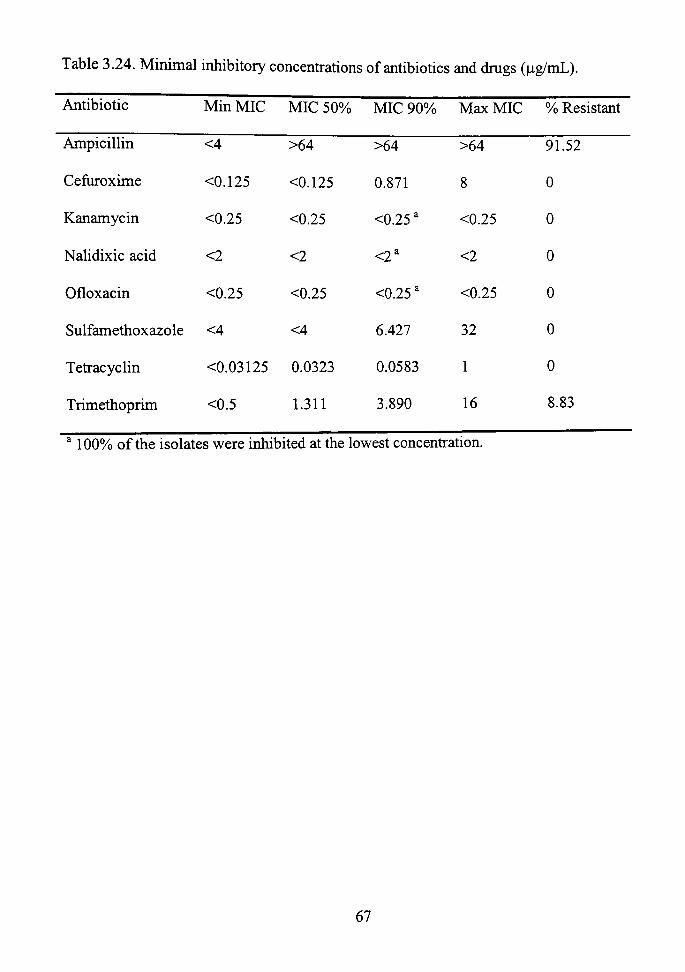
Table 3.24. Minimal inhibitory concenfrations of antibiotics and drugs (|ag/mL).
Antibiotic
Ampicillin
Cefuroxime
Kanamycin
Nalidixic acid
Ofloxacin
Min MIC
<4
<0.125
<0.25
<2
<0.25
MIC 50%
>64
<0.125
<0.25
<2
<0.25
MIC 90%
>64
0.871
<0.25'
<2*
<0.25 *
Max MIC
>64
8
<0.25
<2
<0.25
% Resistant
91.52
0
0
0
0
Sulfamethoxazole
Tefracyclin
Trimethoprim
<4
<0.03125
<0.5
<4
0.0323
1.311
6.427
0.0583
3.890
32
1
16
0
0
8.83
100% of the isolates were inhibited at the lowest concenfration.
67

0.5 1 8 16
Minimal Inhibitory Concentration (i^g/mL)
Figure 3.4. Cumulative percentage plot for the trimethoprim MICs of the 283 isolates.
100
00 CO
. ^ 3 <u u Ul <L>
CL, (L> > CO
3
u
0.125 - 0.25 0.5 1 2 4 8
Minimal Inhibitory Concentration (jig/mL)
Figure 3.5. Cumulative percentage plot for the cefiiroxime MICs of the 283 isolates.
68

Table 3.25. Ampicillin-sensitive aeromonads. (W = isolated from water; S = isolated from sediment)
Unique code YS2-4
C2-8
C3-2
H2-9
HS3-6
P3-3
P3-4
P3-5
P3-8
PS3-6
2R3-5
MS2-5
M2-5
MS2-6
M2-8
M3-5
2R3-1
SS3B-13
Biolog Identification^
A. hydrophila-like DNA Group 2
A. caviae DNA Group 4
A. caviae DNA Group 4
Aeromonas sp.
A. veronii/sobria DNA Group 8
Not performed putative A. veronii/sobria DNA Group 8 Not performed putative A. veronii/sobria DNA Group 8 Not performed putative A. veronii/sobria DNA Group 8 Not performed putative A. veronii/sobria DNA Group 8 Not performed putative A. veronii DNA Group 10 A. sobria DNA Group 7
A. sobria DNA Group 7
A. veronii/sobria DNA Group 8
Not performed putative A. sobria DNA Group 7
A. hydrophila DNA Group 1
A. veronii/sobria DNA Group 8
A. sobria DNA Group 7
A. encheleia
Source
Brazos(S)
Clapp (W)
Clapp (W)
Higinbotham (W)
Higinbotham (S)
Pecos (W)
Pecos (W)
Pecos (W)
Pecos (W)
Pecos(S)
Rural 2 (W)
Maxey (S)
Maxey (W)
Maxey (S)
Maxey (W)
Maxey (W)
Rural 2 (W)
Stevens (S)
69

Table 3.25. Continued.
Unique code SS2-3
SS2-6
SS2-7
SS2-8
SS2-9
S3-5
Biolog Identification'
Aeromonas sp.
Not performed putative Aeromonas sp.
Not performed putative Aeromonas sp.
Not performed putative Aeromonas sp.
Not performed putative Aeromonas sp.
A.jandaei DNA Group 9
Source
Stevens (S)
Stevens (S)
Stevens (S)
Stevens (S)
Stevens (S)
Stevens (W)
A putative species identification means that the isolate was not identified using the Biolog system. However, the organism shares the same phenotypic characteristics and source of isolation as an organism that was positively identified by Biolog.
Table 3.26. Isolates resistant to 8 |j.g/mL tiimethoprim, their sources, and ratio of cell mass grown in 40% nutrient broth with 8 ng/mL trimethoprim to cell mass grown in 40% nutiient broth without trimethoprim. The cell mass of each culture was measured by absorbance at 550 nm. (W = isolated from water; S = isolated from sediment)
Unique Code Source Biolog Identification
C2-4
HS2-11
Ratio
Clapp (W) Aeromonas sp. 0.466
Higinbotham (S) A. hydrophila DNA Group 1 0.391
70

Table 3.27. Isolates resistant to 4 |ag/mL tiimethoprim, then sources, identification, and ratio of cell mass grown in 40% nutrient broth with 4 |ig/mL tiimethoprim to cell mass grown in 40% nutiient broth without tiimethoprim. The cell mass of each culture was measured by absorbance at 550 nm. (W = isolated from water; S = isolated from sediment)
Unique code
YS2-3
S2-3
S2-5
S2-6
S2-7
SS2-3
SS2-4
SS2-6
C2-1
C2-3
C2-5
C2-6
C2-9
CS2-2
CS2-3
CS2-6
CS2-7
Source
Brazos(S)
Stevens (W)
Stevens (W)
Stevens (W)
Stevens (W)
Stevens (S)
Stevens (S)
Stevens (S)
Clapp (W)
Clapp (W)
Clapp (W)
Clapp (W)
Clapp (W)
Clapp (S)
Clapp (S)
Clapp (S)
Clapp (S)
Biolog Identification*
Not performed putative A. encheleia
Not performed putative A. encheleia
Not performed putative A. encheleia
Not performed putative Aeromonas sp.
Not performed putative A. encheleia
Not performed putative Aeromonas sp.
Not performed putative Aeromonas sp.
Not performed putative Aeromonas sp.
A. media-like DNA Group 5A
Not performed putative A. media-like DNA Group 5 A Not performed putative A. media-like DNA Group 5A Not performed putative A. media-like DNA Group 5A Aeromonas sp.
A. hydrophila DNA Group 1
Not performed putative A. encheleia
Not performed putative A. hydrophila DNA Group 1 Not performed putative A. hydrophila DNA Group 1
Ratio
0.266
0.645
0.292
0.409
0.382
0.460
0.681
0.521
0.528
0.546
0.434
0.482
0.343
0.600
0.406
0.272
0.582
71

Table 3.27. Continued.
Unique code Source
CS2-8
YS3B-2
YS3C-4
SS3-2
SS3B-13
MS3-6
Clapp (S)
Brazos(S)
Brazos(S)
Biolog Identification* Ratio
Not performed putative A. hydrophila DNA 0.990 Group 1 Not performed putative A. icthiosmia 0.344
Not performed putative A. icthiosmia 0.327
Stevens (S) Not performed putative^, sobria DNA Group 0.441 7
Stevens (S) A. encheleia 0.410
Maxey (S) Not performed putative A. veronii DNA Group 10
0.385
A putative species identification means that the isolate was not identified using the Biolog system. However, the organism shares the same phenotypic characteristics and source of isolation as an organism that was positively identified by Biolog.
72

Table 3^8. Isolates with frimethoprim resistance and a metal resistance. (W = isolated from water; S = isolated from sediment)
Unique code
S2-6
C2-9
CS2-3
a > . . •
Biolog identification*
Aeromonas sp.
Aeromonas sp.
Not performed putativcy^. encheleia
Source
Stevens (W)
Clapp (W)
Clapp (S)
Resistances
Trimethoprim; NaAs02
Trimethoprim; NaAs02
Trimethoprim; HgCb
A putative species identification means that the isolate was not identified using the Biolog system. However, die organism shares the same phenotypic characteristics and source of isolation as an organism that was positively identified by Biolog.
Table 3.29. Isolates with one resistance and ampicillin sensitivity. (W = isolated from water; S = isolated from sediment)
Unique code Biolog identification* Source Resistance
M2-8 A. hydrophila DNA Group 1
SS2-3 Aeromonas sp.
SS2-6 Not performed putative Aeromonas sp.
Maxey (W) HgCh
Stevens (S) Trimethoprim
Stevens (S) Trimethoprim
* A putative species identification means that the isolate was not identified using the Biolog system. However, the organism shares the same phenotypic characteristics and source of isolation as an organism that was positively identified by Biolog.
73

Table 3.30. Number of resistant isolates, their sample types and sources. (NS = not sampled)
Source
Brazos
Stevens
Clapp
Maxey
Higinbotham
Rio Hondo
Pecos
Rural 1
Rural 2
Total
Sample
Sediment
3
5
9
2
1
3
0
0
0
23
Type
Water
1
4
6
1
0
1
0
1
0
14
Date of Sampling
March
2
7
14
2
1
NS
NS
NS
NS
26
July
2
2
1
1
0
4
0
1
0
11
Total
4
9
15
3
1
4
0
1
0
37
74

£ o t t l
Lion
« 3
£ 3 g_ CO
alat
ed
ansf
oi
Res
is'
enot
y Y
.;2 II
00 i-T a •3 ^ £
fro
7 3 ii
;3 "o
CO
II
IS.
(W
sola
te
• ^ j
3 3 CO
;ury
-res
i
u
dm
e rs
enite
- an
CO
s fr
om
1. P
lasm
id i
sola
tion
di
men
t).
CO u
Tab
le 3
. s
^ t+H X3 —' O PH
73 ii
* i CO
"o CO
73
•£ CO CO
K, ^ ^
e(s
ista
nc'
Res
tio
n*
CO
ntif
ic
Bio
log
Ide
lode
So
urce
u
Uni
que
Yes
en
ite
Ars
CO
.2 o
s sp
ei
Aer
omon
a.
Cla
pp (
S)
CS3
-10
to
>^
Yes
en
ite
Ars
p , 3
NA
Gro
il
aD
A.h
ydro
ph
1 R
ural
1 (W
) 1R
3-8
CO
ii ;^
Yes
rc
ury
ii
•2
A. e
nche
le
Cla
pp (
S)
CS2
-1
< Z
o Z
rcur
y
^ (D
puta
tivi
73 ii
Not
per
foi
ench
elei
a H
ondo
(S)
T
S3-9
< Z
o Z
£ P H
leni
te,
met
ho
CO
O
Aer
omon
as s
pe
Stev
ens
(W
S2-6
< Z
o Z
£ P H
rcur
y,
met
ho
X (U
puta
tiv
rmed
N
ot p
erfo
en
chel
eia
Cla
pp (
S)
CS2
-3
<;
z
o
z
£ P H
met
ho
•2 s
/I. e
nche
h 3
Stev
ens
(S]
, - H
SS3B
-
<
3 ^
0 0 - o O i -
N
;r, th
e id
enti]
o Z
£ P H
met
ho
Tri
1 — ^
p-3 o
NA
Gr
Q ^.
hyd
roph
ila
c/y
Hig
inbo
tha
1 — 1
HS2
-1
> >. (1) ^ H
? > O - ^ S 'OT
O
tern
, as
p
CO >
0 0 ^ O H--
"o £ s-l u 3 X3 00 •t3 gJJ 00 o
dusi
n as
an
dent
ifie
so
latio
n
* ^ ^ . ^ H
„ lU
ate
was
d
sour
c he
isol
tic
s an
m
eans
tha
t t
c ch
arac
teri
s ca
tion
noty
pi
tati
ve s
peci
es i
dent
ifi
shar
es t
he s
ame
phe
Bio
log.
3 PH
< rt
75

CHAPTER IV
DISCUSSION
Biolog Identification
All of the isolates identified by the Biolog system were in the genus Aeromonas.
This shows that the modified Aeromonas medium and subsequent screening procedures
were reliable in isolating only Aeromonas species as opposed to other genera that inhabit
water or sediment. It can also be assumed that the other 225 isolates not identified by
Biolog are also in the genus Aeromonas since they underwent the same screening
procedures as those subsequently identified. For each group of resistant and sensitive
isolates, a representative organism of each group was also identified by the Biolog
system.
The carbon utilization pattems of the isolates were consistent with those listed in
Bergey 's Manual of Determinative Bacteriology (Holt et al., 1993). All of these results
were also consistent with those of Valera and Esteve (2002), except for the Aeromonas
encheleia isolates. Eight of the isolates were identified as A. encheleia by the Biolog
system. This species was originally isolated from European eels in 1995 (Esteve et al.,
1995). It is expected to belong to a new DNA hybridization group 16 (Camahan and
Altwegg, 1996). Valera and Esteve (2002) tested fourteen^, encheleia sfrains, while this
stiidy only evaluated eight. Sample size could account for the discrepancies between the
two studies. Since the species designation is new, it is still undergoing changes in
taxonomy.
76

A. enteropelogenes and A. icthiosmia were each identified once. A.
enteropelogenes is most likely a synonym for A. trota. This is based on 16S rRNA
analysis (Collins et al., 1994). However, A. trota is characteristically sensitive to
ampicillin (Camahan et al., 1991; Overman and Janda, 1999). The A. enteropelogenes
isolate from this study was resistant to ampicillin at over 64 |ig/mL. A. icthiosmia is
probably a synonym for A. veronii (Collins et al., 1994).
A. hydrophila was also isolated frequently. This species is known to cause
septicemia, myonecrosis, wound infections, Aeromonas arthritis, peritonitis, and has been
associated with hemolytic uremic syndrome. (Janda and Abbott, 1996). The other
isolates A. veronii, A. jandaei, and A. caviae have been associated with human wound
infections and septicemia. A. media and A. sobria have been associated with human
diarrheal disease (Joseph, 1996).
Antibiotic and Drug Resistance
All isolates were tested for antibiotic and drug resistance, and their MICs were
determined. Ampicillin resistance was prevalent with over 91% of the isolates having an
MIC of greater than or equal to 64 [jg/mL. Ampicillin is a semisynthetic penicillin in the
P-lactam group that impedes the synthesis of the cell wall (Madigan et al., 2000). This
resistance was not surprising since most sfrains of Aeromonas are inherently resistant to
the antibiotic. Ko and colleagues (1996) found 99.6% of then clinical isolates to be
resistant to ampicillin while other studies have shown up to 100% resistance (Vila et al.,
2002).
77

The only other antibiotic or drug the Aeromonas isolates showed resistance to was
tiimetiioprim. Trimethoprim is a drug that blocks folate biosynthesis and indirectly
disrupts several metabolic pathways including purine biosyntiiesis (Snyder and
Champness, 1997). This study differs from most others in that trimethoprim was
considered alone instead of in conjunction with sulfamethoxazole. Kampfer el al. (1999)
tested 217 clinical and non-clinical Aeromonas isolates against trimethoprim alone and
found none of tiiem to be resistant. This is in confrast to the 8.83% found in this stiidy.
Resistance to sulfamethoxazole also differed between the two studies. Sulfamethoxazole
is a folic acid analog that inhibits a different enzymatic step in folate biosynthesis than
trimethoprim (Madigan et al., 2000). This study showed no sulfamethoxazole resistance,
while a published value was close to 26% (Kampfer el al., 1999).
No other resistances were found to cefiiroxime, kanamycin, nalidixic acid,
ofloxacin, or tetracycline. Cefuroxime is a cephalosporin in the P-lactam group and
inhibits cell wall synthesis (Madigan et al., 2000). Other studies with cefiiroxime also
show a low incidence of resistance. These studies range from 0 to 1.38% of isolates
displaying resistance (Kampfer et al., 1999; Overman and Janda, 1999; Vila et al., 2002).
Warren (1998) did a similar study in aeromonads from the Lubbock playas. One isolate
out of 151 aeromonads was cefuroxime resistant (Warren, 1998).
The observation that none of the isolates have kanamycin resistance is supported
by Kampfer el al. (1999), who also did not find the resistance among their Aeromonas
isolates. Kanamycin blocks translation by targeting 16S rRNA (Snyder and Champness,
1997).
78
The widespread nalidixic acid sensitivity in this stiidy is in confrast to the 43%
resistance reported by Vila et al. (2002). However, Miranda and Castillo (1998) report
finding less than 4% of their Aeromonas isolates to be resistant to nalidixic acid. One of
the first nalidixic acid-resistant aeromonads was isolated in 1987 (Chang and Bolton,
1987). Otiier nalidixic acid-resistant isolates have been reported since then (Ko et al.,
1996; Vila et al., 2002). Nalidixic acid is a quinolone. It targets DNA gyrase and blocks
rephcation of DNA (Snyder and Champness, 1997).
No resistance to ofloxacin, also a quinolone, was detected either in this study or
previous stiidies (Kampfer el al., 1999; Overman and Janda, 1999; Vila et al., 2002).
Warren (1998) also found no ofloxacin resistance. Susceptibility of the isolates to
ofloxacin was not unusual. Quinolone resistance is due only to chromosomal mutations.
Members of the genus Aeromonas do not have chromosomal resistance genes to
quinolones as they do to ampicillin. These chromosomal genes, if present by mutation,
are not fransferable. Furthermore, since quinolones are synthetic and are not naturally
present in environmental waters, there is no natural selection for resistant phenotypes
(Goni-Urriza et al., 2000). In fact, quinolone sensitivity is so common in aeromonads
that these antibiotics are the first choice for the freatment of Aeromonas infections in
humans (Jones and Wilcox, 1995).
Tefracycline resistance was also found to be low compared to previous studies.
Tefracycline inhibits protein synthesis by blocking proper fiinctioning of the 30S
ribosomal subunit (Chopra and Roberts, 2001). Kampfer et al. (1999) found
79
approximately 10% of tiidr isolates to be resistant whereas 0% is reported here. Warren
(1998) reported 2.6% of his isolates to be tefracycline resistant.
In general, the isolates included in this stiidy showed an equivalent or much lower
resistance than Aeromonas isolates in other stiidies. A possible explanation of these
findings is that some of the earlier studies included clinical isolates that could have
previously been exposed to antibiotics (Kampfer el al., 1999; Ko et al., 1996; Vila et al.,
2002). In otiier studies done solely with environmental aeromonads, they have been
isolated them from waters highly polluted by industiial effluent or raw sewage (Goiii-
Urriza et al., 2000). The six playa lakes and three sfreams in this stiidy receive runoff
from surrounding areas. None of the water bodies are polluted from a single source.
However, the runoff can contain low levels of pollutants or even antibiotics. According
to tiie results of this study and the low incidence of antibiotic resistance present (aside
from ampicillin), the six playa lakes and three streams that were sampled could possibly
be considered a reservoir for antibiotic and metal resistance genes. The resistant
aeromonads from the environment could opportunistically infect humans and cause an
infection that would be difficult to freat.
Metal Resistance
Not much has been done to evaluate the metal resistance profiles of aeromonads
from unpolluted water sources. Many of the bacteria used in past studies to characterize
metal resistance were isolated from waters polluted with metals (Allen et al., 1977;
Francisco et al., 2002; Miranda and Castillo, 1998; Timoney et al., 1978). These bacteria
80

studied were rarely aeromonads. None of the playas or streams in this stiidy are highly
polluted witii metals. However, some metals could have been introduced into these
waters inadvertently. Pesticides and domestic wastes could be a source of metals for all
of die water bodies (Collins and Stotzky, 1989). The Rio Hondo could be contaminated
witii a variety of metals from past mining attempts. This past mining near the Rio Hondo
could explain why tiiree isolates from the stream were resistant to mercury and one was
resistant to arsenite.
Only resistances to arsenite and mercury were found. Resistance to arsenite was
found in 2.83% of the isolates, while 2.47% of the isolates contained mercury resistance.
None of the isolates contained more than one metal resistance. Miranda and Castillo
(1998) found between 29-40% of their aeromonads to be mercury-resistant. They also
found 3.6-8.3% resistance to chromium and 41-62% resistance to copper. This is in stark
confrast to what was found in this study. A possible explanation is that Miranda and
Castillo sampled from freshwater sources polluted with sewage and effluent from
irrigation water in Chile. Versteegh et al. (1989) reported that copper inhibits Aeromonas
at a concenfration as low as 10 |ig/L. Aeromonas isolates are also reportedly much more
sensitive to copper than are other bacteria, such as coliforms and fecal sfreptococci
(Versteegh et al., 1989). Inherent sensitivity of Aeromonas species to copper is supported
by the findings of Calomiris et al. (1984).
81

Resistant Isolates from Sediment
It IS clear from tiiis stiidy that there is a significant increase in recovery of viable
aeromonads from the sediment as opposed to the water, hi general, higher numbers of
bacteria are found in sediment than in water (Cavallo et al., 1999). More resistant
organisms were also isolated from the sediment than the water in this study. However,
tills difference was not statistically significant. An increased number of resistant isolates
from the sediment could arise from the presence of a selective pressure for the resistant
phenotypes. The metals settle to the bottom of the lake and into the sediment. Arefeen
(1995) studied the concenfration of metals of the water and sediments of eleven playa
lakes in Lubbock. All of the sediments had significantiy higher concenfrations of metals
than the water did. He compared the concenfrations in water to the EPA drinking water
standards. The only two playas that both Arefeen (1995) and this study had in conunon
were Clapp and Higinbotham. Both Clapp and Higinbotham were within the EPA
guidelines for arsenic. Clapp was above the EPA standard in chromium concenfrations
and Higinbotham was above in mercury. All of the concenfrations of metals in both the
sediment and the water were well below those concenfrations used in this study to
determine sensitivity and resistance (Arefeen, 1995).
Besides metals being present at low concentrations in the playas, another factor to
consider is whether the metals are in toxic or nontoxic forms. It is not known which form
or forms of the metals are in the playas. For example, Aeromonas species in lakes are not
able to withstand concentrations of mercury as well as those in the sea because the exfra
chloride in the sea water binds to the mercury, making it less toxic (Babich and Stotzky,
82

1979). The environment in which the metal is present may play a role in the toxicity. M
lab experiments, mercury-resistant oral sfreptococci could not be differentiated from their
sensitive counterparts on some types of media supplemented with equal concenfrations of
mercuric chloride, such as mitis-salivarius agar or Columbia agar (Pike et al., 2002). The
pH of the environment can cause changes in toxicity. As pH increases, copper toxicity
also increases, but nickel toxicity decreases (Collins and Stotzky, 1989).
None of tiie metal-resistant isolates, including mercury, came from Higinbotham.
This is somewhat unexpected, since Arefeen (1995) found elevated levels of mercury
present. In fact, Clapp was the source of three of the seven mercury-resistant organisms.
Seven years have passed since Arefeen's stiidy (1995) and this one. The dynamics of the
lake may have changed in this time period.
In this study, a temporal difference in the number of viable aeromonads present in
the urban playa lakes could not be determined. Sampling was done during two months,
March and July. According to previous studies, warmer temperatures yield more bacteria
and more Aeromonas than cooler ones (Cavallo et al., 1999; Holmes et al., 1996; Warren,
1998).
Interestingly, the playa at Clapp Park produced the most resistant organisms.
Over 40% of all resistant aeromonads came from Clapp. Out of all isolates from Clapp,
37.5%) of them were resistant to at least one antimicrobial. The reasons for this are not
apparent. The previous study by Arefeen (1995) did not show high metals
concentrations, except for chromium, that would provide a selective pressure. The playa
is urban and does not receive runoff from livestock yards that may contain antibiotics.
83

No direct selective pressure that increases tiie likelihood of resistant phenotypes has been
identified for the playa at Clapp Park.
Most of the resistant aeromonads in this study belong to the species Aeromonas
encheleia. Earlier studies report that the most commonly isolated resistant aeromonad is
A. hydrophila (Janda et al., 1996; Warren, 1998). A. encheleia has been identified and
characterized only recently (Esteve et al, 1995). The lack of a taxonomic category for .4.
encheleia before 1995 is a plausible explanation for this contradiction in findings. In
addition, multiple isolations of the same organism from the same sample may have
skewed this result.
Plasmid Isolations and Transformations
One of the first studies to suggest that plasmid-mediated antibiotic resistance
occurs in environmental waters was that of Kelch and Lee (1978). Since then, the
successfiil fransfer of plasmids between bacteria in aquatic environments in both the
water and sediment has been demonstrated (Genthner et al., 1988; Sandaa and Enger,
1994). Plasmids encoding resistance to arsenite or mercury were successfiiUy isolated
from Aeromonas isolated in this stiidy and fransformed into E. coli XLl-Blue MRF'.
Plasmids mediating these resistances have been previously reported. The first report of
arsenite resistance encoded on a plasmid was by Novick and Roth (1968). The plasmid
was isolated from Staphylococcus aureus. Plasmids from other bacteria, such as E coli,
also carry determinants for arsenite resistance. An ars operon located on the plasmid
encodes for an oxyanion pump that fransports arsenite, arsenate, and antimonite out of the
84

cell (Kauer and Rosen, 1992). This pump fimctions much the same way as the K"-
franslocating ATPase (Dey and Rosen, 1995). hi order to determine if the plasmids
isolated in this study have the homologous ars operon, more stiidies would need to be
done.
In a previous study, an A. hydrophila isolate from river sediment was found to
have five plasmids and resistance to mercury (Trevors, 1986). However, Trevors (1986)
was not able to conclusively determine whether the resistance genes were carried on any
one of tiie plasmids or located on the chromosome. Other plasmids carrying mercury
resistance have been found in Pseudomonas fluorescens and Acinetobacter sp. isolated
from estiiary sediment (Olson et al., 1979). Bale et al. (1988) showed that large mercury-
resistance plasmids could be fransferred to and from Pseudomonas species in a natural
river environment. Many of the mercury resistance genes carried on plasmids are in the
mer (mercury resistance) operon from TnlO and Tn501 (Silver and Phung, 1996). This
operon codes for organomercurial lyase, which hydrolyzes the bond between mercury
and carbon, and mercuric ion reductase, which reduces Hg(II) to Hg(0). The Hg(0) then
diffiises out of the cell (Bruins et al., 2000; Silver and Phung, 1996). It is not known
whether the plasmid encoding mercury resistance in this study contains the mer operon.
It is plausible that the plasmid may not contain the mer operon. Dahlberg et al. (1997)
isolated plasmids encoding mercury resistance from marine environments that did not
have any homology to the mer operon.
The tiimethoprim-resistant isolates did not have plasmids present that could be
isolated using the plasmid purification kit. hi these isolates, the resistance could be
85
chromosomally encoded. Goni-Urriza et al. (2000) found all of the antibiotic-resistance
genes in their isolates of Aeromonas to be chromosomally encoded. These resistances
included nalidixic acid and tetracycline, among others. Hedges et al. (1985) were able to
isolate plasmids encoding trimetiioprim resistance in Aeromonas. However, these
plasmids also carried genes for resistance to other antibiotics such as tetracycline and
sulfonamides. Trimethoprim resistance was carried by integrons in Aeromonas without
plasmids (Schmidt et al., 2001). This suggests that antibiotic resistance can be
disseminated in the microbial community by DNA elements other than plasmids. Many
Gram-negative genera do contain mobile gene cassettes or integrons that encode for
antibiotic resistance. These integrons can be fransferred between different species of
bacteria without plasmids if the circular forms are stable (Hall, 1997). Besides plasmids
and integrons, other mechanisms of transferring resistance genes could be occurring.
These may include transduction (bacteriophages) or fransformation (Seveno et al., 2002).
The Absence of Linked Antibiotic and Metal Resistance
Out of 283 isolates tested, only three contained more than one type of resistance,
excluding ampicillin resistance (Table 3.34). There were no definite pattems of
resistance to more than one antimicrobial. No plasmids were isolated from the
aeromonads with more than one resistance. Previous studies have shown multiple
resistances to be present on plasmids (Calomiris et al., 1984; Ghosh et al., 2000).
However, this was not the case in this stiidy. The absence of selective pressures in the
86

environmental water sources studied is most likely the key to the absence of the multiple-
resistance plasmids among the aeromonads.
87

LITERATURE CITED
Abbott, S.L, L.S. Sell, M. Catino, Jr., M.A. Hartley, and J.M. Janda. 1998. Misidentification of unusual Aeromonas species as members of the genus Vibrio: a continuing problem. Joumal of Clinical Microbiology 36:1103-1104.
Ahuja, P., H. Mohapatra, R.K. Saxena, and R. Gupta. 2001. Reduced uptake as a mechanism of zinc tolerance in Oscillatoria anguistissima. Current Microbiology 43:305-310.
Albert, M.J., M. Ansaruzzaman, K.A. Talukder, A.K. Chopra, I. Kiihn, M. Rahman, A.S.G. Faruque, M.S. Islam, R.B. Sack, and R. MoUby. 2000. Prevalence of enterotoxin genes in Aeromonas spp. isolated from children with diarrhea, healthy confrols, and the environment. Joumal of Clinical Microbiology 38:3785-3790.
Allen, D.A., B. Austin, and R. Colwell. 1977. Antibiotic resistance pattems of metal-tolerant bacteria isolated from an estuary. Antimicrobial Agents and Chemotherapy 12:545-547.
Amsterdam, D. 1991. Susceptibility testing of antimicrobials in hquid media. In: V. Lorian (Ed.), Antibiotics in laboratory medicine, pp. 53-105. Baltimore: Williams & Wilkins.
Arefeen, Q. A comparison of metals in the sediments and water of eleven playas. Master's Thesis, Texas Tech University, 1995.
Amebrant, K., E. Baath, and A. Nordgren. 1987. Copper tolerance of microfimgi isolated from polluted and unpolluted forest soil. Mycologia 79:890-895.
Austin, B., and C. Adams. 1996. Fish pathogens, hi: B. Austin, M. Altwegg, P.J. Gosling, and S. Joseph (Eds.), The Genus Aeromonas, pp. 197-229. Chichester: John Wiley & Sons Ltd.
Babich, H., and G. Stotzky. 1979. Differential toxicities of mercury to bacteria and bacteriophages in sea and lake water. Canadian Joumal of Microbiology 25: 1252-1257.
Bale M J J C Fry, and M.J. Day. 1988. Transfer and occurrence of large mercury resistance plasmids in river epilitiion. Applied and Environmental Microbiology
54: 972-978.
88

Bass, L., CA. Liebert, M.D. Lee, A.O. Summers, D.G. White, S.G. Thayer, and J.J. Maurer. 1999. Incidence and characterization of integrons, genetic elements mediating multiple-drug resistance, in avian Escherichia coli. Antimicrobial Agents and Chemotherapy 43:2925-2929.
Bastarrachea, F. 1998. On the origin of plasmid-bome, extended-spectiiim, antibiotic resistance mutations in bacteria. Joumal of Theoretical Biology 190:379-387.
Biolog. 1999. GN2 MicroPlate. 8 pp. Hayward, CA: Biolog, fric.
BIO-TEK Instilments Inc., 1991. Automated Microplate Reader EL31 Isx Operator's Manual. Winooski, VT: BIO-TEK hic.
Bruins, M.R., S. Kapil, and F.W. Oehme. 2000. Microbial resistance to metals in the environment. Ecotoxicology and Environmental Safety 45:198-207.
Calomiris, J.J., J.L. Armsfrong, and R.J. Seidler. 1984. Association of metal tolerance with multiple antibiotic resistance of bacteria isolated from drinking water. AppUed and Environmental Microbiology 47:1238-1242.
Camahan, A.M., and M. Altwegg. 1996. Taxonomy. In: B. Austin, M. Altwegg, P.J. Gosling, and S. Joseph (Eds.), The Genus Aeromonas, pp. 1-38. Chichester: John Wiley & Sons Ltd.
Camahan, A.M., T. Chakraborty, G.R. Fanning, D. Verma, A. Ah, J.M. Janda, and S.W. Joseph. 1991. Aeromonas trota sp. nov., an ampicillin-susceptible species isolated from clinical specimens. Joumal of Clinical Microbiology 29: 1206-1210.
Cavallo, R.A., C. Rizzi, T. Vozza, and L. Stabih. 1999. Viable heterotrophic bacteria in water and sediment in 'Mar Piccolo' of Taranto (Ionian Sea, Italy). Joumal of Applied Microbiology 86: 906-916.
Chang, B.J., and S.M. Bolton. 1987. Plasmids and resistance to antimicrobial agents in Aeromonas sobria and Aeromonas hydrophila clinical isolates. Antimicrobial Agents and Chemotherapy 31: 1281-1282.
Chopra, I., and M. Roberts. 2001. Tefracycline antibiotics: mode of action, applications, molecular biology, and epidemiology of bacterial resistance. Microbiology and Molecular Biology Reviews 65: 232-260.
Collins, M.D., A.J. Martinez-Murcia, and J. Cai. 1994. Aeromonas enteropelogenes and Aeromonas icthiosmia are identical to Aeromonas trota and Aeromonas veronii respectively, as revealed by small-subunit rRNA sequence analysis. International Joumal of Systematic Bacteriology 43: 855-856.
89
Collins, Y.E., and G. Stotzky. 1989. Factors affecting the toxicity of heavy metals to microbes. In T.J. Beveridge and R.J. Doyle, (Eds.), Metal Ions and Bacteria, pp. 31-90. New York: John Wiley and Sons Ltd.
Colwell, R.R., M.T. MacDonell, and J. DeLey. 1986. Proposal to recognize the family Aeromonadaceae fam. nov. International Joumal of Systematic Bacteriology 36-473-477.
Courvalin, P. 1994. Transfer of antibiotic resistance genes between Gram-positive and Gram-negative bacteria. Antimicrobial Agents and Chemotherapy 38:1447-1451.
Dahlberg, C , C. Linberg, V.L. Torsvik, and M. Hermansson. 1997. Conjugative plasmids isolated from bacteria in marine environments show various degrees of homology to each other and are not closely related to well-characterized plasmids. Applied and Environmental Microbiology 63: 4692-4697.
Davies, J. 1994. Inactivation of antibiotics and the dissemination of resistance genes. Science 264:375-382.
Davies, J.E. 1997. Origins, acquisition and dissemination of antibiotic resistance determinants. In Antibiotic Resistance: Origins, Evolution, Selection and Spread, pp. 15-35. Chichester: John Wiley & Sons Ltd.
DeChalain, T. 1996. Exploring the use of the medicinal leech: a clinical risk-benefit analysis. Joumal of Reconstmctive Microsurgery 12:165-172.
Dey, S., and B.P. Rosen. 1995. Dual mode of energy coupling by the oxyanion-franslocating ArsB protein. Joumal of Bacteriology 177:385-389.
Esteve, C , M.C. Gutierrez, and A. Ventosa. 1995. Aeromonas encheleia sp. nov., isolated from European eels. Intemational Joumal of Systematic Bacteriology 45:462-466.
Fernandez, M.C, B.N. Giampaolo, S.B. Ibaiiez, M.V. Guaghardo, M.M. Esnaola, L. Conca, P. Valdivia, S.M. Stagnaro, C. Chiale, and H. Frade. 2000. Aeromonas hydrophila and its relation with drinking water indicators of microbiological quality in Argentine. Genetica 108:35-40.
Filler, G., J.H.H. Ehrich, E. Sfrauch, and L. Beutin. 2000. Acute renal failure in an infant associated with cytotoxic Aeromonas sobria isolated from patient's stool and from aquarium water as suspected source of infection. Joumal of Clinical Microbiology 38:469-470.
90

Fox, E., LA. Mikhail, R.L. Haberberger Jr., E.A. Abbatte, and M.H. Ahmed. 1990. Aeromonas hydrophila in drinking waters in Djibouti: commensal germ or diarrhoea agent? Medicine Tropicale 50:237-239.
Francisco, R., M.C. Alporim, and P.V. Morals. 2002. Diversity of chromium-resistant and -reducing bacteria in a chromium-contaminated activated sludge. Journal of Applied Microbiology 92:837-843.
Gentimer, F.J., P.Chatterjee, T. Barkay, and A.W. Bourquin. 1988. Capacity of aquatic bactiia to act as recipients of plasmid DNA. Applied and Environmental Microbiology 54:115-117.
Ghosh, A., A. Singh, P.W. Ramteke, and V.P. Singh. 2000. Characterization of large plasmids encoding resistance to toxic heavy metals in Salmonella abortus equi. Biochemical and Biophysical Research Communications 272: 6-11.
Goiii-Urriza, M., M. Capdepuy, C. Arpin, N. Raymond, P. Caumette, and C Quentin. 2000. Impact of urban effluent on antibiotic resistance of riverine Enterobacteriaceae and Aeromonas spp. Applied and Environmental Microbiology 66:125-132.
Gosling, P.J. 1996. Aeromonas species in disease of animals. In: B. Austin, M. Altwegg, P.J. Gosling, and S. Joseph (Eds.), The Genus Aeromonas, pp. 175-195. Chichester: John Wiley & Sons Ltd.
GroBe, C , G. Grass, A. Anton, S. Franke, A.N. Santos, B. Lawley, N.L. Brown, and D.H. Nies. 1999. Transcriptional organization of the czc heavy-metal homeostasis determinant from Alcaligenes eutrophus. Joumal of Bacteriology 181: 2385-2393.
Groves, D.J., H. Short, A.J. Thewaini, and F.E. Young. 1975. Epidemiology of antibiotic and heavy metal resistance in bacteria: resistance pattems in Staphylococci isolated from populations in Iraq exposed and not exposed to heavy metals or antibiotics. Antimicrobial Agents and Chemotherapy 7:622-628.
Gustavson, T.C, V.T. HoUiday, and S.D. Hovorka. 1994. hi L.V. Urban and A.W. Wyatt (Eds ), Development of playa basins, southern high plains Texas and New Mexico, pp. 5-14. Proceedings of the playa basin symposium. Lubbock: Texas Tech University.
Hall, D.L. Species diversity of aquatic macroinvertebrates in playa lakes: island biogeographic and landscape influences. Master's Thesis, Texas Tech University, Lubbock, TX, 1997.
91

Hall, R.M_1997. Mobile gene cassettes and integrons: moving antibiotic resistance genes m Gram-negative bacteria, hi Antibiotic Resistance: Origins, Evolution, Selection and Spread, pp. 192-205. Chichester: John Wiley & Sons Ltd.
Havela^, A.H., M. During, and J.F.M. Versteegh. 1987. Ampicillin-dextrin agar medium for the enumeration of Aeromonas species in water by membrane filtration. Joumal of Applied Bacteriology 62:279-287.
Hazen, T.C, CB. Fliermans, R.P. Hirsch, and G.W. Esch. 1978. Prevalence and distiibution of Aeromonas hydrophila in the United States. Applied and Environmental Microbiology 36:731-738.
Hedges, R.W., P.Smitii, and G. Brazil. 1985. Resistance plasmids of aeromonads. Joumal of General Microbiology 131:2091-2095.
Holmes, P., L.M. NiccoUs, and D. P. Sartory. 1996. The ecology of Mesophilic Aeromonas in tiie aquatic environment, hi: B. Austin, M. Altwegg, P.J. Gosling, and S. Joseph (Eds.), The Genus Aeromonas, pp. 127-150. Chichester: John Wiley & Sons Ltd.
Holt, J.G., N.R. Krieg, P.H.A. Sneath, J.T. Staley, S.T. Wilhams, (Eds.). 1993. Sergey's Manual of Determinative Bacteriology. 9* ed. 816 pp. Baltimore: Lippincott, Williams, & Wilkins.
Huang, A. Investigation of selected metals in urban playa lakes. Master's Thesis, Texas Tech University, Lubbock, Texas, 1992.
Hughes M.N., and R.K. Poole. 1989. Metals and Micro-organisms, pp. 280-285. London: Chapman and Hall.
Huguet, J.M., and F. Ribas. 1991. SGAP-IOC agar for the isolation and quantification of Aeromonas from water. Joumal of Applied Bacteriology 70:81-88.
Huys, G., P. Kampfer, M. Altwegg, I. Kersters, A. Lamb, R. Coopman, J. Liithy-Hottenstein, M. Vancanneyt, P. Janssen, and K. Kersters. 1997. Aeromonas popojfii sp. nov., a mesophilic bacterium isolated from drinking water production plants and reservoirs. Intemational Joumal of Systematic Bacteriology 47:1165-1171.
Janda, J.M., and S.L. Abbott. 1996. Human pathogens. In: B. Austin, M. Altwegg, P.J. Gosling, and S. Joseph (Eds.), The Genus Aeromonas, pp. 151-173. Chichester: John Wiley & Sons Ltd.
92
Jenkins, J.A., and P.W. Taylor. 1995. An altemative bacteriological medium for the isolation of Aeromonas spp. Joumal of Wildlife Diseases 31:272-275.
Jones, B.L, and M.H. Wilcox. 1995. Aeromonas infections and their freatment. Joumal of Antimicrobials and Chemotherapy 35:453-461.
Joseph, S.W. 1996. Aeromonas gasfrointestinal disease: a case study in causation? hi: B. Austin, M. Altwegg, P.J. Gosling, and S. Joseph (Eds.), The Genus Aeromonas, pp. 311-326. Chichester: John Wiley & Sons Ltd.
Kampfer, P., C Christmann, J. Swings, and G. Huys. 1999. In vitro susceptibilities of Aeromonas genomic species to 69 antimicrobial agents. Systematic and Applied Microbiology 22:662-669.
Kaur, P., and B.P. Rosen. 1992. Plasmid-encoded resistance to arsenic and antimony. Plasmid 27: 29-40.
Kelch, W.J., and J.S. Lee. 1978. Antibiotic resistance pattems of Gram-negative bacteria isolated from environmental sources. Applied and Environmental Microbiology 36: 450-456.
Khardori, N., and V. Fainstein. 1988. Aeromonas and Plesiomonas as etiological agents. Annual Review of Microbiology 42:395-419.
Kilpatiick, M.E., J. Escamilla, A.L. Bourgeois, H.J. Adkins, R.C Rockhill. 1987. Overview of four US Navy overseas research studies on Aeromonas. Experentia 43:365-367.
Ko, W.C, H.M.Wu, T.-C Chang. J.J. Yan, and J.J. Wu. 1998. hiducible p-lactam resistance in Aeromonas hydrophila: therapeutic challenge for antimicrobial therapy. Joumal of Clinical Microbiology 36:3188-3192.
Ko, W.C, K.W. Yu, CY. Liu, CT. Huang, H.S. Leu, and Y.C Chuang. 1996. hicreasing antibiotic resistance in clinical isolates of Aeromonas sfrains in Taiwan. Antimicrobial Agents and Chemotherapy 40:1260-1262.
Kmse, H., H. Sorum. 1994. Transfer of multiple drag resistance plasmids between bacteria of diverse origins in natiiral microenvironments. Applied and Environmental Microbiology 60:4015-4021.
Kuhn, I., M.J. Albert, M. Ansaruzzaman, N.A. Bhuiyan, S.A. Alabi, M.S. Islam, P.K. B. Neogi, G. Huys, P. Janssen, K. Kersters, and R. MoUby. 1997. Charactenzation of Aeromonas spp. Isolated from humans witii diarrhea, from healthy confrols, and from surface water in Bangladesh. Joumal of Clinical Microbiology 35:369-373.
93

;'oo^ TI ' ^- ""^y'' ^- ^ ^ ' ' ' " ' ^- ^^^^ters, K. Krovacek, and T. Stensfrom 1997. Diversity, persistence, and vimlence of Aeromonas strains isolated from dnnkmg water distiibution systems in Sweden. Applied and Environmental Microbiology 63:2708-2715.
Lederberg, J., and R.E. Shope, S.C Oaks, Jr. (Eds.). 1992. Emerging hifections-Microbial Threats to Health in the United States. 294 pp. Washington, DC: National Academy Press.
Levy, S.B. 1997. Antbiotic resistance: an ecological imbalance, hi Antibiotic Resistance: Ongins, Evolution, Selection and Spread, pp. 1-14. Chichester: John Wiley & Sons Ltd.
Liebert, C.A., R.M. Hall, and A.O. Summers. 1999. Transposon Tn27, flagship of the floating genome. Microbiology and Molecular Biology Reviews 63:507-522.
Madigan M.T., J.M. Martinko, and J. Parker. 2000. Brock Biology of Microorganisms. 9' edition, 991 pp. Upper Saddle River, NJ: Prentice Hall.
Marques, A.M., F. Congregado, and D.M. Simon-Pujol. 1979. Antibiotic and heavy metal resistance of Pseudomonas aeruginosa isolated from soils. Joumal of Applied Bacteriology 47:347-350.
McArthur, J.V., and R.C. Tuckfield. 2000. Spatial pattems in antibiotic resistance among sfream bacteria: effects of industiial pollution. Applied and Environmental Microbiology 66:3722-3726.
McHugh, G.L., R.C. Moellering, C C Hopkins, and M.N. Swartz. 1975. Salmonella typhimurium resistant to silver nifrate, chloramphenicol, and ampicillin. Lancet ii: 235-240.
McKeon, D.M, J.P. Calabrese, G.K. Bissonnette. 1995. Antibiotic resistant Gram-negative bacteria in mral groundwater supplies. Water Research 29:1902-1908.
Miranda, CD., and G. Castillo. 1998. Resistance to antibiotic and heavy metals of motile aeromonads from Chilean freshwater. Science of the Total Environment 224:167-176.
Mollhagen, T.R., L.V. Urban, R.H. Ramsey, A.W. Wyatt, CD. McReynolds, and J.T. Ray. 1993. Assessment of non-point source contamination of playa basins in the High Plains of Texas (Brazos Basin Watershed, Phase 1). Texas Tech University, Lubbock, TX.
94

Morozzi, G., G. Cenci, G. Caldini, G. Losito, and A. Morosi. 1986. Relationship between environment spread and presence in hosts of Escherichia coli sfrains resistant to antibiotics and metals. Zenfralblatt fiir Bakteriologie, Mikrobiologie und Hygiene. Sene B, Umwelthygiene, Krankenhaushygiene, Arbeitshygiene, praventive Medizin 182:393-400.
Moyer, N.P. 1996. Isolation and enumeration of aeromonads. hi: B. Austin, M. Altwegg, P.J. Gosling, and S. Joseph (Eds.), The Genus Aeromonas, pp. 39-84. Chichester' John Wiley & Sons Ltd.
Murray, B.E., and S.L. Hodel-Christian.1991. Bacterial resistance: theoretical and practical considerations, mutations to antibiotic resistance, characterization of R plasmids, and detection of plasmid-specified genes. In V. Lorian (Ed.). Antibiotics in Laboratory Medicine, pp. 556-598. Baltimore: Wilhams & Wilkins.
Nakahara, H., T. Ishikawa, Y. Sarai, I. Kondo, and H. Kozukue. 1978. Survey of resistance to metals and antibiotics in clinical isolates of Klebsiella pneumoniae in Japan. Zenfralblatt fiir Bakteriologie, Parasitenkunde, hifektionskrankheiten und Hygiene. Erste Abteilung Originale 240:22-29.
Nayduch, D., G.P. Noblet, and F.J. Stiitzenberger. 2002. Vector potential of houseflies for the bacterium Aeromonas caviae. Medical and Veterinary Entomology 16:193-198.
NCCLS. 2002. Performance Standards for Antimicrobial Susceptibility Testing; Twelfth Informational Supplement. NCCLS, Wayne, PA.
Nies, D.H. 1992. Resistance to cadmium, cobalt, zinc, and nickel in microbes. Plasmid 27:17-28.
Novick, R.P., and Roth, C 1968. Plasmid-linked resistance to inorganic salts in Staphylococcus aureus. Joumal of Bacteriology 95: 1335-1342.
Olson, B.H., T. Barkay, and R.R. Colwell. 1979. Role of plasmids in mercury fransformation by bacteria isolated from the aquatic environment. Apphed and Environmental Microbiology 38: 478-485.
Osterblad, M., J. Leistevuo, T. Leistevuo, H. Jarvinen, L. Pyy, J. Tenovuo, and P. Huovinen. 1995. Antimicrobial and mercury resistance in aerobic Gram-negative bacilli in fecal flora among persons with and without dental amalgam fillings. Antimicrobial Agents and Chemotherapy 39:2499-2502.
95

Ovemian, T.L., and J M. Janda. 1999. Antimicrobial susceptibility pattems of Aeromonas Jandaei, A schubertii, A. trota, and A. veronii biotype veronii. Joumal of Clinical Microbiology 37:706-708.
Overman, T.L., J.F. Kessler, and J.P. Seabolt. 1985. Comparison of API 20E, API Rapid E, and API Rapid NFT for identification of members of the family Vibrionaceae Joumal of Clinical Microbiology 22:778-781.
Pemberton, J.M., S.P. Kidd, and R. Schmidt. 1997. Secreted enzymes of Aeromonas FEMS Microbiology Letters 152:1-10.
Pickup, R.W., H.E.H. Mallinson, G. Rhodes, and L.K. Chatfield. 1997. A novel nickel resistance determinant found in sewage-associated bacteria. Microbial Ecology 33:230-239.
Pike, R., P. Stapleton, V. Lucas, G. Roberts, R. Rowbury, H. Richards, P. Mullany, and M. Wilson. 2002. Effect of medium composition on the susceptibility of oral sfreptococci to mercuric chloride. Current Microbiology 45: 272-276.
Poole, R.K., and G.M. Gadd (Eds.). 1989. Metals: Microbe hiteractions, 146 pp. New York: Oxford University Press.
QIAGEN. 2000. QIAGEN Plasmid Purification Handbook. 82 pp. Valencia, CA: QIAGEN hic.
Rasmussen, L.D., and S.J. Sorensen. 1998. The effect of longterm exposure to mercury on the bacterial community in marine sediment. Current Microbiology 36:291-297.
Rhodes, G., G. Huys, J. Swings, P. McGann, M. Hiney, P. Smith, and R.W. Pickup. 2000. Distribution of oxytefracycline resistance between aeromonads in hospital and aquaculture environments: implication of Tni 727 in dissemination of the tefracycline resistance determinant TetA. Applied and Environmental Microbiology 66:3883-3890.
Rippey, S.R., and V.J. Cabelli. 1979. Membrane filter procedure for enumeration of Aeromonas hydrophila in fresh waters. Applied and Environmental Microbiology 38:108-113.
Roane, T.M, and S.T. Kellogg. 1996. Characterization of bacterial communities in heavy metal contaminated soils. Canadian Joumal of Microbiology 42:593-603.
96

Sandaa, R.A., and 0. Enger. 1994. Transfer in marine sediments of the naturally occurring plasmid pRASl encoding multiple antibiotic resistance. Applied and Environmental Microbiology 60: 4234-4238.
San Francisco, M.J.D. 2002. Personal communication. Department of Biological Sciences, Texas Tech University, Lubbock, TX.
Sartor, C , F. Limouzin-Rerotti, R. Legre, D. Casanova, M.C. Bongrand, R. Sambuc, and M. Drancourt. 2002. Nosocomial infections with Aeromonas hydrophila from leeches. Clinical hifections Diseases 35:E1-E5.
Saltikov, C.W., and B. Olson. 1995. Homology of Escherichia coli R773 arsA, arsB, and arsC genes in arsenic-resistant bacteria isolated from raw sewage and arsenic-enriched creek waters. Applied and Environmental Microbiology 68:280-288.
Schmidt, A.S., M.S. Bmun, I. Dalsgaard, and J.L. Larsen. 2001. Incidence, distribution, and spread of tefracycline resistance determinants and integron-associated antibiotic resistance genes among motile aeromonads from a fish farming environment. AppUed and Environmental Microbiology 67: 5675-5682.
Seveno, N.A., D. KaUifidas, K. SmaUa, J.D. van Elsas, J.M. CoUard, A.D. Karagoumi, and E.M.H. Wellington. 2002. Occurrence and reservoirs of antibiotic resistance genes in the environment. Reviews in Medical Microbiology 13: 15-27.
Sha, J., E.V. Kozlova, and A.K. Chopra. 2002. Role of various enterotoxins in Aeromonas hydrophila—induced gasfroenteritis: generation of enterotoxin gene-deficient mutants and evaluation of their enterotoxic activity. Infection and Lnmunity 70:1924-1935.
Silver, S., and T.K. Misra. 1984. Bacteria fransformation of and resistance to heavy metals. Basic Life Science 28:23-46.
Silver, S., T.K. Misra, and R.A. Laddaga. 1989. Bacterial resistance to toxic heavy metals, hi T.J. Beveridge and R.J. Doyle, (Eds.), Metal Ions and Bacteria, pp. 121-139. New York: John Wiley and Sons Ltd.
Silver, S., and L.T. Phung. 1996. Bacterial heavy metal resistance: new surprises. Annual Review of Microbiology 50:753-789.
Snyder, L., and W. Champness. 1997. Molecular Genetics of Baceteria, 504 pp. Washington, D.C: ASM Press.
97

Summers, A.O., G.A. Jacoby, M.N. Swartz, G. McHugh, and L. Sutton. 1978. Metal cation and oxyanion resistances in plasmids of Gram-negative bacteria. Microbiology 4:128-131.
Tibazarwa, C , S. Wuertz, M. Mergeay, L. Wyns, and D. van der LeUe. 2000. Regulation of the cnr cobalt and nickel resistance detenninant of Ralstonia eutropha (Alcaligenes eutrophus) CH34. Joumal of Bacteriology 182: 1399-1409.
Timoney, J.F., J. Port, J. Giles, and J. Spanier. 1978. Heavy-metal and antibiotic resistance in the bacterial flora of sediments of New York Bight. Applied and Environmental Microbiology 36:465-472.
Trevors, J.T. 1986. Mercury resistant bacteria isolated from sediment. Bulletin of Environmental Contamination and Toxicology 36: 405-411.
Valera, L., and C Esteve. 2002. Phenotypic study by numerical taxonomy of strains belonging to the genus Aeromonas. Joumal of Apphed Microbiology 93:77-95.
Versteegh, J.F., A.H. Havelaar, A.C Hoeksfra, and A. Visser. 1989. Complexing copper in drinking water samples to enhance recovery of Aeromonas and other bacteria. Joumal of AppUed Bacteriology 67:561-566.
Vila, J. F. Marco, L. Soler, M.Chacon, and M.J. Figueras. 2002. In vitro antimicrobial susceptibility of clinical isolates of Aeromonas caviae, Aeromonas hydrophila and Aeromonas veronii biotype sobria. Joumal of Antimicrobials and Chemotherapy 49:701-702.
Wackett, L.P., Y.H. Orme-Johnson, and CT. Walsh. 1989. Transistion metal enzymes in bacterial metabolism. In T.J. Beveridge and R.J. Doyle, (Eds.), Metal Ions and Bacteria, pp. 165-206. New York: John Wiley and Sons Ltd.
Walsh, T.R., W.A. NeviUe, M.H. Haran, D. Tolson, D.J. Payne, J.H. Bateson, A.P. MacGowan, and P.M. Bennett. 1998. Nucleotide and amino acid sequences of the metallo-P-lactamase, ImiS, from Aeromonas veronii bv. sobria. Antimicrobial Agents and Chemotherapy 42: 436-439.
Warren, W.J. Antibiotic resistance of pathogenic bacteria in playa lakes. Master's Thesis, Texas Tech University, Lubbock, TX, 1998.
Westerfield, M. Pathogenic bacteria of urban playa lakes. Master's Thesis, Texas Tech University, Lubbock, TX, 1996.
98

Wireman, J., CA. Leibert, T. Smith, and A.O. Summers. 1997. Association of mercury resistance in the Gram-negative fecal bacteria of primates. AppUed and Environmental Microbiology 63:4494-4503.
Zhang, L., X.Z. Li, and K. Poole. 2000. Multiple antibiotic resistance in Stenotrophomonas maltophilia: involvement of amultidrag efflux system. AppUed and Environmental Microbiology 44: 287-293.
99

PERMISSION TO COPY
In presenting this thesis in partial fulfillment of the requirements for a
master's degree at Texas Tech University or Texas Tech University Health Sciences
Center, I agree that the Library and my major department shall make it freely
available for research purposes. Permission to copy this thesis for scholarly purposes
may be granted by the Director of the Library or my major professor. It is
understood that any copying or pubUcation of this thesis for financial gain shall not
be allowed without my finther written permission and that any user may be Uable for
copyright infringement.
Agree (Permission is granted.)
Student Signature Date
Disagree (Permission is not granted.)
St:udent Signature Date