anti-viral activity of ag ions for viral prevention
TRANSCRIPT

Archives of Immunology and Allergy V1 . I1 . 2018 29
Archives of Immunology and Allergy
Volume 1, Issue 1, 2018, PP: 29-40
Anti-Viral Activity of Ag+ Ions for Viral Prevention, Replication, Cell Surface Receptors, Virus Cleavage, and
DNA damage by Ag+- DNA Interactions Dr. Sci. Tsuneo Ishida
2-3-6, Saido, Midori-Ku, Saitama-Shi, Saitama-Ken, 336-0907, Japan [email protected]
*Corresponding Author: Dr. Sci. Tsuneo Ishida, 2-3-6, Saido, Midori-Ku, Saitama-Shi, Saitama-Ken, 336-0907, Japan.
AbstractSilver ions from dissolved silver nitrate; Silver nitrate (AgNO3): AgNO3→Ag+ + NO3
- Silver nanoparticles (AgNPs): Ag0 + (oxidant)→ Ag++ (oxidant)-, and Ag+ + Lx- → AgL(x-1), L is ligand. These free Ag+ ions are capable to react with binding virus surface receptor proteins. AgNPs show anti-inflammatory properties in both animal models and in clinic. Immunomodulatory action of nanosilver has been appeared to modulate the immune system in a beneficial way, containing 1% (w/w) nanocrystalline silver suppressed two proinflammatory cytokines, II. 12 and TNFα, known to be involved in allergic skin diseases. AgNPs can be used as potential therapeutics for inhibiting outbreaks of influenza. Nanosilver can prevent HIV from connecting to CD4 that at the first stages of HIV proliferation, silver nanoparticles can act as antivirus agent for deactivation of the virus in a short period of time. Further, silver nanoparticles are important efficient for preventing viral infection against Ebola virus, HIV-1, H1N1 influenza, Herpes simplex, Hepatitis B, Tacaribe, RSV, HSV-2, Entrovirus, Chikungunya virus by blocking of viral attachment and entry steps. Chitin/chitosan with nanoscale fiber-like and porous surface structures may absorb heavy metals. Silver nanoparticles exert anti-HIV activity at an early stage of viral replication, most likely as a virucidal agent or as an inhibitor of viral entry that AgNPs bind to gp1 20 in a manner that prevents CD-dependent virion binding, fusion, and infectivity, acting as an effective virucidal agent against cell-free virus (laboratory strains, clinical isolates, T and M tropic strains, and resistant strains) and cell-associated virus, besides AgNPs inhibit post-entry stages of the HIV-1 life cycle. In addition, tannic acid modified AgNPs have the ability to prevent HSV-2 infection by direct inhibition of virus attachment, penetration and post-infection spread.
Viral neuraminidase can be assumed that the neuraminidase enzyme is corresponded to enzyme of bacterial peptidoglycan (PGN) autolysin. When viral neuraminidase enzyme is activated, and virus protein homeostasis is lost, virus degradation proceeds, in which virus leads to apoptotic death and the virus is destroyed. Ag+ ions induced viral neuraminidase activations are enhanced in action sites of virus, subsequently the virus growth is suppressed,and virus destruction occurs. The influenza virus HA is the viral protein that attaches to cell receptors that the HA plays an important role in the release of the viral RNA into the cell, by causing fusion of viral and cellular membrane. HA must be cleaved by cellular proteases to be active as a fusion protein. The other, endolysin is a PGN-hydrolysing enzyme that carries out enzymic digestion of the cell wall PGN at the end of the phage infection cycle, which ensure the release of newly packed phage particles. Hence, these phage particles like virus particles react with cell surface as viral receptor-binding protein, suggesting that leads to virus apoptotic death.
Substituting of Ag+ ions into hydrogen bonds in DNA base-pairing G≡C and A=T pairs respectively, it may be considered that DNA damages due to silver-complex formation within DNA base-pairs G≡C, A=T occur in cytoplasm of cancer cell. Silver atom is twofold coordinated by two N atoms, and N-Ag+-N complex of linear type is formed in DNA base pairs. In ground state, O-Ag+-N, N-Ag+-N, N-Ag+-O twofold coordinated linear type may be formed at the G≡C pair, and whereas, N-Ag+-O, N-Ag+-N complexes of twofold coordinated linear type may be formed at the A =T pair. A=T base pairs are less stable than G≡C base pairs in Ag+-DNA.
Keywords: PGN autolysin, Silver nitrate and AgNPs, Virucide, Viral prevention and replication, Viral hydrolase and degradation, Neuraminidase, Cell surface receptor, Viral destruction, Virus cleavage
Archives of Immunology and Allergy V1 . I1 . 201830
Abbreviations: AgNPs = silver nanoparticles, ASLV = avian sarcoma/leukosis virus, Ch = Chitosan, CoV = coronavirus, Ex 1A = exolysin 1A, GPs = glycoproteins, HA = hemagglutinin, HBV = hepatitis B virus, HEF = hemagglutinin-esterase-fusion, HEs = hemagglutinin-esterases, HIV = human immunodeficiency virus HN = hemagglutinin-neuraminidase, HSV = herpes simplex virus, ISAV = infectious salmon anaemia virus, MTs = metallothionens, NSPs = Nano silver particles, NA = neuraminidase, SeV = Sendai virus, PRO = proteinase domain, PGN = peptidoglycan, Pol II = polymerase II, PRO = proteinase domain, RDE = receptor-destroying enzyme, RSV = Respiratory syncytial virus, TNF-α = Tumor Necrosis Factor-α, TMD = transmembrane domain, TYMV = turnip yellow mosaic virus.
Introduction Silver ions have been mostly efficient for antibacterial function that silver cation causes collapse of the proton motive force at low levels of Ag+, and causes also rapid and extensive loss of membrane integrity in S. aureus which the interaction of Ag+ with thiol groups in membrane proteins/enzymes is thought to be a major mechanism of toxicity [1]. Silver is used in the creation of fine cutlery, for ornamentation, and in therapeutic agents that silver compounds such as silver sulfadiazine and certain salts have been used as treatments for infectious diseases due to their antimicrobial properties [2]. Silver nitrate inactivates herpes simplex virus at low concentration of 30 M or less, which did not affect at all the infectivities of hemagglutinating virus, vesicular stomatitis virus, poliovirus, vaccina virus, and adenovirus [3]. With recent advance of nanotechnology, nanoparticles have been receiving increased attention worldwide in the fields of biotechnology, medicine, and public health. As in the novel nanosilver, silver nanoparticles (AgNPs) have been widely used as novel therapeutic agent extending its use as antibacterial, antifungal, anti-inflammatory, and anti-viral agents that the term nanosilver means nanoparticles of silver ranging in size between 1 nm and 100 nm. Interactions between viral biomolecules and silver nanoparticles suggest that the use of nanosystems may contribute importantly for the enhancement of current prevention of infection and antiviral therapies. AgNPs has been studied particularly on HIV where it was demonstrated the mechanism of antiviral action of the nanoparticles as well as the inhibition the transmission of HIV-1
infection in human cervix organ culture [4]. AgNPs have novel properties for bactericidal and virucidal compounds. The time-course cytotoxic effect of silver nitrate on A549 adenocarcinomic human alveolar basal epithelial cells provides insjghts into the molecular-level understanding of growth suppression mechanism involved in apoptosis that silver nitrate showed inhibitory effects against A549 cells in a dose- and time-dependent manner for 24, 48, and 72 h and induced apoptosis [5].
In this review, firstly bactericidal mechanism by Ag+ ion solution is outlined, secondly metabolism, immunity, inflammation, and toxicity of Ag+ ions are described, and thirdly antiviral activities of Ag+ ions for viral prevention, surface attachment and entry, viral replication, virus glycosyl hydrolase and neuraminidase, cell surface receptors, and cleavage by viral exolysin and bacteriophage endolysin were determined along viral life cycle. Lastly, molecular virucide mechanism of virus destruction is appeared that enhancements of virus hydrolase and degradation by Ag+ ions released lead to viral destruction, and DNA damage by Ag+ substitution into DNA base pairs is considered.
Bactericidal mechanism of Ag+ ion for bacteriaSilver ions as antibacterial agents have been known and succeeded for ages. Silver ions from dissolved silver nitrate and silver sulfadiazine are agents with proved efficacy against Gram-positive and Gram-negative bacteria. Silver nitrate (AgNO3) : AgNO3→Ag+ + NO3
-
This Ag+ ions have the highest antibacterial effect for bacteria, the effect on bacteria can be observed by the structural and morphological changes. However, the mechanism for the antibacterial action of silver ions remains unclear. But author’s opinion for bactericidal mechanism may become in the following. The batericidal mechanism against Gram-positive bacteria may be clear that bacteriolysis and destruction of S.aureus peptidoglycan (PGN) cell wall by Ag+ ions may be due to the inhibition of the PGN elongation by the activities of PGN autolysins. The other, against Gram-negative bacteria, the bacteriolysis of E.coli cell wall by Ag+ ions may be due to destruction of outer membrane structure by degrading of lipoprotein at C-, N-terminals, owing to PGN formation inhibition by activities of PGN autolysins [6]. Further, DNA
Anti-Viral Activity of Ag+ Ions for Viral Prevention, Replication, Cell Surface Receptors, Virus Cleavage, and DNA damage by Ag+- DNA Interactions
Archives of Immunology and Allergy V1 . I1 . 2018 31
damages by Ag+ ion are probably occurred due to silver-complex formation within DNA base-pairs G≡C, A=T in cytoplasm cell, in which silver atom is twofold coordinated by two N atoms, and N-Ag+-N complex of linear type is formed in DNA base pairs [7]. For this bacteriolysis of PGN autolysins, the virucides on virus degrading enzyme, virus autolysin, LytA autolysin, and endolysin/autolysin synthesis may be desirable to be developed for the future virulence factors [8,9,10].
On the other hand, the dynamic development of nanotechnology in recent years has provided possibilities for fabricating various forms of silver nanoparticles (AgNPs) that their most important feature is the highly developed surface area of small-sized particles which allows to increase the antimicrobial efficacy and bioavailability of materials used in the biology and biomedical sector [11]. The dissolution of AgNPs implies an oxidation reaction at the NP surface, from the elemental Ag0 to Ag+, and possibly a subsequent binding of Ag+ to a ligand (L), according to the general scheme [12];
Silver nanoparticles (AgNPs) : Ag0 + (oxidant) → Ag+ + (oxidant)-, and Ag+ +LX -→ AgL(X-1) where the oxidant is oxygen or a reactive oxygen species, or another strong oxidant, and Ag+ ions appear in many cases to determine their toxicity. A feature of the Ag(I)-Cys system is the formation of thixotropic gels even at extraordinary low concentrations of the components (0.1 wt%), although this phenomenon is rarely observed in low-molecular compound solutions that polymer silver (I) cysteine complexes constituting the molecular network of a gel are supposed to be formed [13]. Both silver nitrate and silver nanoparticles tend to regulate in similar ways suggesting that the toxicity is mainly due to release to free silver ions that released silver ions penetrate into host cell and subsequently bind with ligands of reduced sulphur, chloride, thiosulfate, organic material and oxidized intracellularly to Ag2O.
Metabolism, Immunity, Inflammation, and Toxicity of Ag+ IonsSilver absorbed into the body as Ag+ readily binds to intracellular proteins, notably serum albumins and macroglobulins for metabolism and distribution to bone and soft tissues that Ag+ actively absorbed from silver nitrate or silver sulphadiazine induces and binds the cystine-rich proteins―metallothionens (MTs)
I and II in metabolically active cells of the wound margin, in which at recommended environmental concentrations of 0.1 mg/m3 (TLV), faecal excretion of silver would be about 1mg daily [14]. Silver is not absorbed into neurological tissue but is bound as inert precipitates in lysosomal vacuoles of the blood brain barrier and blood-CSF barrier[14]. Impact of Ag+ on the metabolism at the 1 to 50 μM and 100 μM concentrations had been investigated against Shewanella oneidensis suggesting that at lower concentration the metal was exclusively reduced and precipitated outside the cell wall, and that at high silver concentration the structural integrity of the cellular membrane was compromised which was a result of intracellular accumulation of the toxic metal [15].
Oligodynamic Ag+ (there is no known toxicity in humans) for direct immune intervention appears to play no role notwithstanding the mutability of the coronavirus that no functional barrier to the virotoxic effects of oligodynamic Ag+ may be expected regardless of the rapidity or variety of mutations [16]. AgNPs show anti-inflammatory properties in both animal models and in clinic. Immunomodulatory action of nanosilver has been appeared to modulate the immune system in a beneficial way, containing 1% (w/w) nanocrystalline silver suppressed two proinflammatory cytokines, II. 12 and TNFα, known to be involved in allergic skin diseases, and has been also shown that 1% nanocrystalline Ag+ incorporated into catheters and placed intravesically into female rat bladders significantly decreased bladder inflammation and mast cell activation for up to 4 days poststimulation [17]. Sequesters Ag+ ions released from AgNPs, significantly limiting their toxicity, concomitantly reducing microglial inflammation and related neurotoxicity that AgNP toxicity mainly arises from released Ag+ ion interacting with and damaging cell membranes, thiol protein groups and DNA [18].
Toxicity of AgNPs; AgNPs enter the human body most often through the respiratory tract, gastrointestinal tract, skin, and female genital tract, as well as through systematic administration [19]. AgNPs less than 12 nm in size affected early development of fish embryos, caused chromosomal aberrations and DNA damage, and induced proliferation arrest in cell lines of zebrafish indicating that AgNPs must be investigated for their potential teratogenic effects in human[20]. Nanosilver particles (NSPs) have become
Anti-Viral Activity of Ag+ Ions for Viral Prevention, Replication, Cell Surface Receptors, Virus Cleavage, and DNA damage by Ag+- DNA Interactions
Archives of Immunology and Allergy V1 . I1 . 201832
of intense interest in biomedical application, because of their antibacterial, antifungal, antiviral, and anti-inflammatory activity that NSPs may have potential toxicities of some concentrations and can cause various health problems if used improperly. Assessment of NSP toxicity must be conducted so that NSP exposure does not exceed toxic levels [21]. The cytotoxic activity of citrate-stabilized silver NPs against yeast cells most likely does not relate to damage of cell membrane, while the cytotoxic effect of silver halide NPs can be explained because of its attributing to generating Ag+ ions through NPs dissolution [22].
Viral PreventionSilver nanoparticles (AgNPs) have attracted much attentions as antimicrobial agents and have demonstrated efficient inhibitory activity against various viruses, including HIV, HBV, and Tacaribe virus. Mice treated with AgNPs showed lower lung viral titer levels and minor pathologic lesions in lung tissue, and had a marked survival benefit during secondary intranasal passage in vivo [23]. These results provide evidence that AgNPs have beneficial effects in preventing H3N2 influenza virus infection and demonstrated that AgNPs can be used as potential therapeutics for inhibiting outbreaks of influenza. Nanosilver can prevent HIV from connecting to CD4 that at the first stages of HIV proliferation, silver nanoparticles can act as antivirus agent for deactivation of the virus in a short period of time. This process is done through interaction of nanosilver with 2 disulfide bonds located in carboxyl (HIV-1 gp120) so that the process is fulfilled through substitution of single valence Ag+ ion with H+ in thiol group, decrease disulfide bonds leading to protein denaturation [24]. Thus,breaking of this bond can alter spatial structure of the protein and prevent this part from connecting to CD4 [24]. Hence, silver ions make interaction with thiol group through which the disulfide bonds decrease and the virus will be destroyed. Further, silver nanoparticles are important efficient for preventing viral infection against Ebola virus, HIV-1, H1N1 influenza, Herpes simplex, Hepatitis B, Tacaribe, RSV, HSV-2, Entrovirus, Chikungunya virus by blocking of viral attachment and entry steps [25]. Mechanism of the actions is appeared that silver can bind with the glycoproteins(GPs) on the surface of the healthy cells and block or prevent the virus from entering the healthy cell that silver can bind to the Nitrogen and
Oxygen glycosolated proteins and competitively inhibit any further viral activity thus effectively preventing viral infection which the tiny silver particles (5-10 nm) are the correct size to fit into the molecular structure needed for competitive inhibition of the Ebolavirus [25]. This is because the individual GPs on the surface of healthy cells have a molecular structure with spacing of about 10 nm that silver nanoparticles have been shown to be of this exact size and proven to bind with this type of glycoprotein’s size, chemical structure, and charge which silver seeks the nitrogen that makes up the glycoprotein coat [25].
AgNPs Adsorption and Viral EntryChitin/chitosan-AgNP composites show great potential as disinfectant wound dressings, clothes, plastics, and papers, with various application such as masks, air and water filters, table cloths, and protection coats that AgNPs could be directly bound to cotton paper and clothes with nanoscale fiber-like surface structures [26]. In addition, chitin/chitosan with nanoscale fiber-like and porous surface structures may absorb heavy metals. Silver nanoparticles exert anti-HIV activity at an early stage of viral replication, most likely as a virucidal agent or as an inhibitor of viral entry that AgNPs bind to gp1 20 in a manner that prevents CD-dependent virion binding, fusion, and infectivity, acting as an effective virucidal agent against cell-free virus (laboratory strains, clinical isolates, T and M tropic strains, and resistant strains) and cell-associated virus, besides AgNPs inhibit post-entry stages of the HIV-1 life cycle [27]. In addition, tannic acid modified AgNPs have the ability to prevent HSV-2 infection by direct inhibition of virus attachment, penetration and post-infection spread [28].
Replication and Membrane ProteinAgNPs inhibit HBV replication that Ag10NPs and Ag50NPs were able to reduce the extracellular HBV DNA formation of HepAD38 cells by >50% compared with the vehicle control, in which silver nanoparticles could inhibit the in vitro production of HBV RNA and extracellular virions, suggesting that the direct interaction between these nanoparticles and HBV double-stranded DNA or viral particles is responsible for their antiviral mechanism [29]. Against HSV the AgNPs at nontoxic concentration of 100μg/mL were capable of inhibiting HSV-2 replication when administered prior to viral infection or soon after
Anti-Viral Activity of Ag+ Ions for Viral Prevention, Replication, Cell Surface Receptors, Virus Cleavage, and DNA damage by Ag+- DNA Interactions
Archives of Immunology and Allergy V1 . I1 . 2018 33
initial virus exposure which the mode of action of AgNPs occurs during the early phases of viral replication [30]. For all three kinds of treatments with Nanosilver at 0.5μg/mL concentration, viral infectivity was reduced, while pre-penetration and pre and post-penetration were more effective (p<0.05) as it has been reported previously, resulting that Nanosilver may interfere whit viral membrane fusion by inhibition of penetration. However, this effect on the host cells that might be interference with fusion has also been suggested [31]. In the similar test condition, antiviral effect of Nanosilver (p<0.05, CC50 at concentration up to 1μg/mL) against influenza virus had been investigated, it was found that Nanosilver could inhibit interaction between glycoprotein knobs and antibodies to some extent, and the Nanosilver has destructive effect on the virus membrane glycoprotein knobs as well as the cells [31]. Smaller-sized AgNPs are capable of reducing viral infectivity, by blocking interaction of the virus with the cell, which might depend on the size and zeta potential of the silver nanoparticles, the AgNPs could attach to a virus, inhibiting the virus from attaching to host cells and ultimately resulting in attenuation of viral replication [32]. Further, AgNP/polymer composites against H1N1 influenza A virus was investigated [33]. Chitosan(Ch), which is the main constituent of the exoskeleton of crustaceans and exhibits strong anti-bacterial activity, was used as the matrix polymer. AgNPs (3.5, 6.5, and 12.9 nm average diameters) were embedded into the chitosan matrix without aggregation or size alternation which the antiviral activity of the AgNP/Ch composites was evaluated by comparing the TCID50 ratio of viral suspensions treated with the composition to untreated suspensions. Chitosan alone exhibited no antiviral activity. Size dependence of the AgNPs on antiviral activity was observed: antiviral activity was generally stronger with smaller AgNPs in the composites. Detailed of the antiviral mechanism AgNP/Ch could lead to the development of practical AgNP-containing materials that will concerns about the risks of diffusion of AgNPs into the environment [33].
Virus Hydrolase, Glycosyl Hydrolase, Viral Neuraminidase/Hemagglutinin, Cell Surface Receptor, and Virus Degradation Leads to Virus Destruction Bacterial lysozymes were first glycosyl hydrolases to have their three-dimensional structures solved that the two catalytic amino acids were identified as aspartate and glutamate residues which in most
glycosyl hydrolases studied since, only aspartate and/or glutamate residues have been found to perform catalysis [34]. Recent example is viral neuraminidase and bacterial sialidase, where the transition state is thought to be stabilized with the help of a tyrosine [34]. Neuraminidase enzymes are glycoside hydrolase enzymes that cleave the glycosidic linkages of neuraminic acids.
Glycoside hydrolases are enzymes that catalyze the hydrolysis of the glycosidic linkage of glycosides, leading to the formation of a sugar hemiacetal or hemiketal and the corresponding free aglycon.The neuraminidase (NA) is important for the initiation, release of influenza virus infection in human airway epitheliumthat influenza virus NA plays an essential role in release and spread of progeny virions, intracellular viral replication cycle which viral NA provide an important role early in infection and further rationale for the prophylactic use of NA inhibitors [35]. The structure of the influence virus neuraminidase, the special organization of their active site, the mechanism of carbohydrate chains desialylation by neuraminidase, and its role in the influenza virus function at different stages of the viral infectious cycle are considered that the neuraminidase substrate specificity and different approaches relating the activity of this enzyme are investigated, and are provided that along with considerations on the mechanisms of resistance of modern influenza viruses to those antivirals [36]. Further, NA protein from influenza A viruses functions to promote viral release and is one of major surface antigens that the receptor-destroying activity in NA resides in the distal head domain that is linked to the viral membrane by an N-terminal hydrophobic transmembrane domain (TMD)[37]. The NA TMD in H1N1 viruses has become more polar to maintain compatibility with the evolving enzymatic head domain [37]. The interaction of influenza A virus glycoproteins with cell surface receptors is a major determinant of infectivity and therefore transmissibility that factors are necessary to determine pandemic potential which influenza A viruses generally mediate binding to cell surface sialic acid receptors via the hemagglutinin (HA) glycoprotein, with the neuraminidase (NA) glycoprotein being responsible for cleaving the receptor to allow virus release [38]. This N9 NA has an active Hb site which binds to sialic acid, which
Anti-Viral Activity of Ag+ Ions for Viral Prevention, Replication, Cell Surface Receptors, Virus Cleavage, and DNA damage by Ag+- DNA Interactions
Archives of Immunology and Allergy V1 . I1 . 201834
enhances overall virus binding to sialic acid receptor analogue. The N9 NA can also contribute to receptor binding due to unusual kinetic characteristics of the sialidase site which specifically enhance binding to human-like α2, 6-linked sialic acid receptor.
Viral neuraminidase can be assumed that the neuraminidase enzyme is corresponded to enzyme of bacterial peptidoglycan (PGN) autolysin [6]. When viral neuraminidase enzyme is activated, and virus protein homeostasis is lost, virus degradation proceeds, in which virus leads to apoptotic death and the virus is destroyed. Ag+ ions induced viral neuraminidase activationsare enhanced in action sites of virus [39], subsequently the virus growth is suppressed, and virus destruction occurs. Turnip yellow mosaic virus (TYMV) encodes a precursor replication polyprotein that is processed by the endoproteolytic activity of its internal cysteine proteinase domain (PRO) which PRO is actually a multifunctional enzyme with a specific ubiquitin hydrolase (DUB) activity that contributes to viral infectivity [40]. Thus, DUBs can proceed to specifically hydrolyze under Ag+ ions-induced state both iso- and endopeptide bonds with different sequences that were shed light on how such a compact protein achieves a diversity of key functions in viral genome replication and host-pathogen interaction.Influenza viruses induced a host shut off mechanism leading to the general inhibition of host gene expression in infected cells. The large subunit of host RNA polymerase II (Pol II) is degraded in infected cells which this degradation is mediated by the viral RNA polymerase that associated with Pol II, in which increased ubiquitylation of Pol II in infected cells and upon the expression of the viral RNA polymerase suggesting that the proteasome pathway plays a role in Pol II degradation and further, expression of the viral RNA polymerase results in the inhibition of Pol II transcription [ 41].
Viral destruction of cell surface receptors, in general, the glycoproteins of several lipid-enveloped viruses, including orth-myxoviruses (influenza A, B, and C), toroviruses, and coronaviruses, have three important functions: to recognize the receptor on the cell surface, to mediate viral fusion with the cell membrane, and to destroy the receptor [42]. In highly infections influenza A and B viruses, the receptor-binding and membrane-fusion activities of cell entry carried out by the glycoprotein hemagglutinin (HA) that the
receptor-destroying enzyme (RDE) activity important for virus release is conducted by the glycoprotein/enzyme neuraminidase (NA). In influenza C virus, a single glycoprotein, the hemagglutinin-esterase-fusion (HEF) protein, possesses all three functions [42]. Thus, the structures of HA, HFE, and HE should serve as a basis for furthering out understanding of how viruses recognize their receptors on the cell surface, mediate viral fusion with the cell membrane, and then destroy their receptors [42]. H5Nx avian influenza viruses have a strict avian receptor binding preference, while recombinantly expressed neuraminidases are sensitive to FDA-approved and investigational antivirals [43]. Sendai virus (SeV) particles could not attach to or penetrate the infected cells and the hemagglutinin-neuraminidase (HN) protein of SeV was involved in the interference, in which the α 2,3-linked sialic acids were specifically reduced in the SeV-infected cells, but the level of α2,6-linked sialic acids had not changed [44]. These results provide concrete evidence of destruction of the specific SeV receptor of sialic acid, being relevant to homologous interference by SeV.The hemagglutinin-esterases (HEs) are a family of viral envelop glycoproteins that mediate reversible attachment to O-acetylated sialic acids by acting both as lectins and as receptor-destroying enzymes (RDEs). Coronavirus (CoV) HE arose from an influenza C-like HE fusion protein (HEF) that the HE receptor-binding domain underwent remodeling to such extent that the ligand is now bound in opposite orientation[45]. Thus, the CoV HEs structure offers insight into corona and influenza virus evolution.
Inhibition of influenza C virus infection was observed only at an inhibitor concentration that was about 100-fold higher than the maximum concentration of inhibitor that could be inactivated by the receptor-destroying enzyme of a given amount of virus [46]. Receptor-destroying enzyme is not required to inactivate inhibitors. The infectious salmon anaemia virus (ISAV) acetylesterase activity was inhibited by di-isopropyl fluorophosphate (DEP) in dose-dependent fashion but not by other known hydrolase inhibitors, suggesting that a serine residue is part of the active site, in which the effect of DFP on agglutination/elution of erythrocytes by ISAV demonstrated that the acetylesterase activity is the bona fide receptor-destroying enzyme [47]. The viral cell receptor is an attractive target for anti-viral strategies that
Anti-Viral Activity of Ag+ Ions for Viral Prevention, Replication, Cell Surface Receptors, Virus Cleavage, and DNA damage by Ag+- DNA Interactions

Archives of Immunology and Allergy V1 . I1 . 2018 35
antibodies significantly inhibited avian sarcoma/leukosis virus (ASLV, including the A-J ten subgroups) infection and replication which cell lines with the expression of viral receptor-binding protein could be as efficient tools for isolating functional receptors to identify novel anti-viral targets [48].
Cleavage by Viral Exolystic and Bacteriophage Endolystic LysisViral exolysins are murein hydrolases that virus cleavage can be required for infectivity. On the other hand, endolysins are enzymes used by bacteriophages at the end of their replication cycle to degrade the PGN of the bacterial host from within, resulting in cell lysis and release of progeny virions [49]. Exolysin Ex1A is unknown about the identity and the nature of the cellular receptor, however, the host receptor is probably quite ubiquitous as many cell types are susceptible to Ex1A toxicity [50]. The influenza virus HA is the viral protein that attaches to cell receptors that the HA plays an important role in the release of the viral RNA into the cell, by causing fusion of viral and cellular membrane [51]. HA must be cleaved by cellular proteases to be active as a fusion protein. The other, endolysinis a PGN-hydrolysing enzyme that carries out enzymic digestion of the cell wall PGN at the end of the phage infection cycle, which ensure the release of newly packed phage particles [52]. Hence, these phage particles like virus particles react with cell surface as viral receptor-binding protein, suggesting that leads to virus apoptotic death.
Dna Virus Damage by Ag- DNA Interactions; Silver-nucleotide Interaction and Ag+ -DNA reaction: Ag+ Ions Substitution with in Hydrogen Bonds in Dna Base PairsAg+ ion induced generations of ROS and hydrogen peroxide H2O2 in host cells that DNA virus damage resulting from a release of catalytic Ag+ ion to DNA with generation of +OH radicals, and by reaction of H2O2 with the metal produces the strand breaks in DNA as well as DNA base modifications and deoxyribose fragmentation.The sites of action tending to bind purine base adenine (A), guanine (G) and pyrimidine base cytosine (C), thymine (T) of nucleic acid bases for individual metals can be presumed [53], depending on acid dissociation constant pKa. It has been found that in aqueous solution, the coordination of Ag+ to the N3 and N1 sites of purine rings is pH dependent and coordination to N3 may diminish as pH of the solution increases [54] in which silver ion-nitrogen affinity [55] and silver nanocluster [56] are considered. According to the theory, it is shown in Fig.1 that is represented to substituting of Ag+ ions into hydrogen bonds in DNA base-pairing G≡C and A=T pairs respectively. Thus, it may be considered that DNA damages due to silver-complex formation within DNA base-pairs G≡C, A=T occur in cytoplasm of cancer cell. Silver atom is twofold coordinated by two N atoms, and N-Ag+-N complex of linear type is formed in DNA base pairs (center of Fig.1). In ground state[57], O-Ag+-N, N-Ag+-N, N-Ag+-O
Anti-Viral Activity of Ag+ Ions for Viral Prevention, Replication, Cell Surface Receptors, Virus Cleavage, and DNA damage by Ag+- DNA Interactions
Figure 1. Ag+ substituting within the triple and double hydrogen bondsin DNA base-pairingG:C and A:T pairsa). G≡C base pair, linear twofold coordinated structure, Ag complex formation in G:C pair. Ground state; G-3Ag-C;
O-Ag+-N, N-Ag+-N, N-Ag+-O (stable)b). A=T base pair, planar linear twofold coordinated structure, Ag complex formation in A:T pair. Ground state;
A-2Ag+-T; N-Ag+-O, N-Ag+-N (stable)
Archives of Immunology and Allergy V1 . I1 . 201836
twofold coordinated linear type may be formed at the G≡C pair, and whereas, N-Ag+-O, N-Ag+-N complexes of twofold coordinated linear type may be formed at the A =T pair (right side of Fig.1). A=T base pairs are less stable than G≡C base pairs in Ag+-DNA[58].
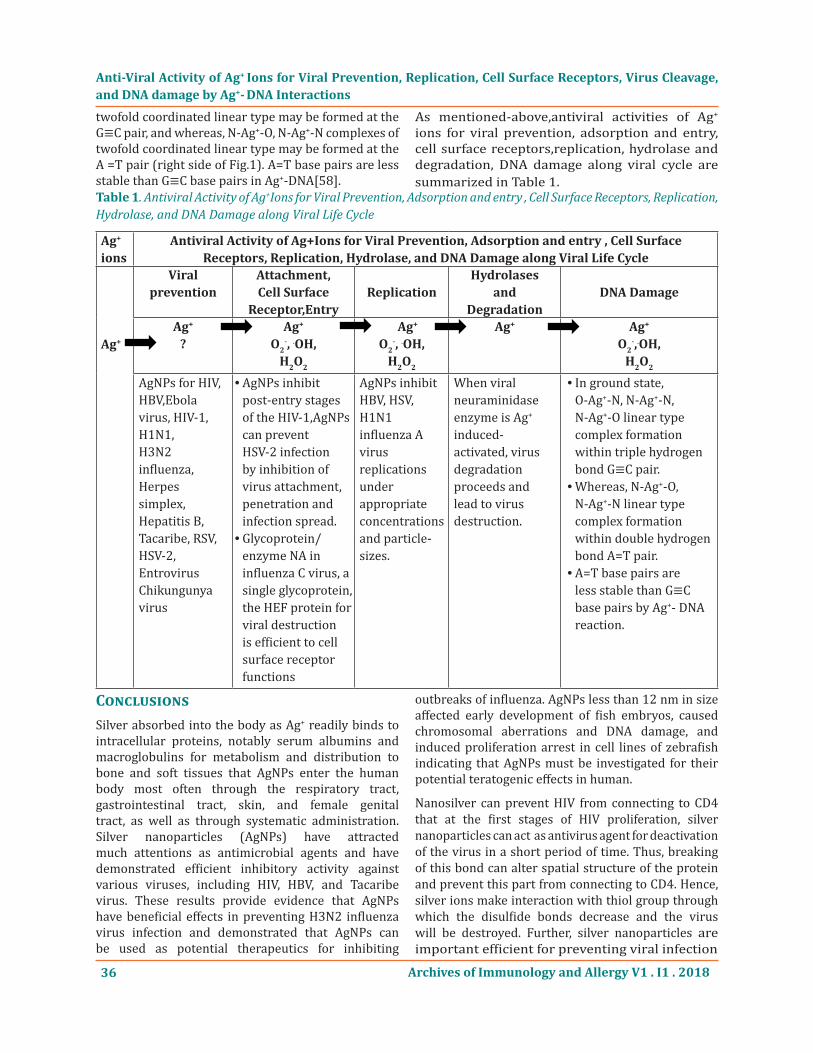
As mentioned-above,antiviral activities of Ag+ ions for viral prevention, adsorption and entry, cell surface receptors,replication, hydrolase and degradation, DNA damage along viral cycle are summarized in Table 1.
Anti-Viral Activity of Ag+ Ions for Viral Prevention, Replication, Cell Surface Receptors, Virus Cleavage, and DNA damage by Ag+- DNA Interactions
Ag+ ions
Antiviral Activity of Ag+Ions for Viral Prevention, Adsorption and entry , Cell Surface Receptors, Replication, Hydrolase, and DNA Damage along Viral Life Cycle
Ag+
Viral prevention
Attachment,Cell Surface
Receptor,EntryReplication
Hydrolasesand
DegradationDNA Damage
Ag+
?Ag+
O2-, .OH,
H2O2
Ag+
O2-, .OH,
H2O2
Ag+ Ag+
O2-,.OH,
H2O2
AgNPs for HIV, HBV,Ebola virus, HIV-1, H1N1,H3N2 influenza, Herpes simplex, Hepatitis B,Tacaribe, RSV,HSV-2, Entrovirus Chikungunya virus
AgNPs inhibit •post-entry stages of the HIV-1,AgNPs can prevent HSV-2 infection by inhibition of virus attachment, penetration and infection spread.Glycoprotein/ •enzyme NA in influenza C virus, a single glycoprotein, the HEF protein for viral destruction is efficient to cell surface receptor functions
AgNPs inhibitHBV, HSV, H1N1 influenza A virusreplications under appropriateconcentrations and particle-sizes.
When viralneuraminidase enzyme is Ag+ induced-activated, virus degradation proceeds andlead to virus destruction.
In ground state, •O-Ag+-N, N-Ag+-N, N-Ag+-O linear type complex formation within triple hydrogen bond G≡C pair.Whereas, N-Ag • +-O, N-Ag+-N linear type complex formation within double hydrogen bond A=T pair.A=T base pairs are •less stable than G≡C base pairs by Ag+- DNA reaction.
ConclusionsSilver absorbed into the body as Ag+ readily binds to intracellular proteins, notably serum albumins and macroglobulins for metabolism and distribution to bone and soft tissues that AgNPs enter the human body most often through the respiratory tract, gastrointestinal tract, skin, and female genital tract, as well as through systematic administration. Silver nanoparticles (AgNPs) have attracted much attentions as antimicrobial agents and have demonstrated efficient inhibitory activity against various viruses, including HIV, HBV, and Tacaribe virus. These results provide evidence that AgNPs have beneficial effects in preventing H3N2 influenza virus infection and demonstrated that AgNPs can be used as potential therapeutics for inhibiting
outbreaks of influenza. AgNPs less than 12 nm in size affected early development of fish embryos, caused chromosomal aberrations and DNA damage, and induced proliferation arrest in cell lines of zebrafish indicating that AgNPs must be investigated for their potential teratogenic effects in human.
Nanosilver can prevent HIV from connecting to CD4 that at the first stages of HIV proliferation, silver nanoparticles can act as antivirus agent for deactivation of the virus in a short period of time. Thus, breaking of this bond can alter spatial structure of the protein and prevent this part from connecting to CD4. Hence, silver ions make interaction with thiol group through which the disulfide bonds decrease and the virus will be destroyed. Further, silver nanoparticles are important efficient for preventing viral infection
Table 1. Antiviral Activity of Ag+Ions for Viral Prevention, Adsorption and entry , Cell Surface Receptors, Replication, Hydrolase, and DNA Damage along Viral Life Cycle
Archives of Immunology and Allergy V1 . I1 . 2018 37
against Ebola virus, HIV-1, H1N1 influenza, Herpes simplex, Hepatitis B, Tacaribe, RSV, HSV-2, Entrovirus, Chikungunya virus by blocking of viral attachment and entry steps. Chitin/chitosan with nanoscale fiber-like and porous surface structures may absorb heavy metals.Silver nanoparticles exert anti-HIV activity at an early stage of viral replication, most likely as a virucidal agent or as an inhibitor of viral entry that AgNPs bind to gp1 20 in a manner that prevents CD-dependent virion binding, fusion, and infectivity, acting as an effective virucidal agent against cell-free virus (laboratory strains, clinical isolates, T and M tropic strains, and resistant strains) and cell-associated virus, besides AgNPs inhibit post-entry stages of the HIV-1 life cycle. In addition, tannic acid modified AgNPs have the ability to prevent HSV-2 infection by direct inhibition of virus attachment, penetration and post-infection spread.
AgNPs inhibit HBV replication that Ag10NPs and Ag50NPs were able to reduce the extracellular HBV DNA formation of HepAD38 cells by >50% compared with the vehicle control, in which silver nanoparticles could inhibit the in vitro production of HBV RNA and extracellular virions, suggesting that the direct interaction between these nanoparticles and HBV double-stranded DNA or viral particles is responsible for their antiviral mechanism. Against HSV the AgNPs at nontoxic concentration of 100μg/mL were capable of inhibiting HSV-2 replication when administered prior to viral infection or soon after initial virus exposure which the mode of action of AgNPs occurs during the early phases of viral replication.
Neuraminidase (NA) enzymes are glycoside hydrolase enzymes that cleave the glycosidic linkages of neuraminic acids. Glycoside hydrolases are enzymes that catalyze the hydrolysis of the glycosidic linkage of glycosides, leading to the formation of a sugar hemiacetal or hemiketal and the corresponding free aglycon. The NA is important for the initiation, release of influenza virus infection in human airway epithelium that influenza virus NA plays an essential role in release and spread of progeny virions, intracellular viral replication cycle which viral NA provide an important role early in infection and further rationale for the prophylactic use of NA inhibitors. Viral neuraminidase can be assumed that the neuraminidase enzyme is corresponded to enzyme of bacterial peptidoglycan (PGN) autolysin. When viral neuraminidase enzyme
is activated, and virus protein homeostasis is lost, virus degradation proceeds, in which virus leads to apoptotic death and the virus is destroyed. Ag+ ions induced viral neuraminidase activations are enhanced in action sites of virus, subsequently the virus growth is suppressed,and virus destruction occurs.
Viral destruction of cell surface receptors, in general, the glycoproteins of several lipid-enveloped viruses, including orth-myxoviruses (influenza A, B, and C), toroviruses, and coronaviruses, have three important functions: to recognize the receptor on the cell surface, to mediate viral fusion with the cell membrane, and to destroy the receptor. In highly infections influenza A and B viruses, the receptor-binding and membrane-fusion activities of cell entry carried out by the glycoprotein hemagglutinin (HA) that the receptor-destroying enzyme (RDE) activity important for virus release is conducted by the glycoprotein/enzyme neuraminidase (NA). In influenza C virus, a single glycoprotein, the hemagglutinin-esterase-fusion (HEF) protein, possesses all three functions. Receptor-destroying enzyme is not required to inactivate inhibitors. The infectious salmon anaemia virus (ISAV) acetylesterase activity was inhibited by di-isopropyl fluorophosphate (DEP) in dose-dependent fashion but not by other known hydrolase inhibitors, suggesting that a serine residue is part of the active site, in which the effect of DFP on agglutination/elution of erythrocytes by ISAV demonstrated that the acetylesterase activity is the bona fide receptor-destroying enzyme. The viral cell receptor is an attractive target for anti-viral strategies that antibodies significantly inhibited avian sarcoma/leukosis virus (ASLV, including the A-J ten subgroups) infection and replication which cell lines with the expression of viral receptor-binding protein could be as efficient tools for isolating functional receptors to identify novel anti-viral targets. Ag+ ion induced viral NA enzyme, NA-HA enzyme, HEF protein, and RDE activation are enhanced, may be led to virus destruction.
Viral exolysins are murein hydrolases that virus cleavage can be required for infectivity. On the other hand, endolysins are enzymes used by bacteriophages at the end of their replication cycle to degrade the PGN of the bacterial host from within, resulting in cell lysis and release of progeny virions. Exolysin Ex1A is unknown about the identity and the nature of the cellular receptor, however, the host receptor
Anti-Viral Activity of Ag+ Ions for Viral Prevention, Replication, Cell Surface Receptors, Virus Cleavage, and DNA damage by Ag+- DNA Interactions
Archives of Immunology and Allergy V1 . I1 . 201838
is probably quite ubiquitous as many cell types are susceptible to Ex1A toxicity. The influenza virus HA is the viral protein that attaches to cell receptors that the HAplays an important role in the release of the viral RNA into the cell, by causing fusion of viral and cellular membrane. HA must be cleaved by cellular proteases to be active as a fusion protein. The other, endolysin is a PGN-hydrolysing enzyme that carries out enzymic digestion of the cell wall PGN at the end of the phage infection cycle, which ensure the release of newly packed phage particles. Hence, these phage particles like virus particles react with cell surface as viral receptor-binding protein, suggesting that leads to virus apoptotic death.
Substituting of Ag+ ions into hydrogen bonds in DNA base-pairing G≡C and A=T pairs respectively, it may be considered that DNA damages due to silver-complex formation within DNA base-pairs G≡C, A=T occur in cytoplasm of host cell-virus. Silver atom is twofold coordinated by two N atoms, and N-Ag+-N complex of linear type is formed in DNA base pairs. In ground state, O-Ag+-N, N-Ag+-N, N-Ag+-O twofold coordinated linear type may be formed at the G≡C pair, and whereas, N-Ag+-O, N-Ag+-N complexes of twofold coordinated linear type may be formed at the A =T pair. A:T base pairs are less stable than G:C base pairs in Ag+-DNA.
ReferencesJon L.Hobman and Lisa C. Crossman; Bacterial [1] antimicrobial metal ion resistance, Journal of Medical Microbiology, 2014;64:472-497.
S.Park, H.H. Park, S.Y. Kim et al; Antiviral [2] properties of silver nanoparticles on a magnetic hybrid colloid, Applied and Environmental Microbiology, 2014; 80, No.8:2343-2350.
F. Shimizu, Y. Shimizu, and K. Kumagai; Specific [3] inactivation of HSV by silver nitrate at low concentrations and biological activities of the inactivous virus, Antimicrobial Agents and Chemotherapy, 1976;20, No.1:57-63.
H.H. Lara, E.N Garza-Trevino, L. Ixtepan-Turrent, [4] and D. K Singh; Silver nanoparticles are broad-spectrum bactericidal and virucidal compounds, Journal of Nanobiotechnology, 2011;9, No.30:1-8.
A. Kaplan, G.A. Ciftci, and H.M. Kutlu; The apoptotic [5] and genomic studies on A549 cell line induced by silver nitrate, Tumor Biology, 2017; ? :1-12.
T.Ishida; Bcteriolysis of Cu[6] 2+ ion solution on the bacterial cell walls and DNA base-pairing damages, Biomedical Research on Trace Elements, 2017; 27:151-161.
T. Ishida; Anticancer activities of silver ions in [7] cancer and tumor cells, and DNA damages by Ag+-DNA base-pairs reactions, MOJ Tumor Research, 2017; 1(1):8-16.
P. Mellroth, R. Daniels, A. Eberhardt et al; LytA, [8] major autolysin of Streptococcus pneumoniae, requires access to nascent peptidoglycan, Journal of Biological Chemistry, 2012; 287, No.14:11018-11029.
M. Morales, A. J. Martin-Galiano, M. Domenech, [9] and E. Garcia; Insight into the evolutionary relationship of LytA autolysin and ply pneumolysin-like genes in Streptococcus pneumoniae and related streptococci, Genome Bio. Evol. 2015; 7(9):2747-2761.
H. Oliveira, C. Sao-Jose and J. Azeredo; Phage-[10] derived PGN degrading enzymes: challenges and future prospects for in vivo therapy, Viruses, 2018; 10:1-18pages.
A. Kedziora, M. Speruda, E. Krzyzewska et al; [11] Similarities and differences between silver ions and silver in nanoforms as antibacterial agents, Molecular Sciences, 2018; 19:1-17pages.
R. Behra, L. Sigg, M.J.D. Clift et al; Bioavailability [12] of silver nanoparticles and ions: from a chemical and biochemical perspective, Journal of the Royal Society Interface, 2013;10(87) :1-15pages.
V.G. Alekseev, A.N. Semenov, and P.M.Pakhomov; [13] Complexation of Ag+ ions with L-Cysteine, Russian Journal of Inorganic Chemistry, 2012; 57, No.7:1041-1044.
Alan B.G. Lansdown; A pharmacological and [14] toxicological profile of silver as an antimicrobial agent in medical devices, Advances in Pharmacological Sciences, 2010; 2010:1-16 pages.
H. Wang, N. Law, G. Pearson et al; Impact of [15] Silver(I) on the metabolism of Shewanella oneidensis, Journal of Bacteriology, 2013; 192, No.4:1143-1150.
Eric J. Rentz Do Comm Cnmo ; Viral pathogen and [16] severe acute respiratory syndrome: Oligodynamic Ag+ for direct immune intervention, Journal of Nutritional & Environmental Medicine, 2003; 13(2):109-118.
Anti-Viral Activity of Ag+ Ions for Viral Prevention, Replication, Cell Surface Receptors, Virus Cleavage, and DNA damage by Ag+- DNA Interactions
Archives of Immunology and Allergy V1 . I1 . 2018 39
V.Edwards-Jones; The benefits of silver in [17] hegiene, personal care and healthcare, Letters in Applied Microbiology, 2009; 49:147-152.
D.A. Gonzalez-Carter, B.F. Leo, P. Ruenraoengsak [18] et al; Silver nanoparticles reduce brain inflammation and related neurotoxicity through induction of H2S-synthesizing enzymes, Scientific Reports, 2017;7:1-14.
Maxwell Murphy, Kang Ting, Xinli Zhang, et al; [19] Current development of Silver nanoparticles preparation, investigation, and application in the field of medicine, Journal of Nanomaterials, 2015; 2015:1-12pages.
K.J. Lee, P.D. Nallathamby, L.M. Browning et al; In [20] vivo imaging of transport and biocompatibility of single silver nanoparticles in early development of zebrafish embryos, ACS Nano., 2007;28:133-143.
L.Ge, Q.Li, M.Wang et al; Nanosilver particles in [21] medical applications: synthesis, performance, and toxicity, International Journal of Nanomedicine, 2014;9(1) :2399-2407.
A I Klimov, P M Zherebin, A A Gusev et al; The [22] silver ions contribution into the cytotoxic activity of silver and silver halides nanoparticles, Nanobiotech 2015, Materials Science and Engineering, 2015; 98:1-10
D. [23] Xiang, Y. Zheng, W. Duan et al; Inhibition of A/Human/Hubei/3//2005(H3N2) influenza virus infection by silver nanoparticles in vitro and in vivo, International Journal of Nanomedicine, 2013; 8:4103-4114.
S. Gavanji, H. Mohabbatkar, H. Baghshahi, A. [24] Zarrabi; Bioinformatics prediction of interaction silver nanoparticles on the disulfide bounds of HIV-1 Gp120 protein, International Journal of Scientific Research in Knowledge, 2014; 2(2):67-74.
G. Pedersen Ph. D.; Preventing viral infection [25] with silver nanoparticles, Salt Lake City-2017, October 27, 1-20.
M. Ishihara, V. Q, Nguyen, Y. Moriet al; Adsorption [26] of silver nanoparticles onto different surface structure of chitin/chitosan and correlation with antimicrobial activities, J. Mol. Sci. 2015; 16:13973-13988.
H.H Lara, N.V Ayala-Nunez, L. Ixtepan-Turrent et [27] al; Mode of antiviral action of silver nanoparticles
against HIV-1, Journal of Nanobiotechnology, 2010; 8:1-10pages.
P. Orlowski, E. Tomaszewska, M. Gniadek et al; [28] Tannic acid modified silver nanoparticles show antiviral activity in HSV type 2 infection, PLOS ONE, 2014; 9:1-15.
Lei Lu, Raymond Wai-Yin Sun, Rong Chen et al; [29] Silver nanoparticles inhibit HBV replication, Antiviral Therapy, 2000;13(28):253-262.
R.L. Hu, S.R. Li, F.J. Hou et al; Inhibition effect [30] of silver nanoparticles on HSV 2, Genetics and Molecular Research, 2014; 13(3):7022-7028.
Mehrbod P., Motamed N., Tabatabaian M. et al; In [31] vitro antiviral effect of “nanosilver” on influenza virus, DARU, 2009; 17, No,2:88-93.
S. Gaikwad, A. Ingle, A. Gade et al;Antiviral [32] activity of mycosynthesized silver nanoparticles against HSV and human parainfluenza virus type 3, International Journal of Nanomedicine, 2013; 8:4303-4314.
Y. Mori, T. Ono, Y. Miyahira et al; Antiviral activity [33] of silver nanoparticle/chitosan composites against H1N1 influenza A virus, Nanoscale Research Letters, 2013; 8:1-6.
Gideon Davies and Bernard Henrissat; Structure [34] and mechanism of glycosyl hydrolases, Current Biology, 1995;3(9) :853-859.
M.N. Matrosovich, T. Y. Matrosovich, T. Gray et [35] al; Neuraminidase is important for the initiation of influenza virus infection in human airway epithelium, Journal of Virology, 2004; 78, No.22:12665-12667.
Y.A. Shtyrya, L.V. Mochalova, N.V. Bovin; Influenza [36] virus neuraminidase: structure and function, ACTA NATURAE, 2009;1(2):26-32.
D.V. da Silva, J. Nordholm, Dan Dou et al; [37] The influenza virus neuraminidase protein transmembrane and head domains have coevolved, Journal of Virology, 2015; 89, No.2:1094-1105.
J. J. Benton, S. A. Wharton, S.R. Martin, J.W. [38] McCauley; Role of neuraminidase in influenza A(H7N9) virus receptor binding, Journal of Virology, 2017;91,Issue 11;1-10pages.
Leif HOLMQUIST; Activation of vibrio cholerae [39] neuraminidase by divalent cations, FEBS LETTERS, 2015; 50,No.2:269-271.
Anti-Viral Activity of Ag+ Ions for Viral Prevention, Replication, Cell Surface Receptors, Virus Cleavage, and DNA damage by Ag+- DNA Interactions
Archives of Immunology and Allergy V1 . I1 . 201840
C. Lombardi, M. Ayach, L. Beaurepaire et al; A [40] compact viral processing proteinase/ubiquitin hydrolase from the OUT family, PLOS Pathogens, 2013; 9.Issue 8:1-13.
F.T. Vreede, A.Y. Chan, J.Sharps, and E.Fodor; [41] Mechanisms and functional implications of the degradation of host RNA polymerase II in influenza virus infected cells, Virology, 2010; 396:125-134.
Andrew D. Mesecar and Kiira Ratia; Viral [42] destruction of cell surface receptors,PNAS,2008;105,No.26:8807-8808.
H. Yang, P.J. Carney, V. P. Mishin et al; Molecular [43] characterizations of surface proteins hemagg lutinin and neuraminidase from recent H5Nx Avian Influenza Viruses, Journal of Virology, 2016;90, No.12:5770-5784.
H.Goto, K.Ohta, Y. Matsumoto et al; Evidence [44] that receptor destruction by the Sendai virus hemagglutinin-neuraminidase protein is responsible for homologous interference, Journal of Virology, 2016;90,No.17:7640-7646.
Q. Zeng, M. A. Langereis, A.L. van Vliet et al; [45] Structure of coronavirus hemagglutinin-esterase offers insight into corona and influenza virus evolution, PNAS, 2008; 105,No.26:9065-9069.
K. Hofling, Hans-Dieter Klenk and Georg Herrler; [46] Inactivation of inhibitors by the receptor-destroying enzyme of influenza C virus, Journal of General Virology,1997; 78:567-570.
M. Kristiansen, M.K. Froystad, A. Lise Rishovd [47] and Tor Gjoen.; Characterization of the receptor-destroying enzyme activity from infectious salmon anaemia virus, Journal of General Virology, 2002; 83:2693-2697.
Mei Mei, Jianqiang Ye, Aijian Qin, et al; [48] Identification of novel viral receptors with cell line expressing viral receptor-binding protein, SCIENTIFIC REPORTS, 2014; 5:1-6.
M. Schmelcher, D.M Donovan, and M.J Loessner; [49] Bacteriophage endolysins as novel antimicrobials, Future Microbiol. 2012;7(10):1147-1171.
E. Reboud, P. Basso, A. P. Maillard et al; Exolysin [50] shapes the virulence of pseudomonas aeruginosa clonal outliers, Toxin, 2017; 9:1-12.
Vincet Racaniello; Influenza HA cleavage is [51] required for infectivity, Virology blog about viruses and viral disease, 2018;8:1-11.
Amol Arunrao Pohane and Vikas Jain; Insight [52] into the regulation of bacteriophage endolysin; multiple means to the same end, Microbiology, 2015; 161:2269-2276.
Tsuneo Ishida : Bacteriolyses of Cu[53] 2+ solution on bacterial cell walls/cell membrane and DNA base pairing damages, Japanese Biomedical Research on Trace Elements, 2016; 27, No.4:151-161.
Maskos K.: The interaction of metal ions with [54] nucleic acids. NMR study of the copper (II) interaction with inosine derivatives. Acta Biochim Polonica 198; 28:317-335.
Steven M. Swasey, Leonardo Espinosa Leal, et [55] al; Silver (I) as DNA glue: Ag+-mediated guanine pairing revealed by removing Watson-Crick constraints, SCENTIFIC REPORTS, 2015;5:1-10
J.Christian Leon, Linda Stegemann, Martin [56] Peterlechner, et al; Formation of silver nanoclusters from a DNA template containing Ag (I)-mediated base pairs, Bioinorganic Chemistry and Applications, 2016;2016:9pages.
L.A. Espinosa Leal and O.Lopez-Acevedo; A [57] theoretical study of the interaction between DNA/RNA and the noble metal atoms of gold and silver; Ground-state properties, Condensed-Matter-Materials-Science, 2014:1-10.
Andrew H-J Wang, Toshio[58] Hakoshima et al; AT base pairs are less stable than GC base pairs in Z-DNA, Cell, 1984;37:321-331.
Anti-Viral Activity of Ag+ Ions for Viral Prevention, Replication, Cell Surface Receptors, Virus Cleavage, and DNA damage by Ag+- DNA Interactions
Citation: Dr. Sci. Tsuneo Ishida. Anti-Viral Activity of Ag+ Ions for Viral Prevention, Replication, Cell Surface Receptors, Virus Cleavage, and DNA damage by Ag+- DNA Interactions. Archives of Immunology and Allergy. 2018; 1(1): 29-40.Copyright: © 2018 Dr. Sci. Tsuneo Ishida. This is an open access article distributed under the Creative Commons Attribution License, which permits unrestricted use, distribution, and reproduction in any medium, provided the original work is properly cited.