anp 1105 the lymphatic system
DESCRIPTION
ANP1105 Lymphatic System NotesTRANSCRIPT

Topic #5The Lymphatic
System
Joanna Komorowski, Ph.D.

Learning objectives
The Lymphatic System – Describe the structure and main functions of
the vessels and organs of the lymphatic system
– Explain the origin of lymph as well as its transport
Chapter 20, pages 753-764

Functions of the Lymphatic System
Maintaining fluid balance-the return of fluid and solutes from peripheral tissues to the blood
The production, maintenance and distribution of lymphocytes-lymph nodes and other lymphoid tissues are the site of production of immunocompetent lymphocytes and macrophages in the specific immune response
Distribution of hormones, nutrients and waste products from their tissues of origin to the general circulation-e.g. the lipids absorbed by the digestive tract commonly fail to enter circulation at the capillary level. They reach the blood stream only after they have travelled along the lymphatic vessels

The lymphatic system
Two components of the lymphatic system:1.lymphatic vessels2.lymphoid tissues and organs
About 3 litres of interstitial fluid is produced daily by the filtration of blood plasma
This excess fluid plus any plasma proteins that escaped from the blood stream are drained from the tissue spaces by lymphatic vessels and returned to the blood

Distribution and structure of lymphatic vesselsLymphatic capillaries
Lymphatic collecting vessels
Lymphatic trunks
Lymphatic ducts
Right and left subclavian veins
Lymph nodes
Lymphatic collecting vessels

Figure 20.1
Lymphatic vessels
A one-way system - lymph flows only toward the heart!!!!!!!
It returns to the heart by the same mechanisms as observed in veins (recall!!)

Lymphatic capillaries
• Absent from bones, teeth, bone marrow, nervous system
• Blind ended
•Very permeable - endothelial cells with minivalves - anchoring collagen fibres
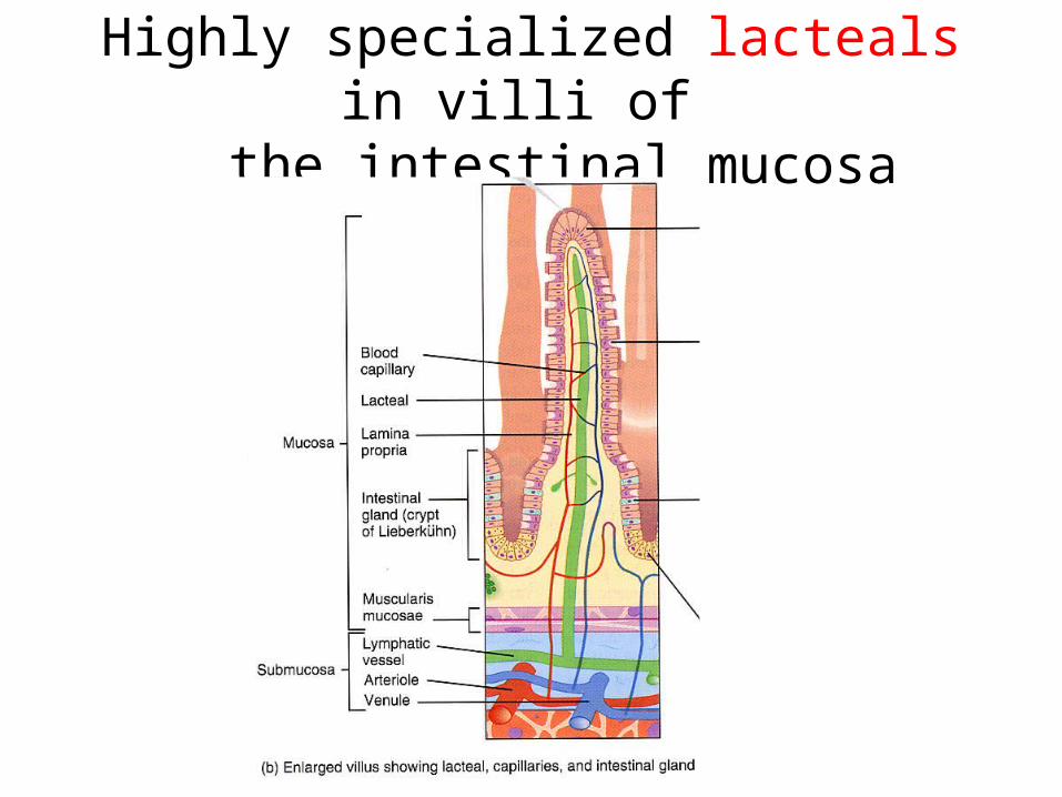
Highly specialized lacteals in villi of the intestinal mucosa

Figure 20.2b
Lymphatic vessels functions
• Collect excess fluid from the interstitium and return this fluid to the bloodstream
• Return leaked proteins to blood
• Carry lipids from the intestine to the blood
• Carry debris, bacteria, cancer cells etc. to lymph nodes for destruction (some may escape and spread through the lymphatic system)

Figure 20.2a
Lymphatics in the skin travel with veins and those of the trunk and GI viscera travel with the deep arteries

Lymphokinetic motion - the flow of lymphThe lymphatic system lacks a pumping organ Vessels are low-pressure conduits Lymph flows down the pressure gradient
Uses the same methods as veins to propel lymph:-pulsations of nearby arteries-muscular and respiratory pumps push lymph forward due to function of the semilunar valves- contractions of smooth muscle in the walls of the lymphatics

Lymphedema is the accumulation of tissue fluid in interstitial spaces, mainly tissues under the skin. It is the result of an overload of fluid not cleared by the lymphatic system
Obstruction of lymph movement

Lymphoid cells and tissuesUndifferentiated stem cells
(originate from red bone marrow)
T lymphocytes (T cells) B lymphocytes (B cells)
• Stimulated by the appropriate antigen T cells divide and differentiate, producing a clone of activated T cells and memory T cells•Activated T cells manage the cell-mediated immune responses; some of them directly attack and destroy the invaders• Memory T cells
• Stimulated by the appropriate antigen B cells divide and differentiate, producing a clone of antibody-secreting plasma cells and memory B cells• B cells manage the antibody-mediated immune responses; their daughter plasma cells secrete antibodies into the blood and other body fluids, immobilizing antigens until they are destroyed by phagocytes
Antigen = anything the body perceives as foreign (bacteria, viruses, fungi, cancer cells)
Mature in the thymus Mature in bone marrow

Lymhoid cells
Besides lympocytes, other lymphoid cells include:
• macrophages -phagocyte foreign substances and help to activate T cells
• dendritic cells – capture antigens and bring them back to the lymph nodes
• reticular cells – produce the reticular fibre stroma supporting other cells in the lymphoid organs

Lymphoid tissue
Lymphoid tissue:• houses and provides proliferation site for lymphocytes• provides ideal surveillance vantage point for lymphocytes and macrophages
Composed of reticular connective tissue (all lymphoid organs except thymus) consisting of:• reticular cells -lymphocytes B and T (travel between the lymphoid tissue and the other tissues of the body)-phagocytes (macrophages)-dendritic cells • reticular fibres that form 3-dimentional mesh

Lymphoid tissue, cont.• Diffuse lymphoid tissue - a few scattered lymphoid
elements; ubiquitous; particularly common in the lamina propria of mucous membranes and lymphoid organs
• Lymphoid follicles (nodules) – solid, spherical bodies with tightly packaged reticular elements and cells; germinal centers; no capsule
- often parts of larger lymphoid organs such as lymph nodes
- aggregations of follicles in the intestinal wall (Peyer’s patches) and in the appendix

Figure 20.4
Lymph nodes• The principal lymphoid organs in the body• Found throughout the body along the course of lymphatic vessels• Encapsulated• Filter the lymph - Macrophages remove & destroy microorganisms • Activate immune system; lymphocytes mount attacks against invaders• Large clusters of lymph nodes near body surface, in the inguinal (groin), axillary, and cervical regions and sites of lymphatic vessels convergence• Afferent and efferent lymphatic vessels


Figure 20.5
Other lymphoid organs
All lymphoid organs are composed of reticular CT
Only lymph nodes filter lymph
Other organs have only efferent lymphatics

Figure 20.6
• Largest lymphoid organ; beneath diaphragm; surrounded by capsule1) site for lymphocyte proliferation; immune surveillance & response2) blood cleansing3) storage of RBC products (iron) for recycling4) RBC production in fetus5) storage of blood platelets
• White pulp – immunity (mostly lymphocytes);
Red pulp – RBCs and venous sinuses; RBCs disposal
plus splenic cords rich in macrophages
• Thin capsule
Spleen

• Palatine tonsils
• Lingual tonsil • Pharyngeal tonsils
(adenoids) • Tubal tonsils
TonsilsThe tonsils gather and removemany pathogens entering the pharynx in food or inhaled air
Cryps trapbacteria and particular matter

Thymus• DOES NOT DIRECTLY fight antigens!!• Site of T cell maturation•Soft structure consisting of two lobes (bilobed)• Prominent in newborns, maximum size at puberty (30-40 grams), atrophies afterwards (15 grams in the elderly)• Most cells – T lymphocytes originating from bone marrow; some macrophages• Surrounded by capsule• Produces hormones thymopoietin and thymosins
• Mature T cells travel via blood to the lymph nodes, spleen, and diffuse lymphatic tissues, where they reside and are responsible for cell-mediated immune responses
Aggregates of lymphoid nodules•Peyer’s patches: lymph nodule clusters (wall of ileum)
•Appendix: lymph nodules forming offshoot of caecum; destroy bacteria; generate memory lymphocytes
MALT = Peyer’s patches + appendix + tonsils + nodules in walls of bronchi + genitourinary organs
MALT – Mucosa-Associated Lymphatic Tissue:-collection of small lymphoid tissue
- MALT protects the digestive and respiratory systems i.e. passages open to the exterior from foreign matter-MEMORY lymphocytes!!!!