anovelbifunctionalalkylphenolanestheticallows ... · taining to different gaba a receptor complexes...
TRANSCRIPT
A Novel Bifunctional Alkylphenol Anesthetic AllowsCharacterization of �-Aminobutyric Acid, Type A (GABAA),Receptor Subunit Binding Selectivity in Synaptosomes*□S
Received for publication, May 9, 2016, and in revised form, July 25, 2016 Published, JBC Papers in Press, July 26, 2016, DOI 10.1074/jbc.M116.736975
Kellie A. Woll‡§, Sruthi Murlidaran¶, Benika J. Pinch�, Jérôme Hénin**, Xiaoshi Wang‡‡, Reza Salari¶§§,Manuel Covarrubias¶¶, William P. Dailey�, Grace Brannigan¶§§, Benjamin A. Garcia‡‡, and Roderic G. Eckenhoff‡1
From the Departments of ‡Anesthesiology and Critical Care and §Pharmacology and the ‡‡Epigenetics Program, Department ofBiochemistry and Biophysics, University of Pennsylvania Perelman School of Medicine, Philadelphia, Pennsylvania 19104, the¶Center for Computational and Integrative Biology and §§Department of Physics, Rutgers University, Camden, New Jersey 08102,the �Department of Chemistry, University of Pennsylvania School of Arts and Sciences, Philadelphia, Pennsylvania 19104, the**Laboratoire de Biochimie Théorique, Institut de Biologie Physico-Chimique, CNRS UMR 8251 and Université Paris Diderot,5013 Paris, France, and the ¶¶Department of Neuroscience and Farber Institute for Neuroscience, Sidney Kimmel Medical College,Thomas Jefferson University, Philadelphia, Pennsylvania 19107
Propofol, an intravenous anesthetic, is a positive modulator ofthe GABAA receptor, but the mechanistic details, including therelevant binding sites and alternative targets, remain disputed.Here we undertook an in-depth study of alkylphenol-basedanesthetic binding to synaptic membranes. We designed, syn-thesized, and characterized a chemically active alkylphenolanesthetic(2-((prop-2-yn-1-yloxy)methyl)-5-(3-(trifluorometh-yl)-3H-diazirin-3-yl)phenol, AziPm-click (1)), for affinity-basedprotein profiling (ABPP) of propofol-binding proteins in theirnative state within mouse synaptosomes. The ABPP strategycaptured �4% of the synaptosomal proteome, including theunbiased capture of five � or � GABAA receptor subunits. Lackof �2 subunit capture was not due to low abundance. Consistentwith this, independent molecular dynamics simulations withalchemical free energy perturbation calculations predictedselective propofol binding to interfacial sites, with higher affin-ities for �/� than �-containing interfaces. The simulations indi-cated hydrogen bonding is a key component leading to propo-fol-selective binding within GABAA receptor subunit interfaces,with stable hydrogen bonds observed between propofol and �/�
cavity residues but not � cavity residues. We confirmed this byintroducing a hydrogen bond-null propofol analogue as a pro-tecting ligand for targeted-ABPP and observed a lack of GABAA
receptor subunit protection. This investigation demonstratesstriking interfacial GABAA receptor subunit selectivity in thenative milieu, suggesting that asymmetric occupancy of hetero-pentameric ion channels by alkylphenol-based anesthetics issufficient to induce modulation of activity.
�-Aminobutyric acid (GABA) is well established as the majorinhibitory neurotransmitter within the adult mammalian brain.The majority of GABA inhibitory activity is a consequence ofbinding to a set of pentameric ligand-gated ion channels calledthe GABA type A (GABAA) receptor. GABAA receptors arelargely heteromeric protein complexes composed of five differ-ent subunits that form a central pore that mediates chlorideflux. Including the multiple isoforms, heterogeneity of thereceptor is extensive with potentially more than 800 combina-tions (1). This complexity can be partially simplified by thedegrees of selective cellular localization for some subunits andisoforms. Synaptic GABAA receptors contribute considerablyto the communication between neurons, including influencingpresynaptic neurotransmitter release as well as inducing post-synaptic hyperpolarization (1– 4). Numerous studies have indi-cated that synaptic GABAA receptors are predominantly of a2�:2�:1� stoichiometry (5, 6) that organizes in an alternatingorder (e.g. ����� anti-clockwise as seen from synaptic cleft)(5–7). The resulting complex yields an abundance of potentialligand interaction surfaces within one heteropentamer, includ-ing at least four unique subunit interfaces. As such, it is justifiedthat the composition and orientation of subunits are function-ally significant, with different pharmacological properties per-taining to different GABAA receptor complexes (1, 8).
Numerous drugs influence GABAA receptor activity, includ-ing general anesthetics that are used extensively in modernmedicine and in scientific research (9). For example, 2,6-diiso-propylphenol (propofol2 ( (Fig. 1) has been strongly implicatedas a modulator of the GABAA receptor. Relatively low concen-trations of this alkylphenol significantly potentiate GABA-in-duced current, an action that hyperpolarizes the post-synapticmembrane and thereby likely contributes to hypnosis and pos-sibly other anesthesia phenotypes (10, 11). Furthermore, mul-tiple reports indicate that phasic inhibition is particularly sen-
* This work was supported by National Institutes of Health Grants GM055876and GM107174, Department of Defense Grant BC123187P1, and NationalScience Foundation Graduate Research Fellowship DGE-1321851. Theauthors declare that they have no conflicts of interest with the contents ofthis article. The content is solely the responsibility of the authors and doesnot necessarily represent the official views of the National Institutes ofHealth.Author’s Choice—Final version free via Creative Commons CC-BY license.
□S This article contains supplemental Table S1 and S2–S32.1 To whom correspondence should be addressed: 311A John Morgan Bldg.,
3620 Hamilton Walk, Philadelphia, PA 19104-6112. Tel.: 215-746-8699; Fax:215-349-5078; E-mail: [email protected].
2 The abbreviations used are: propofol, 2,6-diisopropylphenol; ABPP, affinity-based protein profiling; MD, molecular dynamics; AFEP, alchemical freeenergy perturbation; 1-AMA, 1-aminoanthracene; MS, mass spectrometry;TMT, tandem mass tag; fropofol, 2-fluoro-1,3-diisopropylbenzene; CI, con-fidence interval.
THE JOURNAL OF BIOLOGICAL CHEMISTRY VOL. 291, NO. 39, pp. 20473–20486, September 23, 2016Author’s Choice © 2016 by The American Society for Biochemistry and Molecular Biology, Inc. Published in the U.S.A.
crossmark
SEPTEMBER 23, 2016 • VOLUME 291 • NUMBER 39 JOURNAL OF BIOLOGICAL CHEMISTRY 20473
by guest on Decem
ber 25, 2019http://w
ww
.jbc.org/D
ownloaded from
sitive to low concentrations of propofol, suggesting thatsynaptic GABAergic signaling is a critical pathway for the ane-sthetic’s pharmacological effects (12–14).
Investigations have focused on the potential binding siteswithin heterologously expressed ��� GABAA receptors. Awide range of mutagenesis studies have probed ligand-gatedion channel electrophysiology and have shown that mutation ofvarious residues predicted to reside within subunit interfacialregions alters propofol modulation (9, 15–17). Particular pointmutations within � subunits, such as Asn-265, greatly decreasedpropofol-positive modulation (11, 18). Our previous workusing the tritiated photoaffinity ligand (PAL) meta-azipropofoldemonstrated frequent labeling of interfacial residues withinthe heterologously expressed Cys loop superfamily of recep-tors, including �1�3�2 GABAA receptors (19). These findingsfurther suggest that subunit interfaces are potentially involvedin propofol modulation. Structure-activity relationships apply-ing alkylphenol analogues and/or other chemical derivatives(20, 21), molecular dynamic (MD) simulations (22, 23), as wellas other investigations have suggested complex physicochemi-cal interactions between propofol and GABAA receptors (24).Together, these studies have provided insight regarding thepotential mechanism by which propofol perturbs GABAAreceptor protein dynamics. However, in addition to the biasednature of using heterologously expressed receptors, it is recog-nized that each method has experimental limitations that resultin the current uncertainty regarding alkylphenol interactionswithin the receptor.
Our objective was to advance the current understanding ofanesthetic interactions with heteromeric receptors by address-ing the interaction(s) of alkylphenols with GABAA receptorswithin their native synaptic milieu. Our approach applied anovel chemically active alkylphenol anesthetic for quantitativeaffinity-based protein profiling (ABPP) of propofol within syn-aptosomes. By utilizing a native tissue-derived system, the rel-
ative GABAA receptor subunit expression, pentameric compo-sition, protein-protein interactions, and lipid milieu aremaintained. We assessed these binding results with indepen-dent MD simulations using the alchemical free energy pertur-bation (AFEP) algorithm (25) to predict potential molecularrecognition elements within �1�3�2 GABAA receptor-bindingsites. Finally, we examined the impact of these molecular rec-ognition elements within the synaptic GABAA receptors withphotoaffinity protection experiments. Our studies led to theunbiased identification of GABAA receptor subunits innative synaptic membranes as alkylphenol-binding proteins.Our investigation suggested higher affinity interactions for��/�� and ��/�� interfacial sites relative to �-containingsubunit interfaces and hydrogen bonding as the major recogni-tion element for the alkylphenol-GABAA receptor complex.
Results
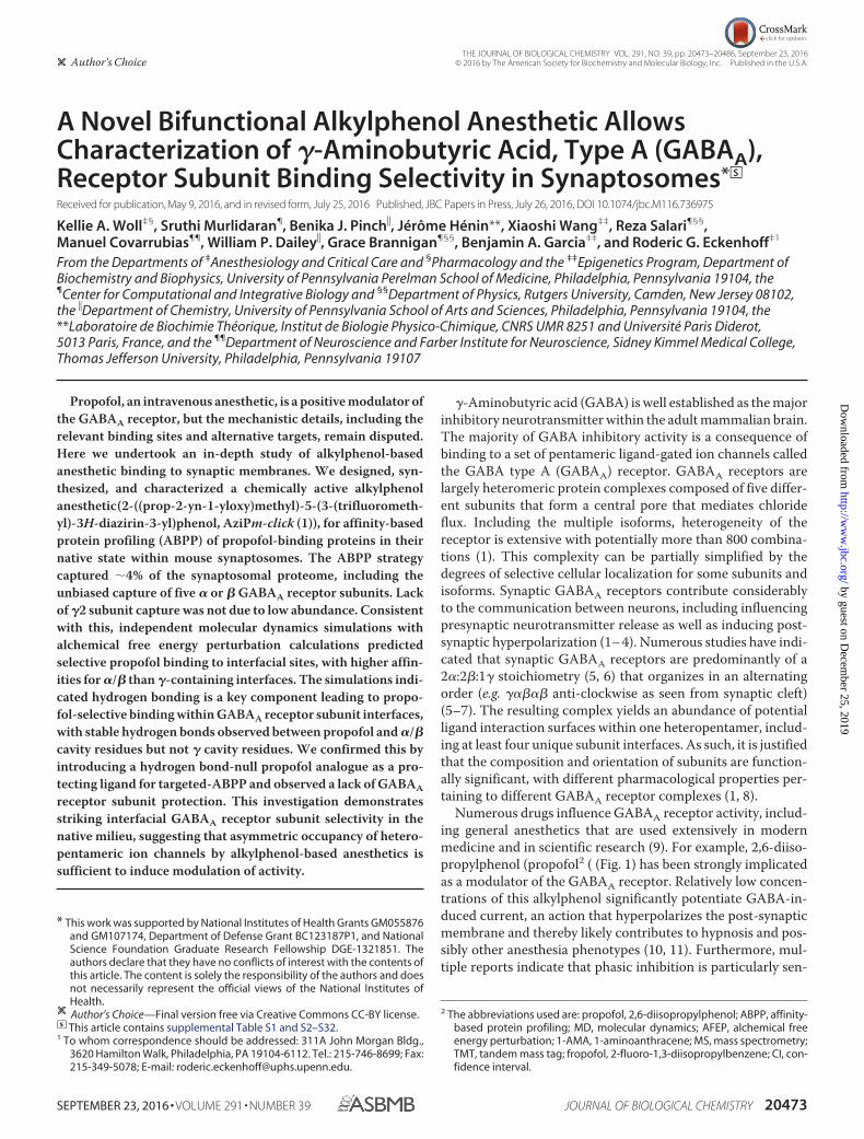
Synthesis of AziPm-click (1)—To identify the alkylphenol-binding proteins within the synaptic proteome, we devel-oped 2-((prop-2-yn-1-yloxy)methyl)-5-(3-(trifluoromethyl)-3H-diazirin-3-yl)phenol, or AziPm-click (1), a photoaffinitytandem bioorthogonal alkylphenol anesthetic ligand (Fig. 1).AziPm-click (1) was designed to integrate two chemically activegroups that allow for ABPP as follows: 1) a diazirine photoreac-tive group to covalently label protein interaction sites, and 2) analkynyl group for covalent attachment of a reporter tag by 1,3-dipolarcycloaddition reaction (e.g. “Click Chemistry”) to cap-ture and identify photoaffinity labeled proteins within the syn-aptic proteome.
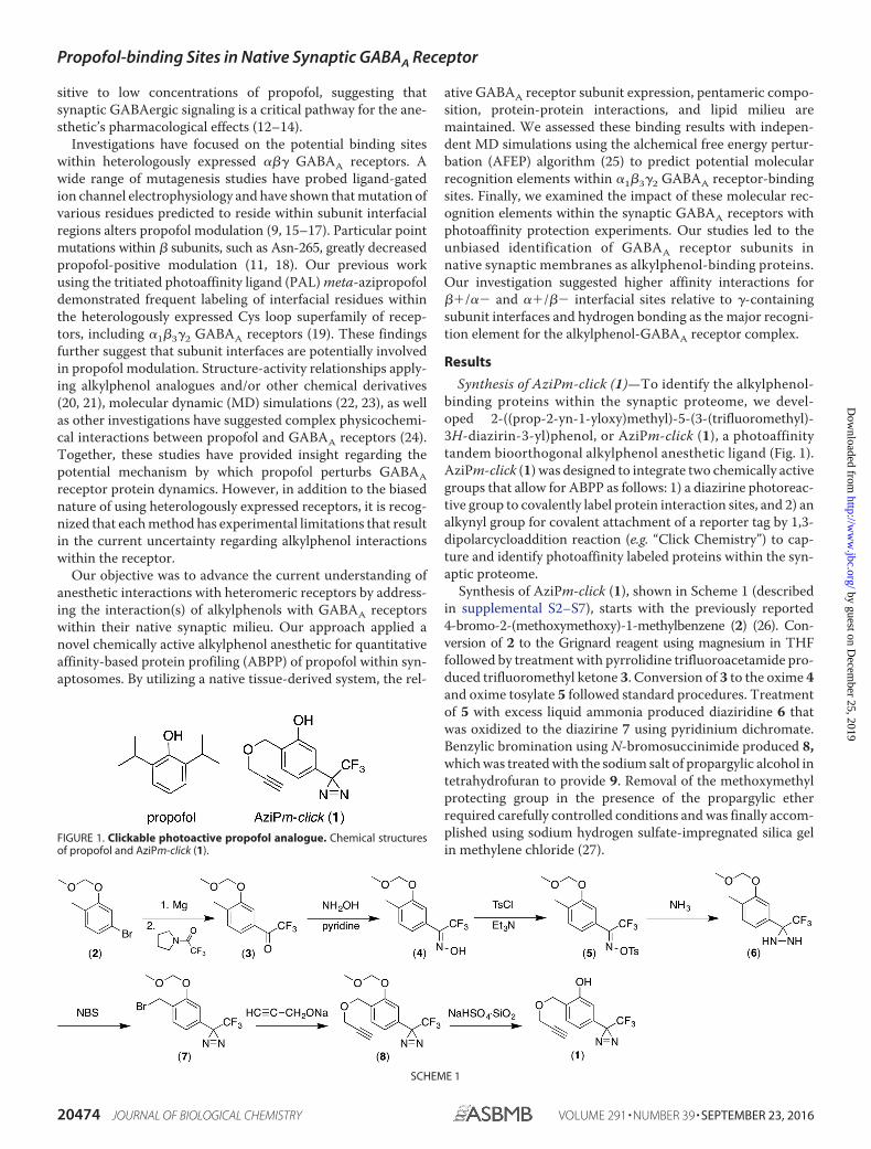
Synthesis of AziPm-click (1), shown in Scheme 1 (describedin supplemental S2–S7), starts with the previously reported4-bromo-2-(methoxymethoxy)-1-methylbenzene (2) (26). Con-version of 2 to the Grignard reagent using magnesium in THFfollowed by treatment with pyrrolidine trifluoroacetamide pro-duced trifluoromethyl ketone 3. Conversion of 3 to the oxime 4and oxime tosylate 5 followed standard procedures. Treatmentof 5 with excess liquid ammonia produced diaziridine 6 thatwas oxidized to the diazirine 7 using pyridinium dichromate.Benzylic bromination using N-bromosuccinimide produced 8,which was treated with the sodium salt of propargylic alcohol intetrahydrofuran to provide 9. Removal of the methoxymethylprotecting group in the presence of the propargylic etherrequired carefully controlled conditions and was finally accom-plished using sodium hydrogen sulfate-impregnated silica gelin methylene chloride (27).
FIGURE 1. Clickable photoactive propofol analogue. Chemical structuresof propofol and AziPm-click (1).
SCHEME 1
Propofol-binding Sites in Native Synaptic GABAA Receptor
20474 JOURNAL OF BIOLOGICAL CHEMISTRY VOLUME 291 • NUMBER 39 • SEPTEMBER 23, 2016
by guest on Decem
ber 25, 2019http://w
ww
.jbc.org/D
ownloaded from
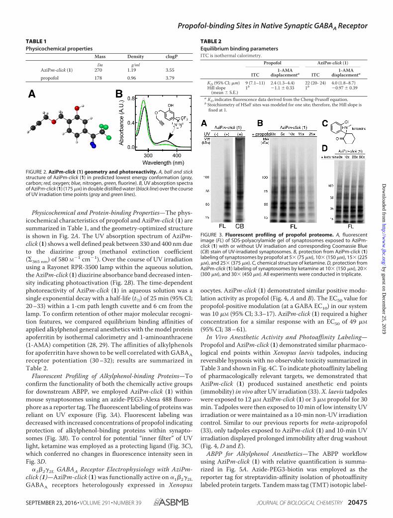
Physicochemical and Protein-binding Properties—The phys-icochemical characteristics of propofol and AziPm-click (1) aresummarized in Table 1, and the geometry-optimized structureis shown in Fig. 2A. The UV absorption spectrum of AziPm-click (1) shows a well defined peak between 330 and 400 nm dueto the diazirine group (methanol extinction coefficient(�365 nm) of 580 M�1 cm�1). Over the course of UV irradiationusing a Rayonet RPR-3500 lamp within the aqueous solution,the AziPm-click (1) diazirine absorbance band decreased inten-sity indicating photoactivation (Fig. 2B). The time-dependentphotoreactivity of AziPm-click (1) in aqueous solution was asingle exponential decay with a half-life (t1⁄2) of 25 min (95% CI;20 –33) within a 1-cm path length cuvette and 6 cm from thelamp. To confirm retention of other major molecular recogni-tion features, we compared equilibrium binding affinities ofapplied alkylphenol general anesthetics with the model proteinapoferritin by isothermal calorimetry and 1-aminoanthracene(1-AMA) competition (28, 29). The affinities of alkylphenolsfor apoferritin have shown to be well correlated with GABAAreceptor potentiation (30 –32); results are summarized inTable 2.
Fluorescent Profiling of Alkylphenol-binding Proteins—Toconfirm the functionality of both the chemically active groupsfor downstream ABPP, we employed AziPm-click (1) withinmouse synaptosomes using an azide-PEG3-Alexa 488 fluoro-phore as a reporter tag. The fluorescent labeling of proteins wasreliant on UV exposure (Fig. 3A). Fluorescent labeling wasdecreased with increased concentrations of propofol indicatingprotection of alkylphenol-binding proteins within synapto-somes (Fig. 3B). To control for potential “inner filter” of UVlight, ketamine was employed as a protecting ligand (Fig. 3C),which conferred no changes in fluorescence intensity seen inFig. 3D.
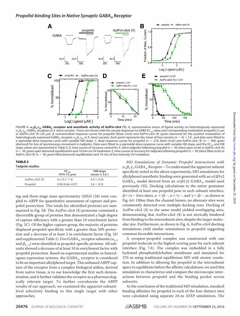
�1�2�2L GABAA Receptor Electrophysiology with AziPm-click (1)—AziPm-click (1) was functionally active on �1�2�2LGABAA receptors heterologously expressed in Xenopus
oocytes. AziPm-click (1) demonstrated similar positive modu-lation activity as propofol (Fig. 4, A and B). The EC50 value forpropofol-positive modulation (at a GABA EC10) in our systemwas 10 �M (95% CI; 3.3–17). AziPm-click (1) required a higherconcentration for a similar response with an EC50 of 49 �M
(95% CI; 38 – 61).In Vivo Anesthetic Activity and Photoaffinity Labeling—
Propofol and AziPm-click (1) demonstrated similar pharmaco-logical end points within Xenopus laevis tadpoles, inducingreversible hypnosis with no observable toxicity summarized inTable 3 and shown in Fig. 4C. To indicate photoaffinity labelingof pharmacologically relevant targets, we demonstrated thatAziPm-click (1) produced sustained anesthetic end points(immobility) in vivo after UV irradiation (33). X. laevis tadpoleswere exposed to 12 �M AziPm-click (1) or 3 �M propofol for 30min. Tadpoles were then exposed to 10 min of low intensity UVirradiation or were maintained as a 10-min non-UV irradiationcontrol. Similar to our previous reports for meta-azipropofol(33), only tadpoles exposed to AziPm-click (1) and 10-min UVirradiation displayed prolonged immobility after drug washout(Fig. 4, D and E).
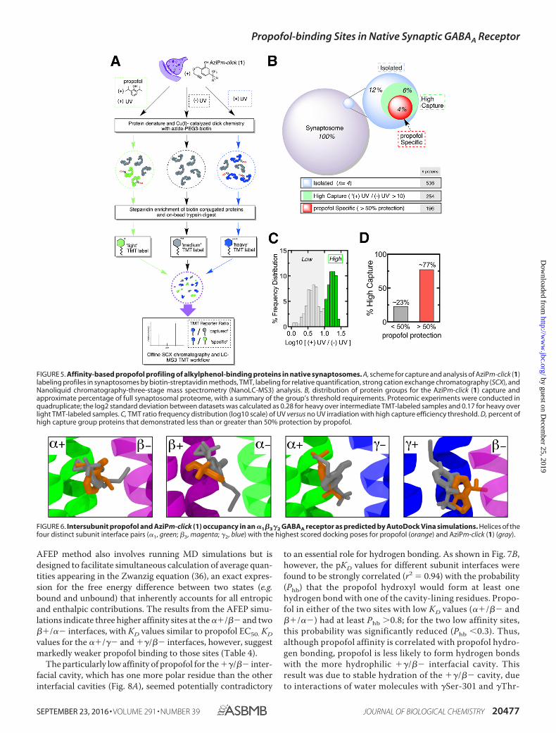
ABPP for Alkylphenol Anesthetics—The ABPP workflowusing AziPm-click (1) with relative quantification is summa-rized in Fig. 5A. Azide-PEG3-biotin was employed as thereporter tag for streptavidin-affinity isolation of photoaffinitylabeled protein targets. Tandem mass tag (TMT) isotopic label-
FIGURE 2. AziPm-click (1) geometry and photoreactivity. A, ball and stickstructure of AziPm-click (1) in predicted lowest energy conformation (gray,carbon; red, oxygen; blue, nitrogen, green, fluorine). B, UV absorption spectraof AziPm-click (1) (175 �M) in double distilled water (black line) over the courseof UV irradiation time points (gray and green lines).
TABLE 1Physicochemical properties
Mass Density clogP
Da g/mlAziPm-click (1) 270 1.19 3.55propofol 178 0.96 3.79
TABLE 2Equilibrium binding parametersITC is isothermal calorimetry.
Propofol AziPm-click (1)
ITC1-AMA
displacementa ITC1-AMA
displacementa
KD (95% CI; �m) 9 (7.1–11) 2.4 (1.3–4.4) 22 (20- 24) 4.0 (1.8–8.7)Hill slope
(mean � S.E.)1b �1.1 � 0.33 1b �0.97 � 0.39
a KD indicates fluorescence data derived from the Cheng-Prusoff equation.b Stoichiometry of HSaF sites was modeled for one site; therefore, the Hill slope is
fixed at 1.
FIGURE 3. Fluorescent profiling of propofol proteome. A, fluorescentimage (FL) of SDS-polyacrylamide gel of synaptosomes exposed to AziPm-click (1) with or without UV irradiation and corresponding Coomassie Blue(CB) stain of UV-irradiated synaptosomes. B, protection from AziPm-click (1)labeling of synaptosomes by propofol at 5� (75 �M), 10� (150 �M), 15� (225�M), and 25� (375 �M). C, chemical structure of ketamine. D, protection fromAziPm-click (1) labeling of synaptosomes by ketamine at 10� (150 �M), 20�(300 �M), and 30� (450 �M). All experiments were conducted in triplicate.
Propofol-binding Sites in Native Synaptic GABAA Receptor
SEPTEMBER 23, 2016 • VOLUME 291 • NUMBER 39 JOURNAL OF BIOLOGICAL CHEMISTRY 20475
by guest on Decem
ber 25, 2019http://w
ww
.jbc.org/D
ownloaded from
ing and three-stage mass spectrometry (MS3) (34) were cou-pled to ABPP for quantitative assessment of capture and pro-pofol protection. The totals for identified proteins are sum-marized in Fig. 5B. The AziPm-click (1) proteome contained adiscernible group of proteins that demonstrated a high degreeof capture efficiency with a greater than 10 enrichment factor(Fig. 5C). Of the higher capture group, the majority of proteinsdisplayed propofol specificity with a greater than 50% protec-tion and a decrease of at least 5 in enrichment factor (Fig. 5Dand supplemental Table 1). Five GABAA receptor subunits (�1,3and �1–3) were identified as propofol-specific proteins. All sub-units showed a decrease of at least 10 in enrichment factor withpropofol protection. Based on experimental studies in heterol-ogous expression systems, the GABAA receptor is consideredto be an important alkylphenol target. This unbiased ABPP cap-ture of the receptor from a complex biological milieu, derivedfrom native tissue, is to our knowledge the first such demon-stration, and it further validates the receptor as a pharmacolog-ically relevant target. To further corroborate the ABPPresults of our approach, we examined the apparent subunit-level selectivity binding to this single target with otherapproaches.
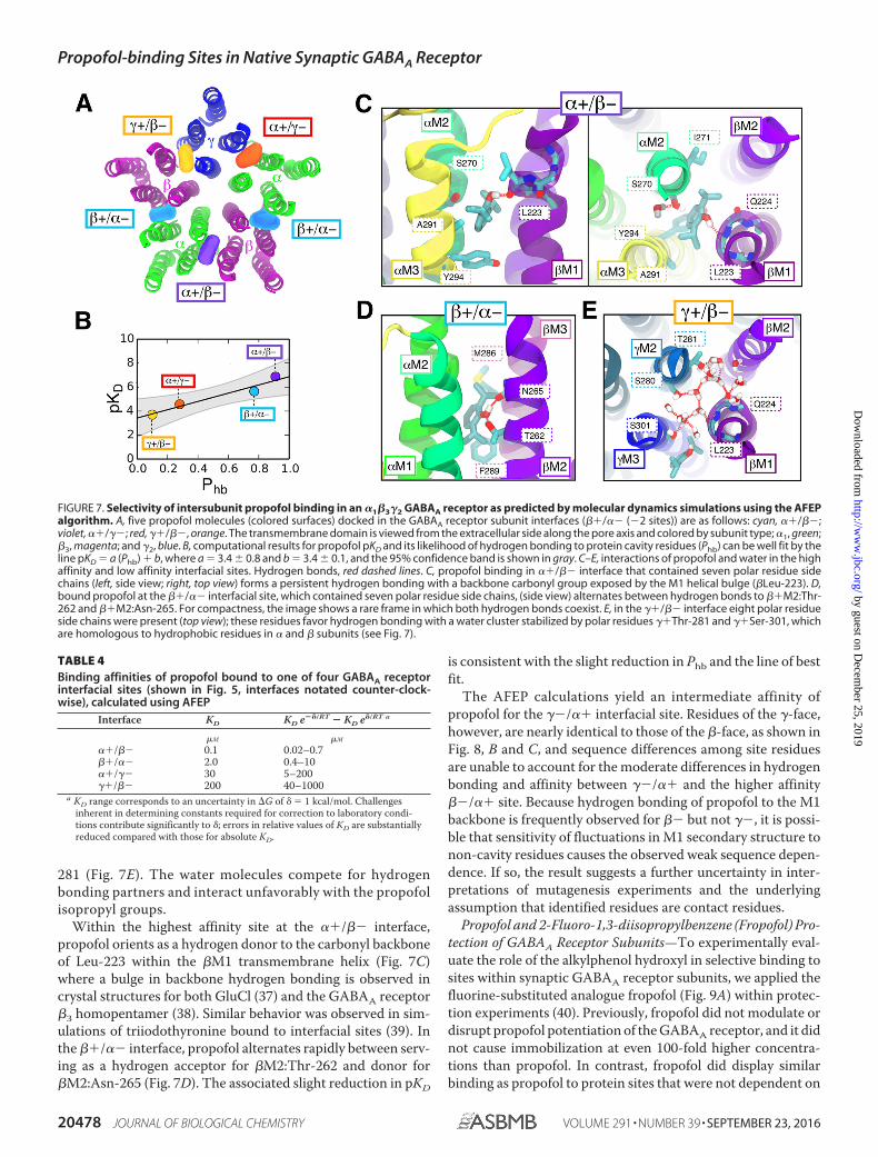
MD Simulations of Dynamic Propofol Interactions with�1�3�2 GABAA Receptor—To understand the apparent subunitspecificity noted in the above experiments, MD simulations foralkylphenol anesthetic binding were generated with an �1�3�2GABAA model derived from an �1�1�2 GABAA model usedpreviously (35). Docking calculations to the entire pentameridentified at least one propofol pose in each subunit interface,��/�� (two sites), � �/��, ��/��, and ��/��, as shown inFig. 6A. Other than the channel lumen, no alternate sites wereconsistently detected over multiple docking runs. Docking ofAziPm-click (1) to the same model yielded overlapping sites,demonstrating that AziPm-click (1) is not sterically hinderedfrom binding to the intersubunit sites, despite the larger molec-ular size. Furthermore, as shown in Fig. 6, AziPm-click dockingsimulations yield similar orientations to propofol suggestingcommon favorable interactions.
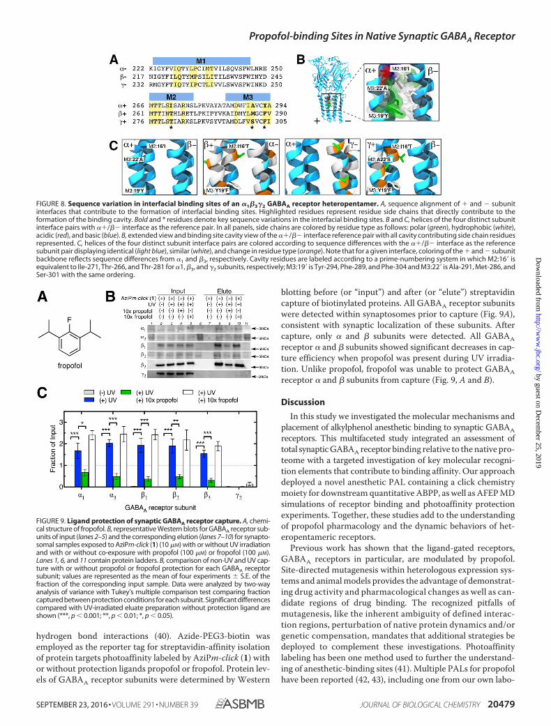
A receptor-propofol complex was constructed with onepropofol molecule in the highest scoring pose for each subunitinterface (Fig. 7A). The complex was embedded in a fullyhydrated phosphatidylcholine membrane and simulated for270 ns using traditional equilibrium MD with atomic resolu-tion. In addition to allowing the propofol in the intersubunitspace to equilibrate before the affinity calculations, we used thissimulation to characterize and compare the microscopic inter-actions between propofol and the binding pocket acrosssubunits.
At the conclusion of the traditional MD simulation, standardbinding affinities for propofol in each of the four distinct siteswere calculated using separate 24-ns AFEP simulations. The
FIGURE 4. �1�2�2L GABAA receptor and anesthetic activity of AziPm-click (1). A, representative traces of ligand activity on heterologously expressed�1�2�2L GABAA receptors in X. laevis oocytes. Traces are shown with the oocyte responses to GABA EC10 value and corresponding modulation propofol (3 �M)or AziPm-click (1) (20 �M). B, concentration-response curves for propofol (black circle) and AziPm-click (1) (green diamond) for the positive modulation ofheterologously expressed GABAA receptor �1�2�2L in X. laevis oocytes. Each point represents the mean of four oocytes (n � 4) � S.E., and data were fitted toa sigmoidal dose-response curve with variable Hill slope. C, dose-response curves for propofol (n � 210; black circle) and AziPm-click (1) (n � 300; greendiamond) for loss of spontaneous movement in tadpoles. Data were fitted to a sigmoidal dose-response curve with variable Hill slope, and the EC50 and Hillslope values are represented in Table 3. D, time course of recovery control for X. laevis tadpoles following propofol (n � 30; black open circle) or AziPm-click (1)(n � 30; green open diamond) equilibration and 10 min no UV treatment. E, time course of recovery for tadpoles following propofol (n � 30; black filled circle) orAziPm-click (1) (n � 30; green filled diamond) equilibration and 10 min of low intensity UV irradiation.
TABLE 3Tadpole studies
EC50(95% CI; �m)
Hill slope(mean � S.E.)
AziPm-click (1) 6.1 (5.1–7.4) 3.0 � 0.54Propofol 0.90 (0.84–0.97) 3.4 � 0.31
Propofol-binding Sites in Native Synaptic GABAA Receptor
20476 JOURNAL OF BIOLOGICAL CHEMISTRY VOLUME 291 • NUMBER 39 • SEPTEMBER 23, 2016
by guest on Decem
ber 25, 2019http://w
ww
.jbc.org/D
ownloaded from
AFEP method also involves running MD simulations but isdesigned to facilitate simultaneous calculation of average quan-tities appearing in the Zwanzig equation (36), an exact expres-sion for the free energy difference between two states (e.g.bound and unbound) that inherently accounts for all entropicand enthalpic contributions. The results from the AFEP simu-lations indicate three higher affinity sites at the ��/�� and two��/�� interfaces, with KD values similar to propofol EC50. KDvalues for the ��/�� and ��/�� interfaces, however, suggestmarkedly weaker propofol binding to those sites (Table 4).
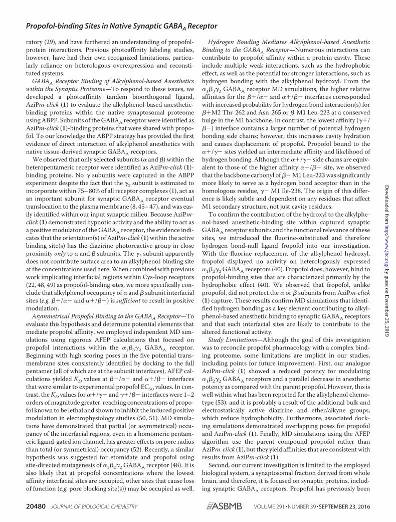
The particularly low affinity of propofol for the ��/�� inter-facial cavity, which has one more polar residue than the otherinterfacial cavities (Fig. 8A), seemed potentially contradictory
to an essential role for hydrogen bonding. As shown in Fig. 7B,however, the pKD values for different subunit interfaces werefound to be strongly correlated (r2 � 0.94) with the probability(Phb) that the propofol hydroxyl would form at least onehydrogen bond with one of the cavity-lining residues. Propo-fol in either of the two sites with low KD values (��/�� and��/��) had at least Phb 0.8; for the two low affinity sites,this probability was significantly reduced (Phb 0.3). Thus,although propofol affinity is correlated with propofol hydro-gen bonding, propofol is less likely to form hydrogen bondswith the more hydrophilic ��/�� interfacial cavity. Thisresult was due to stable hydration of the ��/�� cavity, dueto interactions of water molecules with �Ser-301 and �Thr-
FIGURE 5. Affinity-based propofol profiling of alkylphenol-binding proteins in native synaptosomes. A, scheme for capture and analysis of AziPm-click (1)labeling profiles in synaptosomes by biotin-streptavidin methods, TMT, labeling for relative quantification, strong cation exchange chromatography (SCX), andNanoliquid chromatography-three-stage mass spectrometry (NanoLC-MS3) analysis. B, distribution of protein groups for the AziPm-click (1) capture andapproximate percentage of full synaptosomal proteome, with a summary of the group’s threshold requirements. Proteomic experiments were conducted inquadruplicate; the log2 standard deviation between datasets was calculated as 0.28 for heavy over intermediate TMT-labeled samples and 0.17 for heavy overlight TMT-labeled samples. C, TMT ratio frequency distribution (log10 scale) of UV versus no UV irradiation with high capture efficiency threshold. D, percent ofhigh capture group proteins that demonstrated less than or greater than 50% protection by propofol.
FIGURE 6. Intersubunit propofol and AziPm-click (1) occupancy in an �1�3�2 GABAA receptor as predicted by AutoDock Vina simulations. Helices of thefour distinct subunit interface pairs (�1, green; �3, magenta; �2, blue) with the highest scored docking poses for propofol (orange) and AziPm-click (1) (gray).
Propofol-binding Sites in Native Synaptic GABAA Receptor
SEPTEMBER 23, 2016 • VOLUME 291 • NUMBER 39 JOURNAL OF BIOLOGICAL CHEMISTRY 20477
by guest on Decem
ber 25, 2019http://w
ww
.jbc.org/D
ownloaded from
281 (Fig. 7E). The water molecules compete for hydrogenbonding partners and interact unfavorably with the propofolisopropyl groups.
Within the highest affinity site at the ��/�� interface,propofol orients as a hydrogen donor to the carbonyl backboneof Leu-223 within the �M1 transmembrane helix (Fig. 7C)where a bulge in backbone hydrogen bonding is observed incrystal structures for both GluCl (37) and the GABAA receptor�3 homopentamer (38). Similar behavior was observed in sim-ulations of triiodothyronine bound to interfacial sites (39). Inthe ��/�� interface, propofol alternates rapidly between serv-ing as a hydrogen acceptor for �M2:Thr-262 and donor for�M2:Asn-265 (Fig. 7D). The associated slight reduction in pKD
is consistent with the slight reduction in Phb and the line of bestfit.
The AFEP calculations yield an intermediate affinity ofpropofol for the ��/�� interfacial site. Residues of the �-face,however, are nearly identical to those of the �-face, as shown inFig. 8, B and C, and sequence differences among site residuesare unable to account for the moderate differences in hydrogenbonding and affinity between ��/�� and the higher affinity��/�� site. Because hydrogen bonding of propofol to the M1backbone is frequently observed for �� but not ��, it is possi-ble that sensitivity of fluctuations in M1 secondary structure tonon-cavity residues causes the observed weak sequence depen-dence. If so, the result suggests a further uncertainty in inter-pretations of mutagenesis experiments and the underlyingassumption that identified residues are contact residues.
Propofol and 2-Fluoro-1,3-diisopropylbenzene (Fropofol) Pro-tection of GABAA Receptor Subunits—To experimentally eval-uate the role of the alkylphenol hydroxyl in selective binding tosites within synaptic GABAA receptor subunits, we applied thefluorine-substituted analogue fropofol (Fig. 9A) within protec-tion experiments (40). Previously, fropofol did not modulate ordisrupt propofol potentiation of the GABAA receptor, and it didnot cause immobilization at even 100-fold higher concentra-tions than propofol. In contrast, fropofol did display similarbinding as propofol to protein sites that were not dependent on
FIGURE 7. Selectivity of intersubunit propofol binding in an �1�3�2 GABAA receptor as predicted by molecular dynamics simulations using the AFEPalgorithm. A, five propofol molecules (colored surfaces) docked in the GABAA receptor subunit interfaces (��/�� (�2 sites)) are as follows: cyan, ��/��;violet, ��/��; red, ��/��, orange. The transmembrane domain is viewed from the extracellular side along the pore axis and colored by subunit type; �1, green;�3, magenta; and �2, blue. B, computational results for propofol pKD and its likelihood of hydrogen bonding to protein cavity residues (Phb) can be well fit by theline pKD � a (Phb) � b, where a � 3.4 � 0.8 and b � 3.4 � 0.1, and the 95% confidence band is shown in gray. C–E, interactions of propofol and water in the highaffinity and low affinity interfacial sites. Hydrogen bonds, red dashed lines. C, propofol binding in ��/�� interface that contained seven polar residue sidechains (left, side view; right, top view) forms a persistent hydrogen bonding with a backbone carbonyl group exposed by the M1 helical bulge (�Leu-223). D,bound propofol at the ��/�� interfacial site, which contained seven polar residue side chains, (side view) alternates between hydrogen bonds to ��M2:Thr-262 and ��M2:Asn-265. For compactness, the image shows a rare frame in which both hydrogen bonds coexist. E, in the ��/�� interface eight polar residueside chains were present (top view); these residues favor hydrogen bonding with a water cluster stabilized by polar residues ��Thr-281 and ��Ser-301, whichare homologous to hydrophobic residues in � and � subunits (see Fig. 7).
TABLE 4Binding affinities of propofol bound to one of four GABAA receptorinterfacial sites (shown in Fig. 5, interfaces notated counter-clock-wise), calculated using AFEP
Interface KD KD e��/RT � KD e�/RT a
�M �M
��/�� 0.1 0.02–0.7��/�� 2.0 0.4–10��/�� 30 5–200��/�� 200 40–1000
a KD range corresponds to an uncertainty in �G of � � 1 kcal/mol. Challengesinherent in determining constants required for correction to laboratory condi-tions contribute significantly to �; errors in relative values of KD are substantiallyreduced compared with those for absolute KD.
Propofol-binding Sites in Native Synaptic GABAA Receptor
20478 JOURNAL OF BIOLOGICAL CHEMISTRY VOLUME 291 • NUMBER 39 • SEPTEMBER 23, 2016
by guest on Decem
ber 25, 2019http://w
ww
.jbc.org/D
ownloaded from
hydrogen bond interactions (40). Azide-PEG3-biotin wasemployed as the reporter tag for streptavidin-affinity isolationof protein targets photoaffinity labeled by AziPm-click (1) withor without protection ligands propofol or fropofol. Protein lev-els of GABAA receptor subunits were determined by Western
blotting before (or “input”) and after (or “elute”) streptavidincapture of biotinylated proteins. All GABAA receptor subunitswere detected within synaptosomes prior to capture (Fig. 9A),consistent with synaptic localization of these subunits. Aftercapture, only � and � subunits were detected. All GABAAreceptor � and � subunits showed significant decreases in cap-ture efficiency when propofol was present during UV irradia-tion. Unlike propofol, fropofol was unable to protect GABAAreceptor � and � subunits from capture (Fig. 9, A and B).
Discussion
In this study we investigated the molecular mechanisms andplacement of alkylphenol anesthetic binding to synaptic GABAAreceptors. This multifaceted study integrated an assessment oftotal synaptic GABAA receptor binding relative to the native pro-teome with a targeted investigation of key molecular recogni-tion elements that contribute to binding affinity. Our approachdeployed a novel anesthetic PAL containing a click chemistrymoiety for downstream quantitative ABPP, as well as AFEP MDsimulations of receptor binding and photoaffinity protectionexperiments. Together, these studies add to the understandingof propofol pharmacology and the dynamic behaviors of het-eropentameric receptors.
Previous work has shown that the ligand-gated receptors,GABAA receptors in particular, are modulated by propofol.Site-directed mutagenesis within heterologous expression sys-tems and animal models provides the advantage of demonstrat-ing drug activity and pharmacological changes as well as can-didate regions of drug binding. The recognized pitfalls ofmutagenesis, like the inherent ambiguity of defined interac-tion regions, perturbation of native protein dynamics and/orgenetic compensation, mandates that additional strategies bedeployed to complement these investigations. Photoaffinitylabeling has been one method used to further the understand-ing of anesthetic-binding sites (41). Multiple PALs for propofolhave been reported (42, 43), including one from our own labo-
FIGURE 8. Sequence variation in interfacial binding sites of an �1�3�2 GABAA receptor heteropentamer. A, sequence alignment of � and � subunitinterfaces that contribute to the formation of interfacial binding sites. Highlighted residues represent residue side chains that directly contribute to theformation of the binding cavity. Bold and * residues denote key sequence variations in the interfacial binding sites. B and C, helices of the four distinct subunitinterface pairs with ��/�� interface as the reference pair. In all panels, side chains are colored by residue type as follows: polar (green), hydrophobic (white),acidic (red), and basic (blue). B, extended view and binding site cavity view of the ��/�� interface reference pair with all cavity contributing side chain residuesrepresented. C, helices of the four distinct subunit interface pairs are colored according to sequence differences with the ��/�� interface as the referencesubunit pair displaying identical (light blue), similar (white), and change in residue type (orange). Note that for a given interface, coloring of the � and � subunitbackbone reflects sequence differences from �1 and �3, respectively. Cavity residues are labeled according to a prime-numbering system in which M2:16� isequivalent to Ile-271, Thr-266, and Thr-281 for �1, �3, and �2 subunits, respectively; M3:19� is Tyr-294, Phe-289, and Phe-304 and M3:22� is Ala-291, Met-286, andSer-301 with the same ordering.
FIGURE 9. Ligand protection of synaptic GABAA receptor capture. A, chemi-cal structure of fropofol. B, representative Western blots for GABAA receptor sub-units of input (lanes 2–5) and the corresponding elution (lanes 7–10) for synapto-somal samples exposed to AziPm-click (1) (10 �M) with or without UV irradiationand with or without co-exposure with propofol (100 �M) or fropofol (100 �M).Lanes 1, 6, and 11 contain protein ladders. B, comparison of non-UV and UV cap-ture with or without propofol or fropofol protection for each GABAA receptorsubunit; values are represented as the mean of four experiments � S.E. of thefraction of the corresponding input sample. Data were analyzed by two-wayanalysis of variance with Tukey’s multiple comparison test comparing fractioncaptured between protection conditions for each subunit. Significant differencescompared with UV-irradiated eluate preparation without protection ligand areshown (***, p 0.001; **, p 0.01; *, p 0.05).
Propofol-binding Sites in Native Synaptic GABAA Receptor
SEPTEMBER 23, 2016 • VOLUME 291 • NUMBER 39 JOURNAL OF BIOLOGICAL CHEMISTRY 20479
by guest on Decem
ber 25, 2019http://w
ww
.jbc.org/D
ownloaded from
ratory (29), and have furthered an understanding of propofol-protein interactions. Previous photoaffinity labeling studies,however, have had their own recognized limitations, particu-larly reliance on heterologous overexpression and reconsti-tuted systems.
GABAA Receptor Binding of Alkylphenol-based Anestheticswithin the Synaptic Proteome—To respond to these issues, wedeveloped a photoaffinity tandem bioorthogonal ligand,AziPm-click (1) to evaluate the alkylphenol-based anesthetic-binding proteins within the native synaptosomal proteomeusing ABPP. Subunits of the GABAA receptor were identified asAziPm-click (1)-binding proteins that were shared with propo-fol. To our knowledge the ABPP strategy has provided the firstevidence of direct interaction of alkylphenol anesthetics withnative tissue-derived synaptic GABAA receptors.
We observed that only selected subunits (� and �) within theheteropentameric receptor were identified as AziPm-click (1)-binding proteins. No � subunits were captured in the ABPPexperiment despite the fact that the �2 subunit is estimated toincorporate within 75– 80% of all receptor complexes (1), act asan important subunit for synaptic GABAA receptor eventualtranslocation to the plasma membrane (8, 45– 47), and was eas-ily identified within our input synaptic milieu. Because AziPm-click (1) demonstrated hypnotic activity and the ability to act asa positive modulator of the GABAA receptor, the evidence indi-cates that the orientation(s) of AziPm-click (1) within the activebinding site(s) has the diazirine photoreactive group in closeproximity only to � and � subunits. The �2 subunit apparentlydoes not contribute surface area to an alkylphenol-binding siteat the concentrations used here. When combined with previouswork implicating interfacial regions within Cys-loop receptors(22, 48, 49) as propofol-binding sites, we more specifically con-clude that alkylphenol occupancy of � and � subunit interfacialsites (e.g. ��/�� and ��/��) is sufficient to result in positivemodulation.
Asymmetrical Propofol Binding to the GABAA Receptor—Toevaluate this hypothesis and determine potential elements thatmediate propofol affinity, we employed independent MD sim-ulations using rigorous AFEP calculations that focused onpropofol interactions within the �1�3�2 GABAA receptor.Beginning with high scoring poses in the five potential trans-membrane sites consistently identified by docking to the fullpentamer (all of which are at the subunit interfaces), AFEP cal-culations yielded KD values at ��/�� and ��/�� interfacesthat were similar to experimental propofol EC50 values. In con-trast, the KD values for ��/�� and ��/�� interfaces were 1–2orders of magnitude greater, reaching concentrations of propo-fol known to be lethal and shown to inhibit the induced positivemodulation in electrophysiology studies (50, 51). MD simula-tions have demonstrated that partial (or asymmetrical) occu-pancy of the interfacial regions, even in a homomeric pentam-eric ligand-gated ion channel, has greater effects on pore radiusthan total (or symmetrical) occupancy (52). Recently, a similarhypothesis was suggested for etomidate and propofol usingsite-directed mutagenesis of �1�2�2 GABAA receptor (48). It isalso likely that at propofol concentrations where the lowestaffinity interfacial sites are occupied, other sites that cause lossof function (e.g. pore blocking site(s)) may be occupied as well.
Hydrogen Bonding Mediates Alkylphenol-based AnestheticBinding to the GABAA Receptor—Numerous interactions cancontribute to propofol affinity within a protein cavity. Theseinclude multiple weak interactions, such as the hydrophobiceffect, as well as the potential for stronger interactions, such ashydrogen bonding with the alkylphenol hydroxyl. From the�1�3�2 GABAA receptor MD simulations, the higher relativeaffinities for the ��/�� and ��/�� interfaces correspondedwith increased probability for hydrogen bond interaction(s) for��M2 Thr-262 and Asn-265 or �-M1 Leu-223 at a conservedbulge in the M1 backbone. In contrast, the lowest affinity (��/��) interface contains a larger number of potential hydrogenbonding side chains; however, this increases cavity hydrationand causes displacement of propofol. Propofol bound to the��/�� sites yielded an intermediate affinity and likelihood ofhydrogen bonding. Although the ��/�� side chains are equiv-alent to those of the higher affinity ��/�� site, we observedthat the backbone carbonyl of �� M1 Leu-223 was significantlymore likely to serve as a hydrogen bond acceptor than in thehomologous residue, �� M1 Ile-238. The origin of this differ-ence is likely subtle and dependent on any residues that affectM1 secondary structure, not just cavity residues.
To confirm the contribution of the hydroxyl to the alkylphe-nol-based anesthetic-binding site within captured synapticGABAA receptor subunits and the functional relevance of thesesites, we introduced the fluorine-substituted and thereforehydrogen bond-null ligand fropofol into our investigation.With the fluorine replacement of the alkylphenol hydroxyl,fropofol displayed no activity on heterologously expressed�1�2�2 GABAA receptors (40). Fropofol does, however, bind topropofol-binding sites that are characterized primarily by thehydrophobic effect (40). We observed that fropofol, unlikepropofol, did not protect the � or � subunits from AziPm-click(1) capture. These results confirm MD simulations that identi-fied hydrogen bonding as a key element contributing to alkyl-phenol-based anesthetic binding to synaptic GABAA receptorsand that such interfacial sites are likely to contribute to thealtered functional activity.
Study Limitations—Although the goal of this investigationwas to reconcile propofol pharmacology with a complex bind-ing proteome, some limitations are implicit in our studies,including points for future improvement. First, our analogueAziPm-click (1) showed a reduced potency for modulating�1�2�2 GABAA receptors and a parallel decrease in anestheticpotency as compared with the parent propofol. However, this iswell within what has been reported for the alkylphenol chemo-type (53), and it is probably a result of the additional bulk andelectrostatically active diazirine and ether/alkyne groups,which reduce hydrophobicity. Furthermore, associated dock-ing simulations demonstrated overlapping poses for propofoland AziPm-click (1). Finally, MD simulations using the AFEPalgorithm use the parent compound propofol rather thanAziPm-click (1), but they yield affinities that are consistent withresults from AziPm-click (1).
Second, our current investigation is limited to the employedbiological system, a synaptosomal fraction derived from wholebrain, and therefore, it is focused on synaptic proteins, includ-ing synaptic GABAA receptors. Propofol has previously been
Propofol-binding Sites in Native Synaptic GABAA Receptor
20480 JOURNAL OF BIOLOGICAL CHEMISTRY VOLUME 291 • NUMBER 39 • SEPTEMBER 23, 2016
by guest on Decem
ber 25, 2019http://w
ww
.jbc.org/D
ownloaded from
shown to influence tonic receptor-mediated inhibition that isanticipated to contribute to observed hypnotic sedation andother anesthesia phenotypes, specifically amnesia (13, 14, 54).Because of the lack of extrasynaptic GABAA receptor subunitswithin our proteome (data not shown), we could not determinethe binding character for these receptors.
Finally, although the presented quantitative ABPP strategyallows the first platform for identifying alkylphenol protein tar-gets relative to a native biological milieu, it does not directlyidentify the photoaffinity labeled residues. In part, this is aresult of the challenging elution/digestion of labeled proteinsand large modification size (600 Da) of the biotin PEG3-con-jugated AziPm-click (1) modification. Currently availablecleavable biotin-X-azide linkers have variable cleavage effi-ciency and/or require reagents that perturb downstream quan-titative labeling for native tissue-derived systems (55). Ourefforts for non-quantifiable capture using cleaved biotin linkerand AziPm-click (1) identified modifications on higher abun-dance proteins like the voltage-dependent cation channel (datanot shown) that corresponded with earlier reports (56); how-ever, residue-level modifications on lower abundance proteins,like the GABAA receptor, remained undetected. Thus, we can-not confirm an interfacial location of sites in this study,although this location has been demonstrated in heterologousreceptors (19). Future development of chemically active alkylphe-nol anesthetics, cleavable biotin linkers, as well as enhanced pep-tide enrichment and release methods may allow for increased cap-ture efficiency permitting detection of modifications within thevery low abundance photoaffinity labeled peptides.
Additional Alkylphenol-based Anesthetic Synaptic Targets—It is unlikely that a given drug will only bind and act on a singleprotein target within a proteome. In particular, the small gen-eral anesthetic molecules have been shown to bind to manydifferent proteins (57). Although propofol is thought to havehigher affinity for specific protein targets relative to volatileanesthetics, the projected affinities for major targets, as weobserved with the GABAA receptor, still remain in the lowmicromolar range. Therefore, it is not surprising that a numberof targets (�200) were captured due to the promiscuous bind-ing associated with the general anesthetic. Whether the activityof every identified protein is altered upon alkylphenol binding isnot clear and is not likely. However, some captured targets, inaddition to the GABAA receptor, have been reported as beinginfluenced by propofol. Examples include syntaxin-1A (58),N-methyl-D-aspartate receptor (59), potassium/sodium hyper-polarization-activated cyclic nucleotide-gated channel 1 (60),as well as voltage-gated calcium channels (61) and potassiumchannels (62), all of which may contribute to desirable and/orundesirable pharmacological effects.
Concluding Remarks—Although the GABAA receptor is con-sidered to be an important target for general anesthetics, themechanism of GABAA receptor modulation remains unclear.In this investigation, we aimed to further understand alkylphe-nol binding to native receptors and to evaluate the molecularrecognition elements that mediate affinity. Our results indicatethat propofol binds to the assembled receptor in an asymmetricpattern, with greater affinity for ��/�� and ��/�� interfaces.Hydrogen bonding and cavity hydration were found to be the
likely defining factors that contribute to the differential inter-facial affinity and functional activity. In addition, this work sug-gests that the alkylphenol anesthetic proteome is large andcomplex, providing the opportunity to modulate activity atmany targets. Finally, this work adds to current methodologiesused for the identification of anesthetic targets and a betterunderstanding of allosteric binding interactions.
Experimental Procedures
General Synthetic Procedures—Reagents and solvents wereall used as acquired from commercial sources. 1H, 13C, and 19FNMR spectra were obtained on either a Bruker DMX 500 MHzor a Bruker DMX 360 MHz nuclear magnetic resonance spec-trometer. The detailed synthetic methodology and associatedNMR spectra for intermediates and AziPm-click (1) are pro-vided in the supplemental material. Purity of AziPm-click (1)was determined using reverse phase-HPLC with a C-18 analyt-ical column with a 60-min gradient from 40 to 70% acetonitrilein 0.1% formic acid at a 1 ml/min flow at ambient temperature(21–22 °C).AziPm-click(1)wasmonitoredforUV-visibleabsor-bance at 210 and 365 nm. The retention time for AziPm-click(1) was observed at 22.3 min with a purity of 96%.
Physicochemical Properties—The UV spectrum and extinc-tion coefficient of the AziPm-click (1) diazirine absorption wereobtained from known concentrations in methanolic solutionsand gathered from the Varian Cary 300 Bio UV-visible spectro-photometer. Photoactivation of the diazirine was measured bythe disappearance of the diazirine UV absorption peaks whenexposed to 350 nm light (Rayonet RPR-3500 lamp) �6 cm fromthe light source. Maximum water solubility was approximatedusing the extinction coefficient. Calculated octanol/water par-tition coefficients were generated using XLOGP3 software 22with default settings. The geometry-optimized structures forAziPm-click(1) was calculated at the B3LYP/6 –311�G (2d,p)level of theory using Gaussian 09 (63).
Isothermal Titration Calorimetry—Isothermal titration calo-rimetry isotherms for binding to soluble protein model horsespleen apoferritin were conducted as reported previously (31)and were resolved using a VP-isothermal titration calorimetrymicrocalorimeter (MicroCal, Inc., Northampton, MA). Origin5.0 software was used to best-fit thermodynamic parameters tothe heat profiles.
1-AMA Displacement Fluorescence Assay—1-AMA fluores-cence inhibition has been reported as a reliable measurement ofanesthetic occupation of the horse spleen apoferritin anestheticsite (64). 1-AMA displacement studies were conducted asdescribed previously (29). The fluorescence intensity versusconcentration data were fitted to variable slope Hill models toobtain the IC50 and Hill slope. The KD value was calculatedusing the Cheng-Prusoff equation to correct for the presence ofthe 1-AMA competitors.
Crude Synaptosome Preparation—Mouse crude synapto-somes were prepared as reported previously (65) with modifi-cations. Male C57/B6 mice (8 –12 weeks) were deeply anesthe-tized with isoflurane and intracardially perfused with ice-coldphosphate-buffered saline (PBS; pH 7.4) before decapitation.Brains were extracted and homogenized in ice-cold isolationbuffer (IB; 0.32 M sucrose, 2.5 mM HEPES, 1 mM EDTA (pH 7.4))
Propofol-binding Sites in Native Synaptic GABAA Receptor
SEPTEMBER 23, 2016 • VOLUME 291 • NUMBER 39 JOURNAL OF BIOLOGICAL CHEMISTRY 20481
by guest on Decem
ber 25, 2019http://w
ww
.jbc.org/D
ownloaded from
(10% w/v%) in the presence of protease and phosphatase inhib-itors. The homogenate was centrifuged at 1,000 � g for 10 minat 4 °C. The resulting supernatant was decanted, and the pelletwas homogenized with an equal volume of IB and centrifuged at1,000 � g for 10 min at 4 °C. Both supernatants were pooled andwere centrifuged at 1,000 � g for 10 min at 4 °C. The superna-tant was decanted and centrifuged at 12,000 � g for 20 min at4 °C. The pellet was washed twice by resuspension of the pelletin 2� volumes of IB and centrifuged at 12,000 � g for 15 min at4 °C. The resulting crude preparation of synaptosomes, nowentirely free of the euthanizing isoflurane, was used in followingexperiments. All following protein contents are measured usingBCA assay (Thermo Scientific). Animal care and experimentalprocedures involving mice were carried out according to a pro-tocol approved by the IACUC of the University of Pennsylvania.
Synaptosomal Photoaffinity Labeling—Synaptosomes wereresuspended to 1 mg of protein/ml in HEPES buffer medium (inmM: 140 NaCl, 5 KCl, 5 NaHCO3, 1.2 NaH2PO4, 1 MgCl2, 10glucose, and 10 HEPES (pH 7.4)). Concentrations of AziPm-click (1) with or without the presence of concentrations of com-petitive ligands (propofol, ketamine, or fropofol) in dimethylsulfoxide (DMSO) vehicle (0.3% v/v) were added, and synap-tosomes were gently vortexed for 10 s. The samples wereallowed to equilibrate for 5 min before being transferred to aparafilm-sealed 1-mm path length quartz cuvette. The samplewas then irradiated for 20 min at a peak bandwidth of 350 nm(Rayonet RPR-3500 lamp) �6 cm from the light source. Non-irradiated samples were left in the dark at ambient temperature(22–25 °C) for 20 min. All remaining procedures were con-ducted with restricted light exposure.
Fluorophore Conjugation for Proteome Detection—To 150 �gof photolabeled or control synaptosomes, 8 �l of 10% SDS inwater and 2 �l of 1 mM dithiothreitol (DTT) in water wereadded. Samples were vortexed and heated at 65 °C for 10 min.After a brief cooling, final concentrations of 30 �M azide-PEG3-Fluor 488 (Click Chemistry Tools), 2 mM tris(3-hydroxypropyl-triazolylmethyl)amine (Sigma), 1 mM ascorbic acid (Sigma),and 1 mM CuSO4�5H2O (Sigma) were added to each sample andvortexed vigorously. The samples were left in the dark for 1 h.After, 4� volume of chilled methanol, 1.5� of chilled chloroform,and 3� of chilled double distilled H2O were added and vortexedvigorously. Samples were centrifuged at 1,300 � g for 30 min, andboth liquid layers were carefully removed. The protein pellet waswashed with 500 �l of 1:1 (v/v) methanol/chloroform and centri-fuged at 14,000 � g for 20 min at 4 °C. Washed pellets were air-dried for 10 min and resuspended in 25 �l of 1% SDS and 1%Triton X-100 in 50 mM Tris base buffer. An equal volume of 2�SDS Laemmli buffer was added, and 25 �g of protein was loadedwithout boiling to 4–15% SDS-polyacrylamide gel. Proteins weredirectly visualized within the gel using fluorescence and thenstained with Coomassie G-250 stain. Fluorescent studies werenormalized to Coomassie stain band intensity.
Heterologous Expression of GABAA Receptor Subunits andElectrophysiological Recordings—GABAA receptor expressionin X. laevis oocytes was completed as described previously (40).cDNAs for GABAA receptor �1, �2, and �2L subunits were gen-erously provided by Dr. Robert Pearce (University of Wiscon-sin). All animal care and experimental procedures involving
X. laevis frogs were carried out according to a protocolapproved by the IACUC of Thomas Jefferson University.GABAA receptor currents expressed in X. laevis oocytes wererecorded as reported previously (40). Data acquisition and ini-tial analysis were performed using pClamp 9.2/10.3 (MolecularDevices, Sunnyvale, CA). Macroscopic currents were low-passfiltered at 1 kHz and digitized at 2 kHz. Data were fit to a sig-moidal dose-response curve with variable Hill slope.
Hypnotic Activity and in Vivo Photolabeling in X. laevisTadpoles—Behavioral activity was initially determined inalbino X. laevis tadpoles (stages 45– 47) as described previously(29, 33). All animal care and experimental procedures involvingX. laevis tadpoles were carried out according to a protocolapproved by the IACUC of the University of Pennsylvania.
Biotin Conjugation—To 750 �g of photolabeled or controlsynaptosome samples, 40 �l of 10% SDS and 2 �l of 5 mM DTTin water were added. Samples were then vortexed, heated for 10min at 65 °C, and then briefly cooled. Final concentrations of150 �M azide-biotin (Click Chemistry Tools), 2 mM tris(3-hy-droxypropyltriazolylmethyl)amine (Sigma), 1 mM ascorbic acid(Sigma), and 1 mM CuSO4�5H2O (Sigma) were added to eachsample and vortexed vigorously. The samples were left in thedark at ambient temperature (22–25 °C) for 1 h with mild agi-tation. Directly to each sample 4� volume chilled methanol,1.5� chilled chloroform, and 3� chilled double distilled H2Owere added. Samples were vortexed vigorously and centrifugedat 1,400 � g for 30 min at 4 °C. Both liquid layers were carefullyremoved, and the protein pellet was washed with 2 ml of 1:1(v/v) chilled methanol/chloroform. Samples were centrifugedat 3,500 � g for 30 min at 4 °C. Protein pellets were brieflyair-dried before further processing.
Sample Processing for ABPP Mass Spectrometry Studies—750�g of biotin-conjugated protein sample was resuspended in 500�l of 25 mM NH4HCO3 and 6 M urea in water. Next, 150 �l of 5%Triton X-100 in water, 50 �l of 10% SDS in water, and 1.5 �l of0.5 M DTT were added. The samples were heated for 15 min at65 °C. After briefly cooling, 14 �l of 0.5 M iodoacetamide inwater was added, and the sample was left in the dark for 45 min.Insoluble debris was separated by centrifugation for 10 min at14,000 � g. The supernatant was diluted to 4 ml with PBS, and2 ml of PBS containing 100 �l of 50% streptavidin-agarose resin(Thermo Scientific) was added. Biotinylated proteins within thesample were captured over resin overnight at 4 °C with mildagitation. The resin was first washed with 6 ml of 1% SDS inPBS, and then 7 ml of 0.1 M urea in PBS followed by 10 ml ofPBS. The resin underwent a final wash with 0.9 ml of 50 mM
Tris-HCl and 1 mM CaCl2 in water (pH 8.0) and then resus-pended in 200 �l of 50 mM Tris-HCl, 1 mM CaCl2 in water (pH8.0), and 2 �g of porcine sequencing grade trypsin (Promega).Samples were digested overnight at 37 °C. Samples were thencentrifuged at 2,000 � g for 4 min, and digest supernatant wasdecanted. Beads were washed in 100 �l of PBS centrifuged at5,200 � g for 5 min, and the wash was combined with the digestsupernatant. To the combined sample, trifluoroacetic acid(TFA) was added to 0.4% (v/v) or until pH 2. The sample wasdesalted with Oasis C18 10-mg columns (Waters) as describedpreviously (66). The eluted sample was dried by speed vac andresuspended in 0.1 M HEPES buffer (pH 8.5). Samples were
Propofol-binding Sites in Native Synaptic GABAA Receptor
20482 JOURNAL OF BIOLOGICAL CHEMISTRY VOLUME 291 • NUMBER 39 • SEPTEMBER 23, 2016
by guest on Decem
ber 25, 2019http://w
ww
.jbc.org/D
ownloaded from
labeled with Tandem Mass TagTM 6-plex (TMTsixplexTM)(Thermo Scientific) with the UV(�) sample labeled withTMT6� 128 or 131 reagent, the propofol protection samplelabeled with TMT6� 126 or 129 reagent, and the UV(�) samplelabeled with TMT6� 127 or 130 reagent using product instruc-tions. Appropriate corresponding TMTsixplexTM-labeled sam-ples were pooled and dried by speed vac. The combined sampleswere resuspended in 0.5% acetic acid in water and pH-correctedwith acetic acid until pH was 2. 40 �g of protein was desaltedwith C18 stage tips prepared in-house and dried by speed vac.
Samples were resuspended in 10 mM KH2PO4 (pH 2.6), 30%acetonitrile (v/v) in water, and fractionated by off-line strongcation exchange chromatography prior to mass spectrometry(MS) analysis similar to as reported previously (66). The fullsynaptosome proteome control was prepared similarly withoutTMTsixplexTM labeling.
Mass Spectrometry Analysis—All TMT samples were analyzedwith three-stage mass spectrometry (MS3) TMTsixplexTM quan-tification workflow as described previously (34). Spectral anal-ysis was conducted using Thermo Proteome Discoverer 2.0(Thermo Scientific) and mouse non-redundant (gene-centric)FASTA database. Mascot searches allowed for variable oxida-tion of methionine (�15.9949 m/z) and static modifications ofcysteine residues (�57.0215 m/z; iodoacetamide alkylation)and TMTsixplexTM tags on lysine residues and peptide N ter-mini (�229.162932 m/z). To establish the base synaptosomalproteome, searches allowed for variable oxidation of methio-nine (�15.9949 m/z) and static modifications of cysteine resi-dues (�57.0215 m/z; iodoacetamide alkylation). All studiesmaintained trypsin enzyme specificity filtered with no greaterthan two missed cleavages. The MS2 spectral assignment wasrestricted to a specified false-positive rate of 1%, and a mini-mum of two unique peptides was required for protein identifi-cations. Quantification was based on the theoretical m/z of theindividual TMTsixplexTM reporter ions as reported previously(34). Enrichment factor was defined as the mean (�)UV/(�)UV TMT ratio. Frequency distribution histograms of log2values were generated using GraphPad Prism 7.0.
Western Blotting for Biotin-conjugated Protein Targets—750�g of biotin-conjugated protein sample was resuspended viasonication in 500 �l of 25 mM NH4HCO3 and 6 M urea in water.Following that, 150 �l of 5% Triton X-100 in water, 50 �l of 10%SDS in water, and 1.5 �l of 0.5 M DTT were added. The sampleswere heated for 15 min at 65 °C. Insoluble debris was separatedby centrifugation for 10 min at 14,000 � g. The supernatant wasdiluted to 1 ml with PBS, and 50 �l was removed for the inputsample. An additional 5 ml of PBS containing 100 �l of 50%streptavidin-agarose resin (Thermo Scientific) was added.Biotinylated proteins were captured over resin overnight at 4 °Cwith mild agitation. The resin was first washed with 6 ml of 1%SDS in PBS and then 7 ml of 0.1 M urea in PBS followed by 10 mlof PBS. The resin underwent a final wash with 0.9 ml of PBS andthen was resuspended in 100 �l of 2� SDS Laemmli buffercontaining 100 mM DTT. Samples were then incubated withagitation at 37 °C for 30 min, centrifuged at 700 � g for 2 min,and heated for 15 min at 90 °C. 50 �l of 2� SDS Laemmli buffercontaining 100 mM DTT was joined to the input sample andheated for 5 min at 90 °C. Samples were centrifuged at 14,000 �
g for 10 min prior to electrophoresis using 4 –15% SDS-poly-acrylamide gels with 10 �l of each sample introduced into eachwell. Proteins were then transferred to PVDF membranes. Themembranes were blocked for 1 h with 2.5% BSA in Tris-buff-ered saline containing 0.1% Tween 20 (v/v; TBST). Membraneswere incubated with GABAA receptor subunit antibodies over-night at 4 °C. All antibodies for GABAA receptor subunits werepurchased from Santa Cruz Biotechnology, Inc., and includedrabbit or goat polyclonal �1 ((A-20) sc-31405), �3 ((J-23)sc-122603), �1((N-19) sc-7361), �2 ((C-20) sc-7362), and �2((Q-18) sc-101963) antibodies and monoclonal �3 ((D-12)sc-376252) antibody. For GABAA receptor subunit analysis,membranes were washed three times with TBST prior to a 2-hincubation with appropriate HRP-conjugated secondary anti-body at room temperature. All membranes were then washedtwice with TBST and once with Tris-buffered saline (TBS)before being developed with Amersham Biosciences ECL selectreagent and scanned. Only the net ratio of intensity-detectedband(s) between 75 and 50 kDa was considered. The elutionintensities were normalized to the corresponding input sample.Samples showing no detectable band elution were set to a netratio of intensity of 0. Studies were conducted in quadruplicateand are represented as the fraction of the corresponding input.
Molecular Dynamics Simulations—A model of the �1�3�2GABAA receptor was built by mutating 31 residues in the �subunits from an �1�1�2 GABAA model 3 reported in Hénin etal. (35). The mutations were made using the MUTATORplugin of VMD (67). AutoDock Vina (68) was used to generateinitial coordinates for propofol; default parameters were used,and the search space included the entire pentamer. AutoDockVina returned at least one pose for each subunit interface; theligand conformation with the best score was chosen for eachsite. The complex (GABAA receptor and five propofol mole-cules) was then placed in a 109 � 109 Å phosphatidylcholinemembrane aligned parallel to the xy plane using CHARMM-GUI membrane builder (69). The system was solvated to a totalheight in z of 139 Å, followed by the addition of sodium andpotassium ions that neutralized the system and brought the saltconcentration to 0.15 M. The complete simulation system con-tained about 167,000 atoms.
The CHARMM36 force field was used for protein (70, 71)and phospholipid (72) parameters, with parameters for TIP3Pwaters (73) and ions (74) corresponding to those traditionallyused with CHARMM-based force fields. Propofol parametersrelied on atom types from CHARMM36, as described in LeBardet al. (75), further parameterization and use of a CMAP poten-tial was required to accurately enforce coupling between rota-tion of the hydroxyl and isopropyl groups due to steric clashes.
Atomistic molecular dynamics simulations were run withNAMD version 2.10 (76). All simulations used periodic bound-ary conditions and particle mesh Ewald (PME) electrostatics.Interactions between non-bonded atoms were cut off at 12 Å,and bonds involving hydrogen were constrained using theSHAKE/RATTLE algorithm. A Langevin thermostat andbarostat were used to maintain a temperature and pressure of300 K and 1 atm, respectively, and vanishing surface tensionwas imposed. The simulation time step was 2 fs. Followingthe system generation, 30,000 minimization steps and a 7-ns
Propofol-binding Sites in Native Synaptic GABAA Receptor
SEPTEMBER 23, 2016 • VOLUME 291 • NUMBER 39 JOURNAL OF BIOLOGICAL CHEMISTRY 20483
by guest on Decem
ber 25, 2019http://w
ww
.jbc.org/D
ownloaded from
equilibration protocol that gradually softened restraints onthe protein and ligand were run. Subsequently, we ran a200-ns production run with soft harmonic restraints on theC� atoms (k � 0.5 kcal/mol/Å2). The probability of hydrogenbonding was calculated using a VMD script that measuredthe fraction of frames in which propofol was hydrogen bond-ing to any residue in the site, detected using the VMD geo-metric criterion with a distance cutoff of 3.3 Å and an anglecutoff of 40°. The first 50 ns of the production run were notincluded in the analysis.
Binding affinities were calculated using the AFEP method, atheoretically exact method that involves gradually decoupling(reducing interaction strength) the ligand and the binding sitethroughout an MD simulation (44, 77). The decoupling freeenergy was then corrected by the ligand solvation free energy,as well as the entropic cost of transferring the ligand from theavailable volume per molecule in the standard state (1,660 Å3)to the volume of the ligand-binding site, yielding the standardGibbs free energy of binding, �G0. The dissociation constantKD was calculated using the relationship KD � exp(��G0/RT).Implementation of the method was very closely based on theprocedure used by LeBard et al. (75) for propofol binding tointrasubunit site transmembrane domains of Gloeobacterligand-gated ion channel. Decoupling of propofol from each offour interfaces was carried out in four separate simulations,over 24 windows, with 1 ns/window for a total of 24 ns perinterfacial binding site.
The probability of propofol hydrogen bond formation(Phb) was estimated by calculating the frequency that a singlehydrogen bond with the propofol hydroxyl was detected overthe course of the equilibrium MD simulation. Molecularimages in Figs. 6, A and C–E, and 7 were generated usingVMD (67), and the data in Fig. 6B was plotted and fit usingpython scripts.
Statistics—GraphPad Prism 7.0, ChemDraw Professional15.0, and Microsoft Excel, unless otherwise noted, were usedfor figure preparation and statistical data analysis.
Supplemental Material—1H, 13C, and 19F NMR spectra anddetailed synthetic methods of AziPm-click (1) are presented insupplemental S2–S32. The identified propofol-specific pro-teome is presented in supplemental Table 1.
Author Contributions—K. A. W., X. W., B. A. G., J. H., G. B., S. M.,R. S., R. G. E., M. C., and W. P. D. participated in research design.K. A. W., J. H., B. J. P., X. W., S. M., W. P. D., and R. S. conducted theexperiments. X. W., B. A. G., G. B., R. S., S. M., J. H., W. P. D., andB. J. P. contributed new reagents or analytic tools. K. A. W., X. W.,G. B., S. M., J. H., W. P. D., and R. S. performed data analysis.K. A. W., X. W., B. A. G., J. H., G. B., S. M., R. G. E., M. C., andW. P. D. wrote or contributed to the writing of the manuscript.
Acknowledgments—We thank Robert Pearce (University of Wiscon-sin) and Qiansheng Liang (Thomas Jefferson University) for GABAA
receptor constructs and assistance with oocyte electrophysiology.
References1. Olsen, R. W., and Sieghart, W. (2008) International union of pharmacol-
ogy. LXX. Subtypes of �-aminobutyric acid(A) receptors: classification on
the basis of subunit composition, pharmacology, and function. Update.Pharmacol. Rev. 60, 243–260
2. Wu, X., Wu, Z., Ning, G., Guo, Y., Ali, R., Macdonald, R. L., De Blas, A. L.,Luscher, B., and Chen, G. (2012) �-Aminobutyric acid type A (GABA A)receptor � subunits play a direct role in synaptic versus extrasynaptictargeting. J. Biol. Chem. 287, 27417–27430
3. Hentschke, H., Benkwitz, C., Banks, M. I., Perkins, M. G., Homanics, G. E.,and Pearce, R. A. (2009) Altered GABAA, slow inhibition and networkoscillations in mice lacking the GABAA receptor �3 subunit. J. Neuro-physiol. 102, 3643–3655
4. Kullmann, D. M., Ruiz, A., Rusakov, D. M., Scott, R., Semyanov, A., andWalker, M. C. (2005) Presynaptic, extrasynaptic and axonal GABA(A)receptors in the CNS: where and why? Prog. Biophys. Mol. Biol. 87,33– 46
5. Baumann, S. W., Baur, R., and Sigel, E. (2001) Subunit arrangement of�-aminobutyric acid type A receptors. J. Biol. Chem. 276, 36275–36280
6. Baumann, S. W., Baur, R., and Sigel, E. (2002) Forced subunit assembly in�1�2�2 GABAA receptors. Insight into the absolute arrangement. J. Biol.Chem. 277, 46020 – 46025
7. Baur, R., Minier, F., and Sigel, E. (2006) A GABA(A) receptor of definedsubunit composition and positioning: concatenation of five subunits.FEBS Lett. 580, 1616 –1620
8. Luscher, B., Fuchs, T., and Kilpatrick, C. L. (2011) GABA(A)R trafficking-mediated plasticity of inhibitory synapses. Neuron 70, 385– 409
9. Olsen, R. W., and Li, G. D. (2011) GABA(A) receptors as molecular targetsof general anesthetics: identification of binding sites provides clues toallosteric modulation. Can. J. Anaesth. 58, 206 –215
10. Zecharia, A. Y., Nelson, L. E., Gent, T. C., Schumacher, M., Jurd, R., Ru-dolph, U., Brickley, S. G., Maze, M., and Franks, N. P. (2009) The involve-ment of hypothalamic sleep pathways in general anesthesia: testing thehypothesis using the GABAA receptor �3N265M knock-in mouse. J. Neu-rosci. 29, 2177–2187
11. Sanchis-Segura, C., Cline, B., Jurd, R., Rudolph, U., and Spanagel, R. (2007)Etomidate and propofol-hyposensitive GABAA receptor �3(N265M)mice show little changes in acute alcohol sensitivity but enhanced toler-ance and withdrawal. Neurosci. Lett. 416, 275–278
12. Eckle, V. S., Rudolph, U., Antkowiak, B., and Grasshoff, C. (2015) Propofolmodulates phasic and tonic GABAergic currents in spinal ventral horninterneurones. Br. J. Anaesth. 114, 491– 498
13. McDougall, S. J., Bailey, T. W., Mendelowitz, D., and Andresen, M. C.(2008) Propofol enhances both tonic and phasic inhibitory currents insecond-order neurons of the solitary tract nucleus (NTS). Neuropharma-cology 54, 552–563
14. Nishikawa, K. (2011) Roles of glutamatergic and GABAergic nervous sys-tem in hypnotic and analgesic actions of general anesthetics. Masui 60,534 –543
15. Chang, C. S., Olcese, R., and Olsen, R. W. (2003) A single M1 residue inthe �2 subunit alters channel gating of GABAA receptor in anestheticmodulation and direct activation. J. Biol. Chem. 278, 42821– 42828
16. Eaton, M. M., Cao, L. Q., Chen, Z., Franks, N. P., Evers, A. S., and Akk, G.(2015) Mutational analysis of the putative high-affinity propofol-bindingsite in human �3 homomeric GABAA receptors. Mol. Pharmacol. 88,736 –745
17. Krasowski, M. D., Nishikawa, K., Nikolaeva, N., Lin, A., and Harrison, N. L.(2001) Methionine 286 in transmembrane domain 3 of the GABAA re-ceptor � subunit controls a binding cavity for propofol and other alkyl-phenol general anesthetics. Neuropharmacology 41, 952–964
18. Jonsson Fagerlund, M., Sjödin, J., Krupp, J., and Dabrowski, M. A.(2010) Reduced effect of propofol at human �1�2(N289M)�2 and��3(N290M)�2 mutant GABA(A) receptors. Br. J. Anaesth. 104,472– 481
19. Jayakar, S. S., Zhou, X., Chiara, D. C., Dostalova, Z., Savechenkov, P. Y.,Bruzik, K. S., Dailey, W. P., Miller, K. W., Eckenhoff, R. G., and Cohen, J. B.(2014) Multiple propofol-binding sites in a �-aminobutyric acid type Areceptor (GABAAR) identified using a photoreactive propofol analog.J. Biol. Chem. 289, 27456 –27468
20. Krasowski, M. D., Hong, X., Hopfinger, A. J., and Harrison, N. L. (2002)4D-QSAR analysis of a set of propofol analogues: mapping binding sites
Propofol-binding Sites in Native Synaptic GABAA Receptor
20484 JOURNAL OF BIOLOGICAL CHEMISTRY VOLUME 291 • NUMBER 39 • SEPTEMBER 23, 2016
by guest on Decem
ber 25, 2019http://w
ww
.jbc.org/D
ownloaded from
for an anesthetic phenol on the GABA(A) receptor. J. Med. Chem. 45,3210 –3221
21. Krasowski, M. D., Jenkins, A., Flood, P., Kung, A. Y., Hopfinger, A. J., andHarrison, N. L. (2001) General anesthetic potencies of a series of propofolanalogs correlate with potency for potentiation of �-aminobutyric acid(GABA) current at the GABA(A) receptor but not with lipid solubility.J. Pharmacol. Exp. Ther. 297, 338 –351
22. Olsen, R. W. (2015) Allosteric ligands and their binding sites define �-ami-nobutyric acid (GABA) type A receptor subtypes. Adv. Pharmacol. 73,167–202
23. Bertaccini, E. J., Yoluk, O., Lindahl, E. R., and Trudell, J. R. (2013) Assess-ment of homology templates and an anesthetic-binding site within the�-aminobutyric acid receptor. Anesthesiology 119, 1087–1095
24. León, I., Millán, J., Cocinero, E. J., Lesarri, A., Castaño, F., and Fernández,J. A. (2012) Mimicking anaesthetic-receptor interaction: a combined spec-troscopic and computational study of propofol.phenol. Phys. Chem.Chem. Phys. 14, 8956 – 8963
25. Jiang, W., Phillips, J. C., Huang, L., Fajer, M., Meng, Y., Gumbart, J. C., Luo,Y., Schulten, K., and Roux, B. (2014) Generalized scalable multiple copyalgorithms for molecular dynamics simulations in NAMD. Comput. Phys.Commun. 185, 908 –916
26. Weyermann, P., Gisselbrecht, J. P., Boudon, C., Diederich, F., and Gross,M. (1999) Dendritic iron porphyrins with tethered axial ligands: newmodel compounds for cytochromes. Angew. Chem. Int. Ed. Engl. 38,3215–3219
27. Breton, G. W. (1997) Selective monoacetylation of unsymmetrical diolscatalyzed by silica gel-supported sodium hydrogen sulfate. J. Org. Chem.62, 8952– 8954
28. Lea, W. A., Xi, J., Jadhav, A., Lu, L., Austin, C. P., Simeonov, A., andEckenhoff, R. G. (2009) A high-throughput approach for identification ofnovel general anesthetics. PLoS ONE 4, e7150
29. Hall, M. A., Xi, J., Lor, C., Dai, S., Pearce, R., Dailey, W. P., and Eckenhoff,R. G. (2010) m-Azipropofol (AziPm) a photoactive analogue of the intra-venous general anesthetic propofol. J. Med. Chem. 53, 5667–5675
30. McKinstry-Wu, A. R., Bu, W., Rai, G., Lea, W. A., Weiser, B. P., Liang,D. F., Simeonov, A., Jadhav, A., Maloney, D. J., and Eckenhoff, R. G. (2012)Discovery of Novel General Anesthetic Chemotype Using High-throughputScreening. Anesthesiology 122, 325–333
31. Vedula, L. S., Brannigan, G., Economou, N. J., Xi, J., Hall, M. A., Liu, R.,Rossi, M. J., Dailey, W. P., Grasty, K. C., Klein, M. L., Eckenhoff, R. G., andLoll, P. J. (2009) A unitary anesthetic-binding site at high resolution. J. Biol.Chem. 284, 24176 –24184
32. Liu, R., Loll, P. J., and Eckenhoff, R. G. (2005) Structural basis for high-affinity volatile anesthetic binding in a natural 4-helix bundle protein.FASEB J. 19, 567–576
33. Weiser, B. P., Kelz, M. B., and Eckenhoff, R. G. (2013) In vivo activation ofazipropofol prolongs anesthesia and reveals synaptic targets. J. Biol. Chem.288, 1279 –1285
34. Ting, L., Rad, R., Gygi, S. P., and Haas, W. (2011) MS3 eliminates ratiodistortion in isobaric multiplexed quantitative proteomics. Nat. Methods8, 937–940
35. Hénin, J., Salari, R., Murlidaran, S., and Brannigan, G. (2014) A predictedbinding site for cholesterol on the GABAA receptor. Biophys. J. 106,1938 –1949
36. Zwanzig, R. W. (1954) High-temperature equation of state by a perturba-tion method. 1. Nonpolar gases. J. Chem. Phys. 22, 1420 –1426
37. Hibbs, R. E., and Gouaux, E. (2011) Principles of activation and perme-ation in an anion-selective Cys-loop receptor. Nature 474, 54 – 60
38. Miller, P. S., and Aricescu, A. R. (2014) Crystal structure of a humanGABAA receptor. Nature 512, 270 –275
39. Westergard, T., Salari, R., Martin, J. V., and Brannigan, G. (2015) Correc-tion: interactions of L-3,5,3�-triiodothyronine, allopregnanolone, and iver-mectin with the GABAA receptor: evidence for overlapping intersubunitbinding modes. PLoS ONE 10, e0142514
40. Woll, K. A., Weiser, B. P., Liang, Q., Meng, T., McKinstry-Wu, A., Pinch,B., Dailey, W. P., Gao, W. D., Covarrubias, M., and Eckenhoff, R. G. (2015)Role for the propofol hydroxyl in anesthetic protein target molecular rec-ognition. ACS Chem. Neurosci. 6, 927–935
41. Weiser, B. P., Woll, K. A., Dailey, W. P., and Eckenhoff, R. G. (2014)Mechanisms revealed through general anesthetic photolabeling. Curr.Anesth. Rep. 4, 57– 66
42. Yip, G. M., Chen, Z. W., Edge, C. J., Smith, E. H., Dickinson, R., Hohen-ester, E., Townsend, R. R., Fuchs, K., Sieghart, W., Evers, A. S., and Franks,N. P. (2013) A propofol-binding site on mammalian GABA receptorsidentified by photolabeling. Nat. Chem. Biol. 9, 715–720
43. Stewart, D. S., Savechenkov, P. Y., Dostalova, Z., Chiara, D. C., Ge, R.,Raines, D. E., Cohen, J. B., Forman, S. A., Bruzik, K. S., and Miller, K. W.(2011) p-(4-Azipentyl)propofol: a potent photoreactive general anestheticderivative of propofol. J. Med. Chem. 54, 8124 – 8135
44. Woo, H.-J., and Roux, B. (2005) Calculation of absolute protein-ligandbinding free energy from computer simulations. Proc. Natl. Acad. Sci.U.S.A. 102, 6825– 6830
45. Fang, C., Deng, L., Keller, C. A., Fukata, M., Fukata, Y., Chen, G., andLüscher, B. (2006) GODZ-mediated palmitoylation of GABA(A) recep-tors is required for normal assembly and function of GABAergic inhibi-tory synapses. J. Neurosci. 26, 12758 –12768
46. Keller, C. A., Yuan, X., Panzanelli, P., Martin, M. L., Alldred, M., Sassoè-Pognetto, M., and Lüscher, B. (2004) The �2 subunit of GABA(A) recep-tors is a substrate for palmitoylation by GODZ. J. Neurosci. 24, 5881–5891
47. Nymann-Andersen, J., Wang, H., Chen, L., Kittler, J. T., Moss, S. J., andOlsen, R. W. (2002) Subunit specificity and interaction domain betweenGABA(A) receptor-associated protein (GABARAP) and GABA(A) recep-tors. J. Neurochem. 80, 815– 823
48. Maldifassi, M. C., Baur, R., and Sigel, E. (2016) Functional sites in-volved in modulation of the GABA receptor channel by the intrave-nous anesthetics propofol, etomidate and pentobarbital. Neurophar-macology 105, 207–214
49. Olsen, R. W., Li, G. D., Wallner, M., Trudell, J. R., Bertaccini, E. J., Lindahl,E., Miller, K. W., Alkana, R. L., and Davies, D. L. (2014) Structural modelsof ligand-gated ion channels: sites of action for anesthetics and ethanol.Alcohol Clin. Exp. Res. 38, 595– 603
50. Hill-Venning, C., Belelli, D., Peters, J. A., and Lambert, J. J. (1997) Subunit-dependent interaction of the general anaesthetic etomidate with the�-aminobutyric acid type A receptor. Br. J. Pharmacol. 120, 749 –756
51. Orser, B. A., Wang, L. Y., Pennefather, P. S., and MacDonald, J. F.(1994) Propofol modulates activation and desensitization of GABAAreceptors in cultured murine hippocampal neurons. J. Neurosci. 14,7747–7760
52. Mowrey, D., Cheng, M. H., Liu, L. T., Willenbring, D., Lu, X., Wymore, T.,Xu, Y., and Tang, P. (2013) Asymmetric ligand binding facilitates confor-mational transitions in pentameric ligand-gated ion channels. J. Am.Chem. Soc. 135, 2172–2180
53. James, R., and Glen, J. B. (1980) Synthesis, biological evaluation, and pre-liminary structure-activity considerations of a series of alkylphenols asintravenous anesthetic agents. J. Med. Chem. 23, 1350 –1357
54. Grasshoff, C., Rudolph, U., and Antkowiak, B. (2005) Molecular and sys-temic mechanisms of general anaesthesia: the “multi-site and multiplemechanisms” concept. Curr. Opin. Anaesthesiol. 18, 386 –391
55. Szychowski, J., Mahdavi, A., Hodas, J. J., Bagert, J. D., Ngo, J. T., Landgraf,P., Dieterich, D. C., Schuman, E. M., and Tirrell, D. A. (2010) Cleavablebiotin probes for labeling of biomolecules via azide-alkyne cycloaddition.J. Am. Chem. Soc. 132, 18351–18360
56. Weiser, B. P., Bu, W., Wong, D., and Eckenhoff, R. G. (2014) Sites andfunctional consequence of VDAC alkylphenol anesthetic interactions.FEBS Lett. 588, 4398 – 4403
57. Eckenhoff, R. G., Xi, J., and Dailey, W. P. (2010) Inhalational anestheticphotolabeling. Methods Mol. Biol. 617, 437– 443
58. Herring, B. E., McMillan, K., Pike, C. M., Marks, J., Fox, A. P., and Xie, Z.(2011) Etomidate and propofol inhibit the neurotransmitter release ma-chinery at different sites. J. Physiol. 589, 1103–1115
59. Orser, B. A., Bertlik, M., Wang, L. Y., and MacDonald, J. F. (1995) Inhibi-tion by propofol-(2.6-di-isopropylphenol) of the N-methyl-D-aspartatesubtype of glutamate receptor in cultured hippocampal-neurons. Br. J.Pharmacol. 116, 1761–1768
60. Tibbs, G. R., Rowley, T. J., Sanford, R. L., Herold, K. F., Proekt, A., Hem-mings, H. C., Jr., Andersen, O. S., Goldstein, P. A., and Flood, P. D. (2013)
Propofol-binding Sites in Native Synaptic GABAA Receptor
SEPTEMBER 23, 2016 • VOLUME 291 • NUMBER 39 JOURNAL OF BIOLOGICAL CHEMISTRY 20485
by guest on Decem
ber 25, 2019http://w
ww
.jbc.org/D
ownloaded from
HCN1 channels as targets for anesthetic and nonanesthetic propofol an-alogs in the amelioration of mechanical and thermal hyperalgesia in amouse model of neuropathic pain. J. Pharmacol. Exp. Ther. 345, 363–373
61. Fassl, J., High, K. M., Stephenson, E. R., Yarotskyy, V., and Elmslie, K. S.(2011) The intravenous anesthetic propofol inhibits human L-type cal-cium channels by enhancing voltage-dependent inactivation. J. Clin. Phar-macol. 51, 719 –730
62. Barber, A. F., Liang, Q., and Covarrubias, M. (2012) Novel activation ofvoltage-gated K(�) channels by sevoflurane. J. Biol. Chem. 287,40425– 40432
63. Frisch, M. J., Trucks, G. W., Schlegel, H. B., Scuseria, G. E., Robb, M. A.,Cheeseman, J. R., Scalmani, G., Barone, V., Mennucci, B., Petersson, G. A.,Nakatsuji, H., Caricato, M., Li, X., Hratchian, H. P., Izmaylov, A. F., et al.(2009) Gaussian 09, Revision D.01, Wallingford, CT
64. Butts, C. A., Xi, J., Brannigan, G., Saad, A. A., Venkatachalan, S. P., Pearce,R. A., Klein, M. L., Eckenhoff, R. G., and Dmochowski, I. J. (2009) Identi-fication of a fluorescent general anesthetic, 1-aminoanthracene. Proc.Natl. Acad. Sci. U.S.A. 106, 6501– 6506
65. Hirata, K., Akita, Y., Povalko, N., Nishioka, J., Yatsuga, S., Matsuishi, T.,and Koga, Y. (2008) Effect of L-arginine on synaptosomal mitochondrialfunction. Brain Dev. 30, 238 –245
66. Villén, J., and Gygi, S. P. (2008) The SCX/IMAC enrichment approach forglobal phosphorylation analysis by mass spectrometry. Nat. Protoc. 3,1630 –1638
67. Humphrey, W., Dalke, A., and Schulten, K. (1996) VMD: visual moleculardynamics. J. Mol. Graph. 14, 27–28
68. Trott, O., and Olson, A. J. (2010) Software news and update autodock vina:improving the speed and accuracy of docking with a new scoring function,efficient optimization, and multithreading. J. Comput. Chem. 31, 455– 461
69. Jo, S., Lim, J. B., Klauda, J. B., and Im, W. (2009) CHARMM-GUI mem-
brane builder for mixed bilayers and its application to yeast membranes.Biophys. J. 97, 50 –58
70. Best, R. B., Mittal, J., Feig, M., and MacKerell, A. D. (2012) Inclusion ofmany-body effects in the additive CHARMM protein CMAP potentialresults in enhanced cooperativity of �-helix and �-hairpin formation. Bio-phys. J. 103, 1045–1051
71. MacKerell, A. D., Bashford, D., Bellott, M., Dunbrack, R. L., Evanseck, J. D.,Field, M. J., Fischer, S., Gao, J., Guo, H., Ha, S., Joseph-McCarthy, D.,Kuchnir, L., Kuczera, K., Lau, F. T., Mattos, C., et al. (1998) All-atomempirical potential for molecular modeling and dynamics studies of pro-teins. J. Phys. Chem. B. 102, 3586 –3616
72. Klauda, J. B., Venable, R. M., Freites, J. A., O’Connor, J. W., Tobias, D. J.,Mondragon-Ramirez, C., Vorobyov, I., MacKerell, A. D., Jr., and Pastor,R. W. (2010) Update of the CHARMM all-atom additive force field forlipids: Validation on six lipid types. J. Phys. Chem. B. 114, 7830 –7843
73. Jorgensen, W. L., Chandrasekhar, J., Madura, J. D., Impey, R. W., andKlein, M. L. (1983) Comparison of simple potential functions for simulat-ing liquid water. J. Chem. Phys. 79, 926 –935
74. Beglov, D., and Roux, B. (1994) Finite representation of an infinite bulksystem: solvent boundary potential for computer simulations. J. Chem.Phys. 100, 9050
75. LeBard, D. N., Hénin, J., Eckenhoff, R. G., Klein, M. L., and Brannigan, G.(2012) General anesthetics predicted to block the GLIC pore with micro-molar affinity. PLoS Comput. Biol. 8, e1002532
76. Phillips, J. C., Braun, R., Wang, W., Gumbart, J., Tajkhorshid, E., Villa, E.,Chipot, C., Skeel, R. D., Kalé, L., and Schulten, K. (2005) Scalable molec-ular dynamics with NAMD. J. Comput. Chem. 26, 1781–1802
77. Gilson, M. K., Given, J. A., Bush, B. L., and McCammon, J. A. (1997) Thestatistical-thermodynamic basis for computation of binding affinities: acritical review. Biophys. J. 72, 1047–1069
Propofol-binding Sites in Native Synaptic GABAA Receptor
20486 JOURNAL OF BIOLOGICAL CHEMISTRY VOLUME 291 • NUMBER 39 • SEPTEMBER 23, 2016
by guest on Decem
ber 25, 2019http://w
ww
.jbc.org/D
ownloaded from
and Roderic G. EckenhoffSalari, Manuel Covarrubias, William P. Dailey, Grace Brannigan, Benjamin A. Garcia
Kellie A. Woll, Sruthi Murlidaran, Benika J. Pinch, Jérôme Hénin, Xiaoshi Wang, RezaSynaptosomes
), Receptor Subunit Binding Selectivity inA-Aminobutyric Acid, Type A (GABAγA Novel Bifunctional Alkylphenol Anesthetic Allows Characterization of
doi: 10.1074/jbc.M116.736975 originally published online July 26, 20162016, 291:20473-20486.J. Biol. Chem.
10.1074/jbc.M116.736975Access the most updated version of this article at doi:
Alerts:
When a correction for this article is posted•
When this article is cited•
to choose from all of JBC's e-mail alertsClick here
Supplemental material:
http://www.jbc.org/content/suppl/2016/07/26/M116.736975.DC1
http://www.jbc.org/content/291/39/20473.full.html#ref-list-1
This article cites 76 references, 18 of which can be accessed free at
by guest on Decem
ber 25, 2019http://w
ww
.jbc.org/D
ownloaded from