repository.cshl.edurepository.cshl.edu/36690/1/cshl_ar_2010.pdfannual report 2010 © 2011 by cold...
TRANSCRIPT
AN
NU
AL R
EPOR
T 20102010 ANNUAL REPORT
Cold Spring Harbor Laboratory
Annual Report 2010:Annual Report 2009.qxd 7/8/11 11:45 AM Page 1

2010 ANNUAL REPORT
CO L D S P R I N G H A R B O R L A B O R A TO R Y
frontmatter_Layout 1 7/8/11 1:25 PM Page i
ANNUAL REPORT 2010© 2011 by Cold Spring Harbor Laboratory
Cold Spring Harbor LaboratoryOne Bungtown RoadCold Spring Harbor, New York 11724
www.cshl.edu
Managing Editors Phil Renna, Dagnia ZeidlickisProduction Editor Rena SteuerContents Editors Hema Bashyam, Peter TarrCopy Editors Dorothy Brown, Rena SteuerProduction Manager Denise WeissDesktop Editor Susan SchaeferNonscientific Photography Constance Brukin, Gina MotisiCover Designers Margot Bennett, Denise WeissBook Designer Emily Harste
Front cover: Young Charles Darwin, 2010. Art by Pablo Eduardo
frontmatter_Layout 1 7/8/11 1:25 PM Page ii
ContentsOfficers of the Corporation and Board of Trustees iv–vGovernance viCommittees of the Board viiGeorge W. Cutting, Jr. (1932–2010) viiiCharles E. Harris III (1943–2010) x
PRESIDENT’S REPORT 1 Highlights of the Year 6
CHIEF OPERATING OFFICER’S REPORT 23Long-term Service 25
RESEARCH 27Cancer: Gene Regulation and Cell Proliferation 29Cancer: Genetics 48Cancer: Signal Transduction 72Neuroscience 97Plant Genetics 150Genomics 175Quantitative Biology 194CSHL Fellows 205Author Index 212
WATSON SCHOOL OF BIOLOGICAL SCIENCES 215Dean’s Report 217Spring Curriculum 232Fall Curriculum 236Postdoctoral Program 241Undergraduate Research Program 243Partners for the Future 246
COLD SPRING HARBOR LABORATORY MEETINGS AND COURSES 247Academic Affairs 248Symposium on Quantitative Biology 250Meetings 253Postgraduate Courses 305Seminars 365
BANBURY CENTER 367Executive Director’s Report 369Meetings 372
DNA LEARNING CENTER 411Executive Director’s Report 4132010 Workshops, Meetings, and Collaborations 426Sites of Major Faculty Workshops 1985–2010 430
COLD SPRING HARBOR LABORATORY PRESS 4352010 Press Publications 436Executive Director’s Report 437
FINANCE 441Financial Statements 442Financial Support of the Laboratory 445Corporate Sponsor Program for Meetings Support 456Development 457
LABORATORY MANAGEMENT 466
iii
frontmatter_Layout 1 7/8/11 1:25 PM Page iii

Dinakar SinghFounding Partner,TPG-Axon Capital
Alan C. StephensonChapel Hill, North Carolina
Thomas C. QuickPalm Beach, Florida
William S. RobertsonChairman, Robertson Foun-dation for Government
Officers of the Corporation
Eduardo G. Mestre, Chairman Edward Travaglianti, SecretaryLola N. Grace, Vice Chairman Bruce Stillman, Ph.D., PresidentRobert D. Lindsay, Vice Chairman W. Dillaway Ayres Jr., Chief Operating OfficerJamie C. Nicholls, Treasurer
Board of TrusteesIndividual Trustees
Robert D. LindsayCo-Managing Partner, Lindsay Goldberg
David BoiesChairman, Boies, Schiller &Flexner LLP
Jacob GoldfieldManaging Director, J. Goldfield & Co.
Timothy BroadbentManaging Director, Leveraged Fi-nance, Barclays Capital
Laurie J. Landeau, VMDListowel, Inc.
Stephen M. LessingManaging Director,Barclays Capital
David M. RubensteinCofounder and Managing Di-rector, The Carlyle Group
Alan SeligsonNAK International
Andrew SolomonNew York, New York
Jamie C. NichollsNew York, New YorkChairman of the Board as of Nov., 2010
John C. PhelanManaging Partner,MSD Capital, L.P.
Louise M. ParentExecutive Vice President andGeneral Counsel, American Ex-press Company
Leo A. GuthartFounder and CEO, Topspin Part-ners, Treasurer of the Board as of Nov., 2010
Thomas D. LehrmanPartner, Alta Investors
Marilyn H. Simons, Ph.D.President, The Simons Foundation,Vice Chairman of the Board as of Nov., 2010
iv
Howard MorganManaging Partner, First RoundCapital, Director, Idealab
Lola N. GraceFounder, Middle East Chil-dren’s Institute, HonoraryTrustee as of Nov., 2010
Eduardo G. MestreVice Chairman, EvercorePartners, Honorary Trustee as of Nov., 2010
Kristina Perkin DavisonPartner, iEurope Capital, LLC
Landon T. ClayManaging Member, East HillManagement Company, LLC
frontmatter_Layout 1 7/8/11 1:25 PM Page iv
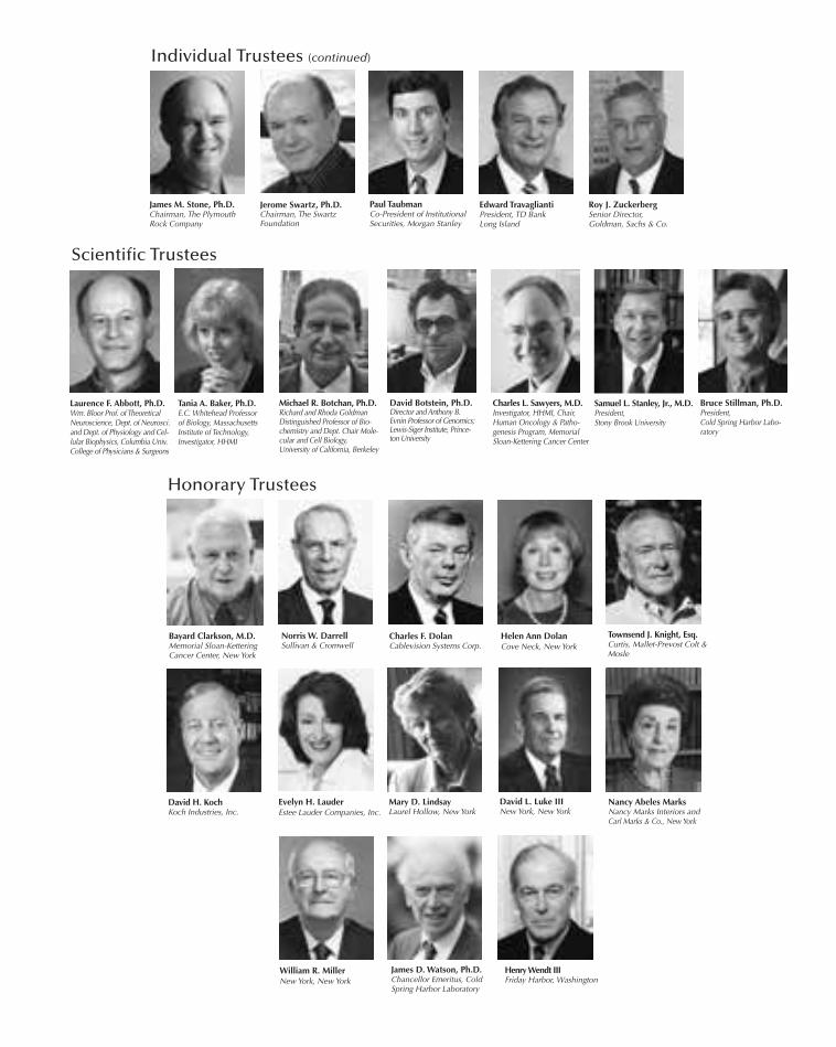
James M. Stone, Ph.D.Chairman, The PlymouthRock Company
Edward TravagliantiPresident, TD BankLong Island
Roy J. ZuckerbergSenior Director, Goldman, Sachs & Co.
Paul TaubmanCo-President of InstitutionalSecurities, Morgan Stanley
Bayard Clarkson, M.D.Memorial Sloan-KetteringCancer Center, New York
Norris W. DarrellSullivan & Cromwell
Mary D. LindsayLaurel Hollow, New York
David L. Luke IIINew York, New York
Townsend J. Knight, Esq.Curtis, Mallet-Prevost Colt &Mosle
James D. Watson, Ph.D.Chancellor Emeritus, ColdSpring Harbor Laboratory
Helen Ann DolanCove Neck, New York
Charles F. DolanCablevision Systems Corp.
Evelyn H. LauderEstee Lauder Companies, Inc.
Scientific Trustees
Tania A. Baker, Ph.D.E.C. Whitehead Professorof Biology, Massachusetts Institute of Technology,Investigator, HHMI
David Botstein, Ph.D.Director and Anthony B.Evnin Professor of Genomics;Lewis-Siger Institute, Prince-ton University
Michael R. Botchan, Ph.D.Richard and Rhoda GoldmanDistinguished Professor of Bio-chemistry and Dept. Chair Mole-cular and Cell Biology,University of California, Berkeley
William R. MillerNew York, New York
Henry Wendt IIIFriday Harbor, Washington
Individual Trustees (continued)
David H. KochKoch Industries, Inc.
Nancy Abeles MarksNancy Marks Interiors andCarl Marks & Co., New York
Samuel L. Stanley, Jr., M.D.President, Stony Brook University
Charles L. Sawyers, M.D.Investigator, HHMI, Chair,Human Oncology & Patho-genesis Program, MemorialSloan-Kettering Cancer Center
Honorary Trustees
Bruce Stillman, Ph.D.President, Cold Spring Harbor Labo-ratory
Laurence F. Abbott, Ph.D.Wm. Bloor Prof. of TheoreticalNeuroscience, Dept. of Neurosci.and Dept. of Physiology and Cel-lular Biophysics, Columbia Univ.College of Physicians & Surgeons
Jerome Swartz, Ph.D.Chairman, The SwartzFoundation
frontmatter_Layout 1 7/8/11 1:25 PM Page v
vi
Governance
The Laboratory is governed by a Board of Trustees of up to 35 members that meets three or fourtimes a year. Authority to act for the Board of Trustees between meetings is vested in the ExecutiveCommittee of the Board of Trustees. The Executive Committee is composed of the Officers of theBoard and any other members who may be elected to the Executive Committee by the Board ofTrustees. Additional standing and ad hoc committees are appointed by the Board of Trustees to pro-vide guidance and advice in specific areas of the Laboratory's operations.
Representation on the Board of Trustees itself is divided between business and community lead-ers and scientists from major educational and research institutions.
The Laboratory is chartered as an educational and research institution by the Board of Regentsof the Education Department of the State of New York. It is authorized to operate a graduate pro-gram under the name “Cold Spring Harbor Laboratory, Watson School of Biological Sciences” andthereat to confer the degrees of Doctor of Philosophy (Ph.D.), Master of Science (M.S.), and Doc-tor of Science (Sc.D.), Honorary.
It is designated as a “public charity” under Section 501(c)(3) of the Internal Revenue Code.
frontmatter_Layout 1 7/8/11 1:25 PM Page vi
Committees of the Board
vii
Audit
Leo A. Guthart, ChairLouise M. ParentAlan C. Stephenson, Esq.
Commercial Relations
James M. Stone, Ph.D., ChairW. Dillaway Ayres, Jr.David Botstein, Ph.D.John P. Maroney, Esq.Robert Martienssen, Ph.D.Alan C. Stephenson, Esq.Bruce Stillman, Ph.D.Jerome Swartz, Ph.D.
Executive Committee
Eduardo G. Mestre, Chair W. Dillaway Ayres, Jr.Timothy BroadbentLeo GuthartJamie C. Nicholls (Chair as of 11/10)Marilyn Simons, Ph.D.Bruce Stillman, Ph.D.James Stone, Ph.D.Paul TaubmanEdward TravagliantiRoy J. Zuckerberg
Finance
Jamie C. Nicholls, ChairW. Dillaway Ayres, Jr.Lola N. GraceLeo Guthart (Chair as of 11/10)Robert D. LindsayJohn C. PhelanWilliam S. RobertsonLari C. RussoMarilyn Simons, Ph.D.Edward Travaglianti
Investment
John C. Phelan, ChairW. Dillaway Ayres, Jr.Thomas D. LehrmanStephen M. LessingJamie C. NichollsWilliam S. RobertsonPaul TaubmanEdward Travaglianti
Nominating
Robert D. Lindsay, ChairW. Dillaway Ayres, Jr.Kristina Perkin DavisonEduardo G. MestreJamie C. NichollsDavid M. RubensteinBruce Stillman, Ph.D.Roy J. Zuckerberg
Robertson Research Fund
W. Dillaway Ayres, Jr.Walter Goldschmidts, Ph.D.Leo GuthartEduardo G. MestreJamie C. NichollsBruce Stillman, Ph.D.
Family Representatives
Katherine ErnstWilliam GridleyJohn LinnartzWalter Meier, M.D.William S. Robertson
Other Committees
Building
Joan AxinnArthur BringsHelen Ann DolanMary Beth DonohueSandra LessingMary D. LindsayTerry LindsayNancy Abeles MarksPeter QuickElizabeth Watson
Planning and Development
Leo GuthartThomas LehrmanHoward MorganCharles L. Sawyers, M.D.Marilyn Simons, Ph.D.Bruce Stillman, Ph.D.Paul TaubmanRoy J. Zuckerberg
Education
Laurie J. Landeau, V.M.D., ChairW. Dillaway Ayres, Jr.Edward A. ChernoffMaria de LessepsLola N. GraceLori HomerSuzanne KleinknechtSuzanne LeedsDavid A. MicklosPeter QuickLawrence Scherr, M.D.Adele SmithersArthur SpiroBruce Stillman, Ph.D.Peter TillesEdward TravagliantiMarianne Dolan Weber
frontmatter_Layout 1 7/8/11 1:25 PM Page vii

viii
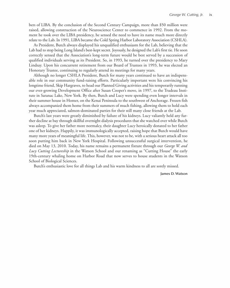
George W. Cutting, Jr.(1932–2010)
George W. Cutting, Jr., known long to all as “Butch,” was born on July 13, 1932, the son of GeorgeW. Cutting and Mary Converse Cutting of Warrentown, Virginia. A graduate of the Rensselaer andBrooks Schools, he entered Yale College where he graduated in 1955 as a political science major.After his 1958 marriage to Lucy Pulling at the Chapel of the Millbrook School (Dutchess County,New York), which her father Edward Pulling had founded and long directed, they lived in NewYork City where Butch was a stockbroker and portfolio manager at Fahnestock and Company. In1958, he and Lucy moved to Oyster Bay Cove, Long Island, occupying a house that they built offYellow Cote Road estate land earlier owned by her grandfather, the illustrious banker, Russell Lef-fingwell. Amidst the idyllic splendor of his estate’s meadows and fields, Butch and Lucy raised theirfour children—George, Jr., Lucy, Cynthia, and Susie.
Liz and I first met Butch and Lucy during the first year of our marriage when Ed and LucyPulling invited us down from Harvard for a February weekend to meet members of the Long IslandBiological Association (LIBA). Ed was then its President, having taken over the reins from NevilleFord, several years after he and his wife moved to Long Island following his retirement as head-master of Millbrook. Then, he and his wife moved into Redcote, the wooden-shingled main houseof the Leffingwell estate. Later, Liz and I began to interact much with the Cuttings at the EastWoods School, immediately across Yellow Cote Road from the Leffingwell land. Butch and Lucyalso sent their children there to be educated.
From the moment of my arrival in February 1968 as the Lab’s new Director, I had the good for-tune to be able to devote almost all of my time to direct its science. All of our fund-raising effortsaimed at private sources in effect were handled by Edward Pulling. He had no use for a develop-ment office as long as he was available to make known the Laboratory’s needs. At a late-1970s lun-cheon at the Piping Rock Club, Ed introduced us to Oliver and Lorraine Grace, who several yearsbefore had moved nearby into a gracious, old waterfront home on Cove Neck. Oliver, long inter-ested in cancer research, joined the Lab’s Board of Trustees in 1983, and his major gift made possi-ble the 1986 opening of our Charles Moore–designed Oliver and Lorraine Grace Auditorium. Bythen, Ed Pulling, at age 86, thought the time had come to retire as President, telling me that he hadfound the perfect person to succeed him—Butch.
Here, Ed made the perfect choice. He knew that Butch, then in his mid 50s, had the time, vi-sion, and energy to attract even more community support for the Lab’s ever-growing research andeducational programs. Butch, as LIBA’s President, also became in 1986 a member of our Board ofTrustees, having an essential role in the Lab’s Second Century Campaign (1989–1992), serving ascochairman of its steering body and directing the special gifts committee that focused on the mem-
frontmatter_Layout 1 7/8/11 1:25 PM Page viii
bers of LIBA. By the conclusion of the Second Century Campaign, more than $50 million wereraised, allowing construction of the Neuroscience Center to commence in 1992. From the mo-ment he took over the LIBA presidency, he sensed the need to have its name much more directlyrelate to the Lab. In 1991, LIBA became the Cold Spring Harbor Laboratory Association (CSHLA).
As President, Butch always displayed his unqualified enthusiasm for the Lab, believing that theLab had to stop being Long Island’s best-kept secret. Joyously, he designed the Lab’s first tie. He sooncorrectly sensed that the Association’s long-term future would be best served by a succession ofqualified individuals serving as its President. So, in 1993, he turned over the presidency to MaryLindsay. Upon his concurrent retirement from our Board of Trustees in 1993, he was elected anHonorary Trustee, continuing to regularly attend its meetings for many years.
Although no longer CSHLA President, Butch for many years continued to have an indispens-able role in our community fund-raising efforts. Particularly important were his convincing hislongtime friend, Skip Hargraves, to head our Planned Giving activities and his temporarily runningour ever-growing Development Office after Susan Cooper’s move, in 1997, to the Trudeau Insti-tute in Saranac Lake, New York. By then, Butch and Lucy were spending even longer intervals intheir summer house in Homer, on the Kenai Peninsula to the southwest of Anchorage. Frozen fishalways accompanied them home from their summers of much fishing, allowing them to hold eachyear much appreciated, salmon-dominated parties for their still many close friends at the Lab.
Butch’s last years were greatly diminished by failure of his kidneys. Lucy valiantly held any fur-ther decline at bay through skillful overnight dialysis procedures that she watched over while Butchwas asleep. To give her father more normalcy, their daughter Lucy heroically donated to her fatherone of her kidneys. Happily, it was immunologically accepted, raising hope that Butch would havemany more years of meaningful life. This, however, was not to be, with a serious heart attack all toosoon putting him back in New York Hospital. Following unsuccessful surgical intervention, hedied on May 13, 2010. Today, his name remains a permanent fixture through our George W. andLucy Cutting Lectureship in the Watson School and our renaming as “Cutting House” the early19th-century whaling home on Harbor Road that now serves to house students in the WatsonSchool of Biological Sciences.
Butch’s enthusiastic love for all things Lab and his warm kindness to all are sorely missed.
James D. Watson
George W. Cutting, Jr. ix
frontmatter_Layout 1 7/8/11 1:25 PM Page ix

x
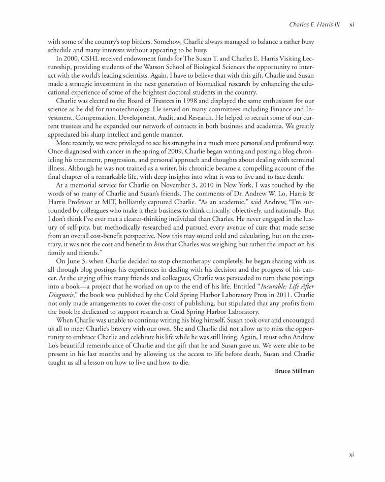
Charles E. Harris III(1943–2010)
Charlie Harris was remarkable both in how he lived his life and how he died. He and his wife Susanhave made invaluable contributions to Cold Spring Harbor Laboratory during the past 20 years thatwill yield results well into the future.
Charlie was raised in Jacksonville, Florida and attended public school until his parents recognizedthe need to remove him from the rather dysfunctional public school system. He attended the HillSchool in Pottsdown, Pennsylvania for the final 2 years of high school before entering Princeton Uni-versity. Later, Charlie graduated from Columbia Business School and successfully pursued a careerin the securities industry and as an innovative venture capitalist. He founded Harris & HarrisGroup, a venture capital firm, in 1983 and subsequently brought the firm public. This was an un-usual path for a venture fund, but then again, Charlie was unique.
For the last decade of his career at Harris & Harris, Charlie became one of the most informedand enthusiastic supporters of the emerging field of nanotechnology, in which he was recognizedas an astute investor. There was something about the merging of an exciting new science and theopportunity to build new companies that attracted Charlie to this field. It enabled him to interactwith and advise many of the top nanotechnology scientists in academia. He was always encourag-ing me to integrate nanotechnology into the research programs at Cold Spring Harbor Laboratory,and he was way ahead of us in this respect.
I know Charlie would have enjoyed this year’s President’s Council fall event, “Tiny Treatments:The Science of Nanotechnology.” He died just the day before the weekend retreat, but he had e-mailed me earlier to say how pleased he was with the agenda. It was Charlie who helped found thePresident’s Council in 1994 as a vehicle to support CSHL Fellows. As a venture capitalist, I believeCharlie looked at the opportunity to support early-career scientists as a smart investment in the fu-ture. Investing in the best and the brightest young investigators is a hallmark of our institution,and I view Charlie and Susan’s dedication to the CSHL Fellows program as perhaps one of thestrongest endorsements of our institutional strategy. The returns on investment from this programare sizable, measurable in terms of scientific advancement and reflected by the grandest of prizes—the Nobel Prize, which in 2009 went to former CSHL Fellow Carol Greider.
Charlie was also an avid thoroughbred horse owner who raced his horses at many prominenttracks, including Belmont and Saratoga. He was often conflicted in time among the alternative ca-reers of family man and father of two, horse owner, businessman, and philanthropist. I rememberthe Saturday afternoon of one President’s Council chaired by Charlie that included a trip to Shel-ter Island on the east end of Long Island. The drive there required a well-planned stop at a sportsbar to watch the Preakness Stakes in Maryland, before we all proceeded to a bird-watching event
frontmatter_Layout 1 7/12/11 9:56 AM Page x
xi
with some of the country’s top birders. Somehow, Charlie always managed to balance a rather busyschedule and many interests without appearing to be busy.
In 2000, CSHL received endowment funds for The Susan T. and Charles E. Harris Visiting Lec-tureship, providing students of the Watson School of Biological Sciences the opportunity to inter-act with the world’s leading scientists. Again, I have to believe that with this gift, Charlie and Susanmade a strategic investment in the next generation of biomedical research by enhancing the edu-cational experience of some of the brightest doctoral students in the country.
Charlie was elected to the Board of Trustees in 1998 and displayed the same enthusiasm for ourscience as he did for nanotechnology. He served on many committees including Finance and In-vestment, Compensation, Development, Audit, and Research. He helped to recruit some of our cur-rent trustees and he expanded our network of contacts in both business and academia. We greatlyappreciated his sharp intellect and gentle manner.
More recently, we were privileged to see his strengths in a much more personal and profound way.Once diagnosed with cancer in the spring of 2009, Charlie began writing and posting a blog chron-icling his treatment, progression, and personal approach and thoughts about dealing with terminalillness. Although he was not trained as a writer, his chronicle became a compelling account of thefinal chapter of a remarkable life, with deep insights into what it was to live and to face death.
At a memorial service for Charlie on November 3, 2010 in New York, I was touched by thewords of so many of Charlie and Susan’s friends. The comments of Dr. Andrew W. Lo, Harris &Harris Professor at MIT, brilliantly captured Charlie. “As an academic,” said Andrew, “I’m sur-rounded by colleagues who make it their business to think critically, objectively, and rationally. ButI don’t think I’ve ever met a clearer-thinking individual than Charles. He never engaged in the lux-ury of self-pity, but methodically researched and pursued every avenue of cure that made sensefrom an overall cost-benefit perspective. Now this may sound cold and calculating, but on the con-trary, it was not the cost and benefit to him that Charles was weighing but rather the impact on hisfamily and friends.”
On June 3, when Charlie decided to stop chemotherapy completely, he began sharing with usall through blog postings his experiences in dealing with his decision and the progress of his can-cer. At the urging of his many friends and colleagues, Charlie was persuaded to turn these postingsinto a book—a project that he worked on up to the end of his life. Entitled “Incurable: Life AfterDiagnosis,” the book was published by the Cold Spring Harbor Laboratory Press in 2011. Charlienot only made arrangements to cover the costs of publishing, but stipulated that any profits fromthe book be dedicated to support research at Cold Spring Harbor Laboratory.
When Charlie was unable to continue writing his blog himself, Susan took over and encouragedus all to meet Charlie’s bravery with our own. She and Charlie did not allow us to miss the oppor-tunity to embrace Charlie and celebrate his life while he was still living. Again, I must echo AndrewLo’s beautiful remembrance of Charlie and the gift that he and Susan gave us. We were able to bepresent in his last months and by allowing us the access to life before death, Susan and Charlietaught us all a lesson on how to live and how to die.
Bruce Stillman
Charles E. Harris III xi
frontmatter_Layout 1 7/8/11 1:25 PM Page xi

frontmatter_Layout 1 7/8/11 1:25 PM Page xii
In his State of the Union address in 1971, President Richard Nixon called upon Congress “to launchan intensive campaign to find a cure for cancer.” Later that year, the National Cancer Act becamelaw, the first salvo in what since has been referred to as “the war on cancer.”
After 40 years, where do we stand? This past year, cancers killed more than 550,000 Americans.More than three times that number were newly diagnosed. These figures make clear that a “cure” isnowhere in sight. Yet, four decades ago, it seemed plausible to imagine that we were on the trail ofa single killer. Today, we possess the sobering knowledge that our quarry is actually hundreds of dif-ferent illnesses and that it is unlikely that a single magic bullet will bring cancer’s carnage to a halt.
Cancer is so very much more complicated than we understood it to be in 1971. Over fourdecades, a major national investment in basic biological research—performed at Cold Spring HarborLaboratory and academic and clinical centers of excellence across the nation and around the world—has yielded increasingly detailed knowledge of cancer at the genetic, cellular, and tissue levels. Thatknowledge has brought us the first effective targeted therapies for certain cancer subtypes. Thesepoint the way to a much more encouraging future.
I would like to recognize in this report a few of the landmark discoveries in which Cold SpringHarbor Laboratory scientists have had important roles, as prelude to describing a new Cancer Ther-apeutics Initiative. Grounded in such outstanding basic science, I am optimistic that the powerfulapproach we are taking at the Laboratory will contribute in the coming years to turning many majorcancer types into manageable chronic illnesses or even cures.
Forty years is an eternity in biomedical science. It is important to remember that when a patientwent to a clinic in 1971, there was very little that an oncologist could determine except for the factthat a cancer was present. Pathology on the tumor could help determine prognosis, but the abilityto characterize tumors beyond gross pathology was rather limited. There were plenty of chemother-apies available, but responses to them were essentially hit or miss.
Forty years ago, we knew that the genetics of individual cancers was important. We knew thatcancer cells had abnormal chromosomes compared to those of normal cells. But the concept thatspecific genes caused cancer had not yet been clearly formulated. Our initial focus, beginning in1968 when Jim Watson became director of Cold Spring Harbor Laboratory and trained his sightson cancer, was on cancer-causing viruses because they carried genes that could promote cancer.
The notion that cancer could have a viral origin dates to the early 20th century and the work ofPeyton Rous at The Rockefeller University, who discovered a virus in a type of chicken tumor thatcould be transferred via injection to baby chicks, which were subsequently observed to develop tu-mors. In the mid 1970s, J. Michael Bishop and Harold Varmus at UCSF found a gene in healthychickens called c-src that was nearly identical to the cancer-causing gene in Rous sarcoma virus.They concluded that the oncogene in the virus did not represent a true virus gene but instead wasa version of the normal cellular gene that the virus had acquired during replication in the host celland thereafter carried along.
In 1981, Michael Wigler here at Cold Spring Harbor Laboratory was one of three researchers inthe United States who independently discovered the first human oncogene, called RAS. It belongsto a family of genes critical in signaling networks that regulate cell growth and division. Soon there-after, CSHL scientist Earl Ruley and MIT’s Robert Weinberg began to reveal some of the mecha-nisms through which oncogenes promote cancer. Their work shed light on the phenomenon ofcooperating oncogenes, instances in which the progression of cancer depends on the products oftwo or more cancer-promoting genes, none of which is sufficient to cause cancer.
PRESIDENT’S REPORT
1
001-026_Pres_High_Annual Report_2009 template 7/12/11 9:59 AM Page 1
This notion dovetailed with the multiple-hit theory of oncogenesis, which led to the idea thatcells in our body had to acquire mutations in multiple oncogenes. Following pioneering researchby Alfred Knudsen at the Fox Chase Cancer Center, whose studies linked inherited cancer withspontaneous mutations in adult cells and predicted the existence of tumor suppressor genes, EdHarlow at CSHL demonstrated that oncogenes could inactivate tumor suppressors, thereby pro-viding another view of genetic cooperation to produce tumors. Thus, cancers could result not simplyfrom the actions of cancer-promoting oncogenes—which encoded proteins that accelerated growthwithin the cell—but also from the simultaneous absence of action on the part of genes called tumorsuppressors, whose normal function was to prevent cellular growth from running amok.
These early studies identified the kinds of malfunctioning or mutated genes that were at work inoncogenesis, and what mechanisms and pathways they undermined to permit uncontrolled cell pro-liferation and prevention of cell death, both of which were required for tumor progression. In parallelwith the genetics of cancer was basic research on cell proliferation control in which many labs atCSHL had a major role and which proved important for understanding cancer. From the mid 1980sto early 1990s, CSHL scientists helped piece together an increasingly comprehensive molecular pic-ture of replication of the genetic material in the cell nucleus and the workings of the cell divisioncycle that governed how cells proliferate. Defects in the control of cell proliferation are the maindrivers of cancer progression, causing increasingly complex mutations in cancer cells that furtherpromote tumor growth, loss of normal controls on cells within a tissue, and eventually metastasis.
In the mid 1970s, CSHL alumni Philip Sharp at MIT, Richard Roberts and Louise Chow atCSHL, and their colleagues made the brilliant discovery of “split genes,” Nobel Prize–winning re-search that enabled us to see how the RNA messages of genes could be spliced together in multipleways, to generate different proteins from a single gene. As Adrian Krainer has shown in recent years,
2 President’s Report
001-026_Pres_High_Annual Report_2009 template 7/12/11 9:59 AM Page 2

President’s Report 3
this alternate splicing contributes to the emergence of cancer in humans. Most interestingly, Adrianhas shown, together with Harvard’s Lew Cantley, that the switching by RNA splicing from oneform of a gene to another form can endow cells with completely different metabolic outcomes, mak-ing cancer cells very different from normal cells. These metabolic changes will likely provide newtherapeutic opportunities that exploit basic differences between cancer and normal cells.
With the realization that cancer is fundamentally a genetic disease, it became imperative that weunderstand the entire human genome. The 1990s marked the beginning of the effort to sequencethe human genome and the genomic era in cancer research, and CSHL was among the leaders andinnovators. The essence of genomics is captured beautifully in work first performed by Mike Wiglerand colleagues around this time. They devised ingenious technical means with which to comparethousands of genes at a time in tumor samples and a patient’s corresponding healthy tissue. Thisimmediately led to the discovery of the PTEN tumor suppressor gene, mutated in many humancancers. Since 2003, Mike and his collaborators have also called our attention to areas of deletionand amplification across entire genomes, revealing, respectively, a vast array of tumor suppressorgenes and oncogenes. This research has introduced a new dimension to the search for the geneticculprits of cancer—phenomena such as gene copy-number variations—not known to exist at thisscale before the advent of technologies that study the entire genome.
Amplified and deleted genomic segments in our genome are commonplace. We all have them,and they are often harmless. But when they occur in certain parts of our DNA, the impact can bedevastating. Alea Mills of our faculty has provided an excellent example in the context of cancer.Following up on knowledge that a large region of human chromosome 1 was very often deleted inhuman cancers, Alea was able to determine that the region contained a novel tumor suppressorgene, CHD5, that proves to be a master control switch regulating other tumor suppressor genes.
The pace of our insights has grown along with our technological capabilities. It has proven possibleto “mine” comparative genomic data obtained from tumor samples to identify, for instance, all over-expressed genes in a particular cancer and then to overexpress the corresponding genes in laboratorymice. It has also been possible to use designer short hairpin RNAs, members of a class of naturallyoccurring small RNA molecules studied in Greg Hannon’s laboratory, to identify many new tumorsuppressor genes or to screen for new therapeutic targets in human cancers.
Building upon human genetics research from Mike Wigler, Jim Hicks, and their clinical colleaguesScott Powers and quantitative biologist Alex Krasnitz have identified many genomic regions in humancancer tissue that are either amplified or deleted, enabling insights gleaned from patients to be incor-porated into the development of animal models of many cancer types, including liver, colon, prostate,pancreas, and breast cancers, as well as various types of leukemia. In recent years, Scott Lowe andothers have made great strides with “mosaic” mouse models, genetic hybrids that use tissue-specificstem cells to introduce quickly into mouse cells the same genetic mutations found in human tumors.These mosaic mice have tumors that mimic the course of human cancers, enabling assessment of whychemotherapy works in some patients and not in others, and validation of whether new therapeutictargets will work on cancers that are resistant to current treatment.
We have learned that the underlying genetics of a tumor determines its response to therapy andcan therefore be exploited for both diagnosis and prognosis of tumor subtypes. Carrying this analysisfurther, Mike Wigler and Jim Hicks developed a method to study genomic heterogeneity within apatient’s breast tumor, allowing them to identify cellular subpopulations as well as map their spatialorganization. This analysis was used to advance our understanding of how a tumor evolves overtime, driven by genetic changes that are not visible if the entire tumor is considered to be uniform.
Using powerful RNA-based tools developed at CSHL, we are learning how to identify new targetsfor cancer therapy and to probe why an existing targeted drug works brilliantly for one patient andfails utterly with another. Previously, both might superficially have appeared to have the same kindof cancer, but now genetic analysis can separate tumor responses into subgroups, even within a par-ticular tumor tissue type. RNA-based technology and cancer genetic techniques are also enabling
001-026_Pres_High_Annual Report_2009 template 7/12/11 9:59 AM Page 3
CSHL scientists to study closely the perplexing phenomenon of resistance to existing drugs. It isnow very clear that new, targeted therapies have to be developed for each genetic subtype of tumor.
Targeted therapies made a huge impact with the development of Gleevec, designed specificallyto block an oncoprotein produced by a mutant gene in the so-called Philadelphia chromosome, amisshapen chromosome discovered at the University of Pennsylvania and Fox Chase Cancer Centerin 1960 and now understood to be the result of a translocation—a fragment of chromosome 9 fusedto a fragment of chromosome 22. Gleevec helps only those patients who have this uncommon mu-tation, which is the cause of most cases of an acute blood cancer called chronic myelogenousleukemia, or CML.
Similarly, Tarceva is a drug that very specifically blocks the product of a mutant version of a genecalled EGFR (epidermal growth factor receptor), present in a subset of lung cancer cases. LikeGleevec, Tarceva is not an indiscriminate killer of cells, both cancerous and healthy, like old-linechemotherapies. Rather, it works well in many patients who have a specific EGFR mutation, but itdoes not help those whose lung cancers have other genetic drivers. However, Tarceva, when effective,typically holds the cancer at bay only for a year or two and then drug resistance emerges. RaffaellaSordella’s lab at CSHL recently has found a new mechanism by which responsive lung cancers de-velop resistance to the drug.
The problem of resistance suggests the difficulty of the task before us and leads me to cautionagainst undue optimism that “a cure” is just around the bend. There are 50-odd major types ofhuman cancers based on tissue type alone, and there are probably six or seven important subtypeswithin each tissue type (and maybe more), each one of which needs to be treated with what I an-ticipate will be a cocktail of targeted drugs rather than a single one. Only then will the resistancethat cancers naturally develop be avoided. In the not-distant future, therefore, major cancers willbe treated in the manner that we now treat HIV infections, with multiple drugs that minimize thedevelopment of resistance. For now, therefore, chronic management of cancer is a more realisticprospect than its eradication, and this will be a major advance if the targeted drugs do not causemajor side effects, as in the case of Gleevec.
Our Cancer Therapeutics Initiative brings together many of the innovative elements I have dis-cussed here. Beginning, importantly, from human tumor samples—which we obtain through ourcollaborations with leading clinical centers—we use our state-of-the-art sequencing and genomeanalysis capabilities to generate tumor profiles. Working with subsets of genes that emerge for geneticanalysis of human tumors, RNA interference (RNAi) technologies can rapidly identify the Achilles’heel of the cancers and suggest new therapeutic targets. Validation of these targets in mouse modelsof human cancer will most likely increase the success rate of drugs that eventually enter into theclinic. We have learned the hard way that there is no substitute for observing the molecular mech-anisms of cancer and their response to therapies within the incredibly complex living environmentin which actual cancers emerge, grow, and spread.
The net impact of our initiative—which I estimate will cost $100 million over a period of years—will be the ability to systematically discover and rapidly validate new targets for cancer drugs. Suchan initiative will require constant interactions with the pharmaceutical industry to bring the validatedtargets to human clinical studies. This will require seamless interactions among scientists in industryand academia. Academic scientists lack the resources to develop drugs, and given well-validated tar-gets, industry has proven to be very effective at developing drugs that work. The problem is that in-dustry has not been good at discovery of targets with a high probability of clinical success. This iswhere I expect academia will excel.
While the Cancer Therapeutics Initiative is needed, CSHL will continue vigorously to pursuebasic research on small RNAs, genome structure and organization, cellular signaling pathways andnetworks, and other aspects of fundamental biology, work that will lead us to other new technicalcapabilities and understanding. It is possible that research performed on our campus will help solvethe technical problems that currently prevent us from using RNAi to directly shut down cancer genes
4 Highlights of the Year
001-026_Pres_High_Annual Report_2009 template 7/12/11 9:59 AM Page 4
in human patients. Other areas of basic research, notably on the immune system, tumor metabolism,and tumor microenvironment, are likely to be of increasing importance in the years just ahead.
There is one additional element in our fight against cancer that I would like to mention, and itconcerns the current state of our clinical trials system. If we and others are successful in identifyingnovel, very specific drug targets in subtypes of the major cancer killers, it is vitally important thatdrugs developed against these targets not get bogged down in regulatory delays. A drug recently de-veloped against a comparatively rare genetic mutation in lung cancer gene called ALK provides acase in point. A recent early-stage clinical trial of an experimental drug called crizotinib was notablysuccessful in patients with non-small-cell-lung cancer (NSCLC) who harbored the ALK mutation,with tumor shrinkage and stabilization in the range of 85%. Strikingly, about three-quarters of thepatients remained on the drug after the clinical trial met its endpoint. Under the current system,the FDA will require the drug developer to randomize treatment in a phase III trial, splitting agroup of ALK-positive patients into two groups, only one of which will receive the drug. The desiredendpoint would be to demonstrate a survival advantage, a process that takes years to play out.
Proceeding in this manner I would argue is unethical and costly. In some cases, such as this one,phase III trials could be bypassed. A drug showing overwhelming responses in multicenter, early-stage trials in a cancer type with poor prognosis should promptly be granted temporary approval. Itshould be placed directly into broad clinical use in appropriate genetically screened patients whowish to be treated with it, including early-stage cancer patients. The drug’s developer, meantime,should be required to report the full course of all patients, irrespective of outcome. Hospitals andclinics performing these trials should be protected from patient litigation if the therapies do notwork, allowing multicenter trials to proceed unhindered by legal complications. For a period ofyears, all adverse side effects and outcomes should be reported and the drug’s temporary approvalrescinded if previously unnoticed safety issues emerge or if the drug proves not to have the desiredeffect when a larger group of patients have been treated. Short of this, however, I believe humani-tarian and cost considerations demand that a new drug found to have overwhelming initial successin a genetically defined subpopulation of patients with otherwise poor prognosis should be madeavailable while further data on efficacy and side effects are being collected.
If we are serious as a society about advancing the state of cancer treatment, we should rethinkthe clinical trials process, particularly as we use new methods of discovery made possible by decadesof remarkable basic scientific and clinical research to find the next generation of targeted therapies.These, if used in combination treatments, promise to make cancer a disease that millions of Amer-icans will be able to live with, while enjoying a decent quality of life. It is not an easy goal, but onethat should be among the nation’s highest priorities.
Bruce Stillman, Ph.D., F.R.S.President
Highlights of the Year 5
001-026_Pres_High_Annual Report_2009 template 7/12/11 9:59 AM Page 5

Highlights of the Year
6
Research
Research at Cold Spring Harbor Laboratory (CSHL) has a major impact in the areas on which ourprincipal investigators focus: cancer, neuroscience, plant biology, and quantitative biology. It hasoften been noted that our influence is especially remarkable for an institution of CSHL’s compar-atively small size. A recent survey by the respected science publisher Thompson Reuters in factplaced CSHL first in a group of 20 “heavy hitters” in molecular biology and genetics, selected fromamong 42,000 research institutions worldwide. During the first decade of the 21st century, researchpapers based on work conducted in CSHL laboratories had more impact—as measured by theirfrequency of citation by peers—than papers originating in any other institution, including the Mas-sachusetts Institute of Technology, the Salk Institute for Biological Studies, Memorial Sloan-Ket-tering Cancer Center, The Rockefeller University, and Harvard University.
This survey is not the only measure of our worth or that of any institution, but it does suggestthe power of the work being performed at CSHL and its relevance, as measured by those who useit—our colleagues at laboratories throughout the nation and across the globe. Together, we are en-gaged in a vital enterprise, in which we bring all of our intellectual skills and technical ingenuity tobear on fundamental questions of biology and generate knowledge that forms the basis for biomed-icine to move forward in its mission to relieve the major causes of human suffering. Below, we sum-marize just a few of the many fascinating and important findings made by CSHL’s dedicated teamof investigators during 2010.
Antisense Therapy Reverses Spinal Muscular Atrophy in Mice
Professor Adrian Krainer achieved a milestone this past year in his continuingeffort to understand spinal muscular atrophy (SMA), the leading genetic causeof death in infants. SMA is the result of mutations in the survival of motorneuron 1 (SMN1) gene. These lead to abnormally low levels of SMN proteinin motor nerve cells of the spinal cord and to the degeneration of those cells.Last year, Adrian and colleagues identified a compound that stimulates SMNproduction by altering RNA splicing. This year, they carried the work an im-portant step further: By introducing chemically modified pieces of RNA calledantisense oligonucleotides (ASOs) into the spinal cords of mice, they succeededin reversing symptoms of Type III SMA. This result exemplifies how superbbasic science—in this case, work in the Krainer lab on the cell’s splicing ma-chinery—can be fertile ground for value-added science, the kind of research activity that adds commercialvalue to fundamental discoveries. Krainer’s team has collaborated with scientists at Isis Pharmaceuticalsin designing and synthesizing ASOs, which can be designed to bind to any piece of RNA. The team ze-roed in on an ASO that optimally enhanced the inclusion of an exon that in people with SMA is“skipped” by cellular machinery that cuts and pastes bits of RNA “message” together to form a templatefor protein manufacture. A particularly encouraging aspect of the team’s progress this year was learninghow to overcome barriers to delivering ASOs directly into the fluid that surrounds the brain and spinalcord. The treatment’s therapeutic effect in mice persisted for half a year after it was discontinued, indi-cating that the ASO is very stable. In addition, the team reported no inflammation or toxicity.
Reversing Alzheimer’s-like Memory Loss in Drosophila
Work published this year by Professor Yi Zhong’s team demonstrated a means of reversing memoryloss in fruit flies caused by brain plaques similar to those implicated in Alzheimer’s disease. Modelinga complex human illness such as Alzheimer’s is an important goal of basic science, and the fly pro-
A. Krainer
001-026_Pres_High_Annual Report_2009 template 7/12/11 9:59 AM Page 6
Highlights of the Year 7
vides us with a suitably simple starting point. The fly brain should not be underestimated,for in it we see significant conservation of DNA sequence found in human genes knownto affect the structure and function of neural networks. Protein fragments of the β-amyloidmolecule associated with Alzheimer’s are known to alter many cell-signaling proteins suchas phosphoinositol-3 kinase (PI3K), causing a wide range of neuronal dysfunctions. Inflies engineered to produce the human β-amyloid protein in their brains, Yi’s team set outto better comprehend the molecular basis of memory loss. This yielded a finding that wentagainst received wisdom that attributed a protective role to the kinase. Yi’s team insteadfound that the increased PI3K activity caused a type of neurotransmission that is patho-logically enhanced when β amyloid is present in the fly brain. Injection of chemicals thatblock the kinase’s action and separate efforts to turn off the gene that encodes it both hadthe effect of restoring normal signals in the fly brain. This research also intriguingly suggests thatbrains affected by Alzheimer’s might become insulin resistant because of elevated PI3K activity.Thus, the kinase becomes a potential target for novel therapeutics.
A Possible Inflammatory Component in Resistance to a TargetedLung Cancer Drug
A critical question about cancer concerns the molecular mechanisms involved in resistance tochemotherapy. Particularly vexing is the phenomenon of resistance to the best drugs developed todate, so-called targeted therapies. Assistant Professor Raffaella Sordella’s lab this year shed new lighton resistance to Tarceva (erlotinib), a targeted therapy approved in 2004 for a subset of patientswith non-small-cell lung cancer (NSCLC) and for some patients with pancreatic cancer.Tarceva’s molecular target is known—the cell membrane receptor called epidermal growthfactor receptor (EGFR)—as are processes that lead to about half of observed cases of re-sistance. But what about the other 50%? Raffaella and colleagues from Weill Cornell Med-ical College and the Boltzmann Institute in Vienna discovered a subpopulation of NSCLCcells that are intrinsically resistant to Tarceva. These tumor cells were observed to secreteelevated amounts of a growth factor called transforming growth factor-β (TGF-β), whichin turn increases secretion of interleukin-6 (IL-6), an immune signaling molecule. Signif-icantly, these effects were independent of the EGFR pathway. The team therefore hypoth-esizes that inflammation is one of the factors that can render a tumor cell resistant totreatment with Tarceva.
A Protein Linked to Leukemia “Bookmarks” Highly ActiveGenes in Dividing Cells
When CSHL Fellow Christopher Vakoc and colleagues demonstrated this year how so-called epi-genetic instructions are stably transferred from one generation of cells to the next, they provided acompelling explanation of how a protein called MLL (mixed lineage leukemia) may beinvolved in triggering leukemia. During cell division, gene activity is normally shut downtemporarily. The dividing cell’s chromosomes condense and expel most of the proteinsthat cling to them, which are called epigenetic marks. These marks at other times in thecell cycle help to determine which genes are accessible to the cellular machinery and canbe expressed and which genes are inaccessible and cannot be expressed. Unlike most otherchromosome-bound epigenetic marks, Chris’ team found that the MLL protein stays teth-ered to the genetic material during cell division. It acts as a “bookmark,” preserving a bitof vital gene expression information. But as the cell divides, sometimes the MLL proteinsshift to new locations on the chromosome. Interestingly, they seem to attach to genes thatare the most active before cell division shuts down all gene activity. This, in turn, can draw
Y. Zhong
R. Sordella
C. Vakoc
001-026_Pres_High_Annual Report_2009 template 7/12/11 9:59 AM Page 7

other proteins to the same area, with the net effect of jump-starting gene expression. Chris is nowstudying how MLL mutations might promote the abnormal proliferation of cells in leukemia.
A Potential Way to Reverse Cancer Cell Metabolism and Tumor Growth
Eighty years ago, Nobel laureate Otto Warburg observed the altered metabolic state of cancer cellsand tried to connect it, biochemically, with processes that give rise to the rapid proliferation thatcharacterizes cancer. In particular, cancer cells are distinct in the way in which they metabolize glu-cose. They also produce large quantities of a by-product called lactate. A protein called PK-M2 is akey mediator of glucose metabolism in cancer cells, and this year, Professor Adrian Krainer led agroup including researchers at Harvard Medical School and The Broad Institute that discoveredthree molecular factors contributing to high levels of PK-M2 in cancer cells. PK-M2 is one of twoisoforms, or slightly varying versions, of an enzyme called pyruvate kinase. A single gene called PK-M gives rise to both, via alternative splicing. Adrian’s expertise in splicing helped the team to un-derstand how the benign isoform of the enzyme, PK-M1, is switched off and the dangerous M2isoform is switched on in cancer cells. By manipulating three known splicing factors, the team wasable to halt M2 production and separately to restore production of the benign M1 isoform. Thissheds light on the so-called Warburg Effect and points to possible new targets for drugs that mightreverse the pathological metabolism of cancer cells.
How Blood Stem Cells Are Maintained in the Bone Marrow Niche
Hematopoietic stem cells (HSCs) have unique abilities that are prized by medical re-searchers. They can self-renew and develop, or differentiate, into any kind of blood cell,which enables them to replenish the body’s entire blood and immune system. Researchershave understood that these qualities are traceable to a distinct locale or niche within thebone marrow that HSCs target, but the identity and function of the niche-forming con-stituents had not been clearly defined until this past year, when Associate Professor GrigoriEnikolopov and colleagues from the medical schools at Harvard, Albert Einstein, andMount Sinai published a report in the journal Nature. HSCs retain their unique features,they observed, in response to signals from another stem cell population, called mesenchy-mal stem cells (MSCs), that create a supportive bone marrow niche for the HSCs. It wasthe first demonstration that one type of stem cell could regulate another type of stem cell.
In a series of experiments, Grisha and the team discovered that genetic factors essential for HSCmaintenance are highly concentrated within neighboring MSCs. They speculate that if we can con-trol the niche, we can also manipulate the HSC population within it. This raises the prospect of de-veloping a drug to target the niche in order to enhance stem cell production. This would be usefulin regeneration therapies or could help to prevent the development of certain leukemias and otherillnesses related to unregulated stem cell proliferation.
Next-Generation Sequencing Enables Team to FindCause of Devastating Rare Illness
Professor Gregory Hannon and his talented graduate student Yaniv Erlich—who in 2010received his Watson School doctorate as well as a prestigious appointment as a Fellow atthe Whitehead Institute—were part of an international team that discovered a geneticmutation that causes Joubert syndrome, a rare inherited neurological disease found mostoften among Ashkenazi Jews. Children whose parents both carry a copy of the mutatedgene, and who inherit a copy from each, develop devastating pathologies including mal-formation of the brain, developmental delay, and muscular and visual impairment. TheCSHL contribution to the discovery of the mutation’s precise location involved a tech-nological insight. Rather than sequence the entire genome of patients in search of the ge-netic culprit, which would be time-consuming and very costly, the team could use a
8 Highlights of the Year
G. Hannon
G. Enikolopov
001-026_Pres_High_Annual Report_2009 template 7/12/11 9:59 AM Page 8
powerful genome fractionation method devised by Greg’s team to sequence only those portions ofthe genome that encode proteins. This is called the exome, and it consists of less than 2% of theentire human genetic sequence. This was one of the very first instances in which next-generationsequencing was used to find the genetic cause of a rare disease and demonstrates that similar methodscan be used to find the causes of other uncommon illnesses that otherwise might not get the atten-tion that their sufferers so desperately need.
Identifying the (Few) Protein Differences between Neanderthalsand Modern Humans
A closely related sequencing technology enabled Professor Hannon, postdoctoral researcher EmilyHodges, and others in Greg’s lab to play an important part in a story that Science called one of2010’s most important. After years of effort, a team led by Svante Pääbo at the Max-Planck Institutein Germany succeeded in piecing together a draft of the full genome of our Neanderthal predeces-sors. This was notable in part because the bone fragments from which the DNA was sampled wereso old—approaching 40,000 years. But it was also remarkable because the fragments were highlycorrupted, some containing as little as two tenths of 1% of Neanderthal DNA. One challenge washow to sift such a tiny portion from the corrupted remainder. This was where the Hannon lab’stechnique called array-capture resequencing proved to be especially useful. They used it to sequence14,000 genes known to be different in humans and our closest relatives on the tree of life—chim-panzees. Although about three-fourths of the proteins encoded by those genes are different in hu-mans and chimps, Greg’s team showed that stunningly few of them differed in humans andNeanderthals. In fact, they found only 88 amino acid differences, correlating with 83 proteins. Inthat register, at least, we are scarcely different from the “cave men.”
A Gene Variant Is Found to Dramatically Boost Tomato Yieldsand Sweetness
Superb basic science gives rise to perspective-altering discoveries such as the one just de-scribed, but it also leads to insights that have immense practical value. An example can befound in the work of Assistant Professor Zachary Lippman, who in collaboration withscientists at Hebrew University in Israel identified a gene that pushes hybrid tomato plantsto increase their yield by as much as 60%. Not only is the yield-boosting power of thegene—which works when plants make flowers—active in different species of tomatoesand under a range of environmental conditions, it also can help to boost the yields ofmany other flowering crops. The team made the discovery while hunting for genes thatboost hybrid vigor, a property first noted by Charles Darwin and then rediscovered atCSHL by George Shull a century ago. Hybrid vigor, or heterosis, can be seen when thebreeding of two plant varieties gives rise to a new generation with higher yield than eitherof the parental lines. The key to the spectacularly high yields in Zach’s plants was a mutation thatleaves only one active copy (instead of the normal two) of the florigen gene, whose function is toinstruct plants to cease making leaves and begin making flowers, which in turn produce fruit. Zachtells us, incidentally, that the super-high-yield tomatoes are surprisingly sweet because the florigenmutation also boost plants’ sugar production.
An Asexual Path to Limitless Food Plant Yield?
A very different approach to boosting yield in food crops is to bypass sexual reproduction altogether.Indeed, this has been a fantasy of plant breeders for many years. When male and female gametes—sperm and egg—combine randomly to generate a unique seed during sexual reproduction, valuableparental traits that have been selected by breeders are erased. A subset of plants does reproduce asex-ually, however, through a process called apomixis. The offspring of the common dandelion, for in-stance, are clones of the parent. In 2010, Professor Rob Martienssen collaborated with scientists in
Highlights of the Year 9
Z. Lippman
001-026_Pres_High_Annual Report_2009 template 7/12/11 9:59 AM Page 9
Mexico to try to coax a flowering plant, the mustard plant Arabidopsis thaliana, to repro-duce via apomixis. Key to the experiment was shutting down the activity of a protein calledArgonaute 9. By doing this, the team tricked an Arabidopsis ovule into manufacturingmultiple gametes, rather than one. These gametes carried the full complement of geneticmaterial for the next generation, then, and not half, as is the case when the plant reproducessexually. The offspring were, in this sense, clones. Intrigued by the observation that mobilegenetic elements, or transposons, seemed to promote sexual reproduction, it seemed logicalto Rob and colleagues to find a molecule that could silence transposons—Argonaute 9 isone—and determine whether it inhibited sexual reproduction. They succeeded. The tricknow will be to detect whether this approach works in other plants that reproduce sexuallyand then specifically in the subset on which we rely for food.
A Protein Critical for Activating DNA Replication
My own research group discovered how a protein called DDK, an essential activator of DNA repli-cation, actually triggers DNA replication in cells. DDK (for Ddf4-dependent protein kinase) is anenzyme that attaches phosphate molecules to other proteins to modify their activity. We found thatit performs this operation, called phosphorylation, on a protein called Mcm4, specifically within adomain that acts as a built-in brake to prevent the DNA double helix from being unwound. Thephosphorylation by DDK releases this brake, thus initiating the replication of unwound DNAstrands. Because DDK is often deregulated in human cancers, this new understanding of its role inDNA replication may help to shape the development of new cancer therapies. Indeed, anti-DDKdrugs have recently been introduced into the clinic. The discovery of this self-inhibitory activitywithin Mcm4 and the finding that DDK is required to overcome it were a surprise. It leads us toask, why such complexity? We suspect that it might have evolved in response to the importance ofprecision and accuracy in DNA replication. This fits with the broad picture that we have assembledover the years of how replication is coordinated and controlled by kinase proteins.
Cold Spring Harbor Laboratory Board of Trustees
The Board of Trustees, which includes up to 35 members, meets in full, executive, and other com-mittee sessions numerous times throughout each year to perform its duties as the governing bodyof the institution. Many significant developments related to board leadership occurred this year anddeserve mention.
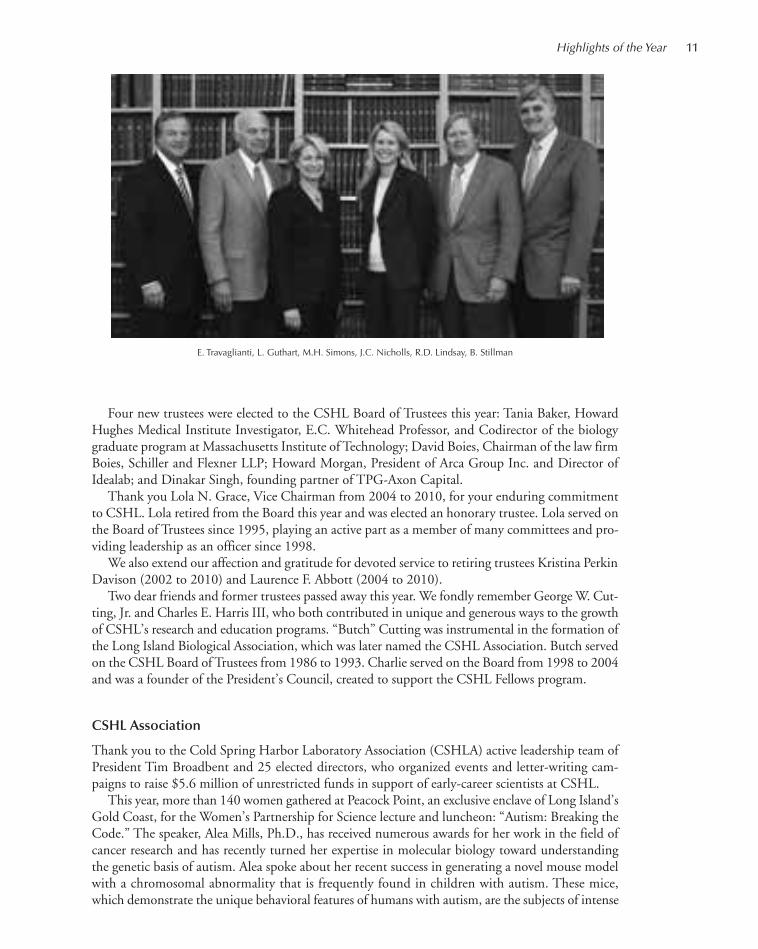
On behalf of the board, I would like to thank Eduardo G. Mestre, who served on the board since2001 and was Chairman from 2004 to 2010. With Eduardo’s leadership during the first decade ofthe 21st century, CSHL achieved unprecedented growth and expansion in infrastructure and pro-grams. Serving on committees ranging from Capital Campaign, Executive, Nominating, Research,
and Robertson Research Fund, he challenged fellow trustees and theleadership of the Laboratory to think strategically. As a result, we wereable to prevail in the face of significant external challenges that threat-ened support for basic research across the country. I am pleased thathe will remain associated with CSHL as an honorary trustee.
On November 6, 2010, the board elected a new Chairman, JamieC. Nicholls, and new slate of officers: Vice Chairs Robert D. Lindsay,comanaging partner Lindsay Goldberg, and Marilyn Simons, Presidentof The Simons Foundation; Treasurer Leo Guthart, CEO of TopspinPartners; and Secretary Ed Travaglianti, President of TD Bank, LongIsland. I look forward to working closely with Jamie, who, as CSHLTreasurer since 2009, has demonstrated her unique ability to translateher business expertise to the nonprofit, academic world.
10 Highlights of the Year
R. Martiensson
E.G. Mestre, D. Ayres
001-026_Pres_High_Annual Report_2009 template 7/12/11 9:59 AM Page 10
Four new trustees were elected to the CSHL Board of Trustees this year: Tania Baker, HowardHughes Medical Institute Investigator, E.C. Whitehead Professor, and Codirector of the biologygraduate program at Massachusetts Institute of Technology; David Boies, Chairman of the law firmBoies, Schiller and Flexner LLP; Howard Morgan, President of Arca Group Inc. and Director ofIdealab; and Dinakar Singh, founding partner of TPG-Axon Capital.
Thank you Lola N. Grace, Vice Chairman from 2004 to 2010, for your enduring commitmentto CSHL. Lola retired from the Board this year and was elected an honorary trustee. Lola served onthe Board of Trustees since 1995, playing an active part as a member of many committees and pro-viding leadership as an officer since 1998.
We also extend our affection and gratitude for devoted service to retiring trustees Kristina PerkinDavison (2002 to 2010) and Laurence F. Abbott (2004 to 2010).
Two dear friends and former trustees passed away this year. We fondly remember George W. Cut-ting, Jr. and Charles E. Harris III, who both contributed in unique and generous ways to the growthof CSHL’s research and education programs. “Butch” Cutting was instrumental in the formation ofthe Long Island Biological Association, which was later named the CSHL Association. Butch servedon the CSHL Board of Trustees from 1986 to 1993. Charlie served on the Board from 1998 to 2004and was a founder of the President’s Council, created to support the CSHL Fellows program.
CSHL Association
Thank you to the Cold Spring Harbor Laboratory Association (CSHLA) active leadership team ofPresident Tim Broadbent and 25 elected directors, who organized events and letter-writing cam-paigns to raise $5.6 million of unrestricted funds in support of early-career scientists at CSHL.
This year, more than 140 women gathered at Peacock Point, an exclusive enclave of Long Island’sGold Coast, for the Women’s Partnership for Science lecture and luncheon: “Autism: Breaking theCode.” The speaker, Alea Mills, Ph.D., has received numerous awards for her work in the field ofcancer research and has recently turned her expertise in molecular biology toward understandingthe genetic basis of autism. Alea spoke about her recent success in generating a novel mouse modelwith a chromosomal abnormality that is frequently found in children with autism. These mice,which demonstrate the unique behavioral features of humans with autism, are the subjects of intense
Highlights of the Year 11
E. Travaglianti, L. Guthart, M.H. Simons, J.C. Nicholls, R.D. Lindsay, B. Stillman
001-026_Pres_High_Annual Report_2009 template 7/12/11 9:59 AM Page 11
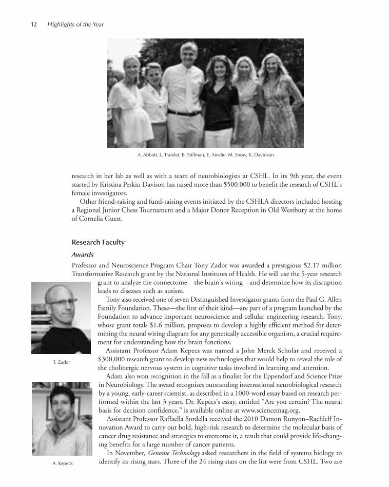
research in her lab as well as with a team of neurobiologists at CSHL. In its 9th year, the eventstarted by Kristina Perkin Davison has raised more than $500,000 to benefit the research of CSHL’sfemale investigators.
Other friend-raising and fund-raising events initiated by the CSHLA directors included hostinga Regional Junior Chess Tournament and a Major Donor Reception in Old Westbury at the homeof Cornelia Guest.
Research Faculty
Awards
Professor and Neuroscience Program Chair Tony Zador was awarded a prestigious $2.17 millionTransformative Research grant by the National Institutes of Health. He will use the 5-year research
grant to analyze the connectome—the brain’s wiring—and determine how its disruptionleads to diseases such as autism.
Tony also received one of seven Distinguished Investigator grants from the Paul G. AllenFamily Foundation. These—the first of their kind—are part of a program launched by theFoundation to advance important neuroscience and cellular engineering research. Tony,whose grant totals $1.6 million, proposes to develop a highly efficient method for deter-mining the neural wiring diagram for any genetically accessible organism, a crucial require-ment for understanding how the brain functions.
Assistant Professor Adam Kepecs was named a John Merck Scholar and received a$300,000 research grant to develop new technologies that would help to reveal the role ofthe cholinergic nervous system in cognitive tasks involved in learning and attention.
Adam also won recognition in the fall as a finalist for the Eppendorf and Science Prizein Neurobiology. The award recognizes outstanding international neurobiological researchby a young, early-career scientist, as described in a 1000-word essay based on research per-formed within the last 3 years. Dr. Kepecs’s essay, entitled “Are you certain? The neuralbasis for decision confidence,” is available online at www.sciencemag.org.
Assistant Professor Raffaella Sordella received the 2010 Damon Runyon–Rachleff In-novation Award to carry out bold, high-risk research to determine the molecular basis ofcancer drug resistance and strategies to overcome it, a result that could provide life-chang-ing benefits for a large number of cancer patients.
In November, Genome Technology asked researchers in the field of systems biology toidentify its rising stars. Three of the 24 rising stars on the list were from CSHL. Two are
12 Highlights of the Year
A. Abbott, L. Trafelet, B. Stillman, E. Ainslie, M. Snow, K. Davidson
A. Kepecs
T. Zador
001-026_Pres_High_Annual Report_2009 template 7/12/11 9:59 AM Page 12
recent WSBS graduates: Yaniv Erlich, for work in “Fast-Paced Bioinformatics,” andNicholas Navin, for work in “The Evolution of Cancer Tumors.” Assistant ProfessorMichael Schatz was recognized for his work on “Genome Assembly and the Cloud.”
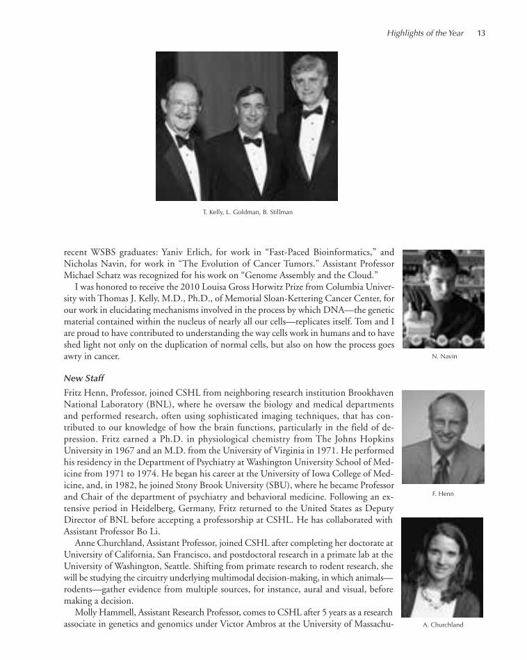
I was honored to receive the 2010 Louisa Gross Horwitz Prize from Columbia Univer-sity with Thomas J. Kelly, M.D., Ph.D., of Memorial Sloan-Kettering Cancer Center, forour work in elucidating mechanisms involved in the process by which DNA—the geneticmaterial contained within the nucleus of nearly all our cells—replicates itself. Tom and Iare proud to have contributed to understanding the way cells work in humans and to haveshed light not only on the duplication of normal cells, but also on how the process goesawry in cancer.
New Staff
Fritz Henn, Professor, joined CSHL from neighboring research institution BrookhavenNational Laboratory (BNL), where he oversaw the biology and medical departmentsand performed research, often using sophisticated imaging techniques, that has con-tributed to our knowledge of how the brain functions, particularly in the field of de-pression. Fritz earned a Ph.D. in physiological chemistry from The Johns HopkinsUniversity in 1967 and an M.D. from the University of Virginia in 1971. He performedhis residency in the Department of Psychiatry at Washington University School of Med-icine from 1971 to 1974. He began his career at the University of Iowa College of Med-icine, and, in 1982, he joined Stony Brook University (SBU), where he became Professorand Chair of the department of psychiatry and behavioral medicine. Following an ex-tensive period in Heidelberg, Germany, Fritz returned to the United States as DeputyDirector of BNL before accepting a professorship at CSHL. He has collaborated withAssistant Professor Bo Li.
Anne Churchland, Assistant Professor, joined CSHL after completing her doctorate atUniversity of California, San Francisco, and postdoctoral research in a primate lab at theUniversity of Washington, Seattle. Shifting from primate research to rodent research, shewill be studying the circuitry underlying multimodal decision-making, in which animals—rodents—gather evidence from multiple sources, for instance, aural and visual, beforemaking a decision.
Molly Hammell, Assistant Research Professor, comes to CSHL after 5 years as a researchassociate in genetics and genomics under Victor Ambros at the University of Massachu-
Highlights of the Year 13
N. Navin
T. Kelly, L. Goldman, B. Stillman
F. Henn
A. Churchland
001-026_Pres_High_Annual Report_2009 template 7/12/11 9:59 AM Page 13
setts. At CSHL, she is applying prediction algorithms to problems in cancer research. She is alsoManager of the CSHL Cancer Center’s Bioinformatics Shared Resource.
Chris Hammell, Assistant Professor, did his doctoral work at Dartmouth College and his post-doctoral work in the lab of Victor Ambros at the University of Massachusetts. There, he became in-terested in the machinery that prepares microRNAs to target specific genes, which they in turnregulate. Using Caenorhabditis elegans and forward genetics, he continues to focus on how mutationsin this machinery could perturb a given microRNA’s gene-regulatory activity so as to give rise to adevelopmental timing defect and set in motion a chain of events culminating in human illness.
Justin Kinney was named our second Quantitative Biology Fellow. He earned his doctorate inphysics from Princeton University and spent the last 2 years in postdoctoral fellowships at Princetonand at CSHL, applying his quantitative skills to biological problems. As a Fellow, he will focus onthe question of how sequences of very specific regions in the genome interact with proteins to executegene expression. He seeks to characterize the sequence–function relationship quantitatively.
Michael Schatz, Assistant Professor, developed methods for large-scale computational analysis ofDNA sequencing data at the University of Maryland. He is known for his pioneering use of cloud
computing for genomics and for the last several years has helped to run a large NationalScience Foundation cloud computing project. His research at CSHL will focus on meta-genomics—trying to understand individual genomes within a larger genomic context—and on genome assembly and validation projects.
Hongwu Zheng, Assistant Professor, earned his Ph.D. in biochemistry at Boston Uni-versity and completed postdoctoral studies at Harvard Medical School. He focuses onglioblastoma, a brain cancer with a poor prognosis. He uses mice to recapitulate geneticand epigenetic aspects of the cancer and approaches the problem from a developmentalperspective. Hongwu is exploring ways to resolve differentiation in cells as a method ofhalting tumor progression.
Promotions
Congratulations to Dinu Albeanu, who was appointed Assistant Professor. Nicholas Navin was pro-moted to the position of Research Investigator in the laboratory of Michael Wigler. Dan Levy waspromoted to the position of Senior Computer Scientist.
Departures
CSHL is proud of our long history as an incubator for early-career researchers who go on to suc-cessful careers all over the world. In 2010, Matthew Vaughn became a Research Associate at TexasAdvanced Computing Center in Austin. Sheldon McKay took on the job of Scientific Lead, En-gagement Team, iPlant Collaborative at the University of Arizona, Tucson. Professor Michael Zhangmoved to Dallas to become Director of the Center for Systems Biology, department of molecularand cell biology, University of Texas, Dallas.
14 Highlights of the Year
M. Hammell C. Hammell J. Kinney M. Schatz
H. Zheng
001-026_Pres_High_Annual Report_2009 template 7/12/11 9:59 AM Page 14

Education Programs
Watson School of Biological Sciences
In the National Research Council (NRC)’s latest assessment of 5000 doctoral programsacross 62 fields at 212 universities nationwide, the Watson School of Biological Sciences(WSBS) was ranked between third and 17th across 20 cumulative categories. In the categoryof citations per publication, CSHL ranked first. The NRC assessment is performed overthe period of 10 years, and so this is the first opportunity that the WSBS program has hadto be included in this national evaluation.
Ten WSBS students, all of whom matriculated between 2004 and 2006, received theirPh.D.s at the 2010 WSBS Commencement Convocation in April. 2010 graduate Yaniv Er-lich won the Fred Hutchinson Cancer Research Center’s Harold M. Weintraub GraduateStudent Award for outstanding achievement during graduate studies.
Honorary degrees were conferred upon Carla Jo Shatz, Ph.D. and Thomas R. Cech,Ph.D. Dr. Cech is a Nobel laureate and pioneer in the study of RNA enzymes and telomerase andwas recently President of the Howard Hughes Medical Institute. Dr. Shatz, whose research hashelped to establish some of the basic principles of early brain development, is Professor of biologyand neurobiology and Director of the Bio-X program at Stanford University School of Medicine.She was also an instructor in our neuroscience advanced courses program.
The 2010 Gavin Borden Visiting Fellow Lecture was presented on March 15 by Dr. Gerald F.Joyce, Dean of the Faculty, Professor, Departments of Chemistry and Molecular Biology, and In-vestigator, The Skaggs Institute for Chemical Biology, The Scripps Research Institute. The title ofthe 16th annual CSHL Gavin Borden lecture was “The Origin of Life in the Laboratory.”
Meetings and Courses
On April 6, CSHL celebrated the opening of Cold Spring Harbor Asia Conferences in Suzhou,China, a meetings program that aims to be the premier hub for scientists throughout Asia who areexploring the frontiers of molecular biology, biomedicine, and biotechnology. The program kickedoff with the first James Watson Cancer Symposium, organized by leading scientists representing im-portant current areas of research: Dr. Xiaodong Wang, a Howard Hughes Medical Institute Inves-tigator affiliated with the University of Texas Southwestern Medical Center and the NationalInstitute of Biological Sciences, Beijing; Dr. Scott W. Lowe of HHMI and CSHL; Dr. Yusuke Naka-mura of the University of Tokyo; Dr. Tak Mak of the University of Toronto; and Dr. Karen Vousden
Highlights of the Year 15
National Research Councilassessment publication
B. Stillman, C.J. Shatz B. Stillman, T.R. Cech
001-026_Pres_High_Annual Report_2009 template 7/12/11 9:59 AM Page 15

of the Beatson Institute for Cancer Research in the United Kingdom. This 6-day meet-ing was followed by the first Francis Crick Neuroscience Symposium, which was sim-ilarly organized by leaders in the field: Dr. Z. Josh Huang of CSHL; Dr. Mu-mingPoo of the CAS Institute of Neuroscience, Shanghai and the University of California,Berkeley; Dr. Linda Richards of the University of Queensland, Australia; Dr. JoshuaSanes of Harvard University; and Dr. Keiji Tanaka of the Laboratory for CognitiveBrain Mapping, Riken, Japan. In all, the new CSHL Asia program, which operatesfrom a 600,000-square-foot facility—the Suzhou Dushu Lake Conference Center—hosted 10 meetings and more than 2000 scientists from around the world, but pri-marily from Pacific Rim countries.
Suzhou is only 60 miles west of a “megacity” even larger than New York—the economic power-house of Shanghai, population 20 million. Importantly, the new conference center is less than anhour by high-speed rail from Shanghai and, served by two regional airports, is only a 2- to 3-hourplane ride from Japan, South Korea, Taiwan, and Hong Kong. Singapore and Sydney, Australia,are, respectively, 5 and 10 hours distant by air.
This year marked the 75th anniversary of the Cold Spring Harbor Laboratory Symposia onQuantitative Biology. The 2010 Symposium, with close to 70 talks and attendance of more than400 scientists, was organized by Terri Grodzicker, David Spector, David Stewart, and me. It focusedon the topic of Nuclear Organization and Function. To celebrate the history of the symposia, JanWitkowski, Jim Watson, and I organized a special 1-day event chaired by Robert Tjian, CSHLalumnus and President, Howard Hughes Medical Institute, called “Biology, Society, and the Future.”More than 225 guests attended the lectures presented by world experts including Charles Sawyers,Memorial Sloan-Kettering Cancer Center; Spencer Wells, National Geographic Society; HenryLouis Gates, Harvard University; Mark Bear, Massachusetts Institute of Technology; Story Landis,National Institute of Neurological Disorders & Stroke; Peter Neufeld, The Innocence Project; CraigVenter, J. Craig Venter Institute; and Richard Roberts, New England BioLabs, Inc.
The Symposium on Quantitative Biology has become the cornerstone for our annual programof Meetings and Courses, which in 2010 posted record attendance. A total of 7500 researchers fromaround the globe attended meetings, and more than 1300 attended training courses on our LongIsland campuses. A new experimental laboratory teaching suite funded by the Howard Hughes Med-ical Institute was opened in our Hillside Laboratories complex.
16 Highlights of the Year
CSH Asia logo
Opening ceremony, CSH Asia Conferences CSH Asia poster session
001-026_Pres_High_Annual Report_2009 template 7/12/11 9:59 AM Page 16

Dolan DNA Learning Center
In its second year of operation, the Harlem DNA Lab made significant strides in reaching under-served students and teachers in New York City schools. The statistics speak for themselves: 75% ofthe 6400 precollege students that attended field trips to the Harlem DNA Lab were African Amer-ican or Latino; 75% of students attending the Harlem DNA Lab came from Title 1 schools, where40% or more of students are considered low income.
CSHL’s DNA Learning Center led by David Micklos also won acclaim for the success of itsiPhone app, “The 3D Brain.” With 50,000 downloads, it reached no. 7 among education apps forthe iPhone and no. 1 of educational iPad apps.
Cold Spring Harbor Laboratory Press
Since 1933, the CSHL Press has continuously evolved and adapted its publications to bestserve the contemporary needs of the scientific community. This year, John Inglis’ teamlaunched a new initiative, CSH Perspectives in Biology, a monthly online publication comprisingreviews spanning the complete spectrum of the molecular life sciences. This new venture is in-tended to provide the life sciences community with authoritative analyses of progress in emerg-ing areas of molecular, cell, and developmental biology, genetics, evolutionary biology,neuroscience, cancer biology, and molecular pathology. The contributions are written by lead-ing researchers in each field and commissioned by a board of eminent academic editors. SubjectCollections gradually accumulate articles as new issues of the journal are published and, whencomplete, each represents a comprehensive survey of the field that it covers.
Development
More than 60 CSHL supporters participated in the Fall President’s Council retreat, the 16th yearof this event. Participants donated more than $25,000 to support early-career scientists in pursuitof the most promising and innovative research projects. The 2-day retreat immerses these generousphilanthropists in the hottest topics in science. This year’s event, organized by Diane Fagiola, tackled“The Science of Nanomedicine,” with a keynote on the applications of nanotechnology in medicineby Bob Langer, David H. Koch Professor at the Massachusetts Institute of Technology. Other expertsincluded Harvard Professor George Whitesides, University of North Carolina chemistry department
Highlights of the Year 17
Symposium attendees
CSH Perspectivesin Biology
001-026_Pres_High_Annual Report_2009 template 7/12/11 9:59 AM Page 17

head Joseph DiSimone, and Dr. William Sherman of Brookhaven National Laboratory’s Center forFunctional Nanomaterials.
For the fifth year, 400 guests gathered in early November at the Mandarin Oriental Hotel inManhattan to honor recipients of the Double Helix Medal—extraordinary individuals who havebenefited human health through game-changing biomedical research or by raising awareness andfunds for such endeavors. Geneticist Mary-Claire King was honored for outstanding contributionstoward understanding the genetics of breast cancer and mental illness. Evelyn H. Lauder receivedthe Medal for her leadership as the Founder and Chairman of The Breast Cancer Research Foun-dation, which has raised more than $300,000 million to support breast cancer research worldwide.Nobel laureate in Economic Sciences John F. Nash was awarded the Medal for having broughtworldwide awareness to and appreciation for people suffering with schizophrenia. The gala event,produced by Charlie Prizzi’s team in the development department, raised more than $3 million. Aspecial feature of this event was a musical performance by composer Carter Burwell, whose biographyincludes a 2-year stint as Chief Computer Scientist at CSHL.
18 Highlights of the Year
President’s Council retreat
J. Nash, M.-C.King, E. Lauder
001-026_Pres_High_Annual Report_2009 template 7/12/11 9:59 AM Page 18
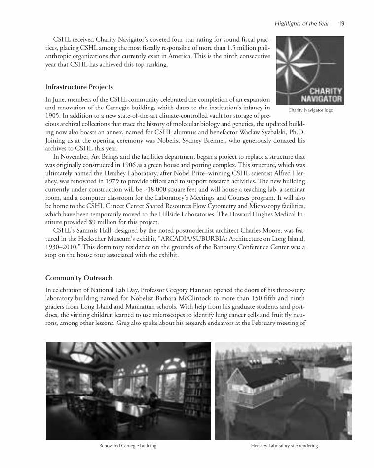
CSHL received Charity Navigator’s coveted four-star rating for sound fiscal prac-tices, placing CSHL among the most fiscally responsible of more than 1.5 million phil-anthropic organizations that currently exist in America. This is the ninth consecutiveyear that CSHL has achieved this top ranking.
Infrastructure Projects
In June, members of the CSHL community celebrated the completion of an expansionand renovation of the Carnegie building, which dates to the institution’s infancy in1905. In addition to a new state-of-the-art climate-controlled vault for storage of pre-cious archival collections that trace the history of molecular biology and genetics, the updated build-ing now also boasts an annex, named for CSHL alumnus and benefactor Waclaw Syzbalski, Ph.D.Joining us at the opening ceremony was Nobelist Sydney Brenner, who generously donated hisarchives to CSHL this year.
In November, Art Brings and the facilities department began a project to replace a structure thatwas originally constructed in 1906 as a green house and potting complex. This structure, which wasultimately named the Hershey Laboratory, after Nobel Prize–winning CSHL scientist Alfred Her-shey, was renovated in 1979 to provide offices and to support research activities. The new buildingcurrently under construction will be ~18,000 square feet and will house a teaching lab, a seminarroom, and a computer classroom for the Laboratory’s Meetings and Courses program. It will alsobe home to the CSHL Cancer Center Shared Resources Flow Cytometry and Microscopy facilities,which have been temporarily moved to the Hillside Laboratories. The Howard Hughes Medical In-stitute provided $9 million for this project.
CSHL’s Sammis Hall, designed by the noted postmodernist architect Charles Moore, was fea-tured in the Heckscher Museum’s exhibit, “ARCADIA/SUBURBIA: Architecture on Long Island,1930–2010.” This dormitory residence on the grounds of the Banbury Conference Center was astop on the house tour associated with the exhibit.
Community Outreach
In celebration of National Lab Day, Professor Gregory Hannon opened the doors of his three-storylaboratory building named for Nobelist Barbara McClintock to more than 150 fifth and ninthgraders from Long Island and Manhattan schools. With help from his graduate students and post-docs, the visiting children learned to use microscopes to identify lung cancer cells and fruit fly neu-rons, among other lessons. Greg also spoke about his research endeavors at the February meeting of
Highlights of the Year 19
Charity Navigator logo
Renovated Carnegie building Hershey Laboratory site rendering
001-026_Pres_High_Annual Report_2009 template 7/12/11 9:59 AM Page 19

the Secret Science Club in Brooklyn, a monthly gathering of ~400 science enthusiasts primarilyfrom New York City.
CSHL’s Partners for the Future program continues to recruit the brightest of Long Island’s bud-ding scientists. This year, seven seniors from high schools across Long Island were accepted into theprogram, giving them daily access to a CSHL mentor and a laboratory in which to conduct a researchproject of their own. The students devote a large part of their year to working in a laboratory andlearning what it is like to be a researcher. At the end of the school year, the students present theirfindings in a scientific seminar in Grace Auditorium. This year, three students working on cancer-related research projects took an additional step, taking the time to translate their scientific presen-tations for the public and the media. We offer thanks to the American Cancer Society for partneringwith us to publicize this as part of its local Relay For Life events.
Spearheaded by Amanda McBrien, Assistant Director of Instruction atthe DNALC, CSHL cohosted a National DNA Day scavenger hunt withlocal museums and merchants in Cold Spring Harbor village. More than200 area residents participated in the weekend event celebrating the sto-ried history of the village. April 25 is congressionally designated as Na-tional DNA Day, commemorating the completion of the HumanGenome Project in April 2003 and the discovery of the double helix struc-ture of DNA in 1953. Each year, the National Human Genome ResearchInstitute encourages national participation in the creation of opportunities
for students, teachers, and families to explore the latest developments in ge-nomic research.
The Harlem DNA Lab Instructor Ileana Rios and a banana DNA extrac-tion experiment have become a regular attraction at the World Science Fes-tival Street Fair in Manhattan. CSHL is pleased to have been able toparticipate once again in this event, which engages many thousands of NewYork City residents in the celebration of science for a week each June.
Thanks to our students and postdocs who make our CSHL tour programsuch a success. This year, our tour guides hosted 500-plus visitors. The 2-hour walking tours showcase the history of our science, research, and edu-
20 Highlights of the Year
CSHL Partners for the Future
DNA Day Scavenger Hunt poster
Community tours logo
001-026_Pres_High_Annual Report_2009 template 7/12/11 9:59 AM Page 20

cation programs. Participants also experience the beauty of the cam-pus’ architecture and landscapes.
The CSHL DNA Learning Center’s public programs continue tobe a hit in the community. During the school year, the DNALCopened its doors on Saturdays for “Saturday DNA,” geared to middleand high school students and their parents. During the summermonths, more than 900 students in grades 5 through 12 attended
week-long camp sessions, on topics ranging from “Fun with DNA” and “World of Enzymes” to“Forensic Detectives” and “Silencing Genomes.”
We are thankful for the partnerships that we have with local organizations with whom we interactin many different ways throughout the year. This year, Assistant Professor Hiro Furukawa lectured ata meeting of the Alzheimer’s Association of the Long Island Board. We hosted a lecture and tour forthe Leukemia & Lymphoma Society board of directors, at which Chris Vakoc spoke. Diane Esposito,Research Investigator in Mike Wigler’s laboratory, was an invited speaker on breast cancer research forthree different occasions, which were sponsored by the Suffolk County Women’s Bar Association, theWomen’s Center of St. Francis Hospital, and the Adelphi N.Y. Statewide Breast Cancer Program.CSHL representatives cheered on participants in the LI2Day Walk and judged the organization’s an-nual scholarship program. We were also well represented at a local Swim Across America event. Wethank these and many other partners who support CSHL through grants and donations.
For details on grants and philanthropic donations received this year, please refer to separate sec-tions of this report. In addition, please visit our newly designed public website, www.cshl.edu forall of the latest news and information about the institution, including electronic access to annualreports, the Harbor Transcript magazine, links to stories about us in the media, and postings aboutresearch results and education program achievements.
Public Lectures at Cold Spring Harbor Laboratory
March 21—Roger Reeves, Ph.D.: “Theory to Therapeutics in Mice and Men with Down Syn-drome,” cosponsored by CSHL, the Association for Children with Down Syndrome, the DownSyndrome Connection of Long Island, and the National Down Syndrome Society.
April 26—Thomas R. Cech, Ph.D., Distinguished Professor, University of Colorado-Boulder; In-vestigator, Howard Hughes Medical Institute; winner, Nobel Prize in Chemistry, 1989: “Learningto Teach, Teaching to Learn.”
June 8—Alea A. Mills, Ph.D., CSHL Associate Professor, with NikitaAnand, Cold Spring Harbor High School, Norah Liang, Patchogue-Med-ford High School, Joshua Weiss, Smithtown High School East: “Next-Gen-eration Cancer Research: Long Island Teens on the Front Line of Discovery.”
June 29—Sydney C. Gary, Ph.D., CSHL Director, research operations;Adam Kepecs, Ph.D., CSHL Assistant Professor; Stephen Dewey, Ph.D.,The Feinstein Institute of Medical Research; Edward Nunes, M.D., Profes-sor of clinical psychiatry, Columbia University: “Addiction: How the BrainMeasures Reward and Response,” cosponsored by CSHL, the North Shore–Long Island Jewish Health System, and St. Johnland Nursing Center.
September 26—Steven Allen, M.D., Associate Chief of Hematology,North Shore-LIJ; Johannes Zuber, M.D., CSHL Clinical Research Fellow; Christopher Vakoc,M.D., Ph.D., CSHL Fellow: “Hitting Leukemia Where It Hurts: Researchers and Physicians Uniteto Knock Out Cancer,” cosponsored by CSHL, The Don Monti Memorial Research Foundation,and the North Shore–Long Island Jewish Health System.
Highlights of the Year 21
Saturday DNA logo
A. Mills
001-026_Pres_High_Annual Report_2009 template 7/12/11 9:59 AM Page 21
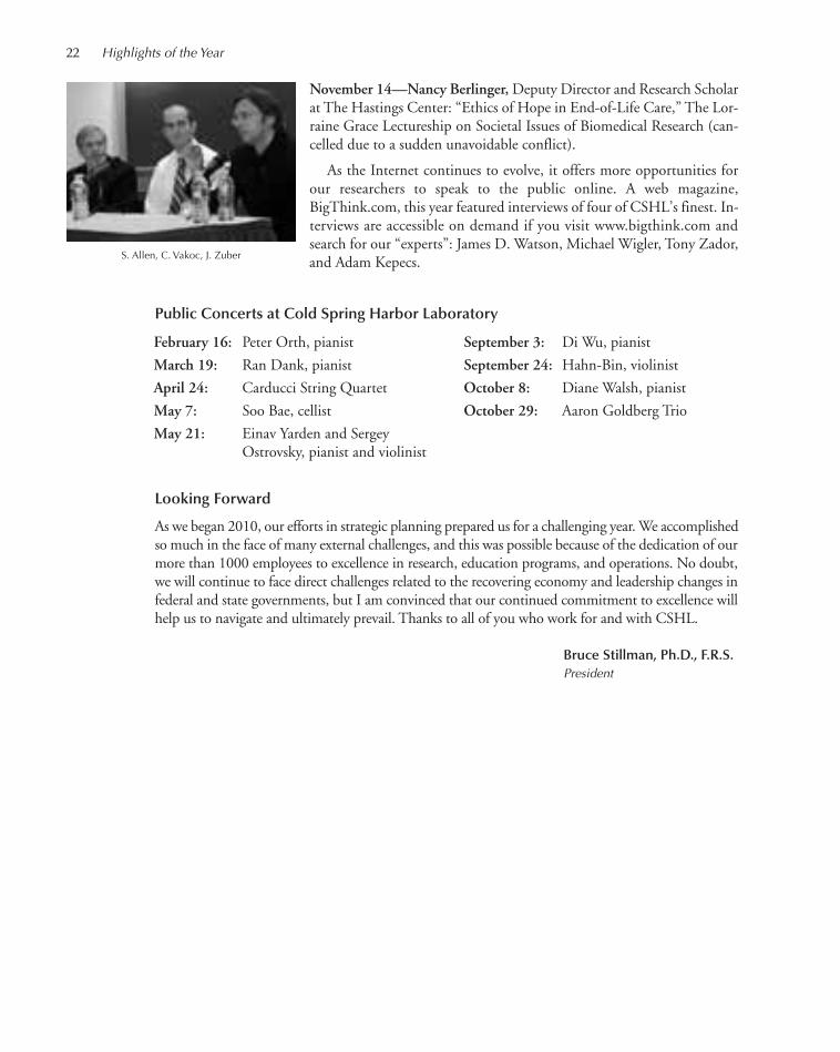
Looking Forward
As we began 2010, our efforts in strategic planning prepared us for a challenging year. We accomplishedso much in the face of many external challenges, and this was possible because of the dedication of ourmore than 1000 employees to excellence in research, education programs, and operations. No doubt,we will continue to face direct challenges related to the recovering economy and leadership changes infederal and state governments, but I am convinced that our continued commitment to excellence willhelp us to navigate and ultimately prevail. Thanks to all of you who work for and with CSHL.
Bruce Stillman, Ph.D., F.R.S. President
November 14—Nancy Berlinger, Deputy Director and Research Scholarat The Hastings Center: “Ethics of Hope in End-of-Life Care,” The Lor-raine Grace Lectureship on Societal Issues of Biomedical Research (can-celled due to a sudden unavoidable conflict).
As the Internet continues to evolve, it offers more opportunities forour researchers to speak to the public online. A web magazine,BigThink.com, this year featured interviews of four of CSHL’s finest. In-terviews are accessible on demand if you visit www.bigthink.com andsearch for our “experts”: James D. Watson, Michael Wigler, Tony Zador,and Adam Kepecs.
Public Concerts at Cold Spring Harbor Laboratory
22 Highlights of the Year
S. Allen, C. Vakoc, J. Zuber
February 16: Peter Orth, pianist
March 19: Ran Dank, pianist
April 24: Carducci String Quartet
May 7: Soo Bae, cellist
May 21: Einav Yarden and Sergey Ostrovsky, pianist and violinist
September 3: Di Wu, pianist
September 24: Hahn-Bin, violinist
October 8: Diane Walsh, pianist
October 29: Aaron Goldberg Trio
001-026_Pres_High_Annual Report_2009 template 7/12/11 9:59 AM Page 22

This was a year of steady progress for the Laboratory. Although there are substantialchallenges on the horizon, we take some comfort from the continued recovery in thefinancial markets since the 2008 debacle, and the resumption of growth, albeitpainfully slow, of the U.S. economy.
What keeps Cold Spring Harbor Laboratory’s culture so dynamic and invigoratingis a sense of constant renewal. As we have absorbed and populated the new HillsideCampus buildings, 11 dynamic new faculty members with diverse research concen-trations have joined our community during the last 18 months. Add to this a newclass of Ph.D. students at the Watson School, our usual complement of 8000 Meetingand Course attendees, and new scientific publications from the Laboratory Press,and we end up with a unique brew of intellectual capital. Our campus landscapecontinued to evolve as well, with the groundbreaking construction of the new Her-shey Building, made possible by a generous grant from the Howard Hughes MedicalInstitute.
It is gratifying to see the asset value of the Laboratory’s endowment fund approaching its pre-2008high watermark. This growth is made possible by a relatively conservative annual spending policy, newdonations, and capital appreciation. The investment return for the 2010 calendar year was a positive10.25% bringing the total value of the fund up to $283 million at year end—this following 20%annual appreciation in 2009. During the last two years, the Investment Committee of the Board ofTrustees has sought to reduce the level of risk and volatility in the portfolio by increasing exposure toconservative and proven hedged equity, absolute return, and international equity managers. Althoughthis might cause performance to be slightly below equity indices in bull markets, it should serve theportfolio well over time by providing a prudent balance of capital preservation and growth.
For the fiscal year ending 12/31/10, the Laboratory operating budget reached approximately $147million—an increase of 14% over the prior year. The substantial revenue growth was largely reflectiveof compelling research resulting in high success rates with both private and public grant applications.Although this is excellent news, there are caveats of which we must be mindful. Of the federal grantawards received by our investigators, 9% came from funding made available through the American Re-covery and Reinvestment Act. These “stimulus” funds, although welcome, are temporary in nature andwill not continue beyond 2 years. Of greatest concern is the uncertainty about the future of federalfunding for research as the U.S. government struggles with massive deficits and partisan political grid-lock. We did achieve our goal of achieving cash-neutral operating results in 2010 after netting depreci-ation expense against expenditures for capital improvements. However, this was partially achieved bydrawing down on the fund for “research start-up” that was prudently raised as part of the $200 millionHillside Campus campaign completed in 2008. This fund is scheduled to be fully spent by 2016. Asalways, we maintain a lean administration and devote great attention to diligently managing expenses.
The challenges ahead are daunting. Doing cutting-edge research is expensive. Recruiting and retainingthe best and brightest investigators while investing in state-of-the-art laboratory equipment and computinginfrastructure demands tremendous resources. It is highly likely that the Laboratory will be forced tomaintain its level of excellence with less funding from the federal government. We are fortunate to havesuch a substantial level of private support, but we will need to increasingly rely upon it in the future.
Most importantly, we are inspired by the Laboratory’s mission and the work going on aroundus. And we are ever grateful to our supporters and our loyal staff.
Dill AyersChief Operating Officer
23
W. Dillaway Ayres Jr.
CHIEF OPERATING OFFICER’S REPORT
001-026_Pres_High_Annual Report_2009 template 7/12/11 9:59 AM Page 23

24
Em
plo
yees
100
0
200
300
400
500
600
700
800
900
1000
1100
1200
Years
Scientists
Staff
SupportTotal
(Consists of full-time and part-time technical support, core services, publications, meetings, library, publicaffairs, buildings and grounds, administrative, personnel, Banbury Center, and DNA Learning Center)
91 92 93 94 95 96 97 98 99 00 01 02 03 04 05 06 07 08 09 10
Do
llars
Years
0
10,000,000
20,000,000
30,000,000
40,000,000
50,000,000
60,000,000
70,000,000
80,000,000
90,000,000
100,000,000
110,000,000
120,000,000
130,000,000
140,000,000
150,000,000
91 92 93 94 95 96 97 98 99 00 01 02 03 04 05 06 07 08 09 10
Actual Expense
Expense Adjusted for Inflation
Operating Expense
8000
9000
7000
6000
5000
4000
Par
tici
pan
ts
Years
Meetings and Courses Participants
3000
2000
1000
009 1091 92 93 94 95 96 97 98 99 00 01 02 03 04 05 06 07 08
001-026_Pres_High_Annual Report_2009 template 7/12/11 9:59 AM Page 24
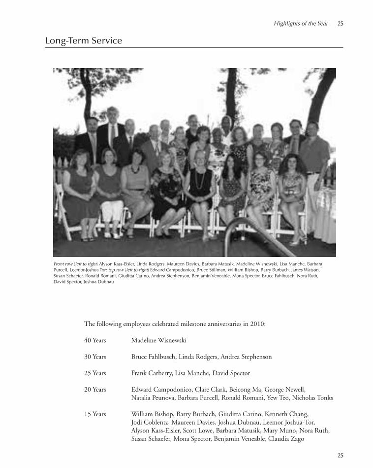
Highlights of the Year 25
Long-Term Service
Front row (left to right) Alyson Kass-Eisler, Linda Rodgers, Maureen Davies, Barbara Matusik, Madeline Wisnewski, Lisa Manche, Barbara Purcell, Leemor-Joshua Tor; top row (left to right) Edward Campodonico, Bruce Stillman, William Bishop, Barry Burbach, James Watson, Susan Schaefer, Ronald Romani, Giuditta Carino, Andrea Stephenson, Benjamin Veneable, Mona Spector, Bruce Fahlbusch, Nora Ruth, David Spector, Joshua Dubnau
The following employees celebrated milestone anniversaries in 2010:
40 Years Madeline Wisnewski
30 Years Bruce Fahlbusch, Linda Rodgers, Andrea Stephenson
25 Years Frank Carberry, Lisa Manche, David Spector
20 Years Edward Campodonico, Clare Clark, Beicong Ma, George Newell, Natalia Peunova, Barbara Purcell, Ronald Romani, Yew Teo, Nicholas Tonks
15 Years William Bishop, Barry Burbach, Giuditta Carino, Kenneth Chang, Jodi Coblentz, Maureen Davies, Joshua Dubnau, Leemor Joshua-Tor, Alyson Kass-Eisler, Scott Lowe, Barbara Matusik, Mary Muno, Nora Ruth, Susan Schaefer, Mona Spector, Benjamin Veneable, Claudia Zago
25
001-026_Pres_High_Annual Report_2009 template 7/12/11 9:59 AM Page 25

001-026_Pres_High_Annual Report_2009 template 7/12/11 9:59 AM Page 26

RESEARCH
027-047_01_CancerGeneExpSect_Annual Report_2009 template 7/12/11 10:03 AM Page 27

See previous page for photos of the following scientific staff:
Row 1: W. Li (Mills lab); R. Solomon (McCombie lab); A. Tolpygo (Mitra lab); S. Weissmueller (Lowe lab)
Row 2: S. Schuck (Stenlund lab); P. Thekkat, A. Rosebrock, F. Karginov, J. Preall (Hannon lab); K. Creasy (Martienssen lab); C. Johns (Gingeras lab)
Row 3: M. Vigliotti (Lucito lab); A. Mofunanya (Powers lab); Z. Glass (Enikolopov lab); N. Simorowski (Furukawa lab)
Row 4: S. Das (Krainer lab); B. Boivin, X. Zhang (Tonks lab); K. Jiang (Lippman lab); J. Milazzo (Lazebnik lab)
Row 5: E. Plavskin (Timmermans lab); M. Rossman (Stillman lab); T. Schalch (Joshua-Tor lab); M. Mahdavi (Dubnau lab)
027-047_01_CancerGeneExpSect_Annual Report_2009 template 7/8/11 11:55 AM Page 28

Gene regulation and cell proliferation focuses on the regulation of gene expression, cell-division cyclecontrol, and chromosome structure in normal and cancer cells.
Through forward genetic approaches in C. elegans, Christopher Hammell and colleagues haveidentified a variety of gene products that modulate the efficacy of and/or regulate the activity ofcore microRNA (miRNA) machinery. miRNAs are RNAs ~22 nucleotides in length that negativelyand specifically regulate mRNA expression by binding to complementary sequences on their targets.Hammell’s efforts have resulted in identification of a novel, highly conserved family of proteinscalled TRIM-NHLs that physically associate with the molecular complex (called miRISC) throughwhich miRNAs induce gene silencing. These proteins, together with particular miRNAs, functionto regulate animal development. In addition to TRIM-NHL proteins, the team has used classicalgenetic approaches to identify other regulatory components that tailor miRNA function (via re-sponding to other developmental cues or physiological signals) to specific genetic pathways and/orcell types. Because most of the components identified in these screens are highly conserved, the labuses model systems including Drosophila, mouse, and human to understand how these proteins workat the molecular level.
Leemor Joshua-Tor’s lab studies the molecular basis of nucleic acid regulatory processes by usingthe tools of structural biology and biochemistry to examine proteins and protein complexes associ-ated with these processes. They use X-ray crystallography to obtain three-dimensional structures ofindividual proteins. Biochemistry and molecular biology enable them to study properties that canbe correlated with protein structure and function. It was Joshua-Tor and her team that first obtainedthe structure of a full-length Argonaute protein, work that instantly solved a long-standing puzzlein the RNAi field. By observing the structure, they realized that Argonaute was the long-soughtSlicer, which performs the critical slicing event in RNAi, cleavage of the mRNA. Joshua-Tor is alsowell known for her work on the helicase enzyme, which acts to unwind DNA strands during theDNA self-replication process.
Adrian Krainer’s lab studies the mechanisms of RNA splicing, the ways in which they go awryin disease, and the means by which faulty splicing can be corrected. Their approach has borne fruitin the study of spinal muscular atrophy (SMA), a neuromuscular disease that is the leading geneticcause of death in infants. Their ability to correct an mRNA splicing defect in SMA that makes agene called SMN2 only partially functional forms the basis of a potentially powerful therapeuticapproach. Last year, the team identified a compound that stimulates SMN protein production byaltering RNA splicing. This year, they demonstrated its efficacy. By introducing chemically modifiedpieces of RNA called antisense oligonucleotides (ASOs) into the spinal cords of mice, they succeededin reversing symptoms of Type III SMA. The lab’s expertise in splicing has also helped to shed lighton the abnormal glucose metabolism of cancer cells, sometimes referred to as the Warburg Effect.Krainer and colleagues this year described for the first time how three molecular splicing factorscontribute to high levels of PK-M2, an isoform of pyruvate kinase and a key mediator of glucosemetabolism in cancer cells.
David L. Spector’s lab studies the spatial organization and regulation of gene expression. Theirin vivo approach is exemplified in a live cell gene expression system that has made possible the ex-amination in real time of the recruitment of members of gene expression and silencing machineries.A current research focus is the distribution and dynamics of nuclear Polycomb complex proteins,known to keep genes in a silent state. The team seeks to target them to segments of DNA as a meansof selectively silencing specific genes. Another focus is the study of long noncoding RNAs (ncRNAs)retained in the nucleus, whose functions are still mostly unknown. This year, Spector and colleagues,
29
CANCER: GENE REGULATION ANDCELL PROLIFERATION
027-047_01_CancerGeneExpSect_Annual Report_2009 template 7/8/11 11:55 AM Page 29
using three-dimensional live cell imaging, discovered that the formation of a type of nuclear sub-compartment called the paraspeckle is triggered by a pair of RNA molecules, which also maintainits structural integrity. The team uncovered this unique structure-building role for the RNAs bykeeping a close watch on them from the moment they came into existence within a cell’s nucleus.This revealed that when the genes for these RNAs are switched on, and the RNAs are made, theyrecruit other RNA and protein components and serve as a scaffolding platform upon which thecomponents assemble to form paraspeckles. Paraspeckles have a role in regulating the release of cer-tain protein-coding mRNAs from the cell nucleus.
Arne Stenlund and colleagues have obtained a detailed understanding of processes required forinitiation of DNA replication from the papillomaviruses, using this system to gain a general bio-chemical understanding applicable in other systems. Papillomaviruses are a large viral family thatinduces cell proliferation at the site of infection, usually giving rise to benign tumors. But certaintypes of human papillomaviruses (HPVs) generate tumors that progress toward malignancy. Amongthese are HPVs that cause most cervical cancers. Members of the Stenlund lab also pursue studiesaimed at developing an effective small-molecule inhibitor of HPVs that might someday be used bywomen who do not receive the preventive anti-HPV vaccine now available, or those already infectedwith HPV who would not be helped by the vaccine.
Bruce Stillman’s lab studies the process by which DNA is copied within cells before they dividein two. Working with yeast and human cells, Stillman and colleagues have identified many of thecellular proteins that function at the DNA replication fork during the S phase, the portion of thecell division cycle when DNA synthesis occurs. Among these proteins are those that facilitate theassembly of chromatin, the protein–DNA complexes that form the chromosomes. The prime focusof current research, however, is the mechanism that initiates the entire process of DNA replicationin eukaryotic cells. At the heart of this mechanism is a protein that binds to “start” sites on the chro-mosomes, called the origin recognition complex (ORC). Stillman’s research has demonstrated thatthe ORC is also involved in the process of segregating duplicated chromosomes in mitosis. Theteam has found the ORC at centrosomes and centromeres, structures that orchestrate chromosomeseparation in mitosis. This year, the team discovered how a protein known to be an essential activatorof DNA replication actually triggers this process. They found that DDK phosphorylates a proteincalled Mcm4, specifically within a domain that acts as a brake to prevent the double helix frombeing unwound. The phosphorylation releases this brake, thus initiating the replication of unwoundDNA strands.
30 Research
027-047_01_CancerGeneExpSect_Annual Report_2009 template 7/8/11 11:55 AM Page 30

DEVELOPMENTAL REGULATION OF MICRORNA ACTIVITY
C.M. Hammell D.M. King
Animal development is remarkably robust and requiresthe integration of both spatial and temporal geneticprograms. Our understanding of the genetic circuitryregulating spatial patterning has illuminated strategiesshared by most organisms, but the underpinnings ofhow temporal gene expression is coordinated in animalsremain obscure. The genetic tractability, invariant celllineage, and ability to observe cell division patterns ina living animal have made Caenorhabditis elegans thepremier organism to address these questions. Mutationsthat alter stage-specific developmental programs haverevealed conserved classes of heterochronic genes thatcontrol the cell fates of the essentially invariant postem-bryonic cell lineages.
One paradigm that emerged from these studies isthat the expression of master regulatory genes requiredfor early temporal developmental programs (usually en-coding transcription factors, RNA-binding proteinsand other regulatory components) is posttranscription-ally repressed by the activity of specific microRNAs(miRNAs). Although much is known about the tran-scriptional activation and biochemical maturation ofmiRNAs, our understanding of how these processes arecoordinated to create sharp transitions in gene expres-sion at the molecular and cellular levels is limited.
Genetic Screens in C. elegans IdentifyComponents That Regulate miRNAExpression and Ensure NormalDevelopmental Timing
Under favorable environmental conditions, C. eleganslarva progress through four larval stages (L1–L4) thatare punctuated by molts to a reproductively competentadult within ~40–50 h after hatching. At each of thesestages, a variety of cells (including hypodermal, neu-ronal, muscular, and intestinal cells) execute stage-spe-cific cell division and cell-fate specification programs.The heterochronic genes control these stage-specificevents. Mutations of components in this regulatorypathway result in two general types of developmentaltiming defects. Precocious heterochronic mutants causestage-specific developmental programs to occur earlier
than normal (usually at the consequence of normal pro-grams). In contrast, retarded mutants reiterate stage-specific cell divisions and, as a consequence, retainjuvenile characteristics and lack adult-specific organsand features. Importantly, each of these phenotypes isrobust, specific, and quantifiable and can be monitoredby directly observing cell division patterns in live ani-mals or through surveying the expression of individualgenes via green fluorescent protein (GFP)-tagged re-porter constructs.
To identify common molecular components thatmodulate the temporal activity of miRNAs, we screenedfor extragenic suppressors of mutations effecting essentialmiRNA processing components or contained mutationsin specific miRNAs that are required for normal devel-opmental timing. We initially chose three classes of mu-tations that separately effect discrete phases of C. eleganslarval development. The first genetic background thatwe screened focused on the lin-4 miRNA that is genet-ically required after the first larval stage to repress the ex-pression of the LIN-14 transcription factor thatfunctions to specify L1 cell fates. lin-4 null alleles (lin-4(e912)) or point mutants that affect only the mature~21-nucleotide miRNA (lin-4(ma161)) fail to down-regulate LIN-14 expression and, as a consequence ofcontinued expression of LIN-14, reiterate L1-specific celldivision programs at all successive larval stages. The sec-ond genetic context took advantage of specific point mu-tations that disrupt the mid-domain of the C. elegansmiRNA-specific Argonaute, alg-1(ma192), that result inseverely retarded heterochronic phenotypes that manifestin the L2 to L3 transition. alg-1(ma192) animals fail todown-regulate the HBL-1 transcription factor that isnormally mediated by the let-7 family of miRNAs (miR-48, miR-241, and miR-84) and sequence elements inthe hbl-1 3′UTR (untranslated region). As the down-regulation of hbl-1 expression is essential to limit thehighly prolific cell divisions of the lateral seam cells, alg-1(ma192) animals hyperproliferate their epidermal cellcomponents. The final genetic background for this sup-pression analysis revolved around the highly conservedregulatory circuit involving the let-7 miRNA required toposttranscriptionally repress the expression of a criticalTRIM-NHL domain protein LIN-41 by binding to se-
31
027-047_01_CancerGeneExpSect_Annual Report_2009 template 7/8/11 11:55 AM Page 31

32 Research
quences in the lin-41 3′UTR. Null mutations of let-7,let-7(mn112), or hypomorphic alleles that mutate theconserved sequence of the let-7 mRNA, let-7(n2585),do not down-regulate LIN-41. As a consequence of in-appropriate LIN-41 expression, the lateral seam cells failto terminally differentiate and reiterate larval cell divisionprograms, and animals burst from their vulval structuresat the normal L4 to adult transition.
Although each of these mutations cause very specificdevelopmental and cellular phenotypes that manifest atdistinct critical stages of larval development, we designeda simple screening strategy that could be adapted for allthree screens. All three classes of mutants mentionedabove share one common phenotype: They lack normaladult-specific gene regulation, which can be monitoredby incorporating a GFP reporter into the screened ani-mals. Specifically, we chose the col-19 promoter (nor-mally driving the adult-specific collagen gene present onthe adult cuticle) to drive a GFP-tagged reporter. Thecol-19 promoter is positively regulated by the LIN-29transcription factor, which is the penultimate gene prod-uct of the heterochronic pathway. Each of these types ofretarded heterochronic mutants fail to turn on col-19::GFP, which is easily observable in a fluorescent dis-secting microscope. Suppressors for each mutant wouldbe found by identifying individual animals that couldrestore col-19::GFP expression in the F2 generation. An-imals positive for this assay would be further character-ized for the correction of the stage-specific cell lineagedefects and production of other adult structures thatwere absent in the initial mutant background.
Characterization of lin-42 and ItsFunction in miRNA Metabolism
One would expect that the suppression of each of theabove mutants (which effect specific phases of develop-mental timing) would result in the identification of atleast three classes of suppressors. Surprisingly, we wereable to identify a single complementation group, rep-resented by multiple alleles in each screen that sup-pressed all retarded heterochronic phenotypes. Severalof these alleles were positionally mapped to the left endof chromosome II. Complementation, rescue with cos-mids containing the wild-type gene, and sequencing of
the mutations indicated that all suppressors in this spe-cial class were alleles of the lin-42 gene.
lin-42 encodes a C. elegans protein that has strikinghomology with the period (Per) genes found in Drosophilamelanogaster, humans, and mice. Period homologs inthese animals coordinate gene expression patterns in a cir-cadian (once a day) rhythm that function as essential neg-ative regulatory components of the cellular clock. As clockproteins, Drosophila, mouse, and human Per cycle inabundance throughout the day, peaking in the subjectivedaytime and reaching lowest levels in the evening. Al-though C. elegans is not thought to have a clear, entrain-able circadian rhythm, the LIN-42 protein also cycles inabundance; it is most abundant during the intermolt pe-riods and is almost absent during each of the molting cy-cles. Our molecular analysis of lin-42 mutants suggeststhat LIN-42 functions to antagonize the accumulation ofmature heterochronic and nonheterochronic miRNAs bydampening their expression at a metabolic step at or up-stream of pre-miRNA processing. Notably, LIN-42 andthe accumulation of primary miRNA (pri-miRNA) levelsare antiphasic to each other, with pri-miRNA abundancepeaking during the larval molts (low LIN-42) and unde-tectable during the intermolt periods (when LIN-42 ex-pression is highest). This complementarity in expressionraises the important question of whether LIN-42 controlsthe accumulation of primary and, therefore, maturemiRNA levels. Importantly, the precocious heterochronicphenotypes associated with lin-42 mutations arise as aconsequence of an inappropriate overaccumulation ofheterochronic miRNAs during the intermolt periods oflarval development. These results suggest that miRNAmaturation or activity can be directly coupled to the reg-ulatory machinery that controls developmental timing.
Future Directions
We are currently in the process of using biochemicalapproaches to identify additional cellular componentsthat function with LIN-42 to modulate miRNA expres-sion and ensure normal developmental timing. Theseefforts will be combined with experiments inDrosophila and mouse cell lines to determine if Periodexpression and activity also control miRNA expressionin these contexts.
027-047_01_CancerGeneExpSect_Annual Report_2009 template 7/8/11 11:55 AM Page 32
33
We study the molecular basis of nucleic acid regulatoryprocesses by using the tools of structural biology andbiochemistry to examine proteins and protein com-plexes associated with these processes. X-ray crystallog-raphy enables us to obtain the three-dimensionalstructures of these molecular machines. Biochemistryand molecular biology allow us to study properties thatcan be correlated to protein structure and function.
Mechanisms of RNAiT. Schalch, C. Faehnle, C. Kuhn, C. Kuscu, S. Lin, D. Bahel, C.Ashar [in collaboration with G.J. Hannon, R.A. Martienssen, Cold Spring Harbor Laboratory; J. Partridge,St. Jude Children’s Research Hospital]
RNA interference (RNAi) has made an enormous im-pact on biology in a very short period of time. Not onlyare we still discovering new cellular pathways for theregulation of gene expression, but RNAi has becomean extraordinarily useful and simple tool for gene si-lencing. Almost from its beginnings, people have usedgenetics, biochemistry, molecular biology, and bioin-formatics to study the mechanism of RNAi and relatedpathways. We argued, however, that in order to get atrue mechanistic understanding of these pathways, wemust understand how the components of the RNAimachinery worked at a molecular level. Therefore, weembarked on structural and biochemical studies of keyproteins in the RNAi pathway.
During RNAi, long double-stranded RNA (dsRNA)is processed to yield short (~19–31 nucleotides) dsRNAsthat trigger the RNAi response. These short RNAs be-come incorporated into effector complexes called theRNA-induced silencing complex (RISC), where in themature complexes, a single-stranded RNA (ssRNA), theantisense strand of the original dsRNA, is retained inthe complex. This short RNA (small interfering RNAs[siRNAs] or microRNAs [miRNAs]) then acts to guidethe RISC complex to its target through base comple-mentarity. The best-characterized pathway, and the onethat is predominantly used for gene knockdown tech-
nology, is a posttranscriptional silencing (PTGS) path-way called “slicing.” Here the RISC complex is targetedto the mRNA and produces an endonucleolytic cut inthe mRNA target, thus preventing gene expression fromproceeding. Other RNAi silencing pathways such astranslational inhibition and transcriptional gene silenc-ing (TGS) are also mediated through RISC complexes.In all cases, these complexes contain a small ssRNA andan Argonaute protein, which serve to define the RISCcomplex. In the past few years, we have been studyingArgonaute family proteins, their complexes, and theirroles in various RNAi silencing pathways.
In the fission yeast Schizosaccharomyces pombe, as-sembly of centrometic heterochromatin requires theRITS complex, which consists of Ago1, Tas3, Chp1,and siRNAs derived from centromeric repeats. Ourcrystal structure of Chp1’s chromodomain in complexwith a trimethylated lysine-9 H3 peptide (H3K9me)revealed extensive sites of contact that contribute toChp1’s high-affinity binding. We found that this high-affinity binding is critical for the efficient establishmentof centromeric heterochromatin, but preassembled het-erochromatin can be maintained when Chp1’s affinityfor H3K9me is greatly reduced. We continue to studythis complex that forms a bridge between the RNAimachinery and chromatin.
The Different Faces of E1: A ReplicativeHexameric HelicaseE.J. Enemark, S.-J. Lee
During DNA replication, two complementary DNAstrands are separated and each becomes a template forthe synthesis of a new complementary strand. Strandseparation is mediated by a helicase enzyme, a molecu-lar machine that uses the energy derived from ATP hy-drolysis to separate DNA strands while moving alongthe DNA. Our crystal structure of the replicative heli-case E1 from papillomavirus bound to ssDNA and nu-cleotide molecules at the ATP-binding sites provided a
STRUCTURAL BIOLOGY OF NUCLEIC ACID REGULATORY PROCESSES
L. Joshua-Tor C. Ashar C. Faehnle C. Kuscu D. SauD. Bahel S. Goldsmith T. Lavy T. SchalchJ. Calarco C. Kuhn S.-J. Lee A. TociljE. Elkayam P.R. Kumar S. Lin D. Wah
027-047_01_CancerGeneExpSect_Annual Report_2009 template 7/8/11 11:55 AM Page 33

34 Research
unique look into the mechanism of translocation of thismolecular machine along DNA. Papillomaviruses are tumor viruses that cause benign
and cancerous lesions in their host. Replication of pa-pillomaviral DNA within a host cell requires the viralE1 protein, a multifunctional protein. E1 initially par-ticipates in recognizing a specific replication originDNA sequence as a dimer with E2, another viral pro-tein. Subsequently, further E1 molecules are assembledat the replication origin until two hexamers are estab-lished. These hexamers are the active helicases that op-erate bidirectionally in the replication of the viral DNA.To unwind DNA, helicases must separate the twostrands while moving along, or translocating, on theDNA. On the basis of the structures of the DNA-bind-ing domain of E1 bound to DNA that we determineda few years ago in collaboration with Arne Stenlund’slab, we suggested a mechanism for DNA strand sepa-ration. However, the mechanism that couples the ATPcycle to DNA translocation has been unclear.The E1 hexameric helicase adopts a ring shape with
a prominent central channel. ATP-binding (and hy-drolysis) sites are located at the subunit interfaces, andmultiple configurations are observed within the hexa-mer. These have been assigned as ATP-type, ADP-type,and apo-type. The configuration of the site for a givensubunit correlates with the relative height of its DNA-binding hairpin in the staircase arrangement. The sub-units that adopt an ATP-type configuration place theirhairpins at the top of the staircase, whereas the hairpinsof apo-type subunits occupy the bottom positions ofthe staircase. The hairpins of the ADP-type subunitsare placed at intermediate positions.A straightforward “coordinated escort” DNA
translocation mechanism is inferred from the staircasedDNA binding and its correlation with the configura-tion at the ATP-binding sites. Each DNA-bindinghairpin maintains continuous contact with one uniquenucleotide of ssDNA and migrates downward via ATPhydrolysis and subsequent ADP release at the subunitinterfaces. ATP hydrolysis occurs between subunits lo-cated toward the top of the staircase, whereas ADP re-lease occurs between subunits located toward thebottom of the staircase. The hairpin at the bottom ofthe staircase releases its associated ssDNA phosphate toconclude its voyage through the hexameric channel.Upon binding a new ATP molecule, this subunit movesto the top of the staircase to pick up the next availablessDNA phosphate, initiating its escorted journeythrough the channel and repeating the process. For onefull cycle of the hexamer, each subunit hydrolyzes one
ATP molecule, releases one ADP molecule, and trans-locates one nucleotide of DNA through the interiorchannel. A full cycle therefore translocates six nu-cleotides with associated hydrolysis of six ATPs and re-lease of six ADPs. A detailed comparison with othermultimeric ATPase motors that highlighted the rolesof individual site residues in the ATPase activity wasalso performed. We continue to study this helicase and the role that
other domains of the protein might have in helicase as-sembly and activity.
NADP Regulates the Yeast GAL Induction System P.R. Kumar, T. Lavy, D. Wah [in collaboration with R.Sternglanz, Stony Brook University; S.A. Johnston, Arizona State University; J. Rabinowitz, Princeton University; A. Caudy, University of Toronto]
Transcriptional regulation of the galactose metabolizinggenes in Saccharomyces cerevisiae depends on three coreproteins: Gal4p, the transcriptional activator that bindsto upstream activating DNA sequences (UASGAL);Gal80p, a repressor that binds to the carboxyl terminusof Gal4p and inhibits transcription; and Gal3p, a cy-toplasmic transducer which upon binding galactose andATP relieves Gal80p repression. The current model ofinduction relies on Gal3p sequestering Gal80p in thecytoplasm.However, the rapid induction of this systemimplies that there is a missing factor. To understand themolecular mechanism of the GAL regulatory system,we have determined the structure of S. cerevisiaeGal80pwith the activation domain of Gal4p. To our surprise, we discovered that nicotinamide
adenine dinucleotide (NAD) is nestled between Gal80pand Gal4p, making several key interactions withGal80p. Although NAD appears to be promoting thisinteraction, we have shown by in vitro pull-down assaysthat NADP disrupts this interaction. Alterations in theNAD(P)-binding site affects the initial rate of GAL in-duction in vivo, but not overall final expression levels. Although we do not understand precisely how this
trigger for GAL regulation functions, nor the involve-ment of NADP versus NAD, we speculate that switch-ing the cell to a fermentable galactose medium causes achange in NADP/NADPH or NADP/NAD ratios inthe cell, and Gal80p effectively senses the metabolicstate of the cell. NADP might be acting as a “secondmessenger” in triggering the system. Alternatively,Gal80p may function as an oxidoreductase enzyme, ac-
027-047_01_CancerGeneExpSect_Annual Report_2009 template 7/8/11 3:42 PM Page 34

Cancer: Gene Regulation and Cell Proliferation 35
tively converting NADPH to NADP in the presence ofa substrate, causing it to disassociate from Gal4p.
We are now examining possible enzymatic activitiesas well as other complexes involved in this classic tran-scriptional switch.
PUBLICATIONS
Faehnle CR, Joshua-Tor L. 2010. Argonaute MID domain takes centrestage, EMBO Reports doi:10.1038/embor.2010.110.
Joshua-Tor L, Hannon GJ. 2010. Ancestral roles of small RNAs: Anago-centric perspective. Cold Spring Harb Perspect Biol 1: a003772.
Seung-Jae Lee
027-047_01_CancerGeneExpSect_Annual Report_2009 template 7/8/11 11:55 AM Page 35

36
Mechanisms of Constitutiveand Alternative Pre-mRNA Splicing
RNA splicing is required for correct expression of mosteukaryotic protein-coding genes. The spliceosome se-lects authentic splice sites with very high fidelity, rely-ing on limited sequence information present through-out introns and exons. In humans, >75% of genes areexpressed via alternative splicing, giving rise to multi-ple protein isoforms. The choice of alternative splicesites is commonly regulated to alter gene expression,either tissue-specifically or in response to a develop-mental program or to signaling pathways. The factthat multiple protein isoforms can be expressed fromindividual genes demonstrates that the classical “onegene–one enzyme” paradigm is no longer valid andprovides an explanation for the unexpectedly smallnumber of genes uncovered by genome-sequencingprojects.
Both constitutive and alternative splicing mecha-nisms involve numerous protein components, as wellas RNA components that are part of small nuclear ri-bonucleoprotein (snRNP) particles. The work in ourlab focuses on the identification and molecular charac-terization of protein factors and sequence elements thatare necessary for the catalysis and fidelity of splicingand/or for the regulation of alternative splice site selec-tion. We are interested in how the spliceosome correctlyidentifies the exons on pre-mRNA, and how certainpoint mutations in either exon or intron sequencescause aberrant splicing, leading to various human ge-netic diseases. Related areas of interest include the re-modeling of mRNP architecture as a consequence ofsplicing, which influences downstream events, such asnonsense-mediated mRNA decay (NMD); the role ofalternative splicing misregulation in cancer; and the de-velopment of effective methods to correct defectivesplicing or modulate alternative splicing, especially ina disease context. A summary of some of our recentlypublished studies is provided below.
Autoregulation and ArginineMethylation of SRSF1
SRSF1 (formerly SF2/ASF) is a prototypical serine- andarginine-rich (SR) protein, with important roles insplicing and other aspects of mRNA metabolism.SRSF1, the gene encoding this protein, is a potentproto-oncogene with abnormal expression in many tu-mors. We found that SRSF1 negatively autoregulatesits expression to maintain homeostatic levels. We char-acterized six alternatively spliced SRSF1 mRNA iso-forms: The major isoform encodes full-length protein,whereas the others are either retained in the nucleus ordegraded by NMD. Unproductive splicing accounts foronly part of the autoregulation, which occurs primarilyat the translational level. The effect is specific to SRSF1and requires RNA recognition motif 2 (RRM2). Theultraconserved 3′-untranslated region (UTR) is neces-sary and sufficient for down-regulation. SRSF1 overex-pression shifts the distribution of target mRNA towardmonoribosomes, and translational repression is partlyindependent of Dicer and a 5′ cap. In collaborationwith Jun Zhu (Duke University), miR-7 was alsoshown to contribute to SRSF1 autoregulation. Thus,multiple posttranscriptional and translational mecha-nisms are involved in fine-tuning the expression ofSRSF1.
Alternative splicing and posttranslational modifica-tions (PTMs) are major sources of protein diversity ineukaryotic proteomes.Functional studies of SR proteinPTMshave exclusively focused on reversible phosphor-ylation of serine residues in their carboxy-terminal RSdomain. We confirmed that human SRSF1 is methyl-ated at residues R93, R97, and R109, whichwere firstidentified in a global proteomic analysis of argininemethylation. We further investigated whether thesemethylated residues regulate the properties of SRSF1,which normally shuttles between the nucleus and thecytoplasm. We found that the three arginines addi-tively control the subcellular localization of SRSF1,
RNA SPLICING
A.R. Krainer M. Akerman S. Das M. Jensen M. Motley R. SinhaO. Anczuków M. Faleiro Y. Liu J. Novatt S. SunI. Aznárez M. Fan Y.-H. Liu X. Roca R.-Y. TzengF. Bezirci O. Fregoso L. Manche K. Sahashi Z. WangD. Chatterjee Y. Hua
027-047_01_CancerGeneExpSect_Annual Report_2009 template 7/8/11 11:55 AM Page 36

Cancer: Gene Regulation and Cell Proliferation 37
and both the positive charge and the methylation stateare important. Mutations that block methylation andremove the positive charge result in cytoplasmic accu-mulation of SRSF1. The consequent decrease in nu-clear SRSF1 levels prevents it from modulatingalternative splicing of target genes, results in highersplicing-enhancer-dependent translation stimulation,and abrogates the enhancement of nonsense-mediatedmRNA decay.
Alternative Splicing and CancerCell Metabolism
Alternative splicing has an important role in cancer,partly by modulating the expression of many oncogenesand tumor suppressors and also because inactivatingmutations that affect alternative splicing of varioustumor suppressor genes account for some of the inher-ited and sporadic susceptibility to cancer. In addition,alternative splicing controls a metabolic switch charac-teristic of all cancer cells, which is known as the War-burg effect. Cancer cells preferentially metabolizeglucose by aerobic glycolysis, characterized by increasedlactate production. This distinctive metabolism involvesexpression of the embryonic M2 isozyme of pyruvatekinase, in contrast to the M1 isozyme normally ex-pressed in differentiated cells, and it confers a prolifer-ative advantage to tumor cells. The M1 and M2pyruvate kinase isozymes are expressed from a singlegene through alternative splicing of a pair of mutuallyexclusive exons. In collaboration with Lewis Cantley(Harvard Medical School), we measured the expressionof M1 and M2 mRNA and protein isoforms in mousetissues, tumor cell lines, and during terminal differen-tiation of muscle cells and showed that alternative splic-ing regulation is sufficient to account for the levels ofexpressed protein isoforms. We further showed that theM1-specific exon is actively repressed in cancer celllines—although some M1 mRNA is expressed in celllines derived from brain tumors—and demonstratedthat the related splicing repressors heterogeneous nu-clear ribonucleoprotein (hnRNP) A1 and A2, as wellas the polypyrimidine-tract-binding protein PTB, con-tribute to this control. Down-regulation of these splic-ing repressors in cancer cell lines using short hairpinRNAs (shRNAs) rescued M1 isoform expression anddecreased the extent of lactate production. These find-ings extended the links between alternative splicing andcancer and identified some of the factors responsiblefor the switch to aerobic glycolysis.
Targeted Antisense Modulation ofAlternative Splicing forTherapeutic Purposes
Spinal muscular atrophy (SMA) is a common, autoso-mal-recessive motor neuron disorder caused by homozy-gous deletion or mutation of the survival-of-motor-neuron gene, SMN1. A closely related SMN1 paralog,SMN2, is present in all patients and differs from SMN1by a C to T transition in exon 7 that causes substantialskipping of this exon, such that SMN2 expresses onlylow levels of functional protein. SMN2 decreases theseverity of SMA in a copy-number-dependent manner.We continue to explore strategies to increase the extentof exon 7 inclusion during splicing of SMN2 transcripts,for eventual therapeutic use in SMA. This translationalresearch is being done in collaboration with Frank Ben-nett and colleagues (Isis Pharmaceuticals), and MarcoPassini and colleagues (Genzyme Corporation).
We previously identified a potent 2′-O-(2-methox-yethyl) (MOE) phosphorothioate-modified antisenseoligonucleotide (ASO-10-27) that blocks an SMN2splicing silencer element in intron 7 and efficiently pro-motes exon 7 inclusion in transgenic mouse peripheraltissues after systemic administration. We have now ad-dressed its efficacy in the spinal cord—a prerequisitefor disease treatment—and its ability to rescue a mildSMA mouse model that develops tail and ear necrosis,resembling the distal tissue necrosis reported in someSMA infants. Using a micro-osmotic pump, we directlyinfused the ASO into a lateral cerebral ventricle in adultmice expressing a human SMN2 transgene; the ASOgave a robust and long-lasting increase in SMN2 exon7 inclusion measured at both the mRNA and proteinlevels in spinal cord motor neurons. A single embryonicor neonatal intracerebroventricular ASO injection strik-ingly rescued the tail and ear necrosis in this mild modelof SMA.
Injection of ASO-10-27 into the cerebral lateral ven-tricles of neonate mice with a severe form of SMA againresulted in a splicing-mediated increase in SMN proteinin the central nervous system (CNS). In addition, thisresulted in an increase in the number of motor neuronsin the spinal cord, which led to improvements in mus-cle physiology, motor function, and survival. Intrathecalinfusion of ASO-10-27 into cynomolgus monkeys de-livered putative therapeutic levels of the oligonucleotideto all regions of the spinal cord. These data demonstratethat CNS-directed ASO therapy is efficacious and thatintrathecal infusion may represent a practical route fordelivering this therapeutic oligonucleotide in the clinic.
027-047_01_CancerGeneExpSect_Annual Report_2009 template 7/8/11 11:55 AM Page 37
38 Research
More generally, these experiments demonstrate thatASOs can be used to efficiently redirect alternativesplicing of target genes throughout the CNS.
Splice Site Recognition andHuman Genetics
We continued to analyze the relationship between splicesite mutations and genetic diseases. In 1997, we discov-ered a very rare class of introns with noncanonical splicesites that are nevertheless recognized by the majorspliceosome, and we characterized one such intron in avoltage-gated sodium channel gene, SCN4A. MasanoriTakahashi (Osaka University) recently identified an in-sertion/deletion mutation in this intron in a myotoniapatient, and we collaborated with his group to charac-terize the nature of the splicing defect responsible forthe phenotype. The mutation prevents correct recogni-tion of the 5′ splice site, resulting in activation of cryp-tic splice sites. This gives rise to a 35-amino-acidinsertion in the sodium channel, which functions ab-normally and causes muscle disease.
PUBLICATIONS
Clower CV, Chatterjee D, Wang Z, Cantley LC, Vander Heiden MG,Krainer AR. 2010. The alternative splicing repressors hnRNP A1/A2
and PTB influence pyruvate kinase isoform expression and cell me-tabolism. Proc Natl Acad Sci 107: 1894–1899.
Hua Y, Sahashi K, Hung G, Rigo F, Passini MA, Bennett CF, KrainerAR. 2010. Antisense correction of SMN2 splicing in the CNS res-cues necrosis in a type III SMA mouse model. Genes Dev 24: 1634–1644.
Manley JL, Krainer AR. 2010. A rational nomenclature for serine/argi-nine-rich protein splicing factors (SR proteins). Genes Dev 24: 1073–1074.
Sinha R, Allemand E, Zhang Z, Karni R, Myers MP, Krainer AR. 2010.Arginine methylation controls the subcellular localization and func-tions of the oncoprotein splicing factor SF2/ASF. Mol Cell Biol 30:2762–2774.
Sun S, Zhang Z, Sinha R, Karni R, Krainer AR. 2010. SF2/ASF autoreg-ulation involves multiple layers of post-transcriptional and transla-tional control. Nat Struct Mol Biol 17: 306–312.
Wu H, Sun S, Tu K, Gao Y, Xie B, Krainer AR, Zhu J. 2010. A splicing-independent function of SF2/ASF in microRNA processing. MolCell 38: 67–77.
In Press
Kubota T, Roca X, Kimura T, Kokunai Y, Nishino I, Sakoda S, KrainerAR, Takahashi MP. 2011. A mutation in a rare type of intron in asodium-channel gene results in aberrant splicing and causes myoto-nia. Hum Mutat (in press).
Passini MA, Bu J, Richards AM, Kinnecom C, Sardi SP, Stanek LM,Hua Y, Rigo F, Matson J, Hung G, et al. 2011. Antisense oligonu-cleotides delivered to the mouse CNS ameliorate symptoms of severespinal muscular atrophy. Sci Transl Med 3: 72ra18.
Thorsen K, Mansilla F, Schepeler T, Øster B, Rasmussen MH, DyrskjøtL, Karni R, Akerman M, Krainer AR, Laurberg S, et al. 2011. Alter-native splicing of SLC39A14 in colorectal cancer is regulated by theWnt pathway. Mol Cell Proteomics 10: M110.002998.
Shipra Das
027-047_01_CancerGeneExpSect_Annual Report_2009 template 7/8/11 11:55 AM Page 38

39
Most cellular processes can trace their beginnings to thenucleus where a gene is activated, resulting in the pro-duction of an RNA molecule that must get processedand transported to the cytoplasm. Although much bio-chemical information is available regarding many of thefactors involved in gene expression, the spatial and tem-poral aspects of gene expression and the dynamics ofthe nuclear domains that the gene expression machin-ery occupies are less well understood. During the pastyear, we have made significant progress in two mainareas: (1) examining the dynamics of gene expression/repression and DNA repair in living cells and (2) char-acterizing the dynamics of a long nuclear retained non-coding RNA.
The Dynamics of GeneExpression/RepressionR. Zhao, Z. Lazar
Our research during the past year has focused on the spa-tial and temporal aspects of gene expression. Transcrip-tion is silenced during cell division; however, it must bereactivated upon exit from mitosis to maintain cell line-age and cell cycle progression. The transmission of thegene expression program from mother to daughter cellshas been suggested to be mediated by gene bookmarking,which may involve transcription factors, histone modi-fications/variants, and/or DNA methylation. However,the mechanism by which gene bookmarking mediatesrapid postmitotic transcriptional reactivation remainsunclear. We used a real-time gene expression system toquantitatively demonstrate that transcriptional activationof the same genetic locus occurs with a significantly morerapid kinetics in postmitotic cells versus interphase cells(Fig. 1). RNA polymerase II large subunit (pol II) andbromodomain protein 4 (BRD4) were recruited to thelocus in a different sequential order upon interphase ini-tiation versus postmitotic reactivation resulting from therecognition by BRD4 of increased levels of histone H4lysine-5 acetylation (H4K5Ac) on the previously acti-
vated locus. BRD4 accelerated the dynamics of mRNAsynthesis in postmitotic cells by decondensing chromatinand hence facilitating transcriptional reactivation.
Taken together, our results suggest the followingmodel to facilitate rapid postmitotic transcriptional ac-tivation. Upon transcriptional induction in interphase,various factors involved in gene expression are recruitedto the locus. Transcriptional activation results in an in-creased level of H4K5Ac at the promoter region of thelocus. Upon entry into mitosis, transcription is shut
CELL BIOLOGY OF THE NUCLEUS
D.L. Spector J. Bergmann M. Hübner Y. MaoM.S. Bodnar R.I. Kumaran C. Zepeda-MendozaM. Eckersley-Maslin Z. Lazar B. ZhangS. Hearn J. Li R. Zhao
Figure 1. Transcriptional activation of the same locus exhibitsfaster kinetics during postmitotic activation than in the previousinterphase. (a,b) Outline of experimental design. After transcrip-tional induction in interphase (a) or postmitosis (b), the mCherry-RNA pol II or MS2-YFP (nascent transcripts) fluorescent signalswere quantified over time and plotted (c,d). For RNA pol II: n =17 for interphase induction, n = 19 for postmitotic reactivation.For MS2: n = 13 for interphase induction, n = 25 for postmitoticreactivation.
027-047_01_CancerGeneExpSect_Annual Report_2009 template 7/8/11 11:55 AM Page 39

40 Research
down due to the condensation of chromatin and lossof the transcription machinery. However, the enhancedlevel of H4K5Ac is maintained during mitosis andtransmitted to daughter cells, serving as a gene book-mark for postmitotic transcriptional reactivation. Uponexit from mitosis, BRD4 is rapidly recruited back tothe daughter loci, presumably through recognition ofH4K5Ac at the promoter region, and this recruitmentleads to rapid chromatin decondensation at the locus,which further facilitates the recruitment of other mem-bers of the transcription complex, i.e., pol II andCDK9. BRD4 and other members of the transcriptionmachinery then work cooperatively to achieve the rapidpostmitotic transcriptional reactivation of the daughterloci in early G1 phase cells.
In summary, we established a system to investigategene bookmarking during postmitotic transcriptionalreactivation and its underlying mechanism with inte-grative approaches. Using a real-time imaging ap-proach, we quantitatively revealed the significantlymore rapid kinetics of postmitotic transcriptional reac-tivation than interphase transcriptional activation at thesame genetic locus in individual living cells, demon-strating that postmitotic transcriptional reactivation ismediated by a distinct mechanism mediated by genebookmarking. Furthermore, tethering experiments re-vealed a previously unrecognized function of BRD4 inchromatin decondensation, which may be key to themechanism mediating the rapid kinetics of transcrip-tional reactivation in postmitotic cells. Together, ourfindings significantly advance the understanding ofgene bookmarking and the mechanism of epigeneticmemory in mediating transcriptional reactivation.
Examination of DNA Repair PathwayChoice Upon Zinc Finger Nuclease-Induced Double-Strand BreaksR.I. Kumaran, J. Li, Z. Lazar
In addition to examining the dynamics of transcription,we have also been interested in examining the assemblyof the DNA-repair nanomachinery at sites of DNA dam-age. We have developed and utilized live-cell imaging ap-proaches in order to gain insight into the issues andchoices that must be considered by a cell to repair DNAdouble-strand breaks (DSBs). We have demonstrated therecruitment of the DNA DSB nonhomologous end join-ing (NHEJ) or homologous recombination (HR) repairnanomachineries to green fluorescent protein (GFP)-
ZFN-induced DSBs, at a multicopy DSB reporter locusin human U2OS 2-6-3 cells. Interestingly, upon expres-sion of both the NHEJ (mCherry-Ku80) and HR (EYFP-Rad51) marker proteins in U2OS 2-6-3 cells, a popu-lation of cells exhibited recruitment of members of bothmachineries, whereas others recruited only HR proteinsat the DSBs. This observation prompted us to carry outa quantitative analysis of the frequency of recruitment ofNHEJ or HR repair pathway proteins to ZFN-inducedDSBs. Such analyses showed that the HR nanomachinerywas recruited to the DSBs in 90% of the asynchronouslydividing U2OS 2-6-3 cells, whereas the NHEJ proteinswere recruited in 66% of the cells. However, the recruit-ment of both the NHEJ and HR machineries may be theconsequence of the 200-copy reporter locus.
To achieve our goal of visualizing the repair processof a single DSB in living cells by the NHEJ/HR ma-chinery, we have established a stable U2OS-pSOEP-62L cell line carrying one to two copies of a DSBreporter locus. In this cell line, we have demonstratedthe recruitment of mCherry-53BP1 to GFP-ZFN-in-duced DSB at a single site in the human genome. Im-aging (at 0.1% laser intensity and 5-ms exposure) ofthe LacI-enhanced yellow fluorescent protein (EYFP)-labeled locus in U2OS-pSOEP-62L followed bypostacquisition processing by a denoising algorithm al-lows for live-cell data to be acquired at 100 times lowerlight exposure. This is especially critical, during fastimage acquisition of dynamic events, at very high tem-poral resolution in a defined window of time in livingcells. In addition, we have also optimized imaging(without considerable photobleaching) the LacI-EYFP-labeled one-to-two copy locus (containing ~5 x 10² to1 x 103 FPs) in U2OS-pSOEP-62L cells using theOMX microscope in structured illumination mode at100-nm super-resolution. We are in the process of mak-ing additional stable cell lines that will express multipleDSB-repair components in a stable, inducible andtightly regulatable way. We have also initiated optimiza-tion of live-cell imaging in the one-to-two DSB re-porter cell line. We followed the dynamic disassemblyof the DSB-repair nanomachinary (mCherry-53BP1)at the one-to-two GFP-ZFN-induced DSBs, every 10min for a period of 5 h. Ongoing studies are examiningthe precise timing of the DSB-repair machinery disas-sembly. Ultimately, by following living cells stably ex-pressing various fluorescent protein markers through acomplete cell cycle, we will be able to distinguish (basedon cell size and morphology) the G1, G2, and M stagesof the cell cycle and correlate this to the recruitment ofNHEJ/HR proteins.
027-047_01_CancerGeneExpSect_Annual Report_2009 template 7/8/11 11:55 AM Page 40

Cancer: Gene Regulation and Cell Proliferation 41
Characterization of a Nuclear-RetainedNoncoding RNA Involved in the Assembly of a Nuclear BodyY. Mao, B. Zhang
The cell nucleus is a highly compartmentalized organelleharboring a variety of dynamic membraneless nuclearbodies. How these subnuclear domains are establishedand maintained is not well understood. During the pastyear, we have continued our analysis of the role of longnuclear retained noncoding RNAs (ncRNAs) in nuclearorganization. We have focused our efforts on the MENε/β locus, which is up-regulated 3.3-fold upon myoblastdifferentiation to myotubes. Two ncRNA isoforms areproduced from a single pol II promoter, differing in thelocation of their 3′ ends. MEN ε is a 3.2-kb polyadenyl-ated RNA, whereas MEN β is a ~20-kb transcript con-taining a genomically encoded poly(A)-rich tract at its3′ end. These RNA transcripts are broadly expressed inadult mouse tissues and conserved among mammals.The MEN ε/β transcripts are localized to nuclearparaspeckles and directly interact with p54/nrb. Knock-down of MEN ε/β expression results in the disruptionof nuclear paraspeckles. During the past year, we inves-tigated the molecular mechanism of how paraspecklesare assembled and organized. Paraspeckles are discreteribonucleoprotein bodies found in mammalian cells andimplicated in nuclear retention of hyperedited mRNAs.We developed a live-cell imaging system that allows forthe inducible transcription of MEN ε/β ncRNAs andthe direct visualization of the recruitment of paraspeckleproteins. The reporter, modified from our previous stud-ies and integrated at a single site in the C2C12 genome,is visualized by expression and binding of enhanced cyanfluorescent protein (ECFP)-LacI at the LacO array.Upon doxycycline (DOX) induction, a minimal cy-tomegalovirus (CMV) promoter drives expression ofMEN ε/β ncRNAs tagged with 24 MS2 RNA stem-loop repeats. Nascent MEN ε/β transcripts are visual-ized by accumulation of EYFP-tagged MS2 viral coatprotein, which binds the MS2 repeats within the tran-scripts. This system allows for the MEN ε/β ncRNAstranscription site, nascent MEN ε/β ncRNAs tran-
scripts, and protein recruitment to be simultaneously vi-sualized in single living cells. Using this system, we wereable to demonstrate that MEN ε/β ncRNAs are essentialto initiate the de novo assembly of paraspeckles. Thesenewly formed structures effectively harbor nuclear re-tained mRNAs, confirming that they are bona fide func-tional paraspeckles. By three independent approaches,we showed that it is the act of MEN ε/β �transcription,but not the ncRNAs alone, that regulates paraspecklemaintenance. Finally, fluorescence recovery after photo-bleaching analyses supported a critical structural role ofMEN ε/β ncRNAs in paraspeckle organization. To-gether, our findings established a model in which MENε/β ncRNAs serve as a platform to recruit proteins toassemble paraspeckles. These data established a seedingmodel for nuclear body formation in which MEN ε/βncRNAs were identified as pivotal nucleating molecules,driven by transcription, to recruit proteins to assembleparaspeckles. Ongoing studies are examining the func-tion of MEN ε/β RNAs in differentiation.
PUBLICATIONS
Ben-Ari Y, Brody Y, Kinor N, Mor A, Tsukamoto T, Spector DL, SingerRH, Shav-Tal Y. 2010. The life of an mRNA in space and time. JCell Sci 123: 1761–1774.
Bernard D, Prasanth KV, Tripathi V, Colasse S, Nakamura T, Xuan Z,Zhang MQ, Sedel F, Jourdren L, Coulpier F, et al. 2010. A long nu-clear retained non-coding RNA regulates synaptogenesis by modu-lating gene expression. EMBO J 29: 3082–3093.
Huebner MR, Spector DL. 2010. Chromatin dynamics. Annu Rev Bio-phys 39: 471–489.
Oda H, Huebner MR, Beck DB, Vermeulen M, Hurwitz J, Spector DL,Reinberg D. 2010. Regulation of the histone H4 monomethylasePR-Set7 by CRL4Cdt2-mediated PCNA-dependent degradation dur-ing DNA damage. Mol Cell 40: 364–376.
Prasanth KV, Camiolo M, Chan G, Tripathi V, Denis L, Nakamura T,Hübner M, Spector DL. 2010. Nuclear organization and dynamicsof 7SK RNA in regulating gene expression. Mol Biol Cell 21: 4184–4196.
Wilusz JE, Spector DL. 2010. An unexpected ending: Non-canonical 3′end processing mechanisms. RNA 16: 259–266.
In Press
Misteli T, Spector DL, Eds. 2011. The nucleus. Cold Spring Harb PerspectBiol, Cold Spring Harbor Laboratory Press, New York.
027-047_01_CancerGeneExpSect_Annual Report_2009 template 7/8/11 11:55 AM Page 41

42
The papillomaviruses are a group of viruses that infectand transform the basal epithelium, inducing prolifera-tion of the cells at the site of infection. The resulting tu-mors (warts) are in most cases benign and will usuallyregress after some time, but certain types of human pap-illomaviruses (HPVs) give rise to tumors that are proneto progress toward malignancy, especially frequently cer-vical carcinoma. Indeed, HPV infection appears to be anecessary cause of invasive cervical carcinoma and thusrepresents one of the few firmly established links be-tween viral infections and the development of cancer.
An impediment to the study of papillomaviruses hasbeen the inability to define simple in vitro cell culturesystems for analysis of the viral life cycle. These virusesnormally require specialized differentiating cells thatonly with difficulty can be generated in cell culture.However, for a bovine papillomavirus (BPV-1), a con-venient cell culture system exists where viral gene ex-pression, oncogenic transformation, and viral DNAreplication can be studied. Thus, BPV has become auseful model for these aspects of the viral life cycle. TheDNA replication properties of the papillomavirusesshow some unique and interesting characteristics. Aspart of their normal life cycle, these viruses can exist ina state of latency, which is characterized by maintenanceof the viral DNA as a multicopy plasmid in infectedcells. The copy number of the viral DNA is tightly con-trolled, and the viral DNA is stably inherited underthese conditions. Papillomaviruses therefore provide aunique opportunity to study plasmid replication inmammalian cells. In addition, the viral DNA replica-tion machinery represents one of the most promisingtargets for antiviral therapy.
In previous years, we have reported the characteriza-tion of the papillomavirus replicon and the identificationof the viral components that are required for viral DNAreplication. In recent years, we have directed our atten-tion toward the biochemical events associated with ini-tiation of DNA replication. We are studying thebiochemical properties of the viral E1 and E2 proteinsand how these two proteins interact with the viral originof DNA replication and with the cellular replication ma-chinery to generate initiation complexes. Our studiesdemonstrate that the E1 protein has all of the character-
istics of an initiator protein, including ori recognition,DNA-dependent ATPase activity, and DNA helicase ac-tivity. The transcription factor E2, whose precise func-tion has remained more elusive, appears to serve largelyas a loading factor for E1. Through direct physical inter-actions with both E1 and the ori, E2 provides sequencespecificity for the formation of the initiation complex.
We are currently attempting to elucidate how the E1and E2 proteins orchestrate the precise biochemicalevents that precede initiation of DNA replication at theviral ori. These events include binding of the initiatorto the ori and the initial opening of the DNA duplex(melting), as well as the assembly and loading of the E1replicative helicase at the replication fork. Our studiesso far indicate that these activities are generated in anordered process that involves the sequential assembly ofE1 molecules on the ori. This sequential assembly gen-erates different complexes with different properties thatin turn recognize ori, destabilize the double helix, andfunction as the replicative DNA helicase.
A Sequestration Module in the AminoTerminus of E1 Controls Accessto the Region Containing the NuclearLocalization Signal
The papillomavirus E1 protein is a multifunctional pro-tein, which carries out essential functions in viral DNAreplication. Although E1 is well-studied, functions havenot been assigned to all parts of the protein. The func-tion of the amino-terminal ~150 residues has especiallyremained mysterious. Although a part of this sequenceis involved in nuclear import and export, these se-quences account for only a fraction on the amino-ter-minal domain. Given the parsimony that usually charac-terizes viral proteins, it is likely that additional functionsare present in the amino-terminal domain. It has previ-ously been demonstrated that a peptide derived fromthe E1 nuclear localization signal (NLS) had DNA-binding activity. We wanted to determine whether wecould detect this DNA-binding activity in the contextof the larger E1 protein. Because full-length E1 encodesone DNA-binding activity in the E1 DNA-binding do-
MOLECULAR BIOLOGY OF PAPILLOMAVIRUSES
A. Stenlund S. Schuck
027-047_01_CancerGeneExpSect_Annual Report_2009 template 7/8/11 11:55 AM Page 42

Cancer: Gene Regulation and Cell Proliferation 43
main (DBD) and another DNA-binding activity in thehelicase domain, detection of this additional DNA-binding activity in the context of the full-length E1would be difficult. We therefore focused on just theamino-terminal domain. We could isolate a minimalfragment containing residues 92–120 that had strongDNA-binding activity. Surprisingly, however, largerfragments had reduced DNA-binding activity or lackedDNA-binding activity altogether. Indeed, we could ob-serve a distinct pattern. The intact amino-terminal frag-ment (E11-159) lacked DNA-binding activity altogether.Deletion of 25, 35, or 45 residues from the amino ter-minus gradually increased the DNA-binding activity ofthe fragment. Similarly, deletion of 30 residues from theE11-159 fragment resulted in robust DNA-binding activ-ity. These results indicate that in the presence of 30–40residues from the amino terminus and 30–40 residuesfrom the carboxyl terminus of the E11-159 fragment, theDNA-binding activity is hidden or sequestered. Re-moval of either the amino-terminal or carboxy-terminalresidues results in strong DNA binding. To determinethe function of this sequestered DNA-binding activity,we generated mutations in the minimal fragment withDNA-binding activity and tested for DNA binding. Weidentified one point mutation, R106A, that failed tobind DNA. Interestingly, when introduced into the viralgenome, this mutation resulted in a severe transforma-tion defect, indicating either that the mutation affectsnuclear localization or that the DNA-binding activityhas a role in the viral life cycle. To establish whether theamino-terminal domain in E1 has a role in the biochem-ical activity of the E1 protein, we expressed and purifiedfull-length E1 containing several of the amino-terminaldeletions described above. Interestingly, although a largedeletion mutant, which removes most of the amino-ter-minal domain, was fully functional for cell-free DNAreplication, several smaller deletions lacked DNA repli-cation activity in the cell-free system. On the basis ofour biochemical assays, these mutants are defective forthe formation of a specific E1 double trimer complex,which serves to generate local melting at the viral originof DNA replication. These results indicate that theamino-terminal domain most likely has a regulatory rolein viral DNA replication and is not essential for the ini-tiation process. The role of the sequestration module stillremains to be determined.
A Mechanism for Local Melting
Preparation of DNA templates for replication requiresthe initial opening of the duplex to expose single-
stranded DNA (ssDNA). This locally melted region isrequired for replicative DNA helicases to initiate un-winding. How local melting is generated in any repliconis unknown, but initiator proteins from some viruses, in-cluding the BPV E1 protein, can perform this process.E1 is therefore an excellent tool and can provide under-standing of the local melting process. Incubation of E1with an ori fragment in the presence of ATP gives rise toan E1 double trimer (DT) complex, which slowly is con-verted into an E1 double hexamer (DH) complex. In theprocess, the ori DNA is unwound as demonstrated bythe appearance of ssDNA in the form of an ssDNA/SSB(single-stranded DNA-binding protein) complex.
To study the local melting process, we generated acomplete set of surface mutations in the E1 helicase do-main based on the available structural information andperformed a screen for mutations that were capable offorming the E1 DT but that failed to form the E1 DH.Of ~70 surface mutants, we identified 10 that were de-fective for the transition between the DT and the DH.These ten mutants were tested for local melting in per-manganate reactivity assays. Nine of the mutants ar-rested the melting process at the same time, within thefirst 2 min of melting, and one mutant arrested themelting at ~10 min. Interestingly, when we determinedwhere these residues were located in the E1 hexamericring structure, we found that the nine mutants in thegroup that arrested early all fell neatly in the interfacebetween adjacent helicase domains in the hexamer. Thetenth mutant was located on the inner surface of thehexameric ring. Because the interface mutants all havewild-type DNA helicase activity, which requires hexa-mer formation, these mutants were not defective forhexamer formation per se. Instead, the defect of the in-terface mutants is in local melting, which is a prerequi-site for DH formation.
These results indicated to us that the E1–E1 interfacein the E1 hexamer was important for local melting butnot for hexamer formation. Because the hexamer is heldtogether by interactions between the oligomerizationdomains, it is likely that the helicase domain interfaceis involved in generating the force required for localmelting. On the basis of these results, we wanted to testa model where the DT generates local melting by un-twisting the ori DNA. Because such a mechanism woulddepend on force being transmitted through the double-stranded DNA (dsDNA), we could test this idea by gen-erating ori templates containing nicks. Interestingly,when nicks were present between the attachment sitesfor the E1 DT, melting could not be observed, indicat-ing that intact dsDNA is required for local melting, con-
027-047_01_CancerGeneExpSect_Annual Report_2009 template 7/8/11 11:55 AM Page 43

44 Research
sistent with an untwisting mechanism. In contrast,when nicks were introduced outside the E1 DT attach-ment sites, no effect on local melting could be observed.These results make it possible for the first time to pro-pose a mechanistic model for local melting. Further-more, although the structure of the interface betweenthe E1 monomers in the DT is not known, the muta-tional screen maps the interface. The sensitivity of theDT-to-DH transition to single-amino-acid substitutionsdemonstrates that this interface is very sensitive to dis-ruption, and because it is also well-conserved, it makes
this interface a potential target for small molecules thatcould interfere with viral replication.
PUBLICATIONS
Liu X, Stenlund A. 2010. Mutations in Sensor 1 and Walker B in thebovine papillomavirus E1 initiator protein mimic the nucleotide-bound state. J Virol 84: 1912–1919.
Liu X, Schuck S, Stenlund A. 2010. Structure-based mutational analysisof the bovine papillomavirus E1 helicase domain identifies residuesinvolved in the nonspecific DNA binding activity required for dou-ble trimer formation. J Virol 84: 4264–4276.
Stephen Schuck
027-047_01_CancerGeneExpSect_Annual Report_2009 template 7/8/11 11:55 AM Page 44

45
Our genome contains the genetic information that istransmitted from one cell to its two daughter cells duringcell proliferation and development of tissues and organs.The genetic information in DNA is also copied, and halfof the duplicated information is passed on from parentto child. The process of duplicating the DNA doublehelix—the process that is central to genome inheri-tance—is therefore of fundamental importance and hasbeen the focus of research in this laboratory for 30 yearsat Cold Spring Harbor. DNA is wrapped up with pro-teins that form chromatin, and chromatin structures,which happen to be different in the various cell types inour body, are also transmitted from one cell generationto the next during the process that yields ~100 trillioncells in our adult body. Thus, the information transfermust be accurate and efficient. This year, progress hasbeen made on understanding how the process of genomeduplication is controlled, and we have gained additionalinsight into the mechanism of chromatin inheritance andhigher-order chromosome structure.
Inheritance of Chromatin andGene Silencing
The genome of cells contains the DNA double helix thatis wrapped with histone proteins to form nucleosomes,the fundamental building blocks for chromatin organi-zation. Each nucleosome consists of two copies of adimer of histones H3 and H4 and two copies of a dimerof H2A and H2B, the eight proteins forming a core pro-tein structure around which ~140 bp of DNA is wound.Strings of nucleosomes make up our entire genome, andgaining access to the DNA in the nucleosomes has amajor role in controlling access to DNA regulatory re-gions by proteins that control gene transcription. Thedifferentiated patterns of gene expression that are inher-ited from one generation to the next in a particular de-velopmental cell lineage are often referred to as epigeneticinheritance. Every cell gets the same DNA sequences, butmodifications of DNA, via DNA methylation, and mod-
ifications on the histones by both methylation and acety-lation, as well as other modifications, set up heritable dif-ferences in gene expression patterns.
In the mid 1980s, we developed a biochemical sys-tem that could assemble nucleosomes on DNA duringDNA replication and this process has been studied eversince. A key player in this process is the chromatin as-sembly factor-1 (CAF-1) that binds histone H3-H4dimers and loads two dimers onto replicating DNA, fol-lowed by loading two dimers of H2A-H2B to form thenucleosomes with DNA wrapped around the outside ofthe eight-histone protein core. CAF-1 was shown tobind to a key DNA replication protein, PCNA (prolif-erating cell nuclear antigen), that also functions as aDNA polymerase clamp, which tethers the DNA poly-merase to the replicating DNA and recruits many pro-teins to the replicating DNA. We have shown thatcertain mutations in PCNA and CAF-1 cause a defectin inheritance of chromatin during DNA replication inboth yeast and human cells. Indeed, CAF-1 is essentialfor human cell proliferation, but in yeast, it causes subtleeffects on chromatin structure and function.
Telomeres, the very ends of chromosomes, have longbeen known to influence the expression of genes that liein their vicinity, and, in particular, they cause expressionof a gene in some cells and shut off expression in othergenetically identical cells. This epigenetic state is oftenreferred to as telomere position-effect variegation(TPEV). Such an epigenetic state of gene expression neartelomeres was first observed in Drosophila, but it isknown to be widespread and even occurring in humancells. A common assay for inheritance of the epigeneticstate of gene expression in chromatin involves the analysisof a URA3 marker gene that is placed near one telomereon chromosome VII in the yeast Saccharomyces cerevisiae.The genetically tractable yeast thus offers a potentiallypowerful system of understanding how telomeric genesilencing occurs. Research by other investigators in the1990s established that the URA3 gene placed at telomereVII-L (URA3-TELVII-L) is expressed in some cells inthe population and thus the cells can grow on medium
DNA REPLICATION AND CHROMATIN INHERITANCE
B. Stillman M. Hossain J. Kinney M. RossmannJ. Jansen A. Mazurek Y.-J. SheuN. Kara S. Mitelheiser P. WendelH. Kawakami S. Ni
027-047_01_CancerGeneExpSect_Annual Report_2009 template 7/8/11 11:55 AM Page 45
46 Research
lacking uracil; however, in other cells in the same popu-lation, the URA3 gene is shut off, causing cells to be re-sistant to the toxic drug 5-fluoroorotic acid (5-FOA) thatthe Ura3 enzyme metabolizes. This situation creates anepigenetic state of expression of the URA3 gene near thistelomere, or TPEV.
We demonstrated a number of years ago that muta-tions in the DNA replication protein PCNA, encodedby the POL30 gene and its partner CAF-1, are requiredfor inheritance of the silenced state of URA3-TELVII-L and thus the yeast could not maintain a 5-FOA-re-sistant phenotype. PCNA and CAF-1 cooperate to loadhistones H3-H4 onto DNA for assembly of nucleo-somes during DNA replication. We used a geneticscreen to identify additional proteins that might func-tion in this pathway and obtained some surprising re-sults that resulted in a reassessment of a large amountof literature on telomeric silencing in yeast.
For three proteins implicated in TPEV, CAF-1,PCNA, and the histone methyltransferase DOT1, ourresults demonstrated that the TPEV phenotype is dueeither to alterations in nucleotide metabolism or to spe-cific promoter effects at that particular locus on chro-mosome VLL and not due to heterochromatinformation, which was widely assumed. Specifically, thelocus into which the URA3 gene was introduced justhappened to be a particularly sensitive locus for TPEVbecause the URA3 gene was expressed at low levels;however, it was induced when the drug 5-FOA wasadded to cells. The increased Ura3 enzyme increasedthe flux of the toxic 5-FOA into nucleotide precursorsof DNA replication via the enzymes ribonucleotide re-ductase and thymidylate synthase. Thus, the very drugused to measure the epigenetic state induced changesin URA3 gene expression, and thus, the phenotype ofthe cells was altered because of metabolic changes andnot changes in gene silencing. These 5-FOA-inducedchanges in metabolism occurred more readily in pol30-8, cac∆, and dot1∆ mutants, thereby inappropriately as-signing them as modulators of TPEV. Although weagreed that SIR proteins do silence gene expression atthis telomere, we suggested that the results of manyother mutant studies implicating a wide variety of genesin TPEV could be explained not by defects in epige-netic inheritance but by other effects of altered nu-cleotide metabolism.
Our research, however, did discover what is mostlikely the real defect in DNA replication-coupled as-sembly by the CAF-1–PCNA pathway in yeast, namely,a defect in global histone H3-H4 density, and hencenucleosome density on chromosomes, that affects the
expression of genes normally not expressed or poorlyexpressed in the haploid cells we studied. This observa-tion is interesting because CAF-1 is essential in humancells and binds histone H3.1 but not histone H3.3, yetCAF-1 is not essential in yeast and yeast have only his-tone H3.3. Thus, the H3.1-CAF-1 axis is essential forpropagation of the genomes of cells with large genomes.Moreover, CAF-1 defects in plants cause profound var-iegation among individuals (stem fasciation), and wesuggest that this is caused by a similar, global defect inhistone deposition that has major secondary conse-quences for gene expression.
Break-Induced and DNAReplication Factors
In last year’s report, we described results of an analysisof the essential nature of the Cdc7-Cbf4 (DDK) pro-tein kinase in DNA replication. We demonstrated thatthe essential target for DDK is a domain in the Mcm4protein that acts as an intrinsic inhibitor of the initia-tion of DNA replication. We collaborated with the labof Jim Haber at Brandeis University to study the roleof DDK in double-stranded DNA break-induced repli-cation (BIR). This replication-associated repair pathwayis an efficient homologous recombination pathway usedto repair a DNA double-strand break when homologyis restricted to one end of a chromosome. The Haberlab has shown that all three major replicative DNApolymerases are required for BIR, including the other-wise nonessential Pol32 subunit of DNA polymerase δ.BIR also requires the replicative DNA helicase (Cdc45,the GINS, and Mcm2-7 proteins) as well as Cdt1. Incontrast, both subunits of the origin recognition com-plex (ORC) and Cdc6, which are required to create aprereplication complex (pre-RC), are dispensable. Inthis collaboration, we showed that the Cdc7 kinase, re-quired for both initiation of DNA replication andpostreplication repair, is also required for BIR. Theseresults suggest that origin-independent BIR involvescross-talk between normal DNA replication factors andpost-replication repair and that DNA replication dur-ing BIR involves many of the key replication proteinsthat are required to replicate the DNA during S phase.
Origin Recognition Complexand Heterochromatin
A number of years ago, Mike Botchan and his col-leagues showed that Drosophila ORC localized with
027-047_01_CancerGeneExpSect_Annual Report_2009 template 7/8/11 11:55 AM Page 46
heterochromatin protein 1 (HP1) and that the Orc1subunit interacted with this protein. Prior work fromthis laboratory demonstrated a similar interaction inhuman cells; however, in the human cell system, therewas a paradox because Orc1 is degraded at the G1-to-S-phase transition and yet HP1 proteins still colocalizewith Orc2 and Orc3 subunits during S and G2 phasewhen there is no Orc1 in the cell. This prompted us toinvestigate the interactions between HP1 and ORC inmore detail.
HP1, which exists in human cells in a number of iso-forms (α, β, and γ), contains both chromo- andchromo-shadow domains and localizes to all heterochro-matic loci in animal cells, including centromeres. It par-ticularly localizes to heterochromatin marked with thehistone K9–trimethylation modification. Surprisingly,it was shown by a number of groups a few years ago thatthe HP1 proteins were dynamically associated with theheterochromatin, with an apparent half-life on the chro-matin of ~4 seconds. We have now demonstrated thatin human cells, multiple ORC subunits associate withHP1α- and HP1β-containing heterochromatic foci.Fluorescent bleaching studies indicated that multiplesubcomplexes of ORC existed at heterochromatin, withOrc1 stably associated with heterochromatin in G1phase, whereas other ORC subunits have transient in-teractions throughout the entire cell division cycle. Inaddition to Orc1, we demonstrated that Orc3 directlybound to HP1α and two domains of Orc3, a coiled-coildomain and a separate MIR domain, that is also presentin CAF-1 independently bound to HP1α. However,both domains were essential for in vivo localization ofOrc3 to heterochromatic foci. Direct binding of bothOrc1 and Orc3 to HP1 suggests that following thedegradation of Orc1 at the G1/S boundary, Orc3 facili-tates assembly of ORC/HP1 proteins to chromatin. Al-though depletion of Orc2 and Orc3 subunits by smallinterfering RNA (siRNA) caused loss of HP1α associa-tion to heterochromatin, loss of Orc1 and Orc5 causedaberrant HP1α distribution only to pericentric hetero-
chromatin surrounding nucleoli. Depletion of HP1αfrom human cells also showed loss of Orc2 binding toheterochromatin, suggesting that ORC and HP1 pro-teins are mutually required for each other to bind to het-erochromatin. Similar to HP1α-depleted cells, Orc2and Orc3 siRNA-treated cells also showed loss of con-densation at α-satellite repeats, suggesting that ORC to-gether with HP1 proteins may be involved in organizinghigher-order chromatin structure and centromere func-tion. More recent studies, to be published later, havedemonstrated that Orc2 and Orc3 have a direct role incentromere activity in human cells.
PUBLICATIONS
Kawakami H, Katayama T. 2010. DnaA, ORC, and Cdc6: Similaritybeyond the domains of life and diversity. Biochem Cell Biol 88: 49–62.
Lydeard JR, Lipkin-Moore ZM, Sheu Y-J, Stillman B, Burgers PM,Haber JE. 2010. Break-induced replication requires all essentialDNA replication factors except for those specific for pre-RC assem-bly. Genes Dev 24: 1133–1144.
Mok J, Kim PM, Lam HY, Piccirillo S, Zhou X, Jeschke GR, SheridanDL, Parker SA, Desai V, Jwa M, et al. 2010. Deciphering protein ki-nase specificity through large-scale analysis of yeast phosphorylationsite motifs. Sci Signaling 3: ra12.
Prasanth SG, Shen Z, Prasanth KV, Stillman B. 2010. Human originrecognition complex is essential for HP1 binding to chromatin andfor heterochromatin organization. Proc Natl Acad Sci 107: 15093–15098.
Sheu Y-J, Stillman B. 2010. The Dbf4-Cdc7 kinase promotes S phaseby alleviating an inhibitory activity in Mcm4. Nature 463: 113–117.
In Press
Hoek M, Myers MP, Stillman B. 2011. An analysis of CAF-1-interactingproteins reveals dynamic and direct interactions with the KU com-plex and 14-3-3 proteins. J Biol Chem 286: 10876–10887.
McJunkin K, Mazurek A, Premsrirut PK, Zuber J, Dow LE, Simon J,Stillman B, Lowe SW. 2011. Reversible suppression of an essentialgene in adult mice using transgenic RNA interference. Proc NatlAcad Sci (in press).
Rossmann MP, Luo W, Tsaponina O, Chabes A, Stillman B. 2011. Acommon telomeric gene silencing assay is affected by nucleotide me-tabolism. Mol Cell 42: 127–136.
Cancer: Gene Regulation and Cell Proliferation 47
027-047_01_CancerGeneExpSect_Annual Report_2009 template 7/8/11 11:55 AM Page 47
CANCER: GENETICS
Cancer genetics focuses on understanding the genetic basis of cancer, cancer progression, and devel-opment of resistance to chemotherapy.
Gregory Hannon is a pioneer in the study of RNA interference (RNAi), a process in which dou-ble-stranded RNA molecules induce gene silencing. Hannon and colleagues have elucidated key el-ements of the RNAi machinery and have led the way in using RNAi to study cancer biology andgenetics, generating a library of short hairpin RNAs (shRNAs) that have been widely applied ingene silencing studies. This year, by analyzing an miRNA molecule produced in developing redblood cells, Hannon’s team discovered an alternative pathway of miRNA generation. Existence ofthis alternative pathway helps to explain evolutionary pressure to maintain a catalytically active Arg-onaute protein in animal cells. In other work, the Hannon lab discovered that mRNAs can be tar-geted for destruction by several modes and molecules, highlighting a previously unanticipatedcomplexity in the control and regulation of the cell’s genetic messages. Also in 2010, innovative se-quencing technologies developed in recent years by Hannon and colleagues were brought to bearin the Neanderthal genome project. The array-capture resequencing method enabled the team toshow the remarkable similarity in proteins encoded by corresponding sets of 14,000 Neanderthaland human genes. Exome resequencing also enabled the team to quickly and economically identifya point mutation that causes Joubert syndrome, a rare and devastating neurological illness that dis-proportionately afflicts Ashkenazi Jews.
Scott Lowe’s laboratory studies cancer gene networks and determines how genetic lesions affectingthese networks contribute to tumor development and resistance to therapy. This year, Lowe and col-leagues showed how mutations in K-ras and p53 act to reinforce one another to change the characterof blood precursor cells, transforming them into cells that can renew themselves—and thus prolif-erate—indefinitely, somewhat as cancer stem cells are theorized to do. Lowe’s team has adaptedRNA interference (RNAi) technology to produce animal models in which genes can be switchedon and off in a spatial, temporal, and reversible manner and has used this to study the impact oftumor suppressor genes on tumor development and tumor maintenance. He also has spearheadeda collaborative effort involving four other CSHL labs to identify novel tumor suppressor genes. Hecombined his earlier integrated oncogenomics approach on human liver cancers with RNAi-basedscreening in a mouse model. The lab also studies cellular senescence, a potentially powerful mech-anism for suppressing tumor formation. This year, they discovered that the gene-suppressing activityof the retinoblastoma (Rb) gene can be traced to its ability to trigger senescence. The team had pre-viously demonstrated that senescence also helps to limit wound-healing responses in liver disease, afinding that suggests that growth arrest has functional relevance, in addition to cancer, in the main-tenance of tissue homeostasis following damage.
Robert Lucito has had an important role along with Michael Wigler in developing innovativetechnologies, including RDA, ROMA, MOMA, and CGH, that have proven to be valuable to cancerresearchers worldwide. As an experimentalist, Lucito conducts studies using these techniques to detectcopy-number changes in large sets of human ovarian and pancreatic cancer tissue samples. His labalso has turned its attention to epigenetics, specifically to the study of methylation throughout thegenome. When methyl groups in sufficient numbers attach to cytosine bases in DNA, the packingof DNA into the chromosomes can be altered, reducing the levels at which methylated genes are ex-pressed. Under circumstances that Lucito is exploring, this may be a means by which normally pro-tective tumor suppressor genes are turned off, rendering cells tumorigenic.
Alea Mills is studying genetic pathways important in cancer and aging, identifying the geneticplayers and determining how aberrations in their functions culminate in human disease. Throughinnovative use of a technique called chromosome engineering, the Mills group identified a tumorsuppressor gene that had eluded investigators for three decades. The gene, called Chd5, was shown
48
048-071_CancerGenet_Annual Report_2009 template 7/8/11 11:56 AM Page 48

by Mills to regulate an extensive cancer-preventing network. The epigenetic role of Chd5 in devel-opment, cancer, and stem cell maintenance is currently being investigated. The Mills lab is alsostudying p63 proteins, which regulate development, tumorigenesis, cellular senescence, and aging,in vivo. They succeeded in halting the growth of malignant tumors by turning on production ofone of the proteins encoded by the p63 gene, called TAp63. During the last year, they discoveredthat a different version of p63, called ∆Np63, reprograms stem cells of the skin to cause carcinomadevelopment—the most prevalent form of human cancer. Modulation of these proteins may offernew ways to treat human malignancies in the future.
Scott Powers’ work focuses on gene mutations that cause cancer and factors that influence re-sponses to specific anticancer drugs. His lab uses technologies that probe the entire genome to iden-tify candidate cancer genes and evaluate their functional role in cell transformation and tumorbiology. They also use whole-genome technologies to guide development of novel cancer diagnosticsand therapeutics. Using DNA copy-number analysis, the Powers group pinpoints novel amplifiedoncogenes and then applies functional studies to address the validity of candidate genes and themechanisms by which they are implicated in oncogenesis. They have successfully applied this ap-proach in liver, colon, and lung cancer. Powers has also had an important role in the developmentof a distinctive CSHL approach to functional study of cancer genes. Called integrative oncoge-nomics, the approach uses a rapid, large-scale screen for genes that are deleted or amplified in humancancers and suspected of being tumor suppressors in the case of deletions, or oncogenes in the caseof amplifications. As a follow-on study to last year’s discovery of 13 previously unknown tumorsuppressors whose deletion resulted in aggressive cancers, this year Powers, together with the Lowelab, discovered 10 new amplified oncogenes. This is now the basis for a new targeted therapeuticstrategy for treating liver cancer.
Michael Wigler’s group uses methods for comparative genome analysis to study cancer (togetherwith Jim Hicks of CSHL) and human genetic disorders. These methods evolved from earlier tech-niques—from representational difference analysis (RDA), used to find tumor suppressors, oncogenes,and pathogenic viruses, to microarray and now to DNA sequence-counting methods, to reveal changesin the numbers of copies of sections of the genome (regions of deletion and duplication), mutationsthat may underlie the evolution of cancers and human genetic disorders. Wigler’s group focuses onbreast cancer and leukemias and is engaged in collaborative clinical studies with major national andinternational research oncologists to discover mutation patterns predicting treatment response and out-come. The group has developed methods for the analysis of the genomes of single cells that have ledto new insights into the clonal evolution and heterogeneity of cancers. This work may lead to a betterunderstanding of metastasis and progression, shed light on the stem cell hypothesis of cancer, and haveclinical application in the early detection of cancer and its recurrence. Wigler and colleagues madegreat headway in the discovery of the causative mutations in autism. Their results show that spontaneousmutation has a far greater role in autism than previously suspected. They have developed a new theoryof autism’s genetic basis that explains otherwise bewildering patterns of inheritance, and they are testingthe new genetic model in other disabling genetic disorders, such as congenital heart disease (withDorothy Warburton at Columbia University) and pediatric cancer (with Ken Offit at Memorial Sloan-Kettering Cancer Center). Wigler also has spearheaded the development of a Center for QuantitativeBiology at CSHL, with initial funding from the Simons and Starr Foundations.
Cancer: Genetics 49
048-071_CancerGenet_Annual Report_2009 template 7/8/11 11:56 AM Page 49

50
RNA INTERFERENCE MECHANISMS AND APPLICATIONS
G. Hannon M. Baer S.W. Chi P. Guzzardo M. Kudla C. Peterson P. ThekatD. Bressan B. Czech A. Haase K. Marran J. Preall E. ToninR. Burgess C. Dos Santos E. Harrison F. Meurdter C. Rebbeck K. WasikA. Canela S. Fenoglio E. Hodges A. Molaro F. Rollins Y. XuK. Chang W.S. Goh I. Ibarra M. Mosquera A. Rosebrock Y. YangS. Cheloufi A. Gordon F. Karginov D. Pappin S. Shaw X. Zhou
treme small RNAs, and two summer undergraduates,Connie Martin and Ashley Maceli having worked withFelix Muerdter and Emily Hodges, respectively.
Genome-Wide Analysis of DNAMethylation During Stem CellDifferentiation and DevelopmentE. Hodges, A. Molaro [in collaboration with A. Smith,University of California, Los Angeles]
DNA methylation is a stable, mitotically inherited epi-genetic mark that contributes to the transcriptional pro-gramming underlying cellular identity and specifi-cation. During the last year, we produced and exten-sively analyzed genome-wide maps of DNA methyl-ation at single-base resolution for the methylomes ofthree specific cell types purified from the hematopoieticlineage: an early stem cell and two derived, terminallydifferentiated cell types of the myeloid and lymphoidbranches. Through this project, we have generated thefirst sequenced methylomes derived from purifiedhuman cell populations, as well as the first to be se-quenced exclusively from female individuals. Examina-tion of the relationships between the three cell typeschosen for this project revealed several interesting find-ings, including dynamic patterns of expansion and con-traction of domains of hypomethylation that correlatedsignificantly with changes in gene transcription. Aber-rant DNA methylation leads to deficiencies in stem cellself-renewal and can ultimately disrupt the normal de-velopment of mature tissues. In addition, abnormal lev-els of DNA methylation near the loci of tumor sup-pressors and oncogenes have been shown to contributeto tumor progression. Therefore, establishing the refer-ence sequence for the methylomes of normal cell typesis critical for understanding the changes that contributeto disease. Furthermore, these data will provide insightinto differential methylation patterns that influence lin-eage decisions.
Our lab continues to focus on three distinct areas. First,we study RNA biology, with a focus on noncodingRNAs. Second, we study the roles of small RNAs incancer, mainly breast cancer, and use these small RNAsas tools to uncover tumor-specific vulnerabilities as po-tential therapeutic targets. Third, we develop technolo-gies, mainly in the areas of mammalian genetics andgenomics, with a particular focus on next-generationsequencing. A selected set of laboratory projects are de-scribed in detail below.
This year, three students obtained their Ph.D. de-grees and departed the laboratory. Ikuko Hotta joinedJim Manley at Columbia and Colin Malone departedto Ruth Lehman’s lab at New York University, both aspostdoctoral fellows. Yaniv Erlich became a Whiteheadfellow at the Massachusetts Institute of Technology.After a brief postdoctoral stint, Adam Rosebrock ac-cepted a position as a fellow at the University ofToronto. Ania Jankowska, who originally joined the labas an undergraduate volunteer from Stony Brook Uni-versity, left her technician position at CSHL to enter aPh.D. program at Columbia University. Finally,Pramod Thekkat moved from CSHL to Novartis.
The lab was joined by four outstanding students.Xin Zhou came from Stony Brook University, whereasWee Siong Goh (aka Sho), Silvia Fenoglio, and KajaWasik came from the Watson School. We were alsojoined by three new postdoctoral fellows. Sung WookChi came from the Darnell lab at The Rockefeller Uni-versity, and Yang Yu joined us from Case Western Re-serve. Stephanie Shaw, who completed her Ph.D. withAdrian Krainer, came back to CSHL after working fora time with Rui Ming Xu.
With the expansion of several large-scale projects,we were joined by a number of new research associates,including Michael Baer, Emily Harrison, Christine Pe-terson, and Kui Tian, who left recently to take a posi-tion in industry.
We continued our tradition of hosting visiting inves-tigators, with Tasman Daish coming to work on mono-
048-071_CancerGenet_Annual Report_2009 template 7/8/11 11:56 AM Page 50

Cancer: Genetics 51
characterized an unprecedented set of repeat copies (in-cluding retrotransposons) as evading de novo methyla-tion in a germ-cell-specific fashion. We also investigatedthe methylation status of CpG islands in mature spermand showed that hypomethylation of these regions wasnot prerequisite for their maintenance during evolutionbecause many CGIs remain methylated in mature germcells. In general, sequence conservation correlated wellwith conservation in methylation status; however, re-gions of divergence exist. We consequently quantifiedthe impact of CpG variation on methylation in regionsof otherwise high sequence conservation and showedthat a certain level of decoupling exists between genomeand epigenome evolution.
Using Oct4-GFP transgenic mice, we sorted and an-alyzed the methylation of 13.5-dpc embryonic gonadand showed that only 4% of all CpGs retained methyl-ation at this stage. We also analyzed the methylationprofiles of mouse 2-day spermatocytes and saw similarpatterns than observed those with human and chimpsamples.
We also performed a detailed characterization ofmethylation as well as transcriptional patterns at severalstages of germ cell maturation in wild-type and Piwi-interacting RNA (piRNA)-deficient animals. We foundthat despite the significant number of retrotransposonspotentially targeted by piRNAs, only a small subset de-pend on the pathway for proper remethylation as theoverall methylation levels only drop by 10%. Weshowed that these elements display a strong transcrip-tional reactivation prior to their silencing, indicatingthat they might help their selective targeting by over-loading Piwi proteins by displaying nascent transcripts.We propose that whereas most remethylation is estab-lished by default as germ cells mature, some retrotrans-posons mimic regions normally protected (e.g.promoters) and require a transcription-dependentpiRNA-mediated pathway to be properly targeted bythe de novo methylation machinery.
Probing the Initiation and EffectorPhases of the Somatic piRNAPathway in DrosophilaA. Haase, S. Fenoglio, F. Meurdter, P. Guzzardo, B. Czech, A.Gordon, D. Pappin [in collaboration with C. Chen, AppliedBiosystems]
Combining RNA interference (RNAi) in cultured cellsand analysis of mutant animals, we probed the roles ofknown piRNA pathway components in the initiationand effector phases of transposon silencing. Squash as-
Structural Variation in HumanCancerA. Canela
Structural variations including copy-number polymor-phisms (CNV), as well as insertions, inversions, andtranslocations, have an important role in tumor devel-opment. Although the mutagenic effects of transloca-tions have long been seen as critical to the developmentof hematological malignancies, only recently have tech-nological advances allowed genome-scale analysis of re-arrangements in common solid tumors. We studystructural variation in breast cancer cells at single-baseresolution by next-generation sequencing. As a first ap-proach, we focused on regions previously characterizedby comparative genomic hybridization (CGH) as con-taining CNVs as landmarks for identifying complexchromosomal rearrangements. We purified these re-gions in breast cancer cell lines by array-based genomiccapture and deciphered them by sequencing the precisestructure of genomic rearrangements, which leads todetection as copy-number loss and gain in the CGH(in collaboration with the Wigler lab at CSHL). Be-cause not all the areas of CNV may contain complexchromosomal rearrangements, we used as an unbiasedapproach large-fragment genomic libraries as “jumpinglibraries” to scan by sequencing the whole genome forstructural variation events. The use of these approachesin a panel of breast cancer cell lines allows us not onlyto characterize structural variation and study theirrepercussion in breast tumorigenesis, but also to iden-tify areas of the genome that act as hot spots of struc-tural variation and their potential as landmarks forchromosomal rearrangements in cancer.
Investigating DNA Methylation Profilesduring Mammalian GametogenesisA. Molaro, E. Hodges, K. Marran [in collaboration withA. Smith, University of California, Los Angeles]
We profiled genome-wide CpG methylation in malegerm cells at various stages of gametogenesis. Usinghigh-throughput bisulfite sequencing, we generated thecomplete map of methylation marks in primary spermcells from humans and chimpanzees and comparedthese with somatic cultured cells (Lister et al., Nature462: 315 [ 2009]). We showed that despite ~70% of allCpGs being methylated, mature germ cells transmit alarge number of hypomethylated regions (HMRs) over-lapping all genomic annotations. Among these, we
048-071_CancerGenet_Annual Report_2009 template 7/8/11 11:56 AM Page 51
52 Research
Pathways Driving MammaryEpithelial DevelopmentC. Dos Santos [in collaboration with A. Smith, University of California, Los Angeles]
Mammary gland stem cells (MaSCs) are a highly dynamicpopulation of cells responsible for the generation of thegland during puberty and its expansion during pregnancy.Understanding how MaSCs are regulated has become aparticularly important area of research, given that theymay be particularly susceptible targets for transformationin breast cancer. However, a major challenge in the fieldis the prospective identification of MaSC, with currentlyused cell surface markers (CD24+ and CD29high) onlybeing capable of enriching for MaSCs. The improvedidentification of MaSCs would enable enormous insightinto the mechanisms of self-renewal pertinent to normalmammary physiology and breast cancer.
To improve specific isolation of MaSCs, we are usinga label-retention strategy to distinguish quiescent stemcells from their more proliferative progenitor cell pop-ulations. Our approach uses a transgenic mouse strainwhere histone H2b is linked to green fluorescent pro-tein (GFP) under the conditional regulation of doxy-cycline (from Elaine Fuchs at Rockefeller).
Our findings indicate that mammary cells that retainH2b-GFP following transient induction are enriched forself-renewal characteristics in vitro and in vivo, beyondall currently used markers for MaSC. Moreover, globalexpression analysis using RNA-Seq of sorted label-retain-ing cells has enabled identification of novel candidate sur-face markers specifically expressed in H2b-GFP-positivecells compared to all other mammary gland cell types.
Ultimately, the discovery of new markers that betterdefine the MaSC compartment may provide comple-mentary insights into mammary gland developmentand mammary tumorigenesis.
Variations in Gene Expressionamong Single CellsS.W. Chi [in collaboration with J. Kulik, R. Taft, Jackson lab,Cold Spring Harbor Laboratory]
A tumor is composed of a heterogeneous population ofcells. One type of diversity can arise as a subpopulationof cells with multilineage potential, “cancer stem cells.”According to this model, mechanisms underlying tumorheterogeneity parallel fundamental principles of differ-entiation processes during mammalian development.These decisions are both preceded and reflected by vari-
sociated physically with Piwi, and reductions in its ex-pression led to modest transposon derepression withouteffects on piRNAs, consistent with an effector role. Al-terations in Zucchini or Armitage reduced both Piwiprotein and piRNAs, indicating functions in the for-mation of a stable Piwi RISC (RNA-induced silencingcomplex). Notably, loss of Zucchini or mutationswithin its catalytic domain led to accumulation of un-processed precursor transcripts from flamenco, consis-tent with a role for this putative nuclease in piRNAbiogenesis.
Functional Dissection of Primary piRNABiogenesis in DrosophilaF. Muerdter, P. Guzzardo, A. Haase, B. Czech
piRNAs together with Piwi-clade Argonaute proteinsconstitute an evolutionary conserved germline-specifcsmall RNA silencing system that is implicated in genesilencing, particularly of repetitive elements, germlinedifferentiation, and maintenance of germline stem cells;mutations in Piwi almost universally lead to sterilityand germ cell loss.
In Drosophila melanogaster, primary piRNA biogen-esis involves the processing of a primary transcript intomature small RNAs by so far unknown factors. To se-lectively search for enzymes involved in such biogenesisor downstream effector pathways, we make use of aninsect cell line derived from the ovary of Drosophila.This cell line, the ovarian somatic sheet (OSS) cell line,only expresses Piwi but not Aubergine or Argonaute3.Consequently, these cells do not show signatures of sec-ondary piRNA biogenesis or the ping-pong cycle.
We are preparing a genome-wide high-throughputRNAi screen in OSS cells. So far, this included estab-lishing the cell line, testing our capability to transfectand knock down endogenous genes, and ultimately de-veloping an assay to screen for transposon derepressionupon disruption of primary piRNA pathways. By test-ing each one of the ~16,500 double-stranded RNAs inour Drosophila RNAi library in individual transfec-tions, we are now able to assess the impact of each par-ticular knockdown on primary piRNA biogenesis orfunction. The disruption of known and novel factorsinvolved in these pathways leads to elevated transposonlevels, which can be detected by quantitative poly-merase chain reaction (PCR). With this unbiased ap-proach, we are going to explore new and maybeunexpected directions in this exciting field of RNAi re-search.
048-071_CancerGenet_Annual Report_2009 template 7/8/11 11:56 AM Page 52

Cancer: Genetics 53
makes the aim of producing a complete high-resolution(microscopic) network map extremely expensive and time-consuming. In contrast, our technique will use a molecularcarrier to transport a genetic “bar code” among neurons.The circuit-specific recombination of bar codes, each onespecific for a single neuron, will be read with great effi-ciency by next-generation sequencing, and bioinformaticswill be used to reconstruct the network map. This methodwill allow us to map at single-cell resolution hundreds ofthousands of cells in a reasonable time. In the past year,we have explored several options to find the ideal molec-ular “carrier” to transport the bar code. We are about tostart a screen to identify novel proteins capable of movinga genetic sequence across synapses. It is our hope that thiswork will contribute to the broader “connectome” projectas part of our collaboration with the Zador lab.
Mechanisms of Transformation in SarcomasE. Tonin, A. Gordon [in collaboration with R. Maestro, CRO, Aviano, Italy]
Sarcomas represent a rare form of tumor (~1% of allneoplasms) arising from still-undefined mesodermalcells. Despite their rarity, their very aggressive behavior,their often early onset, and their poor response to con-ventional therapies (5-year disease-free survival is lessthan 10%) make sarcomas one of the most challengingtypes of tumors. Histologically, they have been subdi-vided into more than 50 different categories, dependingon cell shape and immunophenotipic features. Unfor-tunately, current classification fails to predict tumor be-havior or provide useful information about treatment.Thus, a novel and more efficient classification criteria—the identification of the molecular pathways involvedin the transformation of mesenchymal cells and theirpotential therapeutic targeting—represent importantgoals in the field of cancer research. With these consid-erations, we focus on two main projects.
First, we performed microRNA (miRNA) expressionprofiling of different subtypes of soft tissue sarcomasusing deep-sequencing approaches to identify andquantify small RNAs involved in their developmentand progression. In fact, data available to date clearlysupport the involvement of miRNA in cancer etiologyand strongly suggest their possible use as valid markersof diagnosis and prognosis and, eventually, as new tar-gets or tools for a specific therapy. An aberrant miRNAexpression is correlated with specific biopathologic fea-tures, disease outcome, and response to specific thera-pies in different tumor types, but very little is known
ations in the expression patterns of individual cells, pat-terns that would never be revealed in aggregate expres-sion profiles within a tissue. A second source of diversityis the potential for different cells to harbor private mu-tations, contributing to tumor progression and therapyresistance. This is the driving force behind “clonal evo-lution.” Yet, virtually all studies of developing tissuesand tumors are aggregate analyses of potentially differentcell types. Both types of diversities can be investigatedby analysis of single cells. For this purpose, we are de-veloping a platform to map transcriptome profiles ofsingle cells, initially studying population diversity in de-veloping embryos as a means to validate the approachand then using the method to analyze transcriptome andmutation diversity among single cancer cells. Thesestudies will provide new insights into mechanisms un-derlying tumor heterogeneity and may impact the waywe use genomic information to guide cancer therapy.
We have optimized RNA-Seq methods for profilingmRNAs in a single cell (single-cell RNA-Seq) and suc-cessfully generated cDNA from a single oocyte (~470 pgof total RNA) with great reproducibility (R 2 = 0.8; be-tween two individual oocytes) and coverage (~10,000transcripts detected; reaching the number of transcriptsdetected by microarray in RNA derived from an aggregatecollection of thousands of oocytes). We have also success-fully scaled down our procedure to smaller RNA quanti-ties (~12.5–125 pg of total RNA) within the estimatedrange of RNA from developing embryos up to the 32-cellstage (~15 pg). Twelve eight-cell embryos have been col-lected with the aid of Rob Taft and John Kulik at the Jack-son Labs. Among them, two eight-cell embryos werepreliminary analyzed by single-cell RNA-Seq, showingsome transcript variation in individual eight-cells. We con-tinue to optimize the protocol and complete the rest ofthe eight-cell embryos to further investigate the differentialexpression of transcripts during embryo development.
HITMAN: Methods to TraceNeuronal ConnectivityD. Bressan [in collaboration the Zador Lab, Cold SpringHarbor Laboratory]
Our research seeks to develop a novel technique to resolvethe connectivity map of a complex neural network—namely, the animal brain—with an unprecedented effi-ciency and throughput. Until now, all the efforts directedtoward the resolution of this problem, a fundamental onein neuroscience, have used an approach based on mi-croscopy, by imaging multiple neurons in the brain andreconstructing the pattern of their processes. This strategy
048-071_CancerGenet_Annual Report_2009 template 7/8/11 11:56 AM Page 53
54 Research
sites. This makes it possible to explain the transcriptome-wide systemic effects of miRNA. We investigate the iden-tity of mouse embryonic stem (ES) cells from the per-spective of transcripts inhibited by miRNAs. The dataobtained also give insight into the cellular control of geneexpression on the posttranscriptional level in ES cells.
The Long and Short of Noncoding RNAsJ. Preall
Noncoding RNAs (ncRNAs) are rapidly emerging as cen-tral players in the regulation of the epigenome, and theycan act by guiding X-chromosome inactivation, DNAdemethylation, imprinting, or other chromatin modifica-tions. Chromatin organization and epigenetic regulationby ncRNAs is likely to be a complex, global phenomenon.Many potential mechanisms of ncRNA action would re-quire their stable association with chromatin as they re-cruit protein machinery or guide epigenetic changes.
We have been working to identify novel ncRNAspecies that are stably bound to chromatin with the intention to eventually examine their correlation withepigenetic marks that accompany malignant transforma-tion. We have developed a pulse-chase method for label-ing and sequencing RNAs specifically associated withchromatin that enriches for stably associated nuclearRNAs versus nascent transcripts. This method involvesmetabolic labeling with modified ribonuclosides fol-lowed by in vitro biotinylation and subsequent purifica-tion. We have also been working to identify RNAsbound in the vicinity of known chromatin marks andchromatin-bound protein complexes using a modifiedChIP protocol. There is also much speculation regardingthe physical nature of chromatin-bound RNA, andamong the possibilities is that regions of RNA:DNA hy-brid duplex are formed that tether the RNA to their siteof action. We are currently building sequencing librariesfrom material precipitated by a monoclonal antibody(S9.6) that specifically interacts with the RNA:DNA hy-brid duplex structure. These approaches combined willprovide a unified view of the origins, localization, andstructure of chromatin-bound RNAs that are hypothe-sized to have important roles in epigenetic control andcell fate decisions.
The Conservation of Argonaute CatalysisS. Cheloufi
The Argonaute proteins are highly conserved through-out evolution. They associate with diverse classes of
about sarcomas. We collected formalin-fixed/paraffin-embedded samples (Dr Dei Tos, Treviso City Hospital,Italy) of ~150 untreated primary sarcomas of eight dif-ferent subtypes and their normal counterpart. Briefly,we extracted the total RNA, isolated the small RNA,and then prepared the cDNA libraries. We submittedthem for high-throughput sequencing by Illumina.Data were analyzed using Galaxy bioinformatic tools.So far, we have collected preliminary data that must bestatistically improved, increasing the number of se-quenced samples for each subtype. The most represen-tative group we have is composed of leiomyosarcomas,in which, for instance, we saw a consistent overexpres-sion of miR-144 and miR-451 and a down-regulationof miR-1 and miR-143 (differentiation of smooth mus-cle cells), compared to their normal counterparts.
Second, we focused on the study of one of the mostcommon soft tissue sarcomas in adults, the myxoid li-posarcoma (MLS). Approximately 90% of cases show acharacteristic t(12;16)(q13;p11) translocation, whichproduces the FUS-CHOP (FC) oncogene. Not much isknown about this transcription factor and about how itparticipates in the development and progression of thisneoplasia. A big step forward would be to identify all itspotential binding sites. For that, we decided to use thechromatin immunoprecipitation high-throughput se-quencing (ChIP-Seq) technique to find DNA fragmentsenriched for FC-binding sites. Briefly, we used the ChIPtechnique for FC in MLS cell lines and in FC-trans-fected cells (we used a dedifferentiated liposarcoma cellline as negative control). We then made a DNA libraryof the immunoprecipitated DNA and sequenced it byusing the Solexa platform. The reads we obtained weremapped to the genome using a personalized implemen-tation of Bowtie (an ultrafast, memory-efficient short-read aligner). We then used the peak caller MACS toidentify the enriched regions. From a list of FC targetgenes (HSPA9, HSPA5, BEST1, AKT3, USP30), wepredicted three possible binding-site motifs. All thesedata are now being confirmed by RNA-Seq analysis.
miRNA Regulation of Mouse EmbryonicStem CellsM. Kudla
miRNAs are small RNA molecules, which have recentlyemerged as major regulators of various cellular processes.They act by inhibiting translation or causing degradationof the protein encoding transcript. Thanks to the cross-linking immunoprecipitation (CLIP) method, it is nowpossible to have a direct read-out of the miRNA target
048-071_CancerGenet_Annual Report_2009 template 7/8/11 11:56 AM Page 54

Cancer: Genetics 55
Chong MM, Zhang G, Cheloufi S, Neubert TA, Hannon GJ, LittmanDR. 2010. Canonical and alternate functions of the microRNA bio-genesis machinery. Genes Dev 24: 1951–1960.
Christodoulou F, Raible F, Tomer R, Simakov O, Trachana K, Klaus S,Snyman H, Hannon GJ, Bork P, Arendt D. 2010. Ancient animalmicroRNAs and the evolution of tissue identity. Nature 463: 1084–1088.
Cifuentes D, Xue H, Taylor DW, Patnode H, Mishima Y, Cheloufi S,Ma E, Mane S, Hannon GJ, Lawson N, et al. 2010. A novel miRNAprocessing pathway independent of Dicer requires Argonaute2 cat-alytic activity. Science 328: 1694–1698.
Czech B, Hannon GJ. 2010. Small RNA sorting: Matchmaking for Ar-gonautes. Nat Rev Genet 12: 19–31.
Edvardson S, Shaag A, Zenvirt S, Erlich Y, Hannon GJ, Shanske AL,Gomori JM, Ekstein J, Elpeleg O. 2010. Joubert syndrome 2(JBTS2) in Ashkenazi Jews is associated with a TMEM216 mutation.Am J Hum Genet 86: 93–97.
Erlich Y, Gordon A, Brand M, Hannon GJ, Mitra PP. 2010. Com-pressed genotyping. IEEE Trans Infor Theory 2: 706–723.
Haase AD, Fenoglio S, Muerdter F, Guzzardo PM, Czech B, Pappin DJ,Chen C, Gordon A, Hannon GJ. 2010. Probing the initiation andeffector phases of the somatic piRNA pathway in Drosophila. GenesDev 24: 2499–2504.
Igartua C, Turner EH, Ng SB, Hodges E, Hannon GJ, Bhattacharjee A,Rieder MJ, Nickerson DA, Shendure J. 2010. Targeted enrichmentof specific regions in the human genome by array hybridization. CurrProtoc Hum Genet 66: 18.3.1–18.3.14.
Joshua-Tor L, Hannon GJ. 2010. Ancestral roles of small RNAs: AnAgo-centric perspective. Cold Spring Harb Perspect Biol. Cold SpringHarbor Laboratory Press, New York.
Karginov FV, Cheloufi S, Chong MM, Stark A, Smith AD, Hannon GJ.2010. Diverse endonucleolytic cleavage sites in the mammalian tran-scriptome depend upon microRNAs, Drosha, and additional nucle-ases. Mol Cell 38: 781–788.
Karginov FV, Hannon GJ. 2010. The CRISPR system: Small RNA-guided defense in bacteria and archaea. Mol Cell 37: 7–19.
Malone CD, Hannon GJ. 2010. Molecular evolution of piRNA andtransposon control pathways in Drosophila. Cold Spring Harb SympQuant Biol 74: 225–234.
Murchison EP, Tovar C, Hsu A, Bender HS, Kheradpour P, RebbeckCA, Obendorf D, Conlan C, Bahlo M, Blizzard CA, et al. 2010.The Tasmanian devil transcriptome reveals Schwann cell origins ofa clonally transmissible cancer. Science 327: 84–87.
Rosa-Rosa JM, Gracia-Aznarez FJ, Hodges E, Pita G, Rooks M, XuanZ, Bhattacharjee A, Brizuela L, Silva JM, Hannon GJ, et al. 2010.Deep sequencing of target linkage assay-identified regions in familialbreast cancer: methods, analysis pipeline and troubleshooting. PLoSOne 5: e9976.
Rozhkov NV, Aravin AA, Zelentsova ES, Schostak NG, SachidanandamR, McCombie WR, Hannon GJ, Evgen’ev MB. 2010. Small RNA-based silencing strategies for transposons in the process of invadingDrosophila species. RNA 16: 1634–1645.
Rozhkov NV, Aravin AA, Sachidanandam R, Hannon GJ, Sokolova ON,Zelentsova ES, Shostak NG, Evgen’ev MB. 2010. The RNA inter-ference system differently responds to the same mobile element indistant Drosophila species. Dokl Biochem Biophys 431: 79–81.
In Press
Zuber J, McJunkin K, Fellmann C, Dow LE, Taylor MJ, Hannon GJ,Lowe SW. 2011. Toolkit for evaluating genes required for prolifera-tion and survival using tetracycline-regulated RNAi. Nat Biotechnol(in press).
small RNAs to mediate gene silencing in developmentalprograms and participate in defense responses toviruses, transposons, and cellular stress. Vertebrategenomes encode four argonaute clade proteins. In mice,AGO2 is essential for embryogenesis, hematopoiesis,and oogenesis. The developmental functions of the re-maining Argonaute family members, AGO1, AGO3,and AGO4, remain elusive. We demonstrate that indi-vidual deletions of Ago1, Ago3, and Ago4, or deficiencyof all three Argonautes have no apparent impact onnormal mouse development or fertility. In contrast toits family members, we found that AGO2 is uniquelyexpressed in the extraembryonic lineage and is requiredfor proper placental development. The nucleolytic ac-tivity of Argonautes is deeply conserved, despite its hav-ing no obvious role in miRNA-directed gene silencing.AGO2 is the only family member retaining this enzy-matic activity. To investigate the evolutionary pressureto conserve Argonaute enzymatic activity, we engi-neered a mouse with catalytically inactive Ago2 alleles.Homozygous mutants died shortly after birth with anobvious anemia. Examination of miRNAs and their po-tential targets revealed a loss of miR-451, a small RNAimportant for erythropoiesis. Althhough this miRNAis processed by Drosha, its maturation does not requireDicer. Instead, the pre-miRNA becomes loaded intoAGO2 and is cleaved by the AGO2 catalytic center togenerate an intermediate 3′ end, which is then furthertrimmed. We demonstrate that this novel AGO2-me-diated miRNA biogenesis pathway can be exploited toengineer artificial silencing molecules at both the pri-mary miRNA and the drosha product pre-miRNA, en-abeling us to hijack the pathway at different levels.These findings link the conservation of Argonautecatalysis to a conserved mechanism of miRNA biogen-esis that is important for vertebrate development. Fu-ture studies using the Argonaute genetic models willhelp us define the biological mechanisms that dictatedArgonaute gene family conservation and illuminate thewonders of the diverse RNAi pathways.
PUBLICATIONS
Berezikov E, Liu N, Flynt AS, Hodges E, Rooks M, Hannon GJ, LaiEC. 2010. Evolutionary flux of canonical microRNAs and mirtronsin Drosophila. Nat Genet 42: 6–9.
Burbano HA, Hodges E, Green RE, Briggs AW, Krause J, Meyer M,Good JM, Maricic T, Johnson PL, Xuan Z, et al. 2010. Targeted in-vestigation of the Neandertal genome by array-based sequence cap-ture. Science 328: 723–725.
Cheloufi S, Dos Santos CO, Chong MM, Hannon GJ. 2010. A dicer-independent miRNA biogenesis pathway that requires Ago catalysis.Nature 465: 584–589
048-071_CancerGenet_Annual Report_2009 template 7/8/11 11:56 AM Page 55
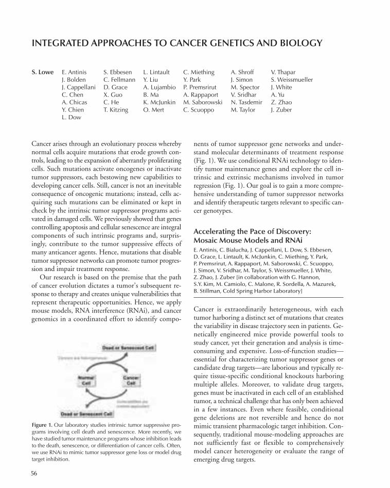
56
Cancer arises through an evolutionary process wherebynormal cells acquire mutations that erode growth con-trols, leading to the expansion of aberrantly proliferatingcells. Such mutations activate oncogenes or inactivatetumor suppressors, each bestowing new capabilities todeveloping cancer cells. Still, cancer is not an inevitableconsequence of oncogenic mutations; instead, cells ac-quiring such mutations can be eliminated or kept incheck by the intrinsic tumor suppressor programs acti-vated in damaged cells. We previously showed that genescontrolling apoptosis and cellular senescence are integralcomponents of such intrinsic programs and, surpris-ingly, contribute to the tumor suppressive effects ofmany anticancer agents. Hence, mutations that disabletumor suppressor networks can promote tumor progres-sion and impair treatment response.
Our research is based on the premise that the pathof cancer evolution dictates a tumor’s subsequent re-sponse to therapy and creates unique vulnerabilities thatrepresent therapeutic opportunities. Hence, we applymouse models, RNA interference (RNAi), and cancergenomics in a coordinated effort to identify compo-
nents of tumor suppressor gene networks and under-stand molecular determinants of treatment response(Fig. 1). We use conditional RNAi technology to iden-tify tumor maintenance genes and explore the cell in-trinsic and extrinsic mechanisms involved in tumorregression (Fig. 1). Our goal is to gain a more compre-hensive understanding of tumor suppressor networksand identify therapeutic targets relevant to specific can-cer genotypes.
Accelerating the Pace of Discovery:Mosaic Mouse Models and RNAiE. Antinis, C. Bialucha, J. Cappellani, L. Dow, S. Ebbesen,D. Grace, L. Lintault, K. McJunkin, C. Miething, Y. Park, P. Premsrirut, A. Rappaport, M. Saborowski, C. Scuoppo, J. Simon, V. Sridhar, M. Taylor, S. Weissmueller, J. White, Z. Zhao, J. Zuber [in collaboration with G. Hannon, S.Y. Kim, M. Camiolo, C. Malone, R. Sordella, A. Mazurek, B. Stillman, Cold Spring Harbor Laboratory]
Cancer is extraordinarily heterogeneous, with eachtumor harboring a distinct set of mutations that createsthe variability in disease trajectory seen in patients. Ge-netically engineered mice provide powerful tools tostudy cancer, yet their generation and analysis is time-consuming and expensive. Loss-of-function studies—essential for characterizing tumor suppressor genes orcandidate drug targets—are laborious and typically re-quire tissue-specific conditional knockouts harboringmultiple alleles. Moreover, to validate drug targets,genes must be inactivated in each cell of an establishedtumor, a technical challenge that has only been achievedin a few instances. Even where feasible, conditionalgene deletions are not reversible and hence do notmimic transient pharmacologic target inhibition. Con-sequently, traditional mouse-modeling approaches arenot sufficiently fast or flexible to comprehensivelymodel cancer heterogeneity or evaluate the range ofemerging drug targets.
INTEGRATED APPROACHES TO CANCER GENETICS AND BIOLOGY
S. Lowe E. Antinis S. Ebbesen L. Lintault C. Miething A. Shroff V. ThaparJ. Bolden C. Fellmann Y. Liu Y. Park J. Simon S. WeissmuellerJ. Cappellani D. Grace A. Lujambio P. Premsrirut M. Spector J. WhiteC. Chen X. Guo B. Ma A. Rappaport V. Sridhar A. YuA. Chicas C. He K. McJunkin M. Saborowski N. Tasdemir Z. ZhaoY. Chien T. Kitzing O. Mert C. Scuoppo M. Taylor J. Zuber L. Dow
Figure 1. Our laboratory studies intrinsic tumor suppressive pro-grams involving cell death and senescence. More recently, wehave studied tumor maintenance programs whose inhibition leadsto the death, senescence, or differentiation of cancer cells. Often,we use RNAi to mimic tumor suppressor gene loss or model drugtarget inhibition.
048-071_CancerGenet_Annual Report_2009 template 7/8/11 11:56 AM Page 56

Cancer: Genetics 57
To address these issues, we developed strategies toaccelerate genetic studies in the mouse. We take a “mo-saic” approach, where cancer-predisposing alleles or re-porters are transduced into stem and progenitor cellsderived from different tissues or mouse strains; thesemodified cells are then transplanted orthotopically intosyngeneic recipients. Because the genetic configurationof the target cells, transduced genes, and recipients iseasily varied, genotypic diversity can be readily pro-duced. To date, we have developed mosaic models oflymphoma and leukemia, as well as carcinomas of theliver, breast, bile duct, and pancreas. Armed with thesemodels, a single investigator can study the impact ofmany cancer genotypes on tumor behavior and rapidlytest new hypotheses based on the results. This versatilityis impossible with germline approaches.
To facilitate loss-of-gene-function studies, weworked with Gregory Hannon to develop inducibleshort hairpin RNA (shRNA) technology as a rapid andreversible alternative to conditional gene deletions. Re-cently, we showed that shRNAs embedded in a naturalmicroRNA (miRNA) structure are effectively expressedfrom polymerase II promoters, thereby enabling robustgene knockdown using constitutive and inducible vec-tors. We then developed effective strategies for suppress-ing proliferation and survival genes where strongselection against shRNA expression creates technicalchallenges (Zuber et al. 2011). We also developed ahigh-throughput miRNA “Sensor” approach to func-tionally identify potent shRNAs (Fellmann et al. 2011).Together with Dr. Hannon, we are developing prevali-dated shRNA libraries.
In vivo RNAi is a powerful tool for studying genefunction in lower organisms such as Caenorhabditis el-egans and Drosophila. To achieve similar capabilities inthe mouse, we initially produced transgenic mice har-boring a tetracycline-inducible p53 shRNA and usedthem to establish a role for p53 loss in tumor mainte-nance. We next developed an efficient and scalable plat-form for mouse production that involves (1) producingpotent shRNAs, (2) using a recombination-mediatedcassette exchange strategy developed by Jaenisch (Mass-achusetts Institute of Technology) to efficiently targetembryonic stem (ES) cells, (3) incorporating an expres-sion cassette that links the tet-responsive shRNA to afluorescent reporter, (4) scaling up ES cell production,and (5) producing germline transgenic mice rapidly bytetraploid embryo complementation (Premsrirut et al.2011).
Using this platform, we are producing hundreds oftargeted ES cell lines and are developing transgenic
strains capable of reversibly suppressing tumor suppres-sors, drug targets, and essential genes (P.K. Premsrirutet al.; K. McJunkin et al.; both in press]. Together withGregory Hannon, we are also using this platform toproduce a series of ES cells capable of inducibly express-ing all known miRNAs, as well as miRNA sensors.These ES cells will be made available as a public re-source to facilitate the study of miRNA biology in vivo.Finally, we are also generating improved tet trans-acti-vator strains that enable potent gene knockdown inmost tissues. Our goal is to develop a general platformthat will enable the spatial, temporal, and reversiblesuppression of any mammalian gene.
Cell Survival and Cell DeathC. Bialucha, Y. Chien, Y. Liu, C. Miething, C. Scuoppo,J. Simon [in collaboration with G. Hannon, Cold SpringHarbor Laboratory; J. Pelletier, McGill University]
Since its inception, our laboratory has studied genesthat modulate cell death and survival. In 1993, wedemonstrated that activated oncogenes could inducep53 to promote apoptosis and suppress transformation.Then, upon joining CSHL, our laboratory character-ized the mechanisms of “oncogene-induced apoptosis.”Some highlights include the identification of ARF as acomponent of oncogene signaling to p53, and severalcell death effectors that mediate p53’s effects. We alsoshowed how phosphoinositol-3 kinase (PI3K) signalingcan attenuate apoptosis and promote tumorigenesis, inpart, via deregulated translation.
Current work in this area exploits the Eµ-myc B-celllymphoma model, where disruption of the ARF-p53pathway, or overexpression of the prosurvival genes Bcl-2 or Akt, cooperates to accelerate lymphomagenesis. Fol-lowing up “hits” from in vivo RNAi screens (see below),we are characterizing new tumor suppressors that mod-ulate apoptosis. For example, we showed that Rad17 actsas a haploinsufficient tumor suppressor that mediates aDNA-damage apoptotic response to replicative stress. Inunpublished work, we identified two tumor suppressorsinvolved in polyamine biology, each of whose suppres-sion attenuates apoptosis and promotes tumorigenesis aspart of a previously obscure tumor suppressor network.Understanding this network and other hits from thisscreen is an important component of future research. Wewill also identify cell survival components that are essen-tial for tumor maintenance. Studies using inducibleshRNA transgenic mice targeting eIF4E and PTEN arealready under way.
048-071_CancerGenet_Annual Report_2009 template 7/8/11 11:56 AM Page 57

58 Research
Senescence and Self-RenewalJ. Bolden, A. Chicas, Y. Chien, A. Lujambio, B. Ma, O. Mert,J. Simon, Z. Zhao, J. Zuber [in collaboration with X. Wang,C. Zhang, M. Zhang, Cold Spring Harbor Laboratory; K. Shannon, S. Kogan, University of California, San Francisco]
Cellular senescence represents an intrinsic tumor sup-pressive program that limits proliferation. In 1997, wediscovered that—in normal cells—oncogenic ras trig-gers a senescence program involving the retinoblastoma(Rb) and p53 tumor suppressor pathways. We went onto identify factors that regulate and execute senescenceand demonstrated an Rb-directed repressive chromatinstate that silences growth promoting genes. Usingmouse models, we also demonstrated that senescencecontributes to the antiproliferative effects of cancertherapy, providing the first evidence that this processlimits tumor progression in vivo. We continue to studythe action of Rb, p53, and chromatin-modifying activ-ities during senescence. By performing a genome-wideanalysis of gene expression, histone modifications, andRb chromatin association, we showed that Rb acts pref-erentially to suppress genes involved in DNA replica-tion, particularly as cells exit the cell cycle intosenescence (Chicas et al. 2010). We believe that thisfunction is crucial for its tumor suppressor role.
We expanded efforts to explore the biology of senes-cence in vivo, recently showing that induction of senes-cence in tumor cells can trigger their clearance by innateimmune cells. These studies reveal a non-cell-au-tonomous program that contributes to p53 action intumor suppression and identifies a novel form of im-mune surveillance involving the innate immune system.We then discovered a role for senescence in limiting liverfibrosis, a tissue-damage response that is a predisposingfactor for cirrhosis and hepatocellular carcinoma. Ourresults revealed the first noncancer pathology for whichsenescence has a protective role, and they imply that itacts to limit certain wound-healing responses. More re-cent studies suggest that the perforin/granzyme B path-way triggered by NK cells contributes to this clearanceand that the NF-κB pathway is a master regulator of thesecretory phenotype. Future studies to further dissectthese and other tumor–host interactions will be greatlyenabled by the use of mosaic models as well as tissue-spe-cific inducible RNAi.
Cells that evade oncogene-induced senescence be-come “immortalized” in a continuous state of self-re-newal. Surprisingly, recent work implies p53 limitsnormal stem cell self-renewal and the reprogramming
of induced pluripotent stem cells. Merging these con-cepts, we showed that p53 loss enables the aberrant self-renewal of Ras-expressing myeloid progenitor cells, thusincreasing their leukemia-initiating potential (Zhao etal. 2010). Together, these observations suggest links be-tween p53 loss, epigenetic reprogramming, and onco-genic transformation. Future work will test whetherp53 inactivation creates a distinct epigenetic state thatmay be targeted therapeutically.
Mechanisms of Drug Resistanceand Tumor MaintenanceC. Chong, C. Miething, A. Rappaport, J. Simon, M. Spector,M. Taylor, Z. Zhao, J. Zuber [in collaboration with C. Vakoc,Cold Spring Harbor Laboratory; S. Kogan, University of Cali-fornia, San Francisco; M. Le Beau, University of Chicago]
Most approved cancer drugs work in only a subset ofcancers, yet even these initially responsive tumors typ-ically develop resistance. Years ago, we showed that p53inactivation could, by disabling apoptosis, produce re-sistance to certain cytotoxic anticancer agents. As thefirst decisive example of how tumor cell genotype couldinfluence treatment outcome, this observation stimu-lated our interest in understanding the interrelationshipbetween cancer development and therapy response.
We continue to identify genes that modulate treat-ment sensitivity, particularly in aggressive cancers. Forexample, we used a lymphoma model to show thattranslational regulators are important determinants ofrapamycin sensitivity and that NF-κB loss causeschemotherapy resistance when senescence is the domi-nant outcome of therapy. We used AML (acute myeloidleukemia) models to study factors that influence high-dose chemotherapy in leukemia and confirmed that p53has a major role in this response. Although some of theseeffects may arise from apoptotic defects, other data sug-gest that p53 loss facilitates the production of so-called“cancer stem cells” considered more refractory to ther-apy (Zhao et al. 2010). We are currently using thesemodels in preclinical studies to test new drugs and drugcombinations.
Beyond resistance mechanisms, our work is also point-ing toward new therapeutic targets. Using inducibleshRNA systems, we are exploring the requirement fortumor suppressor gene inactivation in established tumors.For example, we showed that INK4a/ARF reactivationin aggressive lung carcinomas leads to tumor regressionassociated with enhanced apoptosis (Premsrirut et al.2011). We also showed that Apc loss is required to main-tain the proliferation of T cells in acute lymphoid leu-
048-071_CancerGenet_Annual Report_2009 template 7/8/11 11:56 AM Page 58

Cancer: Genetics 59
kemia, thus validating the Wnt pathway as a therapeutictarget (Premsrirut et al. 2011). Current efforts are study-ing the impact of PTEN restoration in aggressive T-celllymphomas. Future studies will compare these effects topharmacological inhibition of the PI3K pathway.
We are also using RNAi to identify genotype-specificdrug targets. For example, by characterizing how MLL(mixed lineage leukemia) fusion proteins promote self-renewal, we identified the Myb transcription factor asspecifically required for the maintenance of AMLs thatare refractory to conventional chemotherapy. These ob-servations provide direct evidence that targeting aberrantself-renewal programs can eradicate cancer. With Christo-pher Vakoc (CSHL), we screened an shRNA library tar-geting “epigenetic” regulators and have identified severalhistone-modifying activities whose suppression causes theselective arrest or death of leukemic cells. One of these isa target of an available small-molecule inhibitor, and pre-liminary studies indicate that this drug is active againstour AML models and certain human AML lines.
We will continue to apply our mouse modeling andRNAi technologies to identify context-dependent tumormaintenance genes, particularly in those cancers that arerefractory to current therapies. In this regard, we are al-ready producing optimized shRNA libraries targetingrelevant drug targets and developing screening strategiesin vivo. Beyond mosaic models, our shRNA transgenicplatform provides a rapid way to examine the normalfunction of any gene in whole animals as well as the po-tential toxic effects of transient target inhibition.
Integrated Approaches to CancerGene DiscoveryC. Bialucha, C. Chen, S. Ebbesen, C. He, T. Kitzing, Y. Liu, B. Ma, C. Miething, A. Rappaport, M. Saborowski, C.Scuoppo, J. Simon, M. Spector, M. Taylor, S. Weissmueller,Z. Zhao, J. Zuber [in collaboration with X. Wang, NationalInstitutes of Health; A. Kraznitz, M. Wigler, J. Hicks, G. Hannon, S. Powers, Cold Spring Harbor Laboratory]
Cancer genomes are complex and can harbor cancer-pro-moting “driver” mutations together with “passenger” mu-tations that have no biological effect. To accelerate cancergene discovery, we use mosaic mouse models to filterthrough candidates obtained through genomics to iden-tify genes that actively contribute to tumorigenesis. Wepresume that (1) recurrent amplifications and deletionsin human tumors are enriched for oncogenes and tumorsuppressors, respectively, and (2) lesions that give rise tocancer in humans often do so in mice. Since 2006, can-didate testing has identified the cell death inhibitor
cIAP1, the hippo pathway gene Yap, the eIF4E kinaseMNK, and the chromobox protein CBX7 as potentoncogenes, as well as the Rho-GAP DLC1 as a tumorsuppressor, without producing a single germline trans-genic or knockout strain.
We developed strategies to multiplex this approachby transducing tissue stem and progenitor cells withpools of cDNAs or shRNAs corresponding to genesthat are amplified or deleted in human tumors and se-lected for those constructs that promote tumorigenesisfollowing transplantation into recipient mice. Initial ef-forts surveyed nearly 400 recurrently deleted genes inhuman hepatocellular carcinoma (HCC) and identified12 new tumor suppressor genes. In a conceptually sim-ilar screen performed by Scott Powers using full-lengthcDNAs (CSHL), we identified 10 new oncogenes inhuman HCC, including FGF19 as a therapeutic targetfor existing neutralizing antibodies (Sawey et al. 2011).
In parallel to these screening efforts, we are perform-ing our own genomics analysis of human and murinecancers to facilitate functional studies in mice. Suchstudies have already pinpointed candidate tumor sup-pressors in aggressive forms of AML. Validation effortsare currently under way.
Through our screening approaches, we have notedthat recurrent gene amplifications and gene deletions intumors frequently harbor multiple drivers genes. For ex-ample, in a screen for additional tumor suppressors cor-responding on human chromosome 8p—a frequent siteof large heterozygous deletions in human cancers—weidentified several new tumor suppressors that cooperatewith DLC1 suppression to promote tumorigenesis.Moreover, in a second-generation shRNA screen usingthe Eµ-myc model described above, we used shRNApools targeting genes deleted in human B-cell lymphomato identify ~10 tumor suppressors. Surprisingly, fourtumor suppressors corresponded to genes on humanchromosome 6p, two on chromosome 8p, and twolinked to p53 on 17p. These observations suggest thatthe impact of cancer-associated deletions provide agreater selective advantage than single gene mutationsand should be viewed and studied as distinct events.
Beyond identifying new activities of cancer relevance,our approach is revealing unexpected principles aboutthe nature and organization of cancer genes. As exam-ples, we were surprised that so many tumor suppressorsare haploinsufficient, encode secreted proteins, or havepro-oncogenic activities in other contexts. We also didnot expect recurrent amplifications and deletions to con-tain more than one relevant activity, yet our resultsimply this is the rule rather than the exception. We hope
048-071_CancerGenet_Annual Report_2009 template 7/8/11 11:56 AM Page 59

60 Research
that further efforts will facilitate the functional annota-tion of the genomic alterations occurring in human can-cers and identify vulnerabilities these lesions create.
PUBLICATIONS
Bazarov A, Van Sluis M, Hines WC, Bassett E, Beliveau A, Campeau E,Mukhopadhyay R, Lee WJ, Melodyev S, Zaslavsky Y, et al. 2010.P16(Ink4a)-mediated suppression of telomerase in normal and ma-lignant human breast cells. Aging Cell 9: 736–746.
Cancer Target Discovery and Development Network. Schreiber SL,Shamji AF, Clemons PA, Hon C, Koehler AN, Munoz B, Palmer M,Stern AM, Wagner BK, Powers S, et al. 2010. Towards patient-basedcancer therapeutics. Nat Biotech 28: 904–906.
Chicas A, Wang X, Zhang C, McCurrach M, Zhao Z, Mert O, DickinsRA, Narita M, Zhang M, Lowe S. 2010. Dissecting the unique roleof the retinoblastoma tumor suppressor during cellular senescence.Cancer Cell 17: 376–387.
Heyer J, Kwong LN, Lowe SW, Chin L. 2010. Non-germline geneticallyengineered mouse models for translational cancer research. Nat RevCancer 10: 470–480.
Jones L, Wei G, Sevcikova S, Phan V, Jain S, Shieh A, Wong JC, Li M,Dubansky J, Maunakea ML, et al. 2010. Gain of MYC underlies re-current trisomy of the MYC chromosome in acute promyelocyticleukemia. J Exp Med 207: 2581–2594.
Karni-Schmidt O, Castillo-Martin M, HuaiShen T, Gladoun N,Domingo-Domenech J, Sanchez-Carbayo M, Li Y, Lowe SW, PrivesC, Cordon-Cardo C. 2010. Distinct expression profiles of p63 vari-ants during urothelial development and bladder cancer progression.Am J Pathol 178: 1350–1360.
Ko FC, Chan LK, Tung EK, Lowe SW, Ng IO, Yam JW. 2010. Aktphosphorylation of deleted in liver cancer 1 abrogates its suppressionof liver cancer tumorigenesis and metastasis. Gastroenterology 139:1397–1407.
Pineau P, Volinia S, McJunkin K, Marchio A, Battiston C, Terris B, Maz-zaferro V, Lowe SW, Croce CM, Dejean A. 2010. miR-221 overex-
pression contributes to liver tumorigenesis. Proc Natl Acad Sci 107:264–269.
Sendoel A, Kohler I, Fellmann C, Lowe SW, Hengartner MO. 2010.HIF-1 antagonizes p53-mediated apoptosis through a secreted neu-ronal tyrosinase. Nature 465: 577–583.
Xu MZ, Chan SW, Liu AM, Wong KF, Fan ST, Chen J, Poon RT, Zen-der L, Lowe SW, Hong W, et al. 2010. AXL receptor kinase is a me-diator of YAP-dependent oncogenic functions in hepatocellularcarcinoma. Oncogene 30: 1229–1240.
Zhao Z, Zuber J, Diaz-Flores E, Lintault L, Kogan SC, Shannon K,Lowe SW. 2010. p53 loss promotes acute myeloid leukemia by en-abling aberrant self-renewal. Genes Dev 24: 1389–1402.
In Press
Fellmann C, Zuber J, Mcjunkin K, Chang K, Dickins RA, Xu Q, ElledgeSJ, Hannon GJ, Lowe SW. 2011. Functional identification of opti-mized RNAi triggers using a massively parallel Sensor assay. Mol Cell(in press).
McJunkin K, Mazurek A, Premsrirut PK, Zuber J, Dow LE, Simon J,Stillman B, Lowe SW. 2011. Reversible suppression of an essentialgene in adult mice using transgenic RNA interference. Proc NatlAcad Sci (in press).
Pardee TS, Zuber J, Lowe SW. 2011. Flt3-ITD alters chemotherapy re-sponse in vitro and in vivo in a p53-dependent manner. Exp Hematol39: 473–485.
Premsrirut PK, Dow LE, Kim SY, Malone CD, Miething C, ScuoppoC, Zuber J, Dickins RA, Kogan SC, Shroyer KR, et al. 2011. A rapidand scalable system for studying gene function in mice using condi-tional RNA interference. Cell 145: 145–158.
Sawey E, Cai C, Chanrion M, Wu G, Wang T, Zhang J, Zender L, YeeH, Stein L, French DM, et al. 2011. Identification of a therapeuticstrategy targeting amplified FGF19 in liver cancer by oncogenomicscreening. Cancer Cell 19: 347–358.
Zuber J, McJunkin K, Fellmann C, Dow LE, Taylor MJ, Hannon GJ,Lowe SW. 2011. Toolkit for evaluating genes required for prolifera-tion and survival using tetracycline-regulated RNAi. Nat Biotechnol29: 79–83.
048-071_CancerGenet_Annual Report_2009 template 7/8/11 11:56 AM Page 60
61
In addition to genetic mutation, such as amplificationand deletion, epigenetic aberrations are frequent eventsthat can have far-reaching affects in the phenotype of acancer cell. A common epigenetic modification is themethylation of cytosine residues that are next to gua-nine residues. It has been shown that methylation pres-ent in the DNA of the transcriptional control regionhas been involved in the silencing of gene expression oftumor supressors in cancer. We have previously adaptedROMA (representational oligonucleotide microarrayanalysis) to methylation detection oligonucleotide mi-croarray analysis (MOMA). This methodology was use-ful and allowed us to identify genes that weremethylated in the tumor cells, but it only gave us a gen-eral idea of where in the CpG island the methylationhad occurred. In addition, after performing the analysis,sequencing validation was required to determine whichof the array measurements accurately identified methyl-ated fragments. With the development of next-genera-tion sequencing technologies, it is now possible toidentify the methylation status for the vast majority ofCpG islands. Our approach is to sequence MspI repre-sentations of the genome. We have developed severaldifferent approaches to generate the libraries for se-quencing and have decided to prepare Illumina librariesfrom bisulfite-treated MspI representations.
We have presently sequenced three tumors and two celllines. We have determined the coverage and the numberof sequences required to obtain appropriate coverage ofmany of the CpG islands and have developed methods tocarry out the analysis on the sequencing data. Mappingthe reads to the genome is not trivial because the bisulfitetreatment essentially changes the genome sequence to onlythree bases due to the conversion of most cytosines tothymine. We have used a program developed by AndrewSmith (formerly of CSHL) to map the reads to an in silicobisulfite-treated human genome. After mapping the reads,the sequences that map to CpG islands are separated fromother sequences, which represents ~20% of the reads.These reads are contigged using the program Velvet toform larger regions for analysis. The data can now be an-alyzed to determine gene-specific methylation states.
We have thus far used these data to identify whichgenes are methylated in the tumor samples. More im-portantly, we have expression data for these samples andwill use these data to determine which region of theCpG island is methylated for genes that have suppres-sion of transcription. In addition, it will be interestingto determine the effect of methylation of island se-quences within the coding region of the gene. We planon increasing the number of samples for two reasons.We will be analyzing a new set of ovarian tumor sam-ples from our collaborator Dr. Douglas Levine fromMemorial Sloan-Kettering Cancer Center. The datafrom these samples will serve as an independent valida-tion of the genes we have found to be methylated fromour MOMA results (see below). These data will alsogive us a much more accurate view of the regionsmethylated and the purpose of this methylation withrespect to gene expression.
Genomic Analysis of Ovarian CancerE. Lum, M. Vigliotti, K. Wrzeszczynski, N. Cutter,N. Dimitrova
In the United States, there will be ~22,000 new casesof ovarian cancer in 2011. Of these cases, ~14,000 willsuccumb to the disease. To better treat these women,and improve survival, our goal is to determine the mo-lecular changes that have occurred in the patients’ tu-mors and be able to interpret the significance thesechanges have on the growth and development of thetumor. This aberrant growth is a result of chromosomalabnormalities and epigenetic variations. In addition,generally low rates of somatic nucleotide mutation inovarian cancer as compared to other solid tumors sug-gest an increased significance of copy number and epi-genetic aberrations. This type of regulation has beenshown to effect the many tumor suppressors and onco-genes discovered in ovarian cancer. The identificationof genetic and epigenetic alterations from primarytumor cells has become a common method to identifygenes critical to the development and progression of
REPRESENTATION METHYLATION SEQUENCING ANALYSIS
R. Lucito N. Cutter S. PowersN. Dimitrova M. VigliottiE. Lum K. Wrzeszczynski
048-071_CancerGenet_Annual Report_2009 template 7/8/11 11:56 AM Page 61

62 Research
cancer. We provide a bioinformatic analysis of copy-number variation and DNA methylation covering thegenetic landscape of ovarian cancer tumor cells. We in-dividually examined the copy-number variation andDNA methylation for 42 primary serous ovarian cancersamples using our MOMA-ROMA technology and379 tumor samples analyzed by The Cancer GenomeAtlas. We have identified 346 genes with significantdeletions or amplifications among the tumor samples.Utilizing associated gene expression data, we predict156 genes with significantly altered copy number andcorrelated changes in expression. Among these genes,CCNE1, POP4, UQCRB, PHF20L1, and C19orf2 wereidentified within both data sets. We were specificallyinterested in copy-number variation as our base ge-nomic property in the prediction of tumor suppressorsand oncogenes in the altered ovarian tumor. We there-fore identified changes in DNA methylation and ex-pression for all amplified and deleted genes. Wepredicted 615 potential oncogenes and tumor suppres-sors candidates by integrating these multiple genomicand epigenetic data types. Genes with a strong correla-tion for methylation-dependent expression exhibited atvarying copy-number aberrations include CDCA8,ATAD2, CDKN2A, RAB25, AURKA, BOP1, andEIF2C3. Moreover, well-characterized tumor suppres-sors and oncogenes exhibit varying copy-number vari-ations, methylation, and expression features withincancers. We therefore compared significant copy-num-ber variation and methylation effects on expression formany known tumor suppressors, oncogenes, and can-cer-related genes in order to identify altered gene func-tion properties specific to primary ovarian tumors. Weanalyzed multiple genomic modalities from two pri-mary ovarian tumor data sets for the identification ofcandidate genes. We predict genes with tumor suppres-sor and oncogenic properties, identifying new serousovarian cancer candidate genes.
Resistance Mechanismsin Ovarian CancerN. Cutter, M. Vigliotti, K. Wrzeszczynski
Epithelial ovarian cancer is the leading cause of deathfrom gynecological malignancies. Currently, platinum-based chemotherapy (such as cisplatin or carboplatin),coupled with a taxane-based drug such as paclitaxel, isthe primary treatment for ovarian cancer. Approximately25% of patients either present with or rapidly developresistance to chemotherapy, and all recurrent tumors are
resistant. Epigenetic modifications have been associatedwith tumor formation and progression and may con-tribute to therapy response. We have screened a numberof genes and family members for methylation in resistantpatients and not in sensitive patients. We show for onesuch gene, CHD3, a member of the Mi-2 NuRD com-plex, that loss of expression is linked to chemoresistance.CHD3 is silenced through an epigenetic mechanism inboth ovarian cancer cell lines and primary ovarian tu-mors. When ovarian cancer cell lines that are transcrip-tionally silenced for CHD3 are challenged withcarboplatin, they display a striking decrease in growth.Additionally, these cells are more invasive, have migra-tory ability, and display a transformed epithelial-to-mes-enchymal (EMT) phenotype. Moreover, our resultsindicate that cells silenced for CHD3 have an increasedresistance to the chemotherapy drugs carboplatin andcisplatin. Taken together, this provides the first evidencefor a role for CHD3 as an important mediator of chemo-resistance in ovarian cancer, and we hypothesize thatEMT is one of the underlying mechanisms. Further-more, CHD3 might represent a response predictor andpotential therapeutic target for predicting chemoresis-tance in this disease.
Genomic Alterations of PhosphatasesE. Lum, F. Chaudhary, G. Fan [in collaboration withN. Tonks, Cold Spring Harbor Laboratory]
The goal of this collaborative study is to integrate genediscovery technology with experimental strategies devel-oped in Dr. Tonks’ lab for the characterization of the pro-tein tyrosine phosphatase (PTP) family of enzymes, toinvestigate how tyrosine phosphorylation-dependent sig-naling pathways are disrupted in cancer. We have takenadvantage of several different forms of genomic data todetermine which PTPs are affected in cancer cells.
We have used genomic copy-number data, expres-sion data, and now epigenetic DNA methylation datato determine which PTPs are altered in ovarian cancer.We have also expanded our extension of analysis ofPTPs in cancer into the proteins with which PTPs in-teract. An interesting gene in this class encodes theMTSS1 protein that interacts with a PTP, the receptor-like PTP RPTPδ, and regulates cytoskeletal organiza-tion. It is known to be preferentially methylated inseveral cancers, including breast cancer, and its expres-sion is markedly decreased in ovarian cancer.
We first generated short hairpin RNAs (shRNAs) tosuppress expression of the MTSS1 gene in cell culture as-
048-071_CancerGenet_Annual Report_2009 template 7/8/11 11:56 AM Page 62

Cancer: Genetics 63
says, which would mimic the transcriptional repressioncaused by promoter methylation. Because this gene wasselected based only on the comparison of tumor to nor-mal, we did not have a selection for assay development.We surmised that because MTSS1 possessed an actin-binding domain, the protein might have a role in cy-toskeletal rearrangement, cell movement, and invasion.Therefore, we performed an invasion assay with MCF10Amammary epithelial cells, which are used typically as anormal line. We observed that very few cells migrate with-out the addition of the attractant epidermal growth factor(EGF). Furthermore, RNA interference (RNAi)-mediatedsuppression of MTSS1 enhanced migration in the pres-
ence of EGF. There is evidence in the literature that inERBB2-positive breast cancer, there is loss of MTSS1 tran-scription. To determine whether ERBB2 overexpressionsynergizes with loss of MTSS1, we performed an invasionassay in MCF10AN cells. These cells express a chimericERBB2 gene in which the activity of the cytoplasmic PTKdomain of ERBB2 is acutely regulated by a small-moleculedimerizing agent. We noted that the level of invasion ishighest in the cells in which MTSS1 was suppressed andERBB2 activated, demonstrating cooperation betweenthese two gene products. Our current data indicate thatthese effects are mediated via changes in tyrosine phos-phorylation regulated by RPTδ.
Michele Vigliotti
048-071_CancerGenet_Annual Report_2009 template 7/8/11 11:56 AM Page 63

64
The Mills laboratory generates novel mouse models anduses them to identify the genes responsible for humandiseases of interest and to elucidate the mechanismwhereby the encoded proteins regulate the diseaseprocess, with the ultimate goal of applying these find-ings to the human disease under study. These ap-proaches have provided significant insight intodevelopmental syndromes, aging, and cancer. Researchareas include (1) the role of CHD5 in chromatin dy-namics and cancer and (2) function of the p53 ho-molog p63 in development, cancer, and aging.
CHD5 in Chromatin Dynamicsand Cancer
CHD5: A new tumor suppressor mapping to human1p36. CHD5 maps to 1p36, a region of the genomethat has a three-decade history as being deleted in awide variety of human cancers. Despite the rich bodyof evidence indicating that one or more tumor suppres-sors reside in this region, the causative gene had re-mained elusive. By generating mice with gain and lossof the genomic region corresponding to 1p36 usingchromosome engineering technology—a strategy thatallows us to generate precise rearrangements in themouse—we pinpointed a region of the genome withpotent tumor suppressive activity (Bagchi et al., Cell128: 459–475 [2007]; Bagchi and Mills, Cancer Res 68:2551–2556 [2008]). Using a series of genetic and mo-lecular approaches, we identified Chd5 as the tumorsuppressor gene in the region and found that its prod-uct was a master switch for a tumor suppressive net-work. In addition, we discovered that CHD5 wasfrequently deleted in human glioma. Identification ofCHD5 as a tumor suppressor had a major impact inthe cancer field, as it is now known that CHD5 is mu-tated in human cancers of the breast, ovary, andprostate, as well as in melanoma and neuroblastoma,and that CHD5 status correlates directly with patientsurvival following anticancer therapy.
Chd5 modulates chromatin dynamics and cancer.Given the critical importance of CHD5 in human can-cer, we focused on elucidating its mechanism of tumorsuppression. From our earlier work, we know thatChd5 transcriptionally activates the Ink4/Arf locus,thereby facilitating expression of p16 and p19, twotumor suppressors encoded by this locus (Bagchi et al.,Cell 128: 459–475 [2007]). Compromised Chd5 ac-tivity, either by engineered heterozygous deletion of theinterval encompassing Chd5 or by specific depletion ofChd5 using RNA interference (RNAi), cripples thetumor suppressive network, compromising p16/Rb-and p19/p53-mediated pathways, predisposing to can-cer (Mills 2010). Chd5 is a predicted chromatin re-modeling protein, consistent with the idea that itregulates Ink4/Arf by maintaining this locus in a tran-scriptionally competent chromatin state.
Within the past year, we discovered that Chd5’stumor suppressive capability was dependent on its abil-ity to bind amino-terminal tails of histone 3 (H3) thatare unmodified, an activity mediated by the dual planthomeodomains (PHDs) of Chd5. Modeling provideda testable explanation for this Chd5-H3 interaction,and indeed, point mutations within the PHDs effec-tively abolished H3 binding, abrogated the ability ofChd5 to inhibit proliferation, and led to tumorigenesisin vivo. In line with the findings from in vitro assays,global chromatin immunoprecipitation sequencing(ChIP-Seq) analyses indicate that Chd5-bound locitended to have less H3K4Me3 in vivo. These findingsprovide mechanistic insight into Chd5’s ability to mod-ulate chromatin dynamics and cancer.
Chd5-deficient mice are cancer prone. Althoughthe chromosome engineered mouse models were in-valuable for functionally pinpointing the tumor sup-pressive interval, the region affected was 4.3 Mb andcontained 52 annotated genes. To unequivocallydemonstrate the role of Chd5 in the context of sponta-neous tumorigenesis, we generated mice deficient forChd5 and monitored for tumor development. Chd5–/–
MAMMALIAN FUNCTIONAL GENOMICS
A. Mills Y. Chang S. PaulG. Horev Y.-E. SonD.-W. Hwang E. Vernersson-LindahlW. Li A. Vestin
048-071_CancerGenet_Annual Report_2009 template 7/8/11 11:56 AM Page 64

Cancer: Genetics 65
mice were viable, yet like we had found for mice het-erozygous for the 52-gene interval encompassing Chd5,Chd5+/– mice were prone to spontaneous tumors suchas carcinoma, sarcoma, and lymphoma. We are cur-rently analyzing these models to provide a better un-derstanding of the biological role of CHD5 in vivo andto assess how its perturbation affects chromatin dynam-ics that precludes to tumorigenesis. This work shouldprovide the groundwork for developing more effectiveanticancer therapies in the future.
Function of the p53 Homolog p63in Development, Aging, and Cancer
p63 is essential for development. A long-standing focusof the laboratory has been to elucidate the in vivo roleof p63—a p53-related protein that we discovered to bea new member of the p53 family. With the advent ofthe p53 family of proteins, the leading question in thecancer field was whether p63 (and its sibling p73) hadtumor suppressive capabilities similar to p53. Using ge-nomic technologies that we were just in the process ofdeveloping, we generated p63-deficient mouse modelsand discovered that p63 was essential for developmentof the limbs and stratified epithelia (Mills et al., Nature398: 708–713 [1995]). This demonstrated that eventhough p63 had significant homology with p53, it haddistinct biological roles in vivo. These mouse modelsprovided a clue that revealed that p63 mutations wereresponsible for seven different human developmentaldisease syndromes. Because some of these syndrome-re-lated p63 mutations correlated precisely with p53 hot-spot mutations found in human cancer, we used agene-targeted knockin to generate mouse models carry-ing these missense mutations, allowing us to recapitulatethe human disease as well as to establish a link betweenthis syndrome and cellular senescence.
p63 modulates aging and cancer. During our studiesaimed at determining whether p63 had the same tumorsuppressive capabilities as p53, we uncovered an unex-pected link between p63, cellular senescence, and aging(Keyes et al., Genes Dev 19: 1986–1999 [2005]). Theconditional mouse models we established were the firstto implicate p63 as a modulator of cellular senescence,as well as the first to uncover a link between cellularsenescence and organismal aging in vivo. Whereas p53was known as an inducer of senescence, we found thatp63 deficiency triggered this process. Our realization that
p63 compromised mice were not prone to spontaneousor chemically induced tumors suggested that the abilityof p63 loss to trigger senescence provided a robust tumorprotective mechanism that could be exploited therapeu-tically. Indeed, we recently reported that TAp63 isoformsare potent tumor suppressors that induce senescence andshut down growth of p53-deficient/Ras-driven tumorsin vivo (Guo et al., Nat Cell Biol 11: 1451–1457[2009]). Using chromosome engineering, we establishedconditional mouse models in which we could specificallyablate TAp63 isoforms while keeping ∆Np63 isoformsintact, allowing us to demonstrate that TAp63 isoformsprevent sarcoma development in vivo. In contrast to thistumor suppressive role of TAp63 in mesenchymal cells,we discovered that in the context of epithelial cells,∆Np63α bypasses senescence, thereby promoting stem-like proliferation and carcinoma development in vivo(Keyes et al. 2011). We found that ∆Np63α maintainsthe keratin-15-positive stem cell population, promotingsenescence bypass and carcinogenesis by inducing ex-pression of the SNF2-like chromatin remodeler Lsh.These findings provide an explanation for ∆Np63αoverexpression being such a frequent and highly pene-trant event in human carcinoma. In addition, this worksuggests that transformation of the keratin-15-expressingstem population lies at the heart of carcinoma—themost prevalent type of human cancer. Thus, our workidentifies p63 as a key modulator of cellular senescence,with TAp63 isoforms being tumor suppressors that pre-vent sarcoma, and ∆Np63α being an oncogene thatdrives carcinoma development. This not only highlightsthe distinct tissue-specific roles of p63 isoforms; ourwork provides an explanation for the “tumor suppressiveversus oncogenic” debate that has surrounded this p53family member since its discovery. The double-edgedtumor suppressive/oncogenic picture that has emergedfor p63 is now being extended to p53 itself, as the tis-sue-specific roles of the multiple p53 isoforms in cancerare just beginning to be appreciated.
PUBLICATIONS
Mills AA. 2010. Throwing the cancer switch: Reciprocal roles of Polycomband Trithorax proteins. Nat Rev Cancer 10: 669–682.
In Press
Keyes WM, Pecoraro M, Aranda V, Vernersson-Lindahl E, Li W, VogelH, Guo X, Garcia EM, Michurina TV, Enikolopov G, et al. 2011.δNp63α is an oncogene that targets chromatin remodeler Lsh to driveskin stem cell proliferation and tumorigenesis. Cell Stem Cell 8: 164–176.
048-071_CancerGenet_Annual Report_2009 template 7/8/11 11:56 AM Page 65

66
Our laboratory studies cancer in order to find out moreabout its basic causes but also to contribute directly toits treatment. Our primary focus has been the identifi-cation and functional validation of genetically altereddriver genes (oncogenes) that together with geneticallyaltered tumor suppressor genes are the cause of the can-cer. Our approach is always to study the process fromas wide a perspective as possible, which means that weconsider every gene in the genome.
Identification of Driver Genes and Therapeutic Targets inHepatocellular Carcinoma E. Sawey, M. Chanrion, J. Li, A. Mofunanya [in collaboration with S. Lowe, Cold Spring HarborLaboratory; R. Finn, University of California, Los Angeles; D. Chiang, University of North Carolina; D. French, Genentech]
Hepatocellular carcinoma (HCC) afflicts more than560,000 people worldwide each year and has one of theworst 1-year survival rates of any cancer type. Currently,there are no molecular therapies that target specific mu-tations or other genetic alterations in HCC. By per-forming a forward-genetic screen guided by genomicanalysis of human HCC, we identified 18 tumor-pro-moting genes, including CCND1 and its neighbor on11q13.3, FGF19. Although it is widely assumed thatCCND1 is the main driving oncogene of this commonamplicon (15% frequency in HCC), both forward-transformation assays and RNA interference (RNAi)-mediated inhibition in human HCC cells establishedthat FGF19 is an equally important driver gene inHCC. Furthermore, clonal growth and tumorigenicityof HCC cells harboring the 11q13.3 amplicon were se-lectively inhibited by RNAi-mediated knockdown ofCCND1 or FGF19, as well as by an anti-FGF19 anti-body. These results show that 11q13.3 amplificationcould be an effective biomarker for patients most likelyto respond to anti-FGF19 therapy. Our study under-scores the potential for clinical translation of results ob-
tained from genetic screens guided by cancer genomeanalysis.
This year, we completed a pilot program to find mu-tations in the exome of primary HHCs, with the pri-mary goal of finding recurrent oncogenes. On the basisof our analysis in collaboration with Derek Chiang atthe University of North Carolina, CTNNB1 is the mostcommonly mutated oncogene in HCC, and the rest ap-pear to be low-frequency events, similar to what hasbeen found in other epithelial cancers.
Of course, the best targets for treating HCC do notnecessarily have to be the driver oncogenes. In principle,other alterations that occur during HCC progressioncould cause certain tumor dependencies that would notbe found in normal liver cells. To find these, A. Mofu-nanya has been screening human HCC cells for tumordependencies using genome-wide RNAi screens underconditions of hypoxia to more closely mimic the phys-iological state of tumor cells growing in vivo.
Identification of Driver Genesand Therapeutic Targets inOvarian CarcinomaE. Sawey, M. Gallo, R. Gerdes, J. Marchica, J. Li [in collaboration with A. Krasnitz, S. Lowe, Cold Spring Harbor Laboratory; K. Cho, University of Michigan; S. Orsulic, Cedars-Sinai; NCI Cancer Target Discovery and Development Network (CTD2)]
A group of scientists here at CSHL received fundingfrom the National Cancer Institute (NCI) for a pilotproject in translating genomic information emanatingfrom cancer genome projects such as TCGA into ther-apeutic targets. The strategy of the CSHL CTD2 groupfor discovering and developing targets incorporates com-putational analysis of cancer genomes with oncoge-nomic screening and mouse modeling. This year, wehave analyzed more than 500 ovarian cancer (high-gradeserous) genomes and found an amazing degree of DNAcopy-number instability. Through an innovative ap-proach to make sense of this chaos, Alex Krasnitz found
CANCER GENES
S. Powers M. Chanrion J. Li M. Rajaram M. SuC. Eifert J. Marchica K. Revill P. TelloM. Gallo A. Mofunanya E. SaweyR. Gerdes H. Qi S. Sayers
048-071_CancerGenet_Annual Report_2009 template 7/8/11 11:56 AM Page 66

Cancer: Genetics 67
the most consistently recurring alterations, and basedon this analysis, we screened more than 100 of the af-fected genes in a transplantable mouse model for ovariancancer and discovered that 24 of these can promote tu-morigenicity. Although this is just the beginning, ShirleyGuo in Scott Lowe’s group has begun application of the“speedy” mouse model to determine the suitability of asubset of these oncogenes as therapeutic targets.
Studies of the DNA Methylome of HCCK. Revill [in collaboration with R. Finn, University of California, Los Angeles; T. Wang, Massachusetts Institute of Technology]
Recent advances in whole-genome methylation profiling,specifically the Illumina Infinium platform, have allowedus to investigate the methylation status of a cohort ofmore than 50 HCC and normal liver samples for whichwe also investigated the expression and gene copy num-ber (CGH) status. We have been able to identify two verydistinct groups of HCCs based on their methylation sta-tus. In each class of tumors, there are both hypo- and hy-permethylated genes relative to normal liver. Several ofthese tumor-specific alterations have been validated bypyrosequencing. In one class of tumors, many genes thatare normally repressed by polycomb group proteins instem cells are hypermethylated, consistent with thetumor cell of origin being a progenitor stem cell. Theother class of tumors does not show this association andlikely originate from a different type of normal cell. In-triguingly, these two classes also correspond to distinctgroups based on both DNA copy-number analysis andexpression analysis. Combining these three independentmethods of global analysis—transcriptome profiling,epigenome analysis, and aCGH data—has provided arobust new classification scheme for HCC.
Identification of Sensitizersto PI3K InhibitorsC. Eifert [in collaboration with R. Wooster, GlaxoSmithKline]
We previously reported on using Greg Hannon’s RNAilibrary to screen for sensitizers to a polo-like kinase in-hibitor and through that discovered that retinoic acidsensitizes lung cancer cells to this mode of inhibition.This year, we have looked at genes that if inhibited, to-gether with partial inhibition of phosphoinositol-3-ki-nase (PI3K), could enhance killing of cancer cells. Thusfar, we have validated another enzyme upstream of the
PI3-biosynthetic pathway, as well as a relatively under-studied glucose transporter.
Identification of Oncogenic Determinantsin the Tumor Cell of OriginM. Chanrion, E. Sawey
The notion that the phenotype of individual human tu-mors results from its own specific set of accumulatedmutated oncogenes and tumor suppressor genes formsthe major framework behind many studies into the un-derlying causes of specific cancer phenotypes. In con-trast, it is rarely addressed whether the differentiatedstate of the normal cell of origin has a role in the phe-notypic behavior of tumor cells following their acquisi-tion of mutated cancer genes, despite examples where itclearly has profound effects. In fact, certain molecularsubclasses of HCCs are likely to reflect different cells oforigin, but the genes that underlie their different malig-nant properties are poorly understood. We developed anapproach to find oncogenic determinants within tumorcells of origin that draws on pathway and functionalanalysis of different cells of origin in a mouse model in-tegrated with parallel analysis of human HCC. Usingthis approach, we identified higher levels of RXRα indifferentiated mouse hepatocytes and its associated sub-class of human HCC as a potential oncogenic determi-nant. This was confirmed by demonstrating thatRNAi-mediated silencing of RXRα in hepatocytes con-fers immature hepatoblast-like properties to their sus-ceptibility to malignant transformation, consistent withits role in coordinating nuclear receptors involved ingrowth and differentiation. We also found that more dif-ferentiated HCC cells expressing higher levels of RXRαare the only type that can be inhibited by retinoids,which were previously tried on HCC patients indiscrim-inately. These results demonstrate the potential of thisunbiased genomic approach to discover the genes un-derlying differential malignant properties of distincttumor cells of origin and furthermore that such genescan be critical determinants of treatment response.
Identification and Functional Analysisof Genes Involved in Breast Cancer–Stromal Fibroblast InteractionsM. Rajaram [in collaboration with M. Egeblad, Cold SpringHarbor Laboratory]
Although some molecules that mediate the protumori-genic interaction of stromal fibroblasts and cancer cells
048-071_CancerGenet_Annual Report_2009 template 7/8/11 11:56 AM Page 67

68 Research
have been identified, there is still uncertainty about thenature and scope of all relevant interactions. To addressthis issue, we developed a model for breast cancer—stro-mal fibroblast interaction using coculture and coxeno-grafting of human breast cancer cell lines with fibroblaststhat stimulate tumorigenic growth (productive fibrob-lasts) or other fibroblasts that do not (nonproductive fi-broblasts). By transcriptome profiling and pathwayanalysis, we found that the signaling pathways activatedspecifically in productive fibroblasts when coculturedwith tumor cells overlapped significantly with pathwaysactivated in the stroma of human breast cancer samplesrelative to stroma from normal breast tissue. Pathwaysthat were up-regulated in both our model system and inhuman breast cancer stroma included extracellular ma-trix interactions, cytokine–cytokine receptor interac-tions, and integrin signaling. Of note was that not onlywere a large number of ligands and receptors up-regu-lated in productive fibroblasts, but their reciprocal lig-ands/receptors were also up-regulated in the coculturedbreast cancer cells. Because this analysis pinpointed alarge number of potentially important up-regulatedgenes, we used short hairpin RNA (shRNA) knockdownto test for their functional relevance in promotingtumor–stromal cooperativity in vivo. For example, theability of productive fibroblasts containing stableshRNAs toward CCL2 to cooperate with breast cancer
cell lines in a coxenograft model was significantly dimin-ished. Furthermore, a similar effect could be achievedby silencing one of its receptors—CCR1 on the breastcancer cells. Our results indicate that a productive in-teraction may be mediated by several different genes be-longing to diverse pathways acting together to promotetumorigenesis.
PUBLICATIONS
Degenhardt Y, Greshock J, Laquerre S, Gilmartin AG, Jing J, RichterM, Zhang X, Bleam M, Halsey W, Hughes A, et al. 2010. Sensitivityof cancer cells to Plk1 inhibitor GSK461364A is associated with lossof p53 function and chromosome instability. Mol Cancer Ther 9:2079–2089.
Oliver TG, Mercer KL, Sayles LC, Burke JR, Mendus D, Lovejoy KS,Cheng MH, Subramanian A, Mu D, Powers S, et al. 2010. Chroniccisplatin treatment promotes enhanced damage repair and tumorprogression in a mouse model of lung cancer. Genes Dev 24: 837–852.
Schreiber SL, Shamji AF, Clemons PA, Hon C, Koehler AN, Munoz B,Palmer M, Stern AM, Wagner BK, Powers S, et al. 2010. Towardspatient-based cancer therapeutics. Nat Biotechnol 28: 904–906.
In Press
Sawey ET, Chanrion M, Cai C, Wu G, Zhang J, Zender L, Zhao A,Busuttil RW, Yee H, Stein L, French DM, Finn RS, Lowe SW, Pow-ers S. 2011. Identification of a therapeutic strategy targeting ampli-fied FGF19 in liver cancer by oncogenomic screening. Cancer Cell(in press).
A. Mofunanya
048-071_CancerGenet_Annual Report_2009 template 7/8/11 11:56 AM Page 68
69
Our group uses methods for comparative genome analy-sis to study cancer and human genetic disorders. Newmutations, somatic and germline, underlie the cancersand human genetic disorders. Our main focus to datehas been studying mutations that change the numbersof copies of sections of the genome, causing regions ofdeletion and duplication, in cancer and pediatric disor-ders. Our research in cancer is a collaborative effort todiscover mutational patterns that predict clinical out-come. Additionally, we have recently developed methodsfor the analysis of the genomes of single cells, which hasled to new insights into tumor evolution and metastasis.This work has application to the early detection of can-cer and its recurrence. Our work on human genetic dis-orders is predicated on the hypothesis that commondisorders under strong negative pressure result from newand rare variants of high penetrance. Our methodologyis therefore directed to their discovery. We have madeheadway in the discovery of the causative mutations inautism. The same underlying theory and methodologyare being applied to other disabling genetic disorders,such as congenital heart disease and pediatric cancer. Allof our programs are collaborative.
Cancer
Our work in cancer is divided into three parts. First,we do outcome research (Hicks et al., Genome Res 16:1465 [2006]). We use copy-number profiling to findmarkers that predict which breast cancers respond towhich therapies. In particular, we ask which patientswill respond to drugs targeting the Her-2 receptor. Weare also studying ductal carcinoma in situ (DCIS) todetermine if there are reliable markers that reliably pre-dict progression to invasive disease. This work has re-quired finding solutions to the technological problemsof extracting DNA from formalin-fixed and paraffin-
embedded tissue and forming collaborations with clin-ical oncologists (e.g., Larry Norton at Memorial Sloan-Kettering Cancer Center and Lindsay Harris at Yale).
Second, we pursue studies to elucidate the ontogenyof cancer based on the analysis of population substruc-ture. We used subpopulation partitioning to demon-strate the clonal heterogeneity of some breast cancers(Navin et al. 2010), and then developed single-cell se-quencing techniques to study this in greater depth(Navin et al. 2011). The single-cell analysis revealed thepopulation substructure of two cancers. One breast can-cer was what we call monogenomic, with one major ad-vanced subpopulation, and another cancer was what wecalled polygenomic, with multiple advanced subpopu-lations. No intermediate cells were found, suggestingthat successful clones emerge suddenly during tumorformation, probably marking a phase change in thetumor growth pattern, and lead to metastasis. (Thereis an alternate hypothesis to explain the absence of in-termediate cells, namely, that the cancers evolve off site,diverge greatly from their progenitors, and then rein-vade the primary site.) One fundamentally surprisingfinding was the presence of a major subpopulation ofpseudodiploid cells, each with many genomic markers,but none that resembled each other or the advancedtumor subpopulations. We suspect, but have not yetproven, that these cells are indicators of the presence ofan early diploid progenitor of the tumor that benefitsfrom, and may benefit, the advanced subpopulations.
The applications of single-cell genomic sequencingare potentially profound. Critical to all applications isdeveloping the method so that it is inexpensive, en-abling the sequencing of hundreds or even thousandsof single cells in parallel for the cost of sequencing onegenome in depth. Fortunately, to be useful, single-cellsequencing does not need deep coverage, so this goal istheoretically feasible. When we achieve this goal, appli-cations include detecting cancer cells in blood and other
GENOMIC ANALYSIS OF CANCER AND HUMANGENETIC DISORDERS
M. Wigler J. Alexander Y. Eberling R. Kandasamy A. Leotta N. Navin J. RosenbaumP. Andrews D. Esposito J. Katz D. Levy M. Oswald J. TrogeT. Baslan I. Hakker J. Kendall S. Marks M. Riggs Z. WangM. Bekritsky J. Hicks A. Krasnitz J. McIndoo L. Rodgers B. YamromK. Cook J. Huang Y.-H. Lee J. Meth M. Ronemus Z. ZhuH. Cox
048-071_CancerGenet_Annual Report_2009 template 7/8/11 11:56 AM Page 69
70 Research
body fluids for the purpose of monitoring early occur-rence or recurrence, extracting more information frombiopsies, even directing the placement of surgicalboundaries. This is the third part.
Genetic Disorders
After our discovery that copy-number variation is com-mon in the human gene pool (Sebat et al., Science 305:525 [2004]), we studied the role of copy-number vari-ants (CNVs) in human disease and, in particular, in therole of spontaneous (or de novo) germline CNVs. Ourfindings established that germline mutation is a moresignificant risk factor for autism spectrum disorders(ASD) than previously recognized (Sebat et al., Science316: 445 [2007]), and established a new approach forthe further study of the genetic basis of this and othergenetic disorders. We also study the role of spontaneousmutation in congenital heart disease (a collaborationwith Dorothy Warburton at Columbia University),rheumatoid arthritis (with Peter Gregersen at NorthShore University Hospital), and pediatric cancers (withKen Offit at Memorial Sloan-Kettering Cancer Center,New York).
One of the de novo events we identified in autism inour 2007 paper was a deletion on 16p. This event hasnow been shown by two other groups to explain perhapsas much as 1% of autism. We assisted Alea Mills ofCSHL to engineer mice with the orthologous deletionon mouse chromosome 7, and she has continued tosearch for phenotypic consequences. We are hopeful thatthese mice will provide animal models suitable for un-derstanding the underlying neuropathology of the con-dition and the search for palliative treatments.
Analysis of autism incidence in families, a collabora-tion with Kenny Ye at Albert Einstein School of Medi-cine, provided evidence for a unified theory of thegenetic basis for the disorder (Zhao et al., Proc Natl AcadSci 104: 12831 [2007]). Autism families are divided intosimplex (only one affected child) and multiplex (multi-ply affected children). By inspecting the records fromthe AGRE consortium, we found that the risk to a malenewborn in an established multiplex family is nearly50%, the frequency expected of a dominant disorder.Autism incidence and sibling concurrence rates are con-sistent with a model in which new or recent mutationswith strong penetrance explain the majority of autismin males and are consistent with a one-hit event.
We are now in the midst of a larger study of sponta-neous mutation in autism, based on a population of
simplex families (families with only one child on thespectrum, and at least one unaffected child) collectedby the Simons Foundation. Early initial results confirmour previous findings, and we observe de novo (copy-number) mutation more frequently in children withautism than in their unaffected siblings. The statisticalevidence is strong for deletion events, but much weakerfor amplifications, an assessment that was not possiblebefore because of lack of statistical power. Because ournew studies are performed with higher-resolution mi-croarrays, we also see many more examples of narrownew mutations (altering only a few genes), thus expand-ing our list of good candidate genes involved in the dis-order.
Our analysis of autism families has revealed two ad-ditional major findings. The first is statistical evidencethat inheritance of rare copy-number variation also hasa role in contributing to autism risk. The second is therelative absence of inherited rare deletions affectinggenes compared to inherited rare duplications affectinggenes. The clear implication is that most deletions areunder strong negative pressure and is consonant withthe observation that de novo deletions are morestrongly implicated in autism than are duplications.
Our study based on copy number does not pinpointthe genes that cause autism, because even the narrowevents typically contain multiple genes. Pathway analy-sis, performed in collaboration with Ivan Iossifov ofCSHL and Dennis Vitkup of Columbia University,does suggest a plausible set of interrelated genes. Weare now pursuing our leads by sequence analysis oftrios (mother, father, and child) from the Simons col-lection (a collaboration with the McCombie lab atCSHL). We are conducting a search for recurrent denovo point mutations that disrupt function in candi-date genes. From our unified hypothesis, knowledgeof the rate of de novo mutation in the germline, andthe rate of autism in males, we estimate that there areon the order of 300–500 autism genes. In the 1000+trios we expect to sequence, we predict to see a signalin the form of recurrent mutations only in the actualautism genes.
PUBLICATIONS
Navin N, Krasnitz A, Rodgers R, Cook K, Meth J, Kendall J, Riggs M,Eberling Y, Troge J, Grubor V, et al. 2010. Inferring tumor progres-sion from genomic heterogeneity. Genome Res 20: 68–80.
Russnes HG, Moen Vollan HK, Lingjærde OC, Krasnitz A, Lundin P,Naume B, Sørlie T, Borgen E, Rye IH, Langerød A, et al. 2010. Ge-nomic architecture characterizes tumor progression paths and fate inbreast cancer patients. Science: Translational Medicine 2: 38ra47.
048-071_CancerGenet_Annual Report_2009 template 7/8/11 11:56 AM Page 70

Cancer: Genetics 71
In Press
Kamalakaran S, Vinay Varadan V, Giercksky Russnes HE, Levy D,Kendall J, Janevski A, Riggs M, Banerjee N, Synnestvedt M, Schlicht-ing E, et al. 2011. DNA methylation patterns in luminal breast cancer
differ from non-luminal subtypes and can identify relapse risk inde-pendent of other clinical variables. Mol Oncol 5: 77–92.
Navin N, Kendall J, Troge J, Andrews P, Rodgers L, McIndoo J, CookK, Stepansky A, Levy D, Esposito D, et al. 2011. Tumor evolutioninferred by single cell sequencing. Nature 472: 90–94.
Jean McIndoo, Ravi Kandasamy
048-071_CancerGenet_Annual Report_2009 template 7/8/11 11:56 AM Page 71

72
Signal transduction focuses on signaling pathways and cell architecture in normal and cancer cells.Mikala Egeblad and colleagues study tumors, and in particular, the microenvironment in which
tumors arise and live. Solid tumors are abnormally organized tissues that contain cancer cells, stromalcells, and the extracellular matrix. Communications among the different components of the tumorinfluence its progression, for example, by regulating metastasis, the immune response to tumors, orthe response to therapy. The lab seeks to separate functions and behaviors of the different stromalcomponents of tumors, using mouse models of breast cancer and real-time imaging of cells in tumorsin live mice. This enables them to follow the behaviors of and the interactions between cancer andstromal cells in tumors during progression or treatment. Among the tumor-associated stromal cells,their main focus is on myeloid-derived immune cells, a diverse group of cells that can enhance an-giogenesis and metastasis and suppress the immune response against tumors. Egeblad is interestedin how different types of myeloid cells are recruited to tumors and how their behavior in the tumormicroenvironment, for example, their physical interactions with cancer cells and other immunecells, influences cancer progression. Stromal cells and the extracellular matrix can also influencedrug responses, for instance, by impairing drug delivery to the cancer cells. The lab is addressinghow therapy affects cancer and stromal cells in different tumor microenvironments, in part by usingimaging to follow the response to chemotherapy in mice in real time.
Yuri Lazebnik and colleagues study cell fusion in the context of the hypothesis that viruses andother common human pathogens might cause cancer under certain conditions. They have estab-lished that massive chromosomal instability can be engendered by a transient event causing genomicdestabilization without permanently affecting mechanisms such as mitosis or proliferation. Theagent, in this instance, is an otherwise harmless virus that causes chromosomal disruption by fusingcells whose cell cycle is deregulated by oncogenes. The resulting cells have unique sets of chromo-somes and some can produce aggressive epithelial cancers in mice. Having developed a method ofproducing hybrid cells more efficiently—a means of isolating viral fusogenic proteins—Lazebnikand colleagues are attempting to induce fusion under controlled conditions in order to explore theconsequences for cell viability and survival.
Changes in tissue architecture are often the first signs of cancer, but very little is known aboutthe genes, proteins, and pathways that regulate cellular shape and polarity. Senthil Muthuswamyhas developed a new paradigm for thinking about this aspect of cancer biology. Using sophisticatedmodel systems such three-dimensional cell culture platforms and transgenic mice, his team foundthat a protein called Scribble normally regulates proper differentiation of breast epithelial cells andcoerces them into the correct organization and shape and enforces resistance to cancer. They alsofound that deregulation of the Scribble pathway results in the development of undifferentiated tu-mors in mice. Muthuswamy’s team finds that Scribble is frequently mislocalized from cell mem-branes or not expressed in human breast cancer lesions, which suggests that understanding thepathways regulated by Scribble can identify therapies aimed at preventing precancerous lesions frombecoming invasive.
Darryl Pappin’s lab develops chemical and computational methods for analysis of proteins andpeptides. These are fundamental tools for proteomics and are vital in many fields of biological in-vestigation. Proteins and peptides are typically analyzed via mass spectrometry, a method that in-volves fragmenting samples by colliding them with gas atoms in a vacuum. Masses of the resultingfragments are measured, and computer algorithms match results with known or predicted moleculeswhose amino acid sequences are either known or inferred. Pappin has developed search engines formass spectrometry data that enable investigators to sift hundreds of thousands of experimental spec-tra at a time for database matches. He also seeks to reduce sample complexity via an approach hecalls chemical sorting. This includes the use of chelation to enrich phosphopeptides from the total
CANCER: SIGNAL TRANSDUCTION
072-096_03_Cancer_SignTrans_Annual Report_2009 template 7/8/11 1:26 PM Page 72
peptide pool and the use of specific affinity-tagged small-molecule inhibitors to segregate classes ofkinases or phosphatases for more specific mass spectroscopic analysis.
Despite their large variety of genetic abnormalities, cancer cells have been found to be extremelysensitive to the reversal of certain mutations. Raffaella Sordella and colleagues study why cells incertain cancers are responsive to the inhibition of one particular gene or gene product. Why, for in-stance, do non-small-cell lung cancer (NSCLC) cells that have a particular mutation in the EGF(epidermal growth factor) receptor respond dramatically to its inhibition by the drug Tarceva? Thisoccurs in 15%–20% of patients, the great majority of whom, within 1–3 years, develop resistance.Various mutations have been implicated in about half of resistant patients. This year, Sordella andcolleagues discovered a new resistance mechanism in a subpopulation of NSCLC cells that are in-trinsically resistant to Tarceva. These tumor cells were observed to secrete elevated amounts of agrowth factor called transforming growth factor-β (TGF-β), which in turn increases secretion ofinterleukin-6 (IL-6), an immune signaling molecule. Significantly, these effects were independentof the EGF receptor pathway. The team therefore hypothesizes that inflammation is one factor thatcan render a tumor cell resistant to treatment with Tarceva.
Nicholas Tonks and colleagues study a family of enzymes called protein tyrosine phosphatases(PTPs), which remove phosphate groups from other proteins. By changing the phosphorylationstate of proteins, PTPs can profoundly affect the health of entire organisms. Tonks’ group seeks tocharacterize fully the PTP family, understand how its activity modifies signaling pathways, and howthose pathways are abrogated in serious illnesses, from cancer to Parkinson’s disease. The overallgoal is to identify new targets and strategies for therapeutic intervention in human disease. Theyhave, for instance, sought to define the role of JNK stimulatory phosphatase 1 in regulating a sig-naling pathway critical in Parkinson’s and have investigated new roles for PTPs in regulating sig-naling events in breast cancer, identifying three PTPs as potential novel tumor suppressors. Thisyear, Tonks and colleagues published results of experiments suggesting one way in which cascadesof intracellular signals are regulated at what they call a decision point, where cells commit to repairbroken DNA strands or commit suicide following DNA damage. A protein, EYA, was found bythe lab to regulate the formation of specialized microenvironments on DNA called γ-H2A.X foci,which allow the cell to summon repair enzymes to the site of broken DNA strands.
Several years ago, Lloyd Trotman discovered that the loss of a single copy of a master tumor sup-pressing gene called PTEN is sufficient to permit tumors to develop in animal models of prostatecancer. His team later found that complete loss of PTEN paradoxically triggers senescence, an ar-rested state that delays or blocks cancer development in affected cells. These findings explained whymany patients only display partial loss of this tumor suppressor and established a novel mechanismof cancer initiation. His lab has recently been expanding these findings in collaboration with clini-cians at Memorial Sloan-Kettering Cancer Center, with the aim of identifying patients who havedeveloped tumors with metastasis-favoring mutations. The interdisciplinary team aims to generatemouse models that accurately reflect the core genetic changes driving human metastatic prostatecancer, with the aim of developing novel molecular assays that separate the few men who are at riskof developing lethal disease from the rest. Moreover, their mouse models can be used to test newtherapies using small molecules or RNA interference technology developed at CSHL. Trotman’slab also is studying regulation of PTEN stability and nuclear transport, because many patients havetumors that aberrantly target PTEN for cytoplasmic degradation.
Linda Van Aelst’s lab studies how aberrations in intracellular signaling involving enzymes calledsmall GTPases can result in disease. They are particularly interested in Ras and Rho GTPases, whichhelp control cellular growth, differentiation, and morphogenesis. Alterations affecting Ras and Rhofunctions are involved in cancer and various neurodevelopmental disorders. This year, Van Aelst’steam extended their study of mutations in a Rho-linked gene called oligophrenin-1 (OPHN1), partof an effort to connect the genetic abnormalities associated with mental retardation to biologicalprocesses that establish and modify the function of neuronal circuits. In addition to a role forOPHN1 in activity-driven glutamatergic synapse development, they obtained evidence that OPHN1
Cancer: Signal Transduction 73
072-096_03_Cancer_SignTrans_Annual Report_2009 template 7/8/11 1:26 PM Page 73

74 Research
also has a critical role in mediating mGluR-LTD, a form of long-term synaptic plasticity, in CA1hippocampal neurons. Their findings provide novel insight not only into the mechanism and func-tion of mGluR-dependent LTD, but also into the cellular basis by which mutations in OPHN1could contribute to the cognitive deficits in patients. In addition, the Van Aelst team discovered acritical role for a novel activator of Rho proteins in the genesis of cortical neurons. Defects in corticalneurogenesis have been associated with cerebral malformations and disorders of cortical organization.They found that interfering with the function of the Rho activator in neuronal progenitors in em-bryonic cerebral cortices results in an increase in the number of proliferating neuronal progenitorsand defects in the genesis of neurons. This provides novel insight into mechanisms that coordinatethe maintenance of the neural progenitor pool and neurogenesis.
072-096_03_Cancer_SignTrans_Annual Report_2009 template 7/8/11 1:26 PM Page 74

75
Solid tumors are composed of the cancer cells and thestroma, a supportive framework. The stroma includethe extracellular matrix (ECM), which is composed ofproteoglycans, hyaluronic acid and fibrous proteins(e.g., collagen), and stromal cells. The stromal cells in-clude mesenchymal supporting cells (e.g., fibroblastsand adipocytes), cells of the vascular system, and cellsof the immune system. Interactions between epitheliumand stroma are essential for normal organ development.As tumors develop and progress, they undergo dramaticmorphological changes, which involve both the cancercells and the stroma. Stromal components that havebeen proposed to have a more pronounced tumor-pro-moting function in advanced stages than in earlierstages include the immune cell infiltrate.
We have developed an imaging method that permitsreal-time spinning-disk microscopy of tumor–stromainteractions in mouse models of human breast cancer.This allows us to follow, for example, vascular leakage,immune cell infiltration, and cancer cell proliferationand death in real time. We use this technology to un-derstand how breast cancer is affected by the stroma.
Effects of the Tumor Microenvironment onResponse to Cancer Therapy
When tumors do not respond to treatment, patientsdie. However, it is often impossible to predict the clin-ical response. Understanding the mechanism of therapyresistance is therefore of vital concern. Surprisingly littleis known about how cancer cells in intact tumors re-spond to classical chemotherapy, although these drugshave been used for decades. Most knowledge on the re-sponses has been obtained from cell culture or xenograftanimal experiments, but such experiments are often notpredictive of drug responses in patients.
Development of resistance can be caused by cancercell intrinsic factors (genetic or epigenetic changes) orby extrinsic factors such as survival factors secreted fromstromal cells or impairment of drug penetrationthrough the altered tumor ECM. Certain organs, such
as the bone marrow and the thymus, have been shownto offer protection from chemotherapy mediated by se-creted factors from stromal cells (e.g., interleukin 6).However, it is not known how the evolving microenvi-ronment of solid tumors shapes drug sensitivity.
We are using in vivo spinning-disk confocal imagingto study drug sensitivity in the context of evolvingtumor microenvironments. We have treated mousemodels of mammary carcinoma with doxorubicin andobserved very varied drug response between individualtumors. Using imaging, we revealed that the microen-vironment of different tumor stages participated in reg-ulating the drug response. Early-stage lesions respondedpoorly compared to intermediate-sized, early carcinomastage tumors. However, in vitro, sensitivity to doxoru-bicin was similar for cancer cells from different stages.Live imaging further showed that cell death started ~24h after doxorubicin treatment and that stromal cells aswell as cancer cells in the tumor mass were killed. Treat-ment also led to a new microenvironment, as dead cellsrecruited new myeloid-derived cells, changing the mi-croenvironment of the responding tumors. Myeloid cellinfiltration was associated with increased vascular per-meability, and tissue penetration of dextran and doxoru-bicin was most prominent in the early carcinoma stages.When vascular permeability was increased by deletingmatrix metalloproteinase (MMP) 9, an improved re-sponse to doxorubicin was observed in ErbB2-driven tu-mors. In conclusion, imaging intact tumors acutely aftertreatment with chemotherapy revealed a complex andevolving relationship among microenvironment, drugpenetration, and the drug sensitivity of cancer cells.
PUBLICATIONS
Egeblad M, Nakasone E, Werb Z. 2010. The tumor as an organ: Complextissues that interface with the entire organism. Dev Cell 18: 884–901.
Egeblad M, RaschMG, WeaverVM. 2010. Dynamic interplay betweenthe collagen scaffold and tumor evolution. Curr Opin Cell Biol 22:697–706.
Sounni NE, Dehne K, van Kempen LCL, Egeblad M, Affara NI, CuevasI, Wisen J, Junankar S, Korets L, Lee J, et al. 2010. Stromal regula-tion of vessel stability by MMP14 and TGFβ. Dis Model Mech 3:317–332.
INTERACTIONS BETWEEN TUMOR AND HOST IN CANCER
M. Egeblad H.A. Askautrud M. QiuT. Kees M.G. RaschE. Nakasone P. SinhaJ.-H. Park
072-096_03_Cancer_SignTrans_Annual Report_2009 template 7/8/11 1:26 PM Page 75
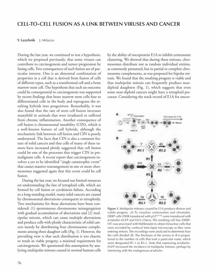
During the last year, we continued to test a hypothesis,which we proposed previously, that some viruses cancontribute to carcinogenesis and tumor progression byfusing cells. Two consequences of such fusion are of par-ticular interest. One is an abnormal combination ofproperties in a cell that is derived from fusion of cellsof different types, such as a transformed cell and a bonemarrow stem cell. The hypothesis that such an outcomecould be consequential to carcinogenesis was supportedby recent findings that bone marrow stem cells fuse todifferentiated cells in the body and reprogram the re-sulting hybrids into progenitors. Remarkably, it wasalso found that the rate of stem cell fusion increasesmanyfold in animals that were irradiated or sufferedfrom chronic inflammation. Another consequence ofcell fusion is chromosomal instability (CIN), which isa well-known feature of cell hybrids, although themechanistic link between cell fusion and CIN is poorlyunderstood. The facts that CIN is also a common fea-ture of solid cancers and that cells of many of these tu-mors have increased ploidy suggested that cell fusioncould be one of the processes that trigger CIN in pre-malignant cells. A recent report that carcinogenesis in-volves a yet to be identified “single catastrophic event”that causes massive rearrangement in one or more chro-mosomes suggested again that this event could be cellfusion.
During the last year, we focused our limited resourceson understanding the fate of tetraploid cells, which areformed by cell fusion or cytokinesis failure. Accordingto a long-standing model, many solid cancers are causedby chromosomal aberrations consequent to tetraploidy.Two mechanisms for these aberrations have been con-sidered: (1) spontaneous chromosome missegregationwith gradual accumulation of aberrations and (2) mul-tipolar mitosis, which can cause multiple aberrationsand produce cells with ploidy characteristic of solid can-cers merely by distributing four chromosome comple-ments among three daughter cells (Fig. 1). However, theprevailing view is that such distribution is too chaoticto result in viable progeny, a minimal requirement forcarcinogenesis. We questioned this assumption by ana-lyzing multipolar mitoses caused in normal human cells
by the ability of oncoprotein E1A to inhibit centrosomeclustering. We showed that during these mitoses, chro-mosomes distribute not as random individual entities,as commonly presumed, but in partial or complete chro-mosome complements, as was proposed for bipolar mi-tosis. We found that the resulting progeny is viable andthat multipolar mitosis can frequently produce near-diploid daughters (Fig. 1), which suggests that evensome near-diploid cancers might have a tetraploid pre-cursor. Considering the track record of E1A for uncov-
76
CELL-TO-CELL FUSION AS A LINK BETWEEN VIRUSES AND CANCER
Y. Lazebnik J. Milazzo
Figure 1. Multipolar mitoses caused by E1A produce diverse andviable progeny. (A) To visualize centrosomes and chromatin,DERP cells (DEIR transduced with p53R175H) were transduced withα-tubulin–EGFP and H2A–Cherry. The resulting cell line (DERP-HT) was processed with blebbistatin to obtain binuclear cells thatwere recorded by confocal time-lapse microscopy as they wereentering mitosis. The recordings were analyzed to determine howthe cells divided (B). The thickness of the arrows in B is propor-tional to the number of cells that took a particular route, whichwere designated R1.1 to R3.2. Note that expressing α-tubulin–EGFP increased the incidence of multipolar mitoses, perhaps byinterfering with the endogenous α-tubulin.
072-096_03_Cancer_SignTrans_Annual Report_2009 template 7/8/11 1:26 PM Page 76

ering oncogenic pathways, and our finding that thisoncogene inhibits centrosome clustering, we proposedthat E1A mimics oncogenic events that cause multipolarmitosis and the consequent chromosomal aberrations inhuman neoplasia.
To facilitate studies on the consequences of cell fu-sion, in particular those to genome stability, we reporteda simple, versatile, scalable, and nontoxic approach tocause cell fusion in vitro. We called this approach V-fu-sion, as it is based on the ability of the vesicular stom-atitis virus G protein (VSV-G), a viral fusogen of broadtropism, to become rapidly and reversibly activated. Wesuggest that this approach will benefit a broad array ofstudies that investigate consequences of cell fusion oruse cell fusion as an experimental tool.
During the last year, we were pleased to note thatour suggestion, which we reported in 2007, that me-tabolism of glutamine should be revisited as a promis-ing target for cancer therapy has been successfully
implemented by a number of laboratories and pharma-ceutical companies.
Finally, we published a commentary that questionsthe prevailing and widely accepted view of cancer as acombination of six hallmarks. We hope that this com-mentary will help to maintain a healthy discussionabout the origin and nature of the disease that remainsas incurable as it has been for decades in a majority ofinstances.
Overall, we hope that our research will help to de-velop an experimental and conceptual framework totest the role of cell fusion, ploidy increase, and chro-mosomal instability in cancer.
PUBLICATIONS
Gottesman A, Milazzo J, Lazebnik Y. 2010. V-fusion: A convenient, non-toxic method for cell fusion. Biotechniques 49: 747–749.
Lazebnik Y. 2010. What are the hallmarks of cancer? Nat Rev Cancer 10:232–233.
Joseph Milazzo
Cancer: Signal Transduction 77
072-096_03_Cancer_SignTrans_Annual Report_2009 template 7/8/11 1:26 PM Page 77

78
Pathologists use a number of criteria to diagnose andpredict prognosis of premalignant breast lesions. Salientamong them are changes in cell number and in cell andtissue architecture. Although we are beginning to un-derstand a lot about the mechanisms that regulate cellproliferation, very little, if anything, is known aboutthe mechanisms that regulate disruption of cell and tis-sue architecture. We believe that gaining better insightsinto the mechanisms that regulate changes in cell archi-tecture during the development and progression of pre-malignant lesions will identify molecular targets thatpathologists can use to better prognose premalignantlesions. In this process, we may also identify novelstrategies to treat premalignant disease.
The epithelial cells lining the luminal space have anasymmetric distribution of membrane proteins wherethe membrane that is in contact with lumen, referredto as the apical surface, is rich in glycoproteins and mi-crovilli, and the membrane that is in contact with theneighboring cell or the surrounding tissue is rich incell–cell and cell–matrix junctions and is referred to asthe basolateral surface. This characteristic organizationis lost early in premalignant disease, and the tissue con-tinues to lose its structure and organization during pro-gression to malignancy. Surprisingly, we know littleabout what role these changes have during the cancerprocess and, more importantly, if the changes observedin premalignant lesions are harbingers of what is yet tocome.
We recently demonstrated that the oncogene ErbB2interacts with Par6/aPKC, a protein complex involvedin the regulation of asymmetric cell division, cell junc-tion biogenesis, and epithelial morphogenesis. This in-teraction was required for the ability of ErbB2 todisrupt epithelial cell polarity and transform three-di-mensionally organized epithelial structures. In additionto interacting with Par6/aPKC, activation of ErbB2 alsoinduces disruption of Par3 from the polarity complex.We now report that expression of Par3 is down-regu-lated both in human breast cancers and in lung metas-tasis of ErbB2 transgenic mice. Down-regulation ofPar3 cooperated with the ErbB2 oncogene to inducemigration, invasion, and metastasis of transformed and
tumor-derived mammary epithelial cells in culture andin vivo. Interestingly, acquisition of invasive andmetastatic ability was not associated with an overt mes-enchymal phenotype. However, epithelial cells lackingPar3 activated a Tiam1/Rac/GTP/IRSp53/WAVE2/Arp2/3 pathway that induced disruption of corticalactin organization, blocked E-cadherin junction matu-ration, and decreased cell–cell cohesion. Changes incortical actin organization and mislocalization ofArp2/3 complex were observed in epithelial cells lack-ing Par3 both in culture and in primary tumors in miceand in humans. Thus, we demonstrated that loss ofPar3 deregulates cortical actin dynamics and E-cadherinjunction maturation to promote metastatic behavior inepithelial cells without inducing a mesenchymal phe-notype.
Previous results from our lab (Zhan et al., Cell 135:865 [2008]) provided evidence that overexpression ofa Scribble mislocalization mutant (ScrP305L) disruptedthree-dimensional morphogenesis of mammary epithe-lial cells, whereas overexpression of wild-type (ScrWT)Scribble did not. Therefore, we sought to investigatethe signaling pathways activated in response to Scribblemislocalization in these cells. In the current study, wefind that ScrP305L, but not ScrWT, specifically in-duces activation of Akt and phosphorylation of down-stream Akt targets. To extend these observations to anin vivo setting, we generated transgenic mice expressingScrP305L in the mammary epithelium, under the con-trol of the mouse mammary tumor virus (MMTV) pro-moter. ScrP305L mice display defects in branchingmorphogenesis, including loss of tertiary ducts andsmall alveolar structures. This is followed by develop-ment of hyperplasia by 6 months of age and tumor for-mation after 1 year. Analysis of P305L tumors reveals astriking heterogeneity in pathology as well as markerexpression, suggesting that Scribble cooperates with sec-ondary genetic events for tumor progression. Impor-tantly, despite their heterogeneity, ScrP305L tumorsshow high levels of activated Akt. Overall, our resultssuggest that the impaired polarization of epithelial cellsnoted in advanced cancers may be a cause, and notmerely a consequence, of human tumorigenesis. To-
EPITHELIAL CELL BIOLOGY AND CANCER
S. Muthuswamy D. Akshinthala K. KrishnamurthyM. Feigin B. Xue
072-096_03_Cancer_SignTrans_Annual Report_2009 template 7/8/11 1:26 PM Page 78
Cancer: Signal Transduction 79
gether, these studies are likely to identify new ways fortreating breast cancer.
PUBLICATIONS
Arias-Romero LE, Villamar-Cruz O, Pacheco A, Kosoff R, Huang M,Muthuswamy SK, Chernoff J. 2010. A Rac-Pak signaling pathwayis essential for ErbB2-mediated transformation of human breast ep-ithelial cancer cells. Oncogene 29: 5839–5849.
Cabodi S, Tinnirello A, Bisaro B, Tornillo G, del Pilar Camacho-LealM, Forni G, Cojoca R, Iezzi M, Amici A, Montani M, et al. 2010.p130Cas is an essential transducer element in ErbB2 transformation.FASEB J 24: 3796–3808.
Huang L, Muthuswamy SK. 2010. Polarity protein alterations in carci-noma: A focus on emerging roles for polarity regulators. Curr OpinGenet Dev 20: 41–50.
Ling C, Zuo D, Xue B, Muthuswamy S, Muller WJ. 2010. A novel rolefor 14-3-3sigma in regulating epithelial cell polarity. Genes Dev 24:947–956.
Sai Dipikaa Akshinthala
072-096_03_Cancer_SignTrans_Annual Report_2009 template 7/8/11 1:26 PM Page 79

80
Identifying Guardians of GermlineGenomes
This work was done in collaboration with A. Haase andG. Hannon.
The Piwi clade of Argonaute proteins and their asso-ciated small RNAs, Piwi-interacting RNA (piRNA), actin an evolutionarily conserved genome defense pathwaythat silences transposons in germline cells. Piwi proteinsare required for germline differentiation and mainte-nance of germline stem cells, with mutations almost uni-versally leading to sterility and germcell loss. Insightsinto piRNA biogenesis and function emanate fromcloning and sequencing of piRNAs associated with dif-ferent Piwi proteins, although the mechanisms of pri-mary piRNA biogenesis and function remain largelyelusive. We used affinity immunoprecipitation to iden-tify proteins associated with Drosophila Piwi and threeother known components of the piRNA silencing path-way: Zucchini (a potential nuclease), Armitage (an RNAhelicase), and Squash (a protein of unknown function).The analysis revealed that Piwi, Armitage, and Squashassociate with one another. Identification of proteinscopurifying with either component revealed a commonset of interactors constituting the core of this RNA-si-lencing machinery. The laboratory is further investigat-ing the molecular function of candidate genes emergingfrom this analysis, in vitro and in vivo.
Quantitative Proteomic Analysisof the Effect of Anti-let-7 onProtein Expression
This work was done in collaboration with C. Dos San-tos, I. Ibarra, S. Obad, S. Kauppinen, and G. Hannon.
The challenge of deciphering the role of hundredsof animal RNAs, in conjunction with the large num-ber of predicted target mRNAs, demands the develop-ment of robust technologies that can dissect the bio-logical functions of individual microRNAs (miRNAs)
and miRNA families in vivo. An alternative approachto miRNA gene knockouts is to use chemically modi-fied antisense oligonucleotides, termed anti-miRs,which bind to the mature miRNA in competition withcellular target mRNAs, leading to functional inhibi-tion of the miRNA and derepression of the targets.The Hannon Laboratory has used seven to eight nu-cleotide fully locked nucleic acid (LNA)-modifiedphosphorothioate oligonucleotides (termed tinyLNAs) to target the seed region of the let-7 family withhigh stability. Western blot analysis showed a concen-tration-dependent increase of the major HMGA2 tar-get in anti-let-7-treated HeLa cells, whereas thecontrols showed no effect. Quantitative proteomicsscreens using whole-cell lysates and iTRAQ (isobarictags for relative and absolute quantitation) labelingconfirmed the increase of HMGA2 while revealingmuch more subtle off-target effects on other proteins.The conclusion is that tiny seed-targeting LNAs canbe used to effectively inhibit the function of individualmiRNAs and entire miRNA families in cultured cells.The study also validated the use of tiny LNA-basedknockdown in exploring miRNA function, with im-portant implications for the development of therapeu-tics targeting disease-associated miRNAs.
Mapping Posttranslational Modificationsof the Splicing Factor SF2/ASF
This work was done in collaboration with Y. Liu, R.-Y.Tzeng, O. Fregoso, R. Sinha, and A. Krainer.
We used a combination of bottom-up and middle-down approaches to map a significant number of post-translational modifications of human SF2/ASF followingmagnetic bead immunoprecipitation of the protein fromHeLa and 293E cells. For the bottom-up approach, atriple-digest protocol was used (trypsin, elastase, and sub-tilisin) followed by two-dimensional MudPIT (multidi-mensional protein identification technology) LC-MS(liquid chromatography/mass spectrometry) analysis of
PROTEOMICS LABORATORY AND PROTEOMICSSHARED RESOURCE
D. Pappin G. Bencze S. PeacockC. Fu C. RuseC.M. Gauss B. WeindorfA. Lelo
072-096_03_Cancer_SignTrans_Annual Report_2009 template 7/8/11 1:26 PM Page 80

Cancer: Signal Transduction 81
the overlapping, nonspecific digest products to increasecoverage. For the middle-down approach, we used Lys-C to generate larger fragments, which were analyzed byboth CID (collision-induced dissociated) and ETD (elec-tron transfer dissociation) fragmentation. In total, weidentified six sites of SY phosphorylation, six sites of KRmonomethylation, and one site of R dimethylation. Eightof these sites (including four sites of phosphorylation) hadnot been previously reported. Subsequent work by mu-tagenesis and cellular assays has shown that three of theR methylations are important for controlling the parti-tioning of the protein between nucleus and cytoplasm.This has functional consequences for its effects on pre-mRNA splicing and nonsense-mediated decay (NMD)in the nucleus and on translation in the cytoplasm.
Novel Regulatory Modificationof PTP1B
This work was done in collaboration with N. Krishnan,N. Tonks, and C. Fu.
Hydrogen sulfide (H2S) has recently been implicatedin the regulation of several biological processes, but cel-lular targets and possible modes of action are not wellunderstood. Protein tyrosine phosphatases (PTPs) havebeen established as regulators of a wide variety of signaltransduction pathways, and they use an essential reactiveCys residue, which is characterized by a low pKa and isvery susceptible to oxidation. We tested whether H2Smay also target the catalytic Cys residue to regulate PTPfunction. Using high-resolution mass spectrometry (MS)we identified a novel covalent modification (persulfa-tion) of the active-site Cys-215 in PTP1B that occurs inresponse to the generation of hydrogen sulfide in vivo.This sulfhydration reaction also inactivates PTP1B, butit is subject to different mechanisms of reduction and re-activation compared to the reversibly oxidized enzyme.To understand the relevance of this modification to thecontrol of cell signaling, we used RNA interference(RNAi) to suppress the critical enzyme in H2S produc-tion, cystathionine-γ-lyase (CSE), in 293T cells, wherewe observed sulfhydration of Cys-215 following ER (en-doplasmic reticulum) stress only in control cells. Usinga novel phosphopeptide-trapping mutant, we searchedfor potential substrates of PTP1B in the ER stress re-sponse and identified a tryptic phosphopeptide derivedfrom PERK (protein-kinase-like ER kinase). The phos-phorylation of Y619 in PERK has a critical role in theactivation of PERK, which phosphorylates the eukaryo-tic translational initiation factor 2α (eIF2α), leading totranslational attenuation. We have since demonstrated
that PTP1B dephosphorylates Y619 PERK and that thisevent is regulated by H2S production in ER stress. Thesedata suggest the possibility that different modes of re-duction and reactivation of PTPs in response to distinctgasotransmitters may introduce new levels of controlover signal transduction.
Interacting Partners of MECP2
This work was done in collaboration with A. Paul andJ. Huang.
MECP2 was identified as a methyl-CpG DNA-bindingprotein and is known to be a trancriptional modulator andregulator of chromatin structures and alternative splicing.MudPIT LC-MS analysis of MECP2 complexes obtainedby magnetic bead immunoprecipitation discovered a hostof new MECP2 interacting proteins. In particular, eukary-otic elongation factor 1A (EF1A1), DEAD- box protein 6(DDX6/ P54), and synaptotagmin-binding cytoplasmicRNA-interacting protein (SYNCRIP) were identified withhigh significance. All three proteins have known roles intranslational regulation and mRNA transport.
Because EF1A1 is integral to the translation machin-ery and is present in kinesin-associated mRNP granules,its interaction with MECP2 could influence the trans-lation state of MECP2-associated mRNAs. DDX6/P54is a well-known member of the cytoplasmic P-bodiesand has been shown to repress translation and shuttlebetween P-body and actively translating fractions. SYN-CRIP was found in kinesin-associated mRNPs as wellas in dendritic mRNA transport granules. The associa-tion of MECP2 with multiple proteins that regulatetranslation and mRNA transport for local protein syn-thesis suggests a potential mechanism of how MECP2deficiency results in altered protein levels for its targettranscripts without changes in mRNA levels. Together,these results established that MECP2 not only is associ-ated with translating mRNAs in the cytoplasm, but alsoimposes a regulatory function by interacting with otherfactors involved in mRNA metabolism and translation.The results establish a novel regulatory function ofMECP2 and suggest translation regulation as a patho-genic mechanism of Rett syndrome.
Functional Analysis of the ProteinPhosphatase Activity of PTEN
This work was done in collaboration with X. Zhangand N. Tonks.
The tumor suppressor phosphatase PTEN displaysintrinsic activity toward both protein and phosphatidyl-
072-096_03_Cancer_SignTrans_Annual Report_2009 template 7/8/11 1:26 PM Page 81
82 Research
inositol phospholipid substrates in vitro. Whereas thelipid phosphatase activity of PTEN is important for itstumor suppressor function, the significance of its proteinphosphatase activity is unknown and represents a gap inour understanding of the function of this important reg-ulator of cell signaling. The Tonks laboratory has devel-oped an in vivo assay of PTEN function, which hasrevealed an effect of its protein phosphatase activity onthe density of neuronal spines in cultured hippocampalorganotypic brain slices. In summary, the data suggestthat the protein phosphatase activity of PTEN and thephosphorylation of Ser and Thr residues in its carboxy-terminal segment are of regulatory significance to thecontrol of spine density. We are thus focused on definingthese critical phosphorylation sites within PTEN andthe identity of the regulatory binding proteins withwhich it associates.
Identification of Origin RecognitionComplex Interactors
The Stillman Laboratory has identified a new mecha-nism by which the pre-recognition complex (pre-RC)is activated by protein kinases at the G1-to-S phase tran-sition that involves targeting the Cdc7-Dbf4 (DDK)protein kinase to a regulatory region of the Mcm4 pro-tein subunit of the MCM2-7 helicase complex. Thestudies led to the identification of an inhibitory aminoacid sequence in Mcm4 that, when deleted, bypassedthe requirement for the DDK kinase. Using MCM2-7immunoprecipitation and mass spectrometry, Cdc45was identified as a major cell-cycle-regulated MCM-in-teracting protein that depended on DDK activation ofthe pre-RC before it could bind MCM2-7.
When Orc2 and Orc3 were depleted from humancells using RNAi, ~30% of the cells arrested with a ter-minal phenotype in which the mitotic chromosomes areabnormally condensed and are not attached to the mi-totic spindle, and the cells have multiple centrosomeswith only one centriole per centrosome. It was shownthat the Orc2- or Orc3-depleted cells attempt to form ametaphase plate and then begin the metaphase-to-anaphase transition, but then the chromosomes pull offthe spindles and form abnormally condensed structures.Thus, the spindle assembly checkpoint (SAC) was sat-isfied, but in the absence of Orc2 or Orc3, chromo-somes lost spindle attachment to the kinetochores whentension was placed on the spindle to pull the cen-tromeres apart. Subsequent biochemistry demonstratedthat Orc2 and Orc3 associated with the BubR1 kineto-
chore protein that is involved in sensing tension whenthe kinetochores are correctly attached to biorientedspindles. Also associated with Orc2 and Orc3 wereCenpE and Plk1, a protein kinase that phosphorylatesBubR1, as well as the heterochromatin protein 1. Theinteractions between Orc2 and Orc3 at centromeres andthe kinetochore proteins only occurred during mitosisand were observed during interphase; in addition, theinteractions were phosphorylation dependent. We arecurrently performing mass spectrometry of ORC sub-unit immunoprecipitations to determine other proteinsassociated with ORC in mitosis and at centrosomes.
The Molecular Basis of OncogeneAddiction
This work was done in collaboration with M. Pinedaand R. Sordella.
The Sordella laboratory is interested in the possibilitythat the platelet-derived growth factor receptor (PDGFR)could modify the activity of SOCS3, because it couldprovide a molecular mechanism explaining the decreasedsensitivity to the epidermal growth factor receptor(EGFR) inhibitor in NSCLC cells expressing PDGFR.Mass spectrometry of SOCS3 was performed after invitro phosphorylation with both PDGFR and EGFRusing cold ATP. Several approaches used both the lineartrap quadrupole (LTQ) and quadrupole time-of-flight(Q-TOF) mass spectrometers and in-solution and in-geldigestion of samples. Initially, several phosphotyrosineswere identified within the protein, but it was suspectedthat they might be due to nonspecific phosphorylation.The protocol was modified by decreasing the temperatureof the kinase assay and varying the concentration of thekinase, and accurate mass Q-TOF LC-MS with phos-pho-chip titanium dioxide enrichment was carried out.Using these approaches, the PDGFR phosphorylationsite in SOCS3 was identified at amino acid 165 (YYIY),located within the gp130-binding pocket. This suggestedthat PDGFR may be involved in the molecular mecha-nism of cytokine regulation.
Quantitation of Alternative SpliceVariants at the Protein Level
This work was done in collaboration with O. Fregoso,M. Jensen, A. Krainer, M. Akerman, and M. Zhang.
The Shared Resource is currently collaborating withthe Krainer Laboratory to develop the use of iTRAQquantitative proteomics to identify and quantify chang-
072-096_03_Cancer_SignTrans_Annual Report_2009 template 7/8/11 1:26 PM Page 82

Cancer: Signal Transduction 83
es in exon-junction peptides as a measure of changes inalternative splicing isoforms of abundant proteins. Inmultiple duplex iTRAQ experiments, protein lysateswere prepared from cytoplasmic and nuclear fractionsof control HeLa cells and cells overexpressing theSF2/ASF splicing factor. The lysates were digested(trypsin) and labeled with two members of the 4-plex
iTRAQ reagent set before mixing. The pooled, labeledpeptides were analyzed by 15-16 salt step MudPIT LC-MS or capillary LC-MS following first-dimension pep-tide separation by OffGel isoelectric fosusing (24fraction; pH3-10). CID spectra are currently beingmatched to a splice peptide isoform database specificallycreated for the project.
072-096_03_Cancer_SignTrans_Annual Report_2009 template 7/8/11 1:26 PM Page 83
84
MOLECULAR TARGETED THERAPY OF LUNG CANCEREGFR MUTATIONS AND RESPONSE OF EGFR INHIBITORS
R. Sordella M. Camiolo M. PinedaS. Fenoglio A. SaborowskiT. Lindsted Z. Yao
In recent years, rapid advances in our understanding ofthe molecular events required for tumor onset and pro-gression have led to the development of novel canceragents referred to as “molecular targeted therapies.” Be-cause they specifically target the product of selective can-cer mutations that are required for cancer cell survival,they are thought to become invaluable therapeutic tools.Specifically in the case of lung cancer, much excitementhas been generated by the finding that patients harboringoncogenic epidermal growth factor receptor (EGFR) mu-tations highly benefit from treatment with selective in-hibitors (erlotinib and gefitinib). Erlotinib and gefitinibare members of a class of quinazolium-derived agents thatinhibit the EGFR pathway by binding in a reversible fash-ion to the EGFR ATP pocket domain. Remarkably, ret-rospective studies showed a striking correlation betweenoccurrence of certain EGFR oncogenic mutations and er-lotinib/gefitinib responses. The presence of deletions inexon 19of EGFR or EGFR L858R missense substitutionsare in fact found in more than 80% of non-small-cell lungcancer (NSCLC) patients that respond to erlotinib orgefitinib treatment. Yet, as in the case of other targetedtherapies, the emergence of resistance presents a majorhurdle for the successful utilization of these agents. Clin-ical data in fact have shown that in the majority of thecases, responses to drug treatment are transient, andwithin a short period of time, patients who initially re-sponded progress or relapse with resistant disease. The ac-quisition of an additional mutation in exon 20 of EGFRresulting in a threonine-to-methionine substitution at po-sition 790 (T790M mutation) and/or amplification of c-MET can account for ~50% of cases of erlotinib-acquiredresistance. However, the mechanisms that lead to resist-ance in the remaining cases are unknown.
Intrinsic and Extrinsic Mechanisms ofResistance to Targeted Therapies
To uncover new molecular mechanisms of gefitinib anderlotinib resistance in NSCLC, we have developed acell-based model system using the broncho-alveolar
cancer cell line H1650. This cell line harbors an onco-genic deletion within the EGFR (delE746-A750) andhas a half-maximal inhibitory concentration (IC50) togefitinib or erlotinib treatment of ~5 µM. By culturingthis cell line in the presence of a constant high concen-tration of erlotinib, we have been able to isolate celllines capable of growing in the presence of up to 20 µM
of the drug. Interestingly, ~13% of the erlotinib-resis-tant cells displayed the morphological appearance ofmesenchymal cells (16 of a total of 123 colonies exam-ined), enhanced motility, and Matrigel invasion com-pared to parental cells. These striking morphologicalfeatures were associated at the molecular level with anincreased expression of the mesenchymal protein Vi-mentin and with decreased expression of the epithelialmarker E-cadherin, as well as with an EGFR-indepen-dent augmented secretion of transforming growth fac-tor-β1 (TGF-β1) and TGF-β2.
We determined that the increased autocrine secretionof TGF-β was sufficient to activate a complex programthat led to the acquisition of mesenchymal-like mor-phology, increased motility, invasion ability, and er-lotinib resistance. In the latter case, we providedevidence that an up-regulation of interleukin-6 (IL-6)secretion by TGF-β was sufficient to unleash cells har-boring mutant EGFR from their EGFR dependency, asmanifested by their decreased sensitivity to erlotinibtreatment.
By using a surface marker signature derived from theerlotinib-resistant cells, we also showed that cells whichare mesenchymal and erlotinib-resistant were alreadypresent in NSCLC-derived cell lines as well as in early-stage treatment-naive tumors.
These data thus indicate that cell-autonomousmechanisms could generate subpopulations of cells in-trinsically resistant to erlotinib treatment. Yet becauseboth IL-6 and TGF-β are secreted factors prominentlyproduced during the inflammatory response, the acti-vation of the tumor microenvironment could also con-tribute to erlotinib resistance. By using a mouse modelsystem in which inflammation was induced either by
072-096_03_Cancer_SignTrans_Annual Report_2009 template 7/8/11 1:26 PM Page 84

topical treatment with a low concentration of 12-O-tetradecanoyl-phorbol-13-acetate (TPA) or with lipo-polysaccharide (LPS), we were indeed able to show thatthe induction of inflammation was successful in stim-ulating IL-6 secretion and decreasing the tumor re-sponse to erlotinib treatment. Hence, our data providecompelling evidence indicating that acquired resistanceto molecular targeted therapies could arise not only asa consequence of genetic and/or epigenetic heterogene-ity within cancer cells, but also through the activationof the tumor microenvironment.
Because we identified cells that are intrinsically re-sistant to erlotinib in NSCLC prior to treatment, it wasalso tempting to speculate that this same mechanism oferlotinib-acquired resistance could explain the hetero-geneity of primary erlotinib responses observed in pa-tients. In fact, although the majority of patientsharboring similar EGFR oncogenic mutations do re-spond to erlotinib treatment, overall responses can varyfrom 5% to 90% and remission could span from 3months to more than 5 years. Notably, several studieshave already reported increased levels of IL-6 in ~30%of NSCLC.
Why IL-6 is required for the survival of the cancercells is yet not clear. Our data seems to indicate a roleof IL-6 in protecting cells from apoptosis. This seemsto be consistent with current literature. Catlett-Falconeet al. (Immunity 10: 105–115 [1999]) in fact demon-strated that IL-6-induced STAT3 protects myelomacells from FAS-induced apoptosis by up-regulating theexpression of BCL-X
L. Similarly, Haga et al. (J Clin In-vest 102: 989 [2003]) demonstrated that constitutivelyactive STAT3 provided protection against FAS-medi-ated liver injury likely through an augmented expres-sion of the antiapoptotic proteins FLIP, BCL-2, andBCL-XL. Consistent with a role of IL-6/STAT3-medi-ated survival, we observed an up-regulation of Surivin,BCL-X1, and BCL-2 in the erlotinib-resistant cells.
Interestingly, the erlotinib-resistant cells character-ized in this study also have an increased metastatic po-tential. They display mesenchymal-like features, arehighly motile and invasive, and can be found with anincreased representation in the bone marrow of patientscompared to primary tumors. Notably, many of thegenes that we see increasingly expressed in the resistantcells compared to the parental line have already beenassociated with a metastatic signature and/or with poorprognosis. These genes include HOXB2, S100A4,S100A2, Tenascin C, SUSD, MCAM, and TCF-4.
Notably, although the epithelial–mesenchymal tran-sition (EMT) has been previously reported to be asso-
ciated with erlotinib resistance, our data indicate thatthe programs that lead to mesenchymalization and drugresistance are distinct. Although we found that TGF-βis required and sufficient for EMT, invasion, and motil-ity, as well as inducing an increased expression of IL-6,reducing the expression of the EMT master regulatorSNAI only impaired EMT/motility/invasion but didnot change the cells’ sensitivity to erlotinib. Further-more, treatment of cells with IL-6 increased their re-sistance to erlotinib but did not induce EMT. Our dataare consistent with the observation that certain cell linesharboring mutant EGFR (e.g., PC9), despite being ex-tremely sensitive to erlotinib, display mesenchymal-likefeatures.
In conclusion, our data provide compelling evidenceindicating that resistance to molecular-targeted thera-pies could arise not only as a consequence of geneticand/or epigenetic changes within cancer cells, but alsothrough the activation of the tumor microenvironment.Hence, the contribution of selective and adaptive mech-anisms adds a new layer to the complexity of cancerdrug resistance and poses new challenges for the clinicaluse of molecular targeted therapies. In particular, itclearly indicates that, in the case of lung tumors drivenby mutant EGFR, treatment based only on the inhibi-tion of EGFR will not be effective and suggests the in-triguing possibility that adjunctive therapies designedto either control inflammation and/or decrease thebioavailability of IL-6 may provide effective means toimprove response to EGFR TKI treatment. Interest-ingly, clinical trials combining Cox2 inhibitors (e.g.,rofecoxib and celecoxib) and gefitinib/erlotinib have al-ready shown encouraging results.
A Novel Splice Isoform of p53 Involvedin Tissue Damage Response Assists in theBypass of Senescence and the Acquisitionof the Metastatic Phenotype
Gene expression profile analysis of CD44high/CD24low-derived cells (H1650-M3) revealed a substantial de-crease in expression of p53 compared to parental cells.Interestingly, when we validated these data by reversetranscriptase–polymerase chain reaction (RT-PCR)analysis for p53 mRNA, we found a less-intense andslower-migrating band in the CD44high/CD24low-de-rived cells compared to parental cells. We reasoned thatthis band might represent an alternatively spliced tran-script from the p53 gene. Indeed, sequence analysis in-
Cancer: Signal Transduction 85
072-096_03_Cancer_SignTrans_Annual Report_2009 template 7/8/11 1:26 PM Page 85
86 Research
dicated the existence in CD44high/CD24low-derived cellsof a novel p53 alternative slice isoform that we refer toas p53Ψ. Interestingly, comparison of the p53 gene se-quence across species pointed out that the intronic se-quence surrounding the alternative acceptor site inintron 6 is highly conserved.
The alternative splicing event observed inCD44high/CD24low-derived cells results in a frameshiftplacing a stop codon 19 amino acids downstream fromthe splice junction and the generation of a protein thatis devoid of the oligomerization domain, the nuclear lo-calization sequence (NLS) and part of the DNA-bindingdomain. As a consequence, p53Ψ is mainly localized inthe cell cytoplasm and lacks transcriptional activity.
Yet, similarly to certain p53 mutants (i.e., R273Hand R280K), p53Ψ expression leads to an increased in-vasion ability and decreased expression of E-cadherin.Interestingly, subcutaneous transplantation of p53-nullcells (H1299) ectopically expressing either p53 wildtype or p53Ψ also indicated that p53Ψ also increasesthe seeding capabilities of tumor cells.
Surprisingly, we noticed that the ectopic expressionof wild-type p53 in the CD44high/CD24low cells resultedin cellular senescence. Interestingly this happens ratherrapidly when compared to oncogene-induced senescence(5 days vs. 12 days in case of oncogene-induced senes-cence) and was exclusive to the CD44high/CD24low cells.
Because CD44high/CD24low cells express several genesthat have been shown to be sufficient to drive senes-cence in a p53-dependent manner (e.g., IL6, IGFBP7,SMARACA2, SMARACA4, SOCS1, and SPIN1), wehypothesize that one function of this alternative splicingevent is to reduce p53 activity and by doing so to pre-vent senescence.
One hallmark of senescent cells is the increased ex-pression of certain secreted proteins—a phenomenonknown as senescence-associated secretory phenotype,or SASP. While the overall physiological role of SASPsecretion by senescent cells is still not completely un-derstood, it has been proposed that in some cases, theseproteins can induce epithelial cell proliferation and re-
sistance to apoptosis. Because the CD44high/CD24low
cells also expressed many reported SASP componentssuch as MMP1, MMP3, MMP10, MICA, ULBP2,PVR, and IL-6 that were found recently to be up-reg-ulated in CD44high/CD24low-derived cells in activatedhepatic stellate cells upon injury, we reasoned that thisnovel p53 alternative splicing could have a physiologicalsignificance during tissue damage/repair. We found thatp53Ψ is indeed expressed in activated stellate cells dur-ing the early phase of injury/response, thus allowingthese cells to linger in a presenescent state and con-tribute to tissue repair through the secretion of certainSASP components. Eventually, as the damage responseprogresses, the activated stellate cells will reexpress wild-type p53, become senescent, and be cleared in the in-jury/resolution phase as previously proposed.
Our experimental evidence thus indicates thatp53Ψ�has an important role in tissue-damage responselikely by preventing premature senescence of cells in-volved in tissue remodeling and injury resolution.
In light of the functional and structural similarities be-tween p53 mutants and p53Ψ, it is tempting to speculatethat p53 gain-of-function mutants by functionally takingoff the p53Ψ splice isoform have highjacked a highly reg-ulated and reversible physiological response occurringduring the tissue-damage response. This phenotypicmimicry may then contribute to tumorigenesis by pre-venting senescence, increasing cell-invasive capabilities,and enhancing the expression of CD44. Fascinatingly,tumors have been referred to as “wounds that never heal.”
PUBLICATIONS
Cancer Target Discovery and Development Network, Schreiber SL,Shamji AF, Clemons PA, Hon C, Koehler AN, Munoz B, Palmer M,Stern AM, Wagner BK, et al. 2010. Towards patient-based cancertherapeutics. Nat Biotechnol 28: 904–906.
Yao Z, Fenoglio S, Gao DC, Camiolo M, Stiles B, Lindsted T, SchledererM, Johns C, Altorki N, Mittal V, Kenner L, Sordella R. 2010. TGF-β IL-6 axis mediates selective and adaptive mechanisms of resistanceto molecular targeted therapy in lung cancer. Proc Natl Acad Sci 107:15535–15540.
072-096_03_Cancer_SignTrans_Annual Report_2009 template 7/8/11 1:26 PM Page 86

87
The phosphorylation of tyrosyl residues in proteins is a keycomponent of the regulation of signaling pathways thatcontrol many fundamental physiological processes, includ-ing cell proliferation, differentiation, and survival. Proteinphosphorylation is a reversible, dynamic process in whichthe net level of phosphate observed in a target substrate re-flects the coordinated activity of the kinases that phosphor-ylate it and the protein phosphatases that catalyze thedephosphorylation reaction. We study the family of pro-tein tyrosine phosphatases (PTPs), which, like the kinases,comprise both transmembrane, receptor-linked forms andnontransmembrane, cytoplasmic species and represent amajor family of signaling enzymes. Overall, the objectiveof the lab is to develop tools for analysis of PTP regulationand function and integrate them with state-of-the-art celland animal models, to define critical tyrosine phosphory-lation-dependent signaling events in human disease andthereby identify novel therapeutic targets. Currently, thereare four broad areas of research in the lab covering func-tional analysis of members of the PTP family, regulationof PTP function, in particular by reversible oxidation, de-velopment of novel approaches to therapeutic interventionin PTP function, and characterization of a knockoutmouse model to define the function of JSP1, a member ofthe PTP family that is a novel regulator of mitogen-acti-vated protein kinase (MAPK) signaling.
During the last year, two of our graduate students,Guang Lin and Aftab Haque, successfully defendedtheir Ph.D.s. They are currently staying in the lab whiletheir papers are under review, but they will be movingon to pursue postdoctoral opportunities in 2011.
Identification of PTPN23 as a NovelRegulator of Cell Invasion in MammaryEpithelial Cells from a Loss-of-FunctionScreen of the “PTPome”
We had constructed a short hairpin RNA (shRNA) library,which contains five shRNAs specifically designed to target
each of the PTPs and would allow us to interrogate thefunction of these signaling enzymes by RNA interference(RNAi). We conducted an RNAi-mediated loss-of-func-tion screen to study systematically the role of the PTP su-perfamily of enzymes in mammary epithelial cell motility,in the absence or presence of the oncoprotein tyrosine ki-nase ErbB2. Although shRNAs directed against most ofthe PTP family members were without effect, consistentwith functional specificity within this family of signalingenzymes, we identified PTPs that either promoted or in-hibited motility. In particular, we have focused on threePTPs that appear to be novel tumor suppressors in breastcancer, and we are currently characterizing their mecha-nism of action. Suppression of PRPN23, PTPRG, andPTPRR enhanced cell motility. Furthermore, we foundthat suppression of PTPN23, but not PTPRG or PTPRR,induced cell invasion. Suppression of PTPN23 increasedE-cadherin internalization, impaired trafficking of E-cad-herin from early endosomes, induced the expression ofmesenchymal proteins, and caused cell scattering. The ac-tivity of β-catenin, a regulator of cell adhesion, and SRC,a protein tyrosine kinase, were elevated when PTPN23 wassuppressed. Moreover, we identified E-cadherin, β-catenin,and SRC as direct substrates of PTPN23. Inhibition ofSRC with the small-molecule inhibitor SU6656 blockedthe effects of PTPN23 depletion. These findings suggestthat loss of PTPN23 may increase the activity of SRC andthe phosphorylation status of the E-cadherin/β-catenin celladhesion/signaling complex to promote tumor growth andmetastasis in breast cancer. Our studies suggest that inhi-bition of SRC may offer a novel therapeutic approach totreatment of tumors in which PTPN23 is lost and, fur-thermore, illustrate how loss-of-function screens reveal newroles for PTPs in mammary epithelial cell biology.
Identification and Characterization of aNovel Inhibitor of PTP1B
To date, drug discovery efforts in signal transductionhave emphasized the protein kinases, in particular pro-
PROTEIN TYROSINE PHOSPHATASES AND THE CONTROLOF SIGNAL TRANSDUCTION
N.K. Tonks G. Bencze G. Fang L. Li U. SchwertassekB. Boivin A. Haque G. Lin M. YangF. Chaudhary N. Krishnan M. Ramesh X.C. ZhangX. Cheng
072-096_03_Cancer_SignTrans_Annual Report_2009 template 7/8/11 1:26 PM Page 87
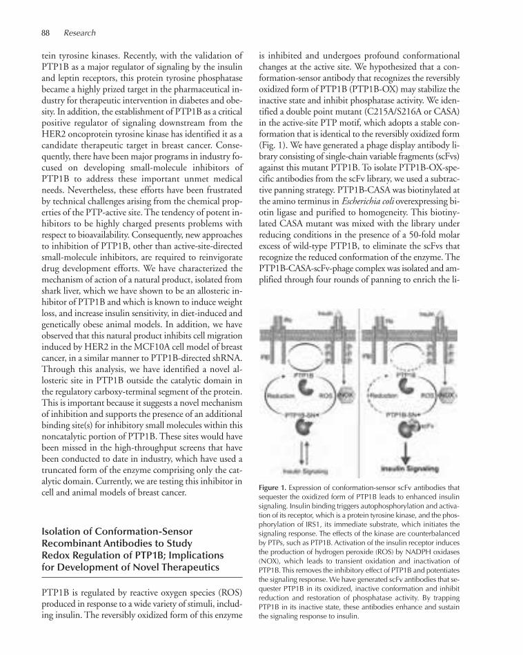
88 Research
tein tyrosine kinases. Recently, with the validation ofPTP1B as a major regulator of signaling by the insulinand leptin receptors, this protein tyrosine phosphatasebecame a highly prized target in the pharmaceutical in-dustry for therapeutic intervention in diabetes and obe-sity. In addition, the establishment of PTP1B as a criticalpositive regulator of signaling downstream from theHER2 oncoprotein tyrosine kinase has identified it as acandidate therapeutic target in breast cancer. Conse-quently, there have been major programs in industry fo-cused on developing small-molecule inhibitors ofPTP1B to address these important unmet medicalneeds. Nevertheless, these efforts have been frustratedby technical challenges arising from the chemical prop-erties of the PTP-active site. The tendency of potent in-hibitors to be highly charged presents problems withrespect to bioavailability. Consequently, new approachesto inhibition of PTP1B, other than active-site-directedsmall-molecule inhibitors, are required to reinvigoratedrug development efforts. We have characterized themechanism of action of a natural product, isolated fromshark liver, which we have shown to be an allosteric in-hibitor of PTP1B and which is known to induce weightloss, and increase insulin sensitivity, in diet-induced andgenetically obese animal models. In addition, we haveobserved that this natural product inhibits cell migrationinduced by HER2 in the MCF10A cell model of breastcancer, in a similar manner to PTP1B-directed shRNA.Through this analysis, we have identified a novel al-losteric site in PTP1B outside the catalytic domain inthe regulatory carboxy-terminal segment of the protein.This is important because it suggests a novel mechanismof inhibition and supports the presence of an additionalbinding site(s) for inhibitory small molecules within thisnoncatalytic portion of PTP1B. These sites would havebeen missed in the high-throughput screens that havebeen conducted to date in industry, which have used atruncated form of the enzyme comprising only the cat-alytic domain. Currently, we are testing this inhibitor incell and animal models of breast cancer.
Isolation of Conformation-SensorRecombinant Antibodies to StudyRedox Regulation of PTP1B; Implicationsfor Development of Novel Therapeutics
PTP1B is regulated by reactive oxygen species (ROS)produced in response to a wide variety of stimuli, includ-ing insulin. The reversibly oxidized form of this enzyme
is inhibited and undergoes profound conformationalchanges at the active site. We hypothesized that a con-formation-sensor antibody that recognizes the reversiblyoxidized form of PTP1B (PTP1B-OX) may stabilize theinactive state and inhibit phosphatase activity. We iden-tified a double point mutant (C215A/S216A or CASA)in the active-site PTP motif, which adopts a stable con-formation that is identical to the reversibly oxidized form(Fig. 1). We have generated a phage display antibody li-brary consisting of single-chain variable fragments (scFvs)against this mutant PTP1B. To isolate PTP1B-OX-spe-cific antibodies from the scFv library, we used a subtrac-tive panning strategy. PTP1B-CASA was biotinylated atthe amino terminus in Escherichia coli overexpressing bi-otin ligase and purified to homogeneity. This biotiny-lated CASA mutant was mixed with the library underreducing conditions in the presence of a 50-fold molarexcess of wild-type PTP1B, to eliminate the scFvs thatrecognize the reduced conformation of the enzyme. ThePTP1B-CASA-scFv-phage complex was isolated and am-plified through four rounds of panning to enrich the li-
Figure 1. Expression of conformation-sensor scFv antibodies thatsequester the oxidized form of PTP1B leads to enhanced insulinsignaling. Insulin binding triggers autophosphorylation and activa-tion of its receptor, which is a protein tyrosine kinase, and the phos-phorylation of IRS1, its immediate substrate, which initiates thesignaling response. The effects of the kinase are counterbalancedby PTPs, such as PTP1B. Activation of the insulin receptor inducesthe production of hydrogen peroxide (ROS) by NADPH oxidases(NOX), which leads to transient oxidation and inactivation ofPTP1B. This removes the inhibitory effect of PTP1B and potentiatesthe signaling response. We have generated scFv antibodies that se-quester PTP1B in its oxidized, inactive conformation and inhibitreduction and restoration of phosphatase activity. By trappingPTP1B in its inactive state, these antibodies enhance and sustainthe signaling response to insulin.
072-096_03_Cancer_SignTrans_Annual Report_2009 template 7/8/11 1:26 PM Page 88
brary with pools of antibodies selective for the mutantform of the enzyme. From the panned pools of scFvs, wehave short-listed candidate antibodies in an activity-based “in-solution” screen, in which we tested the abilityof individual scFvs to stabilize the oxidized, inactive formof PTP1B in enzymatic assays in vitro. These candidatescFvs displayed significant inhibition of the reactivationof PTP1B-OX by reducing agent, but they did not exertany direct inhibitory effect on activity of the reduced,wild-type enzyme. Selected scFvs were characterized fur-ther and shown to bind to the reversibly oxidized con-formation of PTP1B, but not to PTP1B in its reducedactive state, both in vitro and when expressed in 293Tcells as intracellular antibodies or “intrabodies.” Expres-sion of the intrabody had no impact on the basal level oftyrosyl phosphorylation of either the β-subunit of the in-sulin receptor or its substrate IRS-1, but it enhanced andextended the time course of insulin-induced phosphory-lation. Intrabody expression also caused significant en-hancement of signaling downstream from the insulinreceptor, as revealed by increased phosphorylation ofPKB/AKT in insulin-stimulated cells. These effects onsignaling were diminished when we ectopically coex-pressed catalase to quench cellular H2O2. Our data sug-gest that conformation-sensor scFvs can be used aspotential inhibitors of PTP1B by stabilizing the tran-siently inactivated form that is generated following lig-and-induced production of ROS. Reversible oxidationof PTP1B results in a conformation in which the prob-lematic chemical properties of the active site of the re-duced enzyme are circumvented and new bindingsurfaces are presented. Therefore, if it is possible to sta-bilize the oxidized, inactive form of PTP1B with an ap-propriate therapeutic molecule that mimics the effects ofthese antibodies, then this strategy may provide a newapproach for PTP-directed drug development that cir-cumvents the difficulties that are encountered when tar-geting the highly charged PTP-active site.
Hydrogen Sulfide Signals throughSulfhydration of PTP1B
Hydrogen sulfide (H2S), a colorless gas with the odor ofrotten eggs, has been considered a toxic molecule; how-ever, with the burgeoning interest in gasotransmitterfunction, it has recently been implicated in the regula-tion of several biological processes. H2S is generated ina number of different mammalian tissues as a by-prod-uct of the transulfuration pathway in which dietary me-thionine is converted to cysteine. In order to understand
the physiological function of H2S, and its potential rolein signaling, it is essential to identify its cellular targets.PTPs have been established as regulators of a wide vari-ety of signal transduction pathways that underlie fun-damentally important aspects of cell physiology. Themechanism of PTP catalysis utilizes an essential, reactiveCys residue, which is characterized by a low pKa and issusceptible to oxidation. We tested whether H2S mayalso target the catalytic Cys residue to regulate PTPfunction. We focused on PTP1B, the founding memberof this enzyme family. We observed that PTP1B was in-activated by H2S in vitro; the reaction was reversible, asphosphatase activity was fully recovered with reducingagent. Of particular interest was the observation thatH2S-inactivated PTP1B was reactivated 60-fold fasterusing thioredoxin (Trx) than with dithiothreitol (DTT)and glutathione (GSH). This contrasted with oxidizedPTP1B, which was reactivated to a similar (slower) rateby Trx and DTT. One potential explanation for thisstrong preference was provided by analysis of H2S-inac-tivated PTP1B by mass spectrometry, which revealed apersulfide modification of the active-site Cys.
To understand the relevance of this modification tothe control of cell signaling, we used RNAi to suppressthe critical enzyme in H2S production, cystathionine-γ-lyase (CSE), in 293T cells. As it has been demon-strated that H2S accumulation can lead to endoplasmicreticulum (ER) stress, we tested whether PTP1B wasmodified during ER stress that was induced by treat-ment with tunicamycin. PTP1B was immunoprecipi-tated following ER stress from both control and CSEknockdown cells, and the modification status of the ac-tive-site Cys residue was monitored by mass spectrom-etry. We observed sulfhydration of the active-site Cysin control cells, which was attenuated in CSE-deficientcells. To examine further the significance of this modi-fication, we searched for potential substrates of PTP1Bin the ER stress response. The phosphorylation of Y615in PERK (protein-kinase-like endoplasmic reticulum ki-nase) has a critical role in its activation; it then phos-phorylates the eukaryotic translational initiation factor2 (eIF2α), leading to translational attenuation. Ourdata suggest that PTP1B dephosphorylated Y615 inPERK and that this event was regulated by H2S pro-duction in ER stress. We propose that H2S inhibitsPTP1B in the context of ER stress, thereby increasingPERK activity and in turn decreasing the rate of proteintranslation so as to reduce the accumulation of un-folded proteins. The susceptibility of PTPs to sulfhy-dration may also represent a mechanism to preventpermanent inactivation of the enzymes by oxidative
Cancer: Signal Transduction 89
072-096_03_Cancer_SignTrans_Annual Report_2009 template 7/8/11 1:26 PM Page 89

90 Research
stress. These data suggest the possibility that differentmodes of reduction and reactivation of PTPs in re-sponse to distinct gasotransmitters may introduce newlevels of control over signal transduction.
Myristoylation of the Dual-SpecificityPhosphatase JSP1 Is Necessaryfor its Activation of JNK Signalingand Apoptosis
Activation of the c-JUN amino-terminal kinase (JNK)pathway is implicated in a number of important phys-iological processes, from embryonic morphogenesis tocell survival and apoptosis. JNK stimulatory phos-phatase 1 (JSP1) is a member of the dual-specificityphosphatase subfamily of PTPs. In contrast to otherdual-specificity phosphatases, which catalyze inactiva-tion of mitogen-activated protein kinases, expression ofJSP1 activates JNK-mediated signaling. JSP1 (and itsrelative DUSP15) are unique among members of thePTP family in that they contain a potential myristoy-lation site at the amino terminus (MGNGMXK). Weinvestigated whether JSP1 was myristoylated and ex-amined the functional consequences of myristoylation.Using mass spectrometry, we showed that wild-typeJSP1, but not a JSP1 mutant in which glycine 2 wasmutated to alanine (JSP1-G2A), was myristoylated incells. Abrogation of myristoylation did not impair theintrinsic phosphatase activity of JSP1, but it changedthe subcellular localization of the enzyme. Comparedto wild type, the ability of nonmyristoylated JSP1 toinduce JNK activation and phosphorylation of the tran-scription factor c-JUN was attenuated. Upon expres-sion of wild-type JSP1, a subpopulation of cells withhighest levels of the phosphatase was induced to floatoff the dish and undergo apoptosis; in contrast, cells ex-pressing similar levels of JSP1-G2A remained attached.These observations highlight the importance myristoy-lation for JSP1 function.
Pseudophosphatases as Regulatorsof Cell Signaling
Throughout the PTP family are examples of pseudo-phosphatases, which adopt a PTP-like three-dimensionalfold, but for which enzymatic activity is dispensable fortheir signaling function. The prototypic member, STYX,
interacts with the testicular RNA-binding proteinCRHSP-24 and plays an undefined role in spermato-genesis. For many transmembrane receptor-like PTPs,the intracellular segment comprises two PTP-like do-mains that cluster in separate clades upon sequenceanalysis, suggesting conserved, but distinct, functions.The membrane-proximal, D1 domains are catalyticallyactive, whereas the membrane-distal, D2 domains arepseudophosphatases. Various roles have been postulatedfor these D2 domains, from redox sensors to chaperones.Pseudophosphatases are most prevalent among the my-otubularins (MTMs), which regulate the phosphoryla-tion status of phosphatidylinositol phospholipids. Of the14 MTM genes in humans, six encode pseudophos-phatases that form complexes with the active enzymesto regulate both catalytic function and subcellular loca-tion. Roles for pseudophosphatases as phospho-amino-acid-binding proteins have now also been demonstrated.
MK-STYX is a pseudophosphatase member of thedual-specificity phosphatase subfamily of the PTPs. It iscatalytically inactive due to the absence of two aminoacids from the signature motif that are essential for phos-phatase activity. The nucleophilic Cys residue and theadjacent His, which are conserved in all active dual-specificity phosphatases, are replaced by Ser and Phe, re-spectively, in MK-STYX. When we generated mutationsto introduce His and Cys residues into the active site ofMK-STYX, we produced an active phosphatase. Ras-GTPase-activating protein SH3 domain-binding pro-tein 1 (G3BP1), a regulator of RAS signaling, has beenidentified as a binding partner of MK-STYX. We ob-served that G3BP1 bound to native MK-STYX; how-ever, binding to the mutant, catalytically active form ofMK-STYX was dramatically reduced. G3BP1 is also anRNA-binding protein with endoribonuclease activitythat is recruited to “stress granules” (SGs) after stressstimuli and can induce formation of SGs itself followingoverexpression. Stress granules are large subcellular struc-tures that serve as sites of mRNA sorting in which un-translated mRNAs accumulate. We observed thatexpression of MK-STYX inhibited G3BP-induced stressgranule formation; however, the catalytically active mu-tant MK-STYX was impaired in its ability to inhibitG3BP-induced stress granule assembly. These data reveala novel facet of the function of a member of the PTPfamily, illustrating a role for MK-STYX in regulatingthe ability of G3BP1 to integrate changes in growth fac-tor stimulation and environmental stress with the regu-lation of protein synthesis.
072-096_03_Cancer_SignTrans_Annual Report_2009 template 7/8/11 1:26 PM Page 90

PUBLICATIONS
Boivin B, Tonks NK. 2010. Analysis of the redox regulation of proteintyrosine phosphatase superfamily members utilizing a cysteinyl-la-beling assay. Methods Enzymol 474: 35–50.
Boivin B, Yang M, Tonks NK. 2010. Targeting the reversibly-oxidizedprotein tyrosine phosphatase superfamily. Science Signaling 3: 12.
Hinton SD, Myers MP, Roggero VR, Allison LA, Tonks NK. 2010. Thepseudophosphatase MK-STYX interacts with G3BP and decreasesstress granule formation. Biochem J 427: 349–357.
Schwertassek U, Buckley DA, Xu C-F, Lindsay AJ, McCaffrey MW,
NeubertTA, Tonks NK. 2010. Myristoylation of the dual specificityphosphatase JSP1 is necessary for its activation of JNK signaling andapoptosis. FEBS J 277: 2463–2473.
In Press
Hyun SW, Anglin IE, Liu A, Yang S, Sorkin JD, Lillehoj E, Tonks NK,Passaniti A, Goldblum SE. 2011. Diverse injurious stimuli reduceprotein tyrosine phosphatase-µ expression and enhance epidermalgrowth factor receptor signaling in human airway epithelia. Exp LungRes (in press).
Benoit Boivin
Cancer: Signal Transduction 91
072-096_03_Cancer_SignTrans_Annual Report_2009 template 7/8/11 1:26 PM Page 91

92
PTEN is a tumor suppressor that is among the most fre-quently lost or mutated genes of human cancer. It isunique in directly opposing the enzymatic activity ofphosphoinositol 3-kinase (PI3-K) and the downstreamproto-oncogene Akt kinase, which promotes cell survivaland proliferation. This signaling pathway is deregulatedin a majority of, for example, breast, prostate, and braintumors. By modeling Pten loss in mice, we have discov-ered how cells in the prostate can sense the complete lossof Pten and respond to this insult by withdrawing intocellular senescence, which is now recognized as a cell-in-trinsic human antitumoral defense mechanism. Thisfinding defines a clear role for the p53 gene. It is essentialfor preventing lethal prostate cancer by mounting thesenescence response after Pten loss, but does not initiatetumor growth when lost on its own. Moreover, the dis-covery of the senescence response suggests that tumorsshould favor retention of some PTEN. Indeed, manyhuman cancers present with either partial loss of thePTEN gene or with partial functional impairment. Col-lectively, these findings have defined PTEN as a haploin-sufficient tumor suppressor in several tissues and thusemphasize the need for understanding its regulation.
The emergence of patient genome data is now en-abling us to probe for genome-wide changes that areconsistent with the progression features identified inmouse. Conversely, we also probe the mouse tumorgenome for spontaneous changes that drive tumorigen-esis downstream from their engineered genetic changes.This two-pronged approach is allowing us to movefrom identification and validation of new cancer genesto connecting the dots by uncovering the principles thatunderlie PTEN-mutant cancer progression.
Nuclear versus Cytoplasmic AktActivity in CancerM. Chen, D. Nowak, C. Pratt, M. Zeeman [in collaborationwith A. Newton, University of San Diego, California; B. Carver, C. Sawyers, Memorial Sloan-Kettering Cancer Center, New York]
Through our previous studies, we have learned that notonly quantitative, but also qualitative regulation of Akt
kinase controls Pten mutant tumorigenesis and its pro-gression via a senescence defeat or senescence bypasspathway. The qualitative change in active Akt kinase lo-calization from plasma membrane to the nucleus is adirect consequence of loss of promelocytic leukemia(PML) nuclear bodies (NBs). We could show that inmice, PML achieves pAkt inactivation through its abil-ity to recruit both Akt and its phosphatase PP2a intothese bodies, resulting in specifically nuclear Akt inac-tivation. These findings demonstrated the importanceof coordinated Akt inactivation and revealed that thisprocess is efficiently achieved through the phosphatasePp2a. Furthermore, it highlighted how deregulatingcellular partitioning of Akt activity via control of phos-phatases could bypass the senescence response.
Recently, a second direct phosphatase of Akt, namedPHLPP, has been identified. PHLPP joins PTEN andPP2a phosphatases with the potential of regulating tu-morigenesis. In contrast to the qualitative changes men-tioned above, PHLPP controls the amplitude of Aktactivation, similar to PTEN. By studying mice that aremutant for Phlpp or Phlpp and Pten, we have been ableto define Phlpp as a tumor suppressor in prostate. It an-tagonizes Akt activation at the plasma membrane, andthis function is crucial in preventing prostate cancer afterpartial loss of Pten. Intriguingly, combined loss of Ptenand Phlpp triggers the above-mentioned p53 response,and we find that tumors need to abolish p53 to form.Thus, we find that the cooperating phosphatases directAkt activity and localization with entirely differentialoutcomes with respect to p53 activation. We validatethese observations in human prostate cancer progression.
Nuclear PTEN and CancerA. Naguib, H. Cho, M. Zeeman, C. Pratt [in collaborationwith B. Carver, W. Gerald, C. Sawyers, MemorialSloan-Kettering Cancer Center, New York]
Despite its plasma membrane function, PTEN has beenconsistently observed in cell nuclei, but the mechanismand relevance of this localization have remained unclear.We have recently resolved this paradox by demonstrat-ing that contrary to polyubiquitination, nuclear PTEN
EMERGING PRINCIPLES OF TUMOR SUPPRESSION
L. Trotman M. Chen D. NowakH. Cho C. PrattA. Naguib M. Zeeman
072-096_03_Cancer_SignTrans_Annual Report_2009 template 7/8/11 1:26 PM Page 92
Cancer: Signal Transduction 93
import depends on its monoubiquitination and thatmutation of the main PTEN ubiquitination site abol-ishes import in vitro and in patients, giving rise to in-heritable Cowden’s Disease because of low cytoplasmicPTEN stability. But most notably, this mutant retainscatalytic activity, demonstrating that PTEN nuclear im-port is essential for tumor suppression. These findingsexemplify an elemental insight into cancer biology bydemonstrating how the collaboration of a genetic lesion(the inherited mutation) with a posttranslational cellularresponse (enhanced degradation) cooperates in tumori-genesis. Through this analysis, we have furthermore un-raveled a link between two critical means of PTENregulation, namely, stability and nucleocytoplasmicshuttling. We are currently investigating the mediatorsof both processes in vitro by using RNA interference(RNAi) approaches and in vivo through gene targetingin mouse.
Regulation of PTEN Activity in ProstateCancer Initiation and TreatmentH. Cho, M. Zeeman [in collaboration with C. Miething andP. Premsrirut, S. Lowe Lab, Cold Spring Harbor Laboratory]
On the basis of our previously published work, the ac-tual Pten protein levels inside prostatic epithelia decideover benign or malignant tumor formation (Trotmanet al., PLoS Biol 3: 385 [2003]), whereas complete Ptenloss was shown to prevent tumorigenesis through thep53-dependent senescence response until p53 mutationpaves the way for full-blown cancer. Accordingly,prostates of mice harboring partial Pten loss sponta-neously degrade Pten to allow formation of prostatecancer. Yet, importantly, these lesions do not go on tocompletely lose Pten at the gene or protein level, a com-pelling illustration of their ability to maximize prolif-eration while avoiding the senescence response causedby complete Pten loss. Our analysis of human prostatecancer specimens also confirms frequent retention ofclearly reduced PTEN levels. Therefore, by using mousemodels with partial Pten loss, we found that sponta-neous and subtle further reduction of Pten levels trig-gers activation of the downstream oncogenic kinaseAkt, illustrating that a threshold must be crossed forinitiation of cancer.
It is important to note that such spontaneous down-regulation of Pten is never observed in prostates of wild-type animals, sometimes observed in Pten+/– prostates(30% of mice), and is always occurring in Ptenhy/– ani-mals (mice with one-fourth of normal Pten levels).
These observations strongly suggest that lower Pten lev-els increase the probability of its spontaneous reductionbelow the Akt-activating and tumor promoting thresh-old. Thus, they demonstrate the importance of stablePten levels in a tissue even after Pten has received a ge-netic hit. Because this finding should form the basis ofa therapeutic approach, we set out to test the therapeu-tic effect of Pten up-regulation in prostate. An impor-tant open question behind this approach is thatrestoration of PTEN might not just antagonize tumorgrowth, but also revert tumor growth, analogous to theconcept of oncogene addiction, even after cells have suf-fered spontaneous cooperating lesions.
In collaboration with the lab of Dr. Lowe, we haveestablished several mouse models that allow for RNAi-mediated reversible knockdown of Pten in prostate.These models now enable us to systematically test if andhow established tumors respond to restoration of Pten.Thus, they serve as a genetic model for PI3-K-inhibitortherapy. We will also test how Pten restoration treatmentdepends on context through genetic alterations, includ-ing loss of Phlpp, Pml, and other cooperating tumorsuppressors that we have identified. Through this ap-proach, we have knocked down Pten levels in mouseprostate until tumors have formed with features that arehighly similar to those of the traditional knockout mod-els. We are now following up the effects of Pten restora-tion using magnetic resonance imaging (MRI) methods.Of the 250,000 American men diagnosed with prostatecancer each year, roughly half will present with partialPTEN loss in their lesions, and thus, we expect our re-sults to be of great importance in establishing the ge-netic framework for successful treatment of prostatecancer through PTEN stabilization or PI3-K pathwayinhibition.
PUBLICATIONS
Alimonti A, Carracedo A, Clohessy JG, Trotman LC, Nardella C, EgiaA, Salmena L, Sampieri K, Haveman WJ, Brogi E, et al. 2010. Subtlevariations in Pten dose determine cancer susceptibility. Nat Genet42: 454–458.
Alimonti A, Nardella C, Chen Z, Clohessy JG, Carracedo A, TrotmanLC, Cheng K, Varmeh S, Kozma SC, Thomas G, et al. 2010. A noveltype of cellular senescence that can be enhanced in mouse modelsand human tumor xenografts to suppress prostate tumorigenesis. JClin Invest 120: 681–693.
The Cancer Target Discovery and Development Network. Schreiber SL,Shamji AF, Clemons PA, Hon C, Koehler AN, Munoz B, Palmer M,Stern AM, Wagner BK, Powers S, et al. 2010. Towards patient-basedcancer therapeutics. Nat Biotechnol 28: 904–906.
Trotman L, Powers S. 2010. New views into the prostate cancer genome.Cancer Cell 18: 1–2.
072-096_03_Cancer_SignTrans_Annual Report_2009 template 7/8/11 1:26 PM Page 93
94
Research in my laboratory focuses on signal transduc-tion pathways involving Ras and Rho GTPases. Rasand Rho family members have key roles in cellular ac-tivities controlling cell growth control, differentiation,and morphogenesis. Alterations in Ras and Rho func-tions have been shown to result in several diseaseprocesses, including cancer and neurodevelopmentaldisorders. Our ultimate goal is to understand how aber-rations in Ras and Rho signaling components con-tribute to the development of these disease processes.Toward this end, my lab has continued to define thefunctions of selected GTPases, their regulators, and ef-fectors in models of cancer and neurodevelopmentaldisorders. Below are highlighted the main projects thathave been carried out during the past year.
Role for Rho-Linked Mental RetardationProtein Oligophrenin-1 at theHippocampal CA3-CA1 Synapse
Mutations in genes encoding regulators and effectorsof Rho GTPases have been found to underlie variousforms of mental retardation (MR). Oligophrenin-1(OPHN1), which encodes a Rho-GTPase activatingprotein, was the first identified Rho-linked mental re-tardation gene. It was initially identified by the analysisof a balanced translocation t(X;12) observed in a femalepatient with mild mental retardation. Subsequent stud-ies have revealed the presence of OPHN1 mutations infamilies with mental retardation associated with cere-bellar hypoplasia and lateral ventricle enlargement. AllOPHN1 mutations identified to date have been shown,or predicted, to result in OPHN1 loss of function; how-ever, the pathophysiological role of OPHN1 has re-mained poorly understood.
We found that the OPHN1 protein is highly ex-pressed in the brain throughout development, where itis found in neurons of all major regions, including hip-pocampus and cortex, and is present pre- and postsy-
naptically. We have begun to unveil the function ofOPHN1 both at the pre- and postsynaptic site of thehippocampal CA3-CA1 synapse. By temporally andspatially manipulating OPHN1 gene expression, we ob-tained evidence that postsynaptic OPHN1 through itsRho-GAP activity has a key role in the activity-depen-dent maturation and plasticity of excitatory synapses bycontrolling their structural and functional stability. Inparticular, OPHN1 is recruited or stabilized in den-dritic spines by spontaneous activity through the acti-vation of N-methyl-D-aspartate receptors (NMDARs).In turn, OPHN1 signaling regulates activity-dependentAMPAR synaptic incorporation and stabilization, aswell as maintenance of spine structure, thereby permit-ting synaptic maturation and plasticity. Consequently,decreased or defective OPHN1 signaling prevents glu-tamatergic synapse maturation and causes loss of synap-tic structure, function, and plasticity. These dataindicate that normal activity-driven glutamatergicsynapse development is impaired by perturbation ofpostsynaptic OPHN1 function. More recently, we haveobtained evidence that postsynaptic OPHN1 also hasa critical role in mediating mGluR-dependent long-term depression (LTD), a form of plasticity relying onrapid local protein synthesis, in CA1 hippocampal neu-rons. This function of OPHN1 can be dissociated fromits effects on activity-dependent maturation andstrengthening of synapses. Together, these findingspoint to a multifunctional role for postsynapticOPHN1 at CA1 synapses. Independent of its role inactivity-driven glutamatergic synapse development,OPHN1 could operate to weaken synapses in responseto behaviorally relevant stimuli, providing an intriguingpotential explanation for some of the behavioral deficitsexhibited by OPHN1 patients.
With regard to OPHN1 presynaptic function, wefound that OPHN1 is important for efficient retrievalof synaptic vesicles (SVs). The rate of endocytosis wassignificantly reduced in hippocampal neurons in whichOPHN1 was silenced by RNA interference. In addi-
RAS AND RHO GTPASES AND THE CONTROLOF SIGNAL TRANSDUCTION
L. Van Aelst B. Boettner Y. TaiX.L. Gu C.-L. WangJ. Janas Y.-T. YangA. Kobayashi J.-R. Yu
072-096_03_Cancer_SignTrans_Annual Report_2009 template 7/8/11 1:26 PM Page 94
Cancer: Signal Transduction 95
tion, presynaptic OPHN1 knockdown impaired the ef-ficacy of synaptic transmission under high-frequencystimulation, indicating that presynaptic OPHN1 is im-portant for maintaining synaptic efficacy during repet-itive firing at hippocampal synapses. Insight into themechanism by which OPHN1 controls SV retrievalcame from our findings that OPHN1 forms a complexwith endophilin A1, a protein implicated in membranecurvature generation during SV endocytosis. Impor-tantly, OPHN1 mutants defective in endophilin A1binding, or with impaired Rho-GAP activity, failed tosubstitute for wild-type OPHN1, indicating thatOPHN1’s interactions with endophilin A1 and Rhoare important for its function in SV endocytosis. Thesefindings also suggest that defects in efficient SV retrievalmay contribute to the pathogenesis of OPHN1-linkedcognitive impairment.
The Rac Activator DOCK7 Hasan Important Role in Early Stepsof Neuronal Development
DOCK7 is a member of the evolutionarily conservedDOCK180-related protein superfamily, which we iden-tified as a novel activator of Rac GTPases. We foundthat DOCK7 is highly expressed in major regions ofthe brain during early stages of development and, im-portantly, that the protein is asymmetrically distributedin unpolarized hippocampal neurons and becomes se-lectively expressed in the axon. We then obtained evi-dence that DOCK7 has a critical role in the early stepsof axon formation in cultured hippocampal neurons.Knockdown of DOCK7 expression prevents axon for-mation, whereas overexpression induces the formationof multiple axons. We further demonstrated thatDOCK7 and Rac activation leads to phosphorylationand inactivation of the microtubule destabilizing-pro-tein stathmin/Op18 in the nascent axon and that thisevent is important for axon development. Thus, ourfindings unveiled a novel pathway linking the Rac ac-tivator DOCK7 to a microtubule regulatory proteinand highlight the contribution of microtubule dynam-ics to axon development.
More recently, we began to assess the role ofDOCK7 in the development of cortical neurons invivo. To this end, we implemented the in utero electro-poration method that enables highly efficient introduc-tion of vectors coexpressing proteins or short hairpinRNAs (shRNAs) of interest with a fluorescent protein
marker into ventricular zone (VZ) cells in embryoniccerebral cortices. We found that either reducing or in-creasing DOCK7 expression levels in progenitor cellsin the embryonic cerebral cortex impairs normal mi-gration of cortical cells, indicating that appropriate lev-els of DOCK7 are critical for this event. Interestingly,we also obtained evidence for a key role of DOCK7 inthe genesis of neurons. In particular, we observed thatDOCK7 knockdown in the VZ progenitors increasesthe number of proliferating S-phase cells and maintainsthe cells as cycling progenitors, thereby attenuating dif-ferentiation of daughter cells into neurons. On theother hand, ectopic expression of DOCK7 decreasesthe number of progenitors, promotes cell cycle exit, andincreases the number of neurons. Thus, these findingsreveal that DOCK7 function is required for corticalneurogenesis. We are currently unraveling the cellularand molecular mechanisms by which DOCK7 exertsits effects on the genesis and migration of cortical neu-rons, processes that have been associated with numer-ous neurodevelopmental disorders.
Role of Rap1 Signaling inMorphogenetic Processes
The Rap1 protein, a member of the Ras family, was ini-tially identified as an antagonist of oncogenic Ras pro-teins; however, more recent studies indicate that thefunction of Rap1 is largely Ras-independent. Rap1 hasbeen assigned critical functions in the morphogenesisof epithelial tissues, and recent studies suggest that dys-regulated Rap1 signaling may be intrinsic to malignantprocesses. However, how Rap1 controls epithelial mor-phogenesis in vivo has remained largely elusive. Wehave identified a signaling module that contains PDZ-GEF, a Rap1-specific exchange factor, Rap1 itself, andthe adaptor protein AF-6. By investigating develop-mental events in Drosophila as a genetically tractablemodel system, we demonstrated that PDZ-GEF, Rap1,and Canoe, the Drosophila AF-6 ortholog, function ina linear pathway that governs epithelial migration andgastrulation events in the embryo as well as cell shapein expanding imaginal discs, the precursors to diverseadult structures. We found that the activity of this path-way in part impinges on myosin II as a motor proteinthat modulates cell shape in epithelial morphogeneticprocesses.
More recently, we have uncovered an additional rolefor Canoe/AF-6 as a Rap1 effector in mitotically active
072-096_03_Cancer_SignTrans_Annual Report_2009 template 7/8/11 1:26 PM Page 95

96 Research
epithelia, such as imaginal discs. Our experiments showthat AF-6/Canoe is key to a mechanism that reconsti-tutes E-cadherin-based adherens junctions at the apicalmembrane between sister cells late in cytokinesis. A fail-ure in this process causes cells to lose cohesion and mis-sort during tissue morphogenesis. By conductingfurther genetic analysis, we found that AF-6/Canoe inthis context acts on its interacting protein Echinoid, aDrosophila homolog of the Nectin family of IgG adhe-sion proteins. Echinoid expands the apical adherensjunctional belt to its normal circumferential size down-stream from AF-6/Canoe. Surprisingly, our functionaldissection also suggests that an independent functionof Echinoid acts in parallel to AF-6/Canoe in the gen-eral maintenance of adherens junctions. Together, theseresults suggest a more complex relationship between theNectin and AF-6/Afadin proteins in the regulation ofthe mammalian adherens junctions than anticipated sofar. Our observations thus provide insights into a novelmolecular framework that perpetuates adherens junc-tional continuity in mitotically active epithelia.
Oncogenic Tyrosine Kinases TargetDok-1 for Ubiquitin-MediatedProteasomal Degradation toPromote Cell Transformation
Dok-1 (also called p62dok) was initially identified as a ty-rosine phosporylated 62-kD protein associated withRas-GAP in Ph+ chronic myeloid leukemia blasts and inv-Abl-transformed B cells. This protein was termed Dok(downstream of kinases) because it was also found to bea common substrate of many receptor and cytoplasmictyrosine kinases. Subsequently, six additional Dok familymembers have been identified, Dok-1 to Dok-7. Amongthem, Dok-1 and Dok-2 share the ability to bind to anegative regulator of Ras, Ras-GAP. We described pre-viously that Dok-1 attenuates growth-factor-inducedcell proliferation and that Dok-1 inactivation in micecauses a significant shortening of the latency of the fatalmyeloproliferative disease induced by p210bcr-abl, suggest-ing that it possesses tumor suppressive activity in thecontext of myeloid leukemia. In support of this, wefound in collaboration with Dr. Pandolfi’s group thatmice lacking both Dok-1 and Dok-2 spontaneously de-velop a chronic myelogenous leukemia (CML)-like
myeloproliferative disease. Furthermore, more recentstudies have shown that mice with combined Dok-1,Dok-2, and Dok-3 knockouts also develop lung adeno-carcinoma with penetrance and latency dependent onthe number of lost Dok family members.
These studies thus indicate that the Dok-1 toDok-3 proteins possess tumor suppressive activitiesand that their inactivation can contribute todisease/tumor progression associated with deregu-lated protein kinase signaling, as, for example, in thecase of p210bcr-abl-driven CML-like disease in mice.To date, however, very little is known about the reg-ulation of Dok proteins by oncogenic tyrosine kinases(OTKs). We have focused on Dok-1 and mechanismsof its regulation by p210bcr-abl and other OTKs. Wefound that OTKs, including p210bcr-abl and oncogenicforms of Src, down-regulate Dok-1 by targeting it fordegradation through the ubiquitin-proteasome path-way. This process is dependent on the tyrosine kinaseactivity of the oncoproteins and is mediated primarilyby lysine-dependent polyubiquitination of Dok-1. Im-portantly, restoration of Dok-1 levels strongly sup-presses transformation of cells expressing OTKs, andthis suppression is more pronounced in the contextof a Dok-1 mutant that is largely refractory to OTK-induced degradation. Together, our findings indicatethat proteasome-mediated down-regulation of Dok-1is a key mechanism by which OTKs overcome the in-hibitory effect of Dok-1 on cellular transformation andtumor progression.
PUBLICATIONS
Govek EE, Hatten ME, Van Aelst L. 2010. The role of Rho GTPase proteins in CNS neuronal migration. Dev Neurobiol (PMID:21140487).
Nadif Kasri N, Van Aelst L. 2010. Rho-linked mental retardation genes.In Advances in neurobiology 1: Neurochemical mechanisms in disease(ed. Blass JP), pp. 213–241. Springer.
Svitkina T, Lin WH, Webb DJ, Yasuda R, Wayman GA, Van Aelst L,Soderling SH. 2010. Regulation of the postsynaptic cytoskeleton:Roles in development, plasticity, and disorders. J Neurosci 30:14937–14942.
Van Aelst L, Caroni P. 2010. Signaling mechanisms. Curr Opin Neuro-biol 20: 397–399.
In Press
Janas JA, Van Aelst L. 2011. Oncogenic tyrosine kinases target Dok-1for ubiquitin-mediated proteasomal degradation to promote celltransformation. Mol Cell Biol (in press).
072-096_03_Cancer_SignTrans_Annual Report_2009 template 7/8/11 1:26 PM Page 96
97
CSHL neuroscientists focus on understanding how neural activity and neural circuitry underlie be-havior and how disruptions in these circuits lead to neurological and neuropsychiatric disorderssuch as Alzheimer's disease, autism, schizophrenia, and depression. These questions are addressedin two model systems—rodents and Drosophila—using molecular, cellular, genetic, developmental,theoretical, physiological, and behavioral approaches. Neuroscience research at CSHL is highly col-laborative and can be divided into three broad themes: sensory processing, cognition, and cognitivedisorders. In addition, there is an effort to develop new anatomical methods to improve our under-standing of brain circuits, connectivity, and function.
The study of decision-making provides a window on the family of brain functions that comprisecognition. It intervenes between perception and action and can link one to the other. Althoughmuch is known about sensory processing and motor control, much less is known about the circuitryconnecting them. Anne Churchland’s lab investigates the neural machinery underlying decision-making. They use carefully designed paradigms that encourage experimental subjects, both rodentsand humans, to deliberate over incoming sensory evidence before making a decision. To connectthis behavior to its underlying neural circuitry, they measure electrophysiological responses of corticalneurons in rodents as they perform designated tasks. The lab’s current focus is on parietal cortex,which appears to be at the midpoint between sensory processing and motor planning. Churchlandand colleagues also use theoretical models of varying complexity to further constrain how observedneural responses might drive behavior. This approach generates insights into sensory processing,motor planning, and complex cognitive function.
Because biological mechanisms of memory are highly conserved through evolution, many featuresof human memory are observed in simpler organisms such as fruit flies. Joshua Dubnau and col-leagues identify genes that are important for memory and conserved across phyla, many of whichare likely to be relevant to human memory. They recently discovered genes involved in controllingwhen and where specific proteins are synthesized within a neuron. These genes likely regulate neu-ronal communication during learning. Efforts in the Dubnau lab are focused on a genome-wideunderstanding of this neuronal control of protein synthesis. The lab also seeks to discover how theneural circuitry of the fly brain works. They have recently shown that short- and long-term memoriesform in different sets of neurons: One circuit provides a memory that decays quickly and the othera memory that forms slowly but persists. Both types of traces, the team discovered, depend on theactivity of a gene called rutabaga. Dubnau’s team also explores how groups of genes interact to formmemories, an approach designed to shed light on complex gene networks whose dysfunction likelyunderlies human cognitive disorders.
Grigori Enikolopov and colleagues study stem cells in the adult brain. They have generated severalmodels to account for how stem cells give rise to progenitors and, ultimately, to neurons and areusing these models to determine the targets of antidepressant therapies, identify signaling pathwaysthat control generation of new neurons, and search for neuronal and neuroendocrine circuits in-volved in mood regulation. Recent experiments have suggested to the team a new model of howstem cells are regulated in the adult brain, with a focus on stem cells’ decision on whether to di-vide—and embark on a path of differentiation—or remain quiescent. This model explains why thenumber of new neurons decreases with advancing age and may lead to impairments in memory anddepressed mood. This year, Enikolopov and colleagues, as a part of a team of researchers, locatedthe precise source of hematopoietic stem cell (HSC) maintenance and regulation within the bonemarrow: signals emanating from another stem cell population, mesenchymal stem cells (MSCs),that create a supportive niche for the HSCs. This year, the team also identified the elusive target ofdeep brain stimulation (DBS), a type of precursor cell that matures into adult hippocampal neurons.Enikolopov’s group is also part of a team that identified and validated the first biomarker that per-
NEUROSCIENCE
097-149_04_Neurosci_Annual Report_2009 template 7/8/11 11:57 AM Page 97
98 Research
mits neuronal progenitor cells to be tracked, noninvasively, in the brains of humans and animals.The lab is now using these discoveries to reveal how neurogenesis is related to the course of diseasessuch as depression, bipolar disorder, and Parkinson’s.
Hiro Furukawa’s lab is studying neurotransmission at the molecular level. They focus on the struc-ture and function of NMDA (N-methyl-D-aspartate) receptors, ion channels that are expressed inexcitatory neurons. Dysfunctional NMDA receptors cause neurological disorders and diseases in-cluding Alzheimer’s disease, Parkinson’s disease, schizophrenia, depression, and stroke-related is-chemic injuries. NMDA receptors are very large molecules whose three-dimensional atomic structureFurukawa’s group has undertaken to solve by dividing it into several domains. They seek to under-stand the pharmacological specificity of neurotransmitter ligands and allosteric modulators in differentsubtypes of NMDA receptors at the molecular level. Toward this end, they use cutting-edge tech-niques in X-ray crystallography to obtain crystal structures of NMDA receptor domains and validatestructure-based functional hypotheses by a combination of biophysical techniques including electro-physiology, fluorescent analysis, isothermal titration calorimetry, and analytical centrifugation. Thecrystal structure of NMDA receptors will serve as a blueprint for creating and improving the designof therapeutic compounds with minimal side effects for treating neurological disorders and diseases.
To better understand neuronal circuits, Josh Huang and colleagues have developed novel meansof visualizing the structure and connectivity of different cell types at high resolution in living animalsand of manipulating the function of specific cell types with remarkable precision. Huang is partic-ularly interested in circuits that use GABA (γ-amino-n-butyric acid), the brain’s primary inhibitoryneurotransmitter. The lab’s work has direct implications in neurological and psychiatric illness suchas autism and schizophrenia, which involves altered development and function of GABAergic cir-cuits. This year, their work shed new light on synapse validation, which is at the heart of the processby which neural circuits self-assemble and is directly implicated in neurodevelopmental pathologies.Huang’s team looked closely at neurexins, proteins that interact with neuroligins to form the “zipper”that holds synapses together. They discovered that α and β neurexins respond in different ways toneural activity—the α molecules search out compatible connection partners and the β moleculessecure preliminary connections that prove to be strong. This adds to past studies in the lab on theunderlying framework of molecular guides called glial cells that nudge nerve fibers to grow in theright direction and make the right contacts. Huang’s lab has also made good progress in studyingperturbations in the developing GABAergic system in a mouse model of Rhett’s syndrome, one ofthe autism spectrum disorders.
Adam Kepecs and colleagues are interested in identifying the neurobiological principles under-lying cognition and decision-making. They use a reductionist approach, distilling behavioral ques-tions to quantitative behavioral tasks for rats and mice that enable the monitoring and manipulationof neural circuits supporting behavior. Using state-of-the-art electrophysiological techniques, theyfirst seek to establish the neural correlates of behavior and then use molecular and optogenetic ma-nipulations to systematically dissect the underlying neural circuits. Given the complexity of animalbehavior and the dynamics of neural networks that produce it, their studies require quantitativeanalysis and make regular use of computational models. The team also has begun to incorporatehuman psychophysics to validate their behavioral observations in rodents by linking them with anal-ogous behaviors in human subjects. Currently, the team’s research encompasses the study of (1) therole of uncertainty in decision-making, (2) the division of labor among cell types in prefrontalcortex, (3) how the cholinergic system supports learning and attention, and (4) social decisions thatrely on stereotyped circuits. A unifying theme is the use of precisely timed cell-type- and pathway-specific perturbations to effect gain and loss of function for specific behavioral abilities. Throughsuch manipulations of genetically and anatomically defined neuronal elements, the team hopes toidentify fundamental principles of neural circuit function that will be useful for developing therapiesfor diseases such as schizophrenia, Alzheimer’s, and autism spectrum disorder.
Alexei Koulakov and colleagues are trying to figure out the mathematical rules by which thebrain assembles itself, with particular focus on the formation of sensory circuits such as those in-
097-149_04_Neurosci_Annual Report_2009 template 7/8/11 11:57 AM Page 98

Neuroscience 99
volved in visual perception and olfaction. The visual system of the mouse was chosen for study inpart because its components, in neuroanatomical terms, are well understood. What is not knownis how projections are generated that lead from the eye through the thalamus and into the visualcortex, how an individual’s experience influences the configuration of the network, and what pa-rameters for the process are set by genetic factors. Even less is known about the assembly of theneural net within the mouse olfactory system, which, in the end, enables the individual to distinguishone smell from another with astonishing specificity and to remember such distinctions over time.These are among the challenges that engage Koulakov and his team.
Dysfunction of excitatory, glutamatergic synapses in the brain is believed to have an importantrole in the pathogenesis of major psychiatric disorders, including schizophrenia and depression. Butwhat are the causes? Where in the brain does this dysfunction occur? How does it result in the be-havioral symptoms of illness? To address these issues, Bo Li and colleagues are studying normalsynaptic plasticity and disease-related synaptic changes in brain circuits involved in schizophreniaand depression. Their long-term goal is to develop methods allowing manipulation of activity inspecific brain circuits in order to change disease-related behaviors. They use a variety of method-ologies, including patch-clamp recording and calcium imaging of labeled neurons, two-photon im-aging of spine morphology and tagged receptors, in vivo virus injection, RNA interference(RNAi)-based gene silencing, activation of specific axon terminals using light-gated cation channels,activation or silencing of specific brain regions using transgenes, and assessment of the behavioralconsequences of certain manipulations. A new project focusing initially on a gene called ErbB4seeks to determine the genetic causes of NMDA receptor hypofunction, a pathology believed tocontribute to the etiology of schizophrenia.
Partha Mitra seeks to develop an integrative picture of brain function, incorporating theory, in-formatics, and experimental work. His lab is developing computational tools for integrative analysisof neurobiological data, spanning electrophysiological, neuroanatomical, and genomic data frommultiple species pertaining to the brain. In the Mouse Brain Architecture Project, Mitra and col-leagues are well on their way to generating the first-ever brainwide mesoscopic-scale circuit map ofthe entire mammalian brain. This is a first step toward mapping vertebrate brain architecture acrossspecies and in mouse models of human disease, starting with schizophrenia and autism. These mapswill be cross-referenced to the Allen Brain Atlas of gene expression, a complementary landmarkdata set on which the Mitra lab has innovated multiple analyses. These analyses include assessingcoexpression patterns and networks of addiction-related genes and determining evolutionary agesof genes with respect to their brain region of expression.
Pavel Osten’s lab works on identification and analysis of brain regions, neural circuits, and con-nectivity pathways that are disrupted in genetic mouse models of autism and schizophrenia. Ostenhypothesizes that (1) systematic comparison of many genetic mouse models will allow determinationof overlaps in pathology—neural circuit endophenotypes—responsible for the manifestation ofneuropsychiatric disorders and (2) neural-circuit-based classification of autism and schizophreniawill provide key circuit targets for detailed mechanistic studies and therapeutic development. Ostenand colleagues are developing the first systematic approach to the study of neural circuits in mousemodels of psychiatric diseases, based on a pipeline of anatomical and functional methods for analysisof mouse brain circuits. An important part of this pipeline is high-throughput two-photon mi-croscopy for whole-mouse-brain imaging. Other methods include viral-vector-based anatomicaltracing, transgenic and knockin “indicator” mouse lines for monitoring expression of activity-reg-ulated genes, and both in vitro and in vivo two-photon imaging and electrophysiology.
Jonathan Sebat’s lab studies the role of genetic variation, and particularly, gene copy-numbervariation, in schizophrenia, autism, and other neuropsychiatric illnesses. Sebat and collaboratorshave discovered a rare mutation associated with dramatically increased schizophrenia risk, a genecopy-number variation in a chromosomal region, 16p11.2, already linked to autism. Previously,they had demonstrated a significantly increased rate of rare structural mutations in the genomes ofpeople with schizophrenia as compared with healthy controls. They found, moreover, that the mu-
097-149_04_Neurosci_Annual Report_2009 template 7/8/11 11:57 AM Page 99

100 Research
tations were powerful and that the genes disrupted by the mutations nearly half the time were in-volved in pathways known to be important in brain development. One implication is that the uni-verse of genetic risk factors for schizophrenia consists of many different rare mutations, each onepresent in comparatively few individuals, or even a single one. In prior work, Sebat, in collaborationwith Michael Wigler, discovered that spontaneous mutations—genetic errors in children that donot occur in either parent—are far more common in autism than previously thought.
Stephen Shea’s lab studies the neural circuitry underlying social communication and decisions.He uses natural social communication behavior in mice as a model to understand circuits andprocesses that are evolutionarily conserved and therefore shared broadly across species, likely con-tributing to disorders such as autism. Previously at Duke University, Shea and colleagues examinedhow emotion and arousal enable mice, via their olfactory systems, to store memories of other indi-viduals and of related social signals. The team exploited the intimate relationship between memoryand emotion to effectively create memories in anesthetized mice, providing unprecedented accessto neurobiological processes that typically only occur during behavior. To advance understandingof the circuit mechanisms of these memories, this year Shea’s lab developed techniques to maketechnically challenging targeted recordings from a specific type of inhibitory neuron whose functionremains mysterious. The data suggest that these cells not only participate in storing olfactory mem-ories but do so in surprising and complex ways. Also this year, Shea’s team began a series of studiesof a different form of social recognition: auditory recognition of pup vocalizations by their mothers.Preliminary data show that the brains of mother mice encode the sound of a pup’s voice differently,and this may relate to differences in the quality of their maternal care. Ultimately, the Shea lab aimsto include recordings from awake and behaving mice and other techniques to understand the socialdecision process all the way from the level of a live social encounter to the movement of neurotrans-mitter receptors at the synapse.
What is a memory? When we learn an association, information from two different sensory streamssomehow becomes linked together. What is this link in terms of neural activity? For example, after afew bad experiences, we learn that the “green” smell of an unripe banana predicts its starchy taste.How has the neural response to that green smell changed so that it becomes linked to that taste?What are the underlying mechanisms—what synapses change strength or what ion channel propertieschange? These are the questions that drive research in Glenn Turner’s laboratory. His team addressesthese questions by monitoring neural activity using a combination of electrophysiological methodsto examine individual neurons with very high resolution and functional imaging to track the activityof hundreds of neurons simultaneously. They have developed optical techniques to monitor the ac-tivity of the entire set of cells in the learning and memory center of the fly brain. This comprehensiveview of neural activity patterns enables them to actually predict the accuracy of memory formationin separate behavioral experiments. By examining the effects of learning-related genes on this process,they can connect their network-level view of memory formation to the underlying molecular mech-anisms governing the basic cellular and synaptic changes that drive learning.
Anthony Zador and colleagues study how brain circuitry gives rise to complex behavior. Workin the lab is focused on two main areas. First, they ask how the cortex processes sound, how thatprocessing is modulated by attention, and how it is disrupted in neuropsychiatric disorders such asautism. Second, they are developing new methods for determining the complete connectivity of themouse brain at single-neuron resolution. In contrast to previous methods that make use of mi-croscopy, these methods exploit high-throughput DNA sequencing. Because the costs of DNA se-quencing are plummeting so rapidly, these methods have the potential to yield the complete wiringdiagram of an entire brain for mere thousands of dollars.
Yi Zhong’s lab studies the neural basis of learning and memory. The team works with fruit flymodels to study genes involved in human cognitive disorders, including neurofibromatosis, Noonansyndrome (NS), and Alzheimer’s disease. Mutations leading to a lack of function of the neurofibro-matosis 1 (NF1) gene cause noncancerous tumors of the peripheral nervous system as well as learningdefects. The lab’s analyses of Drosophila NF1 mutants have revealed how expression of the mutant
097-149_04_Neurosci_Annual Report_2009 template 7/8/11 11:57 AM Page 100
Neuroscience 101
101
gene affects a pathway crucial for learning and memory formation. The NF1 gene and a gene calledcorkscrew, implicated in NS, share a biochemical pathway. Recently, the lab succeeded in linkingchanges in this pathway due to specific genetic defects in NS with long-term memory deficiencies.In fly models, they discovered the molecular underpinnings of the "spacing effect”—the fact thatmemory is improved when learning sessions are spaced out between rest intervals. Zhong’s teamalso has succeeded in reversing memory deficits in mutant flies, work suggesting that longer restingintervals for Noonan’s patients might reverse their memory deficits. This year, they identified ameans of reversing memory loss in fruit flies while suppressing brain plaques similar to those im-plicated in Alzheimer’s disease. Separately, having discovered that memory decay is an active process,regulated by the Rac protein, the team has proposed that Rac’s role in erasing memory is related toits influence on downstream cytoskeleton remodeling agents.
097-149_04_Neurosci_Annual Report_2009 template 7/8/11 11:57 AM Page 101

Making use of sensory information requires more thansimply relaying incoming signals from the sensory or-gans. It requires interpreting information, classifying it,drawing inferences, and ultimately using the context ofbehavioral goals to make a decision about its meaning.A decision is a commitment to a particular choice orproposition at the expense of competing alternatives.In some situations, decisions involve integration of ev-idence, i.e., they make use of multiple pieces of infor-mation from the environment or from memory. Thesedecisions can provide a framework in which to investi-gate complex cognitive processes and open a windowonto higher brain function in general.
Although previous experiments have begun to revealhow neural systems combine evidence to make deci-sions, they have left a critical gap in our understanding.Specifically, very little is currently known about the neu-ral mechanisms that make it possible to combine infor-mation from multiple sensory modalities for decisions.The gap is apparent, although it is clear from behavioralobservations that neural systems can combine multisen-sory information: When parsing speech in a crowdedroom, for example, the listener makes use of both audi-tory information (the speaker’s vocal sounds) and visualinformation (the speaker’s lip movements). Understand-ing the neural mechanisms of multisensory integrationis critical for two reasons. First, it is essential for a com-plete understanding of decision-making because real-world stimuli rarely affect a single sense in isolation.Therefore, understanding how the brain interprets in-coming information requires understanding how thebrain merges information from different senses. Second,of clinical importance is the fact that several develop-mental abnormalities appear to be related to difficultiesin integrating sensory information. For example, abnor-malities in multisensory processing are a hallmark of in-dividuals with autism spectrum disorder. Impairmentsin multisensory processing are also observed in thosewith a collection of sensory abnormalities known to-gether as sensory processing disorder and may also beevident in patients with Rett syndrome. Understandingthe neural mechanisms of multisensory integrationcould inform treatment of those conditions.
Our long-term goal is to understand how the braincan make decisions that integrate inputs from our mul-tiple senses, stored memories, and innate impulses. Ourcurrent projects, described below, are a first step towardthis complete understanding of perceptual decision-making.
Measuring Behavior on a MultisensoryDecision-Making Task
We present both rat and human subjects with ambigu-ous stimuli about which they make decisions. The stim-uli consist of visual flashes, auditory tones, or both.Subjects judge the repetition rate of the stimulus. Todetermine whether subjects benefit from multisensoryintegration, we compare the speed and accuracy of thesingle sensory and the multisensory conditions.
Uncovering the Neural Mechanismsof Multisensory Decision-Making
We collect electrophysiological responses from singleneurons in the parietal cortex. The parietal cortex isknown to receive inputs from both auditory and visualsensory areas and is known to have a key role in deci-sion-making. By comparing the electrophysiological re-sponses of parietal neurons when the animals areengaged in single sensory versus multisensory decisions,we can explore the nature of the cortical circuitry thatmakes it possible to support the behavioral improve-ment in multisensory integration.
Connecting Behavior and PhysiologyUsing Theoretical Models
Behavior and physiology from decision tasks about vi-sual motion have been consistent with a class of modelsknown as bounded accumulation. In these models, ran-dom samples of evidence are accumulated over time up
NEURAL CIRCUITRY FOR PERCEPTUAL DECISIONS
A. Churchland D. RaposoH. Zamer
102
097-149_04_Neurosci_Annual Report_2009 template 7/8/11 11:57 AM Page 102

to a threshold level or bound. Although this class ofmodel has been successful for visual motion decisions,it is not yet known whether it will generalize to multi-sensory decisions. Our working hypothesis is that themodels will be easily extendable to multisensory deci-sions: Instead of accumulating random samples of evi-dence only across time, the models can be designed toaccumulate random samples of evidence across modal-ities. We are in the process of testing whether this classof models can accurately predict the speed and accuracy
of decisions, and whether its predictions are compatiblewith electrophysiological observations. Other kinds ofmodels that are currently under consideration are mod-els of Bayesian inference, or attractor models.
PUBLICATIONS
Schrater PR, Churchland A. 2010. Cue combination in time: Subjects makeimproved judgments about a time-varying stimulus when it is presentedsynchronously in two modalities. Soc Neurosci Abstr 579: 17.
H. Zamer, A. Churchland
Neuroscience 103
097-149_04_Neurosci_Annual Report_2009 template 7/8/11 11:57 AM Page 103
104
Understanding complex behaviors such as memory willrequire a multidisciplinary approach that will includediscovery and manipulation of the relevant genetic/cellsignaling pathways and the relevant neural circuits.Work in genetic model systems such as Drosophila cancontribute to our understanding in several ways. First,by enabling discovery of genes and genetic pathwaysunderlying behavior, such model systems provide entrypoints for dissection of cellular mechanisms that areoften conserved. Second, systematic manipulation ofgene function in relevant anatomical loci of the brainallows a conceptual integration of findings from cellu-lar, neuroanatomical, and behavioral levels. Our genediscovery efforts already have identified a role in mem-ory for highly conserved pathway(s) involved with sub-cellular control of translational regulation. Many ofthese molecules have counterparts in vertebrates thatalso appear to have important roles in brain functionand, in some cases, may be linked to human cognitivedysfunction. Our genetic investigations also provide in-sight to investigate the neural circuitry relevant tomemory because gene expression patterns often suggesthypotheses that can be tested spatially restricted geneticmanipulations.
Neuronal Translational RegulatoryNetworkW. Li, L. Prazak, J. Dubnau
A fundamental property of the brain is that perceptualexperiences drive modifications in number and strengthof synaptic connections among neurons. These synapsemodifications, which are thought to be neural correlatesof memory and cognition, require synthesis of specificproteins at individual synaptic sites in response to neu-ral activity. The mechanisms governing this local syn-thesis of synaptic proteins are poorly understood. Weare using the Drosophila model to investigate neuronaltranslation in two different ways. First, we are develop-ing a novel approach to screen broadly for neuronaltranslational regulation in terms of both targets andtrans-activators. Second, we are focusing on specific reg-
ulatory interactions that we discover to investigate a fewtranslational regulatory pathways in some detail. Theseefforts are focused on pumilio, a translational repressorthat we have shown has a role in memory as well as onRNA-induced silencing complex-mediated regulation.
A global screen for regulatory interactions. We aredeveloping a platform to identify the suite of regulatorymechanisms and translational targets in the brain. Theapproach involves several steps. First is a high through-put method to create in vivo reporters of translation foreach gene. Second is a strategy to identify the set ofgenes whose translation is governed by a particular reg-ulator. As a proof of principle, we will define the targetsof five key translational regulators, including Fragile-Xprotein. Fragile-X is the most common inherited formof mental retardation. Our method, which is concep-tually similar to enhancer trapping for transcriptionalcontrol, takes advantage of the exceptional genetic ma-nipulability of the Drosophila model system. The highdegree of conservation of gene regulatory mechanismsand function ensures that much of what we learn willbe transferable to humans.
Synaptic targets of pumilio. We identified a role inmemory for a number of mRNA-binding proteins(Dubnau et al., Curr Biol 13: 286 [2003]). These in-clude several with known roles in mRNA localizationand translational control, including staufen and oskar,which are known components of a cellular mRNA lo-calization machinery in oocytes, and pumilio, which isa translational repressor protein whose vertebrate or-thologs are highly conserved but largely unstudied. Al-though pumilio function has been most carefullystudied in the context of embryonic patterning, severalrecent findings demonstrate that pumilio also has animportant role in the nervous system, including long-term memory formation (see, e.g., Berger et al., AlcoholClin Exp Res 32: 895 [2008]; Chen et al., PLoS ComputBiol 4: 340 [2008]; Dubnau et al., Curr Biol 13: 286[2003]). In neurons, pumilio appears to have a role inhomeostatic control of excitability via down-regulationof para, a voltage-gated sodium channel, and may more
GENETICS OF MEMORY IN DROSOPHILA
J. Dubnau M. Cressy L. PrazakW. Li H. Qin
097-149_04_Neurosci_Annual Report_2009 template 7/8/11 11:57 AM Page 104

Neuroscience 105
generally modulate local protein synthesis in neuronsvia translational repression of eIF-4E. Aside from these,the biologically relevant targets of pumilio in the nerv-ous system were largely unknown in any species.
We hypothesized that pumilio might have a role inregulating the local translation underlying synapse-spe-cific modifications during memory formation. To iden-tify relevant translational targets, we used an infor-matics approach (in collaboration with M. Zhang’s labat CSHL) to predict pumilio targets among mRNAswhose products have synaptic localization (Chen et al.,PLoS Comput Biol 4: 340 [2008]). We then used bothin vitro binding and two in vivo assays to functionallyconfirm the fidelity of this informatics screeningmethod. We found that pumilio strongly and specifi-cally binds to RNA sequences in the 3′UTR (untrans-lated region) of four of the predicted target genes,demonstrating the validity of our method. We thendemonstrated that one of these predicted target se-quences, in the 3′UTR of discs large (dlg1), theDrosophila PSD95 ortholog, can functionally substitutefor a canonical Nanos response element (NRE) in vivoin a heterologous functional assay in the embryo. Wealso demonstrated that endogenous levels of dlg1 (Chenet al., PLoS Comput Biol 4: 340 [2008]) and anotherneuronal target, 14-3-3, can be regulated by pumilio ina neuronal context, the adult mushroom bodies (MB),an anatomical site of memory storage. Our current ef-forts focus on several additional neuronal targets ofpumilio as well as on putative cofactors that act in con-cert with pumilio in the early embryo.
Small-RNA-mediated gene regulation in the brain.microRNAs (miRNAs) are ~21–23-nucleotide noncod-ing RNA transcripts that regulate gene expression at theposttranscriptional level by binding to complementarysequences in the 3′UTRs of target mRNAs. miRNAsfunction as part of an RNA-induced silencing complex(RISC) that includes one of the argonaute protein fam-ily members. In Drosophila, miRNAs are most often as-sociated with argonaute-1. Via a forward mutagenesisscreen for defective memory, we identified mutationsthat mapped to an miRNA gene (Dubnau et al., CurrBiol 13: 286 [2003]). Using classical Drosophila molec-ular genetics, we have confirmed a causal relationshipbetween the disrupted expression of the miRNA andthe behavioral defect. We also have identified a role inmemory for argonaute-2, rather than argonaute-1, andhave also shown that argonaute-1 and pumilio interactgenetically. These and other findings suggest a coretranslational regulatory mechanism in the brain in
which common mRNA targets are regulated coordi-nately by pumilio and by a RISC complex involvingsmall RNAs loaded onto argonaute-2.
Neural and Genetic Circuitryof MemoryM. Cressy, H. Qin, J. Dubnau
A common feature of memory and its underlyingsynaptic plasticity is that each can be dissected intoshort-lived forms involving modification or traffickingof existing proteins and long-term forms that requirenew gene expression. An underlying assumption of thiscellular view of memory consolidation is that these dif-ferent mechanisms occur within a single neuron. At theneuroanatomical level, however, different temporalstages of memory can engage distinct neural circuits, anotion that has not been conceptually integrated withthe cellular view. A recent study in our lab has highlightedthe significance of this integrated view. Blum et al. (CurrBiol 19: 1341 [2009]) has shown that rutabaga adenylylcyclase function is required in a specific neuronal cell typein a learning center called the mushroom bodies. Theseso-called γ-lobe neurons require rutabaga function to sup-port short-term memory (STM), but in contrast, rutabagaexpression is needed in α/β-lobe neurons to support long-term memory (LTM). On the basis of this observation,Blum et al. (Curr Biol 19: 1341 [2009]) and Blum andDubnau (2010) proposed a hypothesis that two parallelSTM traces are formed in the MBs, in different cell types.To test this model, we have established a genetic meansto generate animals that are capable of forming only therutabaga-dependent or only the rutabaga-independentSTM forms. This allows us to investigate the cellularsubstrates and consolidation kinetics of each of thesegenetically distinct memory traces.
Enhancer/Suppressor Screening by SelectiveBreeding: Modeling Rubenstein–TaybiSyndrome in DrosophilaM. Cressy, J. Dubnau [in collaboration with P. Mitra, D. Valente, K. Honneger, Cold Spring Harbor Laboratory]
One of the great challenges in understanding the geneticimpact on human disease is that some complex disor-ders, such as schizophrenia, likely emerge from coinher-itance of multiple common gene variants, each of whichwould have little clinical impact on their own. Despitetheir widespread relevance, mechanisms by which multi-
097-149_04_Neurosci_Annual Report_2009 template 7/8/11 11:57 AM Page 105

106 Research
gene interactions modulate phenotype are ill-understoodbecause almost all mechanistic studies of gene interactionare limited to pair-wise studies. To investigate this ques-tion, we have developed and implemented a novel ap-proach in Drosophila, using the biologically importantand clinically relevant cAMP pathway as a model. Weused selective breeding to evolve combinations of allelescapable of suppressing the learning defect associated withmutations in the rutabaga adenylyl cyclase gene. Unlikea classical suppressor screen, our use of experimental evo-lution allows us to explore the potential impact ofhigher-order gene interactions. And unlike a classical se-lective breeding experiment, we constrained the geneticvariability to a set of 23 known loci, providing access to
the underlying causal alleles. Using independent geneticexperiments, we exhaustively tested the effects of each ofthe identified loci as well as of all diallele combinations.Our results indicate that numerous genotypic solutionsare present and that typical solutions involve combina-tions of between three and six loci.
PUBLICATIONS
Blum A, Dubnau J. 2010. Parallel processing of olfactory memories inDrosophila. Fly 4: 163–166.
Qin H, Dubnau J. 2010. Genetic disruptions of Drosophila Pavlovianlearning leave extinction learning intact. Genes, Brain Behav 9: 203–212.
Michael Cressy
097-149_04_Neurosci_Annual Report_2009 template 7/8/11 11:57 AM Page 106
107
Our research is focused on stem cells, on their contri-bution to tissue maintenance and regeneration, and onsignals that control their division and fate. We generateanimal models to study stem cells and their environmentand to investigate the mechanisms that determine thedivision and differentiation choices of these cells. Ourmain interest is in stem cells of the adult brain. Contin-uous production of new neurons in neurogenic regionsof the adult brain is important for learning and memory,neural tissue repair, and response to therapies. We workto understand how different factors, ranging from drugsand surgery to aging and disease, affect adult neural stemcells and their progeny. We also investigate interactionsbetween different types of stem cells that help coordinatetissue growth and repair. Furthermore, we study themultifunctional signaling molecule nitric oxide (NO)for which we have demonstrated an important role inorganism development and tissue differentiation. Fi-nally, we work to translate the results we obtain with an-imals to human physiology and therapy.
Neural Stem Cells and Brain Disorders
Continuous production of new neurons is supportedthroughout life in several areas of the brain, among themthe hippocampus, a brain region that is crucial for cog-nitive function. New neurons are produced from neuralstem cells and their birth is the result of a complex cascadeof events that include symmetric and asymmetric divi-sions of stem and progenitor cells, exit from the cell cycle,programmed elimination of a vast number of newborncells, and continuous changes of their morphology; thiscascade culminates with the young neuron establishingconnections with other cells and becoming integratedinto the preexisting neuronal circuitry. Production of newneurons from stem cells can be modified by a wide rangeof intrinsic and extrinsic factors, acting to increase (e.g.,antidepressants and physical exercise) or decrease (e.g.,stress, aging, and disease) hippocampal neurogenesis.These factors may affect any step of the differentiation
cascade or may converge on a few defined targets. We areworking to unravel the basic rules and mechanisms con-trolling the quiescence, division, self-renewal, and differ-entiation of neural stem cells and their progeny.
To define the steps of the division/differentiationcascade and to determine the targets of important pro-or anti-neurogenic stimuli, we use transgenic reporteranimal lines, often in combination with other geneti-cally modified lines. We use the resulting compoundlines to dissect the neurogenesis cascade into distinctstages and then to identify the steps that are specificallyaffected by a particular stimulus; this analysis has beencomplemented by computational modeling of the stemand progenitor cell dynamics, performed in continuouscollaboration with Dr. Alex Koulakov at CSHL. Thishas allowed us to produce a comprehensive scheme ofthe neurogenic and astrogenic arms of the cascade. Thisdetailed description of the hippocampal stem cell lifecycle provides a conceptual framework with which tomap the targets of various neurogenic stimuli and com-pare different strategies by which stem cells are har-nessed for tissue regeneration.
We have already used this scheme to identify the tar-gets of various antidepressant drugs and treatments, ex-ercise, radiation, chemotherapeutic agents, trauma,ablation of dopaminergic neurons, and aging. In par-ticular, we found that such diverse antidepressant ther-apies as fluoxetine, running, and deep brain stimulationof distant brain regions converge on the same step ofthe cascade and increase the number of divisions of thetransit-amplifying progenitor population. Importantly,these treatments do not affect the pool of stem cells; incontrast, we found that electroconvulsive shock, ahighly efficient antidepressant therapy, activates divi-sion of stem cells in the adult hippocampus.
We have also found that trauma and disease can ac-tivate stem cells of the hippocampus, even when distantregions of the brain are damaged. In particular, wefound that moderate trauma of the cortex induces di-vision of stem cells in the adult hippocampus. Further-more, we found that in the mouse model of Parkinson’s
STEM CELLS, SIGNAL TRANSDUCTION, AND BRAIN FUNCTION
G. Enikolopov N. Bondar T. Michurina G. RudenskyA. Burlet-Parendel J.-H. Park K. SayedZ. Glass N. Peunova M. Tackenberg N. Kudryavtseva
097-149_04_Neurosci_Annual Report_2009 template 7/8/11 11:57 AM Page 107
108 Research
disease, ablation of dopaminergic neurons of the sub-stantia nigra can transiently activate division of stemcells in the hippocampus (which receives dopaminergicinputs from the substantia nigra). We also found thatL-DOPA, used in the clinical treatment of Parkinson’sdisease, does not alter the effect of neuronal ablationon cell division in the hippocampus. Together, our re-sults suggest that even distant events which do not di-rectly impact the hippocampal area, such as corticaltrauma or ablation of midbrain neurons, can activateneural stem cells of the hippocampus. Such activationmay reflect the induction of innate repair and plasticitymechanisms by the injured brain; however, it may alsopotentially lead to a premature exhaustion of the neuralstem cell pool, particularly in the aging organism.
Our results also shed light on the age-related decreasein the production of new neurons. This age-related de-cline in the number of new neurons is profound and hasbeen observed across mammalian species. Given the sig-nificance of new neurons for cognitive function, it isconceivable that reduced neurogenesis contributes toage-related cognitive impairment. We found that age-related decrease in hippocampal neurogenesis undernormal conditions is driven by the disappearance ofneural stem cells via their conversion into mature hip-pocampal astrocytes. Importantly, this astrocytic differ-entiation is coupled to a rapid succession of asymmetricdivisions of the activated stem cells. Thus, in contrast tothe conventional model of multiple cycles of activationand quiescence of stem cells, hippocampal neural stemcells, once activated, leave the stem cell pool. We de-scribed the life cycle of an adult neural stem cell andproposed a “disposable stem cell” model which positsthat an adult hippocampal stem cell is quiescentthroughout the entire postnatal life, but, when activated,it undergoes several rapid asymmetric divisions (even-tually producing a neuron) and then exits the pool ofstem cells by converting into an astrocyte. This modelreconciles observations on the age-related decrease inproduction of new neurons, the age-related increase inastrocyte number, the disappearance of hippocampalneural stem cells, and the remodeling of the neurogenicniche. Together, these continuous changes underlie age-dependent decrease in production of new neurons andmay contribute to age-related cognitive impairment.
With the increasing evidence that adult neurogenesisis important for cognitive function and mood regulation,the ability to detect variations in the level of neurogenesisin the live brain becomes important for diagnostic pur-poses. In a broad collaboration with Dr. Helene Ben-veniste (Stony Brook University and Brookhaven National
Laboratory), we are working to characterize changes inmagnetic resonance spectra in the live human and animalbrain that would be indicative of the changes in neuroge-nesis. By modifying and adjusting widely used methodsof analyzing in vivo spectra, we are able to reliably detectindicative signals in the live animal brain under normalconditions and after electroconvulsive shock. Moreover,this approach is now being investigated in preclinical andclinical trials and may become useful for following neu-rological and psychiatric disorders and cancer.
Stem Cells in Nonneural Tissues
Adult tissues undergo continuous cell turnover in re-sponse to stress, damage, or physiological demand. Newdifferentiated cells are generated from dedicated or fac-ultative stem cells or from self-renewing differentiatedcells. Adult stem cells are located in specialized nichesthat restrict their division and support their undifferen-tiated status. Some time ago, we found that the same re-porter lines that we developed and used to identify stemcells of the brain can also help to highlight stem and pro-genitor cells in a range of other tissues. This list is notlimited to cells of neural origin; it now includes stemcells of such dissimilar structures as brain and spinalcord, hair follicles, liver, pancreas, skeletal muscle, testis,ciliary margin of the eye, anterior pituitary, skin, andbone marrow. This observation provides a means of iso-lating adult stem and progenitor cells, tracing their lin-eage, and studying mechanisms controlling theirquiescence, division, self-renewal, and differentiation.
In an important application of this reporter-basedapproach, in collaboration with Dr. Paul Frenette, weidentified mesenchymal stem cells in bone marrow anddemonstrated that they form a niche for hematopoieticstem cells. These two types of stem cells are found inclose association; furthermore, mesenchymal stem cellsexpress high levels of genes that are important forhematopoietic stem cell maintenance and, when de-pleted, result in a reduced ability of hematopoietic stemcells to home in the brain marrow. This is the first ex-ample of a partnership between two distinct somaticstem cell types, suggesting that such heterotypic stemcell pairs may exist in other tissues.
NO, Development, and Differentiation
Much of our interest is related to a signaling molecule,nitric oxide (NO). NO has been extensively character-
097-149_04_Neurosci_Annual Report_2009 template 7/8/11 11:57 AM Page 108

Neuroscience 109
by function-dependent stabilization of cilia growth anddetermined the signaling pathway that links theXNOS1 activity to ciliogenesis and cilia polarity. More-over, we have now extended these observations to thetracheal and bronchial epithelium of mammals; theseresults indicate a conserved evolutionary role for NOin the regulation of cilia structure and function andhighlight the role of NO in human health and disease.
PUBLICATIONS
Bennett LB, Cai J, Enikolopov G, Iacovitti L. 2010. Heterotopicallytransplanted CVO neural stem cells generate neurons and migratewith SVZ cells in the adult mouse brain. Neurosci Lett 475: 1–6.
Fu YY, Lu CH, Lin CW, Juang JH, Enikolopov G, Sibley E, Chiang AS,Tang S-C. 2010. Three-imensional optical method for integrated vi-sualization of mouse islet microstructure and vascular network withsubcellular-level resolution. J Biomed Opt 15: 1–8.
Khoury M, Bransky A, Korin N, Konak LC, Enikolopov G, Tzchori I,Levenberg S. 2010. A microfluidic traps system supporting prolongedculture of human embryonic stem cells aggregates. Biomed Microde-vices 12: 1001–1008.
Méndez-Ferrer S, Michurina TV, Ferraro F, Mazloom A, MacArthur B,Lira SA, Scadden DT, Ma’ayan A, Enikolopov G, Frenette PS. 2010.Stem cell pairs, mesenchymal and haematopoietic, form a uniqueniche in the bone marrow. Nature 466: 829–834.
Park J-H, Enikolopov G. 2010. Transient elevation of adult hippocampalneurogenesis after dopamine depletion. Exp Neurol 222: 267–276.
Sierra A, Encinas JM, Deudero JJ, Chancey JH, Enikolopov G, Over-street-Wadiche L., Tsirka SE, Maletic-Savatic M. 2010. Microgliashape adult hippocampal neurogenesis through apoptosis-coupledphagocytosis. Cell Stem Cell 7: 483–495.
In Press
Birbrair A, Wang Z-M, Messi ML, Enikolopov GN, Delbono O. 2011.Nestin-GFP transgene reveals neural precursor cells in adult skeletalmuscle. PLOS One 6: 1–16.
Encinas JM, Hamani C, Lozano A, Enikolopov G. 2011. Neurogenichippocampal targets of deep brain stimulation. J Comp Neurol 519:6–20.
Encinas JM, Michurina TV, Peunova N, Tordo J, Peterson DA, FishellG, Koulakov A, Enikolopov G. 2011. Division-coupled astrocyticdifferentiation and age-related depletion of neural stem cells in theadult hippocampus. Cell Stem Cell (in press).
Keyes WM, Pecoraro M, Aranda V, Vernersson-Lindahl E, Li W, VogelH, Guo X, Garcia EL, Michurina TV, Enikolopov G, et al. 2011.DeltaNp63alpha is an oncogene that targets chromatin remodeler Lshto drive skin stem cell proliferation and tumorigenesis. Cell Stem Cell8: 164–176.
ized as an indispensable regulator of vasodilation, neu-rotransmission, immune response, and cell death.Throughout the last two decades, we have demonstratedthat in several developmental and differentiation con-texts, ranging from differentiating neurons to developingfruit flies and frogs, NO helps to control the balance be-tween proliferation and differentiation by suppressingcell division. For instance, it acts as a negative regulatorof cell division in the developing and adult nervous sys-tem, such that by manipulating NO levels, we canchange the number of neural stem and progenitor cells.Furthermore, we found that the neuronal NO synthaseisoform (nNOS), usually associated with brain function,regulates hematopoiesis in vitro and in vivo: nNOS is ex-pressed in stromal cells and produces NO which acts asa paracrine regulator of hematopoietic stem cells. Thus,NO acts as a regulator of stem and progenitor cell activityand cell differentiation in a range of tissues.
Continuing to explore the diversity of NO’s biolog-ical functions, we found that NO also acts as a regulatorof morphogenesis during early development by coordi-nating two major morphogenetic processes, cell divisionand cell movement, through two overlapping but sep-arate signaling pathways. Inhibition of NO synthaseduring early development of Xenopus increases prolif-eration in the neuroectoderm and inhibits convergentextension in the axial mesoderm and neuroectoderm,indicating that during development, NO suppresses di-vision and facilitates movement of cells. Such concur-rent control by NO helps ensure that the crucialprocesses of cell proliferation and morphogenetic move-ments are coordinated during early development.
We have now discovered yet another role of NO andnNOS during early development—its function as a reg-ulator of planar polarity of cilia growth and function inthe mucosecretory epithelium, as exemplified by theskin in Xenopus larvae and the airway epithelium inmammals. In these tissues, the spatial organization ofcilia is important for the polarized beating of cilia andthe directional distribution of mucus. We demonstratedplanar polarity of the distribution of cilia in the Xenopuslarvae and found that NO and XNOS1 (an orthologof nNOS) are important mediators of this polarity. Wealso found a new mode of establishing planar polarity
097-149_04_Neurosci_Annual Report_2009 template 7/8/11 11:57 AM Page 109

110
The research in my group is aimed at understanding themolecular basis for the function of cell surface proteinsthat trigger cellular signal transductions involved in neu-rotransmission in the mammalian brain. It is hoped thatthis work will help in the design of compounds withtherapeutic potentials in neurological diseases and dis-orders including schizophrenia, depression, stroke, andAlzheimer’s disease. Toward this end, we are conductingstructural and functional studies on neurotransmitterreceptor ion channels and intramembrane enzymes,which facilitate and regulate the strength of neurotrans-mission. To achieve our goals, we use X-ray crystallog-raphy to determine three-dimensional structures oftarget proteins and test structure-based mechanistic hy-potheses by site-directed mutagenesis in combinationwith biochemical and biophysical techniques includingelectrophysiology. Our group is currently focused on un-derstanding the structure and function of three classesof cell surface proteins, N-methyl-D-aspartate receptor(NMDAR) and γ-secretase, dysfunctions of which arehighly implicated in the pathogenesis of neurodegener-ative diseases including Alzheimer’s disease.
Structure and Functionof NMDARs
NMDARs belong to the family of iGluRs, which me-diate the majority of excitatory synaptic transmissionin the mammalian brain. Dysfunctional NMDARs areimplicated in neurological disorders and diseases in-cluding seizure, stroke, schizophrenia, Parkinson’s dis-ease, and Alzheimer’s disease. NMDARs aremultimeric ligand-gated ion channels composed of theGluN1 and GluN2 subunits, which bind glycine andL-glutamate, respectively. Unlike non-NMDARs thatcan form functional ion channels as homotetramers,the NMDARs can only function as heteromultimerscomposed of GluN1 and GluN2, and opening of theion channels requires concurrent binding of both
glycine and glutamate to GluN1 and GluN2, respec-tively. The four distinct GluN2 subunits define phar-macological subtypes of NMDARs that differ in ionchannel properties, ligand specificity, and spatial andtemporal expression patterns. Subtype-specific regula-tions of NMDARs have shown some neuroprotectiveeffects and therefore are considered an effective strategyto treat neurological disorders and diseases. There hasbeen an enormous enthusiasm to understand the mo-lecular basis for subtype specificity in NMDARs andto create subtype-specific compounds that can targetspecific neuronal circuits.
The NMDAR subunits are modular proteins com-posed of the amino-terminal domain (ATD), ligand-binding domain (LBD), transmembrane domain(TM), and carboxy-terminal domain (CTD) (Fig. 1).The focus of our study in 2010 was LBD of theGluN2D subunit, which binds to agonists including L-glutamate and NMDA. GluN2D is a predominantsubtype of NMDARs that are expressed during theearly developmental stage of the brain. The GluN2DNMDARs are also distinct from other subtypes of NMDARs in that they elicit significantly slow deacti-vation and have low open channel probability. To un-derstand the molecular basis for the pharmacologicalproperties unique to the GluN2D receptors, we havesolved the crystal structures of GluN2D LBD in com-plex with L- and D-glutamate, L-aspartate, and NMDAand finished refinement of the structural models for allof the forms (Fig. 2). The GluN2D LBD has an overallclamshell-like architecture composed of domain 1 (D1)and domain 2 (D2), similar to the GluN2A LBD pre-viously solved.
All of the agonists bind to the interdomain cleft be-tween the D1 and D2 domains and appear to stabilizeclosed clamshell conformation. Importantly, this is thefirst study that successfully visualizes the mode of bind-ing of an NMDA molecule; therefore, we can assess themechanism of NMDA subfamily specificity. Binding ofNMDA involves displacement of a water molecule (W
MOLECULAR ANALYSIS OF NEURONAL RECEPTORSAND ION CHANNELS
H. Furukawa E. KarakasN. SimorowskiP. Singh
097-149_04_Neurosci_Annual Report_2009 template 7/8/11 11:57 AM Page 110

Neuroscience 111
One intriguing aspect of the GluN2D LBD struc-tures was that a region of the structure, which we callthe hinge loop, located at the back side of the ligand-binding site, is different between the L-glutamate-bound form and all the other forms. That led us to twohypotheses: (1) L-glutamate uniquely activates and de-activates the GluN1/GluN2D NMDARs, and (2)structural variability around the hinge loop may be re-sponsible for regulating speed of deactivation. We havetested these hypotheses by using an electrophysiologicalapproach in collaboration with Stephen Traynelis atEmory University. We have made a series of chimerareceptors between GluN2A and GluN2D within theLBD and measured deactivation speeds by the patch-clamp method coupled to fast perfusion. GluN2A wasused as a chimera partner because it mediates fast de-activation (~50 msec) compared to GluN2D (~3000msec). The chimeric receptors tested, so far, are as fol-lows: (1) GluN2A (GluN2D LBD), (2) GluN2A(GluN2D D1), and (3) GluN2A (GluN2D D2). Asshown in Figure 4, all of the chimeras have substantiallyslow deactivation compared to the wild-type GluN2A.This tentatively indicates that both D1 and D2 are im-portant in regulating deactivation speeds and that theGluN2D LBD domains facilitate slower deactivationspeed compared to GluN2A LBD.
Figure 2. Refined crystal structure of GluN2D LBD. The structureis clamshell-like composed of domain 1 (D1) and domain 2 (D2).Agonists including NMDA (stick) bind at the interdomain cleft(between D1 and D2). Glycine-threonine linker used to tether S1and S2 peptides is shown as gray sphere.
in Fig. 3A) by the N-methyl group whose placement isfavored by the surrounding hydrophobic residues, Tyr-755 and Val-759. The equivalent residues for Tyr-755in the AMPAR (α-amino-3-hydroxy-5-methyl-4-isox-azole proprionic acid receptors) and kainate receptor areleucine and methionine, respectively, pointing awayfrom the binding pocket (Fig. 3C). The correspondingresidues for Val-759 are methionine in AMPARs andthreonine in kainate receptors, which are more hy-drophilic (Fig. 3C). Furthermore, Asp-756 “folds” awayfrom the binding pocket so that its β-carboxyl groupdoes not interfere with the placement of the N-methylgroup of NMDA. The aspartate residue at the 756 po-sition is conserved among the subunits of NMDARs.In non-NMDARs, the equivalent residue of Asp-756 isglutamate, whose longer side chain would collide withthe N-methyl group and disallow the placement of anNMDA molecule in the binding pocket.
Figure 1. Domain organization of NMDAR subunit. The amino-terminal domain (ATD) at the extracellular region is followed byS1, transmembrane region (TM) 1 and 2, P-loop, the S2 segment,TM3, and carboxy-terminal domain (CTD). The S1 and S2 pep-tides form a ligand-binding domain (LBD) that binds agonists andantagonists. ATD in NMDARs bind allosteric modulators (zincand phenylethanol-amines). TM forms the ion channel in thecontext of the heteromultimeric assembly of GluN1 and GluN2subunits, which is blocked by magnesium at resting membranepotential. CTD interacts with intracellular proteins such asPSD95 and CAMKII to mediate cellular signal transduction im-portant for synaptic plasticity.
097-149_04_Neurosci_Annual Report_2009 template 7/8/11 11:57 AM Page 111

112 Research
Structure and Functionof γ-Secretase
γ-Secretase is a multimeric protein complex of fourtransmembrane proteins including presenilin, nicastrin,APH-1, and PEN-2. Together, the complex mediates amolecular event called regulated intramembrane prote-olysis, which is a type of proteolysis that occurs withinthe membrane. Intramembrane proteases are involvedin numerous cellular processes including cholesterolhomeostasis, development, immunosurveillance, andAlzheimer’s disease.
γ-Secretase is an aspartyl-protease that cleaves varioussingle-transmembrane substrates. One of the most well-studied substrates is amyloid protein whose cleaved frag-ment diffuses out of the membrane, forms β-amyloid
oligomers and fibril, and initiates signaling cascades re-sulting in neuronal cell death. Deposition of β-amyloidfibril is one of the hallmarks of the brain affected byAlzheimer’s disease. However, mechanistic understand-ing of activities in γ-secretase and intramembrane pro-teases in general is restricted due to limited numbers ofavailable molecular structures. The structural informa-tion of γ-secretase is currently limited to low-resolutionimages obtained from a single-particle analysis using cry-oelectron microscopy. Our group is interested in reveal-ing atomic structures of γ-secretase to gain insights intointramembrane proteolysis, substrate specificity, inhibi-tion, and functional modulation at the molecular level.Currently, we are working toward understanding themechanism of substrate recognition mediated by one ofthe γ-secretase components, nicastrin.
Figure 3. Ligand-binding site of GluN2D LBD in stereoview. (A,B) The GluN2D LBD structures in complex with L-glutamate and NMDA. Shown on the right-hand side in mesh are electron density maps contoured at 4σ. (C) Orien-tation of nonconserved residues in GluA2 LBD.
097-149_04_Neurosci_Annual Report_2009 template 7/8/11 11:57 AM Page 112

Neuroscience 113
PUBLICATIONS
Hansen K, Furukawa H, Traynelis S. 2010. Control of assembly andfunction of glutamate receptors by the amino-terminal domain. MolPharm 78: 535–549.
In Press
Vance K, Simorowski N, Traynelis S, Furukawa H. 2011. Ligand-specificdeactivation time course of GluN1/GluN2D NMDA receptors. NatCommun (in press).
Figure 4. Roles of hinge loop. (A) Structures of GluN2D LBD in complex with various ligands. Note that the confor-mation of the L-glutamate-bound structure (cyan) is different from all the others. (B) Design of chimeric receptors be-tween GluN2A and GluN2D and deactivation speeds measured by whole-cell patch clamp. The horizontal scale barrepresents 2 sec for all traces, and the vertical scale bar represents ~200 pA for all traces.
097-149_04_Neurosci_Annual Report_2009 template 7/8/11 11:57 AM Page 113
114
We combine genetic, imaging, and physiology ap-proaches to study the organization, function, and de-velopment of neural circuits in cerebral cortex, focusingon GABAergic inhibitory interneurons.
Genetic Dissection of the GABAergicSystem in the Neocortex
In the mammalian neocortex, the delicate balance anddynamic assembly of the functional architecture of neu-ral circuits are achieved through a rich repertoire of in-hibitory control mechanisms. A key obstacle to under-standing cortical circuits is the diversity of GABAergicinterneurons, which poses general questions in neuralcircuit analysis: How are diverse cell types generated andassembled into stereotyped local circuits, and how dothey differentially contribute to circuit operations thatunderlie cortical functions ranging from perception tocognition? Using genetic engineering and the Cre/LoxPstrategy in mice, we have generated and characterized~20 Cre and inducible CreER knockin driver lines thatreliably targeted major classes and lineages of GABAer-gic neurons. More distinct populations are captured byintersection of Cre and Flp drivers and by engaging lin-eage and birth-timing mechanisms. Genetic targetingallows reliable identification, monitoring, and manipu-lation of GABAergic neurons, thereby enabling system-atic and comprehensive analysis from cell-fate specifi-cation, migration, and connectivity to their function innetworks and behavior.
Genetic Analysis of Chandelier Cellsduring Cortical Circuit Assembly
We have genetically captured chandelier cells (CHCs),the most distinctive class of cortical interneurons. CHCsexclusively innervate pyramidal cells at axon initial seg-ments, the site of action potential generation. CHCsmay thus exert decisive control over pyramidal cell firing
and thereby dynamically configure neural ensembles.Because of their exceptional stereotypy and specificity,genetic capture of CHCs establishes a powerful experi-mental paradigm for studying their entire developmen-tal history, from origin to function in cortical circuits.We have found that CHCs undergo massive pruning inboth cell numbers and axons before the emergence ofcharacteristic chandelier arbors in the third postnatalweek. Conserved from mouse to humans, CHCs havebeen implicated in several brain disorders includingschizophrenia and epilepsy. Alteration of CHCs, eitheras a direct consequence of mutations or indirectlythrough developmental compensation and maladapta-tion, is likely a hallmark and a sensitive probe for de-tecting aberrant cortical circuits in mouse models.Genetic analysis of CHCs not only will provide a keyentry point to understanding the assembly and functionof cortical circuitry but will also shed light on the path-ogenic mechanisms of neuropsychiatric disorders.
Role of GABA Transmission Activity-Dependent Developmentof Inhibitory Synapses
The synaptic connectivity of GABAergic interneurons inthe neocortex is often characterized by both specificityand robustness. For example, basket interneurons selec-tively target the soma and proximal dendrite (i.e., peri-somatic region) of pyramidal neurons, and a single basketcell innervates hundreds of pyramidal neurons with tensof synapses on each target cell. Such specific and exuber-ant patterns of synaptic innervation likely contribute tothe effective control of the output and synchrony of py-ramidal neurons, but the underlying developmentalmechanisms are largely unknown. In the past year, wehave made significant progress in understanding howGABA signaling, presynaptic GABAB receptors, andneurexin (NRX)–neuroligin synaptic adhesion moleculescontribute to GABA-mediated regulation of inhibitory
CONSTRUCTION AND FUNCTION OF GABA INHIBITORYCIRCUITS IN NEOCORTEX
Z.J. Huang Y. Fu Y. Lin H. TaniguchiM. He J. Lu P.J. TucciaroneS.K. Krishnan A. Paul P. WuM. Lazarus R. Pike X. Wu
097-149_04_Neurosci_Annual Report_2009 template 7/8/11 11:57 AM Page 114
Neuroscience 115
synapse development. We found that developing basketcell axons and presynaptic boutons are highly dynamic.Interfering with GABAB receptor function by either an-tagonists or single-cell knockout destabilizes nascentGABAergic terminals. These results suggest that local ac-tivation of presynaptic GABAB receptors stabilizes nas-cent synaptic contacts and contributes to activity-de-pendent regulation of inhibitory synapse development.NRXs and neuroligins are key synaptic adhesion mole-cules that also recruit synaptic signaling machineries.NRXs consist of α and β isoforms, but how they couplesynaptic transmission and adhesion to regulate activity-dependent synapse development remains unclear, in partdue to poor understanding of their cell biology and reg-ulation in the relevant neurons. Here, we examined thesubaxonal localization, dynamics, and regulation ofNRX1α and NRX1β in cortical perisomatic inhibitorysynapses. Both isoforms are delivered to presynaptic ter-minals but show a significant and different turnover rateat the membrane. Although NRX1α is highly diffusealong developing axons and filopodia, NRX1β is strictlyanchored at terminals through binding to postsynapticligands. The turnover rate of NRX1β is attenuated byneural activity and presynaptic GABAB receptors. NRXsare thus intrinsically dynamic, but they are stabilized bylocal transmitter release. Such an activity-adjusted adhe-sion system seems to be ideally suited to rapidly exploreand validate synaptic partners guided by synaptic trans-mission.
Impact of GABAergic Deficiencyin Synaptic Transmission and Behavior
A highly reproducible observation in schizophrenia (SZ)is the reduction of mRNA for GAD67—the principleenzyme for GABA synthesis in cortex—in certain typesof cortical interneurons, particularly those expressingparvalbumin (PV). GAD67 expression is regulated bycellular activity, and its reduction in schizophrenia ismost likely a downstream impact of reduced engage-ment of the PV cell network. Because PV cells have animportant role in the generation and maintenance of γwave activity, dysfunction in these cells is thought tocontribute to cognitive deficits in SZ, such as workingmemory impairment. However, there are no effectivetreatments to target GABA-system dysfunction in schiz-ophrenia or to limit cognitive deficits in the disease. Tomodel the cell-type-specific changes observed in SZ, weused a cre/lox system in mice to conditionally knockdown GAD67 in PV cells. Virus-vector-mediated green
fluorescent protein (GFP) expression was used for pairedrecordings from PV to pyramidal cells and for behavioraltests, including Y-maze spontaneous alternation to assessworking memory function.
In young mice, reduction of GAD67 in PV cells ledto weakened synaptic inhibition. Removal of one alleleof Gad1 (heterozygote) led to a 33% reduction ofGAD67 in PV cells and further caused a supralineardeficit in synaptic transmission. Young mutant micealso demonstrated a behavioral phenotype, with re-duced spontaneous alternation in the Y-maze. Addi-tionally, pyramidal cells demonstrated hyperactivespiking properties in response to current injection. Inadult mice, however, both the synaptic and behavioraldeficits had resolved, leading the mutant mice to be in-distinguishable from controls in all measured parame-ters. We therefore have demonstrated that GABAdeficiency does in fact lead to weakened synaptic trans-mission, with a concurrent behavioral phenotype.However, weakened inhibition in juvenile mice appearsto not produce any irreversible changes, as homeostaticmechanisms were able to fully eliminate the impact ofthis artificially induced GAD67 deficit. Our resultsfrom this rodent study support that PV cell function,or possibly GAD67 expression level itself, presents apossible treatment target for cognitive dysfunction.
Cell-Type-Based Epigenomic Analysisof GABAergic Neurons
A systematic understanding of gene expression profiles andthe epigenomes of different classes of interneurons duringdevelopment and in response to stimulus will yield fun-damental insight into the genetic logic underlying the con-struction and function of GABAergic circuits. A majorchallenge in epigenomic analysis of mammalian brains iscellular heterogeneity—distinct epigenomes are inaccessi-ble to sequencing technology. Using genetic engineeringin mice, we are building a comprehensive tagging systemthat allows purification of chromatin DNA, ncRNA (non-coding RNA), and mRNA from genetically defined cellpopulations from tissues. Genetic tagging of nucleic acidsis achieved by cell-type-specific expression of epitope-tagged DNA- or RNA-binding proteins. DNA or RNAspecies from a defined cell type are isolated directly fromtissue homogenates by affinity purification using antibod-ies against the epitope tags or by fluorescence-assisted cellsorting (FACS) of cells from dissociated tissues. We areanalyzing major GABAergic neuron types from severalcortical areas and across key stages of cortical development.
097-149_04_Neurosci_Annual Report_2009 template 7/8/11 11:57 AM Page 115
116 Research
microRNAs (miRNAs) regulate gene expression in asequence-specific manner and are implicated in devel-opment and function of mammalian brain, but the cel-lular heterogeneity impedes in-depth studies of miRNA.We have established a cell-type-based “miRNA-tagging”affinity purification method using a Cre-loxP binary sys-tem in genetically engineered mice. We have demon-strated the success of this method by profiling miRNAsin major types of neurons in the mouse neocortex andcerebellum using deep sequencing. We have discoveredhighly distinct profiles of miRNA expression in differentcell types, novel miRNAs, and novel features of miRNAsbiogenesis. These tools will facilitate the functional stud-ies of miRNAs in the brain.
A Novel Function of MeCP2in mRNA Translation
MECP2 was identified as a nuclear methyl-CpG DNA-binding protein and is best known as a regulator of tran-scription and chromatin. Mutations in MECP2 arelinked to autism spectrum disorders, especially Rett syn-drome (RTT), but the pathogenic mechanisms remainelusive. Here, we report a translation regulatory functionof MECP2. MECP2 is detected in discrete puncta of theneuronal cytoplasm, in brain polyribosomes, and inter-acts with numerous messenger ribonucleoproteins withtranslation regulatory functions. MECP2 further asso-ciates with hundreds of mRNAs which, in its absence,show altered protein levels. Importantly, actively trans-lating mRNAs, but not the steady-state transcript levels,show pronounced changes in the MeCP2-null brain.MECP2-regulated mRNAs are involved in synaptictransmission, cell signaling, and ion homeostasis andlikely contribute to a range of neural and physiologicalprocesses deficient in RTT. Our results establish a novelbiological function of MeCP2 and implicate translationregulation in RTT pathogenesis.
GABAergic Interneurons andNeurodevelopmental Disorders
We are studying alterations of GABAergic inhibitorycircuits in mouse models of RTT, one of the autismspectrum disorders, caused by mutations in the X-linked gene MeCP2. Our general hypothesis is thatMeCP2 mutations perturb the postnatal maturation ofthe connectivity, function, and plasticity in subsets ofinhibitory interneurons in distributed brain areas, lead-ing to altered development and maladaptive plasticity
of neural circuits, and characteristic behavioral deficits. Neuronal circuits in the brain are shaped by experi-
ence during “critical periods” in early postnatal life. Inthe cortex, this activity-dependent development is co-incident with the functional maturation of parvalbu-min (PV+) GABAergic interneurons, although theunderlying mechanisms are unclear. We found that lossof methyl-CpG-binding protein (MECP2) results inaccelerated maturation of PV+ fast-spiking cortical in-terneurons, likely resulting in a shift in critical periodonset using molecular markers and physiological pa-rameters. Furthermore, loss of MeCP2 in PV+ interneu-rons resulted in increased expression of the rate-limitingenzyme GAD67, which synthesizes GABA, the majorinhibitory neurotransmitter in the brain. Previous stud-ies from our lab have provided evidence for the role ofGABA signaling not only in mediating inhibitory trans-mission, but also in regulating activity-dependent mat-uration of inhibitory synapses and innervation patterns.Our current results indicate a requirement for MeCP2in regulating the development of activity-dependentmaturation of PV+ GABAergic interneurons viaGAD67, which in turn innervate the perisomatic re-gion of pyramidal neurons and control their output. AsMeCP2-null mice serve as a model for RTT, a devastat-ing neurodevelopmental disorder, our findings provideinsight into the inappropriate synaptic plasticity under-lying the pathogenesis of this disease.
PUBLICATIONS
Huang ZJ, Taniguchi H, Miao, H, Kuhlman S. 2010. Genetic labelingof neurons in mouse brain. In Imaging in developmental biology: Alaboratory manual, p. 199. Cold Spring Harbor Laboratory Press,Cold Spring Harbor, New York.
Kuhlman SJ, Lu J, Lazarus MS, Huang ZJ. 2010. Maturation ofGABAergic inhibition promotes strengthening of temporally coher-ent inputs among convergent pathways. PLoS Comput Biol 6:e1000797.
Ma W-P, Liu B, Li Y-T, Huang J, Zhang L, Tao H. 2010. Visual repre-sentations by cortical somatostatin-expressing inhibitory neurons. JNeurosci 30: 14371–14379.
Runyan CA, Schummers J, Van Wart A, Kuhlman SJ, Wilson NR,Huang ZJ, Sur M. 2010. Response features of parvalbumin-express-ing interneurons suggest precise roles for subtypes of inhibition invisual cortex. Neuron 67: 847–857.
In Press
Fu Y, Huang ZJ. 2011. Differential dynamics and activity-dependentregulation of α- and β-neurexins at developing GABAergic synapses.Proc Natl Acad Sci (in press).
Miyamichi K, Amat F, Moussavi F, Wang C, Wickersham I, Wall N,Taniguchi H, Huang ZJ, He Z, Callaway EM, et al. 2011. Corticalrepresentation of olfactory input by transsynaptic tracing. Nature (inpress).
097-149_04_Neurosci_Annual Report_2009 template 7/8/11 11:57 AM Page 116

117
We are interested in identifying the neurobiologicalprinciples underlying cognition and decision making.We use a reductionist approach, distilling behavioralquestions to quantitative behavioral tasks for rats andmice that enable us to monitor and manipulate the neu-ral circuits supporting behavior. Using state-of-the-artelectrophysiological techniques, we first seek to estab-lish the neural correlates of behavior and then use mo-lecular and optogenetic manipulations to systematicallydissect their underlying neural circuits.
Given the complexity of animal behavior and the dy-namics of neural networks producing it, our studies re-quire quantitative analysis and computational models toguide and sharpen the neurobiological questions. In ad-dition, we started incorporating human psychophysicsinto our research to validate our behavioral observationsin rodents by linking them with analogous behaviors inhuman subjects.
In terms of topics, our approach is multifaceted. Westudy (1) the roles of uncertainty in decision making,(2) the division of labor between cell types in prefrontalcortex, (3) how the cholinergic system supports learn-ing and attention, and (4) social decisions that rely onshallow, stereotyped circuits. A unifying theme is theuse of cell-type-specific and pathway-specific perturba-tions to effect gain and loss of function for specific be-havioral abilities. Through such manipulations ofgenetically and anatomically defined neuronal ele-ments, we hope to identify fundamental principles ofneural circuit function that will be ultimately useful fordeveloping therapies for diseases such as schizophrenia,Alzheimer’s disease, and autism spectrum disorder.
Role of Orbitofrontal Cortexin Confidence JudgmentsJ. Sanders, J. Hirokawa, A. Lak, G. Costa [in collaborationwith Z.F. Mainen, Champalimaud Neuroscience Program,Portugal]
If you are asked to evaluate your confidence in your de-cision—how sure are you that you made the correctchoice—you can readily answer. What is the neural basis
for such judgments? Is knowledge about beliefs an ex-ample of the human brain’s capacity for self-awareness?Or is there a simpler explanation that might suggest amore basic yet fundamental role for uncertainty in neu-ral computation? Previously, we discovered neurons inorbitofrontal cortex (OFC) that signal decision confi-dence. We are pursuing these initial observations by try-ing to establish that confidence-related neural activity inthe OFC is causally required for confidence judgments.
First, we established a new olfactory decision task forrats, in which an estimate of decision uncertainty or con-fidence about the original decision is turned into a be-havioral action. We designed a task in which we couldmeasure confidence behaviorally on a trial-by-trial basisin order to examine the neural mechanisms of confi-dence judgments. To manipulate confidence, we used anolfactory mixture categorization task in which we cansystematically vary decision uncertainty by changing theratio of the two components in the odor mixture. Tomeasure confidence, we delayed reward delivery aftercorrect choices and measured the time an animal waswilling to wait at the reward ports. This task allows usto examine how the timing of the decision to leave thereward port (“abort decision”) depends on the uncer-tainty about the original decision.
Although we observed neural correlates of confidencein OFC, it may be one of several nodes in a network sub-serving confidence-guided decisions. Interestingly, how-ever, several studies have shown that humans with OFClesions are insensitive to the degree of uncertainty, indi-cating that the OFC may be causally involved in someuncertainty-guided behaviors. Therefore, we hypothesizedthat inactivating OFC leaves the odor-guided decision in-tact while impairing the use of confidence-guided abortdecisions. To test this, we bilaterally implanted doublecannulae to temporary blockade neural activity in OFC.We found that inactivation of OFC disrupted the de-pendence of waiting time on decision confidence withoutchanging the accuracy of the sensory decision. These re-sults establish, for the first time, that there is an anatom-ical locus for confidence and that confidence reports andthe computation of decisions are distinct processes local-ized to different brain regions.
NEUROBIOLOGY OF COGNITION AND DECISION MAKING
A. Kepecs E. Demir D. Kvitsiani S. Rengarajan B. Hangya H.J. Pi J. Sanders J. Hirokawa S. Ranade
097-149_04_Neurosci_Annual Report_2009 template 7/8/11 11:57 AM Page 117

118 Research
If OFC activity directly controls abort decisions,then activation of the relevant neurons should triggeraborting in rats. How might OFC activity directly in-fluence behavior? We are currently testing the hypoth-esis that a distinct output pathway from OFC isresponsible for initiating abort decisions. We use a viralstrategy to retrogradely deliver ChR2 to OFC neuronsfrom several known target areas with motor functions,such as the striatum and the periaqueductal gray. Ourultimate aim is to define a precise neural circuit forcomputing and using decision confidence.
Confidence Judgments in HumansJ. Sanders
Methods for measuring decision confidence in humanshave traditionally been limited to various forms of ver-bal reports and postdecision wagers. Our goal was todevelop a new confidence-reporting task that providesboth implicit and explicit measures of decision confi-dence and is suitable for both rodents and humans inorder to make direct comparisons.
Toward this goal, we developed an auditory stereoclick train discrimination task in which subjects areasked to report the side with the higher click rate. Ourimplicit measure of confidence is based on the notionthat when confident, it makes sense to wait longer foran uncertain reward. Preliminary results demonstratethat both humans and rats optimize their decision-making strategies in this task by adjusting this waitingtime measure to match the quality of sensory evidence.In humans, we can also prompt subjects for explicit self-reports of confidence (1–5 scale), and these correlatewith the implicit waiting time measure.
On the basis of these results, we are now in a posi-tion to use these quantitative measures of decision con-fidence for human studies, through collaborations,using neuroimaging and genetic analysis. Aside from itsusefulness in human studies, by showing that a singleconfidence measure is applicable to humans and ro-dents, our results strengthen the case for using the ratas a model system for studying decision confidence.
Auditory Sequences and NoveltyProcessing in HippocampusJ. Sanders
The hippocampus is thought to have a key role inepisodic memories, those linked to a specific time and
place, as well as in encoding the temporal order and fa-miliarity of events. Electrophysiological studies in the ro-dent hippocampus have traditionally focused on thespatial correlates of neurons, characterizing them as “placecells,” which represent unique spatial locations in space.Although these neurons are known to exhibit a numberof nonspatial correlates as well, their responses to abstractnonspatial attributes remain poorly understood.
We are studying the nonspatial correlates of hip-pocampal neurons in head-fixed mice trained to listenfor a target sound following a sequence of eight unique,natural sounds. We record single units from the CA1subfield of hippocampus while head-fixed mice listento the familiar sequence, novel sequences, and a hybridsequence (familiar order, but interrupted by novel ele-ments). We noted that many CA1 cells have short-la-tency responses to sound onsets and signal abstractattributes not explicitly present in the sounds them-selves: temporal context, reward cues, and reward out-come.
These results show that in a head-fixed setting, theawake rodent hippocampus rapidly encodes abstractproperties of auditory events. Because most recordingsfrom rodent hippocampus describe neurons as “placecells,” we are currently comparing the response ofsound-selective neurons to spatial cues. We expect thatthese results will open a new avenue for exploring non-spatial computations in the hippocampus.
Neural Circuit Dynamics of GeneticallyIdentified Interneuron Typesin Behaving Mice D. Kvitsiani, S. Ranade [in collaboration with Z.J. Huang,Cold Spring Harbor Laboratory]
The medial prefrontal cortex (mPFC) in humans androdents has been implicated in a variety of goal-directedbehaviors, including working memory, inhibitory re-sponse control, and attentional set switching. Electro-physiological recordings from the mPFC show greatdiversity of neuronal responses to specific behavioralvariables. On the other hand, we know that the mam-malian cortex is composed of variety of cell typesamong which GABAergic interneurons exhibit thelargest diversity in connectivity, morphology, and in-trinsic physiology. Does the anatomical and molecularvariety of interneuronal types map onto the diversity ofneuronal responses in behaving animals? To answer thisquestion, we developed a method to electrophysiologi-cally record freely behaving animals from a genetically
097-149_04_Neurosci_Annual Report_2009 template 7/8/11 11:57 AM Page 118

Neuroscience 119
defined neural population. Our method relies on com-bining optogenetic tools with behavioral electrophysi-ology. We focused on somatostatin (SOM)- and par-valbumin (PV)-expressing interneurons that target, re-spectively, the distal dendritic and perisomatic regionsof pyramidal cells and are thought to have complemen-tary functions in regulating principal cell firing. Weused a simple auditory-cued reward anticipation taskwhere mice are trained to run back and forth on a lineartrack and collect water rewards at the end of the trackby nose poking in the water port.
We found that the majority of PV and SOM neu-rons showed distinct and homogeneous response pro-files to behavioral periods when animals were initiatingthe runs on a linear track shortly after reward consump-tion. PV neurons abruptly ramped up their activitywhile SOM neurons slowly decreased firing. We alsoobserved that artificially activating groups of PV andSOM neurons provide respectively short and long in-hibition to putative pyramidal cells, suggesting thatthese two groups of interneurons are temporal special-ists for inhibition.
The homogeneity of responses within PV and SOMinterneurons and their “inhibitory footprint” has strongimplications for mPFC circuit dynamics. We hypothesizethat SOM neurons are in a position to gate intracorticalinputs to layer-5 pyramidal cells. On the other hand, fastand homogeneous modulation of PV interneuron firingmight provide a “reset signal” for ensembles of pyramidalcells by synchronizing their activity.
Function of VIP Interneuronsin Cortical MicrocircuitsH.J. Pi [in collaboration with Z.J. Huang, Cold Spring HarborLaboratory]
We are interested in understanding how the diversityof cortical inhibitory interneurons underlies distinctneural circuit dynamics that direct perception and be-havior. As part of this project, we have begun to studyvasoactive intestinal peptide-expressing (VIP) interneu-rons. Although VIP neurons constitute less than 2% ofthe total cortical neuronal population, previous studiesindicate that VIPs might have a distinct function in thelocal cortical column. VIP neurons are mostly bipolar,and their processes are vertically oriented. Interestingly,these neurons specifically target other inhibitory neu-rons and avoid pyramidal cells. On the basis of theseideas, we are currently testing the hypothesis that VIPinhibitory interneurons provide local excitation to apatch of cortex via disinhibition of other interneurons.
Neural Representation of SocialDecisions and Rewards E. Demir [in collaboration with R. Axel, Columbia University]
Mice, like humans, are social animals. To interact, co-operate, and compete with others, mice have to collectinformation about each others’ identity, fertility, andlikely intent. We want to understand how social infor-mation is represented, computed, and used by mice. Inrodents, a main source of information for social deci-sion making and reward valuation is the chemosensorysystem. These circuits tend to be shallow, from sensoryinput to motor actions, and highly stereotyped, en-abling the systematic dissection of this system.
We have begun our studies by recording from the me-dial amygdala, the first brain region that combines in-formation from the two olfactory systems, the mainolfactory system transducing volatile odors and thevomeronasal system detecting behaviorally significantchemical cues, such as pheromones. To deliver socialstimuli and determine their “value” for a mouse, we de-veloped a new behavioral setup—the “mouse carousel.”This system will improve stimulus delivery precision andrepertoire. In the future, we hope to find molecularmarkers that will enable us to identify and record froma specific population of neurons in the medial amygdala.
Addicted to Light: Optogenetic Controlof Dopamine during Behavior S. Rengarajan
Dopaminergic neurons in the ventral tegmental area(VTA) have been implicated in driving learning and re-ward-seeking behaviors. Electrophysiological recordingsshow that these neurons signal the discrepancy betweenreward prediction and outcome, the “prediction errorsignal,” a theoretically important variable for learning.However, it is not known whether prediction error sig-nals have a causal role in changing behavior.
To test this idea, we are using an optogenetic ap-proach in behaving mice. We are using a two-choicediscrimination paradigm to test whether predictionerror signals influence updating in subsequent trials. Tocontrol VTA firing, we implant fiberoptic probes inTH-cre mice expressing either ChR2 or Arch to respec-tively activate and suppress dopamine neuron firing.Our hope is to artificially induce behavioral updatingby optical activation and suppress normal updating byoptical inhibition of dopaminergic neurons.
097-149_04_Neurosci_Annual Report_2009 template 7/8/11 11:57 AM Page 119

120 Research
Paying Attention to Light: OptogeneticDissection of the Cholinergic Systemduring BehaviorS. Ranade, H. Retallack, S. Rengarajan, J. Woldenberg,B. Hangya
The cholinergic basal forebrain (CBF) is a vitally impor-tant yet poorly understood neuromodulatory system thatis thought to have significant roles in cognitive functionsincluding attention. Projections of CBF innervate theentire cortical mantle and release acetylcholine, whichenhances cortical processing. Although lesion studies andpharmacology have delineated a role for acetylcholine inattention, these techniques are lacking in spatial, tem-poral, and neurochemical specificities. The goal of thisproject is to understand the function of the CBF in at-tention, using a powerful combination of molecular ge-netic, electrophysiological, optogenetic, and psycho-physical techniques.
We use a knockin mouse line (Chat-cre) to specifi-cally target cholinergic neurons in the basal forebrain.We can express optical activators/inactivators deliveredvia viral vectors specifically in these neurons to manip-ulate their activity with high spatiotemporal precision.We have successfully developed visual and auditory ver-sions of sustained attention tasks in mice. In this task,mice report the occurrence or nonoccurrence of a rareand unpredictable sound/light flash at two different lo-cations to receive water. Performance of the task is anindicator of attention. We are currently testing thecausal role of acetylcholine in this task by transientlyinactivating cholinergic neurons with light during be-havior. We also plan to record the activity of “optoge-netically tagged” cholinergic neurons in mice engagedin this task to understand the response dynamics ofthese neurons. In general, these tools enable an un-precedented degree of mechanistic investigation of thebehavioral functions of the CBF system and provide anentry point for future studies of cholinergic degenera-tive diseases, such as Alzheimer’s disease.
Mechanisms of Cholinergic Modulationin the Auditory CortexS. Ranade, T. Hromadka, B. Hangya [in collaborationwith T. Zador, Cold Spring Harbor Laboratory]
Acetylcholine (Ach) has profound physiological effectson cortical neurons. For instance, electrical stimulationof the nucleus basalis leads to desynchronization of the
cortical EEG. Cholinergic agonists increase firing rate,decrease firing correlations between neighboring neu-rons, and change the receptive field properties of corti-cal pyramidal neurons.
What is the physiological mechanism by which Achmodulates coritical neuronal firing? The goal of thisproject is to investigate the cellular and circuit mecha-nisms of cholinergic modulation of spontaneous andstimulus-evoked activity of cortical neurons in A1. Wetargeted the light activator, ChR2, to cholinergic neu-rons (see previous section) projecting to A1 to evokelight-triggered release of Ach. Using this technique, weobserved the effects of Ach on auditory cortical re-sponses. A high percentage of neurons increased theirfiring rate in response to light activation and some alsoshowed changes in their auditory tuning curves. Usingcell-attached recordings, we now plan to investigate theunderlying cellular mechanism of cholinergic modula-tion. Furthermore, by recording from identified cholin-ergic neurons in awake mice, we would like to correlatetheir firing with network states of neurons.
Rapid Light-Induced Transcriptionin Mammalian CellsA. Kepecs [in collaboration with F. Albeanu, Cold SpringHarbor Laboratory]
Genetically encoded optical modulators have revolu-tionized neuroscience. A serious impediment to theiruse in probing neural circuitry during behavior is thatat present we cannot target them to functionally de-fined neural populations—only to neurons defined bygenetic markers or anatomical projections. One way toachieve functional targeting is to first use imaging tech-niques to characterize a functional population of neu-rons and then use light-activated transcription toinduce genetically encoded activity indicators and op-tical modulators specifically in this functionally definedneuron class.
Toward this goal, we are developing a genetically en-coded light-induced transcription system that can beused in the brain in vivo. We are presently testing a two-component system that upon light activation would rap-idly and persistently release a tTa driver to induce thetranscription of any construct under the tTA-responsivepromoter, tetO. In principle, this system is generic, andtherefore, depending on its efficiency and leak, we foreseeapplications beyond neuroscience for precise spatiotem-poral targeting of gene expression.
097-149_04_Neurosci_Annual Report_2009 template 7/8/11 11:57 AM Page 120

Neuroscience 121
PUBLICATIONS
Kvitsiani D, Ranade S, Huang ZJ, Kepecs A. 2010. Identification andneural correlates of somatostatin interneurons in medial prefrontalcortex of behaving mice. Soc Neurosci Abstr 39: 201.7.
Lak A, Costa GM, Mainen ZF, Kepecs A. 2010. Orbitofrontal cortexinactivation impairs confidence judgments. Soc Neurosci Abstr 39:
805.5.Ranade S, Kvitsiani D, Kepecs A. 2010. A novel rhythm in the prefrontal
cortex. β oscillations during a cued-reward task. Soc Neurosci Abstr39: 805.12.
Sanders J, Keecs A. 2010. Unit response to sounds and temporal noveltyin the awake head-fixed mouse hippocampus. Soc Neurosci Abstr 39:671.8.
Duda Kvitsiani
097-149_04_Neurosci_Annual Report_2009 template 7/8/11 11:57 AM Page 121

122
Our laboratory develops mathematical models of neuralprocessing. We work in parallel on three important top-ics. First, we are formulating a mathematical model forcombining genetic information and experience (natureand nurture) during the development of neural connec-tivity. Our model describes how genes can help buildneural networks and how neural activity adds a layer ofplasticity to the network topology that reflects learningand experience. This model has been tested on simplecircuits that are formed in the visual system and can berewired using genetic, surgical, and pharmacologicalmanipulations.
Second, we have been developing the neural networktheory for olfactory processing. Our theory attempts todescribe the olfactory space, which has been an elusiveconcept that excites the imagination of chemists, neu-roscientists, and experimental psychologists alike. In ourtheory, we attempted to link several levels of olfactoryperception: chemical, genetic, neural, and perceptual.Our main result so far has been the mapping betweenchemical and perceptual spaces that was established onthe basis of a database of human olfactory responses. Wehave developed a robust description of the human per-ceptual olfactory space. This description is analogous tounderstanding the main directions in the human colorspace, i.e., red, green, and blue. These main perceptualdimensions can be understood in terms of properties ofthe underlying chemical compounds, at least, on thelevel of correlation. We also proposed a network theoryfor information processing in the olfactory bulb. Wesuggest that the granule cells, the inhibitory neurons ofthe olfactory bulb, form representations of smells usingthe network implementation of sparse overcompleterepresentations. Granule cells are remarkable because,unlike most of the neurons in the adult mammaliannervous system, they are continuously replaced by thenew neurons produced from the neural stem cells.
Third, we have been working on the mathematicaldescription of adult neural stem cell differentiation andproliferation in hippocampus.
These models can describe the evolution in time ofvarious markers that experimental researchers use tostudy the division/differentiation cascade. By compar-ing the computational/theoretical models to experi-
mental data, one can understand changes occurring inneurogenesis due to aging and antidepressant therapies.
Competition Is a Driving Forcein Topographic MappingA. Koulakov [in collaboration with C. Pfeiffenberger,J.W. Triplett, J. Yamada, B.K. Stafford, D. Feldheim,University of California, Santa Cruz]
Connectivity in the brain is established on the basis ofchemical labels and structured activity. For the two-di-mensional, topographically organized projection fromthe retina to the superior colliculus (SC), the Eph familyof receptor tyrosine kinases and their ephrin ligandsexert the role of chemical labels. Despite specific effectsof receptor activation on retinal axons, axons respond torelative levels, rather than absolute levels, of chemical la-bels, which endows the mapping mechanism with sub-stantial robustness. However, it is not known how axonscompare global levels of chemical labels. Here, we pres-ent experimental and computational evidence that inter-axon competition has a nonredundant role as a mappingmechanism for the retinocollicular projection. We in-vestigated topographic maps in the SC of Math5 mutantmice in which the overall number of retinal axons is se-verely reduced. We showed that topographic maps aresubstantially distorted under conditions of reducedcompetition (Fig. 1). RGC axons project almost entirelyto the anterior-medial portion of the SC where repulsionfrom ephrin-A ligands is minimized and where their at-traction to ephrin-Bs is maximized. We conclude thatchemical labels alone do not instruct the formation ofthe map. We provide a mathematical model for topo-graphic mapping that incorporates molecular labels,neural activity, and axon competition.
The Structure of Human OlfactorySpace of MixturesA. Koulakov [in collaboration with A. Enikolopov,D. Rinberg, HHMI Janelia Farm]
We investigated the responses of human observers con-tained in the atlas of human character profiles (AOCP)
THEORETICAL AND COMPUTATIONAL NEUROSCIENCE
A. Koulakov
097-149_04_Neurosci_Annual Report_2009 template 7/8/11 11:57 AM Page 122
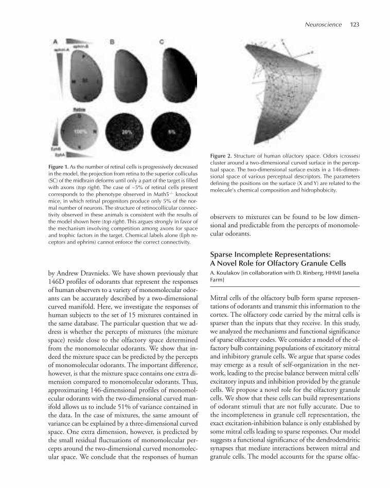
Neuroscience 123
by Andrew Dravnieks. We have shown previously that146D profiles of odorants that represent the responsesof human observers to a variety of monomolecular odor-ants can be accurately described by a two-dimensionalcurved manifold. Here, we investigate the responses ofhuman subjects to the set of 15 mixtures contained inthe same database. The particular question that we ad-dress is whether the percepts of mixtures (the mixturespace) reside close to the olfactory space determinedfrom the monomolecular odorants. We show that in-deed the mixture space can be predicted by the perceptsof monomolecular odorants. The important difference,however, is that the mixture space contains one extra di-mension compared to monomolecular odorants. Thus,approximating 146-dimensional profiles of monomol-ecular odorants with the two-dimensional curved man-ifold allows us to include 51% of variance contained inthe data. In the case of mixtures, the same amount ofvariance can be explained by a three-dimensional curvedspace. One extra dimension, however, is predicted bythe small residual fluctuations of monomolecular per-cepts around the two-dimensional curved monomolec-ular space. We conclude that the responses of human
observers to mixtures can be found to be low dimen-sional and predictable from the percepts of monomole-cular odorants.
Sparse Incomplete Representations:A Novel Role for Olfactory Granule CellsA. Koulakov [in collaboration with D. Rinberg, HHMI JaneliaFarm]
Mitral cells of the olfactory bulb form sparse represen-tations of odorants and transmit this information to thecortex. The olfactory code carried by the mitral cells issparser than the inputs that they receive. In this study,we analyzed the mechanisms and functional significanceof sparse olfactory codes. We consider a model of the ol-factory bulb containing populations of excitatory mitraland inhibitory granule cells. We argue that sparse codesmay emerge as a result of self-organization in the net-work, leading to the precise balance between mitral cells’excitatory inputs and inhibition provided by the granulecells. We propose a novel role for the olfactory granulecells. We show that these cells can build representationsof odorant stimuli that are not fully accurate. Due tothe incompleteness in granule cell representation, theexact excitation-inhibition balance is only established bysome mitral cells leading to sparse responses. Our modelsuggests a functional significance of the dendrodendriticsynapses that mediate interactions between mitral andgranule cells. The model accounts for the sparse olfac-
Figure 1.As the number of retinal cells is progressively decreasedin the model, the projection from retina to the superior colliculus(SC) of the midbrain deforms until only a part of the target is filledwith axons (top right). The case of ~5% of retinal cells presentcorresponds to the phenotype observed in Math5–/– knockoutmice, in which retinal progenitors produce only 5% of the nor-mal number of neurons. The structure of retinocollicular connec-tivity observed in these animals is consistent with the results ofthe model shown here (top right). This argues strongly in favor ofthe mechanism involving competition among axons for spaceand trophic factors in the target. Chemical labels alone (Eph re-ceptors and ephrins) cannot enforce the correct connectivity.
Figure 2. Structure of human olfactory space. Odors (crosses)cluster around a two-dimensional curved surface in the percep-tual space. The two-dimensional surface exists in a 146-dimen-sional space of various perceptual descriptors. The parametersdefining the positions on the surface (X and Y) are related to themolecule’s chemical composition and hidrophobicity.
097-149_04_Neurosci_Annual Report_2009 template 7/8/11 11:57 AM Page 123
124 Research
tory code in the steady state and predicts that transientdynamics may be less sparse.
Disposable Tissue Hypothesis: A NewModel for Hippocampal NeurogenesisA. Koulakov [in collaboration with G. Enikolopov, ColdSpring Harbor Laboratory]
New neurons are continuously generated throughoutthe life of an animal in at least two areas of the mam-malian brain: olfactory bulb and hippocampal dentategyrus. Hippocampal neurogenesis dynamically re-sponds to a multitude of extrinsic stimuli and may beimportant for behavior, pathophysiology, brain repair,and the response to drugs that modulate mood. Newneurons are produced from a limited population ofstem cells whose number declines with age. What fac-tors determine the rate of decline in the hippocampalstem cell population? How can the rate of productionof new neurons be varied? In this study, we have shownthat soon after a stem cell is used for production of newneurons, it becomes an astrocyte, i.e., leaves the poolof neuroprogenitors. Therefore, one can think of thestem cells as disposable: Once they are used, they can-not be reused again. The decline in the number of stemcells therefore occurs in a use-dependent manner. Does
this mean that the more neurons produced, the fasterthe stem cells are lost? Not necessarily. The sequence oftransformations that a cell must undergo before it be-comes a new neuron is very complex. One of the stepsinvolves an intermediate form of neuroprogenitors thatare called amplifying cells. These cells divide rapidly,about once per day, to make more neurons. It turns outthat antidepressant drugs, such as Prozac, affect the di-visions of the intermediate amplifying cells withoutmuch impact on primary (disposable) stem cells. To-gether with experimental group of Grisha Enikolopovat CSHL, we have developed a computational modelfor the division/differentiation cascade that leads to theproduction of new neurons. Comparing the computa-tional model to experimental results leads to detailedinformation about the changes occurring in the neuro-genesis cascade due to antidepressant drugs, aging, andother therapies. Our approach will provide unique in-sights to the computational properties of the neuralstem cells and their importance for mental health.
PUBLICATIONS
Kolterman BE, Koulakov AA. 2010. Is universal coverage good for neu-rons? Neuron 66: 45–56.
Tsigankov DN, Koulakov AA. 2010. Sperry versus Hebb: Topographicmapping in Isl2/EphA3 mutant mice. BMC Neurosci 11: 155.
097-149_04_Neurosci_Annual Report_2009 template 7/8/11 11:57 AM Page 124

125
Research in my laboratory is directed toward under-standing the synaptic mechanisms of aberrant behaviorin animal models of psychiatric disorders. Synaptic plas-ticity is believed to serve as the cellular mechanism forlearning and memory, and impairments in this processhave been linked to psychiatric disorders, includingschizophrenia and depression. We use rodent models ofthese disorders, and employ a number of complemen-tary methodologies, including behavioral assays, elec-trophysiology, two-photon imaging, in vivo circuit tracing,electrical deep-brain stimulation, molecular genetics, andoptogenetic techniques, to address questions in threemajor areas: (1) the synaptic mechanisms of depression;(2) the synaptic mechanisms underlying normal adap-tive behaviors, such as resilience to depression or be-havioral flexibility; (3) the synaptic mechanisms ofschizophrenia.
The Synaptic Circuitry of the LateralHabenula and Behavioral Depression
The cellular basis of depressive disorders is poorly under-stood. Recent studies in monkeys indicate that neuronsin the lateral habenula (LHb), a nucleus that mediatescommunication between forebrain and midbrain struc-tures, can increase their activity when an animal fails toreceive an expected positive reward or receives a stimulusthat predicts aversive conditions (i.e., disappointment oranticipation of a negative outcome). LHb neurons projectto and modulate dopamine-rich regions such as the ven-tral–tegmental area (VTA) that control reward-seekingbehavior and participate in depressive disorders. Ourstudy shows in two learned helplessness models of depres-sion that excitatory synapses onto LHb neurons project-
THE FUNCTION AND PLASTICITY OF CENTRAL SYNAPSESIN ANIMAL MODELS OF PSYCHIATRIC DISORDERS
B. Li S. Ahrens S. LiuH. Chae R. PaikK. Delevich Z. PerovaH. Li M. Wang
a VTA-p WT VTA-p cLH
500 ms20 pA
b
VTA-p cLHms VTA-p aLH
WT aLH0
10
20
40
30
WT cLH
mEP
SC F
req
uen
cy (H
z)
0
10
20
40
30
c
Nu
mb
er o
f cel
ls
mEPSC Frequency (Hz)
all the cells recorded(n=263 from 34 animals)
0 10 20 30
140
040cLH
-ms
d
Cu
m.
Pro
bab
ility
mEPSC Frequency (Hz)
0.5
1
0 10 20 30 40
WT
aLH
cLHms
cLH
mEPSC Amplitude (pA)0 10 20 30 40
0
1
Cu
m.
Pro
bab
ility
Am
plit
ud
e (p
A)
0
20
WT aLH cLH cLH-ms
84 8524 70
WT aLH cLHcLHms
120
100
80
60
40
20
e
n.s.
Figure 1. Increased excitatory synaptic transmission onto VTA-projecting LHb neurons in the learned helplessness models ofdepression. (a) Examples of mEPSCs recorded from VTA-project-ing LHb neurons (VTA-p) from wild-type (WT), cLH, cLHms, andaLH animals. (b) Means (bars) and individual recordings (circles)of mEPSC frequency from VTA-projecting LHb neurons in differ-ent groups of animals. (Left) (WT) 2.4 ± 0.3, n = 65 (6 animals);(cLH) 4.0 ± 0.7, n = 85 (8 animals), p < 0.05, bootstrap; (cLHms)4.7 ± 0.7, n = 70 (8 animals), p < 0.001, bootstrap. (Right) (WT)1.8 ± 0.2, n = 19 (4 animals); (aLH) 3.9 ± 1, n = 24 (4 animals),p < 0.05, bootstrap. (c) Frequency distribution of mEPSC frequen-cies of all cells recorded exhibited bimodal distribution. (d) Thecumulative probability of mEPSC frequency of VTA-projectingLHb neurons in different groups of animals (p < 0.05, K-S testcomparing WT with any other groups). (e) The amplitude ofmEPSC was not different among different animal groups (p > 0.3,bootstrap).
097-149_04_Neurosci_Annual Report_2009 template 7/8/11 11:57 AM Page 125

126 Research
ing to the VTA are potentiated (Fig. 1). Synaptic poten-tiation correlates with an animal’s helplessness behaviorand is due to an enhanced presynaptic release probability.Depleting transmitter release by repeated electrical stim-ulation of LHb afferents, using a protocol that can be ef-fective in depressed patients, dramatically suppressessynaptic drive onto VTA-projecting LHb neurons inbrain slices and can significantly reduce learned helpless-ness behavior in rats (Fig. 2). Our results indicate that in-creased presynaptic action onto LHb neurons contributesto the rodent learned helplessness model of depression.
Depression and Resilience: The Roleof the Medial Prefrontal Cortex
We are testing the hypothesis that normal synaptic plas-ticity in neurons of the medial prefrontal cortex
(mPFC) has an important role in behavioral flexibilityand resilience to depression, whereas its impairmentslead to behavioral depression. The mPFC has been im-plicated in executive function and behavioral flexibility.Recent studies suggest that mPFC is critical in the de-termination of whether an organism has control over astressor. Rats with inactivated mPFC are prone to be-havioral depression induced by stressors (Amat et al.,Nat Neurosci 8: 365–371 [2005]). Aberrant activity ofthe mPFC also accompanies clinical depression, anddeep-brain stimulation in mPFC leads to remission oftreatment-resistant depression (Mayberg et al., Neuron45: 651–660 [2005]). However, the cellular processesin mPFC accompanying behavioral depression are notclear. The cellular mechanisms in mPFC that underliecontrollability—therefore affording resilience to behav-ioral depression—are also unknown.
a
b
d
c
Baseline 150 µA0
8
# of
Lev
er-p
ress
*
Lever-press Test
6
4
2
0Day 1 Day 2
ShamDBS0.2
Frac
tion
of im
mob
ility
Forced swim Test
10 9
0.1 n.s.
Time (min)0 5 10 15 20
Nor
mal
ized
Slo
pe0
DBS
1st pulse2nd pulse
1
1
2
3
(1hr) 24 hLever-press(Baseline)
Test I(150 µA)
Test II(300 µA)
24 h
FST(Day 1)
FST(Day 2)
DBS DBS DBS
50 ms
5 m
V
1
2
3
*
LHb DBS (n=9)LHb Sham (n=14)
LPLR DBS (n=7)
Baseline
Test Com
pletion Tim
e (min)
10
20* *
e
300 µA 150 µA 300 µA
LHb DBS
LPLR DBS
***
Figure 2. DBS in LHb suppresses excitatory synaptic transmissionand reverses learned helplessness. (a, Left) Example EPSPs(paired-pulses) recorded from a VTA-projecting LHb neuron be-fore (1), during (2), and after (3) stimulation mimicking DBS. Ar-rows indicate when paired-pulses were given. (Right) AverageEPSP slope at indicated time points: (1) before (first pulse 1.1 ±0.1; second pulse 0.9 ± 0.1; n = 6 [4 animals]); (2) during (firstpulse 0.2 ± 0.07; second pulse 0.03 ± 0.03), p < 0.001 for bothpulses compared with those in 1; (3) after (first pulse 1.2 ± 0.3;second pulse 0.9 ± 0.2) DBS. (b) A schematic diagram showingthe experimental procedures. (c) Lever presses (left) and test com-pletion time (right) for animals that received DBS or sham stimu-lation in the LHb, or DBS in the LPLR (lateral postthalamic nuclei,laterorostral), before (baseline), or after DBS of different intensi-ties. DBS in LHb (n = 9): Lever press baseline 1.2 ± 0.4; 150 µAsession 3.9 ± 1; 300 µA session (n = 8) 5.8 ± 2. Test completiontime baseline 19.9 ± 0.4; 150 µA session 17.1 ± 1; 300 µA ses-sion (n = 8) 15.4 ± 2. Sham (n = 14): Lever press baseline 1.8 ±0.5; 150 µA session 2.4 ± 0.9; 300 µA session 2.4 ± 1. Test com-pletion time baseline 19.2 ± 0.5; 150 µA session 18.7 ± 0.9; 300µA session 18.5 ± 1.1. DBS in LPLR (n = 7): Lever press baseline0.4 ± 0.2; 150 µA session 2 ± 1; 300 µA session 1.1 ± 0.3. Testcompletion time baseline 20.6 ± 0.2; 150 µA session 19 ± 1.2;300 µA session 19.9 ± 0.3. For DBS in LHb group, *p < 0.05compared with baseline. Sham and DBS in LPLR group: p > 0.05for both measurements at both sessions compared with baseline(bootstrap). (d) Immobility during FST. DBS Day 1: 0.06 ± 0.01,Day 2: 0.06 ± 0.01, n = 9; Sham Day 1: 0.05 ± 0.01, Day 2: 0.17± 0.05, n = 10; DBS vs. Sham in Day 2: ***p < 0.001, bootstrap.(e) Representative cresyl violet staining of coronal brain sections;arrows indicate electrode track in the LHb (upper) or LPLR(lower). All error bars represent S.E.M.
097-149_04_Neurosci_Annual Report_2009 template 7/8/11 11:57 AM Page 126
Neuroscience 127
To determine cellular activity in the mPFC in differ-ent groups of animals, we measured c-Fos expression inthe infralimbic (IL) and prelimbic (PL) regions of themPFC. We found that in the learned helplessness (LH)model of depression, neurons in the mPFC were acti-vated. After the LH procedure, animal behavior divergedto become either depressed or resilient to depression. In-terestingly, the resilient animals had significantly en-hanced neuronal activation compared with depressedanimals, indicating an active coping process. Further ex-periments are needed to examine whether deferentialplastic changes in the synapses onto mPFC neurons inresponse to stress are critical in determining an animal’ssusceptibility or resilience to behavioral depression.
The mPFC and lateral habenula are synapticallyconnected and belong to a synaptic circuitry stronglyimplicated in depression. The first and second projectsare integral components of a synergistic effort aimed ata more complete understanding of the synaptic and cir-cuitry mechanisms underlying depression.
Genetic Deficiency, GlutamatergicHypofunction, and Schizophrenia
In this project, we are studying the genetic causes ofglutamatergic hypofunction, a pathological process be-lieved to contribute to the etiology of schizophrenia.We first focus on ErbB4, a gene that has been linked toboth schizophrenia and bipolar disorder. We choose tostudy brain circuits that have the highest levels ofErbB4 expression.
The thalamic reticular nucleus (RT) is critical inprocessing and filtering sensory stimuli. It is thought
to have an important role in attention, and impairmentin RT function has been implicated in psychiatric dis-orders such as schizophrenia. RT neurons, which areexclusively GABAergic, receive glutamatergic inputsfrom the cortex and thalamus and send projections tothe thalamus where they modulate the activity of thal-amic relay neurons. Interestingly, RT neurons are highlyenriched in ErbB4. Previous studies indicate that ErbB4controls glutamatergic synapse maturation and plastic-ity. On the basis of these findings, we hypothesize thatErbB4 controls the development and function of glu-tamatergic synapses in RT neurons, thereby controllingthe normal function of the RT–thalamic circuitry in at-tention. To manipulate ErbB4 function specifically inthe RT, we took advantage of the observation that so-matostatin (SOM) expression perfectly matches that ofErbB4 in the RT, but not in other brain areas includingcortex and hippocampus. By crossing the SOM-Creline with the ErbB4lox/lox animals, we have generatedSOM_ErbB4–/– mice in which the ErbB4 gene is selec-tively ablated in SOM-positive neurons. We are cur-rently examining the potential synaptic changes in RTneurons in these animals and testing the role of the RT-thalamic circuitry in a behavioral task designed tospecifically test attentional modulation of behavior.
PUBLICATIONS
Li B, Piriz J, Mirrione M, Chung C, Proulx C, Schulz D, Henn F, Ma-linow R. 2010. Synaptic potentiation onto lateral habenula neuronsin the learned helplessness model of depression. Nature 470:535–539.
Perova Z, Li B. 2010. Medial prefrontal cortical plasticity and behavioraldepression. Program No. 667.11; Neuroscience Meeting Planner. SanDiego, CA: Society for Neuroscience. Online.
Hong Goo Chae
097-149_04_Neurosci_Annual Report_2009 template 7/8/11 11:57 AM Page 127

128
Our basic research philosophy is to combine theoreti-cal, computational, and experimental approaches forthe study of complex biological systems, with a partic-ular focus on neurobiological questions. Efforts in ourgroup fall into three main areas: informatics, theory,and experimental work.
Our neuroinformatics research involves the applica-tion of analytical tools to large volumes of neurobiolog-ical data, as well as the development of informaticsinfrastructures for data and knowledge integration. Ourstudy in the area of theoretical engineering applies the-ories developed in human-engineered systems to studythe theoretical principles underlying biological systems.The final area of our research is experimental and haspreviously consisted of collaborative studies in multiplespecies including Drosophila, zebra finch, macaque mon-key, and human infants. We are now undertaking amajor in-house experimental project to generate the firstbrainwide mesoscale connectivity map in the mouse.
Continuing in our laboratory in 2010 are PascalGrange (postdoctoral fellow), Jamie Jones (laboratorytechnician), Jayant Kulkarni (postdoctoral fellow), San-dra Michelsen (administrator), Vadim Pinskiy (graduatestudent), and Caizhi Wu (research associate). SwagatamMukhopadhyay (postdoctoral fellow), David Prevzner(laboratory technician), Alex Tolpygo (laboratory tech-nician), Gregor Havkin (informatics manager), OsamaEl Demerdash (analyst), Wen Huang (graduate student),Joshua Novy (laboratory technician), Andrew Weber(laboratory technician), Zhen Gong (graduate student),Linzie Wood (project assistant), and Thomas Pologruto(consultant) joined us this past year.
We also have close collaborative ties with multiple re-search groups at CSHL and other institutions, which cur-rently include Dr. Nicholas D. Schiff at the Weill MedicalSchool of Cornell University, where Dr. Mitra is an adjunctassociate professor; a consortium of zebra finch researchers;an integrative analysis of memory formation in Drosophilawith Josh Dubnau at CSHL; the Brain Architecture Proj-ect, with collaborators at multiple institutions, notablyHarvey Karten (University of California, San Diego),
Kathy Rockland (Massachusetts Institute of Technology),and Michael Hawrylycz (Allen Institute of Brain Research);and a collaboration with Gregory Hannon at CSHL onimproving next-generation sequencing technologies.
Mouse Brain Architecture ProjectV. Pinskiy, A. Tolpygo, C. Wu, P. Grange, J. Jones, D. Prev-zner, A. Weber, J. Novy, G. Havkin, O. El Demerdash
Brain function is determined by its circuitry, but verylittle is known about how the mammalian brain is con-nected. We have set out to address this problem in theMouse Brain Architecture Project, in which we arecomprehensively determining the patterns of neuralconnectivity between brain regions.
We have obtained two National Institutes of Health(NIH) grants (a Transformative-R01 and an RC1 Chal-lenge Grant) that are running concurrently to producethe connectivity map. The RC1 grant is to produce aconnectivity draft for wild-type C57BL/6J mice and toalign these results to the Allen Brain Atlas. The initialdraft of a connectivity atlas will be available by fall orwinter of 2011. The T-RO1 grant will be used to increasethe sample size and coverage and to compare connectivitymaps of schizophrenia and autism mouse models to areference connectivity map in the C57 mouse brain.These data and the data analysis tools will be released onthe Mouse Brain Architecture project website.
This project is now in full production phase. Thepipeline to process tissue from tracer-injected micethrough to image analysis is fully functional and allequipment has been integrated into a Laboratory Infor-mation Management System (LIMS). This was devel-oped in-house and serves as an electronic lab notebook,facilitates quality control procedures, and is also the meta-data repository that will be finally published on the web.
Our plan involves grouping injections into major brainsystems (i.e., cortical areas, basal ganglia, thalamus, hy-pothalamus, etc.). Each injection site yields informationabout the other regions of the brain projecting to and
INTEGRATIVE SYSTEMS NEUROBIOLOGY
P. Mitra H. Cox J. Jones V. Pinskiy A. Weber O. El Demerdash J. Kulkarni T. Pologruto C. WuP. Grange S. Mukhopadhyay D. PrevznerG. Havkin J. Novy A. Tolpygo
097-149_04_Neurosci_Annual Report_2009 template 7/8/11 11:57 AM Page 128

Neuroscience 129
from the injected region. We are incorporating specificadvice from anatomists who specialize in these individualareas, in terms of injection strategies and interpretationof results. The initial 200 injection sites consist of a setcovering the cortex in a defined grid. CTB (cholera toxinB), BDA (biotin dextran amine), fluorescent-modified ra-bies viruses, and AAV (adeno-associated virus) will be theinitial tracers. We have already injected more than 400production animals at 100 different sites.
For these experiments, a computer-guided apparatusfor stereotactic injections into the mouse brain has beendeveloped. A laser scanner of the mouse skull, assembledby Vadim Pinskiy and Thomas Pologruto, gives rise toa profile of the surface of the skull of the live animal. Areference atlas of the mouse skull was provided by MarkHenkelman (Mouse Imaging Center, University ofToronto). A registration algorithm developed by PascalGrange allows us to align the skull and to work out itsvariation in size relative to the reference. The injectionis guided accordingly. The method was presented at theSociety for Neuroscience meeting in November 2010.
Other improvements to the pipeline include devel-opment of an embedding system that consistently ori-ents the brains for sectioning and allows two brains tobe sectioned simultaneously. We have also developed aGiemsa counterstain for the BDA and CTB tracers. Thisincreases the signal/noise ratio for the diaminobenzidine(DAB) staining and subsequent registration. A set ofNissl-stained sections for each brain will be used to cap-ture the cytoarchitecture. All of these improvements tothe pipeline increase both the throughput rate and theproper segmentation of individual brains and registra-tion to a common atlas space. The 20-µM brain sectionsare imaged using an automated microscope and new andupdated software has been written to aid with data crop-ping, transfer, and storage. This will be incorporatedinto the LIMS and used for quality control of the tissuesamples and data analysis. Other software components,currently under development, will take the stacks of dig-itized microscopy images and perform segmentation andthree-dimensional reconstruction to reconstitute thethree-dimensional brain. This brain volume is then reg-istered with the Allen Reference Atlas (ARA) (Fig. 1).
Although the total amount of data generated by theproject is expected to be large (terabytes to petabytes),we expect that the principal challenges will be in termsof software to enable suitable compression schemes topermit effective transmission through the Internet(which remains a low-bandwidth medium), as well assuitable visualization and search tools. The Allen Insti-tute has extensive experience in this domain, and Dr.
Mitra has established a collaborative relationship withDr. Mike Hawrylycz at the Allen Institute.
The preliminary data will be made available to theresearch community via an informatics platform (alongwith basic informatics tools to query, organize, visual-ize, and manage the data). We have implemented soft-ware to transform proprietary image data into readableand web-compatible formats and are beginning to as-semble the hardware and software infrastructure forsharing image data with collaborators and the outsideworld. We anticipate that the data set produced will beimportant for analysis of many kinds, and we expect tobe actively engaged in new analyses even beyond the 5-year grant period. The availability of public data setsand open-source duplicable pipeline technologies willpotentially impact the entire neuroscience community.
We are additionally developing a novel technique forassaying long-range connectivity in the human brainand are pursuing the aims of mining and integratinginformation about brain connectivity from the researchliterature using computational linguistics techniques.Finally, this project has also benefited from a summercourse that Dr. Mitra cofounded at CSHL in the be-ginning of June 2010. Through this project, ColdSpring Harbor Laboratory has become a leading centerfor filling the critical knowledge gap urged by Dr.Crick, and championed by Jim Watson.
For more information, see the Brain ArchitectureProject website: http://brainarchitecture.org
Analysis of Gene Expression in the Mouse BrainP. Grange
We are determining networks of spatial coexpression inthe brain, of addiction-related genes. An initial list ofmore than 400 genes related to addiction was compiledfrom on-line databases. For each pair of genes in thelist, the spatial overlap between gene expression patternsacross the brain was computed. The resulting coexpres-sion networks of genes can be rendered graphically—genes with similar expression energies appear asclustered nodes. These graphs have been made availableonline to the research community. This project was sup-ported the NIDA grant 1R21DA027644-01, Co-ex-pression networks of addiction-related genes.
We also developed numerical methods to rank genesand sets of genes as markers of brain regions. Localiza-tion scores for individual genes are available online(www.addiction.brainarchitecture.org, which also shows
097-149_04_Neurosci_Annual Report_2009 template 7/8/11 11:57 AM Page 129
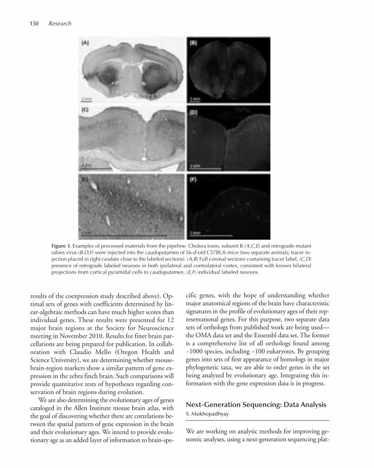
130 Research
results of the coexpression study described above). Op-timal sets of genes with coefficients determined by lin-ear-algebraic methods can have much higher scores thanindividual genes. These results were presented for 12major brain regions at the Society for Neurosciencemeeting in November 2010. Results for finer brain par-cellations are being prepared for publication. In collab-oration with Claudio Mello (Oregon Health andScience University), we are determining whether mouse-brain-region markers show a similar pattern of gene ex-pression in the zebra finch brain. Such comparisons willprovide quantitative tests of hypotheses regarding con-servation of brain regions during evolution.
We are also determining the evolutionary ages of genescataloged in the Allen Institute mouse brain atlas, withthe goal of discovering whether there are correlations be-tween the spatial pattern of gene expression in the brainand their evolutionary ages. We intend to provide evolu-tionary age as an added layer of information to brain-spe-
cific genes, with the hope of understanding whethermajor anatomical regions of the brain have characteristicsignatures in the profile of evolutionary ages of their rep-resentational genes. For this purpose, two separate datasets of orthologs from published work are being used—the OMA data set and the Ensembl data set. The formeris a comprehensive list of all orthologs found among~1000 species, including ~100 eukaryotes. By groupinggenes into sets of first appearance of homologs in majorphylogenetic taxa, we are able to order genes in the setbeing analyzed by evolutionary age. Integrating this in-formation with the gene expression data is in progress.
Next-Generation Sequencing: Data AnalysisS. Mukhopadhyay
We are working on analytic methods for improving ge-nomic analyses, using a next-generation sequencing plat-
Figure 1. Examples of processed materials from the pipeline. Cholera toxin, subunit B (A,C,E) and retrograde mutantrabies virus (B,D,F) were injected into the caudoputamen of 56-d-old C57BL/6 mice (two separate animals; tracer in-jection placed in right caudate close to the labeled sections). (A,B) Full coronal sections containing tracer label, (C,D)presence of retrograde labeled neurons in both ipsilateral and contralateral cortex, consistent with known bilateralprojections from cortical pyramidal cells to caudoputamen, (E,F) individual labeled neurons.
097-149_04_Neurosci_Annual Report_2009 template 7/8/11 11:57 AM Page 130
Neuroscience 131
form (single-molecule real-time sequencing, Pacific Bio-systems). In collaboration with the McCombie lab atCSHL, the objectives of this project are to investigateerror profiles in sequencing results and to improve base-calling algorithms by modeling the physical processes in-volved in polymerase kinetics and signal from thefluorescent-tagged (phospholinked) nucleotide incorpo-ration. The short-term goal includes developing algo-rithms for direct detection of DNA modifications byutilizing their signatures on polymerase kinetics. We arein the process of developing new tools for the various se-quencing methods used in the platform, such as Strobesequencing and Circular Consensus sequencing. Ourlong-term goal is to use the single-molecule signature ofpolymerase kinetics to refine our understanding of poly-merase motor itself.
Multimedia Signal Processingfor Quantative Phenotypingin Autism Spectrum DisordersZ. Havkin
We have been studying phenotypes of autism spectrumdisorders (ASDs) with support from the Simons Foun-dation Autism Research Initiative (SFARI). The work hasfocused on the application of multimedia signal process-ing (MMSP) techniques to audiovisual recordings of
ASD patients made during the administration of a stan-dardized diagnostic instrument (the Autism DiagnosticObservation Schedule). The ultimate goal of this researchis to objectively determine early-age ASD phenotypes, asit has been previously shown that early diagnosis and in-tervention leads to substantially improved outcomes.
Our key clinical collaborators are Woodfords FamilyServices in Maine and the Autism & CommunicationDisorders Center at the University of Michigan. Wewill be applying our MMSP techniques to their longi-tudinal studies which include ASD patients, non-ASDpatients, and typically developing peers.
PUBLICATIONS
Bohland JW, Bokil H, Pathak SD, Lee CK, Ng L, Lau C, Kuan C,Hawrylycz M, Mitra PP. 2010. Clustering of spatial gene expressionpatterns in the mouse brain and comparison with classical neu-roanatomy. Methods 50: 105–112.
Bokil H, Andrews P, Kulkarni JE, Mehta S, Mitra PP. 2010. Chronux:A platform for analyzing neural signals. J Neurosci Methods 192: 146–151.
Bolduc FV, Valente D, Mitra P, Tully T. 2010. An assay for social inter-action in Drosophila fragile X mutants. Fly 4: 216–225.
Erlich Y, Gordon A, Brand M, Hannon GJ, Mitra PP. 2010. Compressedgenotyping. IEEE Trans Inf Theory 56: 706–723.
Menzer DL, Bokil H, Ryou JW, Schiff ND, Purpura KP, Mitra PP. 2010.Characterization of trial-to-trial fluctuations in local field potentialsrecorded in cerebral cortex of awake behaving macaque. J NeurosciMethods 186: 250–261.
Alexander Tolpygo
097-149_04_Neurosci_Annual Report_2009 template 7/8/11 11:57 AM Page 131

132
What causes autism and schizophrenia? It is well estab-lished that both disorders are highly heritable, and today,geneticists are uncovering the relevant susceptibility genesand loci. The wealth of genetic data, however, does notguarantee rapid progress in understanding the diseasemechanisms. In fact, the susceptibility genes comprise aheterogeneous group that includes genes regulating tran-scription, protein degradation, synaptic signaling, andcell adhesion. What do these genes have in common?How can we best progress from identifying genes to test-ing therapeutic strategies?
We have developed a high-throughput pipeline ofmethods that can be used to systematically search forchanges in brain functions in mouse models carryinggenetic mutations linked to autism and schizophrenia.Our current work aims at identifying disrupted braincircuits that lie downstream from autism genes. Oncesuch circuit deficits are known, we and others can focuson determining the underlying cellular and molecular
mechanisms. Importantly, the same methods that weuse to discover disrupted circuits in the mouse braincan be also used to screen drugs that eliminate the ab-normality and restore normal functions. Our approachthus uniquely links basic and translational research,promising rapid progress from mechanistic studies totesting therapeutic strategies.
Sectioning-Based Serial Two-PhotonMicroscopy
A novel imaging technique, which we termed section-ing-based serial two-photon (SSTP) microscopy, is a keypart of our experimental approach (Fig. 1). The SSTPmicroscope, developed in a collaboration with Tissue-Vision, Inc. (Cambridge), works as follows: (1) A topview of a fixed mouse brain is imaged as a mosaic of20x objective fields of view (“tiles”), (2) a built-in vi-bratome cuts off the imaged top region, and (3) the cy-
IDENTIFICATION OF DISRUPTED BRAIN CIRCUITS IN MOUSEMODELS OF AUTISM AND SCHIZOPHRENIA
P. Osten Y. Bao R. Palaniswamy K.U. VenkatarajuL. Kadiri N. Takada D. XuY. Kim J. Taranda L. Zhang
Figure 1. SSTP microscopy. (A) Microscope scheme. The x-y-z stage moves the brain under the objective, so thatthe top view is imaged as a mosaic. The stage also moves the brain to the vibratome for sectioning. (B) An exampleof a GFP (green fluorescent protein) mouse brain imaged in 280 coronal sections.
A B
097-149_04_Neurosci_Annual Report_2009 template 7/8/11 11:57 AM Page 132

Neuroscience 133
we are testing a hypothesis that the functions of braincircuits involved in mediating social interactions are dis-rupted downstream from autism candidate genes. Theaims of both experiments are (1) to find differences inbrain functions between mutant and wild-type litter-mates and (2) to compare these differences between dif-ferent mutants in order to identify common disruptionsdownstream from multiple autism genes.
Anatomical Tracing andElectrophysiology
Our current work focuses on identification of brain areasand circuits disrupted in five genetic mouse models ofautism. Once such regions are found, our next step is touse electrophysiology and anatomical mapping to studyin detail the cellular mechanisms underlying the circuitdeficits. Electrophysiological experiments are done invitro in acute brain sections and in vivo by whole-cellrecording in anesthetized mice. Anatomical mapping isdone using traditional tracers, such as virus-vector-basedGFP expression for anterograde tracing and choleratoxin B for retrograde tracing (Fig. 3).
Summary
We hope that our work will provide better understand-ing of neural circuit disruptions underlying autism andschizophrenia and lead to therapeutic developments inthe future.
Figure 2. Brain activation in mice in-jected with (A) saline or (B) haloperi-dol (1 mg/kg). (C) Preliminary quantita-tion of c-fos-GFP-positive neurons be-tween the two conditions in individualcoronal sections. The asterisk marksthe approximate position of prominentc-fos-GFP induction in the striatum(B,C).
cles of mosaic imaging and sectioning are repeated.Once all data are collected, image-processing and brain-morphing algorithms are used to assemble the three-di-mensional brain volume for data analysis (Fig. 1).
Transgenic “Indicator” Mice
To study mouse brain functions by SSTP microscopy,we use transgenic indicator mice expressing GFP as areporter of neural activation, such as c-fos-GFP miceexpressing GFP from the promoter of the activity-reg-ulated immediate early gene (IEG) c-fos. These miceenable visualization of brain activation at cellular reso-lution in response to behavioral tasks or applicationsof drugs, such as the antipsychotic drug haloperidol(Fig. 2).
The use of SSTP microscopy to screen brain circuitsin genetic mouse models of autism involves (1) breed-ing of the selected mouse models with the indicator c-fos-GFP mice and (2) analysis of c-fos-GFP activationin the mouse offspring. We have initiated work on fivemouse models of autism: 16p11.2 deletion and dupli-cation mice generated by our collaborator Alea Mills atCSHL and previously published Neuroligin R451C,Neuroligin 4 knockout, and CNTNAP2/Caspr2knockout mice. We currently focus on two questions.We are testing a hypothesis that autism susceptibilitygenes cause an imbalance in brain excitation and inhi-bition, which leads to higher propensity for seizures andcontributes to disruptions of brain circuits relevant toautism manifestation. In a parallel set of experiments,
097-149_04_Neurosci_Annual Report_2009 template 7/8/11 11:57 AM Page 133
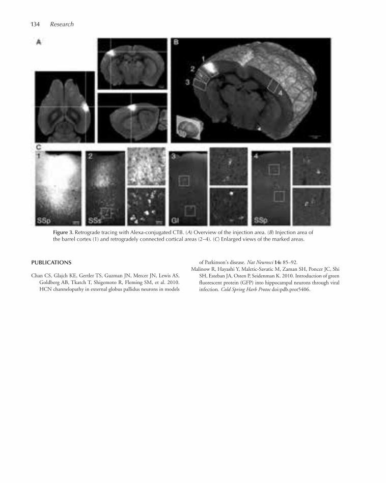
134 Research
PUBLICATIONS
Chan CS, Glajch KE, Gertler TS, Guzman JN, Mercer JN, Lewis AS,Goldberg AB, Tkatch T, Shigemoto R, Fleming SM, et al. 2010.HCN channelopathy in external globus pallidus neurons in models
of Parkinson’s disease. Nat Neurosci 14: 85–92.Malinow R, Hayashi Y, Maletic-Savatic M, Zaman SH, Poncer JC, Shi
SH, Esteban JA, Osten P, Seidenman K. 2010. Introduction of greenfluorescent protein (GFP) into hippocampal neurons through viralinfection. Cold Spring Harb Protoc doi:pdb.prot5406.
Figure 3. Retrograde tracing with Alexa-conjugated CTB. (A) Overview of the injection area. (B) Injection area ofthe barrel cortex (1) and retrogradely connected cortical areas (2–4). (C) Enlarged views of the marked areas.
097-149_04_Neurosci_Annual Report_2009 template 7/8/11 11:57 AM Page 134

–1.0
1.0
0.5
0
Inva
riant
set
z-s
core
Chr. 16p11.2 z-score
–0.5
–3 –2 –1 0 1 2 3
Figure 1. Detection of 16p11.2 microduplications by MeZOD.
The major focus of our laboratory is to identify geneticcauses of mental illness using a successful approach thatwe pioneered: high-resolution analysis of rare copy-number variants (CNVs) in the human genome. Wehave developed an experimental design that allows usto identify rare mutations that confer high risk for dis-ease. Our approach is to screen the genomes of patientswith schizophrenia and bipolar disorder to identifydeletions and duplications of the DNA that disruptgenes. These mutations are subsequently tested for as-sociation with disease in families and in large samplesof patients and healthy individuals. This approach isbased in part on the findings of our previous geneticstudies of autism spectrum disorders (ASDs), which es-tablished an important role for rare spontaneously oc-curring CNVs in the etiology of ASDs. With supportfrom the Stanley Foundation, we have now successfullyapplied this approach to studies of schizophrenia andbipolar disorder, and our work has already had a signif-icant impact on the field. Our findings, coupled withstudies by other groups, have shown that rare CNVshave an important role in the genetics of psychiatricdisorders. Furthermore, we have shown that a substan-tial number of the risk variants detected in the DNAof patients are not inherited from a mother or fatherbut instead occur by spontaneous mutation. Most in-dividual CNVs that have been definitively identified todate increase risk of mental illness by ≥10-fold, stronglysuggesting that these mutations have a causal role.
Analysis of Copy-Number Variationin SchizophreniaV. Vacic, S. McCarthy
Studies by our group and others have demonstrated thatindividually rare structural variants contribute to risk ofschizophrenia (SZ) and other neurocognitive disorders.Multiple studies have now shown that the mutationalburden of rare structural variants is significantly greaterin patients with SZ than in healthy controls. In addition,specific loci have been identified where structural muta-
RARE STRUCTURAL VARIATION IN THE GENOMEAND ITS ROLE IN HUMAN DISEASE
J. Sebat M. Kusenda V. VacicD. Malhotra K. YeS. McCarthy S. Yoon
tions are significantly associated with SZ. For example,large deletions at 1q21.1, 15q13.3, and 22q11.2 havebeen shown to confer substantial risk of SZ. A recentstudy by our group has found that microduplications of16p11.2 are significantly associated with SZ (Fig. 1, Table1; see McCarthy et al., Nat Genet 41: 123 [2009]). TheSZ-associated regions described here confer significantrisk. For instance, the observed odds ratios for the mi-croduplication of 16p11.2 range from 8.3 to 25.8.
These findings suggest that rare structural variantshave a role in the etiology of SZ. Furthermore, evidencesuggests that little of the overall contribution of rareCNVs to SZ can be explained by the handful of locidescribed above (Sebat et al., Trends Genet 25: 528[2009]). We hypothesize that genetic risk of SZ consistsin part of rare variants, and these risk alleles involvemany different genes. Analysis of structural variation inlarger samples is required to definitively identify novelCNV risk factors.
In an effort to identify novel CNV risk factors inSZ, we have recently carried out a two-stage study ofcopy number variation, using a primary sample of 742cases and 856 controls scanned in the Nimblegen HD2platform, and using as a replication data set CNV callsmade from the GAIN and MGS studies of SZ and theInternational Schizophrenia Consortium (ISC). Thereplication sample consisted of 7488 cases and 6689controls, bringing the combined sample size to 8230cases and 7545 controls.
135
097-149_04_Neurosci_Annual Report_2009 template 7/8/11 11:57 AM Page 135

136 Research
Investigating the Role of Rare CNVsin Bipolar DisorderD. Malhotra
Our research in bipolar disorder (BD) aims to make asignificant contribution to the field in two ways: by im-proving the scientific understanding of BD through ge-netic studies and by creating a novel genomic resourcefor geneticists. To these ends, we have initiated a geneticstudy, the genetics of early-onset mania (GEM). Thegoals of our genetic study of bipolar disorder are (1) toperform genome-wide analysis of copy-number varia-tion in bipolar families, (2) to assess the overall contri-bution of de novo and inherited mutations in sporadicand familial BD, and (3) to identify novel candidategenes for further study.
Central to this study is the recruitment of new bipo-lar families who will form the GEM family collection.GEM is unique from other family collections in that asignificant fraction of patients are “sporadic” cases anda significant fraction of patients are young at onset(AAO < 18). This new collection will be made availableto the scientific community where we anticipate it willbecome a valuable resource for researchers who are in-terested in investigating genetic and epigenetic causesof bipolar disorder.
We have completed a preliminary analysis of data fromour genetic study of BD (described in this report), andthese data showed that rare structural variants, includingde novo mutations and inherited CNVs, contribute toBD. We have performed an analysis of de novo CNVs infamilies and observed a higher rate of de novo mutation
In the first stage, we screened the primary sample forCNVs that are present in multiple cases and not presentin a sample of matched controls. After eliminatingCNVs that were less than 100 kb in size and CNVs thatdid not impact genes, we identified a total of 66 uniquetarget loci. To measure statistical association in thereplication sample, we tested the difference in locus-based mutational burden between cases and controlsusing a Fisher’s exact conditional test. In this study, allof the previously identified loci described above showeda statistically significant association (data not shown).In addition, duplications at a single 7q36.3 region wassignificantly associated with SZ after correction for the66 loci tested (α = 0.0008).
The 7q36.3 region represents a novel locus thatconfers significant risk of SZ (see Table 2). This findingserves to highlight novel genes within the region. Oneof particular interest is the vasointestinal peptide re-ceptor 2 (VIPR2). VIPR2 is a class II G-protein-cou-pled receptor that is activated by both vasoactiveintestinal peptide (VIP) and pituitary-adenylyl-cyclase-activating polypeptide (PACAP). VIP and PACAPhave been shown to have wide pharamacological effectsand biological functions. Notably, PACAP is involvedin pain-related behavior, psychomotor functioning,and memory performance. PACAP-deficient mice dis-play increased locomotor, exploratory and explosivejumping activity, and a deficit in prepulse inhibitionof the acoustic startle response, all of which are atten-uated by risperidone. In addition, within the suprachi-asmatic nucleus, VIPR2 is required for maintenance ofnormal circadian rhythms.
Table 2. Significant Association of 7q36.3 Microduplications with Schizophrenia
Primary sample Replication sample
Locus CNV type cases controls cases controls OR (96% CI) replication P value
7q36.6 duplication 2 0 12 0 INF (3.3, INF) 0.00022p16.3/NRXN1 deletion 2 0 14 2 6.4 (p1.7, INF) 0.00223q29 duplication 2 0 8 0 INF (2.0, Infinity) 0.0049
Table 1. Duplications and Deletions at 16p11.2 among Persons with Schizophrenia and Controls
Subjects Deletions Duplications
Series Diagnosis (n) n % n % OR (95% CI) P value
Primary schizophrenia 1906 1 0.05 12 0.63 24.8 (3.3, 199) 1.2 x 10⁻⁵
controls 3971 3 0.08 1 0.03Replications schizophrenia 2645 0 0.00 9 0.34 8.4 (1.3, 50.5) 0.022
controls 2420 1 0.04 1 0.04Combined schizophrenia 4551 1 0.02 21 0.46 14.5 (3.3, 62.0) 4.3 x 10⁻⁵
controls 6381 4 0.06 2 0.03
097-149_04_Neurosci_Annual Report_2009 template 7/8/11 11:57 AM Page 136
Neuroscience 137
in BD compared to healthy controls. Furthermore, weobserved a higher rate of de novo mutation in sporadiccases as compared to patients with a family history ofmental illness. These results provide strong preliminaryevidence that de novo CNVs contribute in part to the ge-netics of BD. A majority of these mutations involve genes,thus highlighting strong candidate regions for furtherstudy. In addition, we have performed an analysis of rareinherited CNVs in families. These results provide prelim-inary evidence for the increased mutational burden of rareinherited CNVs in BD.
Analysis of Structural Variationin the 1000 Genomes ProjectS. Yoon, K. Ye
The primary goal of this project is to define genome-wide patterns of structural variation by high-resolutionanalysis of CNV using two advanced technologies: mi-croarray comparative genomic hybridization (CGH)and next-generation sequencing (NGS). We have acomplete collection of all primary data for the project,including a total of more than 500 HapMap samples.We have begun the process of validating CNVs andgenotype calls and have designed a custom Agilent arraycontaining dense coverage of ~800 common CNVs andan additional set of rare inherited and de novo CNVs.Microarray CGH analysis will be performed onHapMap samples using this custom array to validate
our CNV calls and to better define the breakpoints ofvariants identified in this study.
Through our participation in the 1000 genomesproject, we have performed an analysis of structuralvariation in whole-genome sequence data on multipleindividuals. For these studies, we developed a novelmethod for detection of CNVs based on coveragedepth: event-wise testing (EWT). This call set is cur-rently being integrated with CNV call sets from multi-ple groups using complementary approaches. Theseresults will constitute the first official data release of the1000 genomes pilot project, which will tentatively bepublished in late fall.
PUBLICATIONS
1000 Genomes Project Consortium, Durbin RM, Abecasis GR, Alt-shuler DL, Auton A, Brooks LD, Gibbs RA, Hurles ME, McVeanGA. 2010. A map of human genome variation from population-scalesequencing. Nature 467: 1061–1073.
In Press
Mills RE, Walter K, Stewart C, Handsaker RE, Chen K, Alkan C, Aby-zov A, Yoon SC, Ye K, Cheetham RK, et al. 2011. Mapping copynumber variation by population-scale genome sequencing. Nature470: 59–65.
Vacic V, McCarthy S, Malhotra D, Murray F, Chou HH, Peoples A,Makarov V, Yoon S, Bhandari A, Corominas R, et al. 2011. Dupli-cations of the neuropeptide receptor gene VIPR2 confer significantrisk for schizophrenia. Nature [Epub ahead of print] PMID:21346763 (in press).
097-149_04_Neurosci_Annual Report_2009 template 7/8/11 11:57 AM Page 137
138
The overarching research goal in our laboratory is tounderstand how processing in specific brain circuitsworks to support natural communication behaviors.We aim to reveal neural mechanisms that allow organ-isms to detect and recognize familiar individuals, togather information about their identity and social sta-tus, and to select appropriate behaviors. Mice are capa-ble of acquiring detailed profiles of one another fromthe smells and sounds experienced during their socialencounters. These dossiers may include information ona mouse’s sex, genetic identity, reproductive state, levelsof distress or sexual interest, or even recently consumedfoods, details that are indispensable for survival andmating success. Initially, we are working to understandthe neuronal activities and mechanisms in primary sen-sory brain areas that support these forms of communi-cation. In the future, we anticipate moving deeper intothe brain to ascertain where the sensory data from thoseregions are collected and integrated into hormonal andelectrical signals that promote appropriate behavioralchoices.
The scientific benefit of this approach is twofold.First, we want to identify fundamental principles ofhow the brain controls complex behavior. To this end,it is our belief that the nervous system’s function is bestinterpreted in the context of the behaviors it was evo-lutionarily designed to perform. Thus, it is advanta-geous to use natural behaviors such as intraspecificcommunication. Second, impairment of social percep-tion and cognition is a core feature of autism spectrumdisorder (ASD); for example, patients may have diffi-culty perceiving and interpreting communication ges-tures such as speech, facial expressions, and “bodylanguage.” This broad feature is recapitulated in manymouse models of ASD that carry genetic variants iden-tified in human ASD populations. Therefore, if we canascertain the neural circuit substrates of social behaviorin normal mice, we can make and test predictions forhow the circuitry is affected in the mouse models. Theresults are likely to tell us more about the synaptic mod-ifications that occur in human autism.
There are three broad areas of research in the lab.One concerns olfactory communication and memories
for familiar individuals. Another direction is an exam-ination of vocal (auditory) communication betweenmother mice and their pups. Our final avenue of studyinvolves recording from neurons in awake, behaving an-imals to reveal the neural encoding of social cues duringa live encounter with another mouse.
Noradrenaline and Memoriesfor Familiar IndividualsH. Demmer, D. Eckmeier, J. Dos Santos
How do we remember individuals who we have previ-ously encountered? Substantial evidence indicates thatmany animals remember each other based on olfactorycues. Memories are especially strong for individuals en-countered during key life events such as mating with anew partner or the birth of a litter of young. These im-portant events typically evoke massive release of the neu-rochemical noradrenaline (NA), initiating a heightenedstate of emotion and arousal. This surge appears to causelong-lasting modifications to the responses to odorantsin the olfactory bulb, which is the first processing stationfor scent in the mammalian brain. Indeed, it was hypoth-esized that the coincidence of an odor stimulus with asurge of NA is minimally sufficient to store a memory.We exploited the intimate relationship among NA, ol-factory bulb activity, and behavior to create and studyolfactory memories in the anesthetized mouse. We dis-covered that, indeed, when NA release is evoked by stim-ulating locus coeruleus, the source of most NA, while thesleeping mouse sniffed an odorant, neural responses tothat odor underwent specific long-term alterations. Re-markably, once awake, the mouse’s subsequent behaviortoward the odorant was also changed. In other words,the mouse seemed to remember the odor and treat it asthough it were familiar.
The ability to induce ethologically relevant memoriesunder anesthesia opens up exciting possibilities for ob-serving the synaptic mechanisms underpinning suchmemories using advanced techniques that are currentlyimpossible in behaving animals. For example, we are be-ginning to use a variety of imaging approaches in genet-
NEURAL CIRCUITRY FOR SOCIAL COMMUNICATION
S. Shea H. Demmer D. EckmeierJ. Dos Santos R. Hu
097-149_04_Neurosci_Annual Report_2009 template 7/8/11 11:57 AM Page 138

Neuroscience 139
ically modified mice during memory formation. Theseexperiments will allow us to separately visualize olfactorybulb input and output as well as wide-scale neuronalpopulations to ascertain how and where NA-dependentplasticity is coordinated and interacts among neuronalpopulations. We are also beginning to use high-resolu-tion electrophysiological techniques, targeted to specificcell types in order to build a circuit picture of how dif-ferent olfactory bulb cell types adapt their firing to resultin long-term changes to circuit output.
How are olfactory memories for individuals storedmechanistically among the specific synaptic connec-tions of the various neuronal types in the olfactorybulb? To answer this question, this year in our lab, Dr.Heike Demmer developed techniques for making tech-nically challenging targeted recordings from a specifictype of inhibitory neuron (granule cells) whose func-tion remains mysterious. She then made these record-ings during the induction of NA-dependent plasticityto examine how they contribute to memories. The datasuggest that these cells not only participate in storingolfactory memories, but do so in surprising and com-plex ways. Although we anticipated that the granulecells might increase their activity to suppress responsesto remembered odors, we found the opposite was true(Fig. 1). One interpretation is that NA input uncouplesgranule cells’ synaptic output from their action poten-tial firing. This unique property could be an importantfactor in maintaining memory specificity.
How are changes in the firing rate of olfactory bulbneurons read out by deeper stations in the brain? It has
been hypothesized that NA may modulate behavior bysuppressing input to downstream targets that mediateinnate behavioral responses. To begin testing this hy-pothesis, Julien Dos Santos made recordings from someof these deep-brain structures including the bed nucleusof the stria terminalis and the medial amygdala. He wasable to demonstrate that these regions respond robustlyto biologically important stimuli such as urine from theopposite sex and predator odors. This work sets thestage for examining how NA shapes these responses andrelating these effects to different behaviors.
Vocal Communication betweenMothers and PupsR. Hu
Far outside the range of our hearing, in the ultrasoundrange, mice are constantly holding conversations withone another in a language that is poorly understood atbest. Many types of vocalizations are emitted by malesand females, juveniles and adults, in a variety of behav-ioral contexts. We would like to better understand theperceptual significance of these calls to the mice andhow they are used to guide behavioral choices.
One form of vocalization that is actually reasonablywell understood is the ultrasonic distress vocalization(USV). Young mice prior to vision and full mobilitywill occasionally become separated from the nest. Thisis stressful for them and they will therefore call out totheir mother with a very-high-frequency peep. New
Figure 1. Granule cell interneurons of the main olfactory bulb show long-term suppression of odor responses afterlocus coeruleus (LC) stimulation. This figure depicts the effects on a granule cell’s odor responses of electrical LCstimulation (which releases NA) paired to that odor. (Left panel) Two PSTHs showing the pre- and poststimulationresponse of the granule cell to the odor. (Right panel) A plot of response strength (spikes/s above baseline) for eachof 120 consecutive trials of odor presentation with the shaded region denoting the period of LC stimulation pairing.
097-149_04_Neurosci_Annual Report_2009 template 7/8/11 11:57 AM Page 139

140 Research
mothers develop sensitivity to these cries and respondby moving toward their source (phonotaxis) to retrievethe pup. Mothers will also approach a speaker emittingplayback of synthetic calls, providing a simple assay fortheir perception of manipulated calls. Such experimentssuggest that there are sharp limits to the types of soundsthat will elicit phonotaxis, possibly implying a neuralselectivity mechanism that creates a perceptual bound-ary between pup distress cries and other sounds and vo-calizations. Moreover, nulliparous females who havenever given birth do not show approach responses topups or their calls, suggesting that the underlying neu-ral responses may also differ.
To begin to search for such neural correlates of ma-ternal behavior, Ruilong Hu, a summer URP student,made recordings from the auditory cortex of mothersand naïve nulliparous females and assessed the responseto a series of synthetic pup calls. He found that neuronsin the auditory cortex of mothers were much moresharply tuned to pup-call frequencies as compared tothe neurons of naïve females (Fig. 2). These data implythat somehow maternal experience rewires the brain tomore optimally discriminate biologically importantstimuli. How is this neural selectivity constructed at thesynaptic level? We have hypothesized that the answercritically involves intracortical inhibitory processing.Therefore, we are using optogenetic techniques to in-
activate inhibitory inputs to ultrasound-sensitive neu-rons to assess their necessity for proper auditory pro-cessing.
Neural Activity during Social EncountersD. Eckmeier
We are building a setup for recording individual neu-rons during social encounters and other behavioral as-says involving the perception of social and nonsocialinformation. There are two broad related goals to thisapproach. The first goal is to examine the encoding ofsocial information such as body odors and vocalizationsin primary sensory structures of awake animals. We hy-pothesize that activity in response to these signals maybe labile to associative learning, attention, and arousalwhich we may be able to manipulate in the context ofsocial encounters. The second goal is to record fromneurons in deep-brain neuromodulatory centers duringthese encounters as well. Neurons that release noradren-aline and dopamine are likely responsive to social sig-nals and may modulate encoding of sensory data andassociative plasticity. Understanding the context-depen-dent activity patterns of these neurotransmitters istherefore critical to developing models for how they af-fect behavior.
Figure 2. Auditory cortical neurons in mothers show greater spectral selectivity for USV frequenciesthan neurons in naïve females. (Left panel) A schematic representation of the bandpass noise stimulithat were randomly presented to anesthetized mothers and naïve females during recordings of au-ditory cortical neurons. (Right panel) Neurophysiological results. As more low-frequency contentwas added to the stimulus, responses in mothers dropped off precipitously, demonstrating that theseneurons are more selective for USV content. All responses were normalized to the response to thenarrowest band stimulus.
097-149_04_Neurosci_Annual Report_2009 template 7/8/11 11:57 AM Page 140
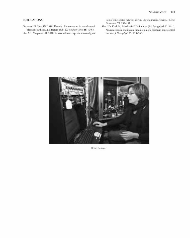
Neuroscience 141
PUBLICATIONS
Demmer HS, Shea SD. 2010. The role of interneurons in noradrenergicplasticity in the main olfactory bulb. Soc Neurosci Abstr 36: 730.5.
Shea SD, Margoliash D. 2010. Behavioral state-dependent reconfigura-
tion of song-related network activity and cholinergic systems. J ChemNeuroanat 39: 132–140.
Shea SD, Koch H, Baleckaitis DD, Ramirez JM, Margoliash D. 2010.Neuron-specific cholinergic modulation of a forebrain song controlnucleus. J Neurophys 103: 733–745.
Heike Demmer
097-149_04_Neurosci_Annual Report_2009 template 7/8/11 11:57 AM Page 141

142
The brain has a tremendous capacity to form manyhighly accurate memories; it is precisely this facility thatis lost in diseases such as Alzheimer’s and other demen-tias. Our overall goal is to understand how brain areasinvolved in learning and memory represent differentstimuli with distinct patterns of neural activity and howthese activity patterns are modified by learning.
We use the olfactory system of Drosophila as a modelfor investigating these questions. Our research focusesmostly on a brain area involved in learning and memoryknown as the mushroom body (MB), which is analogousto the olfactory cortex in the mammalian brain. To un-derstand how olfactory information is represented in theMB, we monitor neural activity using both electrophysi-ological and functional imaging techniques. We havefound that MB neurons are extremely odor selective intheir responses. This high selectivity is a general feature ofbrain areas involved in learning and memory, includingthe hippocampus and cerebellum. Using the simplicityand genetic manipulability of Drosophila, our goal is to es-tablish how this specificity arises and how it is maintainedin the face of learning-related changes. The extremely pow-erful genetic tools in Drosophila enable us to manipulateneural activity in precisely defined ways that help us to un-derstand how this circuit functions. For example, by con-trolling activity of the neurons immediately presynaptic tothe MB, we can determine how MB neurons integrate dif-ferent inputs and establish the number, strength, and dy-namics of the synaptic connections across these layers.Using this type of approach, we are currently evaluatinghow MB response properties arise and how they are mod-ified by the action of important neuromodulatory systems.Additionally, extensive genetic studies of olfactory learningand memory have identified many of the genes involvedin learning and memory; one of our goals is to understandhow these molecules affect network-level activity.
Functional Imaging of Population Codingin Olfaction: Neural Activity to Perception R. Campbell, K. Honegger
A central goal in systems neuroscience is to understandthe relationship between neural activity and sensory
perception. How different do two response patternshave to be in order to be perceived as distinct? This dif-ference can be considered a unit of the neural code: thesmallest difference in activity that the animal can per-ceive. We have addressed this question in the olfactorysystem of Drosophila, focusing on a brain area knownas the MB, which is essential for learned olfactory dis-crimination. Using imaging techniques to track morethan half the population of MB neurons, we addressthis question with a completeness not currently feasiblein mammalian systems.
We compared behavioral measures of olfactory dis-crimination with neural activity patterns monitoredusing calcium imaging. Flies were trained to form aPavlovian association with one odor and then to choosebetween the trained odor and a different odor in a Tmaze. To track neural activity, we targeted expressionof the calcium sensor, GCaMP 3.0, specifically to MBneurons using genetic tools available in Drosophila. Weused two-photon imaging to record activity patterns,incorporating a piezoelectric z-motor to image the en-tire MB in a three-dimensional volume during a singleodor presentation. This approach enabled us to rou-tinely monitor neural activity of most MB neurons. Bymeasuring odor responses on a trial-by-trial basis, wecould accurately quantify response variability. This en-abled us to construct an algorithm to classify odorsbased on the patterns of active neurons, taking into ac-count the noise inherent in neuronal responses.
MB odor responses were sparse, consistent with elec-trophysiological recordings. Different monomolecularodors could be readily distinguished from one anotherbased on activity patterns, even when odors evoked ex-ceptionally similar patterns of activity in the populationof olfactory receptor neurons. Behavioral tests showed asimilar level of discriminability. We then tested a seriesof progressively similar binary blends of odor, which en-abled us to construct a psychometric curve characterizingdiscriminability as a function of stimulus similarity. Weestablished a corresponding neurometric curve describ-ing our ability to classify odors based on those activitypatterns. There was a close correspondence between theneurometric and psychometric curves, indicating that
NEURAL CODING AND MEMORY FORMATION IN THE DROSOPHILAOLFACTORY SYSTEM
G. Turner R. Campbell T. HigeE. Gruntman K. Honegger
097-149_04_Neurosci_Annual Report_2009 template 7/8/11 11:57 AM Page 142

Neuroscience 143
we can interpret the patterns of MB activity in naive fliesin a way that accurately predicts how finely they canlearn to discriminate odors. Overall, our results showthat small differences in input are nevertheless easily dis-tinguished in the MB population, a transformation thatlikely underlies accurate information storage.
Robustness of Sparse Coding in theDrosophila Mushroom BodyK. Honegger, R. Campbell
Theoretical work suggests that sparse representationsare useful for learning and memory because they enablesynaptic changes to modify representation of one stim-ulus without interfering with another. However, an im-portant underlying assumption is that these sparserepresentations are robust to natural variations in thestimulus. There are two broad challenges to maintain-ing a sparse representation: variations in stimulus in-tensity and variations in stimulus complexity. We haveexamined how the sparse, specific MB responses aremaintained in the face of these challenges.
We examined robustness to stimulus intensity by lin-early varying odor concentration across two orders ofmagnitude. We found that in general, responses are ex-tremely similar across different concentrations, a usefulfeature that would enable odor recognition indepen-dent of intensity. Interestingly, there is a form of plas-ticity that sets this invariance: When the same odor ispresented in a series of pulses of steadily increasing con-centration, the MB response appears to be completelyconcentration invariant. When pulses are steadily de-creasing, however, response strength is proportional toodor concentration. This suggests that when travelingup a concentration gradient, the MB only conveys in-formation about odor identity, perhaps to confirm thatthe odor is still present, whereas decreasing odor inten-sity should be readily detectable by neurons down-stream from the MB.
To evaluate the effects of stimulus complexity, wetested responses to multicomponent odors, includingnaturally occurring smells. Several considerations sug-gest that natural odors may evoke responses in the MBfundamentally different from those produced bymonomolecular odorants. In other systems such as vi-sion, it is the stimuli that have meaning to the animalthat evoke strong responses in neurons at the deeper lay-ers. Studying deeper olfactory areas with monomolecu-lar odorants the animal has never before encounteredmay have led to an impoverished view of MB responseproperties. Additionally, our observations that individual
Kenyon cells (KCs) integrate input from several differentprojection neurons (PNs) (see below) raise the possibil-ity that many KCs respond selectively to odor blends,such as natural odors. In contrast to these expectations,we find that natural odors evoke responses that are com-parably sparse and selective to responses to monomole-cular odorants.
Additionally, imaging allows us to determine whetherodor responses are in some way spatially clustered, withpockets of dense activity. In earlier layers of the olfactorysystem, there is a clear spatial organization of responses;however, in the MB, no obvious structure exists and re-sponses appear to be distributed randomly. We alsoruled out the possibility that MB neurons are spatiallyarranged according to their odor-tuning properties. Al-together, these results are consistent with the notion thatthe MB is a purely associative area: Natural stimuli thathave intrinsic meaning do not elicit qualitatively distinctresponses from monomolecular stimuli nor is there anyobvious spatial segregation based on odor response pref-erences that would suggest olfactory information ischanneled into distinct anatomical pathways.
Integration Properties of MB NeuronsE. Gruntman
The nervous system has to represent a large number ofdifferent stimuli using a relatively small number of neu-rons. At the sensory layer, combinatorial coding by largeensembles of neurons enables a small number of cells torepresent a much larger number of different stimuli. Indeeper layers, sparse representations by highly stimulus-selective neurons can ensure that similar stimuli evokemarkedly distinct patterns of activity, a feature thoughtto be important for accurate memory formation. Theolfactory system in Drosophila melanogaster displaysthese fundamental characteristics: The olfactory receptorneurons represent odors combinatorially, and they aremaintained as functional units by projecting to formsynapses in distinct glomeruli within the second layer ofthe system, the antennal lobe. At the third layer, a learn-ing and memory area termed the MB, neurons knownas KCs are highly stimulus specific and odor responsesare relatively sparse in this population.
One hypothesis for how KCs achieve their high odorspecificity is that they integrate across several differentinput channels and require the combined activity ofthose inputs in order to spike. Projection neurons fromthe second layer of the system relay information fromdistinct glomeruli to the KCs; however, it is yet un-known whether each individual KC receives informa-
097-149_04_Neurosci_Annual Report_2009 template 7/8/11 11:57 AM Page 143
144 Research
tion from the same glomerulus, thus acting as an am-plifier, or from different glomeruli, acting as an inte-grator.
We expressed the calcium sensor GCaMP3.0 in sin-gle KCs and used two-photon imaging to monitor theolfactory responses of individual dendritic sites in a liv-ing fly. This enabled us to construct an odor-tuningcurve for each dendritic input site. By comparing tun-ing curves for different sites, we directly tested whetherKCs integrate combinations of different inputs andwhether the integration is a prerequisite for a spikingresponse. Our results suggest that some KC types inte-grate information whereas others mainly amplify. Wealso found that KC spiking requires simultaneous acti-vation of most of the input units. By integrating acrossdifferent input channels, KCs in effect read the combi-natorial code of the earlier layers.
Decoding Sparse RepresentationsT. Hige
Sparse representations are useful for learning and mem-ory, but how do downstream neurons integrate this in-formation? Ultimately, the information in layers withsparse representations has to be converted into a behav-ioral response. How does this process occur and howdo neurons downstream from a sparsely respondingbrain area integrate that information?
In Drosophila, this process appears to occur imme-diately after the MB, as the 2500-Mb neurons convergeonto an estimated 50 output neurons. Individual mem-bers of this output population can be green fluorescentprotein (GFP) labeled using the genetic tools available
in Drosophila, making this an excellent system to deter-mine the basic principles of information processingdownstream from sparse representations. Using GFP-targeted intracellular recordings, we have characterizedthe odor responses of one of these MB output neurons.In contrast to the highly odor-specific tuning of MBneurons, this output cell responds to a broad array ofdifferent odors. These functional properties are reflectedin the cell’s anatomy: It has very extensive dendriticprocesses within the output lobes of the MB, suggestingit receives highly convergent input from many MB neu-rons. Although the neuron responds to almost all testedodors, it nevertheless responds distinctly to differentodors, suggesting that this neuron is capable of carryingodor-specific information. However, the odor-tuningproperties of this neuron vary among individual flies—the same neuron has very different tuning curve shapesin different animals. One explanation for this variabilityis that it reflects synaptic plasticity that operates on adevelopmental timescale. Our working hypothesis isthat the tuning curves of the MB output neurons are aproduct of the prior olfactory experience of the indi-vidual fly. We are currently testing this by examiningthe role of olfactory experience in shaping the responseproperties of these neurons.
PUBLICATIONS
Campbell RA, Turner GC. 2010. The mushroom body. Curr Biol 20:R11–R12.
Murthy M, Turner GC. 2010. In vivo whole-cell recordings in theDrosophila brain. In Drosophila neurobiology methods: A laboratorymanual (ed. S Waddell et al). Cold Spring Harbor Laboratory Press,Cold Spring Harbor, New York.
097-149_04_Neurosci_Annual Report_2009 template 7/8/11 11:57 AM Page 144
145
My laboratory is interested in how neural circuits un-derlie normal processing and attention in the auditorycortex, and how this processing is disrupted in cognitivedisorders such as autism. To address these questions, weuse a combination of computational, electrophysiolog-ical and imaging techniques at the molecular, synaptic,cellular, circuit, and behavioral levels.
Sequencing the ConnectomeS. Chattopadhyay, G. Cao, H. Oyibo, S. Peikon, H. Zhan [in collaboration with J. Dubnau, L. Enquist, Princeton University]
The brain is a complex network, consisting of billions ofneurons connected by trillions of synapses. The details ofthese connections—which neurons form synaptic connec-tions with which other neurons—are crucial in determin-ing brain function. Malformation of these connectionsduring prenatal and early postnatal development can leadto mental retardation, autism, or schizophrenia; loss ofspecific connections later in life is associated with neu-rodegenerative diseases such as Alzheimer’s. An efficientmethod for determining the brain’s wiring diagram wouldtransform neuroscience research.
Inspired in part by the success of the Allen BrainAtlas, the Human Genome Project, and other major ef-forts, there is growing excitement in neuroscience todetermine the complete connectivity diagram—the“connectome”—of the brain. So far, the complete con-nectome has been established for only one organism,the tiny worm Caenorhabditis elegans, with 302 neuronsconnected by ~7000 synapses. However, determiningthe connectome of even this simple nervous system wasa heroic task, requiring more than 50 person-years oflabor to collect and analyze the electron micrographs.
All current approaches to determining the connectomeare based on microscopy. Unfortunately, microscopy ispoorly suited to the study of neural connectivity becausebrains are macroscopic structures, whereas the synapticconnections between neurons require electron microscopy.Reconstructing the complete wiring diagram of such a
brain is akin to piecing together the complete roadmap ofthe United States from a collection of postage-stamp-sizedphotographs.
To circumvent the considerable challenges associatedwith determining the connectome based on microscopy,we are developing an entirely novel approach based onhigh-throughput DNA sequencing technology. Sequenc-ing technology has not previously been applied in the con-text of neural connectivity. The appeal of using sequencingis that it is already fast—sequencing hundreds of millionsof individual pieces of DNA in a single day is now rou-tine—and, like microprocessor technology, getting fasterexponentially. Moreover, the cost of sequencing is plum-meting: Predictions are that it will be possible to sequencean entire human genome (~3B nucleotides) for $1000within a few years. Thus, by converting brain connectivityfrom a problem of microscopy to a problem of sequenc-ing, we render it tractable using current techniques.
A cheap and rapid method for deciphering thewiring diagram of a neural circuit of an entire brainwould have a profound impact on neuroscience re-search. Knowing the neuronal wiring diagram wouldprovide a foundation for understanding neuronal func-tion and development, in the same way that knowingthe complete genomic sequence provides the startingpoint for much of modern biological research in thepostgenomic era. Moreover, many neuropsychiatric dis-eases such as autism and schizophrenia are thought toresult from disrupted neuronal connectivity, but iden-tifying the disruptions even in mouse models is a majorchallenge given current technology. Our approach mayhelp to usher in an era when understanding the con-nectivity of a new mouse model of neuropsychiatric dis-ease is the routine first step to analyzing its deficits.
Disruption of Auditory Cortical Circuitsby Autism Candidate GenesT. Hromadka, Q. Xiong
Autism is a highly heritable disorder thought to arisethrough disruption of neural circuits. Many candidate
NEURAL CIRCUITRY OF UNDERLYING NORMAL AND ABNORMALPROCESSING IN THE CORTEX
A. Zador K. Borges T. Hromadka H. Oviedo A. Reid H. ZhanG. Cao S. Jaramillo H. Oyibo Q. Xiong P. ZnamenskiyS. Chattopadhyay S. Koh S. Peikon Y. Yang
097-149_04_Neurosci_Annual Report_2009 template 7/8/11 11:57 AM Page 145

146 Research
genes have been implicated, but how these genes lead tothe autistic phenotype remains unclear. We hypothesizethat the circuit defect underlying autism involves an im-balance between excitatory and inhibitory neural activity.To test this hypothesis, we are using in vitro and in vivomethods to assess circuit dysfunction in the auditory cor-tex. Previous work from my laboratory has shown thatexcitation and inhibition are exquisitely balanced in theauditory cortex, and so this assay should be a very sensi-tive measure of disruption in animal models in whichautism candidate genes have been disrupted.
Role of Interneurons in AuditoryCortex FunctionK. Borges, A. Reid
Fast synaptic inputs to neurons in the auditory cortex areeither inhibitory or excitatory. Cortical interneurons aretremendously diverse. One interneuron subclass, definedmolecularly by the expression of parvalbumin (“PV+”),seems to be ideally positioned to mediate the fast com-ponent of the characteristic barrage of inhibition elicitedby a sound. We are testing the hypothesis that PV+ in-hibitory interneurons mediate fast sound-evoked in-hibitory synaptic currents in the auditory cortex.
Our proposal seeks to establish a causal link betweena physiological property—the fast sound-evoked inhi-bition that contributes to receptive field dynamics—and a component of the underlying cortical circuitry.We approach the problem at three different levels, frombrain slices through in vivo physiology to behavior. Al-though we are currently focusing on the role of one par-ticular interneuron subclass (PV+), our approachcombining electrophysiological and molecular tools canreadily be generalized to other subclasses and can be ex-tended to probe the circuitry underlying other sensoryand behaviorally elicited neuronal responses.
Processing of Spatial Informationin the Auditory CortexS. Koh
In vision and somatosensation, spatial information isalready present at the receptor level. In audition, spatialinformation is not available at the cochlear level andmust be computed using information from both hemi-spheres. A subpopulation of neurons in the primary au-ditory cortex conveys sound location and sound motioninformation, but little is known at the neural circuit
level because of technical difficulties in identifying neu-rons during in vivo recordings.
We have previously developed a technique calledPINP (photostimulation-assisted identification of neu-ronal populations) which allows us to “tag” subpopu-lations of neurons based on their axonal projections.The tag is a light-gated ion channel channelrhodopsin-2 (ChR2), which can be triggered by a brief flash ofblue laser with millisecond precision. We inject herpessimplex virus (HSV), which can be taken up by axonsand travels in a retrograde fashion, in order to deliverChR2 specifically to the neurons projecting to the in-fected area. ChR2-tagged neurons can be identifiedduring in vivo recording by responsiveness to a flash ofblue light.
We are using PINP to test the hypothesis that layer-3 ACx neurons projecting to the contralateral ACx aremore sensitive to auditory spatial information thanother subpopulations in ACx. Layer-3 neurons seem tobe ideal candidates, deduced from facts (1) that manylayer-3 ACx cells project to the contralateral ACx, un-like the visual and somatosensory cortex where layer-3cells predominantly project to ipsilateral cortical areasand (2) that processing of spatial information requiresinputs from both hemispheres.
Temporal Expectation Modulates Neuronal Responses in theAuditory CortexS. Jaramillo
When a stimulus occurs at a predictable instant in time,anticipation of the stimulus improves the speed and ac-curacy with which it is detected. We have developed atwo-alternative choice task paradigm in freely movingrats to study the neural mechanisms underlying thisphenomenon in the auditory system. Behavioral meas-urements confirm that valid expectations improvedboth reaction times and detection thresholds. We arealso using tetrodes to record responses from single neu-rons in the auditory cortex of rats performing the task.Responsive neurons often showed an increased evokedresponse to tones immediately preceding the expectedmoment of appearance of the target when comparedagainst responses to the same tones occurring long be-fore the expected target. In addition, grouping behav-ioral trials according to the subject’s reaction timereveals correlations between the strength of the neu-ronal responses and performance (Jaramillo and Zador2011).
097-149_04_Neurosci_Annual Report_2009 template 7/8/11 11:57 AM Page 146
Neuroscience 147
Mapping of Auditory Cortex Circuitry UsingLaser-Scanning Photo StimulationH. Oviedo [in collaboration with I. Bureau, K. Svoboda,Cold Spring Harbor Laboratory]
It is widely assumed that the organization of the sensorycortex can be described by a “canonical” circuit. Ac-cording to this view, sensory input from the thalamusarrives at cortical layer 4, propagates to layer 2/3, andthen descends to layer 5 before exiting a brain region.However, until recently, it has been technically difficultto test this hypothesis directly. We are applying a newapproach—laser-scanning photostimulation—to mapthe circuitry within the rodent auditory cortex. Usingthis approach, we can directly compare the circuitrywithin the auditory cortex with that of other sensorycortices, such as the better-studied barrel cortex. Ourresults indicate that although the auditory cortex is or-ganized according to many of the same general princi-ples, the detailed structure appears to be quite different(Oviedo et al. 2010).
Using Cortical Timing Informationto Guide BehaviorY. Yang, G. Otazu, M. DeWeese
It is well established that animals can exploit the finetemporal structure of some stimuli; for example, inter-aural time differences of less than 1 msec are used forspatial localization of sound. It is also clear that corticalneurons can lock with millisecond precision to the finetemporal structure of some stimuli. However, it hasbeen difficult to establish whether the fine temporalstructure of cortical responses can be used in a behav-
ioral context to guide decisions. Indeed, in the case ofspatial localization of sound, the relevant interauraltime difference cues are processed below the level of thecortex by means of specialized circuitry.
We have therefore adopted a direct approach toprobe the precision with which cortical timing infor-mation can be used to guide behavior in the rat (Yanget al., Nat Neurosci 11: 1262 [2008]). To bypass sub-cortical auditory pathways, we stimulate the primaryauditory cortex directly, using transient biphasic cur-rent trains delivered via chronically implanted intra-cortical microelectrodes. The behavioral paradigm weuse is a two-alternative choice task in which stimulus1 consists of the simultaneous stimulation of two in-tracortical sites (A and B), and stimulus 2 consists ofsequential stimulation of the two sites separated by abrief interval dt(A–dT–B). After the subjects aretrained to perform to criterion (1–2 weeks) with theinitial long interstimulus interval (dT > 50 msec) forstimulus 2, we probe the subjects’ psychophysicalthreshold by reducing dT, until the subjects can nolonger distinguish between the two stimuli. Our exper-iments indicate that the cortex can make use of infor-mation on a timescale as short as 3 msec (Yang et al.,Nat Neurosci 11: 1262 [2008]).
PUBLICATIONS
Oviedo HV, Bureau I, Svoboda K, Zador AM. 2010. The functionalasymmetry of auditory cortex is reflected in the organization of localcortical circuits. Nat Neurosci 13: 1413–1420.
In Press
Jaramillo S, Zador AM. 2011. The auditory cortex mediates the percep-tual effects of acoustic temporal expectation. Nat Neurosci (in press).
097-149_04_Neurosci_Annual Report_2009 template 7/8/11 11:57 AM Page 147
148
We combine genetic and functional analyses to studythe cellular and molecular bases of learning and mem-ory in Drosophila. One of our major approaches is toresearch genes identified in human disorders with acomponent of cognitive dysfunction. We are currentlyworking with genes indicated in neurofibromatosis 1,Noonan syndrome, Alzheimer’s disease, schizophrenia,and autism. Studies of these genes will allow us to gainnew insights into brain functions that have been dif-ficult to work with otherwise, such as forgetting, spac-ing effect, and memory retrieval. Another major efforthas been devoted to imaging brain activities of livingflies with the hope that we will be able to gain insightsinto sensory coding and memory traces of such cod-ing. Such effort led us to identify “perceptive coding”neurons, through which we believe that sensory cod-ing is transformed into perceptive coding that directlypredicts behaviors. Some of our research is detailedbelow.
Trace Conditioning in DrosophilaY.-C. Shuai
Trace conditioning is valued as a simple experimentalmodel to assess how brains associate events that are dis-crete in time. Organisms as simple as the fruit fly areable to acquire trace conditioning by learning to avoidan odorant that precedes an electric shock punishmentby many seconds. We observed that this type of learn-ing is supported by mechanisms remarkably divergentfrom the well-characterized simultaneous condition-ing, in which presentation of the odorant and punish-ment overlap. First, the rutabaga-encoded adenylcyclase, which is central to simultaneous conditioningand is thought to act as a coincidence detector, turnsout to be dispensable in trace conditioning. Second,inhibition of the Rac-forgetting mechanism does notaffect acquisition of simultaneous conditioning. Re-markably, this same manipulation enhances trace con-ditioning.
Food-Seeking Neuron in DrosophilaJ. Beshel
For all motile organisms, survival depends on accuratelyidentifying and localizing appropriate food sources.Odors are one of the most important sensory cues ani-mals use to track, evaluate, and select among availablefoods. Fruit flies are no exception, using olfactory cuesto navigate their environment and make determinationsregarding the relative merits of following such a cue toits source. We identified the neuropeptide Y homologDrosophila neuropeptide F (dNPF) as critical in medi-ating innate and satiety state-appropriate approach be-haviors to olfactory cues from likely food sources. Wemonitored single dNPF-positive neurons and showedthat these neurons respond to olfactory stimuli with in-creases in intracellular calcium. In vivo odor-evoked ac-tivity profiles are larger in response to the category ofnatural food odors than to other complex odorants, syn-thetic monomolecular odorants, or monomolecularcomponents of natural food odors. Moreover, these in-creases are satiety-state-dependent, with hungry flies ex-hibiting larger levels of activity as compared to satiatedflies. Remarkably, the level of dNPF neuronal activationexactly parallels the degree of odor-associated food seek-ing in hungry and satiated flies and furthermore suggestscoding of preference as opposed to simple binary attrac-tion-aversion. Additionally, silencing dNPF neuronseradicates food seeking in hungry flies, whereas activat-ing these neurons makes satiated flies behave as thoughhungry. Together, these results suggest that single neu-rons can encode the significance of an encountered stim-ulus, adjusting its comparative worth based on internalneed. Such neurons, as we demonstrate, ultimately maypredict behavior with astonishing specificity.
NF1 and Long-Term Memory in DrosophilaC. Xu
Neurofibromatosis type 1 (NF1) is a single gene disor-der that affects about one in every 3500 people in the
NEURAL BASIS OF LEARNING AND MEMORY IN DROSOPHILA
Y. Zhong J. Beshel Y.-C. ShuaiB. Lu C. Xu
097-149_04_Neurosci_Annual Report_2009 template 7/8/11 11:57 AM Page 148
Neuroscience 149
population. The gene involved, named neurofibromin,encodes a large 25-kD protein and is highly conservedthroughout the animal kingdom. In addition to tumorpredisposition in various somatic tissues, NF1 patientsalso show learning difficulties and cognitive defects.Drosophila melanogaster is an established learning andmemory model, and its NF1-coding sequence shares~75% similarity with that of human NF1. For thesetwo reasons, Drosophila is an attractive model for study-ing how NF1 affects cognitive function. Our lab haspreviously reported that flies lacking NF1 show im-paired immediate memory. After repetitive training,NF1 mutants can eventually perform as well as wildtype, but they are unable to sustain the same level of
long-term memory (LTM) as wild-type animals. Thisindicates that NF1 is involved in LTM. The stages andbiochemical pathways involved in turning the imme-diate memory into LTM, however, are complicated andless well understood. We are currently conducting be-havioral experiments on NF1 mutant animals to un-cover which stages are NF1 dependent.
PUBLICATIONS
Chiang H-C, Wang L, Xie Z, Yau A, Zhong Y. 2010. PI3 kinase signal-ing is involved in Aβ-induced memory loss in Drosophila. Proc NatlAcad Sci 107: 7060–7065.
Shuai YC, Lu BY, Hu Y, Wang LZ, Sun K, Zhong Y. 2010. Forgettingis regulated through Rac activity in Drosophila. Cell 140: 579–589.
Jennifer Beshel
097-149_04_Neurosci_Annual Report_2009 template 7/8/11 11:57 AM Page 149

150
Uncovering the molecular processes governing how plants grow and develop is of fundamental bi-ological interest and importance, with vital implications both for agriculture and human health.Plant genetics has had a major role in the advance of all areas of research at CSHL almost since theLaboratory’s inception, epitomized by the work of Nobel laureate Barbara McClintock, who dis-covered transposable genetic elements in her work on maize in the 1940s. Plant geneticists at CSHLhave been among the leaders of efforts to sequence the first plant genomes and continue to be atthe center of sequencing and genome-annotation projects involving a host of cereal crops that feedthe planet’s growing population. CSHL plant geneticists also have been pioneers in the study ofRNA interference, stem cell research, and, most recently, efforts to spur the development of next-generation biofuels and related alternative energy sources.
David Jackson and colleagues study genes and signals that regulate plant growth and architecture.They are investigating a unique way in which plant cells communicate, by transporting regulatoryproteins via small channels called plasmodesmata. These channels, which direct the flow of nutrientsand signals through growing tissues, are regulated during development. The team discovered a gene,CCT8, that controls the transport of a transcription factor, KNOTTED1, between cells in the plantstem cell niche, or meristem. KNOTTED1 is critical for stem cell maintenance, and studies of theCCT8 gene indicate that movement of KNOTTED1 between cells is required for this function.The lab also continues to identify other genes that control plant architecture through effects onstem cell maintenance and identity. A recent example is the discovery of a subunit of a heterotrimericG protein that is conserved throughout animals and plants; their studies indicate that this gene con-trols stem cell proliferation. A second example is the identification of a gene, GRASSY TILLERS1,that appears to have been instrumental in the domestication of the corn plant, a process that led tothe development of agriculture ~10,000 years ago. The lab has also started to characterize system-wide networks of gene expression in inflorescence development, using “next-generation” profilingmethods. Jackson has also undertaken to develop a collection of maize lines that can drive expressionof any reporter or experimental gene in any tissue type. These tools, of great interest to maize re-searchers, are being made available to the broader scientific community, enabling experiments neverbefore possible in crop plants.
Zachary Lippman’s research focuses on identifying genes that cause tomato plants to producetheir flowers in an elegant repetitive zigzag arrangement on a branching structure called an inflo-rescence. Proceeding from a base of knowledge that they have built regarding a universal growthhabit represented by the tomato, called sympodial growth, Lippman’s lab is addressing the funda-mental question of how plants are able to stop making leaves and start making flowers. Of particularinterest is how these “reproductive phase transitions” have contributed to the evolution of diverseinflorescence branching patterns in the tomato’s larger Solananaceae family, which includes plantsthat make just one flower in each inflorescence as well as plants whose inflorescences produce dozensof branches and hundreds of flowers. Using classical and modern genetic techniques, Lippman isdissecting the gene networks that are responsible for the variation in inflorescence branching foundin nature. He hopes to leverage these discoveries to improve crop yields in several Solanaceous plantssuch as pepper, eggplant, and potato. Already, a collaboration between Lippman’s group and scien-tists at Hebrew University has identified a gene called florigen that is responsible for making flowersin plants and can be altered using simple genetic tricks that push hybrid tomato plants to increasetheir yield by as much as 60%. Lippman plans to extend this finding to corn through a collaborationwith David Jackson’s group.
Epigenetic mechanisms of gene regulation—chemical and conformational changes to DNA andthe chromatin that bundles it—have important impact on genome organization and inheritanceand on cell fate. These mechanisms are conserved in eukaryotes and provide an additional layer of
PLANT GENETICS
150-174_05_PlantGenet_Annual Report_2009 template 7/8/11 11:58 AM Page 150
151
information superimposed on the genetic code. Robert Martienssen, a pioneer in the study of epi-genetics, investigates mechanisms involved in gene regulation and stem cell fate in yeast and modelplants including Arabidopsis and maize. He and colleagues have shed light on a phenomenon calledposition-effect variegation, caused by inactivation of a gene positioned near densely packed chro-mosomal material called heterochromatin. They have found that heterochromatin is programmedby small RNA molecules arising from repeating genetic sequences. Recently, Martienssen and col-leagues described a remarkable process by which “companion cells” to sperm in plant pollen grainsprovide them with instructions that protect sperm DNA from transposon damage. With collabora-tors in Mexico, Martienssen this year coaxed Arabidopsis, a flowering plant, to reproduce asexuallyin a process related to apomixis. The key was shutting down the Argonaute 9 protein, which trickedan ovule into manufacturing multiple gametes, rather than one. These gametes carried the full com-plement of genetic material for the next generation. This is an important step in a long-time goal ofplant breeding: generating clonal offspring to perpetuate hybrid vigor. Martienssen’s group also con-tinues to work on problems related to the creation of plant-based biofuels.
The growing tips of plants, called meristems, contain a population of stem cells that serve as apersistent source of daughter cells from which new organs arise. They also produce signals importantfor the determination and patterning of lateral organs. Marja Timmermans and colleagues are usinga genomic approach to study genes active in the meristem. They have also used mutational analysesto unravel the mechanism that suppresses stem cell fate during organ development. They have shownthat this process requires a highly conserved epigenetic mechanism. In particular, they have foundthat the chromatin-remodeling factor HIRA, through interaction with specific DNA-binding pro-teins, mediates the recruitment of Polycomb repressive complexes to stem cell factors, an action thatstably represses their expression in differentiating organs. The Timmermans lab has also identifiedregulatory mechanisms that allow for the precise spatial accumulation of developmentally importantsmall RNAs in plants. This work has revealed that polarity in leaves is established via opposing gra-dients of mobile small RNAs that act as morphogen-like signals. Their most recent findings identifieda third small RNA gradient involved in maintenance of organ polarity. These findings illustrate thecomplexity with which small RNAs generate developmental patterns. Mathematical modeling sug-gests that such opposing small RNA gradients might serve to generate robustness during develop-ment.
Plant Genetics 151
150-174_05_PlantGenet_Annual Report_2009 template 7/8/11 11:58 AM Page 151

152
Our research aims to identify genes, signals, and path-ways that regulate plant growth and development. Allorganisms develop by carefully controlling the flow ofinformation (“signals”) that passes between cells andtissues. We are particularly interested in discoveringthese signals and finding out how they are transmittedand how they function. As an example, we have iden-tified genes that control how plant cells communicatewith each other through small channels, called plas-modesmata. These channels were described in plantsmore than 100 years ago, but their central importanceand how they are regulated is just beginning to emerge.Plasmodesmata are critical for plant growth, becausethey allow the passage of nutrients and signals throughgrowing tissues. In the past year, we elucidated an im-portant mechanism by which transport through thesechannels is controlled, via a chaperonin gene that reg-ulates the specific transport of a transcription factor,KNOTTED1. This factor is a homeodomain proteinthat is expressed in the plant stem cells and is necessaryto keep these cells in a pluripotent state. We found thatKN1 function depends on chaperonin activity, high-lighting the importance of cell-to-cell trafficking inplant stem cell function.
We also continue to identify maize genes with novelroles in shoot development and plant architecture. Wehave recently identified genes that control branching,stem cell proliferation, and leaf growth. One of these,compact plant2, encodes a subunit of a heterotrimericG protein that has previously not been linked with stemcell proliferation in plants. Studies of CT2 have, for thefirst time, linked the action of a plant hormone,giberellin, with the control of stem cell proliferation.Finally, we continue to develop a collection of “fluores-cent-protein”-tagged maize lines that are an essentialresource for all maize researchers. This is the first col-lection of its kind and promises to enhance maize ge-netics research through characterization of developmen-tal gene pathways and identification of maize promotersthat can be used in crop improvement.
Characterization of a DominantPhyllotaxy Mutant, Abphyl2,in MaizeF. Yang, V. Llaca, H. Sakai, DuPont Crop Genetics, Delaware; Robyn Johnston, Cornell University,New York
Plant morphology and diversity are largely dependenton phyllotaxy, which is the geometric arrangement ofnew leaf primordia formed by the shoot apical meri-stem (SAM). Auxin and its polar transport throughPINFORMED1 (PIN1) proteins are crucial for control-ling phyllotactic patterns. Recently, our studies onABPH1 in maize have shown that cytokinin hormonesignaling, as well as its cross-talk with auxin, have an im-portant role in this process. abphyl1 (abph1) mutationschange maize phyllotaxy from alternate to decussate.Here, a similar phyllotaxy mutant, Abph2, is described.Abph2 is dominant and originated from a Chinese in-bred line; it has a decussate leaf pattern that becomesvisible at leaf ~4–5 stage. Map-based cloning done inthree genetic backgrounds has mapped Abph2 in a re-gion of ~20 kb on chromosome 7, containing five pre-dicted genes in the reference B73 genome. However,direct sequencing of the open reading frames (ORFs) ofthese five genes, as well as their transcript level analysisby reverse transcriptase–polymerase chain reaction (RT-PCR), did not give any obvious clues as to the identityof the Abph2 gene. Next, a bacterial artificial chromo-some (BAC) library generated from the Abph2 mutantwas screened using probes located within the 20-kbmapping interval and identified six positive clones. Oneof them fully covers the 20-kb mapping interval. We arecurrently analyzing the sequence of this BAC to obtainclues into the molecular basis of the Abph2 mutation.Meanwhile, putative knockout lines of the dominantAbph2 mutation were screened by ethylmethanesul-fonate (EMS) mutagenesis. These putative knockoutswill be used to confirm the gene, once a candidate hasbeen identified. In addition, introgression lines are being
DEVELOPMENTAL BIOLOGY: STEM CELLS, SIGNALING,AND CONTROL OF PLANT ARCHITECTURE
D. Jackson P. Bommert A. Goldshmidt Y.K. Lee M. Pautler X.M. XuK. Chen N. Hariharan L. Lefevre H. Szilagyi F. YangS. DeBlasio U. Hernandez L. Panda J. Wang T. ZadroznyA. Eveland B.I. Je
150-174_05_PlantGenet_Annual Report_2009 template 7/8/11 11:58 AM Page 152
Plant Genetics 153
generated to investigate the influence of different geneticbackgrounds on Abph2 phenotypes. Among three intro-gression lines in A619, B73, and W23 backgrounds, re-spectively, Abph2 phenotypes show a dramatic enhance-ment in A619, characterized by splitting of the SAMinto two to three distinct shoots. In contrast, B73 andW23 appear to weaken the Abph2 phenotypes, since thedecussate leaf pattern becomes visible much later. Analy-sis of additional introgression lines are in progress. Fi-nally, informed by the fact that auxin patterning andPIN1 maxima in the leaf initiation site are diminishedin abph1, we are currently analyzing auxin distributionin Abph2meristems, using PIN1-YFP and DR5-RFP asmarkers. Preliminary analysis revealed that the expres-sion of PIN1 in the Abph2 SAM appears to be de-creased, suggesting a common mechanism by whichthese mutants control phyllotaxy. More detailed analysisto confirm these observations is under way.
Regulation of the Cell-to-Cell Traffickingof the Transcription Factor KNOTTED1:Identification of Chaperonins andAdditional Factors throughGenetic ScreensX.M. Xu, J. Wang, L. Panda, N. Hariharan
Cell-to-cell communication has critical roles in speci-fying cell fate and coordinating development in all mul-ticellular organisms. A special paradigm for suchcommunication in plants is the selective trafficking ofinformational macromolecules, e.g., transcription fac-tors and small RNAs through plasmodesmata (PDs),channels that traverse the cell wall and connect all plantcells. In addition to cell-fate specification, PDs are alsoinvolved in viral movement, transport of metabolites,and cell-to-cell spread of RNA interference (RNAi),which points to their fundamental importance in co-ordinating plant defense, metabolism, and develop-ment. Despite the discovery of PD more than 100 yearsago, and our increasing recognition of their functionalsignificance, the underlying components and mecha-nisms of PD trafficking remain poorly understood.Hence, we are taking an unbiased genetic strategy todissect these molecular components and mechanisms,using a transgenic reporter system.
The maize KNOTTED1 (KN1) homeodomainprotein was the first plant protein found to selectivelytraffic through PDs, and its trafficking has been sug-gested to be important for its function in shoot stem
cell maintenance. A gain-of-function trafficking assayin Arabidopsis was developed to demonstrate that thecarboxy-terminal region of KN1 (KN1C), which con-tains the homeodomain, is necessary and sufficient forKN1 trafficking in vivo. The trafficking assay relies oncomplementation of leaf hair (trichome) developmentin hairless glabra1 (gl1) mutants. GL1, a MYB tran-scription factor, is required in the epidermis for tri-chome initiation and acts cell autonomously. However,expression of a fusion between GL1 and KN1C in cellsunderlying the epidermis can rescue trichomes in a gl1mutant background, because the fusion protein cantraffic through PDs into the epidermal cells. Thus, thissystem provides a simple and tractable model to under-stand how proteins traffic cell to cell. Factors critical forKN1 trafficking can be uncovered through isolatingmutants defective in trichome rescue. Through screen-ing ~2500 M2 plants, several mutants have been ob-tained which all show an attenuated KN1 trafficking.
Through combining traditional mapping and illu-mina-based high-throughput sequencing, one such mu-tant was identified as a chaperonin gene. Using artificialmicroRNA knockdowns, we found that additionalchaperonin subunits also function during KN1 traffick-ing, suggesting that the whole chaperonin complex is re-quired for trafficking. Physical association between thechaperonins and KN1 also strengthens the idea that thechaperonins are directly involved in KN1 trafficking.Genetic interaction data provide evidence that the en-dogenous function of KN1-related genes in Arabidopsisalso requires chaperonin, and chaperonins are coex-pressed with KN1-related genes in the SAM (Fig. 1),clearly supporting the functional relevance of chaper-onin-mediated trafficking through PDs. Furthermore,tissue-specific complementation assays point to a rolefor chaperonins during the posttranslocational refoldingprocess. In addition, chaperonins also appear to be es-sential for the PD trafficking of non-cell-autonomousproteins other than KN1, suggesting chaperonin de-pendency is a general mechanism for protein trafficking.Together, our data highlight the importance of confor-mational changes for PD trafficking and identify a chap-erone that promotes protein trafficking through PDs.
Meanwhile, we also took a transgenic approach toaddress the in vivo significance of KN1 cell-to-cell traf-ficking. For the ease of transformation, we worked withArabidopsis and used the Arabidopsis homolog of KN1,SHOOTMERISTEMLESS (STM), as our model pro-tein. Like KN1, STM has also been shown to move be-tween cells, and mutations in STM cause a defect in
150-174_05_PlantGenet_Annual Report_2009 template 7/8/11 11:58 AM Page 153

154 Research
meristem maintenance. Through attaching a nuclearlocalization signal (NLS) to STM, we have developeda modified version of STM with reduced mobility. Inthe stm mutant background, we expressed this less mo-bile form of STM in its native expression domain. Byasking if the less-mobile STM can complement stmmu-tants, we can tell whether mobility is essential for itsfunction. Preliminary data showed that the less-mobileSTM complements the mutant less well, when com-pared to the nonmodified STM, suggesting that mo-bility is important for proper STM function. Effortsare under way to address at which developmental stage,and where in the meristem, STM movement is re-quired. In summary, our analysis has furthered our un-derstanding of the developmental regulation andmechanisms of selective cell-to-cell trafficking.
Illuminating Maize Biology: UsingFluorescent Proteins to Study theMechanisms Controlling MaizeInflorescence DevelopmentS. DeBlasio, A. Goldshmidt, T. Zadrozny, J. Wang [incollaboration with A. Chan, J. Craig Venter Institute;A. Sylvester, University of Wyoming]
Like animals, the formation and patterning of repro-ductive structures (inflorescences) in maize depend onhormonally induced changes in gene expression thatoccur within meristematic stem cells in response tocues from the environment. These stem cells and theirdaughter cells undergo several important identity tran-sitions to produce the male tassel and the female earinflorescences. First, a highly organized group of stemcells called SAM develops within the embryo and startsgenerating the vegetative organs of the plant, includingthe leaves. Later in development, the SAM transitionsinto the inflorescence meristem, which in turn pro-
duces several files of intermediate meristems along itsflank, which will produce the floral structures. Howthese floral organs develop under normal and stressconditions has a major effect on agronomic processesincluding crop yield and nutrient content. Therefore,studying the underlying mechanisms controlling meri-stem maintenance and differentiation will ultimatelyuncover guiding information for crop improvement.One of the ways in which our lab is studying theseprocesses is through the use of fluorescent protein (FP)technology.
When fused to a protein, FPs make the attached, nor-mally invisible protein visible and easy to track. Becauseof their relatively small size, FP fusions rarely interferewith native protein targeting, function, and trafficking.Thus, FP technology is a powerful tool that can be usedto noninvasively mark protein expression and localiza-tion to study various subcellular processes, live and invivo. To date, we have generated more than 90 stable,natively expressed, maize FP fusion lines that highlightmost subcellular compartments and allow us to studyhormone signaling, cytoskeletal behavior, vesicle traf-ficking, and other important developmental and physi-ological processes. For example, a modified FP syntheticreporter for the plant hormone auxin has provided aneasy way to visualize the changes in auxin signaling thatoccur during inflorescence development. This reporterintrogressed into the classical ramosa mutants (in whichspikelet pair meristems lose determinacy and formbranches) has revealed distinct differences in how auxinis distributed within the mutant meristems, even beforethey deviate morphologically from normal development(Fig. 2). We are also using these lines to immunoprecip-itate tagged proteins and isolate additional componentsof these pathways. In the case of FP-tagged transcrip-tional regulators, chromatin immunoprecipitation(ChIP) is also being used to identify putative DNA-binding sites and/or to confirm gene targets that have
Figure 1. Expression of the chaperoningene (CCT8-GFP, B) overlaps with thatof STM-YFP in the shoot apical meristem(A); expression is shown as white areasand is arrowed.
150-174_05_PlantGenet_Annual Report_2009 template 7/8/11 11:58 AM Page 154
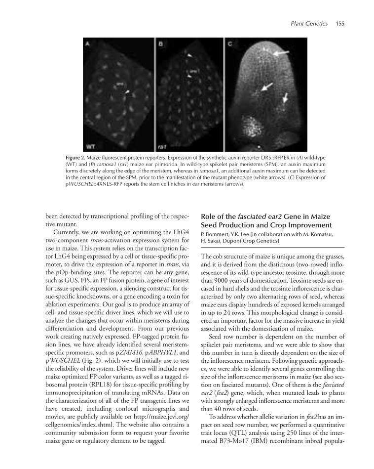
Plant Genetics 155
been detected by transcriptional profiling of the respec-tive mutant.
Currently, we are working on optimizing the LhG4two-component trans-activation expression system foruse in maize. This system relies on the transcription fac-tor LhG4 being expressed by a cell or tissue-specific pro-moter, to drive the expression of a reporter in trans, viathe pOp-binding sites. The reporter can be any gene,such as GUS, FPs, an FP fusion protein, a gene of interestfor tissue-specific expression, a silencing construct for tis-sue-specific knockdowns, or a gene encoding a toxin forablation experiments. Our goal is to produce an array ofcell- and tissue-specific driver lines, which we will use toanalyze the changes that occur within meristems duringdifferentiation and development. From our previouswork creating natively expressed, FP-tagged protein fu-sion lines, we have already identified several meristem-specific promoters, such as pZMM16, pABPHYL1, andpWUSCHEL (Fig. 2), which we will initially use to testthe reliability of the system. Driver lines will include newmaize optimized FP color variants, as well as a tagged ri-bosomal protein (RPL18) for tissue-specific profiling byimmunoprecipitation of translating mRNAs. Data onthe characterization of all of the FP transgenic lines wehave created, including confocal micrographs andmovies, are publicly available on http://maize.jcvi.org/cellgenomics/index.shtml. The website also contains acommunity submission form to request your favoritemaize gene or regulatory element to be tagged.
Role of the fasciated ear2 Gene in MaizeSeed Production and Crop ImprovementP. Bommert, Y.K. Lee [in collaboration with M. Komatsu,H. Sakai, Dupont Crop Genetics]
The cob structure of maize is unique among the grasses,and it is derived from the distichous (two-rowed) inflo-rescence of its wild-type ancestor teosinte, through morethan 9000 years of domestication. Teosinte seeds are en-cased in hard shells and the teosinte inflorescence is char-acterized by only two alternating rows of seed, whereasmaize ears display hundreds of exposed kernels arrangedin up to 24 rows. This morphological change is consid-ered an important factor for the massive increase in yieldassociated with the domestication of maize.
Seed row number is dependent on the number ofspikelet pair meristems, and we were able to show thatthis number in turn is directly dependent on the size ofthe inflorescence meristem. Following genetic approach-es, we were able to identify several genes controlling thesize of the inflorescence meristems in maize (see also sec-tion on fasciated mutants). One of them is the fasciatedear2 (fea2) gene, which, when mutated leads to plantswith strongly enlarged inflorescence meristems and morethan 40 rows of seeds.
To address whether allelic variation in fea2 has an im-pact on seed row number, we performed a quantitativetrait locus (QTL) analysis using 250 lines of the inter-mated B73-Mo17 (IBM) recombinant inbred popula-
Figure 2.Maize fluorescent protein reporters. Expression of the synthetic auxin reporter DR5::RFP.ER in (A) wild-type(WT) and (B) ramosa1 (ra1) maize ear primorida. In wild-type spikelet pair meristems (SPM), an auxin maximumforms discretely along the edge of the meristem, whereas in ramosa1, an additional auxin maximum can be detectedin the central region of the SPM, prior to the manifestation of the mutant phenotype (white arrows). (C) Expression ofpWUSCHEL::4XNLS-RFP reports the stem cell niches in ear meristems (arrows).
150-174_05_PlantGenet_Annual Report_2009 template 7/8/11 11:58 AM Page 155

156 Research
tion, in three replicates. The mean row number of B73was ~18, whereas Mo17 develops ~11 rows. The meanrow number in the IBM families was ~14, with a rangefrom ~10 to 20. We found the most significant seed rownumber QTL to be located on chromosome 4, overlap-ping with the fea2 locus, suggesting that allelic variationin fea2 could regulate row number.
To substantiate these findings, we searched for hy-pomorphic EMS-induced alleles, via TILLING. Wewere able to isolate four new alleles of fea2, all predictedto be weak. Analysis of seed row number in three ofthem showed a statistically significant increase. Subse-quent genotyping confirmed a significant correlationbetween row number increase and presence of the new-ly identified, weak fea2 allele, providing further evi-dence that allelic variation at the fea2 locus can lead tochanges in seed row number. Field trials to assess theimpact of increased seed rows on maize yield are cur-rently in progress.
The Regulation of Meristem Size in MaizeP. Bommert, M. Pautler , Y.K. Lee, B.I. Je, H. Szilagyi,A. Eveland, J. Kendall [in collaboration with M. Komatsu, H. Sakai, Dupont Crop Genetics]
All plant organs derive from populations of stem cellscalled meristems. These stem cells have two purposes:to divide in order to replace themselves and to give riseto daughter cells, which will differentiate into lateralorgans or stems. Consequently, plant meristems mustprecisely control the size of the stem cell niche via a net-work of positive and negative feedback signals. A lossof function in a negative regulator of stem cell fate canresult in an enlarged or fasciated meristem phenotypeand a dramatic alteration in the morphology of themaize ear and tassel.
Maize is an excellent genetic model system, becauseof a large collection of developmental mutants and asequenced reference genome. Our lab has undertakena forward genetic approach to identify key regulatorsof stem cell homeostasis and meristem size. Two previ-ously cloned mutants, fasciated ear2 and thick-tasseldwarf1, encode orthologs of the Arabidopsis thalianagenes CLAVATA1 and CLAVATA2, indicating the well-known CLAVATA-WUSCHEL regulatory feedbackloop is conserved from dicots to monocots. However,little else is known about the control of this importantdevelopmental process in maize. Here, we describe ourprogress in identifying additional genes contributing tostem cell niche homeostasis.
ct2 is a classical mutant of maize that exhibits a fas-ciated inflorescence phenotype as well as semi-dwarfism. ct2 was identified by positional cloning andwas found to encode the α-subunit of a heterotrimericGTPase. The protein was localized by a fluorescent pro-tein translational fusion and was expressed strongly inmeristems. Several experiments demonstrated that ct2is deficient in giberrellic acid (GA) signaling, a planthormone with no previous connection to meristem sizecontrol. We are currently using both genetic and pro-teomic approaches to determine how CT2 interactswith established pathways for regulating meristem size.
Another fasciated ear mutant that we recentlycloned is fea3. fea3 is predicted to be similar to previ-ously described stem cell receptor genes, and sequenceanalysis indicates that it might encode a membrane-bound receptor. The fea3 mutant possesses several fa-vorable characteristics relevant to crop improvement.First, plant height does not seem to be affected as inmost fasciated ear mutants. Second, although mutanttassels have a higher spikelet density than wild type, theoverall size of the tassel is not reduced. This could leadto increased pollen shed, a desirable trait for hybridcrop production. Future work will include character-izing the spatial expression of fea3 and profiling thetranscriptome to understand which pathways withwhich it interacts.
fea1905 is another semidwarfed mutant, with mas-sively fasciated ears and tassels. We mapped the mutantto an ~2.7-Mb region on chromosome 6, which con-tains ~30 genes. We are using targeted resequencing bygenomic hybrid capture of the interval to find thecausative mutation. Probes were designed for Agilent244K arrays that tiled gene models in the mapping in-terval, and hybridization was carried out according toprotocols from Emily Hodges (Hannon lab, CSHL).Preliminary results suggest that the amount of DNAcaptured is limited and not enriched for the target re-gion. We are also beginning to use mRNA-Seq to searchfor mutations in transcripts arising from within the in-terval. A complementary approach is to create doublemutants to assemble genetic pathways. We have ana-lyzed double mutants between fea1905 and ramosa1and ramosa2, two mutants that display increased inflo-rescence branching. The ramosa mutants appear to beepistatic to fea1905, indicating that they likely actdownstream. This represents a previously unknownconnection between meristem size control and meris-tem determinancy. Future work will include molecularcloning of the gene, expression analysis, and transcrip-tome profiling.
150-174_05_PlantGenet_Annual Report_2009 template 7/8/11 11:58 AM Page 156

Plant Genetics 157
In addition, we are in the process of mapping ~50fasciated ear mutants from nontargeted EMS mutage-nesis screens. Our current approach involves creatingF2 mapping populations and rough mapping by bulkedsegregant analysis. We take advantage of the precisequantitative genotyping capability of the SequenomMassArray system to look for areas of the genome thatare linked to the phenotype. We have determined roughpositions for several of these mutants and are proceed-ing with fine mapping and molecular cloning.
Resolving Networks Controlling AxillaryMeristem Determinacy A. Eveland, A. Goldshmidt [in collaboration withD. Ware, Cold Spring Harbor Laboratory; M. Beatty, H. Sakai, DuPont Crop Genetics; R. Schmidt,University of California, San Diego; E. Vollbrecht,Iowa State University]
A rising challenge in developmental biology is elucidat-ing the mechanisms by which genes are regulated inspace and time, and how their actions form networksthat modulate differentiation and development. We usethe maize inflorescence as our model to study howbranching patterns are determined at the molecularlevel and the genetic interactions responsible for inflo-rescence architecture. Branching in grass inflorescencesis determined by the developmental fate of highly or-ganized stem cell populations called axillary meristems.These meristems can either maintain continual growthof indeterminate branches or terminate in determinatestructures such as flowers. Actions of genes that regulatemeristem determinacy, as well as the genetic interac-tions between them, are responsible for the wide varia-tion in architectures among inflorescences throughoutthe plant kingdom. The genetic control of branchingin maize, especially in ears where kernels are borne, hasclear relevance to grain yield and harvesting ability. Inaddition, its unique inflorescence architecture, charac-terized by flowers borne on spikelet pairs, is shared ex-clusively among the Andropogoneae tribe, whichincludes important energy crops such as sorghum,switchgrass, and sugarcane. Loss-of-function mutationsin the RAMOSA (RA) genes cause a branched pheno-type, due to derepression of branching in spikelet pairmeristems (SPMs). We make use of these branchingmutants as well as Illumina’s high-throughput sequenc-ing technology to identify transcriptional signatures re-lated to a gene network controlling inflorescence archi-tecture.
Primary objectives of this work are to establish aframework for studying developmental networks thatcontrol branching and to use both experimental andcomputational approaches to generate and addresstestable hypotheses. Initially, this framework consists oflarge-scale expression data sets and known genetic in-teractions. Candidate genes identified as putative reg-ulators of meristem determinacy will be tested as towhether they interact with RA genes and for their po-sition in a network controlling SPM determinacy. Thisyear, we published results from an analysis of sequence-based expression profiles in maize inflorescences, whichidentified genes that were differentially expressed be-tween wild-type and ramosa3 mutants. We identifiedstrand-specific expression signatures and classes of dif-ferentially expressed genes having putative functions inSPM determinacy. Work is currently under way to an-alyze loss-of-function mutations in these candidate loci,as well as crosses to the ramosa mutants to study poten-tial genetic interactions.
We have also generated mRNA-Seq libraries at var-ious stages of development from ears and tassels as wellas from loss-of-function ra mutants. A pipeline foranalysis of these data sets was developed and includesmethods for data processing, mapping short reads tothe maize reference genome, and statistical analyses toidentify differentially expressed genes and/or those shar-ing similar expression patterns. We leveraged a set ofhigh-confidence maize gene models and comparativefunctional information from other model plant speciesto classify transcription factor (TF) family members,based on their specific expression signatures in time andspace. We used a clustering approach to group coex-pressed genes during ear and tassel development. Aspredicted by their colocalized expression in ear primor-dia, RA1 and RA3 also show coexpressed profiles duringboth ear and tassel development. In addition, genes thatwere differentially expressed in ra1 and ra3 mutantstended to be coexpressed with RA1 and RA3. As a con-trol for the mRNA-Seq data, we analyze dynamicchanges in expression profiles of the Auxin responseDR5::RFP.ER marker and additional developmentalmarkers, such as pWUS1::NLS::RFP. Overlaying thesemarker profiles on the mRNA-Seq-based expressionmap will provide insight for linking dynamic changesin gene expression to hormonal signaling pathways instem cells during meristem differentiation.
We are integrating the RNA-Seq data sets with othertypes of genomics data and further experimentation.We are currently analyzing DNA-occupancy maps ofRA1, using chromatin immunoprecipitation followed
150-174_05_PlantGenet_Annual Report_2009 template 7/8/11 11:58 AM Page 157

158 Research
by high-throughput sequencing (ChIP-Seq). This willprovide a genome-wide view of gene targets potentiallyregulated by RA1. Using these data, we can infer puta-tive mechanisms of regulation acting through RA1 tocontrol branching in maize. We have also sequencedtranscriptomes of comparable inflorescence stages insorghum and will use these data in comparative analyseswith the maize RNA-Seq data. These analyses will helpin selecting candidate genes that act as key developmen-tal regulators. The function of such candidate genes willbe tested in follow-up genetic and molecular experi-ments.
Identification of Genes Participatingin the RAMOSA3 NetworkA. Goldshmidt, M. Pautler, D. Jackson
The characterization of molecular mechanisms control-ling the branching of cereal inflorescences and the iden-tification of novel genes that participate in this processare crucial for the improvement of yield traits of com-mercial crops. In our current research, we have beenusing maize mutants ramosa1 (ra1), ramosa2 (ra2), andramosa3 (ra3), which have increased branching in theinflorescences, in order to characterize molecular net-works regulated by these genes. Ramosa3 is of particularinterest, because it encodes a predicted metabolic en-zyme, a trehalose phosphate phosphatase, rather than atranscription factor or signaling molecule that are moretypical of developmental regulators. We have used aclassic genetic approach—analysis of multiple mutantphenotypes—for identification of the relationships be-tween RAMOSA genes and other genes involved in de-velopmental networks. We first characterized doublemutants of the ramosa genes with mutants involved inauxin transport (barren inflorescence2 [bif2]) and inauxin synthesis (sparse inflorescence1 [spi1]). Here, wefound additive interactions, suggesting that networksregulated by the RAMOSA genes act in parallel to theauxin-related networks. Second, we characterized dou-ble mutants with mutants of genes involved in net-works regulating meristem maintenance, such asknotted1 (kn1), fasciated ear2 (fea2), and thick-tasseldwarf1 (td1). Here, our analysis revealed that pheno-types of the ra1 and ra2 mutants were enhanced by kn1loss-of-function mutations, whereas ra3 mutant phe-
notypes were suppressed. ra2 and ra3 mutants showedadditive interactions with fea2, whereas fea2 phenotypeswere suppressed by ra1, and all three ramosa mutantsshowed additive interactions with td1. These results in-dicate that RAMOSA gene activities are involved in net-works regulating meristem maintenance. However, ourresults indicate that different RAMOSA genes may reg-ulate determinacy by different mechanisms, some ofwhich are overlapping and some distinct.
We also wish to gain a more detailed understandingof the RAMOSA3 network using chemical mutagenesisfollowed by suppressor enhancer screens. So far, 850 seg-regating families were screened, and 46 suppressors and17 enhancers were identified. Due to variability of thera3mutant phenotype, it is important to verify enhance-ment/suppression in different environments. Therefore,all modifiers were regrown in at least two different fieldsfor verification and crossed to ra3 mutants in a differentgenetic background to generate mapping populations.Nine suppressor and three enhancer families have beenverified and are being prepared for mapping. Interest-ingly, only one out of the nine suppressors and one outof the three enhancers showed single-mutant phenotypes,resulting in dwarfism and validating the use of the mod-ifier approach. In the next year, we will focus on mappingand identification of these modifiers and characterizenovel genes participating in the RAMOSA3 network.
PUBLICATIONS
Brutnell TP, Wang L, Swartwood K, Goldshmidt A, Jackson D, Zhu X-G, Kellogg E, Van Eck J. 2010. Setaria viridis: A model for C4 pho-tosynthesis. Plant Cell 8: 2537–2544.
Chuck G, Whipple C, Jackson D, Hake S. 2010. The maize SBP-boxtranscription factor encoded by tasselsheath4 regulates bract devel-opment and the establishment of meristem boundaries. Development137: 1243–1250.
DeBlasio SL, Sylvester AW, Jackson D. 2010. Illuminating plant biology:Using fluorescent proteins for high-throughput analysis of proteinlocalization and function in plants. Brief Func Genom Proteom 9:129–138.
Eveland A, Satoh Nagasawa N, Goldshmidt A, Meyer S, Beatty M, SakaiH, Ware D, Jackson D. 2010. Digital gene expression signatures formaize development. Plant Physiol 54: 1024–1039.
Whipple CJ, Hall DH, DeBlasio S, Taguchi-Shiobara F, Schmidt RJ,Jackson D. 2010. A conserved mechanism of bract suppression inthe grass family. The Plant Cell 22: 565–578.
Xu X, Jackson DP. 2010. Lights at the end of the tunnel: New views ofplasmodesmal structure and function. Curr Opin Plant Biol 13: 684–692.
150-174_05_PlantGenet_Annual Report_2009 template 7/8/11 11:58 AM Page 158
159
Beauty and agricultural productivity in the Plant King-dom is directly linked to the number and arrangementof branches and flowers on specialized shoots known asinflorescences. Inflorescence branching is determinedby specialized groups of pluripotent stem cells at grow-ing tips called shoot apical meristems (SAMs), whichundergo reproductive “phase transitions” in responseto environmental cues coinciding with innate molecularevents. Inflorescences can be simple, producing only asingle flower, or they can be highly complex, producingdozens of branches and hundreds of flowers. Our re-search is focused on the hypothesis that the rates atwhich SAMs undergo reproductive transitions dictateinflorescence diversity, which provides a basis for tax-onomy as well as a primary point of selection in domes-tication and crop improvement. Little is known aboutthe molecular dynamics regulating reproductive phasetransitions, especially for species with different growthhabits. In particular, one of the most fundamental, yetpoorly understood, decisions is whether an inflores-cence grows indefinitely (indeterminate growth) or ifsignals are received to end growth (determinategrowth). Determinate plants produce the most elabo-rate inflorescence architectures, particularly amongspecies having the widely deployed “sympodial” growthhabit, represented by tomato. We are uniting geneticswith quantitative genomics to discover the genes andnetworks that control sympodial reproductive phasetransitions, which are a hallmark of perennial species,such as trees, and represent the growth habit of half ofthe world’s flowering plants. We are taking advantageof several tools in tomato (Solanum lycopersicum) to un-derstand how sympodial reiteratation is modulated togive rise to its multiflowered inflorescence, and whatmolecular mechanisms are responsible for variation ininflorescence branching in the larger nightshade(Solanaceae) family. Importantly, the Solanaceae com-prises several crop and ornamental species, includingpotato, pepper, eggplant, petunia, and tobacco, as wellas many edible wild species. Therefore, our research ad-dresses questions that are relevant to evolution as wellas agriculture.
In a related project, we are exploring the phenome-non of hybrid vigor, or heterosis, which occurs whendifferent inbred plants are crossed to produce superiorhybrids yielding more flowers, fruits, and seeds. Het-erosis, first described by Darwin, is widespread in na-ture and is responsible for the dramatic and necessaryincrease in crop yields achieved over the last century.We are testing the hypothesis that heterosis can becaused by subtle changes in gene dosage originatingfrom heterozygous single-gene mutations affectinggrowth and development. In a screen involving dozensof tomato heterozygous mutants, we have identifiedthree heterosis genes, including a reproductive transi-tion gene and an inflorescence branching gene. A con-clusion from our findings during the last year is thatthere are several unknown genes that regulate reproduc-tive transitions and inflorescence architecture, whichare major factors influencing reproductive capacity andyield. Therefore, our continued efforts to expose newgenes and networks affecting flowering in tomato willprovide new targets for functional studies that can beapplied to crop improvement.
Sympodial Growth in Tomato
Tomato is a powerful model to study mechanisms ofsympodial reproductive transitions because it is com-posed of three distinct shoot systems, each of which un-dergoes a termination event (Fig. 1). The first termin-ation originates from the embryonic meristem, whichproduces about eight leaves before ending growth witha multiflowered inflorescence. The next termination isin the sympodial shoot (SYM), which grows out fromthe last axillary meristem produced from the primaryshoot. All SYMs develop three leaves before producinga final inflorescence, and each tomato shoot after theprimary transition is derived from the indefinite reiter-ation of SYMs. The third termination occurs in the in-florescence, which produces about seven flowers in azigzag arrangement. The inflorescence is also com-pound, consisting of reiterating sympodial inflorescence
PLANT DEVELOPMENTAL BIOLOGY, STEM CELLS, ANDFLORAL BRANCHING SYSTEMS
Z. Lippman K. Jiang C. MacAlisterK. Liberatore S.-J. Park
150-174_05_PlantGenet_Annual Report_2009 template 7/8/11 11:58 AM Page 159
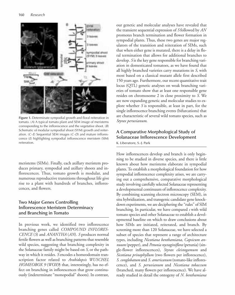
160 Research
meristems (SIMs). Finally, each axillary meristem pro-duces primary, sympodial and axillary shoots and in-florescences. Thus, tomato growth is modular, andnumerous reproductive transitions throughout life giverise to a plant with hundreds of branches, inflores-cences, and flowers.
Two Major Genes ControllingInflorescence Meristem Determinacyand Branching in Tomato
In previous work, we identified two inflorescencebranching genes called COMPOUND INFLORES-CENCE (S) and ANANTHA (AN). S produces normalfertile flowers as well as branching patterns that resemblewild species, suggesting that branching complexity inthe Solanaceae family might be based on S, or the path-way in which it resides. S encodes a homeodomain tran-scription factor related to Arabidopsis WUSCHELHOMEOBOX 9 (WOX9) that, interestingly, has no ef-fect on branching in inflorescences that grow continu-ously (indeterminate “monopodial” shoots). In contrast,
our genetic and molecular analyses have revealed thatthe transient sequential expression of S followed by ANpromotes branch termination and flower formation insympodial plants. Thus, these two genes are major reg-ulators of the transition and reiteration of SIMs, suchthat when either gene is mutated, there is a delay in flo-ral termination that allows for additional branches todevelop. S is the key gene responsible for branching vari-ation in domesticated tomatoes, as we have found thatall highly branched varieties carry mutations in S, withmost based on a classical mutant allele first described150 years ago. Furthermore, our recent quantitative traitlocus (QTL) genetic analyses on weak branching vari-eties of tomato show that at least one responsible generesides on chromosome 2 in close proximity to S. Weare now expanding genetic and molecular studies to ex-plore whether S is responsible, at least in part, for thesingle inflorescence branching events (bifurcations) thatare characteristic of several wild tomato species, such asStyrax peruvianum.
A Comparative Morphological Study ofSolanaceae Inflorescence DevelopmentK. Liberatore, S.-J. Park
How inflorescences develop and branch is only begin-ning to be studied in diverse species, and there is littleknown about how meristems elaborate in sympodialplants. To establish a morphological foundation for howsympodial inflorescence complexity arises, we are carry-ing out a comprehensive, comparative morphologicalstudy involving carefully selected Solanaceae representinga developmental continuum of inflorescence complexity.By combining scanning electron microscopy (SEM), insitu hybridization, and transgenic candidate gene knock-down experiments, we are deciphering the “rules” of SIMbranching. In particular, we have compared s with wildtomato species and other Solanaceae to establish a devel-opmental baseline on which to draw conclusions abouthow SIMs are initiated, reiterated, and branch. Byscreening more than 120 Solanaceae, we have selected asubset of species that represent a range of architecturetypes, including Nicotiana benthamiana, Capsicum an-nuum (pepper), and Petunia nyctaginiflora (petunia) (sin-gle-flower inflorescences), Styrax cleistogamum andSicotiana prinophyllum (two flowers per inflorescence),S. conglobatum and S. americanum (tomato-like inflores-cence), and S. peruvianum and Nicotiana tobaccum(branched, many flowers per inflorescence). We have al-ready studied in detail the ontogeny of N. benthamiana
Figure 1. Determinate sympodial growth and floral reiteration intomato. (A) A typical tomato plant and SEM image of meristemscorresponding to the inflorescence and the vegetative shoot. (B)Schematic of modular sympodial shoot (SYM) growth and reiter-ation. (C–E) Sequential SEM images (C–D) and mature inflores-cence (E) highlighting sympodial inflorescence meristem (SIM)reiteration.
150-174_05_PlantGenet_Annual Report_2009 template 7/8/11 11:58 AM Page 160

Plant Genetics 161
and S. peruvianum, which has revealed that primary tran-sitions are faster and slower, respectively, compared totomato. We have also developed an interest in assessingif there is any relationship between the level of branchingand species-specific phyllotactic patterning. To addressthese questions, we are developing a series of markergenes fused to fluorescence proteins to generate reporterlines for studying sympodial transitions in four dimen-sions (time and space) using confocal and two-photonmicroscopy.
A Comparative Molecular Studyof Inflorescence DevelopmentK. Liberatore [in collaboration with X. Wu, University ofSouthern California]
As mentioned above, it is unclear to what extent repro-ductive transitions are similar or different in plants suchas the monopodial Arabidopsis and the sympodialtomato. We are determining if branching genes such asS in tomato are functionally conserved across diversespecies by using a series of transgenic knockdown tools(e.g., artificial microRNAs) to determine if WOX9 hasan unforeseen role in inflorescence development in Ara-bidopsis. Curiously, mutations in Arabidopsis WOX9have been characterized, but phenotypic defects seemto be restricted to the early stages of embryonic pattern-ing. This early embryonic defect and resulting seedlinglethality has prevented a more detailed analysis of laterstages of development. By selectively targeting the elim-ination of WOX9 transcripts, we hope to determine ifand when WOX9 functions to regulate inflorescencemeristem elaboration. Our first set of experiments sug-gest targeting WOX9 using RNA interference (RNAi)constructs driven by a floral-specific promoter causesthe Arabidopsis floral meristem to branch in a way thatis reminiscent of s mutations in tomato.
The Molecular Dynamics UnderlyingSympodial Meristem Transitions inTomato and Related SolanaceaeS.-J. Park, K. Jiang [in collaboration with M. Schatz, Cold Spring Harbor Laboratory]
We are taking advantage of the three types of meristemsof tomato (primary shoot, SIM, and SYM) to elucidatethe dynamic changes in gene expression that take placeduring reproductive transitions in plants, with a focus ondeterminate sympodial growth. Tomato meristems, like
other Solanaceae, are extremely large and easily dissectedand collected in sufficient amounts for RNA extraction.Using a simple acetone fixation protocol we have devel-oped this past year, we are growing uniform batches ofwild-type plants and precisely harvesting meristems hav-ing no more than two leaf primordia every 3 days untilfloral termination. After the transition, we collect SYM,SIM, and FM (floral meristem) tissue. To date, we havecompleted gene expression analyses on seven stages ofsympodial growth using quantitative deep mRNA se-quencing (Illumina mRNA-Seq), and we have achievedexcellent temporal resolution to reveal that reproductivetransitions are highly quantitative and dynamic (Fig. 2).Furthermore, each meristem stage has its own molecularidentity and age. We have been able to quantify meristemstate using several algorithms, which predict that theSYM shares identity with the late vegetative meristem(LVM) stage from the primary shoot. This suggests thatupon inception, SYMs are preprogrammed as if youngleaves have already been initiated. These data provide anearly clue to explain how sympodial shoots reiterate ifthey somehow sense the termination rate of the primaryshoot, perhaps through changes in apical dominance,which then causes LVM genes to turn on early and strongin each initiating SYM. Interestingly, SIMs have dualidentities, overlapping with the transition meristem(TM) and FM. The SYM and SIM also express their
Figure 2. Six representative clusters of tomato gene expressiondynamics showing gradual (A–C) and transient, stage-specificpeaks (D–F) as sympodial meristems undergo termination. Datawere grouped into 25 clusters (see text for details) and up to fiverepresentative genes are plotted out of the total in each cluster(indicated at top) (A). Early vegetative meristem (EVM) genes, (B)late vegetative meristem (LVM) genes, (C) reproductive (TM, SIM,FM) genes, (D) TM genes, (E) SIM genes, (F) SYM genes.
150-174_05_PlantGenet_Annual Report_2009 template 7/8/11 11:58 AM Page 161

162 Research
own stage-enriched genes, indicating that each meristemhas unique identity. We are now performing quantitativetranscriptomics on several Solanaceae species from amorphological allelic series to decipher the differentialdynamics that determine whether a SIM exists, to whatextent it reiterates, and whether it branches. To study dif-ferences in sympodial programs, our project requires thatquantitative transcriptomics be performed on nonmodelSolanaceae to compare sympodial reproductive transi-tions. We have verified that other species’ meristems canbe harvested at similar temporal resolutions and, mostimportant, that reference transcriptomes can be estab-lished to enable robust species-specific quantification byusing N. benthamiana, a single-flowered species repre-senting the most distantly related Solanaceae within ourchosen species set. Our findings have thus far indicatedthat de novo transcriptome assembly and quantificationin nonmodel Solanaceae are achievable and will provepowerful for comparing expression dynamics among di-verse sympodial programs. These data are also providinga first list of candidate genes for functional studies usingreverse genetic tools.
Novel Inflorescence Branching MutantsC. MacAlister
To increase our knowledge of the genes and networks reg-ulating sympodial inflorescence patterning, we are inves-tigating new mutants with inflorescence architecturedefects. In particular, we are characterizing and cloningthe gene responsible for a new tomato mutant called fasci-ated inflorescence (fin), which produces a highly branched
inflorescence with greatly enlarged flowers and fruits withincreased organs (Fig. 3). The phenotypes of fin mutantssuggest some genes have dual roles in regulating determi-nacy, affecting both the inflorescence and floral meri-stems. In contrast, s and an seem to be specific to theinflorescence meristem. Indeed, our analysis of fin:s dou-ble mutants indicates that these genes operate in separategenetic pathways. We have cloned the FIN gene by unit-ing map-based cloning with mRNA-Seq and found thatFIN encodes a protein of unknown function, lacking anyknown protein motifs. Mutants of fin are reminiscent ofclavata mutants in Arabidopsis, except that fin producesan enlarged meristem early in the vegetative phase of de-velopment and continues to enlarge as the primary repro-ductive transition ensues. This results in an oblongenlarged meristem that produces leaves ectopically, altersphyllotaxy, and initiates several additional sympodialshoots (SYMs). In addition to standard molecular analy-sis, we are now studying an allelic series of fin mutants inmore detail by (1) performing mRNA-Seq on fin meri-stems before and after the reproductive transition, (2)studying double-mutant combinations with other tomatoinflorescence and flowering mutants, and (3) exploringphenotypic consequences of mutations in the orthologousgenes of Arabidopsis, which is facilitated by fin being amember of a small gene family all the way down to moss.
We are also studying a classical mutation called ter-minating flower (tmf ), which uniquely causes an earlytermination of the primary inflorescence meristem togenerate a single terminal flower that fails to produceSIMs and SYMs. Interestingly, all axillary meristemsdevelop normally, generating typical SIMs and SYMs.By taking advantage of our recent efforts to sequencethe most closely related wild species of tomato, S.pimpinellifolium, we have map-based cloned the defec-tive gene in tmf and found that it encodes yet anotherprotein of unknown function. Our finding of severalnew genes controlling tomato inflorescence architecturehighlights that novel mechanisms regulate determinatesympodial growth.
Identification and Characterizationof Single-Gene Mutations CausingHeterosis in TomatoK. Jiang [in collaboration with D. Zamir, HebrewUniversity, Israel]
The genetic and molecular bases for how hybrid prog-eny outperform their parents (heterosis) have not beenresolved. On the basis of classical studies in noncrop
Figure 3.New tomato mutant (fasciated inflorescence) producesgreatly enlarged flowers and fruits.
150-174_05_PlantGenet_Annual Report_2009 template 7/8/11 11:58 AM Page 162

Plant Genetics 163
plants and recent findings in yeast, we have tested thehypothesis that single-gene mutations cause phenotypicoverdominance in the heterozygous state through a re-duction in gene dosage. By taking advantage of a large“mutant library” in an isogenic cultivated tomato back-ground (Solanum lycopersicum, cv. M82), we have iden-tified multiple genes that show heterosis throughoverdominance. We have discovered that SINGLEFLOWER TRUSS (SFT ), which is the genetic originatorof the flowering hormone florigen that drives the repro-ductive transition in all plants, increases yield up to 60%when loss-of-function alleles are heterozygous. Yieldoverdominance from sft heterozygosity translates to sev-eral distinct genetic backgrounds and environments, andwe show that several traits integrate pleiotropically todrive heterosis in a multiplicative manner. Our resultssuggest that a simple reduction in the dosage of a singlegene can cause dramatic transgressive variation. Thesefindings provide the first example of a single overdomi-nant gene for yield, and they suggest that screening formutations with heterozygous effects on growth can im-prove productivity in other agricultural organisms. Onthe basis of these findings, we have extended our studiesto screen for other mutants that show heterosis in theheterozygous state. In collaboration with Dani Zamir inIsrael, we have found that heterozygosity for the com-pound inflorescence mutant increases yield by causing20%–40% of inflorescences to branch once (bifurcate),
without any pleiotropic effects on growth. Importantly,we have found that heterosis from s/+ heterozygotes isheritable through meiosis and acts additively with otherheterotic mutants, thereby opening the door for pyra-miding heterosis genes to increase flower and fruit pro-duction even further. A fundamental question onsingle-gene heterosis is how subtle changes in genedosage affect the transcriptional networks that drivephenotypic heterosis. Because of the greatly reduced ge-nomic complexity of single-gene heterosis, we havebegun performing targeted and global quantitative geneexpression analyses to link heterotic effects with tran-scriptional changes in developmental pathways andlarger transcriptional networks. To our surprise, we havefound that the florigen-encoding SFT, which is believedto be expressed only in leaves [4, 5], is transcribed specif-ically in the TM and SIM meristem stages and overlapswith transcription of its antagonist SP. One intriguingpossibility we will explore is whether local transcriptionand a changing balance of SFT and SP in transitioningmeristems from sft/+ heterozygotes is the basis for thisform of heterosis.
PUBLICATIONS
Krieger U, Lippman ZB, Zamir D. 2010. The flowering gene SINGLEFLOWER TRUSS drives heterosis for yield in tomato. Nat Genet 42:459–463.
Ke Jiang
150-174_05_PlantGenet_Annual Report_2009 template 7/8/11 11:58 AM Page 163

164
Plants and fission yeast provide excellent model organ-isms to address the question of how epigenetic informa-tion is propagated to daughter cells, including transposonregulation, heterochromatic silencing, and gene imprint-ing, important both for plant breeding and for humanhealth. We are investigating the inheritance of hetero-chromatic silencing and the role of RNA interference(RNAi) in the fission yeast Schizosaccharomyces pombe andthe plant Arabidopsis thaliana, as well as continuing ourwork on Zea mays. In the past year, we have found thatRNAi is important in determining germ cell fate as wellas in transposon activation, whereas in fission yeast, trans-posable elements (TE) control DNA replication via thehighly conserved centromere-binding protein B (CENP-B), which contributes to their epigenetic regulation.
This year, we welcomed postdocs Yannick Jacob andJie Ren, computational biologist Evan Ernst, graduatestudent Stephane Castel, and research technician ChantalLeBlanc. We said farewell to Rulan Shen, who retiredafter more than 15 years in the plant group. DanielleIrvine accepted a faculty position at the Murdoch Chil-dren’s Research Institute in Melbourne, Australia. MattVaughn left for a position at the Texas Advanced Com-puting Center at the University of Texas in Austin, andKlavs Hansen returned to Copenhagen. Filipe Borges wasa visiting student from the Gulbenkian institute (Lisbon,Portugal), Sophie Thomain visited from École NormaleSupèrieure, and Benoit Arcangioli joined the laboratoryfor 6 months on sabbatical from Institut Pasteur (Paris).
CENP-B Preserves Genome Integrityat Replication Forks Paused by theRetrotransposon LTRM. Zaratiegui, M. Vaughn, D. Irvine [in collaboration with J. Bahler, University College London; B. Arcangioli, Institut Pasteur]
CENP-B is a widely conserved DNA-binding factor as-sociated with heterochromatin and centromeric satellite
repeats. In fission yeast, CENP-B homologs have beenshown to silence long terminal repeat (LTR) retrotrans-posons by recruiting histone deacetylases. However,CENP-B factors also have unexplained roles in DNAreplication. We have shown that a molecular functionof CENP-B is to promote replication fork progressionthrough the LTR. Mutants have increased genomic in-stability caused by replication fork blockage that de-pends on the DNA-binding factor switch-activatingprotein 1 (Sap1), which is directly recruited by theLTR. The loss of Sap1-dependent barrier activity allowsthe unhindered progression of the replication fork, butthis results in rearrangements deleterious to the retro-transposon. We conclude that retrotransposons influ-ence replication polarity through recruitment of Sap1and transposition near replication fork blocks, whereasCENP-B counteracts this activity and promotes forkstability. Our results may account for the role of LTRin fragile sites and for the association of CENP-B withpericentromeric heterochromatin and tandem satelliterepeats in mammalian genomes including humans.
RNAi Promotes Heterochromatic HistoneModification through Replication-CoupledTranscriptional SilencingM. Zaratiegui, J. Ren, A.-Y. Chang, D. Irvine, S. Castel [in collaboration with F. Antequera, University of Salamanca, Spain; W.Z. Cande, University of California,Berkeley; B. Arcangioli, Institut Pasteur]
Heterochromatin comprises tightly compacted repetitiveregions of eukaryotic chromosomes. It is inheritedthrough mitosis and has roles in transcriptional silencingand genome integrity. In fission yeast, the epigenetic in-heritance of heterochromatin requires RNAi to guide his-tone modification, which occurs during the DNA repli-cation phase of the cell cycle. The alternating arrangementof origins of replication and small RNA clusters in fissionyeast centromeres provokes the collision of the transcrip-
EPIGENETIC INHERITANCE AND REPROGRAMMING IN PLANTSAND FISSION YEAST
R. Martienssen B. Arcangioli K. Creasey D. Irvine J. Ren M. Tanurdžic’F. Borges E. Ernst Y. Jacob A. Schorn S. ThomainJ. Calarco P. Finigan C. LeBlanc R. Shen F. van ExS. Castel J.-J. Han U. Ramu J. Simorowski M. VaughnA.-Y. Chang K. Hansen M. Regulski C. Tang M. Zaratiegui
150-174_05_PlantGenet_Annual Report_2009 template 7/8/11 11:58 AM Page 164
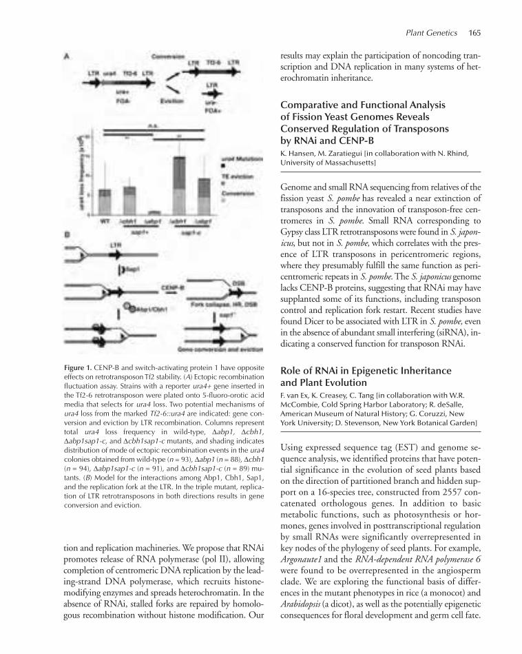
Plant Genetics 165
tion and replication machineries. We propose that RNAipromotes release of RNA polymerase (pol II), allowingcompletion of centromeric DNA replication by the lead-ing-strand DNA polymerase, which recruits histone-modifying enzymes and spreads heterochromatin. In theabsence of RNAi, stalled forks are repaired by homolo-gous recombination without histone modification. Our
results may explain the participation of noncoding tran-scription and DNA replication in many systems of het-erochromatin inheritance.
Comparative and Functional Analysisof Fission Yeast Genomes RevealsConserved Regulation of Transposonsby RNAi and CENP-BK. Hansen, M. Zaratiegui [in collaboration with N. Rhind,University of Massachusetts]
Genome and small RNA sequencing from relatives of thefission yeast S. pombe has revealed a near extinction oftransposons and the innovation of transposon-free cen-tromeres in S. pombe. Small RNA corresponding toGypsy class LTR retrotransposons were found in S. japon-icus, but not in S. pombe, which correlates with the pres-ence of LTR transposons in pericentromeric regions,where they presumably fulfill the same function as peri-centromeric repeats in S. pombe. The S. japonicus genomelacks CENP-B proteins, suggesting that RNAi may havesupplanted some of its functions, including transposoncontrol and replication fork restart. Recent studies havefound Dicer to be associated with LTR in S. pombe, evenin the absence of abundant small interfering (siRNA), in-dicating a conserved function for transposon RNAi.
Role of RNAi in Epigenetic Inheritanceand Plant EvolutionF. van Ex, K. Creasey, C. Tang [in collaboration with W.R. McCombie, Cold Spring Harbor Laboratory; R. deSalle,American Museum of Natural History; G. Coruzzi, NewYork University; D. Stevenson, New York Botanical Garden]
Using expressed sequence tag (EST) and genome se-quence analysis, we identified proteins that have poten-tial significance in the evolution of seed plants basedon the direction of partitioned branch and hidden sup-port on a 16-species tree, constructed from 2557 con-catenated orthologous genes. In addition to basicmetabolic functions, such as photosynthesis or hor-mones, genes involved in posttranscriptional regulationby small RNAs were significantly overrepresented inkey nodes of the phylogeny of seed plants. For example,Argonaute1 and the RNA-dependent RNA polymerase 6were found to be overrepresented in the angiospermclade. We are exploring the functional basis of differ-ences in the mutant phenotypes in rice (a monocot) andArabidopsis (a dicot), as well as the potentially epigeneticconsequences for floral development and germ cell fate.
Figure 1. CENP-B and switch-activating protein 1 have oppositeeffects on retrotransposon Tf2 stability. (A) Ectopic recombinationfluctuation assay. Strains with a reporter ura4+ gene inserted inthe Tf2-6 retrotransposon were plated onto 5-fluoro-orotic acidmedia that selects for ura4 loss. Two potential mechanisms ofura4 loss from the marked Tf2-6::ura4 are indicated: gene con-version and eviction by LTR recombination. Columns representtotal ura4 loss frequency in wild-type, ∆abp1, ∆cbh1,∆abp1sap1-c, and ∆cbh1sap1-c mutants, and shading indicatesdistribution of mode of ectopic recombination events in the ura4colonies obtained from wild-type (n = 93), ∆abp1 (n = 88), ∆cbh1(n = 94), ∆abp1sap1-c (n = 91), and ∆cbh1sap1-c (n = 89) mu-tants. (B) Model for the interactions among Abp1, Cbh1, Sap1,and the replication fork at the LTR. In the triple mutant, replica-tion of LTR retrotransposons in both directions results in geneconversion and eviction.
150-174_05_PlantGenet_Annual Report_2009 template 7/8/11 11:58 AM Page 165
166 Research
Arabidopsis thaliana Chromosome 4Replicates in Two Phases That Correlatewith Chromatin StateM. Tanurdžic’, M. Vaughn [in collaboration with L. Hanley-Bowdoin, B. Thompson, North Carolina State University]
We used flow cytometry and tiling microarrays to pro-file DNA replication of Arabidopsis chromosome 4(chr4) during early, mid, and late S phase. Replicationprofiles for early and mid S phase were similar and en-compassed the majority of the euchromatin. Late Sphase exhibited a distinctly different profile that in-cludes the remaining euchromatin and essentially all ofthe heterochromatin. Termination zones were consis-tent between experiments, allowing us to define 163putative replicons on chromosome 4 that clustered intolarger domains of predominately early or late replica-tion. Early-replicating sequences, especially the initia-tion zones of early replicons, displayed a pattern ofepigenetic modifications specifying an open chromatinconformation. Late replicons, and the terminationzones of early replicons, showed an opposite pattern.Histone H3 acetylated on lysine 56 (H3K56ac) was en-riched in early replicons, as well as the initiation zonesof both early and late replicons. Replicon organizationin Arabidopsis is strongly influenced by epigenetic mod-ifications to histones and DNA.
Control of Female Gamete Formationby a Small RNA Pathway in ArabidopsisF. van Ex, K. Creasey [in collaboration with J.-P. Vielle-Calzada, Langebio, CINVESTAV, Mexico]
In the ovules of most sexual flowering plants, female ga-metogenesis is initiated from a single surviving gameticcell, the functional megaspore, formed after meiosis ofthe somatically derived megaspore mother cell (MMC).The Arabidopsis protein ARGONAUTE 9 (AGO9)controls female gamete formation by restricting thespecification of gametophyte precursors in a dosage-de-pendent, non-cell-autonomous manner. Mutations inAGO9 lead to the differentiation of multiple gameticcells that are able to initiate gametogenesis. Mutationsin RNA-DEPENDENT RNA POLYMERASE 6 exhibita defect identical to that of ago9mutants, indicating thatthe movement of small RNA silencing out of somaticcompanion cells is necessary for controlling the specifi-cation of gametic cells. AGO9 preferentially interactswith 24-nucleotide small RNAs derived from TEs, and
its activity is necessary to silence TEs in female gametesand their accessory cells. Our results show that AGO9-dependent small RNA silencing is crucial to specify cellfate in the Arabidopsis ovule and that epigenetic repro-gramming in companion cells is necessary for small-RNA-dependent silencing in plant gametes.
Small RNA and Epigenetic Reprogrammingin the Arabidopsis Male GermlineY. Jacob, J. Calarco, C. Tang, C. LeBlanc, F. Borges [in collaboration with J. Becker, J. Feijó, Gulbenkian Institute, Lisbon, Portugal]
Most of the core proteins involved in the microRNA(miRNA) pathway in plants have been identified, andhundreds of miRNA sequences processed in the Ara-bidopsis sporophyte have been discovered by exploitingnext-generation sequencing technologies. On the basisof comparative analysis of miRNAs identified in spermcells by in-depth sequencing, 25 potentially novel miRNAs processed in sperm cells and pollen were iden-tified, as well as enriched variations in the sequencelength of known miRNAs, which might indicate sub-functionalization by association with a putative germline-specific Argonaute complex. ARGONAUTE 5 (AGO5),by close homology with AGO1 and localizing preferen-tially to the sperm cell cytoplasm in mature pollen, maybe part of such a complex, as might 21-nucleotide “epi-genetically activated” siRNAs that accumulate in sperm.We are examining epigenetic modifications of the ge-nome during pollen development and how these mightinfluence the profile of small RNA.
Transposon Instability in InterspeciesHybrids Is Mediated by RNAiM. Tanurdžic’, P. Finigan, E. Ernst, M. Vaughn [in collaboration with L. Comai, University of California, Davis; B.C. Meyers, University of Delaware; R.W. Doerge,Purdue University]
Silencing of transposable elements ensures genome sta-bility and lowers the mutagenic effects of transposition.We have shown that COPIA retrotransposons from thepollen parent are activated in Arabidopsis interspecifichybrids, but only when matching 24-nucleotide siRNAare absent from the seed parent. Activated elements loseDNA methylation, and hybrid leaves accumulate 21-nu-cleotide “epigenetically activated siRNA” (easiRNA)normally found in pollen, along with RNA and extra-chromosomal cDNA transposition intermediates. Our
150-174_05_PlantGenet_Annual Report_2009 template 7/8/11 11:58 AM Page 166
Plant Genetics 167
results indicate that small RNA from the germline pro-vides a general recognition mechanism for transposonsin interspecific hybrids, reminiscent of hybrid dysgenesisand hybrid lethality in Drosophila, and may contributeto postfertilization hybridization barriers in a similar way.
Sequencing the Maize MethylomeM. Regulski [in collaboration with J. Kendall, D. McCombie, J. Hicks, D. Ware, Cold Spring Harbor Laboratory; S. Tingey, A. Rafalski, DuPont/Pioneer Genetic Discovery]
We have sequenced the methylome of B73 and Mo17inbreds at 28.7- and 19.7-fold coverage, respectively,using bisulphite conversion and Illumina next-genera-tion sequencing. In agreement with previous results, wehave found that the vast majority of cytosine methyla-tion in all three contexts (CG, CXG, and CHH) arefound in intergenic regions and transposable elements,with exons containing only 5%–10% CG methylation.CHH methylation is rare and confined mostly to in-tergenic regions. Exemplars of transposable elementsthat differ in DNA methylation have been found, andvariation between Mo17 and B73 was detected in re-gions of primary sequence similarity. Strategies for de-termining the inheritance and biological significance ofepigenetic variation are being developed.
The Maize Mutant Tunicate1 Is Causedby Ectopic Expression of a MADS-BoxTranscription Factor, Zmm19, in aDosage-Dependent MannerJ.-J. Han [in collaboration with M. Aukerman, DuPont Crop Genetics; D. Jackson, Cold Spring Harbor Laboratory]
Pod corn was once regarded as the ancestral form ofcultivated corn and was prized by pre-Columbian peo-ples for its magical properties. The Tunicate1 (Tu1)mutant of maize is a naturally occurring dominant mu-tation, resulting in a striking pod corn phenotype. TheZmm19 MADS box gene maps close to Tu1 and waspreviously sequenced from Tu1 mutants and half-tu-nicate derivatives (T. Meunster, G. Thiessen, unpubl.).This analysis revealed that Tu1 has duplicated copiesof Zmm19 interrupted by a novel mutator-like DNAtransposon in the 5′UTR (untranslated region), caus-ing misexpression, but fine mapping proved impossi-ble. We have found that yellow fluorescent protein(YFP)- and red fluorescent protein (RFP)-tagged
Zmm19 transgenic maize plants phenocopy Tu1.Moreover, misexpression of miR172 is detected in Tu1,suggesting that Zmm19 may target the miR172 gene,a conclusion supported by the phenotype of miR172transgenic lines. The 22-Mb region surrounding thetu1 locus in inbred B73 was selected for finishing dur-ing the maize genome sequencing project, and we havefound that insertion of the mutator-like element re-sulted in a 1.8-Mb inversion immediately upstream ofZmm19 in Tu1. This rearrangement prohibits recom-bination, and so likely accompanied the origin of thisancient mutation.
Aquatic Plants as Biofuel FeedstocksE. Ernst, M. Regulski [in collaboration with W.R. McCombie, Cold Spring Harbor Laboratory; J. Shanklin, Brookhaven National Laboratory; V. Citovsky, SUNY Stony Brook]
Aquatic flowering plants, such as common duckweed,have considerable potential as biofuel feedstocks due totheir very rapid growth and high carbon accumulationper unit mass. Work is currently under way to assemblea draft of the 450-Mb Lemna gibba genome. 300 mil-lion Illumina GAIIx paired-end reads from short insertand mate pair libraries have contributed to an initial as-sembly of 300 Mb in contigs over 1 kb, with the largestreaching 250 kb. In addition, we obtained sequencesfrom both small RNA and gene transcript libraries,which have already been used to identify 10 genes cen-tral to the oil biosynthesis pathway. These two librariesalso enabled cloning native miRNA precursors to im-plement artificial miRNA gene silencing in Lemna. Wesuccessfully induced flowering in cultured commonduckweed and continue to explore methods for large-scale seed production. Advances in transformationmade this year by Brookhaven National Laboratory inconjunction with the progress in sequencing thegenome and transcriptome should lead to the develop-ment of duckweed strains tailored to the production oflipid biofuels.
PUBLICATIONS
Cibrian-Jaramillo A, De la Torre-Barcena JE, Lee EK, Katari MS, LittleDP, Stevenson DW, Martienssen R, Coruzzi GM, DeSalle R. 2010.Using phylogenomic patterns and gene ontology to identify proteinsof importance in plant evolution. Genome Biol Evol 2: 225–239.
Lee TJ, Pascuzzi PE, Settlage SB, Shultz RW, Tanurdzic M, RabinowiczPD, Menges M, Zheng P, Main D, Murray JA, et al. 2010. Arabidop-sis thaliana chromosome 4 replicates in two phases that correlate with
150-174_05_PlantGenet_Annual Report_2009 template 7/8/11 11:58 AM Page 167

168 Research
chromatin state. PLoS Genet 6: e1000982. Martienssen RA. 2010. Small RNA makes its move. Science 328: 834–
835. Martienssen RA. 2010. Heterochromatin, small RNA and post-fertiliza-
tion dysgenesis in allopolyploid and interploid hybrids of Arabidopsis.New Phytol 186: 46–53.
Olmedo-Monfil V, Duran-Figueroa N, Arteaga-Vazquez M, Demesa-Ar-avalo E, Autran D, Grimanelli D, Slotkin RK, Martienssen RA,Vielle-Calzada JP. 2010. Control of female gamete formation by asmall RNA pathway in Arabidopsis. Nature 464: 628–632.
Zaratiegui M, Vaughn MW, Irvine DV, Goto D, Watt S, Bähler J, Ar-cangioli B, Martienssen RA. 2010. CENP-B preserves genome in-tegrity at replication forks paused by retrotransposon LTR. Nature469: 112–115.
In Press
Borges F, Pereira PA, Slotkin RK, Martienssen RA, Becker JD. 2011. Mi-croRNA activity in the Arabidopsismale germline. J Exp Bot (in press).
Calarco JP, Martienssen RA. 2011. Genome reprogramming and smallinterfering RNA in the Arabidopsis germline. Curr Opin Genet Dev(in press).
Hansen KR, Hazan I, Shanker S, Watt S, Verhein-Hansen J, Bahler J,Martienssen RA, Partridge JF, Cohen A, Thon G. 2011. H3K9me-independent gene silencing in fission yeast heterochromatin by Clr5and histone deacetylases. PLoS Genet (in press).
Jacob Y, Martienssen RA. 2011. Chromatin reprogramming: Genderequality during Arabidopsis germline differentiation. Curr Biol (inpress).
Kate Creasey
150-174_05_PlantGenet_Annual Report_2009 template 7/8/11 11:58 AM Page 168

169
Specification of adaxial–abaxial (upper/lower) polarity inthe developing leaf drives the flattened outgrowth of theleaf blade and directs the differentiation of distinct celltypes within the leaf’s adaxial/top and abaxial/bottom do-mains. Both are important innovations in the evolutionof land plants that maximize photosynthesis while mini-mizing water loss to the environment. In addition to beinga key developmental process, adaxial–abaxial patterning isof particular interest from a mechanistic point of view andhas proven to be an excellent model to study small RNA-mediated gene regulation. We previously showed that pat-terning of this developmental axis involves a cascade ofopposing small RNAs, in which microRNA (miRNA)miR390 triggers the biogenesis of the TAS3-derived 21-nucleotide tasiR-ARF on the adaxial side of developingleaves. These confine the accumulation of abaxial deter-minants, including AUXIN RESPONSE FACTOR3(ARF3) and miR166, to the lower side of leaves. miR166,in turn, demarcates the abaxial side by repressing expres-sion of class III homeodomain leucine zipper (HD-ZIPIII)transcription factors that specify adaxial fate.
Importantly, our analysis of the TAS3 trans-acting shortinterfering RNA (tasiRNA) pathway in Arabidopsis dem-onstrated that tasiR-ARF functions as a mobile positionalsignal in adaxial–abaxial patterning. Movement of thissmall RNA from its defined source of biogenesis in the twouppermost cell layers of leaves creates a gradient of accu-mulation that dissipates abaxially. The tasiR-ARF gradientgenerates a sharply defined expression domain of the abax-ial determinant ARF3 on the bottom side of leaf primordia,suggesting that small RNAs can establish pattern througha morphogen-like activity. This work provided the first di-rect evidence that small RNAs are mobile and can functionas instructive signals in development, thereby revealing anovel patterning activity of small RNAs.
Dissecting Small RNA Mobility in PlantsD. Skopelitis
Given the scope of miRNA-regulated gene networks, thecell-to-cell movement of small RNAs has important im-plications with respect to their potential to act as instruc-
tive signals in development and in response to physio-logical and stress stimuli. We are using artificial miRNAstargeting easy-to-score reporter genes to study parametersof miRNA movement, such as tissue specificity, direc-tionality, dose dependence, and the kinetics of move-ment. Previously generated lines, expressing artificialmiRNAs from the epidermis, against PHYTOENEDE-SATURASE (PDS) resulted in a range of photobleachingphenotypes, due to silencing of PDS in the subepidermalmesophyll layers, consistent with small RNA mobilityfrom the epidermis into underlying cells. To further in-vestigate miRNA mobility in multiple distinct develop-mental contexts, we are expressing miRNAs targetingcell-autonomous GUS and GFP reporters from a num-ber of promoters that drive expression in various spa-tiotemporal patterns. A first analysis showed thatmiRNAs expressed from the vascular-specific SUC2 andATHB8 promoters lead to miRNA-mediated gene silenc-ing in the surrounding mesophyll and in the pavementcells of the epidermis, consistent with the idea that miRNAs can traffic from the vasculature into adjacentcells. Interestingly, no reporter silencing was observed inthe trichomes of the epidermis, which are symplasticallyisolated. The tissue specificity of the promoters was ver-ified using transgenic lines that express the respectivetranscriptional fusions to the GUS reporter gene, and theaccumulation of the artificial miRNAs is being moni-tored by in situ hybridization. Genetic resources to sim-ilarly investigate properties of tasiRNA mobility are alsobeing generated. In addition, with the knowledge thatmiRNAs and tasiRNAs can traffic from the epidermisinto underlying tissue layers, we are carrying out forwardgenetic screens to identify factors influencing this process.
The START Domain Regulates HD-ZIPIIIActivity and Organ PolarityA. Husbands
On the basis of our previous observations regarding theexpression and function of miR166 and tasiR-ARF, it isevident that adaxial–abaxial patterning involves a cascadeof positional signals. The mobile signals that pattern the
PLANT DEVELOPMENTAL GENETICS
M. Timmermans A. Benkovics C. Fernandez-Marco A. Husbands K. PetschC. Clendaniel A. Gal-on M. Javelle E. PlavskinM. Dotto C. Hu M. Lodha D. Skopelitis
150-174_05_PlantGenet_Annual Report_2009 template 7/8/11 11:58 AM Page 169

170 Research
newly formed leaf are distinct from those that maintainpolarity during subsequent development. We have re-cently started a new project to identify additional signalsin adaxial–abaxial patterning. Defining the identity ofmobile positional signals remains not only a pressingquestion in the field of organ polarity, but also one of themain challenges in contemporary plant development.
The adaxial-promoting HD-ZIPIII transcription fac-tors contain a predicted START lipid-binding domain(Fig. 1A). Modeling of the START domain of the HD-ZIPIII member PHABULOSA (PHB) suggests structuralsimilarity to human PC-TP, a phosphatidylcholine-bind-ing START domain (Fig. 1B,C). This model and se-quence conservation with other START domains wasused to predict amino acids in PHB critical for STARTdomain ligand binding. To assess the role of the STARTdomain to PHB function, we generated transgenic Ara-bidopsis lines that express START domain variants of aPHB–YFP fusion protein. These PHB-YFP reporters alsocarry silent mutations in the miR166-target site, whichnormally leads to the formation of severely adaxializedleaves (Fig. 1D–G). Expression of a PHB-YFP variant, inwhich residues lining the ligand-binding pocket in theSTART domain are mutated (Fig. 1C), yielded plantswith less-severe leaf phenotypes, whereas lines expressinga PHB-YFP variant with a deletion of the START domainresemble wild-type plants. These findings reveal positiveregulation of HD-ZIPIII activity by an unknown ligand.
Experiments to determine the mechanism via whichthe START domain controls HD-ZIPIII function areongoing. Ligand binding does not appear to affect thesubcellular localization of these transcription factors, asconfocal imaging showed that the PHB-YFP derivativescorrectly localize to the nucleus. We are currently inves-tigating whether ligand binding affects PHB’s ability tobind DNA, activate transcription, or interact with re-quired protein partners. We have also developed a yeast-based assay that allows the high-throughput screeningof chemical libraries for compounds that are bound bythe HD-ZIPIII START domain and modulate proteinactivity. Compounds identified in this screen may notonly provide critical insight into the nature of the HD-ZIPIII ligand, but also identify agonists/antagonists ofthe START domain that enable innovative experimen-tation into the mechanism via which HD-ZIPIII pro-teins regulate diverse aspects of plant development,including stem cell activity and organ polarity.
Characterization of Novel tasiRNALoci in MaizeM. Dotto [in collaboration with M. Aukerman, M. Beatty, R. Meeley, DuPont-Pioneer]
tasiRNAs are processed from noncoding TAS precursortranscripts. Following cleavage by a miRNA-loaded
Figure 1. Activity of the HD-ZIPIII transcription factors is regulated by the START lipid-binding domain. (A)Schematic representation of the domain structure of HD-ZIPIII proteins. (HD) Homeodomain, (LZ) leucine zipper,(START) START lipid-binding domain, (MEHKLA) MEKHLA/PAS transcription regulatory domain. (B) Representationof the structure of the human PC-TP START domain modeled in SwissExpasy. (C) Predicted fold of the PHB STARTdomain modeled in SwissExpasy. Amino acids mutated in the PHB-YFP derivative are highlighted in black (arrows).(D) Distribution of phenotypes observed in plants transformed with various PHB derivatives that either remainunder control of miR166 (left) or carry mutations that render the transcripts resistant to miR166 (right). (PHB) Full-length PHB protein; (PHB*) PHB derivative with critical amino acids in the START ligand-binding pocket mutated;(PHB-∆) PHB derivative with the START domain deleted; (N) number of transformants analyzed. (E–G) Phenotypesin the transformants were grouped into three categories: (E) severely adaxialized, indicated in black, (F) weaklyadaxilized, indicated in light gray, (G) largely normal, indicated in dark gray. Mutation or deletion of the STARTdomain suppresses the ability of miR166-resistant PHB to adaxialize developing leaves.
150-174_05_PlantGenet_Annual Report_2009 template 7/8/11 11:58 AM Page 170

Plant Genetics 171
ARGONAUTE (AGO) complex, one of the cleavageproducts is converted into double-stranded RNAthrough the activities of RNA-DEPENDENT RNAPOLYMERASE6 (RDR6) and LEAF BLADELESS1(LBL1/SGS3) and subsequently processed by DICER-LIKE4 (DCL4) into phased 21-nucleotide siRNAs.Arabidopsis contains four characterized TAS gene fam-ilies. AGO1 is required for the biogenesis of tasiRNAsfrom the TAS1, TAS2, and TAS4 loci, whereasmiR390-loaded AGO7 triggers the biogenesis of TAS3tasiRNAs. We previously identified four TAS3 loci inthe maize genome; however, the TAS1, TAS2, andTAS4 loci are not conserved between Arabidopsis andmaize. To identify potential novel TAS loci in maize,we used a deep-sequencing approach to compare thesmall RNA content between wild-type and lbl1 mutantapices. This identified four novel TAS candidates, all ofwhich are targeted by miR390 and generate tasiRNAsclosely related to the known tasiR-ARFs. We are cur-rently using bioinformatics approaches to identify ad-ditional phased small RNA clusters in an attempt todetermine whether these TAS3 loci are the only phasedsiRNA generating loci in the maize genome.
In a separate approach, we are characterizing smallRNAs that associate with specific AGO proteins. Themaize genome encodes 20 AGO proteins, including oneAGO7 and four AGO1 homologs. We are generatingtransgenic maize plants expressing FLAG-tagged versionsof ZmAGO7 and ZmAGO1b to isolate associated smallRNAs via immunoprecipitation. We expect to identifynovel tasiRNAs, miRNAs, and possibly the transcriptsthat are targets of these small RNAs. For additional infor-mation regarding the small RNA pathways in maize, weare also characterizing small RNAs that associate withZmAGO10. This AGO protein has been implicated inthe miRNA- and tasiRNA-mediated regulation of meris-tem maintenance and leaf development in Arabidopsis.Phylogenetic analysis showed that the maize genome con-tains two close homologs of AtAGO10. Transposon in-sertion alleles for both genes have been identified, and weare currently assessing possible contributions of theZmAGO10 homologs to meristem function and leaf de-velopment in maize. We analyzed the small RNAs associ-ated with ZmAGO10 by deep sequencing the smallRNAs present in immunoprecipitates with a ZmAGO10-specific peptide antibody. We found that ZmAGO10 pro-teins preferentially bind 21-nucleotide small RNAscontaining a 5′-U, which are features common to nearlyall plant miRNAs. However, the immunoprecipitationfraction was enriched for just a subset of known miRNAsand tasiRNAs, and these include miR166 and tasiR-ARF,
which we had previously shown to function in the speci-fication of maize leaf polarity. A previously uncharacter-ized miRNA was most enriched in the immuno-precipitation fraction. In situ hybridization showed thatthis miRNA also accumulates in a polar pattern, estab-lishing a gradient on the adaxial side of developing leaves,presenting the possibility that organ polarity is also regu-lated by a fourth small RNA signal. Screens for mutationsaffecting this miRNA and its targets are ongoing.
Small RNA-Regulated Gene Networksin Adaxial–Abaxial PatterningK. Petsch
Mutants that disrupt tasiRNA biogenesis exhibit defectsin leaf polarity, which are characterized by sectors of abaxialidentity on the adaxial leaf surface and/or by radializationof the leaves. In maize, the severity of these phenotypes islargely dependent on the inbred background, with specificinbreds, e.g., A619, Mo17, and W22, typically enhancingthe defects of tasiRNA biogenesis mutants, whereas otherbackgrounds, e.g., B73, lead to a weaker phenotype. Thisinherent diversity across different maize inbreds providesa useful tool to discover novel enhancers/suppressors ofsmall RNA biogenesis or function, as well as inbred-spe-cific targets of the tasiRNA pathway. We are using the se-vere lbl1-rgd1mutant allele of lbl1 to identify such geneticmodifiers. The lbl1-rgd1 is embryo-lethal in W22, but itdevelops into a viable seedling in B73. Genetic analysis in-dicates the presence of at least one major modifier locuson chromosome 8 that is dominant in the W22 back-ground. Currently, we are using mapping populations tofine map this inbred-specific enhancer/suppressor. In ad-dition, we are performing a transcriptomics analysis oflbl1-rgd1mutant embryos and nonmutant siblings in theW22 and B73 backgrounds. Similar experiments are alsoin progress to characterize genetic modifiers of dcl4 mu-tants. By combining these approaches, we hope to achievea better resolution of the factors contributing to naturalvariation in the tasiRNA pathway.
Adaxial–Abaxial Patterningby the AS1-AS2 ComplexA. Husbands, A. Benkovics
Our findings indicate that establishment of organ polar-ity requires the precise spatiotemporal accumulation andrelative efficacy of tasiR-ARF and miR166. We are usinggenetic and biochemical approaches to identify genes
150-174_05_PlantGenet_Annual Report_2009 template 7/8/11 11:58 AM Page 171

172 Research
that regulate the accumulation and activities of these po-larizing small RNAs. We have previously shown that theArabidopsis DNA-binding proteins ASYMMETRICLEAVES1 (AS1) and AS2 form a complex that con-tributes to organ polarity. Using chromatin immunopre-cipitation (ChIP), we have identified several polaritydeterminants that are direct targets of the AS1-AS2 com-plex. These include selected MIR166 precursors as wellas components of the TAS3 tasiRNA pathway. Our ChIPresults have been verified using reporter constructs andreverse-transcriptase–polymerase chain reaction (RT-PCR) analysis, and we are currently using ChIP-Seq toexamine AS1-AS2 targets in a genome-wide context.
The abaxial determinants ARF3 and ARF4 are targetsof the TAS3-derived tasiR-ARFs. We have shown that tran-script levels for both ARF genes are up-regulated in as2.Considering that tasiRNA pathway components are directtargets of AS1-AS2, these transcription factors may controlARF3 and ARF4 expression through regulation of tasiR-ARF accumulation. To test the interaction between theAS1/AS2 and tasiR-ARF pathways, we generated as2 plantsexpressing a tasiR-ARF-insensitive allele of ARF3 (ARF3-D). Interestingly, double mutants develop highly serratedleaves with ectopic leaf-like outgrowths at the proximalmargins. Removal of ARF3 activity results in complete sup-pression of leaflet formation, as does the chemical disrup-tion of polar auxin transport. These and other data led usto propose that leaflet formation occurs at auxin maximain response to ARF3 activity. Confirming this, as2 ARF3-D plants expressing the PIN1:GFP polar auxin transportreporter show colocalization of auxin maxima with sites ofincipient leaflet formation and prolonged activity of thisreporter. We are currently examining the putative connec-tion among the tasiR-ARF pathway, auxin transport, andthe production of compound leaf architecture.
The Ancestral Role of the tasiRNA PathwayE. Plavskin [in collaboration with R. Quatrano,Washington University, St. Louis, Missouri; M. Hasebe,National Institute for Basic Biology, Okazaki, Japan]
The diversity of multicellular organisms raises the questionof how so many varied morphologies evolved. A numberof studies have demonstrated that novel structures oftenarise through the hijacking of existing developmental path-ways for new functions. The pathways controlling leaf de-velopment seem to be no exception, as many of these havebeen shown to be conserved in the moss Physcomitrellapatens, whose ancestors diverged from the lineage of flow-ering plants ~100 million years before leaves first evolved
in the latter. Thus, studying the role of leaf polarity path-ways in moss provides a unique opportunity to explorethe evolution of complex novel structures.
We are focusing on the miR390-dependent tasiRNApathway, which regulates expression of the abaxial de-terminants ARF3 and ARF4.The genes involved in bio-genesis of tasiRNAs, including SGS3, RDR6, andDCL4, and the tasiRNA targets are conserved amongmaize, Arabidopsis, and P. patens. Elucidating the func-tion of this pathway in Physcomitrella may lead to anunderstanding of its ancestral role in plant develop-ment. To dissect the developmental processes in P.patens regulated by tasiR-ARFs, we are taking advantageof the unique ability of this moss to be transformed byhomologous recombination and characterizing knock-outs of genes involved in tasiRNA biogenesis, as well asthe targets of the moss tasiR-ARFs. Our preliminary re-sults indicate a role for tasiRNA regulation in filamen-tous stem cell division in moss. sgs3 loss-of-functionmutants display increased branching in the filamentousstage of growth, and mutants of some tasiRNA targetsare lethal, potentially reflecting a defect in the abilityof filamentous stem cells to divide. These results are fur-ther supported by a transcriptional reporter of miR390in moss, which shows expression in dividing cells. Be-cause the miR390-dependent tasiRNA pathway is alsoinvolved in meristem maintenance, our preliminary re-sults suggest a conserved role for the pathway in plantstem cell function. We are continuing to explore thefunction of miR390-dependent tasiRNA biogenesis inmoss through the generation of additional knockoutsand expression reporters. We have also begun exploringthe function of a miRNA regulatory site in tasiRNAtargets, which presents the unique opportunity to studythe coregulation of a gene by a tasiRNA and miRNA.
Establishment of Determinacy duringOrgan DevelopmentM. Lohda, C. Clendaniel
Plants have the distinctive ability to form new organsthroughout their lifetime, which can span hundreds oreven thousands of years. The growing tip of a plant con-tains a population of stem cells that are located withina specialized niche, termed the shoot apical meristem(SAM). These stem cells divide to maintain the SAMand to generate daughter cells from which lateral or-gans, such as leaves and flowers, arise. Our project inthe lab aims to dissect the genetic networks that regu-late stem cell homeostasis and that distinguish indeter-
150-174_05_PlantGenet_Annual Report_2009 template 7/8/11 11:58 AM Page 172

Plant Genetics 173
minate stem cells from their differentiating derivatives.Stem cell activity in the SAM is maintained in part by
the class I KNOX homeobox genes. To give rise to differ-entiating structures, such as leaves, KNOX gene expressionneeds to be maintained in a stable “off” state throughoutlateral organ development. We have previously shownthat this process is mediated by the transcription factorsAS1 and AS2, which form heterodimers that bind specificsites in the KNOX promoters and recruit the chromatin-remodeling factor HIRA to these loci in developingleaves. We have now identified a role for a Polycomb re-pressive complex 2 (PRC2) in the stable silencing ofKNOX genes during leaf development. PRC2 complexeshave histone H3K27 trimethylation (H3K27me3) activ-ity, and we have identified this repressive mark on nucle-osomes at the BP and KNAT2 loci in leaves of Arabidopsis.Mutations in the PRC2 component CURLY LEAF(CLF) lead to reduced levels of this repressive H3K27me3chromatin mark at the KNOX loci and to ectopic expres-sion of KNOX genes in developing leaves. Likewise,H3K27me3 levels at the KNOX loci are dramatically re-duced in the as1 and as2mutants, identifying these DNA-binding proteins as upstream components in the KNOXsilencing pathway and possible recruitment factors for thePRC2 complex. We have used biomolecular fluorescencecomplementation (BiFC) assays to study possible directinteractions between AS1 or AS2 and the PRC2 subunits.This assay revealed that AS1 but not AS2 interacts withtwo of the PRC2 subunits. Similar assays also revealed adirect interaction between HIRA and PRC2 components.To complement these BiFC assays, we are performing im-munoprecipitation assays using an epitope-tagged versionof the PRC2 component FIE, which will test an interac-tion among PRC2, AS1, and the chromatin-remodelingfactor HIRA in vivo. In addition, tagged FIE is being usedin chromatin immunoprecipitation assays to show directbinding of PRC2 at BP and KNAT2. In the future, thesesame FIE-HA lines will be used to study genome-widetargets of PRC2 by ChIP-Seq.
We have also established a role for the polycomb re-pressive complex 1 (PRC1) in the stable silencing ofKNOX genes during leaf development. PRC1 is a down-stream component in the polycomb pathway that main-tains long-term silencing. In plants, this complex containsLIKE HETEROCHROMATIN PROTEIN1 (LHP1),which recognizes the H3K27me3 signature deposited byPRC2. We found that LHP1 directly binds silencedKNOX loci, and as expected, this binding is partially lostin an as2mutant. These data demonstrate that cellular dif-ferentiation is achieved via an epigenetic mechanism inwhich the AS1-AS2 complex serves to recruit Polycomb
group proteins to pluripotency factors to suppress their ex-pression during organ differentiation.
Small RNA-Regulated GeneNetworks in SAM FunctionM. Javelle
A number of key regulatory genes involved in meristemindeterminacy and organ initiation are under the con-trol of miRNAs. To gain insight into small RNA-con-trolled gene networks required for SAM function andorganogenesis, we are characterizing the precise expres-sion patterns of selected mature miRNAs by in situ hy-bridization. Six miRNAs (miR394, miR528, miR160,miR167, miR164, and miR319) have been analyzedthus far. Each miRNA shows a distinct expression pat-tern, suggesting diverse contributions of small RNAsand the pathways they target to the regulation of SAMfunction. The observed expression patterns suggest thatnearly all six miRNAs have functions associated withleaf initiation and leaf development. Two of these smallRNAs show a polar pattern of expression in developingleaves, presenting the possibility that maintenance oforgan polarity is regulated by additional mobile smallRNA signals. In addition, we used laser microdissectionto collect cells from distinct functional domains withinthe SAM and developing leaf primordia of maize to an-alyze the expression profiles of the small RNA precur-sors and downstream targets. Cells from 12 distinctregions of the shoot apex—including the epidermal andsubepidermal layers of the SAM, the stem-cell-contain-ing tip of the SAM and determinate leaf primordia P0to P3, the abaxial and adaxial sides of young leaf pri-mordia, and tissues in the vasculature and stem—werecollected separately. The quality of RNA isolated fromthese tissue samples has been verified using knownmarker genes. A first expression analysis of miRNA pre-cursor genes revealed that complex regulatory mecha-nisms underlie the generation of the distinct miRNAexpression patterns. These RNA samples will also beanalyzed by deep sequencing to compare the globalgene expression profiles of these distinct cell popula-tions in the SAM and developing leaf primordia.
Characterization of chups, an EmbryonicPatterning Mutant in Arabidopsis C. Fernandez-Marco
Embryogenesis in Arabidopsis proceeds through a seriesof highly stereotyped divisions and gives rise to a struc-
150-174_05_PlantGenet_Annual Report_2009 template 7/8/11 11:58 AM Page 173

174 Research
ture that contains two stem cell niches—the shoot api-cal meristem and the root apical meristem—which arethe sources of all cells for postembryonic development.An asymmetric division of the zygote gives rise to theapical embryonic lineage and the basal extra-embryonicsuspensor. After several more rounds of cell division,the upper tier of the embryo generates the shoot apicalmeristem, whereas the upper cell of the suspensor un-dergoes two successive asymmetric divisions to give riseto the root apical meristem. We have identified a mu-tant that delays embryonic development. At the timethat wild-type embryos have reached maturity, mutantembryos show globular or heart stage morphologies.We have called this mutant chupa chups (chups), whichmeans lollipop in Spanish, as a reminder of the globularshape of mutant embryos. Despite the delay in embryo-genesis, patterning of the shoot apical meristem is un-affected in chups mutants. In contrast, cell division ofthe hypophysis, the specialized cell of the suspensor thatultimately gives rise to the quiescent center of the rootstem cell niche, is perturbed in chups. Auxin transportand signaling are key to the specification of the hypoph-ysis and patterning of the root stem cell niche. Prelim-inary data show that expression of markers for auxintransport and signaling are altered in chups and that an
auxin maxima is no longer established at the boundarybetween the proembryo and suspensor, which affectsspecification of the hypophysal cell. Although chupsmutant embryos are not normally viable, mutants canbe rescued by transferring fertilized ovules onto culturemedium. Consistent with the above-mentioned defects,rescued mutant seedlings lack a root. chups maps to thetop arm of chromosome IV. We are currently determin-ing the underlying mutation. The outcome of these ex-periments will allow us to characterize and positionCHUPA CHUPS in the genetic network that patternsthe root stem cell niche during embryogenesis.
PUBLICATIONS
Chitwood DH, Timmermans MCP. 2010. Small RNAs are on the move.Nature 467: 415–419.
Douglas RN, Wiley D, Sarkar A, Springer N, Timmermans MCP, Scan-lon MJ. 2010. ragged seedling2 encodes an ARGONAUTE7-like pro-tein required for mediolateral expansion, but not dorsiventrality, ofmaize leaves. Plant Cell 22: 1441–1451.
Kidner CA, Timmermans MCP. 2010. Signaling sides: Adaxial–abaxialpatterning in leaves. In Current topics in developmental biology: Plantdevelopment (ed. MCP Timmermans), pp. 141–168. Elsevier, Am-sterdam.
Timmermans MCP, Ed. 2010. Current topics in developmental biology:Plant development, pp. 1–446. Elsevier, Amsterdam.
Eugene Plavskin
150-174_05_PlantGenet_Annual Report_2009 template 7/8/11 11:58 AM Page 174

175
The genomics program is composed of faculty working across disciplines and research areas. Theirmain research interests are genomic organization, structural variation of the human genome as re-lated to disease, computational genomics and transcriptional modeling, and sequencing technology.The genomics faculty is located at both the main campus and the Woodbury Genome Center, whichis located a few miles from the main campus. Faculty in the genomics program conduct research inthe areas of human genetics, functional genomics, small RNA biology, and bioinformatics.
Thomas Gingeras and colleagues study where and how functional information is stored ingenomes. These efforts help us to explain the biological and clinical effects of disease-causing genemutations in humans and other organisms. Gingeras is a leader of the ENCODE (ENCyclopediaof DNA Elements) and modENCODE (model genome ENCODE) projects of the National Insti-tutes of Health. His research has revealed that almost the entire lengths of genomes in organismsranging from bacteria to humans can be transcribed into RNA and that most RNA products arenot destined to be translated into proteins. In fact, noncoding RNAs (ncRNAs) are proving to beinvolved in a variety of other important biological functions. Some have been shown to be criticalcomponents in the pre- and posttranscriptional and translational processes. Others serve as scaffoldsupon which large protein complexes are assembled. This year, Gingeras and colleagues published18 papers detailing results of the modENCODE project to date. With these, the annotated portionsof the Drosophila and Caenorhabditis elegans genomes have now grown severalfold, bringing func-tional regulatory networks into view in unprecedented detail.
The insights of W. Richard McCombie and colleagues have led to the introduction and optimiza-tion of novel methods of high-throughput genome sequencing. His team has made it possible to cat-alog variation among individual organisms in a way that would have been unthinkable a decade ago.They have brought online a new generation of Solexa sequencers and optimized their function to alevel at which 10 billion DNA bases can be sequenced in a typical day, and on some days, as manyas 20 billion. McCombie’s team has been involved in international efforts culminating in genomesequences for maize and rice, two of the world’s most important food crops. They have also had animportant role in projects to sequence the flowering plant Arabidopsis thaliana, the fission yeastSchizosaccharomyces pombe, and Homo sapiens. In 2010, they contributed to the sequencing of Theo-broma cacao, the tree variety whose seeds are used to make the world’s finest chocolate. McCombie’sgroup is currently involved in other important projects to sequence genes of special interest to humanhealth, including DISC1, a strong candidate gene for schizophrenia, as well as genomic regions likelyimplicated in bipolar illness. In collaboration with the Memorial Sloan-Kettering Cancer Center, thegroup is using a method called hybrid resequencing, developed with Greg Hannon, to look at mu-tations in samples collected from patients with prostate cancer.
Lincoln Stein’s lab is developing databases, data analysis tools, and user interfaces to organize,manage, and visualize the vast body of information generated by biologists. They run the modEN-CODE (model genome ENCODE) Data Coordination Center, which is responsible for collecting,integrating, and publishing the information collected by the ENCODE (ENCyclopedia of DNAElements) consortium in a form that can be extended and combined with information from otherhuman and model organism genome databases, and they contributed to two major modENCODE-papers published in Science in December 2010. The group also develops algorithms for identifyingkey actors in cancer genomes as well as software for identifying aberrant splicing and expression ofgenes in cancers. Stein’s group manages and curates the WormBase and Reactome databases and isinvolved in the iPlant Collaborative, an effort to better enable plant biology researchers to collaboratein cyberspace.
Using multidisciplinary approaches that combine computational analysis, modeling, and pre-diction with experimental verification, Doreen Ware’s lab seeks a deeper understanding of the evo-
GENOMICS
175-193_06_Genomics_Annual Report_2009 template 7/8/11 12:01 PM Page 175

176 Research
lution of genome sequences in plants and their implications for agricultural improvement. By look-ing comparatively across the genomes of plants in the same lineage, they seek answers to the follow-ing questions: How are genes conserved and lost over time? What are the fates of duplicated genes?Ware’s team also studies gene regulation in plants, looking at cis-regulatory elements and character-izing transcription factors and miRNA genes and their respective targets, with the objective of un-derstanding how these parts of the plant genome work together in determining spatial and temporalexpression of genes. The lab had an important role in the 4-year project to produce a referencegenome of maize, providing annotations for the genome and helping to generate a draft haplotypemap, a gauge of maize’s genetic diversity. They have devoted special attention to examining diversitywithin maize and grape, aiming to accelerate the development of strategies to introduce newgermplasm needed to meet demands of an increasing population and a changing environment. Thisyear, Ware was part of a team that uncovered a surprising degree of genetic diversity in the domes-ticated grape plant. The lab also has brought fully sequenced genomes into an integrated data frame-work to enhance the power of their comparative studies. This framework now includes 13 genomesof model plants such as Arabidopsis and important agricultural species including maize, rice,sorghum, grape, and poplar.
175-193_06_Genomics_Annual Report_2009 template 7/8/11 12:01 PM Page 176

177
ENCODE ProjectK. Bell, S. Chakrabortty, C. Davis, A. Dobin, J. Drenkow, M. Fastuca, S. Jha, W. Lin, K. Persaud, F. Schlesinger, L.-H. See, H. Wang, C. Xue, C. Zalenski
Overall goals. The goals of the ENCODE Transcrip-tome Group are to generate a comprehensive collectionof maps detailing the sites of RNA transcription and tocharacterize the diverse collection of RNAs producedat these loci providing transcriptional start sites (TSS),transcriptional termination sites (TTS), RNA splicessites, chemical modifications of RNAs at 5′ends, andthe possible product–precursor relationships betweenmapped long and short RNA transcripts. The Tran-scriptome Group consists of members from ColdSpring Harbor Laboratory (Hannon), the University ofLausanne (Reymond), the University of Geneva (An-tonarakis), RIKEN (Hayashizaki-Carninci), GIS(Ruan-Wei), and Guigo (CRG).
Type of mapped elements. ere are nine differentclasses of RNA elements and two copy-number varia-tion maps for the tier-one cell lines that have beenmapped as part of this project. ese elements and thecell lines/subcellular compartments that were mappedare presented in Table 1.
Summary of maps generated for 2010. A total of 226data sets obtained from 113 samples have been submittedto the ENCODE data coordination center (DCC) at the
University of California (Santa Cruz) this year. All sam-ples were done in duplicate, and the RNAs described inTable 1 were isolated from whole-cell, nucleus, or cytoso-lic compartments. For two cell lines studied, the chro-matin, nucleolus, and nucleoplasm subcompartmentswere also analyzed. A detailed dashboard created by ourcollaborators at CRG in Barcelona of this current year’ssummary and the summary for the last 3.5 years can befound at http://genome.crg.es/~jlagarde/encode_ RNA_dashboard/demo_9.html or at the main ENCODE website at http://encodewiki.ucsc.edu/EncodeDCC/index.php/Transcriptome_project_%28Gingeras%29.
ModENCODE ProjectC. Davis, A. Dobin, D. Fagegaltier, S. Jha, C. Xue
Two studies have been successfully completed this year.The first involves the analysis of the transcriptional out-put of long RNAs from 25 cell lines identified by themodENCODE consortium as being the common setof control cell lines for the project. Drosophilamelanogaster cell lines have become valuable resourcesfor developmental geneticists and cell biologists, andtheir use continues to grow. The analyses are detailedin Cherbas et al. (2010). The results of this analysis in-dicated that unannotated transcription is substantial(19% of total euchromatic signal in a typical line).Conservatively, we have identified 1405 novel tran-scribed regions (contigs); 684 of these appear to benovel exons of neighboring, often distant, genes. Of the14,807 annotated genes probed, 64% were expressedat a detectable level in at least one cell line, but only21% of genes were detected in all 25 lines. Each cellline, on average, expressed 5885 genes including a coreset of 3109 genes expressed in all of the lines. Gene ex-pression levels vary over several orders of magnitude.Major known signaling pathways are well represented
TRANSCRIPTOMES: VIEW OF GENOME ORGANIZATIONAND REGULATION AND FUNCTIONAL ROLESOF NONCODING RNAS
T.R. Gingeras P. Batut J. Drenkow I. Lassa L.-H. SeeK. Bell D. Fagegaltier W. Lin H. WangS. Chakrabortty M. Fastuca K. Persaud C. XueC. Davis S. Jha F. Schlesinger C. ZalenskiA. Dobin
Table 1. Data Types Analyzed in All Samples
RNA types Total A+ A–
Long (>200 nt) X X X Short (<200 nt) XCAGE X X XPET Ditag X XDNA PET
175-193_06_Genomics_Annual Report_2009 template 7/8/11 12:01 PM Page 177

178 Research
in the lines and all lines appear to be sensitive to insulin,hedgehog, and BMP (bone morphogenetic protein) sig-nals. Despite variations among lines, most differentia-tion pathways are “off” and survival and growthpathways are “on.” About 50% of the genes expressedby each line are not part of the core set and these showremarkable individuality; 31% are expressed at a higherlevel in at least one cell line than in any single develop-mental stage, suggesting that each line is an enrichedversion of cells that are a small part of the whole organ-ism. This is most strikingly illustrated by the imaginaldisc-derived lines where most of the lines can be as-signed on the basis of known spatial expression patternsto small regions within the developing imaginal disc.These mappings show remarkable and unexpected sta-bility of even fine-grained spatial determination. As an-other sign of individuality, no two cell lines are identicalin the pattern of transcription factor expression. Weconclude that each line, although derived from a mixedcell population, has retained distinctive features of anindividual founder cell superimposed on a core “cellline” gene expression pattern. All of the data and ex-pression scores for genes and exons are available fromthe DCC of modENCODE (http://modencode.org)and also from the websites of the Drosophila GenomicResources Center (https://dgrc.cgb.indiana.edu/) andFlyBase (http://flybase.org/).
The second study involves the transcriptional analy-ses of 30 developmental time points during the matu-ration of the D. melanogaster embryo to 4-week-oldadult (Gravely et al. 2010). These data were combinedwith analyses of the regulatory elements found in thefly genome and was included as one part of an overallintegration manuscript (The modENCODE Consor-tium 2010). All data cited in this manuscript is avail-able on http://www.modencode.org/.
Role of Antisense RNA as a GlobalRegulation of Gene ExpressionA. Dobin, D. Fagegaltier, I. Lassa
Antisense transcripts are RNA molecules that map inan overlapping fashion to the complementary strand ofsense strand transcripts. Their function in regulatinggene expression is recognized but is considered to be re-stricted to a limited number of genes in an organism.In a genome-wide transcriptional analysis of Staphylo-coccus aureus, the use of antisense transcription was ob-served to enable the processing of sense transcripts bymeans of creating double-stranded substrates for RNase
III endoribonuclease (I. Lassa et al., in prep.). Removalof RNase III activity uncovers a large but discrete col-lection of antisense transcripts hidden from detection,as well as allowing for increased levels of the correspon-ding sense transcripts. These results explain why detec-tion of pervasive antisense transcription has escapedprevious detection and provide a mechanism to definethe minimum levels of sense RNA expression. Such amechanism appears to be a common feature of allGram-positive organisms tested thus far.
Novel Exon Detection by Hierarchical Clustering and Spike-In Controls forRNA-SeqF. Schlesinger, C. Davis
Within the ENCODE consortium, our lab has gener-ated a collection of more than 120 strand-specific, deeplysequenced RNA-Seq data sets from poly(A)+ andpoly(A)– RNAs from different compartments of severalcell lines, including technical and biological replicatesand spike-in standards (see ENCODE project describedabove). These data were tested for incorrect strand as-signments of sequencing reads, overamplification, strongsequence biases, mapping problems, and other issues thatcould affect downstream analysis with the help of exter-nal RNA standards (see Spike-In section below). The ini-tial detection of novel expressed elements using bothsimple coverage analysis and read clustering, consideringsplit-read information about splice sites, was carried outusing the element detection algorithm developed for thispurpose. The reproducibility of these elements was testedusing the irreproducible discovery rate (IDR) analysis(http://www.encodestatistics.org/publications/IDR101.pdf), a novel statistical framework for rank-order corre-lations of significance values between replicates. In thisway, the robustness of algorithm being developed is mea-sured relative to the input noise. This analysis showed alarge number of significant, reproducible unannotatedexpressed elements including many that are antisense andintronic to known transcripts.
Clustering software. A software package for RNA fea-ture detection by read clustering has been developed andimplemented. Functional genomic features are detectedas clusters of reads from sequencing experiments abovelocal background. They are found by recursively parti-tioning the genome into blocks of different signals usingmaximum-likelihood splits, Bayesian model comparison,and a negative binomial error model. This error model
175-193_06_Genomics_Annual Report_2009 template 7/8/11 12:01 PM Page 178

Genomics 179
accounts for overdispersion of reads relative to Poissonsampling. This overdispersion can be shown and quanti-fied relative to the known structure and abundance of theexternal RNA spike-in standards present in our data sets.Using this correct error model is important to preventdetection of spurious significant signal enrichment withinfeatures, such as exons and false differences between datasets. In some functional genomics experiments, only thenumber of reads in a given region is relevant.
In those cases, the complex read-mapping informa-tion can be reduced to a cumulative sparse vector ofread counts (“signal”) along the genome for very fastcomputation of the signal in any genomic region. How-ever, for the shotgun RNA-Seq, the read alignmentsthemselves contain information, most notably on splic-ing of transcripts and the exact end of exons. For thispurpose, the program was extended to consider readsindividually, only counting those falling fully within agiven region and considering split-mapping informa-tion about splice sites when choosing endpoints of clus-ters. The tool is multithreaded to exploit the easyparallelism of the recursive clustering problem. All thisallows handling and analysis of very large data sets (e.g.,>200 million reads of paired-end 76-bp RNA-Seq data)in a relatively short time (depending on the parametersused for analysis) with reasonable memory require-ments. In addition to extensive manual inspection onthe genome browser, reproducibility analysis suggeststhat the derived significance values for novel clusters area meaningful measure of confidence and that the clusterlocations and boundaries are consistent between repli-cates in the presence of biological noise. Further evalu-ation of the tool will be possible using the results fromENCODE validation RACE (rapid amplification ofcDNA ends) experiments of novel intergenic and anti-sense transcripts currently being performed.
Spike-in controls.Through collaboration with a groupat the National Institute of Standards and Technology(NIST), we obtained a pool of 96 reference RNAs withdiverse sequence properties and spanning a six order ofmagnitude range of concentration. Adding them as“spike-in controls” to RNA-Seq libraries primarily allowsus to derive a standard curve and convert read counts intoabsolute RNA concentrations in the studied sample. Inthis way, robust comparisons between different samplesin the absence of internal controls are possible. Addition-ally, the spike-ins were used to assess the random errorand biases of our RNA-Seq protocols to help with reliabledownstream analysis, especially the detection of novel fea-tures. Fitting different regression models between read
counts of each spike-in in different replicate librariesshowed the presence of significant overdispersion of noisecompared to pure sampling noise. The same can be ob-served when analyzing variance in the read distributionalong each reference RNA. This could be fit by a negativebinomial distribution which serves as a robust null modelwhen searching for new features within a sample or fordifferential expression between samples. They also allowedus to study the sequence-dependent biases in read counts,both within a transcript (where we see strong effects ofrandom hexamer reverse transcription priming) and be-tween transcripts (where overall GC content has thestrongest influence). These effects are quite strong and canbe included in models analyzing RNA-Seq data. ExternalRNAs make this analysis easier and more robust as theydo not suffer from biological variance, alternative iso-forms, incomplete annotation, and similar issues. Espe-cially their use in deriving standard curve and errorestimates for quantification depending on transcriptabundance and sequence features is of general interest (X.Deng et al., in prep.).
Transposons and Evolution ofTranscriptional Regulation inthe Drosophila Clade P. Batut
Transposable elements (TEs) have long been proposedto have a role in the evolution of gene regulation andhave in some particular instances been shown to indeeddo so. The overall aim of this project is to study the ex-tent to which transposons have contributed to the evo-lution of transcriptional regulation in the Drosophilagroup and to characterize some of the mechanismsthrough which they may have achieved this. Specifi-cally, the role of TE in reshaping the temporal aspectsof gene expression throughout embryonic developmentis an important area to explore. Two mechanisms arebeing explored through which they may do so: namely,either the contribution of new transcriptional start sites(TSSs) and proximal promoters to existing genes or theaddition of new regulatory sequences that affect the ac-tivity of existing promoters. A combination of toolscomprising experimental and computational genomicsis being used to undertake a comparative study of thedevelopmental regulation of transcription by trans-posons in several species of drosophilids.
Evidence for transposon-derived transcriptionalpromoters. The massive transcriptome data produced
175-193_06_Genomics_Annual Report_2009 template 7/8/11 12:01 PM Page 179
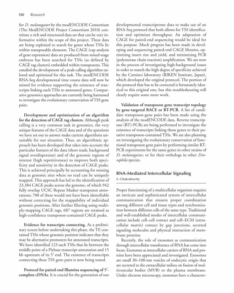
180 Research
for D. melanogaster by the modENCODE Consortium(The ModENCODE Project Consortium 2010) con-stitute a rich and structured data set that can be very in-formative within the scope of this project. These dataare being exploited to search for genes whose TSSs liewithin transposable elements. The CAGE (cap analysisof gene expression) data set produced from mixed-stageembryos has been searched for TSSs (as defined byCAGE tag clusters) embedded within transposons. Thisentailed the development of a peak-calling algorithm tai-lored and optimized for this task. The modENCODERNA-Seq developmental time course data will now bemined for evidence supporting the existence of tran-scripts linking such TSSs to annotated genes. Compar-ative genomics approaches are currently being harnessedto investigate the evolutionary conservation of TSS genepairs.
Development and optimization of an algorithmfor the detection of CAGE tag clusters. Although peakcalling is a very common task in genomics, the veryunique features of the CAGE data and of the questionswe have set out to answer make current algorithms un-suitable for our situation. Thus, an algorithmic ap-proach has been developed that takes into account theparticular features of the data (short reads, backgroundsignal overdispersion) and of the genomic regions ofinterest (high repetitiveness) to improve both speci-ficity and sensitivity in the detection of CAGE peaks.This is achieved principally by accounting for missingdata at genomic sites where no read can be uniquelymapped. This approach has led to the identification of23,384 CAGE peaks across the genome, of which 942fully overlap UCSC Repeat Masker transposon anno-tations; 700 of these would not have been identifiablewithout correcting for the mappability of individualgenomic positions. After further filtering using multi-ply-mapping CAGE tags, 687 regions are retained ashigh-confidence transposon-contained CAGE peaks.
Evidence for transcripts connecting. As a prelimi-nary screen before undertaking this phase, the TE con-tained TSSs whose genomic position indicates that theymay be alternative promoters for annotated transcripts.We have identified 123 such TSSs that lie between themiddle point of a Flybase transcript annotation and 15kb upstream of its 5′ end. The existence of transcriptsconnecting these TSS gene pairs is now being tested.
Protocol for paired-end Illumina sequencing of 5′-complete cDNAs. It is crucial for the generation of our
developmental transcriptome data to make use of anRNA-Seq protocol that both allows for TSS identifica-tion and optimizes throughput. An adaptation ofCAGE for paired-end sequencing would be ideal forthis purpose. Much progress has been made in devel-oping and sequencing paired-end CAGE libraries, op-timizing insert size and yield, and minimizing PCR(polymerase chain reaction) amplification. We are nowin the process of investigating high-background issuesin order to match the high degree of specificity achievedby the Carninci laboratory (RIKEN Institute, Japan),which developed the original protocol. The portion ofthe protocol that has to be corrected is fortunately iden-tical to this original one, but this troubleshooting willclearly require some more work.
Validation of transposon gene transcript topologyby gene-targeted RACE or RT-PCR. A list of candi-date transposon-gene pairs has been made using theanalysis of the modENCODE data. Reverse transcrip-tase (RT)-PCRs are being performed to investigate theexistence of transcripts linking those genes to their pu-tative transposon-contained TSSs. We are also planningon investigating the evolutionary conservation of func-tional transposon gene pairs by performing similar RT-PCR experiments for the same genes in other strains ofD. melanogaster, or for their orthologs in other Dro-sophila species.
RNA-Mediated Intercellular SignalingS. Chakrabortty
Proper functioning of a multicellular organism requiresan intricate and sophisticated system of intercellularcommunication that ensures proper coordinationamong different cell and tissue types and synchroniza-tion between different cells of the same type. Traditionaland well-established modes of intercellular communi-cation include cell–cell contact and cell–ECM (extra-cellular matrix) contact by gap junctions, secretedsignaling molecules and physical interaction of mem-brane proteins.
Recently, the role of exosomes as communicationthrough intercellular transference of RNA has come intofocus. Exosomes as intercellular carriers of RNA and pro-teins have been appreciated and investigated. Exosomesare small 30–100-nm vesicles of endocytic origin thatare secreted to the extracellular milieu on fusion of mul-tivesicular bodies (MVB) to the plasma membrane.Under electron microscopy, exosomes have a character-
175-193_06_Genomics_Annual Report_2009 template 7/8/11 12:01 PM Page 180
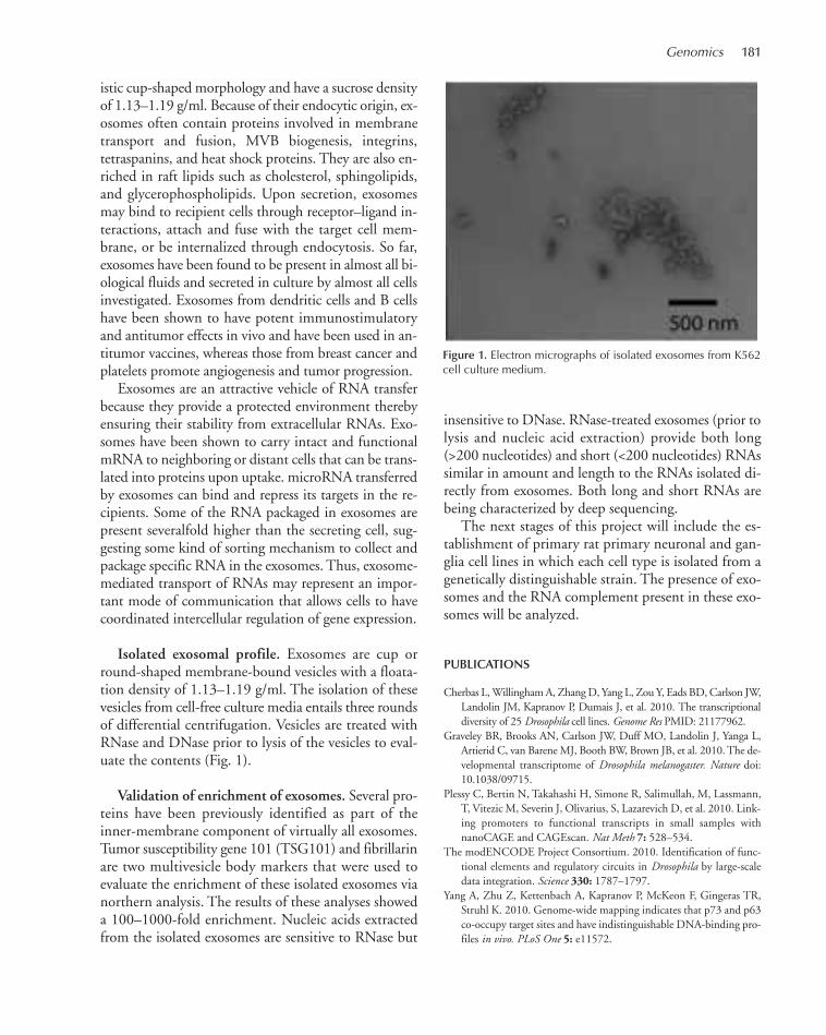
Genomics 181
istic cup-shaped morphology and have a sucrose densityof 1.13–1.19 g/ml. Because of their endocytic origin, ex-osomes often contain proteins involved in membranetransport and fusion, MVB biogenesis, integrins,tetraspanins, and heat shock proteins. They are also en-riched in raft lipids such as cholesterol, sphingolipids,and glycerophospholipids. Upon secretion, exosomesmay bind to recipient cells through receptor–ligand in-teractions, attach and fuse with the target cell mem-brane, or be internalized through endocytosis. So far,exosomes have been found to be present in almost all bi-ological fluids and secreted in culture by almost all cellsinvestigated. Exosomes from dendritic cells and B cellshave been shown to have potent immunostimulatoryand antitumor effects in vivo and have been used in an-titumor vaccines, whereas those from breast cancer andplatelets promote angiogenesis and tumor progression.
Exosomes are an attractive vehicle of RNA transferbecause they provide a protected environment therebyensuring their stability from extracellular RNAs. Exo-somes have been shown to carry intact and functionalmRNA to neighboring or distant cells that can be trans-lated into proteins upon uptake. microRNA transferredby exosomes can bind and repress its targets in the re-cipients. Some of the RNA packaged in exosomes arepresent severalfold higher than the secreting cell, sug-gesting some kind of sorting mechanism to collect andpackage specific RNA in the exosomes. Thus, exosome-mediated transport of RNAs may represent an impor-tant mode of communication that allows cells to havecoordinated intercellular regulation of gene expression.
Isolated exosomal profile. Exosomes are cup orround-shaped membrane-bound vesicles with a floata-tion density of 1.13–1.19 g/ml. The isolation of thesevesicles from cell-free culture media entails three roundsof differential centrifugation. Vesicles are treated withRNase and DNase prior to lysis of the vesicles to eval-uate the contents (Fig. 1).
Validation of enrichment of exosomes. Several pro-teins have been previously identified as part of theinner-membrane component of virtually all exosomes.Tumor susceptibility gene 101 (TSG101) and fibrillarinare two multivesicle body markers that were used toevaluate the enrichment of these isolated exosomes vianorthern analysis. The results of these analyses showeda 100–1000-fold enrichment. Nucleic acids extractedfrom the isolated exosomes are sensitive to RNase but
insensitive to DNase. RNase-treated exosomes (prior tolysis and nucleic acid extraction) provide both long(>200 nucleotides) and short (<200 nucleotides) RNAssimilar in amount and length to the RNAs isolated di-rectly from exosomes. Both long and short RNAs arebeing characterized by deep sequencing.
The next stages of this project will include the es-tablishment of primary rat primary neuronal and gan-glia cell lines in which each cell type is isolated from agenetically distinguishable strain. The presence of exo-somes and the RNA complement present in these exo-somes will be analyzed.
PUBLICATIONS
Cherbas L, Willingham A, Zhang D, Yang L, Zou Y, Eads BD, Carlson JW,Landolin JM, Kapranov P, Dumais J, et al. 2010. The transcriptionaldiversity of 25 Drosophila cell lines. Genome Res PMID: 21177962.
Graveley BR, Brooks AN, Carlson JW, Duff MO, Landolin J, Yanga L,Artierid C, van Barene MJ, Booth BW, Brown JB, et al. 2010. The de-velopmental transcriptome of Drosophila melanogaster. Nature doi:10.1038/09715.
Plessy C, Bertin N, Takahashi H, Simone R, Salimullah, M, Lassmann,T, Vitezic M, Severin J, Olivarius, S, Lazarevich D, et al. 2010. Link-ing promoters to functional transcripts in small samples withnanoCAGE and CAGEscan. Nat Meth 7: 528–534.
The modENCODE Project Consortium. 2010. Identification of func-tional elements and regulatory circuits in Drosophila by large-scaledata integration. Science 330: 1787–1797.
Yang A, Zhu Z, Kettenbach A, Kapranov P, McKeon F, Gingeras TR,Struhl K. 2010. Genome-wide mapping indicates that p73 and p63co-occupy target sites and have indistinguishable DNA-binding pro-files in vivo. PLoS One 5: e11572.
Figure 1. Electron micrographs of isolated exosomes from K562cell culture medium.
175-193_06_Genomics_Annual Report_2009 template 7/8/11 12:01 PM Page 181

182
Investigating Genome Variation:Infrastructure Development andQuality Assurance
The Genome Center continued to expand resources forlarge-scale genomic studies in 2010, in terms of bothnext-generation sequencing and analysis capabilitiesand usage by the wider CSHL community. We contin-ued to focus on training and development of standardoperating procedures to ensure that quality remainedhigh during this expansion (see Fig. 1).
Four new Illumina HiSeq 2000 instruments were in-troduced to the Genome Center that increased capacityby fourfold over the Illumina Genome Analyzer IIx(GAIIx) instruments that we replaced. In addition, weupgraded the Illumina software and incorporated animproved enzyme in the reagent kits that allowed forreduced run times.
We also became a beta test site for the new PacBioRS technology that allows for amplification-free single-molecule sequencing. We are focusing on increasingyield and accuracy on this platform, but we have al-ready achieved long read lengths that should comple-ment our shorter Illumina reads.
To address the increased demand for secondaryanalysis, several postdocs joined the bioinformaticsstaff, and new alignment and variant detection pipelineswere investigated. We will continue to update and aug-ment the informatics workflow in order to providecomprehensive and efficient analyses.
We began working with the Wigler lab on the devel-opment of a new LIM system to provide more detailedsample reports, automated status updates, and qualitymetrics. These improvements will enhance users’ abilityto monitor and modify the large and diverse projectsthat are possible with next-generation sequencing.
We have begun the testing of cloud computing andare working with resources such as Galaxy, which pro-vide a simplified web interface to help ease the burden
on local servers, as well as to accommodate users whorequire assistance with bioinformatic components oftheir data analysis. Streamlining the analysis pipelineand the tools available to the community will remainan important goal in 2011. This will be done in collab-oration with the Bioinformatics Shared Resource of theCSHL Cancer Center (Molly Hammell, Director).
Investigation of Bipolar DisorderGenetics Using Exome Captureand Resequencing
is work was done in collaboration with J. Potash, R.Karchin, and A. Chakravarty (Johns Hopkins Schoolof Medicine) and I. Iossifov (Cold Spring Harbor Lab-oratory).
Bipolar disorder I (BP) is a major mental illness witha significant genetic component, as demonstrated byfamily, twin, and adoption studies (Smoller and Finn,Am J Med Genet C Semin Med Genet 123C: 48 [2003]).Molecular investigation into the genetics of BP has in-cluded linkage analyses and, more recently, several largegenome-wide association studies (GWAS) (Wendland
DEVELOPMENT OF NEXT-GENERATION SEQUENCINGTECHNOLOGY
W.R. McCombie M. Bell I. Grabill D. Lewis M. Ossipov L. Cardone A. Ji S. Mavruk J. Parla G. Cheang A. Karim S. McCarthy R. Solomon E. Ghiban M. Kramer P. Mocombe J.C. Yao M. Gierszewska S. Lata S. Muller
Figure 1. Comparison of total bases generated versus total high-quality bases for monthly Illumina runs from December 2009through December 2010. We use Q30 bases as a metric to en-sure very high data quality.
175-193_06_Genomics_Annual Report_2009 template 7/8/11 12:01 PM Page 182

Genomics 183
and McMahon 2010). Although GWAS has yielded asmall number of genome-wide significant results, the“missing heritability” problem suggests that the major-ity of variants contributing to BP are rare variants.Other approaches are therefore necessary to uncoverthese rare BP susceptibility variants. We are applyingexome capture and resequencing toward finding raregenetic variants that contribute to BP.
Our work in 2010 involved the exome capture andresequencing of 186 samples, which included a pilot-family-based study, and BP probands and unrelated con-trols. Each sample was prepared for capture and se-quencing using Illumina-compatible sequencing adapterswith or without bar codes, individually captured, and se-quenced on one lane of an Illumina GAIIx instrumentprogrammed for PE 76 cycle sequencing. Sequence dataquality, genome alignment, and exome capture statisticswere computed to determine the efficiency of the captureand sequencing using an analysis pipeline developed byIvan Iossifov. Samples that required more raw sequencedata to obtain improved exome coverage were includedin a second round of sequencing and pooled during se-quencing when possible (with barcode multiplexing).
On average, 36.5 million pass-filter sequence readpairs (5.5 billion bases) were produced per captured sam-ple, with 99.7% of bases attributed to library DNA(rather than sequencing adapter), 93.5% of read pairsproperly mapped with unique chromosome start and endcoordinates, 57.5% of properly mapped read pairsmapped to exome probe sequences, and 31.3% of thetotal input pass-filter sequence data mapped to the exometarget. The raw sequence data that we produced provided81.6% of the exome covered at greater than 20x depth,13,694 coding single-nucleotide polymorphisms (SNPs),7545 synonymous SNPs, and 6149 nonsynonymousSNPs per sample, on average (Table 1). On the basis ofour current exome data, we have identified 275 geneswith possibly damaging, nonsynonymous SNPs found incases but not in controls. There also appears to be an en-richment of nonsynonymous SNPs in BP GWAS-iden-tified genes in cases compared to controls.
Further work for this project will include implement-ing a custom exome capture design that provides highercoverage of genes that encode postsynaptic proteins; thiswill allow us to additionally investigate the neurologicalbasis of BP, exome captures with at least ~650 additionalcases and ~350 additional controls, and more in-depthanalyses of SNPs identified in this project.
Family-Based Analysis of BipolarDisorder by Whole-GenomeResequencing
is work was done in collaboration with D. Black-wood, K. Evans, and D. Porteous (University of Edin-burgh).
We are studying a large Scottish pedigree wheremany family members have bipolar disorder or recur-rent major depressive disorder. A genetic study of thisfamily by our collaborators at the University of Edin-burgh suggested that a 20-Mb haplotype on chromo-some 4 has significant linkage to bipolar disorder (LeHellard et al., Biol Psychiatry 61: 797 [2007]).
We decided to use next-generation sequencing (Illu-mina GAIIx) to resequence the entire genome of indi-viduals from this family, focusing analysis on the 20-Mbcritical region on chromosome 4. We hope to discoverrare variants associated with bipolar disorder susceptibil-ity. Three affected (two with BD, one with MDD) andtwo unaffected married-in individuals from this familywere selected for whole-genome sequencing using paired-end 101- and 125-cycle runs. We achieved 58x–36x cov-erage on each individual. Reads from all flow cells foreach individual sample were then aligned to UCSC hg8.We processed the mapping and SNP calling by using theBurrows–Wheeler alignment (BWA) and sequence align-ment map (SAM) tools (Li et al., Bioinformatics 25: 2078[2009]) pipeline. Potential polymerase chain reaction(PCR) duplicates were removed, and error-prone variantcalls were then filtered out by using a minimum consen-sus quality score of 20 and a minimum read depth of
Table 1. Overview of Human Exome Coverage and SNP Calling Results for the 186 Case and ControlSamples Captured and Resequenced in 2010
% Exome target Total no. No. synonymous No. nonsynonymous covered at ≥20x coding SNPs SNPs SNPsMin 65.43 11,677 6,399 5278
Max 92.56 18,493 10,100 8393
Mean 81.60 13,694 8,545 6149
Human exome captures were performed individually with SeqCap EZ Exome Library v1.2 (Nimble-Gen) and sequenced with paired-end 76-base reads on GAIIx sequencing instruments (Illumina).
175-193_06_Genomics_Annual Report_2009 template 7/8/11 12:01 PM Page 183
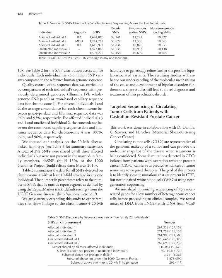
184 Research
10x. See Table 2 for the SNP distribution across all fiveindividuals. Each individual has ~3.6 million SNP vari-ants compared to the reference human genome sequence.
Quality control of the sequence data was carried outby comparison of each individual’s sequence with pre-viously determined genotype (Illumina IVb whole-genome SNP panel) or exon-based capillary sequencedata (for chromosome 4). For affected individuals 1 and2, the average concordance for each chromosome be-tween genotype data and Illumina sequence data was94% and 93%, respectively. For affected individuals 3and 1 and unaffected individual 2, the concordance be-tween the exon-based capillary sequence data and Illu-mina sequence data for chromosome 4 was 100%,97%, and 96%, respectively.
We focused our analysis on the 20-Mb disease-linked haplotype (see Table 3 for summary statistics).A total of 292 SNPs were shared by all three affectedindividuals but were not present in the married-in fam-ily members, dbSNP (build 130), or the 1000Genomes Project (build release date: March 2010).
Table 3 summarizes the data for all SNPs detected onchromosome 4 with at least 10-fold coverage in any oneindividual. The number in parentheses refers to the num-ber of SNPs that lie outside repeat regions, as defined byusing the RepeatMasker track (default settings) from theUCSC Genome Browser (http://genome.ucsc.edu/).
We are currently extending this study to other fam-ilies that show linkage to the chromosome-4 20-Mb
haplotype to genetically refine further the possible bipo-lar-associated variants. The resulting studies will en-hance our understanding of the molecular mechanismsof the cause and development of bipolar disorder; fur-thermore, these studies will lead to novel diagnoses andtreatment of this psychiatric disorder.
Targeted Sequencing of CirculatingTumor Cells from Patients withCastration-Resistant Prostate Cancer
is work was done in collaboration with D. Danilla,C. Sawyer, and H. Scher (Memorial Sloan-KetteringCancer Center).
Circulating tumor cells (CTCs) are representative ofthe genomic makeup of a tumor and can provide themolecular snapshot of the disease when treatment isbeing considered. Somatic mutations detected in CTCsisolated from patients with castration-resistant prostatecancer (CRPC) can serve as predictive markers of tumorsensitivity to targeted therapies. The goal of this projectis to identify somatic mutations that are present in CTC,but not in paired white blood cells (WBCs) using next-generation sequencing.
We initialized optimizing sequencing of 75 cancer-related genes for a low number of heterogeneous cancercells before proceeding to clinical samples. We testedmixes of DNA from LNCaP with DNA from VCaP
Table 2. Number of SNPs Identified by Whole-Genome Sequencing Across the Five Individuals
Exonic Synonymous NonsynonymousIndividual Diagnosis SNPs SNPs coding SNPs coding SNPs
Affected individual 1 BD 3,694,870 53,541 11,295 10,827Affected individual 2 MDD 3,714,782 53,672 11,330 10,863Affected individual 3 BD 3,619,932 51,836 10,876 10,553Unaffected individual 1 – 3,573,886 51,635 10,952 10,438Unaffected individual 2 – 3,594,225 51,155 10,699 10,265
Table lists all SNPs with at least 10x coverage in any one individual.
Table 3. SNP Discovery by Sequence Analysis of Five Family 22 Individuals
SNPs on chromosome 4 Number
Affected individual 1 267,358 (127,159)Affected individual 2 271,759 (129,130)Affected individual 3 261,995 (124,580)Unaffected individual 1 270,646 (128,372)Unaffected individual 2 267,699 (127,232) Subset shared by all three affected individuals 116,034 (56,626) Subset of above not present in unaffected individuals 30,110 (14,720) Subset of above not present in dbSNP 3,261 (1,363) Subset of above not present in 1000 Genomes Project 1,676 (590) Subset of above that map to 20-Mb linkage region 292 (117)
175-193_06_Genomics_Annual Report_2009 template 7/8/11 12:01 PM Page 184

Genomics 185
prostate cancer cells at 20%, 50%, 80%, and 100% toestablish the limit of detection from deep sequencing.We next tested ratio mixes of 50 cells total for isolationand wheat germ agglutinin (WGA). With each set ofsamples, we performed PCR amplification using a Rain-Dance microfluidic PCR device to stream an emulsionof sample DNA into a microfluidic device that mergesthat stream with a mix of emulsion droplets containinglarge numbers of distinct PCR primer pairs. The resultis that each reaction occurs individually after the streamsmerge, because each droplet contains a specific primerpair and eliminates primer competition. The emulsionis used as input for PCR and then broken to isolate thetotal DNA from the amplification for the gene panel ofinterest. Sequencing was performed on an IlluminaGAII using single-end 36- and 76-cycle runs. Our initialanalysis has confirmed the gene panel sequencing cov-erage (see Fig. 2) and that at least in some cases we cansee amplified region amplification by counting the num-ber of reads that match to the amplified region com-pared to other, nonamplified regions of the genome.
We then performed targeted sequencing on 29 flo-rescence-activated cell sorted (FACS) CTC and WBCwhole-genome-amplified samples isolated from eightpatients with progressive CRPC. Before sequencing, wealso performed PCR amplification using a RainDancemicrofluidic PCR device on each sample. We identified~10–100 coding single-nucleotide variants (SNVs) ineach CTC sample that were not present in the pairedWBC samples. Figure 3 shows a missense amplified re-gion mutation that is present in CTC50 but not inpaired WBC20 and WBC10 in a selected patient.
Currently, we are validating selected variants usingSanger sequencing and also extending the study to ad-
ditional patients enrolled in amplified-region-targetedclinical trials using exome sequencing. This study maylead to identification of biomarkers to guide treatmentselection for patients with CRPC.
Targeted Resequencing of the HumanDisrupted-in-Schizophrenia-1 GeneUsing Long-Range PCR and IlluminaSequencing
is work was done in collaboration with D. Black-wood, D. Porteous, P. ompson, and I. Deary (Uni-versity of Edinburgh) and P. Visscher and A. McRae(University of Queensland).
Disrupted-In-Schizophrenia-1 (DISC1) is a strongcandidate gene for bipolar disorder, schizophrenia, andcognitive disorders. A translocation disrupting this novelgene was first described by our collaborators in Edin-burgh who showed that it segregated significantly withpsychiatric disorder in a Scottish pedigree. In conjunc-tion with our collaborators, we initiated a pilot projectto resequence ~523 kb on chromosome 1 containingDISC1 using long-range PCR. We designed primers totile across the 0.5-Mb region and showed that we wereable to cover up to 98% of our amplified target at >30xcoverage with a minimum of 20x needed for accuratemutation discovery. The planned sequencing of ~523kb of chromosome 1 in 2088 samples was completedduring 2009; the data were processed, and putativeSNPs were identified using MAQ (Li, Genome Res 18:1851 [2008]). The sample set consisted of 266 bipolardisorder, 277 schizophrenia, 271 major depression, and1274 control individuals (the majority of controls are
Figure 2. Coverage of 75 targeted genes from 50-cell VCaP after WGA. Mean coverage of the amplified AR gene was ~5000x.
175-193_06_Genomics_Annual Report_2009 template 7/8/11 12:01 PM Page 185
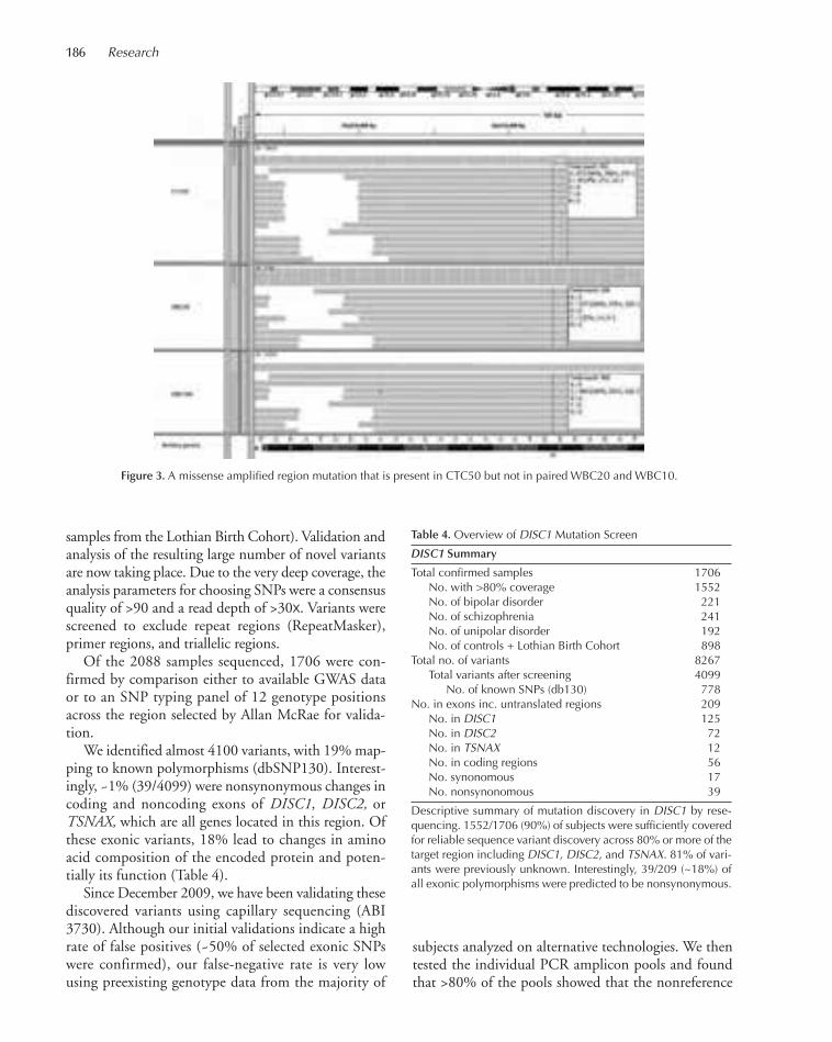
186 Research
samples from the Lothian Birth Cohort). Validation andanalysis of the resulting large number of novel variantsare now taking place. Due to the very deep coverage, theanalysis parameters for choosing SNPs were a consensusquality of >90 and a read depth of >30x. Variants werescreened to exclude repeat regions (RepeatMasker),primer regions, and triallelic regions.
Of the 2088 samples sequenced, 1706 were con-firmed by comparison either to available GWAS dataor to an SNP typing panel of 12 genotype positionsacross the region selected by Allan McRae for valida-tion.
We identified almost 4100 variants, with 19% map-ping to known polymorphisms (dbSNP130). Interest-ingly, ~1% (39/4099) were nonsynonymous changes incoding and noncoding exons of DISC1, DISC2, orTSNAX, which are all genes located in this region. Ofthese exonic variants, 18% lead to changes in aminoacid composition of the encoded protein and poten-tially its function (Table 4).
Since December 2009, we have been validating thesediscovered variants using capillary sequencing (ABI3730). Although our initial validations indicate a highrate of false positives (~50% of selected exonic SNPswere confirmed), our false-negative rate is very lowusing preexisting genotype data from the majority of
Figure 3. A missense amplified region mutation that is present in CTC50 but not in paired WBC20 and WBC10.
subjects analyzed on alternative technologies. We thentested the individual PCR amplicon pools and foundthat >80% of the pools showed that the nonreference
Table 4. Overview of DISC1 Mutation Screen
DISC1 Summary
Total confirmed samples 1706 No. with >80% coverage 1552 No. of bipolar disorder 221 No. of schizophrenia 241 No. of unipolar disorder 192 No. of controls + Lothian Birth Cohort 898Total no. of variants 8267 Total variants after screening 4099 No. of known SNPs (db130) 778No. in exons inc. untranslated regions 209 No. in DISC1 125 No. in DISC2 72 No. in TSNAX 12 No. in coding regions 56 No. synonomous 17 No. nonsynonomous 39
Descriptive summary of mutation discovery in DISC1 by rese-quencing. 1552/1706 (90%) of subjects were sufficiently coveredfor reliable sequence variant discovery across 80% or more of thetarget region including DISC1, DISC2, and TSNAX. 81% of vari-ants were previously unknown. Interestingly, 39/209 (~18%) ofall exonic polymorphisms were predicted to be nonsynonymous.
175-193_06_Genomics_Annual Report_2009 template 7/8/11 12:01 PM Page 186

Genomics 187
allele was detected in the amplicons. Thus, it appearsthat we introduced a small number of variants into theprocess (in the long-range PCR) and then effectivelydetected them. As a follow-up validation, 460 randomvariants were chosen across the entire region, and ~79%of those variants were confirmed by ABI capillary se-quencing. Thus, the rate of validation outside exons ap-pears to be higher, which seems to correlate withfindings in the community that evolutionary con-straints on exons make variants in those coding regionsmore likely to be errors due to the presence of fewerreal, rare variants. We are continuing with various val-idation studies to identify the major sources of falsepositives, including testing alternative alignment soft-ware Novoalign (Novocraft), which allows for moresensitive read alignment.
To further investigate our variant calls, our collabora-tors compared our data to 903 variants (from 60HapMap samples) that were called in the European-onlyset of the 1000 Genomes Project (www.1000genomes.org). Minor allele frequency distribution looked quitesimilar (see Fig. 4), although we called more rare variants,probably due to our greater sequence depth.
We are working on revising our validation plan basedon our ongoing results. We plan to complete final analy-sis of the data this year and publish our findings. It is
known that DISC1 causes cognitive disorders in the ini-tial translocation family. This study will finally deter-mine how frequently other rare mutations in DISC1contribute to these disorders and give some perspectiveon the types of variants that may contribute to disease.
PUBLICATIONS
Wendland JR, McMahon FS. 2010 Behavioral neurobiology of bipolardisorder and its treatment (ed. HK Manji, CK Zarate Jr.), pp. 19–30.Springer-Verlag, Dordrecht.
Figure 4. Comparison of minor allele frequency in 903 overlap-ping variants in the 1000 Genomes (EUR) University of MichiganMACH call set and the CSH DISC1 variant call set.
Rebecca Solomon
175-193_06_Genomics_Annual Report_2009 template 7/8/11 12:01 PM Page 187

188
I moved the bulk of my laboratory to the Ontario In-stitute for Cancer Research in Toronto, Ontario, in2009, but I still maintain a small group at CSHL work-ing on the iPlant Collaborative project.
The iPlant Collaborative is a virtual organization offaculty, students, postdoctoral fellows, staff, high-perfor-mance computing experts, and professional software de-velopers drawn from CSHL, the University of Arizona,and the Texas Advanced Computer Center. iPLant is de-signed to solicit and meet biological grand challenge ques-tions from the plant science community, and, in closeconsultation with this community, create a cyberinfra-structure that makes it possible to meet these challenges.In 2009, two self-forming grand challenge teams werechosen by a community-representative Board of Directorsto work with iPlant personnel to develop “Discovery En-vironment (DE),” a cyberinfrastructure and user interfaceto help with the biological grand challenges.
Our group works on the iPlant Tree of Life (iPToL)and the Genotype to Phenotype (Gen2Phen) GrandChallenge Project. The fundamental aim of the firstproject is to assemble a comprehensive phylogenetic treeof half a million known species of green plants for betterknowledge of evolutionary relationships, which will yieldnew insights across the plant sciences. The second proj-ect aims to create databases and tools to integrate andanalyze experiments that connect the genotype of plantsto their physical properties, such as agronomically im-portant traits. Both projects are working toward releasingweb-based discovery environments during 2011.
The list of publications below represent work both atCSHL and at the Ontario Institute for Cancer Research.
PUBLICATIONS
Cancer Target Discovery and Development Network, Schreiber SL,Shamji AF, Clemons PA, Hon C, Koehler AN, Munoz B, Palmer M,
Stern AM, Wagner BK, et al. 2010. Towards patient-based cancertherapeutics. Nat Biotechnol 28: 904–906.
Gerstein MB, Lu ZJ, Van Nostrand EL, Cheng C, Arshinoff BI, Liu T,Yip KY, Robilotto R, Rechtsteiner A, Ikegami K, et al. 2010. Inte-grative analysis of the Caenorhabditis elegans genome by the mod-ENCODE project. Science 330: 1775–1787.
Harris TW, Antoshechkin I, Bieri T, Blasiar D, Chan J, Chen WJ, DeLa Cruz N, Davis P, Duesbury M, Fang R, et al. 2010. WormBase:A comprehensive resource for nematode research. Nucleic Acids Res38: (Database issue) D463–467.
Hurwitz BL, Kudrna D, Yu Y, Sebastian A, Zuccolo A, Jackson SA, WareD, Wing RA, Stein L. 2010. Rice structural variation: a comparativeanalysis of structural variation between rice and three of its closestrelatives in the genus Oryza. Plant J 63: 990–1003.
International Cancer Genome Consortium, Hudson TJ, Anderson W,Artez A, Barker AD, Bell C, Bernabé RR, Bhan MK, Calvo F, EerolaI, et al. 2010. International network of cancer genome projects. Na-ture 464: 993–998.
Jassal B, Jupe S, Caudy M, Birney E, Stein L, Hermjakob H, D’Eusta-chio P. 2010. The systematic annotation of the three main GPCRfamilies in Reactome. Database 10: baq018.
modENCODE Consortium. Roy S, Ernst J, Kharchenko PV, Kherad-pour P, Negre N, Eaton ML, Landolin JM, Bristow CA, Ma L, LinMF, et al. 2010. Identification of functional elements and regula-tory circuits by Drosophila modENCODE. Science 330: 1787–1797.
Nègre N, Brown CD, Shah PK, Kheradpour P, Morrison CA, HenikoffJG, Feng X, Ahmad K, Russell S, White RA, et al. 2010. A compre-hensive map of insulator elements for the Drosophila genome. PLoSGenet 6: e1000814.
Reese MG, Moore B, Batchelor C, Salas F, Cunningham F, Marth GT,Stein L, Flicek P, Yandell M, Eilbeck K. 2010. A standard variationfile format for human genome sequences. Genome Biol 11: R88.
Stein LD. 2010. The case for cloud computing in genome informatics.Genome Biol 11: 207.
Voight BF, Scott LJ, Steinthorsdottir V, Morris AP, Dina C, Welch RP,Zeggini E, Huth C, Aulchenko YS, Thorleifsson G, et al. 2010.Twelve type 2 diabetes susceptibility loci identified through large-scale association analysis. Nat Genet 42: 579–589.
Wang L, Stein LD. 2010. Localizing triplet periodicity in DNA andcDNA sequences. BMC Bioinformatics 11: 550.
Wu G, Feng X, Stein L. 2010. A human functional protein interactionnetwork and its application to cancer data analysis. Genome Biol 11:R53.
GENOME-SCALE DATABASES OF PATHWAYS, GENETIC VARIATIONS,AND EVOLUTION
L. Stein S. Avraham B. Faga A. Kanapin G. WuS. Cain G. Gopinathrao W. Spooner Z. ZhaN. de la Cruz T. Harris P. Van Buren W. Zhao
175-193_06_Genomics_Annual Report_2009 template 7/8/11 12:01 PM Page 188

189
Our lab has dual goals of conducting plant genomics re-search and of enabling access to genomics resources anddiscovery tools to the broader research community. Re-search includes a broad range of activities in physical, sta-tistical, and functional genomics in both model systemsand crop plants, with applications in agriculture. Duringthe past year, we were joined by postdoc Christos Nout-sos, Marcela Karey Monaco, undergraduate internMichael Levy, and high school intern Rigina Gallagher.We said good-bye to Liya Ren and Shuly Avraham. Onsabbatical from University of Missouri, Columbia,Michael McMullen has joined us as a visiting professor.
GrameneA. Chuah, M. Karey Monaco, S. Pasternak, W. Spooner, J. Stein, J. Thomason, S. Wei, K. Youens-Clark [in collaboration with E. Buckler, USDA, Cornell University; S. McCouch, Cornell University; P. Jaiswal, Oregon State University]
Gramene was conceived from the notion that the juxta-position of annotation across multiple genomes by com-parative mapping provides a synthesis of knowledgegreater than the sum of its parts. Gramene is a collabo-rative project that leverages sequence and functional in-formation from plant reference genomes to promoteresearch and translational genomics in agriculture. TheGramene website (http://www.gramene. org) serves as aportal to multiple genome browsers and manually cu-rated databases of genes, proteins, biochemical pathways,quantitative trait loci (QTL), ontologies, germplasm, andgenetic diversity data. In the last year, our group has com-pleted several milestones on the Gramene project, includ-ing its 31st and 32nd releases since 2000. Of the manyimprovements made, we increased the number of partialand complete sequenced genomes in our Ensembl ge-nome browser to 19 scientifically and economically im-portant species including rice, corn, grape, sorghum,poplar, and the basal land plant Selaginella. Gramene isalso home to reference sequences for three cultivated andeight wild species of rice, making it the single most com-prehensive genus-level (Oryza) resource in plants. This
year, we released pathway databases for maize (MaizeCyc)and Brachypodium (BrachyCyc), complementing existingpathway databases for rice, sorghum, and other plants.Gramene is also positioning itself as a principal resourcefor conducting population-based genetics research. Weadded several new diversity/phenotype data sets coveringArabidopsis, rice, and maize and have added new toolsfor querying single-nucleotide polymorphisms (SNPs)and visualizing genome-wide association studies. All ofthis work has contributed to a 38% increase of traffic onthe site during the last 2 years from users all over theworld. Our databases are created in partnership with theEuropean Bioinformatics Institute (EBI) and are nowalso available through the Ensembl Plant portal. Duringthe next year, we will continue to add new referencegenomes, data sets, and functionalities to this resource.
Maize ProjectS. Pasternak, A. Olson, J. Stein [in collaboration with the Maize Genome Sequencing Consortium;J. Glaubitz, Cornell University; E. Buckler, USDA, Cornell University; M. McMullen, USDA, University of Missouri, Columbia]
Our group leads data coordination, analysis, and visu-alization for the Maize Genome Sequencing Consor-tium, a 4-year, multi-institutional project funded by theNational Science Foundation (NSF). In the last year,we released version 5b.60 of MaizeSequence.org, theofficial public portal of the maize project. This releaseis based on the most recent version of the reference as-sembly (B73 RefGen_v2) for which we conducted anew evidence-based gene build. The new gene set in-cludes improved gene models for thousands of loci andhundreds of new high-confidence loci that were not de-tected in the previous version of the assembly. Despitethe many improvements to the reference sequence,there are still gaps of missing genes and stretches of se-quences that are not definitively positioned withinchromosomes. In a new initiative, we are working tocapture missing gene space using de novo assemblies ofwhole-genome shotgun sequence reads. We have devel-
PLANT GENOME ORGANIZATION, EVOLUTION, AND FUNCTION
D. Ware J.-M. Chia M. Karey Monaco Z. Lu S. Pasternak J. Thomason K. Youens-ClarkA. Chuah S. Kumari C. Noutsos W. Spooner P. Van Buren L. ZhangA. Eveland Z. Liu A. Olson J. Stein S. Wei
175-193_06_Genomics_Annual Report_2009 template 7/8/11 12:01 PM Page 189

190 Research
oped a novel approach that builds gene-centered scaf-folds by guided assembly onto cDNA sequences. To an-chor these, we are taking advantage of a very high-resolution genetic map containing nearly half a millionmarkers generated by a genotype by sequencing ap-proach. Preliminary data indicate that we are able toanchor thousands of de novo contigs corresponding tohundreds of genes. These data will contribute to futureimproved assemblies of the maize reference genome.
Maize HapMap ProjectJ.-M. Chia [in collaboration with E. Buckler, USDA, Cornell University; M. McMullen, USDA, University of Missouri, Columbia]
Maize has abundant diversity that is evident from thelarge variation of observable traits in the species. Thisdiversity translates to the DNA level, where large dif-ferences exist between individuals of the species. Muchof this genetic diversity in maize resolves to single DNAbase differences (SNPs), but perhaps more significantly,structural variations as well as copy-number variationshave a strong impact in the genetic diversity in thespecies. As a result, the concept of a maize genome isfluid, with DNA content varying greatly between indi-viduals of the species. It is estimated that 30% of thegenome of each maize inbred line is unique to itself andthis unique space contains genes, thus likely to impacttraits.
With this in mind, we set out to study the geneticvariation in the species. In this iteration of the MaizeHapMap Project, using high-throughput DNA se-quencing technologies, we scale out the breadth anddepth of the study by resequencing more than 100 in-bred lines of the Zea mays genus. The volume of se-quence generated per inbred line is approximately fivetimes the size of the genome, and from these sequences,we can identify variant loci. Given the complexity ofthe genome, these variations were scored using carefullydesigned population genetics-based filters that leveragethe carefully constructed maize breeding populations.In total, more than 55 million SNPs were identified.Furthermore, by comparing the distribution of readdepth across the genome we could determine copy-number and structural variations. The results reveal theplasticity of the genome with more than 80% of thegenome affected by copy-number or structural varia-tion. We used the combined set of SNPs and structuralvariations as markers and tested the potential of theseto affect maize traits. The results highlight the signifi-
cant impact of structural variation on traits. Finally, be-cause wild ancestors of domesticated maize were in-cluded in the sampling population, based on regions oflower genetic diversity, we are able to address questionsabout genes that were selected for during the domesti-cation and improvement of maize as a crop.
Maize breeders have long been exploiting the diver-sity in maize for improving the yield of this crop throughselective breeding programs, contributing to its currentposition at the apex of the production crop list. We arein an era where traditional crop-breeding techniques arebeing supplemented and, in some cases, supplanted bymolecular breeding approaches that speed up the breed-ing process. Understanding the diversity in maize andhow it impacts agronomic traits is crucial in implement-ing these new approaches and will be an important plat-form for sustainable agriculture in the future.
The iPlant CollaborativeJ. Lu, C. Noutsos
This project employs more than 50 staff and is headquar-tered at the University of Arizona (principal investigatorStephen Goff ). Dozens of collaborators hail from over20 institutions.
Driven by the large-scale generation of data, rangingfrom genomics to imaging, research in biology is becom-ing increasingly information-based. Lagging behind isthe ability of individuals to make efficient use of this in-formation because of limitations in data access, compu-tational infrastructure, and available tools. In addition,to tackle big questions in biology, researchers need to col-laborate in effective ways to reach across institutional anddisciplinary lines. The iPlant Collaborative (http://iplantcollaborative.org) is an NSF-funded cyberinfrastructureproject that provides access to high-performance com-puting, data storage, and tools via customized “DiscoveryEnvironments.” The project’s mission is to foster the de-velopment of a diverse, multidisciplinary community ofscientists, teachers, and students who will apply transfor-mative approaches and computational thinking to ad-dress grand-challenge questions in plant biology. In itsfirst 3 years, iPlant has engaged the plant science com-munity to develop two Grand Challenge projects: Geno-type to Phenotype (iPG2P) and the Tree of Life (iPToL).
The goal of the iPToL Grand Challenge is to assem-ble a comprehensive phylogenetic tree that describes thediversification and evolutionary histories of the half-million described species of green plants. The sheerscale of this endeavor will require a 100-fold scale-up
175-193_06_Genomics_Annual Report_2009 template 7/8/11 12:01 PM Page 190
Genomics 191
of existing phylogenetic reconstruction methods andconcomitant innovations in tree visualization and post-analysis. One fundamental requirement is to start withstandardized taxonomic names for each species—a non-trivial task. Erroneous and synonymous taxonomicnames are a major challenge for virtually every field ofplant biology. Large organismal databases (GBIF,SpeciesLink, VegBank, SALVIAS, TraitNet, GenBank,TreeBASE) are plagued by taxonomic error and uncer-tainty. In some databases, up to 30% of names do notmatch to any published name. Furthermore, 5%–20%of published names may be synonymous. Currently,correcting and harmonizing taxonomy are the time-consuming and ad hoc responsibility of the individualresearcher. Thus, a subproject of iPToL is developmentof iPlant’s Taxonomic Name Resolution Service(TNRS). Creating this TNRS tool will remove a barrierscientists must now overcome to conduct higher-qualitycomparative biodiversity science. With its deploymentin iPlant’s Discovery Environment, TNRS will be avail-able to others wishing to perform similar taxonomicname resolutions on their data sets, enabling a widercommunity to expand the public scientific knowledgebase. The TNRS is currently in beta-testing phase.
The iPG2P project aims to elucidate the relationshipbetween plant genotypes and the resultant phenotypes incomplex environments, one of the foremost challenges inbiology. We contributed efforts to incorporate ultra-high-throughput sequence (UHTS) data analysis work flowsinto the Discovery Environment. The work flows enableusers to work with data from Illumina and AB SOLiDsequencing machines to (1) preprocess UHTS sequencedata, (2) determine transcript abundance (RNA-Seq), and(3) perform variant detection analysis. This will allow thegeneral research community to access UHTS technology.
The Maize Methylome ProjectJ. Lu [in collaboration with A. Rafalski, DuPont Crop Genetics; R. Martienssen, Cold Spring Harbor Laboratory]
Heritable differences between maize germplasms can becaused by epigenetic loci that affect gene function bymodification of chromatin structure. One type of mod-ification, DNA methylation, has an important role inthe regulation of gene expression and control of trans-posable elements. To begin to understand how variationsin methylation affect phenotype, we have sequenced themethylomes of two heterotic maize inbreds, B73 andMo17. The method used bisulfite to convert unmethyl-
ated cytosine to thymine, followed by Illumina sequenc-ing to generate paired-end reads. For each background,we have generated 20x–30x coverage over the mappableportion of the maize genome. This has revealed hun-dreds of millions of cytosine methylation sites mappedwith single-base resolution. Preliminary analysis showsthat patterns of methylation correlate closely with an-notated genome features, such as genes, expressed re-gions, and repeats. Future work will pinpoint thedifferences between the two inbreds to provide a basisfor investigation of how these differences affect gene reg-ulation and other biological functions.
A Systems Approach to InferDevelopmental Networks in MaizeA.L. Eveland, S. Kumari [in collaboration with D. Jackson, Cold Spring Harbor Laboratory; M. Beatty, H. Sakai, DuPont Crop Genetics]
The integration of various genome-scale data types canreveal biologically significant information at the systemslevel that may otherwise not be accessible from analysisof a single data set. Recently, the availability of a maizereference genome and next-generation genomics toolshas promoted maize as a central model for grain yieldand bioenergy. In this work, we evaluated methods foranalyzing sequence-based expression data with respectto the maize genome, as well as in comparative analyseswith other plant genomes. We developed analysispipelines, which include various computational meth-ods to interrogate large data sets and extract relevantmetadata in the context of heterogeneous genomic in-formation. These analyses serve as a baseline for infer-ring networks underlying maize developmental pro-cesses and response to environment. Here, we initiallyfocused on a system for maize inflorescence develop-ment and utilized RNA-Seq libraries and predicted cis-regulatory modules in the promoter region of maizeinflorescence genes based on the position weight ma-trices (PWM).
A primary objective in 2010 was to establish robustmethods for analysis of sequence-based expression dataand for deriving biological significance related to maizedevelopmental processes. Our RNA-Seq pipeline in-cludes data processing, mapping sequence reads to a ref-erence genome, quantifying expression of a given locus,and using statistical methods to test for differential ex-pression and to cluster genes with similar expressionsignatures. An initial study was published based on ourmaize inflorescence development model, which de-
175-193_06_Genomics_Annual Report_2009 template 7/8/11 12:01 PM Page 191
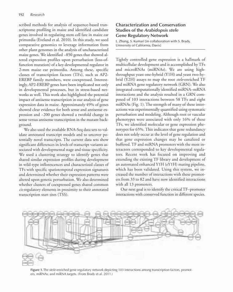
192 Research
scribed methods for analysis of sequence-based tran-scriptome profiling in maize and identified candidategenes involved in regulating stem cell fate in maize earprimordia (Eveland et al. 2010). In this study, we usedcomparative genomics to leverage information fromother plant genomes in the analysis of uncharacterizedmaize genes. We identified ~850 genes that showed al-tered expression profiles upon perturbation (loss-of-function mutation) of a key developmental regulator in2-mm maize ear primordia. Among these, specificclasses of transcription factors (TFs), such as AP2-EREBP family members, were coexpressed. Interest-ingly, AP2-EREBP genes have been implicated not onlyin developmental processes, but in stress-based net-works as well. This work also highlighted the potentialimpact of antisense transcription in our analysis of geneexpression data in maize. Approximately 49% of genesshowed clear evidence for both sense and antisense ex-pression and ~200 genes showed a twofold change insense versus antisense transcription in the mutant back-ground.
We also used the available RNA-Seq data sets to val-idate annotated transcript models and to uncover po-tentially novel transcripts. The current data sets showsignificant differences in levels of transcript variants as-sociated with developmental stage and tissue specificity.We used a clustering strategy to identify genes thatshared similar expression profiles during developmentin wild-type inflorescences and characterized classes ofTFs with specific spatiotemporal expression signaturesand determined whether their expression patterns werealtered upon genetic perturbation. We also determinedwhether clusters of coexpressed genes shared commoncis-regulatory elements in proximity to their annotatedtranscription start sites (TSS).
Characterization and ConservationStudies of the Arabidopsis steleGene Regulatory NetworkL. Zhang, S. Kumari [in collaboration with S. Brady,University of California, Davis]
Tightly controlled gene expression is a hallmark ofmulticellular development and is accomplished by TFsand microRNAs (miRNAs). We are using high-throughput yeast one-hybrid (Y1H) and yeast two-hy-brid (Y2H) assays to map the root stele-enriched TFand miRNA gene regulatory network (GRN). We alsointegrated computationally identified miRNA–mRNAinteractions and the analysis resulted in a GRN com-posed of 103 interactions between 58 TFs and eightmiRNAs (Fig. 1). The strength of many of these inter-actions was experimentally quantified using systematicperturbation and modeling. Although root or vascularphenotypes were associated with only 16% of theseTFs, we identified molecular or gene expression phe-notypes for 65%. This indicates that gene redundancydoes not solely occur at the level of gene regulation andthat gene expression changes may be canalized orbuffered. TF and miRNA promoters with the most in-teractors corresponded to key developmental regula-tors. Recent work has focused on improving andextending the existing TF library and development ofan automated enhanced Y1H (eY1H) mating pipeline,which has been validated. Using this system, we in-creased the number of interactions with these promot-ers from 33 to 82 and have now identified interactionswith all 13 promoters.
Our next goal is to identify the critical TF–promoterinteractions with conserved function in different species.
Figure 1.The stele-enriched gene regulatory network depicting 103 interactions among transcription factors, promot-ers, miRNAs, and miRNA targets. (From Brady et al. 2011.)
175-193_06_Genomics_Annual Report_2009 template 7/8/11 12:01 PM Page 192
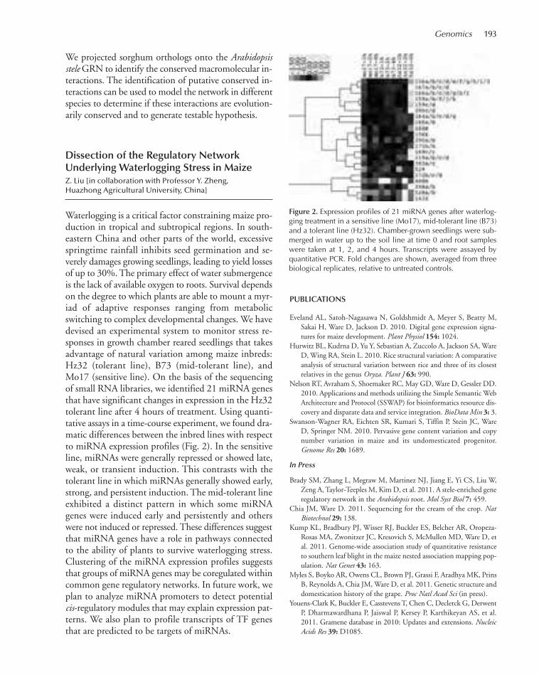
Genomics 193
We projected sorghum orthologs onto the Arabidopsisstele GRN to identify the conserved macromolecular in-teractions. The identification of putative conserved in-teractions can be used to model the network in differentspecies to determine if these interactions are evolution-arily conserved and to generate testable hypothesis.
Dissection of the Regulatory NetworkUnderlying Waterlogging Stress in MaizeZ. Liu [in collaboration with Professor Y. Zheng, Huazhong Agricultural University, China]
Waterlogging is a critical factor constraining maize pro-duction in tropical and subtropical regions. In south-eastern China and other parts of the world, excessivespringtime rainfall inhibits seed germination and se-verely damages growing seedlings, leading to yield lossesof up to 30%. The primary effect of water submergenceis the lack of available oxygen to roots. Survival dependson the degree to which plants are able to mount a myr-iad of adaptive responses ranging from metabolicswitching to complex developmental changes. We havedevised an experimental system to monitor stress re-sponses in growth chamber reared seedlings that takesadvantage of natural variation among maize inbreds:Hz32 (tolerant line), B73 (mid-tolerant line), andMo17 (sensitive line). On the basis of the sequencingof small RNA libraries, we identified 21 miRNA genesthat have significant changes in expression in the Hz32tolerant line after 4 hours of treatment. Using quanti-tative assays in a time-course experiment, we found dra-matic differences between the inbred lines with respectto miRNA expression profiles (Fig. 2). In the sensitiveline, miRNAs were generally repressed or showed late,weak, or transient induction. This contrasts with thetolerant line in which miRNAs generally showed early,strong, and persistent induction. The mid-tolerant lineexhibited a distinct pattern in which some miRNAgenes were induced early and persistently and otherswere not induced or repressed. These differences suggestthat miRNA genes have a role in pathways connectedto the ability of plants to survive waterlogging stress.Clustering of the miRNA expression profiles suggeststhat groups of miRNA genes may be coregulated withincommon gene regulatory networks. In future work, weplan to analyze miRNA promoters to detect potentialcis-regulatory modules that may explain expression pat-terns. We also plan to profile transcripts of TF genesthat are predicted to be targets of miRNAs.
PUBLICATIONS
Eveland AL, Satoh-Nagasawa N, Goldshmidt A, Meyer S, Beatty M,Sakai H, Ware D, Jackson D. 2010. Digital gene expression signa-tures for maize development. Plant Physiol 154: 1024.
Hurwitz BL, Kudrna D, Yu Y, Sebastian A, Zuccolo A, Jackson SA, WareD, Wing RA, Stein L. 2010. Rice structural variation: A comparativeanalysis of structural variation between rice and three of its closestrelatives in the genus Oryza. Plant J 63: 990.
Nelson RT, Avraham S, Shoemaker RC, May GD, Ware D, Gessler DD.2010. Applications and methods utilizing the Simple Semantic WebArchitecture and Protocol (SSWAP) for bioinformatics resource dis-covery and disparate data and service integration. BioData Min 3: 3.
Swanson-Wagner RA, Eichten SR, Kumari S, Tiffin P, Stein JC, WareD, Springer NM. 2010. Pervasive gene content variation and copynumber variation in maize and its undomesticated progenitor.Genome Res 20: 1689.
In Press
Brady SM, Zhang L, Megraw M, Martinez NJ, Jiang E, Yi CS, Liu W,Zeng A, Taylor-Teeples M, Kim D, et al. 2011. A stele-enriched generegulatory network in the Arabidopsis root. Mol Syst Biol 7: 459.
Chia JM, Ware D. 2011. Sequencing for the cream of the crop. NatBiotechnol 29: 138.
Kump KL, Bradbury PJ, Wisser RJ, Buckler ES, Belcher AR, Oropeza-Rosas MA, Zwonitzer JC, Kresovich S, McMullen MD, Ware D, etal. 2011. Genome-wide association study of quantitative resistanceto southern leaf blight in the maize nested association mapping pop-ulation. Nat Genet 43: 163.
Myles S, Boyko AR, Owens CL, Brown PJ, Grassi F, Aradhya MK, PrinsB, Reynolds A, Chia JM, Ware D, et al. 2011. Genetic structure anddomestication history of the grape. Proc Natl Acad Sci (in press).
Youens-Clark K, Buckler E, Casstevens T, Chen C, Declerck G, DerwentP, Dharmawardhana P, Jaiswal P, Kersey P, Karthikeyan AS, et al.2011. Gramene database in 2010: Updates and extensions. NucleicAcids Res 39: D1085.
Figure 2. Expression profiles of 21 miRNA genes after waterlog-ging treatment in a sensitive line (Mo17), mid-tolerant line (B73)and a tolerant line (Hz32). Chamber-grown seedlings were sub-merged in water up to the soil line at time 0 and root sampleswere taken at 1, 2, and 4 hours. Transcripts were assayed byquantitative PCR. Fold changes are shown, averaged from threebiological replicates, relative to untreated controls.
175-193_06_Genomics_Annual Report_2009 template 7/8/11 12:01 PM Page 193
CSHL recently opened the Simons Center for Quantitative Biology (SCQB). The areas of expertisein the SCQB include applied mathematics, computer science, theoretical physics, and engineering.Members of the SCQB interact closely with other CSHL researchers and apply their approaches toresearch areas including genomic analysis, population genetics, neurobiology, evolutionary biology,and signal and image processing.
Gurinder S. “Mickey” Atwal and colleagues are applying insights from the physical sciences tothe study of biological phenomena. Specifically, they develop and use mathematical and computa-tional tools to address quantitative principles governing the behavior of correlated “many-body” bi-ological systems. Such systems range from molecular interactions in a single cell to the evolution ofHomo sapiens. They are now seeking to understand evolutionary forces acting on the genome in thecontext of human diseases. In collaborations with colleagues at the Institute for Advanced Study inPrinceton, Atwal has modeled the process by which genetic variants, or alleles, evolve. This has re-cently led to surprising insights about the role in reproduction played by p53, a master tumor sup-pressor gene. This work also bears on the question of demonstrating recent selective pressures actingon our genomes.
Alexander Krasnitz and colleagues use mathematical and statistical tools to discover key geneticelements involved in cancer and to understand how cancer cells evolve. Array-based comparativegenome hybridization, a technique developed in the Wigler lab, and more recently, sequencing ex-periments have revealed subtle patterns of frequent and widespread aberration in cancer genomes.Krasnitz hypothesizes that aberrant genomic loci observed to recur in a range of cancer types areunder selection and therefore are enriched in important cancer genes. He has developed a novel,comprehensive methodology to study such “epicenters” and has used it to analyze multiple-genomedata sets in breast, lung, colon, and liver cancer. The results have been shared with cancer biologylabs across CSHL, and they have been a key enabling agent of functional studies using mouse modelsand RNA interference (RNAi). Krasnitz has begun to apply advanced statistical methods to thelatest generation of experimental data, which have characterized tumor samples down to the levelof single cells. With such data, he and colleagues seek to learn how specific cancer types evolve andhow cancer cells migrate in metastasis.
Michael Schatz is a computational biologist and an expert at large-scale computational exami-nation of DNA sequencing data including the alignment, assembly, and analysis of short sequencingreads. He is a pioneer in applying the power of many computers at once to solve data-intensive bi-ological problems—often called “cloud computing.” Schatz has developed several breakthrough ap-plications under the National Science Foundation (NSF) cloud computing initiative, and he bringsthat expertise to harness the collective power of the thousands of computing cores on the CSHLcampus to genomics, particularly within the CSHL Cancer Center and the plant biology group. In2009, Schatz published the first cloud-based sequence analysis tool, called Cloudburst, which dra-matically accelerates mapping short sequencing reads to a reference genome with applications togenotyping and expression profiling. Since then, he has published a series of high-throughput algo-rithms using this technology for single-nucleotide polymorphism (SNP) profiling, sequencing errorcorrection, machine learning, and other important applications. He is currently developing a cloud-computing-based genome assembler called Contrail and a suite of large-scale comparative genomicstools that he plans to use in a collaborative project with the Wigler lab to identify de novo mutationswithin ~3000 families in which only one child is affected with autism.
Michael Zhang’s laboratory develops mathematical and computational methods that can be com-bined with advanced experimental technologies to transform data into biological knowledge abouttranscription and gene expression, work that has manyfold implications for the study of cancer and
QUANTITATIVE BIOLOGY
194
194-204_07_QuantBiol_Annual Report_2009 template 7/8/11 12:19 PM Page 194

other diseases. Their tools, used by investigators throughout CSHL and beyond, are designed toidentify functional genetic elements within molecular sequences as well as pathways that controland regulate gene expression. Zhang’s group has developed a series of computational tools thatmake use of statistical pattern-recognition techniques to identify exons, promoters, and posttrans-lational modification signals in large genomic DNA sequences. They also study alternative splicingof exons and collaborate with other labs to characterize splicing enhancers and silencers.
Quantitative Biology 195
194-204_07_QuantBiol_Annual Report_2009 template 7/8/11 12:19 PM Page 195
196
Suyash Shringapure, a summer research intern during2009, has returned to Carnegie Mellon University toresume his graduate studies at the computer science de-partment, but he will continue to collaborate with BudMishra and myself on the project we initiated at CSHL.Joshua Weiss completed work as a volunteer as a mem-ber of the Partners for the Future program.
Genome-Wide InterchromosomalInteractions and Population Differentiation
Physically interacting proteins or subunits of proteins areexpected to evolve in a correlated manner in order to pre-serve the functional interactions between them. The se-quence changes accumulated during the evolution of oneof the interacting units can be compensated by changesin the other. Recent studies have shown that strong pair-wise correlations exist between amino acid residues of thesame protein necessitated by compensatory mutations.Furthermore, we previously showed that functional sin-gle-nucleotide polymorphisms (SNPs) in different genesin the p53 network are correlated across different humanpopulations, arguably to maintain homeostatic balanceof p53 activity. We performed a genome-wide analysis ofall the interchromosomal pairwise interactions of exonicSNPs in all 11 Hapmap populations. We find extensiveevidence of significant interactions, backed up by ananalysis of the similarity of protein functions. Protein net-work analysis demonstrated that correlated proteins wereseparated by significantly fewer nodes in the protein–pro-tein interaction network. Finally, we showed that corre-lated SNPs exhibit greater population differentiation,indicating that local geographic adaptation in humans isdependent on networks of correlated polymorphismsrather than simply independent mutations.
Cancer Risk and Haplotype Structureof the TSC1 and TSC2 Genes
TSC1 acts coordinately with TSC2 in a complex to in-hibit mTOR, an emerging therapeutic target and known
promoter of cell growth and cell cycle progression. Per-turbation of the mTOR (mammalian target of rapa-mycin) pathway, through abnormal expression orfunction of pathway genes, could lead to tumorigenesis.TSC1 and TSC2 expression is reduced in invasive breastcancer as compared with normal mammary epithelium.In a collaborative effort with the Cancer Institute of NewJersey, association studies were performed on TSC1 andTSC2 SNPs for their associations with clinical featuresof breast cancer. TSC1 and TSC2 haplotypes were con-structed from genotyping of multiple loci in both genesin healthy volunteers. SNPs were selected for furtherstudy using a bioinformatics approach based on SNP as-sociations with drug response in NCI-60 cell lines andevidence of selection bias based on haplotype frequencies.Genotypings for five TSC1 and one TSC2 loci were per-formed on genomic DNA from 1137 women with breastcancer. This study found that for TSC1 rs7874234, TTvariant carriers had a 9-year later age at diagnosis of es-trogen-receptor positive (ER+), but not ER–, ductal car-cinomas (P = 0.0049). No other SNP locus showed anassociation with age at diagnosis, nor any other breastcancer phenotype. TSC1 rs7874234 is hypothesized tobe functional in ER+ breast cancer because the T allele,but not the C allele, may create an ER element site, re-sulting in increased TSC1 transcription and subsequentinhibition of mTOR.
Regulation of Human Fertility by SNPsin the p53 Pathway
We continued our work in collaboration with CornellMedical School and the Cancer Institute of New Jersey,investigating the association of human fertility and ge-netic polymorphisms in the p53 pathway. The tumorsuppressor protein p53 has an important role in maternalreproduction in mice through transcriptional regulationof leukemia inhibitory factor (LIF), a cytokine crucial forblastocyst implantation. To determine whether these ob-servations could be extended to humans, a list of SNPsin the p53 pathway that can modify the function of p53
QUANTITATIVE BIOLOGY
G.S. Atwal R. Aboukhalil S. ShringapureY. Cai J. WeissB. Fendler
194-204_07_QuantBiol_Annual Report_2009 template 7/8/11 12:19 PM Page 196

Quantitative Biology 197
was assembled and used to study their impact on humanfertility. Selected alleles in SNPs in LIF, Mdm2, Mdm4,and Hausp genes, each of which regulates p53 levels incells, was found to be enriched in IVF (in vitro fertiliza-tion) patients. More recently, we have begun to investi-gate the association of SNPs in p63 and p73 and theinitial results are encouraging, although we await an in-crease in sample numbers for the results can be deemedto be statistically significant.
Fine-Scale Detection of Recent SelectionSignatures Using Haplotype Entropy
Creation of a coherent genomic map of recent selectionis one of the greatest challenges for a better understand-ing of human evolution. Various methods have providedthe means for genome-wide linkage disequilibriumanalysis; however, there is still a gap for scanning withshort-intermediate resolution. Entropy, a measure of dis-order in physics, has been proposed as a new concept tomeasure genetic diversity and strength of linkage dis-equilibrium of sequences. We produced a fine-scale ge-nomic scan using haplotype entropy. The analysis with amedian resolution size of ~23 kb for HapMap SNP dataof African and Caucasian populations depicted new re-cent selection signatures, as well as known signatures.Gene enrichment analysis on the signatures showed as-sociations with diverse diseases such as cardiovascular,immunological, neurological, and skeletal and musculardiseases. The novel entropy framework would be of valuefor deciphering recent evolutionary histories of humans.
Localization of TumorSuppressor Genes
Preliminary results from the Lowe Lab’s ongoing ef-forts to identify new tumor suppressor genes (TSGs)by in vivo screens in mice have found instances oftumor suppressor genes in close proximity to eachother. We carried out a systematic analysis of the spatialdistribution of known (TSGs) to test the hypothesisthat there exists an evolutionary selection pressure tocluster TSGs throughout the genome. To this end, wedeveloped a computational and analytical model to de-termine the extent to which we expect to find adjacentTSGs under no selection. Preliminary results indicatea strong tendency for tumor suppressor genes to clusterand for this clustering to occur across many species.Furthermore, by analyzing genome-wide microarraydata, we show that proximal TSGs exhibit significantcorrelation in their expressions. Ongoing investigationsare focused on understanding the implications of thisspatial distribution of TSGs for the risk and develop-ment of human cancer.
PUBLICATIONS
Mehta MS, Vazquez A, Kulkarni DA, Kerrigan JE, Atwal G, Metsugi S,Toppmeyer DL, Levine AJ, Hirshfield KM. 2010. Polymorphic vari-ants in TSC1 and TSC2 and their association with breast cancer phe-notypes. Breast Cancer Res Treat 125: 861–868.
Mizuno H, Atwal GS, Wang H, Levine AJ, Vazquez A. 2010. Fine-scaledetection of population-specific linkage disequilibrium using haplo-type entropy in the human genome. BMC Genetics 11: 27.
194-204_07_QuantBiol_Annual Report_2009 template 7/8/11 12:19 PM Page 197
198
Research in our group is focused on in silico cancer ge-nomics. In the last 5 years, there has been explosivegrowth in the volume as well as quality and detail of can-cer-related genomic data available for analysis. This de-velopment came about through rapid accumulation ofpublic data sets generated by individual laboratoriesworldwide, through large-scale cancer genome projectssuch as The Cancer Genome Atlas (TCGA) and the In-ternational Cancer Genome Consortium (ICGC), andwith the advent of new experimental methodologies, es-pecially next-generation sequencing. We see our goal inchanneling this flood of data into a number of clinicallyrelevant applications, including pinpointing and priori-tizing targets for functional analysis, discovery of genomicmarkers for clinical outcome, and molecular classificationof cancer. All our work is done in close coordination withexperimental studies performed by the Wigler, Powers,Lowe, and Stillman laboratories at CSHL.
Recurrence and Search for Driversin Cancer Genomes
Evidence suggests that recurrent somatic genomic aber-rations in cancer are at least partially due to selectivepressure on regions harboring oncogenes and tumorsuppressor genes. Conversely, one can expect regions ofrecurrent aberration to be enriched in such driver genes.Recurrence is particularly pronounced in cancer-relatedsomatic copy-number variation. We set out to buildcomputational tools exploiting this property of cancergenomes and make optimal use of both in-house andpublic DNA copy-number data in order to derive, foreach cancer type considered, a set of regions of recurrentand consistent copy-number variation and to compileprioritized lists of candidate driver genes. Starting withgenomic profiles of individual tumors, we sift throughputative copy-number events and eliminate experimen-tal errors. Next, we mask germline mutations and infer,for any given genomic location, how many copy-num-ber changes it has undergone. Finally, we round eventboundaries to the nearest gene affected by the event.
The resulting set of preprocessed genomic profiles
serves as input for our multigenome analysis. Severalversions of the latter exist. In the simplest of these, eachcopy-number event is interpreted as evidence that genesaffected by the event are under selective pressure. Wefurther assume that this evidence is shared equallyamong all of the genes affected by the event. For eachgene, its share of evidence is then summed over allevents of a given sign (gains or losses) to result in a scoreprioritizing the gene as a candidate oncogene or tumorsuppressor. In early 2010, we applied this technique toa set of ~240 DNA copy-number profiles of serousovarian tumor tissues, collected and analyzed by TCGAusing Agilent 244A array-CGH (comparative genomehybridization) platform. Our findings formed the bulkof an oncogene candidate list of 105 genes. Oncogenicpotential of these genes was subsequently examined byScott Powers’ lab in a murine ovarian cell line; 35% ofthe candidates were found to induce oncogenic trans-formation upon overexpression.
An alternative strategy consists of deriving a set ofgenomic regions subject to recurrent and consistentcopy-number variation. For a region contained in anevent, we say that the region “explains” its portion ofthe event. The algorithm first finds a region that givesmaximal explanation of the data and proceeds itera-tively finding, at each step, the most explanatory regionfor the data not explained so far. This procedure, com-plete with statistical analysis of significance and repro-ducibility of findings, is dubbed FOCAL (finder ofconsistently aberrant loci). The benefit of this analysisis twofold. First, regions delineated by FOCAL are ex-pected to be enriched in cancer-related genetic ele-ments. Second, a drastic reduction of complexity isachieved: from between 105 and 106 CGH array probereadings per tumor to a much smaller (dozens or hun-dreds) set of observables, each indicating the degree towhich a FOCAL region is present in a tumor.
FOCAL has found a number of applications in thecourse of 2010. A set of ~230 DNA copy-number pro-files of advanced breast tumors of patients in twoCALGB (cancer and leukemia group B) clinical trialswas generated by Dr. James Hicks at CSHL. The datafrom the two trials were combined in order to examine
IN SILICO CANCER GENOMICS
A. Krasnitz M. KatoG. Sun
194-204_07_QuantBiol_Annual Report_2009 template 7/8/11 12:19 PM Page 198
Quantitative Biology 199
clinical response to trastuzumab. Although trastuzumabis routinely prescribed to patients with an amplificationof ERBB2 gene, we identified two FOCAL regions aspotential markers for benefit from trastuzumab in pa-tients with no such amplification. This finding suggeststhe possibility of significantly improving the overall sur-vival in ~15% of advanced breast cancer cases.
In collaboration with Dr. Kandasamy Ravi of theWigler lab, we applied FOCAL to genomic profiles ofmammary gland tumors in mice with a variety of ge-netic backgrounds. Here again, we observed that reduc-tion of the profiles to strings of values at FOCALregions retains enough biological information to ro-bustly classify the tumors by their genetic backgrounds(TP53 loss, TP53 and PTEN loss, TP53 and BRCA1loss, BRCA1 loss, point mutation of BRCA1).
Finally, we used FOCAL to analyze a massive (516profiles) high-resolution (Agilent 1.1 million probeCGH platform) DNA copy-number data set accumu-lated by TCGA for serous ovarian cancer and used theoutput for molecular subtyping of the disease.
Statistically Supported Tree-BasedClustering and Molecular Classificationof Cancer
Tree-based clustering has long been an exploratory toolof choice for unsupervised interpretation of complexbiological data, and it has found a broad range of ap-plications, from fossil-driven reconstruction of phylo-genies to molecular subtyping of cancer. In our recentstudy, we sought to substantially boost the utility of thistechnique by assigning statistical significance to thebranches of the tree and thereby enabling the researcherto identify statistically meaningful subsets of the data.The validity of this novel approach does not depend on
the form of dissimilarity between data items or on theagglomeration rule for growing the tree. We tested thistechnique in a number of contexts. In application to apublished set of expression profiles of breast cancer, wefound that established molecular subtypes of the diseasecorrespond to statistically significant branches of a treegrown from the set. Likewise, application of themethod to a set of sequence-based copy-number pro-files of 100 individual cells harvested from a breasttumor resulted in a four-way partition of the set corre-sponding to the four major cell populations found inthe tumor and differing from each other by overallamount of DNA in the nucleus.
Next, we used statistically supported clustering topartition the TCGA set of 516 genomic profiles intofour classes. Our preliminary analysis suggests that thereare significant genomic and clinical differences amongthe classes in terms of tumor grade, disease-free survival,and point mutation frequency. To our knowledge, thismay be a first example of molecular subtyping of cancerbased on genomic data, rather than transcriptional data.
PUBLICATIONS
Floer M, Wang X, Prabhu V, Berrozpe G, Narayan S, Spagna D, AlvarezD, Kendall J, Krasnitz A, Stepansky A, et al. 2010. A RSC/nucleo-some complex determines chromatin architecture and facilitates ac-tivator binding. Cell 141: 407–418.
Navin N, Krasnitz A, Rodgers R, Cook K, Meth J, Kendall J, Riggs M,Eberling Y, Troge J, Grubor V, et al. 2010. Inferring tumor progres-sion from genomic heterogeneity. Genome Res 20: 68–80.
Russnes HG, Vollan HK, Lingjaerde OC, Krasnitz A, Lundin P, NaumeB, Sørlie T, Borgen E, Rye IH, Langerød A, et al. 2010. Genomicarchitecture characterizes tumor progression paths and fate in breastcancer patients. Sci Transl Med 2: 38ra47.
Schreiber SL, Shamji AF, Clemons PA, Hon C, Koehler AN, Munoz B,Palmer M, Stern AM, Wagner BK, Powers S, et al. 2010. Towardspatient-based cancer therapeutics. Nat Biotechnol 28: 904–906.
194-204_07_QuantBiol_Annual Report_2009 template 7/8/11 12:19 PM Page 199
200
I joined CSHL in July from the Center for Bioinfor-matics and Computational Biology at University ofMaryland College Park. Previously, I participated in thegenome assembly and comparative genomics group atthe Institute for Genomic Research (TIGR, now the J.Craig Venter Institute) and in the Ravel Lab for Micro-bial Genomics at the University of Maryland School ofMedicine. Since joining the lab, I have recruitedMatthew Titmus from the Molecular and Cell Biologygraduate program at Stony Brook University.
Cloud Computing and the DNA Data Race
In the race between DNA sequencing throughput andcomputer speed, sequencing is winning by a mile. Se-quencing throughput is currently ~200–300 billions ofbases per run on a single sequencing machine, and it isimproving at a rate of about fivefold per year. In com-parison, computer performance generally follows“Moore’s Law,” doubling only every 18 or 24 months.As the gap in performance widens, the question of howto design higher-throughput analysis pipelines becomescrucial. One option is to enhance and refine the algo-rithms to make better use of a fixed amount of com-puting power. Unfortunately, algorithmic break-throughs of this kind, like scientific breakthroughs, aredifficult to plan or foresee. The most practical optionis to develop methods that make better use of multiplecomputers and processors in parallel.
Parallel computing systems have existed for decadesin different forms, but a new system called MapReduceis especially promising for accelerating data intensive ge-nomics research. MapReduce was developed at Googlefor their large data analysis, especially for scanning acrossbillions of webpages to find the most relevant pages fora search query. Google currently uses MapReduce to an-alyze ~1 exabyte of data every month. The power ofMapReduce is that it can intelligently distribute com-putation across a cluster with hundreds or thousands ofcomputers, each analyzing a portion of the data setstored locally on the compute node. After an initialround of independent parallel computations, the ma-
chines efficiently exchange intermediate results, fromwhich the final results are computed in parallel. The sys-tem was originally developed for text and Web process-ing and was only available at Google, but an open sourceimplementation called Hadoop is now available to in-stall on any cluster for any application domain.
The first-ever application of this technology to ge-nomics, which I in published 2009, was to acceleratethe short-read mapping algorithm CloudBurst. InCloudBurst, many computers work together usingHadoop to map reads to a reference genome allowingfor any number of differences to determine from whereeach read originated and if any variations are present. Inexperiments with 96 cores, the mapping algorithm was~100 times faster than using a single core, thus reducingthe runtime from several weeks to a few hours. Thecompute cluster used for these experiments was com-pletely virtual and was leased from the Amazon ElasticCompute Cloud (EC2), which offers computing timein their cloud at a small fee per node per hour used.
In a follow-up system called Crossbow, I partneredwith colleagues at the University of Maryland to extendthe design of CloudBurst to further enable high-per-formance genotyping. Crossbow identified 3.2 millionsingle-nucleotide polymorphisms (SNPs) from 2.7 bil-lion short reads comprising 38x coverage of the humangenome. Using Hadoop to distribute the computationacross 320 computer cores rented from Amazon EC2,Crossbow completed the analysis in less than 4 hourswhereas it would have taken >1000 hours on a singlecomputer.
Since these pioneering efforts, numerous applica-tions have been developed to leverage Hadoop andcloud computing. For example, in the last year, I andcolleagues developed a system called Quake to detectand correct errors in DNA-sequencing reads. Quake isnow a standard component of several genome assemblyand analysis pipelines, and it uses a maximum likeli-hood approach incorporating quality values and nu-cleotide-specific miscall rates to pinpoint and accuratelycorrect errors. By using Hadoop, Quake can scale tovirtually any sized genome, including correcting morethan 1 billion reads from a deep coverage humangenome resequencing study. In a second project, I de-
COMPUTATIONAL GENOMICS
M.C. Schatz M. Titmus
194-204_07_QuantBiol_Annual Report_2009 template 7/8/11 12:19 PM Page 200

Quantitative Biology 201
veloped a Hadoop-based framework called Schimmyfor accelerating an entire class of analysis problems re-lated to graphs. Graphs themselves are analyzed inmany important contexts, including module detectionof protein–protein interaction networks, and genomeassembly. Many graphs of interest are difficult to ana-lyze because of their large size, often spanning millionsof vertices and billions of edges. Schimmy optimizesHadoop for large-scale graph analysis, and our experi-ments demonstrate how Schimmy reduces the runtimeof an important machine-learning graph algorithmcalled PageRank on a graph with 1.4 billion edges bynearly 70%. Future work for my lab is to develop ad-ditional high-throughput sequence analysis pipelinesusing Hadoop and other enabling high-performancetechnologies.
Genome Assembly
A second major research focus of my Lab is developingenhanced methods for de novo genome assembly and ap-plying those methods to reconstruct the genomes of pre-viously unsequenced organisms. Second-generationsequencing technology can now be used to sequence theequivalent of an entire human genome in a matter of daysand at low cost. Sequence read lengths, initially very short,have rapidly increased since the technology first appeared,and we now are seeing a growing number of efforts to se-quence large genomes de novo from these short reads.
de Bruijn graphs are a theoretical framework under-lying several modern genome assembly programs, espe-cially those that deal with very short reads. In a studypublished last year, I and colleagues described a novelapplication of de Bruijn graphs to analyze the global re-peat structure of prokaryotic genomes. This study wasthe first survey of the repeat structure of a large numberof genomes, and it gives an upper-bound on the per-formance of genome assemblers for de novo reconstruc-tion of genomes across a wide range of read lengths.This work demonstrated that the majority of genes inprokaryotic genomes can be reconstructed uniquelyusing very short reads even if the genomes themselvescannot. In addition, the nonreconstructible genes areoverwhelmingly related to mobile elements (trans-posons, IS elements, and prophages).
In a major application of genome assembly, I con-tributed to a substantial effort to sequence the domesticturkey (Meleagris gallopavo) using a combination of twonext-generation sequencing platforms along with a de-
tailed comparative bacterial artificial chromosome(BAC) physical contig map. Heterozygosity of the se-quenced source genome allowed discovery of more than600,000 high-quality single-nucleotide variants. De-spite this heterozygosity, the current genome assembly(~1.1 Gb) includes 917 Mb of sequence assigned tospecific turkey chromosomes. Annotation identifiednearly 16,000 genes, with 15,093 recognized as proteincoding and 611 as noncoding RNA genes. Compara-tive analysis of the turkey, chicken, and zebra finchgenomes, and comparing avian to mammalian species,supports the characteristic stability of avian genomesand identifies genes unique to the avian lineage. Cleardifferences are seen in the number and variety of genesof the avian immune system where expansions andnovel genes are less frequent than examples of gene loss.The turkey genome sequence provides resources to fur-ther understand the evolution of vertebrate genomesand genetic variation underlying economically impor-tant quantitative traits in poultry. This integrated ap-proach may be a model for providing both gene andchromosome level assemblies of other species with agri-cultural, ecological, and evolutionary interest.
Furthermore, in collaboration with researchers fromthe USDA, I contributed to the de novo assembly andanalysis of the ectoparasitic mite Varroa destructor,which is a primary pest of domestic honey bee (Apismellifera). This collaboration extended their previouswork to sequence another major pest of honey bees, themicrosporidian Nosema ceranae. These two organismsare especially significant to agriculture because they aretwo of the most likely agents of honeybee colony col-lapse disorder, and assembling their genomes enablesunique opportunities for understanding the molecularbasis of this serious condition. In the current work,they presented an initial survey of the V. destructorgenome carried out to advance our understanding ofVarroa biology and to identify new avenues for mitecontrol. The genome size was estimated by flow cytom-etry to be 565 Mbp, larger than most sequenced insectsbut modest relative to some other Acari. GenomicDNA pooled from ~1000 mites was sequenced to 4.3×coverage with 454 pyrosequencing. The 2.4 Gbp of se-quencing reads were assembled into 184,094 contigswith an N50 of 2262 bp, totaling 294 Mbp of se-quence after filtering. Genic sequences that have ho-mology with other eukaryotic genomes were identifiedon 13,031 of these contigs, totaling 31.3 Mbp. Align-ment of protein sequence blocks conserved among V.destructor and four other arthropod genomes indicated
194-204_07_QuantBiol_Annual Report_2009 template 7/8/11 12:19 PM Page 201

202 Research
a higher level of sequence divergence within this mitelineage relative to the tick Ixodes scapularis. A numberof microbes potentially associated with V. destructorwere identified in the sequence survey, including ~300kbp of sequence deriving from one or more bacterialspecies of the Actinomycetales. The presence of this bac-terium was confirmed in individual mites by poly-merase chain reaction (PCR) assay but variedsignificantly by age and sex of mites. Fragments of anovel virus related to the Baculoviridae were also iden-tified in the survey. The rate of SNPs in the pooledmites was estimated to be 6.2 × 10⁻⁵ per base pair, alow rate consistent with the historical demography andlife history of the species.
This survey has provided general tools for the re-search community and novel directions for investigat-ing the biology and control of Varroa mites. Ongoingdevelopment of Varroa genomic resources will be aboon for comparative genomics of underrepresentedarthropods and will further enhance the honey bee andits associated pathogens as a model system for studyinghost–pathogen interactions.
PUBLICATIONS
Cornman SR, Schatz MC, Johnson JS, Chen Y-P, Pettis J, Hunt G, Bour-geois L, Elsik C, Anderson D, Grozinger CM, et al. 2010. Genomicsurvey of the ectoparasitic mite Varroa destructor, a major pest of thehoney bee Apis mellifera. BMC Genomics 11: 602.
Dalloul RA, Long JA, Zimin AV, Aslam L, Beal K, Blomberg LA, Bouf-fard P, Burt DW, Crasta O, Crooijmans RPMA. 2010. Multi-plat-form next-generation sequencing of the domestic turkey (Meleagrisgallopavo): Genome assembly and analysis. PLoS Biol 8: e1000475.
Kelley DR, Schatz MC, Salzberg SL. 2010. Quake: Quality-aware de-tection and correction of sequencing reads. Genome Biol 11: R116.
Kingsford C, Schatz MC, Pop M. 2010. Assembly complexity of prokar-yotic genomes using short reads. BMC Bioinformatics 11: 21.
Lin J, Schatz MC. 2010. Design patterns for efficient graph algorithmsin MapReduce. Proceedings of the Eighth Workshop on Mining andLearning with Graphs Workshop (MLG-2010).
Schatz MC. 2010. The missing graphical user interface for genomics.Genome Biol 11: 128.
Schatz MC, Delcher AL, Salzberg SL. 2010. Assembly of large genomesusing second-generation sequencing. Genome Res 20: 1165–1173.
Schatz MC, Landmead B, Salzberg SL. 2010. Cloud computing and theDNA data race. Nat Biotechnol 28: 691–693.
Schatz MC, Phillippy AM, Gajer P, DeSantis TZ, Anderson GL, RavelJ. 2010. Integrated microbial survey analysis of prokaryotic commu-nities for the PhyloChip microarray. App Environ Microbiol 76:5636–5638.
Michael Schatz
194-204_07_QuantBiol_Annual Report_2009 template 7/8/11 12:19 PM Page 202

203
In the last year, the majority of my lab moved to theUniversity of Texas at Dallas and I became an adjunctprofessor at CSHL. D. Lewis moved to the McCombielab, M. Tang moved to the Lucito lab, K. Wrzeszczynskimoved to the Hicks lab, and M. Kato moved to theKrasnitz lab. J. Kinney has become a CSHL Computa-tional Junior Fellow. Miss Diana LaScala-Gruenewaldwas our URP student from Massachusetts Institute ofTechnology, and we had visiting Ph.D. student Yu Liufrom Tsinghua University working in the Huang lab ona joint project. M. Akerman and J. Wu remain at CSHLworking closely with the Krainer lab. W. Liu and Y. Maalso remain at CSHL to work on the STARR project incollaboration with Dr. Ming Li at Memorial Sloan-Ket-tering Cancer Center.
Comparison of Sequencing-Based Methods to Profile DNA Methylation and Identification of Monoallelic Epigenetic Modifications
Analysis of DNA methylation patterns relies increas-ingly on sequencing-based profiling methods. The fourmost frequently used sequencing-based technologies arethe bisulfite-based methods MethylC-seq and reducedrepresentation bisulfite sequencing (RRBS), and the en-richment-based techniques methylated DNA immuno-precipitation sequencing (MeDIP-seq) and methylatedDNA-binding domain sequencing (MBD-seq). In col-laboration with the NIH Roadmap Epigenome Com-sortium, we applied all four methods to biologicalreplicates of human embryonic stem cells to assess theirgenome-wide CpG coverage, resolution, cost, concor-dance, and the influence of CpG density and genomiccontext. The methylation levels assessed by the twobisulfite methods were concordant (their difference didnot exceed a given threshold) for 82% for CpGs and99% of the non-CpG cytosines. Using binary methyl-ation calls, the two enrichment methods were 99%concordant and regions assessed by all four methods
were 97% concordant. We combined MeDIP-seq withmethylation-sensitive restriction enzyme (MRE-seq) se-quencing for comprehensive methylome coverage atlower cost. This, along with RNA-seq and ChIP-seq ofthe embryonic stem (ES) cells, enabled us to detect re-gions with allele-specific epigenetic states, identifyingmost known imprinted regions and new loci withmonoallelic epigenetic marks and monoallelic expres-sion (Harris et al. 2010). In particular, we show thatour new version of software RMAP—which imple-ments function for pair-end and bisulfite next-genera-tion sequencing reads (Smith et al., Bioinformatics 25:2841 [2009])—is more sensitive than Bowtie, a popularreads mapping software.
Development of the Human Cancer microRNA Network
In collaboration with Drs. Bandyopadhyay and Mitraat the Indian Statistical Institute, a cancer-miRNA (mi-croRNA) network (Bandyopadhyay et al. 2010) was de-veloped by mining the literature of experimentallyverified cancer-miRNA relationships. This network re-veals several new and interesting biological insights thatwere not evident in individual experiments, but becomeevident when studied in the global perspective. Fromthe network, a number of cancer-miRNA modules havebeen identified based on a computational approach tomine associations between cancer types and miRNAs.The modules that are generated based on the associationare found to have a number of common predicted targetonco/tumor suppressor genes. This suggests a combina-torial effect of the module-associated miRNAs on targetgene regulation in selective cancer tissues or cell lines.Moreover, neighboring miRNAs (a group of miRNAslocated within 50 kb of genomic location) of these mod-ules show similar dysregulation patterns, suggesting acommon regulatory pathway. In addition, neighboringmiRNAs may also show similar dysregulation patterns(differentially coexpressed) in the cancer tissues. In this
COMPUTATIONAL GENOMICS
M.Q. Zhang M. Akerman D. Lewis M. TangM. Kato W. Liao K. Wrzeszczynski J. Kinney Y. Ma J. WuD. LaScala-Gruenewald
194-204_07_QuantBiol_Annual Report_2009 template 7/8/11 12:19 PM Page 203

study, we found that 67% of the cancer types have atleast two neighboring miRNAs showing down-regula-tion that is statistically significant (P < 10–7, randomiza-tion test). A similar result is obtained for the neigh-boring miRNAs showing up-regulation in a specific can-cer type. These results elucidate the fact that neighboringmiRNAs might be differentially coexpressed in cancertissues as those in the normal tissue types. Additionally,the cancer-miRNA network efficiently detects hub miR-NAs dysregulated in many cancer types and identifycancer-specific miRNAs. Depending on the expressionpatterns, it is possible to identify those hubs that havestrong oncogenic or tumor suppressor characteristics.This work was based on our new SFSSClass, an inte-grated method for miRNA-based tumor classification(Mitra et al. 2010).
Dissecting the Unique Role of theRetinoblastoma Tumor Suppressor during Cellular Senescence
The retinoblastoma (RB) protein family (RB, p107,and p130) has overlapping and compensatory functionsin cell cycle control. However, cancer-associated muta-tions are almost exclusively found in RB, implying thatRB has a nonredundant role in tumor suppression. Incollaboration with the Lowe lab at CSHL, we demon-strated that RB preferentially associates with E2F targetgenes involved in DNA replication and is uniquely re-quired to repress these genes during senescence but notother growth states. Consequently, RB loss leads to in-appropriate DNA synthesis following a senescence trig-ger and, together with disruption of a p21-mediated
cell cycle checkpoint, enables extensive proliferationand rampant genomic instability. Our results (Chicaset al. 2010) identified a nonredundant RB effectorfunction that may contribute to tumor suppression andreveal how loss of RB and p53 cooperate to bypasssenescence.
PUBLICATIONS
Bandyopadhyay S, Mitra R, Maulik U, Zhang MQ. 2010. Developmentof the human cancer microRNA network. Silence 1: 6.
Bernard D, Prasanth KV, Tripathi V, Colasse S, Nakamura T, Xuan Z,Zhang MQ, Sedel F, Jourdren L, Coulpier F, et al. 2010. A long nu-clear-retained non-coding RNA regulates synaptogenesis by modu-lating gene expression. EMBO J 29: 3082–3093.
Chicas A, Wang X, Zhang C, McCurrach M, Zhao Z, Ozlem M, Dick-ins R, Narita M, Zhang MQ, Lowe SW. 2010. Dissecting the uniquerole of the retinoblastoma tumor suppressor during cellular senes-cence. Cancer Cell 17: 376–387.
Harris RA, Wang T, Coarfa C, Nagarajan RP, Hong C, Downey SL,Johnson BE, Fouse SD, Delaney A, Zhao Y, et al. 2010. Comparisonof sequencing-based methods to profile DNA methylation and iden-tification of monoallelic epigenetic modifications. Nat Biotechnol 28:1097–1105.
Mitra R, Bandyopadhyay S, Maulik U, Zhang MQ. 2010. SFSSClass:An integrated approach for miRNA based tumor classification.Bioinformatics 1: S22.
Wang SM, Zhang MQ. 2010. The progress and challenge for transcrip-tome study on human CD34+ hematopoietic stem-progenitor cells.J Cell Physiol 223: 549–552.
In Press
Renault VM, Thekkat PU, Hoang KL, White JL, Brady CA, Kenzel-mann D, Venturelli OS, Johnson TM, Xuan Z, Santo EE, et al.2011. The pro-longevity gene FoxO3 is a direct target of the tumorsuppressor p53. Oncogene (in press).
Zhang MQ. 2011. Computational promoter prediction in a vertebrategenome. In Handbook of statistical bioinformatics (ed. H. Lu et al.).Springer Handbooks of Computational Statistics. (In press.)
204 Research
194-204_07_QuantBiol_Annual Report_2009 template 7/8/11 12:19 PM Page 204
205
COLD SPRING HARBOR LABORATORY FELLOWS
In 1986, Cold Spring Harbor Laboratory began a Fellows Program to encourage independent researchby outstanding young scientists who, during their graduate studies, displayed exceptional promise ofbecoming leading scientists of the future. The purpose of this program is to provide an opportunityfor these young scientists to work independently at the Laboratory for a period of 3 years on projectsof their own choosing. Fellows are provided with a salary, research support, and technical assistance sothat they can accomplish their goals free of distraction. The interactions among research groups at theLaboratory and the program of courses and meetings on diverse topics in biology contribute to a re-search environment that is ideal for innovative science by these Fellows. The CSHL Fellows Programhas been tremendously successful and has served as a paradigm for several analogous programs at otherinstitutions, most recently a Fellows Program sponsored by the National Institutes of Health.
The success of the program is apparent from the list of distinguished alumni. Most notably, CarolGreider, recipient of the 2009 Nobel Prize in Physiology or Medicine for her work on telomeraseand telomere function, joined the Fellows Program in 1998. After completing her fellowship, Carolwas on the CSHL faculty for 9 years, and she is currently the Daniel Nathans Professor and Directorof Molecular Biology and Genetics at Johns Hopkins University School of Medicine.
Previous CSHL Fellows Adrian Krainer (1986), Scott Lowe (1995), and Marja Timmermans(1998) are now professors at the Laboratory. Scott is the Deputy Director of the CSHL CancerCenter and a Howard Hughes Medical Institute Investigator, and Marja is the Director of the Fel-lows Program. Eric Richards (1989) currently is the Vice President of Research and Senior Scientistat the Boyce Thompson Institute for Plant Research at Cornell University; David Barford (1991)is a Fellow of the Royal Society and Professor of Molecular Biology at the Institute of Cancer Re-search in London; Ueli Grossniklaus (1994) is Professor at the Institute of Plant Biology, Universityof Zürich, Switzerland; and Térence Strick (2000) left at the end of his fellowship to become aGroup Leader at the Institute Jacques Monod in Paris. Both Lee Henry (2000) and Ira Hall (2004)moved to Virginia upon completion of their fellowships. Lee joined a project headed by ThomasSüdhof at HHMI’s Janelia Farm in Ashburn, and Ira is Assistant Professor in the Department ofBiochemistry & Molecular Genetics at the University of Virginia in Charlottesville. Patrick Paddi-son, who had joined the Fellows Program in 2004, currently is an Assistant Member at the FredHutchinson Cancer Research Center in Seattle, Washington.
In 2010, the Laboratory hosted two Fellows: Florin Albeanu did his graduate studies at HarvardUniversity in the laboratories of Venki Murthy and Markus Meister, where he investigated the logicof odor maps in the olfactory bulb of rodents. As a Fellow, Florin has taken advantage of opticalimaging, electrophysiology, and optogenetic tools to understand what computations occur at theneural circuit level in the olfactory bulb of behaving mice. His research program has been very suc-cessful and he has therefore recently been promoted to Assistant Professor. Chris Vakoc joined usfrom Gerd Blobel’s laboratory at the University of Pennsylvania, where he studied chromatin loopingmechanisms in long-range enhancer function. As a Fellow, Chris is studying the role of histonelysine methylation in normal and malignant hematopoiesis.
Understanding Neuronal Circuits in theMammalian Olfactory BulbF. Albeanu, H.G. Chae, P.G. da Silva, M. Davis, A. Dhawale, F.Marbach, H. Patel, S. Rengarajan
In many regions of the brain, neurons form an orderedrepresentation of the outside world. For example, the
“homunculus” of the somatosensory cortex is a point-to-point topographic map of the body surface onto the brainsurface. The spatially organized convergence of sensoryinputs often leads to similar response properties in targetneurons that are in close vicinity. Whether their individ-ual information content is redundant or independent de-pends on the circuit architecture (the interplay between
205-214_08_FELLOWS_Annual Report_2009 template 7/8/11 12:20 PM Page 205

206 Research
common input, lateral signals, and feedback from otherbrain areas) and the computational goals of the network.
We use optogenetic tools (such as two-photon im-aging of genetically encoded neuronal activity reportersor light-gated neuronal activity switches) coupled withelectrophysiological measurements (extracellular andintracellular recordings) to study how sensory systemsencode information from the environment. We wantto understand how these inputs get processed at differ-ent junctions or synapses of the underlying neuronalcircuits and how these representations change with thestate of the system and its circuits (awake vs. anes-thetized, naïve vs. conditioned). The broad scope of thiseffort is observing how perceptions arise.
The olfactory system, particularly the olfactory bulb(OB), in rodents provides us with an ideal substrate toanswer these questions, given its well-defined circuitryand multilayered organization. Rodents, being nocturnalanimals, depend heavily on olfaction for survival—find-ing food and mates or avoiding predators. Airbornechemicals or odors are translated into neuronal signals byspecific receptors in the nose and sent first to the bulb andthen to higher centers in the brain (olfactory cortex). Thebulb thus being the relay center provides the opportunityto study both the nature of the inputs it receives and thenature of output it sends to the brain and the computa-tions that allow for this input–output transformation.
In the bulb, sensory neurons expressing the same typeof olfactory receptor converge in tight focus, forming~2000 clusters of synapses called glomeruli. The layoutof glomeruli on the bulb is highly reproducible acrossindividuals with a precision of one part in 1000. How-ever, nearby glomeruli are as diverse in their responsesto odors as distant ones, lacking an apparent chemotopicarrangement (Soucy et al., Nat Neurosci 12: 2 [2009]).From each glomerulus, a few dozen mitral cells (princi-pal output neurons of the OB) carry the output furtherto the olfactory cortex and several other brain areas. Themitral cells typically have only one primary dendrite thatprojects to a single glomerulus, but they can sample in-puts on their primary and secondary dendrites fromfunctionally diverse glomeruli via several types of in-terneurons (Fig. 1). Thus, a few dozen mitral cells shareinput from the same parent glomerulus (sister cells), butthey may have different inhibitory surrounds.
Are Outputs from the SameGlomerulus Redundant?
We generated transgenic mice expressing channelrhodop-sin-2 in all olfactory sensory neurons under the control of
the olfactory marker protein (OMP) promoter. By selec-tively stimulating individual glomeruli via DLP-patternedillumination and recording extracellularly from the mitralcell layer using tetrodes, we identified mitral cells that re-ceive common input (Fig. 2). We found that sister mitralcells had highly correlated responses to odors as measuredby average spike rates, but their spike timing with respectto respiration was differentially altered. In contrast, non-sister mitral cells correlated poorly on both these measures.Sister mitral cell pairs fired synchronously (at the samephase) within a respiration cycle at rest, and upon odorpresentation, they became desynchronized in a stimulus-specific manner. We therefore propose that sister cells carrytwo different channels of information to the cortex: (1) av-erage activity representing shared glomerular input and (2)phase-specific information that refines odor representationsand is substantially independent for sister mitral cells(Dhawala et al., Nat Neurosci 13: 1404–1412 [2009]).
What Roles Does the Inhibitory NetworkHave in Shaping the Bulb Outputs?
Activity in the bulb is a rich mix of excitation and inhi-bition, via both direct inputs and feedforward and feed-back connections by a wide variety of cell types.Juxta-glomerular cells are local interneurons that sur-round the glomeruli and send neurites within and acrossglomeruli. In the external plexiform layer, another classof interneurons, the granule cells, mediates cross-talk be-tween mitral cells via reciprocal synapses with lateral mi-tral cell dendrites (Fig. 2). Both interneuron types aregeneric denominations for heterogeneous neuronal pop-ulations with respect to wiring, gene expression, andphysiological properties. Despite decades of study in theslice preparation, little is known about how these in-terneurons operate in the intact circuit in vivo.
Figure 1. Main olfactory bulb wiring schematics. (Courtesy of F.Markopoulos.)
205-214_08_FELLOWS_Annual Report_2009 template 7/8/11 12:20 PM Page 206

Cold Spring Harbor Laboratory Fellows 207
First, we aimed at understanding whether bulb in-terneurons modulate the spiking time of mitral cells, i.e.,the changes in phase tuning described above. We havestarted by expressing light-gated molecular switches ofneuronal activity (such as channelrhodopsin 2 andhalorhodopsin) in genetically defined subsets of bulb in-terneurons. We are using a Cre/Lox strategy and takingadvantage of several interneuron-specific Cre lines gen-erated by Josh Huang’s group at CSHL. In particular,we are targeting local dopaminergic interneurons in theglomerular layer (tyrosine-hydroxylase-positive) thatmediate interglomerular cross-talk, as well as deeper in-terneurons situated in the external plexiform layer (par-valbumin and or VIP-positive) that synapse with thelateral dendrites of mitral cells.
Optical Dissection of Olfactory Microcircuits: What ComputationsAre Performed on the Inputsby the Bulb Circuitry?
We set out to sample activity of large and diverse neu-ronal populations from the two layers of the bulb in re-sponse to numerous odorants. Recent developments in
multiphoton microscopy and new generations of genet-ically encoded neuronal activity reporters allow monitor-ing both glomeruli (inputs) and mitral cell (outputs)responses simultaneously within the same animal and inresponse to the same set of odorants. Toward this end,we built two custom multiphoton microscopes that allowfor fast x-y-z scanning and two large-scale odor deliverymachines (165 odorants). Preliminary experiments indi-cate that activity patterns at the level of mitral cell bodiesare not simply scaled down versions of glomerular rep-resentations (Fig. 3). Therefore, we have started to sys-tematically investigate the contributions of lateral signalsand feedback from other areas to bulb input-outputtransform via optogenetic strategies.
Dissecting the Spatial and TemporalFeatures of the Glomerular Codein Awake Behaving Animals viaOptogenetics Strategies
We are using light activation of glomeruli in awake head-restrained OMP-chr2 mice to ask how different spa-tiotemporal input patterns get processed at the level of themitral cells and influence behavior. Using light activation
Figure 2. DLP patterned illumination in ORC mice maps the parent glomeruli of mitral cells in vivo. (A, Left) ADLP projector coupled to a system of three lenses delivered light patterns to activate individual glomeruli on thedorsal surface of the bulb. A CCD (charge-coupled device) camera was used to visualize the bulb surface andrecord the position of the light stimulus. (Top panel, right) Dorsal surface of the bulb with a tetrode positioned inthe mitral cell layer. One square light spot is being projected onto the surface of the bulb. (Inset) Cartoon schematicof glomeruli on the bulb, a subglomerular-size light spot and dual tetrodes positioned in the mitral cell layer. (a)DLP projector, with color wheel removed from optical path, (b) focusing lens, (c) blue excitation filter, (d) dichroicmirror, (e) emission filter, (f) CCD camera, (g) dual-tetrode, (h) olfactory bulb. (B, Top) Raw voltage traces corre-sponding to the four channels of a tetrode showing light-induced changes in firing patterns of the units recorded.(Center) Raster plot shows changes in spiking activity of a single isolated unit across 25 light-stimulation trials,each 200 msec long; dots mark spike occurrence times. (Bottom) Peristimulus time histogram (PSTH) with 25-msec time bins, summing spikes over the trials shown in the center panel.
205-214_08_FELLOWS_Annual Report_2009 template 7/8/11 12:20 PM Page 207
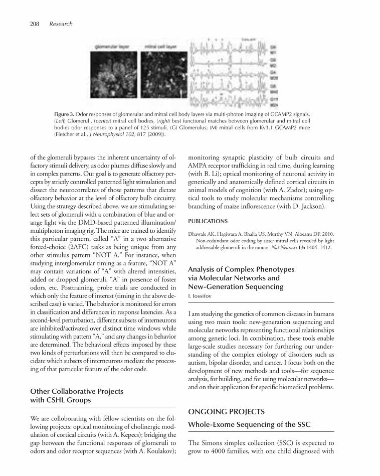
208 Research
of the glomeruli bypasses the inherent uncertainty of ol-factory stimuli delivery, as odor plumes diffuse slowly andin complex patterns. Our goal is to generate olfactory per-cepts by strictly controlled patterned light stimulation anddissect the neurocorrelates of those patterns that dictateolfactory behavior at the level of olfactory bulb circuitry.Using the strategy described above, we are stimulating se-lect sets of glomeruli with a combination of blue and or-ange light via the DMD-based patterned illumination/multiphoton imaging rig. The mice are trained to identifythis particular pattern, called “A” in a two alternativeforced-choice (2AFC) tasks as being unique from anyother stimulus pattern “NOT A.” For instance, whenstudying interglomerular timing as a feature, “NOT A”may contain variations of “A” with altered intensities,added or dropped glomeruli, “A” in presence of fosterodors, etc. Posttraining, probe trials are conducted inwhich only the feature of interest (timing in the above de-scribed case) is varied. The behavior is monitored for errorsin classification and differences in response latencies. As asecond-level perturbation, different subsets of interneuronsare inhibited/activated over distinct time windows whilestimulating with pattern “A,” and any changes in behaviorare determined. The behavioral effects imposed by thesetwo kinds of perturbations will then be compared to elu-cidate which subsets of interneurons mediate the process-ing of that particular feature of the odor code.
Other Collaborative Projectswith CSHL Groups
We are colloborating with fellow scientists on the fol-lowing projects: optical monitoring of cholinergic mod-ulation of cortical circuits (with A. Kepecs); bridging thegap between the functional responses of glomeruli toodors and odor receptor sequences (with A. Koulakov);
monitoring synaptic plasticity of bulb circuits andAMPA receptor trafficking in real time, during learning(with B. Li); optical monitoring of neuronal activity ingenetically and anatomically defined cortical circuits inanimal models of cognition (with A. Zador); using op-tical tools to study molecular mechanisms controllingbranching of maize inflorescence (with D. Jackson).
PUBLICATIONS
Dhawale AK, Hagiwara A, Bhalla US, Murthy VN, Albeanu DF. 2010.Non-redundant odor coding by sister mitral cells revealed by lightaddressable glomeruli in the mouse. Nat Neurosci 13: 1404–1412.
Analysis of Complex Phenotypesvia Molecular Networks andNew-Generation SequencingI. Iossifov
I am studying the genetics of common diseases in humansusing two main tools: new-generation sequencing andmolecular networks representing functional relationshipsamong genetic loci. In combination, these tools enablelarge-scale studies necessary for furthering our under-standing of the complex etiology of disorders such asautism, bipolar disorder, and cancer. I focus both on thedevelopment of new methods and tools—for sequenceanalysis, for building, and for using molecular networks—and on their application for specific biomedical problems.
ONGOING PROJECTS
Whole-Exome Sequencing of the SSC
The Simons simplex collection (SSC) is expected togrow to 4000 families, with one child diagnosed with
Figure 3.Odor responses of glomerular and mitral cell body layers via multi-photon imaging of GCAMP2 signals.(Left) Glomeruli, (center) mitral cell bodies, (right) best functional matches between glomerular and mitral cellbodies odor responses to a panel of 125 stimuli. (G) Glomerulus; (M) mitral cells from Kv3.1 GCAMP2 mice(Fletcher et al., J Neurophysiol 102, 817 [2009]).
205-214_08_FELLOWS_Annual Report_2009 template 7/8/11 12:20 PM Page 208
Cold Spring Harbor Laboratory Fellows 209
autism spectrum disorder and with at least one unaf-fected child. Such a collection is ideally suited for iden-tification of de novo mutations with a strong effect onthe disorder. Recent improvements of the techniquesfor enrichment of coding genomic sequences and of thenew-generation sequencing technology made feasiblewhole-exome sequencing for a large number of samples.The generated data are of high quality and can be usedto identify de novo single-nucleotide, short-indel, andcopy-number mutations. This project was carried outin collaboration with Michael Wigler and Richard McCombie at CSHL.
Analysis of the Pathways Affectedby De Novo Copy-NumberMutations Identified in 2000 Familiesfrom the SSC Collection
Recent evidence suggests that rare variants, includingcopy-number variants (CNVs), have a significant role inthe etiology of autism. Although many such variants havebeen identified, the specific molecular networks associ-ated with this complex disorder remain largely unknown.In this study, we develop a method for network-basedanalysis of genetic associations (NETBAG). We useNETBAG to identify a large biological network of genesaffected by rare de novo CNVs in autism. The genesforming the network are primarily related to synapse de-velopment, axon targeting, and neuron motility. Thisproject is being carried out in collaboration with MichaelWigler (CSHL) and Dennis Vitkup (Columbia Univer-sity).
Analysis of the Overall Sharingof Parental Genome by Concordantand Discordant Autistic Siblings
The goal is to test the hypothesis that a substantial pro-portion of inherited autism is caused by a dominant mu-tation inherited from the mother, with different mothershaving different affected loci. So far, we see a suggestiveincrease of sharing of the mothers’ genomes in a cohortof ~300 multiplex families from the AGRE collectionwith microsatellite markers. The next step is to see if thesignal is preserved and statistically significant in a largerset of 700 families genotyped with a 10K single-nu-cleotide polymorphism (SNP) array. We are carrying outthis project in collaboration with Michael Wigler andKenny Ye at CSHL.
Sequencing of Treatment-RelatedtAML 5q and 7q Chromosomes
A large proportion of treatment-related acute myeloidleukemia (tAML) cases present with hemizygous 7q or5q chromosomes. In collaboration with Mona Spectorand Scott Lowe at CSHL, we are sequencing 50 sampleswith the hope of identifying a gene within the 5q and 7qregions with recurrent variants affecting protein sequence.
Epigenetics and CancerC.R. Vakoc
Chromatin is the fundamental packaging material of eu-karyotic genomes, composed of DNA in its native com-plex with protein. The basic organizational unit ofchromatin is the nucleosome, composed of an octamerof core histones wrapped by 147 bp of DNA. Chromatinis charged with the critically important task of both pro-tecting the genetic code and animating its many func-tions. Replication, recombination, and transcription ofDNA in the cell all occur in a chromatin context, andhence, elaborate control mechanisms have evolved togovern chromatin structure in these diverse scenarios.The central interest of our laboratory is to understandthe enzymatic mechanisms that control chromatin andhow such pathways establish heritable chromatin statesand participate in the pathophysiology of cancer.
A major focus of our work is a class of molecules re-ferred to as human Trithorax proteins, also known as themixed-lineage leukemia (MLL) family. MLL proteins arelarge, modular chromatin regulators that use catalytic(methyltransferase activity for histone H3 lysine 4) andnoncatalytic (chromatin-binding and scaffold functions)mechanisms to maintain the “ON” state of gene expres-sion. One fascinating feature of MLL and its orthologsin other species is their capacity to maintain heritablestates of gene activity, as exemplified by MLL inDrosophila maintaining heritable programs of Hox geneexpression, a phenomenon known as cellular memory.Although most of the classic epigenetic inheritance phe-nomena have been linked with the “OFF” state of ex-pression, the epigenetic maintenance of the “ON” stateby MLL stands alone as one of the only known mecha-nisms in nature that stably preserves active chromatin ina manner heritable through somatic cell divisions. Onekey interest in our laboratory is to define the molecularmechanism of epigenetic inheritance by MLL proteins.
In addition to having essential functions in maintain-ing active chromatin during normal ontogeny and dif-
205-214_08_FELLOWS_Annual Report_2009 template 7/8/11 12:20 PM Page 209

210 Research
ferentiation, the MLL protein is also a proto-oncogenein acute leukemia. MLL is frequently mutated via chro-mosomal translocation to generate oncogenic fusion pro-teins. MLL fusion proteins induce a hyperactive chro-matin state as compared to wild-type MLL, which leadsto up-regulation of MLL’s normal target genes to blockhematopoietic cell differentiation and induce leukemia.Thus, MLL leukemias represent a clear situation wherechromatin dysfunction directly causes a human cancer.A major thrust of our research is to use MLL leukemia asa model system to study how epigenetic pathways par-ticipate in cancer pathogenesis. Our primary approach isto evaluate whether therapeutic targeting of epigeneticpathways can eradicate leukemia cells in vivo. BecauseMLL leukemia represents a poor-prognosis subtype ofthis disease, we hope to identify novel therapeutic targetsin this cancer by mining epigenetic pathways.
RNAi Screening to Expose EpigeneticVulnerabilities in AMLE. Wang, J. Shi
The central hypothesis our work on acute myeloidleukemia (AML) is that suppression of epigenetic path-ways can be an effective means to achieve selective eradi-cation of leukemia cells in vivo. However, the challengeis in how one identifies candidate epigenetic targets asnovel cancer therapies. To this end, we have begun RNAinterference (RNAi) screening as a means of revealing anddistinguishing the epigenetic requirements between nor-mal and malignant cells. First, we generated a customshort hairpin RNA (shRNA) library targeting all knownchromatin regulators, which includes ~1100 shRNAs tar-geting 250 genes. Next, we introduced this library in a 1-
Figure 1. RNAi screen for unique epigenetic requirements needed for MLL-leukemia growth. (A) Experimentalstrategy. A mosaic mouse model of MLL–AF9/NrasG12D was transduced with the Epigenetics-246 shRNA library.(B) Primary screen identified 129 shRNAs that inhibit proliferation (in gray) (C) Comparison of shRNA effects inleukemia and 32D myeloid contexts. The triangle demarcates shRNAs that tested positive in the screen by prefer-entially inhibiting leukemia proliferation. Positive control shRNAs are labeled.
205-214_08_FELLOWS_Annual Report_2009 template 7/8/11 12:20 PM Page 210

Cold Spring Harbor Laboratory Fellows 211
by-1 or pooled fashion into a mouse model of MLL–AF9/ NrasG12D leukemia. We then scored the impact ofeach shRNA on AML growth in vitro and compared theeffects on a number of nontransformed hematopoieticcell lines, representing normal cells. Through this ap-proach, we were able to identify several candidate shRNApathways, which we want to validate in vivo. Using a sim-ilar “negative-selection” strategy, we introduced shRNAstargeting various epigenetic pathways into normal orleukemic cells and transplanted into lethally or sublethallyirradiated recipients. The impact of each shRNA wasscored using a two-color fluorescence-activated cell sorting(FACS)-based assay. This analysis reliably distinguishedthe in vivo growth requirement for normal and malignantcells and identified several candidate epigenetic pathways.
Among the top hits in the RNAi screen were the Eedand Suz12 genes, which both encode subunits of thePRC2 (Polycomb repression complex). PRC2 is a histonemethyltransferase that selectively modifies histone H3 atlysine 27. This chromatin modification has a clear role ingene repression, which it serves as a docking site for otherrepressive complexes. Identifying two subunits in thescreen provides strong evidence that this complex was in-volved in AML progression. Following knockdown ofPRC2 in MLL-AF9/NrasG12D leukemia cells, we observedrobust differentiation of leukemic blasts into cells with anobvious macrophage morphology, indicating that PRC2is required to prevent differentiation of leukemic cells. Thisphenotype bears striking resemblance to suppressingMLL-AF9, the driver oncogene in this disease. To identifythe core downstream targets of PRC2 in AML, we per-formed expression microarrays comparing evaluatingglobal changes in gene expression signatures, followed bylarge-scale chromatin immunoprecipitation experimentsto distinguish direct from indirect targets. Using a combi-
nation of overexpression and loss-of-function approaches,we were able to pinpoint the Ink4a/Arf tumor suppressorlocus as the key downstream target of PRC2 in AML. Inaddition, we were also able to demonstrate that the onco-genic drivers of AML in this model, MLL-AF9 andNrasG12D, both provide a signal that stimulates Ink4a/Arfexpression, which threatens to inhibit growth of AML cellsin vitro. In our model, we speculate that PRC2 provides abuffering system in AML for diminishing oncogenic stress.By suppressing PRC2 with RNAi, we eliminate this bufferand unravel the transformed cellular state through induc-tion of Ink4a/ Arf expression. We speculate that PRC2 willrepresent a promising new therapeutic target in AML.
Another hit identified in the RNAi screen is the BET-bromo domain protein Brd4. Notably, among the 250candidate genes in the screen, Brd4 inhibition displayedthe most profound elimination of leukemia cells in vitro,making it the top hit. Like PRC2, suppression of Brd4leads to similar induction of myeloid differentiation withassociated loss of leukemia stem cell populations. Quitefortunately, our studies have converged with those of theJames Bradner laboratory at Harvard, which has recentlydeveloped small-molecule inhibitors of the Brd4 bromo-domains. In collaboration with Bradner and Scott Lowe’slaboratory at CSHL, we have begun investigating the an-tileukemic activity of Brd4 inhibitors in vitro and in vivo.This example represents the overall effort in the lab ofidentifying epigenetic sensitivities in cancer that mightbe exploited using small-molecule inhibitors, whichcould be developed in the future as drugs in AML.
PUBLICATIONS
Speck NA, Vakoc CR. 2010. PAF is in the cabal of MLL1-interactingproteins that promote leukemia. Cancer Cell 17: 531–532.
205-214_08_FELLOWS_Annual Report_2009 template 7/8/11 12:20 PM Page 211
Aboukhalil R., 196Ahrens S., 125Akerman M., 36, 203Akshinthala D., 78Albeanu F., 205Alexander J., 69Anczuków O., 36Andrews P., 69Antinis E., 56Arcangioli B., 164Ashar C., 33Askautrud H.A., 75Atwal G.S., 196Avraham S., 188Aznárez I., 36
Baer M., 50Bahel D., 33Bao Y., 132Baslan T., 69Batut P., 177Bekritsky M., 69Bell K., 177Bell M., 182Bencze G., 80, 87Benkovics A., 169Bergmann J., 39Beshel J., 148Bezirci F., 36Bodnar M.S., 39Boettner B., 94Boivin B., 87Bolden J., 56Bommert P., 152Bondar N., 107Borges F., 164Borges K., 145Bressan D., 50Burgess R., 50Burlet-Parendel A., 107
Cai Y., 196Cain S., 188Calarco J., 33, 164Camiolo M., 84Campbell R., 142Canela A., 50Cao G., 145Cappellani J., 56Cardone L., 182Castel S., 164Chae H., 125
Chae H.G., 205Chakrabortty S., 177Chang A.-Y., 164Chang K., 50Chang Y., 64Chanrion M., 66Chatterjee D., 36Chattopadhyay S., 145Chaudhary F., 87Cheang G., 182Cheloufi S., 50Chen C., 56Chen K., 152Chen M., 92Cheng X., 87Chi S.W., 50Chia J.-M., 189Chicas A., 56Chien Y., 56Cho H., 92Chuah A., 189Churchland A., 102Clendaniel C., 169Cook K., 69Cox H., 69, 128Creasey K., 164Cressy M., 104Cutter N., 61Czech B., 50
da Silva P.G., 205Das S., 36Davis C., 177Davis M., 205de la Cruz N., 188DeBlasio S., 152Delevich K., 125Demir E., 117Demmer H., 138Dhawale A., 205Dimitrova N., 61Dobin A., 177Dos Santos C., 50Dos Santos J., 138Dotto M., 169Dow L., 56Drenkow J., 177Dubnau J., 104
Ebbesen S., 56Eberling Y., 69Eckersley-Maslin M., 39
Eckmeier D., 138Egeblad M., 75Eifert C., 66El Demerdash O., 128Elkayam E., 33Enikolopov G., 107Ernst E., 164Esposito D., 69Eveland A., 152, 189
Faehnle C., 33Faga B., 188Fagegaltier D., 177Faleiro M., 36Fan M., 36Fang G., 87Fastuca M., 177Feigin M., 78Fellmann C., 56Fendler B., 196Fenoglio S., 50, 84Fernandez-Marco C., 169Finigan P., 164Fregoso O., 36Fu C., 80Fu Y., 114Furukawa H., 110
Gal-on A., 169Gallo M., 66Gauss C.M., 80Gerdes R., 66Ghiban E., 182Gierszewska M., 182Gingeras T.R., 177Glass Z., 107Goh W.S., 50Goldshmidt A., 152Goldsmith S., 33Gopinathrao G., 188Gordon A., 50Grabill I., 182Grace D., 56Grange P., 128Gruntman E., 142Gu X.L., 94Guo X., 56Guzzardo P., 50
Haase A., 50Hakker I., 69Hammell C.M., 31
Han J.-J., 164Hangya B., 117Hannon G., 50Hansen K., 164Haque A., 87Hariharan N., 152Harris T., 188Harrison E., 50Havkin G., 128He C., 56He M., 114Hearn S., 39Hernandez U., 152Hicks J., 69Hige T., 142Hirokawa J., 117Hodges E., 50Honegger K., 142Horev G., 64Hossain M., 45Hromadka T., 145Hu C., 169Hu R., 138Hua Y., 36Huang J., 69Huang Z.J., 114Hübner M., 39Husbands A., 169Hwang D.-W., 64
Ibarra I., 50Irvine D., 164
Jackson D., 152Jacob Y., 164Janas J., 94Jansen J., 45Jaramillo S., 145Javelle M., 169Je B.I., 152Jensen M., 36Jha S., 177Ji A., 182Jiang K., 159Jones J., 128Joshua-Tor L., 33
Kadiri L., 132Kanapin A., 188Kandasamy R., 69Kara N., 45Karakas E., 110
212
AUTHOR INDEX
205-214_08_FELLOWS_Annual Report_2009 template 7/8/11 12:20 PM Page 212
Author Index 213
Karey Monaco M., 189Karginov F., 50Karim A., 182Kato M., 198, 203Katz J., 69Kawakami H., 45Kees T., 75Kendall J., 69Kepecs A., 117Kim Y., 132King D.M., 31Kinney J., 45, 203Kitzing T., 56Kobayashi A., 94Koh S., 145Koulakov A., 122Krainer A.R., 36Kramer M., 182Krasnitz A., 69, 198Krishnamurthy K., 78Krishnan N., 87Krishnan S.K., 114Kudla M., 50Kudryavtseva N., 107Kuhn C., 33Kulkarni J., 128Kumar P.R., 33Kumaran R.I., 39Kumari S., 189Kuscu C., 33Kusenda M., 135Kvitsiani D., 117
LaScala-Gruenewald D., 203Lassa I., 177Lata S., 182Lavy T., 33Lazar Z., 39Lazarus M., 114Lazebnik Y., 76LeBlanc C., 164Lee S.-J., 33Lee Y.-H., 69Lee Y.K., 152Lefevre L., 152Lelo A., 80Leotta A., 69Levy D., 69Lewis D., 182, 203Li B., 125Li H., 125Li J., 39, 66Li L., 87Li W., 64, 104Liao W., 203
Liberatore K., 159Lin G., 87Lin S., 33Lin W., 177Lin Y., 114Lindsted T., 84Lintault L., 56Lippman Z., 159Liu S., 125Liu Y.-H., 36Liu Y., 36, 56Liu Z., 189Lodha M., 169Lowe S., 56Lu B., 148Lu J., 114Lu Z., 189Lucito R., 61Lujambio A., 56Lum E., 61
Ma B., 56Ma Y., 203MacAlister C., 159Malhotra D., 135Manche L., 36Mao Y., 39Marbach F., 205Marchica J., 66Marks S., 69Marran K., 50Martienssen R., 164Mavruk S., 182Mazurek A., 45McCarthy S., 135, 182McCombie W.R., 182McIndoo J., 69McJunkin K., 56Mert O., 56Meth J., 69Meurdter F., 50Michurina T., 107Miething C., 56Milazzo J., 76Mills A., 64Mitelheiser S., 45Mitra P., 128Mocombe P., 182Mofunanya A., 66Molaro A., 50Mosquera M., 50Motley M., 36Mukhopadhyay S., 128Muller S., 182Muthuswamy S., 78
Naguib A., 92Nakasone E., 75Navin N., 69Ni S., 45Noutsos C., 189Novatt J., 36Novy J., 128Nowak D., 92
Olson A., 189Ossipov M., 182Osten P., 132Oswald M., 69Oviedo H., 145Oyibo H., 145
Paik R., 125Palaniswamy R., 132Panda L., 152Pappin D., 50, 80Park J.-H., 75, 107Park S.-J., 159Park Y., 56Parla J., 182Pasternak S., 189Patel H., 205Paul A., 114Paul S., 64Pautler M., 152Peacock S., 80Peikon S., 145Perova Z., 125Persaud K., 177Peterson C., 50Petsch K., 169Peunova N., 107Pi H.J., 117Pike R., 114Pineda M., 84Pinskiy V., 128Plavskin E., 169Pologruto T., 128 Powers S., 61, 66Pratt C., 92Prazak L., 104Preall J., 50Premsrirut P., 56Prevzner D., 128
Qi H., 66Qin H., 104Qiu M., 75
Rajaram M., 66Ramesh M., 87
Ramu U., 164Ranade S., 117Raposo D., 102Rappaport A., 56Rasch M.G., 75Rebbeck C., 50Regulski M., 164Reid A., 145Ren J., 164Rengarajan S., 117, 205Revill K., 66Riggs M., 69Roca X., 36Rodgers L., 69Rollins F., 50Ronemus M., 69Rosebrock A., 50Rosenbaum J., 69Rossmann M., 45Rudensky G., 107Ruse C., 80
Saborowski A., 84Saborowski M., 56Sahashi K., 36Sanders J., 117Sau D., 33Sawey E., 66Sayed K., 107Sayers S., 66Schalch T., 33Schatz M.C., 200 Schlesinger F., 177Schorn A., 164Schuck S., 42Schwertassek U., 87Scuoppo C., 56Sebat J., 135See L.-H., 177Shaw S., 50Shea S., 138Shen R., 164Sheu Y.-J., 45Shi J., 210Shringapure S., 196Shroff A., 56Shuai Y.-C., 148Simon J., 56Simorowski J., 164Simorowski N., 110Singh P., 110Sinha P., 75Sinha R., 36Skopelitis D., 169Solomon R., 182Son Y.-E., 64
205-214_08_FELLOWS_Annual Report_2009 template 7/8/11 12:20 PM Page 213

Titmus M., 200Tocilj A., 33Tolpygo A., 128Tonin E., 50Tonks N.K., 87Troge J., 69Trotman L., 92Tucciarone P.J., 114Turner G., 142Tzeng R.-Y., 36
Vacic V., 135Vakoc C.R., 209Van Aelst L., 94Van Buren P., 188, 189van Ex F., 164Vaughn M., 164Venkataraju K.U., 132Vernersson-Lindahl E., 64Vestin A., 64Vigliotti M., 61
Wah D., 33Wang C.-L., 94Wang E., 210Wang H., 177Wang J., 152Wang M., 125Wang Z., 36, 69
Ware D., 189Wasik K., 50Weber A., 128Wei S., 189Weindorf B., 80Weiss J., 196Weissmueller S., 56Wendel P., 45White J., 56Wigler M., 69Wrzeszczynski K., 61, 203Wu C., 128Wu G., 188Wu J., 203Wu P., 114Wu X., 114
Xiong Q., 145Xu C., 148Xu D., 132Xu X.M., 152Xu Y., 50Xue B., 78Xue C., 177
Yamrom B., 69Yang F., 152Yang M., 87Yang Y.-T., 94
Yang Y., 50, 145Yao J.C., 182Yao Z., 84Ye K., 135Yoon S., 135Youens-Clark K., 189Yu A., 56Yu J.-R., 94
Zador A., 145Zadrozny T., 152Zalenski C., 177Zamer H., 102Zaratiegui M., 164Zeeman M., 92Zepeda-Mendoza C., 39Zha Z., 188Zhan H., 145Zhang B., 39Zhang L., 132, 189Zhang M.Q., 203Zhang X.C., 87Zhao R., 39Zhao W., 188Zhao Z., 56Zhong Y., 148Zhou X., 50Zhu Z., 69Znamenskiy P., 145Zuber J., 56
214 Research
Sordella R., 84Spector D.L., 39Spector M., 56Spooner W., 188, 189Sridhar V., 56Stein J., 189Stein L., 188Stenlund A., 42 Stillman B., 45Su M., 66Sun G., 198Sun S., 36Szilagyi H., 152
Tackenberg M., 107Tai Y., 94Takada N., 132Tang C., 164Tang M., 203Taniguchi H., 114Tanurdži ′c M., 164Taranda J., 132Tasdemir N., 56Taylor M., 56Tello P., 66apar V., 56ekat P., 50omain S., 164omason J., 189Timmermans M., 169
205-214_08_FELLOWS_Annual Report_2009 template 7/8/11 12:20 PM Page 214
WATSON SCHOOL OFBIOLOGICAL SCIENCES
215-246_wsbs_Annual Report_2009 template 7/8/11 12:21 PM Page 215
EXECUTIVE COMMITTEE
ChairLeemor Joshua-Tor
MembersMickey AtwalTerri GrodzickerAdrian KrainerAlea A. MillsLinda Van AelstDavid L. Spector (Director of Research)
Student RepresentativesMegan Bodnar, WSBSMatthew Lazarus, SBU (until October)Matthew Camiolo, SBU (from November)
SecretariesAlyson Kass-Eisler (until February)Keisha John (from March)
ADMISSIONS COMMITTEE
ChairGregory Hannon
MembersMickey AtwalLeemor Joshua-Tor (ex officio)Adam KepecsAdrian R. KrainerZachary LippmanW. Richard McCombie (ex officio)Nicholas TonksLloyd TrotmanLinda Van Aelst
SecretaryDawn Pologruto
QUALIFYING EXAM COMMITTEE
ChairLinda Van Aelst
MembersW. Richard McCombieMarja TimmermansGlenn Turner
CURRICULUM DEVELOPMENT AND INTEGRATION COMMITTEE
ChairAdrian R. Krainer
MembersDavid JacksonLeemor Joshua-TorNicholas TonksGlenn Turner
SecretaryAlyson Kass-Eisler
ADMINISTRATION
Leemor Joshua-Tor, Ph.D., Professor and DeanDawn Pologruto, B.A., Director, Admissions and Student AffairsAlyson Kass-Eisler, Ph.D., Postdoctoral Program Officer and Curriculum Director Keisha John, Ph.D., AssociateKimberley Geer, Administrative Assistant
EXTERNAL ADVISORY COMMITTEE
Keith Yamamoto (Chair)Executive Vice Dean, School of MedicineUniversity of California, San Francisco
Victor CorcesProfessor, Department of BiologyEmory UniversityProfessor, Howard Hughes Medical Institute
Gail MandelSenior Scientist, Vollum InstituteOregon Health and Science UniversityInvestigator, Howard Hughes Medical Institute
Marguerite ManginAcademic Programs Director and Senior Research AssociateThe Rockefeller University
Barbara MeyerProfessor of Genetics and Development, University of California, BerkeleyInvestigator, Howard Hughes Medical Institute
Frank SolomonProfessor, Department of Biology and Center for Cancer ResearchMassachusetts Institute of Technology
215-246_wsbs_Annual Report_2009 template 7/8/11 12:21 PM Page 216

217
WATSON SCHOOL OF BIOLOGICALSCIENCES DEAN’S REPORT
Yet another year has flown by here at the Watson School of BiologicalSciences (WSBS). In 2010, we were extremely fortunate to recruitDr. Keisha John back to the School as our new Associate. Keisha wasin the School’s Entering Class of 2004 and did her doctoral researchwith Dr. Linda Van Aelst. She graduated in April 2009 and was apostdoctoral fellow with Dr. Mary Beth Hatten at The RockefellerUniversity. Keisha’s professional interests moved in the direction ofeducation administration and we are thrilled to have her as the newestmember of “Team Watson.”
Faculty Changes
Five new faculty members joined the Laboratory in 2010: Florin Albeanu, Anne Churchland,Christopher Hammell, Michael Schatz, and Hongwu Zheng.
Dinu Florin Albeanu, an Assistant Professor, first came to CSHL in 2008 as a Cold Spring Har-bor Fellow. He is a neuroscientist who uses electrophysiology and imaging to study how sensoryinput, such as olfaction, encodes information from the environment and ultimately leads to per-ceptions.
Assistant Professor Anne Churchland was a postdoctoral fellow at the University of Washingtonbefore coming to CSHL. Her research focuses on the circuitry connecting sensory processing andmotor control. She uses both humans and rodents to study how sensory input leads to decision-making.
Assistant Professor Christopher Hammell was a postdoctoral fellow at the University of Massa-chusetts Medical School before coming to the CSHL. His research focuses on the regulation of themicroRNA machinery, which has an important role in the regulation of gene expression. In partic-ular, he is using the model organism, Caenorhabditis elegans, to understand the role of microRNAsin development.
Following a career as a software engineer, Michael Schatz, received his Ph.D. from the Centerfor Bioinformatics and Computational Biology at the University of Maryland before joining CSHLas an Assistant Professor. His research combines computational biology with advances from high-performance computing to address problems in the field of genomics.
Before coming to CSHL as an Assistant Professor, cancer biologist Hongwu Zheng was an Instructorof Medicine at the Dana-Farber Cancer Institute. He uses multiple approaches in his research to studythe causal relationship between aberrant differentiation and gliomagenesis. He applies this knowledgeto target differentiation control pathways as a novel approach for treating malignant gliomas.
We are very excited to have each of these exciting newcomers work with our students. They havealready become members of student thesis committees, and instructors and guest lecturers in ourfall courses. We look forward to their participation in additional WSBS activities in the future.
Just one faculty member departed the Laboratory in 2010: Michael Zhang. Michael served asan instructor in the first entering class’ Fundamentals of Bioinformatics Specialized Disciplinescourse. He also served on student thesis committees, as a rotation advisor, and on the admissionscommittee. We are grateful for his contributions and wish him well in his new endeavor.
Team Watson: (Left to right) Alyson Kass-Eisler, Dawn Meehan,Pologruto, Keisha John, Leemor Joshua-Tor, and Kim Geer
215-246_wsbs_Annual Report_2009 template 7/8/11 12:21 PM Page 217
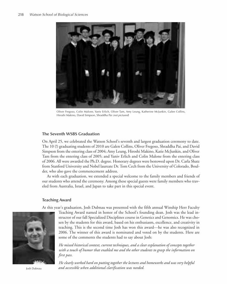
218 Watson School of Biological Sciences
The Seventh WSBS Graduation
On April 25, we celebrated the Watson School’s seventh and largest graduation ceremony to date.The 10 (!) graduating students of 2010 are Galen Collins, Oliver Fregoso, Shraddha Pai, and DavidSimpson from the entering class of 2004; Amy Leung, Hiroshi Makino, Katie McJunkin, and OliverTam from the entering class of 2005; and Yaniv Erlich and Colin Malone from the entering classof 2006. All were awarded the Ph.D. degree. Honorary degrees were bestowed upon Dr. Carla Shatzfrom Stanford University and Nobel laureate Dr. Tom Cech from the University of Colorado, Boul-der, who also gave the commencement address.
As with each graduation, we extended a special welcome to the family members and friends ofour students who attend the ceremony. Among these special guests were family members who trav-eled from Australia, Israel, and Japan to take part in this special event.
Teaching Award
At this year’s graduation, Josh Dubnau was presented with the fifth annual Winship Herr FacultyTeaching Award named in honor of the School’s founding dean. Josh was the lead in-structor of our fall Specialized Disciplines course in Genetics and Genomics. He was cho-sen by the students for this award, based on his enthusiasm, excellence, and creativity inteaching. This is the second time Josh has won this award—he was also recognized in2006. The winner of this award is nominated and voted on by the students. Here aresome of the comments the students had to say about Josh:
He mixed historical context, current techniques, and a clear explanation of concepts togetherwith a touch of humor that enabled me and the other students to grasp the information onfirst pass.
He clearly worked hard on putting together the lectures and homeworks and was very helpfuland accessible when additional clarification was needed.
Oliver Fregoso, Colin Malone, Yaniv Erlich, Oliver Tam, Amy Leung, Katherine McJunkin, Galen Collins, Hiroshi Makino, David Simpson, Shraddha Pai (not pictured)
Josh Dubnau
215-246_wsbs_Annual Report_2009 template 7/8/11 12:21 PM Page 218

2010 WSBS DOCTORAL RECIPIENTS
Thesis Academic Student advisor mentor Current position
Galen Collins William Tansey Marja Postdoctoral fellow with Dr. Fred Goldberg at Harvard Timmermans Medical School
Yaniv Erlich Gregory Hannon John R. Inglis Whitehead Fellow, Whitehead Institute, Massachusetts (Independent Position)
Oliver Fregoso Adrian R. Krainer Nicholas Tonks Postdoctoral fellow in Dr. Michael Emerman’s laboratory at Fred Hutchinson Cancer Research Center, Washington (to start early 2011)
Amy Leung William Tansey David L. Spector Postdoctoral fellow with Dr. Rama Natarajan, City of Hope, California
Hiroshi Makino Roberto Malinow Hollis Cline Postdoctoral fellow with Dr. Yang Dan at the University of California, Berkeley
Colin Malone Gregory Hannon David J. Stewart Postdoctoral fellow with Dr. Ruth Lehmann at The Skirball Institute of Biomolecular Medicine, New York University School of Medicine
Katherine McJunkin Scott Lowe Terri Grodzicker Postdoctoral fellow with Dr. Victor Ambross at University of Massachusetts Medical Center
Shraddha Pai Carlos D. Brody Anthony Zador Postdoctoral fellow with Dr. Arturas Petronis at the Centre for Addiction and Mental Health, University of Toronto, Canada
David Simpson William Tansey Scott Lowe Postdoctoral fellow with Dr. Alejandro Sweet-Cordero at Stanford University (to begin early 2011)
Oliver Tam Gregory Hannon David Jackson Postdoctoral fellow with Dr. Janet Rossant at The Hospital for Sick Children, University of Toronto, Canada
2010 THESIS DISSERTATION DEFENSES
ENTERING CLASS OF 2004
Galen Collins, March 15, 2010
Activator turnover and proteolysis in transcriptional activation.
Thesis Examining CommitteeChair: David L. SpectorResearch mentor: William TanseyAcademic mentor: Marja TimmermansCommittee member: Adrian R. KrainerCommittee member: Michael Hampsey, Robert Wood Johnson Medical SchoolExternal examiner: Bruce Futcher, Stony Brook University
Oliver Fregoso, February 25, 2010
Elucidating the functions of splicing regulatory proteinsthrough the use of high-throughput proteomics.
Thesis Examining CommitteeChair: Scott PowersResearch mentor: Adrian R. KrainerAcademic mentor: Nicholas TonksCommittee member: Michael P. MyersCommittee member: Gregory HannonCommittee member: Brian Chait, The Rockefeller UniversityExternal examiner: Alan M. Zahler, University of California, Santa Cruz
Shraddha Pai, January 14, 2010
Neuroanatomical localization of timing-specific brain loci inthe rat.
Thesis Examining CommitteeChair: Josh DubnauResearch mentor: Carlos D. BrodyAcademic mentor: Anthony ZadorCommittee member: Partha MitraCommittee member: Lizbeth Romanski, The Rockefeller UniversityExternal examiner: Mark Laubach, Yale University
David Simpson,March 16, 2010
Regulation of myc-induced apoptosis in the mammary epithelia.
Thesis Examining CommitteeChair: Gregory HannonResearch mentor: William TanseyAcademic mentor: Scott LoweCommittee member: Senthil K. MuthuswamyCommittee member: George Prendergast, Lankenau Institute for Medical ResearchExternal examiner: Steven McMahon, Kimmel Cancer Center
215-246_wsbs_Annual Report_2009 template 7/8/11 12:21 PM Page 219

220 Watson School of Biological Sciences
2010 THESIS DISSERTATION DEFENSES (continued)
ENTERING CLASS OF 2005
Amy Leung, March 5, 2010
Regulation of chromatin by histone H2B ubiquitylation.
Thesis Examining CommitteeChair: David JacksonResearch mentor: William TanseyAcademic mentor: David L. SpectorCommittee member: Robert MartienssenCommittee member: Rolf Sternglanz, Stony Brook UniversityExternal examiner: Mary Ann Osley, University of New Mexico
Hiroshi Makino, March 8, 2010
AMPA receptor dynamics and synaptic plasticity at excitatorysynapses.
Thesis Examining CommitteeChair: Anthony ZadorResearch mentor: Roberto MalinowAcademic mentor: Bo Li Committee member: Pavel OstenCommittee member: Linda Van AelstExternal examiner: Edward Ziff, New York University
Katherine McJunkin, February 25, 2010
Inducible RNAi targeting essential genes.
Thesis Examining CommitteeChair: Gregory HannonResearch mentor: Scott LoweAcademic mentor: Terri GrodzickerCommittee member: Raffaella SordellaExternal examiner: Vivek Mittal, Memorial Sloan- Kettering Cancer CenterExternal examiner: Andrea Ventura, Cornell University Medical College
Oliver Tam, February 11, 2010
Characterization of small RNA populations and DNA methylation in mammalian development.
Thesis Examining CommitteeChair: Marja TimmermansResearch mentor: Gregory HannonAcademic mentor: David JacksonCommittee member: Zachary LippmanCommittee member: Richard Schultz, Stony Brook UniversityExternal examiner: Robert A. Taft, The Jackson Laboratory
ENTERING CLASS OF 2006
Yaniv Erlich, March 19, 2010
Compressed sequencing.
Thesis Examining CommitteeChair: Adrian R. KrainerResearch mentor: Gregory HannonAcademic mentor: John R. InglisCommittee member: Partha P. MitraCommittee member: Michael Q. ZhangExternal examiner: David Donoho, Stanford University
Colin Malone, February 8, 2010
Evolution, inheritance, and specialization of transposon con-trol pathways in Drosophila.
Thesis Examining CommitteeChair: David L. SpectorResearch mentor: Gregory HannonAcademic mentor: David J. Stewart Committee member: Robert MartienssenCommittee member: Ruth Lehmann, New York UniversityExternal examiner: Craig Mellow, University of Massachusetts
ENTERING CLASS OF 2010
Arkarup Bandyopadhyay, Tata Institute of Fundamental Research, University of Delhi, India
Academic mentor: Zachary Lippman
Colleen Carlston, Harvard University Academic mentor: Alexander Gann
Matthew Koh, University of California, Berkeley Academic mentor: Bo Li
Lisa Krug, Skidmore College Academic mentor: Stephen Shea
Mark Orcholski, Buck Institute, Dominican University
Academic mentor: Nicholas Tonks
John Sheppard, Northwestern University Academic mentor: Josh Dubnau
Jack Walleshauser, University of North Carolina,Greensboro
Academic mentor: Christopher Hammell
215-246_wsbs_Annual Report_2009 template 7/8/11 12:21 PM Page 220

2010 Entering class: (Left to right) Lisa Krug, Arkarup Bandyopadhyay, Colleen Carlston, John Sheppard,Jack Walleshauser, Mark Orcholski, and Matthew Koh.
The 2010 class enjoys a sail on Oyster Bay.
Admissions 2010
The School received 170 applications for the 2010/2011 academic year and is deeply indebted to itsAdmissions Committee, which reviewed, interviewed, and selected candidates for our doctoral pro-gram. The Admissions Committee for the 2010 entering class comprised Gregory Hannon (chair),Mickey Atwal, Leemor Joshua-Tor, Adam Kepecs, Adrian R. Krainer, Zachary Lippman, W. RichardMcCombie, Nicholas Tonks, Lloyd Trotman, and Linda Van Aelst—a truly remarkable team!
Entering Class of 2010
On August 30, 2010, the Watson School opened its doors for the 12th time to welcome yet another newclass. This year, seven students joined the School: Arkarup Bandyopadhyay, Colleen Carlston, MatthewKoh, Lisa Krug, Mark Orcholski, John Sheppard, and Jack Walleshauser. Colleen was a participant inthe summer Undergraduate Research Program in 2008 under the mentorship of Josh Dubnau.
Dean’s Report 221
215-246_wsbs_Annual Report_2009 template 7/8/11 12:22 PM Page 221

DOCTORAL THESIS RESEARCH
Academic Research Student mentor mentor Thesis research
ENTERING CLASS OF 2005Patrick M. Finigan Senthil K. Muthuswamy Robert Martienssen Epigenetic mechanisms involved in centromereBeckman Graduate Student function.
Frederick D. Rollins Jan A. Witkowski Gregory Hannon An RNAi screen for modifiers of cellular Cashin Fellow response to the targeted therapeutic Erlotinib.
ENTERING CLASS OF 2006Eyal Gruntman Josh Dubnau Glenn Turner Olfactory perception in Drosophila.Elisabeth Sloan Livingston Fellow
Amy Rappaport William Tansey Scott Lowe Identification and characterization of tumorBarbara McClintock Fellow suppressor genes in acute myeloid leukemia.Thesis defense: March 2011
Claudio Scuoppo Scott Lowe Gregory Hannon Identification of novel oncosuppressors throughEngelhorn Scholar an in vivo RNAi screen in the Eµ-myc model.Thesis defense: March 2011
ENTERING CLASS OF 2007Megan Bodnar Nicholas Tonks David L. Spector The nuclear choreography of chromatin Starr Centennial Scholar dynamics, gene expression, and gene repression
in embryonic stem cells.
Ralph Burgess Bruce Stillman Gregory Hannon Recombination hot spots: Characterizing fine-Starr Centennial Scholar scale variation in the frequency of meiotic
recombination across the mammalian genome.
Joseph Calarco David Jackson Leemor Joshua-Tor Transposable element regulation in theDavid H. Koch Fellow flowering plant.
Saya Ebbesen David J. Stewart Scott Lowe In vivo identification and characterization NIH Predoctoral Trainee of novel tumor suppressors relevant to
human breast cancer.
Paloma Guzzardo Adrian R. Krainer Gregory Hannon Dissecting the piRNA pathway in theLeslie C. Quick, Jr. Fellow Drosophila ovarian somatic sheet.NIH Predoctoral Trainee
Kyle Honegger John R. Inglis Glenn Turner Neuronal and circuit mechanisms creating Crick-Clay Fellow sparse odor representations in the mush-NIH Predoctoral Trainee room body of Drosophila.
Marek Kudla David Jackson Gregory Hannon CLIP method as a way of direct readout ofGeorge A. and Marjorie H. Anderson Fellow microRNA target sites.
Hassana Oyibo Hiro Furukawa Anthony Zador Reconstruction of connectivity of neurons by Farish-Gerry Fellow reading of oligonucleotide labels (ROC-N-ROL).
Michael Pautler Robert Lucito David Jackson The RAMOSA pathway and inflorescence William R. Miller Fellow branching in maize.
Maria Pineda Adrian R. Krainer Raffaella Sordella Mechanism of “addiction” to receptor CSHL Women’s Partnership for Science tyrosine kinases in non-small-cell lung Student carcinoma.
Yevgeniy Plavskin Jan A. Witkowski Marja Timmermans The evolution of the miR390-dependent Alfred Hershey Fellow tasiRNA pathway and its function in the NIH Predoctoral Trainee bryophyte development.
Joshua Sanders Bruce Stillman Adam Kepecs Trans-regional coordination of activity in Edward and Martha Gerry Fellow the mouse brain.
Zhenxun Wang Terri Grodzicker Adrian R. Krainer Alternative splicing of pyruvate kinase M A*STAR Fellow in tumorigenesis.
222 Watson School of Biological Sciences
215-246_wsbs_Annual Report_2009 template 7/8/11 12:22 PM Page 222

DOCTORAL THESIS RESEARCH (continued)
Academic Research Student mentor mentor Thesis research
Petr Znamenskiy Terri Grodzicker Anthony Zador Pathways for attention and action in the David and Fanny Luke Fellow auditory system.
ENTERING CLASS OF 2008Philippe Batut Alexander Gann Thomas Gingeras Transposable and evolution of transcriptional Florence Gould Fellow regulation in the Drosophila clade.
Mitchell Bekritsky W. Richard McCombie Michael Wigler High-throughput microsatellite genotyping. Beckman Graduate Student
Dario Bressan Z. Josh Huang Gregory Hannon A genomic approach toward the elucidation ofLindsay Fellow connectivity patterns at cellular resolution in
complex neural networks.
Carrie Clendaniel David Jackson Marja Timmermans Role of HIRA in the regulation of develop-Beckman Graduate Student mentally important target loci.
Melanie Eckersley-Maslin Gregory Hannon David L. Spector Making the choice: Mechanistic insights Genentech Foundation Fellow into random autosomal monoallelic expres-
sion in mammalian cells.
Sang Geol Koh Glenn Turner Anthony Zador Attention-dependent information routing George A. and Marjorie H. Anderson Fellow in the mouse auditory cortex.
Katie Liberatore Adrian R. Krainer Zachary Lippman Inflorescence development and heterosis.NSF Graduate Research Fellow
Ozlem Mert John R. Inglis Scott Lowe Characterization of the role of E2F7 in George A. and Marjorie H. Anderson Fellow oncogene-induced senescence and tumori-
genesis.
Elizabeth Nakasone Alea A. Mills Mikala Egeblad Understanding the effects of cytotoxic chemo-DoD BCRP Predoctoral Fellow therapeutics on the innate immune response.
Zinaida Perova Linda Van Aelst Bo Li Role of medial prefrontal cortex in behav-Charles A. Dana Fellow ioral depression.
Felix Schlesinger Gregory Hannon Thomas Gingeras Classification of novel transcription start sites. Crick-Clay Fellow
Nilgun Tasdemir Josh Dubnau Scott Lowe Investigating the molecular overlaps be-Lindsay Fellow tween epigenetic reprogramming and trans-
formation.
Elvin Wagenblast Jan A. Witkowski Gregory Hannon Role of stem/progenitor cells in mammary Boehringer Ingelheim Fonds Scholar gland and breast tumors.
Susann Weissmueller Raffaella Sordella Scott Lowe In vivo identification and characterizationAnnette Kade Fellow of tumor suppressor genes in hepatocellular
carcinoma.
ENTERING CLASS OF 2009Stephane Castel Lloyd Trotman Robert Martienssen RNAi-mediated heterochromatin in S. pombe.Cashin Fellow Proposal defense: February 2011
Kristen Delevich Stephen Shea Bo Li Elucidating the role of Disrupted-in-Schizo-NIH Predoctoral Trainee phrenia-1 in development of prefrontal cortical Proposal defense: January 2011 circuits.
Silvia Fenoglio Linda Van Aelst Gregory Hannon RNAi screening to identify putative therapeutic Elisabeth Sloan Livingston Fellow targets for the treatment of pancreatic cancer.Proposal defense: December 2010
Wee Siong Goh Hiro Furukawa Gregory Hannon Determining piRNA primary biogenesis, and A*STAR Fellow MIWI and late piRNA function in mice using Proposal defense: January 2011 C. elegans as a model system for genetic screening.
Dean’s Report 223
215-246_wsbs_Annual Report_2009 template 7/8/11 12:22 PM Page 223

224 Watson School of Biological Sciences
Academic Mentoring
The Watson School takes great pride in the level of mentoring that it offers its students. One of thevery special aspects in this regard is our two-tiered mentoring approach whereby each student receivesan academic mentor as well as a research mentor. Entering students select by mutual agreement amember of the research or nonresearch faculty to serve as an academic mentor—a watchful guardianto look over and encourage students through the sometimes-trying process of their doctoral educa-tion. This program continues to receive much support from the faculty who volunteer to be academicmentors, and it has rightfully become a vital ingredient in our success. The following are this year'snew academic mentors for the entering class of 2010:
The Fall Term Curriculum
Our faculty continues to do an outstanding job of developing and delivering the curriculum. We areextremely grateful for their considerable time and effort in maintaining the high-quality course workthat we strive to provide. The Curriculum Development and Integration Committee (CDIC)—AdrianKrainer (Chair), David Jackson, Leemor Joshua-Tor, Nicholas Tonks, and Glenn Turner—continuesto carefully monitor and develop the curriculum. In addition to the outstanding course instructors andguest lecturers from within the Laboratory, our courses also continue to attract an impressive array ofguest lecturers from other institutions.
New Courses in Quantitative Biology
This year, we developed two new courses in the rapidly emerging field of quantitative biology. Today’sscience often relies on the use of theoretical modeling and the analysis of vast quantities of data. Wetherefore need to train scientists who not only are experts in biology, but also have skills in mathematics,physics, chemistry, and computation. The new courses were designed to expand the curriculum to theareas of statistics, programming, bioinformatics, and theoretical modeling and analysis and take differentapproaches to quantitative thinking. The first course, Physical Biology of the Cell, which was developedby Leemor Joshua-Tor and Rob Phillips from the California Institute of Technology, teaches the students
DOCTORAL THESIS RESEARCH (continued)
Academic Research Student mentor mentor Thesis research
Ian Peikon Mickey Atwal Anthony Zador Reverse engineering the brain.George A. and Marjorie H. Anderson FellowProposal defense: February 2011
Kaja Wasik Jan A. Witkowski Gregory Hannon A screen for novel components of the piRNA George A. and Marjorie H. Anderson Fellow pathway in Drosophila melanogaster.Proposal defense: February 2011
Cinthya Zepeda Mendoza Thomas Gingeras David L. Spector Analysis of higher-order chromatin organizationGonzalo Rio Arronte Fellow at the mouse syntenic region of human 1p36Proposal defense: January 2011 upon genomic copy-number changes.
STUDENT MENTOR
Arkarup Bandyopadhyay Zachary LippmanColleen Carlston Alex GannMatthew Koh Bo LiLisa Krug Stephen Shea
STUDENT MENTOR
Mark Orcholski Nicholas TonksJohn Sheppard Josh DubnauJack Walleshauser Christopher Hammell
215-246_wsbs_Annual Report_2009 template 7/8/11 12:22 PM Page 224
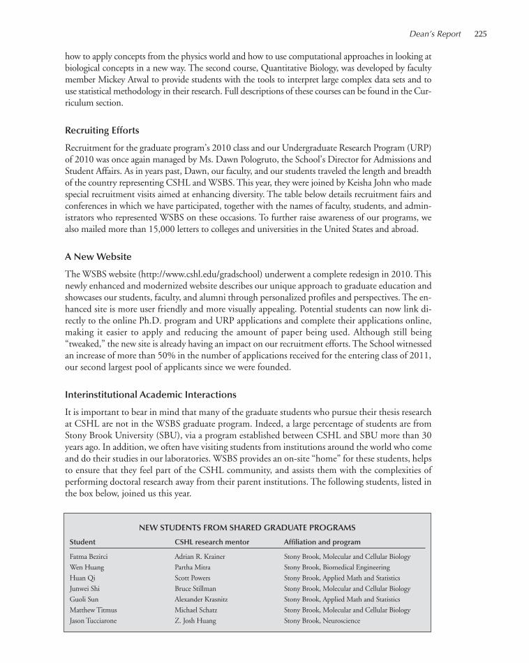
Dean’s Report 225
how to apply concepts from the physics world and how to use computational approaches in looking atbiological concepts in a new way. The second course, Quantitative Biology, was developed by facultymember Mickey Atwal to provide students with the tools to interpret large complex data sets and touse statistical methodology in their research. Full descriptions of these courses can be found in the Cur-riculum section.
Recruiting Efforts
Recruitment for the graduate program’s 2010 class and our Undergraduate Research Program (URP)of 2010 was once again managed by Ms. Dawn Pologruto, the School’s Director for Admissions andStudent Affairs. As in years past, Dawn, our faculty, and our students traveled the length and breadthof the country representing CSHL and WSBS. This year, they were joined by Keisha John who madespecial recruitment visits aimed at enhancing diversity. The table below details recruitment fairs andconferences in which we have participated, together with the names of faculty, students, and admin-istrators who represented WSBS on these occasions. To further raise awareness of our programs, wealso mailed more than 15,000 letters to colleges and universities in the United States and abroad.
A New Website
The WSBS website (http://www.cshl.edu/gradschool) underwent a complete redesign in 2010. Thisnewly enhanced and modernized website describes our unique approach to graduate education andshowcases our students, faculty, and alumni through personalized profiles and perspectives. The en-hanced site is more user friendly and more visually appealing. Potential students can now link di-rectly to the online Ph.D. program and URP applications and complete their applications online,making it easier to apply and reducing the amount of paper being used. Although still being“tweaked,” the new site is already having an impact on our recruitment efforts. The School witnessedan increase of more than 50% in the number of applications received for the entering class of 2011,our second largest pool of applicants since we were founded.
Interinstitutional Academic Interactions
It is important to bear in mind that many of the graduate students who pursue their thesis researchat CSHL are not in the WSBS graduate program. Indeed, a large percentage of students are fromStony Brook University (SBU), via a program established between CSHL and SBU more than 30years ago. In addition, we often have visiting students from institutions around the world who comeand do their studies in our laboratories. WSBS provides an on-site “home” for these students, helpsto ensure that they feel part of the CSHL community, and assists them with the complexities ofperforming doctoral research away from their parent institutions. The following students, listed inthe box below, joined us this year.
NEW STUDENTS FROM SHARED GRADUATE PROGRAMS
Student CSHL research mentor Affiliation and program
Fatma Bezirci Adrian R. Krainer Stony Brook, Molecular and Cellular Biology
Wen Huang Partha Mitra Stony Brook, Biomedical Engineering
Huan Qi Scott Powers Stony Brook, Applied Math and Statistics
Junwei Shi Bruce Stillman Stony Brook, Molecular and Cellular Biology
Guoli Sun Alexander Krasnitz Stony Brook, Applied Math and Statistics
Matthew Titmus Michael Schatz Stony Brook, Molecular and Cellular Biology
Jason Tucciarone Z. Josh Huang Stony Brook, Neuroscience
215-246_wsbs_Annual Report_2009 template 7/8/11 12:22 PM Page 225
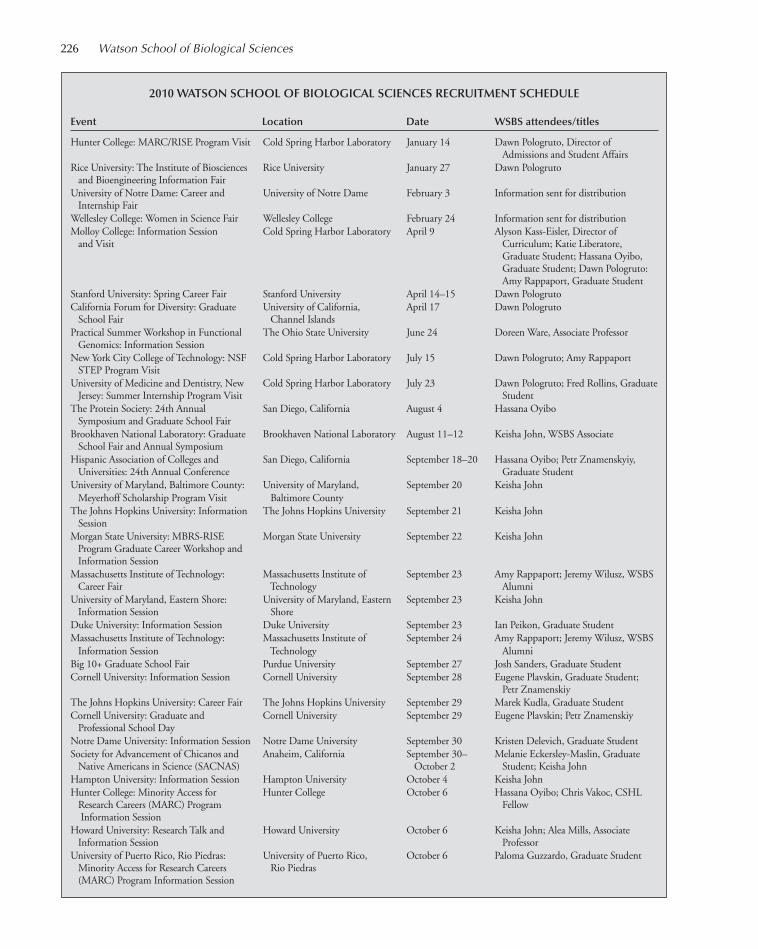
226 Watson School of Biological Sciences
2010 WATSON SCHOOL OF BIOLOGICAL SCIENCES RECRUITMENT SCHEDULE
Event Location Date WSBS attendees/titles
Hunter College: MARC/RISE Program Visit Cold Spring Harbor Laboratory January 14 Dawn Pologruto, Director of Admissions and Student Affairs
Rice University: The Institute of Biosciences Rice University January 27 Dawn Pologrutoand Bioengineering Information Fair
University of Notre Dame: Career and University of Notre Dame February 3 Information sent for distributionInternship Fair
Wellesley College: Women in Science Fair Wellesley College February 24 Information sent for distributionMolloy College: Information Session Cold Spring Harbor Laboratory April 9 Alyson Kass-Eisler, Director of
and Visit Curriculum; Katie Liberatore, Graduate Student; Hassana Oyibo, Graduate Student; Dawn Pologruto:Amy Rappaport, Graduate Student
Stanford University: Spring Career Fair Stanford University April 14–15 Dawn PologrutoCalifornia Forum for Diversity: Graduate University of California, April 17 Dawn Pologruto
School Fair Channel IslandsPractical Summer Workshop in Functional The Ohio State University June 24 Doreen Ware, Associate Professor
Genomics: Information SessionNew York City College of Technology: NSF Cold Spring Harbor Laboratory July 15 Dawn Pologruto; Amy Rappaport
STEP Program VisitUniversity of Medicine and Dentistry, New Cold Spring Harbor Laboratory July 23 Dawn Pologruto; Fred Rollins, Graduate
Jersey: Summer Internship Program Visit StudentThe Protein Society: 24th Annual San Diego, California August 4 Hassana Oyibo
Symposium and Graduate School Fair Brookhaven National Laboratory: Graduate Brookhaven National Laboratory August 11–12 Keisha John, WSBS Associate
School Fair and Annual SymposiumHispanic Association of Colleges and San Diego, California September 18–20 Hassana Oyibo; Petr Znamenskyiy,
Universities: 24th Annual Conference Graduate StudentUniversity of Maryland, Baltimore County: University of Maryland, September 20 Keisha John
Meyerhoff Scholarship Program Visit Baltimore CountyThe Johns Hopkins University: Information The Johns Hopkins University September 21 Keisha John
SessionMorgan State University: MBRS-RISE Morgan State University September 22 Keisha John
Program Graduate Career Workshop andInformation Session
Massachusetts Institute of Technology: Massachusetts Institute of September 23 Amy Rappaport; Jeremy Wilusz, WSBS Career Fair Technology Alumni
University of Maryland, Eastern Shore: University of Maryland, Eastern September 23 Keisha JohnInformation Session Shore
Duke University: Information Session Duke University September 23 Ian Peikon, Graduate StudentMassachusetts Institute of Technology: Massachusetts Institute of September 24 Amy Rappaport; Jeremy Wilusz, WSBS
Information Session Technology AlumniBig 10+ Graduate School Fair Purdue University September 27 Josh Sanders, Graduate StudentCornell University: Information Session Cornell University September 28 Eugene Plavskin, Graduate Student;
Petr ZnamenskiyThe Johns Hopkins University: Career Fair The Johns Hopkins University September 29 Marek Kudla, Graduate StudentCornell University: Graduate and Cornell University September 29 Eugene Plavskin; Petr Znamenskiy
Professional School DayNotre Dame University: Information Session Notre Dame University September 30 Kristen Delevich, Graduate StudentSociety for Advancement of Chicanos and Anaheim, California September 30– Melanie Eckersley-Maslin, Graduate
Native Americans in Science (SACNAS) October 2 Student; Keisha JohnHampton University: Information Session Hampton University October 4 Keisha JohnHunter College: Minority Access for Hunter College October 6 Hassana Oyibo; Chris Vakoc, CSHL
Research Careers (MARC) Program FellowInformation Session
Howard University: Research Talk and Howard University October 6 Keisha John; Alea Mills, AssociateInformation Session Professor
University of Puerto Rico, Rio Piedras: University of Puerto Rico, October 6 Paloma Guzzardo, Graduate StudentMinority Access for Research Careers Rio Piedras(MARC) Program Information Session
215-246_wsbs_Annual Report_2009 template 7/8/11 12:22 PM Page 226

2010 WATSON SCHOOL OF BIOLOGICAL SCIENCES RECRUITMENT SCHEDULE (continued)
Event Location Date WSBS attendees/titles
University of California, Berkeley: Graduate University of California, Berkeley October 6–7 Megan Bodnar, Graduate StudentSchool Fair
Oberlin College: Information Session Oberlin College October 7 Saya Ebbesen, Graduate StudentNorthwestern University: Information Session Northwestern University October 7–8 Mitch Bekritsky, Graduate StudentBarry University: Minority Access for Barry University October 8 Maria Pineda
Research: Careers (MARC) Program Information Session
Washington University, St. Louis: Washington University, St. Louis October 12 Keisha JohnInformation Session
University of Wisconsin, Madison: Graduate University of Wisconsin, Madison October 13 Dario Bressan, Graduate StudentSchool Fair
Princeton University: Information Session Princeton University October 14 Michael Pautler, Graduate StudentUniversity of North Carolina, Chapel Hill: University of North Carolina, October 15 Zina Perova, Graduate Student
Graduate School Fair Chapel HillPrinceton University: Graduate and Princeton University October 15 Michael Pautler
Professional School FairRowan University: Information Session Rowan University October 15 Dawn PologrutoDuke University: Graduate School Fair Duke University October 18 Stephen Shea, Assistant ProfessorHoward University: Information Session Howard University October 19 Keisha JohnQueens College, City University of New Queens College, City University October 20 Keisha John
York: MARC-USTAR Program of New YorkInformation Session
Emory University: Graduate School Fair Emory University October 21 Keisha JohnSpelman College: MARC-USTAR Program Spelman College October 21 Keisha John
Information SessionState University of New York, Farmingdale: Cold Spring Harbor Laboratory October 22 Keisha John; Hassana Oyibo; Michael
Information Session and Visit Pautler; Dawn PologrutoTemple University: MARC Program: Temple University October 22 Keisha John
Information SessionCalifornia Institute of Technology: Career Fair California Institute of Technology October 26 Elizabeth Nakasone, Graduate StudentUniversity of California, Los Angeles: University of California, Los October 27 Elizabeth Nakasone
Graduate School Fair AngelesYale University: Information Session Yale University October 28 Kyle Honegger, Graduate StudentUniversity of Southern California: University of Southern California October 28 Elizabeth Nakasone
Information SessionUniversity of Pittsburgh: Information Session University of Pittsburgh October 28 Kristen DelevichMorehouse College: Graduate School Fair Morehouse College October 28–29 Keisha John
and Research Training SymposiumHarvard University: Information Session Harvard University October 29 Kyle HoneggerAtlanta University Center Consortium: Clark Atlanta University October 29–30 Keisha John
(Morehouse College, Spelman College, and Clark Atlanta University)
Vassar College: Genetics and Bioinformatics Cold Spring Harbor Laboratory November 1 Keisha John; Justin Kinney, CSHL Course Visit and Information Session Fellow; Sheldon McKay; Dawn
Pologruto; Amy RappaportAnnual Biomedical Conference for Charlotte, North Carolina November 10–13 Saya Ebbesen; Paloma Guzzardo;
Minority Students (ABRCMS) Keisha John; Dawn PologrutoUniversity of New Mexico: Information University of New Mexico November 10 Katie Liberatore, Graduate Student
SessionSigma Xi Annual Conference and Research Raleigh, North Carolina November 11–13 Maria Pineda
SymposiumAmerican Indian Science and Engineering Albuquerque, New Mexico November 11–12 Katie Liberatore
Society: 2010 National ConferenceQueensborough Community College, Queensborough Community November 16 Information sent for distribution
CUNY: Cooperative Education Forum College, CUNYBrooklyn College of CUNY: MARC and Brooklyn College of CUNY November 16 Keisha John
RISE Program: Information SessionThe American Society for Cell Biology Philadelphia, Pennsylvania December 11 Megan Bodnar
Annual Meeting: Graduate School Fair
Dean’s Report 227
215-246_wsbs_Annual Report_2009 template 7/8/11 12:22 PM Page 227

228 Watson School of Biological Sciences
Graduate Student Symposium
Continuing the tradition that began in the summer of 2008, three Graduate Student Symposiawere held in 2010 at the Laboratory’s Genome Research Center in Woodbury: one each in January,May, and October. Each Symposium consisted of a full day of activities including 20-minute and10-minute talks by senior students and poster presentations by more junior students. Lunch anda wine and cheese reception rounded out the program and provided opportunities for more infor-mal interactions. As with the inaugural event held in August 2008, the students and faculty in at-tendance found the Symposia to be very worthwhile events. The Graduate Student Symposium isopen to the entire Laboratory community, but it is run largely by the students themselves. We aregrateful to the two student chairs of the Symposium—Megha Rajaram (SBU) and Maria Pineda(WSBS)—for their dedication and hard work and to Keisha John for overseeing the administrativeelements.
Graduate Student and Postdoctoral Fellow Departures
With each year come not only new arrivals, but also departures. The following graduate studentsand postdoctoral fellows departed from the Laboratory during 2010:
Postdoctoral FellowsUli C. Bialucha Dheeraj Malhotra Rajesh K. Prakash Michael WittingerChunlin Cai Louis Manganas Vladimir Vacic Zhiguang YuchiIver K. Hansen Nael Nadif Kasri Daniel Valente Lixing ZhanDanielle Irvine Michaela Oswald-Pisarski Haibin Wang Zhihua ZhangValery Krizhanovsky Mario Pagani
Graduate StudentsAnne Burlet-Parendel Jiahao Huang Shraddha Pai Rahul SinhaSungwook Choi Mary Kusenda Benjamin Roland Shuying SunGalen Collins Lucile Lefevre Patrick Russell Oliver TamAshesh Dhawale Amy Leung Laurianne Scourzic Sophie ThomainJulien Dos Santos Hiroshi Makino Grzegorz Sienski Yang YangEwa Grabowska Colin Malone David Simpson Jianping ZhangIkuko Hotta
Executive Committee
A large measure of the Watson School's success can be traced to the sage advice, guidance, and gov-ernance of the School's Executive Committee. I wish to thank faculty members Mickey Atwal, TerriGrodzicker, Adrian Krainer, Alea Mills, David Spector, and Linda Van Aelst who served on the Ex-ecutive Committee through 2010. Terri Grodzicker completed her second term in December 2010.We are grateful for the insights and advice that Terri brought to the Committee as a member for 6years. As happens each year, there was also turnover among the student representatives. SBU repre-sentative Matthew Lazarus completed his term in October, and he was replaced by Matthew Cami-olo. WSBS representative Megan Bodnar also completed her term in December. We are thankful toMatthew and Megan for their honest and thoughtful advice.
Special Events: The Gavin Borden Fellow
The annual Gavin Borden Visiting Fellow (so named after the energetic and charismatic publisherof Molecular Biology of the Cell, who died of cancer in 1991) brings to CSHL an eminent researcherand educator to give the Gavin Borden Lecture, which is dedicated to the graduate students at the
215-246_wsbs_Annual Report_2009 template 7/8/11 12:22 PM Page 228
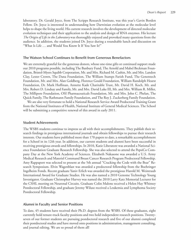
laboratory. Dr. Gerald Joyce, from The Scripps Research Institute, was this year’s Gavin BordenFellow. Dr. Joyce is interested in understanding how Darwinian evolution at the molecular levelhelps to shape the living world. His current research involves the development of directed molecularevolution techniques and their application to the analysis and design of RNA enzymes. His lectureThe Origin of Life in the Laboratory was thoroughly enjoyed and provoked many questions from theaudience. In addition, the students joined Dr. Joyce during a roundtable lunch and discussion on“What Is Life … and Would You Know It If You Saw It?”
The Watson School Continues to Benefit from Generous Benefactors
We are extremely grateful for the generous donors, whose one-time gifts or continued support madeour 2010 programs possible, including The Banbury Fund, The Arnold and Mabel Beckman Foun-dation, Bristol-Myers Squibb Corporation, Mr. and Mrs. Richard M. Cashin, Mr. and Mrs. LandonClay, Lester Crown, The Dana Foundation, The William Stamps Farish Fund, The GenentechFoundation, Mr. and Mrs. Alan Goldberg, Florence Gould Foundation, William Randolph HearstFoundation, Dr. Mark Hoffman, Annette Kade Charitable Trust, Mr. David H. Koch, Mr. andMrs. Robert D. Lindsay and Family, Mr. and Mrs. David Luke III, Mr. and Mrs. William R. Miller,The Millipore Foundation, OSI Pharmaceuticals Foundation, Mr. and Mrs. John C. Phelan, TheQuick Family, The Rathmann Family Foundation, and The Roy J. Zuckerberg Family Foundation.
We are also very fortunate to hold a National Research Service Award Predoctoral Training Grantfrom the National Institutes of Health, National Institute of General Medical Sciences. The Schoolwill be submitting a competitive renewal of this award in early 2011.
Student Achievements
The WSBS students continue to impress us all with their accomplishments. They publish their re-search findings in prestigious international journals and obtain fellowships to pursue their researchinterests. Our students have published more than 170 papers to date, a remarkable accomplishmentfor a School in its 12th year. In addition, our current students and alumni have been successful inreceiving prestigious awards and fellowships. In 2010, Katie Liberatore was awarded a National Sci-ence Foundation Graduate Research Fellowship. She was also selected to attend the PepsiCo Com-pany Day at the New York Academy of Sciences. Elizabeth Nakasone was awarded a U.S. ArmyMedical Research and Materiel Command Breast Cancer Research Program Predoctoral Fellowship.Amy Rappaport was selected to present at the 5th annual “Cracking the Code with the Bear” Re-search Symposium. Elvin Wagenblast was awarded a predoctoral fellowship from the BoehringerIngelheim Fonds. Recent graduate Yaniv Erlich was awarded the prestigious Harold M. WeintraubInternational Award for Graduate Studies. He was also named a 2010 Genome Technology YoungInvestigator. Graduate Christopher Harvey was named the 2010 Larry Katz Memorial Lecturer forthe CSHL meeting on Neuronal Circuits. Graduate Colin Malone received a Helen Hay WhitneyPostdoctoral Fellowship, and graduate Jeremy Wilusz received a Leukemia and Lymphoma SocietyPostdoctoral Fellowship.
Alumni in Faculty and Senior Positions
To date, 45 students have received their Ph.D. degrees from the WSBS. Of these graduates, eightcurrently hold tenure-track faculty positions and two hold independent research positions. Twenty-seven of our former students are pursuing postdoctoral research and five of our alumni completedtheir postdoctoral studies and have moved onto positions in administration, management consulting,and journal editing. We are so proud of them all!
Dean’s Report 229
215-246_wsbs_Annual Report_2009 template 7/8/11 12:22 PM Page 229
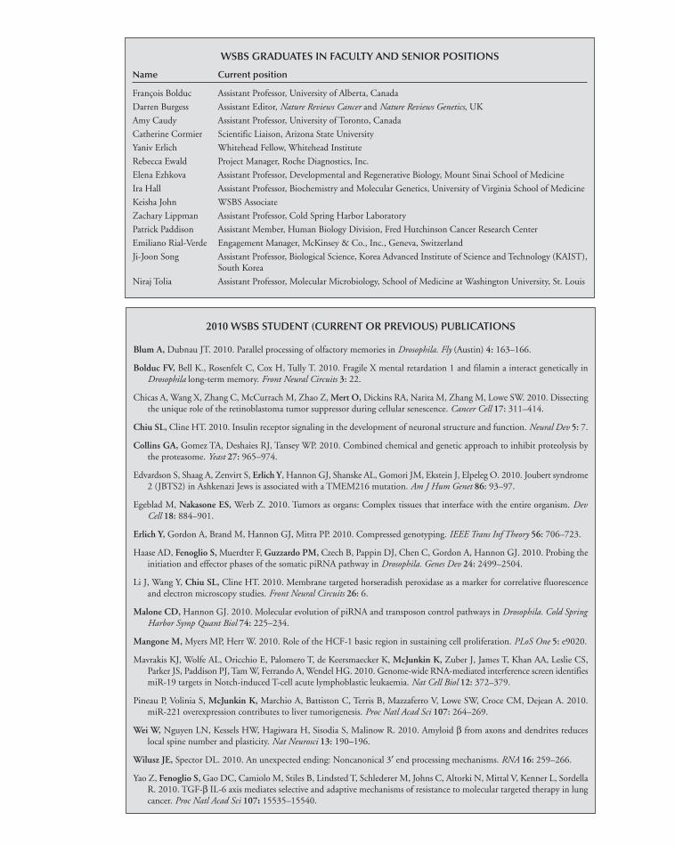
2010 WSBS STUDENT (CURRENT OR PREVIOUS) PUBLICATIONS
Blum A, Dubnau JT. 2010. Parallel processing of olfactory memories in Drosophila. Fly (Austin) 4: 163–166.
Bolduc FV, Bell K., Rosenfelt C, Cox H, Tully T. 2010. Fragile X mental retardation 1 and filamin a interact genetically inDrosophila long-term memory. Front Neural Circuits 3: 22.
Chicas A, Wang X, Zhang C, McCurrach M, Zhao Z, Mert O, Dickins RA, Narita M, Zhang M, Lowe SW. 2010. Dissectingthe unique role of the retinoblastoma tumor suppressor during cellular senescence. Cancer Cell 17: 311–414.
Chiu SL, Cline HT. 2010. Insulin receptor signaling in the development of neuronal structure and function. Neural Dev 5: 7.
Collins GA, Gomez TA, Deshaies RJ, Tansey WP. 2010. Combined chemical and genetic approach to inhibit proteolysis bythe proteasome. Yeast 27: 965–974.
Edvardson S, Shaag A, Zenvirt S, Erlich Y, Hannon GJ, Shanske AL, Gomori JM, Ekstein J, Elpeleg O. 2010. Joubert syndrome2 (JBTS2) in Ashkenazi Jews is associated with a TMEM216 mutation. Am J Hum Genet 86: 93–97.
Egeblad M, Nakasone ES, Werb Z. 2010. Tumors as organs: Complex tissues that interface with the entire organism. DevCell 18: 884–901.
Erlich Y, Gordon A, Brand M, Hannon GJ, Mitra PP. 2010. Compressed genotyping. IEEE Trans Inf Theory 56: 706–723.
Haase AD, Fenoglio S, Muerdter F, Guzzardo PM, Czech B, Pappin DJ, Chen C, Gordon A, Hannon GJ. 2010. Probing theinitiation and effector phases of the somatic piRNA pathway in Drosophila. Genes Dev 24: 2499–2504.
Li J, Wang Y, Chiu SL, Cline HT. 2010. Membrane targeted horseradish peroxidase as a marker for correlative fluorescenceand electron microscopy studies. Front Neural Circuits 26: 6.
Malone CD, Hannon GJ. 2010. Molecular evolution of piRNA and transposon control pathways in Drosophila. Cold SpringHarbor Symp Quant Biol 74: 225–234.
Mangone M, Myers MP, Herr W. 2010. Role of the HCF-1 basic region in sustaining cell proliferation. PLoS One 5: e9020.
Mavrakis KJ, Wolfe AL, Oricchio E, Palomero T, de Keersmaecker K, McJunkin K, Zuber J, James T, Khan AA, Leslie CS,Parker JS, Paddison PJ, Tam W, Ferrando A, Wendel HG. 2010. Genome-wide RNA-mediated interference screen identifiesmiR-19 targets in Notch-induced T-cell acute lymphoblastic leukaemia. Nat Cell Biol 12: 372–379.
Pineau P, Volinia S, McJunkin K, Marchio A, Battiston C, Terris B, Mazzaferro V, Lowe SW, Croce CM, Dejean A. 2010.miR-221 overexpression contributes to liver tumorigenesis. Proc Natl Acad Sci 107: 264–269.
Wei W, Nguyen LN, Kessels HW, Hagiwara H, Sisodia S, Malinow R. 2010. Amyloid β from axons and dendrites reduceslocal spine number and plasticity. Nat Neurosci 13: 190–196.
Wilusz JE, Spector DL. 2010. An unexpected ending: Noncanonical 3′ end processing mechanisms. RNA 16: 259–266.
Yao Z, Fenoglio S, Gao DC, Camiolo M, Stiles B, Lindsted T, Schlederer M, Johns C, Altorki N, Mittal V, Kenner L, SordellaR. 2010. TGF-β IL-6 axis mediates selective and adaptive mechanisms of resistance to molecular targeted therapy in lungcancer. Proc Natl Acad Sci 107: 15535–15540.
WSBS GRADUATES IN FACULTY AND SENIOR POSITIONS
Name Current position
François Bolduc Assistant Professor, University of Alberta, Canada
Darren Burgess Assistant Editor, Nature Reviews Cancer and Nature Reviews Genetics, UK
Amy Caudy Assistant Professor, University of Toronto, Canada
Catherine Cormier Scientific Liaison, Arizona State University
Yaniv Erlich Whitehead Fellow, Whitehead Institute
Rebecca Ewald Project Manager, Roche Diagnostics, Inc.
Elena Ezhkova Assistant Professor, Developmental and Regenerative Biology, Mount Sinai School of Medicine
Ira Hall Assistant Professor, Biochemistry and Molecular Genetics, University of Virginia School of Medicine
Keisha John WSBS Associate
Zachary Lippman Assistant Professor, Cold Spring Harbor Laboratory
Patrick Paddison Assistant Member, Human Biology Division, Fred Hutchinson Cancer Research Center
Emiliano Rial-Verde Engagement Manager, McKinsey & Co., Inc., Geneva, Switzerland
Ji-Joon Song Assistant Professor, Biological Science, Korea Advanced Institute of Science and Technology (KAIST), South Korea
Niraj Tolia Assistant Professor, Molecular Microbiology, School of Medicine at Washington University, St. Louis
215-246_wsbs_Annual Report_2009 template 7/8/11 12:22 PM Page 230

WSBS Family Events
Finally, I am pleased to announce that this was a great year for the WSBS students as we celebratedsome wonderful personal occasions. On April 24, Sang Geol Koh, from the entering class of 2008,and his wife Bomi welcomed a son, Aiden, who weighed in at 8 lbs 11 oz. On May 30, Katie Lib-eratore, also from the class of 2008, married her fiancée Brian Kramer in New Mexico. On June21, Kyle Honegger from the entering class of 2007 married Laura Bozue in their home state of Illi-nois. Silvia Fenoglio from the entering class of 2009 married postdoctoral fellow Agustin Chicason July 18. Silvia and Agustin welcomed their daughter Lucia on December 22. Our congratulationsand best wishes to all of them.
Leemor Joshua-TorProfessor and Dean
Dean’s Report 231
Aiden Koh Lucia Chicas
Kyle and Laura Katie and Brian Silvia and Agustin
215-246_wsbs_Annual Report_2009 template 7/8/11 12:22 PM Page 231
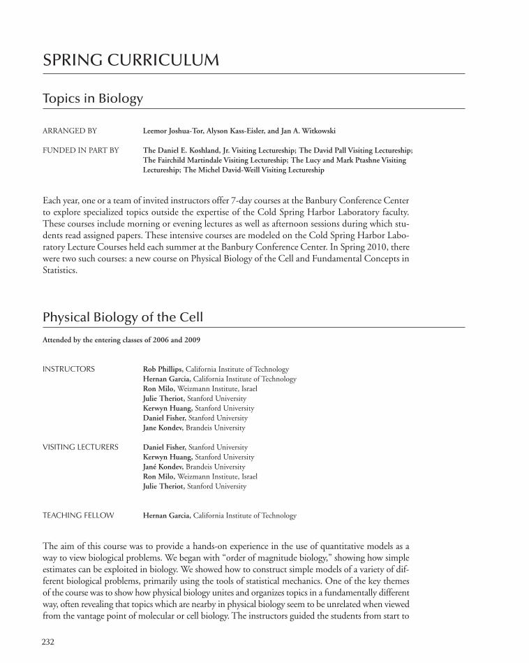
232
SPRING CURRICULUM
Topics in Biology
ARRANGED BY Leemor Joshua-Tor, Alyson Kass-Eisler, and Jan A. Witkowski
FUNDED IN PART BY The Daniel E. Koshland, Jr. Visiting Lectureship; The David Pall Visiting Lectureship;The Fairchild Martindale Visiting Lectureship; The Lucy and Mark Ptashne Visiting Lectureship; The Michel David-Weill Visiting Lectureship
Each year, one or a team of invited instructors offer 7-day courses at the Banbury Conference Centerto explore specialized topics outside the expertise of the Cold Spring Harbor Laboratory faculty.These courses include morning or evening lectures as well as afternoon sessions during which stu-dents read assigned papers. These intensive courses are modeled on the Cold Spring Harbor Labo-ratory Lecture Courses held each summer at the Banbury Conference Center. In Spring 2010, therewere two such courses: a new course on Physical Biology of the Cell and Fundamental Concepts inStatistics.
Physical Biology of the Cell
Attended by the entering classes of 2006 and 2009
INSTRUCTORS Rob Phillips, California Institute of TechnologyHernan Garcia, California Institute of TechnologyRon Milo, Weizmann Institute, IsraelJulie Theriot, Stanford UniversityKerwyn Huang, Stanford UniversityDaniel Fisher, Stanford UniversityJane Kondev, Brandeis University
VISITING LECTURERS Daniel Fisher, Stanford UniversityKerwyn Huang, Stanford UniversityJané Kondev, Brandeis UniversityRon Milo, Weizmann Institute, IsraelJulie Theriot, Stanford University
TEACHING FELLOW Hernan Garcia, California Institute of Technology
The aim of this course was to provide a hands-on experience in the use of quantitative models as away to view biological problems. We began with “order of magnitude biology,” showing how simpleestimates can be exploited in biology. We showed how to construct simple models of a variety of dif-ferent biological problems, primarily using the tools of statistical mechanics. One of the key themesof the course was to show how physical biology unites and organizes topics in a fundamentally differentway, often revealing that topics which are nearby in physical biology seem to be unrelated when viewedfrom the vantage point of molecular or cell biology. The instructors guided the students from start to
215-246_wsbs_Annual Report_2009 template 7/8/11 12:22 PM Page 232
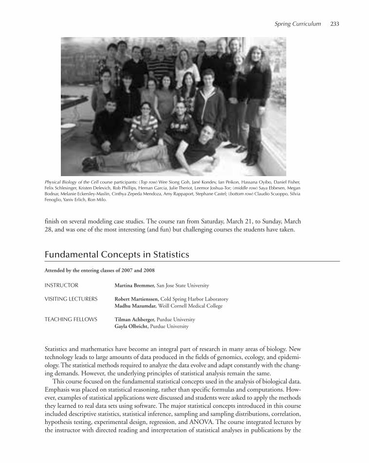
Spring Curriculum 233
finish on several modeling case studies. The course ran from Saturday, March 21, to Sunday, March28, and was one of the most interesting (and fun) but challenging courses the students have taken.
Fundamental Concepts in Statistics
Attended by the entering classes of 2007 and 2008
INSTRUCTOR Martina Bremmer, San Jose State University
VISITING LECTURERS Robert Martienssen, Cold Spring Harbor LaboratoryMadhu Mazumdar, Weill Cornell Medical College
TEACHING FELLOWS Tilman Achberger, Purdue UniversityGayla Olbricht, Purdue University
Statistics and mathematics have become an integral part of research in many areas of biology. Newtechnology leads to large amounts of data produced in the fields of genomics, ecology, and epidemi-ology. The statistical methods required to analyze the data evolve and adapt constantly with the chang-ing demands. However, the underlying principles of statistical analysis remain the same.
This course focused on the fundamental statistical concepts used in the analysis of biological data.Emphasis was placed on statistical reasoning, rather than specific formulas and computations. How-ever, examples of statistical applications were discussed and students were asked to apply the methodsthey learned to real data sets using software. The major statistical concepts introduced in this courseincluded descriptive statistics, statistical inference, sampling and sampling distributions, correlation,hypothesis testing, experimental design, regression, and ANOVA. The course integrated lectures bythe instructor with directed reading and interpretation of statistical analyses in publications by the
Physical Biology of the Cell course participants: (Top row) Wee Siong Goh, Jané Kondev, Ian Peikon, Hassana Oyibo, Daniel Fisher,Felix Schlesinger, Kristen Delevich, Rob Phillips, Hernan Garcia, Julie Theriot, Leemor Joshua-Tor; (middle row) Saya Ebbesen, MeganBodnar, Melanie Eckersley-Maslin, Cinthya Zepeda Mendoza, Amy Rappaport, Stephane Castel; (bottom row) Claudio Scuoppo, SilviaFenoglio, Yaniv Erlich, Ron Milo.
215-246_wsbs_Annual Report_2009 template 7/8/11 12:22 PM Page 233
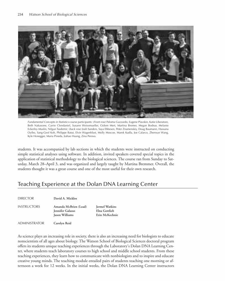
234 Watson School of Biological Sciences
students. It was accompanied by lab sections in which the students were instructed on conductingsimple statistical analyses using software. In addition, invited speakers covered special topics in theapplication of statistical methodology to the biological sciences. The course ran from Sunday to Sat-urday, March 28–April 3, and was organized and largely taught by Martina Bremmer. Overall, thestudents thought it was a great course and one of the most useful for their own research.
Teaching Experience at the Dolan DNA Learning Center
DIRECTOR David A. Micklos
INSTRUCTORS Amanda McBrien (Lead) Jermel WatkinsJennifer Galasso Elna GottliebJason Williams Erin McKechnie
ADMINISTRATOR Carolyn Reid
As science plays an increasing role in society, there is also an increasing need for biologists to educatenonscientists of all ages about biology. The Watson School of Biological Sciences doctoral programoffers its students unique teaching experiences through the Laboratory’s Dolan DNA Learning Cen-ter, where students teach laboratory courses to high school and middle school students. From theseteaching experiences, they learn how to communicate with nonbiologists and to inspire and educatecreative young minds. The teaching module entailed pairs of students teaching one morning or af-ternoon a week for 12 weeks. In the initial weeks, the Dolan DNA Learning Center instructors
Fundamental Concepts in Statistics course participants: (Front row) Paloma Guzzardo, Eugene Plavskin, Katie Liberatore,Beth Nakasone, Carrie Clendaniel, Susann Weissmueller, Ozlem Mert, Martina Bremer, Megan Bodnar, Melanie Eckerley-Maslin, Nilgun Tasdemir; (back row) Josh Sanders, Saya Ebbesen, Peter Znamenskiy, Doug Baumann, HassanaOyibo, Sang-Geol Koh, Philippe Batut, Elvin Wagenblast, Molly Moscoe, Marek Kudla, Joe Calarco, Zhenxun Wang,Kyle Honegger, Maria Pineda, Jiahao Huang, Zina Perova.
215-246_wsbs_Annual Report_2009 template 7/8/11 12:22 PM Page 234

taught the Watson School students the didactic process; it was not until the fifth week that the grad-uate students taught on their own. At the end of the 12 weeks, the students were very excited abouttheir teaching experience.
Laboratory Rotations
The most important element of a doctoral education is learning to perform independent researchthat leads to a unique contribution to human knowledge. After the fall course term, students par-ticipate in laboratory rotations. These rotations provide students and faculty the opportunity to getto know each other and to explore possibilities for doctoral thesis research. At the end of each rota-tion, students make short presentations of their studies to the other students and their rotation ad-visors. These talks give students an opportunity to share their laboratory experiences and to learnhow to give a scientific presentation. With this latter goal in mind, in addition to the research men-tors, the instructors of the Scientific Exposition and Ethics core course and members of the School’sExecutive Committee attend the talks and give individual feedback to students on their presentations.This year, 13 WSBS faculty members and one CSHL fellow served as rotation mentors, some men-toring more than one student.
ROTATION MENTORS Josh Dubnau Bo Li Bruce StillmanGregory Hannon Robert Martienssen Marja TimmermansZ. Josh Huang Raffaella Sordella Christopher VakocDavid Jackson David L. Spector Linda Van AelstAdam Kepecs
Spring Curriculum 235
215-246_wsbs_Annual Report_2009 template 7/8/11 12:22 PM Page 235
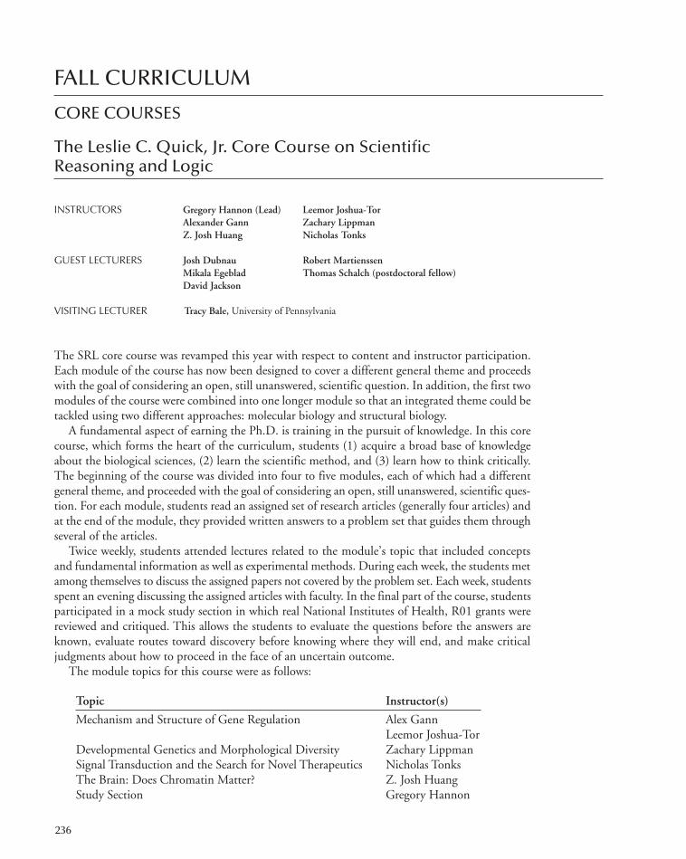
FALL CURRICULUM
CORE COURSES
The Leslie C. Quick, Jr. Core Course on Scientific Reasoning and Logic
INSTRUCTORS Gregory Hannon (Lead) Leemor Joshua-TorAlexander Gann Zachary LippmanZ. Josh Huang Nicholas Tonks
GUEST LECTURERS Josh Dubnau Robert MartienssenMikala Egeblad Thomas Schalch (postdoctoral fellow)David Jackson
VISITING LECTURER Tracy Bale, University of Pennsylvania
The SRL core course was revamped this year with respect to content and instructor participation.Each module of the course has now been designed to cover a different general theme and proceedswith the goal of considering an open, still unanswered, scientific question. In addition, the first twomodules of the course were combined into one longer module so that an integrated theme could betackled using two different approaches: molecular biology and structural biology.
A fundamental aspect of earning the Ph.D. is training in the pursuit of knowledge. In this corecourse, which forms the heart of the curriculum, students (1) acquire a broad base of knowledgeabout the biological sciences, (2) learn the scientific method, and (3) learn how to think critically.The beginning of the course was divided into four to five modules, each of which had a differentgeneral theme, and proceeded with the goal of considering an open, still unanswered, scientific ques-tion. For each module, students read an assigned set of research articles (generally four articles) andat the end of the module, they provided written answers to a problem set that guides them throughseveral of the articles.
Twice weekly, students attended lectures related to the module’s topic that included conceptsand fundamental information as well as experimental methods. During each week, the students metamong themselves to discuss the assigned papers not covered by the problem set. Each week, studentsspent an evening discussing the assigned articles with faculty. In the final part of the course, studentsparticipated in a mock study section in which real National Institutes of Health, R01 grants werereviewed and critiqued. This allows the students to evaluate the questions before the answers areknown, evaluate routes toward discovery before knowing where they will end, and make criticaljudgments about how to proceed in the face of an uncertain outcome.
The module topics for this course were as follows:
Topic Instructor(s)
Mechanism and Structure of Gene Regulation Alex GannLeemor Joshua-Tor
Developmental Genetics and Morphological Diversity Zachary LippmanSignal Transduction and the Search for Novel Therapeutics Nicholas TonksThe Brain: Does Chromatin Matter? Z. Josh HuangStudy Section Gregory Hannon
236
215-246_wsbs_Annual Report_2009 template 7/8/11 12:22 PM Page 236
Fall Curriculum 237
The Darrell Core Course on Scientific Exposition and Ethics
INSTRUCTORS David J. Stewart (Lead)Alea A. Mills Arne Stenlund
TEACHING ASSISTANT Jonathan Ipsaro
GUEST LECTURERS Walter GoldschmidtsJason WenJan A. Witkowski
VISITING LECTURERS Olga Akselrod, The Innocence ProjectKeith Baggerly, University of Texas, M.D. Anderson Cancer CenterRobert Charrow, Greenberg Traurig, LLCAvner Hershlag, North Shore University Hospital Amy Harmon, The New York TimesBoyana Konforti, Editor, Nature Structural & Molecular BiologyPaul Offit, Children’s Hospital of PhiladelphiaJoy Simha, Young Survivors Coalition, National Breast Cancer CoalitionLiz Wohl, National Breast Cancer Coalition
The Scientific Exposition and Ethics (SEE) core course in 2010 was taught by the same instructorteam as last year, with David Stewart taking the lead role, joined by Alea Mills and Arne Stenlund.This year, CSHL postdoctoral fellow Jonathan Ipsaro participated in the course as a teaching assis-tant. As usual, the course hosted expert guest lecturers who covered topics that included scientificpresentations, the editorial review process, ethical and legal responsibilities of scientists, DNA pro-filing, and postconviction appeals, patient advocacy, and reproductive genetics.
This core course offered instruction in the fundamental elements of scientific exposition—writingskills and public speaking—and ethics. The ability to communicate effectively and to appreciatethe intricacies of ethical issues are essential skills for biologists; both subjects were taught in a seriesof example-based lectures and discussion groups. Writing skills included the fundamentals of modernscientific English and the organization and preparation of papers, research abstracts, and grant ap-plications. Oral presentation skills were taught by instructors with different modes of presentation.Together with instructors, students critiqued formal seminar presentations at the Laboratory. In-struction and discussions about ethics included the ethical implications of biological discovery onsociety as well as the nature and boundaries of ethical behavior of scientists and their rights and re-sponsibilities. A primary objective of the course was that students consider exposition and ethicsintegral aspects of scientific research.
Research Topics
ORGANIZERS Kimberley Geer Alyson Kass-Eisler
This core course provided students with an in-depth introduction to the fields of research that theLaboratory scientists investigate. Students and faculty attended a weekly Research Topics seminar,at which faculty members and CSHL fellows presented their current research topics and methods
215-246_wsbs_Annual Report_2009 template 7/8/11 12:22 PM Page 237
of investigation each Wednesday evening over dinner. The students learned how to approach im-portant problems in biology. These seminars, together with the annual fall Laboratory In-HouseSymposium, provided students with a basis for selecting laboratories in which to do rotations. Theweekly speakers are listed below:
Florin Albeanu David Jackson Alea A. Mills Nicholas TonksGurinder (Mickey) Atwal Leemor Joshua-Tor Partha Mitra Lloyd TrotmanAnne Churchland Adam Kepecs Pavel Osten Glenn TurnerJosh Dubnau Alexei Koulakov Darryl Pappin Christopher VakocMikala Egeblad Adrian R. Krainer Michael Schatz Linda Van AelstGrigori Enikolopov Alexander Krasnitz Stephen Shea Doreen WareHiro Furukawa Bo Li Raffaella Sordella Michael WiglerThomas Gingeras Zachary Lippman David L. Spector Anthony ZadorChristopher Hammell Scott Lowe Arne Stenlund Hongwu ZhengGregory Hannon Robert Martienssen Bruce Stillman Yi ZhongZ. Josh Huang W. Richard McCombie Marja Timmermans
Specialized Disciplines Courses
A new Specialized Disciplines course in Quantitative Biology was developed by Mickey Atwal toprovide the students with the tools to interpret large, complex data sets and to use statistical method-ology in their research. It is one of two new courses in the areas of statistics, programming, bioin-formatics, and theoretical modeling and analysis that the School developed in 2010 to complementour existing curriculum. With the addition of this course, the students in the entering class of 2010took a total of four Specialized Disciplines courses this fall.
The other three Specialized Disciplines courses are Cellular Structure and Function, Genetics and Ge-nomics, and Systems Neuroscience. Cellular Structure and Function was again taught by lead instructorLinda van Aelst and Raffaella Sordella, but ran as a week-long intensive course when the students firstarrived. Josh Dubnau led Genetics and Genomics and was joined again by Thomas Gingeras. AnthonyZador led the Systems Neuroscience course, assisted by Adam Kepecs and Glenn Turner.
Cellular Structure and Function
INSTRUCTORS Linda Van Aelst (Lead)Raffaella Sordella
GUEST LECTURERS Darryl PappinDavid L. SpectorBruce Stillman
VISITING LECTURERS Gregg Gundersen, Columbia UniversityAron Jaffe, NovartisAlexey Khodjakov, Wadsworth LaboratorySandy Simon, The Rockefeller UniversityMarc Symons, The Feinstein Institute for Medical Research
With the complete genome sequence available for many organisms, there is now an increasing emphasison understanding the function of the gene products. This understanding requires an increasing ap-
238 Watson School of Biological Sciences
215-246_wsbs_Annual Report_2009 template 7/8/11 12:22 PM Page 238

preciation of the structure and function of the cell as well as dynamic associations within the cell. Thiscourse provided a basic overview of the structural and functional organization of cells with particularemphasis on cellular compartmentalization and communication. Topics of focus included the cy-toskeleton, cell adhesion and signaling, membrane transport, gene expression, and nuclear organization.In addition, the course provided insight into the basic toolbox of the cell biologist of the 21st century.
Genetics and Genomics
INSTRUCTORS Josh Dubnau (Lead)Thomas Gingeras
GUEST LECTURER Christopher Hammell
VISITING LECTURERS Hopi Hoekstra. Harvard UniversityTim Tully, Dart Neuroscience
This course placed modern human genetics and genomics into the context of classical organismalgenetics. History, perspective, and technique were described around four levels of analysis: naturallyoccurring variation, genome evolution, genetic screens, and gene epistasis. How do gene mutationshelp to define biological processes? How are more complex traits genetically dissected into simpler(underlying) components? What concepts and techniques are used to organize genes into pathwaysand networks? How are genes mapped, cloned, and engineered to identify functional domains ofproteins? What gene variation exists in natural populations? What are the functional consequencesof gene variation? How is it detected? How are genomes organized and coordinately regulated? Howcan genomic information be cataloged, organized, and mined? These questions and concepts werefleshed out using examples from the literature.
Quantitative Biology
INSTRUCTORS Mickey Atwal (Lead)Michael Schatz
GUEST INSTRUCTORS Justin Kinney (CSHL Fellow)Alexander Krasnitz
With the advent of high-throughput technologies in biology, it has become necessary for biologicalresearchers to be able to analyze and interpret high-dimensional data and frame hypotheses mathe-matically. To this end, the aim of this course was to equip the students with a basic training in modernstatistical methods. By the end of the course, the students were able not only to answer many of thesimple statistical questions that arise in data analyses, but also to became familiar with the more complextechniques used by fellow computational biologists. Topics included error fluctuations, calculating thesignificance of an experimental result, Bayesian inference, information theory, power calculations, di-mensional reduction, and DNA sequence analyses. In addition, this course introduced mathematicalmodeling, motivated by the classic examples in quantitative biology such as the Delbrück-Luria ex-periment, Hopfield's kinetic proofreading, and Kimura's neutral theory of population genetics.
Fall Curriculum 239
215-246_wsbs_Annual Report_2009 template 7/8/11 12:22 PM Page 239
Systems Neuroscience
INSTRUCTORS Anthony Zador (Lead)Adam KepecsGlenn Turner
GUEST INSTRUCTORS Florin AlbeanuAlexei KoulakovStephen Shea
This course provided an overview of key aspects neuroscience. The emphasis was on spanning levels:How can we go from molecules through cells and circuits to behavior?
There were three main components to the class: lectures, problem sets, and a final project. Thelast week of class was spent on the final project in which the students found and presented a neu-roscience paper which spanned levels, for example, from the molecular to the synaptic or the circuitlevel to the behavioral.
240 Watson School of Biological Sciences
215-246_wsbs_Annual Report_2009 template 7/8/11 12:22 PM Page 240

241
POSTDOCTORAL PROGRAM
PROGRAM DIRECTOR Nicholas Tonks
PROGRAM ADMINISTRATOR Alyson Kass-Eisler
Cold Spring Harbor Laboratory (CSHL) is proud of its rich tradition in postdoctoral educationand has a deep commitment to the postdoctoral experience. CSHL has long been recognized as aplace for nurturing young scientists, with postdoctoral researchers being an integral part of the dis-covery process. The Laboratory currently employs ~160 postdoctoral fellows working in the labs of47 principal investigators. Current fields of research expertise include genetics; molecular, cellular,and structural biology; neuroscience; cancer; plant biology; genomics; and quantitative biology. Thepostdoctoral community at CSHL is diverse and international, arriving at the Laboratory with manydifferent backgrounds and with needs that change during the course of the postdoctoral years. ThePostdoctoral Program Office works closely with the fellows and the Laboratory’s administration tocoordinate and organize educational and career development activities. Dr. Alyson Kass-Eisler, thePostdoctoral Program Officer, and Dr. Nicholas Tonks, the Scientific Director of the PostdoctoralProgram, provide the day-to-day support for the postdoctoral program, which is overseen by theDean of the Watson School, Dr. Leemor Joshua-Tor.
In 2010, we held a number of courses and workshops aimed at postdoctoral fellows. On Decem-ber 9, the Office of Sponsored Programs, the Development Department, and the Postdoc ProgramOffice held a very popular course on Grant Writing. The sessions included Introduction to Grantsand External Funding, Human Subjects/Tissue use in Research, The Grant Abstract—Do’s, Don’ts,Peer Review Panel Discussion, Effective Writing Skills, Fellowships, Career Development Awards,and Resources for Postdocs, and One-on-One Consultations.
CSHL has been a member of a special ambassador recruitment program from Merck ResearchLaboratories (MRL) since 2005. This program provides the postdoctoral fellows and students atCSHL an inside connection to a scientist working at MRL. As a result of this program, Dr. MaartenHoek, a postdoctoral fellow from Dr. Bruce Stillman’s laboratory, was hired as a Research Scientistin 2007 and Dr. Zuo Zhang, a postdoctoral fellow from Dr. Adrian Krainer’s laboratory, joinedMRL as a Research Scientist in January 2009. Drs. Li and Hoek visited the laboratory again on Jan-uary 15 for one-on-one recruitment session. In addition, Dr. Li gave a talk on Identification of Glu-cose-Dependant Insulin Secretion Targets in Pancreatic β Cells by Combining Defined-MechanismCompound Library Screening and siRNA Gene Silencing, to give the fellows a feel for the type ofresearch being done in industry.
Leadership Training was held April 7 for postdocs interested in planning for their next careerstep. Sessions included Mentoring Competencies for Success, Excellence Starts with RecruitingHigh-Level Performers, Selecting A-Players—Right People Doing Right Things, Staffing on ThreeInterfaces—Science, You, and the Chemistry with Your Staff, Behavior-Based Interviewing Guide-lines, Defining Your Team—Providing Direction, and Setting and Managing Expectations.
In the afternoon session on Individual Development Plans (IDP) for Postdoctoral Fellows, Dr.Ginsburg talked about creating an IDP and how the IDP can be a valuable tool for career develop-ment. Stemming from this session, a group of postdoctoral fellows started their own “IDP” groupto meet monthly to discuss their career goals and plans to achieve them. The second annual NationalPostdoc Appreciation Day (which this year was changed to National Postdoc Appreciation Week)was held during the week of September 20. To celebrate, CSHL hosted an outdoor pizza party and
215-246_wsbs_Annual Report_2009 template 7/8/11 12:22 PM Page 241

a job-searching roundtable discussion. It was a nice opportunity for the Lab to say thank you for allthat the postdocs do.
Five years ago, the School initiated a prize for the best posters by a postdoctoral fellow and thebest poster by a graduate student at the Laboratory’s annual In-House Symposium. Each "prize" isgiven in the form of an educational grant of $500. In addition to providing a forum for the post-doctoral fellows to show off their research, and potentially win a prize, it gives the entire scientificcommunity a chance to come together and share ideas beyond the wall of their individual labora-tories. It has been a great success for both the presenters and the laboratory community as a whole.Seventeen posters were presented for the postdoctoral prize, which was shared this year by FedorKarginov, a postdoctoral fellow in Greg Hannon’s laboratory, and Navasona Krishnan in Nick Tonk’slaboratory.
During the past couple of years, we have increasingly widened the role of postdoctoral fellows inthe curricular activities of the School. Our fellows have been involved in preparing our students forthe challenging Fall Term curriculum by working as one-on-one tutors during the summer. Through-out the Fall Term, postdoctoral fellows are used as tutors for all aspects of the curriculum, providingone-on-one tutoring, group tutoring, and participating in discussion sessions. In 2007, the ScientificExposition and Ethics course took further advantage of the expertise of our postdoctoral communityby hiring two fellows as teaching assistants (TAs). These two fellows were an integral part of thecourse by providing their expertise in discussions, editing of the students writing work, and critiquingoral presentations. This format was highly successful and was used again in Fall 2010. Jon Ipsaro, apostdoctoral fellow from Leemor Joshua-Tor’s laboratory, was this year’s TA.
Since 2003, all postdoctoral fellows and graduate students at the Laboratory have been enrolledin a special initiative of the New York Academy of Science (NYAS), called the Science Alliance. TheScience Alliance for graduate students and postdoctoral fellows is a consortium of universities, teach-ing hospitals, and independent research facilities in the New York City metropolitan area that haveformed a partnership with NYAS. The aim of the Alliance is to provide career and professional de-velopment monitoring for postdoctoral fellows and graduate students in science and engineering,through a series of live events and a dedicated web portal. In addition, the Science Alliance givesgraduate students and postdoctoral fellows the opportunity to network with their peers across in-stitutions and with key leaders in academia and industry. This year, the Science Alliance meetingsand workshops included The Art of Networking for Science Professionals, Evaluating and Negoti-ating Job Offers: Demystifying the Process, Going on the Academic Market: Strategies for Scientists,and Writing for Biomedical Publication. In addition, in June of this year, Alyson Kass-Eisler andDr. Keisha John attended a roundtable discussion with other local leaders in postdoctoral training.They took part in a Workshop on Improvisation for Creativity, Collaboration, and Communicationin the Workplace and participated in breakout sessions on Career Development Programming: Ad-dressing Issues and Sharing Ideas for Future Initiatives.
This year, the Laboratory applied to the National Institutes of Health for a postdoctoral trainingprogram in cancer biology. The primary goal of the Cancer Gene Discovery and Cancer BiologyPostdoctoral Training Program is to prepare the next generation of cancer biologists to pursue in-dependent research that integrates cancer gene discovery with functional biology in order to facilitatethe development of novel cancer therapeutic, diagnostic, and/or preventive strategies. We are verypleased to report that the application has been recommended for funding by the review panels.
Finally, a most important measure of our postdoctoral program’s success is the ability of post-doctoral fellows to secure positions at the end of their training. In 2010, the Laboratory’s departingpostdoctoral fellows went on to positions at Albert Einstein College of Medicine; Cold Spring Har-bor Laboratory; Donders Centre for Neurosciences, The Netherlands; North Shore University Hos-pital; Mt. Sinai School of Medicine, N.Y.; Murdoch Children's Research Institute, Melbourne,Australia; University of Buenos Aires, Argentina; University of Texas, Austin; U.S. Army Corps ofEngineers; Engineer Research and Development Center; and Weizmann Institute of Science, Israel.
242 Watson School of Biological Sciences
215-246_wsbs_Annual Report_2009 template 7/8/11 12:22 PM Page 242

UNDERGRADUATE RESEARCH PROGRAM
PROGRAM DIRECTOR Zachary Lippman
PROGRAM ADMINISTRATOR Dawn Pologruto
The fundamental objective of this program is to give students an opportunity to conduct first-rateresearch. Participants learn about scientific reasoning, laboratory methods, theoretical principles,and scientific communication. The specific objectives of the program are to (1) give college under-graduates a taste of conducting original research at the cutting edge of science, (2) encourage aware-ness of the physical and intellectual tools necessary for modern biological research, (3) foster anawareness of the major questions currently under investigation in the biomedical and life sciences,and (4) promote interactions with laboratory scientists through an immersion in the research envi-ronment.
During the 10-week program, URPs work with CSHL senior staff members on independent re-search projects, specifically in the areas of cancer biology, neuroscience, plant biology, cellular andmolecular biology, genetics, macromolecular structure, and bioinformatics.
URPs work, live, eat, and play among CSHL scientists. They have a very busy academic andsocial calendar for the summer. They attend lectures in the Goldberg Faculty Lecture Series fromCSHL faculty members and outside faculty members including, in 2010, talks by Dr. Stanley Maloy,San Diego State University, and Dr. Alfred Goldberg, Harvard University. URPs also attend a Bioin-formatics Workshop Series, where they learn how to identify patterns in DNA and protein sequencesand how to interpret them. Lectures by CSHL faculty are specifically designed for URPs. URPswere also invited to join Dr. and Mrs. Watson for a pizza party and Dr. and Mrs. Stillman for dinner.BBQ and pool parties, volleyball games, sailing lessons, designing the URP T-shirt, competing inthe annual CSHL Petri Dish Race and Scavenger Hunt, and the ever-famous URP versus PI volley-ball match rounded out the engaging program. 2010 was the second year the URPs beat the facultyteam in volleyball—quite an accomplishment!
At the beginning of the summer, URPs write an abstract and present a talk on their proposed re-search. Concluding the program in August, URPs prepare a final report and present their results ina 15-minute talk at the URP Symposium. The following students, selected from 720 applicants,took part in the 2010 program:
243
Robert Aboukhalil, McGill UniversityAdvisor: Dr. Mickey AtwalSponsor: Robert H.P Olney Fund FellowshipColocalization of tumor suppressor genes.
Emily Bottle, University of CambridgeAdvisor: Dr. Anthony ZadorSponsor: Former URP Fund FellowshipScreening the brain areas involved in sensorimotor association.
Alexandra Bryson, Texas A&M UniversityAdvisor: Dr. Bruce StillmanSponsor: Former URP Fund FellowshipExogenous expression of DDX5 RNA helicase.
Diana Cai, Columbia CollegeAdvisor: Dr. Yi ZhongSponsor: Former URP Fund FellowshipCombining two gene-targeting systems to investigate cross-talk
between the mushroom body and central complex inDrosophila.
Joseph Cammarata, Hunter CollegeAdvisor: Dr. Zach LippmanSponsor: Hunter College Scholar and National Science
Foundation ScholarSearching for a marker of meristem reiteration in Solanum
lycopersicum.
215-246_wsbs_Annual Report_2009 template 7/8/11 12:22 PM Page 243

244 Watson School of Biological Sciences
The URPs show off their class T-shirts.
Jonathan Coravos, Bowdoin CollegeAdvisor: Dr. Josh DubnauSponsor: National Science Foundation ScholarAre dopamine receptors expressed in Drosophila glia?
Helen Cha, Williams CollegeAdvisor: Dr. Marja TimmermansSponsor: National Science Foundation ScholarThe effect of a small RNA gradient on sharpening the adaxial-
abaxial boundary in Arabidopsis thaliana.
Tiffany Coupet, Johns Hopkins UniversityAdvisor: Dr. Scott PowersSponsor: William Townsend Porter Foundation FellowshipExploring synergistic interactions with RNAi in combination with
a PI3K inhibitor.
Martin Fan, Washington University, St. LouisAdvisor: Dr. Adrian R. KrainerSponsor: Former URP Fund FellowshipCharacterizing the tumorigenic potential of several splicing
factors.
Nisha Hariharan, University of California, BerkeleyAdvisor: Dr. David JacksonSponsor: Former URP Fund FellowshipCell-to-cell trafficking via plasmodesmata in Arabidopsis thaliana.
Ruilong Hu, Washington University, St. LouisAdvisor: Dr. Stephen SheaSponsor: URP Fund FellowshipThe mechanism of neural selectivity for pup isolation calls in the
mouse.
Edward Larkin, University of Notre DameAdvisor: Dr. Z. Josh HuangSponsor: University of Notre Dame URP Scholar The birth and development of cortical chandelier cells.
Diana LaScala-Gruenewald, Massachusetts Institute ofTechnology
Advisor: Dr. Michael Q. ZhangSponsor: 30th Anniversary URP ScholarAn interactive genomic map between budding yeast species for the
study of DNA replication,
Ashley Maceli, Suffolk UniversityAdvisor: Dr. Gregory HannonSponsor: James D. Watson FellowshipMammalian genomic simplification methods for studying DNA
methylation.
Connie Martin, University of California, RiversideAdvisor: Dr. Gregory HannonSponsor: Howard Hughes Medical Institute ScholarTransposon insertion profiling.
Meg McCue, Dartmouth CollegeAdvisor: Dr. Partha MitraSponsor: Former URP Fund FellowshipThe brain architecture project: Quantitative image analysis.
Matthias Minderer, University of CambridgeAdvisor: Dr. Lloyd TrotmanSponsor: Former URP Fund FellowshipThe nuclear import mechanism of PTEN.
215-246_wsbs_Annual Report_2009 template 7/8/11 12:22 PM Page 244

Luis Montano, Universidad Nacional Autonoma de MexicoAdvisor: Dr. W. Richard McCombieSponsor: Former URP Fund ScholarSolution-based exome capture: Is it useful to detect human
variation?
Claudio Morales-Perez, University of Puerto RicoAdvisor: Dr. Hiro FurukawaSponsor: National Science Foundation FellowshipUnderstanding the molecular mechanism of antagonism in the
NMDA receptor.
Angelina Regua, Molloy CollegeAdvisor: Dr. Leemor Joshua-TorSponsor: National Science Foundation FellowshipOrganization of ClrC (Clr4-Rik1-Cul4) complex.
Hanna Retallack, Harvard UniversityAdvisor: Dr. Adam KepecsSponsor: Former URP Fund FellowshipAcetylcholine and the basal forebrain in a sustained attention task.
Sarah Shareef, Harvard UniversityAdvisor: Dr. Christopher VakocSponsor: Dorcus Cummings ScholarCondensin localization along mitotic chromatin.
Ayse Trolander, Carleton CollegeAdvisor: Dr. Alea A. MillsSponsor: Former URP Fund Fellowshipp63 point mutation causing EEC syndrome alters gene expression
in vitro.
Anil Wadhwani, Northwestern UniversityAdvisor: Dr. Florin AlbeanuSponsor: Former URP Fund FellowshipNeuromodulation of olfactory sensory input: A photon counting
approach.
Unikora Yang, Brown UniversityAdvisor: Dr. Bo LiFellowship: Steamboat Foundation ScholarEstablishing a novel attentional behavior test for mouse models of
schizophrenia.
Undergraduate Research Program 245
215-246_wsbs_Annual Report_2009 template 7/8/11 12:22 PM Page 245

PARTNERS FOR THE FUTURE
PROGRAM DIRECTOR David Jackson
PROGRAM ADMINISTRATOR Kathy LaForgia
The Partners for the Future Program, established by Dr. James Watson in 1990, provides an oppor-tunity for talented Long Island high school students to have hands-on experience in biomedical re-search at Cold Spring Harbor Laboratory. This highly competitive program is open to Long Islandhigh school students in their junior year. Each high school science chairperson may nominate up tothree students. The top candidates are interviewed by CSHL scientists and up to 11 top studentsare chosen to participate in the program. Students selected to the program are paired with a scientistmentor and spend a minimum of 10 hours per week, September through March of their senioryear, conducting original research. At the conclusion, the students present their projects to an en-thusiastic audience of the students’ scientific mentors and colleagues, CSHL administrators, parents,and teachers. Although the students learn a great deal about modern biology and state-of-the-artresearch techniques, the main advantage of the program is that they are exposed to day-to-day lifein a lab. Interacting with scientists and support staff, the students learn to define and pursue a re-search goal while solving problems that may occur along the way.
The 2010–2011 Partners for the Future—chosen from among 24 semifinalist nominations—are listed below.
Partner High School CSHL Mentor Laboratory
Laura Bergsten Cold Spring Harbor High School Adam Naguib Lloyd TrotmanGloria DiMino Garden City High School Mikala Egeblad Mikala EgebladRigina Gallagher Sacred Heart Academy Doreen Ware Doreen WareMelissa Gluck Sanford H. Calhoun High School Hilary Cox Partha MitraDaniel Kim Syosset High School Michael Wittinger Alea MillsNeha Kinariwalla Sayville High School Fred Van Ex Robert MartienssenVictoria Lellis Harborfields High School Guy Horev Alea MillsKevin Liaw The Wheatly School Florin Albeanu Florin AlbeanuHeather Szilagyi Cold Spring Harbor High School Peter Bommert David JacksonJordan Woldenberg Friends Academy Sachin Ranade Adam KepecsDeanna Zhu Syosset High School Zachary Lippman Zachary Lippman
2010/2011 Partners for the Future participants
246
215-246_wsbs_Annual Report_2009 template 7/8/11 12:22 PM Page 246

MEETINGS AND COURSES
247-304_Meetings_Annual Report_2009 template 7/8/11 12:23 PM Page 247

ACADEMIC AFFAIRS
The meetings and courses program attracts scientists from all over the world to Cold Spring Har-bor Laboratory to communicate, learn, and critique the latest ideas and approaches in the biologicalsciences. The program consists of advanced laboratory and lecture courses, as well as large meetingsand biotechnology conferences that are held almost year-round. More than 8500 scientists rangingfrom graduate students and postdoctoral fellows to senior faculty came from around the world toattend these events at Cold Spring Harbor. A growing international program complements the mainprogram of meetings and courses.
In 2010, the 30 laboratory and lecture courses that were held at the Laboratory attracted morethan 1320 participants (including teaching faculty, students, and technicians). These courses covereda diverse array of topics in molecular biology, neurobiology, structural studies, and bioinformatics.The primary aim of the courses remains to teach students the latest advances that can be immediatelyapplied to their own research. Courses are always being evaluated and updated to include the latestconcepts and approaches. Increasingly, many courses feature a strong computational component asbiology grows ever more interdisciplinary, incorporating methodologies from computer science,physics, and mathematics.
Instructors, course assistants, and course lecturers come from universities, medical schools, re-search institutes, and companies around the world to teach at Cold Spring Harbor. Their excellenceand dedication make the course program work so well. We especially thank Drs. Ravi Allada, VickiAlan, Beverly Errede, Ke Hu, Raymond Keller, John Kirby, Kristen Kroll, Amy Lee, John Murray,Larysa Pevny, Susan Lovett, Anca Segall, Beth Stevens, William Tyler, and Mei Zhen, who all retiredthis year after many years of service.
Grants from a variety of sources support the courses. The core support provided through the re-cently renewed grant from the Howard Hughes Medical Institute remains critical to our course pro-gram. The courses are further supported by multiple awards from the National Institutes of Health
248
Grace Patio—Symposium 75
247-304_Meetings_Annual Report_2009 template 7/8/11 12:23 PM Page 248
Academic Affairs 249
(NIH) and the National Science Foundation (NSF), and additional support for individual coursesis provided from various foundations and other sources. The courses also depend on equipment andreagents that are loaned or donated by a large number of companies. These are invaluable for makingit possible to keep up with the latest technologies.
The Laboratory held 25 academic meetings this year, which brought together almost 7500 sci-entists from around the world to discuss their latest research. A major milestone for the meetingsprogram was the 75th anniversary of the annual Symposium. To do justice to this anniversary, aspecial one-day celebration on “75 Years in Science—Biology, Society, and the Future” was organizedby Jan Witkowski and held immediately prior to the Symposium. The celebration was also markedwith a commemorative booklet with essays by leading scientists who have participated in these Sym-posia. The Symposium proper this year focused on the nucleus, reflecting the growing convergencebetween cell biological and mechanistic studies that are providing a major new synthesis in our un-derstanding of how this command-and-control center of the cell operates. The Symposium attractedover 400 participants including notable nuclear biologists such as Günter Blobel, Joseph Gall, RobertGoldman, John Gurdon, Steven Henikoff, Robert Kingston, Barbara Meyer, Kim Nasmyth, DanielaRhodes, and Pam Silver.
Cold Spring Harbor Laboratory meetings are unique in assembling a program with very few in-vited speakers. Instead, meeting organizers are encouraged to select talks from abstracts that are sub-mitted on the basis of scientific merit, ensuring that the latest findings will be presented and thatyoung scientists will have the chance to describe their work. The annual meetings on Gene Expres-sion and Signaling in the Immune System, The Biology of Genomes, Retroviruses, and TranslationalControl were all oversubscribed, and many others attracted strong or record attendances. The Ice-landic volcano disrupted attendance at spring meetings, but thankfully, travel restrictions were soonlifted. A new biotechnology conference on RNA and Oligonucleotide Therapeutics was held inApril this year. Many of these meetings have become essential for those in the field and are held ona biannual basis. Partial support for individual meetings is provided by grants from NIH, NSF,foundations, and companies. Core support for the meetings program is provided by the Laboratory'sCorporate Sponsor Program.
The joint Cold Spring Harbor/Wellcome Trust Conference series held at the genome campussouth of Cambridge, England, included four meetings on Computational Cell Biology, Systems Bi-ology: Networks, Infectious Disease Genomics, and Global Health, and Genome Informatics. Initi-ated in 2001, more than 3500 scientists have attended meetings on topics as diverse as genomeinformatics, host–pathogen interactions, and prion biology. The Wellcome Trust Conference teamhave now developed the tools and skills to manage the academic conference program independently.
Finally, 2010 saw the continued development of the Cold Spring Harbor Asia program, as awholly owned affiliate of Cold Spring Harbor Laboratory with a mission to develop an educationalhub based in Suzhou, China. Two inaugural Symposia—the Watson Symposium on Cancer andthe Crick Symposium on Neuroscience—nine other scientific meetings, and one Summer Schoolwere held throughout the year.
The meetings and courses program staff is composed of a lean team of talented professionals,many who wear multiple “hats,” who handle the complexities of database design, programming,web and multimedia design, and educational grants management.
Terri GrodzickerDean
Academic Affairs
David StewartExecutive Director
Meetings and Courses Program
247-304_Meetings_Annual Report_2009 template 7/8/11 12:23 PM Page 249
75TH COLD SPRING HARBOR LABORATORY SYMPOSIUM ON QUANTITATIVE BIOLOGY
Dynamic Organization of Nuclear Function
June 2–7 408 Participants
ARRANGED BY Terri Grodzicker, David Spector, David Stewart, and Bruce Stillman,Cold Spring Harbor Laboratory
The decision to focus the historic 75th Symposium on the Dynamic Organization ofNuclear Function reflects the growing convergence between descriptive cell biologicalapproaches to the nucleus and mechanism-driven approaches to nuclear processes thatare now illuminating the structural and functional operations of the primary controlcenter of the cell. Many previous Symposia have been devoted to nuclear organizationand function, notably Genes and Chromosomes: Structure and Organization (1941);Genetic Mechanisms: Structure and Function (1956); Chromosome Structure andFunction (1973); Chromatin (1977); Structures of DNA (1982); DNA and Chro-mosomes (1993); and Epigenetics (2004). Every decade has seen important advancesin the field, so it is not surprising that past Symposia have shed light from many angleson the function of the central organelle of biology.
A major theme of this year’s Symposium addressed the emerging relationships be-tween chromosome structure, genome function, and gene expression. Techniques havenow emerged that allow the monitoring of chromatin states in cell lines and in vivo using moleculartags and sophisticated microscopy during differentiation. New methods to pinpoint the molecularmechanisms and intranuclear organization underlying transcriptionally active versus inactive chro-matin are revealing extraordinary organizational complexity within the nucleus.
Epigenetic mechanisms such as DNA methylation and histone posttranslational modificationssuch as acetylation and methylation, imprinting, RNA interference, and gene silencing are also sub-ject to dynamic organization in the nucleus. Epigenetic changes are important contributors to thepathogenesis of disease in humans, animals, and plants, and they may lie at the heartof many important gene–environment interactions. Research is illuminating how epi-genetic mechanisms play important roles in regulating the structure and function ofthe genome, and the 2010 Symposium particularly highlighted the relationship be-tween the genome, the epigenome, and the nuclear architecture and environment.
The role of signaling in regulating transcription, mRNA export, and chromatinstructure was also examined. For example, signaling from or at the nuclear envelopereveals novel mechanisms for influencing nuclear envelope morphology, breakdown,and cell differentiation. Chromatin dynamics was another major theme, both of thechromatin fiber and of chromatin-binding proteins involved in replication and repair,with new results discussed in areas such as the programming of DNA replication ori-gins and their specificity. The latest developments in our understanding of mitoticand meiotic chromosome dynamics, as well as the roles for nuclear transport factorsin cell cycle progression, were also covered.
Tremendous progress is also being made in analyzing the mechanisms of assembly for nuclearbodies and their functional roles. Regulated transport into and out of the nucleus through the nu-
250
B. Stillman
T. Grodzicker
247-304_Meetings_Annual Report_2009 template 7/8/11 12:23 PM Page 250

75th Cold Spring Harbor Laboratory Symposium on Quantitative Biology 251
clear pore complex and the operation of selective transport mechanisms also featured prominently,leading to vigorous discussion of the competing models for facilitated transport, as well as reportsof new factors required for mRNA export, tRNA shuttling, and pore complex assembly. There isgrowing evidence that pore permeability and nuclear leakiness change in a regulated way with celldifferentiation and age.
The meeting addressed how nuclear organization and function are linked to the underpinningsof several pathophysiological states including the laminopathies such as A-type lamin dysfunctionin the premature aging disease Progeria and the molecular determinants of cancer-prone symptomsof xeroderma pigmentosum and Cockayne syndrome, of the fatal bone marrow failure syndromedyskeratosis congentia, and of the Roberts and Cornelia de Lange developmental syndromes. Therelationship between nuclear architecture, aging, and disease was the subject of a masterful DorcasCummings Lecture on “The Unexpected Link between Premature Aging and Nuclear Organization”by Robert Goldman to a combined audience of scientists and lay friends and neighbors.
In arranging this Symposium, the organizers were dependent on the guidance of a broad cadreof advisors including Drs. Robert Goldman, Robert Kingston, Kim Nasmyth, Daniela Rhodes, andPam Silver. Opening night speakers included John Gurdon, Rob Martienssen, Barbara Meyer, andGünter Blobel. Joseph Gall presented the Reginald Harris Lecture on “Transcription, RNA Pro-cessing, and Nuclear Bodies.” Steven Henikoff closed the meeting with a masterful and thought-provoking summary.
This Symposium was attended by more than 400 scientists from over 25 countries, and theprogram included 68 invited presentations and 219 poster presentations. To disseminate the latestresults and discussion of the Symposium to a wider audience, attendees were able to share manyof the Symposium talks with their colleagues who were unable to attend using the Leading Strandvideoarchive, and a series of interviews with leading experts in the field was arranged during theSymposium and distributed as free video from the Cold Spring Harbor Symposium Chats web-site.
Funds to support this meeting were obtained from the National Institute of General MedicalSciences, a branch of the National Institutes of Health. Financial support from the corporate spon-sors of our meetings program is essential for these Symposia to remain a success and we are mostgrateful for their continued support.
M. Dundr, I. Raska, W. Bickmore, D. Spector
247-304_Meetings_Annual Report_2009 template 7/8/11 12:23 PM Page 251
PROGRAM
IntroductionB. Stillman, Cold Spring Harbor Laboratory
Chromatin Modifications and Gene ExpressionChairperson: W. Bickmore, MRC Human Genetics Unit,Edinburgh, United Kingdom
DNA Replication and Genome IntegrityK. Nasmyth, University of Oxford, United Kingdom
Reprogramming and DifferentiationChairperson: T. de Lange, The Rockefeller University, New York
Reginald B. Harris Lecture: Transcription, RNA Processing,and Nuclear Bodies
J.G. Gall, Carnegie Institution for Science, Baltimore, Maryland
RNA BiologyChairperson: D. Spector, Cold Spring Harbor Laboratory
Chromosome Structure and MitosisChairperson: A. Amon, Massachusetts Institute of Technology,Cambridge
Heterochromatin Formation and Gene SilencingChairperson: G. Almouzni, CNRS UMR218, Institut Curie,Paris, France
S. Henikoff
Dorcas Cummings Lecture: The Unexpected Link betweenPremature Aging and Nuclear Organization
R. Goldman, Northwestern University Medical School, Illinois
Transcription Meets RNA ProcessingChairperson: J. Steitz, HHMI/Yale University, New Haven,Connecticut
Nuclear PeripheryChairperson: W. Earnshaw, University of Edinburgh, UnitedKingdom
Chromatin Structure and OrganizationChairperson: S. Gasser, Friedrich Miescher Institute forBiomedical Research, Basel, Switzerland
Genome RegulationChairperson: E. Heard, CNRS UMR128, Institut Curie, Paris,France
SummaryChairperson: Steven Henikoff, Fred Hutchinson Cancer ResearchCenter, Washington
T. Khoduchenko, J. GallR. Tjian, C. Venter
252 Meetings
247-304_Meetings_Annual Report_2009 template 7/8/11 12:23 PM Page 252

253
MEETINGS
Neuronal Circuits
March 10–13 189 Participants
ARRANGED BY Cori Bargmann, The Rockefeller UniversityEd Callaway, Salk Institute for Biological SciencesDmitri Chklovskii, Janelia Farm Research Campus
In the quest to understand the brain, neuronal circuits represent a central level of description. Es-tablishing connectivity in neuronal circuits seems as essential for solving the brain as having a geo-graphic map for planning one’s travel. Although such realization motivated Cajal to describe avariety of neuronal circuits using Golgi stains, his contributions were technologically limited. Withthe recent appearance of novel molecular genetic, imaging, and computational techniques, a com-prehensive description of the wiring diagram—an old dream of neuroscientists—is about to becomea reality.
Because technological advances have been made in different organisms and systems, we wantedto create a forum that brings together researchers working on different topics, yet focus on neuronalcircuits. This meeting included six broad slide sessions covering the olfactory system, the visual sys-tem, methods, behavior learning and memory, motor systems, and the cortex, as well as a very in-teractive poster session.
For the third meeting of this kind, the response of the field wasvery enthusiastic. The meeting brought together participants fromall over the world, most of whom made either oral or poster pre-sentations. Invited talks were given by Larry Abbott (ColumbiaUniversity), Richard Axel (Columbia University), Axel Borst (Max-Planck Institute of Neurobiology), Sreekanth Chalasani (Salk In-stitute), Karl Deisseroth (Stanford University), Marla Feller(University of California, Berkeley), Joseph Fetcho (Cornell Uni-versity), Charles Gilbert (The Rockefeller University), Shaul Hes-trin (Stanford University School of Medicine), Shawn Lockery(University of Oregon), Liqun Luo (Stanford University), JeffreyMagee (LSUHSC), Ian Meinertzhagen (Janiela Farm ResearchCampus of HHMI), Markus Meister (Harvard University), May-Britt Moster (CBM), Michael Nusbaum (University of Pennsylvania), I. Sebastian Seung (MIT),and Rachel Wilson (Harvard Medical School). The meeting provided an important clearinghousefor ideas and approaches and helped scientists in the field obtain the most up-to-date information,enabling them to meet, to network, and to establish collaborations. Based on the uniformly enthu-siastic comments of the participants, the intensity of the oral and poster sessions, and the largecrowds that stayed up late every night at the bar to discuss science, the meeting was a great success.
A highlight of the meeting program was the inaugural Larry Katz Memorial Lecture, made pos-sible by the generous donation of many colleagues in the field. Excellent nominations were received.A committee composed of the three organizers and Liqun Luo selected one speaker: ChristopherHarvey, Princeton University. He gave a wonderful talk about his graduate and postdoctoral work.
K. Briggman, S. Lockery
247-304_Meetings_Annual Report_2009 template 7/8/11 12:23 PM Page 253

PROGRAM
Circuits and BehaviorChairperson: L. Luo, Stanford University, California
Connectomics/VisionChairperson: D. Chklovskii, HHMI Janiela Farm ResearchCampus, Ashburn, Virginia
Cortex and Other CircuitsChairperson: E.M. Callaway, Salk Institute for Biological Studies,La Jolla, California
Methods/ChemosensationChairperson: S. Lockery, University of Oregon, Eugene
E. Callaway, D. Chklovskii, C. Bargmann
K. Kolkman, C. Root
254 Meetings
Q. Wen, I. Rabinowich
Chemosensation/MotorChairperson: C. Bargmann, The Rockefeller University, NewYork
Neural ComputationsChairperson: M.-B. Moser, Norwegian University of Science andTechnology, Trondheim, Norway
Larry Katz Memorial LectureC.D. Harvey, Princeton University, New Jersey
247-304_Meetings_Annual Report_2009 template 7/8/11 12:23 PM Page 254

PTEN Pathways and Targets
March 16–20 129 Participants
ARRANGED BY Suzanne Baker, St. Jude Children’s Research HospitalLewis Cantley, Beth Israel Deaconess Medical CenterPier Paolo Pandolfi, Harvard Medical SchoolRamon Parsons, Columbia University
This third meeting on PTEN pathways was enormously successful. The meeting brought together topscientists studying the PTEN/PI-3 kinase pathway using biochemical and molecular approaches aswell as in vivo model systems. An exciting series of talks and posterpresentations addressed the complex regulation of this signal trans-duction cascade and its critical role in normal development, cancer,aging and metabolism, autism, and other pathological disease states.The conference provided an opportunity for researchers of all levelsto get together to discuss their latest research findings and technicalapproaches toward the analysis of the PTEN pathway. Particularemphasis was placed on pathway regulation and cross-talk, humancancer pathogenesis, disease models, and therapy.
A total of 76 investigators presented in the nine scientific ses-sions, with 41 platform sessions and 35 poster presentations and129 registered attendees. The seven platform and two poster ses-sions were marked by dynamic and enthusiastic exchanges of new results. In the scientific sessions,many audience members participated in the question and answer sessions, and the poster sessionswere well attended.
Essential funding for the meeting was provided by the National Cancer Institute, a branch ofthe National Institutes of Health.
PROGRAM
255
Upstream of PTENChairperson: R. Parsons, Columbia University, New York
Downstream from PTENChairperson: L. Trotman, Cold Spring Harbor Laboratory
PTEN RegulationChairperson: P. Devreotes, Johns Hopkins University School ofMedicine, Baltimore, Maryland
Development and Disease ModelsChairperson: H. Wu, University of California, Los Angeles
D. Guerin, B. Manning, R. Shaw
mTORC1 and Intersecting PathwaysChairperson: B. Manning, Harvard School of Public Health,Boston, Massachusetts
Therapy and Cancer ModelsChairperson: J. Engelman, Massachusetts General Hospital, Boston
Aging, Metabolism, and CancerChairperson: A. Brunet, Stanford University, California
247-304_Meetings_Annual Report_2009 template 7/8/11 12:23 PM Page 255
Systems Biology: Global Regulation of Gene Expression
March 23–27 284 Participants
ARRANGED BY Brad Bernstein, Massachusetts General Hospital, Harvard Medical School, Broad InstituteMartha Bulyk, Brigham & Women’s Hospital, Harvard Medical SchoolHarmen Bussemaker, Columbia UniversityMarian Walhout, University of Massachusetts Medical School
Systems biology aims to understand the emergent properties of molecular networksin cells by using systematic and global approaches. One of the most actively re-searched areas of systems biology in recent years has been global regulation of geneexpression, which coordinates complex metabolic and developmental programs inorganisms. This seventh conference in the series, like the ones in previous years,captured the continuing rapid progress and many new discoveries in this youngand exciting field.
A total of 284 scientists attended the 4-day meeting, which featured 46 talksand 109 poster presentations covering a broad range of topics. Two keynotespeeches were exciting highlights of the meeting. The first was presented by Dr.Kevin Struhl, who has been a leader in the area of transcriptional regulation formany years. Dr. Struhl presented a detailed story on the transcriptional and epige-netic changes that accompany cellular transformation in a model of breast cancer.The second keynote was presented by Dr. Steve Henikoff, a leader in the field ofepigenetics with a long history of creatively integrating systems approaches intohis research. Dr. Henikoff presented a global study of the role of histone variantsand histone exchange in the regulation of the Drosophila genome.
Compared to previous conferences, five aspects featured prominently in thisyear’s meeting: (1) Next-generation sequencing technologies—representing in-creasingly standard means for data acquisition—are now being applied to studytranscription factor localization, nucleosome positioning, chromatin modifica-tions, and higher-order genome organization; the large amounts of new data havestimulated the generation of a plethora of new genomic methods, computationalalgorithms, resources, and biological insights; (2) new imaging technologies areallowing researchers to examine the dynamics of gene expression in vivo and inreal time, and in some cases, at a single-molecule level; (3) new resources on thebinding specificities of transcription factors and RNA-binding proteins continue to be created, pro-viding opportunities for systematic analysis of gene regulatory networks in Drosophila and mammals;(4) there is much excitement around the newly appreciated and emerging roles for different classesof noncoding RNAs in genome regulation; and (5) new computational algorithms are being adoptedto analyze the gene regulatory networks. Generally, the speakers in these sessions were young, withmost being in the early stage of their careers. Finally, to help computational biologists and experimentscientists better communicate with each other, this year’s meeting also included a premeeting work-shop that was focused on introductory experimental and computational topics.
This meeting was funded in part by the National Cancer Institute, a branch of the National In-stitutes of Health, and the National Science Foundation.
256
M. Bulyk
H. Bussemaker
247-304_Meetings_Annual Report_2009 template 7/8/11 12:23 PM Page 256

PROGRAM
Keynote Address: Mapping Genome-Wide Nucleosome Dynamics
S. Henikoff, Fred Hutchinson Cancer Research Center, Seattle,Washington
Keynote Address: An Epigenetic Switch That Links Inflamma-tion to Cancer—Regulatory Circuits and Cancer Treatment
K. Struhl, Harvard Medical School, Boston, Massachusetts
TranscriptionChairperson: T. Hughes, University of Toronto, Canada
Transcriptional Regulatory NetworksChairperson: X.S. Liu, Dana-Farber Cancer Institute, Boston,Massachusetts
ChromatinChairperson: R. Morse, Wadsworth Center, Albany, New York
Systems Biology: Global Regulation of Gene Expression 257
EpigeneticsChairperson: J. Stamatoyannopoulos, University of Washington,Seattle
cis-Regulatory LogicChairperson: L. Mirny, Massachusetts Institute of Technology,Cambridge
Posttranscriptional RegulationChairperson: M. Walhout, University of Massachusetts MedicalSchool, Worcester
Emerging TechnologyChairperson: B. Andrews, University of Toronto, Canada
Variation and EvolutionChairperson: M. Bulyk, Brigham & Women’s Hospital, HarvardMedical School, Boston, Massachusetts
R. Hardison, G. Bernardi L. Albert A. Gerber, J. Lieb
247-304_Meetings_Annual Report_2009 template 7/8/11 12:23 PM Page 257

RNA and Oligonucleotide Therapeutics
April 7–10 90 Participants
ARRANGED BY Ryszard Kole, AVI BioPharmaAdrian Krainer, Cold Spring Harbor LaboratoryBruce Sullenger, Duke University
This first conference on RNA and oligonucleotide therapeutics was focused on clinical trials and thedevelopment of oligonucleotides as drugs, but it brought together top scientists from academia andindustry interested in a variety of RNA-based therapeutics, not only oligonucleotides. The theme“from the bench to the bedside” covered approaches ranging from an-tisense oligonucleotides and aptamers through modulation of splicing,targeting expanded triplet repeats, and immunostimulation to smallinterfering RNAs (siRNAs) and microRNAs (miRNAs) as potentialdrugs and targets. The all-important issue of RNA therapeutics de-livery by a variety of methods was also covered. The applications ofthese varied technologies included rare diseases, cancer, cardiovasculardiseases, and bacterial and viral infections.
The participants came from 11 companies and from universitiesand research institutions from the United States and abroad. Theseven scientific sessions featured 37 platform talks and 20 postersand included 90 registered attendees. Animated and insightful ex-changes during the sessions continued throughout. A number ofparticipants expressed interest in attending this conference next year.The meeting opened with a keynote by Nobel laureate Dr. Sydney Altman and the sessions addressedmodulation of RNA splicing, antisense oligonucleotides, posttranscriptional control of gene expres-sion, siRNA, miRNA and aptamers as therapeutics, delivery of RNA therapeutics, and im-munomodulating oligonucleotides.
The topic of the meeting attracted funding from several companies including major fundingfrom AVI Biopharma.
258
A. Krainer, R. Kole, B. Sullenger
Oligonucleotide TherapyChairperson: A. Krainer, Cold Spring Harbor Laboratory
Delivery of RNA TherapeuticsChairperson: S. Dowdy, HHMI/University of California, SanDiego
Oligonucleotide-Induced Immunostimulation and OtherMechanisms
Chairperson: S. Agrawal, IDERA Pharmaceuticals, Cambridge,Massachusetts
PROGRAMKeynote Address: RNA and Therapeutic ModesS. Altman, Yale University, Connecticut
Exon Skipping TherapyChairperson: R. Kole, AVI BioPharma, Bothell, Washington
siRNA, miRNA, and Aptamers as TherapeuticsChairperson: B. Sullenger, Duke University, Durham, North Carolina
Posttranscriptional Control of Gene ExpressionChairperson: C. Thornton, University of Rochester, New York
247-304_Meetings_Annual Report_2009 template 7/8/11 12:23 PM Page 258

Gene Expression and Signaling in the Immune System
April 21–25 327 Participants
ARRANGED BY Doreen Cantrell, University of Dundee, United KingdomSankar Ghosh, Columbia University College of Physicians & SurgeonsDan Littman, HHMI/New York University School of MedicineMark Schlissel, University of California, Berkeley
This meeting was held for the fifth time in 2010, taking place in the context of global travel restric-tions due to the Icelandic volcano eruption. Although ~380 scientists registered for the meeting,53 had to cancel due to flight cancellations in Europe. Nonetheless, ~25% of attendees were fromoutside of the United States, 41% were women, and 53% were either graduate students or postdocs.Although there are many immunology meetings, the unique aspect of this Cold Spring Harbormeeting is its focus on molecular and biochemical aspects of the development andfunction of the immune system. In addition, this meeting attracts a broad range ofscientists who utilize the immune system as a model to ferret out basic principles ofbiological regulation.
Talks were presented by a mix of invited speakers and investigators selected fromamong a group of more than 200 submitted abstracts. Due to last-minute cancella-tions, several investigators were asked to substitute for scheduled speakers and theywelcomed the opportunity. A significant number of women and junior investigatorswere asked to give talks. Most speakers focused almost exclusively on their unpublishedwork. A major new approach to the study of gene regulation permeated several talks—ChIP-Seq analysis of specific chromosomal protein DNA interactions. Meinrad Bus-slinger spoke about Pax5 and CTCF-binding sites in the IgHC locus that helped himdefine an important new potential regulatory element in the V region of that complexlocus. David Schatz presented exciting new data on the genome wide binding of V(D)J recombinasecomponents to specifically modified histones as well as RSS elements. Cornelius (Kees) Murre (Uni-versity of California, San Diego) presented an update to his three-dimensional model of interphasestructure of a complex genetic locus within nuclear space. The product of a collaboration withphysicists and mathematicians, this model based on fluorescent in situ hybridization data helps ex-plain how more than 100 immunoglobulin V
H gene segments distributed over greater than amegabase of DNA can recombine with DH segments at roughly similar frequencies. Young investi-gator Greg Barton described the surprising discovery of how an enteric pathogen uses receptorsfrom the innate immune system to trigger the expression of its own pathogenicity genes. Finally, inone of the last talks of the meeting, coorganizer Dan Littman revealed new details of how HIV in-teracts with but does not infect dendritic cells.
These talks highlighted the relevance of the data discussed at this meeting, not just to immunol-ogy but to the broader scientific community. Oral presentations were supplemented by two after-noon poster sessions that were extremely well-attended and provided a forum for all meetingparticipants (including many graduate students and postdocs) to share their most recent data. Inaddition, attendees took advantage of walking tours of the CSHL campus, a bountiful lobster ban-quet, and an evening performance by the Yale-based music group “The Cellmates,” with dancingextending into the early morning hours.
Continuing support was obtained from the NIH/NIAID as well as from six corporate sponsors.
M. Schlissel
259
247-304_Meetings_Annual Report_2009 template 7/8/11 12:23 PM Page 259

PROGRAM
Stem Cells and Early Developmental DecisionsChairperson: S. Smale, University of California School ofMedicine, Los Angeles
Regulation of Immune Cell DevelopmentChairperson: M. Schlissel, University of California, Berkeley
Chromatin Structure and EpigeneticsChairperson: H. Singh, HHMI/University of Chicago
Antigen Receptor Gene Assembly and ModificationChairperson: F. Alt, HHMI/Children’s Hospital, Boston,Massachusetts
SignalingChairperson: A. Weiss, HHMI/University of California, SanFrancisco
Regulation of Immune Cell Function IChairperson: D. Littman, HHMI/New York University School ofMedicine
Regulation of Immune Cell Function IIChairperson: D. Cantrell, University of Dundee, UnitedKingdom
Innate ImmunityChairperson: S. Ghosh, Columbia University College ofPhysicians & Surgeons, New York
E. Chan, M. Krangel, H. Singh
G. Amarante-Mendes, T. Mak
K. Murphy, T. Zhang
S. Chatterjee, S. Volpi
260 Meetings
247-304_Meetings_Annual Report_2009 template 7/8/11 12:23 PM Page 260
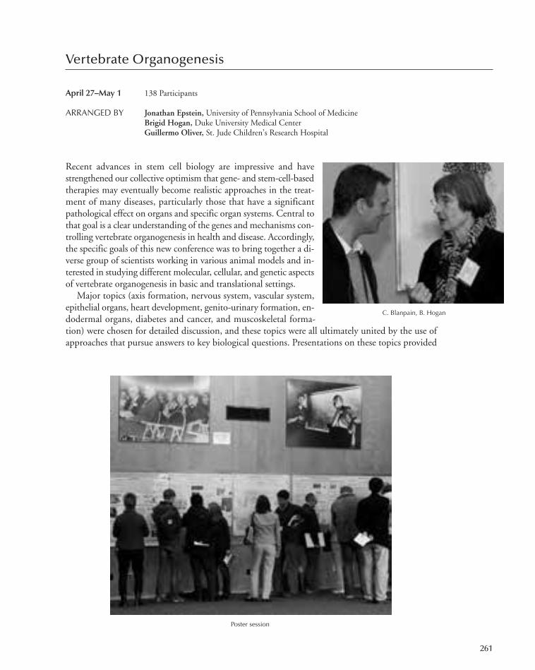
Vertebrate Organogenesis
April 27–May 1 138 Participants
ARRANGED BY Jonathan Epstein, University of Pennsylvania School of MedicineBrigid Hogan, Duke University Medical CenterGuillermo Oliver, St. Jude Children’s Research Hospital
Recent advances in stem cell biology are impressive and havestrengthened our collective optimism that gene- and stem-cell-basedtherapies may eventually become realistic approaches in the treat-ment of many diseases, particularly those that have a significantpathological effect on organs and specific organ systems. Central tothat goal is a clear understanding of the genes and mechanisms con-trolling vertebrate organogenesis in health and disease. Accordingly,the specific goals of this new conference was to bring together a di-verse group of scientists working in various animal models and in-terested in studying different molecular, cellular, and genetic aspectsof vertebrate organogenesis in basic and translational settings.
Major topics (axis formation, nervous system, vascular system,epithelial organs, heart development, genito-urinary formation, en-dodermal organs, diabetes and cancer, and muscoskeletal forma-tion) were chosen for detailed discussion, and these topics were all ultimately united by the use ofapproaches that pursue answers to key biological questions. Presentations on these topics provided
C. Blanpain, B. Hogan
Poster session
261
247-304_Meetings_Annual Report_2009 template 7/8/11 12:23 PM Page 261
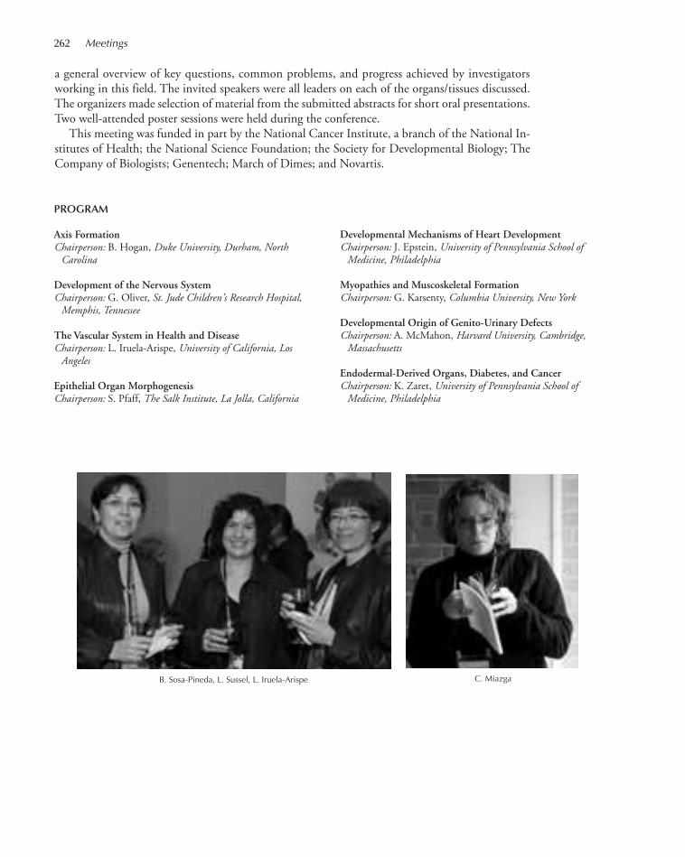
a general overview of key questions, common problems, and progress achieved by investigatorsworking in this field. The invited speakers were all leaders on each of the organs/tissues discussed.The organizers made selection of material from the submitted abstracts for short oral presentations.Two well-attended poster sessions were held during the conference.
This meeting was funded in part by the National Cancer Institute, a branch of the National In-stitutes of Health; the National Science Foundation; the Society for Developmental Biology; TheCompany of Biologists; Genentech; March of Dimes; and Novartis.
B. Sosa-Pineda, L. Sussel, L. Iruela-Arispe
PROGRAM
Axis FormationChairperson: B. Hogan, Duke University, Durham, NorthCarolina
Development of the Nervous SystemChairperson: G. Oliver, St. Jude Children’s Research Hospital,Memphis, Tennessee
The Vascular System in Health and DiseaseChairperson: L. Iruela-Arispe, University of California, LosAngeles
Epithelial Organ MorphogenesisChairperson: S. Pfaff, The Salk Institute, La Jolla, California
C. Miazga
262 Meetings
Developmental Mechanisms of Heart DevelopmentChairperson: J. Epstein, University of Pennsylvania School ofMedicine, Philadelphia
Myopathies and Muscoskeletal FormationChairperson: G. Karsenty, Columbia University, New York
Developmental Origin of Genito-Urinary DefectsChairperson: A. McMahon, Harvard University, Cambridge,Massachusetts
Endodermal-Derived Organs, Diabetes, and CancerChairperson: K. Zaret, University of Pennsylvania School ofMedicine, Philadelphia
247-304_Meetings_Annual Report_2009 template 7/8/11 12:23 PM Page 262

Molecular Chaperones and Heat Shock Response
May 4–8 240 Participants
ARRANGED BY F. Ulrich Hartl,Max-Planck Institute for BiochemistryDavid Ron, New York University School of MedicineJonathan Weisssman, HHMI/University of California, San Francisco
This meeting continued the rich tradition of a forum where cutting-edge analysis of molecularmechanisms of protein folding is presented in the context of its biological importance and healthrelevance. The high attendance, large number of posters (94), and lively audience participation inthe sessions and in the informal activities that followed are testament to the success of the meetingand to the successful balance of the sessions. The field ofchaperone research is evolving and this meeting is surelyevolving with it.
In addition to well-established biochemical and biophys-ical methods for studying chaperone function, this meetingfeatured presentations using single-molecule methods and ad-vanced computational approaches to the study of signalingnetworks involved in control of chaperone gene expression.This year’s meeting also featured two new sessions: one onEvolution and Regulation of Protein Folding Machines andthe other on Manipulating Chaperone Networks and ProteinFolding. The former provided a forum for work on the sys-tems biology aspect of protein folding and the latter for workof special relevance to the prospects of translating the basicscience to applications in health and biotechnology.
Among the cutting-edge themes explored were presentations of genetic interaction networks inmammalian systems. Talks covered protein trafficking in the cell that emphasized the fluidity of sub-cellular compartmentalization, intracellular protein turnover, connections between ribosomes andchaperones, and novel signaling pathways involved in protein-folding homeostasis.
This meeting was funded in part by the National Institute on Aging; the National Heart, Lung,and Blood Institute; the National Institute of General Medical Sciences; branches of the NationalInstitutes of Health; and Enzo Life Sciences, Inc.
263
D. Kaganovich, B. Bukau
J. Bonner, D. Czyz, M. Hayer-Hartl
247-304_Meetings_Annual Report_2009 template 7/8/11 12:23 PM Page 263
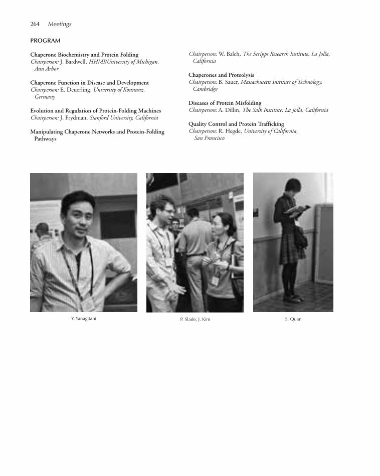
PROGRAM
Chaperone Biochemistry and Protein FoldingChairperson: J. Bardwell, HHMI/University of Michigan, Ann Arbor
Chaperone Function in Disease and DevelopmentChairperson: E. Deuerling, University of Konstanz, Germany
Evolution and Regulation of Protein-Folding MachinesChairperson: J. Frydman, Stanford University, California
Manipulating Chaperone Networks and Protein-FoldingPathways
264 Meetings
Y. Yanagitani P. Slade, J. Kim S. Quan
Chairperson: W. Balch, The Scripps Research Institute, La Jolla,California
Chaperones and ProteolysisChairperson: B. Sauer, Massachusetts Institute of Technology,Cambridge
Diseases of Protein MisfoldingChairperson: A. Dillin, The Salk Institute, La Jolla, California
Quality Control and Protein TraffickingChairperson: R. Hegde, University of California,San Francisco
247-304_Meetings_Annual Report_2009 template 7/8/11 12:23 PM Page 264

The Biology of Genomes
May 11–15 548 Participants
ARRANGED BY Susan Celniker, Lawrence Berkeley National LaboratoryAndrew Clark, Cornell UniversityChris Ponting, UK Medical Research Council, University of OxfordGeorge Weinstock,Washington University
This annual meeting marked the 23rd annual gathering of genome scientists at Cold Spring HarborLaboratory. The meeting built upon the remarkable progress in the mapping, sequencing, annota-tion, and analysis of genomes from many human individuals, “modelorganisms,” other animals, and prokaryotes. New DNA-sequencingtechnologies have caused a steep rise in the quantity of sequences beinggenerated, and the meeting reported many breakthroughs in our un-derstanding of sequence variants that contribute to disease, includingcancer and complex disease, and to phenotypic traits. The new data arealso providing important insights into population structure for humansand other species. In addition to those on genome sequence, there werea number of reports of transcriptome and epigenomic profiling. Inves-tigators from around the world attended the meeting, with more than350 abstracts presented, describing a broad array of topics relating tothe production and analysis of genomes from diverse organisms.
There were numerous reports on progress in using the new sequenc-ing technologies, with mounting expectation for further advances as these continue to be appliedwidely. The keynote presentations were delivered by Cori Bargmann and Martin Blaser.
The ELSI (Ethical, Legal, and Social Implications) panel was chaired by Susan Wolf and the dis-cussion topic was Returning Research Results to Participants in Large-Scale Genomics Studies.
Major sponsorship for this meeting was provided by Roche. Additional funding was providedby Illumina, Inc. and the National Human Genome Research Institute, a branch of the NationalInstitutes of Health.
265
G. Weinstock, J. Watson
PROGRAM
Functional and Cancer GenomicsChairpersons: L. Ding, Washington University School ofMedicine, St. Louis, Missouri; P. Farnham, University ofCalifornia, Davis
Genetics of Complex TraitsChairpersons: D. MacArthur, Wellcome Trust Sanger Institute,Hinxton, United Kingdom; C. Ober, University of Chicago,Illinois
High-Throughput Genomics and GeneticsChairpersons: B. Graveley, University of Connecticut HealthCenter, Farmington; J. Shendure, University of Washington,Seattle
Computational GenomicsChairpersons: L. Duret, CNRS, Université Lyon 1, Villeurbanne,France; J. Wortman, University of Maryland School ofMedicine, Baltimore
ELSI Panel and Discussion: Returning Research Results toParticipants in Large-Scale Genomics Studies
Moderator: S.M. Wolf, J.D., University of MinnesotaPanelists: Y. Joly, D.C.L., McGill University, Canada; K.
Kato, Ph.D., Kyoto University, Japan; J. Kaye,Ph.D., Oxford University, United Kingdom; I.Kohane, M.D., Ph.D., Harvard Medical School,Massachusetts
247-304_Meetings_Annual Report_2009 template 7/8/11 12:23 PM Page 265

Evolutionary GenomicsChairpersons: L. Kruglyak, Princeton University, New Jersey; D.
Petrov, Stanford University, California
Population Genomic VariationChairpersons: D. Adams, Wellcome Trust Sanger Institute,Hinxton, United Kingdom; R. Nielsen, University ofCalifornia, Berkeley
266 Meetings
Keynote Speakers C. Bargmann, The Rockefeller University, New YorkGenetic Variation in Animal Behavior: Genes, Neurons, and
Maybe Some PrinciplesM. Blaser, New York University, New York
Genetics and Genomics of Nonhuman SpeciesChairpersons: K. Pollard, Gladstone Institutes, San Francisco,California; M. Purugganan, New York University, New York
M. Banck, A. Beutler, P. Berylein
H. Blocker D. Bentley, J. Mullikin
F. Casals, M. Zilversmit
247-304_Meetings_Annual Report_2009 template 7/8/11 12:23 PM Page 266

The Cell Cycle
May 18–22 301 Participants
ARRANGED BY Sue Biggins, Fred Hutchinson Cancer Research CenterNicholas Dyson,Massachusetts General Hospital Cancer CenterJohannes Walter, Harvard Medical School
This biannual meeting is internationally recognized for its ability to bring together scientists whostudy cell cycle regulation in eukaryotes ranging from yeast to humans. As usual, the meeting beganwith a keynote talk from a leader in the field, and on this occasion, we were fortunate to have Dr.Douglas Koshland, from the University of California, Berkeley, tellus about the initial discovery of cohesin and the subsequent con-tributions his lab has made to the field. After the keynote talk, theevening session continued with five talks focused on key findingsabout the regulation of cohesion.
The remainder of the meeting was organized around seven lecturesessions and two poster sessions, which focused on the major stagesof the cell division cycle, with an emphasis in each session on themolecular mechanisms that govern cell cycle progression. Many ofthese sessions focused on long-standing problems in cell cycle con-trol, including the transcriptional mechanisms controlling entry inthe cell cycle, the mechanisms that initiate and control chromosomeduplication, the mechanisms that trigger the complex events of chro-mosome segregation in mitosis, and the numerous checkpoint systems that ensure that cell cycle eventsoccur in the correct order. Cutting-edge techniques to address problems as well as talks that emphasizedmodeling were represented. The meeting ended with a popular proteolysis session that brought togetherdifferent perspectives from investigators who study the role of proteolysis in a variety of cell cycleprocesses. There were also a number of talks and posters about quiescence and other specialized statesof the cell cycle.
The meeting also connected cell cycle regulation with cancer biology with reports about the func-tions of oncogenes and tumor suppressors in cell cycle control. As always, major model systems forcell cycle analysis were represented, and the striking phylogenetic conservation of cell cycle regulatorymechanisms was readily evident. The meeting also served to demonstrate the impact of whole-genome analysis on basic cell biology, and these achievements will no doubt continue to drive rapidprogress in this field. It was another landmark meeting for the cell cycle field, and the participantscontinue to look forward to equally exciting meetings in future years.
PROGRAM
S. Biggins, N. Dyson, J. Walter
267
G1 and CDK RegulationChairpersons: S. Haase, Duke University, Durham, North Carolina;
J. Lees, Massachusetts Institute of Technology, Cambridge
S PhaseChairpersons: J. Diffley, Cancer Research UK London ResearchInstitute, United Kingdom; B. Duronio, University of NorthCarolina, Chapel Hill
Keynote Address: Lessons Learned from Trying to Answer to a Higher Order
D. Koshland, University of California, Berkeley
Chromosome Cohesin and CondensationChairpersons: D. Morgan, University of California, SanFrancisco; J.-M. Peters, Research Institute of MolecularPathology, Vienna, Austria
247-304_Meetings_Annual Report_2009 template 7/8/11 12:23 PM Page 267

The DNA-Damage ResponseChairpersons: D. Toczyski, University of California, SanFrancisco; H. Piwnica-Worms, HHMI/Washington University,St. Louis, Missouri
Cell Cycle Regulatory PathwaysChairpersons: P. O’Farrell, University of California, SanFrancisco; B. Edgar, Universität Heidelberg, Germany
Chromosome SegregationChairpersons: D. Pellman, Dana-Farber Cancer Institute,Boston, Massachusetts; T. Kapoor, The Rockefeller University,New York
B. Mardin, J. Keck B. Gomez-Escoda
K. Tschop, K. Engeland
268 Meetings
J. Witkowski, J. Watson R. Medema
Metaphase/AnaphaseChairpersons: R. Medema, University Medical CentreUtrecht, The Netherlands; T. Davis, University ofWashington, Seattle
ProteolysisChairpersons: B. Schulman, St. Jude Children’s ResearchHospital, Memphis, Tennessee; J. Pines, University ofCambridge, United Kingdom
247-304_Meetings_Annual Report_2009 template 7/8/11 12:23 PM Page 268
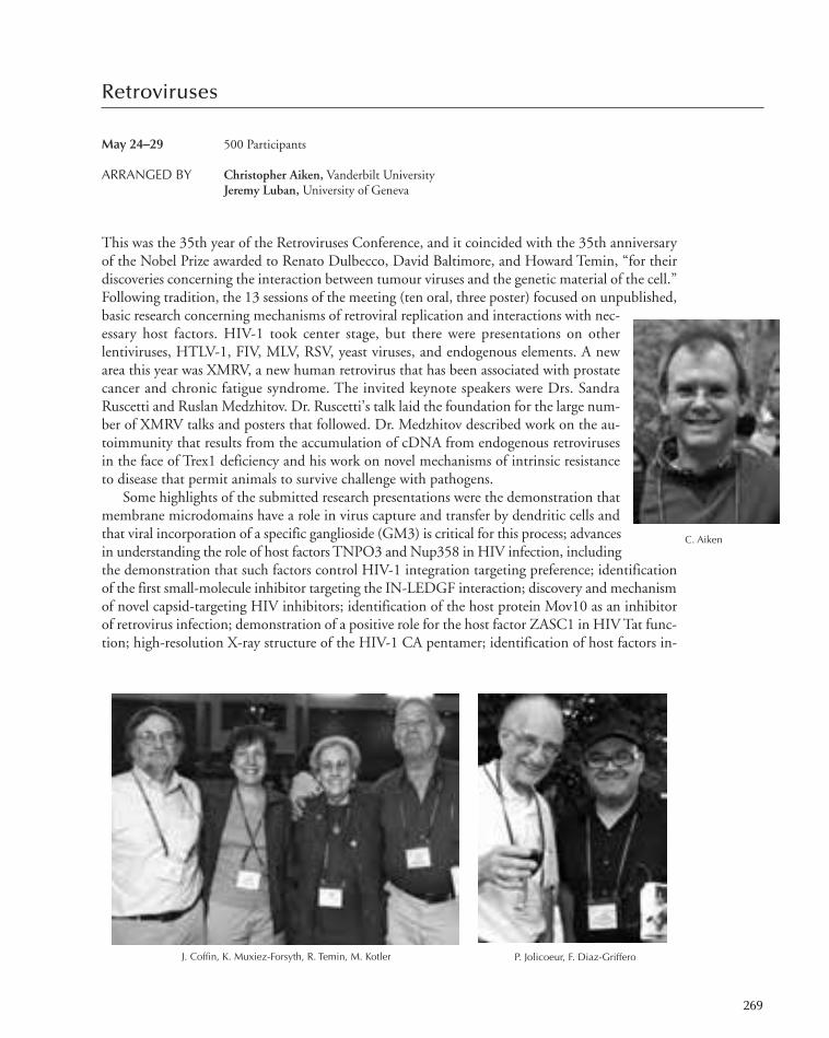
Retroviruses
May 24–29 500 Participants
ARRANGED BY Christopher Aiken, Vanderbilt UniversityJeremy Luban, University of Geneva
This was the 35th year of the Retroviruses Conference, and it coincided with the 35th anniversaryof the Nobel Prize awarded to Renato Dulbecco, David Baltimore, and Howard Temin, “for theirdiscoveries concerning the interaction between tumour viruses and the genetic material of the cell.”Following tradition, the 13 sessions of the meeting (ten oral, three poster) focused on unpublished,basic research concerning mechanisms of retroviral replication and interactions with nec-essary host factors. HIV-1 took center stage, but there were presentations on otherlentiviruses, HTLV-1, FIV, MLV, RSV, yeast viruses, and endogenous elements. A newarea this year was XMRV, a new human retrovirus that has been associated with prostatecancer and chronic fatigue syndrome. The invited keynote speakers were Drs. SandraRuscetti and Ruslan Medzhitov. Dr. Ruscetti’s talk laid the foundation for the large num-ber of XMRV talks and posters that followed. Dr. Medzhitov described work on the au-toimmunity that results from the accumulation of cDNA from endogenous retrovirusesin the face of Trex1 deficiency and his work on novel mechanisms of intrinsic resistanceto disease that permit animals to survive challenge with pathogens.
Some highlights of the submitted research presentations were the demonstration thatmembrane microdomains have a role in virus capture and transfer by dendritic cells andthat viral incorporation of a specific ganglioside (GM3) is critical for this process; advancesin understanding the role of host factors TNPO3 and Nup358 in HIV infection, includingthe demonstration that such factors control HIV-1 integration targeting preference; identificationof the first small-molecule inhibitor targeting the IN-LEDGF interaction; discovery and mechanismof novel capsid-targeting HIV inhibitors; identification of the host protein Mov10 as an inhibitorof retrovirus infection; demonstration of a positive role for the host factor ZASC1 in HIV Tat func-tion; high-resolution X-ray structure of the HIV-1 CA pentamer; identification of host factors in-
269
C. Aiken
P. Jolicoeur, F. Diaz-GrifferoJ. Coffin, K. Muxiez-Forsyth, R. Temin, M. Kotler
247-304_Meetings_Annual Report_2009 template 7/8/11 12:23 PM Page 269

volved in epigenetic silencing of retroviruses; identification of a role of Rab11a in HIV-1 Env in-corporation; mechanistic insights into Vpu and Nef conteraction of the host restriction factor BST-2/Tetherin; and progress from several labs on the controversial role of XMRV in human disease.
Stephen Hare from the Cerepanov lab reported on their landmark achievement of the cocrystalstructure of the a foamy virus integrase in complex with DNA, and Luban’s group reported evidencethat TRIM5 proteins can act as innate sensors for the retroviral capsid and participate in immunesignaling, suggesting that TRIM5 may function as a pattern recognition receptor akin to TLR3 andRIG-I. Finally, Dan Littman presented exciting evidence that forcing HIV-1 to infected dendriticcells in culture induces a type I interferon response that depends on interactions of cyclophilin Awith nascent Gag protein in the infected cell. Many other noteworthy advances were reported butcannot be listed here due to space constraints.
On the final evening of the meeting, a special session celebrated the 35th anniversary of theNobel Prize awarded to the codiscoverers of reverse transcriptase, Dr. Howard Temin and Dr. DavidBaltimore. Two invited speakers for the occasion, Dr. Rayla Temin and Dr. Baltimore, were intro-duced by Dr. John Coffin and Dr. Stephen Goff, respectively. The legacies of Temin and Baltimorewere strongly evident during the session, not only from the historical descriptions of their monu-mental achievements, but also owing to the fact that the majority of meeting attendees confirmedthat they received scientific training, either directly or indirectly, from one of these giants of thefield. Dr. James Watson was present to celebrate his fellow Nobel laureates.
This meeting was funded in part by the National Institute of Allergy and Infectious Diseases, abranch of the National Institutes of Health.
PROGRAM
EntryChairpersons: J. Mak, The Burnet Institute, Melbourne,Australia; C. Berlioz-Torrent, INSERM U1016, Paris, France
RT and Trim5Chairpersons: F. Diaz-Griffero, Albert Einstein College ofMedicine, Bronx, New York; W. Johnson, Harvard MedicalSchool, Boston, Massachusetts
Goin’ NuclearChairpersons: M. Naghavi, University College Dublin, Ireland;
E. Bacharach, Tel Aviv University, Israel
Keynote AddressS. Ruscetti, National Cancer Institute, Frederick, MarylandUnderstanding the Biological Effects of the Human Retrovirus
XMRV: Lessons Learned from Studying Murine Retroviruses
H. Taylor, O. Ciagoz S. Garforth, C. Lwatula
270 Meetings
247-304_Meetings_Annual Report_2009 template 7/8/11 12:23 PM Page 270
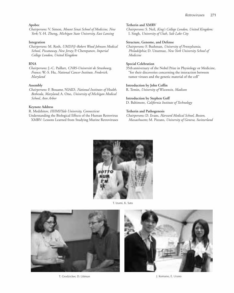
ApobecChairpersons: V. Simon, Mount Sinai School of Medicine, NewYork; Y.-H. Zheng, Michigan State University, East Lansing
IntegrationChairpersons: M. Roth, UMDNJ–Robert Wood Johnson MedicalSchool, Piscataway, New Jersey; P. Cherepanov, ImperialCollege London, United Kingdom
RNAChairpersons: J.-C. Paillart, CNRS-Université de Strasbourg,France; W.-S. Hu, National Cancer Institute, Frederick,Maryland
AssemblyChairpersons: F. Bouamr, NIAID, National Institutes of Health,Bethesda, Maryland; A. Ono, University of Michigan MedicalSchool, Ann Arbor
Keynote AddressR. Medzhitov, HHMI/Yale University, ConnecticutUnderstanding the Biological Effects of the Human Retrovirus
XMRV: Lessons Learned from Studying Murine Retroviruses
Tetherin and XMRVChairpersons: S. Neil, King’s College London, United Kingdom:
I. Singh, University of Utah, Salt Lake City
Structure, Genome, and DefenseChairpersons: F. Bushman, University of Pennsylvania,Philadelphia; D. Unutmaz, New York University School ofMedicine
Special Celebration35th anniversary of the Nobel Prize in Physiology or Medicine,
“for their discoveries concerning the interaction betweentumor viruses and the genetic material of the cell”
Introduction by John CoffinR. Temin, University of Wisconsin, Madison
Introduction by Stephen GoffD. Baltimore, California Institute of Technology
Tetherin and PathogenesisChairpersons: D. Evans, Harvard Medical School, Boston,Massachusetts; M. Pizzato, University of Geneva, Switzerland
T. Izumi, K. Sato
Retroviruses 271
T. Grodzicker, D. Littman J. Komano, E. Urano
247-304_Meetings_Annual Report_2009 template 7/8/11 12:23 PM Page 271

75 Years in Science: Biology, Society, and the Future
June 1 212 Participants
ARRANGED BY Jan Witkowski, Banbury Center, Cold Spring Harbor Laboratory
Reginald Harris became the director of the Biological Laboratory at Cold Spring Harbor in 1924.He set about revitalizing the Laboratory, making biophysics the new focus of research and, mostnotably, established the Cold Spring Harbor Laboratory Symposia on Quantitative Biology. Theirgoal, Harris wrote, was “to consider a given biological problem from its chemical, physical andmathematical, as well as from its biological aspects.”
The first symposium was held in 1933 and they have been held annually ever since, with the ex-ception of 1943–1945, surely making them one of the longest-lived series of meetings in the biologicalsciences. The success of the Symposia was due not only to the quality of the science presentations,but also to the very generous funding by two foundations, the Rockefeller Foundation and theCarnegie Foundation. These together helped support the meetings from 1934 through 1960.
The Symposia topics have always been at the forefront of contemporary research. Prior to WorldWar II, biophysics research was the theme of the Symposia after the war; genetics came to dominatethe Symposia for more than 20 years, culminating in the 1996 Symposium, The Genetic Code.Since then, Symposia topics have ranged over all aspects of ex-perimental biology—DNA, RNA, and proteins; cell biology;developmental biology; human genetics; cancer; neuroscience;physiology; and immunology and virology.
Such a storied history had to be celebrated and so a 1-daymeeting was held immediately before the start of the 75thSymposium. We included two examples of the power of mo-lecular genetics when applied to human beings—what is beingrevealed of our origins and the use of DNA fingerprinting tofree the wrongly convicted. Molecular genetics was also thefoundation of two presentations on key areas of biomedicalresearch: cancer and neurodevelopmental disorders. Finally,we looked deep into the crystal ball with a talk on syntheticgenomes. The banquet, always a lively event, was further en-livened by Sydney Brenner who was the after-dinner speaker,and he and Jim Watson cut a cake celebrating the occasion.
Reginald Harris left a legacy that has stimulated the generations of biologists who have comeeach year to talk, argue, and gossip at the Symposium. Just as Harris promoted biophysics as thekey to a deep understanding of the nature of living organisms, so contemporary organizers promotemolecular approaches for the investigation of biological systems. As we celebrated the occasion ofthe 75th Symposium, there was every reason to believe that the Symposia will continue to fulfillHarris’s vision.
Generous financial support was provided equally by the Rockefeller Foundation and the CarnegieCorporation of Washington. Additional support was provided through a personal gift from Dr.Charles Weissmann.
S. Henikoff, J. Gall
272
247-304_Meetings_Annual Report_2009 template 7/8/11 12:23 PM Page 272
PROGRAM
Opening RemarksB. Stillman, Cold Spring Harbor LaboratoryChairperson: R. Tjian, Howard Hughes Medical Institute, ChevyChase
Devising Cancer TherapiesChairperson: C. Sawyers, Memorial Sloan-Kettering CancerCenter, New York
CommentaryT. Jacks, Massachusetts Institute of Technology, Cambridge
The Journey of Man: A Genetic OdysseyS. Wells, National Geographic Society, Washington, District ofColumbia
CommentaryH.L. Gates, Jr., Harvard University, Cambridge, Massachusetts
75 Years in Science: Biology, Society, and the Future 273
E. Heard, J. Haber
Fulfilling the Promise of Molecular Medicine in a Develop-mental Brain Disorder
M. Bear, Massachusetts Institute of Technology, Cambridge
CommentaryS. Landis, National Institute of Neurological Disorders & Stroke,Bethesda, Maryland
The Wrongly Convicted: DNA and JusticeP. Neufeld, Innocence Project, New York
CommentaryJ.C. Venter, J. Craig Venter Institute, Rockville, Maryland; R.
Roberts, New England BioLabs, Inc., Ipswich, Massachusetts
Closing RemarksJ.D. Watson, Cold Spring Harbor Laboratory
247-304_Meetings_Annual Report_2009 template 7/8/11 12:23 PM Page 273

Glia In Health and Disease
July 22–26 193 Participants
ARRANGED BY Beth Stevens, Children’s Hospital/Harvard Medical SchoolWilliam Talbot, Stanford University
Glial cells constitute 90% of cells in the human nervous system, but their roles in the developing,adult, and diseased brain are still poorly understood. In this summer’s meeting, students and scien-tists from across the world gathered to discuss their recent progress. In the first part of the meeting,sessions focused on understanding the normal roles of glia,including sessions on the genetic analysis of glial function,neural development, myelination, axon–glial interactions,microglia function, and astrocyte function at synapses andblood vessels. In the last half of the meeting, sessions focusedon recent progress on understanding the roles of glia in CNSregenerative failure and disease processes, including glioma,epilepsy, and multiple sclerosis. There was active discussionrelated to the protective and destructive roles of “reactive glio-sis” in CNS injury and neurodegenerative diseases includingALS (Lou Gehrig’s disease) and glaucoma.
One of the highlights of the meeting was an animatedroundtable discussion led by Ben Barres and Martin Raff onkey issues related to glial biology and life in science. Postersessions were well attended with active discussion. There was again a sense of excitement that ourunderstanding of the function of glia is quickly moving forward, with glia having central roles innormal brain development, function, and disease. This excitement was reflected in the presence ofseveral top journal editors interested in enhancing their coverage of latest advances concerning glia.
This meeting was funded in part by the National Cancer Institute, a branch of the National In-stitutes of Health.
274
W. Talbot, B. Stevens
L. Goutebroze, J. Ness-Myers A. Agarwal A. Giangrande, M. Raff
247-304_Meetings_Annual Report_2009 template 7/8/11 12:23 PM Page 274
PROGRAM
Genetic Analysis of Glial FunctionChairpersons: B. Popko, University of Chicago, Illinois; M.
Freeman, University of Massachusetts Medical School,Worcester
Myelinating CellsChairpersons: K. Jessen, University College London, UnitedKingdom; P. Brophy, University of Edinburgh, UnitedKingdom
Axon–Glial InteractionsChairpersons: J. Chan, University of California, San Francisco;
E. Peles, Weizmann Institute of Science, Rehovot, Israel
Glial Function at SynapsesChairpersons: D. Bergles, Johns Hopkins University, Baltimore,Maryland; G. Corfas, Children’s Hospital, Boston,Massachusetts
Microglia Function in Health and DiseaseChairpersons: A. Bessis, Ecole Normale Supérieure, Paris, France;
M. Carson, University of California, Riverside
Roundtable Discussion: Glia in Health and Disease—OpenQuestions and Future Directions
Chairpersons: B. Barres, Stanford University School of Medicine,California; M. Raff, University College London, UnitedKingdom
Gliovascular Interactions and Astrocyte FunctionChairpersons: R. Daneman, University of California, SanFrancisco; M. Nedegaard, University of Rochester, New York
CNS Injury and DiseaseChairpersons: M. Sofroniew, University of California School of Medicine, Los Angeles; H. Zong, University of Oregon,Eugene
Glial Development and Epigenetic MechanismsChairpersons: D. Rowitch, University of California, SanFrancisco; P. Casaccia, Mount Sinai School of Medicine, New York
H. Colognato, D. Parkinson M.-L. Schmiedt
Glia in Health and Disease 275
247-304_Meetings_Annual Report_2009 template 7/8/11 12:23 PM Page 275

Mechanisms and Models of Cancer
August 17–21 436 Participants
ARRANGED BY Dafna Bar-Sagi, New York University Medical CenterJacqueline Lees, MIT Center for Cancer ResearchCharles J. Sherr, HHMI/St. Jude Children’s Research HospitalWilliam Weiss, University of California, San Francisco
Molecular alterations affecting tumor suppressor genes and oncogenes have long been recognized tospark the development of human cancers. During the last few years, steady advances in technologicaldevelopments have allowed a more exquisitely detailed dissection of signaling networks that are per-turbed during different stages of tumor initiation and progressionand that determine the responses of cancers to both conventionaland targeted therapeutics. In addition, there has been the emergingappreciation that interactions of tumor cells with their tissue mi-croenvironment conjoin to alter tumor cell metabolism, affect themanner by which a variety of host inflammatory and immune re-sponses affect cancer progression, and determine properties of in-vasion and metastasis.
This third meeting convened an international group of inves-tigators whose collective work focused on these themes. Oral andposter presentations provided numerous examples of how new in-sights have been gained from application of interdisciplinary ap-proaches that utilize genetics, developmental biology, genomicsand proteomics, and model organisms (principally sophisticatedmouse models) to advance the development of rational therapeutics. A keynote address by EileenWhite illustrated the importance of autophagy (self-cannibalization) as a pathway to which cancercells show addiction and demonstrated its relevance for therapeutic targeting. There were eight oralsessions and two poster sessions. The meeting will be held again in the summer of 2012.
This conference was funded in part by the National Cancer Institute, a branch of the NationalInstitutes of Health.
PROGRAM
W. Weiss, J. Lees
276
Apoptosis and AutophagyChairperson: E. White, Rutgers University, New Brunswick, New Jersey
Keynote AddressJ. Brugge, Harvard Medical School, Massachusetts
Signaling Mechanisms IChairperson: R. Shaw, The Salk Institute for Biological Studies,La Jolla, California
Mouse ModelsChairperson: C. Abate-Shen, Columbia University MedicalSchool, New York
Rb and p53 NetworksChairperson: K. Vousden, Beatson Institute for Cancer Research,Glasgow, United Kingdom
Microenvironment, Inflammation, and MetastasesChairperson: L. Parada, University of Texas SouthwesternMedical Center, Dallas
Experimental TherapeuticsChairperson: R. DePinho, Dana-Farber Cancer Institute,Boston, Massachusetts
Genomics, Proteomics, EpigeneticsChairperson: F. White, Massachusetts Institute of Technology,Cambridge
Signaling Mechanisms IIChairperson: M. Eilers, University of Würzburg, Germany
247-304_Meetings_Annual Report_2009 template 7/8/11 12:23 PM Page 276

Molecular Genetics of Bacteria and Phages
August 24–28 169 Participants
ARRANGED BY James Hu, Texas A&M UniversityPetra Levin,Washington UniversityMalcolm Winkler, Indiana University Bloomington
This meeting featured 74 oral presentations and 82 poster presentations. Bacteria and phages con-tinue to provide critical insights into diverse areas of biology, ranging from ecology, evolution, anddiversity, to gene expression and development, to infectious diseases. Exciting presentations coveredall of these topics and continued the historical strength of Phage meet-ings in combining long-term themes of molecular microbiology withcutting-edge approaches and exciting new research areas. A highlightof the meeting was the keynote lecture by long-time participant SusanGottesman, who coupled a review of her career in bacterial molecularbiology with recognition of the 100th anniversary of the birth ofJacques Monod. Another celebration was for the long, continuous par-ticipation of Wisconsin’s Waclaw Szybalski, whose generous supportof the CSHL library was warmly recognized.
The opening session focused on development, cell signaling, andcell–cell interactions. Session chair Mike Laub touched on several re-curring themes of the meeting in his description of how Caulobacteruses the kinetics of phosphorylation and dephosphorylation of keyregulators to control its asymmetric cell division. Michelle Giglio pre-sented an overview of the Human Microbiome Project and chaired thesecond session with many other thought-provoking examples of how genomes and metagenomesare opening new areas of biology. Ry Young chaired the session on Bacterial Cell Surfaces, and pre-sented a talk on the discovery of spanin proteins that mediate the destruction of the inner membrane,peptidoglycan, and outer membrane by phages of Gram-negative bacteria.
Gail Christie spoke on the functions and evolutions of superantigen-encoding pathogenicity is-lands that are mobilized by helper phages. Bryce Nichols’ talk concerned the detection of nanoRNAsthat can prime transcription initiation in vivo. Alan Wolfe spoke on the role of pathways that me-diate the synthesis of acetyl phosphate and acetylation of target proteins. Robert Britton started hissession with a talk on the role of conserved GTPases in ribosome assembly. David Rudner, whochaired the last session, talked about new proteins that regulate DNA replication during sporulation.Regular talks and posters were presented by a mix of PIs, postdocs, and advanced graduate students,whose high-quality presentations are a unique aspect of this meeting.
This meeting was funded in part by the National Institute of Allergy and Infectious Diseases, abranch of the National Institutes of Health, and the National Science Foundation.
J. Hu, P. Levin
277
PROGRAM
Development, Cell Signaling, and Cell–Cell InteractionsChairperson: M. Laub, HHMI/Massachusetts Institute ofTechnology, Cambridge
Molecular Evolution, Systems Biology, and GenomicsChairperson: M. Giglio, University of Maryland Schoolof Medicine, Baltimore
247-304_Meetings_Annual Report_2009 template 7/8/11 12:23 PM Page 277
Bacterial Cell SurfacesChairperson: R. Young, Texas A&M University, College
Station
Bacteriophage BiologyChairperson: G. Christie, Virginia Commonwealth University,
Richmond
Mechanism and Regulation of TranscriptionChairperson: B. Nickels, Rutgers University, Piscataway, New Jersey
Bacterial Metabolism and PhysiologyChairperson: A. Wolfe, Loyola University, Chicago, Illinois
Translation and Posttranscriptional RegulationChairperson: R. Britton, Michigan State University, East
Lansing
Keynote Address: Beyond Transcription: Complex Regulatory Networks for Responding to Changing Environments
S. Gottesman, National Cancer Institute
DNA Replication, Recombination, and TranspositionChairperson: D. Rudner, Harvard Medical School, Boston,
Massachusetts
278 Meetings
K. Potrykus, M. Cashel A. Labner-Oleson, D. Chattoraj F. Pouillot, H. Blois
247-304_Meetings_Annual Report_2009 template 7/8/11 3:54 PM Page 278

Nuclear Receptors and Disease
Aug. 31–Sept. 4 205 Participants
ARRANGED BY Keith Yamamoto, University of California, San FranciscoRon Evans, Salk Institute Biological StudiesJerrold M. Olefsky, University of California, San Diego
Forty-eight nuclear receptors are encoded in the human genome, and, collectively, they exert broadcontrol over most biological processes. The theme of this meeting was to bring together scientistsfrom different disciplines to exchange ideas and forge new in-sights into the biology of nuclear receptors. Accordingly, themeeting focused on the role of nuclear receptors in metabolismand cancer. This represented the third time the nuclear receptorcommunity gathered at the Cold Spring Harbor Laboratory fortheir biannual meeting.
The meeting brought together an enthusiastic group of par-ticipants engendering lively discussions and debates, and an openexchange of data and ideas. The invited Keynote talks were givenby David Mangelsdorf, Bruce Spiegelman, and Laurie Goodyear.Other invited speakers included Johan Auwerx, Juleen Zierath,Walter Wahli, Katja Lamia (The Salk Institute for BiologicalStudies), Alan Saltiel (University of Michigan), DaYoung Oh(University of California, San Diego), Philipp Scherer (Universityof Texas Southwestern Medical Center), Michael Stallcup (University of Southern California), Susanne Mandrup (University of Southern Denmark), Bert O’Malley (Baylor College of Medicine),Donald McDonnell (Duke University Medical Center), Myles Brown (Dana-Farber Cancer Insti-tute), Vincent Giguere (McGill University), Mitchell Lazar (University of Pennsylvania School ofMedicine), Christopher Newgard (Duke University Medical Center), Christopher Glass (Universityof California, San Diego), Gokhan Hotamisligil (Harvard School of Public Health), Laszlo Nagy(University of Debrecen), W. Lee Kraus (University of Texas Southwestern Medical Center), PeterTontonoz (University of California, Los Angeles), and Gordon Hager (NCI, National Institutes ofHealth).
J. Olefsky, R. Evans, K. Yamamoto
PROGRAM
Keynote AddressesD.J. Mangelsdorf, University of Texas Southwestern MedicalSchool, Dallas
Nuclear Receptor Modulators (NuRMs) in WormsB. Spiegelman, Dana-Farber Cancer Institute, Boston,Massachusetts
PPARγ and the Antidiabetic PPARγ Ligands: A New Look atan Old Friend
L. Goodyear, Joslin Diabetes Center, Boston, MassachusettsSkeletal Muscle Metabolism: AMPK and Beyond
Physiology and MetabolismChairperson: A. Saltiel, University of Michigan, Ann Arbor
Inflammation and Metabolic DiseaseChairperson: B. Desvergne, University of Lausanne, Switzerland
Ligands and CofactorsChairperson: R.M. Evans, The Salk Institute, La Jolla,California
279
247-304_Meetings_Annual Report_2009 template 7/8/11 12:23 PM Page 279
CancerChairperson: C. Glass, University of California, San Diego
Mechanisms of Insulin Resistance and AdipogenesisChairperson: J.M. Olefsky, University of California, San Diego
D. Oh, E. Bae, K. Kemper
J. Judkins, D. Mangelsdorf K. Duszka, N. Herman-Kleiter
280 Meetings
M. Rozendaal, J. Nwachukwu
Inflammation IIChairperson: S. Mandrup, University of Southern Denmark,Odense
Chromatin and TranscriptionChairperson: M. Stallcup, University of Southern California, LosAngeles
247-304_Meetings_Annual Report_2009 template 7/8/11 12:23 PM Page 280
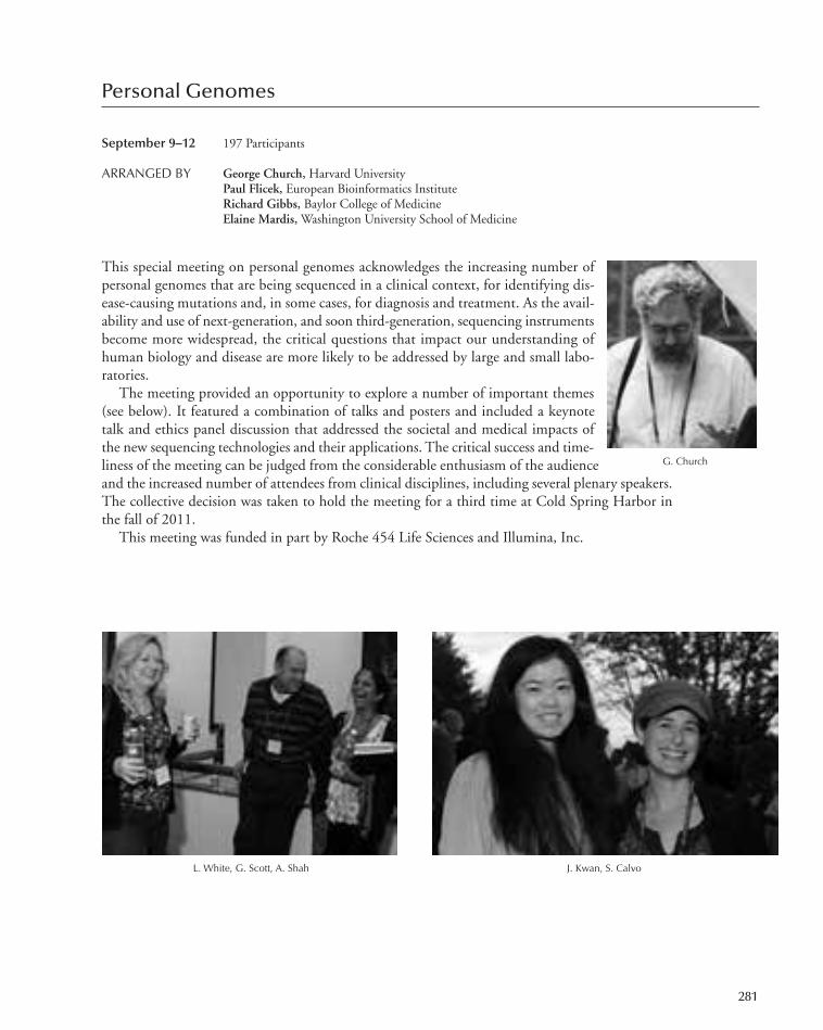
Personal Genomes
September 9–12 197 Participants
ARRANGED BY George Church, Harvard University Paul Flicek, European Bioinformatics InstituteRichard Gibbs, Baylor College of MedicineElaine Mardis,Washington University School of Medicine
This special meeting on personal genomes acknowledges the increasing number ofpersonal genomes that are being sequenced in a clinical context, for identifying dis-ease-causing mutations and, in some cases, for diagnosis and treatment. As the avail-ability and use of next-generation, and soon third-generation, sequencing instrumentsbecome more widespread, the critical questions that impact our understanding ofhuman biology and disease are more likely to be addressed by large and small labo-ratories.
The meeting provided an opportunity to explore a number of important themes(see below). It featured a combination of talks and posters and included a keynotetalk and ethics panel discussion that addressed the societal and medical impacts ofthe new sequencing technologies and their applications. The critical success and time-liness of the meeting can be judged from the considerable enthusiasm of the audienceand the increased number of attendees from clinical disciplines, including several plenary speakers.The collective decision was taken to hold the meeting for a third time at Cold Spring Harbor inthe fall of 2011.
This meeting was funded in part by Roche 454 Life Sciences and Illumina, Inc.
G. Church
L. White, G. Scott, A. Shah J. Kwan, S. Calvo
281
247-304_Meetings_Annual Report_2009 template 7/8/11 12:23 PM Page 281

PROGRAM
Personal Genome LandscapeChairpersons: D. Conrad, Wellcome Trust Sanger Institute,Hinxton, United Kingdom; J. Wang, Beijing GenomicsInstitute, Shenzhen, China
Keynote AddressesE.D. Green, National Human Genome Research Institute,Maryland
Genomics in 2K10 and Beyond: Charting a Course forGenomic Medicine
L.E. Hood, Institute for Systems Biology, Seattle, WashingtonSystems Genetics and Systems BiologyH. Greely, Stanford University, Stanford, CaliforniaPreparing for the Coming Tsunami of Clinical Genomic
Information
Ethics Panel: Teaching Genomics and Related Ethics toMedical Professonals
Personal Cancer GenomesChairpersons: S. Grimmond, University of Queensland, St.Lucia, Australia; S. Jones, BC Cancer Agency, Vancouver,Canada
Personal Transcriptomes and Other ApplicationsChairpersons: P. Laird, University of Southern California, LosAngeles; R. Myers, HudsonAlpha Institute for Biotechnology,Huntsville, Alabama
Inherited DiseasesChairpersons: L. Jorde, University of Utah School of Medicine,Salt Lake City; D. Altshuler, Broad Institute, Cambridge,Massachusetts
Technologies for Personal GenomesChairperson: G. Church, Harvard Medical School, Boston,Massachusetts
A. Dopazo, L. Brizuela
E. Green, H. Sussman
282 Meetings
247-304_Meetings_Annual Report_2009 template 7/8/11 12:23 PM Page 282

Translational Control
September 13–17 411 Participants
ARRANGED BY Thomas Dever, National Institutes of HealthPaul Fox, The Cleveland ClinicAlan Hinnebusch, National Institute of Child Health & Human Development,
National Institutes of HealthElisa Izaurralde,Max-Planck Institute for Developmental Biology
This meeting attracted participants from around the world, and included Jamie Cate, Rachel Green,Eric Klann, and Paul Schimmel as keynote speakers, eight platform sessions and three poster sessionsthat covered 337 abstracts. Novel findings on the mechanism of translation included the first high-resolution crystal structure of the eukaryotic 80S ribosome, a 5.5Å cryo-EM structure of the wheat ribosome, and multiparticlecryo-EM analysis of translocating ribosomes that revealed new in-termediate tRNA hybrid states.
There were several breakthroughs regarding the mechanism ofinitiation. Interaction of the eIF4G subunit of the mRNA cap-binding complex with poly(A)-tail binding protein PABP wasfound to be dispensable for initiation in yeast, being functionallyredundant with an RNA-binding domain in eIF4G. Among manyother findings, evidence was presented that ribosomes can circum-vent proteins or oligonucleotides bound to the 5′UTR, presumablyby pathways independent of linear scanning. Important develop-ments concerning elongation and termination included new find-ings on yeast No-Go mRNA decay quality control. Several lines ofevidence suggest that the termination and ribosome recycling machineries are more closely relatedthan previously anticipated. New methods were described for monitoring the dynamics of translationelongation in living cells, and ribosomal profiling by deep-sequencing of ribosome-protected mRNAfragments. Pharmacological approaches to suppressing nonsense mutations were described as a new
C. Hellen, A. Hinnebusch
Wine and cheese party
283
247-304_Meetings_Annual Report_2009 template 7/8/11 12:23 PM Page 283

therapeutic approach to treating genetic diseases.In the arena of translational control in disease, new results
show that a lack of rRNA modification impairs cell cycle arrestin early oncogenesis by preventing a switch to IRES-mediatedtranslation of tumor suppressor p53. New insights into virus–host interactions were obtained, and novel initiation mecha-nisms were reported for plant positive-strand RNA viruses. Incalicivirus translational reinitiation, ribosomes can reach startsites not only by direct positioning, but also by movementalong the viral RNA in a prokaryotic-like fashion. Regardingtranslational control in development and the CNS, a novelrequirement for the eIF2 kinase PERK in regulating LTD wasdemonstrated, which has important behavioral consequencesin un-learning previous associations. In Aplysia sensory neu-rons, the effects of TOR in increasing cap-dependent translation were shown to be independent ofthe major eIF4E- binding protein and S6 kinase. TORC2 was found to associate with polyribosomesand cotranslationally phosphorylate AKT, regulating its stability.
Finally, new insights into connections between translation and mRNA turnover were also re-ported, including the fact that exon junction complexes (EJCs) are not deposited at all exon–exonjunctions. There is evidence that EJCs associate with the nonsense-mediated mRNA decay (NMD)factors SMG1 and UPF1 in a way that is promoted by the cap-binding protein of the pioneer trans-lation initiation complex and, subsequently, SMG6 by a mechanism that is inhibited by EJC asso-ciation with the UPF3 NMD factor. This represents just a sampling of the many new findingspresented at the meeting, with vigorous discussion by graduate students, postdocs, and professorsat both oral sessions and poster sessions. The informality of the environment at Cold Spring Harborencourages intense and fruitful debate and discussion of novel ideas and approaches.
PROGRAM
Ribosome Structure and FunctionChairperson: J. Dinman, University of Maryland, College Park
Factors and MechanismsChairperson: J. Lorsch, Stanford University, California
Viruses and DiseaseChairperson: G. Meyers, Bundesforschungsanstalt fürViruskrankheiten der Tiere, Tübingen, Germany
mRNA TurnoverChairperson: L. Maquat, University of Rochester Medical Center,New York
Development, CNS, and SignalingChairperson: W. Sossin, McGill University, Montréal, Canada
Trans-Acting RegulatorsChairperson: P. Fox, The Cleveland Clinic, Ohio
Elongation and TerminationChairperson: D. Bedwell, University of Alabama, Birmingham
RNA Regulatory ElementsChairperson: S. Thompson, University of Alabama,Birmingham
A. Contreras, A. Rodrigues, E. Bueno
284 Meetings
247-304_Meetings_Annual Report_2009 template 7/8/11 12:23 PM Page 284

Axon Guidance, Synaptic Plasticity, and Regeneration
September 21–25 364 Participants
ARRANGED BY Graeme Davis, University of California, San FranciscoAlex Kolodkin, Johns Hopkins UniversityCarol Mason, Columbia University
The human brain has billions of nerve cells (neurons), and each neuron is typically connected tohundreds of other neurons via synapses in a highly precise fashion. This complex neural wiring un-derlies the ability of humans, and other animals, to interact with the outside world to learn and toperform complex behaviors. Defects in the development of neu-ral connections are being increasingly linked to the cause of neu-rological disease. One of the major challenges for the field ofneuroscience is to understand how nerve connections are madeaccurately and reliably. In the past decade, our understandingof the mechanisms that control axon growth and guidance,synaptogenesis, and the remodeling of neural circuits during de-velopment has progressed rapidly from phenomenology to theidentification of specific molecular control mechanisms.
This seventh meeting in the series focused on key issues inaxon guidance, circuit formation, synaptogenesis, and plasticity.For the first time, it highlighted regeneration and included ses-sions devoted to particular problems in the assembly, plasticity,and repair of the nervous system. Speakers were chosen from among the participants submittingabstracts by session chairs who are leaders in the field. The response of the field to the 2010 confer-ence was one of overwhelming enthusiasm, with more than 360 participants, 257 of whom sub-mitted abstracts; 49 abstracts were selected for talks, in seven sessions, the remaining abstracts beingpresented as posters. Senior researchers, starting assistant professors, postdoctoral fellows, and grad-uate students were well represented as speakers and participants. Session chairs at the meeting werewell-balanced between men and women and had a clear international presence with participantsfrom Europe and Asia.
Many of the outstanding talks this year were by graduate students. All of the major areas of re-search in the field were covered, as were all of the major approaches (cellular, physiological, anatom-ical, molecular, dynamic imaging, biochemical, and genetic). In addition, there were three keynoteaddresses. The first, by a speaker outside of the field of neural development, was given by ProfessorPeter Devreotes and reviewed his work on the motility, polarization, and directional sensing duringchemotaxis of the single cell forms of Dictyostelium. The second and third keynote lectures were apairing of speakers who gave provocative talks on how specificity is achieved and the limits to exactcircuit formation and function. Tom Jessell focused on work that extends his findings regardingtranscriptional regulation of cell identity to the afferent–target interactions leading to motor outputin the vertebrate spinal cord. Eve Marder spoke on the physiological and computational aspects ofcompensation in network performance, with the lobster CNS as her model.
The meeting provided an important forum for ideas and approaches and helped scientists in thefield get the most up-to-date information, enabling them to meet, network, and establish collabo-rations. As at the previous meeting, a need was identified for an additional poster session becausethe number and quality of abstracts submitted for posters was so high. The three poster sessionswere extremely well attended. In addition, fewer talks were scheduled in the evening, and a talk ses-
S. Arber, G. Davis
285
247-304_Meetings_Annual Report_2009 template 7/8/11 12:23 PM Page 285

sion after the evening keynote was eliminated to allow time for discussion and attendee interactions.Based on the enthusiastic comments of the participants, the intensity of the oral and poster sessions,and the large crowds that stayed up late every night at the bar to discuss science, the meeting was agreat success.
This meeting was funded in part by National Institute for Neurological Diseases and Stroke, abranch of the National Institutes of Health.
PROGRAM
Axon to Synapse IChairpersons: K. Martin, University of California, Los Angeles;
Y. Zou, University of California, San Diego
Synapse to Circuit IChairpersons: M. Feller, University of California, Berkeley; A.
Chedotal, Institut de la Vision, INSERM, Paris, France
Special Lecture: The Cell’s Compass: Chemoattractant Biasof an Excitable Network of Parallel Signaling PathwaysDirects Cell Migration
P. Devreotes, Johns Hopkins University School of Medicine,Maryland
Stem Cells, Regeneration, and Disease IChairpersons: Y. Jin, HHMI/University of California, SanDiego; F. Bradke, Max-Planck Institute of Neurobiology,Martinsried, Germany
Axon to Synapse IIChairpersons: S. Arber, Biozentrum, Basel, Switzerland; B.
Sabatini, Harvard Medical School, Boston, Massachusetts
Synapse to Circuit IIChairpersons: J. Kaplan, Massachusetts General Hospital,Harvard Medical School, Boston; G. Turrigiano, BrandeisUniversity, Waltham, Massachusetts
Stem Cells, Regeneration, and Disease IIChairpersons: Y. Zuo, University of California, Santa Cruz; S.
Temple, Albany Medical College, New York
Special LecturesT. Jessell, HHMI/Columbia University, New YorkThe Neurons and Networks of Spinal Motor ControlE. Marder, Brandeis University, MassachusettsCompensation in Robust Network Performance
Axon to Synapse IIIChairpersons: M. Halloran, University of Wisconsin, Madison; T.
Schwarz, Children’s Hospital, Boston, Massachusetts
286 Meetings
Relaxing on the lawn
247-304_Meetings_Annual Report_2009 template 7/8/11 12:23 PM Page 286

Molecular Genetics of Aging
Sept. 28–Oct. 2 289 Participants
ARRANGED BY Steven Austad, University of Texas Health Science Center, San Antonio Judith Campisi, Buck Institute for Age Research, Lawrence Berkeley
National LaboratoryDavid Sinclair, Harvard University Medical School
Aging is the largest single risk factor for developing a panoply of diseases, ranging from neurode-generation to cancer. In recent years, enormous progress has been made in understanding how ge-netics and the environment collaborate to bring about the aging of organisms. Among theremarkable findings is the identification of molecular pathways that control aging in diversespecies—from yeast to humans. This conference provided an intense forum for the latest resultsand emerging ideas in aging research.
The conference opened with a session on genetic variation—an exploration into why species,and particularly individuals within a species, age at different rates. A related session discussed geneticand epigenetic stability, which focused on the mechanisms that main-tain the DNA sequence and its packaging (chromatin), how these en-tities change during aging, and how such changes affect organismalhealth and fitness. A subsequent session highlighted the role of mito-chondria and energy metabolism in preserving organismal health andlongevity, and the molecules that are crucial for optimal energy pro-duction and utilization. One session was devoted to cellular stress re-sponses, and the intimate relationship between stress resistance andlongevity. The conference then featured a session on stem cells, whichemphasized recent findings on how these important sources of tissuerepair and regeneration change with age, and how aging alters the tis-sue environment in which stem cells must function. Another sessiondiscussed protein homeostasis—how protein aggregates (which drivea number of age-related diseases) are formed and eliminated by spe-cific genetic pathways. A session then focused on the power of geneticmanipulations of conserved pathways to alter the rates and outcomeof aging. The final session highlighted the latest findings on the role of nutrition and nutrient sensingin aging and discussed interventional strategies that are currently being developed.
The conference brought together scientists from diverse fields, mirroring the expanding multi-disciplinarity of the field, and reflected the excitement and growing understanding of the processesthat drive aging and the possibilities for developing interventions into both the aging process andthe chronic diseases of aging.
This meeting was funded in part by the National Institute on Aging, a branch of the NationalInstitutes of Health.
PROGRAM
287
J. Sorrentino, C. Burd
Genetic VariationChairpersons: N. Barzilai, Albert Einstein College of Medicine,Bronx, New York; R. Westendorp, Leiden University MedicalCenter, The Netherlands
Genetic and Epigenetic StabilityChairpersons: L. Donehower, Baylor College of Medicine,Houston, Texas; J.P. Issa, University of Texas M.D. AndersonCancer Center, Houston
247-304_Meetings_Annual Report_2009 template 7/8/11 12:23 PM Page 287

Oral Presentations from AbstractsChairpersons: S. Austad, University of Texas Health ScienceCenter, San Antonio; J. Campisi, Lawrence Berkeley National Laboratory, Buck Institute for Age Research,California; D. Sinclair, Harvard Medical School, Boston, Massachusetts
Metabolism and BioenergeticsChairpersons: C. Mobbs, Mount Sinai School of Medicine, NewYork; R. De Cabo, NIA, National Institutes of Health,Bethesda, Maryland
Cellular and Organismal Stress ResponsesChairpersons: D. Peeper, Netherlands Cancer Institute,Amsterdam; S.-J. Lin, University of California, Davis
Tissue Maintenance/Stem CellsChairperson: G. Enikolopov, Cold Spring Harbor Laboratory
Protein HomeostasisChairpersons: N. Tavernakis, Foundation for Research andTechnology, Greece; R. Morimoto, Northwestern University,Evanston, Illinois
Genetic ManipulationsChairperson: G. Lithgow, Buck Institute for Age Research,Novato, California
Nutrient Sensing and InterventionsChairpersons: B. Kennedy, University of Washington, Seattle; A.
Dillin, Salk Institute, La Jolla, California
N. Hartman, L. Zidek M. Bonkowski, S. Gravina H. Siegfried
E. Jaehing, S.-J. Lin
288 Meetings
247-304_Meetings_Annual Report_2009 template 7/8/11 12:23 PM Page 288

Germ Cells
October 5–9 247 Participants
ARRANGED BY John Eppig, The Jackson LaboratoryMargaret Fuller, Stanford University School of MedicinePhillip Newmark, HHMI/University of Illinois at Urbana, Champaign
The tone for this meeting was set in the first session, with five speakers focusing on issues of germcell specification, migration, and fate. As with all of the sessions, lively discussion followed each ofthe talks. Likewise, the diversity in the model systems used for study, ranging from flies to worms,sea urchin, fish, and mammals, highlighted deeply conserved emerging mechanisms. Session 2 fo-cused on an area of particularly timely interest, Germ Line Stem Cells, a theme revisited with dif-ferent emphasis throughout the meeting, particularly in Session 4, which focused on the switchfrom mitotic to meiotic cell divisions. Evolving concepts about the nature of the germ cell nicheand the molecules driving stem cell dynamics provoked considerable discussion.
Poster Sessions (Sessions 3 and 6) presented studies on virtually all aspects of germ cell develop-ment in diverse model species. There was even a presentation of ovarian development in the plainsviscacha with its unusual characteristics of absence of follicularatresia and massive numbers of ovulated eggs. Sessions 7through 9 reported rapid advancement of our understandingof the regulation of meiosis and the oocyte-to-embryo transi-tion. This was emphasized by studies on the role of small in-terfering RNAs and other posttranscriptional controlmechanisms and the functions of maternal factors essentialfor early embryonic development.
There has been remarkable progress since the last Germ Cellsmeeting (in 2008). The evolution of new methods in molecularand genetic analyses, as well as concepts in control mechanismssuch as small interfering RNA and molecular determinants ofpluripotency, have greatly advanced the field of germ cell biol-ogy. Participants enjoyed breathtaking live-cell/organ imagingthat provided unique perspectives on germ cell behavior andthe possible functions of germ cell niches. The field looks forward to new applications of this excitingtechnology that will help shape our views of germ cell development. An international cast, includingspeakers from Japan, Australia, Taiwan, Germany, Switzerland, and the UK, participated in the meet-ing. There was general excitement about continued advancements that will affect broad basic and clin-ical fields of development, reproduction, and clinical sciences.
This meeting was funded in part by the National Institute of Child Health and Human Develop-ment, a branch of the National Institutes of Health; the Lalor Foundation; and the March of Dimes.
289
P. Lasko, L. Cooley
Germ Line Stem CellsChairperson: J. Kimble, HHMI/University of Wisconsin,Madison
PROGRAM
Germ Cell Specification, Migration, and FateChairperson: A. Spradling, HHMI/Carnegie Institution,Baltimore, Maryland
247-304_Meetings_Annual Report_2009 template 7/8/11 12:23 PM Page 289

Germ Line Stem Cells, Gonocyte Proliferation, and theSwitch from Mitosis to Meiosis
Chairperson: B. Braun, The Jackson Laboratory, Bar Harbor,Maine
Transcriptional Control and Epigenetic ProgrammingChairperson: H. Lin, Yale University, New Haven, Connecticut
Posttranscriptional Control and Small RNAs in Gametogen-esis and Early Embryos
Chairperson: R. Lehmann, HHMI/Skirball Institute, New YorkUniversity School of Medicine, New York
MeiosisChairperson: M.A. Handel, The Jackson Laboratory, BarHarbor, Maine
Cell Biology of Germ CellsChairperson: D. Page, HHMI/Massachusetts Institute ofTechnology, Cambridge
Germ Line/Soma Communication and Sex DeterminationChairperson: E. Matunis, Johns Hopkins University School ofMedicine, Baltimore, Maryland
S. Hammond, M. Potok B. Braun, D. Licatolosi
290 Meetings
247-304_Meetings_Annual Report_2009 template 7/8/11 12:23 PM Page 290
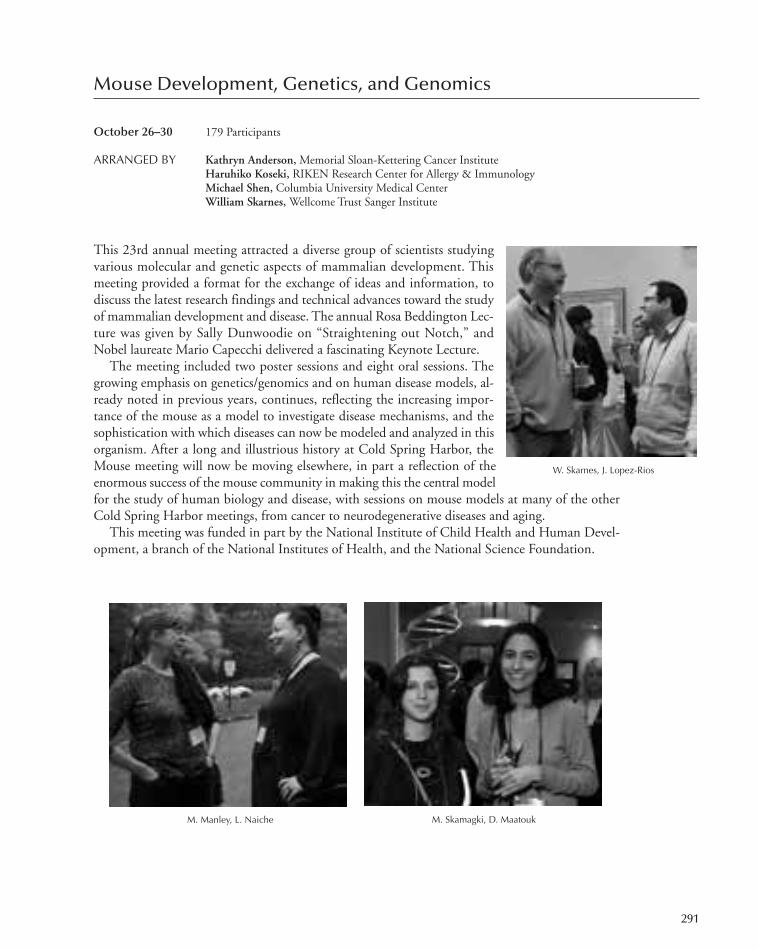
Mouse Development, Genetics, and Genomics
October 26–30 179 Participants
ARRANGED BY Kathryn Anderson,Memorial Sloan-Kettering Cancer InstituteHaruhiko Koseki, RIKEN Research Center for Allergy & ImmunologyMichael Shen, Columbia University Medical CenterWilliam Skarnes, Wellcome Trust Sanger Institute
This 23rd annual meeting attracted a diverse group of scientists studyingvarious molecular and genetic aspects of mammalian development. Thismeeting provided a format for the exchange of ideas and information, todiscuss the latest research findings and technical advances toward the studyof mammalian development and disease. The annual Rosa Beddington Lec-ture was given by Sally Dunwoodie on “Straightening out Notch,” andNobel laureate Mario Capecchi delivered a fascinating Keynote Lecture.
The meeting included two poster sessions and eight oral sessions. Thegrowing emphasis on genetics/genomics and on human disease models, al-ready noted in previous years, continues, reflecting the increasing impor-tance of the mouse as a model to investigate disease mechanisms, and thesophistication with which diseases can now be modeled and analyzed in thisorganism. After a long and illustrious history at Cold Spring Harbor, theMouse meeting will now be moving elsewhere, in part a reflection of theenormous success of the mouse community in making this the central modelfor the study of human biology and disease, with sessions on mouse models at many of the otherCold Spring Harbor meetings, from cancer to neurodegenerative diseases and aging.
This meeting was funded in part by the National Institute of Child Health and Human Devel-opment, a branch of the National Institutes of Health, and the National Science Foundation.
291
M. Skamagki, D. Maatouk
W. Skarnes, J. Lopez-Rios
M. Manley, L. Naiche
247-304_Meetings_Annual Report_2009 template 7/8/11 12:23 PM Page 291

PROGRAM
Human Disease and CancerChairperson: G. Karsenty, Columbia University, New York
OrganogenesisChairperson: K. Kaestner, University of Pennsylvania MedicalSchool, Philadelphia
TechnologyChairperson: A. Nagy, Samuel Lunenfeld Research Institute, Mt.Sinai Hospital, Toronto, Canada
PatterningChairperson: P. Soriano, Mount Sinai School of Medicine, NewYork
Keynote AddressChairperson: M. Capecchi, University of Utah School ofMedicine
H. Zong, M. CapecchiF. Spitz, P. Tschopp
292 Meetings
NeurobiologyChairperson: S. Pfaff, HHMI/The Salk Institute, La Jolla,California
EpigeneticsChairperson: T. Bestor, Columbia University College ofPhysicians & Surgeons, New York
Genetics and GenomicsChairperson: J. Baker, Stanford University, California
Rosa Beddington LectureChairperson: S.L. Dunwoodie, Victor Chang Cardiac ResearchInstitute, Sydney, Australia
Germ Cells and Stem CellsChairperson: K. Hochedlinger, Massachusetts General Hospital,Boston
247-304_Meetings_Annual Report_2009 template 7/8/11 12:23 PM Page 292
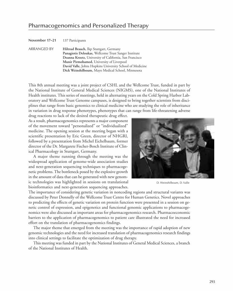
Pharmacogenomics and Personalized Therapy
November 17–21 137 Participants
ARRANGED BY Hiltrud Brauch, Ikp Stuttgart, GermanyPanagiotis Deloukas,Wellcome Trust Sanger InstituteDeanna Kroetz, University of California, San FranciscoMunir Pirmohamed, University of LiverpoolDavid Valle, Johns Hopkins University School of MedicineDick Weinshilboum,Mayo Medical School, Minnesota
This 8th annual meeting was a joint project of CSHL and the Wellcome Trust, funded in part bythe National Institute of General Medical Sciences (NIGMS), one of the National Institutes ofHealth institutes. This series of meetings, held in alternating years on the Cold Spring Harbor Lab-oratory and Wellcome Trust Genome campuses, is designed to bring together scientists from disci-plines that range from basic genomics to clinical medicine who are studying the role of inheritancein variation in drug response phenotypes, phenotypes that can range from life-threatening adversedrug reactions to lack of the desired therapeutic drug effect.As a result, pharmacogenomics represents a major componentof the movement toward “personalized” or “individualized”medicine. The opening session at the meeting began with ascientific presentation by Eric Green, director of NHGRI,followed by a presentation from Michel Eichelbaum, formerdirector of the Dr. Margarete Fischer-Bosch Institute of Clin-ical Pharmacology in Stuttgart, Germany.
A major theme running through the meeting was thewidespread application of genome-wide association studiesand next-generation sequencing techniques to pharmacoge-netic problems. The bottleneck posed by the explosive growthin the amount of data that can be generated with new genom-ic technologies was highlighted in sessions on translationalbioinformatics and next-generation sequencing approaches.The importance of considering genetic variation in noncoding regions and structural variants wasdiscussed by Peter Donnelly of the Wellcome Trust Centre for Human Genetics. Novel approachesto predicting the effects of genetic variation on protein function were presented in a session on ge-netic control of expression, and epigenetics and functional genomic applications to pharmacoge-nomics were also discussed as important areas for pharmacogenomics research. Pharmacoeconomicbarriers to the application of pharmacogenomics to patient care illustrated the need for increasedeffort on the translation of pharmacogenomics findings.
The major theme that emerged from the meeting was the importance of rapid adoption of newgenomic technologies and the need for increased translation of pharmacogenomics research findingsinto clinical settings to facilitate the optimization of drug therapy.
This meeting was funded in part by the National Institutes of General Medical Sciences, a branchof the National Institutes of Health.
293
D. Weinshilboum, D. Valle
247-304_Meetings_Annual Report_2009 template 7/8/11 12:23 PM Page 293

PROGRAM
Opening Session
Translational BioinformaticsChairperson: E. Schadt, Pacific Biosciences, Menlo Park,California
Next-Generation Sequencing Approaches inPharmacogenomics
Chairperson: D. Nickerson, University of Washington, Seattle
Genome-Wide Analyses and CNVsChairperson: P. Donnelly, Wellcome Trust Centre for HumanGenetics, Oxford, United Kingdom
A. Alfireule, P. Deloukas M. Radonjic, B. Drögemüller, G. Wright
A. Frauman
294 Meetings
Genetic Control of ExpressionChairperson: S. Brenner, University of California, Berkeley
Epigenetics and Functional GenomicsChairperson: M. Esteller, Bellvitge Biomedical Research Institute,Barcelona, Spain
ELSI, Pharmacoeconomic Issues, and Population-Based Pharmacogenetics
Chairperson: D. Veenstra, University of Washington, Seattle
247-304_Meetings_Annual Report_2009 template 7/8/11 12:23 PM Page 294
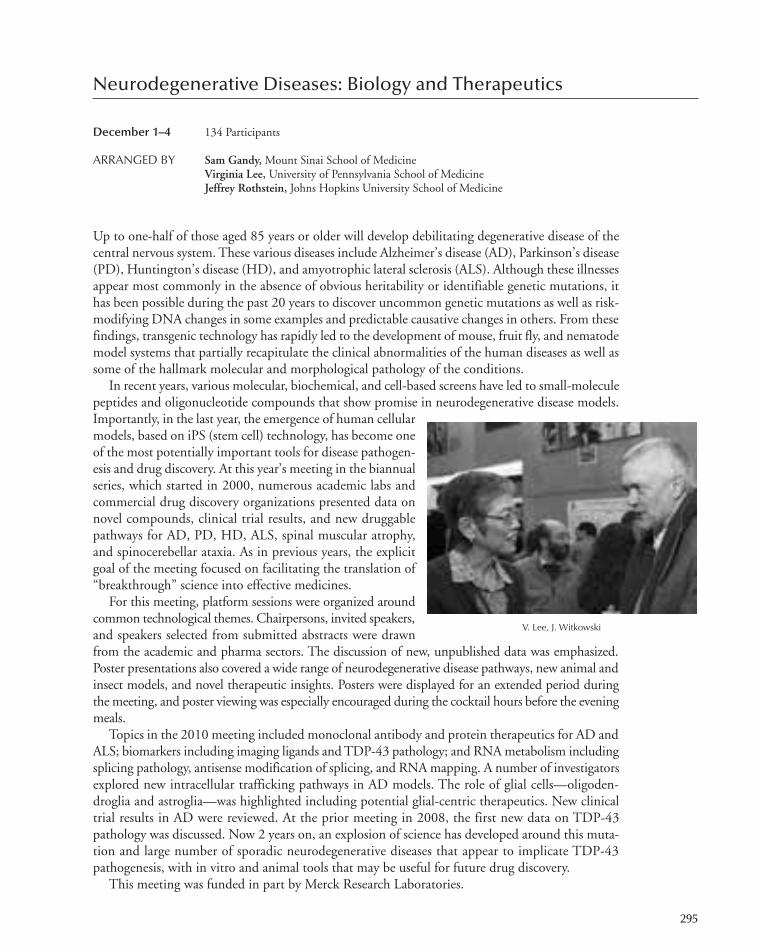
Neurodegenerative Diseases: Biology and Therapeutics
December 1–4 134 Participants
ARRANGED BY Sam Gandy,Mount Sinai School of MedicineVirginia Lee, University of Pennsylvania School of MedicineJeffrey Rothstein, Johns Hopkins University School of Medicine
Up to one-half of those aged 85 years or older will develop debilitating degenerative disease of thecentral nervous system. These various diseases include Alzheimer’s disease (AD), Parkinson’s disease(PD), Huntington’s disease (HD), and amyotrophic lateral sclerosis (ALS). Although these illnessesappear most commonly in the absence of obvious heritability or identifiable genetic mutations, ithas been possible during the past 20 years to discover uncommon genetic mutations as well as risk-modifying DNA changes in some examples and predictable causative changes in others. From thesefindings, transgenic technology has rapidly led to the development of mouse, fruit fly, and nematodemodel systems that partially recapitulate the clinical abnormalities of the human diseases as well assome of the hallmark molecular and morphological pathology of the conditions.
In recent years, various molecular, biochemical, and cell-based screens have led to small-moleculepeptides and oligonucleotide compounds that show promise in neurodegenerative disease models.Importantly, in the last year, the emergence of human cellularmodels, based on iPS (stem cell) technology, has become oneof the most potentially important tools for disease pathogen-esis and drug discovery. At this year’s meeting in the biannualseries, which started in 2000, numerous academic labs andcommercial drug discovery organizations presented data onnovel compounds, clinical trial results, and new druggablepathways for AD, PD, HD, ALS, spinal muscular atrophy,and spinocerebellar ataxia. As in previous years, the explicitgoal of the meeting focused on facilitating the translation of“breakthrough” science into effective medicines.
For this meeting, platform sessions were organized aroundcommon technological themes. Chairpersons, invited speakers,and speakers selected from submitted abstracts were drawnfrom the academic and pharma sectors. The discussion of new, unpublished data was emphasized.Poster presentations also covered a wide range of neurodegenerative disease pathways, new animal andinsect models, and novel therapeutic insights. Posters were displayed for an extended period duringthe meeting, and poster viewing was especially encouraged during the cocktail hours before the eveningmeals.
Topics in the 2010 meeting included monoclonal antibody and protein therapeutics for AD andALS; biomarkers including imaging ligands and TDP-43 pathology; and RNA metabolism includingsplicing pathology, antisense modification of splicing, and RNA mapping. A number of investigatorsexplored new intracellular trafficking pathways in AD models. The role of glial cells—oligoden-droglia and astroglia—was highlighted including potential glial-centric therapeutics. New clinicaltrial results in AD were reviewed. At the prior meeting in 2008, the first new data on TDP-43pathology was discussed. Now 2 years on, an explosion of science has developed around this muta-tion and large number of sporadic neurodegenerative diseases that appear to implicate TDP-43pathogenesis, with in vitro and animal tools that may be useful for future drug discovery.
This meeting was funded in part by Merck Research Laboratories.
295
V. Lee, J. Witkowski
247-304_Meetings_Annual Report_2009 template 7/8/11 12:23 PM Page 295
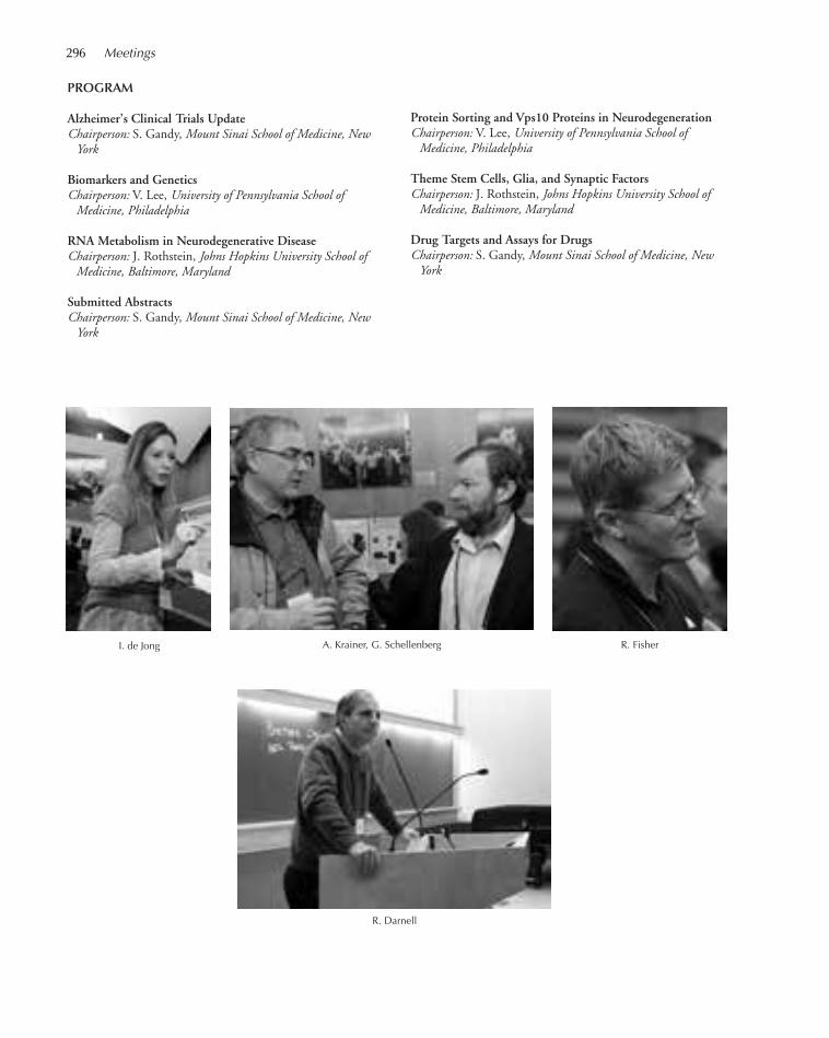
PROGRAM
Alzheimer’s Clinical Trials UpdateChairperson: S. Gandy, Mount Sinai School of Medicine, NewYork
Biomarkers and GeneticsChairperson: V. Lee, University of Pennsylvania School ofMedicine, Philadelphia
RNA Metabolism in Neurodegenerative DiseaseChairperson: J. Rothstein, Johns Hopkins University School ofMedicine, Baltimore, Maryland
Submitted AbstractsChairperson: S. Gandy, Mount Sinai School of Medicine, NewYork
R. Darnell
Protein Sorting and Vps10 Proteins in NeurodegenerationChairperson: V. Lee, University of Pennsylvania School ofMedicine, Philadelphia
Theme Stem Cells, Glia, and Synaptic FactorsChairperson: J. Rothstein, Johns Hopkins University School ofMedicine, Baltimore, Maryland
Drug Targets and Assays for DrugsChairperson: S. Gandy, Mount Sinai School of Medicine, NewYork
A. Krainer, G. Schellenberg R. FisherI. de Jong
296 Meetings
247-304_Meetings_Annual Report_2009 template 7/8/11 12:23 PM Page 296

Automated Imaging and High-Throughput Phenotyping
December 5–8 114 Participants
ARRANGED BY Philip Benfey, Duke UniversityAnne Carpenter, Broad Institute of Harvard and Massachusetts Institute of Technology Robert Waterston, University of WashingtonUwe Ohler, Duke University
This first meeting on Automated Imaging and High-Throughput Phenotyping was enormouslysuccessful. A total of 42 investigators presented in the six scientific sessions, with nearly 32 platformand poster presentations and 114 registered attendees. The six platform sessions and one poster ses-sion were marked by dynamic and enthusiastic exchanges of new results. In the scientific sessions,many audience members participated in the question and answer sessions, and the poster sessionwas well attended.
Sessions at the meeting are listed below. Two additional sessions engaged the community in dis-cussion: Future directions for the community as a whole, and Guidelines for databasing and pub-lishing phenotyping data.
LemnaTec GmbH and Molecular Devices provided partial support for the meeting.
PROGRAM
297
Cellular PhenotypingChairperson: R. Waterston, University of Washington, Seattle
Imaging Technologies and PlatformsChairperson: A. Carpenter, Broad Institute of Harvard andMassachusetts Institute of Technology, Cambridge
Developmental PhenotypingChairperson: F. Piano, New York University, New York
Future Directions
A. Carpenter, P. Benfey
Stimulus/Response PhenotypingChairperson: U. Ohler, Duke University, Durham, NorthCarolina
Image Databasing and Knowledge RepresentationChairperson: P. Benfey, Duke University, Durham, NorthCarolina
Workshop on Databasing/Publication Guidelines
Organismal PhenotypingChairperson: S. Fraser, California Institute of Technology,Pasadena
F. Piano, R. Waterston
247-304_Meetings_Annual Report_2009 template 7/8/11 12:23 PM Page 297
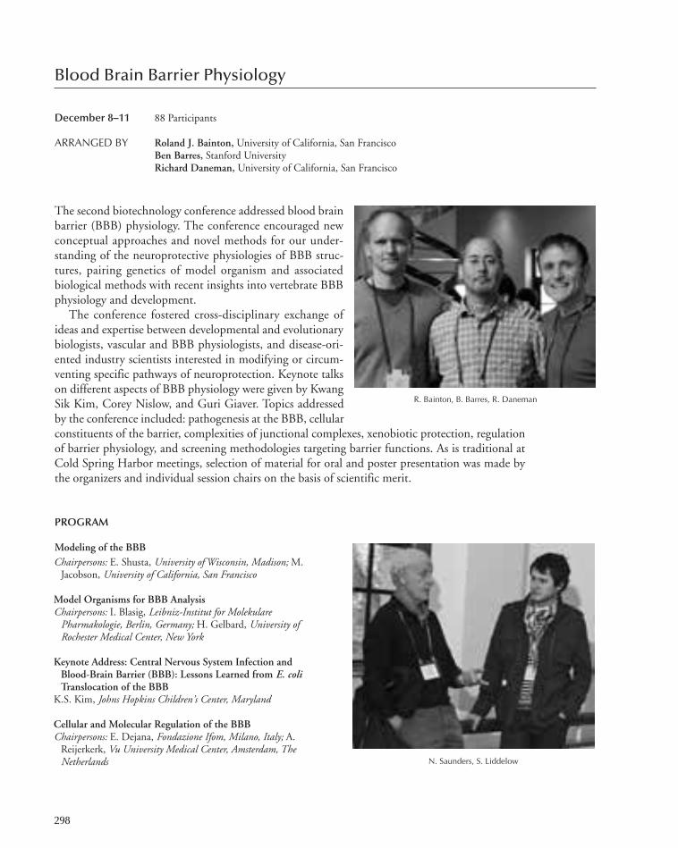
Blood Brain Barrier Physiology
December 8–11 88 Participants
ARRANGED BY Roland J. Bainton, University of California, San FranciscoBen Barres, Stanford UniversityRichard Daneman, University of California, San Francisco
The second biotechnology conference addressed blood brainbarrier (BBB) physiology. The conference encouraged newconceptual approaches and novel methods for our under-standing of the neuroprotective physiologies of BBB struc-tures, pairing genetics of model organism and associatedbiological methods with recent insights into vertebrate BBBphysiology and development.
The conference fostered cross-disciplinary exchange ofideas and expertise between developmental and evolutionarybiologists, vascular and BBB physiologists, and disease-ori-ented industry scientists interested in modifying or circum-venting specific pathways of neuroprotection. Keynote talkson different aspects of BBB physiology were given by KwangSik Kim, Corey Nislow, and Guri Giaver. Topics addressedby the conference included: pathogenesis at the BBB, cellularconstituents of the barrier, complexities of junctional complexes, xenobiotic protection, regulationof barrier physiology, and screening methodologies targeting barrier functions. As is traditional atCold Spring Harbor meetings, selection of material for oral and poster presentation was made bythe organizers and individual session chairs on the basis of scientific merit.
PROGRAM
Modeling of the BBB
R. Bainton, B. Barres, R. Daneman
Chairpersons: E. Shusta, University of Wisconsin, Madison; M.Jacobson, University of California, San Francisco
Model Organisms for BBB AnalysisChairpersons: I. Blasig, Leibniz-Institut for MolekularePharmakologie, Berlin, Germany; H. Gelbard, University ofRochester Medical Center, New York
Keynote Address: Central Nervous System Infection andBlood-Brain Barrier (BBB): Lessons Learned from E. coliTranslocation of the BBB
K.S. Kim, Johns Hopkins Children’s Center, Maryland
Cellular and Molecular Regulation of the BBBChairpersons: E. Dejana, Fondazione Ifom, Milano, Italy; A.
Reijerkerk, Vu University Medical Center, Amsterdam, TheNetherlands N. Saunders, S. Liddelow
298
247-304_Meetings_Annual Report_2009 template 7/8/11 12:23 PM Page 298
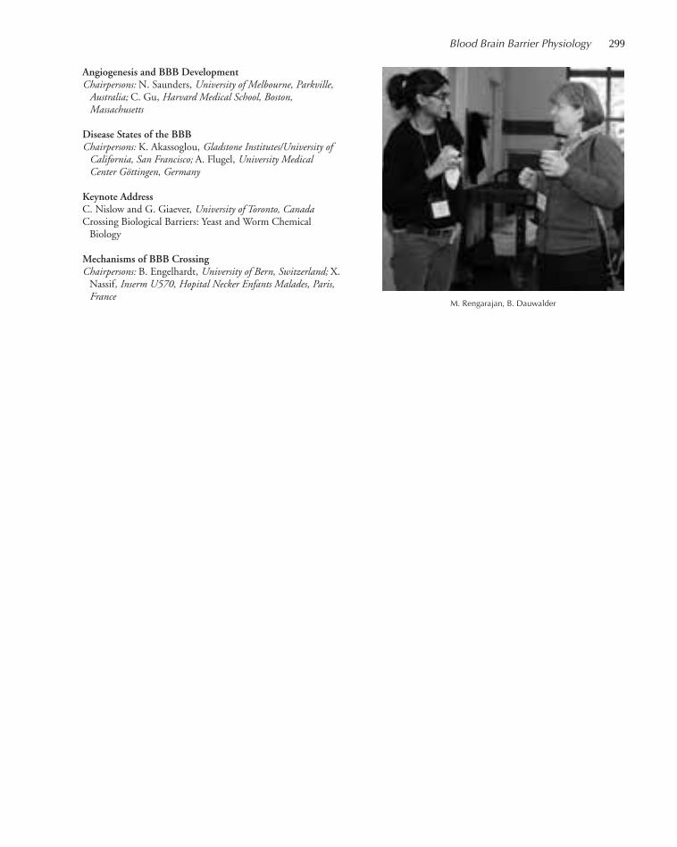
Angiogenesis and BBB DevelopmentChairpersons: N. Saunders, University of Melbourne, Parkville,Australia; C. Gu, Harvard Medical School, Boston,Massachusetts
Disease States of the BBBChairpersons: K. Akassoglou, Gladstone Institutes/University ofCalifornia, San Francisco; A. Flugel, University MedicalCenter Göttingen, Germany
Keynote AddressC. Nislow and G. Giaever, University of Toronto, CanadaCrossing Biological Barriers: Yeast and Worm Chemical
Biology
Mechanisms of BBB CrossingChairpersons: B. Engelhardt, University of Bern, Switzerland; X.
Nassif, Inserm U570, Hopital Necker Enfants Malades, Paris,France
M. Rengarajan, B. Dauwalder
Blood Brain Barrier Physiology 299
247-304_Meetings_Annual Report_2009 template 7/8/11 12:23 PM Page 299
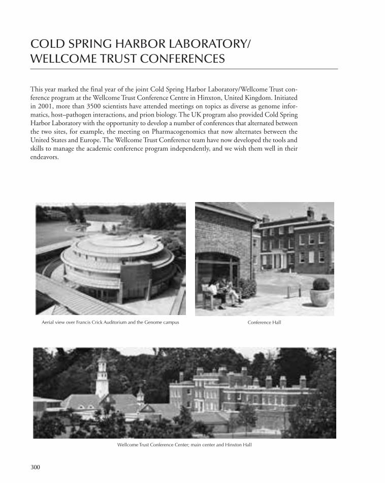
COLD SPRING HARBOR LABORATORY/WELLCOME TRUST CONFERENCES
This year marked the final year of the joint Cold Spring Harbor Laboratory/Wellcome Trust con-ference program at the Wellcome Trust Conference Centre in Hinxton, United Kingdom. Initiatedin 2001, more than 3500 scientists have attended meetings on topics as diverse as genome infor-matics, host–pathogen interactions, and prion biology. The UK program also provided Cold SpringHarbor Laboratory with the opportunity to develop a number of conferences that alternated betweenthe two sites, for example, the meeting on Pharmacogenomics that now alternates between theUnited States and Europe. The Wellcome Trust Conference team have now developed the tools andskills to manage the academic conference program independently, and we wish them well in theirendeavors.
Aerial view over Francis Crick Auditorium and the Genome campus Conference Hall
Wellcome Trust Conference Center; main center and Hinxton Hall
300
247-304_Meetings_Annual Report_2009 template 7/8/11 12:23 PM Page 300

Computational Cell Biology
February 10–14 63 Participants
ARRANGED BY Benjamin Geiger,Weizmann InstituteEdda Klipp,Max-Planck Institute for Molecular GeneticsBela Novak, Oxford Centre for Integrative Systems Biology
The specific goal for this meeting was to foster fruitful and creative dialogue between experimentalcell biologists and mathematical–computational modelers with common interests in the regulationof cell physiology. Emphasis was placed on areas of greatest current interest and importance, suchas cell signaling, motility, cell proliferation and death, calcium dynamics, microbial physiology, anddevelopment. Sessions were also devoted to new experimental technologies and software develop-ments.
Joint Cold Spring Harbor Laboratory/Wellcome Trust conferences at Hinxton are supported inpart with funding courtesy of The Wellcome Trust.
PROGRAM
Keynote AddressesJ. Ferrell, Stanford University, CaliforniaCooperative Phosphorylation in the Regulation of Wee1AB. Scheres, Utrecht University, The NetherlandsMultilevel Analysis of Plant Development
Intracellular SignalingChairperson: M. White, University of Liverpool, UnitedKingdom
Tools and SoftwareChairperson: N. Le Novère, EMBL-European BioinformaticsInstitute, Hinxton, United Kingdom
Software Demo and Poster Session I
Socializing at the meeting during coffee break
301
Self-Organization in CellsChairperson: B. Mulder, FOM Institute AMOLF, Amsterdam,The Netherlands
Cell MotilityChairperson: F. Nédélec, European Molecular BiologyLaboratory, Heidelberg, Germany
Software Demo and Poster Session II
Patterns in DevelopmentChairperson: J. Traas, École Normale Supérieure de Lyon, France
Cell BiophysicsChairperson: K. Kruse, Universität des Saarlandes, Germany
247-304_Meetings_Annual Report_2009 template 7/8/11 12:23 PM Page 301
Systems Biology: Networks
August 11–15 108 Participants
ARRANGED BY Patrick Aloy, Institute for Research Biomedicine, SpainAnne-Claude Gavin, EMBL, GermanyTrey Ideker, University of California, San DiegoMarian Walhout, University of Massachussetts Medical School
The 2010 conference was again held at the Wellcome Trust Conference Centre on the WellcomeTrust Genome Campus in Hinxton, United Kingdom. Network Biology attempts to understandbiology from the point of view of global and local systems properties of molecular networks. Theconference addressed the structure, function, and dynamics of a variety of cellular networks such asgenetic, transcriptional, posttranscriptional gene regulatory, protein–protein interaction, signaling,and metabolic networks. A central theme of the meeting was to understand how network propertiesrelate to phenotypes, including human disease.
Joint Cold Spring Harbor Laboratory/Wellcome Trust conferences at Hinxton are supported inpart with funding courtesy of The Wellcome Trust.
PROGRAM
Gene Regulatory Networks IChairperson: L. Serrano, Centre for Genomic Regulation,Barcelona, Spain
Keynote AddressesC. Boone, University of Toronto, Toronto, CanadaThe Genetic Landscape of a CellE.M. Marcotte, University of Texas, AustinDeaf Plants, Bleeding Yeast, and Other Surprising Disease
Models from Deeply Conserved Gene Networks
Protein–Protein Interaction NetworksChairperson: V. Cheung, University of Pennsylvania,Philadelphia
Genetic NetworksChairperson: H. Zhu, Johns Hopkins University, Baltimore,Maryland
Network MedicineChairperson: P. Braun, Dana-Farber Cancer Institute, Boston,Massachusetts
An animated discussion after one session
302
Gene Regulatory Networks IIChairperson: G. Superti-Furga, Austrian Academy of Sciences,Vienna, Austria
Signaling NetworksChairperson: A. Godzik, Burnham Institute for MedicalResearch, La Jolla, California
Metabolic NetworksChairperson: D. Pe’er, Columbia University, New York
247-304_Meetings_Annual Report_2009 template 7/8/11 12:23 PM Page 302
Infectious Disease Genomics and Global Health
September 12–15 86 Participants
ARRANGED BY Matthew Berriman,Wellcome Trust Sanger InstituteJane Carlton, New York UniversityJulian Parkhill,Wellcome Trust Sanger InstituteGeorge Weinstock,Washington University School of Medicine
This fourth conference was held at the Wellcome Trust Conference Centre on the Wellcome TrustGenome Campus in Hinxton, United Kingdom. The meeting explored how the recent progress inhost, pathogen, and vector genome data and other large data sets is changing the way we thinkabout infectious diseases with an emphasis on global health issues.
Joint Cold Spring Harbor Laboratory/Wellcome Trust conferences at Hinxton are supported inpart with funding courtesy of The Wellcome Trust.
PROGRAM
Keynote AddressD. Kwiatkowski, Oxford University, Sanger Institute
Epidemiology and Public HealthChairpersons: C. Arias, University of Texas Medical School,Houston; P. Bejon, Kemri Wellcome Trust Research Program,Kilifi, Kenya
Viruses IChairpersons: R. Heyderman, Malawi-Liverpool Wellcome Trust,Blantyre, Malawi; P. Kellam, Wellcome Trust Sanger Institute,Hinxton, United Kingdom
Viruses IIChairpersons: C. Simmons, Hospital for Tropical Diseases, Ho
Chi Minh City, Vietnam; L. Kramer, Wadsworth Center,SUNY Albany, New York
Population GenomicsChairpersons: D. Conway, Medical Research Council, TheGambia; P. Keim, Northern Arizona University, Flagstaff
BacteriaChairpersons: N. Day, Mahidol Oxford Tropical MedicineResearch Unit (Moru), Bangkok, Thailand; J. Vinetz,University of California, San Diego
Parasites and VectorsChairpersons: T. Anderson, Southwest Foundation forBiomedical Research, San Antonio, Texas; A. Das, NationalInstitute of Malaria Research (ICMR), New Delhi, India
Coffee break
303
247-304_Meetings_Annual Report_2009 template 7/8/11 12:23 PM Page 303

Genome Informatics
September 15–19 256 Participants
ARRANGED BY Inanc Birol, BC Cancer Agency, CanadaMichele Clamp, The Broad Institute of Harvard and Massachusetts Institute of Technology James Kent, University of California, Santa Cruz
This conference focuses on large-scale genome informatics. Biology is an experimental science thatis experiencing an explosion of new data. This requires biologists to increase the scale and sophisti-cation in the information technology used for their research. The conference scope encompassesthe management and the analysis of these data, such as whole-genome comparisons within andamong species and strains, the analysis of results from high-throughput experiments to uncover cel-lular pathways and molecular interactions, and the design of effective algorithms to identify regu-latory sequence motifs. It was the tenth Cold Spring Harbor Laboratory/Wellcome Trust Conferenceon Genome Informatics to be held at the Wellcome Trust Conference Centre on the WellcomeTrust Genome Campus in Hinxton, United Kingdom.
Joint Cold Spring Harbor Laboratory/Wellcome Trust conferences at Hinxton are supported inpart with funding courtesy of The Wellcome Trust.
PROGRAM
Keynote Address: RNA Wikiproject—CommunityAnnotation of RNA Families
A. Bateman, Wellcome Trust Sanger Institute, Hinxton, UnitedKingdom
Sequencing Pipelines and AssemblyChairpersons: Z. Torok, Astrid Research, Debrecen, Hungary; I.
Birol, BC Cancer Agency, Vancouver, Canada
Population and Statistical GenomicsChairpersons: R. Durbin, Wellcome Trust Sanger Institute,Hinxton, United Kingdom; E. Margulies, NIGRII, NationalInstitutes of Health, Bethesda, Maryland
Environmental and Medical GenomicsChairpersons: T. Thorgeirsson, University of California, SantaCruz; M. Clamp, Bioteam, Middleton, Massachusetts
Databases, Data Mining, Visualization, and CurationChairpersons: D. Dooling, Washington University School ofMedicine, St. Louis, Missouri; M. Krzywinski, BC CancerAgency, Vancouver, Canada
Epigenomics and Gene RegulationChairpersons: W. Noble, University of Washington, Seattle;
Z. Weng, University of Massachusetts Medical School,Worcester
Comparative and Evolutionary GenomicsChairpersons: S. Batzoglou, Stanford University, California; P.
Flicek, European Bioinformatics Institute, Hinxton, UnitedKingdom
Sequencing and Gene PredictionChairpersons: A. Mortazavi, California Institute of Technology,Pasadena; L. Pachter, University of California, Berkeley
Discussing and looking over posters
304
247-304_Meetings_Annual Report_2009 template 7/8/11 12:23 PM Page 304
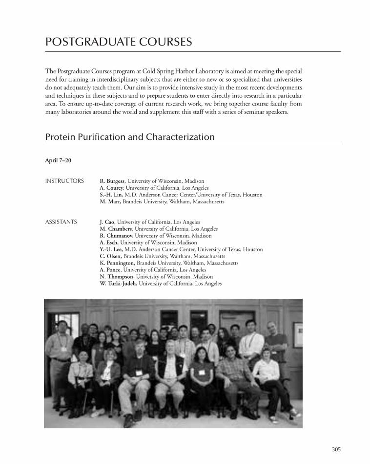
POSTGRADUATE COURSES
The Postgraduate Courses program at Cold Spring Harbor Laboratory is aimed at meeting the specialneed for training in interdisciplinary subjects that are either so new or so specialized that universitiesdo not adequately teach them. Our aim is to provide intensive study in the most recent developmentsand techniques in these subjects and to prepare students to enter directly into research in a particulararea. To ensure up-to-date coverage of current research work, we bring together course faculty frommany laboratories around the world and supplement this staff with a series of seminar speakers.
Protein Purification and Characterization
April 7–20
INSTRUCTORS R. Burgess, University of Wisconsin, MadisonA. Courey, University of California, Los AngelesS.-H. Lin, M.D. Anderson Cancer Center/University of Texas, HoustonM. Marr, Brandeis University, Waltham, Massachusetts
ASSISTANTS J. Cao, University of California, Los AngelesM. Chambers, University of California, Los AngelesR. Chumanov, University of Wisconsin, MadisonA. Esch, University of Wisconsin, MadisonY.-U. Lee, M.D. Anderson Cancer Center, University of Texas, HoustonC. Olsen, Brandeis University, Waltham, MassachusettsK. Pennington, Brandeis University, Waltham, MassachusettsA. Ponce, University of California, Los AngelesN. Thompson, University of Wisconsin, MadisonW. Turki-Judeh, University of California, Los Angeles
305
305-366_courses_Annual Report_2009 template 7/8/11 12:24 PM Page 305

This course was for scientists who were not familiar with techniques of protein isolation and char-acterization. It was a rigorous program that included laboratory work all day and a lecture with dis-cussion sessions every evening. Each student became familiar with each of the major techniques inprotein purification by actually performing four separate isolations including (1) a regulatory proteinfrom muscle tissue, (2) a sequence-specific DNA-binding protein, (3) a recombinant protein over-expressed in Escherichia coli, and (4) a membrane-bound receptor. A variety of bulk fractionation,electrophoretic, and chromatographic techniques included precipitation by salts, pH, and ionicpolymers; ion exchange, gel filtration, hydrophobic interaction, and reverse phase chromatography;lectin affinity, ligand affinity, oligonucleotide affinity, and immunoaffinity chromatography; poly-acrylamide gel electrophoresis, and electroblotting; and high-performance liquid chromatography.Procedures were presented for solubilizing proteins from inclusion bodies and refolding them intoactive monomeric forms. Methods of protein characterization were utilized to include immunolog-ical and biochemical assays, peptide mapping, amino acid analysis, protein sequencing, and massspectrometry. Emphasis was placed on strategies of protein purification and characterization. Guestlecturers discussed protein structure, modification of proteins, methodologies for protein purifica-tion and characterization, and applications of protein biochemistry to cell and molecular biologyand oncology.
This course was supported with funds provided by the National Cancer Institute.
PARTICIPANTS
Da Ros, M., M.Sc., University of Turku, FinlandDantoft, T., M.S., Aarhus University, Faculty of HealthScience, Denmark
Du, J., Ph.D., Medical College of Wisconsin, MilwaukeeFlores, G., B.S., University of Puerto Rico, San JuanFoeger, N., B.A., Washington University in St. Louis, Missouri Giniatullina, A., M.S., VU University Amsterdam, TheNetherlands
Girgis, H., Ph.D., Massachusetts General Hospital, BostonJin, J.-K., B.S., University of Texas M.D. Anderson CancerCenter, Houston
Kalendova, A., M.S., Institute of Molecular Genetics AS CR,
Prague, Czech RepublicLi, D., B.S., Massachusetts Institute of Technology,Cambridge
Lloyd, F., M.D., Indiana/Purdue University at Indianapolis,Indianapolis
Mosebi, S., Ph.D., Research Scientist at Mintek, Randburg,South Africa
Park, Y., Ph.D., University of Arkansas, Pine BluffRak, R., B.S., Tel-Aviv University, IsraelRincon, H., Ph.D., Fred Hutchinson Cancer Research Center,Seattle, Washington
Xu, J., Ph.D., University of California, Irvine
SEMINARS
Burgess, R., University of Wisconsin, Madison: Introductionto protein purification; RNA polymerase–Sigma factorinteractions and use of LRET-based assays for drug discoveryand biochemistry.
Chumanov, R., University of Wisconsin, Madison: Halo tagand purification of CARMI.
Courey, A., University of California, Los Angeles: System-wideanalyses of Groucho and SUMO in Drosophila development.
Lin, S.-H., M.D. Anderson Cancer Center, Houston, Texas:Cadherin-11 in prostate cancer bone metastasis.
Marr, M., Brandeis University, Waltham, Massachusetts:Transcriptional and translational cellular responses to stress.
Thompson, N., University of Wisconsin, Madison:Immunoaffinity chromatography.
306 Postgraduate Courses
305-366_courses_Annual Report_2009 template 7/8/11 12:24 PM Page 306

Cell and Developmental Biology of Xenopus
April 9–20
INSTRUCTORS R. Keller, University of Virginia, CharlottesvilleK. Kroll, Washington University School of Medicine, St. Louis, Missouri
CO-INSTRUCTORS A. Sater, University of Houston, TexasG. Thomsen, Stony Brook University, Stony Brook, New York
ASSISTANTS B. Dzamba, University of Virginia, CharlottesvilleT. Nakayama, University of Virginia, CharlottesvilleK. Pfister, University of Virginia, Charlottesville
Xenopus is the leading vertebrate model for the study of gene function in development. The com-bination of lineage analysis, gene-knockout strategies, experimental manipulation of the embryo,and genomic/bioinformatic techniques makes it ideal for studies on the molecular control of embryopatterning, morphogenesis, and organogenesis. The course combined intensive laboratory trainingwith daily lectures from recognized experts in the field. Students learned both emerging technologiesand classical techniques to study gene function in Xenopus development. An important element wasthe informal interaction between students and course faculty. Technologies included oocyte and
307
305-366_courses_Annual Report_2009 template 7/8/11 12:24 PM Page 307

embryo culture, lineage analysis and experimental manipulation of embryos, time-lapse imaging ofmorphogenesis, gain- and loss-of-function analyses using mRNAs and antisense oligos, whole-mount in situ hybridization, immunocytochemistry, RT-PCR, genomic/bioinformatic techniques,preparation of transgenic embryos, and use of Xenopus tropicalis for genetic analyses. This coursewas designed for those new to the Xenopus field, as well as for those wanting a refresher course inthe emerging technologies. The course was open to investigators from all countries.
This course was supported with funds provided by the National Institute of Child Health andHuman Development, the National Science Foundation, and the Howard Hughes Medical Insti-tute.
PARTICIPANTS
308
308 Postgraduate Courses
Amin, N., Ph.D., University of North Carolina, Chapel HillBarnett, C., B.A., University of Alaska, AnchorageBertke, M., B.S., University of Notre Dame, Notre Dame,Indiana
Charney, R., B.A., University of California, Irvine, CaliforniaCheng, W.-C., B.Sc., Kings College, London, UnitedKingdom
Gibb, N., B.Sc., University of Aberdeen, United KingdomLloyd, R., Ph.D., Institute of Zoology, Zoological Society ofLondon, United Kingdom
Messina, A., M.A., University of Trento, Italy
Nedelkovska, H., B.S., University of Rochester MedicalCenter, New York
Ploper, D., B.S., HHMI/University of California, Los AngelesRiechert, S., M.S., Cancer Research, UK–London ResearchInstitute, United Kingdom
Schneider, I., M.Sc., University of Hohenheim, Stuttgart,Germany
Stellabotte, F., Ph.D., House Ear Institute, Los Angeles,California
Walentek, P., M.S., University of Hohenheim, Stuttgart,Germany
SEMINARS
Christian, J., Oregon Health and Science University, Portland:TGF-β superfamily signaling in early Xenopus development.
Grainger, R., University of Virginia, Charlottesville: Genomicapproaches for enhancer identification in Xenopus.
Harland, R., University of California, Berkeley: Biology of theorganizer.
Houston, D., University of Iowa, Iowa City: Roles of localizedmaternal mRNAs in Xenopus axis formation.
Keller, R., University of Virginia, Charlottesville: Introductionto Xenopus morphogenesis.
Khokha, M., Yale University School of Medicine, New Haven,Connecticut: Introduction to Xenopus tropicalis.
Kintner, C., The Salk Institute, La Jolla, California: Cellbiological analysis of early development.
Nakayama, T., University of Virginia, Charlottesville:Approaches for manipulating gene expression in Xenopus.
Vize, P., University of Calgary, Canada: Xenbase.Zimmerman, L., National Institute for Medical Research,London, United Kingdom: Genetics in Xenopus tropicalis.
305-366_courses_Annual Report_2009 template 7/8/11 12:24 PM Page 308

Workshop on Schizophrenia and Related Disorders
June 9–15
INSTRUCTORS A. Malhotra, Zucker Hillside Hospital, Glen Oaks, New YorkA. Law, National Institute of Mental Health, NIH, Bethesda, Maryland
CO-INSTRUCTOR J. Hall, University of Edinburgh, United Kingdom
ASSISTANT P. DeRosse, Zucker Hillside Hospital, Glen Oaks, New York
This workshop provided students with the most current understanding of the molecular, cellular,and neural systems underlying the disturbances in brain function in these devastating illnesses. Dur-ing the 7-day workshop, students learned about the clinical aspects of schizophrenia, schizoaffectivedisorder, and bipolar disorder, as well as explored in detail the genetic and neurobiological under-pinnings of these complex psychiatric disorders. The Workshop included sessions focused on TheClinical Syndrome, Basic Neurobiology, Cognitive Neuroscience, Neuroimaging, Genetics and Ge-nomics, Endophenotypes, and Gene Expression and Gene Modulation. In addition to hearing aboutthe most recent research in these areas, controversial topics and challenges to basic assumptions inthe field were explored and discussed. A diverse faculty brought the most up-to-date results andtheories to the students, making this Workshop a valuable resource for young researchers startingout in this fast-moving and expansive field. Not only did it help them build the foundation for theirfuture research, it also introduced them to many potential collaborators working to understandschizophrenia and similar disorders from different perspectives.This course was supported with funds provided by the Oliver Grace Fund.
309
305-366_courses_Annual Report_2009 template 7/8/11 12:24 PM Page 309

310 Postgraduate Courses
PARTICIPANTS
Balu, D., Ph.D., McLean Hospital, Belmont, MassachusettsBecerril, K., B.S., Washington University, St. Louis, MissouriChang, W., B.S., University of California San Diego, La JollaCui, X., Ph.D., Sigma Advanced Genetic Engineering (SAGE)Labs, St. Louis, Missouri
Dowd, E., B.S., Washington University, St. Louis, MissouriGallego, J., Ph.D., Zucker Hillside Hospital, Glen Oaks, NewYork
Kleinhaus, K., Ph.D., New York University School ofMedicine
Maher, B., Ph.D., University of Connecticut, StorrsMalavasi, E., B.S., University of Edinburgh, United KingdomMorris, H., Ph.D., Johns Hopkins Medical Institution,Baltimore, Maryland
Nucifora, F., Ph.D., Johns Hopkins School of Medicine,Baltimore, Maryland
Radhakrishnan, R., Ph.D., Yale University School ofMedicine, New Haven, Connecticut
Smith, S., B.S., University of Missouri, Saint LouisStilo, S., Ph.D., Psychosis Clinical Academic Group, London,United Kingdom
Sussmann, J., Ph.D., Edinburgh University, Edinburgh,United Kingdom
Wang, X., Ph.D., University of Southern California, LosAngeles
Ward, K., B.S., Pfizer Global Research and Development,Groton, Connecticut
Woods, G., B.S., University of California, DavisZhang, J., Ph.D., Zucker Hillside Hospital, Glen Oaks, NewYork
Zhu, S., Ph.D., Johns Hopkins University School ofMedicine, Baltimore, Maryland
SEMINARS
Abi-Dargham, A., Columbia University, New York: PETimaging in schizophrenia.
Akbarian, S., University of Massachusetts, Medical School,Worcester: Epigenetics in schizophrenia.
Calicott, J., National Institute of Mental Health, NIH,Bethesda, Maryland: Functional MR in schizophrenia.
Cornblatt, B., Zucker Hillside Hospital, Glen Oaks, NewYork: Cognitive and psychosocial risk factors.
DeRosse, P., Zucker Hillside Hospital, Glen Oaks, New York:Introduction to the clinical syndrome.
Fletcher, P., University of Cambridge, United Kingdom: Acognitive neuroscientific model of the positive symptoms ofschizophrenia.
Hall, J., University of Edinburgh, United Kingdom: Cognitionof schizophrenia and imaging genomics.
Hyde, T., National Institute of Mental Health, NIH, Bethesda,Maryland: Postmortem gene expression inneurodevelopment.
Jarskog, F., Columbia University, New York: Treatmentstrategies.
Law, A., National Institute of Mental Health, Bethesda,Maryland: NRG/ErbB signaling and risk for schizophrenia:The genetics of gene regulation and function.
Lawrie, S., University of Edinburgh, United Kingdom:Structural imaging in schizophrenia.
Lewis, D., University of Pittsburgh, Pennsylvania: CorticalGABA systems in schizophrenia.
Li, B., Cold Spring Harbor Laboratory: Glutamatergic synapseand its receptors: From cognitive function to psychiatricdisorder.
Malhotra, A., Zucker Hillside Hospital, Glen Oaks, NewYork: Pharmacogenomics in schizophrenia.
Moghaddam, B., University of Pittsburgh, Pennsylvania: Theglutamatergic system.
O’Donnell, P., University of Maryland, Baltimore: Thedopaminergic system.
O’Donovan, M., University of Wales, Cardiff, UnitedKingdom: GWAS and candidate genes in schizophrenia.
Porteous, D., University of Edinburgh, United Kingdom:Common and rare variants in schizophrenia: The DISC1example.
Sebat, J., University of California, San Diego: Rare variants inschizophrenia.
Song, H., Johns Hopkins University, Baltimore, Maryland:Neurogenesis and iPS in schizophrenia.
Susser, E., Columbia University, New York: Epidemiology andenvironmental risk factors.
Weinberger, D., National Institute of Mental Health,Bethesda, Maryland: Intermediate phenotypes andschizophrenia-associated genes.
305-366_courses_Annual Report_2009 template 7/8/11 12:24 PM Page 310

Advanced Bacterial Genetics
June 9–29
INSTRUCTORS J. Kirby, University of Iowa, Iowa CityS. Lovett, Brandeis University, Waltham, MassachusettsA. Segall, San Diego State University, San Diego, California
ASSISTANTS K. Elliott, University of Georgia, Westminster, South CarolinaJ. Rostron, University of Iowa, Iowa CityC. Weitzel, Brandeis University, Waltham, MassachusettsJ. Willett, University of Iowa, Iowa City
This course presented logic and methods used in the genetic dissection of complex biologicalprocesses in diverse bacteria. Laboratory methods included classical mutagenesis using transposons,mutator strains, and chemical and physical mutagens; detection and quantitation of gene expressionchanges using various reporter genes and real-time PCR; the mapping of mutations using geneticand physical techniques; modern approaches to the generation and analysis of targeted gene dis-ruptions and fusions using PCR and cloning methods; epitope insertion mutagenesis; site-directedmutagenesis; and fluorescence microscopy. Key components of the course were the use of sophisti-
311
305-366_courses_Annual Report_2009 template 7/8/11 12:24 PM Page 311

cated genetic methods in the analysis of model bacteria (including Escherichia coli, Salmonella, Bacil-lus subtilis, and Myxococcus xanthus) and the use of the wealth of new genomic sequence informationto motivate these methods. Invited lecturers presented various genetic approaches to study bacterialmechanisms of metabolism, development, and pathogenesis.
This course was supported with funds provided by the National Science Foundation.
PARTICIPANTS
312 Postgraduate Courses
Arnoldini, M., M.Sc., ETH Zurich, SwitzerlandBarrett, E., B.S., Dartmouth College, Hanover, NewHampshire
Brolund, A., M.Sc., Karolinska Institute, Stockholm, SwedenCady, K., B.S., Dartmouth Medical School, Lebanon, NewHampshire
Carpino, J., B.S., Queens College and CUNY GraduateCenter, Flushing, New York
Hao, L., M.S., University of Wisconsin, MadisonJun, S., B.Sc., Harvard University, Cambridge, MassachusettsKuehn, S., Ph.D., The Rockefeller University, New YorkLazova, M., M.Sc., FOM Institute AMOLF, Amsterdam, The Netherlands
Lewis, E., B.S., University of California, IrvineLiu, X., Ph.D., Harvard Medical School, Boston,Massachusetts
Rae, R., B.S., Max-Planck Institute for DevelopmentalBiology, Tuebingen, Germany
Rodriguez Esperon, M., B.S., Institute De Invest. ClementeEstable, Montevideo, Uruguay
Sankararaman, S., Ph.D., University of California, BerkeleyShanahan, E., B.Sc., Centenary Institute University of Sydney,Camperdown, Australia
Viburiene, R., M.S., University of Oslo, Norway
SEMINARS
Edwards, R., San Diego State University, California: Usinggenomics and metagenomics to understand the world around us.
Hughes, D., Uppsala University, Sweden: Bacterial mutantsselected to cope with stressful growth conditions.
Kirby, J., University of Iowa, Iowa City: Chemosensoryregulation of biofilm formation.
Laub, M., Massachusetts Institute of Technology/HHMI,Cambridge: Specificity and evolution of bacterial signalingpathways.
Lazazzera, B., University of California, Los Angeles:Identifying pathways to control surface-adhered biofilmformation.
Lovett, S., Brandeis University, Waltham, Massachusetts:Replication gap repair.
Rohwer, F., San Diego State University, California: Humanvirome.
Rudner, D., Harvard Medical School, Boston, Massachusetts:Regulation of essential cell biological processes duringbacterial differentiator.
Segall, A., San Diego State University, California: Inhibitors ofDNA repair and the serendipity of science.
Storz, G., National Institute of Child Health and HumanDevelopment, Bethesda, Maryland: The genes that weremissed.
Whitaker, R., University of Illinois, Urbana: Dynamics ofhost–virus interactions across space and time.
Yildiz, F., University of California, Santa Cruz: Analysis ofbiofilm formation in Vibrio cholerae.
305-366_courses_Annual Report_2009 template 7/8/11 12:24 PM Page 312

Ion Channel Physiology
June 9–29
INSTRUCTORS N. Golding, University of Texas, AustinA. Lee, University of Iowa, Iowa CityM. Nolan, University of Edinburgh, United Kingdom
ASSISTANTS C. Bladen, University of Calgary, Alberta, CanadaK. Buchanan, University College London, United KingdomP. Dodson, University of Edinburgh, United KingdomA. Graves, Northwestern University, Evanston, IllinoisK. Hansen, Emory University, Atlanta, GeorgiaP. Kammermeier, University of Rochester, New YorkS. Khurana, University of Texas, AustinL. Scott, University of Texas, AustinK. Vance, Emory University, Atlanta, Georgia
The primary goal of this course was to investigate, through lectures and laboratory work, the prop-erties of ion channels that allow neurons to carry out their unique physiological functions in a varietyof neural systems. Areas of particular interest included channels that (1) are activated by neuro-transmitters at central and peripheral synapses, (2) are activated by voltage changes in axons anddendrites, (3) respond to neuromodulators with changes in functional properties, or (4) are devel-opmentally required and regulated. The research interests of guest lecturers reflected these areas ofemphasis.
313
305-366_courses_Annual Report_2009 template 7/8/11 12:24 PM Page 313
The laboratory component of the course introduced students to electrophysiological approachesfor the study of ion channels in their native environments. Hands-on exercises included patch-clamp recording of ion channel activity in acutely isolated or cultured cells or neurons in brain slicepreparations. Different recording configurations were used (e.g., whole-cell, cell-free, and nucleatedpatches) to examine macroscopic or single-channel activity. Similarly, various methods of ligandand drug application were demonstrated. The advantages and disadvantages of each method, prepa-ration, and recording technique were considered in relation to the specific scientific questions beingasked. Admissions priority was given to students and postdocs with a demonstrated interest andspecific plans, as well as a supportive environment to apply these techniques to a defined problem.
This course was supported with funds provided by the Howard Hughes Medical Institute.
PARTICIPANTS
Altimimi, H., B.S., McGill University, Montreal, CanadaGavin, C., B.A., The Scripps Research Insitute, Florida
Campus, JupiterGoodfellow, N., B.S., University of Toronto, CanadaGutnisky, D., Ph.D., Janelia Farm-HHMI, Ashburn,
VirginiaKalia, J., M.S., National Institutes of Health, Bethesda,
MarylandKim, S.E., M.S., The Scripps Research Institute, La Jolla,
California
Kuo, C., Ph.D., Duke University Medical Center, Durham,North Carolina
Madry, C., Ph.D., University College London, UnitedKingdom
Moscato, E., B.A., University of Pennsylvania, PhiladelphiaSchoonover, C., Ph.D., Columbia University, New YorkSurmeli, G., B.S., Columbia University, New YorkTauchmann, M., B.A., Arizona State University, Tempe
SEMINARS
Deisseroth, K., Stanford University, Stanford, California:Channel engineering.
Dolphin, A., University College London, United Kingdom:Voltage-gated Ca2+ channels. The α2δ subunits of voltage-gated calcium channels: Involvement in disease and targetsite for drugs.
Dudman, J., Janelia Farm Research Campus, Ashburn,Virginia: Hyperpolarization-activated channels.
Farrant, M., University College London, United Kingdom:Inhibitory synaptic transmission and differential activationof GABAA receptor subtypes.
Golding, N., University of Texas, Austin: Synaptic integrationand integrative properties for auditory temporal coding.
Jan, L., University of California, San Francisco: K+ channelsand Ca+-activated C1-channels.
Kaczmarek, L., Yale University School of Medicine, NewHaven, Connecticut: Ion channel modulation. Regulation of neuronal timing by short-term and long-term channelmodulation.
Larsson, P., University of Miami, Florida: Voltage-gated K+
channels and molecular mechanisms of voltage-gated protonchannels.
Raman, I., Northwestern University, Evanston, Illinois: Nachannels. Biophysics and physiology. Sodium currentsspecializations in spontaneous spiking and cerebellarsignaling.
Sjostrom, J., University College London, United Kingdom:Plasticity and LTP. Spike-timing-dependent plasticity inneocortex.
314 Postgraduate Courses
305-366_courses_Annual Report_2009 template 7/12/11 10:07 AM Page 314
Molecular Embryology of the Mouse
June 9–29
INSTRUCTORS K. Hadjantonakis, Memorial Sloan-Kettering Cancer Institute, New YorkL. Pevny, University of North Carolina, Chapel HillJ. Rivera-Perez, University of Massachusetts Medical School, Worcester
ASSISTANTS D. Escalante-Alcalde, Institute of Cellular Physiology–UNAM, Distrito Federal, CP, MexicoA. Ferrer-Vaquer, Memorial Sloan-Kettering Cancer Institute, New YorkD. Matsushima, University of North Carolina, Chapel HillG. Tortelote, University of Massachusetts Medical School, Worcester
This intensive laboratory and lecture course was designed for biologists interested in applying theirexpertise to the study of mouse development. Lectures provided the conceptual basis for contempo-rary research in mouse embryogenesis and organogenesis, and laboratory practicals provided extensivehands-on introduction to mouse embryo analysis. Experimental techniques covered included in vitroculture and manipulation of pre- and postimplantation embryos, embryo transfer, culture and geneticmanipulation of embryonic stem cells, production of chimeras by embryo aggregation and by EScell injection, and transgenesis by pronuclear microinjection. In addition, this year’s practicals featuredincreased emphasis on phenotypic analysis of mutants, including techniques of histology, in situ hy-bridization, immunohistochemistry, skeletal preparation, organ culture, and tissue recombination.The course also introduced the generation of iPS cells (induced pluripotent cells) and time-lapse mi-croscopy of early gastrulation embryos and organ cultures (eye, kidney, and gut).
This course was supported with funds provided by the National Cancer Institute.
315
305-366_courses_Annual Report_2009 template 7/8/11 12:24 PM Page 315
316 Postgraduate Courses
PARTICIPANTS
Akinci, U., Ph.D., Erasmus Medical Center, Rotterdam, TheNetherlands
Aylor, D., Ph.D., University of North Carolina, Chapel HillAzzoni, E., M.S., H. San Raffaele–DIBIT, Milan, ItalyBezzi, M., M.D., Institute of Molecular and Cell Biology,Singapore
Chang, C., M.S., The Rockefeller University, New YorkCooper, L., Ph.D., University of Illinois, UrbanaGoetz, R., Dipl., University of Freiburg, GermanyGünesdogan, U., Dipl., Max-Planck Institute for BiophysicalChemistry, Göttingen, Germany
Lim, C., Ph.D., Institute of Medical Biology, SingaporeMuta, Y., Ph.D., RIKEN, Kobe, JapanPeyrot, S., Ph.D., University of Texas, AustinRyall, J., Ph.D., National Institutes of Health, Bethesda,Maryland
Vagin, V., Ph.D., Cold Spring Harbor LaboratoryWat, M., B.S., Baylor College of Medicine, Houston, Texas
SEMINARS
Anderson, K., Memorial Sloan-Kettering Cancer Institute,New York: ENU screens and signaling pathways.
Behringer, R., The University of Texas/M.D. Anderson CancerCenter: Transgenics and Evo-Devo.
Capel, B., Duke University Medical Center, Durham, NorthCarolina: Germ cells and the transcriptional network in thegonad.
Chapman, D., University of Pittsburgh, Pennsylvania:Somitogenesis.
Conlon, F., University of North Carolina, Chapel Hill: Heartdevelopment.
Costantini, F., Columbia University, College of Physicians &Surgeons, New York: Kidney development.
Dickinson, M., Baylor College of Medicine, Houston, Texas:Cardiovascular function and imaging.
Eggenschwiler, J., Princeton University, New Jersey: Geneticdissection of neural tube development.
Epstein, D., University of Pennsylvania, Philadelphia: Earlyneural development.
Hadjantonakis, K., Memorial Sloan-Kettering CancerInstitute, New York: Imaging early embryo morphogenesis.
Hochedlinger, K., Masschusetts General Hospital, Boston:Nuclear reprogramming and iPS cells.
Hogan, B., Duke University Medical Center, Durham, NorthCarolina: Lung development and progenitor cells.
Johnson, R., Anderson Cancer Center, Houston, Texas: Limbdevelopment and hippo signaling.
Joyner, A., Memorial Sloan-Kettering Cancer Institute, NewYork: Patterning and fate mapping of the neural system.
Lovell-Badge, R., MRC National Institute for MedicalResearch, London, United Kingdom: Primary sexorientation.
Magnuson, T., University of North Carolina, Chapel Hill:Epigenetics.
Nagy, A., Samuel Lunenfeld Research Institute, Toronto,Canada: ES cells, chimeras, and recombinases.
Papioannou, V., Columbia University, New York: The mouseas a model system.
Pevny, L., University of North Carolina, Chapel Hill:Transcriptional regulation of retinogenesis.
Rivera, J., University of Massachusetts Medical School,Worcester: Lineage analysis and early postimplantationdevelopment.
Robertson, E., University of Oxford, United Kingdom: Nodalsignaling and axial development.
Shen, M., Columbia Unversity Medical Center, New York:Prostate development, stem cells, and cancer.
Solter, D., Institute of Medical Biology, Singapore, Singapore:Preimplantation development.
Soriano, P., Mount Sinai School of Medicine, New York: Genetrapping and signaling pathways.
Sussel, L., Columbia University, New York: Pancreasdevelopment.
Takahashi, J., HHMI/University of Texas SouthwesternMedical Center, Dallas: Circadian rhythms.
Tam, P., Children’s Medical Research Institute, Westmead,Sydney, Australia: Gastrulation.
Threadgill, D., North Carolina State University, Raleigh:Mouse genetics.
Trainor, P., Stowers Institute for Medical Research, KansasCity, Missouri: The neural crest in craniofacial development,evolution, and disease.
Turnbull, D., New York University Medical Center, New York:Imaging modailities and applications: Ultrasound and MRI.
305-366_courses_Annual Report_2009 template 7/8/11 12:24 PM Page 316
Integrative Statistical Analysis of Genome-Scale Data
June 16–29
INSTRUCTORS H. Bussemaker, Columbia University, New YorkV. Carey, Harvard University, Boston, MassachusettsM. Reimers, Virginia Commonwealth University, Richmond
ASSISTANTS N. Altman, Penn State, University ParkS. Bekiranov, University of Virginia, CharlottesvilleM. Figueroa,Weill Cornell Medical College, New YorkT. Guennel, Virginia Commonwealth University, RichmondA. Lazarovici, Columbia University, New YorkT. Riley, Columbia University, New York
Genome-scale data sets are a central feature of modern biological research. Experimental and com-putational biologists who want to get the most from their data sets need to have a firm grasp of sta-tistical and analytical methodology. This course was designed to build competence in quantitativemethods for the analysis of high-throughput molecular biology data. Detailed lectures and presen-tations by guest speakers in the morning and evening were combined with hands-on computer tu-torials in the afternoon, in which the methods covered in the lectures were applied to actualhigh-throughput data for yeast and humans. Students were assumed to have a basic familiarity withthe R programming language at the start of the course.
This course was supported with funds provided by the National Cancer Institute.
317
305-366_courses_Annual Report_2009 template 7/8/11 12:24 PM Page 317

318 Postgraduate Courses
PARTICIPANTS
Bartonicek, N., B.S., European Molecular BiologyLaboratories, Cambridge, United Kingdom
Berko, E., B.A., Albert Einstein College of Medicine, Bronx,New York
Cai, Y., M.S., Cold Spring Harbor LaboratoryCho, M., M.D., Brigham & Women’s Hospital, Boston,Massachusetts
Fendler, B., Ph.D., Cold Spring Harbor LaboratoryFertig, E., Ph.D., Johns Hopkins University, Baltimore,Maryland
Grant, J., Ph.D., University of Alberta, Edmonton, CanadaJonsson, P., M.S., University of Houston, TexasKrupka, M., B.A., Albert Einstein College of Medicine,Bronx, New York
Liang, W., Ph.D., Translational Genomics Research Institute,Phoenix, Arizona
Mandal, A., B.Sc., Institute of Genomics & IntegrativeBiology, Delhi, India
Matarin, M., B.S., ION/UCL, London, United Kingdom
Mayfield, J., Ph.D., Duke University, Durham, NorthCarolina
Muller, J., M.S., A*Star, SingaporeObholzer, N., B.S., Harvard Medical School, Boston,Massachusetts
Pereira, J., Ph.D., Fox Chase Cancer Center, Philadelphia,Pennsylvania
Rosenbloom, K., B.A., University of California, Santa CruzRoss, E., M.B.A., Howard Hughes Medical Institute, Salt LakeCity, Utah
Sorokin, E., B.A., University of Wisconsin, MadisonStokowy, T., M.Sc., Silesian University of Technology, Gilwice,Poland
Wang, C., M.S., Uppsala University, SwedenWee, C., B.Sc., Temasek Lifesciences Laboratory, SingaporeYuen, R., B.Sc., Child & Family Research Institute,Vancouver, Canada
Zhang, W., B.Sc., Roswell Park Cancer Institute, Buffalo, NewYork
SEMINARS
Altman, N., Penn State, University Park: Multiplecomparisons: Concepts and methods and experimentaldesign and basic statistical ideas. Exploratory data analysisand differential expression using RNA-Seq data.
Bekiranov, S., University of Virginia, Charlottesville: Physicsof hybridization. High-throughout sequencing technology.
Bravo Corrado, H., Johns Hopkins University, Baltimore,Maryland: Base calling.
Bussemaker, H., Columbia University, New York: Gene-setbased analysis of functional genomics data. Representationsof protein-DNA-binding specificity I. Representations ofprotein-DNA-binding specificity II. Algorithms for de novomotif discovery. Linear modeling of transcriptionalregulation.
Carey, V., Harvard University, Boston, Massachusetts:Bioconductor infrastructure for NGS.
Davis, S., National Institutes of Health, Bethesda, Maryland:Methylation and copy-number methods and integratedanalysis of the NCI60.
Futcher, B., Stony Brook University, Stony Brook, New York:Lessons learned from a successful microarray experiment.
Gottardo, R., Clinical Research Institute of Montreal, Quebec, Canada: ChIP-chip data analysis. Research seminar.
Horvath, S., University of California, Los Angeles: Cancerclassification. Research seminar. Cocorrelation analysis.
Liu, S., Harvard University, Cambridge, Massachusetts: ChlP-chip and ChlP-Seq analysis.
Lucito, R., Cold Spring Harbor Laboratory: Methylation and copy-number analysis.
Reimers, M., Virginia Commonwealth University, Richmond: Biases in RNA-Seq data. Integrated analysis of the NCI60.
Taub, M., Johns Hopkins University, Baltimore, Maryland:Intro to RNA-Seq. Processing RNA-Seq data. Biases inRNA-Seq data.
305-366_courses_Annual Report_2009 template 7/8/11 12:24 PM Page 318
Workshop on Circuit and Molecular Architectureof the Vertebrate Brain
June 17–29
INSTRUCTORS Z.J. Huang, Cold Spring Harbor LaboratoryP. Mitra, Cold Spring Harbor LaboratoryK. Rockland, Massachusetts Institute of Technology, Cambridge andRIKEN Brain Science Institute, Japan
ASSISTANTS J. Hahn, University of Southern California, Los AngelesV. Pinskiy, Cold Spring Harbor LaboratoryN. Wall, Salk Institute for Biological Studies, La Jolla, CaliforniaH. Wang, Cold Spring Harbor Laboratory
In comparison with complete reference genomes now available for multiple species, our knowledgeabout the neuronal and circuit architecture of the vertebrate nervous systems is relatively sparse.However, this situation is rapidly changing, enabled by technical advances as well as resurgent andwidespread interest in the neuroscientific community in mapping out neural circuitry at unprece-dented scales, ranging from the reconstruction of local microcircuits to the mapping of brain-widemesocircuits. This circuit architecture naturally and logically complements the molecular architectureas delineated by the mapping of brain-wide gene expression patterns. Experimental efforts are underway in multiple species, promising to advance our knowledge of the wiring logic of the vertebratebrain. This fundamentally impacts on our understanding of brain function and evolution and alsohas an essential role in understanding pathological changes in circuitry that underlie neurologicaland neuropsychiatric disorders.
This new workshop brought together classical neuroanatomical approaches along with the newtechniques that are enabling a new generation of neuroanatomical research into the circuit and mo-lecular architecture of the vertebrate brain. The workshop had three main components: classical, mo-
319
305-366_courses_Annual Report_2009 template 7/8/11 12:24 PM Page 319

lecular, and computational neuroanatomy. An experimental component of the workshop involvedgross dissection (sheep brain); microscopic viewing of glass histology slides (sets of 20 slides per stu-dent); and injection-based tract tracing in the mouse, using classical and viral tracer substances, inwild-type and transgenic mice. Lectures covered classical (tracer injections, sectioning, histochemistry,imaging) and molecular (genetic engineering of mice as well as viral tracers, optogenetic probing ofcircuits) techniques. Material was presented by simultaneous viewing slides under light microscopy aswell as digital images, including an in-depth orientation to Internet resources. Anatomical topics in-cluded specific circuits and pathways, molecular topics included development and cell-type specificity.The computational component involved hands-on algorithmic analysis and interpretation of digitalneuroanatomical data sets, from both EM and light microscopy, and discussion of 3-0 brain morphingand enregistering of disparate date sets to a common reference. Species covered included rodents andhuman and nonhuman primates, with special lectures on other vertebrate lineages.
The course was supported by funds provided through Cold Spring Harbor Laboratory.
PARTICIPANTS
320 Postgraduate Courses
Beier, K., B.S., Harvard Medical School, Boston,Massachusetts
Bogart, L., B.S., Harvard University, Boston, MassachusettsBressan, D., M.Sc., Cold Spring Harbor LaboratoryHarrison, T., B.Sc., University of British Columbia,Vancouver, Canada
Laramee, M.-E., M.S., Université du Québec á Trois-Riviéres,Canada
McCarren, H., B.S., University of Pennsylvania, Philadelphia
Morgan, J., Ph.D., University of California, Davis,Sacramento
Takashima, Y., Ph.D., Carnegie Mellon University, Pittsburgh,Pennsylvania
Vannini, E., B.S., Scuola Normale Superiore, C.N.R., Pisa, ItalyWolf, L., Ph.D., Tel-Aviv University, IsraelYu, H.-H., Ph.D., Monash University, Clayton, AustraliaZhang, X., B.S., National Institutes of Biological Sciences,Beijing, China
SEMINARS
Allman, J., California Institute of Technology, Pasdena: Thevon economo Neurons (VENs) in fronto-insular and anteriorcingulate cortex in apes and humans.
Amaral, D., University of California, Davis: The organizationof the nonhuman primate and human hippocampalformation.
Bohland, J., Boston University, Massachusetts: An overview ofimage analysis challenges in high-throughput neuroanatomy.Imaging processing tutorial.
Cardona, A., University of Zurich, Switzerland: Neuronalreconstruction tutorial with Fiji open source software.
Chklovskii, D.B., HHMI/Janelia Farm, Ashburn, Virginia:Wiring principles and/or EM reconstruction pipeline.
Deisseroth, K., Stanford University, California: Optogenetics:development application.
Fischl, B., Martinos Center for Biomedical Imaging/MGH,Boston, Massachusetts: Introduction to MRI: What you canmeasure. Introduction to computational neuroanatomy usingMRI.
Greenspan, R., The Neurosciences Institute, La Jolla,California: Invertebrate neuroanatomy.
Hawrylycz, M., Allen Institute for Brain Science, Seattle,Washington: Computational neuroanatomy of geneexpression. Tutorial on Allen Institute tools.
Helmstaedter, M., Max-Planck Institute for Medical Research,Heidelberg, Germany: Dense circuit reconstruction fromblockface electron microscopy data. EM reconstruction tutorial.
Henkelman, M., University of Toronto, Canada: Magneticresonance imaging and computer analysis for mouseneuroanatomy.
Huang, Z.J., Cold Spring Harbor Laboratory: Geneticdissection of neural circuits in mouse brain. Introductoryremarks: Why is molecular genetics useful in systemsneuroscience.
Jeromin, A., Banyan Biomarkers, Inc., Alachua, Florida: High-throughput brain mapping: Projects and approaches.Neuroanatomy pipeline tour.
Karten, H., University of California, San Diego, La Jolla:Evolutionary origins of mammalian neocortex.
Kleinfeld, D., and Karten, H., University of California, SanDiego, La Jolla: Introduction to wide-field and digitalimaging. Brain image databases. Optical sectioning and 3Dreconstruction.
Lee, J.H., University of California, Los Angeles: Optogeneticstimulation and fMRI.
Li, B., Cold Spring Harbor Laboratory: Synapse, circuit, andpsychiatric disorders.
Martin, K., University of Zurich, Switzerland, Mapping thematrix.
Mello, C., Oregon Health and Science University, Portland:Gene expression in zebra finch.
Mitra, P., Cold Spring Harbor Laboratory: Course introduction.Osten, P., Cold Spring Harbor Laboratory: Viral tracers.Peterson, D., Rosalind Franklin University of Medicine and
305-366_courses_Annual Report_2009 template 7/8/11 12:24 PM Page 320
Workshop on Circuit and Molecular Architecture of the Vertebrate Brain 321
Science, North Chicago, Illinois: Quantitative histology:Introduction to design-based stereology.
Pinskiy, V., Cold Spring Harbor Laboratory and Jeromin, A.,Banyan Biomarkers, Inc., Alachua, Florida: Neuroanatomypipeline tour.
Rockland, K., Massachusetts Institute of Technology, and RIKENBrain Science Institute, Cambridge: Introduction to experi-mental techniques (stains and example connections). Grossneuroanatomy lab. Histology Lab I. Histology Lab II. PPT.
Rosa, M., Monash University, Victoria, Australia: Toward acomputational atlas of the primate cerebral cortex and itsconnections.
Safdieh, J., Weill Cornell Medical College, New York: Generalorganization of the human central and peripheral nervoussystem. Clinical case studies.
Saper, C., Harvard Medical School, Boston, Massachusetts:Techniques using thermoregulation as a platform.
Sestan, N., Yale University, New Haven, Connecticut:Molecular control of cortical projection neuron identity andconnectivity.
Swanson, L., University of Southern California, Los Angeles:Studying long connections in the nervous system: Past,present, and future.
Wall, N., Salk Institute for Biological Studies, La Jolla,California: Modified rabies virus as a retrograde trans-synaptic tracer and means for genetically targeting neuronalcircuits.
Witter, M., Norwegian University of Science and Technology,Trondheim, Norway: Modern nueroanatomy applied tostudy the entorhinal-hippocampal network.
Xiaoxia, C., Sigma Advanced Genetic Engineering (SAGE)Labs, St. Louis, Missouri: Genetic engineering of the ratgenome using zinc finger nucleases.
Zeng, H., Allen Institute for Brain Science, Seattle,Washington: Mouse genetic tool kit for a genetically basedconnectional atlas.
305-366_courses_Annual Report_2009 template 7/8/11 12:24 PM Page 321

322
Computational Neuroscience: Vision
June 18–July 1
INSTRUCTORS G. Boynton, University of Washington, SeattleG. Horwitz, University of Washington, SeattleS. Treue, German Primate Center, Göttingen, Germany
ASSISTANTS M. Cohen, Harvard Medical School, Cambridge, MassachusettsR. Kiani, HHMI/Stanford University School of Medicine, California
Computational approaches to neuroscience will produce important advances in our understandingof neural processing. Prominent success will come in areas where strong inputs from neurobiological,behavioral, and computational investigation can interact. The theme of the course was that an un-derstanding of the computational problems, the constraints on solutions to these problems, andthe range of possible solutions can help guide research in neuroscience. Through a combination oflectures and hands-on experience with MATLAB-based computer tutorials and projects, this in-tensive course examined visual information processing from the retina to higher cortical areas, spatialpattern analysis, motion analysis, neuronal coding and decoding, attention, and decision-making.
This course was supported with funds provided by the Howard Hughes Medical Institute.
PARTICIPANTS
Anton-Erxleben, K., Ph.D., New York University, New YorkCicmil, N., B.S., University of Oxford, United KingdomCzuba, T., B.S., University of Texas, AustinDold, H., B.S., Berlin Institute of Technology, GermanyErnst, Z., B.S., University of Washington, Seattle
Freeman, J., B.S., New York University, New YorkHeilbronner, S., B.S., Duke University Medical Center,Durham, North Carolina
Jepson, L., B.S., Salk Institute for Biological Studies, La Jolla,California
305-366_courses_Annual Report_2009 template 7/8/11 12:24 PM Page 322

Computational Neuroscience: Vision 323
Marshel, J., B.S., Salk Institute for Biological Studies, La Jolla,California
Moeeny, A., Ph.D., National Institutes of Health, Bethesda,Maryland
Morgenstern, Y., B.S., York University, Toronto, CanadaNi, A., B.S., Harvard Medical School, Boston, Massachusetts
Putzeys, T., B.S., University of Leuven, BelgiumSaid, C., B.S., Princeton University, New JerseySaproo, S., B.S., University of California, San Diego, La JollaScharff, A., B.S., University of Washington, Seattle
Scholvinck, M., Ph.D., University College London, UnitedKingdom
Smolyanskaya, A., B.S., Harvard Medical School, Boston,Massachusetts
Tan, C., B.S., Massachusetts Institute of Technology,Cambridge
Twarog, N., B.S., Massachusetts Institute of Technology,Cambridge
Wang, H., B.S., New York University, New YorkWestendorff, S., B.S., German Primate Center, Goettingen,Germany
SEMINARS
Adelson, T., Massachusetts Institute of Technology,Cambridge: The elements of early vision.
Boynton, G., University of Washington, Seattle: Introductionto Matlab tutorials and fMRI in the LGN and V1.
Brainard, D., University of Pennsylvania, Philadelphia: Colorvision.
Carrasco, M., New York University, New York: Attention:Psychophysics and neuroimaging.
Chance, F., University of California, Irvine: Normalizationmodels II.
Chichilnisky, E.J., The Salk Institute, La Jolla, California:Retina I. Retina II.
Dan, Y., University of California, Berkeley: Early ventralstream.
Geisler, W., University of Texas, Austin: Image statistics.Heeger, D., New York University, New York: Normalizationmodels I.
Hirsch, J., University of Southern California, Los Angeles:LGN/V1 anatomy and physiology.
Horwitz, G., University of Washington, Seattle: V1/whitenoise analysis.
Movshon, J.A., New York University, New York: MTphysiology.
Reid, C., Harvard Medical School, Boston, Massachusetts: V1.Rust, N., University of Pennsylvania, Philadelphia: Populationcoding/decoding.
Shadlen, M., University of Washington, Seattle: Decisionmaking.
Simoncelli, E., New York University, New York: Linearsystems.
Snyder, L., Washington University School of Medicine, St.Louis, Missouri: Higher dorsal stream.
Sommer, F., Redwood Center for Theoretical Neuroscience,University of California, Berkeley: Models of LGN and V1.
Treue, S., German Primate Center, Goettingen, Germany:Attention physiology.
Tsao, D., California Institute of Technology, Pasadena: Higherventral stream.
Webster, M., University of Nevada, Reno: High-leveladaptation.
305-366_courses_Annual Report_2009 template 7/8/11 12:24 PM Page 323

Computational Cell Biology
July 2–22
INSTRUCTORS T. Elston, University of North Carolina, Chapel HillL. Loew, University of Connecticut Health Center, FarmingtonG. Smith, College of William & Mary, Williamsburg, VirginiaJ. Tyson, Virginia Tech, Blacksburg
ASSISTANTS S.A. Brown, University of Connecticut Health Center, FarmingtonM. LaMar, College of William and Mary, Williamsburg, VirginiaE. Ramirez Hoyos, University of Connecticut Health Center, FarmingtonD. Siegal-Gaskins, Ohio State University, Columbus
Computational cell biology is the field of study that applies the mathematics of dynamical systemstogether with computer simulation techniques to the study of cellular processes. The field encom-passes several topics that have been studied long enough to be well established in their own right,such as calcium signaling, molecular motors and cell motility, the cell cycle, and gene expression dur-ing development. In addition to providing a recognizable larger community for topics such as these,this course provided a base for the development of newer areas of inquiry, for example, the dynamicsof intracellular second-messenger signaling, of programmed cell death, of mitotic chromosome move-ments, and of synthetic gene networks. Unlike computational genomics or bioinformatics, compu-tational cell biology focused on simulation of the molecular machinery (genes–proteins–metabolites)that underlie the physiological behavior (input-output characteristics) of living cells.
324
305-366_courses_Annual Report_2009 template 7/8/11 12:24 PM Page 324

This 3-week course incorporated a series of didactic lectures on the mathematics of dynamicalsystems, computational simulation techniques, cell biology, and molecular biology. Practicing the-oreticians and experimentalists rotated for 1- to 3-day visits during the course to give lectures andinteract with the students. Midway through the course, students selected an area for independentstudy, and the focus of the last week of the course largely on these projects, supplemented by con-tinued visiting lecturers.
This course was supported with funds provided by the National Science Foundation.
PARTICIPANTS
Computation Cell Biology 325
Alsing, A., M.S., Center for Models of Life, Denmark,Copenhagen
Ay, A., Ph.D., Michigan State University, E. LansingBerthoumiel, S., M.Sc., INRIA, Montbonnot, FranceBrody, Y., M.Sc., Bar-lian University, Ramat-Gan, IsraelChettaoui, C., B.A., INRIA and INRA, Paris, FranceClowney, E., B.S., University of California, San Franciscode Luis B., M.B.A., North Carolina State University, Raleigh
Diedrichs, D., M.S., University of Iowa, Iowa CityEbisuya, M., Ph.D., Kyoto University, Japan Gresham, D., Ph.D., New York University, New YorkHoang-Trong, T., B.E., George Mason University, Manassas, Virginia
Jose, D., M.Sc., Max Delbruck Center for MolecularMedicine, Berlin, Germany
Kamenz, J., Dipl., Friedrich Miescher Laboratory, Tubingen,Germany
Keifenheim, D., B.S., University of Massachusetts MedicalSchool, Worcester
Korencic, A., M.A., University of Ljubljana, SloveniaLong, B., Ph.D., Rice University, Houston, Texas Marriott, H., Ph.D., University of Sheffield, United Kingdom Pearce, S., B.S., Purdue University, W. Lafayette, IndianaTsui, R., B.A., University of California San Diego
SEMINARS
Blackwell, A., George Mason University Krasnow Institute,Fairfax, Virginia: Signaling pathway in synaptic plasticityand learning. Modeling the role of temporal pattern andspatial localization in synaptic plasticity.
Civelekoglu, S.G., University of California, Davis:Microtubule-based motility in the mitotic spindle and thecilium.
Cytrynbaum, E., University of British Columbia, Vancouver,Canada: Stochastic modeling of polymer dynamics.Stochastic modeling with Matlab.
Hasty, J., University of California, San Diego: Gene networks.
Haugh, J., North Carolina State University, Raleigh: SignalingRTKS.
Hucka, M., California Institute of Technology, Pasadena:SBML + model databases and translation.
Iyengar, R., Mt. Sinai School of Medicine, New York:Signaling models.
Keener, J., University of Utah, Salt Lake City: Models ofcardiac physiology. Cardiac exercises.
Loew, L., University of Connecticut Health Center,Farmington: Virtual cell and ODEs. Virtual cell and ODEsexcerises. Virtual cell and spatial modeling. Virtual cell andspatial/stochastic exercises. Dendritic nucleation.
Ma’ayan, A., Mount Sinai School of Medicine, New York:Network analysis algorithms and datasets.
Mendes, P., University of Manchester, United Kingdom:Copasi and parameter estimation.
Mogilner, A., University of California, Davis: Microtubuledynamics and centering.
Phair, R., Integrative Bioinformatics, Los Altos, California:Cell biology, metabolism, and tracers.
Sachs, K., Stanford University, California: Bayesian analysis ofreaction networks.
Samant, A., Mathworks, Natick, Massachusetts: Network(ode) modeling with SimBiology. Matlab tutorial.
Sherman, A., NIH/National Institute of Diabetes, Digestive,and Kidney Diseases, Bethesda, Maryland: Whole-cellmodels. Cellular biophysics and ODEs. Reaction-diffusionand advection. Markov chain models in cell biology. Waves.
Terman, D., Ohio State University, Columbus: Dynamicalsystems. Introduction to XPP/setup and exercises.Dynamical systems and XPP.
Tyson, J., Virginia Tech, Blacksburg: How do cells computenetwork motifs and functional modules. Temporalorganization of the cell cycle. Design principles ofbiochemical oscillators. XXP exercises. History ofcomputational cell biology XPP exercises.
You, L., Duke University, Durham, North Carolina: Cellcycle. Modeling and engineering bacterial populationdynamics by cell–cell communication.
305-366_courses_Annual Report_2009 template 7/8/11 12:24 PM Page 325
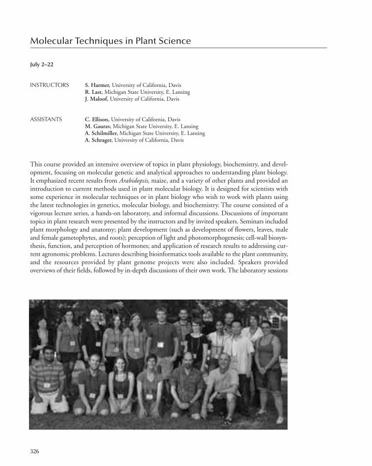
Molecular Techniques in Plant Science
July 2–22
INSTRUCTORS S. Harmer, University of California, DavisR. Last, Michigan State University, E. LansingJ. Maloof, University of California, Davis
ASSISTANTS C. Ellison, University of California, DavisM. Gaurav, Michigan State University, E. LansingA. Schilmiller, Michigan State University, E. LansingA. Schrager, University of California, Davis
This course provided an intensive overview of topics in plant physiology, biochemistry, and devel-opment, focusing on molecular genetic and analytical approaches to understanding plant biology.It emphasized recent results from Arabidopsis, maize, and a variety of other plants and provided anintroduction to current methods used in plant molecular biology. It is designed for scientists withsome experience in molecular techniques or in plant biology who wish to work with plants usingthe latest technologies in genetics, molecular biology, and biochemistry. The course consisted of avigorous lecture series, a hands-on laboratory, and informal discussions. Discussions of importanttopics in plant research were presented by the instructors and by invited speakers. Seminars includedplant morphology and anatomy; plant development (such as development of flowers, leaves, maleand female gametophytes, and roots); perception of light and photomorphogenesis; cell-wall biosyn-thesis, function, and perception of hormones; and application of research results to addressing cur-rent agronomic problems. Lectures describing bioinformatics tools available to the plant community,and the resources provided by plant genome projects were also included. Speakers providedoverviews of their fields, followed by in-depth discussions of their own work. The laboratory sessions
326
305-366_courses_Annual Report_2009 template 7/8/11 12:24 PM Page 326

provided an introduction to important techniques currently used in plant research. These includedstudies of plant development, mutant analysis, histochemical staining, transient gene expression,gene silencing, applications of fluorescent protein fusions, protein interaction and detection, pro-teomics approaches, several different approaches for quantifying metabolites, transient transforma-tion, and techniques commonly used in genetic and physical mapping. The course also includedseveral short workshops on important themes in plant research.
This course was supported with funds provided by the National Science Foundation.
PARTICIPANTS
Molecular Techniques in Plant Science 327
Arias-Garzon, T., B.S., University of Missouri, ColumbiaCasas, M., B.S., The Ohio State University, ColumbusKosma, D., Ph.D., Michigan State University, E. LansingLindh, I., M.Sc., Orebro University, Sweden Liu, B., M.S., Norwegian University of Science andTechnology, Trondheim, Norway
Markelz, R.J.C., B.Sc., Institute of Genomic Biology, Urbana,Illinois
McCarthy Suarez, I., Ph.D., IBMCP-CSIC, Valencia, Spain Muller, K., Ph.D., Simon Fraser University, Burnaby BC,Canada
Sasidharan, R., Ph.D., Utrecht University, The NetherlandsSattely, E., Ph.D., Harvard Medical School, Boston,Massachussetts
Schopper, S., M.Sc., Lund University, SwedenStephan, A., B.S., The Johns Hopkins University, Baltimore,Maryland
Wilkins, O., B.Sc., Dartmouth College, Hanover, NewHampshire
SEMINARS
Bailey-Serres, J., University of California, Riverside: Plantstress responses.
Birchler, J., University of Missouri, Columbia: Maize genetics.Birnbaum, K., New York University, New York: Rootdevelopment.
Bomblies, K., Harvard University, Boston, Massachusetts:Genetics of speciation.
Bowman, J., Monash University, Melbourne, VIC, Australia:Evolution of plant development.
Brutnell, T., Boyce Thompson Institute, Ithaca, New York:Plastids. Short read sequencing.
Coruzzi, G., New York University, New York: Plant systemsbiology.
Grossniklaus, U., University of Zurich, Switzerland:Gametophyte development.
Harmer, S., University of California, Davis: Circadianrhythms.
He, S.Y., Medical College of Wisconsin, Milwaukee:Plant/pathogen interactions.
Jackson, D., Cold Spring Harbor Laboratory: Shoot meristemdevelopment.
Jander, G., Boyce Thompson Institute, Ithaca, New York:Quantitative genetics.
Juntawong, P., University of California, Riverside:Quantitative PCR.
Kellogg, E.T., University of Missouri, St. Louis: Phylogenetics.Kerk, N., Yale University, New Haven, Connecticut:Introduction to plant biology.
Kuhlemeier, C., University of Bern, Switzerland: Phyllotaxy.Kutchan, T., Donald Danforth Plant Science Center, St. Louis,Missouri: Bioactive plant metabolites.
Last, R., Michigan State University, E. Lansing: Secondarymetabolites.
Maloof, J., University of California, Davis: Light regulation ofplant development.
Oldroyd, G., John Innes Centre, Norwich, United Kingdom:Rhizobial interactions/calcium signaling.
Pauly, M., University of California, Berkeley: Cell walls andbiofuels.
Poethig, S., University of Pennsylvania, Philadelphia:Vegetative development.
Roossnick, M., University of Colorado, Denver: Plant/virusinteractions.
Schaller, G.E., Dartmouth College, Hanover, NewHampshire: Cytokinin signaling.
Shukla, V., Dow Agro Sciences LLC, Indianapolis, Indiana:Modification of plant genome.
Spadling, E., University of Wisconsin, Madison: Photosensorysignal transduction.
Sussex, I., Yale University, New Haven, Connecticut:Introduction to plant biology.
Timmermans, M., Cold Spring Harbor Laboratory:microRNA regulation.
Vision, T., University of North Carolina, Durham: Genomeevolution/speciation.
Walling, L., University of California, Riverside: Plant/pestinteractions.
Zheng, N., University of Washington, Seattle: Regulatedprotein degradation and plant.
305-366_courses_Annual Report_2009 template 7/8/11 12:24 PM Page 327
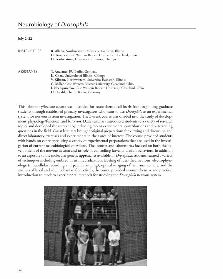
Neurobiology of Drosophila
July 2–22
INSTRUCTORS R. Allada, Northwestern University, Evanston, IllinoisH. Broihier, Case Western Reserve University, Cleveland, OhioD. Featherstone, University of Illinois, Chicago
ASSISTANTS T. Andlauer, FU Berlin, GermanyK. Chen, University of Illinois, ChicagoV. Kilman, Northwestern University, Evanston, IllinoisC. Miller, Case Western Reserve University, Cleveland, OhioI. Nechipurenko, Case Western Reserve University, Cleveland, OhioD. Owald, Charite Berlin, Germany
This laboratory/lecture course was intended for researchers at all levels from beginning graduatestudents through established primary investigators who want to use Drosophila as an experimentalsystem for nervous system investigation. The 3-week course was divided into the study of develop-ment, physiology/function, and behavior. Daily seminars introduced students to a variety of researchtopics and developed those topics by including recent experimental contributions and outstandingquestions in the field. Guest lecturers brought original preparations for viewing and discussion anddirect laboratory exercises and experiments in their area of interest. The course provided studentswith hands-on experience using a variety of experimental preparations that are used in the investi-gation of current neurobiological questions. The lectures and laboratories focused on both the de-velopment of the nervous system and its role in controlling larval and adult behaviors. In additionto an exposure to the molecular genetic approaches available in Drosophila, students learned a varietyof techniques including embryo in situ hybridization, labeling of identified neurons, electrophysi-ology (intracellular recording and patch clamping), optical imaging of neuronal activity, and theanalysis of larval and adult behavior. Collectively, the course provided a comprehensive and practicalintroduction to modern experimental methods for studying the Drosophila nervous system.
328
305-366_courses_Annual Report_2009 template 7/8/11 12:24 PM Page 328

This course was supported with funds provided by the National Institute of Mental Health andthe Howard Hughes Medical Institute.
Neurobiology of Drosophila 329
PARTICIPANTS
Aw, S., Ph.D., Duke–NUS Graduate Medical School,Singapore
Bahl, A., Dipl., Max-Planck Institute of Neurobiology,Martinsried, Germany
Duncan, K., Ph.D., EMBL, Heidelberg, GermanyGruber, M., M.A., Johannes Gutenberg-University Mainz,Germany
Halstead, J., M.Sc., Oxford University, United KingdomKeller, P., Dipl., HHMI/Janelia Farm Research Campus,Ashburn, Virginia
Lee, L., B.S., Florida Atlantic University, Boca RatonRamdya, P., Ph.D., UNIL/EPFL, Lausanne, SwitzerlandRobertson, D., Sc.B., University of California, San FranciscoSchweizer Burguete, A., Ph.D., University of Pennsylvania,Philadelphia
Tschida, K., B.A., Duke University, Durham, North CarolinaVanlandingham, P., Ph.D., University of Oklahoma, Norman
SEMINARS
Allada, R., Northwestern University, Evanston, Illinois:Circadian rhythms and sleep.
Benton, R., University of Lausanne, Switzerland: Olfactorysignal processing and physiology/imaging of the olfactorysystem.
Broihier, H., Case Western University, Cleveland, Ohio: AxonPathfinding. Genetic screens.
Dubnau, J., Cold Spring Harbor Laboratory: Learning andmemory.
Ejima, A., Kyoto University, Japan: Courtship.Featherstone, D., University of Illinois, Chicago: Informationtransfer in the nervous system and introduction toelectrophysiology.
Fouquet, W. and Andlauer, T.F., Freie Universitat Berlin,Germany: Synpatic ultrastructure and live imaging offluorescently-tagged synaptic proteins.
Freeman, M., University of Massachusetts, Worcester: Glia.Bioinformatics/web tools.
Goodwin, S., Glasgow University, United Kingdom; Jefferis,G., University of Cambridge, United Kingdom: Mosaictechniques. Adult brain anatomy.
Lee, C., NIH/National Institute of Child Health and HumanDevelopment, Bethesda, Maryland: Visual systemdevelopment.
Levitan, E., University of Pittsburgh, Pennsylvania: Imaging,peptide release/dense core vesicle trafficking.
Luo, L., Stanford University, California: Combined lecturewith advanced techniques in molecular neurobiology course.
O’Connor-Giles, K., University of Wisconsin, Madison: NMJstructure and development.
Rothenfluh, A., University of Texas Southwestern, Dallas:Ethanol/drugs.
Shen, P., University of Georgia, Athens: Larval feedingbehavior.
Skeath, J., Washington University School of Medicine, St.Louis, Missouri: Neural stem cells.
Turner, G., Cold Spring Harbor Laboratory: CNS physiologyand imaging.
Verstreken, P., K.U. Leuven, Belgium: Vesicle cycling andneurotransmitter release.
Waddell, S., University of Massachusetts, Worcester: Learningand memory.
305-366_courses_Annual Report_2009 template 7/8/11 12:24 PM Page 329

Genetics of Complex Human Diseases
July 6–12
INSTRUCTORS A. Al-Chalabi, Kings College London, United KingdomL. Almasy, Southwest Foundation for Biomedical Research, San Antonio,
Complex diseases are conditions that are influenced by the actions of multiple genes, their interac-tions with each other and with the environment. This lecture course considered the difficulties instudying the genetic basis of complex disorders such as diabetes, cardiovascular disease, cancer,Alzheimer’s disease, schizophrenia, and epilepsy. We discussed genetic–epidemiologic study designs,including family and case/control studies, as well as methods for quantifying the strength of the ge-netic influences on a disease. A major focus was the identification of specific gene effects using bothlinkage and association analysis and their variants. We discussed the efficiency and robustness ofdifferent designs for such analysis and how evidence from epidemiologic studies informs both thedesign and interpretation of molecular genetic studies. Study design and methods for analysis ofquantitative risk factors related to complex diseases was covered as well as copy-number variation,haplotype mapping, SNP tagging, meta-analysis, and gene–environment interaction. An overviewof next-generation sequencing was given to provide participants with insight into the applicationsof these techniques. Illustrations were provided through discussion of results from ongoing studiesof a variety of complex diseases and related risk factors.
This course was supported with funds provided by the Howard Hughes Medical Institute.
330
305-366_courses_Annual Report_2009 template 7/8/11 12:25 PM Page 330

Genetics of Complex Human Diseases 331
SEMINARS
Al-Chalabi, A., King’s College London, United Kingdom:GWAS and principles of association.
Almasy, A., Southwest Foundation for Biomedical Research,San Antonio, Texas: Introduction to linkage and quantitativetraits.
Borecki, I., Washington University School of Medicine, St.Louis, Missouri: Genetic epidemiology.
de Bakker, P., Brigham & Women’s Hospital, HarvardMedical School, Boston, Massachusetts: GWAS–Meta-analysis.
Loos, R., Cambridge University, United Kingdom: GxEinteraction.
Neale, B., Massachusetts General Hospital, Boston: SNPimputation.
Purcell, S., Massachusetts General Hospital, Boston: Analysisof GWAS.
Rampersaud, E., University of Miami, Florida: Family-basedassociation.
Sham, P.-C., University of Hong Kong, China: Powerconsiderations.
Sinsheimer, J., David Geffen School of Medicine, Los Angeles,California: Statistics 101.
Tobin, M., University of Leicester, United Kingdom: Copy-number variation.
Trembath, R., King’s College London, United Kingdom:Next-generation sequencing
PARTICIPANTS
Brisbin, A., B.S., Cornell University, Ithaca, New YorkByrne, S., Ph.D., Trinity College, Dublin, Dundalk, IrelandCropp, C., Ph.D., National Human Genome ResearchInstitute/NIH, Baltimore, Maryland
Demacq, C., Ph.D., State University of Campinas(UNICAMP), Campinas, Brazil
Dunn, E., B.S., Harvard School of Public Health, Norwood,Massachusetts
Hall, D., Ph.D., Rush University, Chicago, IllinoisJiang, Y., B.S., Emory University, Atlanta, GeorgiaKepp, K., B.S., Estonian Biocentre and University of Tartu,Estonia
Li, R., Ph.D., National Institutes of Health, Rockville,Maryland
Losh, M., Ph.D., University of North Carolina, Chapel HillManjarrez Orduño, N., Ph.D., The Feinstein Institute,Manhasset, New York
McLaughlin, R., B.S., Trinity College, Dublin, Ireland
Melton, P., Ph.D., Southwest Foundation for BiomedicalResearch, San Antonio, Texas
Moghraby, J., Ph.D., Kind Saud bin Abdul Aziz University-Health Science, Riyadh, Saudi Arabia
Nielsen, K., B.S., Technical University of Denmark, LyngbyPolsky, D., Ph.D., New York University Langone MedicalCenter, New York
Reynolds, R., Ph.D., University of Alabama, BirminghamShulman, J., Ph.D., Brigham & Women’s Hospital, Boston,Massachusetts
Tiwari, A., Ph.D., Center for Addiction & Mental Health,Toronto, Canada
Wang, Y., B.S., Center for Integrative Genetics; Aas, NorwayZhang, J., Ph.D., The Zucker Hillside Hospital, Glen Oaks,New York
Zlojutro, M., Ph.D., Southwest Foundation for BiomedicalResearch, San Antonio, Texas
305-366_courses_Annual Report_2009 template 7/8/11 12:25 PM Page 331

Advanced Techniques in Molecular Neuroscience
July 6–22
INSTRUCTORS C. Lai, Indiana University, BloomingtonR. Lansford, California Institute of Technology, PasadenaB. Stevens, Children’s Hospital Boston, Harvard Medical School, Boston, Massachusetts
ASSISTANTS S. Gattu, Indiana University, BloomingtonD. Huss, California Institute of Technology, PasadenaE. Lehrman, Harvard University/Children’s Hospital, Boston, MassachusettsK. Podorski, University of British Columbia, Vancouver, CanadaE. Robinson, Indiana University, BloomingtonA. Rosen, Harvard Medical School, Boston, Massachusetts
This course was designed to provide neuroscientists at all levels with a conceptual and practical un-derstanding of several of the advanced techniques in molecular neuroscience. The course was gearedtoward students who were familiar with the basic techniques of molecular biology.
The course curriculum was divided into three sections: an extensive and up-to-date set of labo-ratory exercises, daily lectures on theoretical and practical aspects of advanced molecular biologicaland imaging techniques, and a series of evening lectures given by invited speakers which served toshowcase the ways in which these molecular techniques have been used to advance specific areas ofneurobiology. The laboratory exercises were designed to provide each student with a working fa-miliarity with these advanced techniques using proven protocols and reagents. The experiments andprotocols were chosen to provide the course participants with sufficient hands-on expertise so thatthey may return to their home laboratories and, with a modicum of practice and perseverance, suc-cessfully implement the use of these advanced technologies.
332
305-366_courses_Annual Report_2009 template 7/8/11 12:25 PM Page 332

Each laboratory exercise was preceded by a lecture that covered the purpose of the experimentand included an in-depth presentation of the concepts, background, and theory. The participantswere guided through a step-by-step analysis of the actual protocol, which were provided both inhard copy and on the course website. The laboratory portion of each course included gene deliverysystems including mammalian cell transfection, single-cell electroporation, the selection and use ofanimal viral vectors, as well as the introduction of DNAs using in vivo electroporation in Xenopustadpoles, quail embryos, and rodents; loss-of-function approaches to study gene function usingRNAi approaches; the use of optogenetics to probe neural circuits within animals; the design andmanipulation of bacterial artificial chromosome (BAC) transgenic vectors and their use in achievingcell-type-specific expression in animals; comparison of quantitative PCR, RNA-Seq, and microarrayanalyses to profile gene expression; methods to identify gene expression in single cells and in sub-cellular regions of single cells; and techniques to isolate, propogate, and analyze desired neural andglial populations. As many of these advanced techniques also included the use of more standardprocedures to first modify and then “move” DNA fragments from one vector to another, the labo-ratory exercises also provided training in these methods, which were essential to the successful exe-cution of the more sophisticated approaches. Finally, course participants were introduced tobioinformatics and a wide range of Internet resources that are available to molecular neuroscientists.
This course was supported with funds provided by the National Institute of Mental Health andthe Howard Hughes Medical Institute.
PARTICIPANTS
Advanced Techniques in Molecular Neuroscience 333
Bajic, D., Ph.D., Children’s Hospital, Boston, MassachusettsBarrett, R., B.A., University of California, IrvineBrouwers, N., Ph.D., VIB and University of Antwerp, BelgiumHayden, M., M.A., Stanford University Hospital and Clinics,California
He, H., Ph.D., The Scripps Research Institute, La Jolla,California
Heng, M., B.S., Vollum Institute, OHSU, Portland, OregonHeraud, J., B.Sci., Medical University of Vienna, AustriaKirby, E., B.S., University of California, BerkeleyKlaus, A., M.Sc., National Institutes of Health, Bethesda,Maryland
Lotfipour, S., Ph.D., University of California, Los AngelesNogaroli, L., Ph.D., Federal University of Rio de Janeiro,Brazil
Sink, K., Ph.D., Emory University, Atlanta, GeorgiaStephenson-Jor, M., B.Sc., Karolinska Institutet, Stockholm,Sweden
Toledo, A., M.S., Universidad de la Republica, Monvevideo,Uruguay
Wheeler, T., B.S., University of Rochester, New YorkZong, C., Ph.D., Harvard University, Cambridge,Massachusetts
SEMINARS
Barres, B., Stanford University School of Medicine, California:Neuron–glial interactions in the CNS.
Cline, H., The Scripps Research Institute, La Jolla, California:Distinguished Alumni Lecture. Molecular dissection ofneural circuit development.
Darnell, R., The Rockefeller University, New York: DecodingRNA maps in the brain.
Eberwine, J., University of Pennsylvania Medical School,Philadelphia: Metastability of the henome: Impact onneuronal phenotype and dendritic biology.
Lichtman, J., Harvard University, Cambridge, Massachusetts:Connectomics.
Lipscombe, D., Brown University, Providence, Rhode Island:Neuronal calcium channels: Spliced for optimal function.
Liquin, L., Stanford University, California: Mapping olfactionin the fly and mouse brain.
Lledo, P.M., Pasteur Institute, Paris, France: Development ofneural stem cells in the adult circuits.
305-366_courses_Annual Report_2009 template 7/8/11 12:25 PM Page 333

Workshop on Biology of Social Cognition
July 14–20
INSTRUCTORS J. Mitchell, Harvard University, Cambridge, MassachusettsD. Skuse, University College London Institute of Child Heath, United Kingdom
ASSISTANT D. Tamir, Harvard University, Cambridge, Massachusetts
The past few years have seen remarkable advances in our knowledge of the genetic, molecular, andneural factors that contribute to social behavior. At the same time, sophisticated analytical and the-oretical approaches have helped to make sense of the data. This week-long workshop sought to pro-vide a comprehensive overview of these topics. Although the emphasis was on social cognition inhumans, there were also study days dedicated to state-of-the-art presentations on comparative ap-proaches and evolutionary models. Finally, all themes were related to the clinical consequences ofdysfunctional social cognition and the role of translational research. The course included introduc-tory seminars on key themes, offered every morning. There were afternoons devoted largely to prac-tical sessions that provided hands-on experiments in consultation with seminar leaders, roundedoff by after-supper keynote lectures by leading scientists that reflect cutting-edge and future viewsrelated to our theme for that day.
This workshop was supported with funds provided by Autism Speaks; the INCORE Consortium;Nancy Lurie Marks Family Foundation; and the Simons Foundation.
334
305-366_courses_Annual Report_2009 template 7/8/11 12:25 PM Page 334

Workshop on Biology of Social Cognition 335
PARTICIPANTS
Albo, Z., B.S., Baylor College of Medicine, Houston, TexasBartholomeusz, C., Ph.D., The University of Melbourne,Carton South, Australia
Chevallier, C., Ph.D., King’s College London, UnitedKingdom
Contreras, J., B.S., Harvard University, Cambridge,Massachusetts
Cook, J., B.S., University College London, United KingdomCosta, R., B.S., University of Lisbon, Portugal, LisboaDiehl, M., B.S., University of Rochester, New YorkHillebrandt, H., B.S., University College London, UnitedKingdom
Koelkebeck, K., Ph.D., University of Muenster, GermanyKret, M., B.S., Tilburg University, The NetherlandsLee, V., B.S., McMaster University, Hamilton, Canada
Light, S., B.S., University of Wisconsin, MadisonMenyhart, O., B.S., Cornell University, Ithaca, New YorkMoore, L., B.S., University of California, Los AngelesMoore III, W., B.S., University of Oregon, EugeneMuralidharan, A., B.S., Emory University, Atlanta, GeorgiaPaxton, R., B.S., Emory University, Atlanta, GeorgiaSimmons, J., Ph.D., National Institutes of Health, Bethesda,Maryland
Stjepanovic, D., B.S., The University of Queensland, St.Lucia, Australia
Theodoridis, A., B.S., University of California, BerkeleyTroiani, V., B.S., University of Pennsylvania School ofMedicine, Philadelphia
Zaki, J., B.S., Columbia University, New York
SEMINARS
Amodio, A., New York University, New York: Social cognitionand emotion.
Blair, J., National Institute of Mental Health, Bethesda,Maryland: Psychopathy.
Blakemore, S., University College of London, UnitedKingdom: Development of social cognition.
Bourgeron, T., Pasteur Institute, Paris, France: Autismgenetics.
Cahill, L., University of California, Irvine: Sex differences inemotional processing.
Couzin, I., University of Princeton, New Jersey: Evolution ofcollective behavior.
Cushman, F., Harvard University, Cambridge, Massahusetts:The moral faculty: Domain specificity, neural circuitry, anduniversality.
Dean, M., Brown University, Providence, Rhode Island:Introduction to neuroeconomics.
Giedd, J., National Institute of Mental Health, Bethesda,Maryland: Neuroimaging of the developing brain.
Haxby, J., Dartmouth College, Hanover, New Hampshire:Face processing.
Huhman, K., Georgia State University, Atlanta: Conditioneddefeat in hamsters.
Matsuzawa, T., Kyoto University, Aichi, Japan: Socialcognition in primates.
Mitchell, J., Harvard University, Cambridge, Massachusetts:Investigating theory of mind.
Murray, R., King’s College London, United Kingdom: Socialcognition and major mental illness.
Ochsner, K., Columbia University, New York: Regulation ofemotion and social cognition.
Phelps, E., New York University, New York: Emotion andcognition.
Schultz, B., Children’s Hospital of Philadelphia, Pennsylvania:The neural basis of autism.
Shaw, P., National Institutes of Mental Health, Bethesda,Maryland: Genes, brain, and behavior.
Skuse, D., University College London, United Kingdom: Thebiology of social cognition in autistic disorders. X-linkedgenes and social cognition.
Wieben, E., Mayo Clinic, Rochester, Minnesota: Introductionto genomics.
Young, L., Emory University, Atlanta, Georgia: Neuropeptidesand social cognition: From pair-bonding voles to autism.
305-366_courses_Annual Report_2009 template 7/8/11 12:25 PM Page 335

Brain Tumors
July 22–28
INSTRUCTORS D. Gutmann,Washington University School of Medicine, St. Louis, MissouriE. Holland, Memorial Sloan-Kettering Cancer Center, New YorkS. Majumder, University of Texas M.D. Anderson Cancer Center, Houston
ASSISTANT S. Singh, University of Texas M.D. Anderson Cancer Center, Houston
This one-week discussion course provided a clinical overview of brain tumors and emphasized mo-lecular mechanisms involved in the growth and development of brain tumors with special emphasison neural differentiation, signaling mechanisms, DNA replication, chromatin modulation, stemcells, mouse models, genomics, imaging techniques, genetically modified mouse techniques, nano-technology, mechanism-based therapeutic strategies, biobanks, and ethical concerns. Attendees wereable to interact with senior investigators on a one-to-one basis in an informal environment.
This course was supported with generous funding provided by the American Brain Tumor As-sociation.
336
305-366_courses_Annual Report_2009 template 7/8/11 12:25 PM Page 336

337
PARTICIPANTS
Annibali, D., Ph.D., CNR, Toronto, CanadaAref, D., B.S., University of Toronto, CanadaDziurzynski, K., Ph.D., University of Texas M.D. AndersonCancer Center, Houston
Gajadhar, A., B.S., Hospital for Sick Children, University ofToronto, Canada
Jones, T., Ph.D., University of Tennessee, MemphisKohli, L., B.S., University of Alabama, BirminghamKristoffersen, K., B.S., Copenhagen National UniversityHospital, Denmark
Lathia, J., Ph.D., Cleveland Clinic Foundation, OhioLi, S., Ph.D., University of California, Los AngelesMcDevitt, M., Ph.D., Memorial Sloan-Kettering CancerCenter, New York
Moiyadi, A., Ph.D., Tata Memorial Center, Mumbai, IndiaPanosyan, E., Ph.D., University of California, Los AngelesPaolella, B., B.S., Dartmouth College, Lebanon, NewHampshire
Põlajeva, J., B.S., Uppsala University, SwedenPollard, S., Ph.D., University College London, UnitedKingdom
Read, R., Ph.D., The Salk Institute for Biological Studies, LaJolla, California
Rich, B., Ph.D., Dana Farber Cancer Institute, Boston,Massachusetts
Sathyan, P., Ph.D., M.D. Anderson Cancer Research Center,Houston, Texas
Song, Y., Ph.D., National Cancer Institute, Frederick,Maryland
Vanner, R., B.S., University of Toronto, CanadaWells, E., Ph.D., Children’s National Medical Center,Washington, D.C.
Zinn, P., Ph.D., Dana Farber Cancer Institute/HarvardUniversity, Boston, Massachusetts
SEMINARS
Aldape, K., University of Texas M.D. Anderson CancerCenter, Houston: Molecular subgroups of gliomas andprognostic markers.
Becher, O., Memorial Sloan-Kettering Cancer Center, NewYork: Clinical aspects of pediatric brain tumors.
Bondy, M., M.D. Cancer Center, Houston, Texas: Geneticand molecular epidemiology of brain tumors.
Brennan, C., Memorial Sloan-Kettering Cancer Center, NewYork: Molecular subclasses of glioma and accessing TheCancer Genome Atlas.
Eberhart, C., Johns Hopkins University School of Medicine,Baltimore, Maryland: The role of Notch signaling in braintumor initiation and growth.
Enikolopov, G., Cold Spring Harbor Laboratory: Quiescenceand division of neural stem cells.
Fuller, G., M.D. Anderson Cancer Center, Houston, Texas:Clinical overview of brain tumors: Neuroradiology andneuropathology.
Gilbertson, R., St. Jude’s Children’s Research Hospital,Memphis, Tennessee: Developmental neurobiology oncology.
Gladson, C., Cleveland Clinic, Ohio: Angiogenesis inmalignant glioma tumors: Can we effectively target it?
Gutmann, D., Washington University School of Medicine, St.Louis, Missouri: Using neurofibromatosis-1 to understandpediatric brain tumors.
Holland, E., Memorial Sloan-Kettering Cancer Center, NewYork: Modeling proneural/PDGF gliomas in mice.
James, C.D., University of California, San Francisco:Transplantable orthotopic tumor models for therapeutictesting.
Kornblum, H., University of California Molecular andMedical Pharmacology, Los Angeles: Methods and rationalesin evaluating neural and brain tumor stem cells.
Lang, Jr., F., M.D. Anderson Cancer Center, Houston, Texas:Clinical management of brain tumors: The surgicalperspective.
Majumder, S., University of Texas M.D. Anderson CancerCenter, Houston: Cellular flexibility determines both normalbrain development and brain tumors.
Pelroy, R., National Cancer Institute, Bethesda, Maryland:Funding opportunities at the National Institutes of Health.
Pieper, R., University of California, San Francisco: Themultiple roles of ubiquitination in the development andtreatment of brain cancer.
Reilly, K., National Cancer Institute, Bethesda, Maryland:Using mouse models of brain cancer to understand geneticsusceptibility factors.
Rich, J., Cleveland Clinic, Ohio: Brain tumor stem cells.Roussel, M., St. Jude’s Children’s Research Hospital,Memphis, Tennessee: Critical signaling pathways incerebellum development and cancer.
Rowitch, D., University of California, San Francisco:Generation of mouse models of pediatric brain cancer.
Snyder, E., Burnham Institute, La Jolla, California: Using ourgrowing knowledge of fundamental stem cell biology tounderstand brain tumor development and behavior.
Stiles, C., Dana Farber Cancer Institute, Boston,Massachusetts: Separated at birth? Transcription factors innormal and malignant neural progenitors.
Wechsler-Reya, R., Duke University Medical Center, Durham,North Carolina: What mouse models teach us aboutmedulloblastoma biology.
Yung, W.K.A., M.D. Anderson Cancer Center, Houston,Texas: Molecular targeted therapy in malignant gliomas.
Brain Tumors 337
305-366_courses_Annual Report_2009 template 7/8/11 12:25 PM Page 337
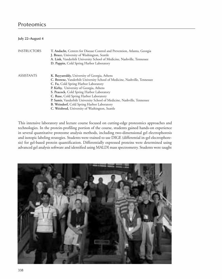
Proteomics
July 22–August 4
INSTRUCTORS T. Andacht, Centers for Disease Control and Prevention, Atlanta, GeorgiaJ. Bruce, University of Washington, SeattleA. Link, Vanderbilt University School of Medicine, Nashville, TennesseeD. Pappin, Cold Spring Harbor Laboratory
ASSISTANTS K. Bayyareddy, University of Georgia, AthensC. Browne, Vanderbilt University School of Medicine, Nashville, TennesseeC. Fu, Cold Spring Harbor LaboratoryP. Kirby, University of Georgia, AthensS. Peacock, Cold Spring Harbor LaboratoryC. Ruse, Cold Spring Harbor LaboratoryP. Samir, Vanderbilt University School of Medicine, Nashville, TennesseeB. Weindorf, Cold Spring Harbor LaboratoryC. Weisbrod, University of Washington, Seattle
This intensive laboratory and lecture course focused on cutting-edge proteomics approaches andtechnologies. In the protein-profiling portion of the course, students gained hands-on experiencein several quantitative proteome analysis methods, including two-dimensional gel electrophoresisand isotopic labeling strategies. Students were trained to use DIGE (differential in-gel electrophore-sis) for gel-based protein quantification. Differentially expressed proteins were determined usingadvanced gel analysis software and identified using MALDI mass spectrometry. Students were taught
338
305-366_courses_Annual Report_2009 template 7/8/11 12:25 PM Page 338

differential and quantitative mass spectrometry approaches to profile and identify changes in pro-teomes and were trained in high-sensitivity microcapillary liquid chromatography coupled withnanospray-ESI and tandem mass spectrometry. Both single dimension and multidimensional mi-crocapillary liquid chromatography separations coupled to mass spectrometry were taught. Studentsgained hands-on experience purifying and identifying protein complexes and posttranslational mod-ifications using the latest technologies. This course also included discussion and application of mul-tiple reaction monitoring (MRM) methods for targeted proteomics. Students developed and appliedMRM transitions and chromatographic methods to quantify targeted proteins in complex proteomesamples, such as serum or whole-cell lysates. A strong emphasis was placed on data analysis through-out the course. The overall aim of the course was to provide students with the fundamental knowl-edge and hands-on experience necessary to be able to perform and analyze proteomics experimentsand to learn to identify new opportunities in applying proteomics approaches to their own research.
This course was supported with funds provided by the National Cancer Institute and the HowardHughes Medical Institute.
Proteomics 339
PARTICIPANTS
Bellail, A., Ph.D., Emory University, Atlanta, GeorgiaCilia, M., Ph.D., USDA-ARS, Ithaca, New YorkGruninger, J., M.Sc., University of British Columbia,Vancouver, Canada
Igel, U., M.A., Royal Institute of Technology, Stockholm,Sweden
Kigar, S., B.A., University of Wisconsin, MadisonKimura, M., Ph.D., Gifu University School of Medicine,Japan
Leal, M., B.A., Federal University of Sao Paulo, BrazilMande, K., Ph.D., King Abdullah University of Science andTechnology, Jeddah, Saudi Arabia
McDonald, Z., B.Sc., University of Cape Town, South Africa
Møldrup, M., M.S., University of Copenhagen, DenmarkMoore, R., Ph.D., National Institute of Allergy & InfectiousDisease, Hamilton, Montana
Richlen, M., Ph.D., Woods Hole Oceanographic Institution,Massachusetts
Roedkaer, S., B.A., University of Southern Denmark, OdenseM, Denmark
Seizl, M., Ph.D., Ludwig-Maximilians-Universistät, Munich,Germany
Zarr, M., B.A., Johns Hopkins School of Medicine, Baltimore,Maryland
Zou, J., B.S., Xavier University of Louisiana, New Orleans
SEMINARS
Andacht, T., Centers for Disease Control and Prevention,Atlanta, Georgia: Fundamentals and applications of two-dimensional DIGE in proteomics.
Bruce, J., University of Washington, Seattle: Fundamentals ofmass spectrometry and proteomics.
Chait, B., The Rockefeller University, New York: Proteomicanalysis of protein machines.
Chaurand, P., University of Montreal, Canada: Imaging massspectrometry of cells and tissue.
Clauser, K., Broad Institute of MIT and Harvard, Cambridge,Massachusetts: De novo interpretation of tandem massspectra.
Cristea, I., Princeton University, New Jersey: Proteomic probesof the living interactome.
Dufresne, C., Thermo Fisher Scientific, West Palm Beach,Florida: Mass spectrometry approaches to identify andquantify PTMs.
Eng, J., University of Washington, Seattle: Analysis of tandemMS data using the transproteomic pipeline.
Friedman, D., Vanderbilt University, Nashville, Tennessee:Variation and experimental design in proteomic analyses.
Kelleher, N., Northwestern University, Evanston, Illinois: Top-down mass spectrometry.
MacCoss, M.J., University of Washington, Seattle: Targetedproteomics for quantitative analysis and biomarkervalidation.
Magee, M., Arizona State University, Tempe: Functionalproteomics: Protein microarrays.
Pappin, D., Cold Spring Harbor Laboratory: Quantitativeproteomics and iTRAQ.
Wolf-Yadlin, A., Harvard University, Cambridge,Massachusetts: Enhanced phosphoproteomics in mammaliansystems.
Zhang, B., Vanderbilt University School of Medicine,Nashville, Tennessee: Functional and network analysis ofproteomics data.
305-366_courses_Annual Report_2009 template 7/8/11 12:25 PM Page 339

Eukaryotic Gene Expression
July 27–August 16
INSTRUCTORS J. Espinosa, HHMI/University of Colorado, BoulderW.L. Kraus, Cornell University, Ithaca, New YorkA. Shilatifard, Stowers Institute for Medical Research, Kansas City, MissouriD. Taatjes, University of Colorado, Boulder
ASSISTANTS M. Galbraith, University of Colorado, BoulderM. Knuesel, University of Colorado, Boulder M. Mohan, Stowers Institute for Medical Research, Kansas City, MissouriD. Ruhl, Cornell University, Ithaca, New York
This course was designed for students, postdocs, and principal investigators who have recently ven-tured into the exciting area of gene regulation. The course focused on state-of-the-art strategies andtechniques used in the field. Emphasis was placed on both in vitro and in vivo protein–DNA in-teractions and on novel methodologies to study gene regulation. Students made nuclear extracts,performed in vitro transcription reactions, and measured RNA levels using primer extension. Char-acterizations of the DNA-binding properties of site-specific transcription factors was carried outusing electrophoretic mobility shift and DNase I footprinting assays. In addition, students learnedtechniques for the assembly and analysis of chromatin in vitro. This included transcription assays,chromatin footprinting, and chromatin remodeling assays. During the past few years, the gene reg-ulation field has developed in vivo approaches to study gene regulation. Students were exposed tothe chromatin immunoprecipitation technique. They also used RNAi for specific knock-down ex-periments in mammalian cells. In addition, determining cellular gene expression profiles has beenaccelerated tremendously by DNA microarray technology. Students received hands-on training inperforming and interpreting results from DNA microarrays.
This course was supported with funds provided by the National Cancer Institute.
340
305-366_courses_Annual Report_2009 template 7/8/11 12:25 PM Page 340

Eukaryotic Gene Expression 341
PARTICIPANTS
Bistulfi, G., B.S., Roswell Park Cancer Institute, Buffalo, NewYork
Camp, J., B.S., University of North Carolina, Chapel HillChatterjee, D., M.Sc., Cold Spring Harbor LaboratoryChen, S., Ph.D., University of California, San Franciscode Oliveira, K., B.S., Max Delbrück Center, Berlin, GermanyDuncan, A., M.S., University of Texas, HoustonGehani, S., M.S., Biotech Research and Innovation Centre,Copenhagen N, Denmark
Hoeg, F., Dipl., Gene Center at the University of Munich,Germany
Klein, A., Ph.D., University of Texas Southwestern MedicalCenter, Dallas
Kostromitskaia, J., M.S., Centre for Molecular Biology andNeuroscience, Oslo, Norway
Mazumdar, S., M.S., Tufts University, Boston, MassachusettsMigliori, V., M.D., Institute of Molecular and CellularBiology, Singapore
Nichol, J., Ph.D., McGill University, Montreal, CanadaSharma, N., M.S., University of Aarhus, DenmarkTreutlein, B., M.S., Ludwig-Maximilians University, Munich,Germany
Zhang, X., Ph.D., Cornell University, Ithaca, New York
SEMINARS
Adelman, K., National Institute of Environmental HealthSciences/NIH, Research Triangle Park, North Carolina:Pausing of RNA polymerase II disrupts DNA-encodednucleosome organization to enable precise gene regulation.
Almouzni, G., Institut Curie, Paris, France: Variation on thetheme of chromatin assembly.
Carey, M., University of California School of Medicine, LosAngeles: Biochemical mechanisms of chromatintranscription.
Conaway, J., Stowers Institute for Medical Research, KansasCity, Missouri: Introduction to the general transcriptionmachinery.
Cramer, P., LMU University of Munich, Germany: Structure-function studies of eukaryotic transcription.
Gill, G., Tufts University School of Medicine, Boston,Massachusetts: Regulation of chromatin structure andtranscription by Sp3 and SUMO.
Kadonaga, J., University of California, San Diego, La Jolla:Secrets of the RNA polymerase II core promoter.
Kingston, R., Massachusetts General Hospital/HarvardMedical School, Boston: In vivo biochemistry: Interrogatingnucleosomes and nucleosome-modifying complexes on thegenome.
Kurdistani, S., University of California, Los Angeles: Role of alinker histone in regulating expression of gene familyclusters.
Levine, M., University of California, Berkeley: Whole-genomeanalysis of transcriptional precision in the Drosophilaembryo.
Lieb, J., University of North Carolina, Chapel Hill: Genome-wide measurement of transcription-factor-binding dynamicsby competition ChIP.
Luger, K., HHMI/Colorado State University, Fort Collins:The expected, the unexpected, and the just plain weird:Insights from new experimental approaches to studynucleosome structure.
Pugh, F., Pennsylvania State University, University Park:Genome-wide interplay of the transcription machinery andchromatin.
Spector, D., Cold Spring Harbor Laboratory: Intranucleardynamics.
Washburn, M., Stowers Institute for Medical Research,Kansas City, Missouri: Driving biological discovery withquantitative shotgun proteomics.
305-366_courses_Annual Report_2009 template 7/8/11 12:25 PM Page 341

Imaging Structure and Function in the Nervous System
July 27–August 16
INSTRUCTORS S. Thompson, University of Maryland School of Medicine, BaltimoreW. Tyler, Arizona State University, TempeJ. Waters, Northwestern University, Chicago, Illinois
ASSISTANTS O. KoHo, University College London, United KingdomA. Nimmerjahn, Stanford University, CaliforniaS. Page, University of Maryland School of Medicine, BaltimoreA. Scimemi, National Institute of Neurological Disorders and Stroke/NIH, Bethesda, Maryland
Advances in light microscopy, digital image processing, and the development of a variety of powerfulfluorescent probes present expanding opportunities for investigating the nervous system, from synap-tic spines to networks in the brain. This intensive laboratory and lecture course provided participantswith the theoretical and practical knowledge to utilize emerging imaging technologies. The primaryemphasis of the course was on vital light microscopy. Students learned the principles of light mi-croscopy, as well as use of different types of electronic cameras, laser-scanning systems, functionalfluorophores, delivery techniques, and digital image-processing software. In addition to transmittedlight microscopy for viewing cellular motility, the course examined a variety of molecular probes ofcell function, including calcium-sensitive dyes, voltage-sensitive dyes, photo-activated (caged) com-pounds, exocytosis tracers, and optogenetic activators. Issues arising in the combination of imagingwith electrophysiological methods were covered. Particular weight was given to multiphoton laser-
342
305-366_courses_Annual Report_2009 template 7/8/11 12:25 PM Page 342

Imaging Structure and Function in the Nervous System 343
scanning microscopy and to newly available biological fluorophores, especially green-fluorescentprotein (GFP) and its variants. We used a spectrum of neural and cell biological systems, includingliving animals, brain slices, and cultured cells.
This course was supported with funds provided by the National Institute of Mental Health andthe Howard Hughes Medical Institute.
Russo, M., B.A., Columbia University, New YorkShobe, J., Ph.D., University of California, Los AngelesShuai, Y., Ph.D., Cold Spring Harbor LaboratoryTremblay, M.-E., Ph.D., University of Rochester MedicalCenter, New York
van Ham, T., Ph.D., Massachusetts General Hospital/HarvardMedical School, Boston
Xu, S. (Pei), B.S., Washington University, St. Louis, Missouri
SEMINARS
Albeanu, F., Cold Spring Harbor Laboratory: Synapto-phlourins and synaptic vesicle imaging,
Bruchez, M., Carnegie Mellon University, Pittsburgh,Pennsylvania: New and future indicators.
Deisseroth, K., Stanford University, California: Advancedoptogenetics/optical probing of brain circuits.
Denk, W., Max-Planck Institute for Medical Research,Heidelberg, Germany: Extended 2P imaging and block-faceEM.
DiGregorio, D., Universite Paris 5, France: Flash photolysis.Emilani, V., CNRS, INSERM, University Paris Descartes,France: Holography.
Engert, F., Harvard University, Cambridge, Massachusetts:Shot noise. 2P microscopy.
George, N., Olympus America, Inc., Center Valley,Pennsylvania: Objectives. BX51 optics.
Gould, T., Yale University School of Medicine, New Haven,Connecticut: STED and PALM.
Griesbeck, O., Max-Planck Institute of Neurobiology,Martinsried, Bayern, Germany: XFPs and geneticallyencoded sensors.
Hirsch, J., Columbia University, New York: fMRI.Huebener, M., Max-Planck Institute of Neurobiology,Martinsried, Germany: Intrinsic imaging.
Kilborn, K., Intelligent Imaging Innovations, Inc., SantaMonica, California: Deconvolution.
Lanni, F., Carnegie-Mellon University, Pittsburgh,Pennsylvania: Basic microscopy.
Lee, J., University of California, Los Angeles: Advancedoptogenetics.
Litchman, J., Harvard University, Cambridge, Masschusetts:Confocal microscopy.
Mrsic-Flogel, T., University College London, UnitedKingdom: Organic calcium indicators bulk loading.
Prakriya, M., Northwestern University, Evanston, Illinois:Foerster resonance energy transfer.
Racca, C., Newcastle University, Newcastle upon Tyne, UnitedKingdom: Electron microscopy.
Simpson, J., MBF Bioscience, San Diego, California: MBF,sterology, and structure analysis.
Stutzmann, G., Rosalind Franklin University, N. Chicago,Illinois: Two-photon imaging in AD mouse models.
Tsai, P., University of California, San Diego, La Jolla: Opticsbench lab exercises–Basics Koehler. Optics bench labexercises–Scanning. Scanning and fluorescence. Opticsbench lab exercises–Scanning. Optics bench lab exercises–Confocal. CCD cameras.
Tyler, J., Arizona State University, Tempe: Image J and Fiji;Ultrasound neuromodulation.
Waters, J., Northwestern University, Chicago, Illinois: Organiccalcium indicators. Single-cell Ca imaging in vivo. CCDcameras; Homebrew microscope including laser safety.
Wilbrecht, L., University of California, San Francisco: GFPapplications spine morphology.
Witkowski, J., Cold Spring Harbor Laboratory: Ethics.Zhang, F., Harvard University, Cambridge, Massachusetts:Light-triggered activation/silencing.
PARTICIPANTS
Clancy, K., B.S., University of Berkeley, CaliforniaCohen, L., Ph.D., The Hebrew University of Jerusalem, IsraelFord, C., Ph.D., Oregon Health & Science University,Portland
Gu, Y.I., B.S., Johns Hopkins University, Baltimore, MarylandKreile, A., M.S., Max-Planck Institute of Neurobiology,Martinsried, Germany
Nadif, K.N., Ph.D., CSHL/DCN, Radboud University, ColdSpring Harbor
305-366_courses_Annual Report_2009 template 7/8/11 12:25 PM Page 343

Yeast Genetics and Genomics
July 27–August 16
INSTRUCTORS D. Burke, University of Virginia, CharlottesvilleB. Errede, University of North Carolina, Chapel HillJ. Smith, University of Virginia, Charlottesville
ASSISTANTS K. Gardner, University of North Carolina, Chapel HillJ. McClure, University of Virginia, CharlottesvilleM. Wells, University of Virginia, Charlottesville
This is a modern, state-of-the-art laboratory course designed to teach the students the full repertoireof genetic approaches needed to dissect complex problems in the yeast Saccharomyces cerevisiae.Combinations of classical genetic approaches are emphasized, including the isolation and charac-terization of mutants, tetrad analysis, complementation, and mitotic recombination. Molecular ge-netic techniques, including various types of yeast transformation, gene replacement with plasmidsand PCR, construction and analysis of gene fusions, and generation of mutations in cloned genes,were also emphasized. Students used classical and molecular approaches to gain experience in iden-tifying and interpreting various kinds of genetic interactions including suppression and syntheticlethality. Students were immersed in yeast genomics and performed and interpreted experimentswith DNA arrays. Students gained first-hand experience in modern cytological approaches such asepitope tagging and imaging yeast cells using indirect immunofluorescence, GFP–protein fusions,
344
305-366_courses_Annual Report_2009 template 7/8/11 12:25 PM Page 344

SEMINARS
Bekiranov, S., University of Virginia, Charlottesville:Analyzing Tag4 arrays.
Hinnebusch, A., National Institutes of Health, Bethesda,Maryland: Nitogren regulation of gene expression.
Hollingsworth, N., State University of New York, StonyBrook: Phosphorylation and the regulation of meioticrecombination.
Kaeberlin, M., University of Washington, Seattle: Genetics ofaging.
Levin, D., Boston University, Massachusetts: Stress-inducedtranscriptional regulation through a novel MAPK-drivenmechanism.
Lew, D., Duke University, Durham, North Carolina: Cellpolanity in yeast.
Yeast Genetics and Genomics 345
and a variety of fluorescent indicators for various subcellular organelles. Lectures on fundamentalaspects of yeast genetics were presented along with seminars given by outside speakers on topics ofcurrent interest.
This course was supported with funds provided by the National Human Genome Research In-stitute and the Howard Hughes Medical Institute.
PARTICIPANTS
Abeydeera, N.D., Ph.D., Texas A&M University, CollegeStation
Bohnert, M., Ph.D., University of Freiburg, Germany Bosman, J., Ph.D., University of Groningen, Haren, TheNetherlands
Bussemaker, H., Ph.D., Columbia University, New YorkChakravarty, A., M.Sc., Memorial Sloan-Kettering CancerCenter, New York
Chang, M., Ph.D., Nanyang Technological University,Singapore
De Haro, L., Ph.D., University of New Mexico, Albuquerque
Elfick, A., Ph.D., University of Edinburgh, United KingdomGoltermann, L., M.Sc., Copenhagen University, DenmarkHofree, M., B.Sc., University of California, San Diego, LaJolla
Houser, J., B.S., University of North Carolina, Chapel HillKogej, T., Ph.D., University of Ljubljana, SloveniaRosby, R., Ph.D., Brown University, Providence, Rhode IslandSrivas, R., Ph.D., University of California, San Diego, La JollaStein, V., MS, University of Queensland, Brisbane, AustraliaWaisberg, M., Ph.D., National Institutes of Health, Rockville,Maryland
Ramanathan, S., Harvard University, Cambridge,Massachusetts: Using Bayesian statistics to uncoverinformation about proteins and networks.
Rusche, L., Duke University, Durham, North Carolina:Evolution of transcription silencing.
Strathern, J., National Cancer Institute, Frederick, Maryland:Detection of transcription errors.
Thorner, J., University of California, Berkeley: A signalingpathway for maintenance of plasma membrane lipidhomeostasis.
Troyanskaya, O., Princeton University, New Jersey: Integratinglarge-scale data.
Whiteway, M., National Research Council of Canada,Montreal, Quebec: Transcriptional rewiring in fungi.
305-366_courses_Annual Report_2009 template 7/8/11 12:25 PM Page 345
Stem Cells
July 30–August 8
INSTRUCTORS R. McKay, National Institutes of Health, Bethesda, MarylandM. Shen, Columbia University Medical Center, New York
ASSISTANT J. Chenoweth, National Institute of Neurological Disorders and Stroke/NIH, Bethesda, Maryland
Stem cells construct organs and tissues in development. They sustain tissues in the adult and restorethem after injury. Because of these properties, isolating and manipulating stem cells has become amajor new element in biomedical science. This workshop course covered a series of biological sub-jects relating to stem cells and regenerative medicine. Topics included embryology and development,reprogramming, degenerative disease, cancer stem cells, and human genetics.
This 10-day-long discussion course brought together leading researchers in the stem cell fieldwith a small group of international students. The purpose of the workshop was to provide partici-pants with an opportunity to achieve an advanced understanding of the scientific and clinical im-portance of stem cells. A major aim of the workshop was to discuss, in considerable detail, therelationship between stem cells and disease. The significance of this relationship with respect to de-veloping new approaches to treating and understanding human disease was explored.
This course was supported with funds provided by the Howard Hughes Medical Institute.
346
305-366_courses_Annual Report_2009 template 7/8/11 12:25 PM Page 346

Rich, J., Cleveland Institute, Ohio: The evolving under-standing of cancer stem cells.
Robertson, E., University of Oxford, United Kingdom: Celllineage specification and patterning in the pre- and earlypostimplantation embryo.
Shen, M., Columbia University Medical Center, New York:Prostate development, stem cells, and cancer.
Studer, L., Memorial Sloan-Kettering Cancer Center, NewYork: Modeling neural development and disease in humanpluripotent stem cells.
Tesar, P., Case Western University, Cleveland, Ohio: Derivationand utilization of distinct pluripotent stem cell states.
Tlsty, T., University of California School of Medicine, SanFrancisco: Mechanisms of epigenetic plasticity in stem cellsand cancer.
Xiao, A., Yale School of Medicine, New Haven, Connecticut:Investigating chromatin structure in early embryonicdevelopment.
Stem Cells 347
PARTICIPANTS
Baridi, A., B.S., Cambridge Research Institute, UnitedKingdom
Beagle, B., B.S., University of Rochester Medical School, NewYork
Bolanos, D., Ph.D., Universidad de los Andes, Bogota,Colombia
Elewa, A., B.S., University of Massachusetts Medical School,Worcester
Feinberg, A., Ph.D., Carnegie Mellon University, Pittsburgh,Pennsylvania
Feng, Y., B.S., Center for Molecular and Vascular Biology,Leuven, Belgium
Jaishankar, A., B.S., National Institutes of Health, Bethesda,Maryland
Lorenzo, L., B.S., University of Maryland, Baltimore
Marrus, S., Ph.D., Washington University School ofMedicine, St. Louis, Missouri
Micali, N., Ph.D., National Institute of NeurologicalDisorders and Stroke/NIH, Bethesda, Maryland
Schneider, J., Ph.D., Vanderbilt University Medical Center,Nashville, Tennessee
Talchai, C., B.S., Columbia University, New YorkTang, T., Ph.D., National Institute of Mental Health/NIH,Bethesda, Maryland
Tsang, S.N., Ph.D., Columbia University, New YorkTurco, M., B.S., European Institute of Oncology (IFOM-IEOCampus), Milano, Italy
Vicente-Dueñas, C., Ph.D., Instituto de Biología Molecular yCelular del Cáncer, Salamanca, Spain
Yeung, M., B.S., Karolinska Institutet, Stockholm, Sweden
SEMINARS
Hadjantonakis, K., Memorial Sloan-Kettering Cancer Center,New York: Live imaging cell dynamics in the early mouseembryo.
Hochedlinger, K., Massachusetts General Hospital, Boston:Equivalency of ESCs and iPSCs?
Johnston, L., Columbia University, New York: Socialinteractions among growing cells: Promoting tissue fitness.
Lee, J., Massachusetts General Hospital, Boston: The X as amodel to study stem cell biology and reprogramming.
Lemischka, I., Mt. Sinai School of Medicine, New York:Pursuing pluripotency from a systems biology perspective.
McKay, R., National Institutes of Health, Bethesda, Maryland:Stem cells in development and disease.
Ohlstein, B., Columbia University Medical Center, New York:Investigating the role of stem cells in tissue homeostasis andorganogenesis.
Reddien, P., Whitehead Institute, Cambridge, Massachusetts:Planarians as a model for the study of stem cells andregeneration.
305-366_courses_Annual Report_2009 template 7/8/11 12:25 PM Page 347

Caenorhabditis elegans
October 11–26
INSTRUCTORS B. Conradt, Dartmouth Medical School, Hanover, New HampshireE. Lambie, Dartmouth College, Hanover, New HampshireE. Miska, University of Cambridge, United KingdomM. Zhen, Samuel Lunenfeld Research Institute, Toronto, Canada
ASSISTANTS L. Barbier, Samuel Lunenfeld Research Institute, Toronto, CanadaM. Po, University of Toronto, CanadaS. Rolland, Dartmouth Medical School, Hanover, New HampshireA. Sapetschnig, University of Cambridge, United Kingdom
This course was designed to familiarize investigators with C. elegans as an experimental system, withan emphasis on both classical genetic analysis and reverse genetic approaches. A major goal was toteach students how to successfully exploit the information generated by the C. elegans genome project.The course was suited both for those who have a current training in molecular biology and someknowledge of genetics, but have no experience with C. elegans, as well as students with some priorworm experience who wished to expand their repertoire of expertise. The following topics were cov-ered both in the laboratory and by lectures from experts in the field: worm pushing, C. elegans data-bases and worm bioinformatics, anatomy and development, forward genetics, chemical andtransposon mutagenesis, generation of transgenic animals, expression pattern analysis, reverse genetics,construction and screening of deletion libraries, and RNA inactivation. The course was designed toimpart sufficient training to students in the most important attributes of the C. elegans system to en-able students to embark on their own research projects after returning to their home institutions.
This course was supported with funds provided by the National Institute of Child Health andHuman Development.
348
305-366_courses_Annual Report_2009 template 7/8/11 12:25 PM Page 348

PARTICIPANTS
Baas, S., B.S., Vanderbilt University Medical Center,Nashville, Tennessee
Beets, I., M.S., K.U. Leuven, BelgiumBuzzi, L., B.Sc., Fundacion Instituto Leloir, Buenos Aires,Argentina
Cottee, P., Ph.D., The University of Alabama, BirminghamFarhadifar, R., Ph.D., Harvard University, Cambridge,Massachusetts
Hermand, D., Ph.D., FUNDP, Namur, BelgiumJoyce, A., B.S., Washington University School of Medicine, St.Louis, Missouri
Kemeny, S., Ph.D., Columbia University, New YorkLim, M., B.A., University of Pennsylvania/CHOP,Philadelphia
Caenorhabditis elegans 349
Liu, Z., B.S., Chinese Academy of Science, Beijing, ChinaMarquis, B., M.S., National Institute of Standards &Technology, Gaithersburg, Maryland
Mi-Mi, L., B.S., State University of New York UpstateMedical University, Syracuse
Rayes, D., Ph.D., Instituto de Investigaciones Bioquimicas(NIBIBB), Bahia Blanca, Argentina
Roux, A., Ph.D., University of California, San FranciscoTran, P., Ph.D., University of Pennyslvania, Philadelphiavan Zon, J., Ph.D., Massachusetts Institute of Technology,Cambridge
SEMINARS
Ausubel, F., Massachusetts General Hospital, Boston: The C.elegans immune defense response.
Besseréau, J.-L., Ecole Normale Superieure-INSERM, Paris,France: Deconstructing the C. elegans neuromuscularjunction (with the help of transposons).
Fraser, A., The Wellcome Trust Sanger Institute, Hinxton,United Kingdom: RNAi screening in C. elegans.
Goodman, M., Stanford University, California: Using C.elegans to deconstruct touch sensation.
Hall, D., Albert Einstein College of Medicine, Bronx, NewYork: Introduction on C. elegans anatomy. Modern EMmethods for nematode.
Hobert, O., Columbia University College of Physicians &Surgeons, New York: Of worm neurons and machines.
Hunter, C., Harvard University, Cambridge, Massachusetts:The discovery and genetic analysis of systemic RNAi.
Jin, Y., University of California, San Diego, La Jolla: C. elegansaxon regeneration.
Koelle, M., Yale University School of Medicine, New Haven,Connecticut: Chloride transporters that regulate inhibitoryneurotransmission in C. elegans.
Kornfeld, K., Washington University School of Medicine, St.Louis, Missouri: C. elegans as a model for aging research.
Roy, P., Mt. Sinai-Samuel Lunenfeld Research Institute,Toronto, Canada: C. elegans as a platform for chemicalbiology.
Schafer, W., University of California, San Diego, La JollaSchedl, T., Washington University School of Medicine, St.Louis, Missouri: The germline stem cell versus meioticdevelopment decision in C. elegans.
Schnabel, R., Institut fuer Genetik, Brainschweig, Germany:A practical approach to multi-channel 4D microscopy.Phainothes and pattern formation by cell focusing in the C.elegans embryo.
Sommer, R., Max-Planck Institute for Developmental Biology,Tubingen, Germany: Vulva development. Vulva induction inC. elegans and Pristiochus pacificus: A mechanisticcomparison.
305-366_courses_Annual Report_2009 template 7/8/11 12:25 PM Page 349

Programming for Biology
October 11–26
INSTRUCTORS S. Lewis, Lawrence Berkeley National Laboratory, Berkeley, CaliforniaS. Prochnik, DOE–Joint Genome Institute/University of California, BerkeleyJ. Tisdall, DuPont Experimental Station, Wilmington, Delaware
ASSISTANTS J. Chia, Cold Spring Harbor LaboratoryE. Lee, Lawrence Berkeley National Laboratory, Berkeley, CaliforniaJ. Leipzig, DuPont Experimental Station, Wilmington, DelawareD. Messina, Stockholm University, SwedenS. Robb, University of Utah, Salt Lake CityJ. Thomason, Cold Spring Harbor LaboratoryK. Youens-Clark, Cold Spring Harbor Laboratory
A computer is an indispensable tool for biological research, but the mere use of web-based toolsalone is not enough for today’s biologist who needs to work with data from myriad sources in dis-parate formats. This need will become ever more important as new technologies accelerate the ex-ponential rate of acquiring new growth biological data. Designed for students and researchers withlittle or no prior programming experience, this 2-week course gave biologists the bioinformaticsskills necessary to exploit this abundance of biological data.
The course is based around the Perl scripting language, because of its ease of learning and its in-credible wealth of ready-built code modules (e.g., bioperl) designed to solve common biologicalproblems. Starting with introductory coding, and continuing with a survey of available biologicallibraries and practical topics in bioinformatics, students end by learning how to construct and runpowerful and extensible analysis pipelines in a straightforward manner. The course combines formallectures with hands-on sessions in which students work to solve problem sets covering common
350
305-366_courses_Annual Report_2009 template 7/8/11 12:25 PM Page 350
scenarios in the acquisition, validation, integration, analysis, and visualization of biological data.For their final projects, which run during the second week of the course, students posed problemsusing their own data and worked with each other and the faculty to solve them.
This course was supported with funds provided by the National Human Genome Research In-stitute.
PARTICIPANTS
Programming for Biology 351
Abu-Ali, G., Ph.D., U.S. Food & Drug Administration,Laurel, Maryland
Akhtar-Zaidi, B., B.A., Case Western Reserve University,Cleveland, Ohio
Ameres, S., Ph.D., University of Massachusetts MedicalSchool, Worcester
Cristancho, M., B.Sc., National Center for Coffee Research,Chinchina, Columbia
David, D., Ph.D., University of California, San FranciscoDelgado Vega, A.M., M.D., Uppsala University, SwedenFrank, M., B.A., Cornell University, Ithaca, New YorkHakhverdyan, Z., B.A., The Rockefeller University, New YorkHall, A., Ph.D., University of Liverpool, United KingdomLopatina, A., B.S., Russian Academy of Sciences, Moscow,Russia
Lowe, I., M.S., University of California, Davis
SEMINARS
Begovic, E., University of California, Berkeley: Molecularevolution I. Molecular evolution II.
Cain, S., Ontario Institute for Cancer Research, Medina,Ohio: Gbrowse.
Deschamps, S., Pioneer Hi-Bred International,Wilmington,Delaware: Deep sequencing I.
Hide, W., Harvard University, Boston, Massachusetts: Geneexpression.
Leipzig, J., DuPont, Wilmington, Delaware: HTML. Deepsequencing II. Deep sequencing and assembly. Introductionto R.
Lewis, S., Lawrence Berkeley National Laboratory, California:Gene ontologies.
McCombie, W., Cold Spring Harbor Laboratory: Geneticvariation and deep sequencing. DBI.
McKay, S., University of Arizona, Tucson: Database designand SQL. DBI. CGI.
Lowry, D., Ph.D., University of Texas, AustinNi, Z., Ph.D., Cornell University, Ithaca, New YorkO’Donnell, A., Ph.D., Columbia University, New YorkOnishi-Seebach, M., Ph.D., European Molecular BiologyLaboratory, Heidelberg, Germany
Polymenidou, M., Ph.D., University of California, San DiegoRobertson, L., Ph.D., U.S. Geological Survey, Kearneysville,West Virginia
Rohland, N., Ph.D., Harvard Medical School, Cambridge,Massachusetts
Rynearson, S., B.S., Eccles Institute of Human Genetics, SaltLake City, Utah
Sandberg, J., M.Sc., Royal Institute of Technology, Stockholm,Sweden
Schorn, A., Ph.D., Cold Spring Harbor LaboratoryTeng, G., Ph.D., Yale University, New Haven, Connecticut
Messina, D., Stockholm Bioinformatics Centre, Sweden: PerlVI, subroutines, and modules. Perl IV–References.
Pearson, W., University of Virginia, Charlottesville: BLASTand sequence alignment. BLAST workshop.
Prochnik, S., DOE–Joint Genome Institute, Walnut Creek,California: Perl VII (object-oriented programming). Perlpipelines.
Robb, S., University of Utah, Salt Lake City: Bioperl I. BioperlII.
Thomas, P., SRI International, Menlo Park, California: Geneexpression.
Tisdall, J., DuPont Experimental Station, Wilmington,Delaware: Perl I. Perl III (arrays, hashes). Scientificcomputing. Perl V–Regular expressions.
305-366_courses_Annual Report_2009 template 7/8/11 12:25 PM Page 351

X-Ray Methods in Structural Biology
October 11–26
INSTRUCTORS W. Furey, V.A. Medical Center and University of Pittsburgh, PennsylvaniaG. Gilliland, Centocor, Inc., Radnor, PennsylvaniaA. McPherson, University of California, IrvineJ. Pflugrath, Rigaku Americas Corporation, The Woodlands, Texas
ASSISTANT G. Obmolova, Centocor, Inc., Radnor, Pennsylvania
Crystallography and X-ray diffraction yield a wealth of structural information unobtainable throughother methods. This intensified laboratory/computational course focused on the major techniquesused to determine the three-dimensional structures of macromolecules. It was designed for scientistswith a working knowledge of protein structure and function, but who are new to macromolecularcrystallography. Topics covered included basic diffraction theory, crystallization (proteins, nucleicacids, and complexes), crystal characterization, X-ray sources and optics, synchrotrons, crystal freez-ing, data collection, data reduction, multiple isomorphous replacement, multiwavelength anomalousdiffraction, molecular replacement, solvent flattening, noncrystallographic symmetry averaging,electron density interpretation, molecular graphics, structure refinement, structure validation, co-ordinate deposition, and structure presentation. Participants learned through extensive hands-onexperiments. One or more proteins were crystallized and the structure(s) determined by severalmethods, in parallel with lectures on theory and informal discussions behind the techniques. Ap-plicants were familiar with the creation and editing of simple text files on Linux workstations usinga screen-based editor (either vi or emacs).
This course was supported with funds provided by the National Cancer Institute and the HowardHughes Medical Institute.
352
305-366_courses_Annual Report_2009 template 7/8/11 12:25 PM Page 352

PARTICIPANTS
Cheng, H.-C., B.S., University of California, San FranciscoElmlund, D., M.S., Stanford University, Palo Alto, CaliforniaEngel, C., M.Sc., Ludwig-Maximilians–Universistät, Munich,Germany
Fagan, C., B.S., Emory University, Atlanta, GeorgiaFestin, G., B.S., University of California, IrvineGallagher, C., B.Sc., University of California/HHMI, SanFrancisco
Hausmann, J., M.S., Netherlands Cancer Institute,Amsterdam, The Netherlands
Koh, J., Ph.D., HHMI at The Rockefeller University, NewYork
Lin, J., Ph.D., University of Nebraska, Lincoln
X-Ray Methods in Structural Biology 353
Mattle, D., M.Sc., Aarhus University, DenmarkMiller, A., B.A., Memorial Sloan-Kettering Cancer Center,New York
Murray, L., Ph.D., Yale University, New Haven, ConnecticutRothberg, B., Ph.D., Temple University School of Medicine,Philadelphia, Pennsylvania
Stevenson, H., B.S., University of Pittsburgh, PennsylvaniaTumbale, P., Ph.D., National Institute of EnvironmentalHealth Sciences/NIH, Research Triangle Park, NorthCarolina
Wang, K., Ph.D., Oregon Health & Sciences University,Portland
SEMINARS
Adams, P., Lawrence Berkeley Laboratory, California:Introduction to PHENIX (and CNS). Structure refinement.
Cohen, A., Stanford Synchrotron Radiation Laboratory,California: Remote synchrotron data collection.
Einspahr, H., IUCr Journals, Chester, United Kingdom: Actacrystallographica F.
Emsley, P., University of York, United Kingdom: Model-building tools in coot.
Furey, W., V.A. Medical Center and University of Pittsburgh,Pennsylvania: Anomalous data collection consideration.Patterson group therapy. Isomorphous replacement andanomalous scattering. Solvent flattening/phase combination,Noncrystallographic symmetry averaging. MAD phasing: Aclassical approach. Solving structures with BnP.
Furukawa, H., Cold Spring Harbor Laboratory: Structure andfunction of NMDA receptors.
Gilliland, G., Centorcor Inc., Radnor, Pennsylvania: Courseoverview: The structure determination of biologicalmacromolecules.
Joshua-Tor, L., Cold Spring Harbor Laboratory: DNAtranslocation in a replicative hexameric helicase.
Kleywegt, G., EMBL-EBI, Cambridge, United Kingdom: Just because it’s in Nature, doesn’t mean it’s true…(macromolecular structure validation).
McPherson, A., University of California, Irvine:Crystallization of macromolecules I. Crystallization ofmacromolecules II. Symmetry, periodicity, unit cells, spacegroups, Miller planes, and lattices. Waves, vectors, andcomplex numbers. Fundamental diffraction relationshipsand Bragg’s law. Diffraction patterns, reciprocal space, andEwald’s sphere. Fourier transforms and the electron densityequation. Patterson methods. Crystal growth mechanisms.Heavy atoms and anomalous scatterers.
Perrakis, A., Netherlands Cancer Institute, Amsterdam, TheNetherlands: Automated model building and refinementwith ARP/WARP.
Pflugrath, J., Rigaku Americas Corporation, The Woodlands,Texas: Data collection: Design and setup. Cryocrystal-lography. HKL2000 tutorial. Scaling and mergingsynchrotron data.
Read, R., University of Cambridge, United Kingdom: UsingSAD data in Phaser. Molecular replacement: New structuresfrom old.
Richardson, D., Duke University Medical Center, Durham,North Carolina: Detection and repair of model errors usingMolProbity.
Richardson, J., Duke University Medical Center, Durham,North Carolina: Structure presentation.
Schalch, T., Cold Spring Harbor Laboratory: Macromolecularrepresentation and animation using PyMOL/eMovie.
Sweet, R., Brookhaven National Laboratory, Upton, NewYork: A slightly different view of fundamental crystal-lography. X-ray sources and optics. Scaling and mergingsynchrotron data.
Terwilliger, T., Los Alamos National Laboratory, New Mexico:Automated structure solution and model building.
Tronrud, D., University of Oregon, Eugene: Macromolecularrefinement I. Macromolecular refinement II. Differenceelectron density maps. CSI CSHL: Forensic crystallography.
Westbrook, J., Rutgers/The State University of New Jersey,Piscataway: Automating PDB deposition.
Yeh, J., University of Pittsburgh, Pennsylvania: Approaches formembrane protein crystallization.
305-366_courses_Annual Report_2009 template 7/8/11 12:25 PM Page 353

Advanced Sequencing Technologies and Applications
October 13–26
INSTRUCTORS E. Mardis,Washington University School of Medicine, St. Louis, MissouriG. Marth, Boston College, Chestnut Hill, MassachusettsW. McCombie, Cold Spring Harbor LaboratoryA. Quinlan, University of Virginia, CharlottesvilleM. Zody, Broad Institute, Cambridge, Massachusetts
ASSISTANTS A. Farrell, Boston College, Chestnut Hill, MassachusettsM. Kramer, Cold Spring Harbor LaboratoryV. Magrini,Washington University School of Medicine, St. Louis, MissouriS. McGrath,Washington University School of Medicine, St. Louis, MissouriJ. Walker,Washington University School of Medicine, St. Louis, Missouri
During the last decade, large-scale DNA sequencing has markedly impacted the practice of modernbiology and is beginning to affect the practice of medicine. With the recent introduction of severalnext-generation sequencing technologies, costs and timelines have been reduced by orders of mag-nitude, facilitating investigators to conceptualize and perform sequencing-based projects that hereto-fore were cost- and infrastructure-prohibitive. Furthermore, the application of these technologiesto answer questions previously not experimentally approachable is broadening their impact and ap-plication.
This intensive 2-week course explored applications of next-generation sequencing technologies,with a focus on commercially available platforms. Students were instructed in the detailed operationof several next-generation and third-generation sequencing platforms, including sample preparationprocedures, general data handling through pipelines, and in-depth data analysis. A diverse range ofapplications and analyses were explored, including RNA-Seq, de novo DNA sequencing, assembly
354
305-366_courses_Annual Report_2009 template 7/8/11 12:25 PM Page 354

of bacterial genomes, and human microbiome sample analysis, among others. Guest lecturers high-lighted their own applications of these technologies and focused on the significant bioinformatics-based analysis component of analyzing massively parallel sequencing-derived data sets.
This course was supported with funds provided by the Applied Biosystems, Illumina, and 454Life Sciences.
PARTICIPANTS
Advanced Sequencing Technologies and Applications 355
Ainsley, J., Ph.D., Tufts University, Boston, MassachusettsAndersen, H., Ph.D., University of Southern Denmark,Odense, Denmark
Bangarusamy, D., Ph.D., King Abdullah University of Scienceand Technology, Thuwal, Saudi Arabia
Belton, J.-M., B.S., University of Massachusetts MedicalSchool, Worcester
Cui, Y., Ph.D., University of Kansas Medicine Center, KansasCity
Harbst, K., M.Sc., Lund University, SwedenIngles, S., Ph.D., University of Southern California, LosAngeles
Joseph, V., Ph.D., Memorial Sloan-Kettering Cancer Center,New York
Karmin, M., M.Sc., University of Tartu and EstonianBiocentre, Tartu, Estonia
Lagier-Tourenne, C., M.D., University of California, SanDiego, La Jolla
Liu, P., Ph.D., National Heart, Lung, and Blood Institute/NIH, Bethesda, Maryland
Maestro, R., Ph.D., CRO Aviano National Cancer Institute,Aviano (PN), Italy
Mafofo, J., Ph.D., University of the Western Cape, CapeTown, South Africa
Marie, C., Ph.D., University of Virginia, CharlottesvilleSalomon, M., B.S., University of Florida, GainesvilleZhidkov, I., M.Sc., Ben Gurion University of the Negev, BeerSheva, Israel
SEMINARS
Church, D., DHHS/NIH/NLM/NCBI, Bethesda, Maryland:Status of the human and mouse reference genomes and theshort-read archive.
Eichler, E., University of Washington, Seattle: Structuralvariation in human genomes and its association to geneticdisease.
Eid, J., Pacific Biosciences Incorporated, Menlo Park,California: The Pacific biosciences platform.
Leamon, J., Ion Torrent, Guilford, Connecticut: The iontorrent sequencing technology.
Mardis, E., Washington University School of Medicine, St.Louis, Missouri: Next-generation cancer genomics.
McPherson, J., Ontario Institute for Cancer Research,Toronto, Canada: Cancer genomics at the Ontario Institutefor Cancer Research.
Navin, N., Cold Spring Harbor Laboratory: Genomicheterogeneity in breast cancer.
Schatz, M., Cold Spring Harbor Laboratory: Cloudcomputing with next-generation sequencing data.
Weinstock, G., The Genome Center at Washington UniversitySchool of Medicine, St. Louis, Missouri: Metagenomics andthe human microbiome project.
305-366_courses_Annual Report_2009 template 7/8/11 12:25 PM Page 355
Immunocytochemistry, In Situ Hybridization, Super-Resolution, and Live Cell Imaging
October 13–26
INSTRUCTORS V. Allan, University of Manchester, United KingdomK. Hu, Indiana University, BloomingtonJ. Murray, University School of Medicine, Philadelphia, Pennsylvania
CO-INSTRUCTOR J. Peng, Stanford University, California
ASSISTANTS Y.-C. Hwang, University of California, Santa CruzA. Oguro-ando, University of California, Los Angeles
This course focused on specialized techniques in microscopy, in situ hybridization, immunocyto-chemistry, and live cell imaging related to localizing DNA, RNA, and proteins in fixed cells as wellas protein and RNA dynamics in living cells. The course emphasized the use of the latest equipmentand techniques in fluorescence microscopy, including confocal laser-scanning microscopy; decon-volution methods; several super-resolution methods including structured illumination, STORM,and PALM; digital image processing; and time-lapse imaging of living specimens. The course was
356
305-366_courses_Annual Report_2009 template 7/8/11 12:25 PM Page 356

designed to present students with state-of-the-art technology and scientific expertise in the use oflight microscopy to address basic questions in cellular and molecular biology. The course was de-signed for the molecular biologist who is in need of microscopic approaches and for the cell biologistwho is not familiar with the practical application of the advanced techniques presented in the course.Among the methods presented were the preparation of tagged nucleic acid probes; fixation methods;detection of multiple DNA sequences in single nuclei or chromosome spreads; cellular localizationof RNA; localization of nucleic acids and proteins in the same cells; use of a variety of reporter mol-ecules and nonantibody fluorescent tags; indirect antibody labeling; detection of multiple proteinsin a single cell; labeling antibodies with two fluorophores for STORM; the use of GFP variants tostudy protein expression in living cells by conventional microscopy and PALM; and use of photo-activable and -switchable fluorescent proteins for studying localization and dynamics. In eachmethod, several experimental protocols were presented, allowing the students to access the relativemerits of each and relate them to their own research. Students were encouraged to bring their ownnucleic acid, protein, or antibody probes to the course, which were used in addition to those pro-vided by the instructors. The laboratory exercises were supplemented with lectures given by inviteddistinguished scientists, who presented up to the minute reports on current methods and researchusing the techniques being presented.
This course was supported with funds provided by the National Cancer Institute.
Immunocytochemistry, In Situ Hybridization, Super-Resolution, and Live Cell Imaging 357
PARTICIPANTS
Albania, L., M.S., University of Padua, Padova, ItalyAspalter, I., M.S., London Research Institute, UnitedKingdom
Badding, M., B.S., University of Rochester Medical Center,New York
Baluch, D., Ph.D., Arizona State University, TempeChi, R., Ph.D., University of Miami, FloridaCicotte, K., B.S., University of New Mexico, AlbuquerqueElsing, A., M.S., Abo Akademi University, Turku, FinlandFrancis, D., B.A., Mount Sinai School of Medicine, New York
SEMINARS
Allan, V., University of Manchester, United Kingdom:Immunocytochemistry.
Davidson, M., The Florida State University, Tallahassee: Alibrary of fluorescent proteins for live cell imaging.
Day, R., Indiana University School of Medicine, Indianapolis:Visualizing protein interactions using FLIM and FRET.
Dernberg, A., University of California/HHMI Berkeley:Chromosomes dynamics during meiosis.
Goodwin, P., Applied Precision, Inc., Issaquah, Washington:Introduction to structured illumination methods.
Murray, J., University of Pennsylvania School of Medicine,Philadelphia: Introduction to light and fluorescencemicroscopy. Digital detectors and digital imagingfundamentals. Microscopy of thick specimens.
Fu, X., Ph.D., Children’s National Medical Center,Washington, D.C.
Kanaoka, M., Ph.D., Nagoya University, JapanPaszek, P., Ph.D., University of Liverpool, United KingdomPedersen, S., Ph.D., University of Copenhagen, DenmarkRao, A., Ph.D., Carnegie Mellon University, Pittsburgh,Pennsylvania
Singh, K., Ph.D., Cleveland Clinic, OhioZhao, Y., Ph.D., UMDNJ–New Jersey Medical School,Newark
North, A., The Rockfeller University, New York: Immuno-cytochemistry on tissue sections: principles and practice.
Ried, T., National Cancer Institute/NIH, Bethesda, Maryland:Mechanisms and consequences of chromosomal aberrationsin cancer cells.
Shroff, H., and York, A., National Institutes of Health,Bethesda, Maryland: Super-resolution methods for cellbiology.
Spector, D., Cold Spring Harbor Laboratory: Visualizing geneexpression/silencing in living cells.
305-366_courses_Annual Report_2009 template 7/8/11 12:25 PM Page 357

Computational and Comparative Genomics
November 3–9
INSTRUCTORS W. Pearson, University of Virginia, CharlottesvilleL. Stubbs, University of Illinois, Urbana
ASSISTANTS K. Nowick, Max-Planck Institute for Moelcular Genetics, Berlin, GermanyD. Triant, Louisana State University, Baton Rouge
This course presented a comprehensive overview of the theory and practice of computational meth-ods for the identification and characterization of functional elements from DNA sequence data.The course focused on approaches for extracting the maximum amount of information from proteinand DNA sequence similarity through sequence database searches, statistical analysis, and multiplesequence alignment. Additional topics included alignment and analysis of short-read “next-gen”sequencing data, identification of conserved signals in aligned and unaligned sequences, regulatoryelement and motif recognition, and integration of genetic and sequence information in biologicaldatabases. The course combined lectures with hands-on exercises; students were encouraged to posechallenging sequence analysis problems using their own data. The course made extensive use oflocal WWW pages to present problem sets and the computing tools to solve them. Advanced pro-gramming skills were not required. The course was designed for biologists seeking advanced trainingin biological sequence and genome analysis, computational biology core resource directors and staff,and scientists in other disciplines, such as computer science, who wish to survey current researchproblems in biological sequence analysis.This course was supported with funds provided by the National Human Genome Research In-
stitute.
358
305-366_courses_Annual Report_2009 template 7/8/11 12:25 PM Page 358
PARTICIPANTS
Computational and Comparative Genomics 359
Blech-Hermoni, Y., B.S., Cleveland Clinic Foundation, OhioBurge, C., Ph.D., Cornell University, Ithaca, New YorkCabrera, A., B.S., The Ohio State University, WoosterCorrea, A., Ph.D., Florida International University, N. MiamiDey, N., B.A., University of California, San FranciscoEgan, J., Ph.D., Mayo Clinic, Scottsdale, ArizonaFarrar, M., B.S., Howard Hughes Medical Institute, Ashburn,Virginia
Karki, A., B.S., South Dakota State University, BrookingsKavanaugh, L., Ph.D., Syngenta Biotechnology, Inc., ResearchTriangle Park, North Carolina
Kim, J., Ph.D., Asan Medical Center, Seoul, South KoreaLim, H.-C., B.Env.S., University of Illinois, Urbana-Champaign
Madissoon, E., M.Sc., Karolinska Institute, Stockholm,Sweden
Rea, T., M.S., University of Michigan, Ann ArborSingh, S., Ph.D., University of Minnesota, St. PaulStranneheim, H., M.S., SCiLifeLab-KTH, Stockholm,Sweden
Summers, H., B.S., Cornell University, Ithaca, New YorkTincopa Marca, L.-R., M.S., International Potato Center,Lima, Peru
Tripathi, P., M.S., South Dakota State University, BrookingsTsoi, S., Ph.D., University of Alberta, Edmonton, Canada Tutaj, M., M.S., Medical College of Wisconsin, MilwaukeeUnckless, R., M.S., University of Rochester, New YorkWijeratne, A., Ph.D., Ohio State University, WoosterYap, E., D.Phil., DSO National Labs, Singapore
similarity searching. Similarity searching II–Strategies.Suboptimal global alignments. Sensitive sequencecomparison, PSSMs, and HMMs.
Sinha, S., University of Illinois, Urbana-Champaign:Identifying functional elements I. Functional elements II.
Stubbs, L., University of Illinois, Urbana: Introduction togenome biology. Genome biology II.
Taylor, J., Emory University, Atlanta, Georgia: Galaxy forhigh-throughput analysis.
Wold, B., California Institute of Technology, Pasadena: Next-generation biology.
SEMINARS
Edwards, R., San Diego State University, California:Metagenomics goals, strategies, and tools. Introduction tometagenomics projects. Metagenomics II–Analysis pipelines.
Mackey, A., University of Virginia, Charlottesville: Analysis ofnext-generation sequence data I. Introduction to genomebiology projects.
Overduin, B., EMBL–European Bioinformatics Institute,Hinxton, Cambridge, United Kingdom: The ENSEMBLdatabase of genomes I. ENSEMBL/BioMart.
Pearson, W., University of Virginia, Charlottesville:Introduction and overview. Protein evolution and sequence
305-366_courses_Annual Report_2009 template 7/8/11 4:00 PM Page 359
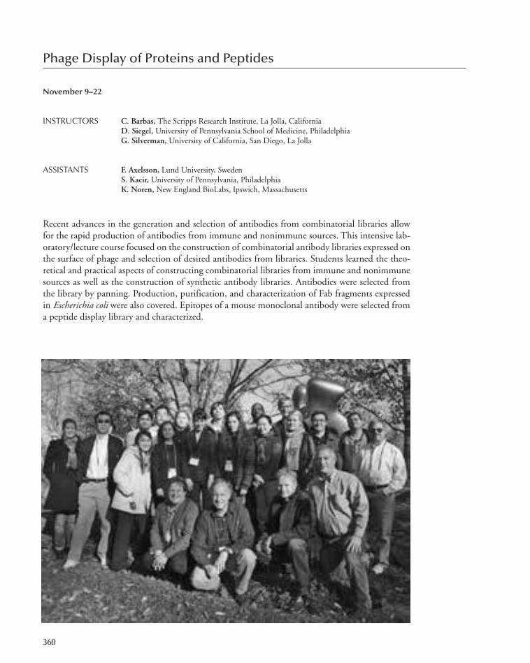
Phage Display of Proteins and Peptides
November 9–22
INSTRUCTORS C. Barbas, The Scripps Research Institute, La Jolla, CaliforniaD. Siegel, University of Pennsylvania School of Medicine, PhiladelphiaG. Silverman, University of California, San Diego, La Jolla
ASSISTANTS F. Axelsson, Lund University, SwedenS. Kacir, University of Pennsylvania, PhiladelphiaK. Noren, New England BioLabs, Ipswich, Massachusetts
Recent advances in the generation and selection of antibodies from combinatorial libraries allowfor the rapid production of antibodies from immune and nonimmune sources. This intensive lab-oratory/lecture course focused on the construction of combinatorial antibody libraries expressed onthe surface of phage and selection of desired antibodies from libraries. Students learned the theo-retical and practical aspects of constructing combinatorial libraries from immune and nonimmunesources as well as the construction of synthetic antibody libraries. Antibodies were selected fromthe library by panning. Production, purification, and characterization of Fab fragments expressedin Escherichia coli were also covered. Epitopes of a mouse monoclonal antibody were selected froma peptide display library and characterized.
360
305-366_courses_Annual Report_2009 template 7/8/11 12:25 PM Page 360
The lecture series, presented by a number of invited speakers, emphasized PCR of immunoglob-ulin genes, the biology of filamentous phage, the utility of surface expression libraries, expressionof antibodies in E. coli and mammalian cells, antibody structure and function, catalytic antibodies,directed protein evolution, retroviral and cell display libraries, the immunobiology of the antibodyresponse, and recent results on the use of antibodies in therapy. The theory and practical implicationsfor selection from phage displayed libraries of random peptides, cDNA products, and semisyntheticproteins was also explored.This course was supported with funds provided by the Howard Hughes Medical Institute.
PARTICIPANTS
Phage Display of Proteins and Peptides 361
Acharya, J., Ph.D., National Cancer Institute, Frederick,Maryland
Batra, G., Ph.D., University of Turku, FinlandChing, A., M.Sc., Butantan Institute, Sao Paulo, BrazilChiu, E., M.S., Mount Sinai Hospital, Toronto, CanadaDarris, C., B.S., Tennessee State University, NashvilleDerda, R., Ph.D., Harvard University, Cambridge,Massachusetts
Dubiley, S., Ph.D., IBG RAS, Moscow, RussiaHasan, A., M.D., Memorial Sloan-Kettering Cancer Center,New York
Lew, S., Ph.D., Columbia University, New YorkLima, S., M.Sc., University of São Paulo, BrazilLindberg, H., M.Sc., KTH, Stockholm, SwedenMohr, A., M.Sc., University of Copenhagen, DenmarkNovinger, L., B.S., University of Vermont College ofMedicine, Burlington
Prudencio, C., Ph.D., University of São Paulo, BrazilQian, Y., Ph.D., University of North Carolina, Chapel HillSanchez, A., M.S., Biokit, S.A., Barcelona, Spain
SEMINARS
Barbas, C.S., Scripps Research Institute, La Jolla, California:Functional antibodies and instant immunity.
Dreier, B., University of Zurich, Switzerland: Ribosomedisplay libraries.
Ladner, R., Dyax Corporation, Ijamsville, Maryland: Phagedisplay of peptides and their development at therapeutics.
Noren, C., New Englands Biolabs, Ipswich, Massachusetts:Functional peptide libraries.
Rader, C., National Institutes of Health, Bethesda, Maryland:Toward better monoclonal antibodies for cancer therapy:From antigen discovery to antibody engineering.
Siegel, D., University of Pennsylvania, Philadelphia: Cell-surface selection of phage-displayed antibodies.
Sidhu, S., University of Toronto, Ontario, Canada: Phagedisplay of synthetic antibodies and SH2 domains.
Silverman, G., University of California, San Diego: Overviewof the adaptive immune system.
Stahl, S., Royal Institute of Technology, Stockholm, Sweden:Phage display of affibodies.
305-366_courses_Annual Report_2009 template 7/8/11 12:25 PM Page 361
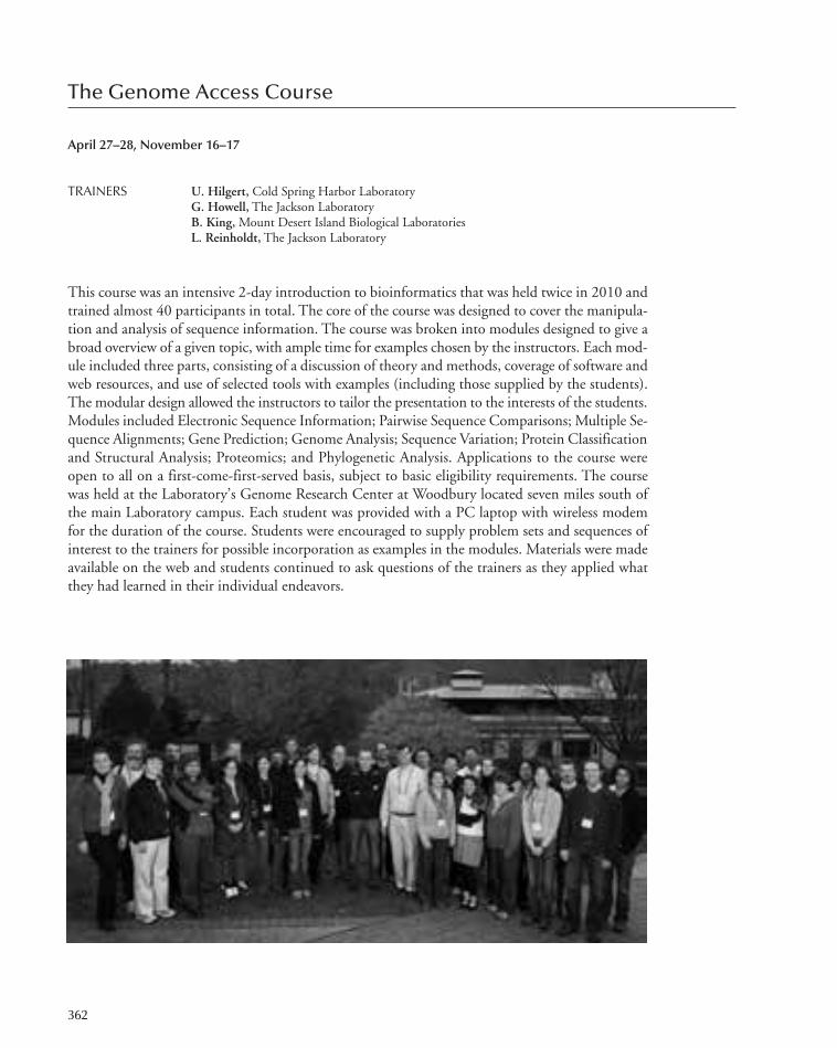
The Genome Access Course
April 27–28, November 16–17
TRAINERS U. Hilgert, Cold Spring Harbor LaboratoryG. Howell, The Jackson LaboratoryB. King, Mount Desert Island Biological LaboratoriesL. Reinholdt, The Jackson Laboratory
This course was an intensive 2-day introduction to bioinformatics that was held twice in 2010 andtrained almost 40 participants in total. The core of the course was designed to cover the manipula-tion and analysis of sequence information. The course was broken into modules designed to give abroad overview of a given topic, with ample time for examples chosen by the instructors. Each mod-ule included three parts, consisting of a discussion of theory and methods, coverage of software andweb resources, and use of selected tools with examples (including those supplied by the students).The modular design allowed the instructors to tailor the presentation to the interests of the students.Modules included Electronic Sequence Information; Pairwise Sequence Comparisons; Multiple Se-quence Alignments; Gene Prediction; Genome Analysis; Sequence Variation; Protein Classificationand Structural Analysis; Proteomics; and Phylogenetic Analysis. Applications to the course wereopen to all on a first-come-first-served basis, subject to basic eligibility requirements. The coursewas held at the Laboratory’s Genome Research Center at Woodbury located seven miles south ofthe main Laboratory campus. Each student was provided with a PC laptop with wireless modemfor the duration of the course. Students were encouraged to supply problem sets and sequences ofinterest to the trainers for possible incorporation as examples in the modules. Materials were madeavailable on the web and students continued to ask questions of the trainers as they applied whatthey had learned in their individual endeavors.
362
305-366_courses_Annual Report_2009 template 7/8/11 12:25 PM Page 362
The Genome Access Course 363
April 27–29
Chowanadisai, W., Marine Biological Laboratory, WoodsHole, Massachusetts
Dolan, P., University of Rochester/University of Alabama,Birmingham, New York
Ernst, P., Dartmouth Medical School, Hanover, NewHampshire
Eswarakumar, V., Yale University, New Haven, ConnecticutFaucher, S., Public Health Agency of Canada, Ottawa Gebhardt, C., Harvard University, Cambridge, Massachusetts
Ghiban, E., Cold Spring Harbor LaboratoryGierszewska, M., Cold Spring Harbor LaboratoryGlaser, R., Stevenson University, MarylandKucera, S., University of Tampa, FloridaLi, Z., The Rockefeller University, New YorkLu, S., Harvard University, Cambridge, MassachusettsMcFadden, S., Independent Scientific Research Advocates,Bethesda, Maryland
Sattely, E., Harvard Medical School, Boston, Massachusetts
November 16–17
Ander, B., University of California, Davis M.I.N.D. Institute,Sacramento
Boland, L., University of Richmond, VirginiaBradley, B., Yale University, New Haven, ConnecticutCruz, V., USDA, ARS, NCGRP, Fort Collins, ColoradoDadras, S., University of Connecticut, FarmingtonDirk, M., Stony Brook University, New YorkDolezal, D., SUNY Upstate Medical University, Syracuse,New York
Duman, E., Stony Brook University, New YorkFowler, T., Tufts University, Boston, MassachusettsFranco, S,. Johns Hopkins University, Baltimore, MarylandJurkiewicz, S., SUNY Stony Brook, New YorkKung, V., Northwestern University, Chicago, IllinoisLogue, M., Boston University School of Medicine,Massachusetts
Marko, M., Cleveland Clinic, OhioOzer, E., Northwestern University, Chicago, IllinoisPanella, L., USDA, ARS, CRL, Fort Collins, ColoradoPatil, V., UMDNJ–New Jersey Medical School, NewarkReilley, A., National Center for Genetic ResourcesPreservation, Fort Collins, Colorado
Richards, C., National Center for Genetic ResourcesPreservation, Fort Collins, Colorado
Romero, S., Yale University, New Haven, ConnecticutStone, A., Arizona State University, TempeSzpara, M., Princeton University, New JerseyChaozhe, Y., University of Alabama, BirminghamZuo, L., Yale University, West Haven, Connecticut
305-366_courses_Annual Report_2009 template 7/8/11 12:25 PM Page 363

The Laboratory acknowledges the generosity of the following companies that loaned equipment and reagentsto the various courses:
364 Postgraduate Courses
454 Life SciencesAccuri Cytometers Inc.Advanced Spectral ImagingAgilent Technologies Inc.Andor TechnologyApplied BiosystemsApplied PrecisionArt Robbins InstituteAstro-Med Inc.BD BiosciencesBerthold Technologies USA,LLC
Bio-Rad LaboratoriesBioptechs Inc.Bruker Daltonics Inc.Carl Zeiss Inc.Charles River LaboratoriesChroma TechnologyCorporation
Coherent LaserConOpticsCoolLED
Corning Inc.CrystaLaserDagan CorporationDavid Kopf InstrumentsDoric LensesDrummond ScientificCompany
Epicentre TechnologiesEppendorf North AmericaFotodyne Inc.GE HealthcareHamamatsu Photonic SystemsHamilton Thorne BiosciencesHampton ResearchHarvard Apparatus Inc.HekaIllumina Inc.Intelligent ImagingInnovations Inc.
James A. Hill InstrumentServices
Kinetic Systems Inc.
Leica Microsystems Inc.Life TechnologiesLI-CORLuigs & NeumannMDS Analytical TechnologiesNalge Nunc InternationalNanodrop TechnologiesNarishige International USAInc.
NascoNeoBiosystemsNew England Biolabs Inc.Nikon Inc.Olympus America Inc.PerkinElmer Life andAnalytical Sciences
PhotometricsPhotonic Instruments Inc.PiercePromega CorporationQiagen Inc.Q-Imaging Inc.
Roche NimblegenScientifica Ltd.SciGeneSigma-AldrichSinger InstrumentsSpectra-Physics Lasers Inc.Stanford PhotonicsStanford Research SystemsStratageneSutter InstrumentsTecan US, Inc.The Jackson LaboratoryThermo Fisher ScientificTokai Hit Co., Ltd..VDS Vosskuhler GmbHWarner InstrumentsWaters CorporationWorld Precision Instruments
305-366_courses_Annual Report_2009 template 7/8/11 12:25 PM Page 364

365
SEMINARS
INVITED SPEAKER PROGRAMEach year, Cold Spring Harbor Laboratory invites speakers from outside the institution to present theirfindings. These weekly seminars keep the CSHL staff current on the latest scientific developments andbroaden their perspectives. Graduate students and postdoctoral fellows meet with the seminar speakersfor lunch immediately after the seminar, allowing an opportunity for the exchange of ideas in an in-formal setting.
Title HostJanuaryDr. Eric Holland, Memorial Sloan-Kettering Brain tumor stem-like cells and where they live. Grisha EnikolopovCancer Center
Dr. Brian Chait, The Rockefeller University Proteomic and nanoengineering probes of Leemor Joshua-Tornucleocytoplasmic transport.
Dr. Steve Henikoff, Fred Hutchinson Cancer Histone variant dynamics and epigenetics. Chris VakocResearch Center
FebruaryDr. David Bartel, Whitehead Institute microRNAs and other small regulatory RNAs. Leemor Joshua-TorDr. Philip Benfey, Duke University Development rooted in interwoven networks. Marja TimmermansDr. Amy Wagers, Harvard University Reversing dysfunction of aged stem cells. Terri Grodzicker
MarchDr. Hugo Bellen, Baylor College of Medicine A forward genetic approach to study neurodegeneration. Linda Van AelstDr. Josh Sanes, Harvard University Synaptic specificity in the visual system. Josh HuangDr. Karl Deisseroth, Stanford University Optogenetics: New developments, new applications. Tony ZadorDr. Lisa Coussens, University of California, Inflammation and cancer: Polarized immune Mikala EgebladSan Francisco responses regulate cancer development.
AprilDr. Eric Nestler, Mount Sinai School of Transcriptional and epigenetic mechanisms of addiction. Grigori EnikolopovMedicine
Dr. Gary Ruvkun, Harvard University Comprehensive identification of C. elegans small Leemor Joshua-TorRNA pathway genes.
Dr. David Tuveson, Cancer Research UK Modeling cancer biology and medicine. Lloyd TrotmanDr. David Eisenberg, University of California, Structural studies of the amyloid state. Leemor Joshua-TorLos Angeles
MayDr. Karel Svoboda, Howard Hughes Medical The neural circuits underlying somatosensation. Tony ZadorInstitute
OctoberDr. Irving Weissman, Stanford University Normal and neoplastic stem cells. Grigori EnikolopovDr. Manuel Serrano, Spanish National Cancer Tumor suppressors at the interface of cancer and Scott LoweResearch Centre aging.
NovemberDr. Angelika Amon, Massachusetts Institute The consequences of aneuploidy. Terri Grodzickerof Technology
Dr. David Lewis, University of Pittsburgh Cortical E/I balance and cognitive dysfunction in Josh Huangschizophrenia.
Dr. Mark Ptashne, Memorial Sloan-Kettering Chromatin architecture: What determines it and Alex GannCancer Center what is its function?
305-366_courses_Annual Report_2009 template 7/8/11 12:25 PM Page 365

366 Seminars
DecemberDr. James Surmeier, Northwestern University Calcium, mitochondrial oxidant stress and Pavel OstenMedical School Parkinson’s disease.Dr. Rachel Green, Johns Hopkins University Quality control during translation in bacteria and yeast. Leemor Joshua-Tor
IN-HOUSE SEMINAR PROGRAMCold Spring Harbor Laboratory’s In-House Seminars were initiated to provide a semiformal avenuefor communication between the various research groups at the Laboratory. The seminars also afforda necessary opportunity for the graduate students and postgraduate staff to develop their skills inorganizing, presenting, and defending their research.
TitleJanuaryMike Wigler A unified genetic model for autism.Prem Premsrirut (Lowe Lab) SLowe mice no more: A rapid and efficient method to produce conditional transgenic
mice using RNAi.Mikel Zaratiegui (Martienssen Lab) Centromere-binding protein B preserves genome integrity at replication forks paused
by retrotransposons.Anthony Mazurek (Stillman Lab) Discovery of an essential role for the DEAD-box RNA helicase, DDX5, in cell cycle
control and breast cancer development.
FebruaryFlorin Albeanu Understanding olfactory neuronal circuits in the mammalian brain.Alexander Dobin (Gingeras Lab) STARgazing reveals novel UFTs.Shane McCarthy (Sebat Lab) Rare structural variants: Insights into the genetics of schizophrenia.
MarchPavel Osten Search for neural circuit endophenotypes in mouse models of autism.Hysell Oviedo (Zador Lab) Form follows function: Linking circuits, connectivity, and function in the auditory cortex.Raffaella Sordella Molecular targeted cancer therapies: Quo vadis?
AprilGlenn Turner Population coding in the Drosophila mushroom body.Michael Zhang Machine learning and learning machines.Alea Mills Engineering the genome: Cancer, chromatin, and chronic behavior.
OctoberChris Vakoc Epigenetics of malignant hematopoiesis.Fritz Henn Toward a new pathophysiology of depression.Mikala Egeblad Seeing tumors in context.
November 5James Watson Judah Folkman, anti-angiogenesis and the curing of cancer.Antoine Molaro and Emily Hodges DNA methylation dynamics across mammalian evolution and development.(Hannon Lab)Zhenxun Wang (Krainer Lab) Investigating the mechanism of pyruvate kinase-M mutually exclusive splicing.
DecemberXianfeng (Morgan) Xu (Jackson Lab) Cell-to-cell trafficking of a plant stem cell regulator is mediated by chaperonins.Josh Huang Toward a genetic dissection of neural circuits in cerebral cortex: Chandeliers light up the
path from epigenomes to cognition.Shuang Ni (Stillman Lab) ORC subunits Orc2 and Orc3 are required for stable spindle attachment at human
chromosome kinetochores during mitosis.
305-366_courses_Annual Report_2009 template 7/12/11 10:11 AM Page 366

BANBURY CENTER
367-410_LAR_Banbury_Annual Report_2009 template 7/8/11 12:26 PM Page 367

367-410_LAR_Banbury_Annual Report_2009 template 7/8/11 12:26 PM Page 368

The Banbury Center is now in its 33rd year and continues to be used intensively. In 2010, therewere 22 science-related meetings attended by 577 participants. In addition, the Center was madeavailable to community groups on five occasions. The Meetings and Courses Program held sixlecture courses during the summer months and the Watson School of Biological Sciences used theCenter for its two Topics in Biology courses.
Of the 577 participants, 486 (71%) were from the United States. They were drawn from 33states, the geographical distribution reflecting the degree of biomedical research in the United States.The 91 foreign participants came from 18 countries, the majority from the United Kingdom. Theratio of male to female participants remains at 2:1.
The meeting topics were unusually wide-ranging, even for the Banbury Center. The Center hasalways held meetings on policy issues, issues where biomedical research is relevant to matters of so-cietal interest. The first of two such meetings in 2010 concernedscience and economic policy. The American Recovery and Rein-vestment Act (ARRA) provided an extra $10.4 billion funding overa 2-year period for the National Institutes of Health (NIH) to pro-mote biomedical research through funding new projects, construc-tion, and the purchase of equipment. However, although this hasbeen a great success, the question arises as to what will happenwhen the ARRA funding comes to an end. How Can We Maintainthe Stability of Biomedical Research and Development at the End ofthe ARRA? examined first how NIH spent the ARRA funding andsecond how federal agencies, universities, research institutes, andindividual investigators can manage the ending of ARRA support.Participants included officials from NIH, universities, research in-stitutes, economists, and scientists.
The second meeting, funded by the Ellison Medical Foundation, discussed Easeful Death: 21stCentury Perspectives on Assisted Suicide. End-of-life issues involve profound legal, moral, religious, andbiomedical questions and evoke intense passions. The Banbury Center is admirably suited to tackledifficult topics, although these are more usually controversies in the interpretation of scientific data.Discussions covered three broad areas. First, there was an extended discussion on whether there aredistinctions to be drawn between a physician acquiescing to a patient’s refusal of food and water, as-sisting in suicide by making it possible through providing materials, and the physician actively takingpart in the suicide. Second, participants from Belgium and The Netherlands presented data on whetherthe legalization of euthanasia and assisted suicide changes the behaviors of patients and physicians,and whether the slippery slope argument carries any weight. Third, we discussed the changes in thepolitical and legal apparatus that will have to come about before there can be acceptance of easefuldeath as a proper end to life. Participants included scientists, physicians, lawyers, philosophers, andreligious leaders of differing opinions and although no consensus was likely to have been reached, thediscussions were of remarkable quality and interest.
Turning to more typical Banbury Center topics, there were several meetings on cancer. EnergyMetabolism, the Cell Cycle, and Cancer explored an old hypothesis on the fundamental nature ofcancer in the light of modern molecular analysis. The Warburg Hypothesis was advanced by OttoWarburg in 1924 when he found that tumor cells mainly generate energy anaerobically by glycolysis
BANBURY CENTEREXECUTIVE DIRECTOR’S REPORT
369
M. Battin, A. Asche, E. MacDonald
367-410_LAR_Banbury_Annual Report_2009 template 7/8/11 12:26 PM Page 369
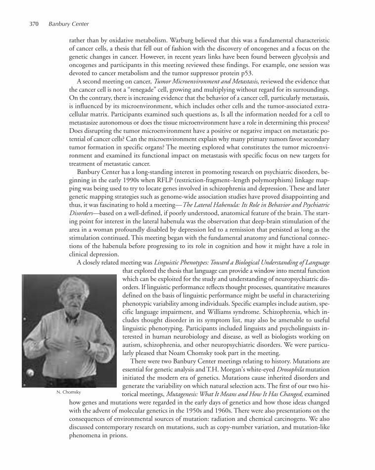
370 Banbury Center
rather than by oxidative metabolism. Warburg believed that this was a fundamental characteristicof cancer cells, a thesis that fell out of fashion with the discovery of oncogenes and a focus on thegenetic changes in cancer. However, in recent years links have been found between glycolysis andoncogenes and participants in this meeting reviewed these findings. For example, one session wasdevoted to cancer metabolism and the tumor suppressor protein p53.
A second meeting on cancer, Tumor Microenvironment and Metastasis, reviewed the evidence thatthe cancer cell is not a “renegade” cell, growing and multiplying without regard for its surroundings.On the contrary, there is increasing evidence that the behavior of a cancer cell, particularly metastasis,is influenced by its microenvironment, which includes other cells and the tumor-associated extra-cellular matrix. Participants examined such questions as, Is all the information needed for a cell tometastasize autonomous or does the tissue microenvironment have a role in determining this process?Does disrupting the tumor microenvironment have a positive or negative impact on metastatic po-tential of cancer cells? Can the microenvironment explain why many primary tumors favor secondarytumor formation in specific organs? The meeting explored what constitutes the tumor microenvi-ronment and examined its functional impact on metastasis with specific focus on new targets fortreatment of metastatic cancer.
Banbury Center has a long-standing interest in promoting research on psychiatric disorders, be-ginning in the early 1990s when RFLP (restriction-fragment–length polymorphism) linkage map-ping was being used to try to locate genes involved in schizophrenia and depression. These and latergenetic mapping strategies such as genome-wide association studies have proved disappointing andthus, it was fascinating to hold a meeting—The Lateral Habenula: Its Role in Behavior and PsychiatricDisorders—based on a well-defined, if poorly understood, anatomical feature of the brain. The start-ing point for interest in the lateral habenula was the observation that deep-brain stimulation of thearea in a woman profoundly disabled by depression led to a remission that persisted as long as thestimulation continued. This meeting began with the fundamental anatomy and functional connec-tions of the habenula before progressing to its role in cognition and how it might have a role inclinical depression.
A closely related meeting was Linguistic Phenotypes: Toward a Biological Understanding of Languagethat explored the thesis that language can provide a window into mental functionwhich can be exploited for the study and understanding of neuropsychiatric dis-orders. If linguistic performance reflects thought processes, quantitative measuresdefined on the basis of linguistic performance might be useful in characterizingphenotypic variability among individuals. Specific examples include autism, spe-cific language impairment, and Williams syndrome. Schizophrenia, which in-cludes thought disorder in its symptom list, may also be amenable to usefullinguistic phenotyping. Participants included linguists and psycholinguists in-terested in human neurobiology and disease, as well as biologists working onautism, schizophrenia, and other neuropsychiatric disorders. We were particu-larly pleased that Noam Chomsky took part in the meeting.
There were two Banbury Center meetings relating to history. Mutations areessential for genetic analysis and T.H. Morgan’s white-eyed Drosophilamutationinitiated the modern era of genetics. Mutations cause inherited disorders andgenerate the variability on which natural selection acts. The first of our two his-torical meetings, Mutagenesis: What It Means and How It Has Changed, examined
how genes and mutations were regarded in the early days of genetics and how those ideas changedwith the advent of molecular genetics in the 1950s and 1960s. There were also presentations on theconsequences of environmental sources of mutation: radiation and chemical carcinogens. We alsodiscussed contemporary research on mutations, such as copy-number variation, and mutation-likephenomena in prions.
N. Chomsky
367-410_LAR_Banbury_Annual Report_2009 template 7/8/11 12:26 PM Page 370

N. Federoff, C. Weisman, E. Friedberg, S. Brenner, A. Creager
Executive Director’s Report 371
Lewis Lehrman (The Lehrman Institute) wishes to encourage the use of genetic and genomicanalyses to inform our knowledge of history and has established a program to foster interactionsbetween scientists and historians. Examples of the power of such interactions are evident in studiestracking the movements of early human beings out of Africa and subsequent spread of populationsthroughout the globe; where and how key events in domestication of plants and animals occurred;and what DNA sequencing is telling us about our relationship with Neanderthals. Participants inDNA, Genetics, and the History of Mankind reviewed these and other topics. The meeting closedwith a free-ranging discussion on how to promote interactions between historians and scientists andon what topics should be covered in the follow-up meeting.
The Banbury Center could not operate at its high level without the hard work of many people.The Center is especially fortunate in having Janice Tozzo and Susanne Igneri ensuring that the meet-ings run smoothly, and Basia Polakowski making sure that participants are welcome in RobertsonHouse. Sonny Leute, Alvin Watson, and Fredy Vasquez look after the grounds, dealing with vastamounts of leaves in the fall and, this year, vast amounts of snow in the winter. Jon Parsons is inde-fatigable in handling AV requirements and Connie Brukin took the photographs which enliven thereport. Culinary Services feeds our participants and Housekeeping copes admirably with the rapidturnover of guests.
Jan WitkowskiExecutive Director
367-410_LAR_Banbury_Annual Report_2009 template 7/8/11 12:26 PM Page 371
Levine Lab Presentation #1D. Carpizo: p53 allele-specific synthetic lethality.
Cordon-Cardo Lab Presentation #1 O. Karni-Schmidt: New findings on p53 and its possibleroles in bladder cancer and development.
Prives Lab Presentation #1S. Singer: Nup98, a potential tumor suppressor regulatesselect p53 target gene expression by a novel mechanism.
Lowe Lab Presentation #1C. Scuoppo: The polyamine–hypusine axis defines a tumorsuppressor network in human lymphoma.
PI PLANNING MEETING
Cordon-Cardo Lab Presentation #2A. Jia: microRNAs in bladder cancer progression.
Levine Lab Presentation #2H. Mizuno: Stem cell signatures in breast cancer with p53mutations.
BANBURY CENTER MEETINGS
p53 Retreat
January 28–30
FUNDED BY Columbia University, New York, New York
ARRANGED BY C. Prives, Columbia University, New York, New York S. Lowe, Cold Spring Harbor Laboratory, New York
The p53 protein first identified in 1979 and believed to be an oncoprotein was shown in 1989 tobe a tumor suppressor. It has a central role in cell cycle control, regulation of DNA repair, and theinitiation of apoptosis and is the gene most frequently mutated in cancers. p53 was named the 1993“Molecule of the Year” by Science. It is not surprising then that p53 has been the subject of intensiveinvestigation, not least by research groups at Cold Spring Harbor Laboratory (Lowe), Princeton(Levine), and Columbia (Prives and Cordon-Cardo). Members of these laboratories came to BanburyCenter to report on their work and to promote interactions between the groups.
372
Prives Lab Presentation #2W. Freed-Pastor: Mutant p53 prevents formation ofmammary acini in three-dimensional culture.
Lowe Lab Presentation #2L. Dow: How to make a hairy mouse: New transgenicshRNA technologies to allow reversible loss of functionphenotypes in adult mice.
Guest LecturersR. Parsons: PTEN regulation in cancer: What is thecontribution of p53 mutation?J. Manley: mRNA processing and cancer: Roles for p53 andother tumor suppressors and oncogenes.
Prives Lab Presentation #3C. Priest: A novel Mdm2 mutant that degrades itselfpreferentially over p53.
Lowe Lab Presentation #3Z. Zhao: p53 affects self-renewal in AML.
Wrap-up Discussion
367-410_LAR_Banbury_Annual Report_2009 template 7/8/11 12:26 PM Page 372

373
The Lateral Habenula: Its Role in Behavior and Psychiatric Disorders
February 28–March 3
FUNDED BY Marie Robertson Memorial Fund
ARRANGED BY F. Henn, Brookhaven National Laboratory, Upton, New YorkB. Li, Cold Spring Harbor Laboratory, New York
Depression is the disorder that is predicted to cause the greatest morbidity in the world by 2020,and a 2009 survey found 16% of Americans suffer from depression. Recently, deep-brain stimulationhas been suggested as a possible therapy for refractory cases, and data suggest that the target of thisstimulation is the lateral habenula. This is a relatively little-studied part of the brain, and given thesefindings, this was the right time to critically review what is known of the function of this structure,its connections, and how they function. Participants included anatomists, physiologists, neurophar-macologists, and clinicians.
Welcoming Remarks: J. Witkowski, Banbury Center, Cold Spring Harbor Laboratory, New YorkBackground: J. Watson, Cold Spring Harbor Laboratory, New YorkIntroduction: F. Henn, Brookhaven National Laboratory, Upton, New York
B. Li, Cold Spring Harbor Laboratory, New York
SESSION 1: The Lateral Habenula: Anatomical/Functional Connections and Cellular BiologyChairperson: O. Hikosaka, National Eye Institute, Bethesda, Maryland
M. Herkenham, National Institute of Mental Health,Bethesda, Maryland: The anatomical connections of thehabenula nuclei, with a historical perspective.
S. Sesack, University of Pittsburgh, Pennsylvania: Projectionsfrom the lateral habenula to midbrain dopamine neurons:indirect inhibitory control via the rostromedial mesopontinetegmentum.
T. Jhou, National Institute on Drug Abuse, Baltimore, Maryland:
A convergence of aversion: Fear, disappointment, andinhibition in the rostromedial tegmentum (RMTg), a habenula target.
S. Haber, University of Rochester Medical Center, New York: The place of habenula in the reward circuit.
P. Shepard, University of Maryland, Baltimore: Stimulation of the lateral habenula inhibits the activity of midbraindopamine neurons at the population level.
367-410_LAR_Banbury_Annual Report_2009 template 7/8/11 12:26 PM Page 373

374 Banbury Center
SESSION 3: The Lateral Habenula and Cognitive FunctionChairperson: B. Moghaddam, University of Pittsburgh, Pennsylvania
SESSION 4: The Lateral Habenula and the Neural Mechanisms Underlying Clinical DepressionChairperson: F.A. Henn, Brookhaven National Laboratory, Upton, New York
W. Drevets, University of Oklahoma-Tulsa University Schoolof Community Medicine: Abnormalities of habenularstructure and function in mood and anxiety disorders.
J. Roiser, University College London, United Kingdom: Roleof the habenula in psychiatric disorders.
A. Sartorius, Central Institute of Mental Health, Mannheim,
Germany: Lateral habenula and treatment-resistantdepression: Results of functional inhibition of the lateralhabenula in congenitally helpless rats and in a first patient.
G. Northoff, University of Ottawa, Ontario, Canada: Subcortical regions and their relevance in emotion processingin depression.
F. Henn, Brookhaven National Laboratory, Upton, New York:An animal model of depression: A key to understanding theroad to the habenula.
B. Li, Cold Spring Harbor Laboratory, New York: Thesynaptic circuitry of lateral habenula and learnedhelplessness.
R. Malinow, University of California, San Diego: The synapticand cellular changes in the lateral habenula of learnedhelplessness in rats.
G. Goelman, Hadassah Hebrew University Medical Center,Jerusalem, Israel: Role of the habenula in depression thataccompanies Parkinson’s disease.
SESSION 5: The Lateral Habenula and Animal Models of Psychiatric DisordersChairperson: R. Dolan, University College London, United Kingdom
SESSION 2: The Lateral Habenula: Anatomical/Functional Connections, and Cellular Biology (Cont’d)Chairperson: S. Haber, University of Rochester Medical Center, New York
R. Veh, Charité—Universitätsmedizin Berlin, Germany:Intrinsic properties and connections of the lateral habenularcomplex in the rat.
U. Kim, Pennsylvania State University College of Medicine,Hershey: Morphological and electrophysiological propertiesof the habenula.
R. Blakely, Vanderbilt School of Medicine, Nashville,Tennessee: Ironing out a relationship between serotonin and
the habenula: Insights from a genetic reference mousepopulation
H. Okamoto, RIKEN Brain Science Institute, Wako City,Japan: Functional analysis of the dorsal habenula in zebrafish, an equivalent structure to the mammalian medialhabenula.
E. Turner, Seattle Children’s Research Institute, Washington: Agene regulatory program for habenula development.
O. Hikosaka, National Eye Institute, Bethesda, Maryland:Role of the lateral habenula in value-based decision-making.
L. Lecourtier, Louis Pasteur University, Strasbourg, France:Role of the habenula in cognitive processes, linked to itsregulatory role over many neurotransmitter systems.
H. Piggins, University of Manchester, United Kingdom: Role of the habenula in the temporal regulation of brain states and behavior.
M. Ullsperger, Radboud University Nijmegen, The Nether-lands: Role of the habenula in performance monitoring andcognitive control. Approaches in human and animalresearch.
G. Yadid, Bar-Ilan University, Ramat-Gan, Israel: Electricalstimulation of the lateral habenula for modulation of thereward system: Application to depression and addiction.
J. Watson, R. Malinow B. Li, S. Sesack
367-410_LAR_Banbury_Annual Report_2009 template 7/8/11 12:26 PM Page 374

375
Epigenetic Reprogramming and Transgenerational Inheritance
March 7–10
FUNDED BY The Cold Spring Harbor Laboratory Corporate Sponsor Program
ARRANGED BY R. Martienssen, Cold Spring Harbor Laboratory, New YorkW. Reik, Babraham Institute, Cambridge, United Kingdom
It is becoming clear that in mammals, plants, and other organisms, epigenetic mechanisms interfacewith genetic ones in regulating developmental decisions and pathways. Epigenetic reprogrammingin the germ line and in early embryos allows pluripotency and stem cell plasticity, whereas the re-striction of developmental plasticity also involves epigenetic mechanisms. Erasure of epigeneticmarks in the germ line may be incomplete, leading to epigenetic inheritance of altered developmentalpotential across generations. In plants, there is growing evidence for a parallel reprogramming eventin both the male and the female germ lines. In pollen, this results in transposon activation followedby small RNA production and transport into gametes. This represents a mechanism for reinforce-ment of transposon control in each generation.
Welcoming Remarks: J. Witkowski, Banbury Center, Cold Spring Harbor Laboratory, New YorkIntroductory Remarks: W. Reik, Babraham Institute, Cambridge, United Kingdom
SESSION 1: Developmental DecisionsChairperson: M.A. Surani, University of Cambridge, United Kingdom
T. Hiiragi, Max-Planck Institut für Molekulare Biomedizin,Muenster, Germany: Stochastic patterning in thedevelopment of pluripotency in mouse embryo.
M.-E. Torres-Padilla, Institute de Génétique et de Biologie,Strasbourg, France: Histone variants establish specializedchromatin signatures during reprogramming.
K. Hochedlinger, Harvard Stem Cell Institute, Cambridge,Massachusetts: Epigenetic similarities and differences between ES cells and iPS cells.
E. Heard, Institut Curie, Paris, France: Lessons from in vivo studieson X-chromosome inactivation and reactivation in differentanimals.
H. Blau, Stanford University School of Medicine, California:Role of active DNA demethylation by AID inreprogramming toward pluripotency.
P. Avner, Institut Pasteur, Paris, France: Developmentalregulation of X-chromosome inactivation.
367-410_LAR_Banbury_Annual Report_2009 template 7/8/11 12:26 PM Page 375

R. Martienssen, Cold Spring Harbor Laboratory, New York:Heterochromatin reprogramming and small RNA silencing inplant germ cells.
F. Berger, National University of Singapore: Reprogramminghistone modifications in Arabidopsis.
J. Walter, Saarland University, Saarbrücken, Germany:Epigenetic reprogramming in the mouse zygote: Lessonsfrom bisulphate sequencing.
M. Surani, University of Cambridge, United Kingdom:Resetting the epigenome in embryos and germ line in themouse.
R. Fischer, University of California, Berkeley: Reprogrammingthe endosperm genome.
W. Reik, Babraham Institute, Cambridge, United Kingdom:Reprogramming and programming of DNA methylationpatterns in the genome.
SESSION 3: Imprints and InheritanceChairperson: T. Bestor, Columbia University, New York, New York
H. Sasaki, Medical Institute of Bioregulation, Kyushu,Fukuoka, Japan: Role of the PIWI pathway in DNAmethylation of the imprinted Rasgrf 1 locus in the malemouse germline.
A. Ferguson-Smith, University of Cambridge, UnitedKingdom: Genomic imprinting and the stability of theepigenetic program in developmental processes.
V. Colot, Ecole Normale Superieure, Paris, France:Transgenerational inheritance of epigenetic variation inArabidopsis: Mechanisms and phenotypic impact.
D. Bourc’his, Institut Curie, Paris, France: Dnmt3L-independent way to methylate mammalian genomes.
W. Kelly, Emory University, Atlanta, Georgia: Epigeneticinheritance and reprogramming through the germline in C.elegans.
J. Paszkowski, University of Geneva, Switzerland: Stability oftransgenerational epigenetic inheritance.
SESSION 4: Environmental Effects and EvolutionChairperson: J. Rafalski, Dupont Experimental Station, Wilmington, Delaware
N. Heintz, The Rockefeller University, New York, New York: The significance of 5-hydroxymethylctytosine in neuronal function.
A. Rao, Harvard Medical School, Boston, Massachusetts:Biological functions of TET proteins, enzymes that convert5 methylcytosine to hydroxymethylcytosine DNA.
D. Ruden, Wayne State University, Detroit, Michigan:Epigenomic reprogramming of aggression in killer bees.
SESSION 2: Reprogramming and ReproductionChairperson: E. Heard, Institut Curie, Paris, France
376 Banbury Center
J. Finnegan, CSIRO, Black Mountain, Australia: Flicking theswitch on polycomb-regulated genes.
B. Hohn, Friedrich Miescher Institute of Biomedical Research,Basel, Switzerland: Environmental influences on plantgenome dynamics.
U. Grossniklaus, Institute of Plant Biology, University ofZurich, Switzerland: Role of epigenetic regulation inevolution: The control of pollination syndromes.
SESSION 5: Methylation MechanismsChairperson: H. Sasaki, Medical Institute of Bioregulation, Kyushu, Higashiaku, Japan
T. Bestor, Columbia University, New York, New York: How muchtissue-specific DNA methylation and how important?
Y. Zhang, HHMI, University of North Carolina, Chapel Hill: Could the DNA demethylase please stand up?
S. Jacobsen, University of California, Los Angeles: DNA methylation in Arabidopsis.
General Discussion: W. Reik, Babraham Institute, Cambridge, UnitedKingdom
Concluding Remarks: R. Martienssen, Cold Spring Harbor Laboratory,New York H. Blau, Y. Lazebnik
367-410_LAR_Banbury_Annual Report_2009 template 7/8/11 12:26 PM Page 376

377
The Second NIMH-Sponsored Brain Camp
March 13–16
FUNDED BY National Institute of Mental Health
ARRANGED BY M. Akil, National Institute of Mental Health, Bethesda, MarylandT. Insel, National Institute of Mental Health, Bethesda, Maryland
We were very happy that the NIMH Brain Camp returned to Banbury in 2010 after its successfulinauguration in 2009. The goal of the Brain Camp is to identify areas of neuroscience that are ofinterest and relevance to psychiatrists and to communicate these to a small group of outstandingpsychiatry residents and research fellows. Some of the most distinguished and thoughtful neurosci-entists in the country contributed to the meeting. The outcome of the meeting will be the start ofa neuroscience curriculum that can eventually be shared with psychiatry training programs aroundthe country.
Introduction and Charge: M. Akil, National Institute of Mental Health, Bethesda, MarylandRethinking Mental Illness: T. Insel, National Institute of Mental Health, Bethesda, Maryland
SESSION 1: Circuitry Underlying Aggression/Circuitry of Emotional LearningR. Yuste, Columbia University, New York, New YorkD. Salzman, Columbia University, New York, New York
SESSION 4: Circuitry Underlying Fear, Anxiety, and RecoveryJ. LeDoux, New York University, New York, New YorkE. Phelps, New York University, New York, New York
Roundtable Discussion with All Speakers
SESSION 5: Translation: From Neurobiology to TreatmentsK. Berman, National Institute of Mental Health, Bethesda,Maryland
M. Bear, Massachusetts Institute of Technology, Cambridge
SESSION 2: How Circuits DevelopC. Nelson, Harvard University, Boston, MassachusettsB.J. Casey, Cornell University, Ithaca, New York
Discussion with the Organizers: Teaching neuroscience inmedical school and during psychiatry training: What’s missing?
SESSION 3: Stress from Molecules to CircuitsH. Akil, University of Michigan, Ann Arbor
367-410_LAR_Banbury_Annual Report_2009 template 7/8/11 12:26 PM Page 377

How Can We Maintain the Stability of Biomedical Researchand Development at the End of the ARRA?
April 25–27
FUNDED BY Howard Hughes Medical InstituteAlfred P. Sloan Foundation
ARRANGED BY R. Freeman, NBER Science and Engineering Workforce Project, Harvard University, Cambridge, MassachusettsP. Stephan, Andrew Young School of Policy Studies, Georgia State University, AtlantaA. Wang, NBER Science and Engineering Workforce Project, Harvard University, Cambridge, MassachusettsJ. Witkowski, Banbury Center, Cold Spring Harbor Laboratory, New York
The American Recovery and Reinvestment Act (ARRA) has provided an extraordinary $8.2 billionin extramural funding to the National Institutes of Health and $3 billion to the National ScienceFoundation as well as sizable funds to other agencies. These funds have been used to promote re-search, construct and renovate buildings and facilities, and purchase shared instrumentation. Bio-medical research has benefited greatly from the ARRA, but there are increasing concerns about whatwill happen when the program comes to an end. It is important that the young researchers whosecareers have been promoted through the support of the stimulus funds are not abandoned and thatpromising projects initiated using ARRA funds are not delayed. This workshop was held to discussways in which the government, universities, and other groups can “smooth” spending over timeand find other ways to avoid problems.
Welcoming Remarks: J. Witkowski, Banbury Center, Cold Spring Harbor Laboratory, New YorkIntroduction: ARRA and NIH, and Government Support for Science
R. Freeman and A. Wang, NBER Science and Engineering Workforce Project, Harvard University, Cambridge, Massachusetts
378
367-410_LAR_Banbury_Annual Report_2009 template 7/8/11 12:26 PM Page 378

Maintaining the Stability of Biomedical Research and Development at the End of the ARRA 379
SESSION 1: How NIH Spent the ARRA Funding andThoughts About Post-ARRAPoints to ConsiderL. Tabak, National Institute of Dental and Craniofacial Research, Bethesda, Maryland
J. Niederhuber, National Cancer Institute, Bethesda, Maryland
P. Stephan, Andrew Young School of Policy Studies, Georgia State University, Atlanta
SESSION 2:What Is the Hard-Landing Scenario for ResearchCareers? What Is the Best-Case Scenario?Points to Consider M. Teitelbaum, The Alfred P. Sloan Foundation, New York, New York
J. Wiest, National Cancer Institute, Bethesda, MarylandH. Garrison, Federation of American Societies for Experimental Biology, Bethesda, Maryland
SESSION 3: How Can Federal Agencies Manage the End ofARRA?Points to ConsiderS. Turner, University of Virginia, CharlottesvilleJ. McGowan, National Institute of Allergy and Infectious Diseases, Bethesda, Maryland
D. Mowery, University of California, Berkeley
W. Schaffer G. Marschke, P. Stephan R. Freeman
SESSION 4: How Can Universities and Research InstitutesManage the End of ARRA?Points to ConsiderD. Korn, Harvard University, Cambridge, MassachusettsB. Stillman, Cold Spring Harbor Laboratory, New York
SESSION 5: How Can PIs Manage the End of ARRA?Points to Consider G. Marschke, National Bureau of Economic Research, Cambridge, Massachusetts
R. Kolter, Harvard Medical School, Boston, MassachusettsM. Carlson, Howard Hughes Medical Institute, Chevy Chase, Maryland
F. Murray, MIT Sloan School, Cambridge, Massachusetts
SESSION 6: Innovative Approaches to Managing Support forSciencePoints to ConsiderW. Goldschmidts, Cold Spring Harbor Laboratory, New York
D. Goroff, The Alfred P. Sloan Foundation, New York, New York
W. Schaffer, National Institutes of Health, Bethesda, Maryland
Summary and Concluding RemarksR. Freeman, NBER Science and Engineering Workforce Project, Harvard University, Cambridge, Massachusetts
367-410_LAR_Banbury_Annual Report_2009 template 7/8/11 12:26 PM Page 379

380
Linguistic Phenotypes: Toward a Biological Understandingof Language
May 2–5
FUNDED BY The Simons FoundationUniversité de Québec à MontréalOliver Grace Fund
ARRANGED BY R. Berwick, Massachusetts Institute of Technology, CambridgeN. Chomsky, Massachusetts Institute of Technology, CambridgeA. Di Sciullo, Université de Québec, Montréal, CanadaP. Mitra, Cold Spring Harbor Laboratory, New YorkK. Wexler, Massachusetts Institute of Technology, Cambridge
The general idea of this meeting was that language provides a window into mental function thatcan be exploited for the study and understanding of neuropsychiatric disorders. One of the out-standing problems in the study of neuropsychiatric disorders is the relative paucity of objectivelydefined phenotypic measures, which can be used for diagnoses and in psychiatric genetic studies.Linguistic performance reflects thought processes, so the hypothesis is that quantitative measuresdefined on the basis of linguistic performance could be used to characterize phenotypic variabilityamong individuals. Autism (which includes communication disorders), specific language impair-ment, as well as Williams syndrome (where language seems to be a relative strength) are of particularinterest in this regard. Schizophrenia (which includes thought disorder in its symptom list) mayalso be amenable to useful linguistic phenotyping. Participants included linguists and psycholinguistsinterested in human neurobiology and disease, as well as biologists working on autism, schizophrenia,and other neuropsychiatric disorders.
A major goal of the meeting was to bring together scientists who are doing linguistic phenotypingwith experts in the syndromes and geneticists, in an attempt to push along what we hope will be anexciting field that can make major contributions to one of the most important scientific (theoreticaland applied) topics concerning humans.
367-410_LAR_Banbury_Annual Report_2009 template 7/8/11 12:26 PM Page 380
Linguistic Phenotypes: Toward a Biological Understanding of Language 381
Introductory Remarks: J. Witkowski, Banbury Center, Cold Spring Harbor Laboratory, New York
INTRODUCTORY SESSIONChairperson: R. Berwick, Massachusetts Institute of Technology, Cambridge
SESSION 1Chairperson: A.-M. Di Sciullo, Université du Québec à Montréal, Canada
P. Mitra, Cold Spring Harbor Laboratory, New York:Introduction.
N. Chomsky, Massachusetts Institute of Technology, Cambridge: Language as a biological organ: What is it? How does it develop? And why?
K. Wexler, Massachusetts Institute of Technology, Cambridge:Optional infinitives: A worked-out model of a particularlinguistic characterization/phenotype.
S. Fisher, University of Oxford, United Kingdom: Building bridges between ge
nes, brains, and language.A. Monaco, Wellcome Trust Centre for Human Genetics, Oxford, United Kingdom: Genetics of specificlanguage impairment.
N. Modyanova and K. Wexler, Massachusetts Institute ofTechnology, Cambridge: Linguistic phenotypes of morecomplex syntax: Phases and determiners in SLI, Williamssyndrome, and autism.
A. Perovic, University College London, United Kingdom:Grammatical impairments in autism spectrum, Williamssyndrome, and Down syndrome.
L. Kovelman, University of Michigan, Ann Arbor: Optionalinfinitive: Evidence of how adult brain processes grammaticalerrors that are typical.
SESSION 2: Chairperson: K. Wexler, Massachusetts Institute of Technology, Cambridge, Massachusetts
L.-A. Petitto, University of Toronto, Canada: The phonetic,genetic, and brain-based changes that give rise to earlylanguage acquisition. Genes, brains, and cognition: space and language in Williams syndrome.
M.-T. Guasti, University of Milan-Biocca, Milan, Italy: SLIand dyslexia: Differences between linguistic disorders inchildren.
General DiscussionB. Landau, Johns Hopkins University School of Medicine,Baltimore, MarylandK. Wexler, Massachusetts Institute of Technology, CambridgeP. Mitra, Cold Spring Harbor Laboratory, New York
Moderated Discussion: White Paper
SESSION 3 Chairperson: P. Mitra, Cold Spring Harbor Laboratory, New York
D. Kimbrough Oller, University of Memphis, Tennessee and P. Niyogi, University of Chicago, Illinois: Vocal developmenttheory and the automated identification disorders includingautism.
P. Suppes, Stanford University, California: Brainrepresentations of linguistic constituents.
P. Freed, Columbia University, New York, New York:Applications to clinical psychiatric practice.
N. Schiff, Weill Cornell Medical College, New York, NewYork: Recovery of consciousness after severe brain injury.Opportunities for linguistics to inform research.
D. Fox, Massachusetts Institute of Technology, Cambridge:Logic, language, and modularity.
SESSION 4Chairperson: N. Modyanova, Massachusetts Institute of Technology, Cambridge
L. Osborne, University of Toronto, Canada: Duplication ofgenes on human chromosome 7q11.23 and their role inspeech and expressive language.
C. Mervis, University of Louisville, Kentucky: Speech andlanguage abilities of individuals with Williams syndrome.
S. Desiderio, Johns Hopkins School of Medicine, Baltimore,Maryland: TFII-I, a target of genetic lesions associated withthe Williams–Beuren cognitive profile.
W. McCombie, Cold Spring Harbor Laboratory, New York:Genetics becomes genomics.
Discussion: Future Planning
367-410_LAR_Banbury_Annual Report_2009 template 7/8/11 12:26 PM Page 381

382
Tumor Microenvironment and Metastasis
May 5–7
FUNDED BY Champalimaud Foundation and the Champalimaud Metastasis Programme
ARRANGED BY R. Kalluri, Harvard Medical School, Boston, MassachusettsJ. Massague, Memorial Sloan-Kettering Cancer Center, New York, New York
Approximately 80% of deaths related to cancer are associated with metastasis, and the central unan-swered question remains the mechanism behind the systemic spread of cancer and secondary tumorformation in distant organs. It is clear that the microenvironment of a cancer cell has a key role ininfluencing its behavior and that this environment includes other cells and the tumor-associatedextracellular matrix. The question, then, is what role does the tumor microenvironment have inmetastasis? Is all the information needed for a cell to metastasize cell-autonomous or does the tissuemicroenvironment have a role in determining this process? Does disrupting the tumor microenvi-ronment have a positive or negative impact on metastatic potential of cancer cells? Can the mi-croenvironment explain why many primary tumors favor secondary tumor formation in specificorgans? The meeting explored what constitutes the tumor microenvironment and examined its func-tional impact on metastasis with specific focus on new targets for treatment of metastatic cancer.
Welcoming Remarks: J. Witkowski, Banbury Center, Cold Spring Harbor Laboratory, New YorkIntroductory Remarks: J. Watson, Cold Spring Harbor Laboratory, New York
SESSION 1Chairperson: R. Kalluri, Harvard Medical School, Boston, Massachusetts
J. Massague, Memorial Sloan-Kettering Cancer Center, New York, New York: Surviving the microenvironment.
Z. Werb, University of California, San Francisco: Transcriptional regulation of metastasis.
367-410_LAR_Banbury_Annual Report_2009 template 7/8/11 12:26 PM Page 382
Tumor Microenvironment and Metastasis 383
SESSION 2Chairperson: Z. Werb, University of California, San Francisco
H. Dvorak, Beth Israel Deaconess Medical Center, BostonMassachusetts: Angiogenesis: The wrong therapeutic target?
D. McDonald, University of California, San Francisco:Angiogenesis inhibitors: Risk and return.
A. Harris, The Weatherall Institute of Molecular Medicine,
SESSION 3Chairperson: K. Cichowski, Brigham & Women’s Hospital, Boston, Massachusetts
Oxford, United Kingdom: Role of notch signaling and anti-VEGF therapy resistance.
H. Hurwitz, Duke University Medical Center, Durham, NorthCarolina: Anti-angiogenesis therapy for cancer-expected andunexpected outcomes.
M. Clarke, Stanford University, Palo Alto, California:Regulation of self-renewal by the microenvironment innormal epithelial stem cells and epithelial cancer cells or stemcells and cancer, two faces.
R. Bjerkvig, University of Bergen, Norway: Cancer stem cells:Can they be defined?
S. Haggarty, Massachusetts General Hospital, Boston: Patient-specific stem cell modes for characterizing disease andtherapeutic discovery.
J. Chang, Baylor College of Medicine, Houston, Texas: Multi-targeting of key self-renewal pathways.
SESSION 4Chairperson: J. Massague, Memorial Sloan-Kettering Cancer Center, New York, New York
R. Kalluri, Harvard Medical School, Boston, Massachusetts:Pericyte coverage of tumor vessels: An adaptive host response tocontrol tumor hypoxia?
J. Sleeman, University of Heidelberg, Germany: Thesignificance of lymphatic dissemination for metastasis: Blindalley, highway, or beacon?
K. Cichowski, Brigham & Women’s Hospital, Boston,Massachusetts: The inflammation, the microenvironment,and prostate cancer metastasis.
D. Cheresh, University of California, San Diego, La Jolla:microRNA-132 mediated loss of p120RasGAP activatesquiescent endothelium to facilitate pathological angiogenesisand tumor growth.
SESSION 5Chairperson: S. Mohla, National Cancer Institute, Bethesda, Maryland
S. Rafii, Weill Cornell Medical Center, New York, New York:Contribution of the activated vascular niche to tumor growth.
D. Lyden, Weill Cornell Medical Center, New York, NewYork: Early cellular molecular events for the information ofmetastatic niche.
R. Hynes, Massachusetts Institute of Technology, Cambridge:Extracellular matrices of tumors.
SESSION 6Chairperson: Y. Kang, Princeton University, New Jersey
S. Dias, Portuguese Institute of Oncology, Lisbon, Portugal:Metabolism and metastasis.
S. Muthuswamy, Ontario Cancer Institute, University ofToronto, Canada: Cell polarity and cancer progression.
O. Casanovas, Catalan Institute of Oncology, Barcelona,Spain: Tumor-adaptive responses to antiangiogenic therapies.
E. Sahai, Cancer Research UK, London, United Kingdom:Cancer cell invasion in a complex environment.
R. Sordella, Cold Spring Harbor Laboratory, New York:Intrinsic and extrinsic regulation of metastatic spread ofNSCLC.
SESSION 7Chairperson: D. Lyden, Weill Cornell Medical College, New York
M. Skobe, Mount Sinai School of Medicine, New York, NewYork: Role of lymphangiogenesis in tumor metastasis.
J. Condeelis, Albert Einstein College of Medicine, Bronx, NewYork: Imaging of the tumor microenvironment of metastasisand the cell types within.
Y. Kang, Princeton University, New Jersey: Tumor–stromalinteractions in bone metastasis: Novel targets fortherapeutic invention.
367-410_LAR_Banbury_Annual Report_2009 template 7/8/11 12:26 PM Page 383

384
Genetic Variation at a Single Locus for Prediction andPrevention of Late-Onset Alzheimer’s Disease
May 9–10
FUNDED BY United Biomedical, Inc.
ARRANGED BY C. Finstad, United Biomedical, Inc., Hauppauge, New YorkC. Wang, United Biomedical, Inc., Hauppauge, New York
Heterogeneity of response of individuals selected for clinical trials is a serious problem. Participantsin this workshop explored ways to incorporate “pharmacogenetics” as a tool in the selection of in-dividuals with pre-Alzheimer’s disease for anti-Aβ peptide immunotherapy. There was also discussionof how sequence variation is used to develop genome-wide association maps for determining howgenomes are organized and regulated and their role in disease.
Opening Remarks: J. Watson, Cold Spring Harbor Laboratory, New YorkC. Wang, United Biomedical, Inc., Hauppauge, New York
SESSION 1: Genetic Variation and Early Detection of Alzheimer’s Disease
A. Roses, Deane Drug Discovery Institute, Duke Universityand Cabernet Pharmaceuticals, Durham, North Carolina:Tomm40 variable-length polymorphism predicts the age oflate-onset Alzheimer’s disease (LOAD).
T. Gingeras, Cold Spring Harbor Laboratory, New York:Origin of phenotypes: Genes and transcripts.
367-410_LAR_Banbury_Annual Report_2009 template 7/8/11 12:26 PM Page 384
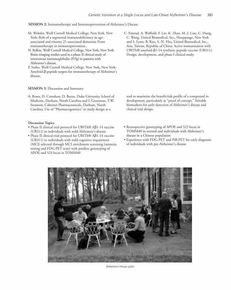
Genetic Variation at a Single Locus and Late-Onset Alzheimer’s Disease 385
SESSION 2: Immunotherapy and Immunoprevention of Alzheimer’s Disease
M. Weksler, Weill Cornell Medical College, New York, NewYork: Role of a segmental immunodeficiency in age-associated and trisomy 21-associated dementia: Fromimmunotherapy to immunoprevention.
N. Relkin, Weill Cornell Medical College, New York, New York:Brain-imaging studies used in a phase II clinical study ofintravenous immunoglobulin (IVIg) in patients withAlzheimer’s disease.
P. Szabo, Weill Cornell Medical College, New York, New York:Amyloid-β peptide targets for immunotherapy of Alzheimer’sdisease.
C. Finstad, A. Walfield, F. Lin, K. Zhao, M.-J. Liao, C. Hung,C. Wang, United Biomedical, Inc., Hauppauge, New Yorkand S. Lynn, B. Kuo, S.-N. Hsu, United Biomedical, Inc.,Asia, Taiwan, Republic of China: Active immunization withUBITh® amyloid-β1-14 synthetic peptide vaccine (UB311):Design, development, and phase I clinical study.
SESSION 3: Discussion and Summary
A. Roses, D. Crenshaw, D. Burns, Duke University School ofMedicine, Durham, North Carolina and I. Grossman, T.W.Swanson, Cabernet Pharmaceuticals, Durham, NorthCarolina: Use of “Pharmacogenetics” in study design as a
tool to maximize the benefit/risk profile of a compound indevelopment, particularly at “proof of concept.” Suitablebiomarkers for early detection of Alzheimer’s disease andclinical trial design.
Discussion Topics• Phase II clinical trial protocol for UBITh® Aβ1-14 vaccine (UB311) in individuals with mild Alzheimer’s disease
• Phase II clinical trial protocol for UBITh® Aβ1-14 vaccine(UB311) in individuals with mild cognitive impairment(MCI) selected through MCI enrichment screening (amnesiatesting and FDG-PET scan) with posthoc genotyping ofAPOE and 523 locus in TOMM40
• Retrospective genotyping of APOE and 523 locus inTOMM40 in normal and individuals with Alzheimer’sdisease in a Chinese population
• Experience with FDG-PET and PiB-PET for early diagnosisof individuals with pre-Alzheimer’s disease
Robertson House patio
367-410_LAR_Banbury_Annual Report_2009 template 7/8/11 12:26 PM Page 385

386
Mutagenesis: What It Means and How It Has Changed
May 15–18
FUNDED BY Oliver Grace Cancer Fund
ARRANGED BY J. Drake, National Institute of Environmental Health, Research Triangle Park, North CarolinaJ. Witkowski, Banbury Center, Cold Spring Harbor Laboratory, New York
Mutation is, of course, an integral part of genetics, although what are considered mutations haschanged over the years as new techniques have led to new knowledge, leading to revisions of what“mutation” encompasses. This discussion meeting reviewed the developing concepts of mutationand understanding of the varied forms of mutation from the early days of Drosophila genetics tothe present. Although the theme of the meeting was the history of the concept of mutation, con-temporary research was also included, for example, on copy-number variation.
Welcoming Remarks: J. Witkowski, Banbury Center, Cold Spring Harbor Laboratory, New YorkIntroductory Remarks: J. Watson, Cold Spring Harbor Laboratory, New York
SESSION 1
S. Mueller-Wille, University of Exeter, United Kingdom: Thetaxonomic roots of mutation: Constant varieties, sports, andpure lines.
E. Carlson, Bloomington, Indiana: Mutation and the gene inH.J. Muller’s course, career, and influence.
S. Brenner, Salk Institute for Biological Studies, San Diego,California: Discovery of frame-shift mutants.
R. Falk, Hebrew University of Jerusalem, Israel: Mutagenesis asa genetic research strategy.
L. Campos, Drew University, Madison, New Jersey: Fromexperimental evolution to genetic engineering: Mutation atCold Spring Harbor Laboratory.
W. Maas, New York University School of Medicine, New York,New York: Role of serendipitous mutants in the elucidationof gene action and its regulation.
Who Are They? Identifying Participants in the Cold SpringHarbor Laboratory Symposia Photographs
367-410_LAR_Banbury_Annual Report_2009 template 7/8/11 12:26 PM Page 386
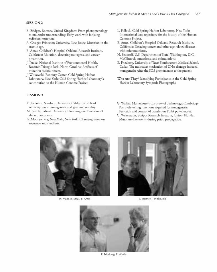
Mutagenesis: What It Means and How It Has Changed 387
SESSION 2
B. Bridges, Romsey, United Kingdom: From phenomenologyto molecular understanding: Early work with ionizingradiation mutation.
A. Creager, Princeton University, New Jersey: Mutation in theatomic age.
B. Ames, Children’s Hospital Oakland Research Institute,California: Mutation, detecting mutagens, and cancerprevention.
J. Drake, National Institute of Environmental Health,Research Triangle Park, North Carolina: Artifacts ofmutation ascertainment.
J. Witkowski, Banbury Center, Cold Spring HarborLaboratory, New York: Cold Spring Harbor Laboratory’scontribution to the Human Genome Project.
W. Maas, R. Maas, B. Ames S. Brenner, J. Witkowski
E. Friedberg, E. Witkin
L. Pollock, Cold Spring Harbor Laboratory, New York:International data repository for the history of the HumanGenome Project.
B. Ames, Children’s Hospital Oakland Research Institute,California: Delaying cancer and other age-related diseaseswith micronutrients.
N. Fedoroff, U.S. Department of State, Washington, D.C.:McClintock, mutations, and epimutations.
E. Friedberg, University of Texas Southwestern Medical School,Dallas: The molecular mechanism of DNA-damage-inducedmutagenesis: After the SOS phenomenon to the present.
Who Are They? Identifying Participants in the Cold SpringHarbor Laboratory Symposia Photographs
SESSION 3
P. Hanawalt, Stanford University, California: Role oftranscription in mutagenesis and genomic stability.
M. Lynch, Indiana University, Bloomington: Evolution of the mutation rate.
G. Montgomery, New York, New York: Changing views onsequence and synthesis.
G. Walker, Massachusetts Institute of Technology, Cambridge:Positively-acting functions required for mutagenesis:Function and control of translesion DNA polymerases.
C. Weissmann, Scripps Research Institute, Jupiter, Florida:Mutation-like events during prion propagation.
367-410_LAR_Banbury_Annual Report_2009 template 7/8/11 12:26 PM Page 387

388
PopTech Science and Public Leadership Fellows Retreat
August 11–15
FUNDED BY PopTech Accelerator
ARRANGED BY L. Filderman, PopTech, Camden, MaineO. Wilder, PopTech, Camden, Maine
The PopTech Science and Public Leadership Fellows Program aims to develop a corps of extremelyhigh potential, socially engaged working scientists who embody science as an essential way of think-ing, discovering, understanding, and deciding, and who can communicate both their work and theimportance of their fields to the public at large. The program gives these scientists intensive, high-quality training, a powerful social network, ongoing mentoring, and opportunities for public lead-ership and engagement. The Banbury Center was delighted to host the Fellows for the first meetingof the science and public leadership group.
Welcome and Introductions: J. Witkowski, Executive Director, Banbury Center, Cold Spring Harbor Laboratory, New York A. Zolli, Executive Director and Curator, PopTech, Camden, Maine
C. Heller, CEO, Heller Communication Design, Brooklyn,New York: Communicating for engagement.
M. Moon, Communication Designer, New York, New York:Art of the great presentation.
S. Marsh, Theatre Arts, Stony Brook University, New York:Improv for scientists—Robertson House.
M. Duenwald, Deputy Editor, Op-Ed Page, The New YorkTimes, New York, New York: Demystifying the Op-Ed page.
L. Witter, Chief Strategy Officer, Fenton, New York: Mediatraining.
H. Schneider, Dean, School of Journalism, Stony BrookUniversity, New York: Communicating science in a changingmedia landscape.
Panel Discussion: The Journalists’ PerspectiveL. Cuthbert, Director, Discovery News, New York, New York
B. Nissen, Senior Producer, NBC News-NBC Learn, New York.M. Moyer, Senior Technology Editor Scientific American,New York, New York and I. Oransky, Executive Editor,Reuters Health: Science, New York, New York: Thejournalist’s perspective.
M. Nisbet, Associate Professor, School of Communication,American University, Washington, D.C. and R. Covey,Senior Vice President, National Geographic Digital Media,Washington, D.C.: Framing scientific issues to fosterengagement.
G. MacKenzie, Founder and President, Gillian MacKenzieAgency, New York, New York; W. Murphy, Senior Editor,Random House, New York, New York; T. Neillsen, CEO,BrightSight Group, Princeton, New Jersey: Creating apersonal brand.
367-410_LAR_Banbury_Annual Report_2009 template 7/8/11 12:26 PM Page 388

Plant Development and Phenomics
September 19–22
FUNDED BY Cold Spring Harbor Laboratory/Pioneer–DuPont Joint Collaborative Research Program
ARRANGED BY D. Jackson, Cold Spring Harbor Laboratory, New YorkH. Sakai, Pioneer–DuPont International, Johnston, Texas
Every year, the Cold Spring Harbor Laboratory–DuPont Pioneer Collaborative meets to review cur-rent projects and to plan future projects. This year’s update focused on the area of phenomics andits relation to plant development and included current research from outside the collaboration byspeakers working in this field.
Welcoming Remarks: J. Witkowski, Banbury Center, Cold Spring Harbor Laboratory, New YorkIntroductory Remarks: D. Jackson, Cold Spring Harbor Laboratory, New York
S. Tingey, DuPont Experimental Station, Wilmington, Delaware
SESSION 1: Informatics, Natural Variation, and YieldChairperson: Z. Lippman, Cold Spring Harbor Laboratory, New York
W. McCombie, Cold Spring Harbor Laboratory, New York:Novel sequencing methodologies and results.
B. Fendler, Cold Spring Harbor Laboratory, New York:Population genetics of microsatellites in humans.
D. Ware, Cold Spring Harbor Laboratory, New York: Profilingmaize using next-generation sequencing approaches.
Z. Lippman, Cold Spring Harbor Laboratory, New York:Flowering, dosage, and yield.
R. Lafitte, Pioneer Hi-Bred International, Johnston, Iowa: Maize field phenotyping for transgene validation.
M. Williams, Dupont Experimental Station, Wilmington,Delaware: Identifying maternal haploid-inducing QTL1 andENU-mutagenesis of maize.
SESSION 2: Development, Epigenetics, and NetworksChairperson: M. Aukerman, Dupont Experimental Station, Wilmington, Delaware
P. Bommert, Cold Spring Harbor Laboratory, New York: Cloningof compact Plant2 and update on other fasciated mutants.
M. Komatsu, DuPont Experimental Station, Wilmington,Delaware: Inflorescence traits and hybrid production.
389
367-410_LAR_Banbury_Annual Report_2009 template 7/8/11 12:26 PM Page 389
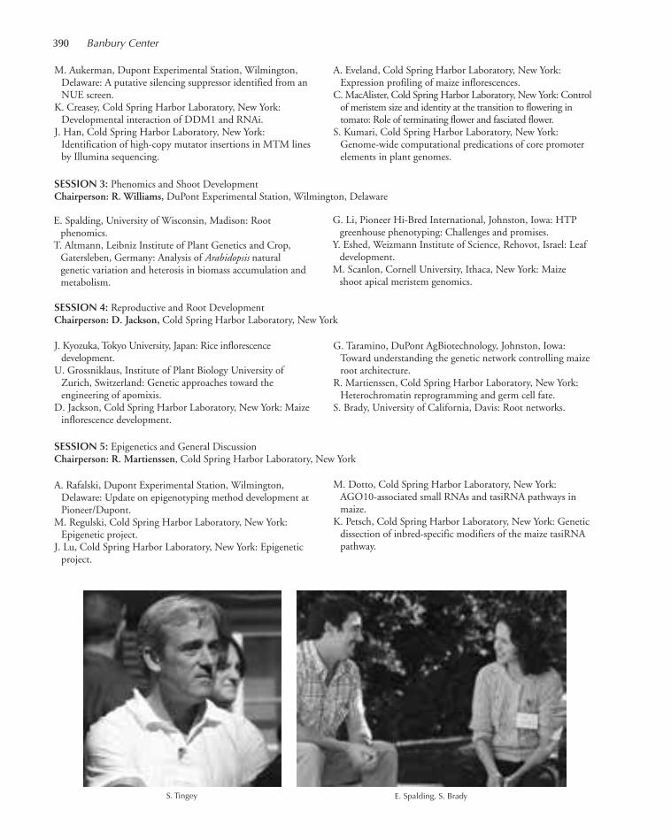
390 Banbury Center
M. Aukerman, Dupont Experimental Station, Wilmington,Delaware: A putative silencing suppressor identified from anNUE screen.
K. Creasey, Cold Spring Harbor Laboratory, New York:Developmental interaction of DDM1 and RNAi.
J. Han, Cold Spring Harbor Laboratory, New York:Identification of high-copy mutator insertions in MTM linesby Illumina sequencing.
A. Eveland, Cold Spring Harbor Laboratory, New York:Expression profiling of maize inflorescences.
C. MacAlister, Cold Spring Harbor Laboratory, New York: Controlof meristem size and identity at the transition to flowering intomato: Role of terminating flower and fasciated flower.
S. Kumari, Cold Spring Harbor Laboratory, New York:Genome-wide computational predications of core promoterelements in plant genomes.
E. Spalding, University of Wisconsin, Madison: Root phenomics.
T. Altmann, Leibniz Institute of Plant Genetics and Crop,Gatersleben, Germany: Analysis of Arabidopsis naturalgenetic variation and heterosis in biomass accumulation andmetabolism.
G. Li, Pioneer Hi-Bred International, Johnston, Iowa: HTPgreenhouse phenotyping: Challenges and promises.
Y. Eshed, Weizmann Institute of Science, Rehovot, Israel: Leafdevelopment.
M. Scanlon, Cornell University, Ithaca, New York: Maizeshoot apical meristem genomics.
SESSION 3: Phenomics and Shoot DevelopmentChairperson: R. Williams, DuPont Experimental Station, Wilmington, Delaware
SESSION 4: Reproductive and Root DevelopmentChairperson: D. Jackson, Cold Spring Harbor Laboratory, New York
J. Kyozuka, Tokyo University, Japan: Rice inflorescencedevelopment.
U. Grossniklaus, Institute of Plant Biology University ofZurich, Switzerland: Genetic approaches toward theengineering of apomixis.
D. Jackson, Cold Spring Harbor Laboratory, New York: Maizeinflorescence development.
G. Taramino, DuPont AgBiotechnology, Johnston, Iowa:Toward understanding the genetic network controlling maizeroot architecture.
R. Martienssen, Cold Spring Harbor Laboratory, New York:Heterochromatin reprogramming and germ cell fate.
S. Brady, University of California, Davis: Root networks.
SESSION 5: Epigenetics and General DiscussionChairperson: R. Martienssen, Cold Spring Harbor Laboratory, New York
A. Rafalski, Dupont Experimental Station, Wilmington,Delaware: Update on epigenotyping method development atPioneer/Dupont.
M. Regulski, Cold Spring Harbor Laboratory, New York:Epigenetic project.
J. Lu, Cold Spring Harbor Laboratory, New York: Epigeneticproject.
M. Dotto, Cold Spring Harbor Laboratory, New York:AGO10-associated small RNAs and tasiRNA pathways inmaize.
K. Petsch, Cold Spring Harbor Laboratory, New York: Geneticdissection of inbred-specific modifiers of the maize tasiRNApathway.
S. Tingey E. Spalding, S. Brady
367-410_LAR_Banbury_Annual Report_2009 template 7/8/11 12:26 PM Page 390
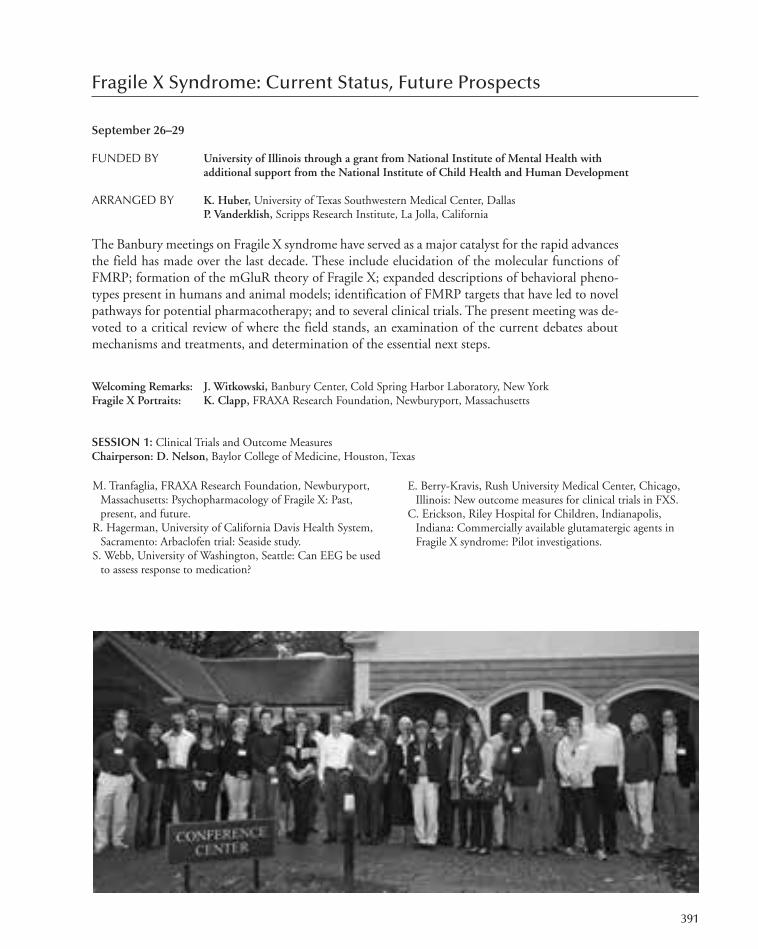
Fragile X Syndrome: Current Status, Future Prospects
September 26–29
FUNDED BY University of Illinois through a grant from National Institute of Mental Health withadditional support from the National Institute of Child Health and Human Development
ARRANGED BY K. Huber, University of Texas Southwestern Medical Center, DallasP. Vanderklish, Scripps Research Institute, La Jolla, California
The Banbury meetings on Fragile X syndrome have served as a major catalyst for the rapid advancesthe field has made over the last decade. These include elucidation of the molecular functions ofFMRP; formation of the mGluR theory of Fragile X; expanded descriptions of behavioral pheno-types present in humans and animal models; identification of FMRP targets that have led to novelpathways for potential pharmacotherapy; and to several clinical trials. The present meeting was de-voted to a critical review of where the field stands, an examination of the current debates aboutmechanisms and treatments, and determination of the essential next steps.
Welcoming Remarks: J. Witkowski, Banbury Center, Cold Spring Harbor Laboratory, New YorkFragile X Portraits: K. Clapp, FRAXA Research Foundation, Newburyport, Massachusetts
SESSION 1: Clinical Trials and Outcome MeasuresChairperson: D. Nelson, Baylor College of Medicine, Houston, Texas
M. Tranfaglia, FRAXA Research Foundation, Newburyport,Massachusetts: Psychopharmacology of Fragile X: Past,present, and future.
R. Hagerman, University of California Davis Health System,Sacramento: Arbaclofen trial: Seaside study.
S. Webb, University of Washington, Seattle: Can EEG be usedto assess response to medication?
E. Berry-Kravis, Rush University Medical Center, Chicago,Illinois: New outcome measures for clinical trials in FXS.
C. Erickson, Riley Hospital for Children, Indianapolis,Indiana: Commercially available glutamatergic agents inFragile X syndrome: Pilot investigations.
391
367-410_LAR_Banbury_Annual Report_2009 template 7/8/11 12:26 PM Page 391

392 Banbury Center
D. Rojas, University of Colorado, Denver School of Medicine:Gamma-band responses as potential biomarkers in autismand FXS.
W. Gan, New York University Medical Center, New York,New York: Abnormal experience-dependent dendritic spineplasticity in a mouse model of Fragile X.
A. Contractor, Northwestern University, Chicago, Illinois:Synapse development in the sensory cortex of FMR1knockout mice.
M. Huntsman, Children’s National Medical Center,Washington, D.C.: Inhibitory neurotransmission defects inFragile X syndrome.
L. Kaczmarek, Yale University School of Medicine, NewHaven, Connecticut: FMRP, ion channels, and theregulation of neuronal timing.
B. Alger, University of Maryland School of Medicine,Baltimore: Endocannabinoids and a mouse model of FragileX syndrome.
O. Manzoni, Neurocentre Magendie, Bordeaux, France:Disorganization of the endocannabinoid perisynapticsignaling machinery in FMR1–/– mice.
R. Wong, SUNY–Downstate Health Science Center,Brooklyn, New York: Mechanism of mGLuR-mediatedplasticity.
D. NelsonF. Tassone
SESSION 3: Receptors to RibosomesChairperson: P. Vanderklish, Scripps Research Institute, La Jolla, California
K. Huber, University of Texas Southwestern Medical Center,Dallas: Mechanisms of mGluR5 dysfunction in Fmrl KOmice.
F. Tassone, Mind Institute, Davis, California: Altered mTOR-dependent signaling and differential mGluR expressionpatterns in Fragile X syndrome.
S. Zukin, Albert Einstein College of Medicine, Bronx, NewYork: FMRP acts via PIKE to regulate mTOR signaling.
G. Bassell, Emory University, Atlanta, Georgia: FMRP andmiRNAs: Partners for translation at the synapse.
SESSION 4: FMRP Isoforms and Their Manipulation in BrainChairperson: I.J. Weiler, University of Illinois, Urbana-Champaign
D. Venkitaramani, University of Illinois, Urbana-Champaign:FMRP isoforms and restoration of function. Spatial,temporal, and splice variations in FMRP function.
SESSION 2: Synapses, Circuits, and RhythmsChairperson: K. Huber, University of Texas Southwestern Medical Center, Dallas
D. Nelson, Baylor College of Medicine, Houston, Texas:FMR1 and FXRs.
SESSION 5: Targets and Model SystemsChairperson: G. Bassell, Emory University, Atlanta, Georgia
C. Westmark, Waisman Center, Madison, Wisconsin: Reversalof Fragile X phenotypes by manipulation of APP/Aβ levels.
R. Jope, University of Alabama, Birmingham: Therapeuticeffects of GSK3 inhibitors in Fragile X mice.
B. Oostra, Erasmus University, Rotterdam, The Netherlands:Rescue of behavioral phenotype and neuronal protrusionmorphology in FMR1 KO mice.
S. Haggarty, Massachusetts General Hospital, Boston: Using
patient-specific iPS cells for modeling pathogenesis andtreatment of Fragile X syndrome.
P. Vanderklish, Scripps Research Institute, La Jolla, California:Strategies for new target discovery and evaluation. Identifica-tion and validation of new targets for the treatment of FXS.
General Discussion: F. Gasparini, Novartis Pharma AG, Basel,Switzerland
367-410_LAR_Banbury_Annual Report_2009 template 7/8/11 12:26 PM Page 392

Genome-Era Pathology, Precision Diagnostics, andPreemptive Care: A Stakeholder Summit
October 13–15
FUNDED BY Various institutions and individual participants
ARRANGED BY M. Boguski, Beth Israel Deaconess Medical Center, Boston, MassachusettsJ. Saffitz, Beth Israel Deaconess Medical Center, Boston, MassachusettsP. Tonellato, Beth Israel Deaconess Medical Center, Boston, Massachusetts
Historically, the discipline of pathology has had a central role to detect, classify, and interpret cellularand molecular markers of disease to guide physicians in the care and management of their patients.In recent years, many high-throughput technologies have been developed and used in research todetermine the molecular and genetic mechanisms underlying diseases, but the pathology communityhas not responded systematically to the challenges and opportunities provided by these technologicalinnovations. The objective of this discussion workshop was to define these opportunities, identifychallenges and barriers to success, and formulate a call to action that will keep pathology at the fore-front of modern medical care.
Welcoming Remarks: J. Witkowski, Banbury Center, Cold Spring Harbor Laboratory, New York
SESSION 1: The Issue and ObjectiveWelcome and Overview: J. Saffitz, Beth Israel Deaconess Medical Center, Harvard Medical School, Boston, Massachusetts
Brief Background PresentationsJ. Schamberg, College of American Pathologists, Waukesha,Wisconsin: Pathology today: Challenges.
E. Green, National Human Genome Research Institute,Bethesda, Maryland: Genome-era pathology.
393
367-410_LAR_Banbury_Annual Report_2009 template 7/8/11 12:26 PM Page 393
394 Banbury Center
J. Saffitz, Beth Israel Deaconess Medical Center, HarvardMedical School, Boston, Massachusetts: Genome-erapathology and preemptive care.
M. Boguski, Beth Israel Deaconess Medical Center, Harvard
Medical School, Boston, MassachusettsDiscussion: Objectives of genome-era pathologyObjective: Define and refine primary objectives, open issues,and potential action items
SESSION 2: Factors for Success
J. Crawford, North Shore LIJ Laboratories, Lake Success, NewYorkDiscussion: Obstacles and challengesObjective: Identify and prioritize primary challenges andidentify potential solutions
P. Tonellato, Beth Israel Deaconess Medical Center, HarvardMedical School, Boston, Massachusetts and R. Haspel, BethIsrael Deaconess Medical Center, Harvard Medical School,Boston, Massachusetts
Discussion: Overcoming the obstacles and challenges.Objective: Review current efforts designed to resolveobstacles. Identify key stakeholders, approaches and methods to overcome barriers and funding options that may provide the resources and forums to act on solutions
J. Crawford, North Shore LIJ Laboratories, Lake Success, NewYork, P. Tonellato, Beth Israel Deaconess Medical Center,Harvard Medical School, Boston, Massachusetts, and M.Boguski, Beth Israel Deaconess Medical Center, HarvardMedical School, Boston, Massachusetts
Discussion: Summarize objectives, action items, and next stepsObjective: Summarize main points and final action items
J. Saffitz, Beth Israel Deaconess Medical Center, HarvardMedical School, Boston, MassachusettsDiscussion: The horizon view
SESSION 3: A Call to Action
S. Tomlins, F. Barr W. Rosenkrans, R. Jeter, H. Halvorson
367-410_LAR_Banbury_Annual Report_2009 template 7/8/11 12:26 PM Page 394

Stem Cells, Genetics, and RNA-Binding Proteins: RecentAdvances in ALS Research and Drug Discovery
October 17–19
FUNDED BY The ALS Association Greater New York Chapter
ARRANGED BY L. Bruijn, The ALS Association, Washington, D.C.T. Maniatis, Columbia University Medical Center, New York, New YorkC. Svendsen, University of Wisconsin, Madison
During the past decade, significant progress has been made in understanding the mech-anisms leading to ALS. The focus has shifted from a motor neuron centric view to arecognition that the neighboring cells and in particular the glia have an integral role inthe disease process. The landscape for ALS is again changing, and this meeting broughttogether leaders in diverse fields of ALS genetics, RNA processing, stem cells, and modelsystems to discuss how to capitalize on the promising research advances in all thesefields and make an impact on ALS discoveries. Progress in developing new in vitro andin vivo systems to better understand the disease and the development of new tools fordrug discovery were key topics of discussion at the workshop. The workshop provideda unique opportunity for academic scientists, clinicians, and the industry to discusshow better to understand the role of the new genes in ALS and how this impacts drugdiscovery.
Introduction: L. Bruijn, The ALS Association, Washington, D.C.Introductory Remarks: J. Witkowski, Banbury Center, Cold Spring Harbor Laboratory, New York
T. Maniatis
395
367-410_LAR_Banbury_Annual Report_2009 template 7/8/11 12:26 PM Page 395

396 Banbury Center
Overview of FUS/TDP43 Genetics and PathologyC. Shaw, Guy’s Hospital, London, United KingdomR. Brown, University of Massachusetts, North Worchester
Emerging Technologies in Genetics: Session Discussion
J. Hardy, Reta Lila Weston Institute of Neurological Studies,University College London, United Kingdom
R. Myers, Hudson Alpha Institute for Biotechnology,Huntsville, Alabama
SESSION 1: Genetics of ALSChairpersons: C. Shaw, Guy’s Hospital, London, United Kingdom and R. Brown, University of Massachusetts,North Worchester
SESSION 2: Disease Mechanisms and ALSChairperson: A. Goldberg, Harvard Medical School, Boston, Massachusetts
Disease Mechanism and Lessons Learned from SODID. Cleveland, University of California, San Diego, La Jolla
Protein Misfolding and Cell Death: Apoptosis, Autophagy,and Necrosis
A. Goldberg, Harvard Medical School, Boston, Massachusetts
J. Rothstein, D. Cleveland
D. Borchelt, University of Florida, Gainesville
Glial–Neuronal Interactions and InflammationJ. Rothstein, Johns Hopkins University School of Medicine,Baltimore, Maryland
B. Barres, Stanford University School of Medicine, California
SESSION 3: Animal ModelsChairperson: G. Cox, The Jackson Laboratory, Bar Harbor, Maine
R. Baloh, Washington University School of Medicine, St.Louis, Missouri
D. Cleveland, University of California, San Diego, La Jolla
P. Wong, Johns Hopkins University, Baltimore, MarylandN. Shneider, Columbia University, New York, New York
SESSION 4: TDP43/FUS and Disease MechanismChairperson: T. Maniatis, Columbia University Medical Center, New York, New York
M. Moore, HHMI/University of Massachusetts MedicalSchool, Worcester
SESSION 5: Stem CellsChairperson: C. Svendsen, Cedar Sinai Medical Center, Los Angeles, California
V. Lee, University of Pennsylvania School of Medicine,Philadelphia
IPS TechnologyF. Soldner, Whitehead Institute, Massachusetts Institute ofTechnology, Cambridge
IPS in ALS–Model SystemsC. Svendsen, Cedar Sinai Medical Center, Los Angeles,California
K. Eggan, Harvard University, Cambridge, Massachusetts
Assay Development Using Stem CellsH. Wichterle, Columbia University, New York, New YorkD. Fischer, BioFocus DPI, Leiden, The NetherlandsS. Finkbeiner, University of California, San Francisco
SESSION 6: Closing SessionChairperson: C. Svendsen, Cedar Sinai Medical Center, LosAngeles, California
T. Maniatis, Columbia University Medical Center, New York,New York
367-410_LAR_Banbury_Annual Report_2009 template 7/8/11 12:26 PM Page 396

The Calculus of Medicine: Treatment of PancreaticCancer as a Prime Exemplar
October 20–22
FUNDED BY Abraxis BioScience, Inc.
ARRANGED BY J. Fleshman, Pancreatic Cancer Action Network, Manhattan Beach, CaliforniaB. Mishra, Courant Institute, New York University, New York, New YorkP. Soon-Shiong, Abraxis BioScience, Inc., Los Angeles, California
This meeting emphasized the importance of the integration of computer science, statistics, andmathematics into biomedical research and clinical trials, focusing on a better understanding of can-cer, with pancreatic cancer as the prime exemplar. Key leaders in oncology research and clinical ap-plication discussed how to shape this exciting field (and community) with a particular focus ongenomics, proteomics, imaging, translational bioinformatics, systems biology, disease modeling, sin-gle-cell analysis and nano-medicine.
Welcoming Remarks: J. Witkowski, Banbury Center, Cold Spring Harbor Laboratory, New YorkIntroductory Remarks: J.-P. Bizarri, Celgene, Summit, New Jersey
SESSION 1: The Biology of Pancreatic CancerChairperson: D. Tuveson, CRUK Cambridge Research Institute, United Kingdom
R. Hruban, Johns Hopkins Medical Institutions, Baltimore,Maryland: Genetics of pancreatic cancer.
D. Simeone, University of Michigan Health Systems, AnnArbor: Pancreatic stem cells.
D. Bar-Sagi, New York University, New York, New York: Inter-and intra-cellular pathways in PDA.
D. Hedley, Ontario Cancer Institute, Toronto, Canada: Effectsof the tumor microenvironment on invasion and metastasis.
397
367-410_LAR_Banbury_Annual Report_2009 template 7/8/11 12:26 PM Page 397

398 Banbury Center
D. Tuveson, CRUK Cambridge Research Institute, UnitedKingdom: Stromal barriers in pancreatic cancer.
M. Lisanti, Kimmel Cancer Center, Philadelphia,Pennsylvania: Warburg in reverse: The autophagic tumorstroma model of cancer.
M. Lotze, University of Pittsburgh, Pennsylvania: Autophagyas a target in pancreatic cancer.
SESSION 2: Clinical Developments in Pancreatic CancerChairperson: M. Tempero, University of California, San Francisco
M. Tempero, University of California, San Francisco: Newparadigms for drug development.
J. Berlin, Vanderbilt-Ingram Cancer Center, Nashville,Tennessee: New agents for treatment of pancreatic cancer.
M. Hidalgo, Centro National de Investigaciones Oncologicas,Madrid, Spain: Targeting the stroma in pancreatic cancer.
P. Philip, Karmanos Cancer Institute, Detroit, Michigan:Targeting IGF-IR in pancreatic cancer.
J. Clark, Massachusetts General Hospital, Boston: ModulatingKRAS as a target in pancreatic cancer.
W. Isacoff, University of California, San Francisco: Novelchemotherapy schedules for pancreatic cancer.
SESSION 3: BioInformatics and BioMarkers in Pancreatic CancerChairperson: B. Mishra, Courant Institute, New York University, New York, New York
C. Cantor, Sequenom Inc., San Diego, California: Cancer-specific nucleic acid sequences.
J. Reed, University of California, Los Angeles: Nanomeasurement approaches for characterizing single cells inpopulations.
B. Mishra, Courant Institute, New York University, New York,New York: Translational cancer bioinformatics.
J. Watson, L. Lisanti D. Bar-Sagi, D. Simeone
367-410_LAR_Banbury_Annual Report_2009 template 7/8/11 12:26 PM Page 398
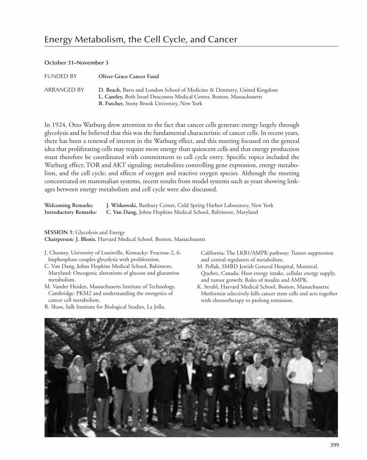
Energy Metabolism, the Cell Cycle, and Cancer
October 31–November 3
FUNDED BY Oliver Grace Cancer Fund
ARRANGED BY D. Beach, Barts and London School of Medicine & Dentistry, United KingdomL. Cantley, Beth Israel Deaconess Medical Center, Boston, MassachusettsB. Futcher, Stony Brook University, New York
In 1924, Otto Warburg drew attention to the fact that cancer cells generate energy largely throughglycolysis and he believed that this was the fundamental characteristic of cancer cells. In recent years,there has been a renewal of interest in the Warburg effect, and this meeting focused on the generalidea that proliferating cells may require more energy than quiescent cells and that energy productionmust therefore be coordinated with commitment to cell cycle entry. Specific topics included theWarburg effect; TOR and AKT signaling; metabolites controlling gene expression, energy metabo-lism, and the cell cycle; and effects of oxygen and reactive oxygen species. Although the meetingconcentrated on mammalian systems, recent results from model systems such as yeast showing link-ages between energy metabolism and cell cycle were also discussed.
Welcoming Remarks: J. Witkowski, Banbury Center, Cold Spring Harbor Laboratory, New YorkIntroductory Remarks: C. Van Dang, Johns Hopkins Medical School, Baltimore, Maryland
SESSION 1: Glycolysis and EnergyChairperson: J. Blenis, Harvard Medical School, Boston, Massachusetts
J. Chesney, University of Louisville, Kentucky: Fructose-2, 6-bisphosphate couples glycolysis with proliferation.
C. Van Dang, Johns Hopkins Medical School, Baltimore,Maryland: Oncogenic alterations of glucose and glutaminemetabolism.
M. Vander Heiden, Massachusetts Institute of Technology,Cambridge: PKM2 and understanding the energetics ofcancer cell metabolism.
R. Shaw, Salk Institute for Biological Studies, La Jolla,
California: The LKB1/AMPK pathway: Tumor suppressionand central regulators of metabolism.
M. Pollak, SMBD Jewish General Hospital, Montreal,Quebec, Canada: Host energy intake, cellular energy supply,and tumor growth: Roles of insulin and AMPK.
K. Struhl, Harvard Medical School, Boston, Massachusetts:Metformin selectively kills cancer stem cells and acts togetherwith chemotherapy to prolong remission.
399
367-410_LAR_Banbury_Annual Report_2009 template 7/8/11 12:26 PM Page 399

400 Banbury Center
SESSION 2: Metabolic RegulationChairperson: J. Chesney, University of Louisville, Kentucky
J. Brugge D. Schenkein A. Caudy
Y. Xiong, University of North Carolina, Chapel Hill: Metabolicregulation in normal and tumor cells.
M. Lisanti, Kimmel Cancer Center, Philadelphia, Pennsylvania:Warburg in reverse: The autophagic tumor stroma model ofcancer.
J. Brugge, Harvard Medical School, Boston, Massachusetts:Regulation of metabolism by extracellular matrix ononcogenes.
L. Cantley, Beth Israel Deaconess Medical Center, Boston,Massachusetts: PI3 kinase and cancer metabolism.
D. Schenkein, Agios Pharmaceuticals, Cambridge,Massachusetts: Mutations in isocitrate dehydrogenase: Rolein disease pathogeneses and potential as a therapeutic target.
T. Mak, Campbell Family Institute for Breast Cancer Researchat PMH, UHN, Toronto, Ontario, Canada: Knock-inmouse model for IDH1 mutations.
J. Blenis, Harvard Medical School, Boston, Massachusetts:Amino acids, mTOR signaling, and metabolism.
B. Manning, Harvard University, Boston, Massachusetts:mTOR activation drives the metabolic reprogramming oftumor cells.
M. Tyers, University of Edinburgh, United Kingdom:Metabolic and growth control by TOR effectors in yeast.
B. Futcher, Stony Brook University, New York: Introductoryremarks.
B. Tu, University of Texas Southwestern Medical Center,Dallas: Metabolic signals that drive cell growth andproliferation.
A. Caudy, Princeton University, New Jersey: Thesedoheptulose bisphosphatase SHB17 shunts carbon fromglycolysis to the pentose phosphate pathway for riboneo-genesis in yeast.
J. Broach, Princeton University, New Jersey: Direct control ofmetabolism by nutrient signaling pathways in yeast.
B. Futcher, Stony Brook University, New York: cAMP,liquidation of storage carbohydrates, and commitment tothe cell cycle.
S. Kohlwein, Karl-Franzens-Universitat Graz, Austria: Lipidrequirements during the cell cycle.
E. White, Cancer Institute of New Jersey, New Brunswick:Role of autophagy in cancer metabolism.
K. Vousden, Beatson Institute for Cancer Research, Bearsden,Glasgow: The regulation of metabolism by p53.
A. Levine, Institute for Advanced Study, Princeton, NewJersey: Role of p53 in regulating metabolic pathways.
L. Cantley, Beth Israel Deaconess Medical Center, Boston,Massachusetts: Concluding remarks and general discussion.
SESSION 3: PI3 Kinase, Isocitrate Dehydrogenase, TORChairperson: L. Cantley, Beth Israel Deaconess Medical Center, Boston, Massachusetts
SESSION 4: Connections between Metabolism and Cell Cycle in YeastChairperson: M. Tyers, University of Edinburgh, United Kingdom
SESSION 5: Cancer Metabolism and p53Chairperson: L. Cantley, Beth Israel Deaconess Medical Center, Boston, Massachusetts
367-410_LAR_Banbury_Annual Report_2009 template 7/8/11 12:26 PM Page 400

Easeful Death: 21st Century Perspectives on Assisted Suicide
November 3–5
FUNDED BY The Ellison Medical Foundation
ORGANIZED BY M. Battin, University of Utah, Salt Lake CityE. MacDonald, Guy’s and St. Thomas Hospital, London, United KingdomT. Murray, The Hastings Center, Garrison, New York, New YorkM. Raff, University College London, United KingdomJ. Witkowski, Banbury Center, Cold Spring Harbor Laboratory, New York
Because of advances in medical care, the proportion of the elderly in the population is increasing.And as a consequence, the number of individuals suffering from Alzheimer’s, Parkinson’s, and motorneuron diseases, as well as other age-related degenerative disorders, is increasing. Individuals sufferingfrom these disorders place an extraordinary burden on their caregivers, who are often also elderlyand who may be unable to cope. End-of-life issues involve profound legal, moral, religious, andbiomedical questions and evoke such intense passions that calm discourse is hard to achieve. TheBanbury Center provided a venue for calm discussions of topics, which included what are the dataon assisted suicide and how (and why) do interpretations of the data differ? What is the evidencefor abuse, what is its incidence and how might it be prevented? How can end-of-life decisions bemade in cases of psychiatric illness (and perhaps dementia)? Participants included scientists, physi-cians, philosophers, lawyers, and religious leaders and included experts on euthanasia and assistedsuicide in Europe.
Welcoming Remarks: J. Witkowski, Banbury Center, Cold Spring Harbor Laboratory, New YorkIntroductory Remarks: M. Raff, University College London, United Kingdom
401
367-410_LAR_Banbury_Annual Report_2009 template 7/8/11 12:26 PM Page 401
402 Banbury Center
SESSION 1Chairperson: T. Murray,The Hastings Center, Garrison, New York
D. Orentlicher, University of Iowa, Iowa City: Drawing linesat the end of death: Where can we draw meaningful linesamong treatment withdrawal, assisted suicide, andeuthanasia?
B. Onwuteaka-Philipsen, VU Medical Center, Amsterdam,The Netherlands: The influence of regulation on the practiceof assisted suicide in The Netherlands.
L. Deliens, Vrije Universiteit, Brussels, Belgium: Belgian dataon euthanasia.
L. Cohen, Baystate Medical Center, Northampton,Massachusetts: Accusations and investigations of euthanasiaand PAS directed at American palliative medicine physicians.
Matters arising from Day 1
J. Prey, M. Raff L. Deliens, T. Murray
Z. Schostak, B. Gert
SESSION 2Chairperson: E. MacDonald, Guy’s and St. Thomas Hospital, London, United Kingdom
SESSION 3Chairperson: R. Payne, Duke Divinity School, Durham, North Carolina
C. Baron, Boston College, Newton, Massachusetts: Law at theend of life: Have we come of age?
J. Lynn, Colorado Foundation for Medical Care, Chevy Chase,Maryland: Political realities and strategies: Improving care inthe “death panel” era.
General Discussion: What is the 21st perspective on assistedsuicide?
M. Battin, University of Utah, Salt Lake City: Discussion.B. Gert, Dartmouth College, Hanover, New Hampshire:Refusal of hydration and nutrition as an alternative tophysician-assisted suicide.
367-410_LAR_Banbury_Annual Report_2009 template 7/8/11 12:26 PM Page 402
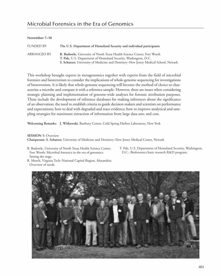
Microbial Forensics in the Era of Genomics
November 7–10
FUNDED BY The U.S. Department of Homeland Security and individual participants
ARRANGED BY B. Budowle, University of North Texas Health Science Center, Fort WorthT. Pals, U.S. Department of Homeland Security, Washington, D.C.S. Schutzer, University of Medicine and Dentistry–New Jersey Medical School, Newark
This workshop brought experts in metagenomics together with experts from the field of microbialforensics and bioterrorism to consider the implications of whole-genome sequencing for investigationsof bioterrorism. It is likely that whole-genome sequencing will become the method of choice to char-acterize a microbe and compare it with a reference sample. However, there are issues when consideringstrategic planning and implementation of genome-wide analyses for forensic attribution purposes.These include the development of reference databases for making inferences about the significanceof an observation; the need to establish criteria to guide decision makers and scientists on performanceand expectations; how to deal with degraded and trace evidence; how to improve analytical and sam-pling strategies for maximum extraction of information from large data sets; and cost.
Welcoming Remarks: J. Witkowski, Banbury Center, Cold Spring Harbor Laboratory, New York
SESSION 1: Overview Chairperson: S. Schutzer, University of Medicine and Dentistry–New Jersey Medical Center, Newark
B. Budowle, University of North Texas Health Science Center,Fort Worth: Microbial forensics in the era of genomics:Setting the stage.
R. Murch, Virginia Tech–National Capital Region, Alexandria:Overview of needs.
T. Pals, U.S. Department of Homeland Security, Washington,D.C.: Bioforensics basic research R&D program.
403
367-410_LAR_Banbury_Annual Report_2009 template 7/8/11 12:26 PM Page 403
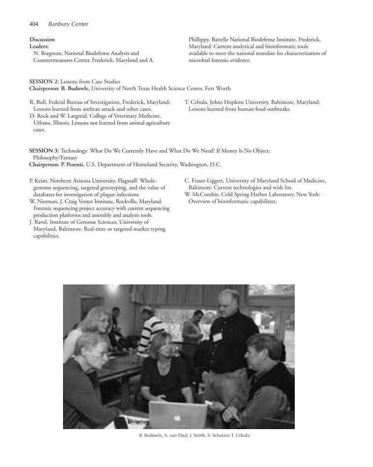
404 Banbury Center
DiscussionLeaders:N. Bergman, National Biodefense Analysis andCountermeasures Center, Frederick, Maryland and A.
Phillippy, Battelle National Biodefense Institute, Frederick,Maryland: Current analytical and bioinformatic toolsavailable to meet the national mandate for characterization ofmicrobial forensic evidence.
SESSION 2: Lessons from Case StudiesChairperson: B. Budowle, University of North Texas Health Science Center, Fort Worth
R. Bull, Federal Bureau of Investigation, Frederick, Maryland:Lessons learned from anthrax attack and other cases.
D. Rock and W. Laegreid, College of Veterinary Medicine,Urbana, Illinois: Lessons not learned from animal agriculturecases.
T. Cebula, Johns Hopkins University, Baltimore, Maryland:Lessons learned from human food outbreaks.
SESSION 3: Technology: What Do We Currently Have and What Do We Need? If Money Is No Object;Philosophy/Fantasy
Chairperson: P. Pesenti, U.S. Department of Homeland Security, Washington, D.C.
P. Keim, Northern Arizona University, Flagstaff: Whole-genome sequencing, targeted genotyping, and the value ofdatabases for investigation of plague infections.
W. Nierman, J. Craig Venter Institute, Rockville, Maryland:Forensic sequencing project accuracy with current sequencingproduction platforms and assembly and analysis tools.
J. Ravel, Institute of Genome Sciences, University ofMaryland, Baltimore: Real-time or targeted marker typingcapabilities.
C. Fraser-Liggett, University of Maryland School of Medicine,Baltimore: Current technologies and wish list.
W. McCombie, Cold Spring Harbor Laboratory, New York:Overview of bioinformatic capabilities.
B. Budowle, A. van Daal, J. Smith, S. Schutzer, T. Cebula
367-410_LAR_Banbury_Annual Report_2009 template 7/8/11 12:26 PM Page 404

Microbial Forensics in the Era of Genomics 405
SESSION 5: Other Technologies Including ProteomicsChairperson: S. Morse, Centers for Disease Control & Prevention, Atlanta, Georgia
S. Velsko, Lawrence Livermore National Laboratory,California: Nongenetic technologies.
T. Angel, Pacific Northwest National Laboratory, Richland,Washington: Proteomic complements to genomics.
R. Bull, Federal Bureau of Investigation, Frederick, Maryland:Potential host forensic signatures.
SESSION 6: Metagenomics Chairperson: C. Fraser-Liggett, University of Maryland School of Medicine, Baltimore
G. Weinstock, Washington University School of Medicine, St.Louis, Missouri: Metagenomics for surveillance.
J. Ravel, Institute for Genomic Research, University ofMaryland, Baltimore: Human microbiome.
Y. Fofanov, University of Houston, Texas: Software and analytical tools.
A. van Daal, Bond University, Gold Coast, Australia: Exampleof specific platform (Illumina HiSeq) capabilities to meetneeds of microbial forensics.
SESSION 7: BioinformaticsChairperson: P. Keim, Northern Arizona University, Flagstaff
S. Velsko, Lawrence Livermore National Laboratory,California: What questions need to be answered?
J. Tiedje, Michigan State University, East Lansing: Populationgenetics issues.
R. Chakraborty, University of North Texas Health Science,Fort Worth: Statistical interpretation issues: Comparison toforensic human DNA.
T. Leighton, Children’s Hospital, Oakland Research Institute,California: Next-generation DNA sequencing analysis ofclonal variants, manipulated populations, and productionprocess trace-back.
S. Velsko, Lawrence Livermore National Laboratory, California:Genetic inference on disease transmission networks.
SESSION 8: Data SharingChairperson: D. Rock, College of Veterinary Medicine, Urbana, Illinois
T. Slezak, Lawrence Livermore National Laboratory,California: Forensic uses of microarrays or databases forsharing forensic data.
B. Budowle, University of North Texas Health Science Center,Fort Worth: Archives and database needs.
SESSION 4: Quality of Sample of Necessary TechnologyChairperson: J. Burans, U.S. Department of Homeland Security, Frederick, Maryland
M. Eshoo, Ibis Biosciences, Inc. Carlsbad, California:Extraction capabilities, DNA repair, and whole-genomeamplification and analysis of trace unculturable specimens.
wrap-up, strategies, and what we would like conveyed to thecommunity, stakeholders, policy makers, and summary formanuscript.
J. Smith, Penn State University, Pennsylvania; BioForensicConsulting, LLC, Edgewood, Maryland: Data exchangerequirements and policies for sharing.
C. Cooke Jr., WINPAC, Washington, D.C.: Quality assurancefor data generation.
SESSION 9: SummaryChairperson: S. Schutzer, University of Medicine and Dentistry–New Jersey Medical School, Newark
B. Budowle, University of North Texas Health Science Center,Fort Worth and Traci Pals, U.S. Department of HomelandSecurity, Washington, D.C.: Prioritization of needs, recap,
367-410_LAR_Banbury_Annual Report_2009 template 7/8/11 12:26 PM Page 405

Signaling through Ubiquitin
November 14–17
FUNDED BY The Cold Spring Harbor Laboratory Corporate Sponsor Program
ARRANGED BY R. Deshaies, California Institute of Technology, PasadenaV. Dixit, Genentech, South San Francisco, CaliforniaW. Tansey, Vanderbilt University Medical Center, Nashville, Tennessee
The object of this meeting was to analyze and discuss how protein ubiquitylation exerts its myriadbiological effects within the cell. Although ubiquitin has traditionally been studied within the contextof protein turnover, it is now clear that ubiquitylation refers to a complex set of posttranslationalmodifications that are interpreted by the cellular machinery to impact a broad range of biologicalprocesses, just one of which is proteolysis. Participants in the meeting discussed the spectrum of ubiq-uitin and related modifications, how they are recognized, and what they do. Discussions drew on ex-perts in biochemistry, cell biology, computational biology, structural biology, genetics, and patho-physiology to identify common emerging themes in how ubiquitin and related proteins work.
Introductory and Welcoming Remarks: J. Witkowski, Banbury Center, Cold Spring Harbor Laboratory, New YorkIntroductory Remarks: W. Tansey, Vanderbilt University Medical Center, Nashville, Tennessee
SESSION 1: Signaling via Ubiquitin Chain Diversity and RecognitionChairperson: T. Hunter, The Salk Institute, La Jolla, California
R. Cohen, Colorado State University, Fort Collins:Recognition of polyubiquitin chains.
K. Hofmann, Miltenyi Biotec GmbH, Gladbach, Germany:UBL receptors: At the crossroads of UPS and autophagy.
S. Polo, Institute of Molecular Oncology Foundation, Milan,Italy: Specificity in chain formation and recognition.
Z. Chen, University of Texas Southwestern Medical Center,Dallas: Ubiquitin signaling in the RIG-I antiviral pathway.
I. Dikic, Johann Wolfgang Goethe University Medical School,Frankfurt, Germany: Ubiquitin signaling networks.
K. Iwai, Osaka University, Japan: Linear polyubiquitination: Anew regulator of NF-κB signaling.
J. Peng, Emory University School of Medicine, Atlanta,Georgia: Exploring ubiquitin pathways by quantitativeproteomics.
406
367-410_LAR_Banbury_Annual Report_2009 template 7/8/11 12:26 PM Page 406

Signaling through Ubiquitin 407
SESSION 2: Signaling via Ub-Like ProteinsChairperson: M. Hochstrasser, Yale University, New Haven, Connecticut
R. Hay, Sir James Black Centre, University of Dundee, UnitedKingdom: SUMO targeted ubiquitin ligases.
J. Huibregtse, University of Texas, Austin: The mechanism andfunction of ISG15 conjugation.
T. Hunter, The Salk Institute, La Jolla, California: Ubiquitin-SUMO cross-talk.
C. Lima, Memorial Sloan-Kettering Cancer Institute, NewYork, New York: Structure and mechanism in the SUMOconjugation pathway.
Y. Ohsumi, Frontier Research Center, Tokyo Institute ofTechnology, Yokohama, Japan: Two ubiquitin-likeconjugation reactions essential for autophagy.
A. Huang, Sanofi-Aventis, Cambridge, Massachusetts:Regulation of axin stability through poly-ADP ribosylation:Linking Wnt pathway signaling activity, ubiquitination, andpoly-ADP-ribosylation.
SESSION 3: Signaling to the ProteasomeChairperson: J. Huibregtse, University of Texas, Austin
D. Finley, Harvard Medical School, Boston, Massachusetts:Editing of ubiquitin at the proteasome.
M. Glickman, Technion-Israel Institute of Technology, Haifa,Israel: Coordination of ubiquitin-processing factors at theproteasome.
M. Hochstrasser, Yale University, New Haven, Connecticut:Proteases of the ubiquitin system.
R. Kopito, Stanford University, California: Why do ubiquitinchains accumulate in neurodegenerative disease?
S. Murata, The University of Tokyo, Japan: Proteasome diversity.
R. Deshaies, California Institute of Technology, Pasadena: Nedd8 links active Cullin-RING ligases to p97 substrateprocessing machinery.
SESSION 4: Ubiquitin–Proteasome Signaling in the NucleusChairperson: R. Hay, Sir James Centre, University of Dundee, United Kingdom
G. Rosenfeld, University of California, San Diego, La Jolla:Ubiquitylation, SUMOylation, methylation, and strategiesin regulated transcriptional programs.
W. Tansey, Vanderbilt University Medical Center, Nashville,Tennessee: Ubiquitin and transcription.
H. Ulrich, Cancer Research UK, London Research Institute,Herts, United Kingdom: Ubiquitin and replication stress.
SESSION 5: Ubiquitin Signaling in Disease StatesChairperson: R. Kopito, Stanford University, California
A. D’Andrea, Dana-Farber Cancer Institute, Boston,Massachusetts: Regulation of the Fanconi anemia pathway todeubiquitination.
R. Youle, National Institute of Health, Bethesda, Maryland:Mechanisms of Parkin-mediated mitophagy.
SESSION 6: Ub-Ligases, Isopeptidases, and Their SubstratesChairperson: C. Lima, Memorial Sloan-Kettering Cancer Center, New York, New York
V. Dixit, Genentech, South San Francisco, California:Ubiquitin modification in cancer.
W. Harper, Harvard Medical School, Boston, Massachusetts:Signaling through the ubiquitin–proteasome pathway.
M. Pagano, New York University School of Medicine, NewYork, New York: SCF ubiquitin ligases and cell proliferation.
P. Kaiser, University of California, Irvine: Interpretation ofubiquitin signal.
P. Matthias, ETH Zurich, Institute of Biochemistry,Switzerland: Regulation and substrates of Cullin-based E-3ligases.
M. Rape, University of California, Berkeley: Role of lysine 11-linked ubiquitin chains in cell cycle control.
B. Schulman, St. Jude Children’s Research Hospital,Memphis, Tennessee: Structural studies of Cullin-RINGligases.
367-410_LAR_Banbury_Annual Report_2009 template 7/8/11 12:26 PM Page 407

DNA, Genetics, and the History of Mankind
November 28–30
FUNDED BY The Lehrman Institute
ARRANGED BY J. Witkowski, Banbury Center, Cold Spring Harbor Laboratory, New York
The impact of molecular genetic studies has spread beyond the immediate discipline of biology,and as the study of genetic variation is essentially an historical study, DNA analysis has been appliedto historical problems in related disciplines. For example, conclusions on the movements of earlyhuman populations based on linguistic and archeological data have been reinforced or modified byDNA analysis. Domestication of animals and plants marked one of the great transitions in humanhistory, and genetic analysis is revealing how and where domestication took place. Genetic analysismay provide reliable information on personal relationships and identification that has been the sub-ject of speculation, for example, Thomas Jefferson’s family and identifying the bodies of the Tsar’sfamily. Participants in this workshop discussed how genetic and DNA analysis can further our un-derstanding of the history of human beings, and how to promote such studies.
Welcoming Remarks: J. Witkowski, Banbury Center, Cold Spring Harbor Laboratory, New York
SESSION 1Chairperson: M. Thomas, University College London, United Kingdom
K. Dobney, University of Aberdeen, United Kingdom and J.-D. Vigne, Muséum National d’Histoire Naturelle, Paris,
France: Mammals to microbes: Role of genetics in exploringpast bio-cultural dynamics using the fossil vertebrae record.
408
367-410_LAR_Banbury_Annual Report_2009 template 7/8/11 12:26 PM Page 408

DNA, Genetics, and the History of Mankind 409
E. Willerslev, Natural History Museum of Denmark,Copenhagen: Early peopling of the New World andextinction of the megafauna.
M. Richards, University of Leeds, United Kingdom:Archaeogenetics and modern human dispersals.
C. Bustamante, Stanford School of Medicine, California:Sequencing admixed genomes and what the thousandgenomes data are telling us.
A. Chakravarti, Johns Hopkins University School of Medicine,Baltimore, Maryland: Admixture: What it means forstudying human populations.
B. Shapiro, Pennsylvania State University, University Park:Appropriately incorporating spatial and temporalinformation into genealogical analysis.
W. McCombie, Cold Spring Harbor Laboratory, New York:Human genetics in the era of “next-generation” sequencing.
SESSION 2Chairperson: J. Buikstra, Center for Bioarchaeological Research, Arizona State University, Tempe
S. Pääbo, Max-Planck Institute for Evolutionary Anthropology,Leipzig, Germany: The contributions of DNA studies: Whatwe would not, and could not, have gained from other sources.
D. Reich, Harvard Medical School, Boston, Massachusetts:Evidence for gene flow from neanderthals into modern humans.
J. Hawks, University of Wisconsin, Madison: Natural selection,population growth, and human migrations in the earlyholocene.
M. Crawford, University of Kansas, Lawrence: The sequelae ofRussian contact in the Aleutian archipelago: Molecularperspectives.
M. Zeder, National Museum of Natural History, Washington,D.C.: Documenting domestication: The intersection ofarchaeology and genetics.
A. Stone, Arizona State University, Tempe: The origins andspread of human tuberculosis.
SESSION 3Chairperson: L. Madrigal, University of South Florida, Tampa
N. Wade, The New York Times, New York, New York: Theevolution of human races and economic systems.
D. O’Rourke, University of Utah, Salt Lake City:
Consultation and consent: Cultural concerns in humanpopulation genetic research.
SESSION 4: Future Developments
J.-D. Vigne, Muséum National d’Histoire Naturelle, Paris,France: The Archaeozoology and Genetics (A&G) WorkingGroup of International Council for Archaeozoology (ICAZ).
D. O’Rourke, University of Utah, Salt Lake City: TheAmerican Association of Physical Anthropologists.
J. Witkowski, Banbury Center, Cold Spring HarborLaboratory, New York
Discussion Points• What might be done to foster cross-disciplinaryinteractions in these fields?
• What topics should be covered in a Banbury Centermeeting in 2011?
• Any other points for discussion?
D. Micklos, S. Pääbo, D. Reich, B. Shapiro
367-410_LAR_Banbury_Annual Report_2009 template 7/8/11 12:26 PM Page 409

367-410_LAR_Banbury_Annual Report_2009 template 7/8/11 12:26 PM Page 410
DNA LEARNING CENTER
411-434_LAR_DNALC_Annual Report_2009 template 7/8/11 4:04 PM Page 411

411-434_LAR_DNALC_Annual Report_2009 template 7/8/11 4:04 PM Page 412
Preparing students and families to thrive in the gene age
ADMINISTRATION INSTRUCTION BIOMEDIA
Lauren Correri Jennifer Aiello Anthony BiondoMary Lamont Elna Carrasco Cornel GhibanValerie Meszaros Jennifer Galasso Uwe HilgertDavid Micklos Natalia Hanson Eun-Sook JeongKaren Orzel Amanda McBrien Mohammed KhalfanCarolyn Reid Erin McKechnie Susan Lauter
Bruce Nash Amy NisselleIleana Rios Todd RochefordTedi Setton Jason WilliamsJermel Watkins Chun-hua Yang
Just as a unique pattern of bars in a universal product code (UPC) identifies each item for sale in astore, a DNA barcode is a DNA sequence that uniquely identifies each living species. This simplepremise was galvanized into action during meetings held in 2003 at Cold Spring Harbor Labora-tory’s (CSHL) Banbury Center and funded by the Alfred P. Sloan Foundation. At these meetings,scientists agreed to use short, standard DNA sequences to classify all living species and launched theInternational Barcode of Life Project (iBOL).
Operating under the iBOL banner, collaborators from more than 150 countries are involved in“campaigns” to census diversity in different plant, fungal, and animal groups, including ants, bees,butterflies, fish, birds, mammals, fungi, and flowering plants, and within ecosystems, including theseas, poles, rainforests, and coral reefs. The 10-year Census of Marine Life announced in Octoberthe first comprehensive list of more than 190,000 marine species, involving 2700 scientists andidentifying 6000 potentially new species.
DNA barcoding is also important in detecting food fraud and products taken from conservedspecies. In advance of formal DNA barcoding, in 1996, Rob DeSalle of the American Museum ofNatural History (AMNH) used related methods to find that five of 23 samples of caviar purchasedin New York City (NYC) were mislabeled and included three samples from threatened sturgeonspecies. The Congressional Research Service recognizes that more than a dozen commercial fish areoften mislabeled, passing off cheaper fish for expensive varieties. In 2008, Trinity High School stu-dents worked with Mark Stoeckle of The Rockefeller University and George Amato of the AMNHto show the pervasiveness of fish fraud in NYC. They found that 25% of 60 seafood items pur-chased in grocery stores and restaurants were mislabeled as more expensive species. One mislabeledfish was an endangered species, the Acadian redfish.
Urban Barcode Project
DNA barcoding came full circle to New York, when in December we received a $300,000 grant fromthe Sloan Foundation for the Urban Barcode Project, a science competition spanning the five bor-oughs of NYC. Student research teams will use DNA barcoding to explore biodiversity in the cityenvironment, including:
• Sampling biodiversity in a park, garden, office, or school.
• Checking for invasive plant or animal species.
DNA LEARNING CENTEREXECUTIVE DIRECTOR’S REPORT
413
411-434_LAR_DNALC_Annual Report_2009 template 7/8/11 4:04 PM Page 413

Logo for the Urban Barcode Project
• Monitoring animal movements or migrations.
• Identifying exotic or endangered food products in markets.
• Detecting food or product fraud.
The competition is open to NYC high school students enrolled in grades 9–12. Teams of two tofour students will work with a teacher sponsor to submit a project proposal for a June or October2011 deadline. Proposals will be judged for originality, creativity, relevance, plausibility, and scien-tific merit. The top 100 teams will be invited to compete in the Urban Barcode Project. Teams mustcomplete their projects by the spring of 2012 and will present their work at a project symposium.The best overall project will win the Grand Prize of $10,000, and an additional $10,000 in runner-up prizes will be awarded.
Sponsoring teachers must participate in a 6-hour training session, which will dovetail with ourexisting training program sponsored by the Howard Hughes Medical Institute (HHMI). Each suc-cessful team will have free access to everything needed for their DNA barcode experiments, includ-ing equipment, protocols, and reagents. Five equipment footlockers will be available for use byindividual schools or for groups of nearby schools to share. During the term of the project, we ex-pect to process ~10,000 DNA sequences. Teams may work on their projects at summer workshopsand Open Lab days at designated locations.
Teams will be assigned a mentor from a NYC university, museum, or other scientific institutionto answer technical questions and provide advice. Many mentors will be drawn from five NYC in-stitutions that are collaborating on the project: AMNH, New York Academy of Sciences, New YorkBotanical Garden, Prospect Park Zoo, and The Rockefeller University. The project’s scientific ad-visor, Mark Stoeckle, is a member of The Rockefeller Program for the Human Environment. In ad-dition to demonstrating the educational promise of DNA barcoding, Dr. Stoeckle was one of theorganizers of the 2003 Banbury meeting that launched the field of DNA barcoding and serves oniBOL’s Scientific Steering Committee.
A dedicated Internet microsite (www.urbanbarcodeproject.org) supports all phases of the proj-ect. This “online lab notebook” includes a video introduction to the barcode experiment, down-loadable PDF lab protocols, teacher preparation, bioinformatics tools, and multimedia resources.Video interviews with scientists, animations, and video podcasts (vodcasts) describe the science andapplications of DNA barcoding, and an active news feed and a link to Dr. Stoeckle’s Barcode Blogprovide up-to-date perspectives on DNA barcoding. Student DNA sequences are automatically up-loaded to the “Blue Line” of DNA Subway, an online tool for DNA barcode and phylogenetic analy-sis developed as part of the National Science Foundation (NSF)-funded iPlant Collaborative. AGoogle Maps utility will track biodiversity discovered by student experiments, and students willhighlight their projects through the social networking website, Facebook.
414 DNA Learning Center
411-434_LAR_DNALC_Annual Report_2009 template 7/8/11 4:04 PM Page 414

Executive Director’s Report 415
Student DNA Sequencing
A decade ago, the DNALC developed a similar program—with simplified biochemistry and a bioin-formatics work flow—to allow students to analyze a portion of their own mitochondrial (mt) chro-mosome. Using a kit distributed by Carolina Biological Supply Company, students isolate DNAfrom cheek cells and then use polymerase chain reaction (PCR) to amplify the mitochondrial con-trol region. Student samples are sent by overnight mail to the DNALC, where student interns pre-pare them for sequencing. The processed samples are then sent to the CSHL Sequencing Facility inWoodbury, and finished DNA sequences are uploaded to the Sequence Server database at ourBioServers website. There, students visualize their sequences and use software to align them withmitochondrial sequences from modern humans and extinct hominids to explore human genetic di-versity and evolution.
The donation of sequencing reagents by Applied Biosystems of Foster City, California has madeit possible to provide students this Sequencing Service free of charge. Since 1998, a total of 49,217student DNA samples have been sequenced, and 1.3 million students and teachers have used the sup-porting Bioservers website. We continued the Sequence Service in 2010, processing 7128 studentDNA samples received from 93 high schools and 57 colleges and universities.
The cost of commercial sequencing, however, has decreased so dramatically during the last yearthat in December, we ceased sequencing on site and shifted all sequencing to Genewiz, a global con-tract research organization with expertise in sequencing. We negotiated a much-reduced price of$3.00 per sample, which is less than our cost of sequencing with free reagents and student labor! Thisarrangement anticipates the increased volume of sequencing demanded by our Urban Barcode Proj-ect, as well as growing interest in using DNA sequencing in education. Student sequences are up-loaded to our server within 48 hours of receipt by Genewiz, a dramatic improvement over the 2-weekturnaround we were previously able to provide.
iPlant Collaborative
The iPlant Collaborative is a consortium headed by the University of Arizona and CSHL to developa computer (cyber) infrastructure to support plant research. iPlant aims to develop tools and inter-faces that will provide scientists with easy access to large-scale plant data sets and high-powered in-formatics tools. Working Groups composed of iPlant staff and plant community members arecurrently building “discovery environments” to help plant scientists solve two “grand challenges”:(1) produce an iPlant Tree of Life (iPToL) encompassing ~500,000 plant species and (2) explore thegenotype-to-phenotype continuum in plants (iPG2P).
Education, outreach, and training (EOT) is integral to iPlant’s mission of helping plant scien-tists make the best use of computer infrastructure to solve biological problems. During the first 2years of the project, EOT worked in parallel with the Grand Challenge teams to create an educa-tional Discovery Environment that allows students and teachers to work with the same data and usethe same tools at the same time as high-level plant researchers. Thisculminated in the release in March of DNA Subway, a student-cen-tered platform for gene annotation and comparison.
DNA Subway uses the metaphor of a subway map as an appeal-ing interface to sophisticated informatics tools. “Riding” on eitherof the initial lines, students can predict and annotate genes in up to150 kb of DNA (Red Line) or identify homologous, or related, se-quences in whole genomes (Yellow Line). By year’s end, a third “BlueLine” to analyze DNA barcodes and other short DNA sequenceswas mostly complete. This line offers the types of sequence analysisthat precedes the massive tree construction envisioned by the iPToL DNA Subway’s Blue Line
411-434_LAR_DNALC_Annual Report_2009 template 7/8/11 4:04 PM Page 415
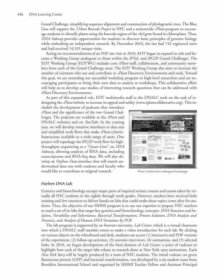
Grand Challenge, simplifying sequence alignment and construction of phylogenetic trees. The BlueLine will support the Urban Barcode Project in NYC and a nationwide iPlant program to encour-age students to identify plants using the barcode region of the rbcl gene found in chloroplasts. Thus,DNA Subway provides opportunities for students to discover basic principles of genome biologywhile embarking on independent research. By December 2010, the site had 742 registered usersand had received 14,535 unique visits.
Acting on recommendations of an NSF site visit in 2010, EOT began to expand its role and be-came a Working Group analogous to those within the iPToL and iPG2P Grand Challenges. TheEOT Working Group (EOTWG) includes core iPlant staff, collaborators, and community mem-bers from each of the Grand Challenge areas. The EOT Working Group also aims to increase thenumber of scientists who use and contribute to iPlant Discovery Environments and tools. Towardthis goal, we are extending our successful workshop program to high-level researchers and are en-couraging participants to bring their own data to analyze at workshops. This collaborative effortwill help us to develop case studies of interesting research questions that can be addressed withiPlant Discovery Environments.
As part of this expanded role, EOT multimedia staff at the DNALC took on the task of re-designing the iPlant website to increase its appeal and utility (www.iplantcollaborative.org). This in-cluded the development of podcasts that introduceiPlant and the significance of the two Grand Chal-lenges. The podcasts are available at the iPlant andDNALC websites and on YouTube. In the comingyear, we will develop intuitive interfaces to data setsand simplified work flows that make iPlant cyberin-frastructure available to a wide range of users. Oneproject will repackage the iPG2P work flow for high-throughput sequencing as a “Green Line” on DNASubway, allowing analysis of RNA data, includingtranscriptome and RNA-Seq data. We will also de-velop an Orphan Data interface that will match un-derworked data sets with students and faculty whowould like to contribute to original research.
Harlem DNA Lab
Genetics and biotechnology occupy major parts of required science courses and exams taken by vir-tually all NYC students in the eighth through tenth grades. However, teachers have received littletraining and few resources to deliver hands-on labs that could make these topics come alive for stu-dents. Thus, the objective of our HHMI program is to use our expertise to prepare NYC teachersto teach a set of six labs that target key genetics and biotechnology concepts: DNA Structure and Iso-lation, Variability and Inheritance, Bacterial Transformation, Protein Isolation, DNA Analysis andForensics, and Analysis of Human DNA Variations by PCR.
The lab program is supported by an Internet microsite, Lab Center, which is a virtual classroominto which a DNALC staff member enters to make a video introduction for each lab. By clickingon various objects on the whiteboard and desk, students can access (1) interactive and PDF versionsof the experiment, (2) follow-up activities, (3) scientist interviews, (4) animations, and (5) selectedlinks. In 2010, we began development of the final element of Lab Center: a series of vodcasts tohighlight how each of the target labs relates to research done at New York–area institutions. EachNew York Story will be largely produced by a team of NYC students. The initial vodcast, on greenfluorescent protein (GFP) and bacterial transformation, was developed by a six-student team fromBrooklyn International School and organized by HHMI Teacher Fellow and Assistant Principal
iPlant Collaborative redesigned homepage
416 DNA Learning Center
411-434_LAR_DNALC_Annual Report_2009 template 7/8/11 4:04 PM Page 416
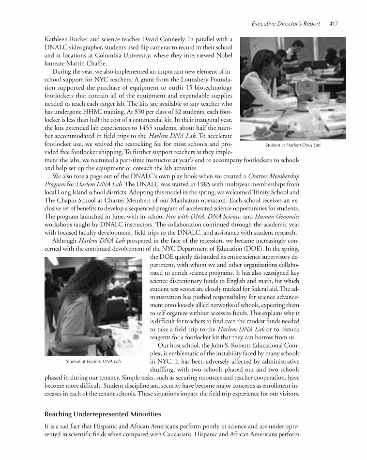
Kathleen Rucker and science teacher David Conneely. In parallel with aDNALC videographer, students used flip cameras to record in their schooland at locations at Columbia University, where they interviewed Nobellaureate Martin Chalfie.
During the year, we also implemented an important new element of in-school support for NYC teachers. A grant from the Lounsbery Founda-tion supported the purchase of equipment to outfit 15 biotechnologyfootlockers that contain all of the equipment and expendable suppliesneeded to teach each target lab. The kits are available to any teacher whohas undergone HHMI training. At $50 per class of 32 students, each foot-locker is less than half the cost of a commercial kit. In their inaugural year,the kits extended lab experiences to 1455 students, about half the num-ber accommodated in field trips to the Harlem DNA Lab. To acceleratefootlocker use, we waived the restocking fee for most schools and pro-vided free footlocker shipping. To further support teachers as they imple-ment the labs, we recruited a part-time instructor at year’s end to accompany footlockers to schoolsand help set up the equipment or coteach the lab activities.
We also tore a page out of the DNALC’s own play book when we created a Charter MembershipProgram for Harlem DNA Lab. The DNALC was started in 1985 with multiyear memberships fromlocal Long Island school districts. Adopting this model in the spring, we welcomed Trinity School andThe Chapin School as Charter Members of our Manhattan operation. Each school receives an ex-clusive set of benefits to develop a sequenced program of accelerated science opportunities for students.The program launched in June, with in-school Fun with DNA, DNA Science, and Human Genomicsworkshops taught by DNALC instructors. The collaboration continued through the academic yearwith focused faculty development, field trips to the DNALC, and assistance with student research.
Although Harlem DNA Lab prospered in the face of the recession, we became increasingly con-cerned with the continued devolvement of the NYC Department of Education (DOE). In the spring,
the DOE quietly disbanded its entire science supervisory de-partment, with whom we and other organizations collabo-rated to enrich science programs. It has also reassigned keyscience discretionary funds to English and math, for whichstudent test scores are closely tracked for federal aid. The ad-ministration has pushed responsibility for science advance-ment onto loosely allied networks of schools, expecting themto self-organize without access to funds. This explains why itis difficult for teachers to find even the modest funds neededto take a field trip to the Harlem DNA Lab or to restockreagents for a footlocker kit that they can borrow from us.
Our host school, the John S. Roberts Educational Com-plex, is emblematic of the instability faced by many schoolsin NYC. It has been adversely affected by administrativeshuffling, with two schools phased out and two schools
phased in during our tenancy. Simple tasks, such as securing resources and teacher cooperation, havebecome more difficult. Student discipline and security have become major concerns as enrollment in-creases in each of the tenant schools. These situations impact the field trip experience for our visitors.
Reaching Underrepresented Minorities
It is a sad fact that Hispanic and African Americans perform poorly in science and are underrepre-sented in scientific fields when compared with Caucasians. Hispanic and African Americans perform
Student at Harlem DNA Lab
Executive Director’s Report 417
Student at Harlem DNA Lab
411-434_LAR_DNALC_Annual Report_2009 template 7/8/11 4:04 PM Page 417

418 DNA Learning Center
~20% lower in science than Caucasians on the International Assessment of Education Progress, arespected benchmark for student achievement. This disparity holds true in grades 4, 8, and 12. Ac-cording to the National Science Board, these groups are underrepresented in postsecondary scienceeducation and science careers compared to their proportion of the overall U.S. population (24%).Underrepresented minorities received 17% of bachelor’s degrees, 13% of master’s degrees, and 7%of doctorates in science and engineering fields in 2007. They accounted for only 10% of college-educated persons employed in science and engineering occupations and received salaries that aver-aged 25% less than those of Caucasian workers.
The National Science Education Standards and other studies emphasize that students need to beengaged in the process of science, asking questions, forming hypotheses, designing experiments,collecting data, analyzing results, and forming conclusions. Underrepresented minorities—who areconcentrated in low-achieving schools and who may receive less science “boosting” at home—haveless access to hands-on experiences that help them to learn about science in the same way as scien-tists.
The DNALC takes seriously the challenge of increasing minority representation in science. TheUrban Barcode Project and our Harlem DNA Lab were established for the express purpose of pro-viding minority and disadvantaged students—and their teachers—the same opportunities for lab-oratory-based learning as those offered students in wealthy suburban school districts. Statistics for2010 show that our Manhattan operation is reaching this target audience. Hispanic and AfricanAmericans made up 69% of students visiting the Harlem DNA Lab and 41% of teachers trainedunder our HHMI program. The strong Harlem numbers, combined with a policy of conductingmany teacher-training workshops at institutions serving minorities, maintained underrepresentedminorities at 25% of 696 teachers trained at 1–10-day workshops.
Doing the best job in science education for underrepresented minorities also requires that an in-stitution practice what it preaches—and provide role models for students to emulate. Thus, dur-ing the last several years, the DNALC has hired four exemplary African American and Latinoeducators, who now compose 33% of the instructional staff. Jermel Watkins, Ph.D., began his sci-ence career as a DNALC intern and completed his graduate training at Stony Brook University.Jason Williams moved into education after research stints in two CSHL laboratories. Ileana Rios,Ph.D., attended elementary school several blocks away from the Harlem DNA Lab and completedher graduate training at City University of New York. Our newest staff member, Kerri-AnnMatthews, brings experience as an informal science educator with the New York Hall of Scienceand other science exhibitions.
Internet Strategies and Visitation
The September issue of Wired magazine ran the full-page headline, “The Web is Dead.” The leadarticle made the point that the World Wide Web (WWW)—navigated by browsers such as Ex-plorer, Firefox, Safari, and Chrome—now accounts for less than 25% of total Internet traffic. Thevast majority of Internet bandwidth is consumed by other types of communication, notably, e-mail,virtual private networks (VPNs), voice-over Internet, music and video sharing, and active gaming.The article also referred, metaphorically, to the struggle between the chaotic, “everything-all-the-time(from-everyone)” world of websites—dominated by search engines such as Google—and the “what-you-need-when-you need-it (from-someone-you-trust)” world of cell phone applications (apps) andsocial media—dominated by sharing software such as Facebook and Apple iTunes. The DNALCnow finds itself in this struggle to adapt to the changing Internet landscape.
The DNALC benefited from its early entry into the Internet world. At a time when there wereonly 10–25 million active websites on the Internet, websites we developed in the late 1990s and early2000s rapidly built audiences. Overall visitation increased steadily, peaking at 7.1 million in 2007.However, our visitation declined and then leveled at ~6 million in 2008–2009. In less than 5 years,
411-434_LAR_DNALC_Annual Report_2009 template 7/8/11 4:04 PM Page 418

(Left) The refreshed DNA from the Beginning website; (right) DNALC YouTube channel
Executive Director’s Report 419
the web had grown to more than 100 million active sites; the DNALC and other small-contentproviders now found it increasingly difficult to build and maintain audiences.
In the face of an exponentially crowded web, search engines such as Google became the primaryarbiters of website visitation. We therefore embarked on an ambitious program to redesign our olderwebsites to make them more “visible” to search engines, a process called search engine optimization(SEO). DNALC.org, the home site from which all other DNALC websites are reached, was thefirst to be revamped according to SEO principals. Relaunched in September 2009, it saw a 24% in-crease in visitation in 2010. DNA from the Beginning, our first major website, is now undergoing aneven more extensive SEO makeover, and we anticipate a similar bump in visitation when it is re-launched in spring 2011.
In parallel with SEO, we have turned to other channels—applications for handheld devices(“apps”) and social media—to broaden the audience for our multimedia products. Our 3D Brain—a detailed rendering of the human brain that can be rotated and explored in three dimensions—pro-vides a remarkable example of the way in which rechanneling content can increase viewership.Originally developed as part of the website Genes to Cognition (G2C) Online, which debuted in spring2009, 3D Brain was later launched as a stand-alone iPhone, iPod, and iPad application. It rose to num-ber seven of 7,100 education apps and number one among 250 iPad apps. In 2010, the web versionof 3D Brain received 54,868 visits, and the app version had 413,874 downloads! Feedback on theiTunes site shows, as one would expect, strong use by college students and faculty. However, it is alsoused by medical professionals: “Excellent! I use this with clients to help them see where some of theirPTSD (posttraumatic stress disorder) symptoms are coming from. Has been very helpful for them.”
During the year, we continued an aggressive program to develop a DNALC channel on YouTube,the site where most people find and share videos. Visitation surged following the addition of nearly100 new videos, including a collection of three-dimensional molecular animations created for theDNA Interactive website by 2010 McArthur Prize–winner Drew Berry. Our collection of 184 videosreceived 280,503 views in 2010. Blogs, supporting each of our major content sites, generated an ad-ditional 283,843 web visits.
As a result of the SEO makeover for the DNALC homepage plus aggressive moves into apps,YouTube, and blogs, Internet visitation rose 14% to 7.1 million in 2010, equaling the 2007 peak.App downloads, YouTube views, and blog views contributed 933,220 visits, or 13% of total Inter-net traffic. The amount of data served by DNALC websites surged 35% in 2010, to 6669 gigabytes(GB), or 6.5 terabytes (TB). DNALC.org accounted for 43% of all data served.
411-434_LAR_DNALC_Annual Report_2009 template 7/8/11 4:04 PM Page 419
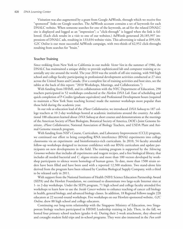
Visitation was also augmented by a grant from Google AdWords, through which we receive free“sponsored” links on Google searches. The AdWords account contains a set of keywords for eachDNALC website. When someone searches for one of the keywords, an ad for the related DNALCsite is displayed and logged as an “impression”; a “click-through” is logged when the link is fol-lowed. (Each click results in a visit to one of our websites.) AdWords generated 20,185,997 im-pressions of DNALC ads, resulting in 133,034 website visits. This advertising is valued at $99,438.G2C Online is our most successful AdWords campaign, with two-thirds of 62,952 click-throughsresulting from searches for “brain.”
Teacher Training
Since trekking from New York to California in our mobile Vector Van in the summer of 1986, theDNALC has maintained a unique ability to provide sophisticated lab and computer training at es-sentially any site around the world. The year 2010 was the zenith of off-site training, with 940 highschool and college faculty participating in professional development activities conducted at 27 sitesacross the United States and Canada. (For a complete list of training activities and host sites, see thetables at the back of this report: “2010 Workshops, Meetings, and Collaborations.”)
With funding from HHMI, and in collaboration with the NYC Department of Education, 290teachers participated in 52 workshops conducted at the Harlem DNA Lab. Ease of scheduling andquick completion of P-Credits (graduate equivalent) and Professional Development hours (requiredto maintain a New York State teaching license) made the summer workshops more popular thanthose held during the academic year.
In our role as education lead of the iPlant Collaborative, we introduced DNA Subway to 187 col-lege teachers at 10 2-day workshops hosted at academic institutions around the country. An addi-tional 188 educators learned about DNA Subway at short courses and demonstrations at the meetingsof the American Society of Plant Biologists, Botanical Society of America, DOE’s Joint Genome In-stitute, iPlant Collaborative, National Association of Biology Teachers, and USDA Plant and Ani-mal Genome research program.
With funding from NSF’s Course, Curriculum, and Laboratory Improvement (CCLI) program,we continued our effort to bring compelling RNA interference (RNAi) experiments into collegeclassrooms via an experiment- and bioinformatics-rich curriculum. In 2010, 54 faculty attendedfollow-up workshops designed to increase confidence with our RNAi curriculum and update par-ticipants on new developments in the field. The training program is supported by the SilencingGenomes website that includes all experiments and reagent recipes, and a free biological library, thatincludes all needed bacterial and C. elegans strains and more than 100 vectors developed by work-shop participants to silence worm homologs of human genes. To date, more than 1500 strain or-ders have been filled and have been used with a reported 12,500 students. Two stand-alone kitsderived from the program have been released by Carolina Biological Supply Company, with a thirdto be released early in 2011.
With support from the National Institutes of Health (NIH) Science Education Partnership Award(SEPA) and the Hewlett Foundation, we continued to disseminate two large-scale Internet sites at1- to 2-day workshops. Under the SEPA program, 71 high school and college faculty attended fiveworkshops to learn how to use the Inside Cancer website to enhance teaching of cancer cell biologyin health, general biology, and advanced biology classes. In addition, 18 Regional Fellows taught 228educators at 22 second-round workshops. Five workshops on our Hewlett-sponsored website, G2COnline, drew 88 high school and college educators.
Continuing our long-term relationship with the Singapore Ministry of Education, two Singa-porean biology teachers participated in HHMI Leadership training in July. Then, in the fall, wehosted four primary school teachers (grades 4–6). During their 2-week attachment, they observedand cotaught student field trips and in-school programs. They were also immersed in the Fun with
420 DNA Learning Center
411-434_LAR_DNALC_Annual Report_2009 template 7/8/11 4:04 PM Page 420
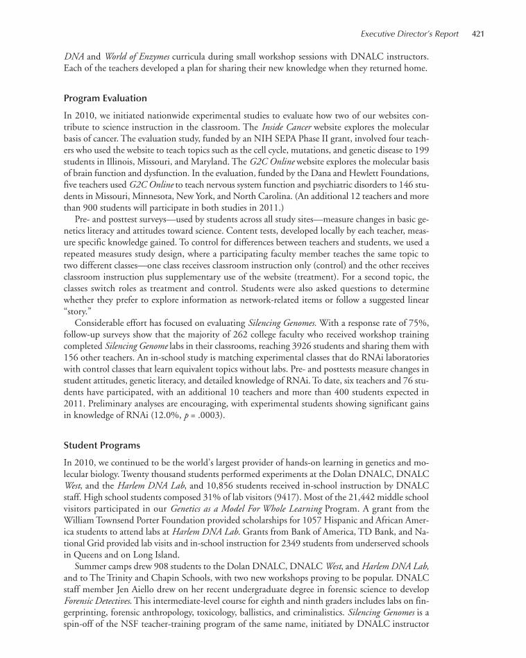
DNA and World of Enzymes curricula during small workshop sessions with DNALC instructors.Each of the teachers developed a plan for sharing their new knowledge when they returned home.
Program Evaluation
In 2010, we initiated nationwide experimental studies to evaluate how two of our websites con-tribute to science instruction in the classroom. The Inside Cancer website explores the molecularbasis of cancer. The evaluation study, funded by an NIH SEPA Phase II grant, involved four teach-ers who used the website to teach topics such as the cell cycle, mutations, and genetic disease to 199students in Illinois, Missouri, and Maryland. The G2C Online website explores the molecular basisof brain function and dysfunction. In the evaluation, funded by the Dana and Hewlett Foundations,five teachers used G2C Online to teach nervous system function and psychiatric disorders to 146 stu-dents in Missouri, Minnesota, New York, and North Carolina. (An additional 12 teachers and morethan 900 students will participate in both studies in 2011.)
Pre- and posttest surveys—used by students across all study sites—measure changes in basic ge-netics literacy and attitudes toward science. Content tests, developed locally by each teacher, meas-ure specific knowledge gained. To control for differences between teachers and students, we used arepeated measures study design, where a participating faculty member teaches the same topic totwo different classes—one class receives classroom instruction only (control) and the other receivesclassroom instruction plus supplementary use of the website (treatment). For a second topic, theclasses switch roles as treatment and control. Students were also asked questions to determinewhether they prefer to explore information as network-related items or follow a suggested linear“story.”
Considerable effort has focused on evaluating Silencing Genomes. With a response rate of 75%,follow-up surveys show that the majority of 262 college faculty who received workshop trainingcompleted Silencing Genome labs in their classrooms, reaching 3926 students and sharing them with156 other teachers. An in-school study is matching experimental classes that do RNAi laboratorieswith control classes that learn equivalent topics without labs. Pre- and posttests measure changes instudent attitudes, genetic literacy, and detailed knowledge of RNAi. To date, six teachers and 76 stu-dents have participated, with an additional 10 teachers and more than 400 students expected in2011. Preliminary analyses are encouraging, with experimental students showing significant gainsin knowledge of RNAi (12.0%, p = .0003).
Student Programs
In 2010, we continued to be the world’s largest provider of hands-on learning in genetics and mo-lecular biology. Twenty thousand students performed experiments at the Dolan DNALC, DNALCWest, and the Harlem DNA Lab, and 10,856 students received in-school instruction by DNALCstaff. High school students composed 31% of lab visitors (9417). Most of the 21,442 middle schoolvisitors participated in our Genetics as a Model For Whole Learning Program. A grant from theWilliam Townsend Porter Foundation provided scholarships for 1057 Hispanic and African Amer-ica students to attend labs at Harlem DNA Lab. Grants from Bank of America, TD Bank, and Na-tional Grid provided lab visits and in-school instruction for 2349 students from underserved schoolsin Queens and on Long Island.
Summer camps drew 908 students to the Dolan DNALC, DNALC West, and Harlem DNA Lab,and to The Trinity and Chapin Schools, with two new workshops proving to be popular. DNALCstaff member Jen Aiello drew on her recent undergraduate degree in forensic science to developForensic Detectives. This intermediate-level course for eighth and ninth graders includes labs on fin-gerprinting, forensic anthropology, toxicology, ballistics, and criminalistics. Silencing Genomes is aspin-off of the NSF teacher-training program of the same name, initiated by DNALC instructor
Executive Director’s Report 421
411-434_LAR_DNALC_Annual Report_2009 template 7/8/11 4:04 PM Page 421
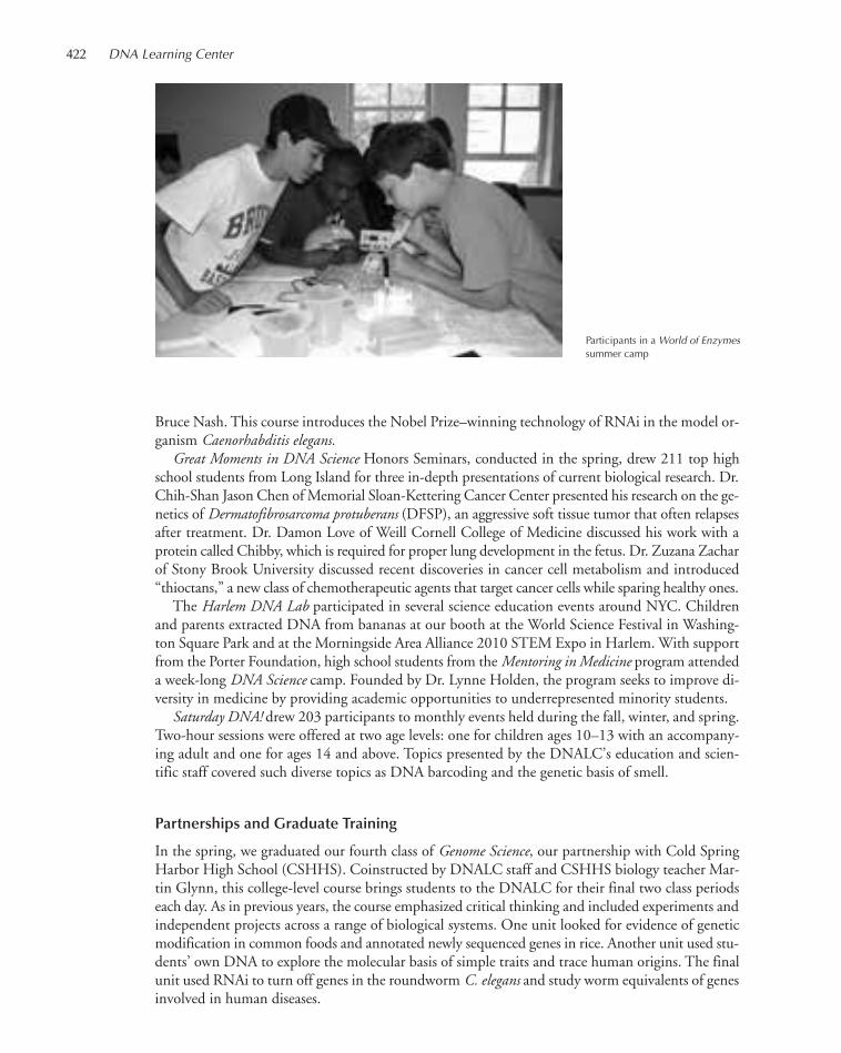
Participants in a World of Enzymessummer camp
422 DNA Learning Center
Bruce Nash. This course introduces the Nobel Prize–winning technology of RNAi in the model or-ganism Caenorhabditis elegans.
Great Moments in DNA Science Honors Seminars, conducted in the spring, drew 211 top highschool students from Long Island for three in-depth presentations of current biological research. Dr.Chih-Shan Jason Chen of Memorial Sloan-Kettering Cancer Center presented his research on the ge-netics of Dermatofibrosarcoma protuberans (DFSP), an aggressive soft tissue tumor that often relapsesafter treatment. Dr. Damon Love of Weill Cornell College of Medicine discussed his work with aprotein called Chibby, which is required for proper lung development in the fetus. Dr. Zuzana Zacharof Stony Brook University discussed recent discoveries in cancer cell metabolism and introduced“thioctans,” a new class of chemotherapeutic agents that target cancer cells while sparing healthy ones.
The Harlem DNA Lab participated in several science education events around NYC. Childrenand parents extracted DNA from bananas at our booth at the World Science Festival in Washing-ton Square Park and at the Morningside Area Alliance 2010 STEM Expo in Harlem. With supportfrom the Porter Foundation, high school students from the Mentoring in Medicine program attendeda week-long DNA Science camp. Founded by Dr. Lynne Holden, the program seeks to improve di-versity in medicine by providing academic opportunities to underrepresented minority students.
Saturday DNA! drew 203 participants to monthly events held during the fall, winter, and spring.Two-hour sessions were offered at two age levels: one for children ages 10–13 with an accompany-ing adult and one for ages 14 and above. Topics presented by the DNALC’s education and scien-tific staff covered such diverse topics as DNA barcoding and the genetic basis of smell.
Partnerships and Graduate Training
In the spring, we graduated our fourth class of Genome Science, our partnership with Cold SpringHarbor High School (CSHHS). Coinstructed by DNALC staff and CSHHS biology teacher Mar-tin Glynn, this college-level course brings students to the DNALC for their final two class periodseach day. As in previous years, the course emphasized critical thinking and included experiments andindependent projects across a range of biological systems. One unit looked for evidence of geneticmodification in common foods and annotated newly sequenced genes in rice. Another unit used stu-dents’ own DNA to explore the molecular basis of simple traits and trace human origins. The finalunit used RNAi to turn off genes in the roundworm C. elegans and study worm equivalents of genesinvolved in human diseases.
411-434_LAR_DNALC_Annual Report_2009 template 7/8/11 4:04 PM Page 422

Executive Director’s Report 423
In the fall, the fifth year of Genome Science began under the tutelage of DNALC staff along witha new CSHHS staff member, Jaak Raudsepp. This was a comfortable change because Jaak is a grad-uate of our former Leadership Institute, the rigorous 3-week program for the nation’s best biologyteachers. Always a proving ground for our latest experiments, the CSHHS students were the first touse DNA barcoding to identify plants collected in the local area. In preparing essays on modern par-allels to the eugenics movement of the early 20th century, students also visited the CSHL ResearchArchives to examine historical documents remaining from the former Eugenics Record Office.
As part of a reinvigorated collaboration with the Center for Science and Mathematics Education(CESAME) at Stony Brook University, CSHHS students completed a module on protein model-ing. Using methods developed by Tim Herman at the Milwaukee School of Engineering, studentsdeveloped three-dimensional molecular models to illuminate how chemotherapeutic drugs interactwith protein receptors on the surface of cancer cells. Using data from primary research papers, stu-dents identified important parts of the drugs and receptors. They then used three-dimensional mod-eling software to highlight key atoms involved in molecular interactions. The modified structure fileswere sent to CESAME, where detailed atomic resolution models of the proteins were manufacturedon state-of-the-art prototyping printers. (These machines are essentially laser printers that layerdroplets of polymer resin in three dimensions.) Students will present their models and explanatoryposters at the spring 2011 CSHL meeting, “The Biology of Cancer.”
During their second semester, graduate students at the CSHL Watson School of Biological Sci-ences work under the guidance of experienced DNALC instructors. The graduate students work inpairs during 12 half-day sessions, learning from and leaning on one another as they develop effec-tive teaching techniques. During the first phase of training, students observe a DNALC staff mem-ber teaching a laboratory class and then organize a lesson plan that integrates their own perspectives.During the second phase, students join the DNALC staff member to coteach parts of a laboratoryclass. After critiques of their lesson plans and coteaching experience, the students move on to inde-pendently teach an entire lesson. After completing rotations with middle- and high-school-level ex-periments, students deliver three additional lessons to demonstrate mastery of teaching and classmanagement skills.
In October, we began a collaboration with the New York Academy of Sciences (NYAS), one ofthe oldest scientific institutions in the United States and a leading organizer of scientific meetings.The NYAS developed a new science mentoring program in which graduate students and postdoc-toral researchers from NYC research institutions present science activities to students in grades K–12. DNALC instructors trained 22 mentors in effective strategies to deliver hands-on geneticsactivities in after-school settings.
Staff and Interns
We were sad when Dr. John Connolly left in February to evaluate clinical trials at the Center for Ap-plied Genomics, Children’s Hospital of Philadelphia. After receiving a Ph.D. in neuropsychologyat Trinity College, Dublin, John came to the DNALC in 2005 to develop our innovative websiteG2C Online, for which he interviewed more than 80 scientists. As Multimedia Coordinator, he be-came indispensable in managing many aspects of our Internet venture—developing the LandeauMultimedia Studio and launching regular blogs for each of our major content sites. Notably, hemoved us into the world of apps with his spectacularly successful 3D Brain. We will miss his Irishbrogue, which often lightened the office atmosphere.
The multimedia staff returned to full force in November, with the arrival from “down under” ofMultimedia and Evaluation Manager Dr. Amy Nisselle. After attending the DNALC’s presentationat the 19th International Congress on Human Genetics in Melbourne in 2003, Amy decided to pur-sue her Ph.D. in multimedia genetics education. In 2008, she spent 3 months at the DNALC, eval-uating the G2C Online website as part of her thesis research. In addition to managing multimedia
411-434_LAR_DNALC_Annual Report_2009 template 7/8/11 4:04 PM Page 423

424 DNA Learning Center
projects, Amy will also increase our academic profile in the world ofscience and technology education.
Our growing involvement in the iPlant Collaborative led to therecruitment in November of Computer Programmer MohammedKhalfan. After receiving a B.S. degree from the University of Toronto,he received a Master’s degree in bioinformatics and computationalbiology at the University of Newcastle upon Tyne, United Kingdom.His initial projects include building the “Blue Line” (phylogenetics)of DNA Subway and redeveloping the iPlant website in Drupal, anopen source content management system (CMS).
Our multimedia group also benefited from fresh ideas from youngpeople. In the fall, we welcomed Todd Rocheford as Design Assistantand Videographer. Todd has an undergraduate degree in video pro-duction from the University of Canberra, Australia. We met Toddthrough his father, Torbert, a Purdue geneticist who developed vita-
min-A-fortified corn. After filming a documentary on his father’s work in Africa, Todd realized thathe wanted to develop educational videos, leading to his position at the DNALC. Undergraduate in-tern Tony Biondo was promoted to Junior Programmer, based largely on the initiative that he tookin developing an Android version of our 3D Brain app. Tony is a sophomore majoring in computerscience at Stony Brook University. An active supporter of open-source software, he won the StudentCluster Competition of the 2009 International Conference on High-Performance Computing atPortland, Oregon. Chris Weidler (Farmingdale State College) continues to support the BioMediaGroup and was especially productive on the DNA from the Beginning upgrade.
Our internship program continued to draw some of Long Island’s most talented high school andcollege students, engaging them in science research and providing practical laboratory experience.We were pleased to accept several new high school interns this year: Devika Gupta (Farmingdale),Jueng Woen Kim (Hauppauge), Yasin Muhammad (Lawrence Woodmere Academy), H. AlanPhipps (Portledge School), David Streitman (Syosset), and Young Joon Suh (John Glenn). The newhires joined a dedicated group of returning interns: Laura Bergsten (Cold Spring Harbor), GeorgeEconomou (Syosset), Jack Greenfield (Oyster Bay), Lindsay Hochberg (Oyster Bay), Max Vaysman(Commack), and Pamela Wax (Harborfields). We bid farewell to a number of high school internsas they left for their freshman year at college: Anouva Kalra-Lall (Case Western Reserve University),Emily Troge (Brandeis University), Sara Wienclaw (University of Delaware), and Kevin Wu (NewYork University).
In addition to regular prep duties, college interns help to fulfill requests from faculty nationwidefor student DNA sequencing and RNAi targeting vectors. Three former high school interns re-turned to assist with summer workshops: Kaitlin Watrud (Gettysburg College), Arielle Scardino(City College of New York), and Seth Shortz (Emory University). New college interns starting in2010 were David Dopfel (Stony Brook), Sarah Justvig (Georgetown), Sulaiman Usman (New YorkInstitute of Technology), Lina-Mari Varghese (Stony Brook), and Katherine Villalon (John Jay).Annie Laurie Benzie (Adelphi University) left the DNALC for an internship at Bellevue Hospital.
Many of our high school interns are involved in science research and compete in state and na-tionwide science competitions. Anouva Kalra-Lall was an Intel semifinalist and a top-four finalistfor the Neuroscience Research Prize. Young Joon Suh took first place in chemistry at the New YorkState Science and Engineering Fair. Laura Bergsten was accepted into the CSHL Partners for the Fu-ture Program.
Additionally, DNALC staff served as mentors to local high school students working on inde-pendent projects. Elliot Horlick (Cold Spring Harbor) worked on two projects with Dr. JermelWatkins: sunblock (SPF) protection against UV-induced DNA damage in yeast and the proteincomplement (proteome) of squamous cell carcinoma. Daniel Krumholz (Oyster Bay) worked with
Mohammed Khalfan and Amy Nisselle.
411-434_LAR_DNALC_Annual Report_2009 template 7/8/11 4:04 PM Page 424

Cold Spring Harbor Laboratory Ex-Officio Members
W. Dillaway Ayres, Jr., Chief Operating OfficerDavid A. Micklos, Executive Director, DNA Learning
Center
Bruce Stillman, President and Chief Executive OfficerKaren Orzel, Senior Development Officer
Corporate Advisory BoardChairperson: Edward A. Chernoff, Motors & Armatures Inc.
CSHL Trustee Liaison: Laurie Landeau, VMD
Michael Aboff, Aboff’s Inc.Paul Amoruso, Oxford & Simpson RealtyRocco S. Barrese, Dilworth & BarreseEdward Blaskey, TD BankThomas J. Calabrese, Daniel Gale Sothebys International
RealtyJeffrey CarstensRichard A. Catalano, KPMG, LLPJames Chinitz, Population DiagnosticsMarian Conway, New York Community Bank
FoundationDan Decker, Eppendorf North AmericaRobert Dickstein, Ph.D., Pall CorporationDavid Epstein, Ph.D., OSI Pharmaceuticals, Inc.Candido E. Fuentes-Felix, M.D., F.A.C.S.David Garbus, Huntington Business Products CentreLawrence Goodman, Curtis, Mallet-Prevost, Colt &
Mosle LLPMary Jane Helenek, Luitpold PharmaceuticalsArthur D. Herman, Herman Development Corporation
Diana Hoadley, JP MorganRobert Isaksen, Bank of AmericaNeil M. Kaufman, Davidoff, Malito & HutcherJohn C. Kean III, Kean Development CorporationNorman Kelker, Enzo Life SciencesRobert Keller, National Grid FoundationJames E. Mattutat, Ingleside Investors, Inc.John Molloy, H2MJohn Passarelli, M.D.Patricia Petersen, Daniel Gale Sothebys Real EstateFrank Posillico, Alliance Real Estate CorpChristiaan B. ReynoldsWilliam D. RocheRaju Sarwal, M.D.Charles R. Schueler, CablevisionLee Shuett, Nikon InstrumentsKurt Timmel, Marsh USAJeffrey K. Tupper, U.S. Trust Company of New YorkRobert Van NostrandHans Zobel, Ziehm Medical LLC
Edward A. Chernoff, President, Motors & Armatures, Inc.Maria DeLessepsLola N. Grace, Sterling Grace Capital Management
(Secretary/ Treasurer, CSHL)Lori HomerSuzanne KleinknechtSuzanne Leeds
Peter QuickAdele SmithersArthur SpiroPeter TillesEdward Travaglianti, President, TD Bank Long IslandMarianne Dolan Weber, Chairman, Dolan Foundations
DNALC CommitteeChairperson: Laurie Landeau, VMD, Trustee, CSHL
Executive Director’s Report 425
Dr. Bruce Nash to use RNAi to “knock down” genes in C. elegans that are homologous to humangenes involved in serotonin signaling.
Expert Advisors and Corporate Support
We are fortunate to have high-level support from two advisory bodies: the DNALC Committeeand the Corporate Advisory Board (CAB). The DNALC Committee consists of community lead-ers and members of CSHL senior management and the Board of Trustees, who oversee strategic de-velopment including capital funding and the evolution of satellite locations in North America andbeyond. The CAB provides liaison to the Long Island and NYC business communities; its annualfund campaign and golf tournament contributed $155,000 in 2010.
411-434_LAR_DNALC_Annual Report_2009 template 7/8/11 4:04 PM Page 425

2010 Workshops, Meetings, and Collaborations
January 5 Site visit by William Mak, Hong Kong Biotechnology Education Resource Center and Mobile Lab, Hong Kong University, Pokfulam
January 12 Site visit by Denise Philpotts, Trinity School, Harlem DNA LabJanuary 14 NSF Plant Collaborative, Genotype 2 Phenotype Steering Committee Meeting, San Diego, California
HHMI Professional Development Workshop, “PCR and Human DNA Variations, Part 2,” Harlem DNA LabJanuary 16 HHMI Professional Development Workshop, “PCR and Human DNA Variations, Part 2,” Harlem DNA LabJanuary 22 Site visit by Hans Bosch and Laura Savini, WLIW, New YorkJanuary 23 Saturday DNA! “Express Yourself!” DNALCJanuary 29 Site visit by Marilyn Parks, Life Technologies Foundation, Carlsbad, California, and Randi Spatz, Invitrogen,
Carlsbad, CaliforniaFebruary 24 HHMI Professional Development Workshop, “DNA Structure and Isolation,” Harlem DNA LabFebruary 27 Saturday DNA! “Composting 101,” DNALC
HHMI Professional Development Workshop, “DNA Structure and Isolation,” Harlem DNA LabMarch 3 Site visit to Carolina Biological Supply Company, Burlington, North CarolinaMarch 10 Oyster Bay Rotary Club Meeting Luncheon presenting DNALC programs and events, Oyster Bay, New YorkMarch 12 G2C Online Workshop, Santa Clara University, Santa Clara, CaliforniaMarch 13 NIH Inside Cancer Workshop, Santa Clara University, Santa Clara, CaliforniaMarch 17 HHMI Professional Development Workshop, “Variability and Inheritance,” Harlem DNA LabMarch 20 Saturday DNA! “Explore the Realm of Your DNA,” DNALC
HHMI Professional Development Workshop, “Variability and Inheritance,” Harlem DNA LabMarch 20–21 National Science Teachers Association National Conference, “Exploring Gene Function in C. elegans: Mutations
and RNA Interference,” “Genomic Science,” and “Silencing Genomes,” Philadelphia, PennsylvaniaMarch 24–26 Joint Genome Institute User Meeting, Walnut Creek, CaliforniaMarch 26 G2C Online Workshop, Oklahoma City Community College, Oklahoma CityMarch 27 NIH Inside Cancer Workshop, Oklahoma City Community College, Oklahoma CityApril 7 HHMI Professional Development Workshop, “Bacterial Transformation and Protein Isolation,” Harlem DNA LabApril 9 Site visit by graduate students from Molloy College, Rockville Centre, New YorkApril 10 HHMI Professional Development Workshop, “Bacterial Transformation and Protein Isolation,” Harlem DNA LabApril 11–14 NIH SEPA Principal Investigators Meeting, Birmingham, AlabamaApril 13 Great Moments in DNA Science Honors Seminar: “Dead End? Not Anymore! Molecular Targeted Therapy for
Skin Cancer,” Dr. Chih-Shan Jason Chen, Memorial Sloan-Kettering Cancer Center, New YorkApril 16 G2C Online Workshop, iBio Institute, Harold Washington College, Chicago, IllinoisApril 17 NIH Inside Cancer Workshop, iBio Institute, Harold Washington College, Chicago, IllinoisApril 17 Saturday DNA! “The Insomniac’s Guide to Wildlife,” DNALCApril 20 Site visit by Debby Hirshman, Victor Centers for Jewish Genetic Diseases, Philadelphia, PennsylvaniaApril 21 HHMI Professional Development Workshop, “DNA Analysis and Forensics,” Harlem DNA LabApril 23 CSHL Association Directors, viewing of “Naturally Obsessed: The Making of a Scientist,” DNALCApril 23–24 NSF iPlant Collaborative Genomics in Education Workshop, Spelman College, Atlanta, GeorgiaApril 24 HHMI Professional Development Workshop, “DNA Analysis and Forensics,” Harlem DNA LabApril 26 Site visit by Kevin Shine, Verizon Communications, New YorkApril 29 G2C Online Workshop, Biogen Idec, Cambridge, Massachusetts
Great Moments in DNA Science Honors Seminar: “The Role of Wnt/β-catenin Antagonist chibby in Lung Development,” Dr. Damon Love, Weill Cornell College of Medicine, New York
Site visit by Jackie Dorrance, Beckman Foundation, Irvine, CaliforniaSite visit by Robert Isaksen and Lorraine Aycock, Bank of America Long Island, Melville, New YorkSite visit by Judy Calabrese and Maureen Laness, CSHL Directors, Harlem DNA Lab
April 30 NIH Inside Cancer Workshop, Biogen Idec, Cambridge, MassachusettsMay 3 Meeting with Minister of Higher Education, Sheikh Nahyan binMubarak Al Nahyan, Abu Dhubai, United
Arab EmiratesMay 7–8 NSF iPlant Collaborative Genomics in Education Workshop, University of Texas, AustinMay 8 HHMI Professional Development Workshop “DNA Isolation, Inheritance, and Variability,” Harlem DNA LabMay 12 Site visit by Roland Jimenez, Community Bank, Hauppauge, New YorkMay 14–15 HHMI Professional Development Workshop, “PCR, Part 1,” Harlem DNA LabMay 15 Saturday DNA! “Walking Whales and Genetic Tales,” DNALCMay 17 Site visit by Srinivasa Rao, Indian Institute of Biotechnology, Hyderabad, IndiaMay 18 Great Moments in DNA Science Honors Seminar: “Thioctans: A Novel Approach to Cancer Chemotherapy,”
Dr. Zuzana Zachar, Stony Brook University, New YorkMay 21 HHMI Professional Development Workshop, “PCR, Part 2,” Harlem DNA Lab
G2C Online Workshop, University of Colorado, Aurora
426 DNA Learning Center
411-434_LAR_DNALC_Annual Report_2009 template 7/8/11 4:04 PM Page 426
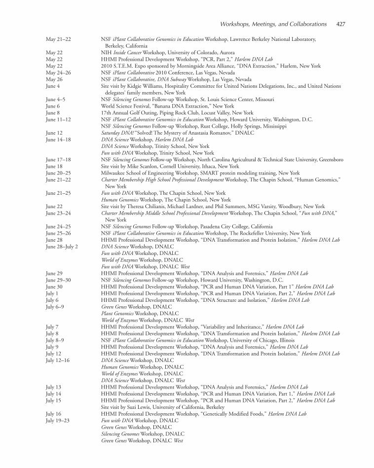
May 21–22 NSF iPlant Collaborative Genomics in Education Workshop, Lawrence Berkeley National Laboratory, Berkeley, California
May 22 NIH Inside Cancer Workshop, University of Colorado, AuroraMay 22 HHMI Professional Development Workshop, “PCR, Part 2,” Harlem DNA LabMay 22 2010 S.T.E.M. Expo sponsored by Morningside Area Alliance, “DNA Extraction,” Harlem, New YorkMay 24–26 NSF iPlant Collaborative 2010 Conference, Las Vegas, NevadaMay 26 NSF iPlant Collaborative, DNA Subway Workshop, Las Vegas, NevadaJune 4 Site visit by Kidgie Williams, Hospitality Committee for United Nations Delegations, Inc., and United Nations
delegates’ family members, New YorkJune 4–5 NSF Silencing Genomes Follow-up Workshop, St. Louis Science Center, MissouriJune 6 World Science Festival, “Banana DNA Extraction,” New YorkJune 8 17th Annual Golf Outing, Piping Rock Club, Locust Valley, New YorkJune 11–12 NSF iPlant Collaborative Genomics in Education Workshop, Howard University, Washington, D.C.
NSF Silencing Genomes Follow-up Workshop, Rust College, Holly Springs, MississippiJune 12 Saturday DNA! “Solved! The Mystery of Anastasia Romanov,” DNALCJune 14–18 DNA Science Workshop, Harlem DNA Lab
DNA Science Workshop, Trinity School, New YorkFun with DNA Workshop, Trinity School, New York
June 17–18 NSF Silencing Genomes Follow-up Workshop, North Carolina Agricultural & Technical State University, GreensboroJune 18 Site visit by Mike Scanlon, Cornell University, Ithaca, New YorkJune 20–25 Milwaukee School of Engineering Workshop, SMART protein modeling training, New YorkJune 21–22 Charter Membership High School Professional Development Workshop, The Chapin School, “Human Genomics,”
New YorkJune 21–25 Fun with DNA Workshop, The Chapin School, New York
Human Genomics Workshop, The Chapin School, New YorkJune 22 Site visit by Theresa Chilianis, Michael Lardner, and Phil Summers, MSG Varsity, Woodbury, New YorkJune 23–24 Charter Membership Middle School Professional Development Workshop, The Chapin School, “Fun with DNA,”
New YorkJune 24–25 NSF Silencing Genomes Follow-up Workshop, Pasadena City College, CaliforniaJune 25–26 NSF iPlant Collaborative Genomics in Education Workshop, The Rockefeller University, New YorkJune 28 HHMI Professional Development Workshop, “DNA Transformation and Protein Isolation,” Harlem DNA LabJune 28–July 2 DNA Science Workshop, DNALC
Fun with DNA Workshop, DNALCWorld of Enzymes Workshop, DNALCFun with DNA Workshop, DNALC West
June 29 HHMI Professional Development Workshop, “DNA Analysis and Forensics,” Harlem DNA LabJune 29–30 NSF Silencing Genomes Follow-up Workshop, Howard University, Washington, D.C.June 30 HHMI Professional Development Workshop, “PCR and Human DNA Variation, Part 1” Harlem DNA LabJuly 1 HHMI Professional Development Workshop, “PCR and Human DNA Variation, Part 2,” Harlem DNA LabJuly 6 HHMI Professional Development Workshop, “DNA Structure and Isolation,” Harlem DNA LabJuly 6–9 Green Genes Workshop, DNALC
Plant Genomics Workshop, DNALCWorld of Enzymes Workshop, DNALC West
July 7 HHMI Professional Development Workshop, “Variability and Inheritance,” Harlem DNA LabJuly 8 HHMI Professional Development Workshop, “DNA Transformation and Protein Isolation,” Harlem DNA LabJuly 8–9 NSF iPlant Collaborative Genomics in Education Workshop, University of Chicago, IllinoisJuly 9 HHMI Professional Development Workshop, “DNA Analysis and Forensics,” Harlem DNA LabJuly 12 HHMI Professional Development Workshop, “DNA Transformation and Protein Isolation,” Harlem DNA LabJuly 12–16 DNA Science Workshop, DNALC
Human Genomics Workshop, DNALCWorld of Enzymes Workshop, DNALCDNA Science Workshop, DNALC West
July 13 HHMI Professional Development Workshop, “DNA Analysis and Forensics,” Harlem DNA LabJuly 14 HHMI Professional Development Workshop, “PCR and Human DNA Variation, Part 1,” Harlem DNA LabJuly 15 HHMI Professional Development Workshop, “PCR and Human DNA Variation, Part 2,” Harlem DNA Lab
Site visit by Suzi Lewis, University of California, BerkeleyJuly 16 HHMI Professional Development Workshop, “Genetically Modified Foods,” Harlem DNA LabJuly 19–23 Fun with DNA Workshop, DNALC
Green Genes Workshop, DNALCSilencing Genomes Workshop, DNALCGreen Genes Workshop, DNALC West
Workshops, Meetings, and Collaborations 427
411-434_LAR_DNALC_Annual Report_2009 template 7/8/11 4:04 PM Page 427
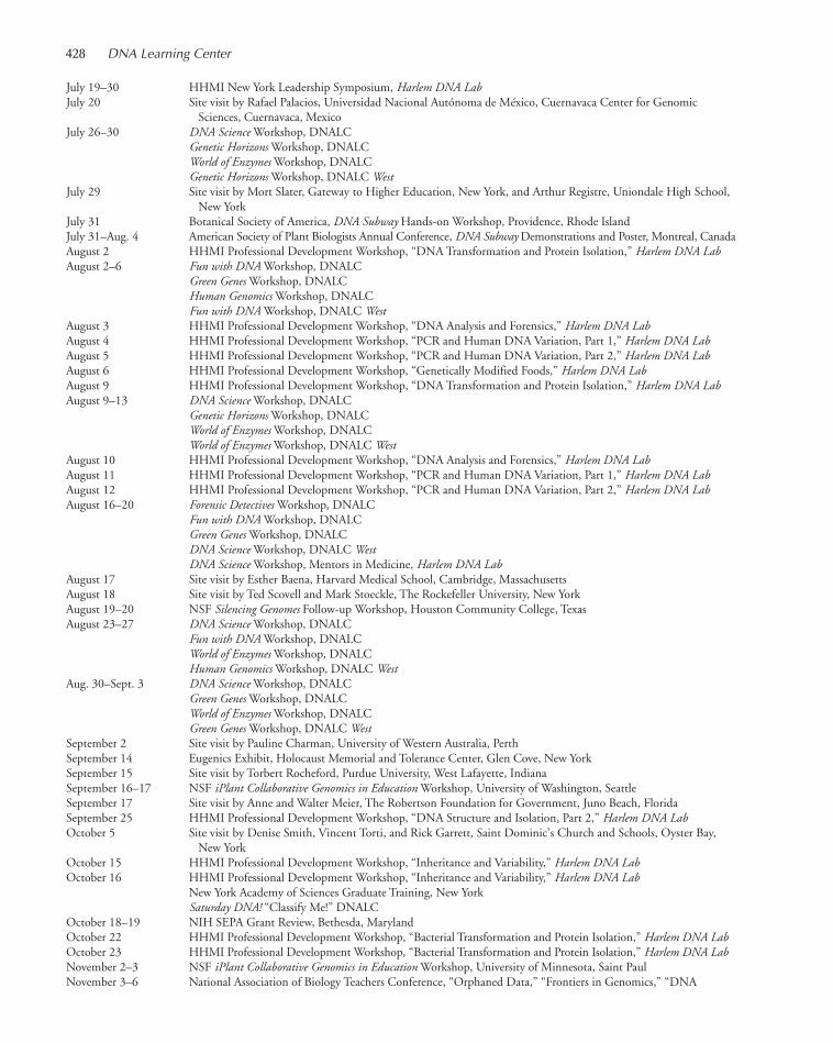
July 19–30 HHMI New York Leadership Symposium, Harlem DNA LabJuly 20 Site visit by Rafael Palacios, Universidad Nacional Autónoma de México, Cuernavaca Center for Genomic
Sciences, Cuernavaca, MexicoJuly 26–30 DNA Science Workshop, DNALC
Genetic Horizons Workshop, DNALCWorld of Enzymes Workshop, DNALCGenetic Horizons Workshop, DNALC West
July 29 Site visit by Mort Slater, Gateway to Higher Education, New York, and Arthur Registre, Uniondale High School, New York
July 31 Botanical Society of America, DNA Subway Hands-on Workshop, Providence, Rhode IslandJuly 31–Aug. 4 American Society of Plant Biologists Annual Conference, DNA Subway Demonstrations and Poster, Montreal, CanadaAugust 2 HHMI Professional Development Workshop, “DNA Transformation and Protein Isolation,” Harlem DNA LabAugust 2–6 Fun with DNA Workshop, DNALC
Green Genes Workshop, DNALCHuman Genomics Workshop, DNALCFun with DNA Workshop, DNALC West
August 3 HHMI Professional Development Workshop, “DNA Analysis and Forensics,” Harlem DNA LabAugust 4 HHMI Professional Development Workshop, “PCR and Human DNA Variation, Part 1,” Harlem DNA LabAugust 5 HHMI Professional Development Workshop, “PCR and Human DNA Variation, Part 2,” Harlem DNA LabAugust 6 HHMI Professional Development Workshop, “Genetically Modified Foods,” Harlem DNA LabAugust 9 HHMI Professional Development Workshop, “DNA Transformation and Protein Isolation,” Harlem DNA LabAugust 9–13 DNA Science Workshop, DNALC
Genetic Horizons Workshop, DNALCWorld of Enzymes Workshop, DNALCWorld of Enzymes Workshop, DNALC West
August 10 HHMI Professional Development Workshop, “DNA Analysis and Forensics,” Harlem DNA LabAugust 11 HHMI Professional Development Workshop, “PCR and Human DNA Variation, Part 1,” Harlem DNA LabAugust 12 HHMI Professional Development Workshop, “PCR and Human DNA Variation, Part 2,” Harlem DNA LabAugust 16–20 Forensic Detectives Workshop, DNALC
Fun with DNA Workshop, DNALCGreen Genes Workshop, DNALCDNA Science Workshop, DNALC WestDNA Science Workshop, Mentors in Medicine, Harlem DNA Lab
August 17 Site visit by Esther Baena, Harvard Medical School, Cambridge, MassachusettsAugust 18 Site visit by Ted Scovell and Mark Stoeckle, The Rockefeller University, New YorkAugust 19–20 NSF Silencing Genomes Follow-up Workshop, Houston Community College, TexasAugust 23–27 DNA Science Workshop, DNALC
Fun with DNA Workshop, DNALCWorld of Enzymes Workshop, DNALCHuman Genomics Workshop, DNALC West
Aug. 30–Sept. 3 DNA Science Workshop, DNALCGreen Genes Workshop, DNALCWorld of Enzymes Workshop, DNALCGreen Genes Workshop, DNALC West
September 2 Site visit by Pauline Charman, University of Western Australia, PerthSeptember 14 Eugenics Exhibit, Holocaust Memorial and Tolerance Center, Glen Cove, New YorkSeptember 15 Site visit by Torbert Rocheford, Purdue University, West Lafayette, IndianaSeptember 16–17 NSF iPlant Collaborative Genomics in Education Workshop, University of Washington, SeattleSeptember 17 Site visit by Anne and Walter Meier, The Robertson Foundation for Government, Juno Beach, FloridaSeptember 25 HHMI Professional Development Workshop, “DNA Structure and Isolation, Part 2,” Harlem DNA LabOctober 5 Site visit by Denise Smith, Vincent Torti, and Rick Garrett, Saint Dominic’s Church and Schools, Oyster Bay,
New YorkOctober 15 HHMI Professional Development Workshop, “Inheritance and Variability,” Harlem DNA LabOctober 16 HHMI Professional Development Workshop, “Inheritance and Variability,” Harlem DNA Lab
New York Academy of Sciences Graduate Training, New YorkSaturday DNA! “Classify Me!” DNALC
October 18–19 NIH SEPA Grant Review, Bethesda, MarylandOctober 22 HHMI Professional Development Workshop, “Bacterial Transformation and Protein Isolation,” Harlem DNA LabOctober 23 HHMI Professional Development Workshop, “Bacterial Transformation and Protein Isolation,” Harlem DNA LabNovember 2–3 NSF iPlant Collaborative Genomics in Education Workshop, University of Minnesota, Saint PaulNovember 3–6 National Association of Biology Teachers Conference, “Orphaned Data,” “Frontiers in Genomics,” “DNA
428 DNA Learning Center
411-434_LAR_DNALC_Annual Report_2009 template 7/8/11 4:04 PM Page 428

Subway,” “RNAi/CBSC,” “Iceman,” “CSI,” “LabCenter,” Minneapolis, MinnesotaNovember 4 Site visit by Paula Olsiewski, Alfred P. Sloan Foundation, Harlem DNA LabNovember 5 Site visit by Verizon Fios One MYLITV for filming November 8 iPlant Collaborative NSF site visit, Tucson, ArizonaNovember 8–9 Science Teachers Association of New York State Conference, presentations on DNALC family of websites,
Rochester, New YorkNovember 12 HHMI Professional Development Workshop, “DNA Analysis and Forensics,” Harlem DNA LabNovember 13 HHMI Professional Development Workshop, “DNA Analysis and Forensics,” Harlem DNA Lab
Saturday DNA! “DNA Barcoding,” DNALCNovember 22 Baruch College, “iPlant Collaborative, DNA Subway,” New York
Marcus Foundation “Gene Screen,” Victor Centers for Jewish Genetic Diseases, Albert Einstein Medical Center, Philadelphia, Pennsylvania
Nov. 29–Dec. 11 Singapore Primary Teachers Attachment, DNALCDecember 3 Site visit by Mohammad Alshehri, King Saud University, and Talal Al-Malki, Taif University, Saudi Arabia
HHMI Professional Development Workshop, “PCR and Human DNA Variation, Part 1,” Harlem DNA LabDecember 4 HHMI Professional Development Workshop, “PCR and Human DNA Variation, Part 1,” Harlem DNA LabDecember 9 Site visit by Suzanne Sunshine, S. Sunshine & Associates, New YorkDecember 11 HHMI Professional Development Workshop, “PCR and Human DNA Variation, Part 2,” Harlem DNA Lab
Saturday DNA! “What Does Your Nose Know?” DNALCDecember 14–15 Site visit by Steve Goff and Eric Lyons, iPlant Collaborative, Tucson, ArizonaDecember 15–16 NSF iPlant Collaborative Genomics in Education Workshop, University of Texas, Brownsville and Texas
Southmost College, BrownsvilleDecember 16 Site visit by James Jorasch and Megan Kingery, Science House, Harlem DNA Lab
Workshops, Meetings, and Collaborations 429
411-434_LAR_DNALC_Annual Report_2009 template 7/8/11 4:04 PM Page 429

Sites of Major Faculty Workshops 1985–2010
Key: Middle School High School College
ALABAMA University of Alabama, Tuscaloosa 1987–1990ALASKA University of Alaska, Fairbanks 1996ARIZONA Arizona State University, Tempe 2009
Tuba City High School 1988ARKANSAS Henderson State University, Arkadelphia 1992CALIFORNIA California State University, Dominguez Hills 2009
California State University, Fullerton 2000California Institute of Technology, Pasadena 2007Cañada College, Redwood City 1997City College of San Francisco 2006Contra Costa County Office of Education, Pleasant Hill 2002, 2009Foothill College, Los Altos Hills 1997Harbor–UCLA Research & Education Institute, Torrance 2003Los Angeles Biomedical Research Institute (LA Biomed), Torrance 2006Laney College, Oakland 1999Lutheran University, Thousand Oaks 1999Oxnard Community College, Oxnard 2009Pasadena City College, Pasadena 2010Pierce College, Los Angeles 1998Salk Institute for Biological Studies, La Jolla 2001, 2008San Francisco State University 1991San Jose State University 2005Santa Clara University 2010University of California, Berkeley 2010University of California, Davis 1986University of California, Northridge 1993
COLORADO Aspen Science Center 2006Colorado College, Colorado Springs 1994, 2007United States Air Force Academy, Colorado Springs 1995University of Colorado, Denver 1998, 2009, 2010
CONNECTICUT Choate Rosemary Hall, Wallingford 1987FLORIDA Armwood Senior High School, Tampa 1991
Florida Agricultural & Mechanical University, Tallahassee 2007–2008North Miami Beach Senior High School 1991University of Miami School of Medicine 2000University of Western Florida, Pensacola 1991
GEORGIA Fernbank Science Center, Atlanta 1989, 2007Morehouse College, Atlanta 1991, 1996, 1997Spelman College, Atlanta 2010
HAWAII Kamehameha Secondary School, Honolulu 1990IDAHO University of Idaho, Moscow 1994ILLINOIS Argonne National Laboratory 1986–1987
iBio Institute/Harold Washington College, Chicago 2010Illinois Institute of Technology, Chicago 2009University of Chicago 1992, 1997, 2010
INDIANA Butler University, Indianapolis 1987IOWA Drake University, Des Moines 1987KANSAS University of Kansas, Lawrence 1995KENTUCKY Murray State University 1988
University of Kentucky, Lexington 1992Western Kentucky University, Bowling Green 1992
LOUISIANA Bossier Parish Community College 2009Jefferson Parish Public Schools, Harvey 1990John McDonogh High School, New Orleans 1993
MAINE Bates College, Lewiston 1995Foundation for Blood Research, Scarborough 2002
430 DNA Learning Center
411-434_LAR_DNALC_Annual Report_2009 template 7/8/11 4:04 PM Page 430
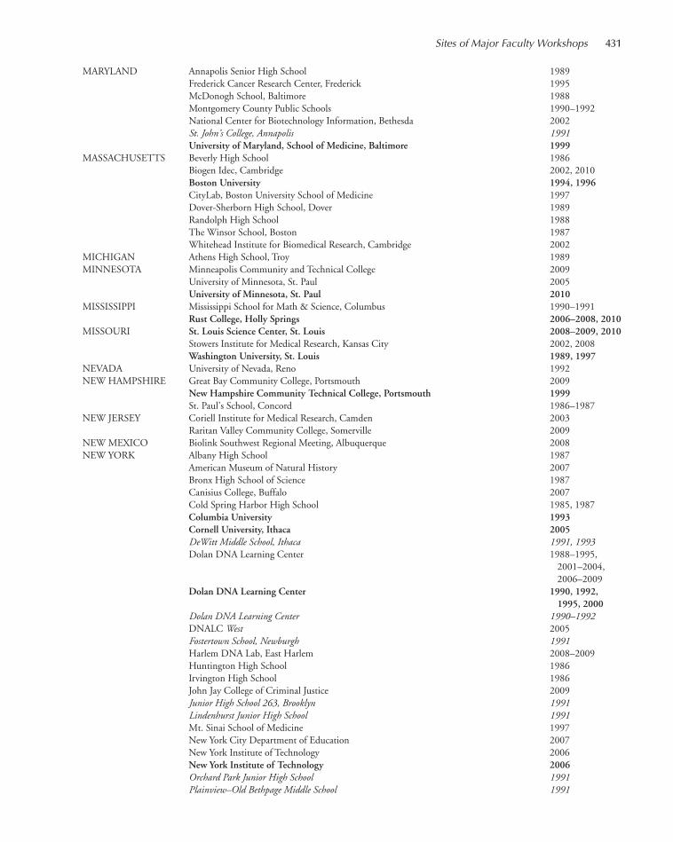
MARYLAND Annapolis Senior High School 1989Frederick Cancer Research Center, Frederick 1995McDonogh School, Baltimore 1988Montgomery County Public Schools 1990–1992National Center for Biotechnology Information, Bethesda 2002St. John’s College, Annapolis 1991University of Maryland, School of Medicine, Baltimore 1999
MASSACHUSETTS Beverly High School 1986Biogen Idec, Cambridge 2002, 2010Boston University 1994, 1996CityLab, Boston University School of Medicine 1997Dover-Sherborn High School, Dover 1989Randolph High School 1988The Winsor School, Boston 1987Whitehead Institute for Biomedical Research, Cambridge 2002
MICHIGAN Athens High School, Troy 1989MINNESOTA Minneapolis Community and Technical College 2009
University of Minnesota, St. Paul 2005University of Minnesota, St. Paul 2010
MISSISSIPPI Mississippi School for Math & Science, Columbus 1990–1991Rust College, Holly Springs 2006–2008, 2010
MISSOURI St. Louis Science Center, St. Louis 2008–2009, 2010Stowers Institute for Medical Research, Kansas City 2002, 2008Washington University, St. Louis 1989, 1997
NEVADA University of Nevada, Reno 1992NEW HAMPSHIRE Great Bay Community College, Portsmouth 2009
New Hampshire Community Technical College, Portsmouth 1999St. Paul’s School, Concord 1986–1987
NEW JERSEY Coriell Institute for Medical Research, Camden 2003Raritan Valley Community College, Somerville 2009
NEW MEXICO Biolink Southwest Regional Meeting, Albuquerque 2008NEW YORK Albany High School 1987
American Museum of Natural History 2007Bronx High School of Science 1987Canisius College, Buffalo 2007Cold Spring Harbor High School 1985, 1987Columbia University 1993Cornell University, Ithaca 2005DeWitt Middle School, Ithaca 1991, 1993Dolan DNA Learning Center 1988–1995,
2001–2004, 2006–2009
Dolan DNA Learning Center 1990, 1992,1995, 2000
Dolan DNA Learning Center 1990–1992DNALC West 2005Fostertown School, Newburgh 1991Harlem DNA Lab, East Harlem 2008–2009Huntington High School 1986Irvington High School 1986John Jay College of Criminal Justice 2009Junior High School 263, Brooklyn 1991Lindenhurst Junior High School 1991Mt. Sinai School of Medicine 1997New York City Department of Education 2007New York Institute of Technology 2006New York Institute of Technology 2006Orchard Park Junior High School 1991Plainview–Old Bethpage Middle School 1991
Sites of Major Faculty Workshops 431
411-434_LAR_DNALC_Annual Report_2009 template 7/8/11 4:04 PM Page 431

State University of New York, Purchase 1989State University of New York, Stony Brook 1987–1990Stuyvesant High School 1998–1999The Rockefeller University 2003The Rockefeller University 2010Titusville Middle School, Poughkeepsie 1991, 1993Trudeau Institute, Lake Saranac 2001Union College, Schenectady 2004United States Military Academy, West Point 1996Wheatley School, Old Westbury 1985
NORTH CAROLINA CIIT Center for Health Research, Triangle Park 2003North Carolina Agricultural & Technical State University, Greensboro 2006–2007,
2009–2010North Carolina School of Science, Durham 1987
OHIO Case Western Reserve University, Cleveland 1990Cleveland Clinic 1987Langston University, Langston 2008North Westerville High School 1990
OKLAHOMA Tulsa Community College, Tulsa 2009Oklahoma City Community College 2000Oklahoma City Community College 2006–2007, 2010Oklahoma Medical Research Foundation, Oklahoma City 2001Oklahoma School of Science and Math, Oklahoma City 1994
OREGON Kaiser Permanente-Center for Health Research, Portland 2003PENNSYLVANIA Duquesne University, Pittsburgh 1988
Germantown Academy 1988Kimmel Cancer Center, Philadelphia 2008
RHODE ISLAND Botanical Society of America, Providence 2010SOUTH CAROLINA Clemson University, Clemson 2004
Medical University of South Carolina, Charleston 1988University of South Carolina, Columbia 1988
TENNESSEE NABT Professional Development Conference, Memphis 2008TEXAS Austin Community College–Rio Grande Campus 2000
Austin Community College–Eastview Campus 2007–2009Houston Community College Northwest, Houston 2009, 2010J.J. Pearce High School, Richardson 1990Langham Creek High School, Houston 1991Midland College, Midland 2008Southwest Foundation for Biomedical Research, San Antonio 2002Taft High School, San Antonio 1991Texas A&M, AG Research and Extension Center, Weslaco 2007Trinity University, San Antonio 1994University of Texas, Austin 1999, 2004, 2010University of Texas, Brownsville 2010
UTAH University of Utah, Salt Lake City 1993University of Utah, Salt Lake City 1998, 2000Utah Valley State College, Orem 2007
VERMONT University of Vermont, Burlington 1989VIRGINIA Eastern Mennonite University, Harrisonburg 1996
Jefferson School of Science, Alexandria 1987Mathematics and Science Center, Richmond 1990Mills Godwin Specialty Center, Richmond 1998Virginia Polytechnic Institute and State University, Blacksburg 2005, 2008–2009
WASHINGTON Fred Hutchinson Cancer Research Center, Seattle 1999, 2001, 2008University of Washington, Seattle 1993, 1998, 2010
WASHINGTON, D.C Howard University 1992, 1996, 2009–2010
WEST VIRGINIA Bethany College 1989WISCONSIN Blood Center of Southeastern Wisconsin, Milwaukee 2003
432 DNA Learning Center
411-434_LAR_DNALC_Annual Report_2009 template 7/8/11 4:04 PM Page 432
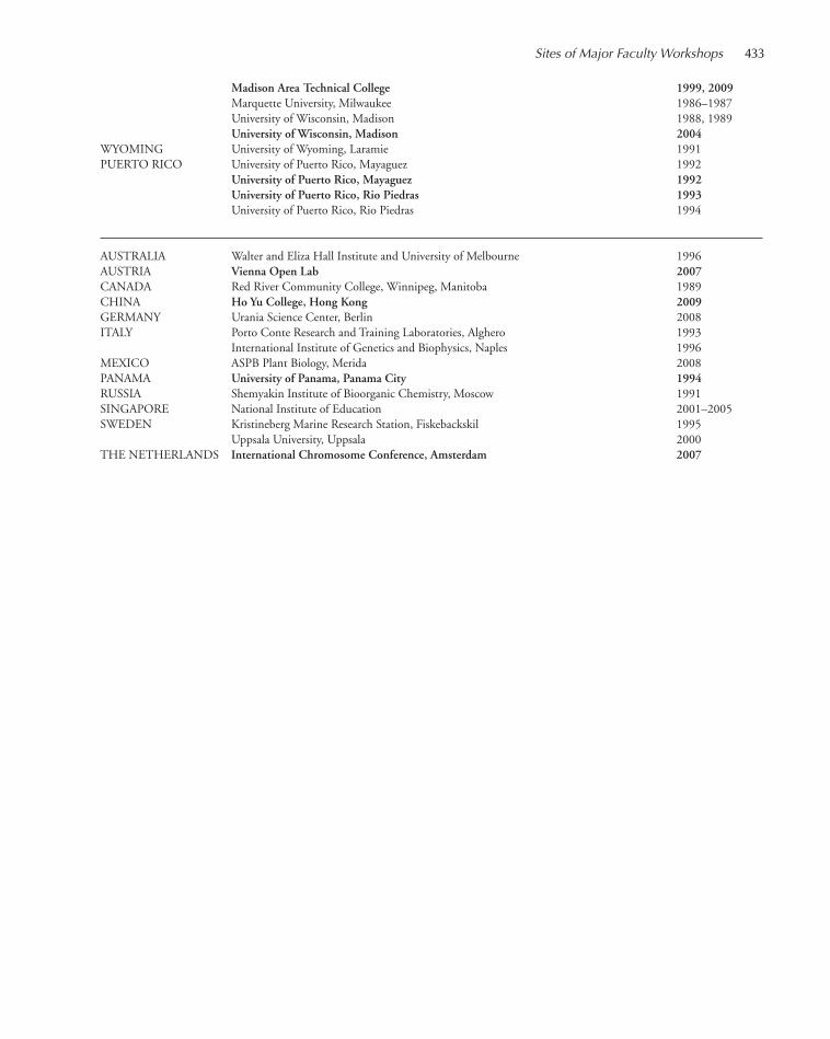
Sites of Major Faculty Workshops 433
Madison Area Technical College 1999, 2009Marquette University, Milwaukee 1986–1987University of Wisconsin, Madison 1988, 1989University of Wisconsin, Madison 2004
WYOMING University of Wyoming, Laramie 1991PUERTO RICO University of Puerto Rico, Mayaguez 1992
University of Puerto Rico, Mayaguez 1992University of Puerto Rico, Rio Piedras 1993University of Puerto Rico, Rio Piedras 1994
AUSTRALIA Walter and Eliza Hall Institute and University of Melbourne 1996AUSTRIA Vienna Open Lab 2007CANADA Red River Community College, Winnipeg, Manitoba 1989CHINA Ho Yu College, Hong Kong 2009GERMANY Urania Science Center, Berlin 2008ITALY Porto Conte Research and Training Laboratories, Alghero 1993
International Institute of Genetics and Biophysics, Naples 1996MEXICO ASPB Plant Biology, Merida 2008PANAMA University of Panama, Panama City 1994RUSSIA Shemyakin Institute of Bioorganic Chemistry, Moscow 1991SINGAPORE National Institute of Education 2001–2005SWEDEN Kristineberg Marine Research Station, Fiskebackskil 1995
Uppsala University, Uppsala 2000THE NETHERLANDS International Chromosome Conference, Amsterdam 2007
411-434_LAR_DNALC_Annual Report_2009 template 7/8/11 4:04 PM Page 433

411-434_LAR_DNALC_Annual Report_2009 template 7/8/11 4:04 PM Page 434

COLD SPRING HARBORLABORATORY PRESS
435-440_LAR_Press_Annual Report_2009 template 7/8/11 12:28 PM Page 435
Serials
Genes & Development, Vol. 24 (www.genesdev.org)Genome Research, Vol. 20 (www.genome.org)Learning & Memory, Vol. 17 (www.learnmem.org)RNA, Vol. 16 (www.rnajournal.org)Cold Spring Harbor Symposia on Quantitative Biology, Vol. 74:
Evolution: The Molecular Landscape, edited by Bruce Stillman, David Stewart, and Jan Witkowski
Cold Spring Harbor Protocols (www.cshprotocols.org)Cold Spring Harbor Perspectives in Biology
(www.cshperspectives.org)
Laboratory Manuals
Emerging Model Organisms: A Laboratory Manual, Volume 2Drosophila Neurobiology: A Laboratory Manual, edited by
Bing Zhang, Marc R. Freeman, and Scott WaddellImaging: A Laboratory Manual, edited by Rafael YusteImaging in Developmental Biology: A Laboratory Manual,
edited by James Sharpe and Rachel O. Wong; Series Editor Rafael Yuste
RNA: A Laboratory Manual, by Donald C. Rio, Manuel Ares Jr.,Gregory J. Hannon, and Timothy W. Nilsen
Handbooks
At the Helm: Leading Your Laboratory, Second Edition, by KathyBarker
Career Opportunities in Clinical Drug Research, by Rebecca J. Anderson
Advanced Textbooks
Guide to the Human Genome, by Stewart Scherer (online and/orprint)
Means to an End: Apoptosis and Other Cell Death Mechanisms, byDouglas R. Green
RNA Worlds: From Life’s Origins to Diversity in Gene Regulation, by John F. Atkins, Raymond F. Gesteland, and Thomas R.Cech
Monographs (Perspectives in BiologyTopic Collections)
Generation and Interpretation of Morphogen Gradients, edited byJames Briscoe, Peter A. Lawrence, and Jean-Paul Vincent
Cell–Cell Junctions, edited by W. James Nelson and Elaine FuchsSymmetry Breaking in Biology, edited by Rong Li and Bruce
BowermanNF-κB: A Network Hub Controlling Immunity, Inflammation,
and Cancer, edited by Michael Karin and Louis M. StaudtThe Origins of Life, edited by David Deamer and Jack W. SzostakThe p53 Family, edited by Arnold J. Levine and David P. LaneThe Nucleus, edited by Tom Misteli and David L. SpectorAuxin Signaling: From Synthesis to Systems Biology, edited by
Mark Estelle, Dolf Weijers, Karin Ljung, and Ottoline Leyser
Neuronal Guidance: The Biology of Brain Wiring, edited by MarcTessier-Lavigne and Alex L. Kolodkin
Immunoreceptor Signaling, edited by Lawrence E. Samelson andAndrey Shaw
Cell Biology of Bacteria, edited by Lucy Shapiro and Richard M.Losick
Science and Society
Speaking of Genetics: A Collection of Interviews, by Jane GitschierSydney Brenner: A Biography, by Errol C. FriedbergThe Honest Look, by Jennifer L. Rohn
Other
Neurobiology of the Leech, edited by Kenneth J. Muller, John G.Nicholls, and Gunther S. Stent (reissue of 1981 title)
CSHL Annual Report 2009, Yearbook EditionCSHL Annual Report 2009Banbury Center Annual Report 2009Watson School of Biological Sciences Annual Report 2009
Websites
http://cshmonographs.org/index.php/monographs; Cold SpringHarbor Monographs Archive Online
http://www.cshl.symposium.org; Symposia, Vol. 74 added toonline Symposia website
http://www.humangenomeguide.org
2010 PRESS PUBLICATIONS
435-440_LAR_Press_Annual Report_2009 template 7/8/11 12:28 PM Page 436

437
Cold Spring Harbor Laboratory’s research activities are complemented by the creation of journals,books, and electronic media that extend the professional education of scientists around the world.With a tradition of excellence originating in the 1930s, Cold Spring Harbor Laboratory Press iscontinuing to redefine its role as a publisher in the digital age. Although distribution channels forscientific information continue to evolve and diversify, the mission of the Press remains to identifyinnovative research, technologies, and scientists, to amplify their effectiveness by selecting, aggre-gating, and curating information that research communities can use, and to deliver the informationwhen, where, and how the communities want it. The task is complex and in 2010, there were sig-nificant achievements. More than 10 million research articles were downloaded internationally fromour journals, over 3000 electronic subscriptions were delivered to academic institutions worldwide,and 35,000 copies of print books were shipped from our warehouses in the United States and Eu-rope. The publication program encompasses six research journals, two of them among the mosthighly ranked in biology; authoritative laboratory manuals; and best-selling primers on professionalskill development for scientists. Their distribution makes a significant contribution to the Labora-tory’s broad educational mission and strengthens Cold Spring Harbor’s association with excellence.
Financially, the established journals performed strongly in 2010. The start-up costs of a new journalwere written off. Sales of print books were weakened by lower backlist orders and delayed publicationof some new titles. To improve the book program’s future financial performance, substantial expensesavings were made through procedural changes and by staff reduction at the year’s end.
Journals
Journal publishing remains the largest contributor of revenue and is vital to the financial sustain-ability of the publishing program. Journal subscriptions did well in what continued to be a chal-lenging market, as academic libraries continued to come to terms with reduced budgets.Nevertheless, total subscription revenue for all five established journal titles increased by more than5%. Despite continued merger activity within the laboratory supplies sector that reduces the po-tential client base, advertising revenue across the journals met its goals, with a continued but stillgradual shift in the relative proportion of income from print and online sales.
Genes & Development completed a satisfactory year with, for the first time, a single center of ed-itorial operations that provided authors with faster and more consistent decisions. Both Genes &Development and Genome Research continued to rank among the most important publications ingenetics and cell and developmental biology, attracting more submissions, maintaining impressiveimpact factors, and, through article downloads, finding intense use in academic centers worldwide.RNA maintained its status as the premier specialist journal in the burgeoning field of RNA biologyand a new 5-year publishing contract was signed with its owner, The RNA Society. Learning &Memory remains a source of specialized work in the molecular analysis of nervous system plasticity.
The subscription base of Cold Spring Harbor Protocols made solid gains and its usage continuedto grow. The new journal Cold Spring Harbor Perspectives in Biology, with content of exceptionalquality, found remarkable usage during an 18-month free trial period, a result that augurs well forsubscription growth. The journal’s unique feature is the publication of collections of articles thathave each been commissioned to provide an in-depth, coherent survey of a single topic. A largenumber of subject collections from cell and molecular biology are in the publication pipeline, andeach collection has commissioning editors with outstanding international reputations. Each article
COLD SPRING HARBOR LABORATORY PRESSEXECUTIVE DIRECTOR’S REPORT
435-440_LAR_Press_Annual Report_2009 template 7/8/11 12:28 PM Page 437
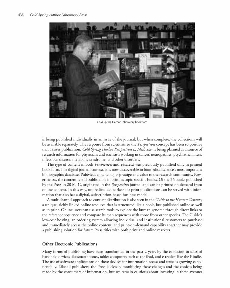
438 Cold Spring Harbor Laboratory Press
is being published individually in an issue of the journal, but when complete, the collections willbe available separately. The response from scientists to the Perspectives concept has been so positivethat a sister publication, Cold Spring Harbor Perspectives in Medicine, is being planned as a source ofresearch information for physicians and scientists working in cancer, neuropathies, psychiatric illness,infectious disease, metabolic syndrome, and other disorders.
The type of content in both Perspectives and Protocols was previously published only in printedbook form. In a digital journal context, it is now discoverable in biomedical science’s most importantbibliographic database, PubMed, enhancing its prestige and value to the research community. Nev-ertheless, the content is still publishable in print as topic-specific books. Of the 26 books publishedby the Press in 2010, 12 originated in the Perspectives journal and can be printed on demand fromonline content. In this way, unpredictable markets for print publications can be served with infor-mation that also has a digital, subscription-based business model.
A multichannel approach to content distribution is also seen in the Guide to the Human Genome,a unique, richly linked online resource that is structured like a book, but published online as wellas in print. Online users can use search tools to explore the human genome through direct links tothe reference sequence and compare human sequences with those from other species. The Guide’slow-cost hosting, an ordering system allowing individual and institutional customers to purchaseand immediately access the online content, and print-on-demand capability together may providea publishing solution for future Press titles with both print and online markets.
Other Electronic Publications
Many forms of publishing have been transformed in the past 2 years by the explosion in sales ofhandheld devices like smartphones, tablet computers such as the iPad, and e-readers like the Kindle.The use of software applications on these devices for information access and reuse is growing expo-nentially. Like all publishers, the Press is closely monitoring these changes and the choices beingmade by the consumers of information, but we remain cautious about investing in these avenues
Cold Spring Harbor Laboratory bookstore
435-440_LAR_Press_Annual Report_2009 template 7/8/11 12:28 PM Page 438

Executive Director’s Report 439
for the distribution of our content. A limited number of our book titles are available for the Kindlereader on a case-by-case basis, and sales, predictably, were low while the device remained expensive.But with the availability of new models at lower prices, and now that Kindle software can run onmany different devices, there may be a larger audience in the future. There is no doubt that ColdSpring Harbor’s book content must find its way into digital media and our biographies, histories,and novels will find part of their audience on digital reading devices. But these are a small part ofour output, and much thought, discussion, and exploration are being devoted to the more importantconsideration of what working professionals need and the choices of digital media they will make.The experience of producing and marketing electronic books has been valuable and so has the cre-ation of an iPhone application for our journal Genome Research that allows abstracts of papers to beread everywhere and full text to be accessed when the phone is connected to a subscribing institu-tion’s network. Our goal remains to make our content available in as many ways as readers require,and we will continue to explore the options available.
Books in Print
The year’s crop of new books included five manuals of techniques, a traditionally strong componentof our publishing program. Molecular biology has been transformed by the discovery of new speciesof RNA molecule that regulate gene function in vivo and in vitro and RNA: A Laboratory Manualis a uniquely authoritative source of expertise in working with these molecules. Imaging technologiesare also transforming the way experimental biology is done, and two new manuals provided instruc-tion in both general and more specific approaches to the use of these methods.
Our widely praised list of professional development handbooks was enhanced by a new bookabout career opportunities in industry and the second edition of At the Helm, Kathy Barker’s enor-mously successful source of advice and counsel for principal investigators.
As knowledge in biology becomes denser and more detailed, it is harder to write short, inform-ative, thought-provoking books on specific topics. Yet Douglas Green has achieved this distinctionwith Means to an End, a personal account of how cells die that has been widely praised. Inspirationfor young scientists of a different but still potent kind is found in Errol Friedberg’s account of thenotable life and work of Sydney Brenner and in a collection of insightful interviews conducted byJane Gitschier with a variety of people in and around science (Speaking of Genetics). And in TheHonest Look, a second novel by the cell biologist Jennifer Rohn, readers can find a human dramabased in a realistic portrayal of the scientific enterprise and the men and women engaged in it. Acomplete list of the year’s new titles can be found at the beginning of this report.
The year’s best-sellers included Molecular Cloning, At the Bench, Symposium 74: Evolution, Sta-tistics at the Bench, Epigenetics, Live Cell Imaging, At the Helm; and RNA: A Laboratory Manual. Newtranslation contracts included Japanese translations of Statistics at the Bench and At the Helm; andChinese translations of Live Cell Imaging and Transcriptional Regulation in Eukaryotes.
The generation of book sales worldwide continues to rely heavily on a monthly electronic newslet-ter. Our websites continue to be cornerstones of our presence in the information landscape and ourlong relationship with Amazon remains essential to the sale of our books in the United States. In-ternationally, we benefit greatly from the local expertise of commission-based sales agents in Ger-many and several associated European countries, China, Japan, India, and South America.Warehousing, order processing, and book distribution outside the United States continue to be han-dled by a company in the United Kingdom.
Staff
During the year, we said farewell to Ingrid Benirschke, Ann Boyle, Mary Cozza, Joan Ebert, MariaFairchild, Lauren Heller, Kaaren Kockenmeister, Kathryn LaForgia, Christina Lo, Katherine MacDon-
435-440_LAR_Press_Annual Report_2009 template 7/8/11 12:28 PM Page 439

ald, Nora Ruth, and Patricia Serpe. We welcomed Antonella Caiazza, Marie Cotter, Joanne McFadden,and Hashi Wijayatilake.
A complete list of staff members of the Press at the end of December 2010 is printed elsewherein this volume. The Press and the Laboratory are fortunate to have the service of such capable anddedicated individuals. Special thanks are due to those who during the year had the responsibility ofleadership in the diverse departments and projects of the Press: Jan Argentine, David Crotty, AlexGann, Terri Grodzicker, Christina Lo, Geraldine Jaitin, Bill Keen, Wayne Manos, Richard Sever,Marcie Siconolfi, Hillary Sussman, Linda Sussman, and Denise Weiss.
John R. InglisExecutive Directorand Publisher
440 Cold Spring Harbor Laboratory Press
435-440_LAR_Press_Annual Report_2009 template 7/8/11 12:28 PM Page 440

FINANCE
441-472_LAR_Finance_Annual Report_2009 template 7/8/11 12:29 PM Page 441
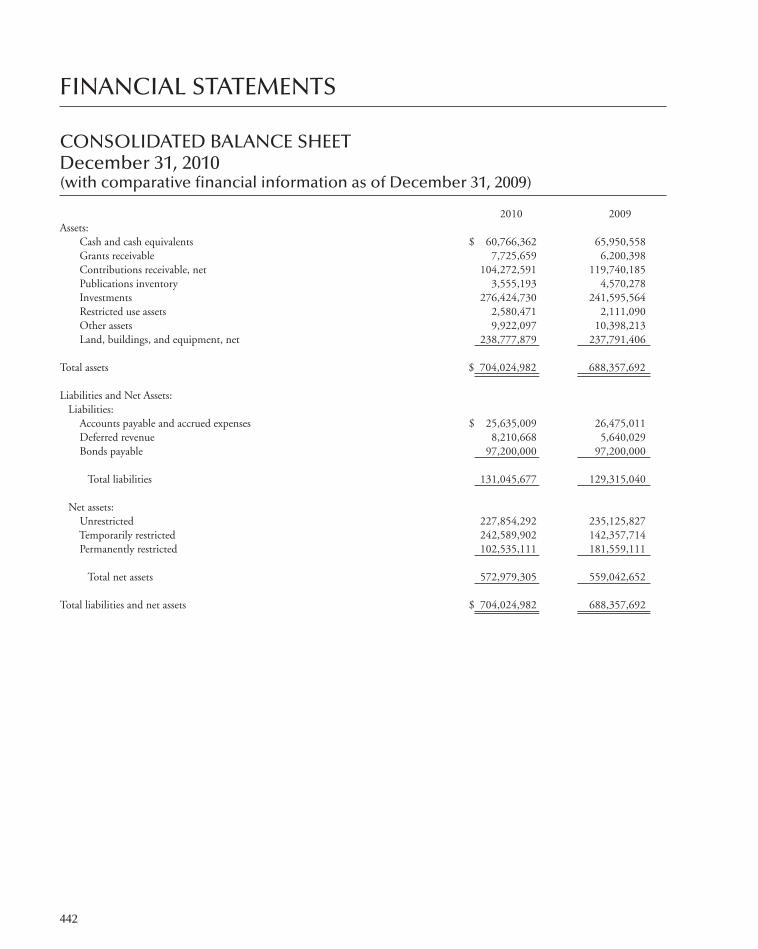
442
FINANCIAL STATEMENTS
CONSOLIDATED BALANCE SHEETDecember 31, 2010(with comparative financial information as of December 31, 2009)
2010 2009Assets:
Cash and cash equivalents $ 60,766,362 65,950,558 Grants receivable 7,725,659 6,200,398 Contributions receivable, net 104,272,591 119,740,185 Publications inventory 3,555,193 4,570,278 Investments 276,424,730 241,595,564 Restricted use assets 2,580,471 2,111,090 Other assets 9,922,097 10,398,213 Land, buildings, and equipment, net 238,777,879 237,791,406
Total assets $ 704,024,982 688,357,692
Liabilities and Net Assets:Liabilities:
Accounts payable and accrued expenses $ 25,635,009 26,475,011 Deferred revenue 8,210,668 5,640,029 Bonds payable 97,200,000 97,200,000
Total liabilities 131,045,677 129,315,040
Net assets:Unrestricted 227,854,292 235,125,827 Temporarily restricted 242,589,902 142,357,714 Permanently restricted 102,535,111 181,559,111
Total net assets 572,979,305 559,042,652
Total liabilities and net assets $ 704,024,982 688,357,692
441-472_LAR_Finance_Annual Report_2009 template 7/8/11 12:29 PM Page 442

443
CONSOLIDATED STATEMENT OF ACTIVITIESYear ended December 31, 2010(with summarized financial information for the year ended December 31, 2009)
Temporarily Permanently 2010 2009 Unrestricted Restricted Restricted Total Total
Revenue and other support:Public support–contributions and
non-federal grant awards $ 13,624,936 30,685,893 1,109,499 45,420,328 67,103,796 Federal grant awards 35,405,944 – – 35,405,944 29,648,968 Indirect cost allowances 25,973,354 – – 25,973,354 21,016,585 Investment return utilized 18,309,184 – – 18,309,184 18,216,748 Program fees 6,971,358 – – 6,971,358 5,484,810 Publications sales 9,251,441 – – 9,251,441 9,136,188 Dining services 4,319,847 – – 4,319,847 4,103,820 Rooms and apartments 3,556,328 – – 3,556,328 3,255,480 Miscellaneous 2,126,513 – – 2,126,513 1,962,873 Net assets released from restrictions 34,646,285 (34,646,285) – – –
Total revenue and other support 154,185,190 (3,960,392) 1,109,499 151,334,297 159,929,268
Expenses:Research 86,810,119 – – 86,810,119 74,005,657 Educational programs 17,121,875 – – 17,121,875 14,964,908 Publications 10,877,055 – – 10,877,055 9,032,225 Banbury Center conferences 1,370,778 – – 1,370,778 1,069,044 Dolan DNA Learning Center
programs 1,406,339 – – 1,406,339 1,467,947 Watson School of Biological Sciences
programs 3,535,758 – – 3,535,758 3,595,810 General and administrative 14,746,870 – – 14,746,870 13,903,883 Dining services 5,137,648 – – 5,137,648 4,712,901
Total expenses 141,006,442 – – 141,006,442 122,752,375
Excess (deficiency) of revenue and othersupport over (under) expenses 13,178,748 (3,960,392) 1,109,499 10,327,855 37,176,893
Other changes in net assets:Investment (loss) return excluding amount
utilized (428,497) 9,028,095 – 8,599,598 24,463,751 Change in fair value of interest-rate swap (4,990,800) – – (4,990,800) 24,524,899 Release of temporarily restricted capital
funds 4,527,558 (4,527,558) – – –Net asset reclassification based on change
in law/interpretation (19,558,544) 99,692,043 (80,133,499) – –
(Decrease) increase in net assets (7,271,535) 100,232,188 (79,024,000) 13,936,653 86,165,543
Net assets at beginning of year 235,125,827 142,357,714 181,559,111 559,042,652 472,877,109
Net assets at end of year $ 227,854,292 242,589,902 102,535,111 572,979,305 559,042,652
441-472_LAR_Finance_Annual Report_2009 template 7/8/11 12:29 PM Page 443

444
CONSOLIDATED STATEMENT OF CASH FLOWSYear ended December 31, 2010(with comparative financial information for the year ended December 31, 2009)
2010 2009Cash flows from operating activities:
Increase in net assets $ 13,936,653 86,165,543 Adjustments to reconcile increase in net assets
to net cash provided by operating activities:Change in fair value of interest rate swap 4,990,800 (24,524,897)Depreciation and amortization 15,443,523 9,282,371 Net appreciation in fair value of investments (23,724,939) (36,992,017)Contributions restricted for long-term investment (1,712,498) (9,121,400)Changes in assets and liabilities:
Grants receivable (1,525,261) 2,397,001 Contributions receivable, net of financing activities 11,309,672 (18,458,455)Publications inventory 1,015,085 (644,311)Other assets 929,237 (611,026)Restricted use assets (469,381) 1,456,592 Accounts payable and accrued expenses, net of financing activities (2,521,650) 32,680 Deferred revenue 2,570,639 1,678,029
Net cash provided by operating activities 20,241,880 10,660,110
Cash flows from investing activities:Capital expenditures (16,429,996) (48,389,574)Proceeds from sales and maturities of investments 36,404,485 140,454,706 Purchases of investments (47,508,712) (131,634,094)Net change in investment in employee residences (453,121) 440,775
Net cash used in investing activities (27,987,344) (39,128,187)
Cash flows from financing activities:Permanently restricted contributions 1,109,499 2,255,040 Contributions restricted for investment in land, buildings,
and equipment 602,999 6,866,360 Decrease in contributions receivable 4,157,922 3,399,927 Decrease in deposits with bond trustee – 11,886,744 (Decrease) increase in accounts payable relating to capital
expenditures (3,309,152) 403,725 Decrease in deposits with swap counterparty – 17,380,247
Net cash provided by financing activities 2,561,268 42,192,043
Net (decrease) increase in cash and cash equivalents (5,184,196) 13,723,966 Cash and cash equivalents at beginning of year 65,950,558 52,226,592
Cash and cash equivalents at end of year $ 60,766,362 65,950,558
Supplemental disclosure:Interest paid $ 3,652,690 3,180,748
Noncash investing and financing activities:Contributed property $ 57,475 98,085
441-472_LAR_Finance_Annual Report_2009 template 7/8/11 12:29 PM Page 444
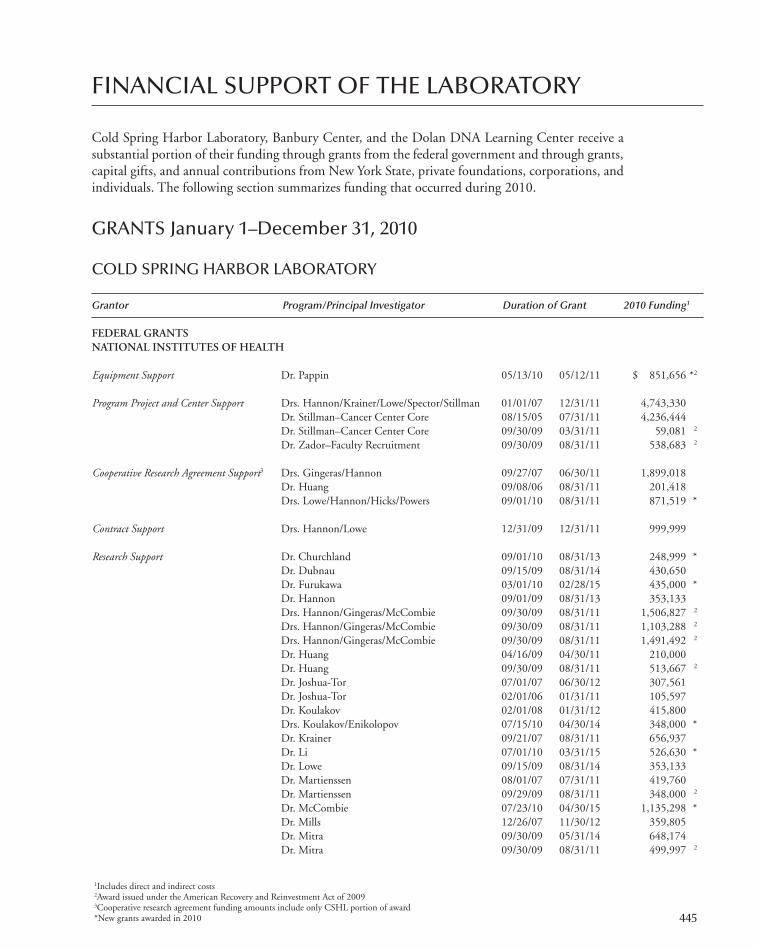
FINANCIAL SUPPORT OF THE LABORATORY
Cold Spring Harbor Laboratory, Banbury Center, and the Dolan DNA Learning Center receive asubstantial portion of their funding through grants from the federal government and through grants,capital gifts, and annual contributions from New York State, private foundations, corporations, andindividuals. The following section summarizes funding that occurred during 2010.
GRANTS January 1–December 31, 2010
COLD SPRING HARBOR LABORATORY
Grantor Program/Principal Investigator Duration of Grant 2010 Funding1
FEDERAL GRANTSNATIONAL INSTITUTES OF HEALTH
Equipment Support Dr. Pappin 05/13/10 05/12/11 $ 851,656 *2
Program Project and Center Support Drs. Hannon/Krainer/Lowe/Spector/Stillman 01/01/07 12/31/11 4,743,330 Dr. Stillman–Cancer Center Core 08/15/05 07/31/11 4,236,444 Dr. Stillman–Cancer Center Core 09/30/09 03/31/11 59,081 2
Dr. Zador–Faculty Recruitment 09/30/09 08/31/11 538,683 2
Cooperative Research Agreement Support3 Drs. Gingeras/Hannon 09/27/07 06/30/11 1,899,018 Dr. Huang 09/08/06 08/31/11 201,418 Drs. Lowe/Hannon/Hicks/Powers 09/01/10 08/31/11 871,519 *
Contract Support Drs. Hannon/Lowe 12/31/09 12/31/11 999,999
Research Support Dr. Churchland 09/01/10 08/31/13 248,999 *Dr. Dubnau 09/15/09 08/31/14 430,650 Dr. Furukawa 03/01/10 02/28/15 435,000 *Dr. Hannon 09/01/09 08/31/13 353,133 Drs. Hannon/Gingeras/McCombie 09/30/09 08/31/11 1,506,827 2
Drs. Hannon/Gingeras/McCombie 09/30/09 08/31/11 1,103,288 2
Drs. Hannon/Gingeras/McCombie 09/30/09 08/31/11 1,491,492 2
Dr. Huang 04/16/09 04/30/11 210,000 Dr. Huang 09/30/09 08/31/11 513,667 2
Dr. Joshua-Tor 07/01/07 06/30/12 307,561 Dr. Joshua-Tor 02/01/06 01/31/11 105,597 Dr. Koulakov 02/01/08 01/31/12 415,800 Drs. Koulakov/Enikolopov 07/15/10 04/30/14 348,000 *Dr. Krainer 09/21/07 08/31/11 656,937 Dr. Li 07/01/10 03/31/15 526,630 *Dr. Lowe 09/15/09 08/31/14 353,133 Dr. Martienssen 08/01/07 07/31/11 419,760 Dr. Martienssen 09/29/09 08/31/11 348,000 2
Dr. McCombie 07/23/10 04/30/15 1,135,298 *Dr. Mills 12/26/07 11/30/12 359,805 Dr. Mitra 09/30/09 05/31/14 648,174 Dr. Mitra 09/30/09 08/31/11 499,997 2
1Includes direct and indirect costs2Award issued under the American Recovery and Reinvestment Act of 20093Cooperative research agreement funding amounts include only CSHL portion of award*New grants awarded in 2010 445
441-472_LAR_Finance_Annual Report_2009 template 7/8/11 12:29 PM Page 445

Grantor Program/Principal Investigator Duration of Grant 2010 Funding1
Dr. Mitra 08/01/10 05/31/13 $ 399,317 *Dr. Muthuswamy 02/01/09 12/31/13 389,393 Drs. Powers/Lowe/Krasnitz/Sordella 09/29/09 08/31/11 2,374,043 2
Dr. Powers 12/02/06 11/30/11 456,045 Dr. D. Spector 04/01/90 03/31/11 676,357 Dr. Stenlund 02/01/08 01/31/12 430,650 Dr. Stillman 06/01/08 05/31/12 644,890 Dr. Tonks 07/01/10 04/30/15 712,640 *Dr. Tonks 01/11/10 12/31/13 408,900 *Dr. Trotman 04/01/10 01/31/14 288,840 *Dr. Turner 07/15/10 06/30/15 361,000 *Dr. Van Aelst 07/01/08 03/31/13 391,500 Dr. Van Aelst 01/01/09 12/31/13 361,050 Drs. Wigler/McCombie 09/30/09 08/31/11 1,395,339 2
Dr. Zador 09/18/08 07/31/13 366,053 Dr. Zador 09/24/09 08/31/11 93,017 2
Dr. Zador 09/27/10 05/31/15 435,000 *Dr. Zhong 06/01/10 05/31/13 348,000 *
Research Subcontracts
NIH/Allen Institute for Brain Science Dr. Mitra 09/15/09 08/31/11 229,681 Consortium Agreement
NIH/Angion Biomedica Corp. Dr. Enikolopov 09/30/09 08/31/11 278,368 Consortium Agreement
NIH/Brookhaven National Laboratory Dr. Stillman 09/01/06 08/31/11 38,421 Consortium Agreement
NIH/Carnegie Mellon University Dr. Osten 09/15/10 07/31/11 134,000 *Consortium Agreement
NIH/Columbia University Consortium Dr. Lowe 08/18/06 07/31/11 402,398 Agreement
NIH/Cornell University Consortium Dr. Mitra 04/03/08 01/31/11 55,118 Agreement
NIH/Georgia Institute of Technology Dr. D. Spector 09/30/06 07/31/11 297,852 Consortium Agreement
NIH/Northwestern University Dr. Osten 09/15/09 08/31/11 33,600 Consortium Agreement
NIH/Paratek Pharmaceuticals, Inc. Dr. Krainer 01/05/09 12/31/10 168,931 Consortium Agreement
NIH/Princeton University Consortium Dr. Zador 05/01/10 04/30/11 43,500 *Agreement
NIH/University of California, San Dr. Egeblad 09/01/09 08/31/14 178,984 Francisco Consortium Agreement
NIH/University of California, Berkeley Dr. Gingeras 05/04/07 03/31/11 655,799 National Laboratory Consortium Agreement
NIH/University of Southern California Dr. Hannon 07/01/10 06/30/14 121,800 *Consortium Agreement
NIH/University of Southern California Drs. Lowe/Sordella 09/28/09 07/31/14 370,000 Consortium Agreement
NIH/University of Texas, Dallas Drs. Krainer/McCombie/M. Zhang 08/01/10 07/31/11 291,888 *Consortium Agreement
Fellowship Support Dr. Borges 08/16/09 08/15/12 52,154 M. Lazarus 07/15/09 07/14/10 25,380
446 Finance
1Includes direct and indirect costs2Award issued under the American Recovery and Reinvestment Act of 2009
*New grants awarded in 2010
441-472_LAR_Finance_Annual Report_2009 template 7/8/11 12:29 PM Page 446

Grantor Program/Principal Investigator Duration of Grant 2010 Funding1
Graduate Training Support Dr. Joshua-Tor, Watson School of Biological 07/01/07 06/30/12 $ 207,432 Sciences
Dr. Joshua-Tor, Watson School of Biological 09/01/09 08/31/11 41,266 2
Sciences
Course Support Immunocytochemistry, In Situ Hybridization, 09/01/10 08/31/15 78,167 *Super-Resolution, and Live Cell Imaging
Advanced Techniques in Molecular Neuroscience 07/01/01 06/30/12 74,678 Caenorhabditis elegans 08/01/98 08/31/11 81,739 Cell and Developmental Biology of Xenopus 04/01/09 03/31/14 60,918 Computational and Comparative Genomics 06/06/91 07/31/13 50,282 Eukaryotic Gene Expression 01/01/83 03/31/11 108,929 Imaging Structure and Function in the Nervous 07/01/01 06/30/12 87,131
System Integrated Data Analysis for High-Throughput 09/01/07 04/30/12 79,188
Biology Molecular Embryology of the Mouse 01/01/83 03/31/11 108,369 Neurobiology of Drosophila 07/01/01 06/30/12 68,010 Programming for Biology 07/07/00 08/31/14 61,800 Protein Purification and Characterization 01/01/83 03/31/11 86,303 Proteomics 07/01/03 06/30/11 80,080 X-Ray Methods in Structural Biology 07/01/07 06/30/12 77,056 Yeast Genetics and Genomics 09/18/10 06/30/13 51,905 *
Meeting Support Axon Guidance, Synaptic Plasticity, and 08/01/10 07/31/11 10,000 *Regeneration
The Biology of Genomes 04/01/08 03/31/13 38,193 The Cell Cycle 03/01/06 02/28/11 4,000 Gene Expression and Signaling in the Immune 04/01/08 03/31/13 10,000
System Molecular Genetics of Aging 03/01/08 02/28/13 30,030 Germ Cells 07/01/08 06/30/13 6,000 Glia in Health and Disease 07/01/10 06/30/11 24,989 *Mechanisms and Models of Cancer 08/01/08 07/31/13 6,000 Molecular Chaperones and Heat Shock Response 05/01/10 04/30/13 20,000 *Molecular Genetics of Bacteria and Phages 07/01/10 06/30/11 18,500 *Mouse Development, Genetics, and Genomics 08/01/10 07/31/11 8,500 *Pharmacogenomics and Personalized Therapy 09/30/10 08/31/15 15,000 *PTEN Pathways and Targets 03/01/06 02/28/11 7,000 75th Symposium: Nuclear Organization and 04/01/10 03/31/11 10,000 *
FunctionSystems Biology: Global Regulation of Gene 03/01/10 02/28/15 5,000 *
ExpressionTelomeres and Telomerase 09/30/10 08/31/11 30,625 *Vertebrate Organogenesis 04/01/10 03/31/15 12,000 *
HEALTH RESOURCES AND SERVICES ADMINISTRATION
Equipment Support Dr. D. Spector 08/01/10 07/31/12 495,000 *
NATIONAL SCIENCE FOUNDATION
Multiple Project Award Support Dr. Jackson 10/01/10 09/30/15 1,120,694 *
Financial Support of the Laboratory 447
1Includes direct and indirect costs2Award issued under the American Recovery and Reinvestment Act of 2009*New grants awarded in 2010
441-472_LAR_Finance_Annual Report_2009 template 7/8/11 12:29 PM Page 447

Grantor Program/Principal Investigator Duration of Grant 2010 Funding1
Drs. McCombie/Martienssen/Ware 08/01/10 07/31/12 $ 2,499,992 *Dr. Ware 10/01/07 09/30/11 1,757,922
Research Support Dr. Jackson 09/01/10 08/31/13 200,000 *Dr. Jackson 07/01/09 06/30/11 78,072 Dr. Lippman 09/15/09 08/31/12 166,841 Dr. Timmermans 08/15/10 07/31/13 200,000 *Dr. Ware 06/15/08 05/31/11 263,886
Research Subcontracts
NSF/Cornell University Consortium Drs. Timmermans/Micklos 09/01/08 08/31/12 408,333 Agreement
NSF/New York University Consortium Drs. McCombie/Martienssen 08/01/10 07/31/14 384,900 *Agreement
NSF/University of Arizona Consortium Dr. Ware 08/15/10 07/31/14 542,429 *Agreement
NSF/University of Arizona Consortium Drs. Stein/Martienssen/Micklos/Ware 02/01/08 01/31/13 2,101,833 Agreement
NSF/University of California, Berkeley Dr. Jackson 08/01/06 07/31/11 228,512 Consortium Agreement
NSF/University of California, Davis Dr. Ware 09/01/08 08/31/11 214,214 Consortium Agreement
NSF/Washington University Consortium Dr. Ware 03/01/10 02/28/11 312,400 *Agreement
Fellowship Support K. Liberatore 06/01/10 05/31/13 40,500 *
Undergraduate Training Support Dr. Lippman 03/01/09 02/29/12 69,420
Course Support Advanced Bacterial Genetics 07/01/09 06/30/14 90,953 Computational Cell Biology 04/15/08 03/31/11 96,338 Molecular Techniques in Plant Science 07/01/10 06/30/11 96,680 *
Meeting Support Molecular Genetics of Bacteria and Phages 08/01/10 07/31/11 7,000 *7th International Conference on Plasmodesmata 03/01/10 02/28/11 15,000 *Systems Biology: Global Regulation of Gene
Expression 06/01/10 05/31/11 7,000 *
UNITED STATES DEPARTMENT OF AGRICULTURE
Research Support Dr. Jackson 09/01/08 08/31/11 133,795 Dr. McCombie 09/11/09 09/10/14 113,436
UNITED STATES DEPARTMENT OF ENERGY
Research Support Dr. Martienssen 09/03/10 09/02/12 852,567 *
UNITED STATES DEPARTMENT OF THE ARMY
Research Support Dr. Hannon 09/01/08 08/31/13 658,118 Drs. Lucito/Tonks 07/01/10 06/30/12 434,000 *Dr. Trotman 08/15/09 08/14/12 162,162
448 Finance
1Includes direct and indirect costs*New grants awarded in 2010
441-472_LAR_Finance_Annual Report_2009 template 7/8/11 12:29 PM Page 448

Grantor Program/Principal Investigator Duration of Grant 2010 Funding1
Drs. Wigler/Hicks/Krasnitz 09/01/09 08/31/11 $ 2,151,000 Dr. Zhong 06/15/10 06/14/13 285,477 *
Fellowship Support Dr. B. Zhang 03/15/10 03/14/11 61,772 *
MISCELLANEOUS SOURCES OF FUNDING
Equipment Support
F.M. Kirby Foundation Dr. Egeblad 05/01/10 04/30/11 100,000 *Eleanor Schwartz Charitable Foundation Dr. McCombie 06/01/10 05/31/11 200,000 *
Program Project Support
The Leukemia & Lymphoma Society Dr. Lowe 10/01/08 09/30/13 1,250,000 Pioneer Hi-Bred International, Inc. Drs. Jackson/Lippman/Martienssen/ 07/01/07 06/30/12 1,600,000
Timmermans/WareThe Simons Foundation/Autism Dr. Wigler 07/01/08 06/30/11 6,664,189 The Simons Foundation/Center for Dr. Wigler 09/01/08 08/31/11 1,500,000
Quantitative BiologyTheodore R. and Vada S. Stanley Drs. Watson/McCombie 07/01/07 06/30/12 5,000,000
Research Support
Paul G. Allen Family Foundation Dr. Zador 10/01/10 09/30/13 611,100 *Alpern Family Foundation Inc. Dr. Egeblad 12/15/10 12/14/11 2,500 *Alzheimer’s Association Dr. Furukawa 03/01/10 02/28/12 49,969 *American Cancer Society Dr. Wigler 01/01/10 12/31/10 10,000 *
Dr. Wigler 01/01/06 12/31/10 60,000 Anonymous Dr. Osten 09/01/09 08/31/11 248,492 The A&P Foundation Dr. Egeblad 06/01/10 05/31/11 20,000 *The Breast Cancer Research Foundation Dr. Wigler 10/01/10 09/30/11 223,000 *Brookhaven National Laboratory/New Dr. Enikolopov 03/01/10 04/30/10 16,202 *
York State Department of Health Carnegie Mellon University/Anonymous Dr. Osten 01/01/10 11/30/11 25,000 *Mr. and Mrs. Edward Chernoff in honor Dr. Egeblad 07/01/10 06/30/11 1,000 *
of Fred and Marika FogelmanMr. and Mrs. Edward Chernoff in Dr. Furukawa 08/15/10 08/14/11 500 *
memory of James J. KehoeClear Channel Worldwide (WALK 97.5 Dr. Egeblad 04/01/10 03/31/11 4,000 *
FM)Damon Runyon Cancer Research Dr. Sordella 01/01/10 12/31/12 150,000 *
FoundationThe Dana Foundation Dr. Li 10/01/09 09/30/12 63,844
Dr. Li 01/01/09 12/31/11 200,000 Dart Neuroscience LLC Dr. Dubnau 05/01/10 04/30/11 250,000 *
Dr. Zhong 06/01/10 05/31/11 125,000 *The Kathryn W. Davis Foundation Dr. Hannon 01/15/07 01/14/11 1,005,660 Deer Park High School Dr. Egeblad 12/01/10 11/30/11 500 *In memory of Raymond Donovan Dr. Trotman 08/01/10 07/31/11 360 *The Ellison Medical Foundation Dr. Enikolopov 09/09/09 09/08/13 252,000 Entertainment Industry Foundation Dr. Hannon 11/01/10 10/31/11 250,000 *Charitable Lead Annuity Trust under Dr. Osten 12/29/10 12/28/11 25,000 *
the Will of Louis FeilBrian Fox through United Way of Dr. Lowe 03/01/10 02/28/11 2,000 *
New York City
Financial Support of the Laboratory 449
1Includes direct and indirect costs*New grants awarded in 2010
441-472_LAR_Finance_Annual Report_2009 template 7/8/11 12:29 PM Page 449

Grantor Program/Principal Investigator Duration of Grant 2010 Funding1
The Joni Gladowsky Breast Cancer Dr. Egeblad 12/01/10 11/30/11 $ 30,000 *Foundation
GlaxoSmithKline Dr. Powers 09/20/06 03/20/11 259,599 Richard and Mindy Gordon Dr. Egeblad 01/01/10 12/31/10 3,000 *
Dr. Lazebnik 01/01/10 12/31/10 2,000 *Greenwich Breast Cancer Alliance Inc. Dr. Egeblad 01/01/10 12/31/11 62,500 *The Irving A. Hansen Memorial Dr. Tonks 07/01/10 06/30/11 20,000 *
FoundationJo-Ellen and Ira Hazan Dr. Enikolopov 12/01/10 11/30/11 20,000 *
Dr. Osten 12/01/10 11/30/11 180,000 *Hearts for Cancer Dr. Egeblad 04/01/10 03/31/11 8,455 *The International Human Frontier Dr. Lippman 07/01/09 06/30/12 100,000
ScienceIsis Pharmaceuticals, Inc. Dr. Krainer 11/01/08 10/31/11 125,000 Islip Breast Cancer Coalition Inc. Dr. Egeblad 05/01/10 04/30/11 5,000 *Knott Family Foundation Drs. Tonks/Pappin 09/01/10 08/31/11 15,000 *The Susan G. Komen Breast Cancer Dr. Egeblad 04/13/10 04/12/13 150,000 *
Foundation, Inc.Charles Henry Leach II Foundation Dr. Enikolopov 06/01/10 05/31/11 2,000 *Mara and Thomas Lehrman Charitable Dr. Erlich 08/01/10 07/31/11 40,000 *
FundThe George Link, Jr. Foundation, Inc. Dr. Sordella 12/01/10 11/30/11 50,000 *The Long Island 2-Day Walk to Fight Dr. Egeblad 08/15/10 08/14/11 31,000 *
Breast CancerThe Lustgarten Foundation Drs. Hannon/Lowe 01/01/10 12/31/12 1,048,204 *
Drs. Powers/Lowe/Van Aelst 02/01/10 01/31/12 424,723 *Carol Marcincuk Fund Dr. Tonks 01/01/10 12/31/10 3,872 *In honor of Carissa Maringo Dr. Egeblad 10/01/10 09/30/11 4,540 *The G. Harold and Leila Y. Mathers Dr. Mitra 09/01/10 08/31/13 204,135 *
Charitable FoundationBreast Cancer Awareness Day in memory Dr. Wigler 01/01/10 12/31/11 72,305 *
of Elizabeth McFarlandThe McKnight Endowment Fund for Dr. Osten 08/01/09 07/31/11 100,000
NeuroscienceThe John Merck Fund Dr. Kepecs 06/01/10 05/31/14 75,000 *The Don Monti Memorial Research Dr. Lowe 03/01/10 02/28/11 300,000 *
FoundationLouis Morin Charitable Trust Dr. Joshua-Tor 12/01/10 11/30/11 70,000 *The Neuberger Berman Foundation Dr. Egeblad 02/01/10 01/31/11 20,000 *New York State Department of Health– Drs. Enikolopov/Koulakov 01/01/09 12/31/11 360,000
New York Stem Cell Science Dr. Timmermans 01/01/09 12/31/10 119,900 (NYSTEM)
Paratek Pharmaceuticals, Inc. Dr. Krainer 01/01/10 12/31/10 37,800 *Philips Research North America Drs. Lucito/Wigler 04/01/09 03/31/11 636,518
Dr. Zhang 01/01/09 12/31/10 227,527 V. Kann Rasmussen Foundation Dr. Trotman 09/01/07 08/31/12 50,000 Christina Renna Foundation, Inc. Dr. Van Aelst 01/01/08 12/31/11 15,000 Diane Emdin Sachs Memorial Fund Dr. Sordella 09/01/10 08/31/11 13,315 *Judi Shesh Memorial Foundation Dr. Egeblad 08/01/10 07/31/11 10,000 *The Simons Foundation Dr. Henn 09/01/10 08/31/12 294,030 *
Dr. Henn 09/01/10 08/31/11 500,978 *Dr. Mills 01/01/10 10/31/10 289,633 *Dr. Mills 07/01/10 06/30/13 350,000 *Dr. Mitra 09/01/09 08/31/11 150,000 Dr. Osten 07/01/09 06/30/11 150,107
450 Finance
1Includes direct and indirect costs*New grants awarded in 2010
441-472_LAR_Finance_Annual Report_2009 template 7/8/11 12:29 PM Page 450
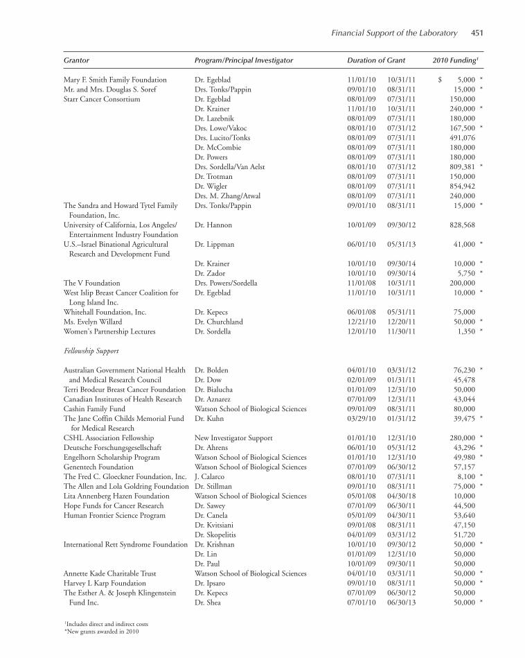
Grantor Program/Principal Investigator Duration of Grant 2010 Funding1
Mary F. Smith Family Foundation Dr. Egeblad 11/01/10 10/31/11 $ 5,000 *Mr. and Mrs. Douglas S. Soref Drs. Tonks/Pappin 09/01/10 08/31/11 15,000 *Starr Cancer Consortium Dr. Egeblad 08/01/09 07/31/11 150,000
Dr. Krainer 11/01/10 10/31/11 240,000 *Dr. Lazebnik 08/01/09 07/31/11 180,000 Drs. Lowe/Vakoc 08/01/10 07/31/12 167,500 *Drs. Lucito/Tonks 08/01/09 07/31/11 491,076 Dr. McCombie 08/01/09 07/31/11 180,000 Dr. Powers 08/01/09 07/31/11 180,000 Drs. Sordella/Van Aelst 08/01/10 07/31/12 809,381 *Dr. Trotman 08/01/09 07/31/11 150,000 Dr. Wigler 08/01/09 07/31/11 854,942 Drs. M. Zhang/Atwal 08/01/09 07/31/11 240,000
The Sandra and Howard Tytel Family Drs. Tonks/Pappin 09/01/10 08/31/11 15,000 *Foundation, Inc.
University of California, Los Angeles/ Dr. Hannon 10/01/09 09/30/12 828,568 Entertainment Industry Foundation
U.S.–Israel Binational Agricultural Dr. Lippman 06/01/10 05/31/13 41,000 *Research and Development Fund
Dr. Krainer 10/01/10 09/30/14 10,000 *Dr. Zador 10/01/10 09/30/14 5,750 *
The V Foundation Drs. Powers/Sordella 11/01/08 10/31/11 200,000 West Islip Breast Cancer Coalition for Dr. Egeblad 11/01/10 10/31/11 10,000 *
Long Island Inc.Whitehall Foundation, Inc. Dr. Kepecs 06/01/08 05/31/11 75,000 Ms. Evelyn Willard Dr. Churchland 12/21/10 12/20/11 50,000 *Women’s Partnership Lectures Dr. Sordella 12/01/10 11/30/11 1,350 *
Fellowship Support
Australian Government National Health Dr. Bolden 04/01/10 03/31/12 76,230 *and Medical Research Council Dr. Dow 02/01/09 01/31/11 45,478
Terri Brodeur Breast Cancer Foundation Dr. Bialucha 01/01/09 12/31/10 50,000 Canadian Institutes of Health Research Dr. Aznarez 07/01/09 12/31/11 43,044 Cashin Family Fund Watson School of Biological Sciences 09/01/09 08/31/11 80,000 The Jane Coffin Childs Memorial Fund Dr. Kuhn 03/29/10 01/31/12 39,475 *
for Medical ResearchCSHL Association Fellowship New Investigator Support 01/01/10 12/31/10 280,000 *Deutsche Forschungsgesellschaft Dr. Ahrens 06/01/10 05/31/12 43,296 *Engelhorn Scholarship Program Watson School of Biological Sciences 01/01/10 12/31/10 49,980 *Genentech Foundation Watson School of Biological Sciences 07/01/09 06/30/12 57,157 The Fred C. Gloeckner Foundation, Inc. J. Calarco 08/01/10 07/31/11 8,100 *The Allen and Lola Goldring Foundation Dr. Stillman 09/01/10 08/31/11 75,000 *Lita Annenberg Hazen Foundation Watson School of Biological Sciences 05/01/08 04/30/18 10,000 Hope Funds for Cancer Research Dr. Sawey 07/01/09 06/30/11 44,500 Human Frontier Science Program Dr. Canela 05/01/09 04/30/11 53,640
Dr. Kvitsiani 09/01/08 08/31/11 47,150 Dr. Skopelitis 04/01/09 03/31/12 51,720
International Rett Syndrome Foundation Dr. Krishnan 10/01/10 09/30/12 50,000 *Dr. Lin 01/01/09 12/31/10 50,000 Dr. Paul 10/01/09 09/30/11 50,000
Annette Kade Charitable Trust Watson School of Biological Sciences 04/01/10 03/31/11 50,000 *Harvey L Karp Foundation Dr. Ipsaro 09/01/10 08/31/11 50,000 *The Esther A. & Joseph Klingenstein Dr. Kepecs 07/01/09 06/30/12 50,000
Fund Inc. Dr. Shea 07/01/10 06/30/13 50,000 *
Financial Support of the Laboratory 451
1Includes direct and indirect costs*New grants awarded in 2010
441-472_LAR_Finance_Annual Report_2009 template 7/8/11 12:29 PM Page 451
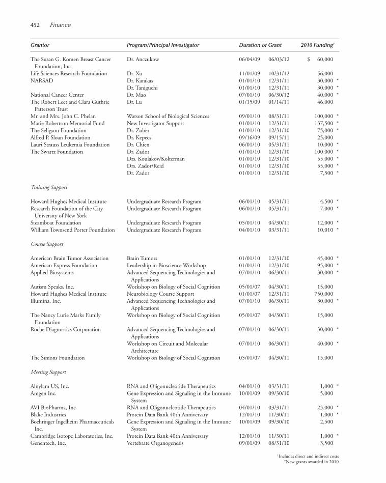
Grantor Program/Principal Investigator Duration of Grant 2010 Funding1
The Susan G. Komen Breast Cancer Dr. Anczukow 06/04/09 06/03/12 $ 60,000 Foundation, Inc.
Life Sciences Research Foundation Dr. Xu 11/01/09 10/31/12 56,000 NARSAD Dr. Karakas 01/01/10 12/31/11 30,000 *
Dr. Taniguchi 01/01/10 12/31/11 30,000 *National Cancer Center Dr. Mao 07/01/10 06/30/12 40,000 *The Robert Leet and Clara Guthrie Dr. Lu 01/15/09 01/14/11 46,000
Patterson TrustMr. and Mrs. John C. Phelan Watson School of Biological Sciences 09/01/10 08/31/11 100,000 *Marie Robertson Memorial Fund New Investigator Support 01/01/10 12/31/11 137,500 *The Seligson Foundation Dr. Zuber 01/01/10 12/31/10 75,000 *Alfred P. Sloan Foundation Dr. Kepecs 09/16/09 09/15/11 25,000 Lauri Strauss Leukemia Foundation Dr. Chien 06/01/10 05/31/11 10,000 *The Swartz Foundation Dr. Zador 01/01/10 12/31/10 100,000 *
Drs. Koulakov/Kolterman 01/01/10 12/31/10 55,000 *Drs. Zador/Reid 01/01/10 12/31/10 55,000 *Dr. Zador 01/01/10 12/31/10 7,500 *
Training Support
Howard Hughes Medical Institute Undergraduate Research Program 06/01/10 05/31/11 4,500 *Research Foundation of the City Undergraduate Research Program 06/01/10 05/31/11 7,000 *
University of New York Steamboat Foundation Undergraduate Research Program 05/01/10 04/30/11 12,000 *William Townsend Porter Foundation Undergraduate Research Program 04/01/10 03/31/11 10,010 *
Course Support
American Brain Tumor Association Brain Tumors 01/01/10 12/31/10 45,000 *American Express Foundation Leadership in Bioscience Workshop 01/01/10 12/31/10 95,000 *Applied Biosystems Advanced Sequencing Technologies and 07/01/10 06/30/11 30,000 *
ApplicationsAutism Speaks, Inc. Workshop on Biology of Social Cognition 05/01/07 04/30/11 15,000 Howard Hughes Medical Institute Neurobiology Course Support 01/01/07 12/31/11 750,000 Illumina, Inc. Advanced Sequencing Technologies and 07/01/10 06/30/11 30,000 *
ApplicationsThe Nancy Lurie Marks Family Workshop on Biology of Social Cognition 05/01/07 04/30/11 15,000
FoundationRoche Diagnostics Corporation Advanced Sequencing Technologies and 07/01/10 06/30/11 30,000 *
Applications Workshop on Circuit and Molecular 07/01/10 06/30/11 40,000 *
ArchitectureThe Simons Foundation Workshop on Biology of Social Cognition 05/01/07 04/30/11 15,000
Meeting Support
Alnylam US, Inc. RNA and Oligonucleotide Therapeutics 04/01/10 03/31/11 1,000 *Amgen Inc. Gene Expression and Signaling in the Immune 10/01/09 09/30/10 5,000
SystemAVI BioPharma, Inc. RNA and Oligonucleotide Therapeutics 04/01/10 03/31/11 25,000 *Blake Industries Protein Data Bank 40th Anniversary 12/01/10 11/30/11 1,000 *Boehringer Ingelheim Pharmaceuticals Gene Expression and Signaling in the Immune 10/01/09 09/30/10 2,500
Inc. SystemCambridge Isotope Laboratories, Inc. Protein Data Bank 40th Anniversary 12/01/10 11/30/11 1,000 *Genentech, Inc. Vertebrate Organogenesis 09/01/09 08/31/10 3,500
452 Finance
1Includes direct and indirect costs*New grants awarded in 2010
441-472_LAR_Finance_Annual Report_2009 template 7/8/11 12:29 PM Page 452

Grantor Program/Principal Investigator Duration of Grant 2010 Funding1
Genentech, Inc. Gene Expression and Signaling in the Immune 10/01/09 09/30/10 $ 2,500 System
Helixis, Inc. The Biology of Genomes 04/01/10 03/31/11 7,500 *Illumina, Inc. Personal Genomes 09/14/09 09/13/10 3,750
The Biology of Genomes 04/01/10 03/31/11 11,000 *Isis Pharmaceuticals, Inc. RNA and Oligonucleotide Therapeutics 04/01/10 03/31/11 3,000 *The March of Dimes Foundation Vertebrate Organogenesis 03/01/10 08/31/10 5,000 *Merck Serono S.A. Gene Expression and Signaling in the Immune 10/01/09 09/30/10 10,000
SystemMolecular Devices, Inc. Automated Imaging and High-Throughput 11/01/10 10/31/11 5,000 *
PhenotypingNew England BioLabs, Inc. Infectious Disease Genomics and Global Health 08/01/10 07/31/11 1,500 *Openeye Scientific Software, Inc. Protein Data Bank 40th Anniversary 12/01/10 11/30/11 2,000 *Penn State University Galaxy Developers 04/01/10 03/31/11 35,050 *Pfizer Inc. RNA and Oligonucleotide Therapeutics 04/01/10 03/31/11 3,000 *Roche Diagnostics Corporation Personal Genomes 03/01/10 02/28/11 49,466 *Schrodinger Protein Data Bank 40th Anniversary 12/01/10 11/30/11 1,000 *Shigemi Inc. Protein Data Bank 40th Anniversary 12/01/10 11/30/11 500 *Alfred P. Sloan Foundation New Faculty Investigator Conference 12/01/10 06/01/11 20,000 *
Library Support
Ellen Brenner Memorial Fund 12/22/10 12/21/11 2,000 *New York University School of Medicine 01/26/10 04/30/10 1,000 *The State of New York 07/01/09 06/30/10 3,850
Financial Support of the Laboratory 453
1Includes direct and indirect costs*New grants awarded in 2010
441-472_LAR_Finance_Annual Report_2009 template 7/12/11 10:13 AM Page 453
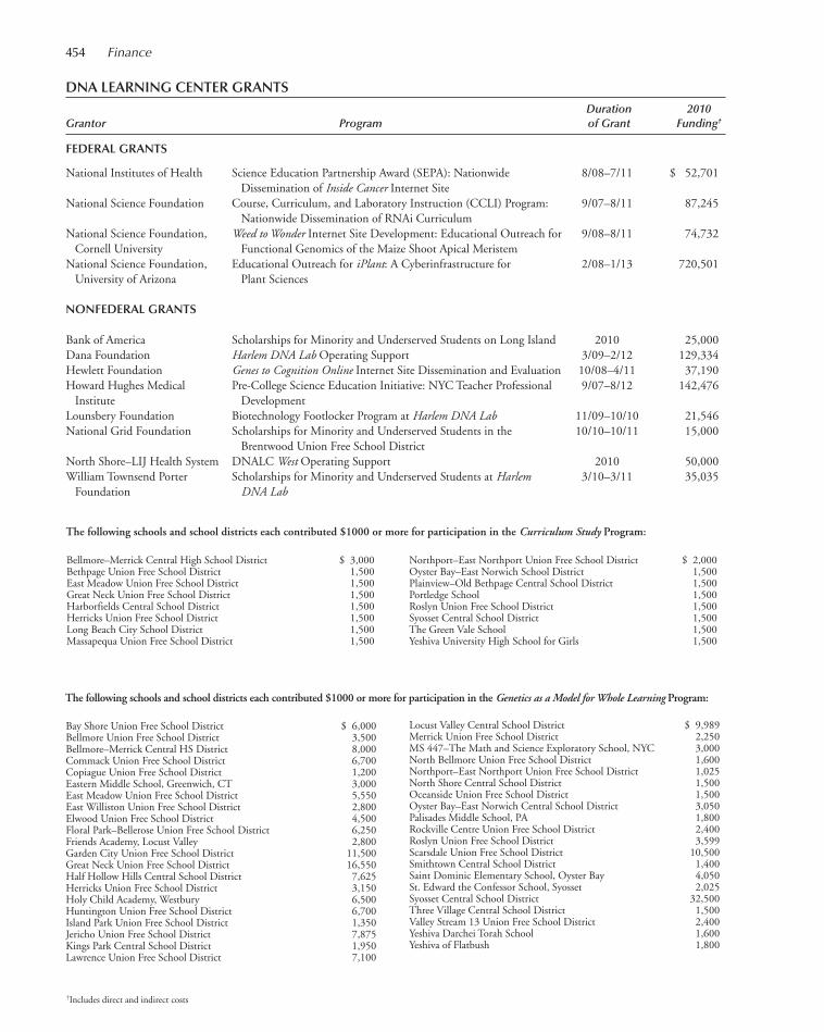
454 Finance
Bay Shore Union Free School District $ 6,000Bellmore Union Free School District 3,500Bellmore–Merrick Central HS District 8,000Commack Union Free School District 6,700Copiague Union Free School District 1,200Eastern Middle School, Greenwich, CT 3,000East Meadow Union Free School District 5,550East Williston Union Free School District 2,800Elwood Union Free School District 4,500Floral Park–Bellerose Union Free School District 6,250Friends Academy, Locust Valley 2,800Garden City Union Free School District 11,500Great Neck Union Free School District 16,550Half Hollow Hills Central School District 7,625Herricks Union Free School District 3,150Holy Child Academy, Westbury 6,500Huntington Union Free School District 6,700Island Park Union Free School District 1,350Jericho Union Free School District 7,875Kings Park Central School District 1,950Lawrence Union Free School District 7,100
Locust Valley Central School District $ 9,989Merrick Union Free School District 2,250MS 447–The Math and Science Exploratory School, NYC 3,000North Bellmore Union Free School District 1,600Northport–East Northport Union Free School District 1,025North Shore Central School District 1,500Oceanside Union Free School District 1,500Oyster Bay–East Norwich Central School District 3,050Palisades Middle School, PA 1,800Rockville Centre Union Free School District 2,400Roslyn Union Free School District 3,599Scarsdale Union Free School District 10,500Smithtown Central School District 1,400Saint Dominic Elementary School, Oyster Bay 4,050St. Edward the Confessor School, Syosset 2,025Syosset Central School District 32,500Three Village Central School District 1,500Valley Stream 13 Union Free School District 2,400Yeshiva Darchei Torah School 1,600Yeshiva of Flatbush 1,800
The following schools and school districts each contributed $1000 or more for participation in the Genetics as a Model for Whole Learning Program:
Northport–East Northport Union Free School District $ 2,000Oyster Bay–East Norwich School District 1,500Plainview–Old Bethpage Central School District 1,500Portledge School 1,500Roslyn Union Free School District 1,500Syosset Central School District 1,500The Green Vale School 1,500Yeshiva University High School for Girls 1,500
DNA LEARNING CENTER GRANTSDuration 2010
Grantor Program of Grant Funding†
FEDERAL GRANTS
National Institutes of Health Science Education Partnership Award (SEPA): Nationwide 8/08–7/11 $ 52,701Dissemination of Inside Cancer Internet Site
National Science Foundation Course, Curriculum, and Laboratory Instruction (CCLI) Program: 9/07–8/11 87,245Nationwide Dissemination of RNAi Curriculum
National Science Foundation, Weed to Wonder Internet Site Development: Educational Outreach for 9/08–8/11 74,732Cornell University Functional Genomics of the Maize Shoot Apical Meristem
National Science Foundation, Educational Outreach for iPlant: A Cyberinfrastructure for 2/08–1/13 720,501University of Arizona Plant Sciences
NONFEDERAL GRANTS
Bank of America Scholarships for Minority and Underserved Students on Long Island 2010 25,000Dana Foundation Harlem DNA Lab Operating Support 3/09–2/12 129,334Hewlett Foundation Genes to Cognition Online Internet Site Dissemination and Evaluation 10/08–4/11 37,190Howard Hughes Medical Pre-College Science Education Initiative: NYC Teacher Professional 9/07–8/12 142,476
Institute DevelopmentLounsbery Foundation Biotechnology Footlocker Program at Harlem DNA Lab 11/09–10/10 21,546National Grid Foundation Scholarships for Minority and Underserved Students in the 10/10–10/11 15,000
Brentwood Union Free School DistrictNorth Shore–LIJ Health System DNALC West Operating Support 2010 50,000William Townsend Porter Scholarships for Minority and Underserved Students at Harlem 3/10–3/11 35,035
Foundation DNA Lab
Bellmore–Merrick Central High School District $ 3,000Bethpage Union Free School District 1,500East Meadow Union Free School District 1,500Great Neck Union Free School District 1,500Harborfields Central School District 1,500Herricks Union Free School District 1,500Long Beach City School District 1,500Massapequa Union Free School District 1,500
†Includes direct and indirect costs
The following schools and school districts each contributed $1000 or more for participation in the Curriculum Study Program:
441-472_LAR_Finance_Annual Report_2009 template 7/8/11 12:29 PM Page 454
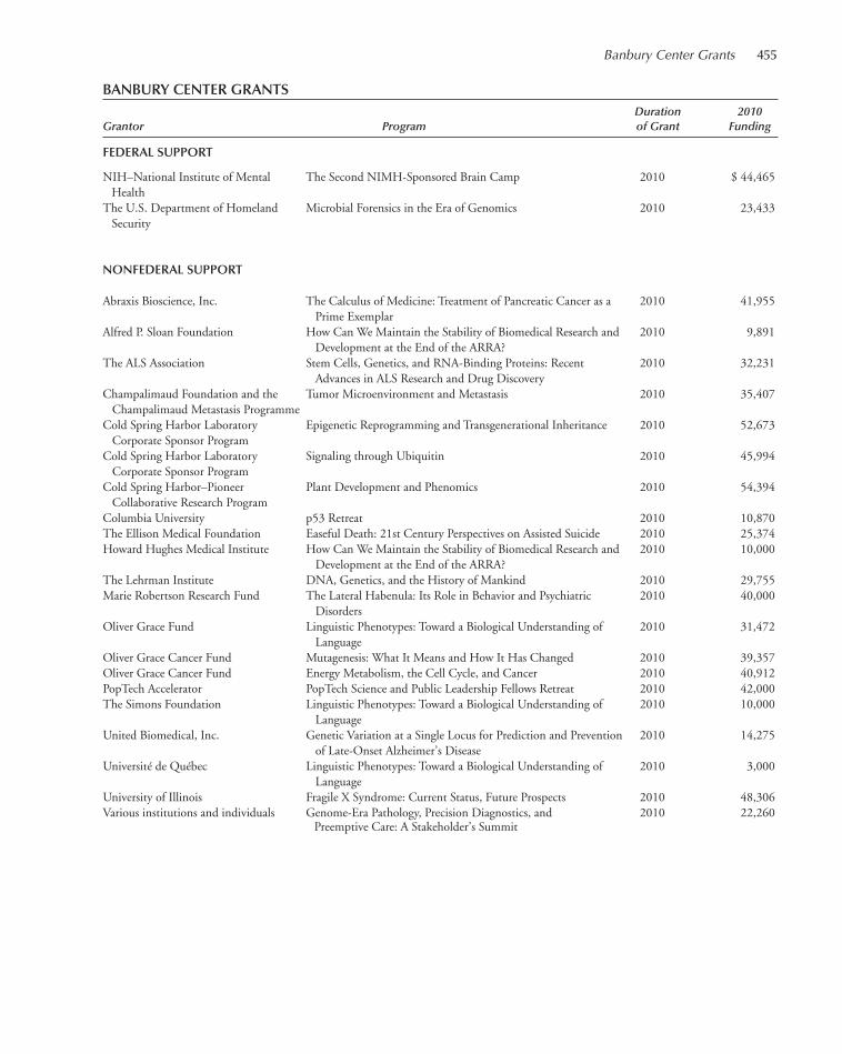
BANBURY CENTER GRANTSDuration 2010
Grantor Program of Grant Funding
FEDERAL SUPPORT
NIH–National Institute of Mental The Second NIMH-Sponsored Brain Camp 2010 $ 44,465Health
The U.S. Department of Homeland Microbial Forensics in the Era of Genomics 2010 23,433Security
NONFEDERAL SUPPORT
Abraxis Bioscience, Inc. The Calculus of Medicine: Treatment of Pancreatic Cancer as a 2010 41,955Prime Exemplar
Alfred P. Sloan Foundation How Can We Maintain the Stability of Biomedical Research and 2010 9,891Development at the End of the ARRA?
The ALS Association Stem Cells, Genetics, and RNA-Binding Proteins: Recent 2010 32,231Advances in ALS Research and Drug Discovery
Champalimaud Foundation and the Tumor Microenvironment and Metastasis 2010 35,407Champalimaud Metastasis Programme
Cold Spring Harbor Laboratory Epigenetic Reprogramming and Transgenerational Inheritance 2010 52,673Corporate Sponsor Program
Cold Spring Harbor Laboratory Signaling through Ubiquitin 2010 45,994Corporate Sponsor Program
Cold Spring Harbor–Pioneer Plant Development and Phenomics 2010 54,394Collaborative Research Program
Columbia University p53 Retreat 2010 10,870The Ellison Medical Foundation Easeful Death: 21st Century Perspectives on Assisted Suicide 2010 25,374Howard Hughes Medical Institute How Can We Maintain the Stability of Biomedical Research and 2010 10,000
Development at the End of the ARRA?The Lehrman Institute DNA, Genetics, and the History of Mankind 2010 29,755Marie Robertson Research Fund The Lateral Habenula: Its Role in Behavior and Psychiatric 2010 40,000
DisordersOliver Grace Fund Linguistic Phenotypes: Toward a Biological Understanding of 2010 31,472
LanguageOliver Grace Cancer Fund Mutagenesis: What It Means and How It Has Changed 2010 39,357Oliver Grace Cancer Fund Energy Metabolism, the Cell Cycle, and Cancer 2010 40,912PopTech Accelerator PopTech Science and Public Leadership Fellows Retreat 2010 42,000The Simons Foundation Linguistic Phenotypes: Toward a Biological Understanding of 2010 10,000
LanguageUnited Biomedical, Inc. Genetic Variation at a Single Locus for Prediction and Prevention 2010 14,275
of Late-Onset Alzheimer’s DiseaseUniversité de Québec Linguistic Phenotypes: Toward a Biological Understanding of 2010 3,000
LanguageUniversity of Illinois Fragile X Syndrome: Current Status, Future Prospects 2010 48,306Various institutions and individuals Genome-Era Pathology, Precision Diagnostics, and 2010 22,260
Preemptive Care: A Stakeholder’s Summit
Banbury Center Grants 455
441-472_LAR_Finance_Annual Report_2009 template 7/8/11 12:29 PM Page 455

456
CORPORATE SPONSOR PROGRAM FOR MEETINGS SUPPORT
The Corporate Sponsor Program continues to provide critical funding for the vigorous meetings pro-gram held at Cold Spring Harbor Laboratory, whether at Grace Auditorium on the main Laboratorycampus or at the Banbury Center. Without the strong foundation provided by the Program, we couldneither plan with confidence for the year’s meetings nor introduce new and unusual topics.
We are especially grateful to the companies that joined us in 2010 as the economic difficultiesbegan to take effect. The year 2011 is going to be especially challenging as the number of companiesshrinks through takeovers, and companies and foundations adopt austerity measures.
The members of the Program receive special privileges in acknowledgment of their contributions.We waive all on-site fees for eight representatives of the Corporate Sponsors at our meetings. Threescientists from Sponsors may attend meetings at the Banbury Center, where attendance is otherwiseonly by invitation of the organizers. Member companies also receive gratis copies of Cold SpringHarbor Laboratory Press publications, including the journals Genes & Development, Learning &Memory, Protein Science, Genome Research, and RNA.
We acknowledge our Sponsors in all relevant publications, including the books of abstracts givento each of the 7000 participants who come to the meetings each year. The names of the sponsoringcompanies are listed on the poster describing the meetings, and this is mailed to approximately 17,000scientists throughout the world. In addition, the companies are listed on the Cold Spring HarborLaboratory website on the Meetings Office and Banbury Center pages. Members in 2010 were thefollowing:
FOUNDATION
Hudson-Alpha Institute for Biotechnology
PLANT CORPORATE SPONSORS
Monsanto CompanyPioneer Hi-Bred International, Inc.
CORPORATE SPONSORS
Agilent Technologies, Inc.AstraZeneca UK Ltd.BioVentures, Inc.Bristol-Myers Squibb CompanyGenentech, Inc.GlaxoSmithKlineLife TechnologiesNew England BioLabs, Inc.OSI Pharmaceuticals, Inc.Sanofi-Aventis
441-472_LAR_Finance_Annual Report_2009 template 7/8/11 12:29 PM Page 456
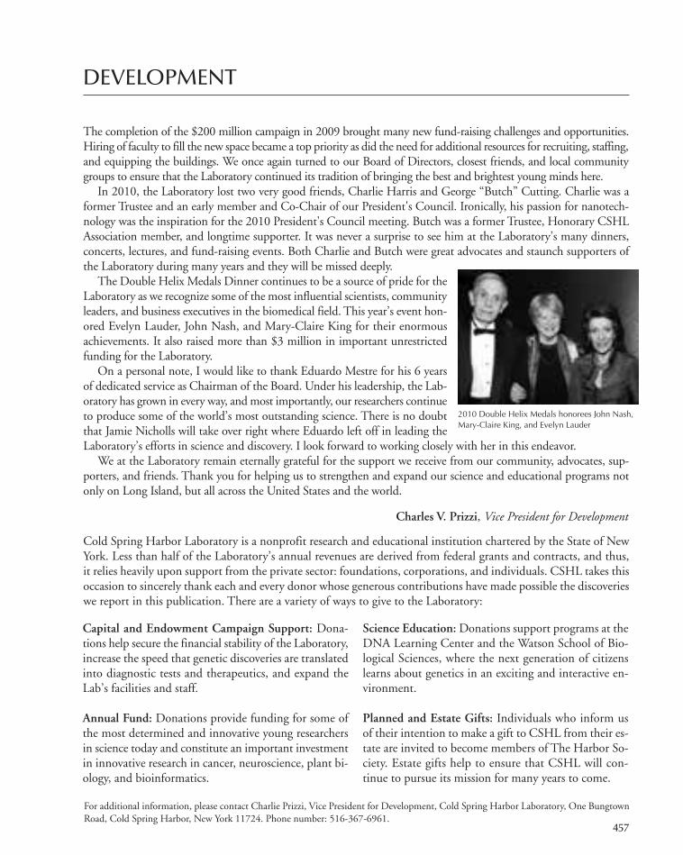
457
DEVELOPMENT
The completion of the $200 million campaign in 2009 brought many new fund-raising challenges and opportunities.Hiring of faculty to fill the new space became a top priority as did the need for additional resources for recruiting, staffing,and equipping the buildings. We once again turned to our Board of Directors, closest friends, and local communitygroups to ensure that the Laboratory continued its tradition of bringing the best and brightest young minds here.
In 2010, the Laboratory lost two very good friends, Charlie Harris and George “Butch” Cutting. Charlie was aformer Trustee and an early member and Co-Chair of our President's Council. Ironically, his passion for nanotech-nology was the inspiration for the 2010 President's Council meeting. Butch was a former Trustee, Honorary CSHLAssociation member, and longtime supporter. It was never a surprise to see him at the Laboratory’s many dinners,concerts, lectures, and fund-raising events. Both Charlie and Butch were great advocates and staunch supporters ofthe Laboratory during many years and they will be missed deeply.
The Double Helix Medals Dinner continues to be a source of pride for theLaboratory as we recognize some of the most influential scientists, communityleaders, and business executives in the biomedical field. This year’s event hon-ored Evelyn Lauder, John Nash, and Mary-Claire King for their enormousachievements. It also raised more than $3 million in important unrestrictedfunding for the Laboratory.
On a personal note, I would like to thank Eduardo Mestre for his 6 yearsof dedicated service as Chairman of the Board. Under his leadership, the Lab-oratory has grown in every way, and most importantly, our researchers continueto produce some of the world’s most outstanding science. There is no doubtthat Jamie Nicholls will take over right where Eduardo left off in leading theLaboratory’s efforts in science and discovery. I look forward to working closely with her in this endeavor.
We at the Laboratory remain eternally grateful for the support we receive from our community, advocates, sup-porters, and friends. Thank you for helping us to strengthen and expand our science and educational programs notonly on Long Island, but all across the United States and the world.
Charles V. Prizzi, Vice President for Development
Cold Spring Harbor Laboratory is a nonprofit research and educational institution chartered by the State of NewYork. Less than half of the Laboratory’s annual revenues are derived from federal grants and contracts, and thus,it relies heavily upon support from the private sector: foundations, corporations, and individuals. CSHL takes thisoccasion to sincerely thank each and every donor whose generous contributions have made possible the discoverieswe report in this publication. There are a variety of ways to give to the Laboratory:
2010 Double Helix Medals honorees John Nash,Mary-Claire King, and Evelyn Lauder
Capital and Endowment Campaign Support: Dona-tions help secure the financial stability of the Laboratory,increase the speed that genetic discoveries are translatedinto diagnostic tests and therapeutics, and expand theLab’s facilities and staff.
Annual Fund: Donations provide funding for some ofthe most determined and innovative young researchersin science today and constitute an important investmentin innovative research in cancer, neuroscience, plant bi-ology, and bioinformatics.
Science Education: Donations support programs at theDNA Learning Center and the Watson School of Bio-logical Sciences, where the next generation of citizenslearns about genetics in an exciting and interactive en-vironment.
Planned and Estate Gifts: Individuals who inform usof their intention to make a gift to CSHL from their es-tate are invited to become members of The Harbor So-ciety. Estate gifts help to ensure that CSHL will con-tinue to pursue its mission for many years to come.
For additional information, please contact Charlie Prizzi, Vice President for Development, Cold Spring Harbor Laboratory, One BungtownRoad, Cold Spring Harbor, New York 11724. Phone number: 516-367-6961.
441-472_LAR_Finance_Annual Report_2009 template 7/8/11 12:29 PM Page 457
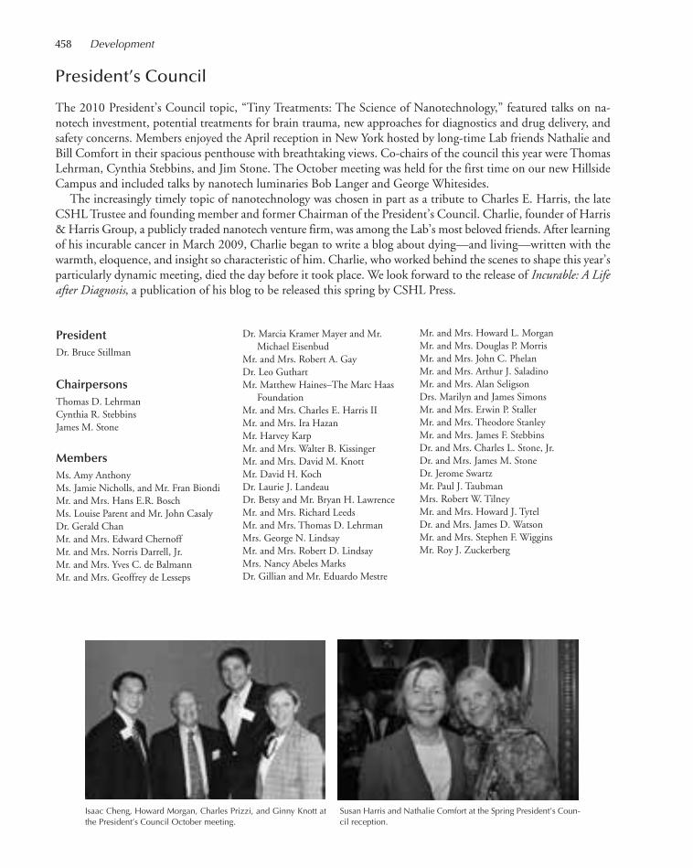
458 Development
Dr. Marcia Kramer Mayer and Mr.Michael Eisenbud
Mr. and Mrs. Robert A. GayDr. Leo GuthartMr. Matthew Haines–The Marc Haas
FoundationMr. and Mrs. Charles E. Harris II Mr. and Mrs. Ira HazanMr. Harvey Karp Mr. and Mrs. Walter B. KissingerMr. and Mrs. David M. KnottMr. David H. KochDr. Laurie J. LandeauDr. Betsy and Mr. Bryan H. Lawrence Mr. and Mrs. Richard LeedsMr. and Mrs. Thomas D. LehrmanMrs. George N. LindsayMr. and Mrs. Robert D. LindsayMrs. Nancy Abeles MarksDr. Gillian and Mr. Eduardo Mestre
Mr. and Mrs. Howard L. MorganMr. and Mrs. Douglas P. MorrisMr. and Mrs. John C. PhelanMr. and Mrs. Arthur J. SaladinoMr. and Mrs. Alan Seligson Drs. Marilyn and James SimonsMr. and Mrs. Erwin P. StallerMr. and Mrs. Theodore StanleyMr. and Mrs. James F. StebbinsDr. and Mrs. Charles L. Stone, Jr.Dr. and Mrs. James M. StoneDr. Jerome SwartzMr. Paul J. TaubmanMrs. Robert W. TilneyMr. and Mrs. Howard J. TytelDr. and Mrs. James D. WatsonMr. and Mrs. Stephen F. WigginsMr. Roy J. Zuckerberg
President’s Council
The 2010 President’s Council topic, “Tiny Treatments: The Science of Nanotechnology,” featured talks on na-notech investment, potential treatments for brain trauma, new approaches for diagnostics and drug delivery, andsafety concerns. Members enjoyed the April reception in New York hosted by long-time Lab friends Nathalie andBill Comfort in their spacious penthouse with breathtaking views. Co-chairs of the council this year were ThomasLehrman, Cynthia Stebbins, and Jim Stone. The October meeting was held for the first time on our new HillsideCampus and included talks by nanotech luminaries Bob Langer and George Whitesides.
The increasingly timely topic of nanotechnology was chosen in part as a tribute to Charles E. Harris, the lateCSHL Trustee and founding member and former Chairman of the President’s Council. Charlie, founder of Harris& Harris Group, a publicly traded nanotech venture firm, was among the Lab’s most beloved friends. After learningof his incurable cancer in March 2009, Charlie began to write a blog about dying—and living—written with thewarmth, eloquence, and insight so characteristic of him. Charlie, who worked behind the scenes to shape this year’sparticularly dynamic meeting, died the day before it took place. We look forward to the release of Incurable: A Lifeafter Diagnosis, a publication of his blog to be released this spring by CSHL Press.
PresidentDr. Bruce Stillman
ChairpersonsThomas D. LehrmanCynthia R. StebbinsJames M. Stone
MembersMs. Amy AnthonyMs. Jamie Nicholls, and Mr. Fran BiondiMr. and Mrs. Hans E.R. BoschMs. Louise Parent and Mr. John CasalyDr. Gerald ChanMr. and Mrs. Edward ChernoffMr. and Mrs. Norris Darrell, Jr.Mr. and Mrs. Yves C. de Balmann Mr. and Mrs. Geoffrey de Lesseps
Susan Harris and Nathalie Comfort at the Spring President’s Coun-cil reception.
Isaac Cheng, Howard Morgan, Charles Prizzi, and Ginny Knott atthe President’s Council October meeting.
441-472_LAR_Finance_Annual Report_2009 template 7/8/11 12:29 PM Page 458
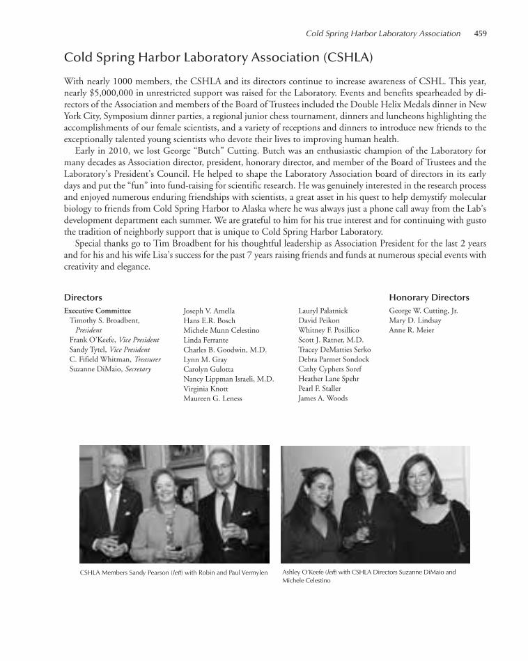
Cold Spring Harbor Laboratory Association 459
Cold Spring Harbor Laboratory Association (CSHLA)
With nearly 1000 members, the CSHLA and its directors continue to increase awareness of CSHL. This year,nearly $5,000,000 in unrestricted support was raised for the Laboratory. Events and benefits spearheaded by di-rectors of the Association and members of the Board of Trustees included the Double Helix Medals dinner in NewYork City, Symposium dinner parties, a regional junior chess tournament, dinners and luncheons highlighting theaccomplishments of our female scientists, and a variety of receptions and dinners to introduce new friends to theexceptionally talented young scientists who devote their lives to improving human health.
Early in 2010, we lost George “Butch” Cutting. Butch was an enthusiastic champion of the Laboratory formany decades as Association director, president, honorary director, and member of the Board of Trustees and theLaboratory’s President’s Council. He helped to shape the Laboratory Association board of directors in its earlydays and put the “fun” into fund-raising for scientific research. He was genuinely interested in the research processand enjoyed numerous enduring friendships with scientists, a great asset in his quest to help demystify molecularbiology to friends from Cold Spring Harbor to Alaska where he was always just a phone call away from the Lab’sdevelopment department each summer. We are grateful to him for his true interest and for continuing with gustothe tradition of neighborly support that is unique to Cold Spring Harbor Laboratory.
Special thanks go to Tim Broadbent for his thoughtful leadership as Association President for the last 2 yearsand for his and his wife Lisa’s success for the past 7 years raising friends and funds at numerous special events withcreativity and elegance.
Lauryl PalatnickDavid PeikonWhitney F. PosillicoScott J. Ratner, M.D.Tracey DeMatties SerkoDebra Parmet SondockCathy Cyphers SorefHeather Lane SpehrPearl F. StallerJames A. Woods
Joseph V. AmellaHans E.R. BoschMichele Munn CelestinoLinda FerranteCharles B. Goodwin, M.D.Lynn M. GrayCarolyn GulottaNancy Lippman Israeli, M.D.Virginia KnottMaureen G. Leness
DirectorsExecutive CommitteeTimothy S. Broadbent,
PresidentFrank O’Keefe, Vice PresidentSandy Tytel, Vice PresidentC. Fifield Whitman, TreasurerSuzanne DiMaio, Secretary
CSHLA Members Sandy Pearson (left) with Robin and Paul Vermylen Ashley O’Keefe (left) with CSHLA Directors Suzanne DiMaio andMichele Celestino
Honorary DirectorsGeorge W. Cutting, Jr.Mary D. LindsayAnne R. Meier
441-472_LAR_Finance_Annual Report_2009 template 7/12/11 10:16 AM Page 459
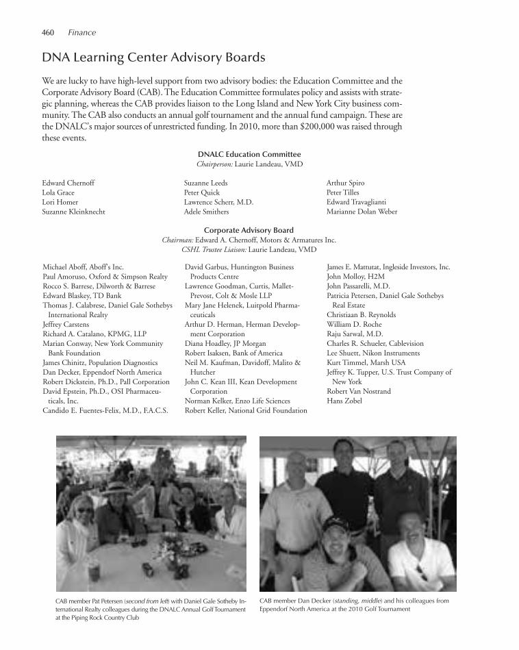
460 Finance
DNA Learning Center Advisory Boards
We are lucky to have high-level support from two advisory bodies: the Education Committee and theCorporate Advisory Board (CAB). The Education Committee formulates policy and assists with strate-gic planning, whereas the CAB provides liaison to the Long Island and New York City business com-munity. The CAB also conducts an annual golf tournament and the annual fund campaign. These arethe DNALC’s major sources of unrestricted funding. In 2010, more than $200,000 was raised throughthese events.
Edward ChernoffLola GraceLori HomerSuzanne Kleinknecht
Suzanne LeedsPeter QuickLawrence Scherr, M.D.Adele Smithers
DNALC Education CommitteeChairperson: Laurie Landeau, VMD
Arthur Spiro Peter TillesEdward TravagliantiMarianne Dolan Weber
Corporate Advisory BoardChairman: Edward A. Chernoff, Motors & Armatures Inc.
CSHL Trustee Liaison: Laurie Landeau, VMD
CAB member Pat Petersen (second from left) with Daniel Gale Sotheby In-ternational Realty colleagues during the DNALC Annual Golf Tournamentat the Piping Rock Country Club
CAB member Dan Decker (standing, middle) and his colleagues fromEppendorf North America at the 2010 Golf Tournament
Michael Aboff, Aboff’s Inc.Paul Amoruso, Oxford & Simpson RealtyRocco S. Barrese, Dilworth & BarreseEdward Blaskey, TD BankThomas J. Calabrese, Daniel Gale Sothebys
International RealtyJeffrey CarstensRichard A. Catalano, KPMG, LLPMarian Conway, New York Community
Bank FoundationJames Chinitz, Population DiagnosticsDan Decker, Eppendorf North AmericaRobert Dickstein, Ph.D., Pall CorporationDavid Epstein, Ph.D., OSI Pharmaceu-
ticals, Inc.Candido E. Fuentes-Felix, M.D., F.A.C.S.
David Garbus, Huntington BusinessProducts Centre
Lawrence Goodman, Curtis, Mallet-Prevost, Colt & Mosle LLP
Mary Jane Helenek, Luitpold Pharma-ceuticals
Arthur D. Herman, Herman Develop-ment Corporation
Diana Hoadley, JP MorganRobert Isaksen, Bank of AmericaNeil M. Kaufman, Davidoff, Malito &
HutcherJohn C. Kean III, Kean Development
CorporationNorman Kelker, Enzo Life SciencesRobert Keller, National Grid Foundation
James E. Mattutat, Ingleside Investors, Inc.John Molloy, H2MJohn Passarelli, M.D.Patricia Petersen, Daniel Gale Sothebys
Real EstateChristiaan B. ReynoldsWilliam D. RocheRaju Sarwal, M.D.Charles R. Schueler, CablevisionLee Shuett, Nikon InstrumentsKurt Timmel, Marsh USAJeffrey K. Tupper, U.S. Trust Company of
New YorkRobert Van NostrandHans Zobel
441-472_LAR_Finance_Annual Report_2009 template 7/8/11 12:29 PM Page 460

Contributions of $5,000,000+ lifetimeAmerican Cancer SocietyAnonymousThe Donald Everett Axinn TrustArnold and Mabel Beckman FoundationMr. and Mrs. Landon T. ClayThe Dana FoundationDart Neuroscience LLCKathryn W. DavisDiana Davis Spencer Foundation (formerly
The Kathryn W. Davis Foundation)The DeMatteis Family FoundationMr. and Mrs. Charles F. Dolan–The Dolan
Family FoundationThe William Stamps Farish FoundationHazen Polsky Foundation (formerly The
Lita Annenberg Hazen Foundation)Howard Hughes Medical InstituteW.M. Keck FoundationDavid H. KochThe Leukemia & Lymphoma SocietyLucille P. Markey Charitable TrustNancy Abeles MarksThe Don Monti Memorial Research
FoundationGEN*NY*sis grant from the New York
State Dormitory AuthorityNew York State Empire State Development
CorporationPioneer Hi-Bred International, Inc.The Quick FamilyThe Robertson Family–Banbury FundThe Simons FoundationMr. and Mrs. Theodore R. Stanley–The
Stanley Medical Research InstituteThe Starr FoundationMr. and Mrs. Henry Wendt III
Contributions of $100,000 in 2010 or$1,000,000+ lifetime
Paul G. Allen Family FoundationAngel FoundationAnonymous
BioBay of Suzhou Venture Capital GroupJamie C. Nicholls and O. Francis Biondi, Jr.The Breast Cancer Research FoundationBristol-Myers Squibb CompanyTerri Brodeur Breast Cancer FoundationBurroughs Wellcome FundThe Mary K. Chapman FoundationMr. and Mrs. Edward A. Chernoff–Motors
& Armatures, Inc.Dr. and Mrs. Bayard Delafield ClarksonLester CrownMr. and Mrs. Roderick H. CushmanMr. and Mrs. Norris Darrell, Jr.Mr. and Mrs. Michel David-WeillIra W. DeCamp FoundationThe Ellison Medical FoundationEntertainment Industry FoundationThe Samuel Freeman TrustColeman Fung FoundationMr. and Mrs. Robert A. GayGenentech, Inc.Kenyon GillespieGlaxoSmithKlineJacob D. GoldfieldThe Allen & Lola Goldring FoundationFlorence Gould FoundationThe Grace FamilyGreenwich Breast Cancer Alliance, Inc.Leo A. GuthartThe Harrison Foundation and The Mary
Anderson Harrison FoundationThe Thomas Hartman Foundation for
Parkinson’s ResearchJanet Strauss and Jeff HawkinsJo-Ellen and Ira HazanWilliam Randolph Hearst FoundationHuman Frontier Science ProgramInternational Rett Syndrome FoundationIsis Pharmaceuticals, Inc.The Karches FoundationF.M. Kirby Foundation, Inc.Mr. and Mrs. Walter B. KissingerMr. and Mrs. John Klingenstein–The Esther
A. & Joseph Klingenstein Fund, Inc.
Mr. and Mrs. Townsend J. KnightSusan G. Komen for the CureLaurie Landeau Foundation LLCThe Forrest C. & Frances H. Lattner
FoundationThe Lehrman InstituteMr. and Mrs. Stephen M. LessingThe Fay J. Lindner FoundationMr. and Mrs. Robert D. Lindsay and
FamilyMary D. LindsayThe Litchfield Charitable TrustEstate of Elisabeth Sloan LivingstonLouis Morin Charitable TrustMr. and Mrs. David L. Luke IIIThe Lustgarten FoundationJosiah Macy, Jr. FoundationThe G. Harold & Leila Y. Mathers
Charitable FoundationThe McKnight Endowment Fund for
NeuroscienceGillian and Eduardo MestreUniversidad Nacional Autónoma de
MéxicoMr. and Mrs. William R. MillerMr. and Mrs. Howard MorganMuscular Dystrophy AssociationNew York State Dormitory AuthorityOSI Pharmaceuticals FoundationLouise Parent and John CasalyThe Pew Charitable TrustsMr. and Mrs. John C. PhelanPhilips Research North AmericaWilliam and Maude Pritchard Charitable
TrustV. Kann Rasmussen FoundationThe Robertson FoundationMr. and Mrs. David M. RubensteinDamon Runyon Cancer Research
FoundationEleanor Schwartz Charitable FoundationEdith and Alan SeligsonMr. and Mrs. Herbert J. SiegelAlfred P. Sloan FoundationMr. and Mrs. Howard SolomonSt. Giles FoundationDr. and Mrs. James M. StoneDr. Jerome Swartz–Swartz Charitable
Foundation
Honor Roll of Donors
CSHL Trustee Jim Stone with Terry Lindsay (left)and Cynthia Stebbins at the President’s CouncilSpring Reception held at the home of Nathalieand Bill Comfort
CSHL Chairman-Elect Jamie Nicholls (center) with (from left to right) Dan Loeb,Fran Biondi, Hartley Rogers, Amy Falls, and Judy Cormier at the Double HelixMedals Dinner held on November 10, 2010 at the Mandarin Oriental in New York
Honor Roll of Donors 461
441-472_LAR_Finance_Annual Report_2009 template 7/12/11 10:16 AM Page 461
462 Finance
Waclaw T. Szybalski, D.Sc.Paul J. TaubmanDoris M. and Peter S. Tilles FoundationThe V Foundation for Cancer ResearchDr. and Mrs. James D. WatsonWhitehall Foundation, Inc.The Roy J. Zuckerberg Family
Foundation
Contributions of $50,000+American Express FoundationMr. and Mrs. Hans E.R. BoschCashin Family FundMr.* and Mrs. George W. Cutting Jr.Genentech FoundationMr. and Mrs. John S. Grace–Gifted
Learning InstituteAnnette Kade Charitable TrustThe Karp FoundationMr. and Mrs. David M. Knott–Knott
Family FoundationMara and Thomas LehrmanLife Sciences Research FoundationThe George Link, Jr. FoundationThe Manhasset Women’s Coalition Against
Breast CancerBreast Cancer Awareness Day in memory
of Elizabeth McFarlandThe John Merck FundMirus Bio LLCNARSADUniversity of Notre DamePall CorporationMr. and Mrs. Henry P. Davison II–The
Perkin FundRoche Diagnostics CorporationMelissa Kerr and Stephen WigginsEvelyn W. WillardWodecroft Foundation
Contributions of $25,000+Alzheimer’s AssociationAmerican Brain Tumor AssociationAnonymous Amy AnthonyApplied BiosystemsAstellas USA FoundationAVI BioPharma, Inc.Bank of America Charitable Foundation,
Inc.
Mr. and Mrs. David Boies–Boies, Schiller& Flexner LLP
The Breast Cancer ProjectMr. and Mrs. George W. Carmany IIIDr. Gerald Chan–The Morningside
FoundationThe Jane Coffin Childs Memorial Fund for
Medical ResearchMr. and Mrs. Yves C. de BalmannMr. and Mrs. Geoffrey de Lesseps–Alpha
Omega Charitable Foundation, Inc.Marcia Kramer Mayer and Michael
EisenbudThe Estee Lauder Companies Inc.F.A.C.T. (Find A Cure Today)Charitable Lead Annuity Trust Under the
Will of Louis FeilMatthew Haines–The Marc Haas
FoundationMr.* and Mrs. Charles E. HarrisThe Hastings Foundation, Inc.Hope Funds for Cancer ResearchIllumina, Inc.Joni Gladowsky Breast Cancer
FoundationBetsy and Hunt LawrenceMr. and Mrs. Richard LeedsLong Island 2-Day Walk to Fight Breast
Cancer, Inc.Nancy Lurie Marks Family FoundationMr. and Mrs. Douglas P. MorrisParatek PharmaceuticalsThe Robert Leet and Clara Guthrie
Patterson TrustPenn State UniversityWilliam Townsend Porter FoundationPatricia QuickThomas C. QuickAndrew W. Solomon and John Habich
SolomonPearl F. and Erwin P. StallerCynthia R. and James F. StebbinsJoan P. TilneySandy and Howard Tytel
Contributions of $10,000+The A&P FoundationAnonymousAutism Speaks, Inc.The Bahnik Foundation, Inc.Frank BrunckhorstJohn K. CastleThe Cutler FamilyMartin Elias and FamilyEppendorf North AmericaFortunato Sons Contracting, Inc.William F. Gerry–Gerry Corbett
FoundationMr. and Mrs. William H. Goodwin, Jr.Michael J. GriffinThe Irving A. Hansen Memorial
FoundationMr. and Mrs. J. Tomilson Hill III–The
Hill Family FoundationDr. Nancy Lippman Israeli and Dr. Ron
IsraeliMr. and Mrs. Thomas Campbell JacksonJudi Shesh Memorial FoundationDrs. Marsha and Henry LauferLife TechnologiesLong Island Radio GroupBridget and John MacaskillMr. and Mrs.* Edward E. MatthewsMerck Serono S.A.National Grid FoundationNeuberger Berman FoundationNichols Foundation, Inc.Ashley and Frank O’KeefeWendy O’NeillJohn G. Passarelli, M.D.Patricia Petersen–Daniel Gale Sotheby’s
International RealtyMr. and Mrs. John R. ReeseChristina Renna Foundation, Inc.Diane Emdin Sachs Memorial WalkLaura and Harry SlatkinDeborah K. SolbertMr. and Mrs. Douglas S. SorefMr. and Mrs. Allen Sperry
*Deceased in 2010
CSHL Trustees John Phelan (left) and Roy Zuckerberg with Melva Bucksbaum and Amy Phelan atthe Double Helix Medals Dinner
CSHLA Director Fife Whitman (left), HeatherWhitman, CSHLA Director Heather Spehr, andRich Spehr
441-472_LAR_Finance_Annual Report_2009 template 7/8/11 12:29 PM Page 462

Steamboat FoundationTD Charitable FoundationTextor Family FoundationThe Lauri Strauss Leukemia FoundationWest Islip Breast Cancer Coalition for
Long IslandDr. and Mrs. Michael WiglerMr. and Mrs. Richard H. Witmer, Jr.Ann Eden Woodward Foundation
Contributions of $5,000+Mr. and Mrs. Lee S. Ainslie IIIAmgen, Inc.Mr. and Mrs. W. Dillaway Ayres, Jr.Bank of New York MellonLisa and Tim BroadbentHelen M. BrooksMr. and Mrs. James J. CelestinoSamara CohenCredit Agricole Corporate Investment
BankMr. and Mrs. Richard T. Cunniff–Cunniff
Family FundDeutsche BankDilworth & Barrese, LLPSuzanne and Michael DiMaioBob and Diane FagiolaThe Fortin Foundation of FloridaRichard GelfondThe Deane A. and John D. Gilliam
FoundationThe Fred C. Gloeckner Foundation, Inc.Mr. and Mrs. Austen T. Gray, Jr.H2M GroupThe Randall and Mary Hack FoundationRobin T. HadleyThe Emma Alyson & Kate Hance Family
FoundationHelixis, Inc.Diana HoadleyDr. Mark HoffmanIslip Breast Cancer CoalitionJason Dennis Francis Inc.Kapp Family FoundationKean Development CompanyAndrea B. and Peter D. KleinThe Edward and Kinga Lampert
FoundationMr. and Mrs. Anthony V. LenessLuitpold Pharmaceuticals, Inc.Mrs. David J. MahoneyMarch of DimesMasthead Cove Yacht Club–Carol
Marcincuk FundRobert L. MarcusAnjali and Prakash MelwaniMolecular Devices, Inc.New York Community Bank FoundationNikon Instruments, Inc.Susanne OlinMr. and Mrs. George D. O’Neill–Abby
and George O’Neill TrustLauryl and Robert PalatnickDavid P. PearsonMr. and Mrs. Simon Pharr
Dr. and Mrs. John E. Postley, Jr.–New YorkPhysicians Foundation, Inc.
Mrs. Richardson Pratt, Jr.Mr. and Mrs. Enrique F. SeniorMary F. Smith Family FoundationLee SteinbergDr. and Mrs. Bruce W. StillmanSally Susman–Pfizer, Inc.Leonard LitwinThe Thompson Family Foundation, Inc.Mr. and Mrs. Remy W. TrafeletMr. and Mrs. Edward TravagliantiRobin and Paul VermylenMrs. Philip R. von StadeRalph Worthington
Contributions of $2,500+Allison AbbottAboff’s Inc.Advantage Title Agency, Inc.Aireco Real EstateAll Pro Carpet Co., Inc.Alpern Family FoundationMr. and Mrs. Robert H.B. BaldwinCristina Mariani-May–The Banfi Vintners
FoundationLiz and Rod BerensJudy and Howard BerkowitzGail A. BindermanBliss Pest Protection Services, LLCBoehringer Ingelheim PharmaceuticalsDr. Steven BrennerMr. and Mrs. Thomas J. Calabrese, Jr.Mr. and Mrs. William J. Candee IIIMrs. Robert L. Clarkson, Jr.Clear Channel Communications–
WALK 97.5Crest Hollow Country Club at
Woodbury, Inc.Mr. and Mrs. Joseph T. DonohueDvirka and Bartilucci Consulting
EngineersMichael L. Focazio–Carissa Maringo FundG & M Earth Moving, Inc.
The Geier FoundationCosby GeorgeGlencore InternationalMr. and Mrs. Lawrence G. GraevBernadette Castro Guida and Peter
Guida, M.D.Mr. Arthur D. Herman–Herman
Development CorporationJames Hicks, Ph.D.Lori and Chris HomerRon Howard and Brian GrazerJoan M. HutchinsThe Stanley R. and Elisabeth G. Jacobs
FoundationMr. and Mrs. Barclay G. Jones IIIMr. and Mrs. Richard KleinknechtLaureen and Ragnar M. KnutsenMelva Bucksbaum and Raymond LearsyGeorgie W. LewisThe MacLean Family TrustMr. and Mrs. William A. MarinoOSI PharmaceuticalsOxford & Simpson Realty Svcs., Inc.Scott J. Paternoster FamilyTerry and Howard PhippsWhitney PosillicoNicole and Charlie PrizziDrs. Marjorie van de Stouwe and Scott
RatnerAnne and Bernard ReynoldsDr. and Mrs. Gerald M. RubinDebra Parmet Sondock and Clifford B.
SondockDrs. Mona and David SpectorHeather and Richard SpehrTD BankNikki TippinsMr. and Mrs. Stanley S. Trotman, Jr.United HealthcareUniversal StudiosHeather and Fifield WhitmanThe Diane and Howard Wohl Family
Foundation, Inc.Wolfensohn Family FoundationCraig Wulff, Fidelity Investments
On December 20, 2010, The Manhasset Women’s Coalition Against Breast Cancer presented Dr. MikalaEgeblad (third from left, front row) with a check for $50,000 to support her breast cancer research.
Honor Roll of Donors 463
441-472_LAR_Finance_Annual Report_2009 template 7/12/11 10:17 AM Page 463
464 Finance
Contributions of $1,000+Mr. and Mrs. Robert J. AdamsAlnylam U.S., Inc.Americana Manhasset and Wheatley PlazaLisa and Doug ArthurAudio Command Systems, Inc.Mary and Paul AuerspergMr. and Mrs. Richard Aurelio–The Aurelio
Family Charitable FoundationErica and Henry BabcockPam and Gene Bernstein–PGJM
FoundationDr. and Mrs. Giuseppe BertaniMr. and Mrs. Xerxes K. BhoteBlake IndustriesElizabeth B. Borden–Katharine Bradford
FoundationRenee A. Fitts and David BotsteinDr. William Braden III–Porter Braden
Endowment FundMr. and Mrs. Michael BrennanBresnan International Aviation, LLCJohn BrittonThe Eli and Edythe Broad FoundationCambridge Isotope Laboratories, Inc.Peter B. CannellMuffy CarlstromCenterbrook Architects and Planners, LLCJane H. ChoateMr. and Mrs. David C. ClarkRita M. ClearyMr. and Mrs. Jonathan ConnorsCortel Business Solutions, Inc.Cosel-Pieper Family FoundationRebecca CreavinCurtis, Mallet-Prevost, Colt & Mosle LLPCurtis CushmanDavidoff Malito & Hutcher LLPMr. and Mrs. Guy L. de ChazalRobert de RothschildMr. and Mrs. David H. DemingMeleanor and Donald DemingNevenka DimitrovaThomas J. Dowling Jr., M.D.Dr. Lester DubnickElectronix Systems Central Station AlarmsEnzo Life SciencesLisa and Johnston Evans
Mary and Donald EversollFarrell Fritz, P.C.Dr. Robert and Marjorie FeeneyMr. and Mrs. Frank FeinbergMr. and Mrs. Michael G. FerrelBrian FoxChristine and Douglas FoxE. Maxwell Geddes, Jr.Mr. and Mrs. Thomas S.T. GimbelJessica and Lawrence GlassmanMr. and Mrs. William H. GroverAmy M. HagedornDanielle and Mark Hamer The Reverend Linda P. HancockMr. and Mrs. Gordon S. HargravesHearts for CancerSheila and Gil HenochW.A. HerzogMr. and Mrs. Joseph HeyenoortMrs. John S. HilsonR. Stewart Honeyman, Jr.Mrs. Bruce Duff HootonA.C. IsraelJ. CrewWalter B. James Fund No. 2Johnston Family FoundationSusan KarchesMr. and Mrs. Clarke Keough–Donald and
Marilyn Keough FoundationPaul KnightNatasha and Stephen KollKPMG, LLCMr. and Mrs. James LargeCharles Henry Leach, II FoundationShirley and Eugene LevittArthur L. LoebThe Honorable and Mrs. Earle I. MackFrançois MaisonrougeMr. and Mrs. Gerard ManoloviciNeil T. McGoldrickMercer Health & Benefits LLCMeriwether FoundationMetLifeMr. and Mrs. Clarence F. MichalisDana and David MicklosDr. Jeffrey H. MillerLeo Model Foundation, Inc.Frances and Keith M. MoffatKathleen A. MooneyMorey Family FoundationMr. and Mrs. Stephen P. MunnMr. and Mrs. Stephen V. MurphyRichard P. and Faye Nespola Charitable
FoundationNew England BioLabs, Inc.Larry Norton, M.D.JoAnne OlianWilliam C. and Joyce C. O’Neil Charitable
TrustOpeneye Scientific Software, Inc.Mr. and Mrs. Richard S. OvertonPhilip A. Bonnadonna–PABCO
Construction CorporationMr. and Mrs. Jeffrey PashMr. and Mrs. Nicholas B. PaumgartenMr. and Mrs. David Peikon
Joyce and John PhelanDr. and Mrs. Anthony F. PiazzaCecile Pickart Student Travel AwardLouise S. PickartMr. and Mrs. Robert A. PilkingtonMr. and Mrs. Robert S. PittsLucy Gordon and Mark PtashneMr. and Mrs. Thomas L. PullingRoad and Heavy Construction Laborers
Local Union 1298George A. RocheMr. and Mrs. Arthur M. Rogers, Jr.Miriam K. RothenbergMr. and Mrs. J. Wright Rumbough, Jr.Mr. and Mrs. Henry E. SalzhauerMr. and Mrs. Oleg J. SamilenkoRaju Sarwal, M.D.Mr. and Mrs. Peter G. SchiffSeawanhaka Corinthian Yacht ClubMr. and Mrs. Trevor A. SmithMr. and Mrs. Ian Kendall Wheeler SnowThe Jerry and Emily Spiegel Family
Foundation, Inc.Mr. and Mrs. James L. SpingarnJoan and Arthur M. SpiroMr. and Mrs. Tom StaceyMarcia and Myron SteinSterling North AmericaJudith and Irwin TantleffPamela M. ThyeMr. and Mrs. Bart T. TiernanLynn and Pat ToneMr. and Mrs. John W. UsseryJohn B. VermylenHelene P. VictorDr. Pamela Rosman and Dr. Philip R.
VullietMr. and Mrs. Douglas A. Warner IIILloyd P. WellsMr. and Mrs. John A. WerwaissNeil WestreichGerald I. WhiteEunice Johnson WinslowMarilyn Monter and Wilfried WitthuhnMayer WolfWomens Council of LIBORIngrid and John WrightZeitz Foundation
Richard Monti, Co-chairman of the Board, andCaroline Monti Saladino, President, of The DonMonti Memorial Research Foundation, avidsupporters of cancer research
CSHL Vice Chairman-Elect Marilyn Simons(right) with Dennis Choi and Jim Simons at theDouble Helix Medals Dinner
441-472_LAR_Finance_Annual Report_2009 template 7/8/11 12:29 PM Page 464

Aetna Foundation, Inc.American Express Charitable FundAmerican Express PAC Match ProgramAmgenBank of America Matching GiftsBristol-Myers Squibb FoundationCredit Agricole Corporate Investment
Bank
FBR Capital Markets CharitableFoundation
Honeywell Matching Gift ProgramiStar Financial, Inc.The J.P. Morgan Chase FoundationJeffries and CompanyThe Henry Luce Foundation, Inc.McKinsey & Company, Inc.
The Millipore FoundationPfizer Foundation Matching Gifts
ProgramSelect Equity Group FoundationUnited Way of New York CityVerizon FoundationWells Fargo FoundationXL Global Services, Inc.
CORPORATE AND FOUNDATION MATCHING GIFT CONTRIBUTORS
Sophie and Dill AyresMr. and Mrs. Fred BergAlexandria BoyattLisa and Dick CashinAntonio CavalleroEdward ChernoffSusan P. ConnorsJohn EastmanSpence EvansJennifer FigoritoMarika Nyman and Fred FogelmanDrew FriedmanMary GoldbergDario and Avan HassankhaniArthur Herman
Rhona and Louis HermanJill HirschVivien and Julie HuangBen IsraeliAnne JustDonald A. Koch, Jr.Laurie Landeau and Robert MazeRebecca and Mark LaneEvelyn LauderPatricia LicataMary D. LindsayCathryn MacInnesMarianna Cuomo MaierMarty’s 101st BirthdayMarcia Kramer Mayer
Mr. and Mrs. H. OrensteinLouise and Stan PickartJustin RosatiJoshua RubinDr. Gregory SabinoMr. and Mrs. Steve ShusterMarty SlatkinErwin StallerDr. Bruce StillmanLeo TakemaruSandy TytelElizabeth WatsonHenry and Charles WhitmanDavid Zarowin
IN HONOR OF
Virginia ArmentroutDonald E. AxinnHerb BaumEnes BongarzoneSilvana Radman BradyThomas BrennanEllen BrennerJohn CaliendoJohn Paul ClearyCarol Ann CosciaAnna CracchioloGeorge CuttingJohn D’AlessandroDanny DavisonPaul F. DeLaneySylvia DelmontePhilip H. DirectorRaymond DonovanLeona DubnickDiane EmdinArdell Bierman FoxLilian FrischNancy Slaughter GayDr. Alan GewirtzJoni Gladowsky
Dr. Christopher G. GoffVeronica GuadagnoLorraine S. GurinskasJorun Gunnlaug HansenCharles E. HarrisMarian HeaveyTracy HedbergJerome D. KalekoAbe KantorDr. Andy KaplanMr. James J. KehoeEthel KilleenKathleen KoernerDoris and Mel KuhnMarie LastigLisa MacInnesCarol MarcincukCarissa MaringoElizabeth McFarlandSarah MeyerTita and Joe MontiRose M. MooneyMonica MoweryGloria Jeanne Hamilton NobleHoward Olian
Gerald David OlinEddi PainterDr. Cecile PickartG. Jeffrey PolettiDr. James Streeter PotterLydia and Christopher ReevesChristina RennaSusan RoseHarriet C. RothGregory RussellDiane Edmin SachsJoel SchechterCharles T. SchiavoniAmbassador Felix SchnyderMartha SeidelAndrew SeligsonGloria ShultzSusan Leslie StuckeySunny and Shelly TobinYoko ToshishigeLynn Ann WellesAlfons WetzlRita WilcoxSuzy Geller WolfDr. Mark Zoller
IN MEMORY OF
Corporate and Foundation Matching Gift Contributors 465
441-472_LAR_Finance_Annual Report_2009 template 7/12/11 10:19 AM Page 465
466
LABORATORY MANAGEMENT
Bruce W. Stillman, PresidentJames D. Watson, Chancellor EmeritusW. Dillaway Ayres Jr., Chief Operating Officer
RESEARCH
David L. SpectorDirector of Research
Sydney GaryDirector, Research Operations
Walter L. GoldschmidtsExecutive Director, Sponsored
Programs
Professors
Thomas Gingeras, Professor andHead, Functional Genomics
Gregory Hannon,* ProgramChair, Bioinformatics and Genetics
Z. Josh HuangDavid JacksonLeemor Joshua-Tor*Adrian KrainerYuri LazebnikScott Lowe,* Program Chair,
Cancer and Molecular BiologyRobert MartienssenW. Richard McCombiePartha MitraLincoln SteinMarja TimmermansNicholas Tonks, Deputy
Director, Cancer SharedResources
Linda Van AelstMichael WiglerAnthony Zador, Program Chair,
NeuroscienceYi Zhong
Associate Professors
Joshua DubnauGrigori EnikolopovAlexei KoulakovAlea MillsSenthil MuthuswamyPavel OstenDarryl PappinScott PowersArne Stenlund
*Employee of the Howard Hughes Medical Institute
Assistant Professors
Dinu AlbeanuGurinder AtwalAnne ChurchlandMikala EgebladHiroyasu FurukawaChristopher HammellAdam KepecsAlexander KrasnitzBo LiZachary LippmanRobert LucitoMichael SchatzStephen SheaRaffaella SordellaLloyd TrotmanGlenn TurnerHongwu Zheng
Research Professor
James Hicks
Research Assistant Professor
Molly Hammell
Senior Computer Scientists
Dan LevyBoris Yamrom
Quantitative Biology Fellows
Ivan IossifovJustin Kinney
Clinical Fellow
Christoph Miething
Cold Spring Harbor Fellow
Christopher Vakoc
Research Investigators
Joan AlexanderKenneth ChangCarrie DavisDiane EspositoXuecui GuoEmily HodgesYimin Hua
Yoon-Ha LeeWeijun LuoShane Edward McCarthyTatyana MichurinaNicholas NavinYoungkyu ParkNatalia PeunovaMichael RegulskiMichael RonemusCristian RuseEric SaweyThomas SchalchYi-Jun SheuMona SpectorDavid Wah*
Adjunct Professors
Timothy TullyMichael Zhang
Adjunct Associate Professors
Anil MalhotraDoreen Ware, USDA Adjunct
Associate Professor
Adjunct Assistant Professor
Lars Zender
Visiting Professors
Matthew RidleyKathleen Rockland
Visiting Scientists/Collaborative Scientists
Nevenka DimitrovaBhubaneswar MishraSophie Rengarajan
Research Specialist I
Beicong Ma*
Manager, Gene TargetingShared Resources
Sang Yong Kim
Database Manager,Genomics
Christopher Zaleski
Postdoctoral Fellows
Sandra AhrensMartin AkermanOlga Anczukow-CamardaIsabel Aznarez DaSilvaJan BergmannJennifer BeshelBenjamin BoettnerBenoit BoivinJessica BoldenPeter BommertKatharine BorgesAlexander CampbellAndres Canela RodriguezGang CaoMaia ChanrionSantanu ChattopadhyayChong ChenMuhan ChenSung Wook ChiAgustin ChicasYu-Chen ChienKatie CreaseyStacy DeBlasioHeike DemmerCamila Dos SantosMarcela DottoLukas DowCheryl EifertElad Elkayam*Andrea EvelandChristopher FaehnleDelphine FagegaltierGaofeng FanMichael FeiginBernard FendlerCristina Fernandez MarcoAlexander GoldshmidtHong Goo ChaePascal GrangeXinglong GuAstrid Desiree HaaseBalazs HangyaToshihide HigeGuy HorevManzar HossainTomas HromadkaMichael HuebnerAman Husbands
441-472_LAR_Finance_Annual Report_2009 template 7/8/11 12:29 PM Page 466

Laboratory Management 467
Ingrid IbarraJonathan IpsaroYannick JacobJaclyn JansenSantiago Jaramillo EscobarMarie JavelleMads Aaboe JensenKe JiangLolahon KadiriErkan KarakasFedor KarginovMamoru KatoHironori KawakamiTim KeesYongsoo KimThomas KitzingAkiko KobayashiBrian KoltermanKannan KrishnamurthyKeerthi KrishnanNavasona KrishnanClaus-Dieter KuhnJayant KulkarniDuda KvitsianiTali Lavy*Seung-Jae Lee*Young Koung LeeDarrin LewisHaohong LiJinyu LiLi LiYing LinYilei LiuYu LiuMukesh LodhaJiangteng LuAmaya Lujambio GoizuetaCora MacAlisterYuntao MaoToma MarinovAnthony MazurekMartine MirrioneSylvain MitelheiserAdaobi MofunanyaAdam Mohamed NaguibSwagatam MukhopadhyayShuang NiChristos NoutsosJaclyn NovattDawid NowakHysell OviedoSundaramurthy PanduranganJae-Hyun ParkJune Hee ParkSoonju ParkJennifer ParlaAnirban PaulShilpi PaulKatherine Petsch
Hyun Jae PiLisa PrazakJonathan PreallHongtao QinIleng RamachandranSachin RanadeClare RebbeckSmitha ReddyAshlan ReidJie RenKate RevillFrancesc RocaAdam RosebrockMarlies RossmannAnna SaborowskiMichael SaborowskiKentaro SahashiAndrea SchornUlla SchwertassekSerif SenturkStephanie ShawYichun ShuaiPuja SinghRahul SinhaDamianos SkopelitisFNU Sneh LataYilin TaiNaoki TakadaChunlao TangMan-Hung Eric TangHiroki TaniguchiMilos TanurdzicJulian TarandaAnte TociljVasily VaginFrederic Van ExEmma Vernersson LindahlChia-Lin WangMinghui WangZihua WangYi WeiKazimierz WrzeszczynskiQiaojie XiongXianfeng XuFang YangJianchao YaoZhan YaoMiguel Zaratiegui BiurrunHuiqing ZhanBin ZhangZhen ZhaoZhu Zhu
Graduate Students
Pedro Alexandre Garcia DaSilva
Arkarup BandyopadhyayTimour BaslanPhilippe Batut
Mitchell BekritskyKimberly BellFatma BezirciMegan BodnarDario BressanRobert BronsteinRalph BurgessYing CaiJoseph CalarcoMatthew CamioloColleen CarlstonStephane CastelSudipto ChakraborttyAn-Yun ChangDeblina ChatterjeeFauzia ChaudharySihem CheloufiHyejin ChoCarrie ClendanielMiriam CohenMichael CressyNoelle CutterBenjamin CzechShipra DasKristen DelevichSaya EbbesenMelanie Eckersley-MaslinChristof FellmannXin FengSilvia FenoglioPatrick FiniganOliver FregosoYu FuWee Siong GohEyal GruntmanPaloma Guzzardo TamargoJong Jin HanAnna Hangyane BenkovicsAftabul HaqueMiao HeKyle HoneggerChun-Hao HuangWen HuangDong-Woo HwangStephanie IzziLiang JinNihan KaraMatthew KohSang-Geol KohLisa KrugMarek KudlaCanan KuscuMatthew LazarusWangzhi LiWanhe LiWilley LiaoKatie LiberatoreGuang LinYu Liu
Zhijie LiuBinyan LuElena LumFred MarbachOzlem MertYifan MoAntoine MolaroFelix MuerdterElizabeth NakasoneDavid Nunes RaposoMark OrcholskiHassana OyiboMichael PautlerIan PeikonZinaida PerovaMaria PinedaVadim PinskiyYevgeniy PlavskinPrem PremsrirutHuan QiMegha RajaramMathangi RameshAmy RappaportMorten RaschFrederick RollinsJoshua SandersJohann SchlesingerClaudio ScuoppoJohn SheppardJunwei ShiSuyash ShringarpureAreekul SodsiriYi SuGuoli SunEric SzelenyiNilgun TasdemirMatthew TitmusElena ToninJason TucciaroneRuei-Ying TzengAssaf VestinElvin WagenblastJack WalleshauserZhenxun WangKaja WasikSusann WeissmuellerJie WuChunsu XuBin XueShuqi YanMing YangYu-Ting YangJia-Ray YuCinthya Zepeda MendozaSiwei ZhangRui ZhaoYanhong ZhaoXin ZhouPetr Znamenskiy
*Employee of the Howard Hughes Medical Institute
441-472_LAR_Finance_Annual Report_2009 template 7/8/11 12:29 PM Page 467
468 Laboratory Management
Scientific Informatics
Peter AndrewsJer-Ming ChiaAaron ChuahAlexander DobinOsama El DemerdashEvan ErnstAssaf GordonGregor HavkinSonali JhaJude KendallMelissa KramerSunita KumariAnthony LeottaWei LinZhenyuan LuSteven MarksMarcela MonacoAndrew OlsonMartin ParadesiShiran PasternakKith PradhanKandasamy RaviJoshua SteinVishal ThaparJames ThomasonKannan Umadevi
VenkatarajuLiya WangXuehong WeiCharles Youens-Clark
Research Associates
Carmelita BautistaBarry BurbachHilary CoxJorg DrenkowCarla GaussZsolt LazarLisa MancheLinda RodgersPatricia WendelCaizhi WuXiaoqun Zhang
Laboratory Facility Managers
Sabrina Boettcher*Nancy Dawkins-PisaniStephanie Goldsmith*Stephen HearnChristopher JohnsPhilip MercurioPamela MoodyTimothy MulliganJoseph SimorowskiSpencer TeplinJennifer TrogeMing Wang
Scientific Administrators
Paloma AndersonSadie AranaChristine BedellPatricia BirdLynn Boss*Maria ChapelleBecky DongIrene GillCarol GundersenJo Leonardo*Idee MallardiBarbara MatusikSandra MichelsenStephanie PeragalloDorothy Tsang HuMadeline Wisnewski
Laboratory Technicians
Sai Dipikaa AkshinthalaChandni AsharMichael BaerDeanna Bahel*Yanghuan BaoJacqueline CappellaniYon ChangGloria CheangXin ChengJoshua CohenMartin DavisMargaret FalkowskiMeagan FastucaMichael GalloRobert GerdesElena GhibanMagdalena GierszewskaZachary GlassDanielle GraceInessa HakkerEmily HarrisonChao HeFangle HuJiahao HuangJustyna JanasJamie JonesAsif KarimSo Young KimDana KingChantal LeBlancAlana LeloJingjing LiTrine LindstedLaura Lintault*Shichong LiuYing Hsiu LiuArti ManoharJohn MarchicaKrista Marran
Senem MavrukJean McIndooJoseph MilazzoPatricia MocombeMaria MosqueraStephanie MullerRonnie PackerRae Hum PaikRamesh PalaniswamySamantha PeacockKimberly PersaudChristine PetersonDavid PevznerChristopher PrattJing QiuUmamaheswari RamuChristian RefakisMichael RiggsJulie RosenbaumDavid Sau*Kasim SayedScott SayersStephen SchuckLei Hoon SeeAditya ShroffJanelle Simon*Noriko SimorowskiRebecca SolomonYoung-Eun SonVaishali SridharMaya StrahlGrace Melanie SucalditoMeredith TaylorPamela TelloKui TianAlexander TolpygoKevin TranchinaAmy ValentineMichele VigliottiEric WangHuaien WangJing WangBenjamin WeindorfJessica WhiteScilla WuYoungJoo YangAllison YuTara ZadroznyHaley ZamerMartha ZeemanLifang Zhang
Media Makers/LaboratoryAides
Nabil AzamyNancy BolanosPatricia BradyFlorinda ChavesTerrance Chisum
Paul HanlonQiaoling HeSalvador HenriquezAdriana HincapieKelly Ann HolfesterAmy JiMelissa LeonardoVincent MeschanHong Jie ShenMargaret Wallace
Research Administration
Maureen Bell
EDUCATION
Banbury Center
Jan A. Witkowski,Executive DirectorSusanne Igneri
Barbara PolakowskiJanice Tozzo
CSHL Press
John R. Inglis,Executive Director
Barbara AcostaJanet ArgentineCynthia BlautCarol BrownKathleen BubbeoMargaret CalicchiaJane CarterKathleen CironeLaureen ConnellHeather Cosel-PieperMarie CotterDavid CrottyJudith CuddihyMaryliz DickersonCorrisa FarmerAlexander GannBibiane GaritePauline HenickNancy HodsonGeraldine JaitinKaaren JanssenWilliam KeenCatherine KellyDelia KingRobert MajovskiVictor ManchisoWayne ManosJacqueline MaturaMala MazzulloJoanne McFaddenMary MulliganStephanie Novara
*Employee of the Howard Hughes Medical Institute
441-472_LAR_Finance_Annual Report_2009 template 7/8/11 12:29 PM Page 468
Meetings and Courses
David J. Stewart,Executive Director
Alfred BamanMark BeaversEdward CampodonicoJoseph CarrieriReginald CayoKatiria CeballosMaoyen ChiWendy CrowleyWilliam DickersonBradley FreyConstance HallaranAndrew MendelsohnNicholas MooreMaureen MorrowMichele NavarroAndrea NewellKenneth OrffValerie PakalukJonathan ParsonsLauren PostynRebecca SchwartzMary SmithMargaret StellabotteAndrea StephensonJames WhitakerBarbara Zane
Watson School of BiologicalSciences
Leemor Joshua-Tor, DeanKimberley GeerKeisha JohnAlyson Kass-EislerDawn Pologruto
Terri I. Grodzicker, Dean ofAcademic Affairs
ADMINISTRATIVEDEPARTMENTS
Comptrollers Office andAccounting
Lari C. Russo, Vice President,Finance and Comptroller
Nicole BalazsSharon BenseKarine BoyceDamian DesiderioMaryanne DetweilerKaren FilaskyMary Ellen GoldsteinElizabeth JanowKaren Jones
Patricia MaroneyAlison McDermottMary Ann MiceliPatricia PennerKristine RomanoPatricia UrenaKatherine ValleAnn Williams
Central Administration
Maureen BerejkaKatharine ColgateCarol LinkDenise RobertsJudith Smith
Culinary Services
James Hope, DirectorAndres AlarconJeannette AmatoIngrid AmayaYamic AmbraJaime BonillaFrancis BowdrenMaryam BrinkwartElmer CanalesGiuditta CarinoWilliam CarmonaJulio CarrascoMarvin ChavezJulie EhrlichSuzanne EspositoYesenia FuentesWilson GarciaguirreAndres GonzalezSilvia GonzalezBarbara GordonJennifer GordonWilliam HerreraPierre Jean-BaptisteJose LizamaJesus MaganaJuan MaganaThomas McIlvaineJessica MillerLaurie MollerFrederik MunterJorge RamirezJose ReyesSusana RomanMarlenis RomeroFaustino VargasJose VelasquezMaria VillatoroMaria VilorioLaura WadeDennis ZelayaJennifer Ziegler
Daycare Center
Deborah Keller, DirectorDenise AielloDanielle ArleoRita BeethamAisheia BrooksSuzanne ChitwoodStephanie DennisSerena DowdKaren FloreNasrin HudaCynthia LyonVivian MathewsDeirdre McDermottPaula MessingerLauren MilroyChristina PotterMeryl Rosen-WellsTheresa Treffeisen
Development
Charles V. Prizzi, Vice PresidentJean DeForestDiane FagiolaCarolyn LaingKaren OrzelWendy SchnepsPaula SmithBailey StarkJill Stone
Facilities
Arthur Brings, Vice President,Chief Facilities Officer
Richard AbreuAlexander AguileraRobert AlbinsonBrian AllenLeslie AllenMayra AmundarayEdward AndersonPeter AndersonJose Araujo-SandovalJerry ArmstrongDillion BaptisteDamir BegonjaSanto BellinoFred BencivengaEdwin BennettJohn BensonLisa BiancoMatthew BirdWilliam BishopThomas BlumeReinaldo BonillaMargaret BrockKeith BrownKelly BrunkOral Bryan
Keriaki PollakisElizabeth PowersTheodore RoederNora RuthMary SabalaSusan SchaeferRichard SeverInez SialianoMarcie SiconolfiMaria SmitAnn SmithRena SteuerSharon StoryHillary SussmanLinda SussmanChristopher VaccaDenise WeissHashanthi Wijayatilake
Dolan DNA Learning Center
David A. Micklos,Executive Director
Jennifer AielloElna CarrascoLauren CorreriJennifer GalassoCornel GhibanNatalia HansonUwe HilgertEun-Sook JeongMary LamontSusan LauterAmanda McBrienColleen McKechnieValerie MeszarosEric NashAmy NisselleCarolyn ReidIleana RiosTodd RochefordTedhar SettonJermel WatkinsJason WilliamsChun-hua Yang
Library and Archives
Ludmila T. Pollock,Executive Director
Paula AbisognioThomas AdamsYow-Ning ChangClare ClarkAngela CornwellHelena JohnsonGail ShermanRhonda VerosCarlos ViteriJudith WieberClaudia ZagoJohn Zarrillo
Laboratory Management 469
441-472_LAR_Finance_Annual Report_2009 template 7/8/11 12:29 PM Page 469

470 Laboratory Management
Amy BurrowsMichael CahnFrank CarberryDessie CarterAndrew CaulfieldMing-Kai ChanDaniel ChapmanRyan CherryChristopher ClarkJodi CoblentzTimothy ColleryRobert CollinsDrew ComerMichael CookKristin CoxGabriela CruzPeter DaleMatthew D'AmicoCharlene DePotoJames DeckerRobert DickersonNicholas DiLolloGlen DiMariaSteven DlugolonskiKevin DonohueJared DowningWesley DreusikeEileen EarlPaul EdwardsRobert EifertSusan EspositoRafael EstrellaYolanda FigueroaDevin FitzgeraldNelson Flores GironTimothy FoleyRobert FowlesJed FrancisAngela GarciaWilman GarciaRobert GenselJeffrey GoldblumRobert GorneyErick GreeneLeo GreeneStephen GregorovicKim GronachanWilman GutierrezJill HabelWayne HamiltonBirgit HansonEvan HarmonAriel HermeleGerard HollerDenise HoppeJoseph HouserMatthew HoyerCraig HubbsChristopher HubertLouis Hunter
Michael HutchinsonWilliam JantzenRandal JonesDaniel JusinoJeffrey KlaverweidenOscar LastraWalter LeuteJian LinTrinidad Lopez ChaconCharles MandellPatricia McAdamsChristopher McEvoyTahid McCoyDavid McGovernJohn MeyerDaniel MillerGuido MosqueraLouis MunoMary MunoGustavo MunozMichael MurphyKenneth NastriGeorge NewellMichael NooneAigoul NourjanovaEugene PacificoWayne PavJason PellegriniJose Pena CorveraJohn PisciottaDavid PollockRaisa PuzisManuel RamonesWilson RamonesSteven RandallCarla RivasRonald RomaniMarlene RubinoIsrael RubioFrank RussoSteven SardoneJohn SchindlerClaudia SchmidStanley SchwarzPeter SchwindCesar SisalimaLane SmithMichael SpichalskyPeter StahlDennis StanfordJohn SundmanClifford SutkevichMatthew SweeneySteven TangMichael TaravellaYew TeoPaul ThelenMatthew TreffeisenAlbaro TrinidadLeigh Tyrrel
James Van WieFredy VasquezBenjamin VeneableFidelia VilorioAlvin WatsonLeslie WenzelGraham WildtCurtis WilliamsDorothy WilliamsJenna WilliamsDaniel Zimmer
Human Resources
Katherine G. Raftery,Vice President
Kerry BirnstillCamille Cava-BelluscioPatricia CrawfordLesley InglisKathryn LaForgiaJennifer MolignanoMarjorie RyanSheri SchlicherPatricia SchoefflerRashmi SinhaJessica TonerRita Wienclaw
Information Technology
Hans Erik Aronson, DirectorJames BenseMelissa BruggemannWilliam BurnsRon CalderonRoy Reeves CrowePeter DiLauroGregg EvansFrank FalcoKelvin FloresJeffrey GoldschrafeTodd HeywoodAlex JerezThomas KarowSean KellyDaniel LiebsterJohnny LindahlMichael MalaveLouis MalfiNicholas MauGerald McCloskeyBhargavkumar PatelBarbara PetersLeila SawickiSalvatore SerafinoMichael SkuthanDavid TrimboliPeter Van BurenSteven Zielinski
Procurement
Philip Lembo, DirectorDeborah AufieroKimberly BronsonCarol BrowerSusan DeAngeloJeffrey DuPreeBruce FahlbuschChristopher OravitzBarbara PurcellKrystyna RzoncaTheresa SaiaLeonard Sposato
Public Affairs
Dagnia Zeidlickis, VicePresident, Communications
Hema BashyamMargot BennettJames DuffyCatherine EberstarkJessica GiordanoDeborah GordonPhilip RennaPeter Tarr
Sponsored Programs
Walter L. Goldschmidts,Executive Director
Maureen DaviesPaola DiMarzioDolores DoxeyCarol DuPreeDiane ErricoDonna GavinJill HemishJoanne JanelliRuta LedinsStacy LeottaKathleen Wall-Rooney
Technology Transfer
John P. Maroney, Vice Presidentand In-House Counsel
Elizabeth Cherian-SamuelVladimir DrozdoffLorraine McInernyLimin Wen
RESEARCH STAFF
DEPARTURES DURING 2010
Adjunct Professors
Hollis ClineRoberto MalinowKarel SvobodaWilliam Tansey
441-472_LAR_Finance_Annual Report_2009 template 7/8/11 12:29 PM Page 470

Adjunct Associate Professors
Carlos BrodyZachary MainenJonathan Sebat
Adjunct Assistant Professors
Mirjana Maletic-SavaticLakshmi Muthuswamy
Research Assistant Professor
Matthew Vaughn
Visiting Scientists/ Collaborative Scientists
Benoit ArcangioliCarmen BerasainNatalya Bondar
Peter ChuangMichelle CookPoppy CrumTasman DaishYuda FangPaulo FerreiraLia FrenkelFernando GoesNathalie HarderJames JarvisNataliya KudryavtsevaInigo LasaOlgica MilenkovicGiao Ngoc NguyenLucy ParkJerry PelletierJoaquin PirizNicholas WallYalin Wang
Clinical Fellow
Johannes Zuber
Research Investigator
Sheldon McKay
Postdoctoral Fellows
Uli BialuchaChunlin CaiIver HansenDanielle IrvineValery KrizhanovskyDheeraj MalhotraLouis ManganasNael Nadif KasriMichaela Oswald-PisarskiMario PaganiRajesh Prakash
Vladimir VacicDaniel ValenteHaibin WangMichael WittingerZhiguang YuchiLixing ZhanZhihua Zhang
Scientific Informatics
Abhishek BhandariJohn ColagioiaSohail KhanVladimir MakarovLiya RenChenghai XueSeungtai Yoon
Research Associate
Cexiong Fu
Laboratory Management 471
441-472_LAR_Finance_Annual Report_2009 template 7/8/11 12:29 PM Page 471

441-472_LAR_Finance_Annual Report_2009 template 7/8/11 12:29 PM Page 472