annual plant reviews, seed development, dormancy and germination
TRANSCRIPT

P1: OTE/SPH P2: OTE
BLUK053-Bradford September 29, 2006 21:43
Seed Development, Dormancyand Germination
i

P1: OTE/SPH P2: OTE
BLUK053-Bradford September 29, 2006 21:43
ii
P1: OTE/SPH P2: OTE
BLUK053-Bradford September 29, 2006 21:43
Seed Development, Dormancyand Germination
Edited by
KENT J. BRADFORDDepartment of Plant SciencesSeed Biotechnology Center
University of CaliforniaDavis, CA 95616-8780
USA
and
HIROYUKI NONOGAKIDepartment of Horticulture
Oregon State UniversityCorvallis, OR 97331-7304
USA
iii

P1: OTE/SPH P2: OTE
BLUK053-Bradford September 29, 2006 21:43
C© 2007 by Blackwell Publishing Ltd
Editorial Offices:Blackwell Publishing Ltd, 9600 Garsington Road, Oxford OX4 2DQ, UK
Tel: +44 (0)1865 776868Blackwell Publishing Professional, 2121 State Avenue, Ames, Iowa 50014-8300, USA
Tel: +1 515 292 0140Blackwell Publishing Asia Pty Ltd, 550 Swanston Street, Carlton, Victoria 3053, Australia
Tel: +61 (0)3 8359 1011
The right of the Author to be identified as the Author of this Work has been asserted inaccordance with the Copyright, Designs and Patents Act 1988.
All rights reserved. No part of this publication may be reproduced, stored in a retrieval system,or transmitted, in any form or by any means, electronic, mechanical, photocopying, recordingor otherwise, except as permitted by the UK Copyright, Designs and Patents Act 1988, withoutthe prior permission of the publisher.
First published 2007 by Blackwell Publishing Ltd
ISBN-10: 1-4051-3983-8ISBN-13: 978-14051-3983-0
Library of Congress Cataloging-in-Publication Data
Seed development, dormancy, and germination / edited by Kent Bradfordand Hiroyuki Nonogaki.
p. cm. – (Annual plant reviews)Includes bibliographical references and index.ISBN-13: 978-1-4051-3983-0 (hardback : alk. paper)ISBN-10: 1-4051-3983-8 (hardback : alk. paper)1. Seeds–Development. 2. Seeds–Dormancy. 3. Germination. I. Bradford,K. J. (Kent J.) II. Nonogaki, Hiroyuki.
QK661.S415 2007581.4′67–dc22
2006026447
A catalogue record for this title is available from the British Library
Set in 10/12 pt Timesby TechBooks, New Delhi, IndiaPrinted and bound in Indiaby Replika Press Pvt Ltd, Kundli
The publisher’s policy is to use permanent paper from mills that operate a sustainable forestrypolicy, and which has been manufactured from pulp processed using acid-free and elementarychlorine-free practices. Furthermore, the publisher ensures that the text paper and cover boardused have met acceptable environmental accreditation standards.
For further information on Blackwell Publishing, visit our website:www.blackwellpublishing.com
iv
P1: OTE/SPH P2: OTE
BLUK053-Bradford September 29, 2006 21:43
Contents
List of Contributors xiii
Preface xv
1 Genetic control of seed development and seed mass 1MASA-AKI OHTO, SANDRA L. STONE AND JOHN J. HARADA1.1 Introduction 11.2 Overview of seed development in angiosperms 11.3 Genetic control of embryo development 3
1.3.1 Central regulators of embryogenesis 31.3.2 Genes involved in the morphogenesis phase of embryo
development 41.3.3 Regulators of the maturation phase of embryo
development 51.4 Genetic control of endosperm development 6
1.4.1 Genes required for cereal endosperm development 71.4.2 Genes that repress autonomous endosperm
development 71.5 Genetic aspects of testa development 8
1.5.1 Genetic regulation of flavonoid biosynthesis andaccumulation 9
1.5.2 Regulators of mucilage biosynthesis and accumulation 91.6 Control of seed mass 10
1.6.1 Genetic factors affecting seed mass 101.6.2 Testa development and seed mass 111.6.3 Endosperm development and seed mass 111.6.4 Sugar transport and metabolism during seed
development 131.6.5 Metabolic control of seed development and size 15
1.7 Perspective 17References 17
2 Seed coat development and dormancy 25ISABELLE DEBEAUJON, LOIC LEPINIEC, LUCILLEPOURCEL AND JEAN-MARC ROUTABOUL2.1 Introduction 252.2 Development and anatomy of the seed coat 25
2.2.1 The seed envelopes 252.2.2 The Arabidopsis testa 27
v
P1: OTE/SPH P2: OTE
BLUK053-Bradford September 29, 2006 21:43
vi CONTENTS
2.3 Role of the seed coat in seed dormancy and germination 292.3.1 Constraints imposed by the seed coat 292.3.2 Flavonoids in Arabidopsis seeds 31
2.3.2.1 Main flavonoid end-products present inseeds 31
2.3.2.2 Molecular genetics of flavonoid metabolism 322.3.2.3 Effects of flavonoids on seed dormancy and
germination 352.3.3 Flavonoids in seed dormancy and germination
of various species 382.3.3.1 Solanaceae 382.3.3.2 Water permeability of testae in Leguminosae
and other species 392.3.3.3 Flavonoids and other phenolics as direct and
indirect germination inhibitors 392.3.3.4 Pre-harvest sprouting (PHS) in cereals 402.3.3.5 Heteromorphism and physiological
heterogeneity among seeds 402.3.3.6 Interactions with endosperm 41
2.4 Link between seed coat-imposed dormancy and longevity 412.5 Concluding remarks 42References 43
3 Definitions and hypotheses of seed dormancy 50HENK W.M. HILHORST3.1 Introduction 503.2 Classifications of dormancy 50
3.2.1 Endogenous dormancy 523.2.2 Exogenous dormancy 53
3.3 Definitions of dormancy 543.4 Primary dormancy 57
3.4.1 Induction of primary dormancy 573.4.1.1 Role of ABA in dormancy induction 573.4.1.2 Developmental programs and dormancy
induction 583.4.2 Release of primary dormancy 59
3.4.2.1 After-ripening 593.4.2.2 Regulation of dormancy in imbibed
seeds 603.5 Secondary dormancy 633.6 Signaling in dormancy 64
3.6.1 Stress signaling 643.6.2 Signaling networks 653.6.3 Environmental signals 65
3.7 Challenges for the future 67References 67
P1: OTE/SPH P2: OTE
BLUK053-Bradford September 29, 2006 21:43
CONTENTS vii
4 Modeling of seed dormancy 72PHIL S. ALLEN, ROBERTO L. BENECH-ARNOLD, DIEGOBATLLA AND KENT J. BRADFORD4.1 Introduction 724.2 Types and phenology of seed dormancy 734.3 Environmental control of dormancy 76
4.3.1 Factors affecting dormancy levels of seed populations 764.3.1.1 Temperature 764.3.1.2 After-ripening 784.3.1.3 Stratification 79
4.3.2 Factors that stimulate germination 804.3.2.1 Fluctuating temperature 804.3.2.2 Light 814.3.2.3 Nitrate 82
4.3.3 Conceptual scheme of dormancy and its relationship tomodeling 82
4.4 Approaches to modeling seed dormancy 824.4.1 Temperature response models and thermal time 834.4.2 Water potential responses and hydrotime models 874.4.3 Interactions of temperature and water potential 894.4.4 Modeling responses to other factors affecting
dormancy and germination 904.5 Examples of seed dormancy models 91
4.5.1 Solanum tuberosum 914.5.2 Bromus tectorum 924.5.3 Polygonum aviculare 96
4.5.3.1 Modeling seed germination responses totemperature 97
4.5.3.2 Modeling seed responses togermination-stimulating factors 100
4.6 Population-based threshold models of seed dormancy 1004.7 Conclusions and future directions 105References 106
5 Genetic aspects of seed dormancy 113LEONIE BENTSINK, WIM SOPPE ANDMAARTEN KOORNNEEF5.1 Introduction 1135.2 Mutant approaches in Arabidopsis 1145.3 Mutant approaches in other species 1185.4 Genetic analyses of natural variation 119
5.4.1 Genetic analysis of natural variation in Arabidopsis 1205.4.2 Natural variation for dormancy in grasses 120
5.5 What do the genetics teach us about dormancy andgermination? 125
References 127
P1: OTE/SPH P2: OTE
BLUK053-Bradford September 29, 2006 21:43
viii CONTENTS
6 Lipid metabolism in seed dormancy 133STEVEN PENFIELD, HELEN PINFIELD-WELLS ANDIAN A. GRAHAM6.1 Introduction 1336.2 Metabolic pathways for TAG breakdown and conversion
to sucrose 1356.2.1 TAG hydrolysis and activation 1356.2.2 Import of fatty acids into the peroxisome 1356.2.3 Activation of fatty acids to acyl-CoA thioesters for
β-Oxidation 1366.2.4 β-Oxidation 137
6.2.4.1 Acyl-CoA oxidases 1376.2.4.2 Multifunctional protein 1376.2.4.3 3-l-Ketoacyl-CoA thiolase 1386.2.4.4 Peroxisomal citrate synthase 138
6.2.5 Glyoxylate cycle and gluconeogenesis 1386.2.5.1 Isocitrate lyase 1396.2.5.2 Malate synthase 1396.2.5.3 Phosphoenolpyruvate carboxylase 139
6.3 Lipid metabolism and seed dormancy 1406.3.1 Importance of the ABC transporter for the transition
from dormancy to germination 1406.3.2 Defects in β-oxidation enzymes, but not in LACS,
affect seed dormancy 1426.3.3 Storage lipid mobilization (glyoxylate cycle and
gluconeogenesis) is not required for seed dormancyrelease 144
6.4 Mechanisms for the involvement of β-oxidation in dormancyrelease 1456.4.1 β-Oxidation does not fuel seed germination 1456.4.2 β-Oxidation and hormonal signaling 1456.4.3 Possible biosynthetic roles for β-oxidation in
regulating germination 1476.4.4 β-Oxidation, reactive oxygen species, and redox control 148
6.5 Conclusions 149References 149
7 Nitric oxide in seed dormancy and germination 153PAUL C. BETHKE, IGOR G.L. LIBOUREL ANDRUSSELL L. JONES7.1 Nitric oxide in plant growth and development 1537.2 Challenges in NO chemistry and biology 1537.3 Tools used in NO research 1547.4 Roles of NO and other N-containing compounds in seed
dormancy and germination 157
P1: OTE/SPH P2: OTE
BLUK053-Bradford September 29, 2006 21:43
CONTENTS ix
7.4.1 Nitrate, nitrite, and ammonium 1587.4.2 Cyanide and azide 1597.4.3 NO donors and germination 1607.4.4 NO scavengers and germination 162
7.5 Biochemical and molecular basis of NO action in seeds 1627.5.1 Synthesis of NO by plants 1627.5.2 NO binding to metal-containing proteins 1647.5.3 NO as an antioxidant 166
7.6 Interactions between NO and phytochrome or ABA 1677.7 Ecological significance of NO 168
7.7.1 Nitrogen and vegetation gap sensing 1687.7.2 Smoke and NO 169
7.8 Unresolved questions and concluding remarks 169References 171
8 A merging of paths: abscisic acid and hormonal cross-talkin the control of seed dormancy maintenance andalleviation 176J. ALLAN FEURTADO AND ALLISON R. KERMODE8.1 Introduction 1768.2 Abscisic acid 177
8.2.1 ABA in seed maturation and the induction of primarydormancy 177
8.2.2 Transcription factors and combinatorial control of seeddevelopment and maturation 180
8.2.3 ABA in dormancy maintenance and termination 1828.2.3.1 ABA synthesis and homeostasis during
dormancy maintenance and termination 1828.2.3.2 ABA signaling factors and the control of
dormancy maintenance and termination 1868.3 Gibberellin 190
8.3.1 GA is antagonistic to ABA during seed development 1908.3.2 GA promotes the transition to germination 191
8.4 Light interactions 1958.4.1 GA synthesis and signaling are promoted by light
through the action of phytochrome 1958.4.2 ABA-associated signaling processes are opposed by
light signaling 1968.5 Ethylene 197
8.5.1 Ethylene counteracts ABA during seed development 1978.5.2 Ethylene promotes the transition from dormancy to
germination 1988.6 Auxin and cytokinin 201
8.6.1 Auxin and cytokinin establish the embryo body planduring seed development 201
P1: OTE/SPH P2: OTE
BLUK053-Bradford September 29, 2006 21:43
x CONTENTS
8.6.2 Auxin and cytokinins have not been intimately linkedto dormancy maintenance or termination 202
8.7 Brassinosteroids 2038.8 G-protein signaling reveals integration of GA, BR, ABA,
and sugar responses 2058.9 Profiling of hormone metabolic pathways in Arabidopsis
mutants reveals cross-talk 2068.10 Summary and future directions 208References 211
9 Regulation of ABA and GA levels during seed development andgermination in Arabidopsis 224SHINJIRO YAMAGUCHI, YUJI KAMIYA AND EIJI NAMBARA9.1 Introduction 2249.2 Biosynthetic and deactivation pathways of ABA and GA 225
9.2.1 ABA biosynthesis 2259.2.2 ABA deactivation 2299.2.3 ABA-deficient mutants and seed germination 2309.2.4 GA biosynthesis 2309.2.5 GA deactivation 2319.2.6 GA-deficient mutants and seed germination 231
9.3 Inhibitors of ABA and GA metabolism: efficacy and sideeffects of drugs 2329.3.1 Drugs to reduce endogenous ABA levels 2329.3.2 Drugs to increase endogenous ABA levels 2349.3.3 Drugs to reduce GA levels 2359.3.4 Side effects of drugs 235
9.4 Regulation of ABA and GA levels in Arabidopsis seeds 2369.4.1 Regulation of ABA and GA levels during seed
development 2369.4.1.1 Roles of ABA and GA during seed
development 2369.4.1.2 FUS3, a balancer of ABA and GA levels 2369.4.1.3 AGL15, a transcriptional regulator of a GA
deactivation gene 2389.4.2 Regulation of ABA metabolism during seed imbibition
in Arabidopsis 2399.4.3 Regulation of GA metabolism during seed imbibition
in Arabidopsis 2409.4.3.1 Regulation of GA biosynthesis by light 2409.4.3.2 Regulation of GA biosynthesis by cold
temperature 2419.5 Conclusions and perspectives 241References 242
P1: OTE/SPH P2: OTE
BLUK053-Bradford September 29, 2006 21:43
CONTENTS xi
10 DE-repression of seed germination by GA signaling 248CAMILLE M. STEBER10.1 Introduction 24810.2 Control of germination by GA signaling 24810.3 The role of the ubiquitin–proteasome pathway in GA
signaling 25210.4 Is RGL2 a ‘master regulator’ of seed germination? 25510.5 Sleepy1 is a positive regulator of seed germination in
Arabidopsis 25710.6 Do DELLA proteins have a conserved role in seed germination? 25810.7 Future directions 260References 260
11 Mechanisms and genes involved in germination sensu stricto 264HIROYUKI NONOGAKI, FENG CHEN AND KENT J. BRADFORD11.1 Introduction 26411.2 Imbibition and water relations of seed germination 26411.3 Testa/endosperm restraint and embryo growth potential 272
11.3.1 Testa and pericarp 27211.3.2 Endosperm 27311.3.3 Cell wall proteins and hydrolases involved in
weakening of covering tissues 27611.3.3.1 Expansins 27611.3.3.2 Xyloglucan endotransglycosylase/
hydrolases 27711.3.3.3 Endo-β-mannanase, α-galactosidase, and
β-mannosidase 27911.3.3.4 Cellulase, arabinosidase, xylosidase 28211.3.3.5 Polygalacturonase and pectin
methylesterase 28311.3.3.6 β-1,3-Glucanase and chitinase 28311.3.3.7 Concerted action of cell wall hydrolases
and expansins 28511.3.4 Embryo growth potential 286
11.3.4.1 Generation of embryo growth potential 28611.3.4.2 Gene expression associated with embryo
growth 28811.4 Approaches to identify additional genes involved in
germination 28911.4.1 Transcriptome and proteome analyses 28911.4.2 Activation tagging and enhancer trapping 29211.4.3 Potential involvement of microRNAs in seed
germination 294References 295
P1: OTE/SPH P2: OTE
BLUK053-Bradford September 29, 2006 21:43
xii CONTENTS
12 Sugar and abscisic acid regulation of germination and transitionto seedling growth 305BAS J.W. DEKKERS AND SJEF C.M. SMEEKENS12.1 Introduction 30512.2 ABA signaling during germination and early seedling growth 305
12.2.1 ABA response mutants isolated ingermination-based screens 305
12.2.2 ABA inhibition of seed germination is suppressed bysugars 306
12.2.3 ABA blocks the transition from embryonic tovegetative growth 307
12.3 Sugar signaling represses germination and the transition tovegetative growth 30912.3.1 Plant sugar signaling and the identification of
sugar-response mutants 30912.3.2 The glucose-insensitive response pathway 31112.3.3 Other factors affecting the glucose response during
early seedling development 31412.3.4 Sugar delays seed germination in Arabidopsis 31512.3.5 Imbibed seeds rapidly lose sensitivity for the
glucose-induced germination delay 31912.4 Conclusions 321References 322
Index 329
P1: OTE/SPH P2: OTE
BLUK053-Bradford September 29, 2006 21:43
List of Contributors
Dr Phil S. Allen Department of Plant and Animal Sciences, Brigham YoungUniversity, 275 WIDB, Provo, UT 84602-5253, USADr Diego Batlla IFEVA-Catedra de Cerealicultura, Facultad de Agronomıa,Universidad de Buenos Aires/CONICET, Av. San Martın 4453, 1417 Buenos Aires,ArgentinaDr Roberto L. Benech-Arnold IFEVA-Catedra de Cerealicultura, Facultad deAgronomıa, Universidad de Buenos Aires/CONICET, Av. San Martın 4453, 1417Buenos Aires, ArgentinaDr Leonie Bentsink Department of Molecular Plant Physiology, Utrecht Univer-sity, Padualaan 8, 3584 CH Utrecht, the NetherlandsDr Paul C. Bethke Department of Plant and Microbial Biology, University ofCalifornia, Berkeley, CA 94720-3102, USAProfessor Kent J. Bradford Department of Plant Sciences, Seed BiotechnologyCenter, University of California, Davis, CA 95616-8780, USADr Feng Chen Department of Plant Sciences, University of Tennessee, Knoxville,TN 37996-4561, USADr Isabelle Debeaujon Laboratoire de Biologie des Semences, Unite Mixte deRecherche 204 Institut National de la Recherche Agronomique/Institut NationalAgronomique Paris-Grignon, 78026 Versailles, FranceDr Bas J.W. Dekkers Department of Molecular Plant Physiology, University ofUtrecht, Padualaan 8, 3584 CH Utrecht, the NetherlandsDr J. Allan Feurtado Department of Biological Sciences, Simon Fraser Univer-sity, Burnaby, BC, Canada V5A 1S6Professor Ian A. Graham Department of Biology, Centre for Novel AgriculturalProducts, University of York, PO Box 373, York YO10 5YW, UKProfessor John J. Harada Section of Plant Biology, College of Biological Sci-ences, University of California, Davis, CA 95616, USADr Henk W.M. Hilhorst Laboratory of Plant Physiology, Wageningen University,Arboretumlaan 4, 6703 BD Wageningen, the NetherlandsDr Yuji Kamiya Plant Science Center, RIKEN, Growth Physiology Group, Lab-oratory for Cellular Growth and Development, 1-7-22 Suehirocho, Tsurumi-ku,Yokohama, 230-0045 JapanDr Allison R. Kermode Department of Biological Sciences, Simon Fraser Uni-versity, Burnaby, BC, Canada V5A 1S6Professor Maarten Koornneef Max Planck Institute for Plant Breeding Re-search, Carl-von-Linne-Weg 10, 50829 Cologne, Germany; and Laboratory ofGenetics, Wageningen University, Arboretumlaan 4, 6703 BD Wageningen, theNetherlands
xiii
P1: OTE/SPH P2: OTE
BLUK053-Bradford September 29, 2006 21:43
xiv LIST OF CONTRIBUTORS
Professor Russell L. Jones Department of Plant and Microbial Biology, Univer-sity of California, Berkeley, CA 94720-3102, USADr Loıc Lepiniec Laboratoire de Biologie des Semences, Unite Mixte deRecherche 204 Institut National de la Recherche Agronomique/Institut NationalAgronomique Paris-Grignon, 78026 Versailles, FranceDr Igor G.L. Libourel Department of Plant Biology, Michigan State University,East Lansing, MI 48824, USADr Eiji Nambara Plant Science Center, RIKEN, Growth Physiology Group,Laboratory for Cellular Growth and Development, 1-7-22 Suehirocho, Tsurumi-ku,Yokohama, 230-0045 JapanProfessor Hiroyuki Nonogaki Department of Horticulture, Oregon State Uni-versity, Corvallis, OR 97331, USADr Masa-aki Ohto Department of Plant Sciences, University of California, Davis,CA 95616, USADr Steven Penfield Department of Biology, Centre for Novel Agricultural Prod-ucts, University of York, PO Box 373, York YO10 5YW, UKDr Helen Pinfield-Wells Department of Biology, Centre for Novel AgriculturalProducts, University of York, PO Box 373, York YO10 5YW, UKDr Lucille Pourcel Laboratoire de Biologie des Semences, Unite Mixte deRecherche 204 Institut National de la Recherche Agronomique/Institut NationalAgronomique Paris-Grignon, 78026 Versailles, FranceDr Jean-Marc Routaboul Laboratoire de Biologie des Semences, Unite Mixtede Recherche 204 Institut National de la Recherche Agronomique/Institut NationalAgronomique Paris-Grignon, 78026 Versailles, FranceProfessor Sjef C.M. Smeekens Department of Molecular Plant Physiology, Uni-versity of Utrecht, Padualaan 8, 3584 CH Utrecht, the NetherlandsDr Wim Soppe Max Planck Institute for Plant Breeding Research, Carl-von-Linne-Weg 10, 50829 Cologne, GermanyDr Camille M. Steber U.S. Department of Agriculture-Agricultural ResearchService and Department of Crop and Soil Science and Graduate Program in Molec-ular Plant Sciences, Washington State University, Pullman, WA 99164-6420, USADr Sandra L. Stone Section of Plant Biology, College of Biological Sciences,University of California, One Shields Avenue, Davis, CA 95616, USADr Shinjiro Yamaguchi Plant Science Center, RIKEN, Growth PhysiologyGroup, Laboratory for Cellular Growth and Development, 1-7-22 Suehirocho,Tsurumi-ku, Yokohama, 230-0045 Japan
P1: OTE/SPH P2: OTE
BLUK053-Bradford September 29, 2006 21:43
Preface
The formation, dispersal, and germination of seeds are crucial stages in the lifecycles of gymnosperm and angiosperm plants. The unique properties of seeds, par-ticularly their tolerance to desiccation, their mobility, and their ability to scheduletheir germination to coincide with times when environmental conditions are fa-vorable to their survival as seedlings, have no doubt contributed significantly tothe success of seed-bearing plants. Humans are also dependent upon seeds, whichconstitute the majority of the world’s staple foods (e.g., cereals and legumes), andthose crops are also dependent upon seeds as propagules for establishing new fieldseach year. Seeds are an excellent system for studying fundamental developmentalprocesses in plant biology, as they develop from a single fertilized zygote into anembryo and endosperm in association with the surrounding maternal tissues. Asgenetic and molecular approaches have become increasingly powerful tools for bi-ological research, seeds have become an attractive system in which to study a widearray of metabolic processes and regulatory systems. The rapid pace of discovery,particularly in the model system Arabidopsis thaliana, and the complexity of themolecular interactions being uncovered provided the rationale for a book by leadingexperts to update our state of knowledge concerning seed development, dormancy,and germination.
This volume focuses on specific aspects of seed biology associated with the roleof seeds as propagules. Thus, important processes in seeds, such as the accumulationof storage reserves and their subsequent mobilization during germination, are notcovered in depth here. Instead, the emphasis in the development section (Chapters 1and 2) is on the processes that contribute to seed growth and to the induction ofdormancy during maturation, rather than on the very early steps of embryogenesis,which are covered in a number of other books and reviews. Dormancy is a rathermysterious physiological state in which imbibed seeds are metabolically active,yet do not progress into germination and growth. As developmental arrest is awidespread phenomenon in biology, insight into seed dormancy will have broadimplications. Chapter 3 discusses the types of dormancy exhibited by seeds and thecurrent hypotheses concerning the mechanisms by which environmental signals aretransduced into regulatory mechanisms controlling dormancy. This is followed inChapter 4 by a discussion and examples of approaches to modeling seed dormancyand germination in an ecological context. Such models have practical utility forvegetation management in both agricultural and wildland contexts, and they alsoidentify and quantify response mechanisms for physiological investigation.
While details are still sketchy, the genetic basis of seed dormancy is beingelucidated in several systems, including Arabidopsis, rice (Oryza sativa), and other
xv
P1: OTE/SPH P2: OTE
BLUK053-Bradford September 29, 2006 21:43
xvi PREFACE
cereals. Chapter 5 provides an overview and update on the genetic regulation of seeddormancy. Genes and mutations affecting dormancy and germination have identifieda number of regulatory pathways, particularly those involving gibberellins (GA)and abscisic acid (ABA), that appear to be crucial for the development, maintenance,and loss of dormancy. Metabolic pathways are also involved, with lipid metabolismin particular playing an important role, as described in Chapter 6. A role for metabolicand respiratory pathways in regulating germination has been known for severaldecades, but new insights from work on nitric oxide discussed in Chapter 7 providean integrating hypothesis for reinterpreting those earlier insights.
While GA and ABA are central players in regulating seed dormancy and ger-mination, other plant hormones, including ethylene, auxin, cytokinins, and brassi-nosteroids, play important supporting roles. The complexity of these interactinghormonal signaling networks associated with seed dormancy is discussed in Chap-ter 8. Feedback loops involving hormonal synthesis, catabolism, and sensitivitygovern diverse aspects of seed dormancy and initiation of germination. The specificgenes encoding key enzymes in these hormonal synthesis and catabolism path-ways are summarized in Chapter 9. The proteins involved in the signaling pathwaysthrough which these hormones act are also being uncovered. Chapter 10 reviewsthe important role of protein degradation pathways in controlling the transcriptionof germination-related genes. Once dormancy has been released and germinationhas been triggered, additional genes and mechanisms are involved in the growth ofthe embryo and its protrusion through any enclosing tissues – processes that are re-viewed in Chapter 11. A final checkpoint appears to occur shortly after germinationin the transition to seedling growth. Seeds are particularly sensitive to the effects ofsugars at this stage, as described in Chapter 12.
Our goal in developing the book was to give a comprehensive look at seedbiology from the point of view of the developmental and regulatory processes thatare involved in the transition from a developing seed through dormancy and intogermination and seedling growth. We wished to illustrate the complexity of theenvironmental, physiological, molecular, and genetic interactions that occur throughthe life cycle of seeds along with the concepts and approaches used to analyzeseed dormancy and germination behavior. It has been over 10 years since a bookdevoted specifically to this topic has been published, and the progress made in thatperiod is remarkable. The utility of Arabidopsis as a model system is evident in thefocus of a number of chapters on work in this species. In addition, other chaptersdescribe the broader implications and applications in ecological contexts of insightsgained from model systems. This book provides plant developmental biologists,geneticists, plant breeders, seed biologists, graduate students, and teachers a currentreview of the state of knowledge on seed development, dormancy, and germinationand identifies the current challenges and remaining questions for future research.The book will have been a success if it contributes to stimulating a new incrementof seed biology research in the next 10 years to match or exceed that of the pastdecade.

P1: OTE/SPH P2: OTE
BLUK053-Bradford September 29, 2006 21:43
PREFACE xvii
We thank the distinguished group of contributors who provided authoritative re-views of their areas of expertise. Their scholarship, diligence, and responsiveness toour editorial demands made it a pleasure to work with them. We also thank GraemeMacKintosh, David McDade, Amy Brown, and their colleagues at Blackwell Pub-lishing who offered us this opportunity and kept us on task to complete it.
Kent J. BradfordHiroyuki Nonogaki

P1: OTE/SPH P2: OTE
BLUK053-Bradford September 29, 2006 21:43
xviii
P1: OTE/SPH P2: OTE
BLUK053-Bradford September 29, 2006 21:43
Annual Plant Reviews
A series for researchers and postgraduates in the plant sciences. Each volume in this series focuses on atheme of topical importance and emphasis is placed on rapid publication.
Editorial Board:
Prof. Jeremy A. Roberts (Editor-in-Chief), Plant Science Division, School of Biosciences, Universityof Nottingham, Sutton Bonington Campus, Loughborough, Leicestershire, LE12 5RD, UK; Dr DavidEvans, School of Biological and Molecular Sciences, Oxford Brookes University, Headington,Oxford, OX3 0BP; Prof. Hidemasa Imaseki, Obata-Minami 2419, Moriyama-ku, Nagoya 463, Japan;Dr Michael T. McManus, Institute of Molecular BioSciences, Massey University, Palmerston North,New Zealand; Dr Jocelyn K.C. Rose, Department of Plant Biology, Cornell University, Ithaca,New York 14853, USA.
Titles in the series:
1. ArabidopsisEdited by M. Anderson and J.A. Roberts
2. Biochemistry of Plant Secondary MetabolismEdited by M. Wink
3. Functions of Plant Secondary Metabolites and their Exploitation in BiotechnologyEdited by M. Wink
4. Molecular Plant PathologyEdited by M. Dickinson and J. Beynon
5. Vacuolar CompartmentsEdited by D.G. Robinson and J.C. Rogers
6. Plant ReproductionEdited by S.D. O’Neill and J.A. Roberts
7. Protein–Protein Interactions in Plant BiologyEdited by M.T. McManus, W.A. Laing and A.C. Allan
8. The Plant Cell WallEdited by J.K.C. Rose
9. The Golgi Apparatus and the Plant Secretory PathwayEdited by D.G. Robinson
10. The Plant Cytoskeleton in Cell Differentiation and DevelopmentEdited by P.J. Hussey
11. Plant–Pathogen InteractionsEdited by N.J. Talbot
12. Polarity in PlantsEdited by K. Lindsey
13. PlastidsEdited by S.G. Moller
14. Plant Pigments and their ManipulationEdited by K.M. Davies
15. Membrane Transport in PlantsEdited by M.R. Blatt
16. Intercellular Communication in PlantsEdited by A.J. Fleming
17. Plant Architecture and Its ManipulationEdited by C. Turnbull
18. PlasmodesmataEdited by K.J. Oparka
19. Plant EpigeneticsEdited by P. Meyer
xix
P1: OTE/SPH P2: OTE
BLUK053-Bradford September 29, 2006 21:43
20. Flowering and Its ManipulationEdited by C. Ainsworth
21. Endogenous Plant RhythmsEdited by A. Hall and H. McWatters
22. Control of Primary Metabolism in PlantsEdited by W.C. Plaxton and M.T. McManus
23. Biology of the Plant CuticleEdited by M. Riederer
24. Plant Hormone SignalingEdited by P. Hedden and S.G. Thomas
25. Plant Cell Separation & AdhesionEdited by J.R. Roberts and Z. Gonzalez-Carranza
26. Senescence Processes in PlantsEdited by S. Gan
27. Seed Development, Dormancy and GerminationEdited by K.J. Bradford and H. Nonogaki
28. Plant ProteomicsEdited by C. Finnie
29. Regulation of Transcription in PlantsEdited by K. Grasser
30. Light and Plant DevelopmentEdited by G. Whitelam
xx
P1: OTE/SPH P2: OTE
BLUK053-Bradford September 27, 2006 7:27
1 Genetic control of seed developmentand seed massMasa-aki Ohto∗, Sandra L. Stone∗ and John J. Harada∗
1.1 Introduction
Seeds are complex structures that consist of three major components, each witha distinct genotype. The embryo that will become the vegetative plant is diploid,possessing one paternal and one maternal genome equivalent. The endosperm, astructure that provides nourishment for the developing embryo and/or seedling, istriploid with two maternal and one paternal genome equivalents. Surrounding theembryo and endosperm is the testa (seed coat) that is strictly of maternal origin.The diversity of genotypes suggests that distinct genetic programs underlie thedevelopment of each seed component. Given that seed growth and developmentmust be coordinated, communication must occur between the different components.
The ability of higher plants to make seeds has provided them with significant se-lective advantages that, in part, account for the success of the angiosperms (Walbot,1978; Steeves, 1983). The seed habit has facilitated fertilization in nonaqueousenvironments and provided protection (ovary wall and integuments/testa) and nour-ishment (nucellus and endosperm) for the female gametophyte and developing em-bryo. The seed is also an elegantly designed dispersal unit. The desiccated embryois metabolically quiescent enabling long-term viability, the testa serves as a perme-ability barrier for gases and water, and storage reserves such as lipids, proteins, andcarbohydrates accumulated within the seed are a nutrient source for seedling growth.Moreover, many seeds are dormant, prohibiting reactivation of the sporophyte untilconditions are appropriate for germination.
This chapter focuses on genetic mechanisms controlling seed development andseed mass. Because this review is not intended to be comprehensive, readers arereferred to a number of recent reviews and to other chapters in this volume for moredetailed information about specific topics (Harada, 1997; Berleth and Chatfield,2002; Berger, 2003; Hsieh et al., 2003; Gehring et al., 2004; Laux et al., 2004;Olsen, 2004; Vicente-Carbajosa and Carbonero, 2005).
1.2 Overview of seed development in angiosperms
Seed development is initiated with the double fertilization event in angiosperms.The haploid egg cell and the diploid central cell of the female gametophyte within
∗ These authors contributed equally to this chapter.
1
P1: OTE/SPH P2: OTE
BLUK053-Bradford September 27, 2006 7:27
2 SEED DEVELOPMENT, DORMANCY AND GERMINATION
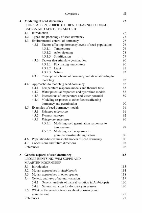
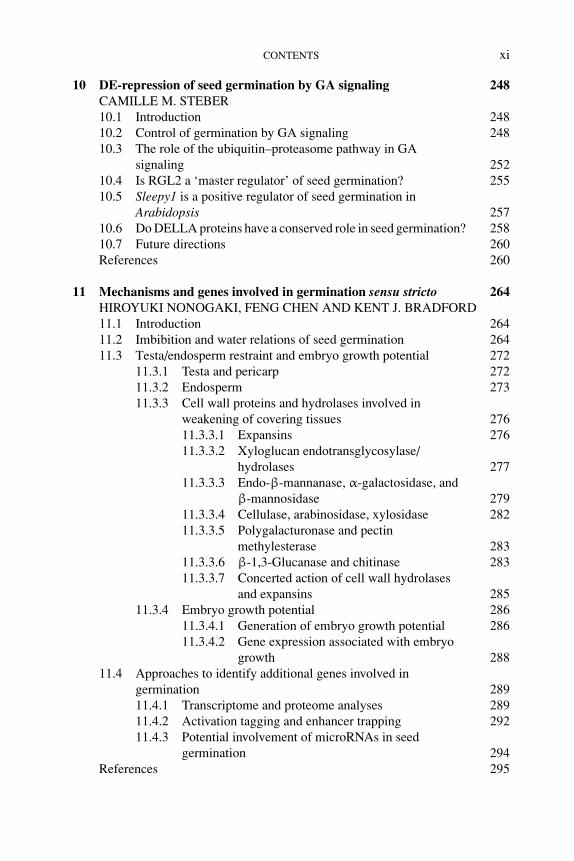
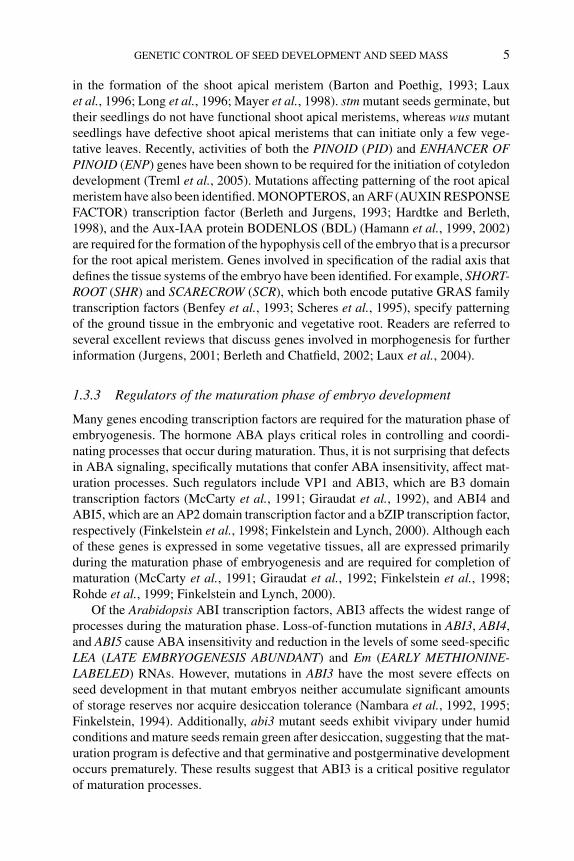
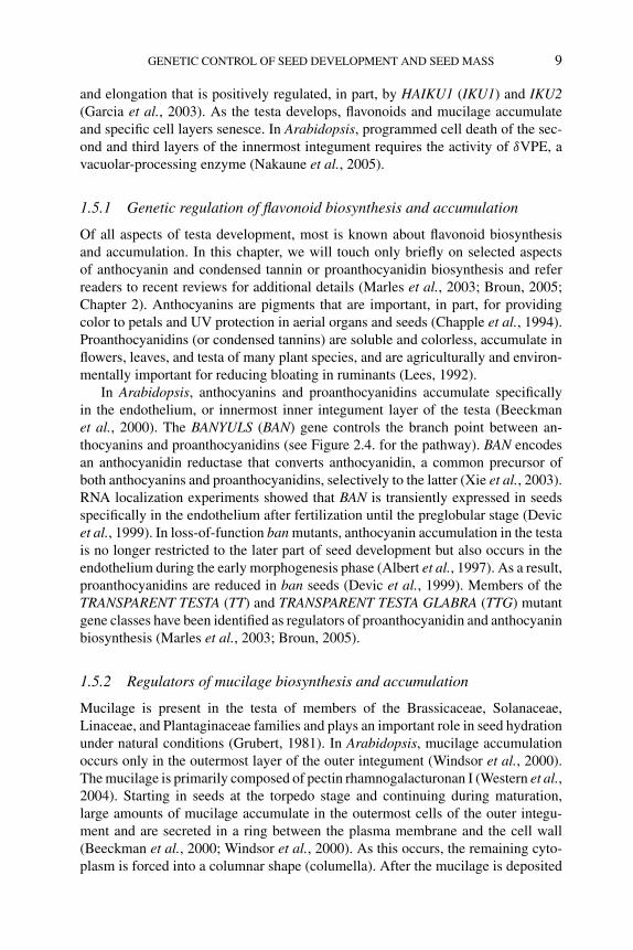
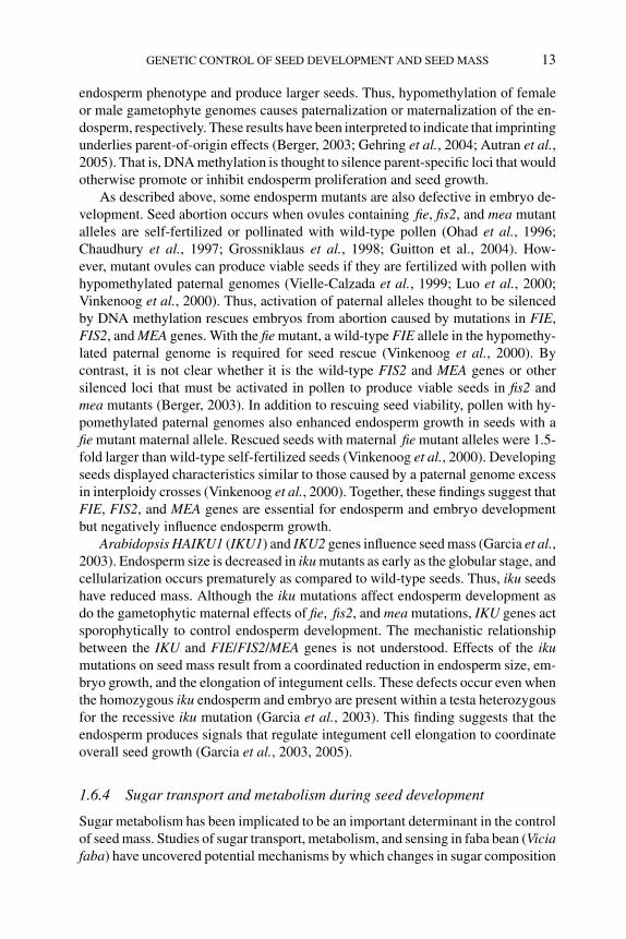
Figure 1.1 Seed development in Arabidopsis. (A) Section through a mature ovule. (B–E) Sections
through seeds containing globular stage (B), heart stage (C), linear cotyledon stage (D), and mature
green stage (E) embryos. (F) Diagram of a mature embryo sac modified from Drews et al. (1998).
Black, gray, and white areas indicate the nucleus, cytoplasm, and vacuole, respectively. (G–I) Images
of whole-mount embryos at the globular (G), heart (H), and linear cotyledon (I) stages. (J) Part of
the seed containing a mature green-stage embryo with the embryo, the single layer of endosperm,
and the testa. (A–E, J) Images from paraffin (A, E) and plastic (B–D, J) embedded sections through
a mature ovule and developing seeds stained with (A, E) periodic acid-Schiff’s, (B–D) toluidine blue
O, and (J) periodic acid-Schiff’s counterstained with aniline blue black. (G–I) Nomarski images of
cleared embryos in seeds. Arrowhead, free endosperm nucleus; a, embryonic axis; ant, antipodal cell;
c, cotyledon; cc, central cell; ce, chalazal endosperm; ch, chalazal pole; e, embryo; end, peripheral
endosperm; ep, embryo proper; es, embryo sac; me, micropylar endosperm; mp, micropylar pole; s,
suspensor; syn, synergid cell; te, testa. Scale bars = 50 μm.
the ovule (Figures 1.1A and 1.1F) each fuse with one sperm cell from the pollentube to form the zygote and the endosperm cell, respectively. The zygote undergoesa series of differentiation events, resulting in the formation of the mature embryoand the suspensor, an ephemeral structure that supports the embryo physically andphysiologically during early embryogenesis (Figures 1.1E, 1.1G–1.1I). The embryoconsists of two major embryonic organ systems: the axis from which the bodyof the vegetative plant is derived and the cotyledon(s) that often functions as astorage organ for macromolecular reserves in dicotyledonous species (Figure 1.1E;reviewed by West and Harada, 1993; Goldberg et al., 1994). The fertilized central cellundergoes a series of nuclear divisions without cytokinesis, resulting in the formationof a syncytium (Figures 1.1B and 1.1C) that later cellularizes (Figure 1.1D; Olsen,2004). The endosperm is either a transient or persistent structure within the seed.In nonendospermic seeds, such as soybean (Glycine max) and peanuts (Arachishypogaea), the endosperm is absorbed completely by the developing embryo. Theendosperm is retained to varying degrees in endospermic seeds. For example, matureArabidopsis thaliana seeds have a single layer of endosperm cells, whereas theendosperm makes up most of the mass of cereal seeds (Figure 1.1J). The other
P1: OTE/SPH P2: OTE
BLUK053-Bradford September 27, 2006 7:27
GENETIC CONTROL OF SEED DEVELOPMENT AND SEED MASS 3
major component of the seed is the testa that is derived from the integuments of theovule and, therefore, is of maternal origin (Figure 1.1J; Boesewinkel and Bouman,1984; Bewley and Black, 1995). Many seeds also have a transient perisperm derivedfrom the nucellar cells of the ovule. In some plants, the perisperm persists and servesas a storage organ (Bewley and Black, 1995).
1.3 Genetic control of embryo development
Embryogenesis in higher plants can be divided conceptually into two distinct phases.During the early morphogenesis phase, the basic body plan of the plant is established.Polarity is expressed with establishment of the shoot–root axis, apical–basal andradial domains from which morphological structures are derived and the embryonicorgan and tissue systems are formed (Jurgens, 2001; Berleth and Chatfield, 2002;Laux et al., 2004). During the maturation phase late in embryogenesis, the embryoaccumulates storage macromolecules including proteins, lipids, and starch, acquiresthe ability to withstand the stresses of desiccation, and enters a state of developmentaland metabolic quiescence as it desiccates (Harada, 1997).
Large-scale genetic screens with T-DNA or chemical mutagens have been usedto identify genes that play critical roles in embryo development. Such screens haveidentified many mutants displaying defects in embryogenesis in Arabidopsis, maize(Zea mays), and rice (Oyrza sativa) (Clark and Sheridan, 1991; Meinke, 1991;Hong et al., 1995). For example, screens for Arabidopsis mutants initially identifiedmore than 300 embryo-defective (emb) mutations that affected embryo development(Meinke, 1991). It has been estimated that these screens have not reached saturationand that there are between 500 and 1000 EMB genes in Arabidopsis (Franzmannet al., 1995; McElver et al., 2001). This estimate indicates that embryo developmentis a complex process with hundreds, if not thousands, of required gene products. Arecent report describing the nucleotide sequences of genes corresponding to 250 embmutations showed that they were enriched for proteins predicted to play roles in basiccellular functions (Tzafrir et al., 2004; also see www.seedgenes.org). For example,the Arabidopsis embryo-lethal schleperless (slp) mutant that has reduced cotyledonshas a defective plastidic chaperonin 60α gene (Apuya et al., 2001), the bio mutantthat arrests at the heart or cotyledon stage is defective in the biotin synthase gene(Patton et al., 1998), and the twin2 (twn2) mutant that produces secondary embryos isdefective in a valyl-tRNA synthetase gene (Zhang and Somerville, 1997). Althoughthe emb mutations affect embryo development, many of these EMB gene productsmay be required for gametogenesis. The mutant alleles may be too weak to be lethalin gametophytes or mutant gametophytes may compensate for the lack of geneproducts because they are surrounded by maternal/paternal tissues heterozygous forthe mutations (Springer et al., 2000).
1.3.1 Central regulators of embryogenesis
As will be discussed, most regulators of processes that occur during embryogenesisfunction during either the morphogenesis phase or the maturation phase. How-ever, the LEAFY COTYLEDON (LEC) genes define a small class of regulators that
P1: OTE/SPH P2: OTE
BLUK053-Bradford September 27, 2006 7:27
4 SEED DEVELOPMENT, DORMANCY AND GERMINATION
function during both phases (reviewed by Harada, 2001). lec1 and lec2 mutants wereidentified in screens for emb mutations, whereas the other lec mutant, fusca3 (fus3),was found in screens for seeds with purple coloration. Analyses of these mutantsshowed that the LEC genes function early in embryogenesis to maintain suspensorcell fate and specify cotyledon identity. Late in embryogenesis, the LEC genes arerequired for the initiation and/or maintenance of maturation and the repression ofprecocious germination (Meinke, 1992; Baumlein et al., 1994; Keith et al., 1994;Meinke et al., 1994; West et al., 1994; Lotan et al., 1998; Stone et al., 2001). Giventheir roles both early and late in embryogenesis, it has been speculated that the LECgenes serve to coordinate the morphogenesis and maturation phases (Harada, 2001).
Additional insight into LEC gene function came from ectopic expression stud-ies. Misexpression of LEC1, LEC2, or FUS3 confers embryonic characteristics toseedlings in that they resemble embryos morphologically and express genes encod-ing seed proteins such as 12S and 2S storage proteins and oleosin (Lotan et al., 1998;Stone et al., 2001; Gazzarrini et al., 2004). Moreover, ectopic LEC1 or LEC2 activityis sufficient to induce somatic embryo formation from vegetative cells, suggestingthat these genes enhance cellular competence to undergo embryogenesis (Lotanet al., 1998; Stone et al., 2001). We note that ectopic expression of WUSCHEL(WUS), a gene with a key role in establishing the shoot apical meristem in the em-bryo (Laux et al., 1996), and of BABYBOOM (BBM), an AP2 (APETALA 2) domainprotein, can also induce somatic embryogenesis in vegetative tissues (Boutilier et al.,2002; Zuo et al., 2002). It is not known whether LEC, WUS, and BBM operate incommon or distinct pathways.
The LEC genes all encode regulatory proteins. LEC1 is a homolog of the HAP3subunit of the CCAAT-binding transcription factor (Lotan et al., 1998). LEC2 andFUS3 both possess B3 domains, a DNA-binding domain most similar to that foundin the transcription factors VIVIPAROUS 1 (VP1) from maize and its apparentortholog, ABSCISIC ACID (ABA) INSENSITIVE 3 (ABI3) from Arabidopsis(McCarty et al., 1991; Giraudat et al., 1992; Luerßen et al., 1998; Stone et al.,2001). Consistent with the finding that defects caused by the lec mutations are pri-marily limited to embryogenesis, all three LEC genes are expressed primarily duringseed development (Lotan et al., 1998; Luerßen et al., 1998; Stone et al., 2001).
1.3.2 Genes involved in the morphogenesis phase of embryo development
Genes specifically involved in establishing the embryo body plan have been identi-fied through screens for emb and seedling-defective mutations. The rationale for thelatter strategy is that mutations that cause defects in morphological development ofthe embryo may not cause lethality, but the defects are likely to be readily detectableduring seedling development (Mayer et al., 1991).
The apical–basal axis of the embryo comprises several pattern elements includ-ing the cotyledons, shoot apical meristem, hypocotyl, root, and root apical meristem(reviewed by Berleth and Chatfield, 2002). Genetic screens identified genes involvedin establishing these elements. For example, SHOOTMERISTEMLESS (STM) andWUS, which encode different types of homeodomain proteins, play critical roles
P1: OTE/SPH P2: OTE
BLUK053-Bradford September 27, 2006 7:27
GENETIC CONTROL OF SEED DEVELOPMENT AND SEED MASS 5
in the formation of the shoot apical meristem (Barton and Poethig, 1993; Lauxet al., 1996; Long et al., 1996; Mayer et al., 1998). stm mutant seeds germinate, buttheir seedlings do not have functional shoot apical meristems, whereas wus mutantseedlings have defective shoot apical meristems that can initiate only a few vege-tative leaves. Recently, activities of both the PINOID (PID) and ENHANCER OFPINOID (ENP) genes have been shown to be required for the initiation of cotyledondevelopment (Treml et al., 2005). Mutations affecting patterning of the root apicalmeristem have also been identified. MONOPTEROS, an ARF (AUXIN RESPONSEFACTOR) transcription factor (Berleth and Jurgens, 1993; Hardtke and Berleth,1998), and the Aux-IAA protein BODENLOS (BDL) (Hamann et al., 1999, 2002)are required for the formation of the hypophysis cell of the embryo that is a precursorfor the root apical meristem. Genes involved in specification of the radial axis thatdefines the tissue systems of the embryo have been identified. For example, SHORT-ROOT (SHR) and SCARECROW (SCR), which both encode putative GRAS familytranscription factors (Benfey et al., 1993; Scheres et al., 1995), specify patterningof the ground tissue in the embryonic and vegetative root. Readers are referred toseveral excellent reviews that discuss genes involved in morphogenesis for furtherinformation (Jurgens, 2001; Berleth and Chatfield, 2002; Laux et al., 2004).
1.3.3 Regulators of the maturation phase of embryo development
Many genes encoding transcription factors are required for the maturation phase ofembryogenesis. The hormone ABA plays critical roles in controlling and coordi-nating processes that occur during maturation. Thus, it is not surprising that defectsin ABA signaling, specifically mutations that confer ABA insensitivity, affect mat-uration processes. Such regulators include VP1 and ABI3, which are B3 domaintranscription factors (McCarty et al., 1991; Giraudat et al., 1992), and ABI4 andABI5, which are an AP2 domain transcription factor and a bZIP transcription factor,respectively (Finkelstein et al., 1998; Finkelstein and Lynch, 2000). Although eachof these genes is expressed in some vegetative tissues, all are expressed primarilyduring the maturation phase of embryogenesis and are required for completion ofmaturation (McCarty et al., 1991; Giraudat et al., 1992; Finkelstein et al., 1998;Rohde et al., 1999; Finkelstein and Lynch, 2000).
Of the Arabidopsis ABI transcription factors, ABI3 affects the widest range ofprocesses during the maturation phase. Loss-of-function mutations in ABI3, ABI4,and ABI5 cause ABA insensitivity and reduction in the levels of some seed-specificLEA (LATE EMBRYOGENESIS ABUNDANT) and Em (EARLY METHIONINE-LABELED) RNAs. However, mutations in ABI3 have the most severe effects onseed development in that mutant embryos neither accumulate significant amountsof storage reserves nor acquire desiccation tolerance (Nambara et al., 1992, 1995;Finkelstein, 1994). Additionally, abi3 mutant seeds exhibit vivipary under humidconditions and mature seeds remain green after desiccation, suggesting that the mat-uration program is defective and that germinative and postgerminative developmentoccurs prematurely. These results suggest that ABI3 is a critical positive regulatorof maturation processes.
P1: OTE/SPH P2: OTE
BLUK053-Bradford September 27, 2006 7:27
6 SEED DEVELOPMENT, DORMANCY AND GERMINATION
The ABI transcription factors interact with each other and with the LEC tran-scription factors during the maturation phase. ABI3 interacts physically with ABI5(Nakamura et al., 2001), although no other interactions among the ABI transcriptionfactors or among the ABI and LEC transcription factors have been reported. ABI3,ABI4, and ABI5 each interacts genetically with LEC1 and FUS3 to control maturationprocesses (Parcy et al., 1997; Brocard-Gifford et al., 2003). However, the precisemechanistic relationship between the ABI and LEC transcription factors remainselusive. For example, LEC1 and FUS3 have been proposed to regulate ABI3 levels(Parcy et al., 1997), whereas LEC1 has been proposed to act through FUS3 andABI3 to control processes during maturation (Kagaya et al., 2005). It has also beenproposed that LEC2 regulates FUS3 accumulation (Kroj et al., 2003). Although theregulatory circuits controlling maturation processes remain to be defined, the ABIand LEC transcription factors clearly play key roles.
A loss-of-function mutation in the Arabidopsis Enhanced Em Level (EEL) genethat encodes a bZIP protein of the same class as ABI5 results in increased levelsof AtEm1 RNA, but has no other visible mutant seed phenotype (Bensmihen et al.,2002). By contrast, abi5 mutant seeds have decreased levels of AtEm1 (Finkelsteinand Lynch, 2000). Bensmihen et al. (2002) showed that EEL and ABI5 compete forthe same binding sites in the AtEm1 promoter, leading to the balanced regulationof AtEm1 in wild-type seeds. Reduction of the expression of EEL alone or of EELin combination with AtbZIP67 and AREB3 (ABA-Responsive Element Bindingprotein 3) that encode closely related bZIP proteins did not generate noticeabledefects in seed development (Bensmihen et al., 2005).
In addition to conferring insensitivity to ABA, the abi mutations also cause de-fects in sugar signaling (Arenas-Huertero et al., 2000; Huijser et al., 2000; Labyet al., 2000; Rook et al., 2001). abi4 and, to a lesser extent, abi3 and abi5 mutantseeds germinate more readily than wild-type seeds in the presence of high concentra-tions of sugars. Consistent with this observation, ectopic expression of ABI3, ABI4,or ABI5 causes seedling growth to be hypersensitive to sugars (Finkelstein, 2000;Brocard et al., 2002; Chapter 12). Although the link between sugar and ABA sig-nal transduction pathways remains unclear, these findings indicate the importanceof sugar signaling in maturation processes, as will be discussed in greater detailsubsequently.
1.4 Genetic control of endosperm development
The endosperm, formed by fertilization of the central cell of the female gametophytewith a sperm cell, initially undergoes free nuclear divisions to produce a syncytium ofendosperm nuclei (Figures 1.1B and 1.1C; reviewed by Berger, 2003; Olsen, 2004).In both maize and Arabidopsis, as representatives of cereals and dicotyledonousoilseeds, respectively, patterning of the endosperm coenocyte (multinucleated cell)occurs with the establishment of three domains along the anterior–posterior axis:the micropylar (Arabidopsis) or embryo-surrounding (maize) region, the peripheralregion, and the chalazal (Arabidopsis) or basal endosperm transfer layer (maize)
P1: OTE/SPH P2: OTE
BLUK053-Bradford September 27, 2006 7:27
GENETIC CONTROL OF SEED DEVELOPMENT AND SEED MASS 7
region (Figures 1.1B–1.1D). Furthermore, maize possesses two additional regions:the aleurone and subaleurone layers. In maize, the basal endosperm transfer layeris involved in transferring nutrients from maternal tissues into the endosperm. Therole of the micropylar or embryo-surrounding region is not known, although it hasbeen speculated to be involved in nourishing the embryo. The peripheral regionbecomes the starchy endosperm in maize in which starch and proteins are stored toprovide nutrients for the growing seedling. The Arabidopsis endosperm also appearsto play a nutritional role, although it serves primarily to nourish the developingembryo rather than the seedling. Cellularization of the endosperm occurs in a wave-like manner from the micropylar endosperm through the peripheral endosperm tothe chalazal endosperm (Figures 1.1C and 1.1D). The endosperm is absorbed bythe growing embryo during seed development in nonendospermic seeds such aspea (Pisum sativum) and endospermic seeds with little persistent endosperm suchas Arabidopsis (Figure 1.1E). By contrast, the endosperm constitutes most of themature seed in cereals.
1.4.1 Genes required for cereal endosperm development
Genetic screens for defective kernel (dek) mutations of maize have identified genesrequired for seed development, and many have primary effects on the endosperm(Neuffer and Sheridan, 1980; Sheridan and Neuffer, 1980; Scanlon et al., 1994).The dek mutants exhibit reductions in seed weight, mitotic activity, and, in mostcases, endoreduplication (DNA replication without cell division) patterns (Kowleset al., 1992). Some of the genes have roles in specifying cell fate. For example,aleurone cell specification requires CRINKLY4, which encodes a protein similar totumor necrosis factor receptor-like receptor kinase (Becraft et al., 1996). Aleuronecell fate is restricted to the outer epidermis of the endosperm by the activity of Dek1,which encodes a membrane protein with homology to animal calpains (Lid et al.,2002). Other maize endosperm mutations have been identified that affect differentparts of the endosperm. For example, the reduced grain filling 1 (rgf1) mutation thataffects pedicel development and the expression of transfer layer-specific markerscauses final grain weight to be 30% of wild type, although the embryo is not affectedby the mutation (Maitz et al., 2000).
1.4.2 Genes that repress autonomous endosperm development
A group of Arabidopsis genes has been shown to be involved in controlling en-dosperm development based on their striking mutant phenotypes. Loss-of-functionmutations in the FERTILIZATION INDEPENDENT ENDOSPERM (FIE), FER-TILIZATION INDEPENDENT SEED 2 (FIS2), MEDEA (MEA), MULTICOPY SU-PRESSOR OF IRA (MSI1), BORGIA (BGA), and RETINOBLASTOMA RELATEDPROTEIN 1 (RBR1) genes cause the initiation of endosperm development in theabsence of fertilization (Ohad et al., 1996; Chaudhury et al., 1997; Grossniklauset al., 1998; Kohler et al., 2003a; Ebel et al., 2004; Guitton et al., 2004). Theseare gametophytic maternal-effect mutations, suggesting that the wild-type alleles of
P1: OTE/SPH P2: OTE
BLUK053-Bradford September 27, 2006 7:27
8 SEED DEVELOPMENT, DORMANCY AND GERMINATION
these genes operate in the female gametophyte, most likely the central cell, to con-trol endosperm development. These genes encode proteins related to those presentin polycomb group complexes that are known to be associated with gene silencingthrough histone modification. Therefore, these genes are likely to repress transcrip-tion at specific loci (Hsieh et al., 2003; Orlando, 2003). The implication is thatendosperm development is repressed prior to fertilization by polycomb group com-plexes that inhibit the expression of genes required for the initiation of endospermdevelopment.
MEA encodes a SET-domain protein similar to the Drosophila ENHANCER OFZESTE (Grossniklaus et al., 1998; Kiyosue et al., 1999; Luo et al., 1999). Other SET-domain proteins have histone methyltransferase activity, indicating their importancein establishing a repressed chromatin state (Cao and Zhang, 2004). FIE, a proteinwith seven WD40 repeats that has similarity to Drosophila EXTRA SEX COMBS(Ohad et al., 1999), interacts physically with MEA (Luo et al., 2000; Spillane et al.,2000; Yadegari et al., 2000), as do their polycomb group counterparts in Drosophila.FIS2 encodes a zinc-finger transcription factor similar to Drosophila SUPPRESSOROF ZESTE 12 (Luo et al., 1999) and may play a role in binding with the promoters ofendosperm-specific genes. MSI1, a WD40 protein with similarity to the Drosophilaretinoblastoma-binding protein P55, interacts physically with FIE and is likely tobe part of the polycomb group complex (Kohler et al., 2003a). RBR1 is similar toa mammalian retinoblastoma tumor suppressor protein that is a negative regulatorof G1 to S phase transition of the cell cycle (Kong et al., 2000). In Drosophila,polycomb group proteins interact with a retinoblastoma protein to form a complexthat represses homeotic genes, thereby blocking mitosis (Dahiya et al., 2001). Theidentity of the BGA gene is not yet known, but the mutant phenotype suggeststhat it encodes a polycomb group protein. A target gene repressed by this plantpolycomb group complex, PHERES1 (PHE1), has been identified (Kohler et al.,2003b). PHERES1 is expressed in the embryo and endosperm after fertilization andis thought to play a role in seed development.
Fertilization of ovules containing mutant alleles of the FIE, FIS, MEA, or MSI1genes causes seed development to be initiated (Ohad et al., 1996; Chaudhury et al.,1997; Grossniklaus et al., 1998; Guitton et al., 2004). However, embryos withinthe mutant seeds abort at about the torpedo stage and endosperm overproliferationoccurs (Kiyosue et al., 1999). Thus, genes encoding these plant polycomb groupproteins have an additional role after fertilization to restrict the extent of endospermgrowth. The effects of these mutations on seed mass will be discussed subsequently.
1.5 Genetic aspects of testa development
The testa encloses the embryo and endosperm. The testa serves a protective functionagainst physical and ultraviolet light damage and, in some seeds, aids in seed dis-persal and in the control of germination (Bewley and Black, 1995). In Arabidopsis,the mature ovule has inner and outer integuments, comprising three and two celllayers, respectively. After fertilization, integuments undergo anticlinal cell division
P1: OTE/SPH P2: OTE
BLUK053-Bradford September 27, 2006 7:27
GENETIC CONTROL OF SEED DEVELOPMENT AND SEED MASS 9
and elongation that is positively regulated, in part, by HAIKU1 (IKU1) and IKU2(Garcia et al., 2003). As the testa develops, flavonoids and mucilage accumulateand specific cell layers senesce. In Arabidopsis, programmed cell death of the sec-ond and third layers of the innermost integument requires the activity of δVPE, avacuolar-processing enzyme (Nakaune et al., 2005).
1.5.1 Genetic regulation of flavonoid biosynthesis and accumulation
Of all aspects of testa development, most is known about flavonoid biosynthesisand accumulation. In this chapter, we will touch only briefly on selected aspectsof anthocyanin and condensed tannin or proanthocyanidin biosynthesis and referreaders to recent reviews for additional details (Marles et al., 2003; Broun, 2005;Chapter 2). Anthocyanins are pigments that are important, in part, for providingcolor to petals and UV protection in aerial organs and seeds (Chapple et al., 1994).Proanthocyanidins (or condensed tannins) are soluble and colorless, accumulate inflowers, leaves, and testa of many plant species, and are agriculturally and environ-mentally important for reducing bloating in ruminants (Lees, 1992).
In Arabidopsis, anthocyanins and proanthocyanidins accumulate specificallyin the endothelium, or innermost inner integument layer of the testa (Beeckmanet al., 2000). The BANYULS (BAN) gene controls the branch point between an-thocyanins and proanthocyanidins (see Figure 2.4. for the pathway). BAN encodesan anthocyanidin reductase that converts anthocyanidin, a common precursor ofboth anthocyanins and proanthocyanidins, selectively to the latter (Xie et al., 2003).RNA localization experiments showed that BAN is transiently expressed in seedsspecifically in the endothelium after fertilization until the preglobular stage (Devicet al., 1999). In loss-of-function ban mutants, anthocyanin accumulation in the testais no longer restricted to the later part of seed development but also occurs in theendothelium during the early morphogenesis phase (Albert et al., 1997). As a result,proanthocyanidins are reduced in ban seeds (Devic et al., 1999). Members of theTRANSPARENT TESTA (TT) and TRANSPARENT TESTA GLABRA (TTG) mutantgene classes have been identified as regulators of proanthocyanidin and anthocyaninbiosynthesis (Marles et al., 2003; Broun, 2005).
1.5.2 Regulators of mucilage biosynthesis and accumulation
Mucilage is present in the testa of members of the Brassicaceae, Solanaceae,Linaceae, and Plantaginaceae families and plays an important role in seed hydrationunder natural conditions (Grubert, 1981). In Arabidopsis, mucilage accumulationoccurs only in the outermost layer of the outer integument (Windsor et al., 2000).The mucilage is primarily composed of pectin rhamnogalacturonan I (Western et al.,2004). Starting in seeds at the torpedo stage and continuing during maturation,large amounts of mucilage accumulate in the outermost cells of the outer integu-ment and are secreted in a ring between the plasma membrane and the cell wall(Beeckman et al., 2000; Windsor et al., 2000). As this occurs, the remaining cyto-plasm is forced into a columnar shape (columella). After the mucilage is deposited
P1: OTE/SPH P2: OTE
BLUK053-Bradford September 27, 2006 7:27
10 SEED DEVELOPMENT, DORMANCY AND GERMINATION
and the columella is formed, a secondary cell wall is deposited external to the col-umella. During late maturation, the testa, including the mucilage, desiccates. Whenthe mature seed imbibes, the mucilage is extruded from the testa to form a halo.Lack of mucilage extrusion is associated with reduced germination and seedlingestablishment under dry conditions (Penfield et al., 2001), suggesting that mucilageis an important factor for seedling establishment under adverse conditions.
It has been proposed that there are at least three pathways controlling mucilagebiosynthesis in the Arabidopsis testa (Western et al., 2004). Two of these pathwaysinvolve a complex composed of AP2 and TTG1 and a bHLH (basic helix-loop-helix) protein, possibly ENHANCER OF GLABRA 3 (EGL3) and/or TT8, and atissue-specific MYB transcription factor. In one pathway, this complex activatesTTG2, which acts through a yet unknown factor to enhance mucilage biosynthesisand accumulation. In the second pathway, the complex activates GL2, which thenactivates MUCILAGE MODIFIED 4 (MUM4), a putative pectin biosynthetic gene.In a third pathway, MYB61 is required for mucilage biosynthesis and accumulationbut is not regulated by AP2 and TTG1.
1.6 Control of seed mass
The mass of a seed is determined by the number and size of cells within tissues thatconstitute its three components: embryo, endosperm, and testa. As will be discussed,interactions among the different components ultimately determine the size of theseed. In this section, we focus on genetic and physiological factors that influenceseed mass.
1.6.1 Genetic factors affecting seed mass
Different accessions or ecotypes of Arabidopsis exhibit variations in seed mass.For example, seeds of the Cape Verde Island (Cvi) ecotype are almost double theweight of those of Ler (Landsberg erecta) (Alonso-Blanco et al., 1999). This naturalvariation in seed mass results from differences in both the number and size of cellswithin the embryo and testa. Reciprocal crosses between the two ecotypes showedthat the variation in cell number is controlled primarily by maternal factors, whereascell size is affected by nonmaternal factors. A number of quantitative trait loci (QTL)affecting seed mass co-localize with QTL affecting ovule and/or seed number, fruitsize, and total leaf number. These traits emphasize the importance of maternal factorsin controlling seed mass. In many species, there exists a negative correlation betweenseed mass and the number of seeds produced, which is attributed to limitations inresources from the mother plant (Harper et al., 1970). Consistent with this finding,plants in which fruit number is controlled either by selected pollination of male sterilemutants or by manual removal of flowers produce seeds that are heavier than thoseof control plants (Jofuku et al., 2005; Ohto et al., 2005). Late-flowering mutants ofArabidopsis produce more leaves than wild type, and they set more seeds (Alonso-Blanco et al., 1999). Moreover, seeds from late-flowering mutants are slightly larger
P1: OTE/SPH P2: OTE
BLUK053-Bradford September 27, 2006 7:27
GENETIC CONTROL OF SEED DEVELOPMENT AND SEED MASS 11
than those from wild-type plants. This finding suggests that the increase in resourceavailability due to higher leaf number may overcome the negative effects of seednumber on seed mass (Alonso-Blanco et al., 1999). These correlations indicate thatgenetic factors affecting seed mass operate primarily through maternal tissues.
1.6.2 Testa development and seed mass
The testa is a maternal component of the seed that affects its mass. Differentiationof ovule integuments into the testa occurs initially through cell divisions, which inArabidopsis end by about 4 days after pollination (DAP). This is followed by a periodof cell elongation that continues until the seed achieves its maximal length at about6–7 DAP (Alonso-Blanco et al., 1999; Western et al., 2000; Garcia et al., 2005).Although the seed has reached maximal size by 6–7 DAP, the embryo is only at thebent-cotyledon stage and has not yet filled the seed, suggesting that testa growth maybe a determinant of seed mass. The ttg2 mutation that affects pigment productionin the testa is a maternal mutation that affects seed mass (Garcia et al., 2005). Thismutation causes reduced growth of integument cells, suggesting that TTG2 mayregulate integument cell elongation. Similarly, seed mass is also affected by manytt mutations that cause defects in flavonoid synthesis in the seed coat (Debeaujonet al., 2000; Chapter 2). It is possible that some intermediates or by-products of theproanthocyanidin biosynthesis pathway accumulate in integument cell walls andchange the cells’ competence to elongate.
1.6.3 Endosperm development and seed mass
The endosperm has a major influence on seed mass (Scott et al., 1998; Garcia et al.,2003). Because the endosperm makes up most of the cereal seed, genes that affectendosperm growth have direct effects on seed size. The endosperm also affectsthe mass of nonendospermic seeds and of endospermic seeds with small amounts ofresidual endosperm. In such seeds, the endosperm proliferates extensively and servesas a sink for nutrients supplied by the maternal plant during early seed developmentto provide nutrients to sustain embryo growth later in embryogenesis. In addition,endosperm growth in coordination with testa growth early during seed developmentmay have a primary role in determining the size of the postfertilization embryo sac,which, in turn, appears to influence embryo and seed size.
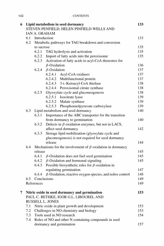
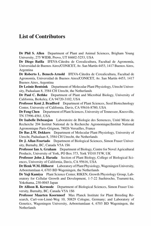
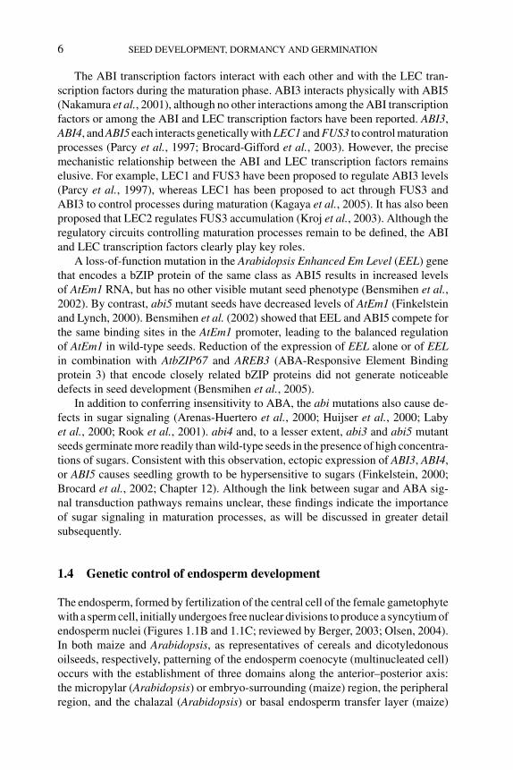
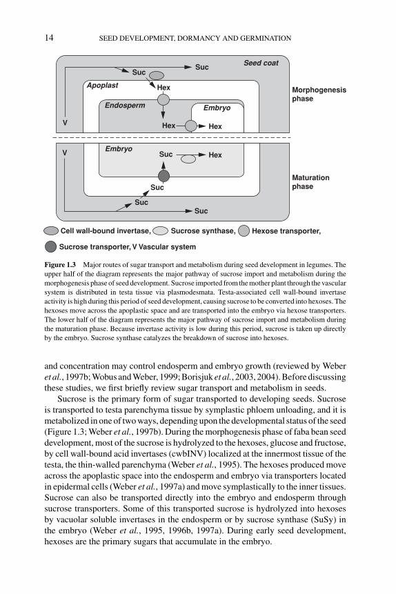
Endosperm growth is influenced by parent-of-origin effects. For example, in-terploidy crosses between diploid females and tetraploid males produce offspringwith an excess of paternal genomes (Figure 1.2). This imbalance between parentalgenomes results in overproliferation of endosperm nuclei, a delay in the onset ofendosperm cellularization, and overgrowth of the chalazal endosperm involved intransferring nutrient resources from the mother plant (Scott et al., 1998). Because ofthese changes in endosperm development, seeds with an excess of paternal genomesare larger than progeny from diploid parents. Reciprocal crosses, resulting in an ex-cess of maternal genomes, have the opposite effect on endosperm development andgive rise to smaller seeds (Figure 1.2; Scott et al., 1998). The parental conflict theory
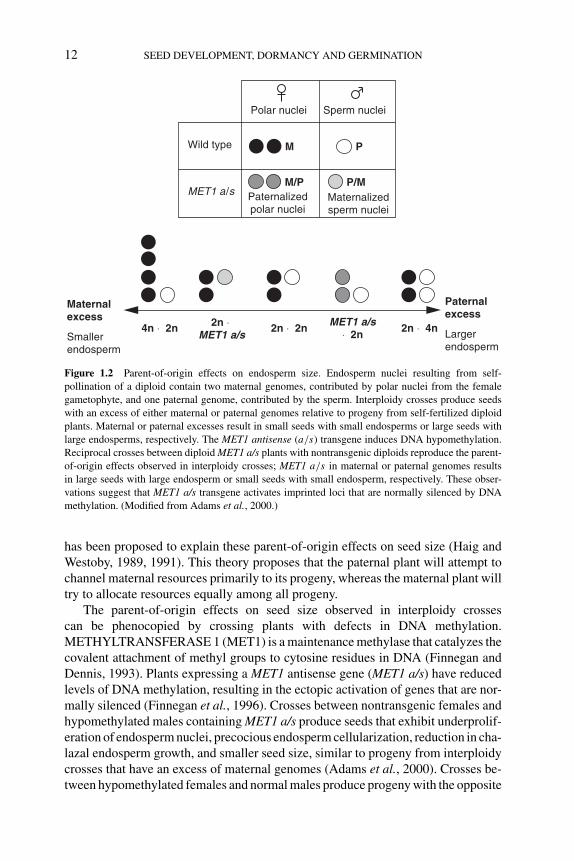
P1: OTE/SPH P2: OTE
BLUK053-Bradford September 27, 2006 7:27
12 SEED DEVELOPMENT, DORMANCY AND GERMINATION
Polar nuclei Sperm nuclei
Wild type
MET1 a/s Paternalized polar nuclei
Maternalized sperm nuclei
M
M/P P/M
P
Maternal excess
Smaller endosperm
Larger endosperm
Paternal excess
2n × 4nMET1 a/s
× 2n2n × 2n2n ×
MET1 a/s4n × 2n
Figure 1.2 Parent-of-origin effects on endosperm size. Endosperm nuclei resulting from self-
pollination of a diploid contain two maternal genomes, contributed by polar nuclei from the female
gametophyte, and one paternal genome, contributed by the sperm. Interploidy crosses produce seeds
with an excess of either maternal or paternal genomes relative to progeny from self-fertilized diploid
plants. Maternal or paternal excesses result in small seeds with small endosperms or large seeds with
large endosperms, respectively. The MET1 antisense (a/s) transgene induces DNA hypomethylation.
Reciprocal crosses between diploid MET1 a/s plants with nontransgenic diploids reproduce the parent-
of-origin effects observed in interploidy crosses; MET1 a/s in maternal or paternal genomes results
in large seeds with large endosperm or small seeds with small endosperm, respectively. These obser-
vations suggest that MET1 a/s transgene activates imprinted loci that are normally silenced by DNA
methylation. (Modified from Adams et al., 2000.)
has been proposed to explain these parent-of-origin effects on seed size (Haig andWestoby, 1989, 1991). This theory proposes that the paternal plant will attempt tochannel maternal resources primarily to its progeny, whereas the maternal plant willtry to allocate resources equally among all progeny.
The parent-of-origin effects on seed size observed in interploidy crossescan be phenocopied by crossing plants with defects in DNA methylation.METHYLTRANSFERASE 1 (MET1) is a maintenance methylase that catalyzes thecovalent attachment of methyl groups to cytosine residues in DNA (Finnegan andDennis, 1993). Plants expressing a MET1 antisense gene (MET1 a/s) have reducedlevels of DNA methylation, resulting in the ectopic activation of genes that are nor-mally silenced (Finnegan et al., 1996). Crosses between nontransgenic females andhypomethylated males containing MET1 a/s produce seeds that exhibit underprolif-eration of endosperm nuclei, precocious endosperm cellularization, reduction in cha-lazal endosperm growth, and smaller seed size, similar to progeny from interploidycrosses that have an excess of maternal genomes (Adams et al., 2000). Crosses be-tween hypomethylated females and normal males produce progeny with the opposite
P1: OTE/SPH P2: OTE
BLUK053-Bradford September 27, 2006 7:27
GENETIC CONTROL OF SEED DEVELOPMENT AND SEED MASS 13
endosperm phenotype and produce larger seeds. Thus, hypomethylation of femaleor male gametophyte genomes causes paternalization or maternalization of the en-dosperm, respectively. These results have been interpreted to indicate that imprintingunderlies parent-of-origin effects (Berger, 2003; Gehring et al., 2004; Autran et al.,2005). That is, DNA methylation is thought to silence parent-specific loci that wouldotherwise promote or inhibit endosperm proliferation and seed growth.
As described above, some endosperm mutants are also defective in embryo de-velopment. Seed abortion occurs when ovules containing fie, fis2, and mea mutantalleles are self-fertilized or pollinated with wild-type pollen (Ohad et al., 1996;Chaudhury et al., 1997; Grossniklaus et al., 1998; Guitton et al., 2004). How-ever, mutant ovules can produce viable seeds if they are fertilized with pollen withhypomethylated paternal genomes (Vielle-Calzada et al., 1999; Luo et al., 2000;Vinkenoog et al., 2000). Thus, activation of paternal alleles thought to be silencedby DNA methylation rescues embryos from abortion caused by mutations in FIE,FIS2, and MEA genes. With the fie mutant, a wild-type FIE allele in the hypomethy-lated paternal genome is required for seed rescue (Vinkenoog et al., 2000). Bycontrast, it is not clear whether it is the wild-type FIS2 and MEA genes or othersilenced loci that must be activated in pollen to produce viable seeds in fis2 andmea mutants (Berger, 2003). In addition to rescuing seed viability, pollen with hy-pomethylated paternal genomes also enhanced endosperm growth in seeds with afie mutant maternal allele. Rescued seeds with maternal fie mutant alleles were 1.5-fold larger than wild-type self-fertilized seeds (Vinkenoog et al., 2000). Developingseeds displayed characteristics similar to those caused by a paternal genome excessin interploidy crosses (Vinkenoog et al., 2000). Together, these findings suggest thatFIE, FIS2, and MEA genes are essential for endosperm and embryo developmentbut negatively influence endosperm growth.
Arabidopsis HAIKU1 (IKU1) and IKU2 genes influence seed mass (Garcia et al.,2003). Endosperm size is decreased in iku mutants as early as the globular stage, andcellularization occurs prematurely as compared to wild-type seeds. Thus, iku seedshave reduced mass. Although the iku mutations affect endosperm development asdo the gametophytic maternal effects of fie, fis2, and mea mutations, IKU genes actsporophytically to control endosperm development. The mechanistic relationshipbetween the IKU and FIE/FIS2/MEA genes is not understood. Effects of the ikumutations on seed mass result from a coordinated reduction in endosperm size, em-bryo growth, and the elongation of integument cells. These defects occur even whenthe homozygous iku endosperm and embryo are present within a testa heterozygousfor the recessive iku mutation (Garcia et al., 2003). This finding suggests that theendosperm produces signals that regulate integument cell elongation to coordinateoverall seed growth (Garcia et al., 2003, 2005).
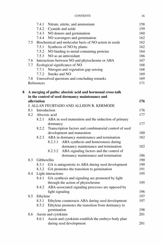
1.6.4 Sugar transport and metabolism during seed development
Sugar metabolism has been implicated to be an important determinant in the controlof seed mass. Studies of sugar transport, metabolism, and sensing in faba bean (Viciafaba) have uncovered potential mechanisms by which changes in sugar composition
P1: OTE/SPH P2: OTE
BLUK053-Bradford September 27, 2006 7:27
14 SEED DEVELOPMENT, DORMANCY AND GERMINATION
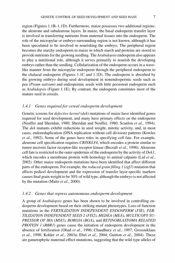
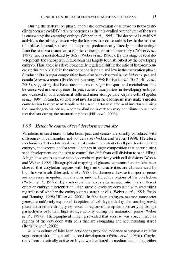
Cell wall-bound invertase,
Sucrose transporter, V Vascular system
Sucrose synthase, Hexose transporter,
Maturation phase
Morphogenesisphase
Seed coat
Embryo
Embryo
Endosperm
Suc
Apoplast
Suc
Hex
Hex Hex
Hex
Suc
Suc
SucSuc
V
V
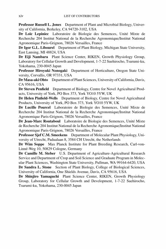
Figure 1.3 Major routes of sugar transport and metabolism during seed development in legumes. The
upper half of the diagram represents the major pathway of sucrose import and metabolism during the
morphogenesis phase of seed development. Sucrose imported from the mother plant through the vascular
system is distributed in testa tissue via plasmodesmata. Testa-associated cell wall-bound invertase
activity is high during this period of seed development, causing sucrose to be converted into hexoses. The
hexoses move across the apoplastic space and are transported into the embryo via hexose transporters.
The lower half of the diagram represents the major pathway of sucrose import and metabolism during
the maturation phase. Because invertase activity is low during this period, sucrose is taken up directly
by the embryo. Sucrose synthase catalyzes the breakdown of sucrose into hexoses.
and concentration may control endosperm and embryo growth (reviewed by Weberet al., 1997b; Wobus and Weber, 1999; Borisjuk et al., 2003, 2004). Before discussingthese studies, we first briefly review sugar transport and metabolism in seeds.
Sucrose is the primary form of sugar transported to developing seeds. Sucroseis transported to testa parenchyma tissue by symplastic phloem unloading, and it ismetabolized in one of two ways, depending upon the developmental status of the seed(Figure 1.3; Weber et al., 1997b). During the morphogenesis phase of faba bean seeddevelopment, most of the sucrose is hydrolyzed to the hexoses, glucose and fructose,by cell wall-bound acid invertases (cwbINV) localized at the innermost tissue of thetesta, the thin-walled parenchyma (Weber et al., 1995). The hexoses produced moveacross the apoplastic space into the endosperm and embryo via transporters locatedin epidermal cells (Weber et al., 1997a) and move symplastically to the inner tissues.Sucrose can also be transported directly into the embryo and endosperm throughsucrose transporters. Some of this transported sucrose is hydrolyzed into hexosesby vacuolar soluble invertases in the endosperm or by sucrose synthase (SuSy) inthe embryo (Weber et al., 1995, 1996b, 1997a). During early seed development,hexoses are the primary sugars that accumulate in the embryo.
P1: OTE/SPH P2: OTE
BLUK053-Bradford September 27, 2006 7:27
GENETIC CONTROL OF SEED DEVELOPMENT AND SEED MASS 15
During the maturation phase, apoplastic conversion of sucrose to hexoses de-clines because cwbINV activity decreases as the thin-walled parenchyma of the testais crushed by the enlarging embryo (Weber et al., 1995). The decrease in cwbINVactivity is the primary reason why the hexoses to sucrose ratio is low in the matura-tion phase. Instead, sucrose is transported predominantly directly into the embryofrom the testa via a sucrose transporter at the epidermis of the embryo (Weber et al.,1997a) and is metabolized by SuSy (Weber et al., 1996b). By this stage of seed de-velopment, the endosperm in faba bean has largely been absorbed by the developingembryo. Thus, there is a developmentally regulated shift in the ratio of hexoses to su-crose; this ratio is high in the morphogenesis phase and low in the maturation phase.Similar shifts in sugar composition have also been observed in Arabidopsis, pea andcanola (Brassica napus) (Focks and Benning, 1998; Borisjuk et al., 2002; Hill et al.,2003), suggesting that basic mechanisms of sugar transport and metabolism maybe conserved in these species. In pea, sucrose transporters in developing embryosare localized in both epidermal cells and inner storage parenchyma cells (Tegederet al., 1999). In canola, soluble acid invertases in the endosperm may make a greatercontribution to sucrose metabolism than seed coat-associated acid invertases duringthe morphogenesis phase, whereas alkaline invertases may contribute to sucrosemetabolism during the maturation phase (Hill et al., 2003).
1.6.5 Metabolic control of seed development and size
Variations in seed mass in faba bean, pea, and cereals are strictly correlated withdifferences in cell number and not cell size (Wobus and Weber, 1999). Therefore,mechanisms that dictate seed size must control the extent of cell proliferation in theembryo, endosperm, and/or testa. Changes in sugar composition that occur duringseed development are thought to control the shift from cell division to seed filling.A high hexoses to sucrose ratio is correlated positively with cell divisions (Wobusand Weber, 1999). Histographical mapping of glucose concentrations in faba beanshowed that cotyledon regions with high mitotic activities are characterized byhigh hexose levels (Borisjuk et al., 1998). Furthermore, hexose transporter genesare expressed in epidermal cells over mitotically active regions of the cotyledons(Weber et al., 1997a). By contrast, a low hexoses to sucrose ratio has a differenteffect on embryo differentiation. High sucrose levels are correlated with seed fillingregardless of whether the embryo stores starch or oils (Weber et al., 1995; Focksand Benning, 1998; Hill et al., 2003). In faba bean embryos, sucrose transportergenes are uniformly expressed in epidermal cell layers during the morphogenesisphase but are more strongly expressed in regions of the epidermis overlying storageparenchyma cells with high storage activity during the maturation phase (Weberet al., 1997a). Histographical imaging revealed that sucrose was concentrated inregions of the cotyledon with cells that are elongating and accumulating starch(Borisjuk et al., 2002).
In vitro culture of faba bean cotyledons provided evidence to support a role forsugar composition in controlling seed development (Weber et al., 1996a). Cotyle-dons from mitotically active embryos were cultured in medium containing either

P1: OTE/SPH P2: OTE
BLUK053-Bradford September 27, 2006 7:27
16 SEED DEVELOPMENT, DORMANCY AND GERMINATION
a high or a low ratio of hexoses to sucrose. Cotyledons cultured in high hexosesmedium maintained mitotic activity, whereas cotyledons cultured with high su-crose medium showed a significant decrease in mitotic activity. Cotyledons in thehigh sucrose medium displayed an increase in cell size and nuclear diameter, andthey started to accumulate starch and storage proteins. Conversely, cotyledons frommaturation-phase embryos exhibited a decrease in storage protein gene expressionwhen cultured with high hexoses medium (Weber et al., 1996a).
Cell wall-bound invertases have been implicated to control seed mass throughtheir role in modulating changes in sugar composition during seed development.For example, two faba bean genotypes that produce seeds of different sizes werecompared (Weber et al., 1996a). Seed coats from the large-seeded genotype have amuch larger region of thin-walled parenchyma that is the site at which sucrose is hy-drolyzed by cwbINV. This seed coat tissue remained intact in the large-seeded geno-type for a longer period of seed development than it did in small-seeded genotypes.Thus, extension of the period of cwbINV activity correlates with a prolonged periodof cell division and a larger seed. A similar mechanism to control cell proliferationin seed tissues through cwbINV also appears to operate in cereals. Maize miniature1(mn1) mutant seeds are smaller than wild type; endosperm weight is approximately20% of normal (Vilhar et al., 2002). The Mn1 gene encodes an endosperm-specificcwbINV that normally accumulates in the basal endosperm transfer layer (Chenget al., 1996). Thus, mn1 mutant endosperm has reduced cwbINV activity that maycause reduced mitotic activity.
In contrast to the role of hexoses produced by cwbINV in cell proliferation,sucrose is related to seed filling. The role of sucrose in the control of seed sizewas tested in Vicia narbonesis, a species closely related to faba bean. A secretedform of yeast invertase expressed during the maturation phase under the control ofthe legumin B4 promoter acts in the apoplast of cotyledons (Weber et al., 1998;Wobus and Weber, 1999). The accumulation of starch and some storage proteinswas reduced in transgenic cotyledon cells. These transgenic cells also had largeand persistent vacuoles instead of protein bodies, and hexoses were present at highlevels in the vacuoles. As a result, mature dried seeds were smaller than wild-typeseeds. The shrunken1 (sh1) mutant of maize also emphasizes the importance ofsucrose in seed filling. sh1 seeds are defective in the accumulation of storage starchin endosperm and are smaller than wild-type seeds (Chourey and Nelson, 1976).Because sh1 encodes SuSy, mutant seeds are unable to utilize sucrose efficiently forfurther metabolism (Echt and Chourey, 1985).
Seeds from the Arabidopsis floral homeotic mutant ap2 are larger than wild typeand they accumulate more storage proteins and lipids (Jofuku et al., 2005; Ohtoet al., 2005). Although the ap2 mutation causes reduced fertility due to defects inflower structure, the mutation appears to have direct effects on seed mass that gobeyond the positive effects that result from a reduction in seed number. Similar to theeffects of natural variation observed among Arabidopsis ecotypes, the ap2 mutationaffects seed mass by increasing cell number and size (Ohto et al., 2005). Sugarcomposition differed between developing ap2 mutant seeds and wild-type seeds.The ratio of hexoses to sucrose remains high in ap2 mutants for an extended period
P1: OTE/SPH P2: OTE
BLUK053-Bradford September 27, 2006 7:27
GENETIC CONTROL OF SEED DEVELOPMENT AND SEED MASS 17
of time as compared to wild type (Ohto et al., 2005). This defect in sugar metabolismmay explain the increase in cell number in ap2 mutant seeds; the prolonged period inwhich the hexoses to sucrose ratio remains high may allow cell division to continuefor a longer period of time in ap2 mutants. Maternal tissues appear to be responsiblefor these changes in sugar composition. The recessive ap2 mutation affects seed massthrough the maternal sporophyte and endosperm genomes (Jofuku et al., 2005). Inaddition, the ap2 mutant testa has irregularly shaped epidermal cells that lack anepidermal plateau and produces reduced amounts of mucilage (Jofuku et al., 1994).Thus, it is possible that the ap2 mutation may induce defects, perhaps involvingtesta-associated cwbINV, that extend the period of seed development during whichthe ratio of hexoses to sucrose remains high.
1.7 Perspective
In the past 15 years, significant progress has been made in defining regulatory genesthat control aspects of seed development. A major challenge is to define the genenetworks that operate during seed development. Identifying all of the genes that areexpressed in the different compartments of the seed and defining their functions willallow for dissection of the regulatory circuitries controlling seed development andthe definition of gene products that underlie the processes that occur during seeddevelopment. Initial progress toward this goal has been made with descriptions ofRNA populations in developing seeds and/or embryos (Girke et al., 2000; Henniget al., 2004; Casson et al., 2005; Grimanelli et al., 2005; Ma et al., 2005). Althoughmuch work remains to be done, the tools are in place to accomplish these goals.Ultimately, this knowledge, integrated with our understanding of the physiological,biochemical, and morphological processes in seeds, will provide a comprehensiveunderstanding of seed development.
Acknowledgements
We thank Sandra Floyd and Samantha Duong for help in preparing some of the micrographs used
in Figure 1.1 and Siobhan Braybrook for her comments about the manuscript. Research cited in this
review from our laboratory was supported by grants from the National Science Foundation and the
United States Department of Energy.
References
S. Adams, R. Vinkenoog, M. Spielman, H. Dickinson and R. Scott (2000) Parent-of-origin effects on
seed development in Arabidopsis thaliana require DNA methylation. Development 127, 2493–
2502.
S. Albert, M. Delseny and M. Devic (1997) BANYULS, a novel negative regulator of flavonoid biosyn-
thesis in the Arabidopsis seed coat. The Plant Journal 11, 289–299.
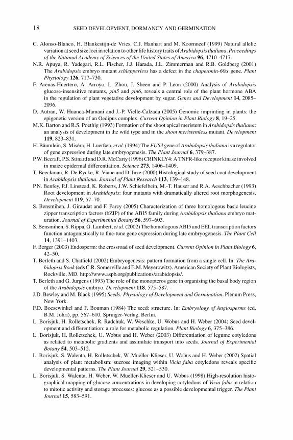
P1: OTE/SPH P2: OTE
BLUK053-Bradford September 27, 2006 7:27
18 SEED DEVELOPMENT, DORMANCY AND GERMINATION
C. Alonso-Blanco, H. Blankestijn-de Vries, C.J. Hanhart and M. Koornneef (1999) Natural allelic
variation at seed size loci in relation to other life history traits of Arabidopsis thaliana. Proceedingsof the National Academy of Sciences of the United States of America 96, 4710–4717.
N.R. Apuya, R. Yadegari, R.L. Fischer, J.J. Harada, J.L. Zimmerman and R.B. Goldberg (2001)
The Arabidopsis embryo mutant schlepperless has a defect in the chaperonin-60α gene. PlantPhysiology 126, 717–730.
F. Arenas-Huertero, A. Arroyo, L. Zhou, J. Sheen and P. Leon (2000) Analysis of Arabidopsisglucose-insensitive mutants, gin5 and gin6, reveals a central role of the plant hormone ABA
in the regulation of plant vegetative development by sugar. Genes and Development 14, 2085–
2096.
D. Autran, W. Huanca-Mamani and J.-P. Vielle-Calzada (2005) Genomic imprinting in plants: the
epigenetic version of an Oedipus complex. Current Opinion in Plant Biology 8, 19–25.
M.K. Barton and R.S. Poethig (1993) Formation of the shoot apical meristem in Arabidopsis thaliana:
an analysis of development in the wild type and in the shoot meristemless mutant. Development119, 823–831.
H. Baumlein, S. Misera, H. Luerßen, et al. (1994) The FUS3 gene of Arabidopsis thaliana is a regulator
of gene expression during late embryogenesis. The Plant Journal 6, 379–387.
P.W. Becraft, P.S. Stinard and D.R. McCarty (1996) CRINKLY4: A TNFR-like receptor kinase involved
in maize epidermal differentiation. Science 273, 1406–1409.
T. Beeckman, R. De Rycke, R. Viane and D. Inze (2000) Histological study of seed coat development
in Arabidopsis thaliana. Journal of Plant Research 113, 139–148.
P.N. Benfey, P.J. Linstead, K. Roberts, J.W. Schiefelbein, M.-T. Hauser and R.A. Aeschbacher (1993)
Root development in Arabidopsis: four mutants with dramatically altered root morphogenesis.
Development 119, 57–70.
S. Bensmihen, J. Giraudat and F. Parcy (2005) Characterization of three homologous basic leucine
zipper transcription factors (bZIP) of the ABI5 family during Arabidopsis thaliana embryo mat-
uration. Journal of Experimental Botany 56, 597–603.
S. Bensmihen, S. Rippa, G. Lambert, et al. (2002) The homologous ABI5 and EEL transcription factors
function antagonistically to fine-tune gene expression during late embryogenesis. The Plant Cell14, 1391–1403.
F. Berger (2003) Endosperm: the crossroad of seed development. Current Opinion in Plant Biology 6,
42–50.
T. Berleth and S. Chatfield (2002) Embryogenesis: pattern formation from a single cell. In: The Ara-bidopsis Book (eds C.R. Somerville and E.M. Meyerowitz). American Society of Plant Biologists,
Rockville, MD. http://www.aspb.org/publications/arabidopsis/.
T. Berleth and G. Jurgens (1993) The role of the monopteros gene in organising the basal body region
of the Arabidopsis embryo. Development 118, 575–587.
J.D. Bewley and M. Black (1995) Seeds: Physiology of Development and Germination. Plenum Press,
New York.
F.D. Boesewinkel and F. Bouman (1984) The seed: structure. In: Embryology of Angiosperms (ed.
B.M. Johri), pp. 567–610. Springer-Verlag, Berlin.
L. Borisjuk, H. Rolletschek, R. Radchuk, W. Weschke, U. Wobus and H. Weber (2004) Seed devel-
opment and differentiation: a role for metabolic regulation. Plant Biology 6, 375–386.
L. Borisjuk, H. Rolletschek, U. Wobus and H. Weber (2003) Differentiation of legume cotyledons
as related to metabolic gradients and assimilate transport into seeds. Journal of ExperimentalBotany 54, 503–512.
L. Borisjuk, S. Walenta, H. Rolletschek, W. Mueller-Klieser, U. Wobus and H. Weber (2002) Spatial
analysis of plant metabolism: sucrose imaging within Vicia faba cotyledons reveals specific
developmental patterns. The Plant Journal 29, 521–530.
L. Borisjuk, S. Walenta, H. Weber, W. Mueller-Klieser and U. Wobus (1998) High-resolution histo-
graphical mapping of glucose concentrations in developing cotyledons of Vicia faba in relation
to mitotic activity and storage processes: glucose as a possible developmental trigger. The PlantJournal 15, 583–591.
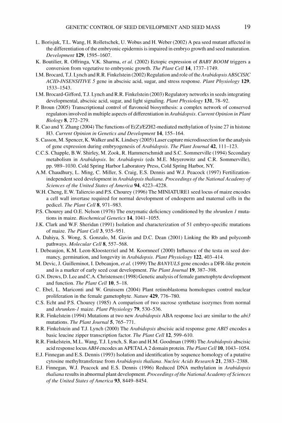
P1: OTE/SPH P2: OTE
BLUK053-Bradford September 27, 2006 7:27
GENETIC CONTROL OF SEED DEVELOPMENT AND SEED MASS 19
L. Borisjuk, T.L. Wang, H. Rolletschek, U. Wobus and H. Weber (2002) A pea seed mutant affected in
the differentiation of the embryonic epidermis is impaired in embryo growth and seed maturation.
Development 129, 1595–1607.
K. Boutilier, R. Offringa, V.K. Sharma, et al. (2002) Ectopic expression of BABY BOOM triggers a
conversion from vegetative to embryonic growth. The Plant Cell 14, 1737–1749.
I.M. Brocard, T.J. Lynch and R.R. Finkelstein (2002) Regulation and role of the Arabidopsis ABSCISICACID-INSENSITIVE 5 gene in abscisic acid, sugar, and stress response. Plant Physiology 129,
1533–1543.
I.M. Brocard-Gifford, T.J. Lynch and R.R. Finkelstein (2003) Regulatory networks in seeds integrating
developmental, abscisic acid, sugar, and light signaling. Plant Physiology 131, 78–92.
P. Broun (2005) Transcriptional control of flavonoid biosynthesis: a complex network of conserved
regulators involved in multiple aspects of differentiation in Arabidopsis. Current Opinion in PlantBiology 8, 272–279.
R. Cao and Y. Zhang (2004) The functions of E(Z)/EZH2-mediated methylation of lysine 27 in histone
H3. Current Opinion in Genetics and Development 14, 155–164.
S. Casson, M. Spencer, K. Walker and K. Lindsey (2005) Laser capture microdissection for the analysis
of gene expression during embryogenesis of Arabidopsis. The Plant Journal 42, 111–123.
C.C.S. Chapple, B.W. Shirley, M. Zook, R. Hammerschmidt and S.C. Sommerville (1994) Secondary
metabolism in Arabidopsis. In: Arabidopsis (eds M.E. Meyerowitz and C.R. Sommerville),
pp. 989–1030. Cold Spring Harbor Laboratory Press, Cold Spring Harbor, NY.
A.M. Chaudhury, L. Ming, C. Miller, S. Craig, E.S. Dennis and W.J. Peacock (1997) Fertilization-
independent seed development in Arabidopsis thaliana. Proceedings of the National Academy ofSciences of the United States of America 94, 4223–4228.
W.H. Cheng, E.W. Taliercio and P.S. Chourey (1996) The MINIATURE1 seed locus of maize encodes
a cell wall invertase required for normal development of endosperm and maternal cells in the
pedicel. The Plant Cell 8, 971–983.
P.S. Chourey and O.E. Nelson (1976) The enzymatic deficiency conditioned by the shrunken 1 muta-
tions in maize. Biochemical Genetics 14, 1041–1055.
J.K. Clark and W.F. Sheridan (1991) Isolation and characterization of 51 embryo-specific mutations
of maize. The Plant Cell 3, 935–951.
A. Dahiya, S. Wong, S. Gonzalo, M. Gavin and D.C. Dean (2001) Linking the Rb and polycomb
pathways. Molecular Cell 8, 557–568.
I. Debeaujon, K.M. Leon-Kloosterziel and M. Koornneef (2000) Influence of the testa on seed dor-
mancy, germination, and longevity in Arabidopsis. Plant Physiology 122, 403–414.
M. Devic, J. Guilleminot, I. Debeaujon, et al. (1999) The BANYULS gene encodes a DFR-like protein
and is a marker of early seed coat development. The Plant Journal 19, 387–398.
G.N. Drews, D. Lee and C.A. Christensen (1998) Genetic analysis of female gametophyte development
and function. The Plant Cell 10, 5–18.
C. Ebel, L. Mariconti and W. Gruissem (2004) Plant retinoblastoma homologues control nuclear
proliferation in the female gametophyte. Nature 429, 776–780.
C.S. Echt and P.S. Chourey (1985) A comparison of two sucrose synthetase isozymes from normal
and shrunken-1 maize. Plant Physiology 79, 530–536.
R.R. Finkelstein (1994) Mutations at two new Arabidopsis ABA response loci are similar to the abi3mutations. The Plant Journal 5, 765–771.
R.R. Finkelstein and T.J. Lynch (2000) The Arabidopsis abscisic acid response gene ABI5 encodes a
basic leucine zipper transcription factor. The Plant Cell 12, 599–610.
R.R. Finkelstein, M.L. Wang, T.J. Lynch, S. Rao and H.M. Goodman (1998) The Arabidopsis abscisic
acid response locus ABI4 encodes an APETALA 2 domain protein. The Plant Cell 10, 1043–1054.
E.J. Finnegan and E.S. Dennis (1993) Isolation and identification by sequence homology of a putative
cytosine methyltransferase from Arabidopsis thaliana. Nucleic Acids Research 21, 2383–2388.
E.J. Finnegan, W.J. Peacock and E.S. Dennis (1996) Reduced DNA methylation in Arabidopsisthaliana results in abnormal plant development. Proceedings of the National Academy of Sciencesof the United States of America 93, 8449–8454.
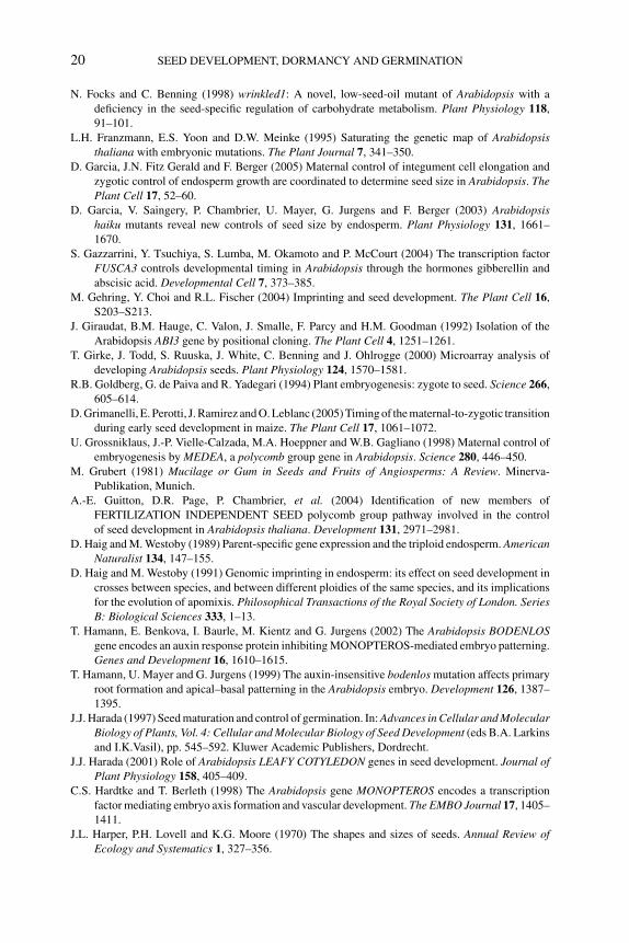
P1: OTE/SPH P2: OTE
BLUK053-Bradford September 27, 2006 7:27
20 SEED DEVELOPMENT, DORMANCY AND GERMINATION
N. Focks and C. Benning (1998) wrinkled1: A novel, low-seed-oil mutant of Arabidopsis with a
deficiency in the seed-specific regulation of carbohydrate metabolism. Plant Physiology 118,
91–101.
L.H. Franzmann, E.S. Yoon and D.W. Meinke (1995) Saturating the genetic map of Arabidopsisthaliana with embryonic mutations. The Plant Journal 7, 341–350.
D. Garcia, J.N. Fitz Gerald and F. Berger (2005) Maternal control of integument cell elongation and
zygotic control of endosperm growth are coordinated to determine seed size in Arabidopsis. ThePlant Cell 17, 52–60.
D. Garcia, V. Saingery, P. Chambrier, U. Mayer, G. Jurgens and F. Berger (2003) Arabidopsishaiku mutants reveal new controls of seed size by endosperm. Plant Physiology 131, 1661–
1670.
S. Gazzarrini, Y. Tsuchiya, S. Lumba, M. Okamoto and P. McCourt (2004) The transcription factor
FUSCA3 controls developmental timing in Arabidopsis through the hormones gibberellin and
abscisic acid. Developmental Cell 7, 373–385.
M. Gehring, Y. Choi and R.L. Fischer (2004) Imprinting and seed development. The Plant Cell 16,
S203–S213.
J. Giraudat, B.M. Hauge, C. Valon, J. Smalle, F. Parcy and H.M. Goodman (1992) Isolation of the
Arabidopsis ABI3 gene by positional cloning. The Plant Cell 4, 1251–1261.
T. Girke, J. Todd, S. Ruuska, J. White, C. Benning and J. Ohlrogge (2000) Microarray analysis of
developing Arabidopsis seeds. Plant Physiology 124, 1570–1581.
R.B. Goldberg, G. de Paiva and R. Yadegari (1994) Plant embryogenesis: zygote to seed. Science 266,
605–614.
D. Grimanelli, E. Perotti, J. Ramirez and O. Leblanc (2005) Timing of the maternal-to-zygotic transition
during early seed development in maize. The Plant Cell 17, 1061–1072.
U. Grossniklaus, J.-P. Vielle-Calzada, M.A. Hoeppner and W.B. Gagliano (1998) Maternal control of
embryogenesis by MEDEA, a polycomb group gene in Arabidopsis. Science 280, 446–450.
M. Grubert (1981) Mucilage or Gum in Seeds and Fruits of Angiosperms: A Review. Minerva-
Publikation, Munich.
A.-E. Guitton, D.R. Page, P. Chambrier, et al. (2004) Identification of new members of
FERTILIZATION INDEPENDENT SEED polycomb group pathway involved in the control
of seed development in Arabidopsis thaliana. Development 131, 2971–2981.
D. Haig and M. Westoby (1989) Parent-specific gene expression and the triploid endosperm. AmericanNaturalist 134, 147–155.
D. Haig and M. Westoby (1991) Genomic imprinting in endosperm: its effect on seed development in
crosses between species, and between different ploidies of the same species, and its implications
for the evolution of apomixis. Philosophical Transactions of the Royal Society of London. SeriesB: Biological Sciences 333, 1–13.
T. Hamann, E. Benkova, I. Baurle, M. Kientz and G. Jurgens (2002) The Arabidopsis BODENLOSgene encodes an auxin response protein inhibiting MONOPTEROS-mediated embryo patterning.
Genes and Development 16, 1610–1615.
T. Hamann, U. Mayer and G. Jurgens (1999) The auxin-insensitive bodenlos mutation affects primary
root formation and apical–basal patterning in the Arabidopsis embryo. Development 126, 1387–
1395.
J.J. Harada (1997) Seed maturation and control of germination. In: Advances in Cellular and MolecularBiology of Plants, Vol. 4: Cellular and Molecular Biology of Seed Development (eds B.A. Larkins
and I.K.Vasil), pp. 545–592. Kluwer Academic Publishers, Dordrecht.
J.J. Harada (2001) Role of Arabidopsis LEAFY COTYLEDON genes in seed development. Journal ofPlant Physiology 158, 405–409.
C.S. Hardtke and T. Berleth (1998) The Arabidopsis gene MONOPTEROS encodes a transcription
factor mediating embryo axis formation and vascular development. The EMBO Journal 17, 1405–
1411.
J.L. Harper, P.H. Lovell and K.G. Moore (1970) The shapes and sizes of seeds. Annual Review ofEcology and Systematics 1, 327–356.
P1: OTE/SPH P2: OTE
BLUK053-Bradford September 27, 2006 7:27
GENETIC CONTROL OF SEED DEVELOPMENT AND SEED MASS 21
L. Hennig, W. Gruissem, U. Grossniklaus and C. Kohler (2004) Transcriptional programs of early
reproductive stages in Arabidopsis. Plant Physiology 135, 1765–1775.
L.M. Hill, E.R. Morley-Smith and S. Rawsthorne (2003) Metabolism of sugars in the endosperm of
developing seeds of oilseed rape. Plant Physiology 131, 228–236.
S.K. Hong, T. Aoki, H. Kitano, H. Satoh and Y. Nagato (1995) Phenotypic diversity of 188 rice embryo
mutants. Developmental Genetics 16, 298–310.
T.F. Hsieh, O. Hakim, N. Ohad and R.L. Fischer (2003) From flour to flower: how polycomb group
proteins influence multiple aspects of plant development. Trends in Plant Science 8, 439–445.
C. Huijser, A. Kortstee, J. Pego, P. Weisbeek, E. Wisman and S. Smeekens (2000) The ArabidopsisSUCROSE UNCOUPLED-6 gene is identical to ABSCISIC ACID INSENSITIVE-4: involvement
of abscisic acid in sugar responses. The Plant Journal 23, 577–585.
K.D. Jofuku, B. Boer, M.V. Montagu and J.K. Okamuro (1994) Control of Arabidopsis flower and
seed development by the homeotic gene APETALA2. The Plant Cell 6, 1211–1225.
K.D. Jofuku, P.K. Omidyar, Z. Gee and J.K. Okamuro (2005) Control of seed mass and seed yield by
the floral homeotic gene APETALA2. Proceedings of the National Academy of Sciences of theUnited States of America 102, 3117–3122.
G. Jurgens (2001) Apical–basal pattern formation in Arabidopsis embryogenesis. The EMBO Journal20, 3609–3616.
Y. Kagaya, R. Toyoshima, R. Okuda, H. Usui, A. Yamamoto and T. Hattori (2005) LEAFY COTYLE-
DON1 controls seed storage protein genes through its regulation of FUSCA3 and ABSCISICACID INSENSITIVE3. Plant and Cell Physiology 46, 399–406.
K. Keith, M. Kraml, N.G. Dengler and P. McCourt (1994) fusca3: a heterochronic mutation affecting
late embryo development in Arabidopsis. The Plant Cell 6, 589–600.
T. Kiyosue, N. Ohad, R. Yadegari, et al. (1999) Control of fertilization-independent endosperm devel-
opment by the MEDEA polycomb gene in Arabidopsis. Proceedings of the National Academy ofSciences of the United States of America 96, 4186–4191.
C. Kohler, L. Hennig, R. Bouveret, J. Gheyselinck, U. Grossniklaus and W. Gruissem (2003a) Ara-bidopsis MSI1 is a component of the MEA/FIE polycomb group complex and required for seed
development. The EMBO Journal 22, 4804–4814.
C. Kohler, L. Hennig, C. Spillane, S. Pien, W. Gruissem and U. Grossniklaus (2003b) The polycomb-
group protein MEDEA regulates seed development by controlling expression of the MADS-box
gene PHERES1. Genes and Development 17, 1540–1553.
L.-J. Kong, B.M. Orozco, J.L. Roe, et al. (2000) A geminivirus replication protein interacts with the
retinoblastoma protein through a novel domain to determine symptoms and tissue specificity of
infection in plants. The EMBO Journal 19, 3485–3495.
R.V. Knowles, M.D. McMullen, G. Yerk, R.L. Phillips, S. Kraemer and F. Srienc (1992) Endosperm
mitotic activity and endoreduplication in maize affected by defective kernel mutations. Genome35, 68–77.
T. Kroj, G. Savino, C. Valon, J. Giraudat and F. Parcy (2003) Regulation of storage protein gene
expression in Arabidopsis. Development 130, 6065–6073.
R.J. Laby, M.S. Kincaid, D. Kim and S.I. Gibson (2000) The Arabidopsis sugar-insensitive mutants
sis4 and sis5 are defective in abscisic acid synthesis and response. The Plant Journal 23, 587–596.
T. Laux, K.F.X. Mayer, J. Berger and G. Juergen (1996) The WUSCHEL gene is required for shoot
and floral meristem integrity in Arabidopsis. Development 122, 87–96.
T. Laux, T. Wurschum and H. Breuninger (2004) Genetic regulation of embryonic pattern formation.
The Plant Cell 16, S190–S202.
G.L. Lees (1992) Condensed tannins in some forage legumes: their role in the prevention of ruminant
pasture bloat. In: Plant Polyphenols, Basic Life Sciences, Vol. 59 (eds R.W. Hemingway and P.E.
Laks), pp. 915–934. Plenum Press, New York.
S.E. Lid, D. Gruis, R. Jung, et al. (2002) The defective kernel 1 (dek1) gene required for aleurone cell
development in the endosperm of maize grains encodes a membrane protein of the calpain gene
superfamily. Proceedings of the National Academy of Sciences of the United States of America99, 5460–5465.
P1: OTE/SPH P2: OTE
BLUK053-Bradford September 27, 2006 7:27
22 SEED DEVELOPMENT, DORMANCY AND GERMINATION
J.A. Long, E.I. Moan, J.I. Medford and M.K. Barton (1996) A member of the KNOTTED class of
homeodomain proteins encoded by the STM gene of Arabidopsis. Nature 379, 66–69.
T. Lotan, M.-A. Ohto, K.M. Yee, et al. (1998) Arabidopsis LEAFY COTYLEDON1 is sufficient to
induce embryo development in vegetative cells. Cell 93, 1195–1205.
H. Luerßen, V. Kirik, P. Herrmann and S. Misera (1998) FUSCA3 encodes a protein with a conserved
VP1/ABI3-like B3 domain which is of functional importance for the regulation of seed maturation
in Arabidopsis thaliana. The Plant Journal 15, 755–764.
M. Luo, P. Bilodeau, E.S. Dennis, W.J. Peacock and A. Chaudhury (2000) Expression and parent-of-
origin effects for FIS2, MEA, and FIE in the endosperm and embryo of developing Arabidopsisseeds. Proceedings of the National Academy of Sciences of the United States of America 97,
10637–10642.
M. Luo, P. Bilodeau, A. Koltunow, E.S. Dennis, W.J. Peacock and A.M. Chaudhury (1999) Genes
controlling fertilization-independent seed development in Arabidopsis thaliana. Proceedings ofthe National Academy of Sciences of the United States of America 96, 296–301.
L. Ma, N. Sun, X. Liu, Y. Jiao, H. Zhao and X.W. Deng (2005) Organ-specific expression of Arabidopsisgenome during development. Plant Physiology 138, 80–91.
M. Maitz, G. Santandrea, Z. Zhang, et al. (2000) rgf1, a mutation reducing grain filling in maize
through effects on basal endosperm and pedicel development. The Plant Journal 23, 29–42.
M.A.S. Marles, H. Ray and M.Y. Gruber (2003) New perspectives on proanthocyanidin biochemistry
and molecular regulation. Phytochemistry 64, 367–383.
K.F.X. Mayer, H. Schoof, A. Haecker, M. Lenhard, G. Jurgens and T. Laux (1998) Role of WUSCHELin regulating stem cell fate in the Arabidopsis shoot meristem. Cell 95, 805–815.
U. Mayer, R.A. Torres Ruiz, T. Berleth, S. Misera and G. Jurgens (1991) Mutations affecting body
organization in the Arabidopsis embryo. Nature 353, 402–407.
D.R. McCarty, T. Hattori, C.B. Carson, V. Vasil, M. Lazar and I.K. Vasil (1991) The Viviparous-1developmental gene of maize encodes a novel transcriptional activator. Cell 66, 895–906.
J. McElver, I. Tzafrir, G. Aux, et al. (2001) Insertional mutagenesis of genes required for seed devel-
opment in Arabidopsis thaliana. Genetics 159, 1751–1763.
D.W. Meinke (1991) Embryonic mutants of Arabidopsis thaliana. Developmental Genetics 12, 382–
392.
D.W. Meinke (1992) A homoeotic mutant of Arabidopsis thaliana with leafy cotyledons. Science 258,
1647–1650.
D.W. Meinke, L.H. Franzmann, T.C. Nickle and E.C. Yeung (1994) Leafy cotyledon mutants of
Arabidopsis. The Plant Cell 6, 1049–1064.
S. Nakamura, T.J. Lynch and R.R. Finkelstein (2001) Physical interactions between ABA response
loci of Arabidopsis. The Plant Journal 26, 627–635.
S. Nakaune, K. Yamada, M. Kondo, et al. (2005) A vacuolar processing enzyme, δVPE, is involved
in seed coat formation at the early stage of seed development. The Plant Cell 17, 876–887.
E. Nambara, K. Keith, P. McCourt and S. Naito (1995) A regulatory role for the ABI3 gene in the
establishment of embryo maturation in Arabidopsis thaliana. Development 121, 629–636.
E. Nambara, S. Naito and P. McCourt (1992) A mutant of Arabidopsis which is defective in seed
development and storage protein accumulation is a new abi3 allele. The Plant Journal 2, 435–
441.
M.G. Neuffer and W.F. Sheridan (1980) Defective kernel mutants of maize. I: Genetic and lethality
studies. Genetics 95, 929–944.
N. Ohad, L. Margossian, Y.-C. Hsu, C. Williams, P. Repetti and R.L. Fischer (1996) Mutation that
allows endosperm development without fertilization. Proceedings of the National Academy ofSciences of the United States of America 93, 5319–5324.
N. Ohad, R. Yadegari, L. Margossian, et al. (1999) Mutations in FIE, a WD polycomb group gene,
allow endosperm development without fertilization. The Plant Cell 11, 407–416.
M.-A. Ohto, R.L. Fischer, R.B. Goldberg, K. Nakamura and J.J. Harada (2005) Control of seed mass
by APETALA2. Proceedings of the National Academy of Sciences of the United States of America102, 3123–3128.
P1: OTE/SPH P2: OTE
BLUK053-Bradford September 27, 2006 7:27
GENETIC CONTROL OF SEED DEVELOPMENT AND SEED MASS 23
O.-A. Olsen (2004) Nuclear endosperm development in cereals and Arabidopsis thaliana. The PlantCell 16, S214–S227.
V. Orlando (2003) Polycomb, epigenomes, and control of cell identity. Cell 112, 599–606.
F. Parcy, C. Valon, A. Kohara, S. Misera and J. Giraudat (1997) The ABSCISIC ACID-INSENSITIVE3,
FUSCA3, and LEAFY COTYLEDON1 loci act in concert to control multiple aspects of Arabidopsisseed development. The Plant Cell 9, 1265–1277.
D.A. Patton, A.L. Schetter, L.H. Franzmann, K. Nelson, E.R. Ward and D.W. Meinke (1998) An
embryo-defective mutant of Arabidopsis disrupted in the final step of biotin synthesis. PlantPhysiology 116, 935–946.
S. Penfield, R.C. Meissner, D.A. Shoue, N.C. Carpita and M.W. Bevan (2001) MYB61 is required for
mucilage deposition and extrusion in the Arabidopsis seed coat. The Plant Cell 13, 2777–2791.
A. Rohde, M. Van Montagu and W. Boerjan (1999) The ABSCISIC ACID-INSENSITIVE 3 (ABI3) gene
is expressed during vegetative quiescence processes in Arabidopsis. Plant, Cell and Environment22, 261–270.
F. Rook, F. Corke, R. Card, G. Munz, C. Smith and M.W. Bevan (2001) Impaired sucrose-induction
mutants reveal the modulation of sugar-induced starch biosynthetic gene expression by abscisic
acid signaling. The Plant Journal 26, 421–433.
M.J. Scanlon, P.S. Stinard, M.G. James, A.M. Myers and D.S. Robertson (1994) Genetic analysis of
63 mutations affecting maize kernel development isolated from Mutator stocks. Genetics 136,
281–294.
B. Scheres, L. Di Laurenzio, V. Willemsen, et al. (1995) Mutations affecting the radial organisation of
the Arabidopsis root display specific defects throughout the embryonic axis. Development 121,
53–62.
R. Scott, M. Spielman, J. Bailey and H. Dickinson (1998) Parent-of-origin effects on seed development
in Arabidopsis thaliana. Development 125, 3329–3341.
W.F. Sheridan and M.G. Neuffer (1980) Defective kernel mutants of maize. II: Morphological and
embryo culture studies. Genetics 95, 945–960.
C. Spillane, C. MacDougall, C. Stock, et al. (2000) Interaction of the Arabidopsis polycomb group
proteins FIE and MEA mediates their common phenotypes.Current Biology 10, 1535–1538.
P. Springer, D. Holding, A. Groover, C. Yordan and R. Martienssen (2000) Parent-of-origin effects on
seed development in Arabidopsis thaliana. Development 127, 1815–1822.
T.A. Steeves (1983) The evolution and biological significance of seeds. Canadian Journal of Botany61, 3550–3560.
S.L. Stone, L.W. Kwong, K.M. Yee, et al. (2001) LEAFY COTYLEDON2 encodes a B3 domain
transcription factor that induces embryo development. Proceedings of the National Academy ofSciences of the United States of America 98, 11806–11811.
M. Tegeder, X.-D. Wang, W.B. Frommer, C.E. Offler and J.W. Patrick (1999) Sucrose transport into
developing seeds of Pisum sativum L. The Plant Journal 18, 151–161.
B.S. Treml, S. Winderl, R. Radykewicz, et al. (2005) The gene ENHANCER OF PINOID controls
cotyledon development in the Arabidopsis embryo. Development 132, 4063–4074.
I. Tzafrir, R. Pena-Muralla, A. Dickerman, et al. (2004) Identification of genes required for embryo
development in Arabidopsis. Plant Physiology 135, 1206–1220.
J. Vicente-Carbajosa and P. Carbonero (2005) Seed maturation: developing an intrusive phase to
accomplish a quiescent state. International Journal of Developmental Biology 49, 645–651.
J.-P. Vielle-Calzada, J. Thomas, C. Spillane, A. Coluccio, M.A. Hoeppner and U. Grossniklaus (1999)
Maintenance of genomic imprinting at the Arabidopsis medea locus requires zygotic DDM1activity. Genes and Development 13, 2971–2982.
B. Vilhar, A. Kladnik, A. Blejec, P.S. Chourey and M. Dermastia (2002) Cytometrical evidence that
the loss of seed weight in the miniature1 seed mutant of maize is associated with reduced mitotic
activity in the developing endosperm. Plant Physiology 129, 23–30.
R. Vinkenoog, M. Spielman, S. Adams, R.L. Fischer, H.G. Dickinson and R.J. Scott (2000) Hy-
pomethylation promotes autonomous endosperm development and rescues postfertilization
lethality in fie mutants. The Plant Cell 12, 2271–2282.
P1: OTE/SPH P2: OTE
BLUK053-Bradford September 27, 2006 7:27
24 SEED DEVELOPMENT, DORMANCY AND GERMINATION
V. Walbot (1978) Control mechanisms for plant embryogeny. In: Dormancy and Developmental Arrest(ed. M.E. Clutter), pp. 113–166. Academic Press, New York.
H. Weber, L. Borisjuk, U. Heim, P. Buchner and U. Wobus (1995) Seed coat-associated invertases of
fava bean control both unloading and storage functions: cloning of cDNAs and cell type-specific
expression. The Plant Cell 7, 1835–1846.
H. Weber, L. Borisjuk, U. Heim, N. Sauer and U. Wobus (1997a) A role for sugar transporters during
seed development: molecular characterization of a hexose and a sucrose carrier in fava bean seeds.
The Plant Cell 9, 895–908.
H. Weber, L. Borisjuk and U. Wobus (1996a) Controlling seed development and seed size in Viciafaba: a role for seed coat-associated invertases and carbohydrate state. The Plant Journal 10,
823–834.
H. Weber, L. Borisjuk and U. Wobus (1997b) Sugar import and metabolism during seed development.
Trends in Plant Science 2, 169–174.
H. Weber, P. Buchner, L. Borisjuk and U. Wobus (1996b) Sucrose metabolism during cotyledon
development of Vicia faba L. is controlled by the concerted action of both sucrose-phosphate
synthase and sucrose synthase: expression patterns, metabolic regulation and implications for
seed development. The Plant Journal 9, 841–850.
H. Weber, U. Heim, S. Golombek, L. Borisjuk, R. Manteuffel and U. Wobus (1998) Expression of
a yeast-derived invertase in developing cotyledons of Vicia narbonensis alters the carbohydrate
state and affects storage functions. The Plant Journal 16, 163–172.
M.A. West and J.J. Harada (1993) Embryogenesis in higher plants: an overview. The Plant Cell 5,
1361–1369.
M.A.L. West, K.L. Matsudaira Yee, J. Danao, et al. (1994) LEAFY COTYLEDON1 is an essential
regulator of late embryogenesis and cotyledon identity in Arabidopsis. The Plant Cell 6, 1731–
1745.
T.L. Western, D.J. Skinner and G.W. Haughn (2000) Differentiation of mucilage secretory cells of the
Arabidopsis seed coat. Plant Physiology, 122, 345–356.
T.L. Western, D.S. Young, G.H. Dean, W.L. Tan, A.L. Samuels and G.W. Haughn (2004) MUCILAGE-MODIFIED4 encodes a putative pectin biosynthetic enzyme developmentally regulated by
APETALA2, TRANSPARENT TESTA GLABRA1, and GLABRA2 in the Arabidopsis seed coat.
Plant Physiology 134, 296–306.
J.B. Windsor, V.V. Symonds, J. Mendenhall and A.M. Lloyd (2000) Arabidopsis seed coat develop-
ment: morphological differentiation of the outer integument. The Plant Journal 22, 483–493.
U. Wobus and H. Weber (1999) Sugars as signal molecules in plant seed development. BiologicalChemistry 380, 937–944.
D.-Y. Xie, S.B. Sharma, N.L. Paiva, D. Ferreira and R.A. Dixon (2003) Role of anthocyanidin reductase,
encoded by BANYULS, in plant flavonoid biosynthesis. Science 299, 396–399.
R. Yadegari, T. Kinoshita, O. Lotan, et al. (2000) Mutations in the FIE and MEA genes that encode
interacting polycomb proteins cause parent-of-origin effects on seed development by distinct
mechanisms. The Plant Cell 12, 2367–2382.
J.Z. Zhang and C.R. Somerville (1997) Suspensor-derived polyembryony caused by altered expres-
sion of valyl-tRNA synthetase in the twn2 mutant of Arabidopsis. Proceedings of the NationalAcademy of Sciences of the United States of America 94, 7349–7355.
J. Zuo, Q.-W. Niu, G. Frugis and N.-H. Chua (2002) The WUSCHEL gene promotes vegetative-to-
embryonic transition in Arabidopsis. The Plant Journal 30, 349–359.
P1: OTE/SPH P2: OTE
BLUK053-Bradford August 27, 2006 17:7
2 Seed coat development and dormancyIsabelle Debeaujon, Loıc Lepiniec, Lucille Pourceland Jean-Marc Routaboul
2.1 Introduction
Two major types of dormancy mechanisms exist: embryo dormancy where the agentsinhibiting germination are inherent to the embryo and coat-imposed dormancy whereinhibition is conferred by the seed envelopes (Bewley, 1997). Generally, complexinteractions between the embryo and covering structures determine whether a seedwill germinate. As a consequence, many intermediate situations are encountered dueto varying contributions by the embryo and envelopes to dormancy. Seed dormancy isa typical quantitative genetic character involving many genes and being substantiallyinfluenced by environmental effects (Koornneef et al., 2002; Alonso-Blanco et al.,2003). It is an adaptative trait allowing germination to occur during the most suitableperiod for seedling establishment and life cycle completion.
Embryo growth potential and characteristics of the seed envelopes that determinethe intrinsic capacity of a seed to germinate are established during development. Thepurpose of this review is to analyze the role of the seed envelopes, particularly thetesta (seed coat), in dormancy and germination. The developmental events leading tothe formation of the testa in Arabidopsis are presented. Special attention is paid to theroles played by flavonoids, particularly proanthocyanidins (PAs), in determining thephysicochemical characteristics of the testa that influence seed dormancy, germina-tion, and longevity in various species. In particular, the recent progress made in thisfield using the model plant Arabidopsis, which also illustrates the power of molec-ular genetics combined with physiology, is emphasized to dissect the mechanismsof seed coat-imposed dormancy.
2.2 Development and anatomy of the seed coat
2.2.1 The seed envelopes
In seed plants, the ovule consists of the embryo sac surrounded by the nucellusand integument(s) (Schneitz et al., 1995). After fertilization, the ovule developsinto a seed, which contains an embryo embedded in nutritive tissues such as theendosperm (angiosperms) or the megagametophyte (gymnosperms). The embryoand the nutritive tissues are surrounded by the testa. In the family Gramineae towhich the cereals such as wheat (Triticum aestivum), rice (Oryza sativa), and maize(Zea mays) belong, the ‘seed’ is a caryopsis (called a grain), i.e., a dry and indehiscent
25
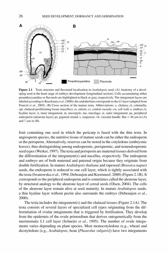
P1: OTE/SPH P2: OTE
BLUK053-Bradford August 27, 2006 17:7
26 SEED DEVELOPMENT, DORMANCY AND GERMINATION
cl
e
oi2oi1ii2ii1'ii1ct
hpe
oi
ii
cwmu
Proanthocyanidins Flavonols
BA
ii1
s
e
pe
ps
cpt
oi2oi1 oi
ii2ii1' ii
ct
mc
cv
vb
Figure 2.1 Testa structure and flavonoid localization in Arabidopsis seed. (A) Anatomy of a devel-
oping seed at the heart stage of embryo development (longitudinal section). Cells accumulating either
proanthocyanidins or flavonols are highlighted in black or gray, respectively. The integument layers are
labeled according to Beeckman et al. (2000); the endothelium corresponds to the ii1 layer (adapted from
Pourcel et al., 2005). (B) Cross section of the mature testa. Abbreviations: c, chalaza; cl, columella;
cpt, chalazal-proliferating tissue (nucellus); ct, cuticle; cv, central vacuole; cw, cell wall; e, embryo; h,
hyaline layer; ii, inner integument; m, micropyle; mu, mucilage; oi, outer integument; pe, peripheral
endosperm (aleurone layer); ps, pigment strand; s, suspensor; vb, vascular bundle. Bar = 40 μm in (A)
and 7 μm in (B).
fruit containing one seed in which the pericarp is fused with the thin testa. Inangiosperm species, the nutritive tissue of mature seeds can be either the endospermor the perisperm. Alternatively, reserves can be stored in the cotyledons (embryonicleaves), thus distinguishing among endospermic, perispermic, and nonendospermicseed types (Werker, 1997). The testa and perisperm are maternal tissues derived fromthe differentiation of the integument(s) and nucellus, respectively. The endospermand embryo are of both maternal and paternal origin because they originate fromdouble fertilization. In mature Arabidopsis thaliana and rapeseed (Brassica napus)seeds, the endosperm is reduced to one cell layer, which is tightly associated withthe testa (Iwanowska et al., 1994; Debeaujon and Koornneef, 2000) (Figure 2.1B). Itcorresponds to the peripheral endosperm and is sometimes called the aleurone layer,by structural analogy to the aleurone layer of cereal seeds (Olsen, 2004). The cellsof the aleurone layer remain alive at seed maturity. In mature Arabidopsis seeds,a thin hyaline layer without pectin also surrounds the embryo (Debeaujon et al.,2000).
The testa includes the integument(s) and the chalazal tissues (Figure 2.1A). Thetesta consists of several layers of specialized cell types originating from the dif-ferentiation of ovular integuments that is triggered by fertilization. They developfrom the epidermis of the ovule primordium that derives ontogenetically from themeristematic L1 cell layer (Schneitz et al., 1995). The number of ovule integu-ments varies depending on plant species. Most monocotyledons (e.g., wheat) anddicotyledons (e.g., Arabidopsis, bean [Phaseolus vulgaris]) have two integuments
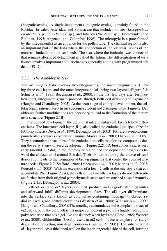
P1: OTE/SPH P2: OTE
BLUK053-Bradford August 27, 2006 17:7
SEED COAT DEVELOPMENT AND DORMANCY 27
(bitegmic ovules). A single integument (unitegmic ovules) is mainly found in theRosidae, Ericales, Asteridae, and Solanaceae that includes tomato (Lycopersiconesculentum), petunia (Petunia sp.), and tobacco (Nicotiana sp.) (Boesewinkel andBouman, 1995; Angenent and Colombo, 1996). The micropyle is a pore formedby the integument(s) as an entrance for the pollen tube. The chalazal region is alsoan important part of the testa where the connection of the vascular tissues of thematernal funiculus to the seed ends. The scar where the funiculus was connectedthat remains after seed detachment is called the hilum. The differentiation of testatissues involves important cellular changes generally ending with programmed celldeath (PCD).
2.2.2 The Arabidopsis testa
The Arabidopsis testa involves two integuments, the inner integument (ii) hav-ing three cell layers and the outer integument (oi) being two-layered (Figure 2.1;Schneitz et al., 1995; Beeckman et al., 2000). In the first few days after fertiliza-tion (daf), integument growth proceeds through both cell division and expansion(Haughn and Chaudhury, 2005). At the heart stage of embryo development, the cel-lular organization of testa tissues becomes evident and distinguishable (Figure 2.1A),although further modifications are necessary to lead to the formation of the maturetesta structure (Figure 2.1B).
During seed development, the individual integumentary cell layers follow differ-ent fates. The innermost cell layer (ii1), also called the endothelium, specializes inPA biosynthesis (Devic et al., 1999; Debeaujon et al., 2003). PAs are flavonoid com-pounds also known as condensed tannins (Marles et al., 2003; Dixon et al., 2005).They accumulate in vacuoles of the endothelium cells as colorless compounds dur-ing the early stages of seed development (Figure 2.2). PA biosynthesis starts veryearly (around 1–2 daf) in the micropylar region and the deposition progresses to-ward the chalaza until around 5–6 daf. Their oxidation during the course of seeddesiccation leads to the formation of brown pigments that confer the color of ma-ture seeds (Figure 2.2; Stafford, 1988; Debeaujon et al., 2003; Marles et al., 2003;Pourcel et al., 2005). With the exception of a few ii2 cells at the micropyle that canaccumulate PAs (Figure 2.1A), the cells of the two other ii layers do not differenti-ate further from their original parenchymatic stage and are crushed at seed maturity(Figure 2.1B; Debeaujon et al., 2003).
Cells of oi1 and oi2 layers both first produce and degrade starch granulesand afterward fulfill different developmental fates. The oi2 layer differentiatesinto the surface cells known as columellae containing mucilage, thickened ra-dial cell walls, and central elevations (Western et al., 2000; Windsor et al., 2000;Haughn and Chaudhury, 2005). The mucilage accumulates in the apoplastic space ofoi2 cells around the columellae. Its major component is pectin, a highly hydrophilicpolysaccharide that has a gel-like consistency when hydrated (Goto, 1985; Westernet al., 2000). Gibberellins (GAs) present in oi2 cells induce α-amylase for starchdegradation preceding mucilage formation (Kim et al., 2005). The subepidermaloi1 layer produces a thickened wall on the inner tangential side of the cell, forming
P1: OTE/SPH P2: OTE
BLUK053-Bradford August 27, 2006 17:7
28 SEED DEVELOPMENT, DORMANCY AND GERMINATION
Embryogenesis DesiccationMaturation
Reserves (lipids, proteins)
Primary dormancy
Degeneration
Filled vacuoles
Endosperm Syncytial
Embryo
Syncytial withmitotic domains
Cellular
Cellularizationcomplete
Desiccation toleranceO
vule
fert
iliza
tion
0 5 10daf
15 20
Flavonols
Proanthocyanidins Oxidative browning
Figure 2.2 Overview of flavonoid biosynthesis and dormancy induction during seed development in
Arabidopsis. Double fertilization results in the formation of a diploid embryo and a triploid endosperm.
Embryo organization is completed during the embryogenesis phase. Reserve accumulation and primary
dormancy establishment take place mainly during the maturation phase, which is followed by desicca-
tion. Flavonoids (flavonols, proanthocyanidins) are synthesized early during development. During the
desiccation phase, proanthocyanidins are oxidized to give brown derivatives that confer mature seed
color (adapted from Baud et al., 2002; Bentsink and Koornneef, 2002; Debeaujon et al., 2003; Lepiniec
et al., 2005; Pourcel et al., 2005; Routaboul et al., 2006). Abbreviation: daf, days after fertilization.
the palisade layer (Goto, 1985; Western et al., 2000). It also accumulates flavonols,which are colorless to pale yellow flavonoids (Figure 2.1; Pourcel et al., 2005).
The cell walls of endothelial cells (ii1) facing the aleurone layer are borderedby an electron-dense layer that reacts positively to osmium tetroxide, suggestingits lipidic nature (Figure 2.1; Beeckman et al., 2000). The same layer also appearsstrongly refringent in seed confocal sections (Garcia et al., 2003). This layer corre-sponds to the cuticle (ct) originally present in the ii during development (Beeckmanet al., 2000). Such a cuticle was found only at the surface of cells facing the en-dosperm and not on the other integument layers (Figure 2.1). Some cells in thechalazal region (pigment strand) of the Arabidopsis testa are also able to accumu-late PAs, which creates a continuum of tannin-producing cells with a bulb-like shapeat the chalazal pole, just above the end of the vascular bundle (vb) from the funicu-lus (Figure 2.1A), corresponding to the pigment strand in cereals (Zee and O’brien,1970; Debeaujon et al., 2003). There is also a very small amount of remaining nu-cellus (chalazal-proliferating tissue) at the chalazal pole of the seed (Figure 2.1A;Beeckman et al., 2000).
P1: OTE/SPH P2: OTE
BLUK053-Bradford August 27, 2006 17:7
SEED COAT DEVELOPMENT AND DORMANCY 29
Testa growth and differentiation proceed coordinately with the development ofthe endosperm and embryo. However, as the integuments are not directly involvedin fertilization, some signal(s) produced during or following fertilization must co-ordinate the differentiation of the testa concomittantly with embryo and endospermdevelopment (Haughn and Chaudhury, 2005). Garcia et al. (2003) presented geneticevidence that the Arabidopsis HAIKU protein is an endosperm-derived signal stim-ulating elongation (but not division) of integument cells, together with endospermand embryo proliferation and growth. On the other hand, prevention of cell elon-gation in the integuments by mutations in the TRANSPARENT TESTA GLABRA2(TTG2) gene restricts endosperm and seed growth (Garcia et al., 2005). The authorssuggest that regulatory cross talk between the integuments and the endosperm isthe primary regulator of the coordinated control of seed size in Arabidopsis (seeChapter 1).
The Arabidopsis testa cells undergo PCD during seed development and matu-ration (Haughn and Chaudhury, 2005). The first cell layers that initiate this processare the parenchymatic ii2 and ii1’ layers from the ii (Figure 2.1). Nakaune et al.(2005) have found a correlation between the presence of VPE (VACUOLAR PRO-CESSING ENZYME), a cysteine proteinase with caspase-like activity, in these celllayers and their subsequent PCD. It is not known whether similar mechanisms pro-mote PCD in the other cell layers. PCD involving a cysteine proteinase (BnCysP1)was also reported to occur in the ii of rapeseed testa, which suggests that this pro-cess is conserved at least among members of the Brassicaceae family (Wan et al.,2002).
Testa structure is used in determining taxonomic relationships among membersof the Brassicaceae family (Vaughan and Whitehouse, 1971; Bouman, 1975). Incontrast to Arabidopsis, the mature testa of rapeseed lacks mucilage in the oi and,for most varieties, exhibits a brown to black color mainly due to the presence ofPAs (Marles and Gruber, 2004). It also has a strong palisade layer composed ofcells with thickened radial walls (stellar cells) that are impregnated with phenolicsat maturity. Seeds at the heart stage of embryo development exhibit a four-layeredoi and a five- to seven-layered ii. PAs are detected in the innermost layer of the ii,which therefore may be functionally homologous to the Arabidopsis endothelium(Iwanowska et al., 1994; Naczk et al., 1998).
2.3 Role of the seed coat in seed dormancy and germination
2.3.1 Constraints imposed by the seed coat
Germination begins with water uptake by the quiescent dry seed and is completedby radicle protrusion through the tissues surrounding the embryo. It occurs whenthe growth potential of the embryo can overcome the constraints imposed by thecovering structures (Bewley and Black, 1994). An intact viable seed is considered tobe dormant when it is unable to germinate under environmental conditions that are
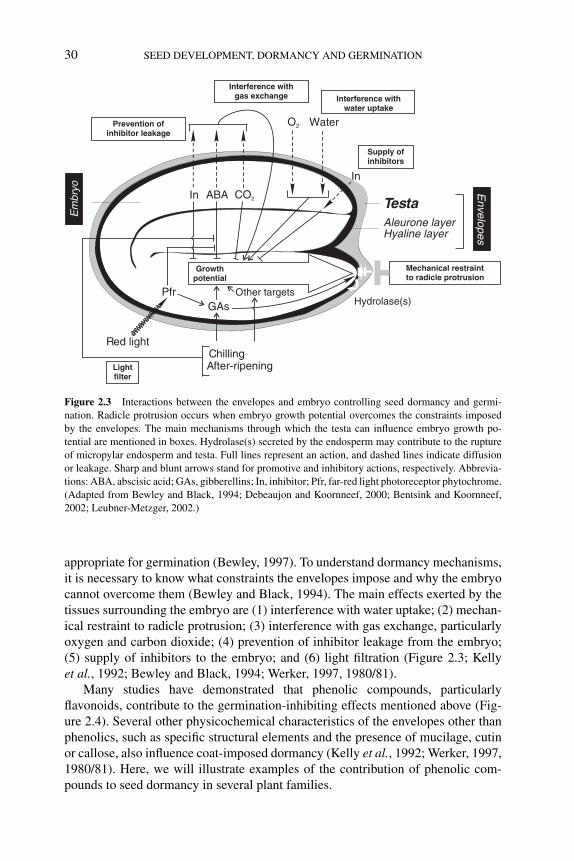
P1: OTE/SPH P2: OTE
BLUK053-Bradford August 27, 2006 17:7
30 SEED DEVELOPMENT, DORMANCY AND GERMINATION
Hyaline layerAleurone layer
Testa
Growthpotential
ABA
In
WaterO2
CO2In
GAsPfr
Red light
Other targetsHydrolase(s)
After-ripeningChilling
Mechanical restraintto radicle protrusion
Interference withwater uptake
Interference withgas exchange
Supply ofinhibitors
Prevention ofinhibitor leakage
Lightfilter
Envelopes
Em
bryo
Figure 2.3 Interactions between the envelopes and embryo controlling seed dormancy and germi-
nation. Radicle protrusion occurs when embryo growth potential overcomes the constraints imposed
by the envelopes. The main mechanisms through which the testa can influence embryo growth po-
tential are mentioned in boxes. Hydrolase(s) secreted by the endosperm may contribute to the rupture
of micropylar endosperm and testa. Full lines represent an action, and dashed lines indicate diffusion
or leakage. Sharp and blunt arrows stand for promotive and inhibitory actions, respectively. Abbrevia-
tions: ABA, abscisic acid; GAs, gibberellins; In, inhibitor; Pfr, far-red light photoreceptor phytochrome.
(Adapted from Bewley and Black, 1994; Debeaujon and Koornneef, 2000; Bentsink and Koornneef,
2002; Leubner-Metzger, 2002.)
appropriate for germination (Bewley, 1997). To understand dormancy mechanisms,it is necessary to know what constraints the envelopes impose and why the embryocannot overcome them (Bewley and Black, 1994). The main effects exerted by thetissues surrounding the embryo are (1) interference with water uptake; (2) mechan-ical restraint to radicle protrusion; (3) interference with gas exchange, particularlyoxygen and carbon dioxide; (4) prevention of inhibitor leakage from the embryo;(5) supply of inhibitors to the embryo; and (6) light filtration (Figure 2.3; Kellyet al., 1992; Bewley and Black, 1994; Werker, 1997, 1980/81).
Many studies have demonstrated that phenolic compounds, particularlyflavonoids, contribute to the germination-inhibiting effects mentioned above (Fig-ure 2.4). Several other physicochemical characteristics of the envelopes other thanphenolics, such as specific structural elements and the presence of mucilage, cutinor callose, also influence coat-imposed dormancy (Kelly et al., 1992; Werker, 1997,1980/81). Here, we will illustrate examples of the contribution of phenolic com-pounds to seed dormancy in several plant families.
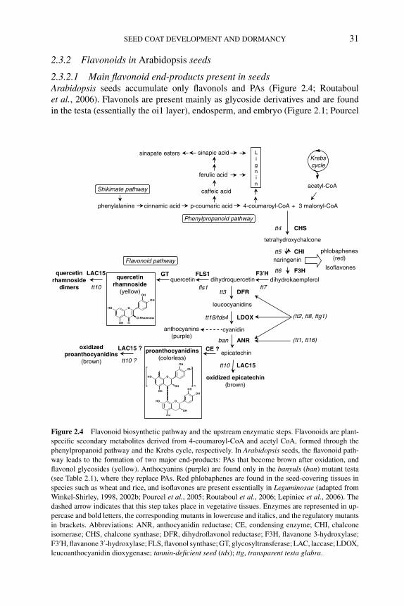
P1: OTE/SPH P2: OTE
BLUK053-Bradford August 27, 2006 17:7
SEED COAT DEVELOPMENT AND DORMANCY 31
2.3.2 Flavonoids in Arabidopsis seeds
2.3.2.1 Main flavonoid end-products present in seedsArabidopsis seeds accumulate only flavonols and PAs (Figure 2.4; Routaboulet al., 2006). Flavonols are present mainly as glycoside derivatives and are foundin the testa (essentially the oi1 layer), endosperm, and embryo (Figure 2.1; Pourcel
F3HFLS1 F3′H
phlobaphenes(red)
phenylalanine cinnamic acid p-coumaric acid 4-coumaroyl-CoA + 3 malonyl-CoA
acetyl-CoA
Krebscycle
caffeic acid
ferulic acid
sinapic acidsinapate esters Lignin
Shikimate pathway
Phenylpropanoid pathway
Flavonoid pathway
quercetinGTquercetin
rhamnosidedimers
tt4
tt5
tt6
tt7
LAC15
tt10 fls1
cyanidin
LDOXtt18/tds4
leucocyanidins
epicatechinCE ?
DFR
ANR
anthocyanins(purple)
oxidizedproanthocyanidins
(brown)
tt3
tt10 ?
LAC15 ?
LAC15tt10
ban
oxidized epicatechin(brown)
(tt1, tt16)
(tt2, tt8, ttg1)
Isoflavones
OHO
OH O
O-Rhamnose
OH
OH
proanthocyanidins(colorless)
OHO
OH
OH
OHO
OH
OH
OH
OH
n
OH
OH
quercetinrhamnoside
(yellow)
Figure 2.4 Flavonoid biosynthetic pathway and the upstream enzymatic steps. Flavonoids are plant-
specific secondary metabolites derived from 4-coumaroyl-CoA and acetyl CoA, formed through the
phenylpropanoid pathway and the Krebs cycle, respectively. In Arabidopsis seeds, the flavonoid path-
way leads to the formation of two major end-products: PAs that become brown after oxidation, and
flavonol glycosides (yellow). Anthocyanins (purple) are found only in the banyuls (ban) mutant testa
(see Table 2.1), where they replace PAs. Red phlobaphenes are found in the seed-covering tissues in
species such as wheat and rice, and isoflavones are present essentially in Leguminosae (adapted from
Winkel-Shirley, 1998, 2002b; Pourcel et al., 2005; Routaboul et al., 2006; Lepiniec et al., 2006). The
dashed arrow indicates that this step takes place in vegetative tissues. Enzymes are represented in up-
percase and bold letters, the corresponding mutants in lowercase and italics, and the regulatory mutants
in brackets. Abbreviations: ANR, anthocyanidin reductase; CE, condensing enzyme; CHI, chalcone
isomerase; CHS, chalcone synthase; DFR, dihydroflavonol reductase; F3H, flavanone 3-hydroxylase;
F3′H, flavanone 3′-hydroxylase; FLS, flavonol synthase; GT, glycosyltransferase; LAC, laccase; LDOX,
leucoanthocyanidin dioxygenase; tannin-deficient seed (tds); ttg, transparent testa glabra.
P1: OTE/SPH P2: OTE
BLUK053-Bradford August 27, 2006 17:7
32 SEED DEVELOPMENT, DORMANCY AND GERMINATION
et al., 2005; Routaboul et al., 2006). In mature seeds, the major flavonol isquercetin-3-O-rhamnoside (Q-3-O-R). Recently, a novel group of biflavonols(quercetin-rhamnoside dimers) has been detected. Both Q-3-O-R and biflavonolsare found mainly in the testa (Routaboul et al., 2006).
PAs are present specifically in the testa (Figure 2.1). In most plant species,PAs are generally polymers of the two flavan-3-ol stereoisomers epicatechin (EC,2-3-cis) and catechin (C, 2-3-trans) (Dixon et al., 2005). However, Arabidopsisseeds accumulate only EC polymers. The mean degree of polymerization variesbetween 5 and 8, depending on the accession (Abrahams et al., 2003; Routaboulet al., 2006). Interestingly, natural variation in the quantity of PAs also occursamong accessions (Lepiniec et al., 2006). A detailed characterization of flavonoidmetabolome in seeds of Arabidopsis mutants and natural accessions using liquidchromatography–tandem mass spectrometry (LC-MS) and nuclear magnetic reso-nance (NMR) methods has been undertaken with the aims of drawing correlationsbetween germination/dormancy behaviors and specific flavonoid products and iso-lating novel flavonoid mutants that may not have clear color phenotypes (Routaboulet al., 2006).
Purple anthocyanins are also present in Arabidopsis, but only in vegetative parts(Shirley et al., 1995). They are conspicuous in 4-day-old seedlings and in agingplants exhibiting chlorophyll degradation.
2.3.2.2 Molecular genetics of flavonoid metabolismThe mutants. Most Arabidopsis mutants impaired in flavonoid biosynthesis havebeen identified through visual screenings of various collections for altered seed coatpigmentation (Koornneef, 1990; Shirley et al., 1995; Lepiniec et al., 2006). As such,they are all affected in PA metabolism. Mutant seed colors range from pale yellow topale brown or gray, with the chalaza and micropyle remaining normally pigmentedin some of them. The fls1 (flavonol synthase 1) mutant, obtained through reversegenetics (Wisman et al., 1998), lacks only flavonols because of a mutation in theFLS1 gene (Figure 2.4) and produces brown seeds like the wild type (WT) due tothe presence of oxidized PAs (Routaboul et al., 2006). No mutant seeds whose testacolor is significantly darker than WT have been reported in Arabidopsis.
Twenty-three genetic complementation groups of testa mutants have been iden-tified. Many mutants have been collectively called transparent testa (tt) mutants (tt1to tt19), or tt glabra (ttg1 and ttg2) (Koornneef, 1990; Shirley et al., 1995; Lepiniecet al., 2006). The banyuls (ban) mutant is unique in the fact that it accumulatesanthocyanins in place of PAs (Albert et al., 1997). The tt10 mutant is also exceptionalbecause it does not affect the biosynthesis of PAs but their subsequent oxidativebrowning (Pourcel et al., 2005). Six independent mutations affecting PA biosynthesis(tannin-deficient seed; tds1 to tds6) have been reported (Abrahams et al., 2002). Itis not known whether these represent additional loci except for tds4, which is allelicto tt18 (Abrahams et al., 2003). In addition, the tt11 and tt14 mutants appear to beallelic to tt18 and tt19, respectively (I. Debeaujon and M. Koornneef, unpublishedresults). Seed pigmentation mutants may be deprived of anthocyanins in vegetativeparts if the mutation affects the general core flavonoid pathway (Figure 2.4). A
P1: OTE/SPH P2: OTE
BLUK053-Bradford August 27, 2006 17:7
SEED COAT DEVELOPMENT AND DORMANCY 33
Table 2.1 Classification of testa mutants based on their flavonoid composition
Seed Plant
Mutant (or WT) PAs Flavonols Anthocyanins Flavonols Anthocyanins
WT, tt7a , tt10b + + − + +tt1, tt2, tt9, tt12, tt13, tt15, tt16, ttg2,
aha10, tds1, tds3, tds5, tds6− + − + +
tt3, tt8, tt17, tt18c , tt19bd , ttg1, tds2 − + − + −tt4, tt5, tt6 − − − − −ban − + + + +fls1 + − − − +
Note: PAs and flavonols present in seeds were assessed either by histochemistry (vanillin or dimethylamino-
cinnamaldehyde staining for PAs, and diphenylboric acid-2-aminoethyl ester for flavonols) or by liquid chromatography-
tandem mass spectrometry (LC-MS). Presence or absence/reduction are denoted by + and −, respectively. Anthocyanins
were detected by visually examining for purple color in seeds or plants. Data from various published and unpublished
works are summarized (Shirley et al., 1995; Albert et al., 1997; Wisman et al., 1998; Focks et al., 1999; Debeaujon
et al., 2001; Abrahams et al., 2002; Johnson et al., 2002; Nesi et al., 2002; Bharti and Khurana, 2003; Shikazono
et al., 2003; Baxter et al., 2005; Pourcel et al., 2005; Routaboul et al., 2006; L. Pourcel, unpublished results).aFlavonols and anthocyanins in tt7 are kaempferol derivatives in place of quercetin derivatives.bSeeds brownish with storage time.cAllelic to tt11 and tds4.d Allelic to tt14.
classification of the 23 mutants on the basis of their flavonoid composition in seedand vegetative tissues is presented in Table 2.1.
The proteins. Twenty genes affecting flavonoid metabolism have already beencharacterized at the molecular level (see Lepiniec et al., 2006, for a detailed re-view). Nine genes encode biosynthetic enzymes (chalcone synthase [CHS], chal-cone isomerase [CHI], flavanone 3-hydroxylase [F3H] , flavanone 3′-hydroxylase[F3′H], dihydroflavonol reductase [DFR], leucoanthocyanidin dioxygenase[LDOX], glycosyltransferase [GT], flavonol synthase 1 [FLS1], and anthocyani-din reductase [ANR]) and one gene encodes a modification enzyme (laccase 15[LAC15]) (Figure 2.4). In addition to FLS1, five other genes (FLS2 to FLS6) ex-hibit sequence homology with flavonol synthases (Alerding et al., 2005), althoughit is not yet known whether they are also involved in flavonol biosynthesis. TheTT10 gene has been cloned and shown to encode a polyphenol oxidase of thelaccase type that belongs to a multigene family containing 17 members (Pourcelet al., 2005). TT10 (LAC15) is involved in the formation of epicatechin quinonesthat spontaneously polymerize into brown derivatives (Figure 2.4). It may cat-alyze the oxidative browning of colorless PAs, which is consistent with the factthat tt10 has normal PA levels but yellow seeds at harvest. TT10/LAC15 also cat-alyzes the formation of biflavonols from quercetin rhamnoside, probably in the oi1layer (see below the description of TT10/LAC15 promoter activity). Interestingly,TT10/LAC15 expression is lower in the Landsberg erecta (Ler) accession than inWassilewskija (Ws-2) and Columbia (Col), consistent with a reduction in enzymeactivity because Ler exhibits twice the amount of soluble PAs (corresponding to less
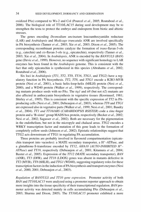
P1: OTE/SPH P2: OTE
BLUK053-Bradford August 27, 2006 17:7
34 SEED DEVELOPMENT, DORMANCY AND GERMINATION
oxidized PAs) compared to Ws-2 and Col (Pourcel et al., 2005; Routaboul et al.,2006). The biological role of TT10/LAC15 during seed development may be tostrengthen the testa to protect the embryo and endosperm from biotic and abioticstresses.
The genes encoding Desmodium uncinatum leucoanthocyanidin reductase(LAR) and Arabidopsis and Medicago truncatula ANR are involved specificallyin PA biosynthesis (Tanner et al., 2003; Xie et al., 2003; Dixon et al., 2005). Thecorresponding recombinant proteins catalyze the formation of trans-flavan-3-ols(e.g., catechin) and cis-flavan-3-ols (e.g., epicatechin), respectively (Tanner et al.,2003; Xie et al., 2004). In Arabidopsis, ANR is encoded by the BANYULS (BAN)gene (Devic et al., 1999). However, no sequence with significant homology to LARenzymes has been found in the Arabidopsis genome. This is consistent with thefact that only epicatechin is synthesized in this species (Abrahams et al., 2002;Routaboul et al., 2006).
Six loci in Arabidopsis (TT1, TT2, TT8, TT16, TTG1, and TTG2) have a reg-ulatory function in PA biosynthesis. TT2, TT8, and TTG1 encode a R2R3-MYBprotein (Nesi et al., 2001), a basic helix-loop-helix (bHLH) protein (Nesi et al.,2000), and a WD40 protein (Walker et al., 1999), respectively. The correspond-ing mutants produce seeds with no PAs. The ttg1 and tt8 (but not tt2) mutants arealso affected in anthocyanin biosynthesis in vegetative tissues (Koornneef, 1981;Shirley et al., 1995). This is consistent with the specific expression of TT2 in PA-producing cells (Nesi et al., 2001; Debeaujon et al., 2003), whereas TT8 and TTG1are expressed also in vegetative parts (Walker et al., 1999; Nesi et al., 2001; Baudryet al., 2004). TT1 and TT16/ABS (CARABIDOPSIS BSISTER) code a zinc-fingerprotein and a ‘B-sister’ group MADS box protein, respectively (Becker et al., 2002;Nesi et al., 2002; Sagasser et al., 2002). Both are necessary for the pigmentationin the endothelium, but not in the micropyle and chalazal areas. TTG2 encodes aWRKY transcription factor and mutation of this gene leads to the formation ofcompletely yellow seeds (Johnson et al., 2002). Epistatic relationships suggest thatTTG2 acts downstream of TTG1 in regulating PA accumulation.
Three proteins are probably involved in flavonoid compartmentation (epicate-chin transport into vacuoles): a MATE secondary transporter, a H+-ATPase, anda glutathione-S-transferase encoded by TT12, AHA10 (AUTO-INHIBITED H+-ATPase) and TT19, respectively (Debeaujon et al., 2001; Kitamura et al., 2004;Baxter et al., 2005). Expression of the TT12 (MATE secondary transporter), BAN(ANR), TT3 (DFR), and TT18 (LDOX) genes was absent in mutants defective inTT2 (MYB), TT8 (bHLH), and TTG1 (WD40), suggesting regulatory roles for thesetranscription factors in the induction of PA biosynthesis and transport enzymes (Nesiet al., 2000, 2001; Debeaujon et al., 2003).
Regulation of BANYULS and TT10 gene expression. Promoter activity of bothBAN and TT10/LAC15 were analyzed using a promoter:reporter approach to obtainmore insights into the tissue specificity of their transcriptional regulation. BAN pro-moter activity was detected mainly in cells accumulating PAs (Debeaujon et al.,2003; Sharma and Dixon, 2005). The TT10/LAC15 promoter exhibited a more
P1: OTE/SPH P2: OTE
BLUK053-Bradford August 27, 2006 17:7
SEED COAT DEVELOPMENT AND DORMANCY 35
complex pattern of regulation than that of the BAN promoter, appearing first inthe endothelium and then in the oi1 cell layer. Expression was correlated withPA- and flavonol-producing cells (Figure 2.1), which is consistent with the rolefor TT10/LAC15 in flavonoid metabolism in the testa. Characterization of BAN-and TT10/LAC15-promoter:reporter constructs in the the tt2, tt8, and ttg1 regula-tory mutants demonstrated that TT2, TT8, and TTG1 are necessary for expressionof BAN but not TT10/LAC15 (Debeaujon et al., 2003; Pourcel et al., 2005). Thesepromoter:reporter constructs provide valuable markers for the PA- and flavonol-accumulating cells in the Arabidopsis testa.
2.3.2.3 Effects of flavonoids on seed dormancy and germinationFlavonoids, particularly PAs, have been shown to reinforce coat-imposed dormancyby increasing testa thickness and mechanical strength. Indeed, cell layers contain-ing pigments generally do not crush as dramatically as the nonpigmented ones(Debeaujon et al., 2000). Moreover, during oxidation PAs have a tendency to cross-link with proteins and carbohydrates in cell walls, thus reinforcing testa struc-ture and also modifying its permeability properties (Marles et al., 2003; Marlesand Gruber, 2004). This was demonstrated using Arabidopsis mutants affected inflavonoid metabolism in the testa (Leon-Kloosterziel et al., 1994; Focks et al., 1999;Debeaujon and Koornneef, 2000; Debeaujon et al., 2000, 2001). Freshly harvestedmutant seeds lacking PAs germinate faster than the corresponding WT brown seeds(Figure 2.5A). Moreover, their mature testa is thinner and more permeable to tetra-zolium salts (Debeaujon et al., 2000). Permeability to water, an important parameterfor germination, could not be examined with tt mutants by the traditional seed watercontent analysis that depends on weighing seeds during imbibition because of thepresence of a hydrophilic mucilage excreted by the oi. NMR imaging may representan alternative method to analyze water distribution inside seeds, as demonstrated intobacco (Nicotiana tabacum) and white pine (Pinus monticola) (Manz et al., 2005;Terskikh et al., 2005).
Reduced seed dormancy observed in tt mutant seeds is controlled maternallybased on germination of F1 seeds resulting from reciprocal crosses between tt mu-tants and WT (Figure 2.5B). This is consistent with the fact that the testa derivesfrom the integuments, which are maternal tissues. Little difference in germinationbetween tt mutant and WT seeds is observed when they are after-ripened, sug-gesting that the ‘seed coat’ effect is particularly conspicuous under physiologicalconditions unfavorable for germination such as embryo dormancy (Debeaujon et al.,2000). Seeds of the Arabidopsis ga1 mutant are deficient in GAs and thus unableto germinate in the absence of additional GAs. The tt4 mutant (CHS deficient;Figure 2.4) is deprived of flavonoids and exhibits reduced testa inhibition of germi-nation. Genetically reducing the testa inhibition in ga1 seeds (in a ga1 tt4 doublemutant) enabled germination of nondormant seeds without GA requirement (Fig-ure 2.5C). This experiment also showed that both cold and light may in part beable to stimulate germination independently from GAs (Debeaujon and Koornneef,2000).
P1: OTE/SPH P2: OTE
BLUK053-Bradford August 27, 2006 17:7
36 SEED DEVELOPMENT, DORMANCY AND GERMINATION
- 2 -No cold
Light
ga1-
1
ga1-
1
ga1
tt4
ga1
tt4
tt4-1
tt4-1
WT
WT
- 4 -ColdLight
ColdDark
- 3 -
No coldDark
- 1 -100
Ger
min
atio
n (%
)
20
40
60
80
0
20
40
60
80
0
C D
A
WTtt2-1tt4-1ttg1-1
2 9 18 27 36 60
Ger
min
atio
n (%
)
Time (days)
0
20
40
60
80
100
0
20
40
60
80
100
WT x tt2-1 tt2-1
WT
tt2-1 x WT
0 2 4 6 8 10
Ger
min
atio
n (%
)
Time (days)
B
0
20
40
60
80
100
Ger
min
atio
n (%
)
0 0.01 0.1 1 10 100GA concentration (μM)4+7
pBAN:BARNASE
tt2-3
WT
Figure 2.5 Effect of transparent testa mutations on dormancy and germination of Arabidopsis seeds.
(A) Effect of dry storage (after-ripening) on dormancy release (adapted from Debeaujon et al., 2000).
(B) Maternal control of seed dormancy in the tt2-1 mutant. The germination behaviors of F1 seed
progenies from reciprocal crosses between tt2-1 and WT are shown and compared to WT and the
mutant parent. The parent line indicated first in the cross (e.g., ‘WT’ in ‘WT × tt2-1’) was used as a
female. The time course of germination after 16 days of storage is presented (adapted from Debeaujon
et al., 2000). (C) Influence of testa mutation on germination behavior of the gibberellin-deficient
mutant ga1-1. The ga1-1 mutation was introduced into a tt4-1 background. The effect of light and cold
stratification (prechilling) on dormancy breakage and germination of WT, single and double mutants
are compared (adapted from Debeaujon and Koornneef, 2000). (D) Permeability of the testa to GA
and GA biosynthesis inhibitor. Germination of WT seeds was examined in the presence of 100 μM
paclobutrazol (GA biosynthesis inhibitor) and various concentrations of GA, and compared to the
germination of typical transparent testa mutant seeds (tt2-3) and seeds of a transgenic line deprived
of proanthocyanidin-producing cells (pBAN:BARNASE) through genetic ablation (see text for details).
Nondormant seeds were used (adapted from Debeaujon et al., 2003).
The permeability of the Arabidopsis testa to exogenous GAs and GA biosyn-thesis inhibitors (tetcyclacis [TET] and paclobutrazol [PAC]) was also increased intt mutants (Figure 2.5D; Debeaujon and Koornneef, 2000). Application of 100 μMPAC reduces germination of both tt2-3 and WT seeds, which is gradually rescuedby increasing concentrations of exogenous GAs. Relatively lower concentrationsof GAs (0.01 μM and above) enables tt2-3 seeds to germinate in the presence ofPAC, while much higher concentrations of GAs (10 μM and above) are required torecover PAC-inhibited WT seed germination (Figure 2.5D). The greater response(permeability) of seeds to GAs compared to WT was also observed in pigmentless
P1: OTE/SPH P2: OTE
BLUK053-Bradford August 27, 2006 17:7
SEED COAT DEVELOPMENT AND DORMANCY 37
seeds produced by the pBAN::BARNASE line (Debeaujon et al., 2003). In this line,the cytotoxic gene BARNASE was expressed using the BAN gene promoter whichdrove specific expression in PA-producing cells, leading to their genetic ablation andas a consequence to the formation of pigment-less seeds. These seeds were capableof germinating in relatively lower concentrations of GAs in the presence of PAC, inthe same way as typical tt seeds (Figure 2.5D). Therefore, flavonoids and no otherphysicochemical factor present in PA-producing cells are significantly responsiblefor the tt germination phenotype.
Abscisic acid (ABA) is synthesized de novo in primary dormant Arabidopsisseeds during imbibition (Debeaujon and Koornneef, 2000; Ali-Rachedi et al., 2004).Freshly harvested ga1 tt4 seeds were able to germinate to 50% in the presence ofcarotenoid synthesis inhibitor norflurazon, which inhibits ABA biosynthesis, whilega1 seeds exhibited only 5% germination (Debeaujon and Koornneef, 2000).
Together, these data suggest that GAs are necessary during Arabidopsis germina-tion essentially to overcome the restraints to radicle protrusion imposed by the testaand de novo synthesized ABA. Removal of seed envelopes (testa and endosperm)allows germination of both ga1 seeds and freshly harvested Cape Verde Islands(Cvi) seeds, which have a strong primary dormancy (Debeaujon and Koornneef,2000; Alonso-Blanco et al., 2003). It is possible that coat removal enables ABAleakage from dormant seeds, thus releasing dormancy, but this still remains to be in-vestigated. Analysis of natural allelic variation between the Cvi and Ler accessionsrevealed a quantitative trait locus (QTL) with a maternal effect (Delay of Germina-tion2 [DOG2]). The authors hypothesized that DOG2 may affect seed coat-imposeddormancy through the genetic structure of the testa, or a factor imported from themother plant (Alonso-Blanco et al., 2003; see Chapter 5). The tt mutants exhibitnormal ABA sensitivity, although some of them showed a slight decrease in sensi-tivity (I. Debeaujon and M. Koornneef, unpublished results). This may indicate thatflavonoids do not affect permeability of the testa to ABA as much as they affect GApermeability. PAs have been shown to act as GA antagonists in pea (Pisum sativum)(Corcoran et al., 1972; Green and Corcoran, 1975). Similarly, (+)-catechin extractedfrom bean seeds was shown to act as an inhibitor of GA biosynthesis, blocking theconversion of GA12-aldehyde to GA12; the catechin was localized mainly in the testaand not in the embryo during seed maturation (Kwak et al., 1988). Moreover, Butaand Lusby (1986) observed that catechin and epicatechin inhibited Lespedeza seedgermination and seedling growth. These data suggest that PAs and their precursorsmay inhibit germination not only by influencing the structural properties of the testaat maturity, but also by acting as biochemical inhibitors of GA metabolism andaction when released from the testa during seed imbibition. Whether this biologi-cal effect is specific to GA metabolism or common to other biosynthetic pathwaysremains to be investigated more thoroughly.
Testa permeability is an important parameter to consider when undertaking ger-mination experiments to test the effect of any exogenous substances on embryobehavior. For example, Rajjou et al. (2004) used tt2 mutant seeds totally lackingPAs in the testa to test the effect of the transcription inhibitor α-amanitin on seedgermination. In preliminary experiments the authors had established that tt2 seeds
P1: OTE/SPH P2: OTE
BLUK053-Bradford August 27, 2006 17:7
38 SEED DEVELOPMENT, DORMANCY AND GERMINATION
were far more permeable to the molecule than WT seeds. Therefore, they were ableto conclude that the inability of α-amanitin to block radicle protrusion was not likelydue to inability of this substance to cross the testa and reach the embryo.
The tt12 mutant came out of a reduced dormancy screen and was isolated on thebasis of a faster seed germination of freshly harvested seeds on water (Debeaujonet al., 2001). Recently, several tt mutants were isolated from a screen for fast seedgermination at 10◦C (Salaita et al., 2005). These additional examples confirm thattesta flavonoid defects enable the seed to germinate quicker in unfavorable condi-tions.
Desiccation plays an important role in switching seeds from a developmentalmode to germinative mode. Premature drying can redirect metabolism from a devel-opmental to a germination program (Kermode, 1995). In tt mutant seeds that havemore permeable testae, the embryo might dehydrate quicker than WT embryos andtherefore shift earlier from the developmental to germinative mode. The earlier lossof dormancy and shift to the germinative mode could cause precocious germination,as was observed in drying siliques of the tt16 mutant (Nesi et al., 2002).
2.3.3 Flavonoids in seed dormancy and germination of various species
2.3.3.1 SolanaceaeMutants affected in flavonoid metabolism were also isolated in tomato. The mutantsanthocyaninless of Hoffmann (ah), anthocyanin without (aw), and baby lea syn-drome (bls) contain drastically reduced or no PAs in the endothelium. Absence ofPAs in the testa probably enhances its permeability to water, since the mutant seedsexhibit increased seed weight (or water content) during imbibition (Atanassova et al.,2004). Moreover, seeds of the three mutants germinate faster than the correspond-ing WT lines not only in optimal but also in stress conditions such as high and lowtemperatures (33 and 13◦C, respectively), high salinity (120 mM NaCl), and highosmoticum (15% polyethyleneglycol). Mutations ah and bls both resulted in coordi-nated reduction in CHS, F3H, and DFR activities (Figure 2.4), while aw completelylacked DFR activity (Atanassova et al., 1997a,b). On the other hand, brownseed(bs1 to bs4) and blackseed (bks1) mutants are tomato mutants with darker testaethan WT, which display poor germination rates and percentages (Downie et al.,2004). They accumulate an additonal dark pigment in the cell layers surroundingthe endothelium, which itself contains normal levels of PAs. In bks1 seeds, the blackpigment is a melanic substance that also enhances the mechanical strength of thetesta. The phenotypes of the bs and bks mutations are determined by the embryoand the endosperm, in contrast to the ah, aw, and bls mutants that exhibit the typ-ical maternal control. These results suggest that the endosperm and embryo maysecrete a factor that influences testa characteristics (Downie et al., 2003). Seeds ofthe bs1 and bs4 mutants have increased catalase activity, and bs4 seeds also exhibitincreased peroxidase activity (Downie et al., 2004). Likewise, transgenic maizegrains that overexpress a gene encoding a fungal laccase-type polyphenol oxidasebecome brown and exhibit poor germination (Hood et al., 2003).
P1: OTE/SPH P2: OTE
BLUK053-Bradford August 27, 2006 17:7
SEED COAT DEVELOPMENT AND DORMANCY 39
2.3.3.2 Water permeability of testae in Leguminosae and other speciesThe fact that flavonoids increase coat-imposed dormancy by restricting the perme-ability of the testa to water is best exemplified by so-called hard seed, which havebeen abundantly described in the Leguminosae family (Wyatt, 1977; Legesse andPowell, 1992, 1996; Serrato-Valenti et al., 1994; Kantar et al., 1996). In the genusPisum, seeds with impermeable testae, such as the wild pea (P. elatius), have highphenolic contents and catechol oxidase activity, while species having seeds withpermeable testae, such as the cultivated pea (P. sativum), do not. It is hypothesizedthat during seed desiccation, oxidation of phenolic compounds into the correspond-ing quinones by catechol oxidase in the presence of molecular oxygen may triggertesta impermeability. Permeability is inversely proportional to the phenolics contentand their degree of oxidation, probably through some tanning reaction (Marbachand Mayer, 1974, 1975; Stafford, 1974; Werker et al., 1979). Gillikin and Graham(1991) proposed that an anionic peroxidase may play a role in the hardening of soy-bean (Glycine max) seed coats. In this species, the majority of peroxidase activitydetected in seeds is localized in the testa. In cotton (Gossypium hirsutum) seed also,oxidation of seed coat tannins during ripening causes seed coloration and reductionof testa permeability to water (Halloin, 1982).
2.3.3.3 Flavonoids and other phenolics as direct andindirect germination inhibitors
Flavonoids (PAs, catechin, epicatechin), phenolic acids (caffeic, p-coumaric, ferulic,sinapic, vanillic acids), and lignans have been considered as possible germinationinhibitors. Seed germination is reduced in the presence of exogenous phenolics in adose-dependent manner (Buta and Lusby, 1986; Reigosa et al., 1999; Gatford et al.,2002; Basile et al., 2003; Cutillo et al., 2003). The presence of phenolic compoundsin developing grains is correlated with the prevention of preharvest sprouting (PHS;i.e., germination on the ear in high relative humidity) in cereals (Weidner et al.,2002). Wrobel et al. (2005) have shown that the amount of tannins and phenolicacids present in mature dry seeds of riverbank grape (Vitis riparia) was reducedduring cold stratification (imbibition in cold conditions), possibly by leaching ofthese compounds into the surrounding medium. Zobel et al. (1989) followed thechanges in phenolic localization in rapeseed seeds during imbibition and found thatafter 3 h of imbibition, part of the phenolic compounds originally found in the testaleached onto the embryo surface, where they could potentially exert an inhibitoryeffect on radicle protrusion.
Tissues surrounding the embryo might interfere with seed germination by im-peding oxygen entry or escape of carbon dioxide, which could inhibit respiration(Bewley and Black, 1994). Flavonoids are efficient antioxidants (Rice-Evans et al.,1997). When present in seed coats, they may fix molecular oxygen through reac-tions catalyzed by polyphenol oxidases and peroxidases and also through nonenzy-matic oxidation, therefore limiting oxygen availability for the embryo. Similarly, thedormancy-imposing glumellae of barley (Hordeum vulgare) grains also consume
P1: OTE/SPH P2: OTE
BLUK053-Bradford August 27, 2006 17:7
40 SEED DEVELOPMENT, DORMANCY AND GERMINATION
oxygen (Lenoir et al., 1986). Restricted oxygen diffusion through the seed coat andan increased sensitivity of the embryo to hypoxia cause a coat-imposed dormancyof muskmelon (Cucumis melo) seeds at low temperature (Edelstein et al., 1995).Porter and Wareing (1974) suggested that the presence of germination inhibitors inXanthium pennsylvanicum seeds results in a high oxygen requirement because theremoval of inhibitors occurs by oxidation. This is supported by experiments usingbeechnut (Fagus sylvatica) seeds, where covering structures prevent germinationby interfering not only with water uptake but also with oxygen availability. In thiscase, oxygen was demonstrated to be involved in oxidative degradation of ABAusing (+)-[3H]ABA (Barthe et al., 2000). The testa may impose an indirect restraintto radicle protrusion by impeding ABA leakage from the embryo in yellow cedar(Chamaecyparis nootkatensis) seeds (Ren and Kermode, 1999).
Flavonoid pigments in the testa are likely to act as filters modifying the spectrumof light received by the embryo. Flavonols present in the testa efficiently absorb ul-traviolet (UV) light, protecting the embryo from radiation damage (Winkel-Shirley,2002a; Griffen et al., 2004). However, no data are available for absorption of otherlight wavelengths, particularly red light known to induce GA biosynthesis throughphytochrome action (Yamaguchi et al., 1998).
2.3.3.4 Pre-harvest sprouting (PHS) in cerealsImportant crop species such as wheat, barley, rice, and sorghum (Sorghum bicolor)exhibit low dormancy during grain development, leading to a susceptibility to PHS.Wheat grain dormancy is controlled both by maternally expressed R (Red graincolor) genes conferring red pericarp pigmentation and by other genes such as Phs,which has a major effect in the embryo. Therefore, wheat PHS is regulated bothby coat-imposed and embryonic pathways controlled by separate genetic systems(Flintham, 2000; Himi et al., 2002; Mares et al., 2002). Dominant alleles of Rpromote the biosynthesis of red phlobaphenes (Figure 2.4). Recently, the wheat Rgene was cloned and shown to encode a MYB-type transcription factor (Himi andNoda, 2005). This protein is involved in activation of the early biosynthetic genesCHS, CHI, F3H, and DFR (see Figure 2.4 for the pathway), which is consistent withits role in phlobaphene biosynthesis (Himi et al., 2005). Genetic resolution from theQTL approach is insufficient to determine whether the R gene increases dormancyby itself or is linked to another dormancy-promoting locus. Analyzing dormancyof white-grained wheats overexpressing R in the pericarp could help answer thisquestion. In weedy rice, which is far more dormant than cultivated rice, red or blackpericarp and red or black hull (palea and lemma) were correlated with deep seeddormancy. QTL have been found for these characters (Gu et al., 2005). Syntenyamong cereal species enables comparison of wheat PHS loci to maize and rice seeddormancy loci, allowing application of the available rice genomic DNA sequenceto PHS research in other cereals (Gale et al., 2002).
2.3.3.5 Heteromorphism and physiological heterogeneity among seedsHeteromorphism (heteroblasty) is caused by maternal factors within an individualplant, such as the position of the seed in the fruit or in the inflorescence, that influence
P1: OTE/SPH P2: OTE
BLUK053-Bradford August 27, 2006 17:7
SEED COAT DEVELOPMENT AND DORMANCY 41
the color, shape, or size and therefore germination capacity of seeds. The resultingphysiological heterogeneity provides a very important ecological advantage, espe-cially under extreme climates (Gutterman, 2000; Matilla et al., 2005). Individualsiliques of Brassica rapa contain seeds that differ in seed color: black, dark brown,and light brown. Light-brown seeds are more water-permeable than black seeds,which is correlated to a faster germination. They are also more responsive to exoge-nous ethylene than black or dark-brown seeds (Puga-Hermida et al., 2003).
Heterogeneity in seed dormancy can also be generated by the light environment.Dormancy of mature Chenopodium album seeds is influenced by the photoperiodthat the mother plant experiences during seed development. Seeds obtained fromplants grown under long days (LD, 18 h light per day) produce small dormantseeds with thick and black testae, whereas plants grown under short days (SD, 8 hlight per day) produce large nondormant seeds with thinner brownish testae. It isgenerally known that photoperiodic plants monitor the length of the dark period.Consequently, a short (1 h) exposure of red light to interrupt the long night undershort-day conditions (termed SDR by the author) mimicked long-day conditionsand made the maternal plants produce dormant seeds (Karssen, 2002). However,these seeds did not have black testae. Dormancy of SDR seeds was released by3-months of after-ripening, while the authentic black testa-dormant seeds producedunder LD were still dormant after 3 months. Together, these data suggested that twotypes of dormancy are present in this species – embryo dormancy and seed coat-imposed dormancy, and that total light energy received by the plant is important fordetermining the second type (Karssen, 2002).
2.3.3.6 Interactions with endospermThe impact of the endosperm on Arabidopsis seed germination is still a matter ofdebate. Interactions between the testa and the endosperm may influence germinationbehavior. It is possible that enzymes secreted by endosperm cells may hydrolyzesome components of testa cell walls and thus reduce their resistance (Dubreucqet al., 2000; Leubner-Metzger, 2002). In turn, hydrolase inhibitors may leach fromthe testa during imbibition. Another hypothesis would be that hydrolytic enzymesweaken the endosperm itself on the model of the tomato endosperm, which wasproposed to be a more important obstacle to radicle protrusion than the testa (Grootet al., 1988; Chen et al., 2002). No mutant affected specifically in the structure ofthe peripheral endosperm has been recovered until now that could shed some lighton its contribution to Arabidopsis seed germination.
2.4 Link between seed coat-imposed dormancy and longevity
When seeds deteriorate, they lose vigor, become more sensitive to stresses duringgermination, and finally become unable to germinate. The rate of aging is stronglyinfluenced by storage temperature, seed moisture content, and seed quality (Walters,1998). The seed coat performs important functions to protect the embryo and seedreserves (Mohamed-Yasseen et al., 1994; Boesewinkel and Bouman, 1995), and
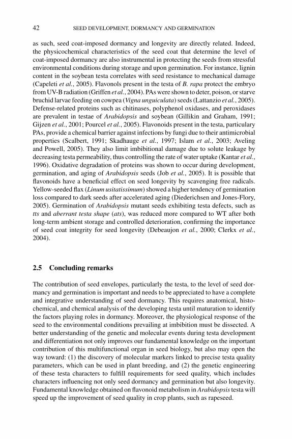
P1: OTE/SPH P2: OTE
BLUK053-Bradford August 27, 2006 17:7
42 SEED DEVELOPMENT, DORMANCY AND GERMINATION
as such, seed coat-imposed dormancy and longevity are directly related. Indeed,the physicochemical characteristics of the seed coat that determine the level ofcoat-imposed dormancy are also instrumental in protecting the seeds from stressfulenvironmental conditions during storage and upon germination. For instance, lignincontent in the soybean testa correlates with seed resistance to mechanical damage(Capeleti et al., 2005). Flavonols present in the testa of B. rapa protect the embryofrom UV-B radiation (Griffen et al., 2004). PAs were shown to deter, poison, or starvebruchid larvae feeding on cowpea (Vigna unguiculata) seeds (Lattanzio et al., 2005).Defense-related proteins such as chitinases, polyphenol oxidases, and peroxidasesare prevalent in testae of Arabidopsis and soybean (Gillikin and Graham, 1991;Gijzen et al., 2001; Pourcel et al., 2005). Flavonoids present in the testa, particularyPAs, provide a chemical barrier against infections by fungi due to their antimicrobialproperties (Scalbert, 1991; Skadhauge et al., 1997; Islam et al., 2003; Avelingand Powell, 2005). They also limit imbibitional damage due to solute leakage bydecreasing testa permeability, thus controlling the rate of water uptake (Kantar et al.,1996). Oxidative degradation of proteins was shown to occur during development,germination, and aging of Arabidopsis seeds (Job et al., 2005). It is possible thatflavonoids have a beneficial effect on seed longevity by scavenging free radicals.Yellow-seeded flax (Linum usitatissimum) showed a higher tendency of germinationloss compared to dark seeds after accelerated aging (Diederichsen and Jones-Flory,2005). Germination of Arabidopsis mutant seeds exhibiting testa defects, such astts and aberrant testa shape (ats), was reduced more compared to WT after bothlong-term ambient storage and controlled deterioration, confirming the importanceof seed coat integrity for seed longevity (Debeaujon et al., 2000; Clerkx et al.,2004).
2.5 Concluding remarks
The contribution of seed envelopes, particularly the testa, to the level of seed dor-mancy and germination is important and needs to be appreciated to have a completeand integrative understanding of seed dormancy. This requires anatomical, histo-chemical, and chemical analysis of the developing testa until maturation to identifythe factors playing roles in dormancy. Moreover, the physiological response of theseed to the environmental conditions prevailing at imbibition must be dissected. Abetter understanding of the genetic and molecular events during testa developmentand differentiation not only improves our fundamental knowledge on the importantcontribution of this multifunctional organ in seed biology, but also may open theway toward: (1) the discovery of molecular markers linked to precise testa qualityparameters, which can be used in plant breeding, and (2) the genetic engineeringof these testa characters to fulfill requirements for seed quality, which includescharacters influencing not only seed dormancy and germination but also longevity.Fundamental knowledge obtained on flavonoid metabolism in Arabidopsis testa willspeed up the improvement of seed quality in crop plants, such as rapeseed.
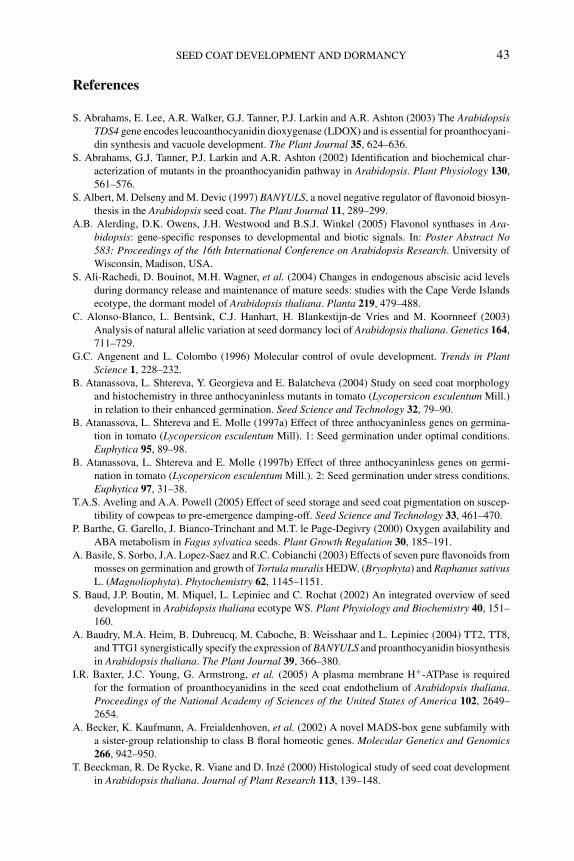
P1: OTE/SPH P2: OTE
BLUK053-Bradford August 27, 2006 17:7
SEED COAT DEVELOPMENT AND DORMANCY 43
References
S. Abrahams, E. Lee, A.R. Walker, G.J. Tanner, P.J. Larkin and A.R. Ashton (2003) The ArabidopsisTDS4 gene encodes leucoanthocyanidin dioxygenase (LDOX) and is essential for proanthocyani-
din synthesis and vacuole development. The Plant Journal 35, 624–636.
S. Abrahams, G.J. Tanner, P.J. Larkin and A.R. Ashton (2002) Identification and biochemical char-
acterization of mutants in the proanthocyanidin pathway in Arabidopsis. Plant Physiology 130,
561–576.
S. Albert, M. Delseny and M. Devic (1997) BANYULS, a novel negative regulator of flavonoid biosyn-
thesis in the Arabidopsis seed coat. The Plant Journal 11, 289–299.
A.B. Alerding, D.K. Owens, J.H. Westwood and B.S.J. Winkel (2005) Flavonol synthases in Ara-bidopsis: gene-specific responses to developmental and biotic signals. In: Poster Abstract No583: Proceedings of the 16th International Conference on Arabidopsis Research. University of
Wisconsin, Madison, USA.
S. Ali-Rachedi, D. Bouinot, M.H. Wagner, et al. (2004) Changes in endogenous abscisic acid levels
during dormancy release and maintenance of mature seeds: studies with the Cape Verde Islands
ecotype, the dormant model of Arabidopsis thaliana. Planta 219, 479–488.
C. Alonso-Blanco, L. Bentsink, C.J. Hanhart, H. Blankestijn-de Vries and M. Koornneef (2003)
Analysis of natural allelic variation at seed dormancy loci of Arabidopsis thaliana. Genetics 164,
711–729.
G.C. Angenent and L. Colombo (1996) Molecular control of ovule development. Trends in PlantScience 1, 228–232.
B. Atanassova, L. Shtereva, Y. Georgieva and E. Balatcheva (2004) Study on seed coat morphology
and histochemistry in three anthocyaninless mutants in tomato (Lycopersicon esculentum Mill.)
in relation to their enhanced germination. Seed Science and Technology 32, 79–90.
B. Atanassova, L. Shtereva and E. Molle (1997a) Effect of three anthocyaninless genes on germina-
tion in tomato (Lycopersicon esculentum Mill). 1: Seed germination under optimal conditions.
Euphytica 95, 89–98.
B. Atanassova, L. Shtereva and E. Molle (1997b) Effect of three anthocyaninless genes on germi-
nation in tomato (Lycopersicon esculentum Mill.). 2: Seed germination under stress conditions.
Euphytica 97, 31–38.
T.A.S. Aveling and A.A. Powell (2005) Effect of seed storage and seed coat pigmentation on suscep-
tibility of cowpeas to pre-emergence damping-off. Seed Science and Technology 33, 461–470.
P. Barthe, G. Garello, J. Bianco-Trinchant and M.T. le Page-Degivry (2000) Oxygen availability and
ABA metabolism in Fagus sylvatica seeds. Plant Growth Regulation 30, 185–191.
A. Basile, S. Sorbo, J.A. Lopez-Saez and R.C. Cobianchi (2003) Effects of seven pure flavonoids from
mosses on germination and growth of Tortula muralis HEDW. (Bryophyta) and Raphanus sativusL. (Magnoliophyta). Phytochemistry 62, 1145–1151.
S. Baud, J.P. Boutin, M. Miquel, L. Lepiniec and C. Rochat (2002) An integrated overview of seed
development in Arabidopsis thaliana ecotype WS. Plant Physiology and Biochemistry 40, 151–
160.
A. Baudry, M.A. Heim, B. Dubreucq, M. Caboche, B. Weisshaar and L. Lepiniec (2004) TT2, TT8,
and TTG1 synergistically specify the expression of BANYULS and proanthocyanidin biosynthesis
in Arabidopsis thaliana. The Plant Journal 39, 366–380.
I.R. Baxter, J.C. Young, G. Armstrong, et al. (2005) A plasma membrane H+-ATPase is required
for the formation of proanthocyanidins in the seed coat endothelium of Arabidopsis thaliana.
Proceedings of the National Academy of Sciences of the United States of America 102, 2649–
2654.
A. Becker, K. Kaufmann, A. Freialdenhoven, et al. (2002) A novel MADS-box gene subfamily with
a sister-group relationship to class B floral homeotic genes. Molecular Genetics and Genomics266, 942–950.
T. Beeckman, R. De Rycke, R. Viane and D. Inze (2000) Histological study of seed coat development
in Arabidopsis thaliana. Journal of Plant Research 113, 139–148.
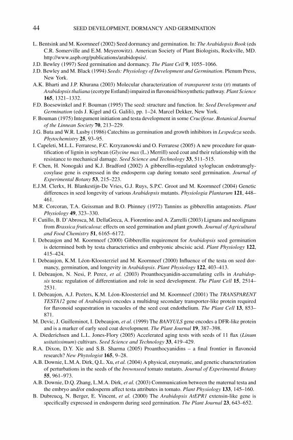
P1: OTE/SPH P2: OTE
BLUK053-Bradford August 27, 2006 17:7
44 SEED DEVELOPMENT, DORMANCY AND GERMINATION
L. Bentsink and M. Koornneef (2002) Seed dormancy and germination. In: The Arabidopsis Book (eds
C.R. Somerville and E.M. Meyerowitz). American Society of Plant Biologists, Rockville, MD.
http://www.aspb.org/publications/arabidopsis/.
J.D. Bewley (1997) Seed germination and dormancy. The Plant Cell 9, 1055–1066.
J.D. Bewley and M. Black (1994) Seeds: Physiology of Development and Germination. Plenum Press,
New York.
A.K. Bharti and J.P. Khurana (2003) Molecular characterization of transparent testa (tt) mutants of
Arabidopsis thaliana (ecotype Estland) impaired in flavonoid biosynthetic pathway. Plant Science165, 1321–1332.
F.D. Boesewinkel and F. Bouman (1995) The seed: structure and function. In: Seed Development andGermination (eds J. Kigel and G. Galili), pp. 1–24. Marcel Dekker, New York.
F. Bouman (1975) Integument initiation and testa development in some Cruciferae. Botanical Journalof the Linnean Society 70, 213–229.
J.G. Buta and W.R. Lusby (1986) Catechins as germination and growth inhibitors in Lespedeza seeds.
Phytochemistry 25, 93–95.
I. Capeleti, M.L.L. Ferrarese, F.C. Krzyzanowski and O. Ferrarese (2005) A new procedure for quan-
tification of lignin in soybean (Glycine max (L.) Merrill) seed coat and their relationship with the
resistance to mechanical damage. Seed Science and Technology 33, 511–515.
F. Chen, H. Nonogaki and K.J. Bradford (2002) A gibberellin-regulated xyloglucan endotransgly-
cosylase gene is expressed in the endosperm cap during tomato seed germination. Journal ofExperimental Botany 53, 215–223.
E.J.M. Clerkx, H. Blankestijn-De Vries, G.J. Ruys, S.P.C. Groot and M. Koornneef (2004) Genetic
differences in seed longevity of various Arabidopsis mutants. Physiologia Plantarum 121, 448–
461.
M.R. Corcoran, T.A. Geissman and B.O. Phinney (1972) Tannins as gibberellin antagonists. PlantPhysiology 49, 323–330.
F. Cutillo, B. D’Abrosca, M. DellaGreca, A. Fiorentino and A. Zarrelli (2003) Lignans and neolignans
from Brassica fruticulosa: effects on seed germination and plant growth. Journal of Agriculturaland Food Chemistry 51, 6165–6172.
I. Debeaujon and M. Koornneef (2000) Gibberellin requirement for Arabidopsis seed germination
is determined both by testa characteristics and embryonic abscisic acid. Plant Physiology 122,
415–424.
I. Debeaujon, K.M. Leon-Kloosterziel and M. Koornneef (2000) Influence of the testa on seed dor-
mancy, germination, and longevity in Arabidopsis. Plant Physiology 122, 403–413.
I. Debeaujon, N. Nesi, P. Perez, et al. (2003) Proanthocyanidin-accumulating cells in Arabidop-sis testa: regulation of differentiation and role in seed development. The Plant Cell 15, 2514–
2531.
I. Debeaujon, A.J. Peeters, K.M. Leon-Kloosterziel and M. Koornneef (2001) The TRANSPARENTTESTA12 gene of Arabidopsis encodes a multidrug secondary transporter-like protein required
for flavonoid sequestration in vacuoles of the seed coat endothelium. The Plant Cell 13, 853–
871.
M. Devic, J. Guilleminot, I. Debeaujon, et al. (1999) The BANYULS gene encodes a DFR-like protein
and is a marker of early seed coat development. The Plant Journal 19, 387–398.
A. Diederichsen and L.L. Jones-Flory (2005) Accelerated aging tests with seeds of 11 flax (Linumusitatissimum) cultivars. Seed Science and Technology 33, 419–429.
R.A. Dixon, D.Y. Xie and S.B. Sharma (2005) Proanthocyanidins – a final frontier in flavonoid
research? New Phytologist 165, 9–28.
A.B. Downie, L.M.A. Dirk, Q.L. Xu, et al. (2004) A physical, enzymatic, and genetic characterization
of perturbations in the seeds of the brownseed tomato mutants. Journal of Experimental Botany55, 961–973.
A.B. Downie, D.Q. Zhang, L.M.A. Dirk, et al. (2003) Communication between the maternal testa and
the embryo and/or endosperm affect testa attributes in tomato. Plant Physiology 133, 145–160.
B. Dubreucq, N. Berger, E. Vincent, et al. (2000) The Arabidopsis AtEPR1 extensin-like gene is
specifically expressed in endosperm during seed germination. The Plant Journal 23, 643–652.
P1: OTE/SPH P2: OTE
BLUK053-Bradford August 27, 2006 17:7
SEED COAT DEVELOPMENT AND DORMANCY 45
M. Edelstein, F. Corbineau, J. Kigel and H. Nerson (1995) Seed coat structure and oxygen availability
control low-temperature germination of melon (Cucumis melo) seeds. Physiologia Plantarum93, 451–456.
J.E. Flintham (2000) Different genetic components control coat-imposed and embryo-imposed dor-
mancy in wheat. Seed Science Research 10, 43–50.
N. Focks, M. Sagasser, B. Weisshaar and C. Benning (1999) Characterization of tt15, a novel trans-
parent testa mutant of Arabidopsis thaliana (L.) Heynh. Planta 208, 352–357.
M.D. Gale, J.E. Flintham and K.M. Devos (2002) Cereal comparative genetics and preharvest sprout-
ing. Euphytica 126, 21–25.
D. Garcia, J.N. Fitz Gerald and F. Berger (2005) Maternal control of integument cell elongation and
zygotic control of endosperm growth are coordinated to determine seed size in Arabidopsis. ThePlant Cell 17, 52–60.
D. Garcia, V. Saingery, P. Chambrier, U. Mayer, G. Jurgens and F. Berger (2003) Arabidopsis haikumutants reveal new controls of seed size by endosperm. Plant Physiology 131, 1661–1670.
K.T. Gatford, R.F. Eastwood and G.M. Halloran (2002) Germination inhibitors in bracts surrounding
the grain of Triticum tauschii. Functional Plant Biology 29, 881–890.
M. Gijzen, K. Kuflu, D. Qutob and J.T. Chernys (2001) A class I chitinase from soybean seed coat.
Journal of Experimental Botany 52, 2283–2289.
J.W. Gillikin and J.S. Graham (1991) Purification and developmental analysis of the major anionic
peroxidase from the seed coat of Glycine max. Plant Physiology 96, 214–220.
N. Goto (1985) A mucilage polysaccharide secreted from testa of Arabidopsis thaliana. ArabidopsisInformation Service 22, 143–145.
F.B. Green and M.R. Corcoran (1975) Inhibitory action of five tannins on growth induced by several
gibberellins. Plant Physiology 56, 801–806.
L.R. Griffen, A.M. Wilczek and F.A. Bazzaz (2004) UV-B affects within-seed biomass allocation and
chemical provisioning. New Phytologist 162, 167–171.
S.P.C. Groot, B. Kieliszewska-Rokicka, E. Vermeer and C.M. Karssen (1988) Gibberellin-induced
hydrolysis of endosperm cell walls in gibberellin-deficient tomato seeds prior to radicle protrusion.
Planta 174, 500–504.
X.Y. Gu, S.F. Kianian, G.A. Hareland, B.L. Hoffer and M.E. Foley (2005) Genetic analysis of adaptive
syndromes interrelated with seed dormancy in weedy rice (Oryza sativa). Theoretical and AppliedGenetics 110, 1108–1118.
Y. Gutterman (2000) Maternal effects on seeds during development. In: Seeds: The Ecology of Regener-ation in Plant Communities, 2nd edn (ed. M. Fenner), pp. 59–84. CAB International, Wallingford,
UK.
J.M. Halloin (1982) Localization and changes in catechin and tannins during development and ripening
of cottonseed. New Phytologist 90, 641–657.
G. Haughn and A. Chaudhury (2005) Genetic analysis of seed coat development in Arabidopsis. Trendsin Plant Science 10, 472–477.
E. Himi, D.J. Mares, A. Yanagisawa and K. Noda (2002) Effect of grain colour gene (R) on grain
dormancy and sensitivity of the embryo to abscisic acid (ABA) in wheat. Journal of ExperimentalBotany 53, 1569–1574.
E. Himi, A. Nisar and K. Noda (2005) Colour genes (R and Rc) for grain and coleoptile upregulate
flavonoid biosynthesis genes in wheat. Genome 48, 747–754.
E. Himi and K. Noda (2005) Red grain colour gene (R) of wheat is a Myb-type transcription factor.
Euphytica 143, 239–242.
E.E. Hood, M.R. Bailey, K. Beifuss, et al. (2003) Criteria for high-level expression of a fungal laccase
gene in transgenic maize. Plant Biotechnology Journal 1, 129–140.
F.M.A. Islam, J. Rengifo, R.J. Redden, K.E. Basford and S.E. Beebe (2003) Association between seed
coat polyphenolics (tannins) and disease resistance in common bean. Plant Foods for HumanNutrition 58, 285–297.
A. Iwanowska, T. Tykarska, M. Kuras and A.M. Zobel (1994) Localization of phenolic compounds in
the covering tissues of the embryo of Brassica napus (L.) during different stages of embryogenesis
and seed maturation. Annals of Botany 74, 313–320.

P1: OTE/SPH P2: OTE
BLUK053-Bradford August 27, 2006 17:7
46 SEED DEVELOPMENT, DORMANCY AND GERMINATION
C. Job, L. Rajjou, Y. Lovigny, M. Belghazi and D. Job (2005) Patterns of protein oxidation in Ara-bidopsis seeds and during germination. Plant Physiology 138, 790–802.
C.S. Johnson, B. Kolevski and D.R. Smyth (2002) TRANSPARENT TESTA GLABRA2, a trichome and
seed coat development gene of Arabidopsis, encodes a WRKY transcription factor. The PlantCell 14, 1359–1375.
F. Kantar, C.J. Pilbeam and P.D. Hebblethwaite (1996) Effect of tannin content of faba bean (Viciafaba) seed on seed vigour, germination and field emergence. Annals of Applied Biology 128,
85–93.
C.M. Karssen (2002) Germination, dormancy and red tape. Seed Science Research 12, 203–216.
K.M. Kelly, J. van Staden and W.E. Bell (1992) Seed coat structure and dormancy. Plant GrowthRegulation 11, 213–222.
A.R. Kermode (1995) Regulatory mechanisms in the transition from seed development to germi-
nation: interactions between the embryo and the seed environment. In: Seed Development andGermination (eds J. Kigel and G. Galili), pp. 273–332. Marcel Dekker, New York.
Y.C. Kim, M. Nakajima, A. Nakayama and I. Yamaguchi (2005) Contribution of gibberellins to the
formation of Arabidopsis seed coat through starch degradation. Plant and Cell Physiology 46,
1317–1325.
S. Kitamura, N. Shikazono and A. Tanaka (2004) TRANSPARENT TESTA 19 is involved in the
accumulation of both anthocyanins and proanthocyanidins in Arabidopsis. The Plant Journal 37,
104–114.
M. Koornneef (1981) The complex syndrome of ttg mutants. Arabidopsis Information Service 18,
45–51.
M. Koornneef (1990) Mutations affecting the testa colour in Arabidopsis. Arabidopsis InformationService 27, 1–4.
M. Koornneef, L. Bentsink and H. Hilhorst (2002) Seed dormancy and germination. Current Opinionin Plant Biology 5, 33–36.
S.S. Kwak, Y. Kamiya, A. Sakurai and N. Takahashi (1988) Isolation of a gibberellin biosynthesis
inhibitor from testas of Phaseolus vulgaris L. Agricultural and Biological Chemistry 52, 149–
152.
V. Lattanzio, R. Terzano, N. Cicco, A. Cardinali, D.D. Venere and V. Linsalata (2005) Seed coat tannins
and bruchid resistance in stored cowpea seeds. Journal of the Science of Food and Agriculture85, 839–846.
N. Legesse and A.A. Powell (1992) Comparisons of water uptake and imbibition damage in 11 cowpea
cultivars. Seed Science and Technology 20, 173–180.
N. Legesse and A.A. Powell (1996) Relationship between the development of seed coat pigmentation,
seed coat adherence to the cotyledons and the rate of imbibition during the maturation of grain
legumes. Seed Science and Technology 24, 23–32.
C. Lenoir, F. Corbineau and D. Come (1986) Barley (Hordeum vulgare) seed dormancy as related to
glumella characteristics. Physiologia Plantarum 68, 301–307.
K.M. Leon-Kloosterziel, C.J. Keijzer and M. Koornneef (1994) A seed shape mutant of Arabidopsisthat is affected in integument development. The Plant Cell 6, 385–392.
L. Lepiniec, I. Debeaujon, J.-M. Routaboul, et al. (2006) Genetics and biochemistry of seed flavonoids.
Annual Review of Plant Biology, 57, 405–430.
L. Lepiniec, M. Devic and F. Berger (2005) Genetic and molecular control of seed development in
Arabidopsis. In: Plant Functional Genomics (ed. D. Leister), pp. 511–564. Haworth Press, Inc.,
New York.
G. Leubner-Metzger (2002) Seed after-ripening and over-expression of class I beta-1,3-glucanase
confer maternal effects on tobacco testa rupture and dormancy release. Planta 215, 959–
968.
B. Manz, K. Muller, B. Kucera, F. Volke and G. Leubner-Metzger (2005) Water uptake and distribution
in germinating tobacco seeds investigated in vivo by nuclear magnetic resonance imaging. PlantPhysiology 138, 1538–1551.
I. Marbach and A.M. Mayer (1974) Permeability of seed coats to water as related to drying conditions
and metabolism of phenolics. Plant Physiology 54, 817–820.
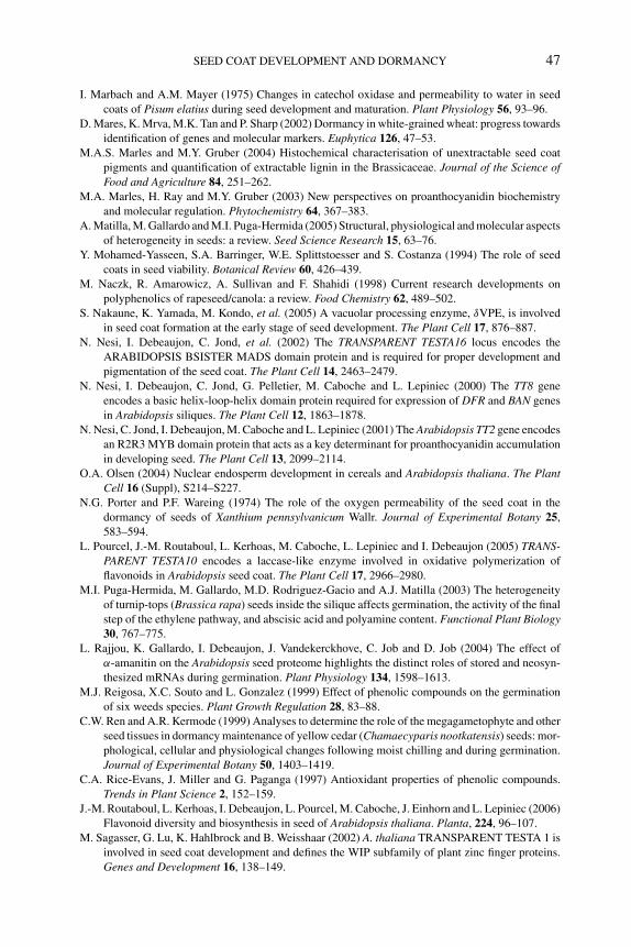
P1: OTE/SPH P2: OTE
BLUK053-Bradford August 27, 2006 17:7
SEED COAT DEVELOPMENT AND DORMANCY 47
I. Marbach and A.M. Mayer (1975) Changes in catechol oxidase and permeability to water in seed
coats of Pisum elatius during seed development and maturation. Plant Physiology 56, 93–96.
D. Mares, K. Mrva, M.K. Tan and P. Sharp (2002) Dormancy in white-grained wheat: progress towards
identification of genes and molecular markers. Euphytica 126, 47–53.
M.A.S. Marles and M.Y. Gruber (2004) Histochemical characterisation of unextractable seed coat
pigments and quantification of extractable lignin in the Brassicaceae. Journal of the Science ofFood and Agriculture 84, 251–262.
M.A. Marles, H. Ray and M.Y. Gruber (2003) New perspectives on proanthocyanidin biochemistry
and molecular regulation. Phytochemistry 64, 367–383.
A. Matilla, M. Gallardo and M.I. Puga-Hermida (2005) Structural, physiological and molecular aspects
of heterogeneity in seeds: a review. Seed Science Research 15, 63–76.
Y. Mohamed-Yasseen, S.A. Barringer, W.E. Splittstoesser and S. Costanza (1994) The role of seed
coats in seed viability. Botanical Review 60, 426–439.
M. Naczk, R. Amarowicz, A. Sullivan and F. Shahidi (1998) Current research developments on
polyphenolics of rapeseed/canola: a review. Food Chemistry 62, 489–502.
S. Nakaune, K. Yamada, M. Kondo, et al. (2005) A vacuolar processing enzyme, δVPE, is involved
in seed coat formation at the early stage of seed development. The Plant Cell 17, 876–887.
N. Nesi, I. Debeaujon, C. Jond, et al. (2002) The TRANSPARENT TESTA16 locus encodes the
ARABIDOPSIS BSISTER MADS domain protein and is required for proper development and
pigmentation of the seed coat. The Plant Cell 14, 2463–2479.
N. Nesi, I. Debeaujon, C. Jond, G. Pelletier, M. Caboche and L. Lepiniec (2000) The TT8 gene
encodes a basic helix-loop-helix domain protein required for expression of DFR and BAN genes
in Arabidopsis siliques. The Plant Cell 12, 1863–1878.
N. Nesi, C. Jond, I. Debeaujon, M. Caboche and L. Lepiniec (2001) The Arabidopsis TT2 gene encodes
an R2R3 MYB domain protein that acts as a key determinant for proanthocyanidin accumulation
in developing seed. The Plant Cell 13, 2099–2114.
O.A. Olsen (2004) Nuclear endosperm development in cereals and Arabidopsis thaliana. The PlantCell 16 (Suppl), S214–S227.
N.G. Porter and P.F. Wareing (1974) The role of the oxygen permeability of the seed coat in the
dormancy of seeds of Xanthium pennsylvanicum Wallr. Journal of Experimental Botany 25,
583–594.
L. Pourcel, J.-M. Routaboul, L. Kerhoas, M. Caboche, L. Lepiniec and I. Debeaujon (2005) TRANS-PARENT TESTA10 encodes a laccase-like enzyme involved in oxidative polymerization of
flavonoids in Arabidopsis seed coat. The Plant Cell 17, 2966–2980.
M.I. Puga-Hermida, M. Gallardo, M.D. Rodriguez-Gacio and A.J. Matilla (2003) The heterogeneity
of turnip-tops (Brassica rapa) seeds inside the silique affects germination, the activity of the final
step of the ethylene pathway, and abscisic acid and polyamine content. Functional Plant Biology30, 767–775.
L. Rajjou, K. Gallardo, I. Debeaujon, J. Vandekerckhove, C. Job and D. Job (2004) The effect of
α-amanitin on the Arabidopsis seed proteome highlights the distinct roles of stored and neosyn-
thesized mRNAs during germination. Plant Physiology 134, 1598–1613.
M.J. Reigosa, X.C. Souto and L. Gonzalez (1999) Effect of phenolic compounds on the germination
of six weeds species. Plant Growth Regulation 28, 83–88.
C.W. Ren and A.R. Kermode (1999) Analyses to determine the role of the megagametophyte and other
seed tissues in dormancy maintenance of yellow cedar (Chamaecyparis nootkatensis) seeds: mor-
phological, cellular and physiological changes following moist chilling and during germination.
Journal of Experimental Botany 50, 1403–1419.
C.A. Rice-Evans, J. Miller and G. Paganga (1997) Antioxidant properties of phenolic compounds.
Trends in Plant Science 2, 152–159.
J.-M. Routaboul, L. Kerhoas, I. Debeaujon, L. Pourcel, M. Caboche, J. Einhorn and L. Lepiniec (2006)
Flavonoid diversity and biosynthesis in seed of Arabidopsis thaliana. Planta, 224, 96–107.
M. Sagasser, G. Lu, K. Hahlbrock and B. Weisshaar (2002) A. thaliana TRANSPARENT TESTA 1 is
involved in seed coat development and defines the WIP subfamily of plant zinc finger proteins.
Genes and Development 16, 138–149.
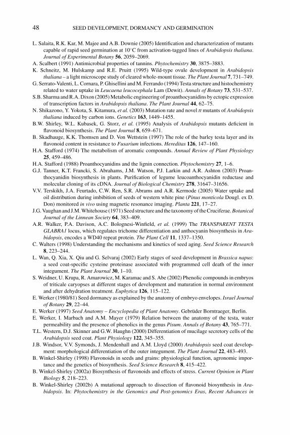
P1: OTE/SPH P2: OTE
BLUK053-Bradford August 27, 2006 17:7
48 SEED DEVELOPMENT, DORMANCY AND GERMINATION
L. Salaita, R.K. Kar, M. Majee and A.B. Downie (2005) Identification and characterization of mutants
capable of rapid seed germination at 10◦C from activation-tagged lines of Arabidopsis thaliana.
Journal of Experimental Botany 56, 2059–2069.
A. Scalbert (1991) Antimicrobial properties of tannins. Phytochemistry 30, 3875–3883.
K. Schneitz, M. Hulskamp and R.E. Pruitt (1995) Wild-type ovule development in Arabidopsisthaliana – a light microscope study of cleared whole-mount tissue. The Plant Journal 7, 731–749.
G. Serrato-Valenti, L. Cornara, P. Ghisellini and M. Ferrando (1994) Testa structure and histochemistry
related to water uptake in Leucaena leucocephala Lam (Dewit). Annals of Botany 73, 531–537.
S.B. Sharma and R.A. Dixon (2005) Metabolic engineering of proanthocyanidins by ectopic expression
of transcription factors in Arabidopsis thaliana. The Plant Journal 44, 62–75.
N. Shikazono, Y. Yokota, S. Kitamura, et al. (2003) Mutation rate and novel tt mutants of Arabidopsisthaliana induced by carbon ions. Genetics 163, 1449–1455.
B.W. Shirley, W.L. Kubasek, G. Storz, et al. (1995) Analysis of Arabidopsis mutants deficient in
flavonoid biosynthesis. The Plant Journal 8, 659–671.
B. Skadhauge, K.K. Thomsen and D. Von Wettstein (1997) The role of the barley testa layer and its
flavonoid content in resistance to Fusarium infections. Hereditas 126, 147–160.
H.A. Stafford (1974) The metabolism of aromatic compounds. Annual Review of Plant Physiology25, 459–486.
H.A. Stafford (1988) Proanthocyanidins and the lignin connection. Phytochemistry 27, 1–6.
G.J. Tanner, K.T. Francki, S. Abrahams, J.M. Watson, P.J. Larkin and A.R. Ashton (2003) Proan-
thocyanidin biosynthesis in plants. Purification of legume leucoanthocyanidin reductase and
molecular cloning of its cDNA. Journal of Biological Chemistry 278, 31647–31656.
V.V. Terskikh, J.A. Feurtado, C.W. Ren, S.R. Abrams and A.R. Kermode (2005) Water uptake and
oil distribution during imbibition of seeds of western white pine (Pinus monticola Dougl. ex D.
Don) monitored in vivo using magnetic resonance imaging. Planta 221, 17–27.
J.G. Vaughan and J.M. Whitehouse (1971) Seed structure and the taxonomy of the Cruciferae. BotanicalJournal of the Linnean Society 64, 383–409.
A.R. Walker, P.A. Davison, A.C. Bolognesi-Winfield, et al. (1999) The TRANSPARENT TESTAGLABRA1 locus, which regulates trichome differentiation and anthocyanin biosynthesis in Ara-bidopsis, encodes a WD40 repeat protein. The Plant Cell 11, 1337–1350.
C. Walters (1998) Understanding the mechanisms and kinetics of seed aging. Seed Science Research8, 223–244.
L. Wan, Q. Xia, X. Qiu and G. Selvaraj (2002) Early stages of seed development in Brassica napus:
a seed coat-specific cysteine proteinase associated with programmed cell death of the inner
integument. The Plant Journal 30, 1–10.
S. Weidner, U. Krupa, R. Amarowicz, M. Karamac and S. Abe (2002) Phenolic compounds in embryos
of triticale caryopses at different stages of development and maturation in normal environment
and after dehydration treatment. Euphytica 126, 115–122.
E. Werker (1980/81) Seed dormancy as explained by the anatomy of embryo envelopes. Israel Journalof Botany 29, 22–44.
E. Werker (1997) Seed Anatomy – Encyclopedia of Plant Anatomy. Gebruder Borntraeger, Berlin.
E. Werker, I. Marbach and A.M. Mayer (1979) Relation between the anatomy of the testa, water
permeability and the presence of phenolics in the genus Pisum. Annals of Botany 43, 765–771.
T.L. Western, D.J. Skinner and G.W. Haughn (2000) Differentiation of mucilage secretory cells of the
Arabidopsis seed coat. Plant Physiology 122, 345–355.
J.B. Windsor, V.V. Symonds, J. Mendenhall and A.M. Lloyd (2000) Arabidopsis seed coat develop-
ment: morphological differentiation of the outer integument. The Plant Journal 22, 483–493.
B. Winkel-Shirley (1998) Flavonoids in seeds and grains: physiological function, agronomic impor-
tance and the genetics of biosynthesis. Seed Science Research 8, 415–422.
B. Winkel-Shirley (2002a) Biosynthesis of flavonoids and effects of stress. Current Opinion in PlantBiology 5, 218–223.
B. Winkel-Shirley (2002b) A mutational approach to dissection of flavonoid biosynthesis in Ara-bidopsis. In: Phytochemistry in the Genomics and Post-genomics Eras, Recent Advances in
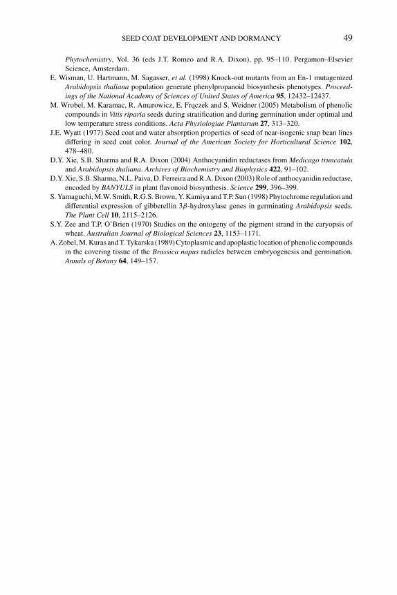
P1: OTE/SPH P2: OTE
BLUK053-Bradford August 27, 2006 17:7
SEED COAT DEVELOPMENT AND DORMANCY 49
Phytochemistry, Vol. 36 (eds J.T. Romeo and R.A. Dixon), pp. 95–110. Pergamon–Elsevier
Science, Amsterdam.
E. Wisman, U. Hartmann, M. Sagasser, et al. (1998) Knock-out mutants from an En-1 mutagenized
Arabidopsis thaliana population generate phenylpropanoid biosynthesis phenotypes. Proceed-ings of the National Academy of Sciences of United States of America 95, 12432–12437.
M. Wrobel, M. Karamac, R. Amarowicz, E. Frqczek and S. Weidner (2005) Metabolism of phenolic
compounds in Vitis riparia seeds during stratification and during germination under optimal and
low temperature stress conditions. Acta Physiologiae Plantarum 27, 313–320.
J.E. Wyatt (1977) Seed coat and water absorption properties of seed of near-isogenic snap bean lines
differing in seed coat color. Journal of the American Society for Horticultural Science 102,
478–480.
D.Y. Xie, S.B. Sharma and R.A. Dixon (2004) Anthocyanidin reductases from Medicago truncatulaand Arabidopsis thaliana. Archives of Biochemistry and Biophysics 422, 91–102.
D.Y. Xie, S.B. Sharma, N.L. Paiva, D. Ferreira and R.A. Dixon (2003) Role of anthocyanidin reductase,
encoded by BANYULS in plant flavonoid biosynthesis. Science 299, 396–399.
S. Yamaguchi, M.W. Smith, R.G.S. Brown, Y. Kamiya and T.P. Sun (1998) Phytochrome regulation and
differential expression of gibberellin 3β-hydroxylase genes in germinating Arabidopsis seeds.
The Plant Cell 10, 2115–2126.
S.Y. Zee and T.P. O’Brien (1970) Studies on the ontogeny of the pigment strand in the caryopsis of
wheat. Australian Journal of Biological Sciences 23, 1153–1171.
A. Zobel, M. Kuras and T. Tykarska (1989) Cytoplasmic and apoplastic location of phenolic compounds
in the covering tissue of the Brassica napus radicles between embryogenesis and germination.
Annals of Botany 64, 149–157.
P1: OTE/SPH P2: OTE
BLUK053-Bradford August 27, 2006 17:8
3 Definitions and hypotheses of seed dormancyHenk W.M. Hilhorst
3.1 Introduction
The term dormancy refers to a state of a whole plant or plant organ that is generallycharacterized by a temporary arrest in growth and development. Dormancy can befound among most forms of plant life and may occur in seeds, bulbs, tubers, buds,and whole plants. Dormancy is a trait that has likely been acquired during evolutionby selection for the ability to survive in adverse environments, such as heat, cold,and drought. There is evidence that the evolutionary origin of dormancy is relatedto climatic changes during the earth’s history and the spread of plant species aroundthe globe. The number of plant species with dormancy tends to increase with thegeographical distance from the equator, i.e., as seasonal variation in precipitationand temperature increases (Baskin and Baskin, 1998). Dormancy has significantlycontributed to the development of new species and the successful dispersion ofalready existing species (Baskin and Baskin, 1998). Several reviews discuss theecology and regulation mechanisms of seed dormancy (Bewley and Black, 1994;Hilhorst, 1995, 1998; Hilhorst et al., 2006; Bewley, 1997; Baskin and Baskin, 1998;Koornneef et al., 2002). As a part of the mechanism to survive adverse conditions,dormancy provides a strategy for seeds to spread germination in time in orderto reduce the risk of premature death by catastrophe. This review discusses seeddormancy classifications and definitions and focuses primarily on recent researchrelated to the regulation of dormancy. Results are discussed in relation to existingtheories. Emphasis will be on the model plant Arabidopsis thaliana, as this specieshas yielded the most significant research progress over the last decade.
3.2 Classifications of dormancy
A number of classification schemes have been proposed to describe seed dormancy.Most classifications that are in use (Table 3.1; Baskin and Baskin, 1998, 2004) arederived from the one that was proposed by Nikolaeva (1977) on the basis of seedmorphology. An additional classification is based on the timing of the occurrenceof dormancy: primary dormancy refers to the type of dormancy that occurs priorto dispersal as part of the seed developmental program, whereas secondary dor-mancy refers to the acquisition of dormancy in a mature seed after imbibition as aresult of the lack of proper conditions for germination (Amen, 1968; see Table 3.1).Dormancy may be located in the embryo or imposed by the tissues that surround the
50
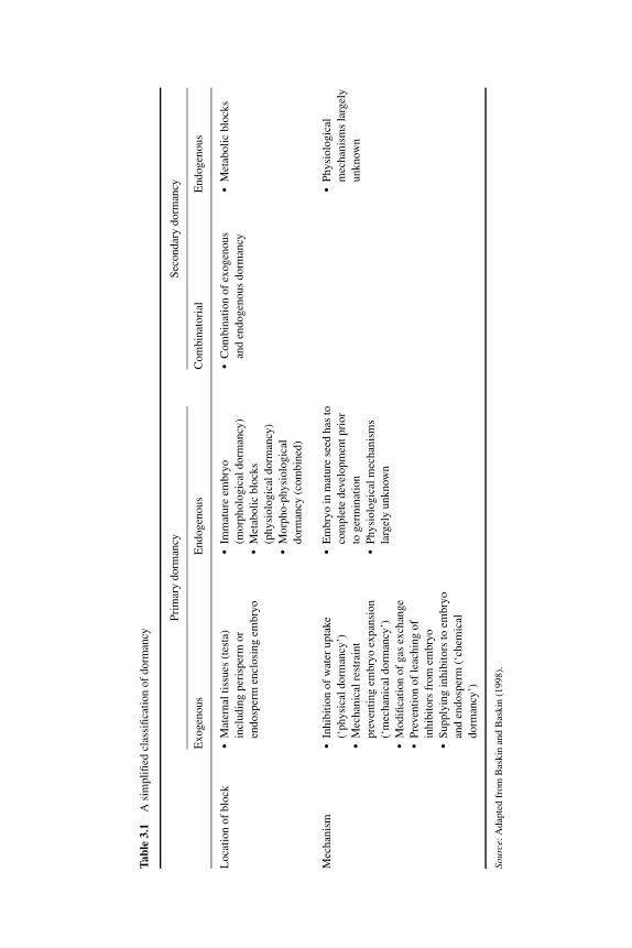
P1: OTE/SPH P2: OTE
BLUK053-Bradford August 27, 2006 17:8
Tabl
e3.
1A
sim
plifi
edcl
assi
ficat
ion
ofdo
rman
cy
Prim
ary
dorm
ancy
Seco
ndar
ydo
rman
cy
Exo
geno
usE
ndog
enou
sC
ombi
nato
rial
End
ogen
ous
Loc
atio
nof
bloc
k� Mat
erna
ltis
sues
(tes
ta)
incl
udin
gpe
risp
erm
oren
dosp
erm
encl
osin
gem
bryo
� Immat
ure
embr
yo(m
orph
olog
ical
dorm
ancy
)� Met
abol
icbl
ocks
(phy
siol
ogic
aldo
rman
cy)
� Morp
ho-p
hysi
olog
ical
dorm
ancy
(com
bine
d)
� Combi
natio
nof
exog
enou
san
den
doge
nous
dorm
ancy
� Metab
olic
bloc
ks
Mec
hani
sm� Inhi
bitio
nof
wat
erup
take
(‘ph
ysic
aldo
rman
cy’)
� Mecha
nica
lres
trai
ntpr
even
ting
embr
yoex
pans
ion
(‘m
echa
nica
ldor
man
cy’)
� Modi
ficat
ion
ofga
sex
chan
ge� Prev
entio
nof
leac
hing
ofin
hibi
tors
from
embr
yo� Sup
plyi
ngin
hibi
tors
toem
bryo
and
endo
sper
m(‘
chem
ical
dorm
ancy
’)
� Embr
yoin
mat
ure
seed
has
toco
mpl
ete
deve
lopm
entp
rior
toge
rmin
atio
n� Phy
siol
ogic
alm
echa
nism
sla
rgel
yun
know
n
� Physi
olog
ical
mec
hani
sms
larg
ely
unkn
own
Sour
ce:A
dapt
edfr
omB
aski
nan
dB
aski
n(1
998)
.
51
P1: OTE/SPH P2: OTE
BLUK053-Bradford August 27, 2006 17:8
52 SEED DEVELOPMENT, DORMANCY AND GERMINATION
embryo. In a number of species, dormancy is imposed by both the embryo and thetissues enclosing it. The completion of germination (radicle protrusion) is the netresult of the opposing forces: the ‘thrust’ of the embryo and the restraints by the sur-rounding tissues. In the case of embryo dormancy the properties of the embryo areof principal importance. In coat-imposed dormancy, the properties of the coveringtissues are the determinants, including mechanical, chemical, and permeability fea-tures, all of which may interfere with the successful completion of germination. Forexample, many seeds possess a testa (seed coat) that poses a mechanical restraint toembryonic growth and that may also contain chemical inhibitors, such as phenoliccompounds, that prevent embryo growth (mechanical and chemical dormancy) (seeChapter 2). Endosperm tissue may restrict embryo growth until the thick endospermcell walls are degraded by hydrolytic enzymes that can be induced by factors (e.g.,plant hormones) derived from the embryo (physiological/mechanical dormancy)(see Chapter 11). Both embryo and coat-imposed dormancy are common and theredoes not seem to be a preference for a specific category or type of dormancy amongplant families or genera (Baskin and Baskin, 1998).
The two principal classes of dormancy, physiological and morphophysiologicaldormancy, have been subdivided into several levels and types based on morpho-logical characteristics and requirements for dormancy release (Baskin and Baskin,2004). Although this subdivision is very useful for phylogenetic, biogeographic,and evolutionary studies, it is not suitable for physiological and molecular stud-ies since this subdivision is largely arbitrary, as it does not distinguish underlyingmechanisms and uses subjective criteria for dormancy intensity. Here, I will providea short overview of the different kinds of dormancy. For an extensive listing anddiscussion of dormancy classifications, see Baskin and Baskin (1998, 2004).
3.2.1 Endogenous dormancy
In some plant families, such as the Orchidaceae and Orobanchaceae, seeds containundifferentiated embryos usually consisting of only 2–100 cells (Watkinson andWelbaum, in press). Unlike other seeds with rudimentary embryos where a substan-tial endosperm provides nutrition, these seeds do not contain sufficient food reservesto complete embryo development and germination. They require exogenous sourcesof nutrition for embryo growth, such as from higher plants (parasitic) or from my-coheterotrophs (symbiotic). Developmental arrest in these seeds occurs at an earlystage of embryonic histodifferentiation. Seeds with undifferentiated embryos are notdormant in a strict sense and are therefore not included in the most current dormancyclassifications (Baskin and Baskin, 2004).
Other seeds with small embryos do complete their morphogenetic phase and havea fully differentiated embryo in terms of structure, but these embryos do not enterthe maturation phase; i.e., they do not expand and accumulate food reserves. Seedsof this type usually contain relatively large amounts of endosperm tissue, whichoften entirely surrounds the small embryo. These embryos have to grow inside thedispersed seed prior to germination. This has been studied particularly well in coffee(Coffea arabica) and celery (Apium graveolens). In both species the surrounding
P1: OTE/SPH P2: OTE
BLUK053-Bradford August 27, 2006 17:8
DEFINITIONS AND HYPOTHESES OF SEED DORMANCY 53
endosperm is digested during embryo growth. Radicle protrusion does not occuruntil the embryo has attained a predefined length and the micropylar endosperm issufficiently digested (Van der Toorn and Karssen, 1992; Da Silva et al., 2004).
Physiological dormancy, which is exhibited by the fully developed and maturedembryo, represents a reversible block to germination; i.e., embryo dormancy couldbe reinduced after the release of primary dormancy. In contrast, coat-imposed dor-mancy is irreversible: it can be released but not induced again. The reversible natureof physiological dormancy allows the occurrence of repeated dormancy cycles inseeds in the soil, following seasonal changes in temperature (Hilhorst et al., 1996).Dormancy cycling is crucial to the establishment and survival of many plant com-munities.
3.2.2 Exogenous dormancy
Embryo coverings include seed and fruit tissues that may be dead or alive. Theircontribution to the level of dormancy may be of a physical, chemical, or mechanicalnature, or combinations of these. Many seeds contain an endosperm that is of bothmaternal (2/3) and paternal (1/3) genetic origin. The endosperm usually functionsas a storage tissue. In some species (e.g., in cereals), it is predominantly dead tissuethat is filled with storage reserves, such as starch granules and protein bodies. Inother cases the endosperm is a living tissue that could contribute significantly tothe dormancy and germination behavior of the seed. The relative contribution of theendosperm to seed size varies greatly, from only a few cell layers surrounding theembryo to an almost complete occupation of the seed. Very often endosperm tissuecontains thickened cell walls. This provides considerable mechanical protection tothe embryo and is also a source of reserve carbohydrates in the form of hemicel-luloses. In a number of species, the primary seed storage tissue is the perisperm, adiploid endosperm-like tissue derived from the maternal nucellus.
The testa derived from the integuments and/or pericarp derived from fruit tissuesmay also delay germination. Physical dormancy is usually the result of imperme-ability of the testa to water or oxygen. In these cases, the seed coat requires chemicalmodification or mechanical scarification before water and oxygen can enter the em-bryo. A range of chemical compounds, including phenolics, derived from seedsor dispersal units may inhibit germination. Such chemicals have been extractedfrom all seed and fruit parts. The plant hormone abscisic acid (ABA) seems to beomnipresent in seed and fruit tissues. ABA present in mature seeds is generallyconsidered to be a remnant of the ABA pools that are involved in the regulation ofseed development (Hilhorst, 1995). If the ABA content in the seed is high enoughto inhibit germination, it has to leach out or be catabolized to allow completion ofgermination. However, the specific role of these compounds in the inhibition of ger-mination is not clear. In most cases a direct relationship between ABA content andits physiological action in seed dormancy is lacking. On the other hand, germinationof seeds containing inhibitory chemicals can be accelerated by extensively rinsingthe seeds with water prior to germination. These chemicals could be important notonly for germination regulation but also for other survival strategies of seeds. It
P1: OTE/SPH P2: OTE
BLUK053-Bradford August 27, 2006 17:8
54 SEED DEVELOPMENT, DORMANCY AND GERMINATION
is possible that many of these inhibitory substances are present primarily to avoidpredation or microbial infections (Baskin and Baskin, 1998). Seeds that combineelements of both endogenous and exogenous dormancy are classified in the groupof combinatorial dormancy (Table 3.1).
Tissues surrounding the embryo will impose a certain mechanical restraint toexpansion of the embryo. Removal of all or part of the covering tissues often leadsto normal embryo growth, indicating that the block to germination is located in thecovering tissues. The embryo can overcome the mechanical restraint of the enclosingtissues by generating a thrust sufficient to penetrate them. Alternatively, the restraintmay be reduced by weakening of the tissues by an active process such as enzymesecretion, which in turn is controlled by the embryo, without apparent changes in theembryonic growth potential. This has been demonstrated for a number of species inwhich the endosperm is digested by hydrolytic enzymes, e.g., tomato (Lycopersiconesculentum; Groot and Karssen, 1987), Datura ferox (Sanchez et al., 1990), coffee(Da Silva et al., 2004), and tobacco (Nicotiana tabacum; Leubner-Metzger, 2003;see Chapter 11).
3.3 Definitions of dormancy
Seed dormancy results in the absence of germination. However, absence of germina-tion can have several causes: the seed can be nonviable, the environment may simplybe limiting germination, or some factors in the seed or dispersal unit itself may blockgermination. Hence, the frequently used definition of seed dormancy is “the absenceof germination of a viable seed under conditions that are favorable to germination”(Harper, 1959). However, this definition is not perfect since it could lead to differentinterpretations. A first caveat relates to the determination of ‘absence of germina-tion’. Many species display very slow germination; e.g., coffee seeds may take up to50 days before radicle protrusion occurs under the optimal growing conditions (DaSilva et al., 2004). Observation before 50 days would classify the seeds as dormantwhereas observation after 50 days would classify them as nondormant. In addition,the time required for germination can vary depending upon the degree or depth ofdormancy. An arbitrary germination time to distinguish between dormant and non-dormant seeds has obvious practical advantages for dormancy classification but isnot useful in physiological and molecular studies of seed dormancy and germina-tion. A second caveat refers to ‘conditions that are favorable to germination’. Lessdormant seeds germinate in a wider range of conditions than more dormant ones(e.g., Karssen, 1982). In the case of temperature, dormant seeds germinate over anarrower range of temperatures whereas nondormant seeds are more tolerant to awider range of temperatures (see Chapter 4). Consequently, dormancy release is as-sociated with a widening of the germination temperature ‘window’ and, vice versa,when dormancy is induced, the germination temperature window becomes narrower.Obviously, fully dormant seeds will not germinate at any temperature. This makes itclear that seed dormancy is not an all-or-nothing property but a relative phenomenonthe expression of which varies with the environment. This implies that when seeds
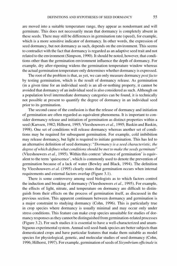
P1: OTE/SPH P2: OTE
BLUK053-Bradford August 27, 2006 17:8
DEFINITIONS AND HYPOTHESES OF SEED DORMANCY 55
are moved into a suitable temperature range, they appear as nondormant and willgerminate. This does not necessarily mean that dormancy is completely absent inthese seeds. There may still be differences in germination rate (speed), for example,which is a more sensitive indicator of dormancy. In other words, the expression ofseed dormancy, but not dormancy as such, depends on the environment. This seemsto contradict with the fact that dormancy is regarded as an adaptive seed trait and notrelated to the environment (Simpson, 1990). It should be noted, however, that condi-tions other than the germination environment influence the depth of dormancy. Forexample, dry after-ripening widens the germination temperature window whereasthe actual germination temperature only determines whether dormancy is expressed.
The root of the problem is that, as yet, we can only measure dormancy post factoby testing germination, which is the result of dormancy release. As germination(in a given time for an individual seed) is an all-or-nothing property, it cannot beavoided that dormancy of an individual seed is also considered as such. Although ona population level intermediate dormancy categories can be found, it is technicallynot possible at present to quantify the degree of dormancy in an individual seedprior to its germination.
The second cause of the confusion is that the release of dormancy and initiationof germination are often regarded as equivalent phenomena. It is important to con-sider dormancy release and initiation of germination as distinct properties within aseed (Karssen, 1982; Hilhorst, 1995; Vleeshouwers et al., 1995; Baskin and Baskin,1998). One set of conditions will release dormancy whereas another set of condi-tions may be required for subsequent germination. For example, cold imbibitionmay release dormancy, but light is required to initiate germination. This has led toan alternative definition of seed dormancy: “Dormancy is a seed characteristic, thedegree of which defines what conditions should be met to make the seeds germinate”(Vleeshouwers et al., 1995). Within this context ‘absence of germination’ is equiv-alent to the term ‘quiescence’, which is commonly used to denote the prevention ofgermination because of a lack of water (Bewley and Black, 1994). The definitionby Vleeshouwers et al. (1995) clearly states that germination occurs when internalrequirements and external factors overlap (Figure 3.1).
There is some controversy among seed biologists as to which factors controlthe induction and breaking of dormancy (Vleeshouwers et al., 1995). For example,the effects of light, nitrate, and temperature on dormancy are difficult to distin-guish from their effects on the process of germination itself, as discussed in theprevious section. This apparent continuum between dormancy and germination isa major constraint to studying dormancy (Cohn, 1996). This is particularly truein crop species where dormancy is usually minimal and may occur only understress conditions. This feature can make crop species unsuitable for studies of dor-mancy responses as they cannot be distinguished from germination-related processes(Figure 3.2). For such studies it is essential to have a well-characterized and unam-biguous experimental system. Annual soil seed-bank species are better subjects thandomesticated crops and have particular features that make them suitable as modelspecies for physiological, genetic, and molecular studies of seed dormancy (Cohn,1996; Hilhorst, 1997). For example, germination of seeds of Sisymbrium officinale is
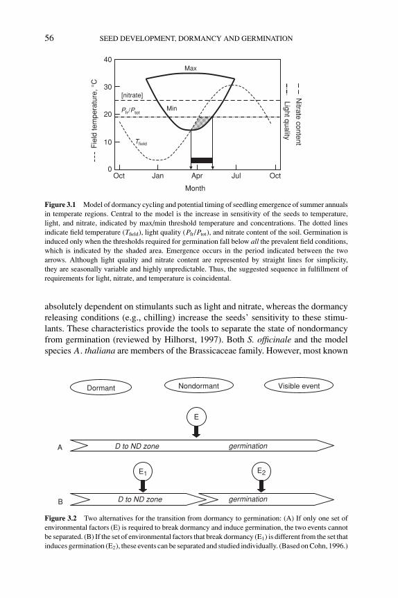
P1: OTE/SPH P2: OTE
BLUK053-Bradford August 27, 2006 17:8
56 SEED DEVELOPMENT, DORMANCY AND GERMINATION
Oct Jan Apr Jul Oct0
10
20
30
40
[nitrate]
Max
Min
Fie
ld te
mpe
ratu
re, °
C
Month
Tfield
Pfr/PtotLight quality
Nitrate content
Figure 3.1 Model of dormancy cycling and potential timing of seedling emergence of summer annualsin temperate regions. Central to the model is the increase in sensitivity of the seeds to temperature,light, and nitrate, indicated by max/min threshold temperature and concentrations. The dotted linesindicate field temperature (Tfield), light quality (Pfr/Ptot), and nitrate content of the soil. Germination isinduced only when the thresholds required for germination fall below all the prevalent field conditions,which is indicated by the shaded area. Emergence occurs in the period indicated between the twoarrows. Although light quality and nitrate content are represented by straight lines for simplicity,they are seasonally variable and highly unpredictable. Thus, the suggested sequence in fulfillment ofrequirements for light, nitrate, and temperature is coincidental.
absolutely dependent on stimulants such as light and nitrate, whereas the dormancyreleasing conditions (e.g., chilling) increase the seeds’ sensitivity to these stimu-lants. These characteristics provide the tools to separate the state of nondormancyfrom germination (reviewed by Hilhorst, 1997). Both S. officinale and the modelspecies A. thaliana are members of the Brassicaceae family. However, most known
D to ND zone germination
D to ND zone germination
E
E1 E2
Dormant Nondormant Visible event
A
B
Figure 3.2 Two alternatives for the transition from dormancy to germination: (A) If only one set ofenvironmental factors (E) is required to break dormancy and induce germination, the two events cannotbe separated. (B) If the set of environmental factors that break dormancy (E1) is different from the set thatinduces germination (E2), these events can be separated and studied individually. (Based on Cohn, 1996.)
P1: OTE/SPH P2: OTE
BLUK053-Bradford August 27, 2006 17:8
DEFINITIONS AND HYPOTHESES OF SEED DORMANCY 57
accessions of Arabidopsis have only a shallow dormancy that is released by rela-tively short periods of dry after-ripening. This makes the species less suitable for dor-mancy research. However, the Cvi accession, which originates from the Cape VerdeIslands, is considered deeply dormant as it requires several months of after-ripeningto release its dormancy. This accession is now in use in a number of genetic andmolecular studies of dormancy (Alonso-Blanco et al., 2003; Chapter 5).
3.4 Primary dormancy
3.4.1 Induction of primary dormancy
3.4.1.1 Role of ABA in dormancy inductionPrimary dormancy is commonly associated with the transient increase in ABA con-tent during seed development. In most of the species studied, ABA levels increaseduring the first half of seed development and decline during late maturation con-comitantly with the decline in seed water content. ABA has been detected in allseed and fruit tissues examined and has been related to a number of developmentalprocesses, including synthesis of storage proteins and late embryogenesis-abundantproteins, suppression of precocious germination, and induction of desiccation tol-erance (for reviews, see McCarty, 1995; Holdsworth et al., 1999; Finkelstein et al.,2002; Chapter 1). Studies with ABA-deficient mutants of Arabidopsis and tomatohave demonstrated that defects in ABA synthesis during seed development result inthe formation of nondormant seeds (Karssen et al., 1983; Koornneef et al., 1984,1985; Groot and Karssen, 1992). In tomato and Arabidopsis, dormancy was inducedonly when the embryo contained the normal ABA allele and was capable of syn-thesizing ABA. Crosses between wild-type and aba (Arabidopsis) or sitiens (sitw,tomato) mutant plants indicated that maternal ABA (i.e., located in testa and fruittissues) does not affect dormancy. Thus, a transient increase in embryonic ABAcontent during seed development is required to induce dormancy. Further supportfor this conclusion was provided by experiments in tobacco where overexpressionof zeaxanthin oxidase, one of the key enzymes in ABA synthesis, resulted in moredormant seed phenotypes than wild type, whereas silencing of this gene yielded lessdormant seed phenotypes (Frey et al., 1999).
Sensitivity to ABA plays an equally important role to ABA content in the induc-tion of dormancy. The ABA-insensitive abi1, abi2, and abi3 mutants display variablereductions in seed dormancy (Koornneef et al., 1984). Conversely, seeds from theABA-hypersensitive era1 (enhanced response to ABA 1) mutant show an enhancedseed dormancy phenotype (Cutler et al., 1996). Several cultivars of wheat (Triticumaestivum) and maize (Zea mays) that exhibit vivipary, also known as preharvestsprouting, have a reduced sensitivity to ABA. Analysis of Arabidopsis seeds andmaize kernels displaying vivipary led to the identification of the ABA-responsivegenes ABI3 and VP1, respectively, which are responsible for these phenotypic char-acteristics (see Chapters 1 and 8). These orthologous genes encode transcriptionfactors of the B3 domain family, which activate the transcription of ABA-inducible
P1: OTE/SPH P2: OTE
BLUK053-Bradford August 27, 2006 17:8
58 SEED DEVELOPMENT, DORMANCY AND GERMINATION
genes that play roles in seed development (Finkelstein et al., 2002). They are in-volved in maintaining the developmental state of seeds and suppressing the transitionto the vegetative or growth stage. Thus, embryos of mutants defective in these genestypically exhibit little or no dormancy and often germinate precociously.
3.4.1.2 Developmental programs and dormancy inductionMutant analyses have identified a number of genes that altered seed dormancy with-out affecting ABA contents throughout seed development, including Arabidopsisabi3, leafy cotyledon1 (lec1), lec2, and fusca3 (fus3) mutants. These mutants allexhibit phenotypes that are characteristic of the vegetative state: e.g., reduced des-iccation tolerance, active meristems, expression of germination-related genes, andabsence of dormancy (Parcy et al., 1997; Chapter 1). Apparently, these genes havepartially overlapping functions in the overall control of seed maturation, leading tomutants defective in many aspects of seed maturation (Parcy et al., 1997). LEC1 andFUS3 loci probably regulate developmental arrest, as mutations in these genes causea continuation of embryo growth. ABI3 is also active during vegetative quiescenceprocesses in other parts of the plant in which it suppresses meristematic activity(Rohde et al., 2000). These results suggest that ABI3, LEC1, and FUS3 are requiredfor the induction and/or the maintenance of dormancy.
Because the abi3, lec1, lec2, and fus3 mutants are defective in seed matura-tion, no dormancy is observed in the mutant seeds, which germinate precociously(Nambara et al., 2000). Detailed analyses of the abi3, lec1, and fus3 mutants haverevealed that they differ in the time of occurrence of precocious germination (Raz etal., 2001). LEC1 and FUS3 probably function earlier to switch off the developmentalprogram and then ABI3 functions at a later time point to induce dormancy (Raz et al.,2001). Double mutants between a gibberellin (GA)-deficient mutant and each of thethree genes also behave differently. For example, GA dependency of germination ismaintained in the fus3 mutant but not in lec1, suggesting that these mutants affectthe germination potential of seeds in different ways. Comparison of LEC1, FUS3,and ABI3 gene expression during seed development and dormancy release in severalaccessions of Arabidopsis varying in degree of dormancy demonstrated that therewas no relationship between expression of these genes and the degree of dormancy(Baumbusch et al., 2004). Thus, it is unlikely that these genes directly control theestablishment of dormancy in Arabidopsis.
Similarly, in tomato it has been suggested that developmental arrest and dor-mancy are separate phenomena. After the completion of embryonic histodifferenti-ation both wild-type (cv. Moneymaker) and ABA-deficient sitw mutant seeds entera state of developmental arrest. In wild-type seeds this is followed by the inductionof dormancy, consistent with a transient rise in ABA content (Hilhorst and Karssen,1992; Hilhorst, 1995). In the sitw mutant there is no induction of dormancy andhence developmental arrest is not maintained, resulting in viviparous germinationwithin overripe fruits. However, at this stage ABA is also undetectable in wild-type seeds, yet germination is suppressed. The osmotic environments of the loculartissues of fruits of both genotypes are comparable (Berry and Bewley, 1992; Liuet al., 1996). Apparently, the mutant seeds possess a greater ‘growth potential’, as
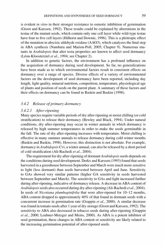
P1: OTE/SPH P2: OTE
BLUK053-Bradford August 27, 2006 17:8
DEFINITIONS AND HYPOTHESES OF SEED DORMANCY 59
is evident in vitro in their stronger resistance to osmotic inhibition of germination(Groot and Karssen, 1992). These results could be explained by aberrations in thetestae of the mutant seeds, which contain only one cell layer while wild-type testaehave four to five cell layers (Hilhorst and Downie, 1996). This is a pleitropic effectof the mutation in abscisic aldehyde oxidase (AAO3), which catalyzes the final stepin ABA synthesis (Nambara and Marion-Poll, 2005; Chapter 9). Numerous mu-tants in Arabidopsis that alter testa properties are known to affect seed dormancy(Leon-Kloosterziel et al., 1994; see Chapter 2).
In addition to genetic factors, the environment has a profound influence onthe acquisition of dormancy during seed development. So far, no generalizationshave been made as to which environmental factors result in consistent effects ondormancy over a range of species. Diverse effects of a variety of environmentalfactors on the development of seed dormancy have been reported, including daylength, light quality, mineral nutrition, competition, temperature, physiological ageof plants and position of seeds on the parent plant. A summary of these factors andtheir effects on dormancy can be found in Baskin and Baskin (1998).
3.4.2 Release of primary dormancy
3.4.2.1 After-ripeningMany species require variable periods of dry after-ripening or moist chilling (or coldstratification) to release their dormancy (Bewley and Black, 1994). Under naturalconditions, dry after-ripening may occur in winter annuals in which dormancy isreleased by high summer temperatures in order to make the seeds germinable inthe fall. The rate of dry after-ripening increases with temperature. Moist chilling iseffective in many summer annuals to release dormancy during cold winter months(Baskin and Baskin, 1998). However, this distinction is not absolute. For example,dormancy in Arabidopsis Cvi, a winter annual, can also be released by a short periodof cold stratification (Ali-Rachedi et al., 2004).
The requirement for dry after-ripening of dormant Arabidopsis seeds depends onthe conditions during seed development. Derkx and Karssen (1993) found that seedsharvested in a greenhouse between September and March were much more sensitiveto light (less dormant) than seeds harvested between April and June. Sensitivityto GAs showed very similar patterns (higher GA sensitivity in seeds harvestedbetween September and March). The sensitivity to GAs and light increased furtherduring after-ripening, indicative of dormancy release. A decrease in ABA content ofArabidopsis seeds also occurred during dry after-ripening (Ali-Rachedi et al., 2004).In seeds of Nicotiana plumbaginifolia that were after-ripened for 10–12 months,ABA content dropped to approximately 40% of that found in dormant seeds with aconcurrent increase in germination rate (Grappin et al., 2000). A similar decreasewas found in tomato seeds after 1 year of dry storage (Groot and Karssen, 1992). Thesensitivity to ABA also decreased in tobacco seeds during after-ripening (Grappinet al., 2000; Leubner-Metzger and Meins, 2000). As ABA is a potent inhibitor ofseed germination, these changes in ABA content or sensitivity are likely related tothe increasing germination potential of after-ripened seeds.
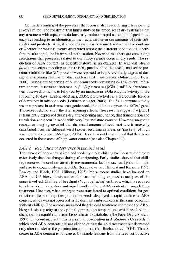
P1: OTE/SPH P2: OTE
BLUK053-Bradford August 27, 2006 17:8
60 SEED DEVELOPMENT, DORMANCY AND GERMINATION
Our understanding of the processes that occur in dry seeds during after-ripeningis very limited. The constraint that limits study of the processes in dry systems is thatany treatment with aqueous solutions may initiate a rapid activation of preformedenzymes leading to an alteration in their activities or in the amounts of their sub-strates and products. Also, it is not always clear how much water the seed containsor whether the water is evenly distributed among the different seed tissues. There-fore, results should be interpreted with caution. Nevertheless, there are convincingindications that processes related to dormancy release occur in dry seeds. The re-duction of ABA content, as described above, is an example. In wild oat (Avenafatua), transcripts encoding avenin (AV10), puroindoline-like (AV1), and serine pro-teinase inhibitor-like (Z1) proteins were reported to be preferentially degraded dur-ing after-ripening relative to other mRNAs that were present (Johnson and Dyer,2000). During after-ripening of N. tabacum seeds containing 8–13% overall mois-ture content, a transient increase in β-1,3-glucanase (βGlu1) mRNA abundancewas observed, which was followed by an increase in βGlu enzyme activity in thefollowing 10 days (Leubner-Metzger, 2005). βGlu activity is a prerequisite for lossof dormancy in tobacco seeds (Leubner-Metzger, 2003). The βGlu enzyme activitywas not present in antisense transgenic seeds that did not express the βGlu1 gene.Those seeds did not show the after-ripening effects. These results suggest that βGlu1is transiently expressed during dry after-ripening and, hence, that transcription andtranslation can occur in seeds with very low moisture content. However, magneticresonance imaging revealed that the small amount of seed moisture is unevenlydistributed over the different seed tissues, resulting in areas or ‘pockets’ of highwater content (Leubner-Metzger, 2005). Thus it cannot be precluded that the eventsoccurred in these areas of high water content (see also Chapter 11).
3.4.2.2 Regulation of dormancy in imbibed seedsThe release of dormancy in imbibed seeds by moist chilling has been studied moreextensively than the changes during after-ripening. Early studies showed that chill-ing increases the seed sensitivity to environmental factors, such as light and nitrate,and also to exogenously applied GAs (for reviews, see Hilhorst and Karssen, 1992;Bewley and Black, 1994; Hilhorst, 1995). More recent studies have focused onABA and GA biosynthesis and catabolism, including expression analyses of thegenes involved. Chilling of beechnut (Fagus sylvatica) embryos, which is requiredto release dormancy, does not significantly reduce ABA content during chillingtreatment. However, when embryos were transferred to optimal conditions for ger-mination after chilling, the germinable seeds displayed a rapid decline in ABAcontent, which was not observed in the dormant embryos kept in the same conditionwithout chilling. The authors suggested that the cold treatment decreased the ABA-biosynthesis capacity at the optimal germination temperature, which resulted in achange of the equilibrium from biosynthesis to catabolism (Le Page-Degivry et al.,1997). In accordance with this is a similar observation in Arabidopsis Cvi seeds inwhich seed ABA contents did not change during the cold treatment but decreasedonly after transfer to the germination conditions (Ali-Rachedi et al., 2004). The de-crease in ABA content is not caused by simple leakage from the seed but by active
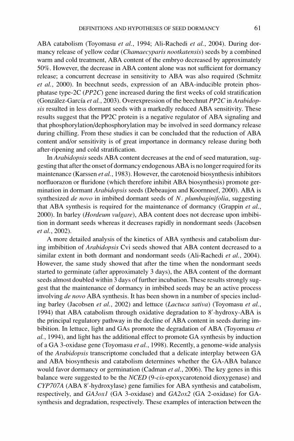
P1: OTE/SPH P2: OTE
BLUK053-Bradford August 27, 2006 17:8
DEFINITIONS AND HYPOTHESES OF SEED DORMANCY 61
ABA catabolism (Toyomasu et al., 1994; Ali-Rachedi et al., 2004). During dor-mancy release of yellow cedar (Chamaecyparis nootkatensis) seeds by a combinedwarm and cold treatment, ABA content of the embryo decreased by approximately50%. However, the decrease in ABA content alone was not sufficient for dormancyrelease; a concurrent decrease in sensitivity to ABA was also required (Schmitzet al., 2000). In beechnut seeds, expression of an ABA-inducible protein phos-phatase type-2C (PP2C) gene increased during the first weeks of cold stratification(Gonzalez-Garcıa et al., 2003). Overexpression of the beechnut PP2C in Arabidop-sis resulted in less dormant seeds with a markedly reduced ABA sensitivity. Theseresults suggest that the PP2C protein is a negative regulator of ABA signaling andthat phosphorylation/dephosphorylation may be involved in seed dormancy releaseduring chilling. From these studies it can be concluded that the reduction of ABAcontent and/or sensitivity is of great importance in dormancy release during bothafter-ripening and cold stratification.
In Arabidopsis seeds ABA content decreases at the end of seed maturation, sug-gesting that after the onset of dormancy endogenous ABA is no longer required for itsmaintenance (Karssen et al., 1983). However, the carotenoid biosynthesis inhibitorsnorfluorazon or fluridone (which therefore inhibit ABA biosynthesis) promote ger-mination in dormant Arabidopsis seeds (Debeaujon and Koornneef, 2000). ABA issynthesized de novo in imbibed dormant seeds of N . plumbaginifolia, suggestingthat ABA synthesis is required for the maintenance of dormancy (Grappin et al.,2000). In barley (Hordeum vulgare), ABA content does not decrease upon imbibi-tion in dormant seeds whereas it decreases rapidly in nondormant seeds (Jacobsenet al., 2002).
A more detailed analysis of the kinetics of ABA synthesis and catabolism dur-ing imbibition of Arabidopsis Cvi seeds showed that ABA content decreased to asimilar extent in both dormant and nondormant seeds (Ali-Rachedi et al., 2004).However, the same study showed that after the time when the nondormant seedsstarted to germinate (after approximately 3 days), the ABA content of the dormantseeds almost doubled within 3 days of further incubation. These results strongly sug-gest that the maintenance of dormancy in imbibed seeds may be an active processinvolving de novo ABA synthesis. It has been shown in a number of species includ-ing barley (Jacobsen et al., 2002) and lettuce (Lactuca sativa) (Toyomasu et al.,1994) that ABA catabolism through oxidative degradation to 8′-hydroxy-ABA isthe principal regulatory pathway in the decline of ABA content in seeds during im-bibition. In lettuce, light and GAs promote the degradation of ABA (Toyomasu etal., 1994), and light has the additional effect to promote GA synthesis by inductionof a GA 3-oxidase gene (Toyomasu et al., 1998). Recently, a genome-wide analysisof the Arabidopsis transcriptome concluded that a delicate interplay between GAand ABA biosynthesis and catabolism determines whether the GA-ABA balancewould favor dormancy or germination (Cadman et al., 2006). The key genes in thisbalance were suggested to be the NCED (9-cis-epoxycarotenoid dioxygenase) andCYP707A (ABA 8′-hydroxylase) gene families for ABA synthesis and catabolism,respectively, and GA3ox1 (GA 3-oxidase) and GA2ox2 (GA 2-oxidase) for GA-synthesis and degradation, respectively. These examples of interaction between the
P1: OTE/SPH P2: OTE
BLUK053-Bradford August 27, 2006 17:8
62 SEED DEVELOPMENT, DORMANCY AND GERMINATION
synthesis and catabolism of ABA and GA during imbibition strongly suggest thatan ABA–GA balance is operational. The results from the studies described aboveare in sharp contrast to the revised hormone balance theory (Karssen and Lacka,1986), which implicated ABA only in seed development and excluded its role duringimbibition and dormancy maintenance, which were postulated to be sensitive onlyto the presence or absence of GA.
There is strong evidence that GAs are an absolute requirement for germination.GA-deficient mutants of tomato (gib1) and Arabidopsis (ga1) require exogenous GAto complete germination (Koornneef and Van der Veen, 1980; Groot and Karssen,1987). As indicated above, the sensitivity to GAs may increase with the progressof dormancy release (Hilhorst et al., 1986; Karssen et al., 1989). An increase inthe content of bioactive GAs during cold stratification in Arabidopsis seeds hasbeen reported, but did not show a close correlation with the control of dormancy(Derkx et al., 1994). Yamauchi et al. (2004) showed that an increase in bioactiveGA1 and GA4 occurred during cold stratification of Arabidopsis seeds. The majorincrease in GA4 content was mediated by enhanced expression of the genes encodingGA 3-oxidase (GA3ox1 and GA3ox2), the enzyme that catalyzes the conversion ofGA9 to GA4. Furthermore, red light irradiation during the cold treatment furtherelevated GA3ox1 transcript accumulation (Yamauchi et al., 2004). Although allthe requirements for dormancy release, namely the increase in GA sensitivity andcontent as well as light sensitivity, may be met during the cold treatment, seeds maynot (or not within the time frame of observation) germinate at this low temperature.It was suggested that cell-type specificity of GA3ox1 mRNA accumulation dependson the environment, including temperature (Yamauchi et al., 2004). It is possiblethat a permissive temperature for germination is essential for spatial changes in GAsynthesis or of its secondary messenger across different cell layers, although anotherpossibility is that the responses of cells to GA (e.g., cell elongation) are inhibited atcold temperature.
The results from these recent studies seem to contradict the long-held model ofdormancy release separated from, and followed by, the induction of germination, asdescribed in a previous section (Figure 3.2B). The synthesis of GAs has been com-monly associated with the promotion of germination rather than the release fromdormancy (Bewley and Black, 1994). Observations that GAs induce germination ofdormant seeds were interpreted as indicating that GAs may bypass the requirementsfor dormancy release, rather than being integral directly to dormancy release. Sim-ilarly, in the previous model, light was not considered to affect dormancy but wasrecognized to be active only during germination, after the seeds had been sensitizedto light and GAs by cold treatment (Hilhorst et al., 1986; Hilhorst and Karssen,1992). Evidence of the induction of GA biosynthetic enzymes by chilling and theirsensitivity to light during chilling may require reexamination of this model.
Similarly, recent studies have shed new light on the role of nitrate in seed dor-mancy and germination. S. officinale seed germination is absolutely dependent onlight and nitrate and both factors are active only in the completion of germinationafter dormancy-releasing cold stratification has enhanced the seed sensitivity to bothexogenous and endogenous nitrate (Hilhorst et al., 1986; Hilhorst, 1990). However,
P1: OTE/SPH P2: OTE
BLUK053-Bradford August 27, 2006 17:8
DEFINITIONS AND HYPOTHESES OF SEED DORMANCY 63
in the case of dormant Arabidopsis Cvi seeds, nitrate could substitute for the longperiod (7–12 months) of dry storage or several days of cold stratification requiredto release dormancy (Ali-Rachedi et al., 2004) and therefore can be regarded asa dormancy-releasing agent. So far, very little is known about the nitrate signal-ing pathways that lead to germination, although there is evidence in S. officinalethat nitrate is a signaling molecule by itself without being reduced to nitrite or NO(Hilhorst and Karssen, 1989; see Chapter 7 for NO signaling). Seeds of an Ara-bidopsis nitrate reductase [NADH] 1 and 2 double mutant nia1 nia2, which retainsonly 0.5% of the nitrate reductase activity of the wild type, are much less dormantthan the wild-type seeds. The mutant seeds contained 100-fold more nitrate than thewild type, probably because the mutant accumulates more nonreduced nitrate thatcould be transported to and stored in the seeds (Alboresi et al., 2005). It has beenproposed that nitrate signaling may have cross talk with the ABA or GA pathways,but there is only circumstantial evidence for this hypothesis (Ali-Rachedi et al.,2004; Alboresi et al., 2005).
3.5 Secondary dormancy
The term secondary dormancy is used for the type of dormancy that is imposedafter seeds have lost primary dormancy. Secondary dormancy may be the result ofa prolonged inhibition of germination. The inhibition may be due to active factors(e.g., endogenous ABA or secondary metabolites) or passive ones (e.g., the lack ofproper conditions for germination) (reviewed by Hilhorst, 1998). The occurrence ofsecondary dormancy is highly relevant to seed behavior in the soil seed bank as it iscentral to so-called dormancy cycling. The concept of dormancy cycling has beendeveloped to explain seasonal flushes of seedling emergence of annual temperatespecies (Karssen, 1982; Hilhorst et al., 1996; Baskin and Baskin, 1998). Dormancycycling involves repeated induction and termination of dormancy parallel to seasonalvariations in temperature (Figure 3.1). In this way seeds avoid germination duringshort favorable spells within the unfavorable season and germinate just prior to thefavorable season for plant growth (Vleeshouwers et al., 1995).
There is no conclusive evidence that ABA is involved in the acquisition of sec-ondary dormancy, although it is very likely. ABA synthesis is required for the main-tenance of primary dormancy in Arabidopsis Cvi and N. plumbaginifolia (Grappinet al., 2000; Ali-Rachedi et al., 2004). In the case of Arabidopsis, the conditions thatmaintain ABA synthesis and seed dormancy are also effective in inducing secondarydormancy (Cadman et al., 2006). Induction of secondary dormancy in Brassicanapus seeds by prolonged incubation in an osmoticum was associated with an in-crease in ABA content and maintenance of ABA sensitivity (Gulden et al., 2004).Moreover, in Arabidopsis seeds possessing secondary dormancy, ABA biosynthe-sis genes show significantly higher expression than in nondormant seeds (Cadmanet al., 2006).
A long-standing question is whether secondary dormancy differs mechanisticallyor physiologically from primary dormancy. Only recently it was shown that there
P1: OTE/SPH P2: OTE
BLUK053-Bradford August 27, 2006 17:8
64 SEED DEVELOPMENT, DORMANCY AND GERMINATION
are significant differences in primary and secondary dormancy of Arabidopsis seeds(Cadman et al., 2006). Dormancy was induced in Cvi seeds by applying severaldifferent temperature regimes of cold (5◦C) and warm (20◦C) incubation, whichmade it possible for seeds to go through two dormancy loss and induction cycles.Primary and secondary dormant seeds required light and nitrate to germinate, butseeds that had undergone two cycles (tertiary dormancy) did not respond to lightand nitrate, although they were still viable. This is clearly indicative of a changedphysiology in tertiary dormant seeds. In addition, there were substantial differencesin the transcriptomes of primary, secondary, and tertiary dormant seeds (Cadmanet al., 2006). However, it remains to be shown that the different forms of secondarydormancy also occur in the field and in other species.
3.6 Signaling in dormancy
3.6.1 Stress signaling
Several studies aiming at the discovery of dormancy-associated genes have identi-fied stress-related genes. From differential screening of dormant and nondormant A.fatua cDNA libraries, several genes were found to be expressed in the dormant butnot in the nondormant seeds (Li and Foley, 1995, 1996). However, when nondormantseeds were stressed by exposure to elevated temperatures, expression of the ‘dor-mancy genes’ was increased. Many of these dormancy-associated genes encode thelate embryogenesis abundant family proteins, which are thought to play a protectiverole during seed desiccation (Philips et al., 2002). Other studies have also identifiedstress-related genes, such as the barley Per1 and Arabidopsis AtPer1, which encodeperoxiredoxin (Prx) (Aalen, 1999). Prx is an enzyme that plays a role in protectingseeds from damage due to exposure to reactive oxygen species. Per1 expressionis induced by osmotic stress and ABA in immature barley embryos (Aalen et al.,1994). The closely related pBS128 transcript is upregulated in both dormant andnondormant imbibed mature embryos of Bromus secalinus (Goldmark et al., 1992).Prx was suggested to play a role in dormancy (Goldmark et al., 1992; Haslekaset al., 1998). However, more conclusive evidence argues against the involvementof Prx in the regulation of dormancy. Overexpression and RNAi experiments withArabidopsis AtPER1 found no correlation between Prx levels and the maintenanceof dormancy (Haslekas et al., 2003). Furthermore, AtPER1 expression in the non-dormant Arabidopsis aba1 mutant was similar to that in wild-type seeds, whichsuggests that expression of the AtPer1 transcript alone is not sufficient to maintaindormancy (Haslekas et al., 1998).
More information is required to draw firm conclusions on cross talk betweenstress- and dormancy-related signaling. Evidently, ABA is a common denominatorin these signaling networks since it is central to stress responses in plants (Finkelsteinet al., 2002; Xiong et al., 2002). Overlapping stress and dormancy responses couldbe related to the ecological relevance of dormancy. Seeds maintain their dormancy
P1: OTE/SPH P2: OTE
BLUK053-Bradford August 27, 2006 17:8
DEFINITIONS AND HYPOTHESES OF SEED DORMANCY 65
during unfavorable periods for growth, during which they are exposed to cold, heat,or drought stress. Thus, seeds have to be equipped to successfully tolerate thesestresses while dormant.
3.6.2 Signaling networks
As expression of a large number of genes is affected by ABA, it is virtually certainthat ABA signaling consists of complex signaling networks. Cis-acting elementsappear to play crucial roles as nodes within these networks (Nakabayashi et al.,2005), and various combinations of cis-acting elements have been identified thatare involved in ABA, stress, and other hormone responses. For example, the pro-moters of the most abundant transcripts present in dry Arabidopsis Cvi seeds weremore likely to contain multiple ABREs (ABA-responsive elements), a combinationof ABRE with CE (coupling element), or a combination of ABRE with RY/Sph(seed-specific motif) (Nakabayashi et al., 2005). Similarly, of the more than 400genes associated with various dormancy states in Arabidopsis Cvi seeds, includ-ing many related to stress, a statistically greater fraction contained one or more ofthese cis-elements as compared to their occurrence in the whole genome (Cadmanet al., 2006). A greater specificity of ABA action may be attained by the bindingof certain transcription factors with specific combinations of cis-acting elementsto activate a smaller number of promoters as compared to the number of genespotentially regulated by ABA, as described for osmotic and cold stress responses(Yamaguchi-Shinozaki and Shinozaki, 2005). Furthermore, posttranscriptional andposttranslational control mechanisms are likely to be involved in further fine-tuningof these responses.
3.6.3 Environmental signals
Although rapid progress is now being made in unraveling hormonal signaling indormancy regulation, especially the mechanisms downstream of ABA and GA per-ception, little attention has been paid to the perception of the environmental sig-nals that evoke the changes in seed dormancy. The perception of light throughphytochrome in initiating germination has been relatively well studied (Casal andSanchez, 1998). With the aid of single, double, and triple mutants of phytochromesA to E in Arabidopsis, it was shown that phytochromes B, D, and E are requiredfor the photoreversible regulation of germination by red (R) and far-red (FR) light(Casal and Sanchez, 1998; Hennig et al., 2002). Light, through phytochrome, alsoincreases bioactive GA content, as described above.
Nitrate is another important environmental signal in seed dormancy. Since ni-trate reductase is not involved in the nitrate-mediated germination of S. officinale andArabidopsis (see above), other primary receptors must be involved in these species.Based on detailed dose–response analysis of nitrate-induced germination of S. of-ficinale, Hilhorst (1990) hypothesized that a low- and high-affinity nitrate receptormediates nitrate signaling. A biphasic germination response to nitrate indicated that
P1: OTE/SPH P2: OTE
BLUK053-Bradford August 27, 2006 17:8
66 SEED DEVELOPMENT, DORMANCY AND GERMINATION
a high-affinity response occurred at 1 mM nitrate whereas a low-affinity responseoccurred at 10 mM nitrate. The Arabidopsis chlorate resistant (chl1-5) mutant isdefective in the dual high/low affinity NRT1.1 (CHL1) nitrate transporter. Mutantseeds failed to germinate at 1 mM nitrate but did germinate at 10 mM nitrate (Al-boresi et al., 2005). Thus, the nitrate receptor (Hilhorst, 1990) hypothesized to beinvolved in nitrate perception and signaling in seed germination may be the dualaffinity nitrate transporter NRT1.1.
While it can be debated whether light and nitrate act on dormancy release orgermination induction, there is little doubt that temperature is an environmentalcue that has a profound influence on the dormancy status of seeds. The changes inseasonal temperatures drive dormancy cycling of seeds in the soil. Seeds in the soilseed bank not only perceive the changes in temperature but also ‘store’ this informa-tion in order to monitor the season they are in. A role for membranes in dormancyregulation has been postulated in the literature for a long time, mainly becauseof the effect of anesthetics on dormancy and germination (reviewed by Hilhorst,1998). Membranes have been suggested to be the primary target for temperatureperception at the cellular level (Murata and Los, 1997) and therefore might mediatetemperature signaling in the regulation of dormancy. On the basis of a consilience ofcircumstantial evidence, Hilhorst (1998) proposed a model in which temperature al-ters membrane fluidity, which in turn results in altered conformation of membraneproteins (e.g., receptors) and/or membrane permeability, and ultimately results in achange in dormancy. The degree of unsaturation of the membrane phospholipids,which is sensitive to temperature, could function as ‘temperature memory’. How-ever, in this model the connection between changes in membrane fluidity and thedownstream signaling events was missing.
Evidence is now becoming available that changes in membrane fluidity may in-deed result in altered gene expression. For example, in cyanobacteria Synechocystis,rigidification of the membranes by cold induces the transient expression of genesencoding desaturases that are required to increase the degree of unsaturation inthe membrane phospholipids and, hence, membrane fluidity (Mikami and Murata,2003). HISTIDINE KINASE 33 (HIK33) regulates expression of a large number ofcold-inducible genes in Synechocystis. HIK33 proteins appear to recognize a de-crease in membrane fluidity, which invokes their dimerization and the subsequentactivation of the kinase (Inaba et al., 2003). It is not known yet which domain(s)in the protein perceive these changes in membrane fluidity. A similar mechanismhas been demonstrated in plants. In cell suspension cultures of Medicago sativa,activity of mitogen-activated protein kinases (MAPKs) is induced by heat and coldstress. Heat specifically activated a heat-shock activated MAPK (HAMK) and coldactivated a distinct stress-activated MAPK (SAMK). The activation of these kinaseswas directly mediated by a decrease (SAMK) or an increase (HAMK) in membranefluidity (Sangwan et al., 2002). Although the identities of the cascades activated bySAMK and HAMK remain to be elucidated, this would be an exciting area for dor-mancy research since putatively similar cascades are activated by opposite changesin membrane fluidity. This could explain why low temperatures release dormancyof seeds from summer annuals but induce dormancy in winter annuals.

P1: OTE/SPH P2: OTE
BLUK053-Bradford August 27, 2006 17:8
DEFINITIONS AND HYPOTHESES OF SEED DORMANCY 67
3.7 Challenges for the future
The now widely accessible transcriptomics technology allows rapid progress in theelucidation of signaling networks involved in the regulation of dormancy and germi-nation. Complete genome sequences have become available for a growing number ofspecies and gene functional analysis by reverse genetics is a realistic option in seedscience. The functions of specific genes on a seed phenotype (e.g., dormancy) canbe rapidly determined by characterizing the phenotype of putative gene knockoutplants, which can be electronically identified in the available databases or by screen-ing the available DNA pools from the mutagenized plant populations. Similarly, pro-teome analysis will also be a robust tool for seed science, which includes analysis ofprotein structure–function relationships, regulation by posttranslational modifica-tion, and protein–protein, protein–DNA/RNA, or protein–small ligand interactions.Dormancy is an adaptive trait and highly dependent on the environment. Forwardgenetics, particularly QTL analysis, in Arabidopsis and cereals has identified manyloci that account for the natural variation in dormancy of several accessions (e.g.Alonso-Blanco et al., 2003; Chapter 5). However, the subsequent fine-mapping toidentify the genes involved is tedious and time-consuming. Recently, a reverse genet-ics method called Ecotilling has been proposed to identify polymorphisms in naturalpopulations (Comai et al., 2004). This method holds the promise to more rapidlyidentify genes that contribute to natural variation in dormancy. However, reversegenetics can only be powerful if efficient and reliable high-throughput phenotypingis available. In addition to the technology, this requires the input of expertise on plantand seed physiology and morphology. Functional genomics will undoubtedly boostour knowledge of signaling pathways and networks that are involved in the seed’s‘decision’ to germinate or remain dormant and likely change current models andtheories of the regulation of dormancy. Only a combination of multiple disciplineswill enable us to address the fundamental questions in seed science.
References
R.B. Aalen, H.-G. Opsahl-Ferstad, C. Linnestad and O.-A. Olsen (1994) Transcripts encoding anoleosin and a dormancy-related protein are present in both the aleurone layer and the embryo ofdeveloping barley (Hordeum vulgare L.) seeds. The Plant Journal 5, 385–396.
R.B. Aalen (1999) Peroxiredoxin antioxidants in seed physiology. Seed Science Research 9, 285–295.A. Alboresi, C. Gestin, M.-T. Leydecker, M. Bedu, C. Meyer and H.-N. Truong (2005) Nitrate, a signal
relieving seed dormancy in Arabidopsis. Plant, Cell and Environment 28, 500–512.S. Ali-Rachedi, D. Bouinot, M.-H. Wagner, et al. (2004) Changes in endogenous abscisic acid levels
during dormancy release and maintenance of mature seeds: studies with the Cape Verde Islandsecotype, the dormant model of Arabidopsis thaliana. Planta 219, 479–488.
C. Alonso-Blanco, L. Bentsink, C.J. Hanhart, H. Blankestijn–de Vries and M. Koornneef (2003)Analysis of natural allelic variation at dormancy loci of Arabidopsis thaliana. Genetics 164,711–729.
R.D. Amen (1968) A model of seed dormancy. Botanical Review 34, 1–31.C.C. Baskin and J.M. Baskin (1998) Seeds: Ecology, Biogeography and Evolution of Dormancy and
Germination. Academic Press, San Diego.
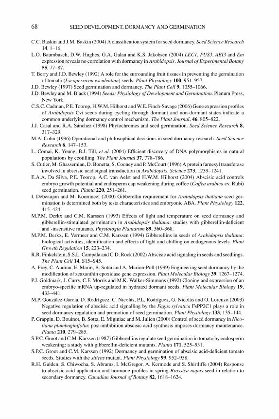
P1: OTE/SPH P2: OTE
BLUK053-Bradford August 27, 2006 17:8
68 SEED DEVELOPMENT, DORMANCY AND GERMINATION
C.C. Baskin and J.M. Baskin (2004) A classification system for seed dormancy. Seed Science Research14, 1–16.
L.O. Baumbusch, D.W. Hughes, G.A. Galau and K.S. Jakobsen (2004) LEC1, FUS3, ABI3 and Emexpression reveals no correlation with dormancy in Arabidopsis. Journal of Experimental Botany55, 77–87.
T. Berry and J.D. Bewley (1992) A role for the surrounding fruit tissues in preventing the germinationof tomato (Lycopersicon esculentum) seeds. Plant Physiology 100, 951–957.
J.D. Bewley (1997) Seed germination and dormancy. The Plant Cell 9, 1055–1066.J.D. Bewley and M. Black (1994) Seeds: Physiology of Development and Germination. Plenum Press,
New York.C.S.C. Cadman, P.E. Toorop, H.W.M. Hilhorst and W.E. Finch-Savage (2006) Gene expression profiles
of Arabidopsis Cvi seeds during cycling through dormant and non-dormant states indicate acommon underlying dormancy control mechanism. The Plant Journal, 46, 805–822.
J.J. Casal and R.A. Sanchez (1998) Phytochromes and seed germination. Seed Science Research 8,317–329.
M.A. Cohn (1996) Operational and philosophical decisions in seed dormancy research. Seed ScienceResearch 6, 147–153.
L. Comai, K. Young, B.J. Till, et al. (2004) Efficient discovery of DNA polymorphisms in naturalpopulations by ecotilling. The Plant Journal 37, 778–786.
S. Cutler, M. Ghassemian, D. Bonetta, S. Cooney and P. McCourt (1996) A protein farnesyl transferaseinvolved in abscisic acid signal transduction in Arabidopsis. Science 273, 1239–1241.
E.A.A. Da Silva, P.E. Toorop, A.C. van Aelst and H.W.M. Hilhorst (2004) Abscisic acid controlsembryo growth potential and endosperm cap weakening during coffee (Coffea arabica cv. Rubi)seed germination. Planta 220, 251–261.
I. Debeaujon and M. Koornneef (2000) Gibberellin requirement for Arabidopsis thaliana seed ger-mination is determined both by testa characteristics and embryonic ABA. Plant Physiology 122,415–424.
M.P.M. Derkx and C.M. Karssen (1993) Effects of light and temperature on seed dormancy andgibberellin-stimulated germination in Arabidopsis thaliana: studies with gibberellin-deficientand -insensitive mutants. Physiologia Plantarum 89, 360–368.
M.P.M. Derkx, E. Vermeer and C.M. Karssen (1994) Gibberellins in seeds of Arabidopsis thaliana:biological activities, identification and effects of light and chilling on endogenous levels. PlantGrowth Regulation 15, 223–234.
R.R. Finkelstein, S.S.L. Campala and C.D. Rock (2002) Abscisic acid signaling in seeds and seedlings.The Plant Cell 14, S15–S45.
A. Frey, C. Audran, E. Marin, B. Sotta and A. Marion-Poll (1999) Engineering seed dormancy by themodification of zeaxanthin epoxidase gene expression. Plant Molecular Biology 39, 1267–1274.
P.J. Goldmark, J. Curry, C.F. Morris and M.K. Walker-Simmons (1992) Cloning and expression of anembryo-specific mRNA up-regulated in hydrated dormant seeds. Plant Molecular Biology 19,433–441.
M.P. Gonzalez-Garcıa, D. Rodrıguez, C. Nicolas, P.L. Rodrıguez, G. Nicolas and O. Lorenzo (2003)Negative regulation of abscisic acid signalling by the Fagus sylvatica FsPP2C1 plays a role inseed dormancy regulation and promotion of seed germination. Plant Physiology 133, 135–144.
P. Grappin, D. Bouinot, B. Sotta, E. Miginiac and M. Julien (2000) Control of seed dormancy in Nico-tiana plumbaginifolia: post-imbibition abscisic acid synthesis imposes dormancy maintenance.Planta 210, 279–285.
S.P.C. Groot and C.M. Karssen (1987) Gibberellins regulate seed germination in tomato by endospermweakening: a study with gibberellin-deficient mutants. Planta 171, 525–531.
S.P.C. Groot and C.M. Karssen (1992) Dormancy and germination of abscisic acid-deficient tomatoseeds. Studies with the sitiens mutant. Plant Physiology 99, 952–958.
R.H. Gulden, S. Chiwocha, S. Abrams, I. McGregor, A. Kermode and S. Shirtliffe (2004) Responseto abscisic acid application and hormone profiles in spring Brassica napus seed in relation tosecondary dormancy. Canadian Journal of Botany 82, 1618–1624.
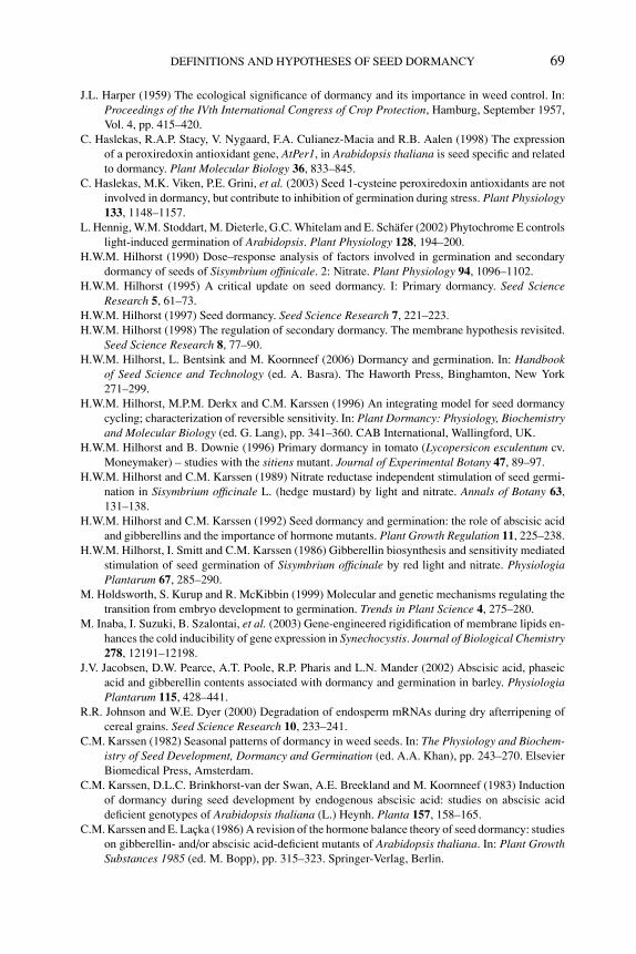
P1: OTE/SPH P2: OTE
BLUK053-Bradford August 27, 2006 17:8
DEFINITIONS AND HYPOTHESES OF SEED DORMANCY 69
J.L. Harper (1959) The ecological significance of dormancy and its importance in weed control. In:Proceedings of the IVth International Congress of Crop Protection, Hamburg, September 1957,Vol. 4, pp. 415–420.
C. Haslekas, R.A.P. Stacy, V. Nygaard, F.A. Culianez-Macia and R.B. Aalen (1998) The expressionof a peroxiredoxin antioxidant gene, AtPer1, in Arabidopsis thaliana is seed specific and relatedto dormancy. Plant Molecular Biology 36, 833–845.
C. Haslekas, M.K. Viken, P.E. Grini, et al. (2003) Seed 1-cysteine peroxiredoxin antioxidants are notinvolved in dormancy, but contribute to inhibition of germination during stress. Plant Physiology133, 1148–1157.
L. Hennig, W.M. Stoddart, M. Dieterle, G.C. Whitelam and E. Schafer (2002) Phytochrome E controlslight-induced germination of Arabidopsis. Plant Physiology 128, 194–200.
H.W.M. Hilhorst (1990) Dose–response analysis of factors involved in germination and secondarydormancy of seeds of Sisymbrium offinicale. 2: Nitrate. Plant Physiology 94, 1096–1102.
H.W.M. Hilhorst (1995) A critical update on seed dormancy. I: Primary dormancy. Seed ScienceResearch 5, 61–73.
H.W.M. Hilhorst (1997) Seed dormancy. Seed Science Research 7, 221–223.H.W.M. Hilhorst (1998) The regulation of secondary dormancy. The membrane hypothesis revisited.
Seed Science Research 8, 77–90.H.W.M. Hilhorst, L. Bentsink and M. Koornneef (2006) Dormancy and germination. In: Handbook
of Seed Science and Technology (ed. A. Basra). The Haworth Press, Binghamton, New York271–299.
H.W.M. Hilhorst, M.P.M. Derkx and C.M. Karssen (1996) An integrating model for seed dormancycycling; characterization of reversible sensitivity. In: Plant Dormancy: Physiology, Biochemistryand Molecular Biology (ed. G. Lang), pp. 341–360. CAB International, Wallingford, UK.
H.W.M. Hilhorst and B. Downie (1996) Primary dormancy in tomato (Lycopersicon esculentum cv.Moneymaker) – studies with the sitiens mutant. Journal of Experimental Botany 47, 89–97.
H.W.M. Hilhorst and C.M. Karssen (1989) Nitrate reductase independent stimulation of seed germi-nation in Sisymbrium officinale L. (hedge mustard) by light and nitrate. Annals of Botany 63,131–138.
H.W.M. Hilhorst and C.M. Karssen (1992) Seed dormancy and germination: the role of abscisic acidand gibberellins and the importance of hormone mutants. Plant Growth Regulation 11, 225–238.
H.W.M. Hilhorst, I. Smitt and C.M. Karssen (1986) Gibberellin biosynthesis and sensitivity mediatedstimulation of seed germination of Sisymbrium officinale by red light and nitrate. PhysiologiaPlantarum 67, 285–290.
M. Holdsworth, S. Kurup and R. McKibbin (1999) Molecular and genetic mechanisms regulating thetransition from embryo development to germination. Trends in Plant Science 4, 275–280.
M. Inaba, I. Suzuki, B. Szalontai, et al. (2003) Gene-engineered rigidification of membrane lipids en-hances the cold inducibility of gene expression in Synechocystis. Journal of Biological Chemistry278, 12191–12198.
J.V. Jacobsen, D.W. Pearce, A.T. Poole, R.P. Pharis and L.N. Mander (2002) Abscisic acid, phaseicacid and gibberellin contents associated with dormancy and germination in barley. PhysiologiaPlantarum 115, 428–441.
R.R. Johnson and W.E. Dyer (2000) Degradation of endosperm mRNAs during dry afterripening ofcereal grains. Seed Science Research 10, 233–241.
C.M. Karssen (1982) Seasonal patterns of dormancy in weed seeds. In: The Physiology and Biochem-istry of Seed Development, Dormancy and Germination (ed. A.A. Khan), pp. 243–270. ElsevierBiomedical Press, Amsterdam.
C.M. Karssen, D.L.C. Brinkhorst-van der Swan, A.E. Breekland and M. Koornneef (1983) Inductionof dormancy during seed development by endogenous abscisic acid: studies on abscisic aciddeficient genotypes of Arabidopsis thaliana (L.) Heynh. Planta 157, 158–165.
C.M. Karssen and E. Lacka (1986) A revision of the hormone balance theory of seed dormancy: studieson gibberellin- and/or abscisic acid-deficient mutants of Arabidopsis thaliana. In: Plant GrowthSubstances 1985 (ed. M. Bopp), pp. 315–323. Springer-Verlag, Berlin.
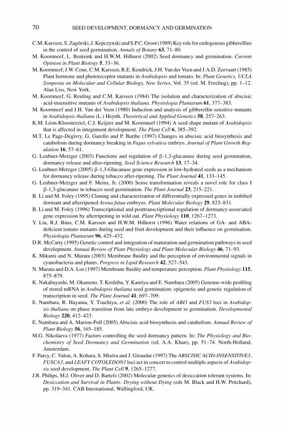
P1: OTE/SPH P2: OTE
BLUK053-Bradford August 27, 2006 17:8
70 SEED DEVELOPMENT, DORMANCY AND GERMINATION
C.M. Karssen, S. Zagorski, J. Kepczynski and S.P.C. Groot (1989) Key role for endogenous gibberellinsin the control of seed germination. Annals of Botany 63, 71–80.
M. Koornneef, L. Bentsink and H.W.M. Hilhorst (2002) Seed dormancy and germination. CurrentOpinion in Plant Biology 5, 33–36.
M. Koornneef, J.W. Cone, C.M. Karssen, R.E. Kendrick, J.H. Van der Veen and J.A.D. Zeevaart (1985)Plant hormone and photoreceptor mutants in Arabidopsis and tomato. In: Plant Genetics, UCLASymposia on Molecular and Cellular Biology, New Series, Vol. 35 (ed. M. Freeling), pp. 1–12.Alan Liss, New York.
M. Koornneef, G. Reuling and C.M. Karssen (1984) The isolation and characterization of abscisicacid-insensitive mutants of Arabidopsis thaliana. Physiologia Plantarum 61, 377–383.
M. Koornneef and J.H. Van der Veen (1980) Induction and analysis of gibberellin sensitive mutantsin Arabidopsis thaliana (L.) Heynh. Theoretical and Applied Genetics 58, 257–263.
K.M. Leon-Kloosterziel, C.J. Keijzer and M. Koornneef (1994) A seed shape mutant of Arabidopsisthat is affected in integument development. The Plant Cell 6, 385–392.
M.T. Le Page-Degivry, G. Garello and P. Barthe (1997) Changes in abscisic acid biosynthesis andcatabolism during dormancy breaking in Fagus sylvatica embryo. Journal of Plant Growth Reg-ulation 16, 57–61.
G. Leubner-Metzger (2003) Functions and regulation of β-1,3-glucanase during seed germination,dormancy release and after-ripening. Seed Science Research 13, 17–34.
G. Leubner-Metzger (2005) β-1,3-Glucanase gene expression in low-hydrated seeds as a mechanismfor dormancy release during tobacco after-ripening. The Plant Journal 41, 133–145.
G. Leubner-Metzger and F. Meins, Jr. (2000) Sense transformation reveals a novel role for class Iβ-1,3-glucanase in tobacco seed germination. The Plant Journal 23, 215–221.
B. Li and M. Foley (1995) Cloning and characterization of differentially expressed genes in imbibeddormant and afterripened Avena fatua embryos. Plant Molecular Biology 29, 823–831.
B. Li and M. Foley (1996) Transcriptional and posttranscriptional regulation of dormancy-associatedgene expression by afterripening in wild oat. Plant Physiology 110, 1267–1273.
Y. Liu, R.J. Bino, C.M. Karssen and H.W.M. Hilhorst (1996) Water relations of GA- and ABA-deficient tomato mutants during seed and fruit development and their influence on germination.Physiologia Plantarum 96, 425–432.
D.R. McCarty (1995) Genetic control and integration of maturation and germination pathways in seeddevelopment. Annual Review of Plant Physiology and Plant Molecular Biology 46, 71–93.
K. Mikami and N. Murata (2003) Membrane fluidity and the perception of environmental signals incyanobacteria and plants. Progress in Lipid Research 42, 527–543.
N. Murata and D.A. Los (1997) Membrane fluidity and temperature perception. Plant Physiology 115,875–879.
K. Nakabayashi, M. Okamoto, T. Koshiba, Y. Kamiya and E. Nambara (2005) Genome-wide profilingof stored mRNA in Arabidopsis thaliana seed germination: epigenetic and genetic regulation oftranscription in seed. The Plant Journal 41, 697–709.
E. Nambara, R. Hayama, Y. Tsuchiya, et al. (2000) The role of ABI3 and FUS3 loci in Arabidop-sis thaliana on phase transition from late embryo development to germination. DevelopmentalBiology 220, 412–423.
E. Nambara and A. Marion-Poll (2005) Abscisic acid biosynthesis and catabolism. Annual Review ofPlant Biology 56, 165–185.
M.G. Nikolaeva (1977) Factors controlling the seed dormancy pattern. In: The Physiology and Bio-chemistry of Seed Dormancy and Germination (ed. A.A. Khan), pp. 51–74. North-Holland,Amsterdam.
F. Parcy, C. Valon, A. Kohara, S. Misera and J. Giraudat (1997) The ABSCISIC ACID-INSENSITIVE3,FUSCA3, and LEAFY COTOLEDON1 loci act in concert to control multiple aspects of Arabidop-sis seed development. The Plant Cell 9, 1265–1277.
J.R. Philips, M.J. Oliver and D. Bartels (2002) Molecular genetics of desiccation tolerant systems. In:Desiccation and Survival in Plants: Drying without Dying (eds M. Black and H.W. Pritchard),pp. 319–341. CAB International, Wallingford, UK.
P1: OTE/SPH P2: OTE
BLUK053-Bradford August 27, 2006 17:8
DEFINITIONS AND HYPOTHESES OF SEED DORMANCY 71
V. Raz, J.H.W. Bergervoet and M. Koornneef (2001) Sequential steps for developmental arrest inArabidopsis seeds. Development 128, 243–252.
A. Rohde, S. Kurup and M. Holdsworth (2000) ABI3 emerges from the seed. Trends in Plant Science5, 418–419.
R.A. Sanchez, L. Sunell, J.M. Labavitch and B.A. Bonner (1990) Changes in the endosperm cell wallsof two Datura species before radicle protrusion. Plant Physiology 93, 89–97.
V. Sangwan, B.L. Orvar, J. Beyerly, H. Hirt and R.S. Dhindsa (2002) Opposite changes in membranefluidity mimic cold and heat stress activation of distinct plant MAP kinase pathways. The PlantJournal 31, 629–638.
N. Schmitz, S.R. Abrams and A.R. Kermode (2000) Changes in abscisic acid content and embryosensitivity to (+)-abscisic acid during the termination of dormancy of yellow cedar seeds. Journalof Experimental Botany 51, 1159–1162.
G.M. Simpson (1990) Seed Dormancy in Grasses. Cambridge University Press, Cambridge, UK.T. Toyomasu, H. Kawaide, W. Mitsuhashi, Y. Inoue and Y. Kamiya (1998) Phytochrome regulates
gibberellin biosynthesis during germination of photoblastic lettuce seeds. Plant Phsiology, 118,1517–1523.
T. Toyomasu, H. Yamane, N. Murofushi and Y. Inoue (1994) Effects of exogenously applied gibberellinand red light on the endogenous levels of abscisic acid in photoblastic lettuce seeds. Plant andCell Physiology 35, 127–129.
P. Van der Toorn and C.M. Karssen (1992) Analysis of embryo growth in mature fruits of celery(Apium graveolens). Physiologia Plantarum 84, 593–599.
L.M. Vleeshouwers, H.J. Bouwmeester and C.M. Karssen (1995) Redefining seed dormancy: anattempt to integrate physiology and ecology. Journal of Ecology 83, 1031–1037.
J.O. Watkinson and G.E. Welbaum (in press) Seeds of the family Orchidaceae. In: The Encyclopediaof Seeds (eds D. Bewley, M. Black and P. Halmer). CABI International, Wallingford, UK.
L. Xiong, H. Lee, M. Ishitani and J.-K. Zhu (2002) Regulation of osmotic stress-responsive geneexpression by the LOS6/ABA1 locus in Arabidopsis. Journal of Biological Chemistry 277, 8588–8596.
K. Yamaguchi-Shinozaki and K. Shinozaki (2005) Organization of cis-acting regulatory elements inosmotic- and cold-stress responsive promoters. Trends in Plant Science 10, 88–94.
Y. Yamauchi, M. Ogawa, A. Kuwahara, A. Hanada, Y. Kamiya and S. Yamaguchi (2004) Activationof gibberellin biosynthesis and response pathways by low temperature during imbibition ofArabidopsis thaliana seeds. The Plant Cell 16, 367–378.
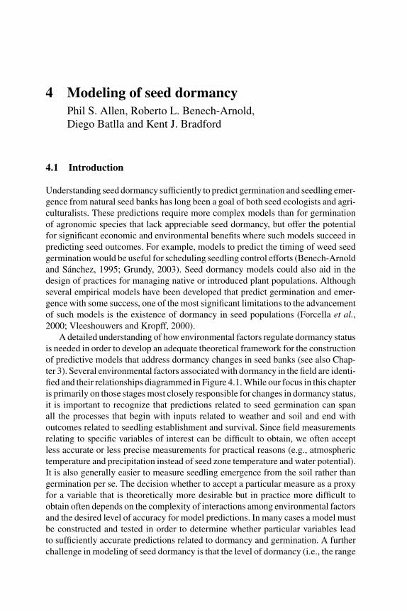
P1: OTE/SPH P2: OTE
BLUK053-Bradford September 27, 2006 7:59
4 Modeling of seed dormancyPhil S. Allen, Roberto L. Benech-Arnold,Diego Batlla and Kent J. Bradford
4.1 Introduction
Understanding seed dormancy sufficiently to predict germination and seedling emer-gence from natural seed banks has long been a goal of both seed ecologists and agri-culturalists. These predictions require more complex models than for germinationof agronomic species that lack appreciable seed dormancy, but offer the potentialfor significant economic and environmental benefits where such models succeed inpredicting seed outcomes. For example, models to predict the timing of weed seedgermination would be useful for scheduling seedling control efforts (Benech-Arnoldand Sanchez, 1995; Grundy, 2003). Seed dormancy models could also aid in thedesign of practices for managing native or introduced plant populations. Althoughseveral empirical models have been developed that predict germination and emer-gence with some success, one of the most significant limitations to the advancementof such models is the existence of dormancy in seed populations (Forcella et al.,2000; Vleeshouwers and Kropff, 2000).
A detailed understanding of how environmental factors regulate dormancy statusis needed in order to develop an adequate theoretical framework for the constructionof predictive models that address dormancy changes in seed banks (see also Chap-ter 3). Several environmental factors associated with dormancy in the field are identi-fied and their relationships diagrammed in Figure 4.1. While our focus in this chapteris primarily on those stages most closely responsible for changes in dormancy status,it is important to recognize that predictions related to seed germination can spanall the processes that begin with inputs related to weather and soil and end withoutcomes related to seedling establishment and survival. Since field measurementsrelating to specific variables of interest can be difficult to obtain, we often acceptless accurate or less precise measurements for practical reasons (e.g., atmospherictemperature and precipitation instead of seed zone temperature and water potential).It is also generally easier to measure seedling emergence from the soil rather thangermination per se. The decision whether to accept a particular measure as a proxyfor a variable that is theoretically more desirable but in practice more difficult toobtain often depends on the complexity of interactions among environmental factorsand the desired level of accuracy for model predictions. In many cases a model mustbe constructed and tested in order to determine whether particular variables leadto sufficiently accurate predictions related to dormancy and germination. A furtherchallenge in modeling of seed dormancy is that the level of dormancy (i.e., the range
72
P1: OTE/SPH P2: OTE
BLUK053-Bradford September 27, 2006 7:59
MODELING OF SEED DORMANCY 73
of conditions under which germination can proceed) is not the same for all individu-als within a seed population, and models need to explicitly account for this variation.
In the sections below we discuss environmental factors affecting dormancy innatural seed banks according to the general framework presented in Figure 4.1, withan emphasis on classifying and understanding the effects of these factors on seeddormancy changes under field situations. The aim of this classification is to facilitateconceptualization of dormancy systems for modeling purposes. Later sections aredevoted to presenting some examples of population-based models that relate seeddormancy behavior to the effects of those environmental factors.
4.2 Types and phenology of seed dormancy
According to Benech-Arnold et al. (2000), ‘Dormancy is an internal condition ofthe seed that impedes its germination under otherwise adequate hydric, thermal andgaseous conditions’. This implies that once the impedance has been removed, seedgermination would proceed under a wide range of environmental conditions. Karssen(1982) proposed that dormancy could be classified into primary and secondarydormancy. Primary dormancy refers to the innate dormancy possessed by seedswhen they are dispersed from the mother plant. Numerous types and patterns ofprimary dormancy have been documented and classified by Baskin and Baskin(2004). Secondary dormancy refers to a dormant state that is induced in nondormantseeds by unfavorable conditions for germination, or reinduced in once-dormant seedsafter primary dormancy has been alleviated. Thus, primary and secondary dormancyrefer only to the timing of occurrence, not to the mechanisms by which dormancyis imposed or released, although knowledge of the mechanistic basis of both typesof dormancy is still rudimentary (see Chapter 3). Release from primary dormancyfollowed by subsequent entrance into secondary dormancy may lead to dormancycycling, indicated in Figure 4.1 by solid and dashed arrows alternating between lowand high dormancy levels. Evidence for dormancy cycling has been obtained forseeds of many species (Fenner, 2000). For species that exhibit dormancy cyclingunder natural field conditions, the transitions into and out of dormancy may continuefor several years before the seeds germinate, decay, or are otherwise lost from thesoil seed bank (Karssen, 1980/81; Baskin and Baskin, 1985; Meyer et al., 1997).Overlapping additions to and losses from the soil seed bank and varying levels ofdormancy within the seed population complicate attempts to model germination andemergence cycles in such species.
In general, seeds are released from dormancy preceding a season with favorableconditions for seedling emergence and survival, whereas dormancy is induced priorto a period with adverse conditions for seedling survival. For example, severalsummer annual species exhibit a high dormancy level in autumn, undergo dormancyrelease during winter and, for the fraction of seeds that fail to germinate in spring,show an increase in dormancy again during the following summer. In contrast, winterannual species show an opposite seasonal dormancy pattern. Therefore, seasonalpatterns of seed dormancy are of high survival value to species by constraining
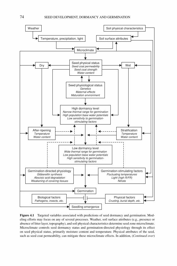
P1: OTE/SPH P2: OTE
BLUK053-Bradford September 27, 2006 7:59
74 SEED DEVELOPMENT, DORMANCY AND GERMINATION
Weather Soil physical characteristics
Microclimate
Seed physical statusSeed coat permeability
Seed coat strengthWater content
Temperature, precipitation, light Soil surface attributes
Dry Wet
After-ripeningTemperatureWater content
StratificationTemperature Water content
Seed physiological statusGenetics
Maternal effectsMaturation environment
High dormancy levelNarrow thermal range for germinationHigh population base water potentials
Low sensitivity to germination-stimulating factors
Low dormancy levelWide thermal range for germination
Low population base water potentialsHigh sensitivity to germination-
stimulating factors
Germination
Seedling emergence
Germination-stimulating factorsFluctuating temperatures
Light (high R/FR)Nitrate
Germination-directed physiologyGibberellin synthesis
Abscisic acid degradationWeakening of covering tissues
Biological factorsPathogens, insects, etc.
Physical factorsCrusting, burial depth, etc.
Figure 4.1 Targeted variables associated with predictions of seed dormancy and germination. Mod-
eling efforts may focus on any of several processes. Weather, soil surface attributes (e.g., presence or
absence of litter layer, topography), and soil physical characteristics determine seed zone microclimate.
Microclimate controls seed dormancy status and germination-directed physiology through its effect
on seed physical status, primarily moisture content and temperature. Physical attributes of the seed,
such as seed coat permeability, can mitigate these microclimate effects. In addition, (Continued over)
P1: OTE/SPH P2: OTE
BLUK053-Bradford September 27, 2006 7:59
MODELING OF SEED DORMANCY 75
germination to periods when environmental conditions favor seedling growth andsurvival (Karssen, 1982; Donohue et al., 2005).
Dormancy is also a relative rather than an absolute condition. The concept ofdegrees of relative dormancy was first introduced by Vegis (1964) following observa-tions that as dormancy is released, the temperature range permissive for germinationwidens until it is maximal. Conversely, as dormancy is induced the range of temper-atures over which germination can proceed narrows until germination is no longerpossible at any temperature. On this basis, Karssen (1982) proposed that seasonalperiodicity in field emergence of annual species is the combined result of seasonalperiodicity in soil temperatures and physiological changes within seeds that alter thetemperature range permissive for germination. Germination in the field is thereforerestricted to periods when the soil temperature and the temperature range over whichgermination can proceed overlap.
The concept of relative dormancy can be expanded to include seed responses toenvironmental factors other than temperature. For example, dormancy release or im-position can also be correlated with changes in sensitivity to water potential, light, ornitrate (Hilhorst, 1990; Bradford, 2002; Batlla and Benech-Arnold, 2005). In somespecies, the transition from dormancy to nondormancy is relatively continuous, andis exhibited by increases in germination percentage and rate under a given set ofconditions. In other species, the conditions for releasing dormancy may be distinctfrom those required for germination, and the latter must be stimulated by factorssuch as light, nitrate, or fluctuating temperatures after dormancy per se has been al-leviated (see also Chapter 3). In these cases, the loss of dormancy is associated withan increasing sensitivity or responsiveness of seeds to these germination-stimulating(or dormancy-terminating) factors (Benech-Arnold et al., 2000).
Seedling emergence of a particular species in the field occurs only when allenvironmental conditions are within the ranges permissive for seed germination.Since the permissive ranges of conditions change with dormancy levels of seedpopulations, models for predicting germination or emergence need to incorporate
←−−−−−−−−−−−−−−−−−−−−−−−−−−−−−−−−−−−−−−−−−−−−−−−−−−−−−−−−−−Figure 4.1 (Continued ) the physiological status of the seed, including its genetic background and
maternal and environmental effects during development and maturation, influences dormancy level.
Dormancy-releasing processes in dry seeds (after-ripening) differ qualitatively from those that occur
in imbibed seeds (stratification). Interactions between environmental factors and seed dormancy sta-
tus are shown via arrows. A high dormancy level (whether primary or secondary) is associated with
a narrow thermal range permissive for germination, relatively high (more positive) population base
water potentials, and a low sensitivity to germination-stimulating factors. Conversely, a low dormancy
level is associated with a wider temperature range permissive for germination, lower (more negative)
population base water potentials, and increased sensitivity to germination stimulating factors. In some
cases following dormancy release through temperature and moisture conditions, germination may need
to be stimulated by factors such as light, nitrate, or fluctuating temperatures. Dashed arrows indicate
that when environmental conditions are not conducive to dormancy-breakage or germination, seeds
can revert to a higher dormancy level (secondary dormancy). Even after germination has occurred,
additional biotic and abiotic stresses can reduce the number of seedlings that successfully emerge from
the soil. (Expanded from Allen and Meyer, 1998, and Benech-Arnold et al., 2000.)
P1: OTE/SPH P2: OTE
BLUK053-Bradford September 27, 2006 7:59
76 SEED DEVELOPMENT, DORMANCY AND GERMINATION
both changes over time of the seeds’ sensitivity to environmental conditions and theirsubsequent responses to those conditions as dormancy levels vary. It is thereforenecessary to identify the environmental factors that modify dormancy levels of seedpopulations and to establish functional relationships between these factors and therates of dormancy release or induction.
4.3 Environmental control of dormancy
4.3.1 Factors affecting dormancy levels of seed populations
4.3.1.1 TemperatureDormancy cycles are often regulated by temperature in temperate environmentswhere water is not seasonally restricted (Baskin and Baskin, 1977, 1984; Kruk andBenech-Arnold, 1998). For example, in some summer annual species dormancy isreleased when imbibed seeds experience low temperatures during winter (stratifi-cation), while the dormancy level is increased by high temperatures experiencedduring summer (Bouwmeester and Karssen, 1992, 1993). Winter annual speciesshow the reverse dormancy pattern, where high temperatures during summer resultin lower dormancy levels and low temperatures during winter induce secondarydormancy (Baskin and Baskin, 1976; Karssen, 1982; Probert, 1992). As noted inFigure 4.1, these changes in dormancy level are expressed through a narrowing andwidening of the temperature range permissive for germination. Germination occursin the field when soil temperature is in the permissive range characterized by twothermal parameters determined by the seed dormancy level, the low limit tempera-ture (Tl) and the high limit temperature (Th) (Figure 4.2). Tl and Th vary among seedswithin the population, consistent with the idea that dormancy level is different forindividual seeds (Kruk and Benech-Arnold, 2000; Batlla and Benech-Arnold, 2003;Batlla et al., 2004). For example, Tl(50) and Th(50) represent the temperatures be-low and above which dormancy is expressed for 50% of the seed population. Thesedistributions of lower and higher temperature limits shift downward and upward inrelation to changes in dormancy level of the population (Figure 4.2). Changes in thedormancy level (or variations in the thermal range where germination can occur)in summer species are due to increases or decreases in Tl, while in winter specieschanges in dormancy level result from fluctuations in Th (Figure 4.2; Baskin andBaskin, 1980).
Although soil temperature is generally considered the primary environmentalfactor regulating dormancy levels in seed bank populations, the effect of tem-perature on dormancy release and induction may be modulated by soil moisture(Adamoli et al., 1973; Reisman-Berman et al., 1991; Christensen et al., 1996; Baueret al., 1998; Bair et al., 2006; Batlla and Benech-Arnold, 2006). For example, inPolygonum aviculare seeds dormancy release occurs most rapidly when seeds aremoist-chilled (stratified) at 4◦C, but dry storage at 4◦C also releases seed dormancy,though at a much slower rate (Kruk and Benech-Arnold, 1998). P. aviculare seedsburied in the field under contrasting soil water content conditions show different
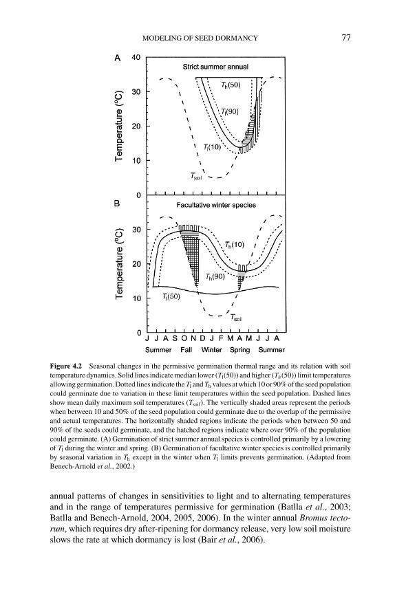
P1: OTE/SPH P2: OTE
BLUK053-Bradford September 27, 2006 7:59
MODELING OF SEED DORMANCY 77
Figure 4.2 Seasonal changes in the permissive germination thermal range and its relation with soil
temperature dynamics. Solid lines indicate median lower (Tl(50)) and higher (Th(50)) limit temperatures
allowing germination. Dotted lines indicate the Tl and Th values at which 10 or 90% of the seed population
could germinate due to variation in these limit temperatures within the seed population. Dashed lines
show mean daily maximum soil temperatures (Tsoil). The vertically shaded areas represent the periods
when between 10 and 50% of the seed population could germinate due to the overlap of the permissive
and actual temperatures. The horizontally shaded regions indicate the periods when between 50 and
90% of the seeds could germinate, and the hatched regions indicate where over 90% of the population
could germinate. (A) Germination of strict summer annual species is controlled primarily by a lowering
of Tl during the winter and spring. (B) Germination of facultative winter species is controlled primarily
by seasonal variation in Th except in the winter when Tl limits prevents germination. (Adapted from
Benech-Arnold et al., 2002.)
annual patterns of changes in sensitivities to light and to alternating temperaturesand in the range of temperatures permissive for germination (Batlla et al., 2003;Batlla and Benech-Arnold, 2004, 2005, 2006). In the winter annual Bromus tecto-rum, which requires dry after-ripening for dormancy release, very low soil moistureslows the rate at which dormancy is lost (Bair et al., 2006).
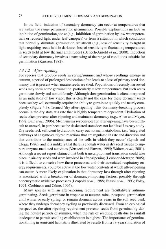
P1: OTE/SPH P2: OTE
BLUK053-Bradford September 27, 2006 7:59
78 SEED DEVELOPMENT, DORMANCY AND GERMINATION
In the field, induction of secondary dormancy can occur at temperatures thatare within the range permissive for germination. Possible explanations include aninhibition of germination per se (e.g., inhibition of germination by low water poten-tials or reduced light under leaf canopies) or from a situation in which conditionsthat normally stimulate germination are absent (e.g., loss of sensitivity to light inlight-requiring seeds held in darkness; loss of sensitivity to fluctuating temperaturesin seeds held at low thermal amplitudes) (Benech-Arnold et al., 2000). Inductionof secondary dormancy involves a narrowing of the range of conditions suitable forgermination (Karssen, 1982).
4.3.1.2 After-ripeningFor species that produce seeds in spring/summer and whose seedlings emerge inautumn, a period of prolonged desiccation often leads to a loss of primary seed dor-mancy that is present when mature seeds are shed. Populations of recently harvestedseeds may show some germination, particularly at low temperatures, but such seedsgerminate slowly and nonuniformly. Although slow germination is often interpretedas an indication of low vigor, this is clearly not the case for these dormant seedsbecause they will eventually acquire the ability to germinate quickly and nearly com-pletely (Figure 4.3). Termed ‘dry after-ripening’, this dormancy-breaking processoccurs in the dry state at a rate that is highly temperature dependent. Hydration ofseeds often prevents after-ripening and maintains dormancy (e.g., Allen and Meyer,1998; Bair et al., 2006). Mechanisms responsible for after-ripening have been diffi-cult to unravel, in part because the desiccated state does not lend itself to easy study.Dry seeds lack sufficient hydration to carry out normal metabolism, i.e., ‘integratedpathways of enzyme-catalyzed reactions that are regulated in rate and direction andthat contribute to the maintenance of the cells in which they occur’ (Lynch andClegg, 1986), and it is unlikely that there is enough water in dry seed tissues to sup-port enzyme-mediated activities (Vertucci and Farrant, 1995; Walters et al., 2001).Although a recent report claimed that both transcription and translation could takeplace in air-dry seeds and were involved in after-ripening (Leubner-Metzger, 2005),it is difficult to conceive how these processes, and their associated respiratory en-ergy requirements, could be active at the low water contents at which after-ripeningcan occur. A more likely explanation is that dormancy loss through after-ripeningis associated with a breakdown of dormancy-imposing factors, possibly throughnonenzymatic oxidative processes (Leopold et al., 1988; Esashi et al., 1993; Foley,1994; Corbineau and Come, 1995).
Many species with an after-ripening requirement are facultatively autumn-germinating. Seeds germinate in response to autumn rains, postpone germinationuntil winter or early spring, or remain dormant across years in the soil seed bankwhere they undergo dormancy cycling as previously discussed. From an ecologicalperspective, the after-ripening requirement prevents seeds from germinating dur-ing the hottest periods of summer, when the risk of seedling death due to rainfallinadequate to permit seedling establishment is highest. The importance of germina-tion timing in semi-arid habitats is illustrated by results from a 38-year simulation of
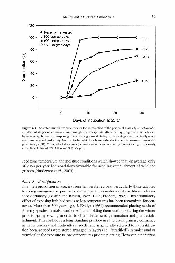
P1: OTE/SPH P2: OTE
BLUK053-Bradford September 27, 2006 7:59
MODELING OF SEED DORMANCY 79
Figure 4.3 Selected cumulative time courses for germination of the perennial grass Elymus elymoidesat different stages of dormancy loss through dry storage. As after-ripening progresses, as indicated
by increasing thermal after-ripening times, seeds germinate to higher percentages and eventually reach
maximum rate and uniformity. Number to the right of each line indicates the population mean base water
potential (ψb(50), MPa), which decreases (becomes more negative) during after-ripening. (Previously
unpublished data of P.S. Allen and S.E. Meyer.)
seed zone temperature and moisture conditions which showed that, on average, only30 days per year had conditions favorable for seedling establishment of wildlandgrasses (Hardegree et al., 2003).
4.3.1.3 StratificationIn a high proportion of species from temperate regions, particularly those adaptedto spring emergence, exposure to cold temperatures under moist conditions releasesseed dormancy (Baskin and Baskin, 1985, 1998; Probert, 1992). This stimulatoryeffect of exposing imbibed seeds to low temperatures has been recognized for cen-turies. More than 300 years ago, J. Evelyn (1664) recommended placing seeds offorestry species in moist sand or soil and holding them outdoors during the winterprior to spring sowing in order to obtain better seed germination and plant estab-lishment. This method is a long-standing practice used to break primary dormancyin many forestry and horticultural seeds, and is generally referred to as stratifica-tion because seeds were stored arranged in layers (i.e., ‘stratified’) in moist sand orvermiculite for exposure to low temperatures prior to planting. However, other terms
P1: OTE/SPH P2: OTE
BLUK053-Bradford September 27, 2006 7:59
80 SEED DEVELOPMENT, DORMANCY AND GERMINATION
such as moist chilling and cold after-ripening are also used to refer to dormancyrelease by cold temperatures in hydrated seeds. Results from many ecophysiologicalstudies of the emergence patterns of buried seed populations indicate that stratifica-tion is a significant process controlling the dormancy status and timing of emergenceof wild species in temperate regions (Probert, 1992; Meyer et al., 1995). The re-quirement for low temperatures prevents seedling establishment and growth underconditions unfavorable for plant survival (i.e., during winter), while allowing germi-nation and establishment under environmental conditions that favor species growthand perpetuation (i.e., during spring) (Karssen, 1982).
In contrast with after-ripening, for stratification to be effective seeds must be atleast partially hydrated and have access to oxygen (Stokes, 1965; Bewley and Black,1982; Vertucci and Leopold, 1986). The period required for stratification may rangefrom a few days to several months, with differences known to exist among species,populations within a species, and individual seeds within a single seed population(Allen and Meyer, 1998; Baskin and Baskin, 2004; Batlla et al., 2004). The optimumtemperatures for dormancy release through stratification are generally in the rangeof 1–5◦C, although seeds of some species can be stratified at temperatures as high as16–17◦C (Roberts and Smith, 1977; Batlla and Benech-Arnold, 2003). Prolongedexposure to temperatures near the upper limit for stratification can lead to secondarydormancy (e.g., Totterdell and Roberts, 1979). In some species, after-ripening mustprecede stratification in order for dormancy loss to occur for at least a fraction ofthe seed population (Baskin and Baskin, 1984; Garvin and Meyer, 2003).
4.3.2 Factors that stimulate germination
Promotion of germination following dormancy release often requires exposure tospecific environmental stimuli (Figure 4.1). For example, fluctuating temperaturesand light result in dormancy loss for many weed seeds (Benech-Arnold et al., 1990b;Scopel et al., 1991; Ghersa et al., 1992), although other factors (i.e., CO2, NO3
–, O2,and ethylene) can be involved in the termination of dormancy under field conditions(Corbineau and Come, 1995; Benech-Arnold et al., 2000). Not all seeds requireexposure to germination-stimulating factors in order for germination to occur. Forexample, seeds within a population may proceed to germinate immediately afterdormancy is released as long as water and temperature conditions are adequate. Inthese cases, there is a gradual transition from dormancy to nondormancy to germina-tion, and it is difficult to distinguish specific stages (see Chapter 3). Whether effortsto model seed dormancy require the inclusion of additional germination-stimulatingfactors to predict germination outcomes depends on the specific requirements of aspecies.
4.3.2.1 Fluctuating temperatureIn addition to signaling changes in season, fluctuating temperatures likely provideseeds with an indication of burial depth. Several characteristics of diurnal tempera-ture cycles could be responsible for stimulating germination (Roberts and Totterdell,
P1: OTE/SPH P2: OTE
BLUK053-Bradford September 27, 2006 7:59
MODELING OF SEED DORMANCY 81
1981). For example, thermal amplitude is important in species such as Chenopodiumalbum, where dormancy-releasing effectiveness improved as the amplitude of tem-perature fluctuations increased from 2.4 to about 15◦C (Murdoch et al., 1988). Inaddition, the response to a given amplitude was greater with a higher mean tem-perature (i.e., average of lower and upper temperature) up to an optimum of about25◦C. In some cases, diurnal temperature cycles tend to be additive in their effect.For example, in Sorghum halepense seeds, 10 stimulatory cycles released twice theproportion of the population from dormancy as was released with only 5 cycles(Benech-Arnold et al., 1990a).
4.3.2.2 LightIn many species, germination is stimulated when hydrated seeds are exposed to light,which is perceived through photoreceptors, particularly those from the phytochromefamily. Phytochromes have two mutually photoconvertible forms: Pfr (consideredthe active form) with maximum absorption at 730 nm (far-red) and Pr with maximumabsorption at 660 nm (red). Phytochromes are synthesized as Pr and the proportion ofthe pigment population (P) in the active form (Pfr/P) in a particular tissue depends onthe light environment to which the seeds are exposed. Exposure of seeds to light witha high red (R) to far-red (FR) ratio (R/FR) leads to larger Pfr/P, which stimulatesgermination. Phytochrome-mediated responses can be classified physiologicallyinto three ‘action modes’ (Kronenberg and Kendrik, 1986). Two of these actionmodes – the very low fluence responses (VLFR) and the low fluence responses(LFR) – are characterized by the correlation between the intensity of the effectand the level of Pfr that is established by the light environment. They differ in thatextremely low levels of Pfr saturate the VLFR and the response is often not reversibleby FR light, while higher Pfr levels are necessary to elicit LFR and the response to Ris often reversible if followed immediately by FR light (Casal and Sanchez, 1998).High irradiance responses (HIR) are the third action mode, which show no simplerelationship to Pfr levels, require extended illumination, and may involve additionalcomponents of the phytochrome system (Heim and Schafer, 1982, 1984).
A number of species with light-mediated seed dormancy are agricultural weeds.Crop or pasture leaf canopies typically reduce the R/FR ratio perceived by seedslocated at the soil surface (Smith, 1982; Pons, 1992). This change in light qualityleads to a low Pfr/P, preventing seed germination. The LFR and/or HIR modes ofaction can mediate this type of inhibition by canopy presence (Deregibus et al., 1994;Batlla et al., 2000). Reductions in canopy density, for example by grazing, result inincreases in R/FR and Pfr/P ratios, promoting germination of seeds present on thesoil surface via the LFR response (Deregibus et al., 1994; Insausti et al., 1995). AVLFR response is typically observed when soil is disturbed by agricultural practices.Seeds of several species acquire a high sensitivity to light as dormancy is releasedthrough a period of after-ripening or stratification in the soil (Scopel et al., 1991;Batlla and Benech-Arnold, 2005). These seeds can respond to exposures on theorder of sub-milliseconds of sunlight, resulting in significant germination from theseed bank when the soil is disturbed by tillage practices.
P1: OTE/SPH P2: OTE
BLUK053-Bradford September 27, 2006 7:59
82 SEED DEVELOPMENT, DORMANCY AND GERMINATION
4.3.2.3 NitrateThe widespread stimulation of germination by nitrate is illustrated by the largenumber of species reported to respond to low doses of this chemical during routineseed testing (International Seed Testing Association, 2004). Although mechanismsthrough which nitrate stimulates germination are largely unknown, they have beenhypothesized to act on cellular membranes or transporters (Karssen and Hilhorst,1992; Vleeshouwers and Bouwmeester, 2001; see Chapter 3 for possible molec-ular mechanisms). As discussed previously for fluctuating temperatures and light,seasonal changes in dormancy of buried seeds may coincide with changes in seedresponsiveness to nitrate (Derkx and Karssen, 1993). However, experimental evi-dence supporting an effect of soil nitrate on dormancy and germination of naturalseed banks under field situations is inconsistent (Egley, 1986).
4.3.3 Conceptual scheme of dormancy and its relationship to modeling
The framework presented in Figure 4.1 and discussed above provides a briefoverview of how environmental factors control seed dormancy. In order to developmodels for describing seed dormancy processes in natural soil seed banks, it is firstnecessary to evaluate the independent variables that impact dormancy levels for aparticular species and determine which ones to measure quantitatively. Functionalrelationships between these variables and the rates of processes they control thenneed to be identified and quantified. Finally, to advance beyond purely empiricalapproaches to predicting seed dormancy and germination outcomes, models needto relate various stages (arrows in Figure 4.1) to the controlling mechanisms likelyto be involved. A wealth of information on the way seeds respond to various light,temperature, and water potential conditions has been obtained from laboratory ex-periments, and simulation modeling allows the best test of whether seed responsesin the laboratory can be used to make meaningful predictions about dormancy loss inthe field. For recent examples of efforts to incorporate controlling mechanisms intomodeling of processes diagrammed in Figure 4.1, see Bauer et al. (1998) for seedzone microclimate; Benech-Arnold et al. (2000), Meyer et al. (2000), Allen andMeyer (2002), and Bradford (2002) for dormancy; Grundy et al. (2000), Alvaradoand Bradford (2002), and Rowse and Finch-Savage (2003) for germination; andShrestha et al. (1999), Vleeshouwers and Kropff (2000), and Finch-Savage (2004)for seedling establishment.
4.4 Approaches to modeling seed dormancy
Many models of dormancy and seedling emergence have been developed, and sev-eral reviews are available (Benech-Arnold and Sanchez, 1995; Allen and Meyer,1998; Forcella et al., 2000; Vleeshouwers and Kropff, 2000; Batlla et al., 2004;Finch-Savage, 2004). Among these models, at least two general approaches canbe distinguished. A pragmatic approach seeks to predict seasonal seedling emer-gence patterns primarily as a function of environmental data such as temperature,
P1: OTE/SPH P2: OTE
BLUK053-Bradford September 27, 2006 7:59
MODELING OF SEED DORMANCY 83
moisture, cultivation, soil type, solar radiation, etc. (e.g., Forcella, 1998; Kebreaband Murdoch, 1999a; Vleeshouwers and Bouwmeester, 2001). These models areprimarily empirically based and use commonly available environmental data (soiltemperature, rainfall, soil moisture, etc.) to predict the likely periods when conditionsare conducive to emergence of a particular species, given its specific requirementsto lose dormancy and complete germination. This ‘engineering’ approach preferscontinuous functions across the range of inputs, may use numerous parameters orvariables to fit different components of the model, and is less concerned about thebiological relevance of the parameters. These approaches have value for developingbroadly applicable predictive forecasts based on meteorological inputs and informa-tion about the composition of the soil seed bank in a particular location. However,as Forcella et al. (2000) concluded, ‘the most critical need for improving modelsof seedling emergence in the future is mechanistic integration of microclimate andmanagement variables with the rates of dormancy alleviation/induction, germina-tion and seedling elongation. Equally important is the integration of these latterthree components of emergence’.
An alternative (or complementary) approach is to attempt to characterize ona physiological basis how seeds respond to environmental conditions, and thenuse that understanding to model how the seeds will respond to the environmentthat they have experienced. While in some ways a more challenging approach todeveloping predictive models, physiologically based models can provide testablehypotheses regarding potential biological mechanisms underlying seed dormancyand germination behavior. A more complete understanding of those mechanismsshould eventually allow for improved predictions of seed behavior. In particular,these models often explicitly incorporate variation in dormancy levels among indi-vidual seeds in the population, which can be linked directly to proportions of theseed bank in a germinable state in a given environment. Physiological models canalso be incorporated as subcomponents of empirical models, such as in anticipat-ing the rates of dormancy loss, which are often included as ‘black boxes’ in suchmodels. Vleeshouwers and Kropff (2000) examined the component processes in asophisticated emergence model and concluded, ‘Research to improve the predictionof weed seedling emergence in the field should . . . focus on improving the simu-lation of seasonal changes in dormancy of buried seeds’. This may be a suitabletarget for physiologically based models. Two common features of these models arethat they account for threshold-type behavior in response to regulatory factors, andthey explicitly address the range of dormancy/germination behavior within seedpopulations.
4.4.1 Temperature response models and thermal time
Germination responses to temperature are generally characterized by the three ‘car-dinal temperatures’ – the minimum, optimum, and maximum. The minimum orbase temperature (Tb) is the lowest temperature at which germination will occur,regardless of how long the seeds are incubated. Similarly, the maximum or ceil-ing temperature (Tc) is the highest temperature at which seeds will germinate. The
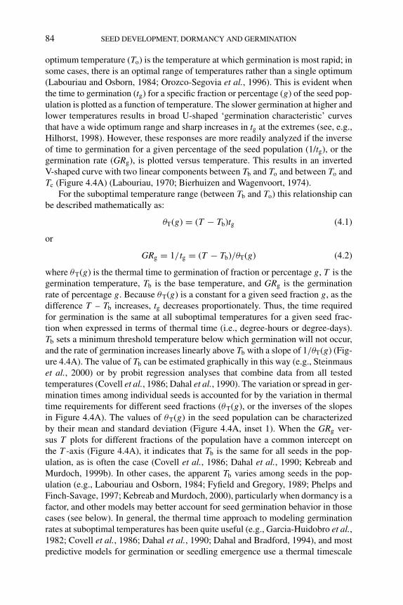
P1: OTE/SPH P2: OTE
BLUK053-Bradford September 27, 2006 7:59
84 SEED DEVELOPMENT, DORMANCY AND GERMINATION
optimum temperature (To) is the temperature at which germination is most rapid; insome cases, there is an optimal range of temperatures rather than a single optimum(Labouriau and Osborn, 1984; Orozco-Segovia et al., 1996). This is evident whenthe time to germination (tg) for a specific fraction or percentage (g) of the seed pop-ulation is plotted as a function of temperature. The slower germination at higher andlower temperatures results in broad U-shaped ‘germination characteristic’ curvesthat have a wide optimum range and sharp increases in tg at the extremes (see, e.g.,Hilhorst, 1998). However, these responses are more readily analyzed if the inverseof time to germination for a given percentage of the seed population (1/tg), or thegermination rate (GRg), is plotted versus temperature. This results in an invertedV-shaped curve with two linear components between Tb and To and between To andTc (Figure 4.4A) (Labouriau, 1970; Bierhuizen and Wagenvoort, 1974).
For the suboptimal temperature range (between Tb and To) this relationship canbe described mathematically as:
θT(g) = (T − Tb)tg (4.1)
or
GRg = 1/tg = (T − Tb)/θT(g) (4.2)
where θT(g) is the thermal time to germination of fraction or percentage g, T is thegermination temperature, Tb is the base temperature, and GRg is the germinationrate of percentage g. Because θT(g) is a constant for a given seed fraction g, as thedifference T – Tb increases, tg decreases proportionately. Thus, the time requiredfor germination is the same at all suboptimal temperatures for a given seed frac-tion when expressed in terms of thermal time (i.e., degree-hours or degree-days).Tb sets a minimum threshold temperature below which germination will not occur,and the rate of germination increases linearly above Tb with a slope of 1/θT(g) (Fig-ure 4.4A). The value of Tb can be estimated graphically in this way (e.g., Steinmauset al., 2000) or by probit regression analyses that combine data from all testedtemperatures (Covell et al., 1986; Dahal et al., 1990). The variation or spread in ger-mination times among individual seeds is accounted for by the variation in thermaltime requirements for different seed fractions (θT(g), or the inverses of the slopesin Figure 4.4A). The values of θT(g) in the seed population can be characterizedby their mean and standard deviation (Figure 4.4A, inset 1). When the GRg ver-sus T plots for different fractions of the population have a common intercept onthe T -axis (Figure 4.4A), it indicates that Tb is the same for all seeds in the pop-ulation, as is often the case (Covell et al., 1986; Dahal et al., 1990; Kebreab andMurdoch, 1999b). In other cases, the apparent Tb varies among seeds in the pop-ulation (e.g., Labouriau and Osborn, 1984; Fyfield and Gregory, 1989; Phelps andFinch-Savage, 1997; Kebreab and Murdoch, 2000), particularly when dormancy is afactor, and other models may better account for seed germination behavior in thosecases (see below). In general, the thermal time approach to modeling germinationrates at suboptimal temperatures has been quite useful (e.g., Garcia-Huidobro et al.,1982; Covell et al., 1986; Dahal et al., 1990; Dahal and Bradford, 1994), and mostpredictive models for germination or seedling emergence use a thermal timescale
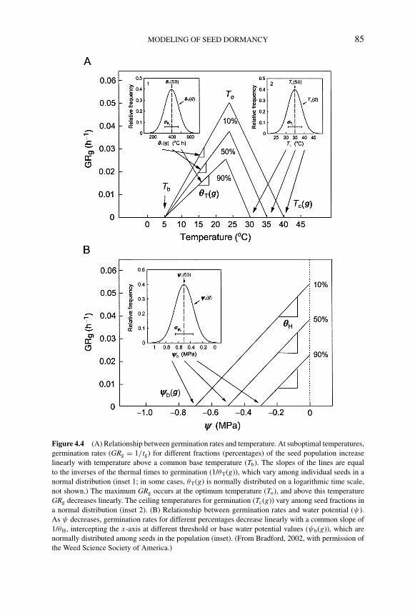
P1: OTE/SPH P2: OTE
BLUK053-Bradford September 27, 2006 7:59
MODELING OF SEED DORMANCY 85
Figure 4.4 (A) Relationship between germination rates and temperature. At suboptimal temperatures,
germination rates (GRg = 1/tg) for different fractions (percentages) of the seed population increase
linearly with temperature above a common base temperature (Tb). The slopes of the lines are equal
to the inverses of the thermal times to germination (1/θT(g)), which vary among individual seeds in a
normal distribution (inset 1; in some cases, θT(g) is normally distributed on a logarithmic time scale,
not shown.) The maximum GRg occurs at the optimum temperature (To), and above this temperature
GRg decreases linearly. The ceiling temperatures for germination (Tc(g)) vary among seed fractions in
a normal distribution (inset 2). (B) Relationship between germination rates and water potential (ψ).
As ψ decreases, germination rates for different percentages decrease linearly with a common slope of
1/θH, intercepting the x-axis at different threshold or base water potential values (ψb(g)), which are
normally distributed among seeds in the population (inset). (From Bradford, 2002, with permission of
the Weed Science Society of America.)
P1: OTE/SPH P2: OTE
BLUK053-Bradford September 27, 2006 7:59
86 SEED DEVELOPMENT, DORMANCY AND GERMINATION
(degree-days) to normalize for temperature variation over time in the field (Forcella,1998; Vleeshouwers and Kropff, 2000).
According to Equations 4.1 and 4.2, tg should continue to decrease (GRg shouldcontinue to increase) indefinitely with increasing temperature, but this is not thecase. Instead, GRg increases to a maximum at To, and then decreases approximatelylinearly at higher temperatures until it intersects the x-axis at Tc (Figure 4.4A).At supraoptimal temperatures, the slopes of these lines for different germinationfractions are often parallel, resulting in different intercepts on the x-axis (differentTc values) (Figure 4.4A) (Covell et al., 1986; Ellis et al., 1987; Ellis and Butcher,1988), although common Tc values for all seeds in a population have been reportedas well (Garcia-Huidobro et al., 1982; Orozco-Segovia et al., 1996). When Tc valuesdiffer among seeds in the population, germination time courses in the supraoptimaltemperature range can be analyzed by holding thermal time constant (θTc) andallowing Tc to vary among different seed fractions (i.e., Tc(g)) (Ellis et al., 1986,1987; Ellis and Butcher, 1988):
θTc = (Tc(g) − T )tg (4.3)
or
GRg = 1/tg = (Tc(g) − T )/θTc (4.4)
Thus, the thermal time required for completion of germination (θTc) is the same forall seeds in the supraoptimal temperature range, but differences in germination ratesare due to different Tc values (Figure 4.4A). That is, seeds having higher Tc valueswill accumulate thermal time (Tc – T ) more rapidly and will therefore completegermination more quickly than seeds having lower Tc values. The seed populationcan then be characterized in terms of the distribution (i.e., the mean and standarddeviation) of Tc(g) values in the supraoptimal range (Figure 4.4A, inset 2).
The presence of dormancy can affect germination responses to temperature.Stratification of Aesculus hippocastanum seeds resulted in a progressive lowering ofTb, such that seeds could eventually germinate at temperatures that initially preventedgermination (Steadman and Pritchard, 2004). Apparent ceiling temperatures forlettuce (Lactuca sativa) seed germination can be altered by ethylene (Dutta andBradford, 1994; Nascimento et al., 2000). An alternative approach to analyzingand modeling this situation was proposed by Benech-Arnold and coworkers (Krukand Benech-Arnold, 1998, 2000; Batlla and Benech-Arnold, 2003) based on theearly work of Washitani (1987). This approach assumes that the actual Tb and Tc
for germination do not change due to dormancy, but rather that there is in additiona lower (Tl) and upper (Th) temperature limit for germination that varies accordingto the depth of dormancy. When T is lower than Tl of a certain fraction of thepopulation, dormancy is expressed in individuals belonging to that fraction but notin seeds from fractions with a Tl lower than T . As dormancy is alleviated by theeffect of stratification, for example, the entire Tl distribution is displaced towardlower temperatures and, consequently, additional fractions of the population willhave a Tl lower than T , and will be able to germinate. Thermal time required forgermination is accumulated relative to Tb, but only once T exceeds Tl for a given
P1: OTE/SPH P2: OTE
BLUK053-Bradford September 27, 2006 7:59
MODELING OF SEED DORMANCY 87
seed fraction, as Tl and Th both vary among seeds (Figure 4.2). This approach allowschanges in temperature responses due to dormancy to be modeled without alteringthe fundamental cardinal temperatures for a species. Its application will be illustratedsubsequently.
Both approaches explicitly account for variation in temperature response pa-rameters among individual seeds, which results in the overall population behavioras the temperature sensitivity shifts during dormancy release or imposition. Theconsequences of both approaches are similar, in that they account for shifts in theability of seeds to germinate at low or high temperatures due to varying degrees ofdormancy by a corresponding physiological shift in the threshold or temperaturelimit values.
4.4.2 Water potential responses and hydrotime models
Seeds cannot germinate without water, and seed germination rates and percentagesare closely attuned to water availability (Bradford, 1995). As for temperature, there isa minimum water potential (ψ) that must be exceeded in order for seeds to completegermination, and seeds in a population vary in the value of this minimum or base ψ
(ψb) (Hegarty, 1978). In analogy with the thermal time concept, Gummerson (1986)proposed that the time to germination for a given seed fraction (g) was inverselyrelated to the difference between the current seed ψ and the ψb for that fraction(ψb(g)). He defined this hydrotime model as:
θH = (ψ − ψb(g))tg (4.5)
or
GRg = 1/tg = (ψ − ψb(g))/θH (4.6)
where θH is the hydrotime constant in MPa-hours or MPa-days. If GRg valuesdetermined by incubating seeds at different constant ψ are plotted as a function ofψ , the resulting relationships are linear for different seed fractions (g), having acommon slope and intercepting the ψ-axis at different ψb(g) values (Figure 4.4B).The common slopes indicate that the total accumulated hydrotime (MPa-hours orMPa-days) to radicle emergence is the same for all seeds in the population, while thedifferent intercepts show that individual seeds vary in their ψb thresholds at whichradicle emergence is prevented. As θH is a constant, tg must increase proportionatelyas ψ is reduced and approaches ψb(g) (see Bradford, 1997, 2002, for illustrations).In most cases, ψb(g) values vary among seeds in the population in a normal orGaussian distribution (e.g., Gummerson, 1986; Bradford, 1990; Dahal and Bradford,1990), although this is not essential to the model. If the distribution is normal, itcan be defined by its mean (ψb(50)) and standard deviation (σψb) (Figure 4.4B,inset). ψb(50) is more properly the median, since it refers to the 50th percentile ofthe population rather than to the average value, but for a normal distribution, themean and the median coincide, and so we will refer to it here as the mean basewater potential. As for temperature, both graphical and probit analysis methods forestimating the hydrotime parameters from original germination time course data at
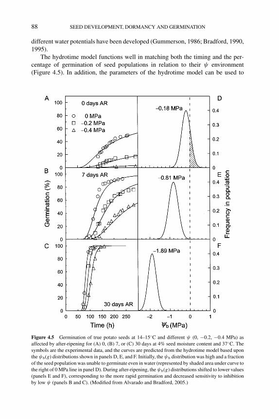
P1: OTE/SPH P2: OTE
BLUK053-Bradford September 27, 2006 7:59
88 SEED DEVELOPMENT, DORMANCY AND GERMINATION
different water potentials have been developed (Gummerson, 1986; Bradford, 1990,1995).
The hydrotime model functions well in matching both the timing and the per-centage of germination of seed populations in relation to their ψ environment(Figure 4.5). In addition, the parameters of the hydrotime model can be used to
Figure 4.5 Germination of true potato seeds at 14–15◦C and different ψ (0, −0.2, −0.4 MPa) as
affected by after-ripening for (A) 0, (B) 7, or (C) 30 days at 4% seed moisture content and 37◦C. The
symbols are the experimental data, and the curves are predicted from the hydrotime model based upon
the ψb(g) distributions shown in panels D, E, and F. Initially, the ψb distribution was high and a fraction
of the seed population was unable to germinate even in water (represented by shaded area under curve to
the right of 0 MPa line in panel D). During after-ripening, the ψb(g) distributions shifted to lower values
(panels E and F), corresponding to the more rapid germination and decreased sensitivity to inhibition
by low ψ (panels B and C). (Modified from Alvarado and Bradford, 2005.)
P1: OTE/SPH P2: OTE
BLUK053-Bradford September 27, 2006 7:59
MODELING OF SEED DORMANCY 89
characterize the properties of seed populations. The θH value quantifies the inherentspeed of germination, the ψb(50) value is an indication of the average water stresstolerance of the population, and the standard deviation of ψb(g) (σψb) indicatesthe uniformity of threshold values which corresponds to the synchrony in germi-nation timing among seeds in the population. These three parameters are sufficientto predict complete germination time courses at any ψ at a constant T (Bradford,1995, 2002). In addition, the hydrotime model is insensitive to whether the ψ ofthe environment changes or the physiological ψb(g) thresholds are altered. That is,equivalent effects on germination timing result either from variation in environmen-tal ψ or from physiological adjustments in the water potential thresholds of seedpopulations (Bradford, 2002). Changes in ψb(g) distributions have proven to be auseful way to characterize and model increases or decreases in seed dormancy levels(Figures 4.5D–4.5F).
A limitation of the hydrotime model as presented above is that it does not accountfor metabolic advancement toward germination that can occur at water potentialsbelow ψb. The practice of seed priming, in which seeds are incubated at ψ <
ψb(g) for extended periods of time and subsequently exhibit more rapid germinationrates, indicates that seeds can accumulate hydrotime at water potentials below ψb.There is a limit or minimum ψ below which metabolic advancement does notoccur (ψmin), and advancement in germination is often linear at water potentialsbetween ψmin and ψb (Tarquis and Bradford, 1992; Bradford and Haigh, 1994;Finch-Savage et al., 2005). Modifications to Equation 4.6 have been suggested thatcan empirically account for this effect on germination timing (Cheng and Bradford,1999). An alternative approach developed by Rowse and colleagues (Rowse et al.,1999; Rowse and Finch-Savage, 2003), termed the ‘virtual osmotic potential’ orVOP model, directly accounts for advancement toward germination at all waterpotentials above ψmin. While sharing the concept of a distribution of ψb(g) thresholdsfor germination, the VOP model assumes that progress toward germination is dueto either accumulation of osmotic solutes to lower embryonic water potential or areduction in the restraint by enclosing tissues or both. As the VOP model incorporatesthe effects of water potentials between ψmin and ψb as well as between ψb andψ , it may be more amenable to predicting germination times under fluctuating ψ
conditions than the standard hydrotime model (Finch-Savage, 2004; Finch-Savageet al., 2005).
4.4.3 Interactions of temperature and water potential
The combined effects of suboptimal temperatures and reduced water potentials ongermination can be modeled by using thermal time instead of actual time in thefollowing equation (Gummerson, 1986; Bradford, 1995):
θHT = (ψ − ψb(g))(T − Tb)tg (4.7)
This hydrothermal model has successfully described germination time courses atconstant suboptimal temperatures in the relatively high ψ range (Gummerson, 1986;Dahal and Bradford, 1994; Alvarado and Bradford, 2002, 2005). This equation
P1: OTE/SPH P2: OTE
BLUK053-Bradford September 27, 2006 7:59
90 SEED DEVELOPMENT, DORMANCY AND GERMINATION
assumes that ψb(g) is constant and independent of temperature, and that Tb isindependent of ψ , which is not always the case, particularly when the incuba-tion temperatures affect seed dormancy (Bradford and Somasco, 1994; Dahal andBradford, 1994; Kebreab and Murdoch, 1999b). In those cases, however, the effectson dormancy can be quantified in terms of their influence on the parameters of Equa-tion 4.7. For example, seeds of lettuce and of many species that normally germinatein the fall or winter are prevented from germinating at warm temperatures (Baskinand Baskin, 1998). In lettuce seeds, this can be attributed to an increase in the ψb(g)distribution (to less negative values) as T increases (Bradford and Somasco, 1994).Several studies have found that ψb(g) values are at a minimum around the opti-mum T and become higher at supraoptimal T (Christensen et al., 1996; Kebreaband Murdoch, 1999b; Meyer et al., 2000; Alvarado and Bradford, 2002; Rowse andFinch-Savage, 2003). While different species may have similar germination timecourses in water, dramatic differences in germination rates are frequently observedwhen those seeds are incubated at reduced water potentials, as will be reflected intheir θHT and ψb(g) values (Allen et al., 2000). Analysis of hydrothermal time pa-rameters has led to important ecological interpretations regarding germination for awide variety of species (Allen et al., 2000).
4.4.4 Modeling responses to other factors affecting dormancyand germination
Population-based threshold models have been applied to describe or predict seedgermination behavior in response to a number of factors. Hormonal (e.g., gib-berellin [GA], abscisic acid [ABA], and ethylene) effects on germination of tomato(Lycopersicon esculentum) and lettuce seeds were quantified using this approach(Ni and Bradford, 1992, 1993; Dutta and Bradford, 1994). Fennimore and Foley(1998) quantitatively described how GA sensitivity of wild oat (Avena fatua) seedschanged during loss of dormancy by after-ripening. Bradford (1996, 2005) has sug-gested that seed responses to light and nitrate would also be amenable to modelingby this approach.
The concept of degrees of relative dormancy described previously presupposesthe existence of environmental thresholds for dormancy expression. Dependingon the dormancy level of the population, there will be a temperature above which(Tb or Tl) or below which (Tc or Th) dormancy is not expressed. Similarly, there willbe a threshold water potential (ψb) above which germination can proceed. Whilethe thermal time and hydrotime models described above assume a linear responseto a given factor above the threshold value, this is not the only possibility. Withrespect to light, for example, the phytochrome LFR response has long been knownto exhibit reciprocity; i.e., a low dose over a longer time is equivalent to a high doseover a shorter time. This is strictly analogous to the threshold models describedabove. However, whether or not a seed is responsive to light at all may be an all-or-none or quantal response, possibly based upon whether the seed expresses a specificmember of the phytochrome gene family (Casal and Sanchez, 1998). Individual
P1: OTE/SPH P2: OTE
BLUK053-Bradford September 27, 2006 7:59
MODELING OF SEED DORMANCY 91
seeds may move from a nonresponsive fraction of the population to a responsivefraction as dormancy is lost. As sensitivity thresholds for various factors shift withchanges in the dormancy level of the population, the range of conditions underwhich germination can proceed will widen if dormancy is released or, conversely,will narrow when dormancy is induced. Since the dormancy level can be differentfor each individual seed within the population, these thresholds cannot be regardedas unique for the whole population; on the contrary, their values can be assumed tobe distributed within the population, with a mean and a standard deviation. Hence,not only the mean value of the threshold(s) but also the entire accompanying distri-bution is shifting with changes in the dormancy level of the population (e.g., Figures4.5D–4.5F).
4.5 Examples of seed dormancy models
To illustrate the application of seed dormancy models, some cases that have beeninvestigated in depth will be presented. As there are many unique types of dormancy,different approaches may be better suited to particular situations and species. Theexamples below illustrate methods that can be used to quantify and predict seeddormancy behavior in relation to the principal factors that influence the rates ofdormancy release.
4.5.1 Solanum tuberosum
Recently harvested true (botanical) potato (Solanum tuberosum) seeds exhibit dor-mancy that can be alleviated by after-ripening. After relatively short after-ripeningperiods, they exhibit conditional dormancy in which they are capable of germinationat low T (<17◦C), but are increasingly inhibited at higher T ; further after-ripeningexpands the upper temperature limit for germination (Pallais, 1995). These changesin germination behavior in relation to after-ripening and germination temperaturecan be modeled using the hydrotime and hydrothermal time models (Alvarado andBradford, 2002, 2005). The dormancy of recently harvested potato seeds is evident intheir delayed initiation of germination, low final germination percentages, and highsensitivity to inhibition by reduced ψ even at relatively low temperatures (14◦C)(Figure 4.5A). After only 7 days of dry storage at 37◦C, germination was muchimproved, and after 30 days of after-ripening, germination was rapid and relativelyinsensitive to reduced ψ (Figures 4.5B and 4.5C). These changes in germinationbehavior as dormancy was relieved can be described by associated shifts in theψb(g) distributions of the seed population to lower values (Figures 4.5D–4.5F). Thehydrotime constant (θH) also increased during after-ripening, and when combinedwith the ψb(g) distribution, the complete germination time courses at all ψ can bepredicted with high precision (Figures 4.5A–4.5C). These relationships held acrossthe entire range of suboptimal temperatures, illustrating that a major consequenceof after-ripening is a lowering of the ψb(g) distribution (Alvarado and Bradford,
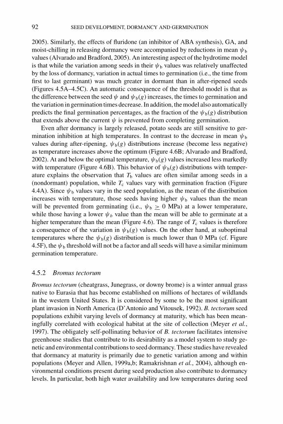
P1: OTE/SPH P2: OTE
BLUK053-Bradford September 27, 2006 7:59
92 SEED DEVELOPMENT, DORMANCY AND GERMINATION
2005). Similarly, the effects of fluridone (an inhibitor of ABA synthesis), GA, andmoist-chilling in releasing dormancy were accompanied by reductions in mean ψb
values (Alvarado and Bradford, 2005). An interesting aspect of the hydrotime modelis that while the variation among seeds in their ψb values was relatively unaffectedby the loss of dormancy, variation in actual times to germination (i.e., the time fromfirst to last germinant) was much greater in dormant than in after-ripened seeds(Figures 4.5A–4.5C). An automatic consequence of the threshold model is that asthe difference between the seed ψ and ψb(g) increases, the times to germination andthe variation in germination times decrease. In addition, the model also automaticallypredicts the final germination percentages, as the fraction of the ψb(g) distributionthat extends above the current ψ is prevented from completing germination.
Even after dormancy is largely released, potato seeds are still sensitive to ger-mination inhibition at high temperatures. In contrast to the decrease in mean ψb
values during after-ripening, ψb(g) distributions increase (become less negative)as temperature increases above the optimum (Figure 4.6B; Alvarado and Bradford,2002). At and below the optimal temperature, ψb(g) values increased less markedlywith temperature (Figure 4.6B). This behavior of ψb(g) distributions with temper-ature explains the observation that Tb values are often similar among seeds in a(nondormant) population, while Tc values vary with germination fraction (Figure4.4A). Since ψb values vary in the seed population, as the mean of the distributionincreases with temperature, those seeds having higher ψb values than the meanwill be prevented from germinating (i.e., ψb > 0 MPa) at a lower temperature,while those having a lower ψb value than the mean will be able to germinate at ahigher temperature than the mean (Figure 4.6). The range of Tc values is thereforea consequence of the variation in ψb(g) values. On the other hand, at suboptimaltemperatures where the ψb(g) distribution is much lower than 0 MPa (cf. Figure4.5F), the ψb threshold will not be a factor and all seeds will have a similar minimumgermination temperature.
4.5.2 Bromus tectorum
Bromus tectorum (cheatgrass, Junegrass, or downy brome) is a winter annual grassnative to Eurasia that has become established on millions of hectares of wildlandsin the western United States. It is considered by some to be the most significantplant invasion in North America (D’Antonio and Vitousek, 1992). B. tectorum seedpopulations exhibit varying levels of dormancy at maturity, which has been mean-ingfully correlated with ecological habitat at the site of collection (Meyer et al.,1997). The obligately self-pollinating behavior of B. tectorum facilitates intensivegreenhouse studies that contribute to its desirability as a model system to study ge-netic and environmental contributions to seed dormancy. These studies have revealedthat dormancy at maturity is primarily due to genetic variation among and withinpopulations (Meyer and Allen, 1999a,b; Ramakrishnan et al., 2004), although en-vironmental conditions present during seed production also contribute to dormancylevels. In particular, both high water availability and low temperatures during seed
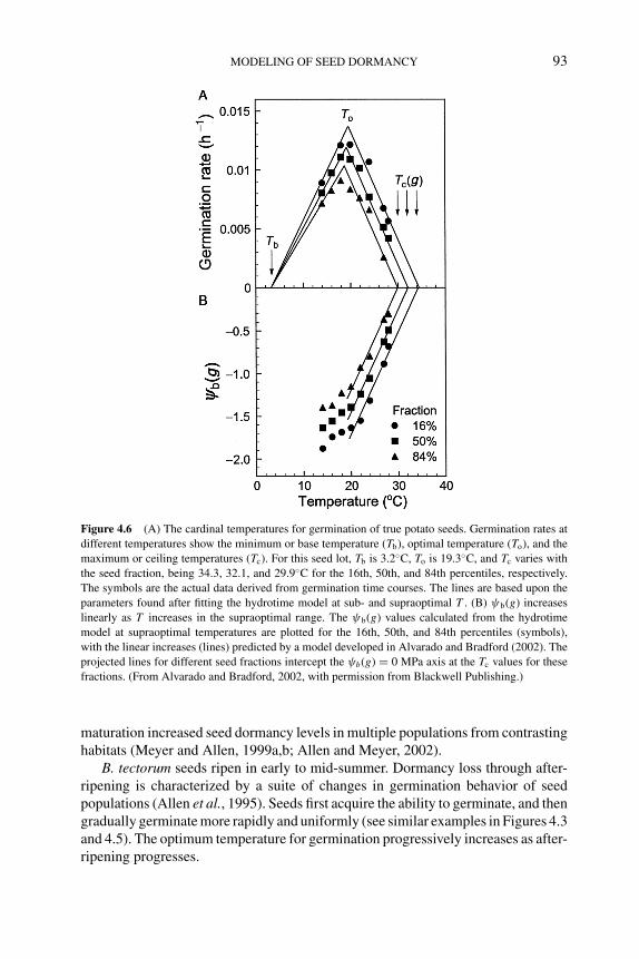
P1: OTE/SPH P2: OTE
BLUK053-Bradford September 27, 2006 7:59
MODELING OF SEED DORMANCY 93
Figure 4.6 (A) The cardinal temperatures for germination of true potato seeds. Germination rates at
different temperatures show the minimum or base temperature (Tb), optimal temperature (To), and the
maximum or ceiling temperatures (Tc). For this seed lot, Tb is 3.2◦C, To is 19.3◦C, and Tc varies with
the seed fraction, being 34.3, 32.1, and 29.9◦C for the 16th, 50th, and 84th percentiles, respectively.
The symbols are the actual data derived from germination time courses. The lines are based upon the
parameters found after fitting the hydrotime model at sub- and supraoptimal T . (B) ψb(g) increases
linearly as T increases in the supraoptimal range. The ψb(g) values calculated from the hydrotime
model at supraoptimal temperatures are plotted for the 16th, 50th, and 84th percentiles (symbols),
with the linear increases (lines) predicted by a model developed in Alvarado and Bradford (2002). The
projected lines for different seed fractions intercept the ψb(g) = 0 MPa axis at the Tc values for these
fractions. (From Alvarado and Bradford, 2002, with permission from Blackwell Publishing.)
maturation increased seed dormancy levels in multiple populations from contrastinghabitats (Meyer and Allen, 1999a,b; Allen and Meyer, 2002).
B. tectorum seeds ripen in early to mid-summer. Dormancy loss through after-ripening is characterized by a suite of changes in germination behavior of seedpopulations (Allen et al., 1995). Seeds first acquire the ability to germinate, and thengradually germinate more rapidly and uniformly (see similar examples in Figures 4.3and 4.5). The optimum temperature for germination progressively increases as after-ripening progresses.
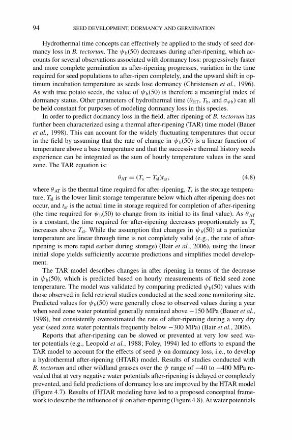
P1: OTE/SPH P2: OTE
BLUK053-Bradford September 27, 2006 7:59
94 SEED DEVELOPMENT, DORMANCY AND GERMINATION
Hydrothermal time concepts can effectively be applied to the study of seed dor-mancy loss in B. tectorum. The ψb(50) decreases during after-ripening, which ac-counts for several observations associated with dormancy loss: progressively fasterand more complete germination as after-ripening progresses, variation in the timerequired for seed populations to after-ripen completely, and the upward shift in op-timum incubation temperature as seeds lose dormancy (Christensen et al., 1996).As with true potato seeds, the value of ψb(50) is therefore a meaningful index ofdormancy status. Other parameters of hydrothermal time (θHT, Tb, and σψb) can allbe held constant for purposes of modeling dormancy loss in this species.
In order to predict dormancy loss in the field, after-ripening of B. tectorum hasfurther been characterized using a thermal after-ripening (TAR) time model (Baueret al., 1998). This can account for the widely fluctuating temperatures that occurin the field by assuming that the rate of change in ψb(50) is a linear function oftemperature above a base temperature and that the successive thermal history seedsexperience can be integrated as the sum of hourly temperature values in the seedzone. The TAR equation is:
θAT = (Ts − Tsl)tar, (4.8)
where θAT is the thermal time required for after-ripening, Ts is the storage tempera-ture, Tsl is the lower limit storage temperature below which after-ripening does notoccur, and tar is the actual time in storage required for completion of after-ripening(the time required for ψb(50) to change from its initial to its final value). As θAT
is a constant, the time required for after-ripening decreases proportionately as Ts
increases above Tsl. While the assumption that changes in ψb(50) at a particulartemperature are linear through time is not completely valid (e.g., the rate of after-ripening is more rapid earlier during storage) (Bair et al., 2006), using the linearinitial slope yields sufficiently accurate predictions and simplifies model develop-ment.
The TAR model describes changes in after-ripening in terms of the decreasein ψb(50), which is predicted based on hourly measurements of field seed zonetemperature. The model was validated by comparing predicted ψb(50) values withthose observed in field retrieval studies conducted at the seed zone monitoring site.Predicted values for ψb(50) were generally close to observed values during a yearwhen seed zone water potential generally remained above −150 MPa (Bauer et al.,1998), but consistently overestimated the rate of after-ripening during a very dryyear (seed zone water potentials frequently below −300 MPa) (Bair et al., 2006).
Reports that after-ripening can be slowed or prevented at very low seed wa-ter potentials (e.g., Leopold et al., 1988; Foley, 1994) led to efforts to expand theTAR model to account for the effects of seed ψ on dormancy loss, i.e., to developa hydrothermal after-ripening (HTAR) model. Results of studies conducted withB. tectorum and other wildland grasses over the ψ range of −40 to −400 MPa re-vealed that at very negative water potentials after-ripening is delayed or completelyprevented, and field predictions of dormancy loss are improved by the HTAR model(Figure 4.7). Results of HTAR modeling have led to a proposed conceptual frame-work to describe the influence of ψ on after-ripening (Figure 4.8). At water potentials
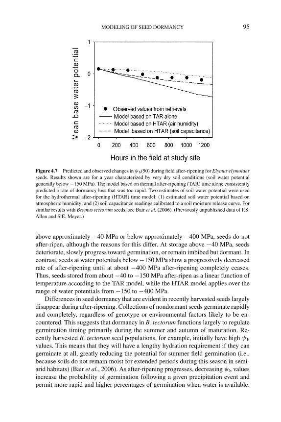
P1: OTE/SPH P2: OTE
BLUK053-Bradford September 27, 2006 7:59
MODELING OF SEED DORMANCY 95
Figure 4.7 Predicted and observed changes in ψb(50) during field after-ripening for Elymus elymoidesseeds. Results shown are for a year characterized by very dry soil conditions (soil water potential
generally below −150 MPa). The model based on thermal after-ripening (TAR) time alone consistently
predicted a rate of dormancy loss that was too rapid. Two estimates of soil water potential were used
for the hydrothermal after-ripening (HTAR) time model: (1) estimated soil water potential based on
atmospheric humidity; and (2) soil capacitance readings calibrated to a soil moisture release curve. For
similar results with Bromus tectorum seeds, see Bair et al. (2006). (Previously unpublished data of P.S.
Allen and S.E. Meyer.)
above approximately −40 MPa or below approximately −400 MPa, seeds do notafter-ripen, although the reasons for this differ. At storage above −40 MPa, seedsdeteriorate, slowly progress toward germination, or remain imbibed but dormant. Incontrast, seeds at water potentials below −150 MPa show a progressively decreasedrate of after-ripening until at about −400 MPa after-ripening completely ceases.Thus, seeds stored from about −40 to −150 MPa after-ripen as a linear function oftemperature according to the TAR model, while the HTAR model applies over therange of water potentials from −150 to −400 MPa.
Differences in seed dormancy that are evident in recently harvested seeds largelydisappear during after-ripening. Collections of nondormant seeds germinate rapidlyand completely, regardless of genotype or environmental factors likely to be en-countered. This suggests that dormancy in B. tectorum functions largely to regulategermination timing primarily during the summer and autumn of maturation. Re-cently harvested B. tectorum seed populations, for example, initially have high ψb
values. This means that they will have a lengthy hydration requirement if they cangerminate at all, greatly reducing the potential for summer field germination (i.e.,because soils do not remain moist for extended periods during this season in semi-arid habitats) (Bair et al., 2006). As after-ripening progresses, decreasing ψb valuesincrease the probability of germination following a given precipitation event andpermit more rapid and higher percentages of germination when water is available.
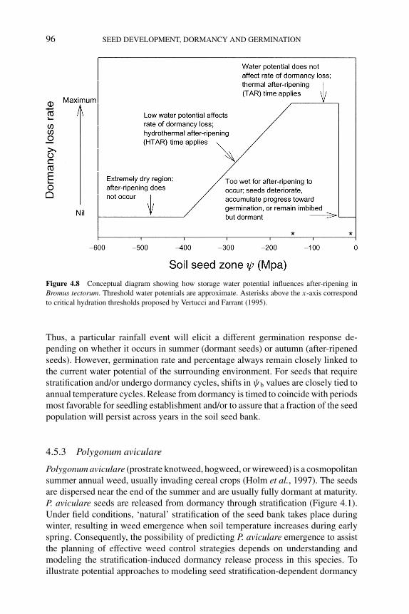
P1: OTE/SPH P2: OTE
BLUK053-Bradford September 27, 2006 7:59
96 SEED DEVELOPMENT, DORMANCY AND GERMINATION
Figure 4.8 Conceptual diagram showing how storage water potential influences after-ripening in
Bromus tectorum. Threshold water potentials are approximate. Asterisks above the x-axis correspond
to critical hydration thresholds proposed by Vertucci and Farrant (1995).
Thus, a particular rainfall event will elicit a different germination response de-pending on whether it occurs in summer (dormant seeds) or autumn (after-ripenedseeds). However, germination rate and percentage always remain closely linked tothe current water potential of the surrounding environment. For seeds that requirestratification and/or undergo dormancy cycles, shifts in ψb values are closely tied toannual temperature cycles. Release from dormancy is timed to coincide with periodsmost favorable for seedling establishment and/or to assure that a fraction of the seedpopulation will persist across years in the soil seed bank.
4.5.3 Polygonum aviculare
Polygonum aviculare (prostrate knotweed, hogweed, or wireweed) is a cosmopolitansummer annual weed, usually invading cereal crops (Holm et al., 1997). The seedsare dispersed near the end of the summer and are usually fully dormant at maturity.P. aviculare seeds are released from dormancy through stratification (Figure 4.1).Under field conditions, ‘natural’ stratification of the seed bank takes place duringwinter, resulting in weed emergence when soil temperature increases during earlyspring. Consequently, the possibility of predicting P. aviculare emergence to assistthe planning of effective weed control strategies depends on understanding andmodeling the stratification-induced dormancy release process in this species. Toillustrate potential approaches to modeling seed stratification-dependent dormancy
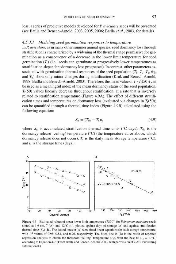
P1: OTE/SPH P2: OTE
BLUK053-Bradford September 27, 2006 7:59
MODELING OF SEED DORMANCY 97
loss, a series of predictive models developed for P. aviculare seeds will be presented(see Batlla and Benech-Arnold, 2003, 2005, 2006; Batlla et al., 2003, for details).
4.5.3.1 Modeling seed germination responses to temperatureIn P. aviculare, as in many other summer annual species, seed dormancy loss throughstratification is characterized by a widening of the thermal range permissive for ger-mination as a consequence of a decrease in the lower limit temperature for seedgermination (Tl) (i.e., seeds can germinate at progressively lower temperatures asstratification-dependent dormancy loss progresses). In contrast, other parameters as-sociated with germination thermal responses of the seed population (Tb, To, Tc, θT,and Th) show only minor changes during stratification (Kruk and Benech-Arnold,1998; Batlla and Benech-Arnold, 2003). Therefore, the mean value of Tl (Tl(50)) canbe used as a meaningful index of the mean dormancy status of the seed population.Tl(50) values linearly decrease throughout stratification, at a rate that is inverselyrelated to stratification temperature (Figure 4.9A). The effect of different stratifi-cation times and temperatures on dormancy loss (evaluated via changes in Tl(50))can be quantified through a thermal time index (Figure 4.9B) calculated using thefollowing equation:
Stt = (Tdc − Ts)ts (4.9)
where Stt is accumulated stratification thermal time units (◦C days), Tdc is thedormancy release ‘ceiling’ temperature (◦C) (the temperature at, or above, whichdormancy release does not occur), Ts is the daily mean storage temperature (◦C),and ts is the storage time (days).
Figure 4.9 Estimated values of mean lower limit temperature (Tl(50)) for Polygonum aviculare seeds
stored at 1.6 (◦), 7 (�), and 12◦C (�), plotted against days of storage (A) and against stratification
thermal time (Stt) (B). The dotted lines in (A) were fitted linear equations for each storage temperature,
with R2 values of 0.98, 0.84, and 0.96, respectively. The fitted line in (B) is the result of repeated
regression analysis to obtain the threshold ‘ceiling’ temperature (Tc), with the best fit (Tc = 17◦C)
according to Equation 4.9. (From Batlla and Benech-Arnold, 2003, with permission of CABI Publishing
International.)
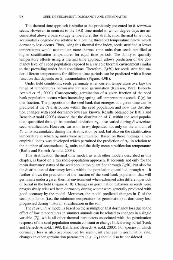
P1: OTE/SPH P2: OTE
BLUK053-Bradford September 27, 2006 7:59
98 SEED DEVELOPMENT, DORMANCY AND GERMINATION
This thermal time approach is similar to that previously presented for B. tectorumseeds. However, in contrast to the TAR time model in which degree-days are ac-cumulated above a base storage temperature, this stratification thermal time indexaccumulates degree-days relative to a ceiling threshold temperature below whichdormancy loss occurs. Thus, using this thermal time index, seeds stratified at lowertemperatures would accumulate more thermal time units than seeds stratified athigher stratification temperatures for equal time periods. The ability to quantifytemperature effects using a thermal time approach allows prediction of the dor-mancy level of a seed population exposed to a variable thermal environment similarto that prevailing under field conditions. Therefore, Tl(50) for seeds stratified un-der different temperatures for different time periods can be predicted with a linearfunction that depends on Stt accumulation (Figure. 4.9B).
Under field conditions, seeds germinate when current temperature overlaps therange of temperatures permissive for seed germination (Karssen, 1982; Benech-Arnold et al., 2000). Consequently, germination of a given fraction of the seedbank population occurs when increasing spring soil temperature exceeds Tl(g) forthat fraction. The proportion of the seed bank that emerges at a given time can bepredicted if the Tl distribution within the seed population and how this distribu-tion changes with seed dormancy level are known. Results obtained by Batlla andBenech-Arnold (2003) showed that the distribution of Tl within the seed popula-tion, quantified through its standard deviation σTl
, also varied during P. aviculareseed stratification. However, variation in σTl
depended not only on the amount ofStt units accumulated during the stratification period, but also on the stratificationtemperature at which Stt units were accumulated. Based on these findings, a newempirical index was developed which permitted the prediction of σTl
in relation tothe number of accumulated Stt units and the daily mean stratification temperature(Batlla and Benech-Arnold, 2003).
This stratification thermal time model, as with other models described in thischapter, is based on a threshold-population approach. It accounts not only for themean dormancy status of the seed population quantified through Tl(50), but also forthe distribution of dormancy levels within the population quantified through σTl
. Itfurther allows the prediction of the fraction of the seed-bank population that willgerminate under a given thermal environment when exhumed after different periodsof burial in the field (Figure 4.10). Changes in germination behavior as seeds wereprogressively released from dormancy during winter were generally predicted withgood accuracy by the model. Moreover, the model predicted changes in Tl of theseed population (i.e., the minimum temperature for germination) as dormancy lossprogressed during ‘natural’ stratification in the soil.
The P. aviculare model is based on the assumption that dormancy loss due to theeffect of low temperatures in summer annuals can be related to changes in a singlevariable (Tl), while all other thermal parameters associated with the germinationresponse of the seed population remain constant or change little during burial (Krukand Benech-Arnold, 1998; Batlla and Benech-Arnold, 2003). For species in whichdormancy loss is also accompanied by significant changes in germination rate,changes in other germination parameters (e.g., θT) should also be considered.
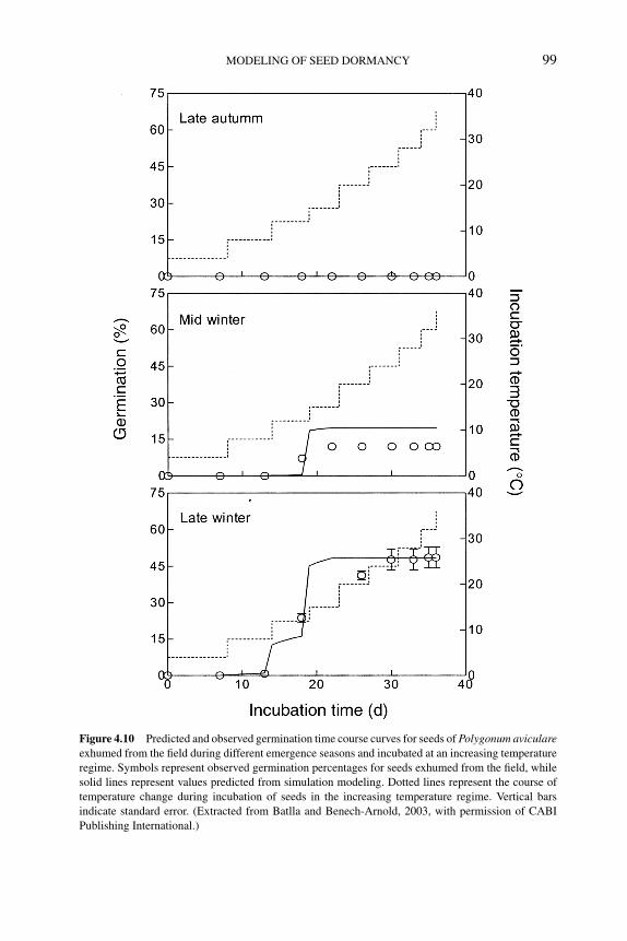
P1: OTE/SPH P2: OTE
BLUK053-Bradford September 27, 2006 7:59
MODELING OF SEED DORMANCY 99
Figure 4.10 Predicted and observed germination time course curves for seeds of Polygonum aviculareexhumed from the field during different emergence seasons and incubated at an increasing temperature
regime. Symbols represent observed germination percentages for seeds exhumed from the field, while
solid lines represent values predicted from simulation modeling. Dotted lines represent the course of
temperature change during incubation of seeds in the increasing temperature regime. Vertical bars
indicate standard error. (Extracted from Batlla and Benech-Arnold, 2003, with permission of CABI
Publishing International.)
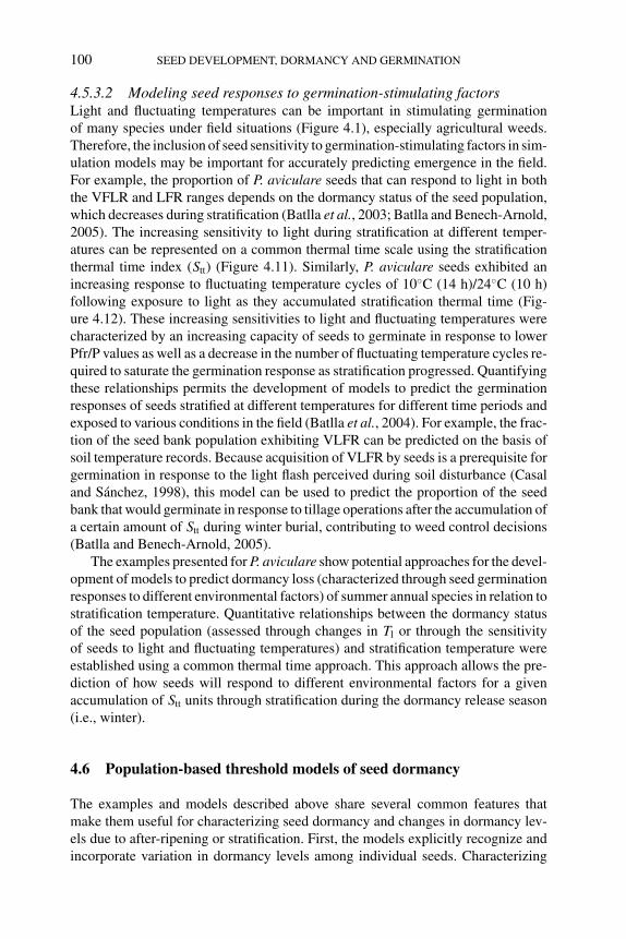
P1: OTE/SPH P2: OTE
BLUK053-Bradford September 27, 2006 7:59
100 SEED DEVELOPMENT, DORMANCY AND GERMINATION
4.5.3.2 Modeling seed responses to germination-stimulating factorsLight and fluctuating temperatures can be important in stimulating germinationof many species under field situations (Figure 4.1), especially agricultural weeds.Therefore, the inclusion of seed sensitivity to germination-stimulating factors in sim-ulation models may be important for accurately predicting emergence in the field.For example, the proportion of P. aviculare seeds that can respond to light in boththe VFLR and LFR ranges depends on the dormancy status of the seed population,which decreases during stratification (Batlla et al., 2003; Batlla and Benech-Arnold,2005). The increasing sensitivity to light during stratification at different temper-atures can be represented on a common thermal time scale using the stratificationthermal time index (Stt) (Figure 4.11). Similarly, P. aviculare seeds exhibited anincreasing response to fluctuating temperature cycles of 10◦C (14 h)/24◦C (10 h)following exposure to light as they accumulated stratification thermal time (Fig-ure 4.12). These increasing sensitivities to light and fluctuating temperatures werecharacterized by an increasing capacity of seeds to germinate in response to lowerPfr/P values as well as a decrease in the number of fluctuating temperature cycles re-quired to saturate the germination response as stratification progressed. Quantifyingthese relationships permits the development of models to predict the germinationresponses of seeds stratified at different temperatures for different time periods andexposed to various conditions in the field (Batlla et al., 2004). For example, the frac-tion of the seed bank population exhibiting VLFR can be predicted on the basis ofsoil temperature records. Because acquisition of VLFR by seeds is a prerequisite forgermination in response to the light flash perceived during soil disturbance (Casaland Sanchez, 1998), this model can be used to predict the proportion of the seedbank that would germinate in response to tillage operations after the accumulation ofa certain amount of Stt during winter burial, contributing to weed control decisions(Batlla and Benech-Arnold, 2005).
The examples presented for P. aviculare show potential approaches for the devel-opment of models to predict dormancy loss (characterized through seed germinationresponses to different environmental factors) of summer annual species in relation tostratification temperature. Quantitative relationships between the dormancy statusof the seed population (assessed through changes in Tl or through the sensitivityof seeds to light and fluctuating temperatures) and stratification temperature wereestablished using a common thermal time approach. This approach allows the pre-diction of how seeds will respond to different environmental factors for a givenaccumulation of Stt units through stratification during the dormancy release season(i.e., winter).
4.6 Population-based threshold models of seed dormancy
The examples and models described above share several common features thatmake them useful for characterizing seed dormancy and changes in dormancy lev-els due to after-ripening or stratification. First, the models explicitly recognize andincorporate variation in dormancy levels among individual seeds. Characterizing
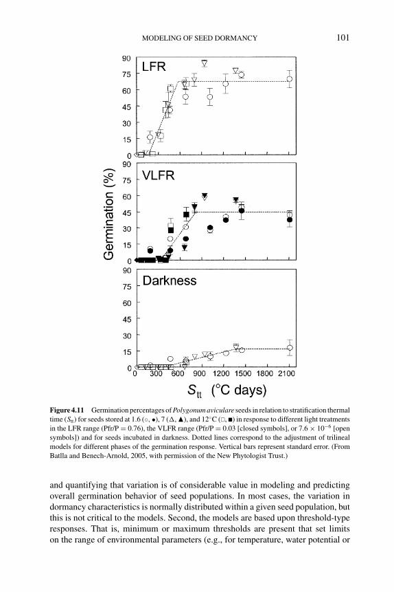
P1: OTE/SPH P2: OTE
BLUK053-Bradford September 27, 2006 7:59
MODELING OF SEED DORMANCY 101
Figure 4.11 Germination percentages of Polygonum aviculare seeds in relation to stratification thermal
time (Stt) for seeds stored at 1.6 (◦, •), 7 (�, �), and 12◦C (�, �) in response to different light treatments
in the LFR range (Pfr/P = 0.76), the VLFR range (Pfr/P = 0.03 [closed symbols], or 7.6 × 10⁻⁶ [open
symbols]) and for seeds incubated in darkness. Dotted lines correspond to the adjustment of trilineal
models for different phases of the germination response. Vertical bars represent standard error. (From
Batlla and Benech-Arnold, 2005, with permission of the New Phytologist Trust.)
and quantifying that variation is of considerable value in modeling and predictingoverall germination behavior of seed populations. In most cases, the variation indormancy characteristics is normally distributed within a given seed population, butthis is not critical to the models. Second, the models are based upon threshold-typeresponses. That is, minimum or maximum thresholds are present that set limitson the range of environmental parameters (e.g., for temperature, water potential or
P1: OTE/SPH P2: OTE
BLUK053-Bradford September 27, 2006 7:59
102 SEED DEVELOPMENT, DORMANCY AND GERMINATION
Figure 4.12 Germination responses of Polygonum aviculare seeds to cycles of a fluctuating temper-
ature regime of 10◦C (14 h)/24◦C (10 h) determined for different values of stratification thermal time
(Stt,◦C days). Germination data used for fitting curves correspond to seeds exhumed after different
days of storage at 1.6, 7, and 12◦C that had accumulated similar values of Stt. Dashed lines were fitted
with an exponential equation: Gn = C [1 −exp(−KN)] + G0, where Gn is the germination percent-
age obtained after N cycles of fluctuating temperature, C is the difference between the germination
percentage obtained with 0 (i.e., constant 15◦C) and N cycles, K is the effect of the number of cycles
on seed germination given by the slope of the response curve, and G0 is the germination percentage
obtained for seeds incubated at constant temperature (0 cycles). The derived parameters C and K can
be used to model and predict the response to fluctuating temperatures of seeds previously receiving
different amounts of stratification (Stt). Vertical bars indicate standard error. (From Batlla et al., 2003,
with permission of CABI Publishing International.)
light) permitting germination. These thresholds set the boundary sensitivity limitsfor seed responses to the various environmental factors controlling dormancy andgermination. For the thermal and hydrotime models, the rates at which dormancy-breaking or germinative processes occur are also proportional to the differencebetween the current level of an environmental parameter and its threshold limit.Thus, as the environmental signal (i.e., difference from the threshold) increases,the rate of either dormancy release or progress toward germination also increases.Since thresholds can vary among individual seeds, the rates of dormancy releaseor progress toward germination also vary among seeds, resulting in the character-istic germination response curves to dormancy-releasing factors (e.g., Figures 4.3and 4.5). Third, the sensitivity threshold distributions for a seed population shiftto higher or lower mean values in response to dormancy-releasing or germination-stimulating factors. Shifting response threshold distributions underlie the wideningor narrowing of the ranges of environmental factors over which germination will oc-cur as dormancy is released or induced. Together, these features of population-based
P1: OTE/SPH P2: OTE
BLUK053-Bradford September 27, 2006 7:59
MODELING OF SEED DORMANCY 103
threshold models provide considerable flexibility and explanatory power for describ-ing and predicting germination behavior.
Some examples of how threshold distributions for different dormancy-relatedfactors are affected by environmental conditions are shown in Figure 4.13. As de-scribed above, dormancy levels can often be characterized by the temperature rangespermissive for germination. The lower limit distribution (Tl(g)) determines the rangeof minimum temperatures at which germination will occur, while the upper limit dis-tribution (Th(g)) specifies the maximum temperature limit range (Washitani, 1987;Kruk and Benech-Arnold, 2000). Seeds whose Tl or Th values are either above orbelow, respectively, the current temperature are prevented from germinating. Underdormancy-releasing conditions (e.g., after-ripening or stratification), these distri-butions shift to lower and higher mean values, respectively, widening the rangeof temperatures permissive of germination (Figures 4.13A and 4.13B). Similarly,ψb(g) distributions shift to more negative values as dormancy is released and tomore positive values as dormancy is induced (Figure 4.13C). Thus, the potential forrapid germination when water is available increases as dormancy is released, butthe rate and extent of germination remain sensitive to the current water potential.Water potential thresholds have also been connected to the hormonal regulation ofdormancy, as high mean ψb values of GA-deficient tomato seeds decreased follow-ing GA4+7 applications that stimulated germination; in contrast, mean ψb valuesincreased following ABA applications that prevented radicle emergence (Ni andBradford, 1993). Ethylene, which expanded the high temperature range for germi-nation of lettuce seeds, prevented the increase in mean ψb values that occurs asT approaches Th (Dutta and Bradford, 1994). Thus, shifts in the distributions ofTl, Th, and ψb are likely to be interconnected, although the specific physiologicaland molecular mechanisms underlying these connections remain to be definitivelyidentified (see Chapter 11).
Changes in dormancy level are also associated with modifications in the sen-sitivity of the seed population to germination-stimulating factors (e.g., light andfluctuating temperature). For example, changes in seed sensitivity to light can becharacterized through the Pfr/P values required for germination, both in the LFRand VLFR range. Whenever the Pfr/P value established by a certain light environ-ment is above that required by a certain fraction of the population, all the fractionsrequiring lower Pfr/P values will advance toward germination. Dormancy releaseshifts the light sensitivity distribution toward lower Pfr/P values, allowing seeds torespond to a wider range of light environments (Figures 4.13D and 4.13E; Bradford,1996, 2005). A similar approach can be used to characterize changes in seed sen-sitivity to fluctuating temperatures due to dormancy alleviation. In this case, thenumber of cycles with a specific amplitude range can be used as the stimulat-ing factor required by the different fractions of the population. As dormancy re-lease occurs over time, seeds first become responsive to temperature cycling, andthen a lower number of cycles is required by an increasing fraction of the popula-tion until finally no response to cycling temperatures remains in nondormant seeds(Figure 4.13F).
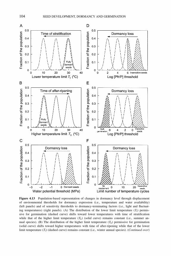
P1: OTE/SPH P2: OTE
BLUK053-Bradford September 27, 2006 7:59
104 SEED DEVELOPMENT, DORMANCY AND GERMINATION
Figure 4.13 Population-based representation of changes in dormancy level through displacement
of environmental thresholds for dormancy expression (i.e., temperature and water availability)
(left panels) and of sensitivity thresholds to dormancy-terminating factors (i.e., light and fluctuat-
ing temperatures) (right panels). (A) The distribution of the lower limit temperature (Tl) permis-
sive for germination (dashed curve) shifts toward lower temperatures with time of stratification
while that of the higher limit temperature (Th) (solid curve) remains constant (i.e., summer an-
nual species). (B) The distribution of the higher limit temperature (Th) permissive for germination
(solid curve) shifts toward higher temperatures with time of after-ripening while that of the lower
limit temperature (Tl) (dashed curve) remains constant (i.e., winter annual species). (Continued over)
P1: OTE/SPH P2: OTE
BLUK053-Bradford September 27, 2006 7:59
MODELING OF SEED DORMANCY 105
4.7 Conclusions and future directions
Mathematical models are powerful tools to evaluate changes in seed dormancy andthe influences of environmental factors that modulate these changes or stimulategermination. The primary focus of this chapter has been to present insights and ap-proaches to describing seed dormancy based on threshold models that account forbehavior of all viable seeds within a population. Most of the assumptions on whichthese models are constructed are, or lead to, hypotheses that aim to give explana-tions for aspects of seed functioning from either a physiological or an ecologicalperspective; model validation constitutes a way of testing those hypotheses. In ad-dition, these models may have very important practical applications. In the case ofweeds, for instance, the possibility of predicting changes in dormancy allows theidentification of periods of maximum probability for seedling emergence accordingto the environmental conditions experienced by the soil seed bank, permitting thedesign of more efficient or effective control schemes.
Although the examples presented demonstrate that considerable progress hasbeen made in modeling seed dormancy, a number of challenges remain. An impor-tant goal must be to integrate the influences of different factors that affect dormancyinto a combined model. As illustrated in Figure 4.13, changes in threshold distri-butions for multiple factors may be occurring simultaneously during dormancy re-lease, and better methods are needed to integrate these into a comprehensive model.While temperature and water potential have been integrated via the hydrothermaltime model, this model has not been fully integrated with the upper and lower tem-perature limits approach. However, changes in both the upper and lower temperaturelimits appear to be associated with shifts in ψb distributions to more negative values(Figure 4.6; Batlla and Benech-Arnold, 2004, 2006). Efforts to extend the thresholdmodeling approach to account for multiple interacting factors have been attempted,but on a relatively limited scale thus far. In most cases, extension of models toincorporate additional factors has been accomplished by using primary parametersderived from threshold models in secondary models that empirically describe howthe primary parameters change over time in response to other factors influencing
←−−−−−−−−−−−−−−−−−−−−−−−−−−−−−−−−−−−−−−−−−−−−−−−−−−−−−−−−−−Figure 4.13 (Continued ) In (A) and (B), overlapping of the distribution of Tl with that of Th indicates
full dormancy. (C) The distribution of the base water potential threshold (ψb) within the population
shifts toward more negative values with dormancy loss. The shaded area indicates the fraction of the
population that will not germinate even if incubated in pure water (because the seeds are fully dormant,
they are incubated at a temperature outside their permissive range, or they require dormancy terminating
factors). (D and E) The distribution of sensitivity thresholds to Pfr shifts toward lower values of Log
[Pfr/P] in both the low fluence response (LFR) (D) and the very low fluence response (VLFR) ranges
(E). The diagonally shaded areas in D and E represent seeds insensitive to light in the LFR and the
VLFR ranges, respectively. The cross-hatched area in (E) represents seeds that are able to germinate in
the dark. (F) The distribution of sensitivity thresholds to number of cycles of fluctuating temperatures
shifts toward lower values with dormancy loss. The diagonally shaded area represents seeds insensitive
to fluctuating temperatures while the cross-hatched area represents seeds that do not require fluctuating
temperatures.
P1: OTE/SPH P2: OTE
BLUK053-Bradford September 27, 2006 7:59
106 SEED DEVELOPMENT, DORMANCY AND GERMINATION
dormancy (e.g., Ni and Bradford, 1993; Alvarado and Bradford, 2002; Batlla et al.,2003, 2004; Batlla and Benech-Arnold, 2004; Finch-Savage, 2004; Bair et al., 2006).While these approaches are practically useful and provide new insights, we shouldcontinue to seek broader conceptual models based on physiological mechanismsrather than on empirical curve fitting, as the former have greater potential for gener-ality. For example, knowledge of the multiple phytochrome genes and their diverseand unique roles in light reception and signaling should inform models attemptingto account for different light response modes. Such specific targets for the physio-logical basis of ψb remain elusive as yet (see Chapter 11). Nonetheless, the overallgoal should continue to be the development of models that describe and predictseed behavior based upon knowledge of the physiological and molecular bases ofthat behavior. Such an approach provides mutually reinforcing hypotheses for bothphysiologists and ecologists.
References
J.M. Adamoli, A.D. Goldberg and A. Soriano (1973) El desbloqueo de las semillas de chamico (Daturaferox L.) enterradas en el suelo: analisis de los factores causales. Revista de InvestigacionesAgropecuarias 10, 209–222.
P.S. Allen and S.E. Meyer (1998) Ecological aspects of seed dormancy loss. Seed Science Research8, 183–191.
P.S. Allen and S.E. Meyer (2002) Ecology and ecological genetics of seed dormancy in downy brome.
Weed Science 50, 241–247.
P.S. Allen, S.E. Meyer and J. Beckstead (1995) Patterns of seed after-ripening in Bromus tectorum L.
Journal of Experimental Botany 279, 1737–1744.
P.S. Allen, S.E. Meyer and M.A. Khan (2000) Hydrothermal time as a tool in comparative germination
studies. In: Seed Biology: Advances and Applications (eds M. Black, K.J. Bradford and J. Vazquez-
Ramos), pp. 401–410. CAB International, Wallingford, UK.
V. Alvarado and K.J. Bradford (2002) A hydrothermal time model explains the cardinal temperatures
for seed germination. Plant Cell and Environment 25, 1061–1069.
V. Alvarado and K.J. Bradford (2005) Hydrothermal time analysis of seed dormancy in true (botanical)
potato seeds. Seed Science Research 15, 77–88.
N.B. Bair, S.E. Meyer and P.S. Allen (2006) A hydrothermal after-ripening time model for seed
dormancy loss in Bromus tectorum L. Seed Science Research 16, 17–28.
C.C. Baskin and J.M. Baskin (1998) Seeds: Ecology, Biogeography, and Evolution of Dormancy andGermination. Academic Press, New York.
J.M. Baskin and C.C. Baskin (1976) High temperature requirement for after-ripening in seeds of winter
annuals. New Phytologist 77, 619–624.
J.M. Baskin and C.C. Baskin (1977) Role of temperature in the germination ecology of three summer
annual weeds. Oecologia 30, 377–382.
J.M. Baskin and C.C. Baskin (1980) Ecophysiology of secondary dormancy in seeds of Ambrosiaartemisiifolia. Ecology 61, 475–480.
J.M. Baskin and C.C. Baskin (1984) Role of temperature in regulating timing of germination in soil
seed reserves of Lamium purpureum (L.). Weed Research 30, 341–349.
J.M. Baskin and C.C. Baskin (1985) The annual dormancy cycle in buried weed seeds: a continuum.
BioScience 35, 392–398.
J.M. Baskin and C.C. Baskin (2004) A classification system for seed dormancy. Seed Science Research14, 1–16.
P1: OTE/SPH P2: OTE
BLUK053-Bradford September 27, 2006 7:59
MODELING OF SEED DORMANCY 107
D. Batlla and R.L. Benech-Arnold (2003) A quantitative analysis of dormancy loss dynamics in
Polygonum aviculare L. seeds: development of a thermal time model based on changes in seed
population thermal parameters. Seed Science Research 13, 55–68.
D. Batlla and R.L. Benech-Arnold (2004) A predictive model for dormancy loss in Polygonum avicu-lare L. seeds based on changes in population hydrotime parameters. Seed Science Research 14,
277–286.
D. Batlla and R.L. Benech-Arnold (2005) Changes in the light sensitivity of buried Polygonum avic-ulare seeds in relation to cold-induced dormancy loss: development of a predictive model. NewPhytologist 165, 445–452.
D. Batlla and R.L. Benech-Arnold (2006) The role of fluctuations in soil water content on the regulation
of dormancy changes in buried seeds of Polygonum aviculare L. Seed Science Research 16,
47–59.
D. Batlla, B.C. Kruk and R.L. Benech-Arnold (2000) Very early detection of canopy presence by
seeds through perception of subtle modifications in red:far red signals. Functional Ecology 14,
195–202.
D. Batlla, B.C. Kruk and R.L. Benech-Arnold (2004) Modeling changes in dormancy in weed soil
seed banks: implications for the prediction of weed emergence. In: Handbook of Seed Physiology:Applications to Agriculture (eds R.L. Benech-Arnold and R.A. Sanchez), pp. 245–270. Haworth
Press, New York.
D. Batlla, V. Verges and R.L. Benech-Arnold (2003) A quantitative analysis of seed responses to
cycle-doses of fluctuating temperatures in relation to dormancy: development of a thermal time
model for Polygonum aviculare L. seeds. Seed Science Research 13, 197–207.
M.C. Bauer, S.E. Meyer and P.S. Allen (1998) A simulation model to predict seed dormancy loss in
the field for Bromus tectorum L. Journal of Experimental Botany 49, 1235–1244.
R.L. Benech-Arnold, C.M. Ghersa, R.A. Sanchez and P. Insausti (1990a) A mathematical model to
predict Sorghum halepense germination in relation to soil temperature. Weed Research 30, 81–89.
R.L. Benech-Arnold, C.M. Ghersa, R.A. Sanchez and P. Insausti (1990b) Temperature effects on
dormancy release and germination rate in Sorghum halepense (L.) Pers. seeds: a quantitative
analysis. Weed Research 30, 91–99.
R.L. Benech-Arnold and R.A. Sanchez (1995) Modeling weed seed germination. In: Seed Developmentand Germination (eds J. Kigel and G. Galili), pp. 545–566. Marcel Dekker, New York.
R.L. Benech-Arnold, R.A. Sanchez, F. Forcella, B.C. Kruk and C.M. Ghersa (2000) Environmental
control of dormancy in weed seed banks in soil. Field Crops Research 67, 105–122.
J.D. Bewley and M. Black (1982) Physiology and Biochemistry of Seeds in Relation to Germination.
Springer-Verlag, Berlin.
J.F. Bierhuizen and W.A. Wagenvoort (1974) Some aspects of seed germination in vegetables. I: The
determination and application of heat sums and minimum temperature for germination. ScientiaHorticulturae 2, 213–219.
H.J. Bouwmeester and C.M. Karssen (1992) The dual role of temperature in the regulation of the
seasonal changes in dormancy and germination of seeds of Polygonum persicaria L. Oecologia90, 88–94.
H.J. Bouwmeester and C.M. Karssen (1993) Annual changes in dormancy and germination in seeds
of Sisymbrium officinale (L.) Scop. New Phytologist 124, 179–191.
K.J. Bradford (1990) A water relations analysis of seed germination rates. Plant Physiology 94, 840–
849.
K.J. Bradford (1995) Water relations in seed germination. In: Seed Development and Germination(eds J. Kigel and G. Galili), pp. 351–396. Marcel Dekker, New York.
K.J. Bradford (1996) Population-based models describing seed dormancy behaviour: implications
for experimental design and interpretation. In: Plant Dormancy: Physiology, Biochemistry andMolecular Biology (ed. A. Lang), pp. 313–339. CAB International, Wallingford, UK.
K.J. Bradford (1997) The hydrotime concept in seed germination and dormancy. In: Basic and AppliedAspects of Seed Biology (eds R.H. Ellis, M. Black, A.J. Murdoch and T.D. Hong), pp. 349–360.
Kluwer Academic Publishers, Boston.
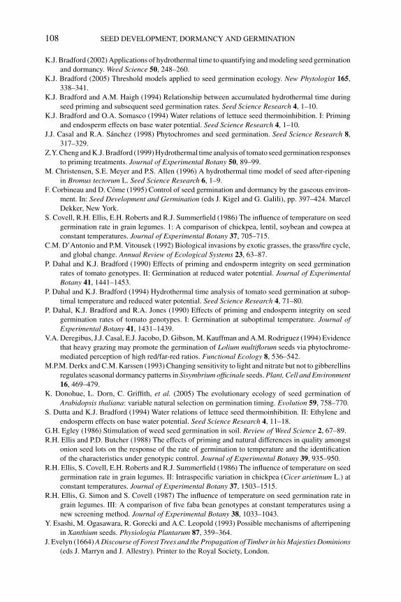
P1: OTE/SPH P2: OTE
BLUK053-Bradford September 27, 2006 7:59
108 SEED DEVELOPMENT, DORMANCY AND GERMINATION
K.J. Bradford (2002) Applications of hydrothermal time to quantifying and modeling seed germination
and dormancy. Weed Science 50, 248–260.
K.J. Bradford (2005) Threshold models applied to seed germination ecology. New Phytologist 165,
338–341.
K.J. Bradford and A.M. Haigh (1994) Relationship between accumulated hydrothermal time during
seed priming and subsequent seed germination rates. Seed Science Research 4, 1–10.
K.J. Bradford and O.A. Somasco (1994) Water relations of lettuce seed thermoinhibition. I: Priming
and endosperm effects on base water potential. Seed Science Research 4, 1–10.
J.J. Casal and R.A. Sanchez (1998) Phytochromes and seed germination. Seed Science Research 8,
317–329.
Z.Y. Cheng and K.J. Bradford (1999) Hydrothermal time analysis of tomato seed germination responses
to priming treatments. Journal of Experimental Botany 50, 89–99.
M. Christensen, S.E. Meyer and P.S. Allen (1996) A hydrothermal time model of seed after-ripening
in Bromus tectorum L. Seed Science Research 6, 1–9.
F. Corbineau and D. Come (1995) Control of seed germination and dormancy by the gaseous environ-
ment. In: Seed Development and Germination (eds J. Kigel and G. Galili), pp. 397–424. Marcel
Dekker, New York.
S. Covell, R.H. Ellis, E.H. Roberts and R.J. Summerfield (1986) The influence of temperature on seed
germination rate in grain legumes. 1: A comparison of chickpea, lentil, soybean and cowpea at
constant temperatures. Journal of Experimental Botany 37, 705–715.
C.M. D’Antonio and P.M. Vitousek (1992) Biological invasions by exotic grasses, the grass/fire cycle,
and global change. Annual Review of Ecological Systems 23, 63–87.
P. Dahal and K.J. Bradford (1990) Effects of priming and endosperm integrity on seed germination
rates of tomato genotypes. II: Germination at reduced water potential. Journal of ExperimentalBotany 41, 1441–1453.
P. Dahal and K.J. Bradford (1994) Hydrothermal time analysis of tomato seed germination at subop-
timal temperature and reduced water potential. Seed Science Research 4, 71–80.
P. Dahal, K.J. Bradford and R.A. Jones (1990) Effects of priming and endosperm integrity on seed
germination rates of tomato genotypes. I: Germination at suboptimal temperature. Journal ofExperimental Botany 41, 1431–1439.
V.A. Deregibus, J.J. Casal, E.J. Jacobo, D. Gibson, M. Kauffman and A.M. Rodriguez (1994) Evidence
that heavy grazing may promote the germination of Lolium multiflorum seeds via phytochrome-
mediated perception of high red/far-red ratios. Functional Ecology 8, 536–542.
M.P.M. Derkx and C.M. Karssen (1993) Changing sensitivity to light and nitrate but not to gibberellins
regulates seasonal dormancy patterns in Sisymbrium officinale seeds. Plant, Cell and Environment16, 469–479.
K. Donohue, L. Dorn, C. Griffith, et al. (2005) The evolutionary ecology of seed germination of
Arabidopsis thaliana: variable natural selection on germination timing. Evolution 59, 758–770.
S. Dutta and K.J. Bradford (1994) Water relations of lettuce seed thermoinhibition. II: Ethylene and
endosperm effects on base water potential. Seed Science Research 4, 11–18.
G.H. Egley (1986) Stimulation of weed seed germination in soil. Review of Weed Science 2, 67–89.
R.H. Ellis and P.D. Butcher (1988) The effects of priming and natural differences in quality amongst
onion seed lots on the response of the rate of germination to temperature and the identification
of the characteristics under genotypic control. Journal of Experimental Botany 39, 935–950.
R.H. Ellis, S. Covell, E.H. Roberts and R.J. Summerfield (1986) The influence of temperature on seed
germination rate in grain legumes. II: Intraspecific variation in chickpea (Cicer arietinum L.) at
constant temperatures. Journal of Experimental Botany 37, 1503–1515.
R.H. Ellis, G. Simon and S. Covell (1987) The influence of temperature on seed germination rate in
grain legumes. III: A comparison of five faba bean genotypes at constant temperatures using a
new screening method. Journal of Experimental Botany 38, 1033–1043.
Y. Esashi, M. Ogasawara, R. Gorecki and A.C. Leopold (1993) Possible mechanisms of afterripening
in Xanthium seeds. Physiologia Plantarum 87, 359–364.
J. Evelyn (1664) A Discourse of Forest Trees and the Propagation of Timber in his Majesties Dominions(eds J. Marryn and J. Allestry). Printer to the Royal Society, London.
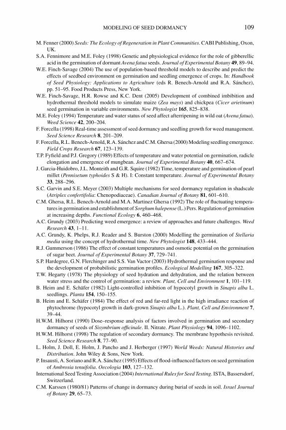
P1: OTE/SPH P2: OTE
BLUK053-Bradford September 27, 2006 7:59
MODELING OF SEED DORMANCY 109
M. Fenner (2000) Seeds: The Ecology of Regeneration in Plant Communities. CABI Publishing, Oxon,
UK.
S.A. Fennimore and M.E. Foley (1998) Genetic and physiological evidence for the role of gibberellic
acid in the germination of dormant Avena fatua seeds. Journal of Experimental Botany 49, 89–94.
W.E. Finch-Savage (2004) The use of population-based threshold models to describe and predict the
effects of seedbed environment on germination and seedling emergence of crops. In: Handbookof Seed Physiology: Applications to Agriculture (eds R. Benech-Arnold and R.A. Sanchez),
pp. 51–95. Food Products Press, New York.
W.E. Finch-Savage, H.R. Rowse and K.C. Dent (2005) Development of combined imbibition and
hydrothermal threshold models to simulate maize (Zea mays) and chickpea (Cicer arietinum)
seed germination in variable environments. New Phytologist 165, 825–838.
M.E. Foley (1994) Temperature and water status of seed affect afterripening in wild oat (Avena fatua).
Weed Science 42, 200–204.
F. Forcella (1998) Real-time assessment of seed dormancy and seedling growth for weed management.
Seed Science Research 8, 201–209.
F. Forcella, R.L. Benech-Arnold, R.A. Sanchez and C.M. Ghersa (2000) Modeling seedling emergence.
Field Crops Research 67, 123–139.
T.P. Fyfield and P.J. Gregory (1989) Effects of temperature and water potential on germination, radicle
elongation and emergence of mungbean. Journal of Experimental Botany 40, 667–674.
J. Garcia-Huidobro, J.L. Monteith and G.R. Squire (1982) Time, temperature and germination of pearl
millet (Pennisetum typhoides S & H). I: Constant temperature. Journal of Experimental Botany33, 288–296.
S.C. Garvin and S.E. Meyer (2003) Multiple mechanisms for seed dormancy regulation in shadscale
(Atriplex confertifolia: Chenopodiaceae). Canadian Journal of Botany 81, 601–610.
C.M. Ghersa, R.L. Benech-Arnold and M.A. Martinez Ghersa (1992) The role of fluctuating tempera-
tures in germination and establishment of Sorghum halepense (L.) Pers. Regulation of germination
at increasing depths. Functional Ecology 6, 460–468.
A.C. Grundy (2003) Predicting weed emergence: a review of approaches and future challenges. WeedResearch 43, 1–11.
A.C. Grundy, K. Phelps, R.J. Reader and S. Burston (2000) Modelling the germination of Stellariamedia using the concept of hydrothermal time. New Phytologist 148, 433–444.
R.J. Gummerson (1986) The effect of constant temperatures and osmotic potential on the germination
of sugar beet. Journal of Experimental Botany 37, 729–741.
S.P. Hardegree, G.N. Flerchinger and S.S. Van Vactor (2003) Hydrothermal germination response and
the development of probabilistic germination profiles. Ecological Modelling 167, 305–322.
T.W. Hegarty (1978) The physiology of seed hydration and dehydration, and the relation between
water stress and the control of germination: a review. Plant, Cell and Environment 1, 101–119.
B. Heim and E. Schafer (1982) Light-controlled inhibition of hypocotyl growth in Sinapis alba L.
seedlings. Planta 154, 150–155.
B. Heim and E. Schafer (1984) The effect of red and far-red light in the high irradiance reaction of
phytochrome (hypocotyl growth in dark-grown Sinapis alba L.). Plant, Cell and Environment 7,
39–44.
H.W.M. Hilhorst (1990) Dose–response analysis of factors involved in germination and secondary
dormancy of seeds of Sisymbrium officinale. II. Nitrate. Plant Physiology 94, 1096–1102.
H.W.M. Hilhorst (1998) The regulation of secondary dormancy. The membrane hypothesis revisited.
Seed Science Research 8, 77–90.
L. Holm, J. Doll, E. Holm, J. Pancho and J. Herberger (1997) World Weeds: Natural Histories andDistribution. John Wiley & Sons, New York.
P. Insausti, A. Soriano and R.A. Sanchez (1995) Effects of flood-influenced factors on seed germination
of Ambrosia tenuifolia. Oecologia 103, 127–132.
International Seed Testing Association (2004) International Rules for Seed Testing. ISTA, Bassersdorf,
Switzerland.
C.M. Karssen (1980/81) Patterns of change in dormancy during burial of seeds in soil. Israel Journalof Botany 29, 65–73.
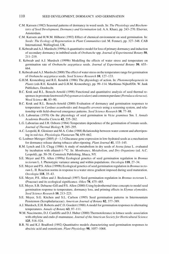
P1: OTE/SPH P2: OTE
BLUK053-Bradford September 27, 2006 7:59
110 SEED DEVELOPMENT, DORMANCY AND GERMINATION
C.M. Karssen (1982) Seasonal patterns of dormancy in weed seeds. In: The Physiology and Biochem-istry of Seed Development, Dormancy and Germination (ed. A.A. Khan), pp. 243–270. Elsevier,
Amsterdam.
C.M. Karssen and H.W.M. Hilhorst (1992) Effect of chemical environment on seed germination. In:
Seeds: The Ecology of Regeneration in Plant Communities (ed. M. Fenner), pp. 327–348. CAB
International, Wallingford, UK.
E. Kebreab and A.J. Murdoch (1999a) A quantitative model for loss of primary dormancy and induction
of secondary dormancy in imbibed seeds of Orobanche spp. Journal of Experimental Botany 50,
211–219.
E. Kebreab and A.J. Murdoch (1999b) Modelling the effects of water stress and temperature on
germination rate of Orobanche aegyptiaca seeds. Journal of Experimental Botany 50, 655–
664.
E. Kebreab and A.J. Murdoch (2000) The effect of water stress on the temperature range for germination
of Orobanche aegyptiaca seeds. Seed Science Research 10, 127–133.
G.H.M. Kronenberg and R.E. Kendrik (1986) The physiology of action. In: Photomorphogenesis inPlants (eds R.E. Kendrik and G.H.M. Kronenberg), pp. 99–114. Marthinus Nijhoff/Dr. W. Junk
Publishers, Dordrecht.
B.C. Kruk and R.L. Benech-Arnold (1998) Functional and quantitative analysis of seed thermal re-
sponses in prostrate knotweed (Polygonum aviculare) and common purslane (Portulaca oleracea).
Weed Science 46, 83–90.
B.C. Kruk and R.L. Benech-Arnold (2000) Evaluation of dormancy and germination responses to
temperature in Carduus acanthoides and Anagallis arvensis using a screening system, and rela-
tionship with field-observed emergence patterns. Seed Science Research 10, 77–88.
L.G. Labouriau (1970) On the physiology of seed germination in Vicia graminea Sm. I. AnnalsAcademia Brasilia Ciencia 42, 235–262.
L.G. Labouriau and J.H. Osborn (1984) Temperature dependence of the germination of tomato seeds.
Journal of Thermal Biology 9, 285–295.
A.C. Leopold, R. Glenister and M.A. Cohn (1988) Relationship between water content and afterripen-
ing in red rice. Physiologia Plantarum 74, 659–662.
G. Leubner-Metzger (2005) β−1,3-Glucanase gene expression in low-hydrated seeds as a mechanism
for dormancy release during tobacco after-ripening. Plant Journal 41, 133–145.
R.M. Lynch and J.S. Clegg (1986) A study of metabolism in dry seeds of Avena fatua L. evaluated
by incubation with ethanol-1-14C. In: Membranes, Metabolism, and Dry Organisms (ed. A.C.
Leopold), pp. 50–58. Comstock Publishing, Ithaca, NY.
S.E. Meyer and P.S. Allen (1999a) Ecological genetics of seed germination regulation in Bromustectorum L. I. Phenotypic variance among and within populations. Oecologia 118, 27–34.
S.E. Meyer and P.S. Allen (1999b) Ecological genetics of seed germination regulation in Bromus tecto-rum L. II. Reaction norms in response to a water stress gradient imposed during seed maturation.
Oecologia 118, 35–43.
S.E. Meyer, P.S. Allen and J. Beckstead (1997) Seed germination regulation in Bromus tectorum L.
(Poaceae) and its ecological significance. Oikos 78, 475–485.
S.E. Meyer, S.B. Debaene-Gill and P.S. Allen (2000) Using hydrothermal time concepts to model seed
germination response to temperature, dormancy loss, and priming effects in Elymus elymoides.
Seed Science Research 10, 213–223.
S.E. Meyer, S.G. Kitchen and S.L. Carlson (1995) Seed germination patterns in Intermountain
Penstemon (Scrophulariaceae). American Journal of Botany 82, 377–389.
A.J. Murdoch, E.H. Roberts and C.O. Goedert (1988) A model for germination responses to alternating
temperatures. Annals of Botany 63, 97–111.
W.M. Nascimento, D.J. Cantliffe and D.J. Huber (2000) Thermotolerance in lettuce seeds: association
with ethylene and endo-β-mannanase. Journal of the American Society for Horticultural Science125, 518–524.
B.R. Ni and K.J. Bradford (1992) Quantitative models characterizing seed germination responses to
abscisic acid and osmoticum. Plant Physiology 98, 1057–1068.
P1: OTE/SPH P2: OTE
BLUK053-Bradford September 27, 2006 7:59
MODELING OF SEED DORMANCY 111
B.R. Ni and K.J. Bradford (1993) Germination and dormancy of abscisic acid-deficient and gibberellin-
deficient mutant tomato seeds. Sensitivity of germination to abscisic acid, gibberellin, and water
potential. Plant Physiology 101, 607–617.
A. Orozco-Segovia, L. Gonzalez-Zertuche, A. Mendoza and S. Orozco (1996) A mathematical
model that uses Gaussian distribution to analyze the germination of Manfreda brachystachya(Agavaceae) in a thermogradient. Physiologia Plantarum 98, 431–438.
N. Pallais (1995) High temperature and low moisture reduce the storage requirement of freshly har-
vested true potato seeds. Journal of the American Society for Horticultural Science 120, 699–
702.
K. Phelps and W.E. Finch-Savage (1997) A statistical perspective on threshold-type germination
models. In: Basic and Applied Aspects of Seed Biology (eds R.H. Ellis, M. Black, A.J. Murdoch
and T.D. Hong), pp. 361–368. Kluwer Academic Publishers, Boston.
T.L. Pons (1992) Seed response to light. In: Seeds: The Ecology of Regeneration in Plant Communities(ed. M. Fenner), pp. 259–284. CAB International, Melksham.
R.J. Probert (1992) The role of temperature in germination ecophysiology. In: Seeds: The Ecology ofRegeneration in Plant Communities (ed. M. Fenner), pp. 285–325. CAB International, Melksham.
A.P. Ramakrishnan, S.E. Meyer, J. Waters, M.R. Stevens, C.E. Coleman and D.J. Fairbanks
(2004) Correlation between molecular markers and adaptively significant genetic variation in
Bromus tectorum (Poaceae), an inbreeding annual grass. American Journal of Botany 91, 797–
803.
O. Reisman-Berman, J. Kigel and B. Rubin (1991) Dormancy pattern in buried seeds of Datura feroxL. Canadian Journal of Botany 69, 173–179.
E.H. Roberts and R.D. Smith (1977) Dormancy and the pentose phosphate pathway. In: The Phys-iology and Biochemistry of Seed Dormancy and Germination (ed. A.A. Khan), pp. 385–411.
Elsevier/North-Holland Biomedical Press, Amsterdam.
E.H. Roberts and S. Totterdell (1981) Seed dormancy in Rumex species in response to environmental
factors. Plant, Cell and Environment 4, 97–106.
H.R. Rowse and W.E. Finch-Savage (2003) Hydrothermal threshold models can describe the germi-
nation response of carrot (Daucus carota L.) and onion (Allium cepa L.) seed populations across
both sub- and supra-optimal temperatures. New Phytologist 158, 101–108.
H.R. Rowse, J.M.T. McKee and E.C. Higgs (1999) A model of the effects of water stress on seed
advancement and germination. New Phytologist 143, 273–279.
A.L. Scopel, C.L. Ballare and R.A. Sanchez (1991) Induction of extreme light sensitivity in buried
weed seeds and its role in the perception of soil cultivations. Plant, Cell and Environment 14,
501–508.
A. Shrestha, E.S. Roman, A.G. Thomas and C.J. Swanton (1999) Modelling germination and shoot-
radicle elongation of Ambrosia artemisiifolia. Weed Science 47, 557–562.
H. Smith (1982) Light quality, photoperception, and plant strategy. Annual Review of Plant Physiology33, 481–518.
K.J. Steadman and H.W. Pritchard (2004) Germination of Aesculus hippocastanum seeds following
cold-induced dormancy loss can be described in relation to a temperature-dependent reduction
in base temperature (Tb) and thermal time. New Phytologist 161, 415–425.
S.J. Steinmaus, T.S. Prather and J.S. Holt (2000) Estimation of base temperatures for nine weed
species. Journal of Experimental Botany 51, 275–286.
P. Stokes (1965) Temperature and seed dormancy. In: Encyclopedia of Plant Physiology (ed.
W. Ruhland), pp. 746–803. Springer, Berlin.
A.M. Tarquis and K.J. Bradford (1992) Prehydration and priming treatments that advance germination
also increase the rate of deterioration of lettuce seeds. Journal of Experimental Botany 43, 307–
317.
S. Totterdell and E.H. Roberts (1979) Effects of low temperatures on the loss of innate dormancy and
the development of induced dormancy in seeds of Rumex obtusifolius L. and Rumex crispus L.
Plant, Cell and Environment 2, 131–137.
A. Vegis (1964) Dormancy in higher plants. Annual Review of Plant Physiology 115, 185–224.

P1: OTE/SPH P2: OTE
BLUK053-Bradford September 27, 2006 7:59
112 SEED DEVELOPMENT, DORMANCY AND GERMINATION
C.W. Vertucci and J.M. Farrant (1995) Acquisition and loss of desiccation tolerance. In: Seed De-velopment and Germination (eds J. Kigel and G. Galili), pp. 237–271. Marcel Dekker, New
York.
C.W. Vertucci and A.C. Leopold (1986) Physiological activities associated with hydration level in
seeds. In: Membranes, Metabolism and Dry Organisms (ed. A.C. Leopold), pp. 35–49. Comstock
Publishing Associates, Ithaca, NY.
L.M. Vleeshouwers and H.J. Bouwmeester (2001) A simulation model for seasonal changes in dor-
mancy and germination of seeds. Seed Science Research 11, 77–92.
L.M. Vleeshouwers and M.J. Kropff (2000) Modeling field emergence patterns in arable weeds. NewPhytologist 148, 445–457.
C. Walters, N.W. Pammenter, P. Berjak and J. Crane (2001) Desiccation damage, accelerated ageing
and respiration in desiccation tolerant and sensitive seeds. Seed Science Research 11, 135–148.
I. Washitani (1987) A convenient screening test system and a model for thermal germination responses
of wild plant seeds: behaviour of model and real seed in the system. Plant, Cell and Environment10, 587–598.
P1: OTE/SPH P2: OTE
BLUK053-Bradford September 27, 2006 7:31
5 Genetic aspects of seed dormancyLeonie Bentsink, Wim Soppe and Maarten Koornneef
5.1 Introduction
Every new plant generation starts with a seed, which usually contains a fully devel-oped embryo that can survive the period between seed maturation and germination.Dormancy, defined as the failure of an intact viable seed to complete germinationunder conditions that are favorable for germination, is an adaptive trait optimizinggermination to the best suitable time that enables the seed to complete its life cycle.In temperate climates, delaying germination until spring can prevent winter mortal-ity. However, autumn germination may have a selective advantage when the risk ofwinter mortality is low by enabling the plants to flower earlier or at a larger size thefollowing year (Donohue, 2002).
Changes in seed dormancy were selected during the domestication of crop plantsfrom wild-plant species, because some features of dormancy that provide ecologi-cal advantages presented agronomic disadvantages within a farmed system. At oneextreme, many weed species show very high levels of dormancy when shed fromthe plant. This causes infestation of land that subsequently requires long-term treat-ment to remove succeeding generations (Basu et al., 2004). Crop plants might haveonly low levels of dormancy that provide a benefit for early and uniform seedlingestablishment. However, this has the disadvantage of possible preharvest sprout-ing (PHS), germination before harvest that reduces the quality of the seed. Thisis a problem especially for cereals when cool and damp conditions occur priorto harvest (Holdsworth et al., 2006). For specific uses, such as barley (Hordeumvulgare) for malting, low dormancy is desired although this increases the risk ofPHS. Therefore, a moderate dormancy just sufficient to prevent PHS is usually de-sired for this trait in crop plants (Ullrich et al., 1997; Li et al., 2003; Gubler et al.,2005).
Seed dormancy varies widely among seed batches, even for seeds of the samegenotype, indicating large environmental influences. This, together with other fac-tors such as polygenic inheritance, makes dormancy a typical quantitative trait,which is often described as deep or shallow, or strong or weak, dormancy. Twodormancy types can be distinguished by the time of their induction. Primary dor-mancy is induced during seed development and secondary dormancy during imbi-bition of mature seeds. Furthermore, there is a distinction between embryo and coat(testa, hull, and/or endosperm)-imposed dormancy. The germination characteristicsof seeds also depend on their physiological quality or vigor (germination potential)in addition to their dormancy status.
113
P1: OTE/SPH P2: OTE
BLUK053-Bradford September 27, 2006 7:31
114 SEED DEVELOPMENT, DORMANCY AND GERMINATION
For a better understanding of seed dormancy and germination it is important(but difficult) to distinguish between dormancy, which is assumed to be inducedduring seed maturation on the mother plant, and germination. These are two dif-ferent physiological states or processes that depend on each other. Germination canoccur only after dormancy is released, and the release of dormancy is visible onlywhen germination takes place. In genetic terminology, dormancy is epistatic to ger-mination. Until we have clear markers that distinguish dormancy (either embryo-or coat-imposed) and germination potential per se, researchers must mainly rely ongermination tests for their analyses of dormancy.
Germination sensu stricto is defined as the events following imbibition until vis-ible embryo protrusion. The completion of germination depends on embryo expan-sion mainly due to cell elongation driven by water uptake. After radicle protrusion,seedling establishment takes place, which can be considered as a separate processfrom germination.
Germination assays are a measure of the integration of many events that hap-pened in the history of the seed on the mother plant and the various environmentalfactors encountered during seed storage and germination. Dormancy is a propertyof the seed and the degree of dormancy defines which conditions should be met toallow the seeds to germinate. Therefore, a more precise method of defining the dor-mancy status of a seed batch requires a description of the environmental necessitiesfor germination (temperature range or time of after-ripening required to overcomedormancy). For genetic studies, it is essential to obtain such quantitative data onseed material without the effects of variation in environmental conditions, includingthe maternal growing and seed storage conditions. In general, dormancy is assayedby a seed germination test under well-defined conditions. These conditions referto the incubation medium, light, and temperature conditions during the assay. It isessential to find those conditions that discriminate maximally between the parents.This implies, for example, that when comparing nondormant with weakly dormantgenotypes one often uses freshly harvested seeds and germination conditions thatmay not be optimal for germination, such as low or high temperatures. The compar-ison of weakly with strongly dormant genotypes will need the use of stored seedsand more promotive temperature conditions.
This chapter begins with a description of the genetic approach to understandseed dormancy using mutants in Arabidopsis and other plant species. Thereafterit continues with an overview of studies that analyze natural variation for seeddormancy in both Arabidopsis and other species with well-developed genetics andlinkage maps (mainly grasses). Finally, an overview is given of the insights intoseed dormancy and germination that resulted from these genetic approaches.
5.2 Mutant approaches in Arabidopsis
Mutants with defects in seed dormancy and germination provide important toolsfor understanding these processes because they directly identify the genes in-volved, nowadays often down to the molecular level. Most genetic variants that
P1: OTE/SPH P2: OTE
BLUK053-Bradford September 27, 2006 7:31
GENETIC ASPECTS OF SEED DORMANCY 115
have been studied were those with dormancy or germination effects that were partof a pleiotropic phenotype, whereby other traits (e.g., dwarfness in gibberellin [GA]-deficient mutants or reduced stomatal closure for abscisic acid [ABA]-deficient mu-tants) were originally used for genetic analysis. This pleiotropism can be expectedas these hormones have additional functions besides their roles in seed dormancyand germination. However, several mutants have been isolated that seem to be spe-cific for seed germination, for instance the dog1 (delay of germination 1) mutant(Bentsink, 2002).
Nondormant mutants have been selected directly by identifying germinatingseeds within nongerminating dormant batches of wild-type seeds (Leon Kloosterzielet al., 1996b; Peeters et al., 2002; L. Bentsink, unpublished results). Another ap-proach would be to select for mutants with increased dormancy levels. Screens foraltered dormancy phenotypes are hampered by the variability of the germinationtrait, which may lead to genetic misclassification of individual seeds and there-fore to false positives in mutant screens. The Arabidopsis genotypes Landsbergerecta (Ler) and Columbia (Col), which are mostly used in Arabidopsis research,show only a low level of dormancy. This dormancy disappears after approximately1 month of after-ripening (van der Schaar et al., 1997). Despite this low levelof dormancy, a mutant screen in Ler for reduced dormancy yielded several mu-tants (reduced dormancy 1-4; rdo1-4) that also showed mild pleiotropic phenotypes(Leon-Kloosterziel et al., 1996b; Peeters et al., 2002). Because of the relativelylow level of dormancy in Ler and Col, it is impossible to saturate the mutations indormancy genes in these accessions. However, this problem can be overcome by theuse of more dormant accessions such as Cape Verde Islands (Cvi) (Koornneef et al.,2000).
The difference between mutants with enhanced germination and wild type canalso be increased by the selection of mutants under conditions that are less favorablefor germination or that extend the germination period. This procedure was appliedby Salaita et al. (2005), who selected for faster germination at 10◦C. Whether themutants identified specifically affect some component of the germination processor dormancy/germination regulation in general is not clear as yet.
Selection of mutants with reduced dormancy or increased germination in thepresence of germination-inhibiting compounds such as ABA is very effective.A number of ABA-insensitive (abi) mutants were isolated using this procedure(Koornneef et al., 1989; Finkelstein, 1994). On the other hand, the selection ofnongerminating seeds in the presence of low, normally not inhibitory, concentra-tions of ABA resulted in so-called enhanced response to ABA (era) mutants (Cutleret al., 1996) or ABA-hypersensitive germination (ahg) mutants (Nishimura et al.,2004). The use of GA biosynthesis inhibitors such as paclobutrazol identified mu-tants in ABA biosynthesis (e.g., aba2 and aba3; Leon-Kloosterziel et al., 1996a)and ABA signal transduction (e.g., abi3; Nambara et al., 1992) in addition to GAsignal transduction mutants such as spindly (spy) (Jacobsen et al., 1996). The sameABA biosynthesis mutants were found during the selection of revertants of nonger-minating GA biosynthesis mutants (Koornneef et al., 1982). Sugars, such as su-crose and various hexoses, inhibit seed germination independently of their osmotic
P1: OTE/SPH P2: OTE
BLUK053-Bradford September 27, 2006 7:31
116 SEED DEVELOPMENT, DORMANCY AND GERMINATION
effects (Pego et al., 2000). Mutants that were insensitive to the inhibitory effectof glucose and sucrose were isolated by several groups and some appeared to bedefective in ABA biosynthesis or appeared to belong to the ABA-insensitive mu-tants. The sugar-insensitive sis4 and sis5 mutants are allelic to respectively aba2 andabi4 (Laby et al., 2000) and sucrose uncoupled 6 (sun6) is an abi4 allele (Huijseret al., 2000). These results indicate that germination is controlled in an ABA- andsugar-dependent way (see Chapter 12). Garciarrubio et al. (1997) showed that theaddition of sugars and amino acids allowed the seeds to germinate in otherwise in-hibitory concentrations of ABA and suggested that ABA inhibits the mobilization offood reserves. However, it cannot be excluded that these sugar effects are mediatedby sugar-signaling effects (Smeekens, 1998; Gibson and Graham, 1999). Penfieldet al. (2004) showed that this inhibition of reserve mobilization by ABA occursonly in the embryo but not in the endosperm, which fuels hypocotyl elongation indarkness.
Mutant screens can also be performed by making use of reporter genes driven bypromoters that are known to be involved in a pathway affecting germination. Mu-tants that affect the reporter genes can thereafter be checked for their germinationphenotype. This approach has been used in genetic screens based on seed maturationand ABA-regulated reporter genes. Carles et al. (2002) used the seed-specific Em(EARLY METHIONINE-LABELLED) promoter and identified an additional alleleof ABI5. Screens with ABA-induced promoters in vegetative tissues revealed twoadditional ABA-related mutants. The ade1 (ABA-deregulated gene expression) mu-tation enhances gene expression in response to ABA (Foster and Chua, 1999) andhos5 (high expression of osmotically responsive gene 5) displays an increased sensi-tivity of gene expression to ABA and osmotic stress (Xiong et al., 1999). The ade1and hos5 mutations, though, have little effect on seed germination. In addition, suchscreens have also identified ABA biosynthesis mutants such as los6 (low expressionof osmotically responsive gene 6/aba1) and los5 (aba3) (Xiong et al., 2001, 2002),which have reduced dormancy.
A different and more unbiased strategy, aiming specifically at seed germinationmutants, makes use of enhancer or promoter trap transgenic lines where the ran-domly inserted reporter gene can be driven by a neighboring enhancer or promotersequence that can have a germination-specific expression pattern. Using this pro-cedure, Dubreucq et al. (2000) identified an extensin-like gene that is expressedin the endosperm during germination at the site of radicle protrusion. A similarapproach identified the mediator of ABA-regulated dormancy (MARD1) gene asbeing involved in dormancy/germination (He and Gan, 2004). A T-DNA insertionin the promoter of this gene resulted in faster germination, germination in dark-ness, and a slight increase in ABA resistance. Liu et al. (2005) recently described alarge set of such reporter inserts and emphasized that this approach is feasible forthe GUS reporter gene only after rupture of the testa to allow penetration of theGUS substrate (see Chapter 11). Knockout mutants of genes with germination- ordormancy-related expression obtained from enhancer or gene trap screens do notalways show phenotypes related to dormancy or germination. This can imply thatthe disrupted genes are not required for germination or that other redundant genescan provide a similar function.
P1: OTE/SPH P2: OTE
BLUK053-Bradford September 27, 2006 7:31
GENETIC ASPECTS OF SEED DORMANCY 117
When redundancy is a cause for the absence of mutant phenotypes in a dysfunc-tional gene, one needs to study double mutants. This approach is well illustrated inthe identification of the GA signal transduction proteins of the DELLA family thatcontrol germination (Tyler et al., 2004; Cao et al., 2005). The study of single, double,and triple mutants combined with the GA-deficient ga1-3 mutant and germinationexperiments in light and darkness allowed the conclusion that RGL2 (RGA [RE-PRESSOR OF gal-3]-LIKE2) is a predominant repressor of seed germination, whichis degraded by the ubiquitin-26S proteasome pathway activated by GA (see Chap-ter 10). However, additional DELLA proteins (GAI [GA-INSENSITIVE], RGA,RGL1) enhance the function of RGL2 and their stability may be affected by light,implying that GA synthesis is induced not only by light but also by GA sensitivity(Cao et al., 2005).
For Arabidopsis, knockouts for nearly every gene can be ordered from the stockcenters and an increasing number of studies are published in which knockouts weretested for a germination phenotype. T-DNA knockouts of two DOF transcriptionfactors (DOF AFFECTING GERMINATION1 [DAG1] and DAG2) revealed a seedgermination phenotype that is determined by the maternal genotype. This is in agree-ment with expression of the gene in the vascular tissue of the funiculus, which is amaternal tissue connected to developing seeds (Papi et al., 2000). DAG1 and DAG2are the first reported genes to be specifically involved in the maternal control ofseed germination, other than by affecting the maternal seed structure. Surprisingly,these two related, very similar genes have opposite effects on germination; DAG1increases dormancy and DAG2 reduces it (Papi et al., 2000; Gualberti et al., 2002).Another example of a knockout mutant with a dormancy phenotype is gpa1 (G pro-tein in Arabidopsis), which encodes a heterotrimeric G protein (Ullah et al., 2002).This is also an example of germination phenotypes that are expressed only undervery specific conditions. Probably heterotrimeric G proteins affect GA signal trans-duction, as overexpression of GCR1, a putative G protein-coupled receptor gene,which is suggested to be involved in the GA signaling pathway, also shows reduceddormancy (Colucci et al., 2002). A similar small effect was observed due to mutationin an arabinoglactan protein where faster germination than wild type was observedin seeds immediately after harvest or stratified in an ABA-containing medium (vanHengel and Roberts, 2003). While these analyses suggest a role in germination,in view of the mild phenotype these are not essential players, although functionalredundancy can be an alternative explanation for small-effect phenotypes.
Since a comparison of a limited number of genotypes in several environmentsallows the discovery of relatively small differences in seed germination behavior, theoutcome of these studies implies that the genes are involved in germination. How-ever, their effects on germination phenotype can be limited; a better quantificationcould be achieved by comparison to mutants exhibiting stronger seed germinationphenotypes such as ABA and GA mutants in the same experiment.
An important additional source of mutants to understand dormancy comes frommutants that were originally isolated in screens that did not make use of ger-mination tests. An example of testing known mutants for their effects on ger-mination is the use of phytochrome mutants. The germination of Arabidopsisseeds is under phytochrome-mediated photocontrol. It was therefore expected that
P1: OTE/SPH P2: OTE
BLUK053-Bradford September 27, 2006 7:31
118 SEED DEVELOPMENT, DORMANCY AND GERMINATION
phytochrome-deficient mutants would be affected in seed germination. Detailedaction spectra for seed germination performed in wild type and phyA and phyB mu-tants revealed a typical red/far-red-reversible response. The low fluence responsewas mediated by PHYB, whereas the germination response mediated by PHYAturned out to be a very low fluence response with a 104-fold higher sensitivity tolight (Shinomura et al., 1996). The observation that phyA phyB double mutantsshow some light-dependent germination indicates the involvement of additionalphotoreceptors (Yang et al., 1995; Poppe and Schafer, 1997).
Testa mutants in Arabidopsis were originally identified based on their alteredseed color or shape. The analysis of these mutants provided strong evidence for theimportance of the testa in the control of seed germination (Debeaujon et al., 2000;reviewed in Chapter 2).
Another important factor that is known to promote germination is nitrate. Withthe help of nitrate reductase-deficient mutants, it was shown that nitrate acts viasignaling and not via nutritional effects. This was concluded because these mutantplants, which show N-deficiency symptoms but accumulate nitrate, produced seedsthat were less dormant than wild-type seeds (Alboresi et al., 2005).
Brassinosteroids (BRs) are a group of over 40 naturally occurring plant steroidhormones found in a wide variety of plant species (Clouse and Sasse, 1998;Schumacher and Chory, 2000). BRs are also involved in the control of germinationin Arabidopsis. It is suggested that the BR signal is required to reverse ABA-induceddormancy and stimulate germination (Steber and McCourt, 2001). BRs overcomethe lack of germination in the sleepy1 (sly1) mutant, probably by bypassing its GArequirement (see Chapter 10). Two BR mutants (det2 [deetiolated2] and bri1 [BR-insensitive1]) showed reduced germination rates (speed) but eventually germinatedwithout BR, indicating that in contrast to GAs, BRs are not absolutely required forgermination (Steber and McCourt, 2001).
Ethylene mimics the action of GAs, as the complete germination of seeds fromthe GA-deficient mutant (which germinate only in the presence of GA) occurs in thepresence of ethylene (Karssen et al., 1989). Ethylene-insensitive mutants such as etr1(ethylene response1) and ein2 (ethylene insensitive2) are more dormant (Chiwochaet al., 2005) or, when nondormant, require more time for germination than wild-type seeds (Siriwitayawan et al., 2003). Ethylene mutants show phenotypes thatresemble ABA- and sugar-signaling mutants and were identified in mutant screensthat aimed at the discovery of ABA signal transduction mutants (Beaudoin et al.,2000; Ghassemian et al., 2000).
5.3 Mutant approaches in other species
Apart from Arabidopsis, mutants affecting dormancy and germination have alsobeen isolated in other species. In maize, viviparous (vp) mutants have been knownfor many years (reviewed in McCarty, 1995). The best studied is the ABA-insensitivevp1 mutant, which encodes a B3 domain seed-specific transcription factor, ortholo-gous to the Arabidopsis ABI3. Several other vp mutants were found to have mutationsin genes encoding ABA biosynthesis enzymes. Among these, the vp14 mutant was
P1: OTE/SPH P2: OTE
BLUK053-Bradford September 27, 2006 7:31
GENETIC ASPECTS OF SEED DORMANCY 119
instrumental to isolate the NCED (9-CIS-EPOXYCAROTENOID DIOXYGENASE)gene, which is responsible for the cleavage of cis-xanthophylls (Tan et al., 1997;Nambara and Marion-Poll, 2005; see Chapter 9).
In rice (Oryza sativa), studies on dormancy mutants are limited (Kurata et al.,2005). In a screen for vivipary in a population with tissue culture-activated Tos17transposons, a mutation was found in the gene encoding the ABA biosynthesis en-zyme zeaxanthin epoxidase, similar to the Arabidopsis aba1 mutant (Agrawal et al.,2001). In this screen, 1400 lines with a viviparous phenotype were reported out of30 000 regenerated plants, suggesting that many more loci might be detected in amore systematic analysis of this mutant group. Although these studies suggest thatmany nondormant mutants can be found in rice, none of them have been character-ized in detail. It is not known whether there are any nondormant mutants that affectgenes not directly related to plant hormones. In barley, a nondormant mutant withno obvious pleiotropic effects was isolated from the cultivar Triumph and the locuswas mapped in the centromere region of chromosome 6 (Romagosa et al., 2001,Prada et al., 2005).
In tomato (Lycopersicon esculentum), mutants affecting seed germination havebeen isolated and characterized extensively. These are various ABA and GA biosyn-thesis mutants whose phenotypes all correspond to the phenotypes of similar mutantsin Arabidopsis. These mutants have been used extensively to investigate seed ger-mination in tomato where weakening of the micropylar region of the endosperm isa requirement for germination (reviewed by Hilhorst et al., 1998; see Chapter 11).
5.4 Genetic analyses of natural variation
The genetics of dormancy has been studied in many plant species and demonstratedthe existence of heritable variation. However, only when specific loci are identifiedby mapping of mutants and/or quantitative trait loci (QTL) can a follow-up analysisusing molecular biology identify the molecular basis of the genetic variation. Thismeans that only species with well-developed genetic and linkage maps are acces-sible to this analysis, including Arabidopsis and several species that belong to theGramineae.
Genetic studies on dormancy have benefited much from the development ofquantitative genetics. The analysis of QTL allows the identification of the num-ber of loci differing between two genotypes, their map positions, and the relativecontribution of each QTL to the total variance for the trait. Germination is a vari-able trait that requires analysis of a high number of seeds. Therefore, the use ofso-called permanent or immortal mapping populations is attractive, where a single(often homozygous) genotype can produce many seeds that can be tested in multipleenvironments (Alonso Blanco and Koornneef, 2000). The molecular identification(cloning) of the underlying genes that show allelic variation is still a major challenge,but the first results are now becoming available. The genes identified in the studyof natural variation can be the same as those identified in mutant screens. However,there are several reasons why this is not always the case. Firstly, the parent lines usedfor mutation experiments can be mutated in specific genes, e.g., those promoting
P1: OTE/SPH P2: OTE
BLUK053-Bradford September 27, 2006 7:31
120 SEED DEVELOPMENT, DORMANCY AND GERMINATION
seed dormancy. This could be the case for Ler and Col in Arabidopsis, where manynatural accessions show much stronger seed dormancy than these commonly usedlaboratory accessions. Furthermore, mutants that show strong pleiotropic effects,such as most ABA mutants, will not survive in nature. Therefore, genes identifiedby analyzing natural variation are expected to be ecologically relevant.
5.4.1 Genetic analysis of natural variation in Arabidopsis
For Arabidopsis, QTL analysis for seed dormancy has been conducted usingseveral recombinant inbred line (RIL) populations (van der Schaar et al., 1997;Alonso-Blanco et al., 2003; Clerkx et al., 2004; L. Bentsink et al., unpublishedresults). These RIL populations have been made by crossing accessions that havedifferent levels of seed dormancy compared to the standard laboratory accessionLer. Thus far these analyses have identified more than 12 regions in the Arabidopsisgenome where QTL associated with dormancy are located (Alonso-Blanco et al.,2003, Clerkx et al., 2004; L. Bentsink et al., unpublished results). These QTL havebeen called DOG (Delay of Germination) and can be divided into different groups.Some QTL are detected only in the low-dormancy accessions, whereas others areidentified as major QTL in the moderate and strongly dormant accessions. Manyof these do not colocate with known dormancy loci (Figure 5.1). In the analysis ofvan der Schaar et al. (1997), the lines were grown in three independent greenhouseexperiments and tested in different germination conditions. Despite little differ-ence in dormancy between the parental lines, altogether 14 QTL were identified,of which most had only small effects. Many of these were detected only in specificgermination conditions or in seeds from one or two harvest dates, indicating thecomplexity of genetic control. The feasibility to clone the genes underlying ma-jor seed dormancy QTL has been demonstrated for the DOG1 locus of which Lercontains a weak allele and the dormant accession Cvi contains a strong allele, aswas shown by analyses of near-isogenic lines (NILs) (Alonso-Blanco et al., 2003).DOG1 was fine-mapped, using the progeny of a cross between the NIL DOG17-1and the less dormant Ler genotype. This indicated that the gene was in a regioncontaining 22 candidate genes. The DOG1 gene was isolated by the identificationof nondormant loss-of-function mutants in one of these 22 candidates. This gene,which is expressed during seed development and downregulated during imbibition,encodes a protein with unknown molecular function. In view of the strong dormantphenotypes observed, it is assumed to be a major and specific regulator of this pro-cess (L. Bentsink, unpublished data). The cloning of additional QTL and the studyof lines containing strong dormancy alleles that have been introgressed from otheraccessions into a Ler background are ongoing and should identify additional genesinvolved in dormancy induction, maintenance, and breakage.
5.4.2 Natural variation for dormancy in grasses
Gramineae species in which the genetics of dormancy have been studied are theimportant cereals rice, barley, wheat (Triticum aestivum), and sorghum (Sorghum
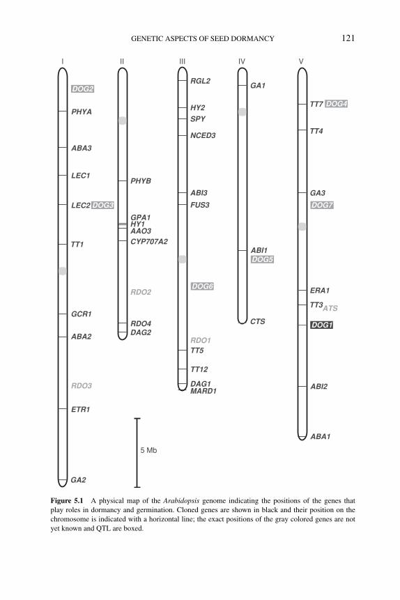
P1: OTE/SPH P2: OTE
BLUK053-Bradford September 27, 2006 7:31
GENETIC ASPECTS OF SEED DORMANCY 121
5 Mb
I II III IV V
ABA3
ABA1
PHYA
PHYB
GPA1
CYP707A2
HY1
HY2
RGL2
SPY
LEC1
LEC2
TT1
GCR1
TT5
TT7
TT4
TT3
TT12
NCED3
ABA2
ETR1
GA2
RDO4
AAO3
RDO2
ATS
RDO3
RDO1DAG2
DAG1MARD1
ABI3
ABI1
ABI2
FUS3
GA1
GA3
ERA1
CTS DOG1
DOG2
DOG3
DOG4
DOG7
DOG5
DOG6
Figure 5.1 A physical map of the Arabidopsis genome indicating the positions of the genes that
play roles in dormancy and germination. Cloned genes are shown in black and their position on the
chromosome is indicated with a horizontal line; the exact positions of the gray colored genes are not
yet known and QTL are boxed.
P1: OTE/SPH P2: OTE
BLUK053-Bradford September 27, 2006 7:31
122 SEED DEVELOPMENT, DORMANCY AND GERMINATION
bicolor). The trait has also been analyzed in the weed species Avena fatua (wildoat). Some studies, especially in barley and rice, made use of combinations of wildand cultivated genotypes.
In the domesticated species, low levels of dormancy may result in PHS but couldbe required for efficient malting (Li et al., 2003) and uniform germination. Nonethe-less, despite selection for low dormancy, residual differences among cultivars arepresent and have been analyzed. However, due to the low dormancy and short after-ripening requirement of most cultivated species, many QTL are not observed in allenvironments and cannot be considered as confirmed QTL. Therefore, estimates ofthe total number of QTL are not always relevant. In this chapter we mainly focuson studies that identified major genes. Such loci can be expected to be amenablefor cloning and could advance our knowledge of dormancy mechanisms. However,knowledge of the positions of minor QTL may also be useful for marker-assistedbreeding.
Barley is of particular interest because the two major uses of this crop havedifferent dormancy requirements. Barley production and end-use quality requirea certain level of seed dormancy, and low or no seed dormancy can lead to PHSand low malting quality. High seed dormancy can result in nonuniformity of seedgermination, causing field stand establishment problems and low malt extract yield(Ullrich et al., 1997). Generally, a moderate level of seed dormancy is thought tobe appropriate for barley cultivars (Han et al., 1999; Romagosa et al., 1999). AQTL analysis in a doubled haploid population derived from a cross between thedormant fodder variety Steptoe and the nondormant malting variety Morex wasvery informative (Gao et al., 2003). In addition, several other cultivar combinationswere analyzed, including the cross of Morex with the relatively dormant Europeanmalting variety Triumph. In both populations a QTL called SD1, located near thecentromere of chromosome 5H, explained 50% or more of the phenotypic variationin all environments tested. This QTL region probably represents three closely linkedloci in a region of 4.4 cM (Han et al., 1999). In crosses involving the cultivarHarrington most of the variation was explained by a QTL near the telomere of5HL (Li et al., 2003). The latter locus was identified as SD2 in the Steptoe/Morex(S/M) population and also in the Triumph/Morex (T/M) cross (Prada et al., 2004).However, in the latter cross, the more dormant allele came from Morex, whichprovided the nondormant allele in the S/M cross, suggesting that this locus may notbe identical to SD2 (Ullrich et al., 2002). Li et al. (2004) compared the syntenicregions of barley, wheat, and rice around the SD2 locus, making use of the availableEST sequence data, and suggested that a GA20-oxidase might be a candidate genefor this QTL. In addition, minor QTL were detected on chromosomes 7H (SD3),4H (SD4), 1H, 2H, and 3H, but these were not detected in all environments and inall studied crosses.
Larger differences in dormancy exist between cultivars and wild-type accessions.In crosses of the cultivar Mono with a desert-derived dormant barley (H. sponta-neum) accession, eight QTL regions were detected, of which five regions overlappedwith the QTL regions identified in cultivar × cultivar crosses, including the SD1and SD2 regions on 5H. In addition, three new regions were identified (Zhang et al.,
P1: OTE/SPH P2: OTE
BLUK053-Bradford September 27, 2006 7:31
GENETIC ASPECTS OF SEED DORMANCY 123
2005). Although more dormant, the wild barley accession also harbored alleles thatresult in reduced dormancy.
In rice, only low levels of dormancy variation are segregating in many cultivarcrosses. Despite this, five QTL could be identified in the frequently studied Nip-ponbare × Kasalath population (Lin et al., 1998). One locus called sdr1, located atthe lower part of chromosome 3, explained almost 25% of the phenotypic variation.Its colocation with the Hd8 flowering time locus appeared to be due to linkage,since the two loci could be separated by fine-mapping. The cloning of this locus iscurrently underway (M. Yano, personal communication). An analysis of the samepopulation in the same experimental field, but in a different year, also yielded fiveQTL (Miura et al., 2002). However, only two of these overlapped with the onesreported by Lin et al. (1998), indicating the strong interaction of dormancy QTLwith the environment that the maternal plant experiences during seed development.The map position of sdr1 seemed to be identical to those of the barley and wheatdormancy QTL on the barley 4HS (SD4) and the wheat 4AL chromosomes, respec-tively. However, this conclusion is somewhat ambiguous because of the complexrearrangements in that region of rice chromosomes 3 and 11 (Li et al., 2004).
In an extensive analysis of a RIL population derived from a cross of an indicacultivar with a common wild rice accession (O. rufipogon), Cai and Morishima(2000) identified 17 QTL, with the alleles from the wild parent increasing dormancy.Several of these loci were also found in studies involving only cultivars. A studyby Gu et al. (2004), involving the cross between an indica cultivar and weedy rice,identified five dormancy QTL that were reproducible across years (Gu et al., 2004,2005). The major QTL, explaining around 20% of the variation in a BC1 and upto 50% in a BC4, was found on chromosome 12 (qSD12) (Gu et al., 2005). Thismap position did not colocate with a major QTL identified by Cai and Morishima(2000), indicating that variation in the genetic control is present among wild andweedy rice genotypes. It is of interest that this locus is the only one that does notcolocate with other domestication-related QTL such as shattering, awn length, blackhull color, or red pericarp color (Gu et al., 2005). Such colocation may be due tovery tightly linked genes or due to pleiotropism, as is suggested for the red pericarplocus (Rc), which is orthologous to the major loci for seed dormancy in wheat andmay be related to the structure of the tissue surrounding the embryo (hull-imposeddormancy). This linkage of dormancy to morphological domestication charactersmight have enhanced the removal of genetic variation in the cultivated germplasmpool.
Bread wheat is an allohexaploid species consisting of three genomes (A, B,and D) that have homeologous colinear chromosomes. PHS has been extensivelystudied in wheat by the analysis of premature germination on ears kept at highhumidity and by α-amylase activity measurements. The α-amylase activity is relatedto dormancy, but also to water uptake and drying, which explains why the presenceof awns (controlled by the Hd loci) and epicuticular wax deposition loci affect thesetraits (King and Wettstein-Knowles, 2000). Although higher α-amylase activity iscorrelated with the progress of germination (indicating low dormancy), late maturityα-amylase activity appears independently of germination (Flintham, 2000).
P1: OTE/SPH P2: OTE
BLUK053-Bradford September 27, 2006 7:31
124 SEED DEVELOPMENT, DORMANCY AND GERMINATION
A major component of dormancy (and PHS) in wheat is pericarp color, which iscontrolled by the Red grain color (R) loci (R-A1, R-B1 and R-D1) located on group5 chromosomes (Flintham, 2000; Groos et al., 2002). Dominant alleles promoteexpression of red pigments (phlobaphenes) within the maternally derived pericarpthat tightly surrounds the embryo. It is assumed that the R genes themselves provideincreased dormancy (Flintham et al., 2002). It has now been shown that these genesencode MYB-type transcription factors controlling flavonoid biosynthesis in a seed-specific manner. These MYB genes are similar to the TT2 gene in Arabidopsis, wherett2 mutants also affect seed dormancy (Debeaujon et al., 2000). Even among the redvarieties, additional dormancy loci can be found and used to reduce PHS. Althoughthe R loci can be used to provide some resistance to PHS, they also provide negativecharacters that reduce the quality of the flour for certain markets. These include color(red wheat seeds produce colored flour, which is perceived negatively by the noodle-making industry), production energy costs in food processing, and taste perception.White wheat contains no active R loci, and is therefore in general more prone toPHS. Alternative loci conferring increased dormancy are needed for white-grainedwheat.
Many QTL have been detected for PHS in various studies (reviewed by Flinthamet al., 2002) and their map positions have been compared among the cereals (Galeet al., 2002). Although many of these have been reported only in specific crossesand environments, some QTL were confirmed across species. Mares et al. (2002)identified several dormancy QTL, including one on chromosome 3D which mapsclosely to the VP1 homologue. A major QTL on chromosome 4AL, named PHS, wasidentified in several mapping populations (Kato et al., 2001; Flintham et al., 2002;Torada et al., 2005) and could be homologous to the barley SD4 locus. Other studiescomparing dormant and nondormant seeds of varieties or induced mutants demon-strated large differences in responsiveness of embryos to ABA (Walker-Simmons,1987; Kawakami et al., 1997). Therefore, it is likely that sensitivity to ABA atspecific developmental periods, together with the induction of desiccation, is a keyprocess related to dormancy induction. The chromosomal locations of loci that influ-ence wheat embryo sensitivity to ABA and dormancy have been investigated (Nodaet al., 2002). A cross between wheat cultivar Chinese Spring (nondormant and ABA-insensitive) and line Kitakei-1354 (dormant and ABA-sensitive) demonstrated theimportance of loci located at 4AL and 2D for these characters.
Genetic studies in sorghum identified two major dormancy QTL (Lijavetzkyet al., 2000). The low dormancy in the PHS-sensitive parent is characterized by lowembryonic ABA sensitivity and higher GA levels (Steinbach et al., 1997; Perez-Flores et al., 2003), which correlated with higher levels of a GA20-oxidase (Perez-Flores et al., 2003). The higher level of VP1 transcript that was maintained inimbibed seeds of the dormant line suggested a relationship between dormancy andVP1 expression. However, no PHS QTL colocated with the VP1 locus. This couldmean that the difference in VP1 expression is only a downstream effect of thedormancy QTL (Carrari et al., 2003).
Wild oat is a noxious weed because dormant seeds may persist several yearsin agricultural soils. Genetic variation for seed dormancy has been reported in this
P1: OTE/SPH P2: OTE
BLUK053-Bradford September 27, 2006 7:31
GENETIC ASPECTS OF SEED DORMANCY 125
species and at least three loci were identified (Fennimore et al., 1999). Two RAPDmarkers were identified that showed linkage with the dormancy trait but these mark-ers have not yet been integrated into the cereal linkage maps.
From the studies reviewed above, it is evident that wild relatives of cultivatedplants are rich sources of dormant alleles, although residual variation is present evenwithin cultivated species. Genetic synteny, especially in cereals, is expected to helpidentify dormancy loci in different species.
5.5 What do the genetics teach us about dormancy and germination?
The identification of genetic variation in seed dormancy can assist in understandingthis important process. The number of genes that are known to be involved indormancy or germination is rapidly increasing. Figure 5.1 shows the map positionsof the genes that play a role in dormancy and germination in Arabidopsis, basedon genetic evidence. The identification of the molecular and biochemical functionsof these genes will increase our knowledge about the relevant processes. However,even before the respective genes were cloned, the study of plant hormone mutants inArabidopsis and tomato showed in a convincing way the importance of ABA and GAfor regulating seed germination. For mutants that show only a dormancy phenotype,cloning of the respective genes can reveal novel processes that are involved indormancy.
Genetic analyses are more efficient in Arabidopsis and rice, compared to otherplant species, because of the availability of many genetic resources, including mu-tations in almost every gene and access to the full genome sequence. In addition, thecomplete DNA sequence allows the application of ‘-omic’ approaches, includingfull genome gene expression analysis and efficient proteomics. Therefore, it is notsurprising that biological research is focusing on Arabidopsis and rice. The identi-fication of mutants and QTL associated with dormancy will ultimately extend thisknowledge to crop plants where genomics approaches are also becoming available.Until such genes are cloned, QTL analyses can identify genomic regions that canbe followed in breeding programs using marker-assisted selection.
Studies using the available mutants that are involved in the GA biosyntheticpathway, especially in Arabidopsis and also in tomato, together with GA biosyn-thesis inhibitors, have shown that GAs play a major role as a promotive agent forgermination. The role of GA during Arabidopsis seed development seems limited,since normal seeds are formed even on rather extreme dwarf mutants. Argumentsthat GAs play their major role only during seed germination come from a combina-tion of proteomic and mutant analyses showing that imbibed ga1 mutant seeds arenot different from wild-type seeds until the moment of radicle protrusion when theyshow lack of germination (Gallardo et al., 2002). This is in agreement with a care-ful analysis of the induction of GA biosynthesis and with gene expression studies(Ogawa et al., 2003). GA biosynthesis occurs after imbibition because germinationcan be inhibited at that time by GA biosynthesis inhibitors. These studies all indi-cated that de novo synthesis of GA after seed maturation is required for germination.
P1: OTE/SPH P2: OTE
BLUK053-Bradford September 27, 2006 7:31
126 SEED DEVELOPMENT, DORMANCY AND GERMINATION
Mutant approaches have shown that RGL2, one of the DELLA proteins that actsas a repressor of GA action and is degraded in a GA-inducible manner, is the ma-jor repressor acting during seed germination (Tyler et al., 2004; Cao et al., 2005;see Chapter 10). Possibly, other signal transduction pathway components involvetrimeric G proteins and SPY.
ABA is a potent inhibitor of germination as seen in the strong phenotypes ofmany ABA-related mutants. ABA most likely plays roles in the induction of dor-mancy during seed maturation and the maintenance of dormancy during imbibition.Reciprocal crosses between wild type and the ABA-deficient aba1 mutants showedthat dormancy is controlled by the ABA genotype of the embryo and not by thatof the mother plant. However, the mother plant is still responsible for the rela-tively high ABA levels found in seeds midway through seed development (Karssenet al., 1983). At this time, ABA may prevent precocious germination, as shown bythe maternal ABA effects in the extreme aba abi3-1 double mutants (Koornneefet al., 1989). Since ABA levels decrease in wild-type seeds at the end of maturation,it was proposed that endogenous ABA is not required for dormancy maintenanceafter its onset during seed development (Karssen et al., 1983). In contrast, the obser-vation that ABA biosynthesis inhibitors such as norfluorazon promote germination(Debeaujon and Koornneef, 2000) indicates that the maintenance of dormancy inimbibed seeds is an active process involving de novo ABA synthesis (Ali-Rachediet al., 2004). ABA catabolism is also involved in reducing ABA levels during imbi-bition, as is shown by the reduced germination of knockout mutants of the CYP707Agene, which encodes an ABA 8′-hydroxylase (Nambara and Marion-Poll, 2005; seeChapter 9).
Genetic approaches revealed interesting relationships among ethylene, ABA,and sugar signaling. Ein mutants show increased dormancy and an ethylene-hypersensitive constitutive triple response (ctr1) mutant resembles ABA- and sugar-signaling mutants (Beaudoin et al., 2000; Ghassemian et al., 2000). These obser-vations, in combination with the nondormant phenotype of the ein2 abi3-4 doublemutant, indicated that ethylene negatively regulates seed dormancy by inhibitingABA action (Beaudoin et al., 2000). However, Ghassemian et al. (2000) suggestedthat in addition to signaling, EIN2 may also affect ABA biosynthesis. Detailedanalyses of plant hormone levels in the etr1-2 mutant by Chiwocha et al. (2005)indicated that the higher ABA levels in the mutant are probably caused by an effecton ABA catabolism. Auxin, cytokinin, and GA levels were also affected, indicatingthe complexity of cross-talk among plant hormones (see Chapter 8). The presenceof cross-talk between sugar signaling and ethylene was suggested by the sugar-insensitive phenotype of ctr1, which has a constitutive response to ethylene (Gibsonet al., 2001) and the sugar-hypersensitive phenotype of etr (Zhou et al., 1998). Ap-parently, strong interactions among ABA, ethylene, and sugar signaling are presentduring germination and early seedling growth.
The involvement of these hormone biosynthesis and sensitivity genes in dor-mancy is not a surprise, since the application of different hormones and biosynthe-sis inhibitors already provided strong indications for the roles of these hormones.However, mutants have helped refine these roles and also identified several of the
P1: OTE/SPH P2: OTE
BLUK053-Bradford September 27, 2006 7:31
GENETIC ASPECTS OF SEED DORMANCY 127
hormone signaling factors. It is expected that many discoveries will still be madeby the identification of genes with clear effects on dormancy and germination thatdo not have specific hormone-related phenotypes. Examples of these are the manydormancy QTL that have been identified, although some might be hormone-related,as suggested in barley (Li et al., 2004). Cloning of the first pleiotropic RDO genesindicated that these genes may be involved in cellular functions that are not specificto dormancy and germination (Y. Liu, personal communication).
Microarray and proteomic data on germination using GA-deficient mutants havebeen published (Gallardo et al., 2002; Ogawa et al., 2003). These studies, togetherwith the use of dormancy-specific mutants and their complemention with wild-typegenes under the control of tissue-specific or inducible promoters, may finally allowa better distinction of the various processes from dormancy induction to seedlingestablishment. The combination of mutant approaches with in-depth molecular anal-ysis of the regulators and ‘-omic’ approaches is expected to give many new insightsinto seed dormancy, which is still a relatively poorly understood process that hasimportance for both ecology and agriculture.
References
G.K. Agrawal, M. Yamazaki, M. Kobayashi, R. Hirochika, A. Miyao and H. Hirochika (2001) Screen-
ing of the rice viviparous mutants generated by endogenous retrotransposon Tos17 insertion.
Tagging of a zeaxanthin epoxidase gene and a novel OsTATC gene. Plant Physiology 125, 1248–
1257.
A. Alboresi, C. Gestin, M.T. Leydecker, M. Bedu, C. Meyer and H.N. Truong (2005) Nitrate, a signal
relieving seed dormancy in Arabidopsis. Plant Cell and Environment 28, 500–512.
S. Ali-Rachedi, D. Bouinot, M.H. Wagner, et al. (2004) Changes in endogenous abscisic acid levels
during dormancy release and maintenance of mature seeds: studies with the Cape Verde Islands
ecotype, the dormant model of Arabidopsis thaliana. Planta 219, 479–488.
C. Alonso-Blanco, L. Bentsink, C.J. Hanhart, H. Blankestijn-De Vries and M. Koornneef (2003)
Analysis of natural allelic variation at seed dormancy loci of Arabidopsis thaliana. Genetics 164,
711–729.
C. Alonso-Blanco and M. Koornneef (2000) Naturally occurring variation in Arabidopsis: an under-
exploited resource for plant genetics. Trends in Plant Science 5, 22–29.
C. Basu, M.D. Halfhill, T.C. Mueller and C.N. Stewart, Jr. (2004) Weed genomics: new tools to
understand weed biology. Trends in Plant Science 9, 391–398.
N. Beaudoin, C. Serizet, F. Gosti and J. Giraudat (2000) Interactions between abscisic acid and ethylene
signaling cascades. The Plant Cell 12, 1103–1115.
L. Bentsink (2002) Genetic Analysis of Seed Dormancy and Seed Composition in Arabidopsis thaliana
Using Natural Variation. Ph.D. Dissertation, Department of Plant Sciences, Laboratory of Ge-
netics, Wageningen University, 159 pp.
H.W. Cai and H. Morishima (2000) Genomic regions affecting seed shattering and seed dormancy in
rice. Theoretical and Applied Genetics 100, 840–846.
J.S. Cao, H.F. Yu, W.Z. Ye, et al. (2005) Identification and characterisation of a gibberellin-related dwarf
mutant in pumpkin (Cucurbita moschata). Journal of Horticultural Science and Biotechnology80, 29–31.
C. Carles, N. Bies-Etheve, L. Aspart, et al. (2002) Regulation of Arabidopsis thaliana Em genes: role
of ABI5. The Plant Journal 30, 373–383.
F. Carrari, R. Benech-Arnold, R. Osuna-Fernandez, et al. (2003) Genetic mapping of the Sorghumbicolor vp1 gene and its relationship with preharvest sprouting resistance. Genome 46, 253–258.
P1: OTE/SPH P2: OTE
BLUK053-Bradford September 27, 2006 7:31
128 SEED DEVELOPMENT, DORMANCY AND GERMINATION
S.D. Chiwocha, A.J. Cutler, S.R. Abrams, et al. (2005) The etr1-2 mutation in Arabidopsis thalianaaffects the abscisic acid, auxin, cytokinin and gibberellin metabolic pathways during maintenance
of seed dormancy, moist-chilling and germination. The Plant Journal 42, 35–48.
E.J.M. Clerkx, M.E. El Lithy, E. Vierling, et al. (2004) Analysis of natural allelic variation of Ara-bidopsis seed germination and seed longevity traits between the accessions Landsberg erecta and
Shakdara, using a new recombinant inbred line population. Plant Physiology 135, 432–443.
S.D. Clouse and J.M. Sasse (1998) Brassinosteroids: essential regulators of plant growth and devel-
opment. Annual Review of Plant Physiology and Plant Molecular Biology 49, 427–451.
G. Colucci, F. Apone, N. Alyeshmerni, D. Chalmers and M.J. Chrispeels (2002) GCR1, the putative
Arabidopsis G protein-coupled receptor gene is cell cycle-regulated, and its overexpression abol-
ishes seed dormancy and shortens time to flowering. Proceedings of the National Academy ofSciences of the United States of America 99, 4736–4741.
S. Cutler, M. Ghassemian, D. Bonetta, S. Cooney and P. McCourt (1996) A protein farnesyl transferase
involved in ABA signal transduction in Arabidopsis. Science 273, 1239–1241.
I. Debeaujon and M. Koornneef (2000) Gibberellin requirement for Arabidopsis thaliana seed ger-
mination is determined both by testa characteristics and embryonic ABA. Plant Physiology 122,
415–424.
I. Debeaujon, K.M. Leon-Kloosterziel and M. Koornneef (2000) Influence of the testa on seed dor-
mancy, germination and longevity in Arabidopsis thaliana. Plant Physiology 122, 403–414.
K. Donohue (2002) Germination timing influences natural selection on life-history characters in Ara-bidopsis thaliana. Ecology 83, 1006–1016.
B. Dubreucq, N. Berger, E. Vincent, et al. (2000) The Arabidopsis AtERP1 extensin-like gene is
specifically expressed in endosperm during seed germination. The Plant Journal 23, 643–652.
S.A. Fennimore, W.E. Nyquist, G.E. Shaner, R.W. Doerge and M.E. Foley (1999) A genetic model
and molecular markers for wild oat (Avena fatua L.) seed dormancy. Theoretical and AppliedGenetics 99, 711–718.
R.R. Finkelstein (1994) Maternal effects govern variable dominance of two abscisic acid response
mutations in Arabidopsis thaliana. Plant Physiology 105, 1203–1208.
J. Flintham, R. Adlam, M. Bassoi, M. Holdsworth and M. Gale (2002) Mapping genes for resistance
to sprouting damage in wheat. Euphytica 126, 39–45.
J.E. Flintham (2000) Different genetic components control coat-imposed and embryo-imposed dor-
mancy in wheat. Seed Science Research 10, 43–50.
R. Foster and N.H. Chua (1999) An Arabidopsis mutant with deregulated ABA gene expression:
implication for negative regulator function. The Plant Journal 17, 363–372.
M.D. Gale, J. Flintham and K.M. Devos (2002) Cereal comparative genetics and preharvest sprouting.
Euphytica 126, 21–25.
K. Gallardo, C. Job, S.P.C. Groot, et al. (2002) Proteomics of Arabidopsis seed germination: A
comparative study of wild-type and gibberellin-deficient seeds. Plant Physiology 129, 823–837.
W. Gao, J.A. Clancy, F. Han, D. Prada, A. Kleinhofs and S.E. Ullrich (2003) Molecular dissection
of a dormancy QTL region near the chromosome 7 (5H) L telomere in barley. Theoretical andApplied Genetics 107, 552–559.
A. Garciarrubio, J.P. Legaria and A.A. Covarrubias (1997) Abscisic acid inhibits germination of mature
Arabidopsis seeds by limiting the availability of energy and nutrients. Planta 203, 182–187.
M. Ghassemian, E. Nambara, S. Cutler, H. Kawaide, Y. Kamiya and P. McCourt (2000) Regulation
of abscisic acid signaling by the ethylene response pathway in Arabidopsis. The Plant Cell 12,
1117–1126.
S.I. Gibson and I.A. Graham (1999) Another player joins the complex field of sugar-regulated gene
expression in plants. Proceedings of the National Academy of Sciences of the United States ofAmerica 96, 4746–4748.
S.I. Gibson, R.J. Laby and D. Kim (2001) The sugar-insensitive1 (sis1) mutant of Arabidopsis is
allelic to ctr1. Biochemical and Biophysical Research Communications 280, 196–203.
C. Groos, G. Gay, M.R. Perretant, et al. (2002) Study of the relationship between pre-harvest sprouting
and grain color by quantitative trait loci analysis in a white × red grain bread-wheat cross.
Theoretical and Applied Genetics 104, 39–47.
P1: OTE/SPH P2: OTE
BLUK053-Bradford September 27, 2006 7:31
GENETIC ASPECTS OF SEED DORMANCY 129
X.Y. Gu, S.F. Kianian and M.E. Foley (2004) Multiple loci and epistases control genetic variation for
seed dormancy in weedy rice (Oryza sativa). Genetics 166, 1503–1516.
X.Y. Gu, S.F. Kianian, G.A. Hareland, B.L. Hoffer and M.E. Foley (2005) Genetic analysis of adaptive
syndromes interrelated with seed dormancy in weedy rice (Oryza sativa). Theoretical and AppliedGenetics 110, 1108–1118.
G. Gualberti, M. Papi, L. Bellucci, et al. (2002) Mutations in the Dof zinc finger genes DAG2 and
DAG1 influence with opposite effects germination of Arabidopsis seeds. The Plant Cell 14, 1253–
1263.
F. Gubler, A.A. Millar and J.V. Jacobsen (2005) Dormancy release, ABA and pre-harvest sprouting.
Current Opinion in Plant Biology 8, 183–187.
F. Han, S.E. Ullrich, J.A. Clancy and I. Romagosa (1999) Inheritance and fine mapping of a major
barley seed dormancy QTL. Plant Science 143, 113–118.
Y. He and S. Gan (2004) A novel zinc-finger protein with a proline-rich domain mediates ABA-
regulated seed dormancy in Arabidopsis. Plant Molecular Biology 54, 1–9.
H.W.M. Hilhorst, S.P.C. Groot and R.J. Bino (1998) The tomato seed as a model system to study seed
development and germination. Acta Botanica Neerlandica 47, 169–183.
M. Holdsworth, L. Bentsink and M. Koornneef (2006) Conserved mechanisms of dormancy and germi-
nation as targets for manipulation of agricultural problems. In: Model Plants, Crop Improvement(eds R. Koebner and R. Varshney). CRC Press, Boca Raton, FL.
C. Huijser, A.J. Kortstee, J.V. Pego, P. Weisbeek, E. Wisman and S. Smeekens (2000) The Arabidopsissucrose uncoupled-6 gene is identical to abscisic acid insensitive-4: involvement of abscisic acid
in sugar responses. The Plant Journal 23, 577–585.
S.E. Jacobsen, K.A. Binkowski and N.E. Olszewski (1996) SPINDLY, a tetratricopeptide repeat protein
involved in gibberellin signal transduction in Arabidopsis. Proceedings of the National Academyof Sciences of the United States of America 93, 9292–9296.
C.M. Karssen, D.L.C. Brinkhorst-van der Swan, A.E. Breekland and M. Koornneef (1983) Induction
of dormancy during seed development by endogenous abscisic acid: studies on abscisic acid
deficient genotypes of Arabidopsis thaliana (L.) Heynh. Planta 157, 158–165.
C.M. Karssen, S. Zaorski, J. Kepczynski and S.P.C. Groot (1989) Key role for endogenous gibberellins
in the control of seed germination. Annals of Botany 63, 71–80.
K. Kato, W. Nakamura, T. Tabiki, H. Miura and S. Sawada (2001) Detection of loci controlling seed
dormancy on group 4 chromosomes of wheat and comparative mapping with rice and barley
genomes. Theoretical and Applied Genetics 102, 980–985.
N. Kawakami, Y. Miyake and K. Noda (1997) ABA insensitivity and low ABA levels during seed
development of non-dormant wheat mutants. Journal of Experimental Botany 48, 1415–1421.
R.W. King and P. Wettstein-Knowles (2000) Epicuticular waxes and regulation of ear wetting and
pre-harvest sprouting in barley and wheat. Euphytica 112, 157–166.
M. Koornneef, C. Alonso-Blanco, L. Bentsink, et al. (2000) The genetics of seed dormancy in Ara-bidopsis thaliana. In: Dormancy in Plants (eds J.D. Viemont and J. Crabbe), pp. 365–373. CAB
International, Wallingford, UK.
M. Koornneef, C.J. Hanhart, H.W.M. Hilhorst and C.M. Karssen (1989) In vivo inhibition of seed
development and reserve protein accumulation in recombinants of abscisic acid biosynthesis and
responsiveness mutants in Arabidopsis thaliana. Plant Physiology 90, 463–469.
M. Koornneef, M.L. Jorna, D.L.C. Brinkhorst-van der Swan and C.M. Karssen (1982) The isolation
of abscisic acid (ABA) deficient mutants by selection of induced revertants in non-germinating
gibberellin sensitive lines of Arabidopsis thaliana (L.) Heynh. Theoretical and Applied Genetics61, 385–393.
N. Kurata, K. Miyoshi, K. Nonomura, Y. Yamazaki and Y. Ito (2005) Rice mutants and genes related
to organ development, morphogenesis and physiological traits. Plant and Cell Physiology 46,
48–62.
R.J. Laby, S. Kincaid, D. Kim and S.I. Gibson (2000) The Arabidopsis sugar-insensitive mutants sis4and sis5 are defective in abscisic acid synthesis and response. The Plant Journal 23, 587–596.
K.M. Leon-Kloosterziel, M. Alvarez Gil, G.J. Ruijs, et al. (1996a) Isolation and characterization of
abscisic acid-deficient Arabidopsis mutants at two new loci. The Plant Journal 10, 655–661.
P1: OTE/SPH P2: OTE
BLUK053-Bradford September 27, 2006 7:31
130 SEED DEVELOPMENT, DORMANCY AND GERMINATION
K.M. Leon-Kloosterziel, G.A. van de Bunt, J.A.D. Zeevaart and M. Koornneef (1996b) Arabidopsismutants with a reduced seed dormancy. Plant Physiology 110, 233–240.
C.D. Li, A. Tarr, R.C.M. Lance, et al. (2003) A major QTL controlling seed dormancy and pre-harvest
sprouting/grain α-amylase in two-rowed barley (Hordeum vulgare L.). Australian Journal ofAgricultural Research 54, 1303–1313.
J.M. Li, J.H. Xiao, S. Grandillo, et al. (2004) QTL detection for rice grain quality traits using an inter-
specific backcross population derived from cultivated Asian (O. sativa L.) and African (O. glaber-rima S.) rice. Genome 47, 697–704.
D. Lijavetzky, M.C. Martinez, F. Carrari and H.E. Hopp (2000) QTL analysis and mapping of pre-
harvest sprouting resistance in Sorghum. Euphytica 112, 125–135.
S.Y. Lin, T. Sasaki and M. Yano (1998) Mapping quantitative trait loci controlling seed dormancy
and heading date in rice, Oryza sativa L., using backcross inbred lines. Theoretical and AppliedGenetics 96, 997–1003.
P.-P. Liu, N. Koizuka, T.M. Homrichhausen, J.R. Hewitt, R.C. Martin and H. Nonogaki (2005) Large-
scale screening of Arabidopsis enhancer-trap lines for seed germination-associated genes. ThePlant Journal 41, 936–944.
D. Mares, K. Mrva, M.K. Tan and P. Sharp (2002) Dormancy in white-grained wheat: progress towards
identification of genes and molecular markers. Euphytica 126, 47–53.
D.R. McCarty (1995) Genetic control and integration of maturation and germination pathways in seed
development. Annual Review of Plant Physiology and Plant Molecular Biology 46, 71–93.
K. Miura, S.Y. Lin, M. Yano and T. Nagamine (2002) Mapping quantitative trait loci controlling seed
longevity in rice (Oryza sativa L.). Theoretical and Applied Genetics 104, 981–986.
E. Nambara and A. Marion-Poll (2005) Abscisic acid biosynthesis and catabolism. Annual Review ofPlant Biology 56, 165–185.
E. Nambara, S. Naito and P. McCourt (1992) A mutant of Arabidopsis which is defective in seed
development and storage protein accumulation is a new abi3 allele. The Plant Journal 2, 435–
441.
N. Nishimura, T. Yoshida, M. Murayama, T. Asami, K. Shinozaki and T. Hirayama (2004) Isola-
tion and characterization of novel mutants affecting the abscisic acid sensitivity of Arabidopsisgermination and seedling growth. Plant and Cell Physiology 45, 1485–1499.
K. Noda, T. Matsuura, M. Maekawa and S. Taketa (2002) Chromosomes responsible for sensitivity of
embryo to abscisic acid and dormancy in wheat. Euphytica 123, 203–209.
M. Ogawa, A. Hanada, Y. Yamauchi, A. Kuwahara, Y. Kamiya and S. Yamaguchi (2003) Gib-
berellin biosynthesis and response during Arabidopsis seed germination. The Plant Cell 15, 1591–
1604.
M. Papi, S. Sabatini, D. Bouchez, C. Camilleri, P. Costantino and P. Vittorioso (2000) Identifica-
tion and disruption of an Arabidopsis zinc finger gene controlling seed germination. Genes andDevelopment 14, 28–33.
A.J.M. Peeters, H. Blankestijn-De Vries, C.J. Hanhart, K.M. Leon-Kloosterziel, J.A.D. Zeevaart and
M. Koornneef (2002) Characterization of mutants with reduced seed dormancy at two novel rdoloci and a further characterization of rdo1 and rdo2 in Arabidopsis. Physiologia Plantarum 115,
604–612.
J.V. Pego, A.J. Kortstee, C. Huijser and S. Smeekens (2000) Photosynthesis, sugars and the regulation
of gene expression. Journal of Experimental Botany 51, 407–416.
S. Penfield, E.L. Rylott, A.D. Gilday, S. Graham, T.R. Larson and I.A. Graham (2004) Reserve
mobilization in the Arabidopsis endosperm fuels hypocotyl elongation in the dark, is independent
of abscisic acid, and requires PHOSPHOENOLPYRUVATE CARBOXYKINASE1. The Plant Cell16, 2705–2718.
L. Perez-Flores, F. Carrari, R. Osuna-Fernandez, et al. (2003) Expression analysis of a GA 20-
oxidase in embryos from two sorghum lines with contrasting dormancy: possible participation
of this gene in the hormonal control of germination. Journal of Experimental Botany 54, 2071–
2079.
C. Poppe and E. Schafer (1997) Seed germination of Arabidopsis thaliana phyA/phyB double mutants
is under phytochrome control. Plant Physiology 114, 1487–1492.
P1: OTE/SPH P2: OTE
BLUK053-Bradford September 27, 2006 7:31
GENETIC ASPECTS OF SEED DORMANCY 131
D. Prada, I. Romagosa, S.E. Ullrich and J.L. Molina-Cano (2005) A centromeric region on chromosome
6(6H) affects dormancy in an induced mutant in barley. Journal of Experimental Botany 56, 47–
54.
D. Prada, S.E. Ullrich, J.L. Molina-Cano, L. Cistue, J.A. Clancy and I. Romagosa (2004) Genetic
control of dormancy in a Triumph/Morex cross in barley. Theoretical and Applied Genetics 109,
62–70.
I. Romagosa, F. Han, J.A. Clancy and S.E. Ullrich (1999) Individual locus effects on dormancy during
seed development and after ripening in barley. Crop Science 39, 74–79.
I. Romagosa, D. Prada, M.A. Moralejo, et al. (2001) Dormancy, ABA content and sensitivity of a barley
mutant to ABA application during seed development and after ripening. Journal of ExperimentalBotany 52, 1499–1506.
L. Salaita, R.K. Kar, M. Majee and A.B. Downie (2005) Identification and characterization of mutants
capable of rapid seed germination at 10◦C from activation-tagged lines of Arabidopsis thaliana.
Journal of Experimental Botany 56, 2059–2069.
K. Schumacher and J. Chory (2000) Brassinosteroid signal transduction: still casting the actors. CurrentOpinion in Plant Biology 3, 79–84.
T. Shinomura, A. Nagatani, H. Hanzawa, M. Kubota, M. Watanabe and M. Furuya (1996) Action
spectra for phytochrome A- and B-specific photoinduction of seed germination in Arabidopsisthaliana. Proceedings of the National Academy of Sciences of the United States of America 93,
8129–8133.
G. Siriwitayawan, R.L. Geneve and A.B. Downie (2003) Seed germination of ethylene perception
mutants of tomato and Arabidopsis. Seed Science Research 13, 303–314.
S. Smeekens (1998) Sugar regulation of gene expression in plants. Current Opinion in Plant Biology1, 230–234.
C.M. Steber and P. McCourt (2001) A role for brassinosteriods in germination in Arabidopsis. PlantPhysiology 125, 763–769.
H.S. Steinbach, R.L. Benech-Arnold and R.A. Sanchez (1997) Hormonal regulation of dormancy in
developing sorghum seeds. Plant Physiology 113, 149–154.
B.C. Tan, S.H. Schwartz, J.A.D. Zeevaart and D.R. McCarty (1997) Genetic control of abscisic acid
biosynthesis in maize. Proceedings of the National Academy of Sciences of the United States ofAmerica 94, 12235–12240.
A. Torada, S. Ikeguchi and M. Koike (2005) Mapping and validation of PCR-based markers associated
with a major QTL for seed dormancy in wheat. Euphytica 143, 251–255.
L. Tyler, S.G. Thomas, J.H. Hu, et al. (2004) DELLA proteins and gibberellin-regulated seed germi-
nation and floral development in Arabidopsis. Plant Physiology 135, 1008–1019.
H. Ullah, J.G. Chen, S.C. Wang and A.M. Jones (2002) Role of a heterotrimeric G protein in regulation
of Arabidopsis seed germination. Plant Physiology 129, 897–907.
S.E. Ullrich, F. Han, W. Gao, et al. (2002) Summary of QTL analyses of the seed dormancy trait in
barley. Barley Newsletter 45, 39–41.
S.E. Ullrich, F. Han and B.L. Jones (1997) Genetic complexity of the malt extract trait in barley
suggested by QTL analysis. Journal of the American Society of Brewing Chemists 55, 1–4.
W. Van der Schaar, C. Alonso-Blanco, K.M. Leon-Kloosterziel, R.C. Jansen, J.W. Van Ooijen and
M. Koornneef (1997) QTL analysis of seed dormancy in Arabidopsis using recombinant inbred
lines and MQM mapping. Heredity 79, 190–200.
A.J. van Hengel and K. Roberts (2003) AtAGP30, an arabinogalactan-protein in the cell walls of
the primary root, plays a role in root regeneration and seed germination. The Plant Journal 36,
256–270.
M.K. Walker-Simmons (1987) ABA levels and sensitivity in developing wheat embryos of sprouting
resistant and susceptible cultivars. Plant Physiology 84, 61–66.
L. Xiong, M. Ishitani, H. Lee and J.K. Zhu (1999) HOS5 – a negative regulator of osmotic stress-
induced gene expression in Arabidopsis thaliana. The Plant Journal 19, 569–578.
L. Xiong, M. Ishitani, H. Lee and J.K. Zhu (2001) The Arabidopsis LOS5/ABA3 locus encodes a
molybdenum cofactor sulfurase and modulates cold stress- and osmotic stress-responsive gene
expression. The Plant Cell 13, 2063–2083.

P1: OTE/SPH P2: OTE
BLUK053-Bradford September 27, 2006 7:31
132 SEED DEVELOPMENT, DORMANCY AND GERMINATION
L. Xiong, H. Lee, M. Ishitani and J.K. Zhu (2002) Regulation of osmotic stress-responsive gene
expression by the LOS6/ABA1 locus in Arabidopsis. The Journal of Biological Chemistry 277,
8588–8596.
Y.Y. Yang, A. Nagatani, Y.J. Zhao, B.J. Kang, R.E. Kendrick and Y. Kamiya (1995) Effects of gib-
berellins on seed germination of phytochrome-deficient mutants of Arabidopsis thaliana. Plantand Cell Physiology 36, 1205–1211.
F. Zhang, G. Chen, Q. Huang, et al. (2005) Genetic basis of barley caryopsis dormancy and seedling
desiccation tolerance at the germination stage. Theoretical and Applied Genetics 110, 445–453.
L. Zhou, J.C. Jang, T.L. Jones and J. Sheen (1998) Glucose and ethylene signal transduction crosstalk
revealed by an Arabidopsis glucose-insensitive mutant. Proceedings of the National Academy ofSciences of the United States of America 95, 10294–10299.
P1: PJM
BLUK053-Bradford August 27, 2006 17:19
6 Lipid metabolism in seed dormancySteven Penfield, Helen Pinfield-Wells and Ian A. Graham
6.1 Introduction
One of the key features of seeds from both angiosperms and gymnosperms is thepropensity to accumulate stores of nutrient compounds to fuel early seedling de-velopment. Stored reserves are used for respiration and growth in the period im-mediately following germination, but before photosynthesis and uptake from theroots begins in earnest. In this review we will focus on the biochemical pathwaysrequired for the catabolism of seed storage reserves and the role these might play inthe control of seed dormancy and germination. In practice we will concentrate on themodel plant Arabidopsis thaliana, although it is reasonable to expect the results tobe broadly applicable to other species. Even seeds conventionally viewed as starch-containing, such as cereals, accumulate significant amounts of oil in both the embryoand the aleurone layer during seed development (Gram, 1982). Consequently, theactivities of key enzymes in lipid catabolism have been demonstrated in cerealsduring early seedling growth (Eastmond and Jones, 2005). Perhaps more surprisingis that the activities of many of the enzymes required for storage oil catabolism arepresent in maturing and dormant seeds prior to germination. In fact, although stor-age reserve mobilization has traditionally been viewed as a postgerminative event,it has become clear that significant breakdown of lipids occurs during the period oftheir synthesis in developing Arabidopsis and rape (Brassica napus) seeds. Further-more, during the final stages of seed maturation, seed oil content actually falls (Baudet al., 2002; Chia et al., 2005). Hence, there is ample scope for these pathways tobe regulating events prior to seed germination, such as the control of dormancy.
Fatty acids are stored in seeds as triacylglycerol (TAG). This consists of threefatty acids esterified to a glycerol backbone, and forms a highly efficient and com-pact energy source for germinating seeds. Fatty acids and glycerol are released fromTAG and undergo further catabolism to provide energy and carbon skeletons forpostgerminative growth. The overall mobilization of TAG involves several consec-utive pathways in various cellular compartments (Figure 6.1). TAGs are hydrolyzedby lipases (Huang, 1992), and then the fatty acids are passed into single-membrane-bound organelles, termed glyoxysomes, where β-oxidation and the glyoxylate cycleoccur (Cooper and Beevers, 1969). Glyoxysomes are identified as distinct fromthe more ubiquitous peroxisomes on the basis that glyoxysomes have an activeglyoxylate cycle. β-Oxidation converts fatty acids to acetyl-CoA, which is sub-sequently condensed into four-carbon sugars via the glyoxylate cycle. These arethen transported to the mitochondria from where they can be either converted to
133
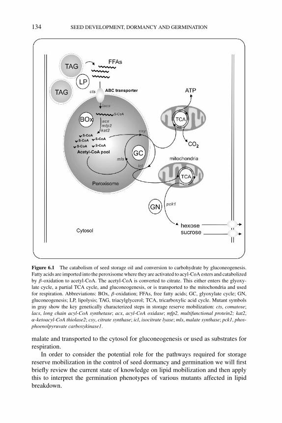
P1: PJM
BLUK053-Bradford August 27, 2006 17:19
134 SEED DEVELOPMENT, DORMANCY AND GERMINATION
Figure 6.1 The catabolism of seed storage oil and conversion to carbohydrate by gluconeogenesis.
Fatty acids are imported into the peroxisome where they are activated to acyl-CoA esters and catabolized
by β-oxidation to acetyl-CoA. The acetyl-CoA is converted to citrate. This either enters the glyoxy-
late cycle, a partial TCA cycle, and gluconeogenesis, or is transported to the mitochondria and used
for respiration. Abbreviations: BOx, β-oxidation; FFAs, free fatty acids; GC, glyoxylate cycle; GN,
gluconeogenesis; LP, lipolysis; TAG, triacylglycerol; TCA, tricarboxylic acid cycle. Mutant symbols
in gray show the key genetically characterized steps in storage reserve mobilization: cts, comatose;
lacs, long chain acyl-CoA synthetase; acx, acyl-CoA oxidase; mfp2, multifunctional protein2; kat2,
α-ketoacyl-CoA thiolase2; csy, citrate synthase; icl, isocitrate lyase; mls, malate synthase; pck1, phos-phoenolpyruvate carboxykinase1.
malate and transported to the cytosol for gluconeogenesis or used as substrates forrespiration.
In order to consider the potential role for the pathways required for storagereserve mobilization in the control of seed dormancy and germination we will firstbriefly review the current state of knowledge on lipid mobilization and then applythis to interpret the germination phenotypes of various mutants affected in lipidbreakdown.
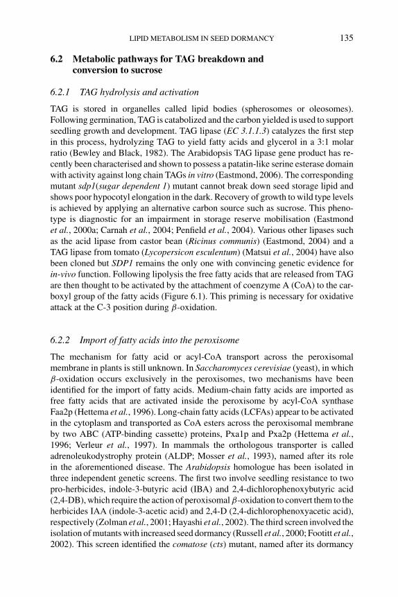
P1: PJM
BLUK053-Bradford August 27, 2006 17:19
LIPID METABOLISM IN SEED DORMANCY 135
6.2 Metabolic pathways for TAG breakdown andconversion to sucrose
6.2.1 TAG hydrolysis and activation
TAG is stored in organelles called lipid bodies (spherosomes or oleosomes).Following germination, TAG is catabolized and the carbon yielded is used to supportseedling growth and development. TAG lipase (EC 3.1.1.3) catalyzes the first stepin this process, hydrolyzing TAG to yield fatty acids and glycerol in a 3:1 molarratio (Bewley and Black, 1982). The Arabidopsis TAG lipase gene product has re-cently been characterised and shown to possess a patatin-like serine esterase domainwith activity against long chain TAGs in vitro (Eastmond, 2006). The correspondingmutant sdp1(sugar dependent 1) mutant cannot break down seed storage lipid andshows poor hypocotyl elongation in the dark. Recovery of growth to wild type levelsis achieved by applying an alternative carbon source such as sucrose. This pheno-type is diagnostic for an impairment in storage reserve mobilisation (Eastmondet al., 2000a; Carnah et al., 2004; Penfield et al., 2004). Various other lipases suchas the acid lipase from castor bean (Ricinus communis) (Eastmond, 2004) and aTAG lipase from tomato (Lycopersicon esculentum) (Matsui et al., 2004) have alsobeen cloned but SDP1 remains the only one with convincing genetic evidence forin-vivo function. Following lipolysis the free fatty acids that are released from TAGare then thought to be activated by the attachment of coenzyme A (CoA) to the car-boxyl group of the fatty acids (Figure 6.1). This priming is necessary for oxidativeattack at the C-3 position during β-oxidation.
6.2.2 Import of fatty acids into the peroxisome
The mechanism for fatty acid or acyl-CoA transport across the peroxisomalmembrane in plants is still unknown. In Saccharomyces cerevisiae (yeast), in whichβ-oxidation occurs exclusively in the peroxisomes, two mechanisms have beenidentified for the import of fatty acids. Medium-chain fatty acids are imported asfree fatty acids that are activated inside the peroxisome by acyl-CoA synthaseFaa2p (Hettema et al., 1996). Long-chain fatty acids (LCFAs) appear to be activatedin the cytoplasm and transported as CoA esters across the peroxisomal membraneby two ABC (ATP-binding cassette) proteins, Pxa1p and Pxa2p (Hettema et al.,1996; Verleur et al., 1997). In mammals the orthologous transporter is calledadrenoleukodystrophy protein (ALDP; Mosser et al., 1993), named after its rolein the aforementioned disease. The Arabidopsis homologue has been isolated inthree independent genetic screens. The first two involve seedling resistance to twopro-herbicides, indole-3-butyric acid (IBA) and 2,4-dichlorophenoxybutyric acid(2,4-DB), which require the action of peroxisomal β-oxidation to convert them to theherbicides IAA (indole-3-acetic acid) and 2,4-D (2,4-dichlorophenoxyacetic acid),respectively (Zolman et al., 2001; Hayashi et al., 2002). The third screen involved theisolation of mutants with increased seed dormancy (Russell et al., 2000; Footitt et al.,2002). This screen identified the comatose (cts) mutant, named after its dormancy
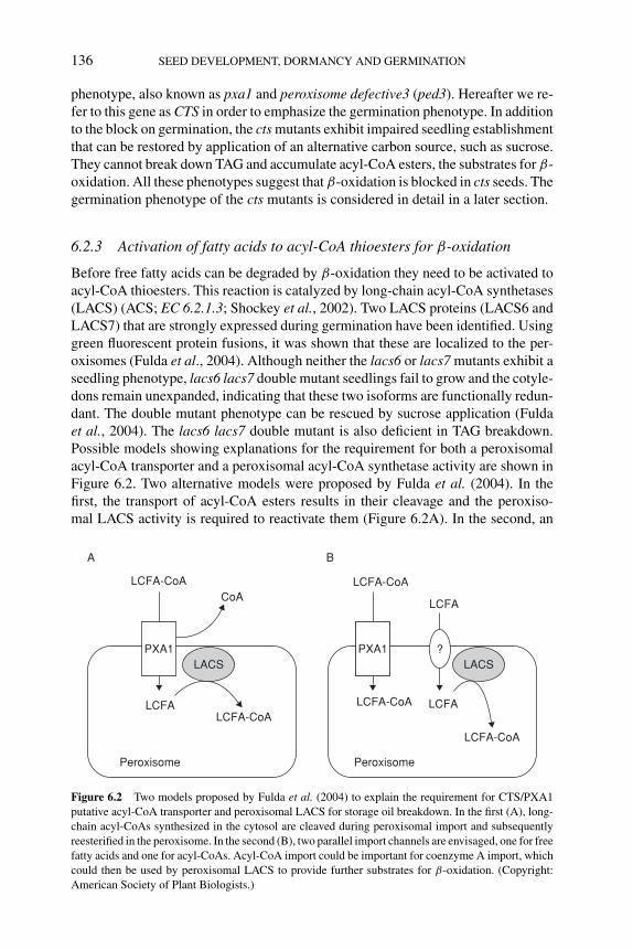
P1: PJM
BLUK053-Bradford August 27, 2006 17:19
136 SEED DEVELOPMENT, DORMANCY AND GERMINATION
phenotype, also known as pxa1 and peroxisome defective3 (ped3). Hereafter we re-fer to this gene as CTS in order to emphasize the germination phenotype. In additionto the block on germination, the cts mutants exhibit impaired seedling establishmentthat can be restored by application of an alternative carbon source, such as sucrose.They cannot break down TAG and accumulate acyl-CoA esters, the substrates for β-oxidation. All these phenotypes suggest that β-oxidation is blocked in cts seeds. Thegermination phenotype of the cts mutants is considered in detail in a later section.
6.2.3 Activation of fatty acids to acyl-CoA thioesters for β-oxidation
Before free fatty acids can be degraded by β-oxidation they need to be activated toacyl-CoA thioesters. This reaction is catalyzed by long-chain acyl-CoA synthetases(LACS) (ACS; EC 6.2.1.3; Shockey et al., 2002). Two LACS proteins (LACS6 andLACS7) that are strongly expressed during germination have been identified. Usinggreen fluorescent protein fusions, it was shown that these are localized to the per-oxisomes (Fulda et al., 2004). Although neither the lacs6 or lacs7 mutants exhibit aseedling phenotype, lacs6 lacs7 double mutant seedlings fail to grow and the cotyle-dons remain unexpanded, indicating that these two isoforms are functionally redun-dant. The double mutant phenotype can be rescued by sucrose application (Fuldaet al., 2004). The lacs6 lacs7 double mutant is also deficient in TAG breakdown.Possible models showing explanations for the requirement for both a peroxisomalacyl-CoA transporter and a peroxisomal acyl-CoA synthetase activity are shown inFigure 6.2. Two alternative models were proposed by Fulda et al. (2004). In thefirst, the transport of acyl-CoA esters results in their cleavage and the peroxiso-mal LACS activity is required to reactivate them (Figure 6.2A). In the second, an
A
LCFA-CoACoA
PXA1LACS LACS
LCFA LCFA
LCFA
Peroxisome
LCFA-CoALCFA-CoA
LCFA-CoA
LCFA-CoA
Peroxisome
PXA1
B
?
Figure 6.2 Two models proposed by Fulda et al. (2004) to explain the requirement for CTS/PXA1
putative acyl-CoA transporter and peroxisomal LACS for storage oil breakdown. In the first (A), long-
chain acyl-CoAs synthesized in the cytosol are cleaved during peroxisomal import and subsequently
reesterified in the peroxisome. In the second (B), two parallel import channels are envisaged, one for free
fatty acids and one for acyl-CoAs. Acyl-CoA import could be important for coenzyme A import, which
could then be used by peroxisomal LACS to provide further substrates for β-oxidation. (Copyright:
American Society of Plant Biologists.)
P1: PJM
BLUK053-Bradford August 27, 2006 17:19
LIPID METABOLISM IN SEED DORMANCY 137
alternative route for entry of free fatty acids into the peroxisome operates in additionto the transport of acyl-CoA esters by CTS. These free fatty acids would then requireactivation by the LACS in the peroxisomal lumen.
6.2.4 β-Oxidation
In each round of the β-oxidation spiral, acetyl-CoA (C2) is cleaved from acyl-CoA (Cn). The remaining acyl-CoA (Cn−2) reenters the β-oxidation spiral and byrepetitive passages through the pathway acetyl-CoA is produced. This is convertedto sucrose via the glyoxylate cycle and gluconeogenesis (Graham and Eastmond,2002). The breakdown of straight-chain saturated acyl-CoAs requires the enzymesacyl-CoA oxidase (ACX, EC 1.3.3.6), multifunctional protein, and 3-l-ketoacyl-CoA thiolase. These enzymes catalyze oxidation, hydration and dehydrogenation,and thiolytic cleavage, respectively, of acyl-CoA. Degradation of unsaturated fattyacids with double bonds at odd numbered carbons or cis double bonds requiresadditional auxiliary enzymes to convert these double bonds to the 2-trans-enoyl-CoA intermediate, which is the substrate for the 2-trans-enoyl-CoA hydratase(EC 4.2.1.17) activity of the multifunctional protein (Graham and Eastmond, 2002;Goepfert et al., 2005). All three families of β-oxidation genes are upregulated co-ordinately during Arabidopsis seed germination and early postgerminative growth(Rylott et al., 2001). This correlates with the period of most rapid fatty acid degra-dation in Arabidopsis (Germain et al., 2001).
6.2.4.1 Acyl-CoA oxidasesAcyl-CoA oxidases (ACXs; EC 1.3.3.6) catalyze the first step of the peroxisomalβ-oxidation sequence, where acyl-CoA is oxidized to �2-trans-enoyl-CoA. Thereaction requires a FAD as a cofactor to generate FADH2, which is then oxidizedby flavoprotein dehydrogenase to produce hydrogen peroxide. Studies have shownthat Arabidopsis contains a family of six ACX isoenzymes with distinct but slightlyoverlapping acyl-CoA chain length specificities (Hooks et al., 1999). Mutants dis-rupted in all six ACX genes have been described (Eastmond et al., 2000b; Rylottet al., 2003; Adham et al., 2005). Of these, the acx1, acx3, and acx4 mutants showweak resistance of root growth to IBA, and acx3 and acx4 show resistance to 2,4-DB,suggesting a defect in β-oxidation. However, only the acx1 acx2 double mutant wasfound to be defective in seedling establishment, suggesting that the overlappingsubstrate specificities of these two isoforms are most important for germinatingseeds (Adham et al., 2005; Pinfield-Wells et al., 2005). The double mutant alsoaccumulates acyl-CoA esters and cannot break down TAG.
6.2.4.2 Multifunctional proteinMultifunctional protein (MFP) has a complex role in β-oxidation, exhibiting mul-tiple activities required in different combinations for the β-oxidation of saturatedand polyunsaturated fatty acids (reviewed in Graham and Eastmond, 2002). Twoactivities, 2-trans-enoyl-CoA hydratase and l-3-hydroxyacyl-CoA dehydrogenase(EC 1.1.1.35), are required for the β-oxidation of saturated fatty acids and thoseunsaturated fatty acids containing a trans double bond at the �2 position. Thedehydrogenase step requires NAD+ and generates NADH. Arabidopsis contains
P1: PJM
BLUK053-Bradford August 27, 2006 17:19
138 SEED DEVELOPMENT, DORMANCY AND GERMINATION
two isoforms of MFP. The first (MFP1) has been characterized genetically as theabnormal inflorescence meristem1 (aim1) locus (Richmond and Bleecker, 1999).This mutant shows aberrant vegetative and reproductive development, underliningthe role of β-oxidation throughout plant development. Seedling establishment isnormal in the aim1 mutant, however. This is not surprising, as the second isoform(MFP2) is strongly and specifically expressed during postgerminative growth, sug-gesting that it plays the predominant role at this stage in development (Eastmondand Graham, 2000). In support of this, mutants in MFP2 show a sugar-dependentshort hypocotyl phenotype in the dark, accumulate acyl-CoAs during seedling es-tablishment, and break down TAG only slowly (Rylott et al., 2006). Although themfp2 mutant phenotype is weaker than other β-oxidation mutants, it is probablethat the activity of AIM1 (MFP1) somewhat compensates for the loss of MFP2 dur-ing germination and seedling establishment. aim1 mfp2 double mutants are embryolethal (Rylott et al., 2006).
6.2.4.3 3-L-Ketoacyl-CoA thiolaseThe enzyme 3-l-ketoacyl-CoA thiolase (KAT; EC 2.3.1.16) catalyzes the last step offatty acid β-oxidation involving thiolytic cleavage of 3-ketoacyl-CoA to acyl-CoA(Cn−2) and acetyl-CoA (C2). The acyl-CoA (Cn−2) then reenters the β-oxidationspiral and the process is repeated until the entire molecule is broken down to acetyl-CoA units. There are four putative thiolase genes in Arabidopsis that contain putativeperoxisomal targeting sequences (Hooks, 2002). Of these, KAT2 is the most impor-tant in the seed. Allelic mutants in KAT2 have been isolated by 2,4-DB resistanceand reverse genetics (Hayashi et al., 1998; Germain et al., 2001). These exhibit thetypical syndrome of compromised seedling establishment that can be rescued bysugar, accumulation of acyl-CoA esters, and inability to break down TAG.
6.2.4.4 Peroxisomal citrate synthaseAlthough traditionally viewed as a glyoxylate cycle enzyme, peroxisomal citratesynthase (pCSY) has recently been shown to be required for respiration ofTAGs (Pracharoenwattana et al., 2005). There are three putative pCSY genes inArabidopsis, but one, CSY1, is only expressed in siliques. Consequently, the csy2csy3 double mutant has revealed the role of this enzyme during seed germinationand vegetative growth. This double mutant fully phenocopies the β-oxidationmutants: TAG breakdown is blocked and seedling establishment is compromisedwithout an alternative carbon source. Mutant seedlings are also 2,4-DB resistant.These phenotypes are not shared with other glyoxylate cycle mutants (see below;Eastmond et al., 2000a; Cornah et al., 2004). Pracharoenwattana et al. (2005)concluded that in the absence of CSY, acetyl-CoA cannot be transported fromperoxisomes on further metabolized, and so β-oxidation is blocked. Thus, CSYactivity is required for carbon export from the peroxisome.
6.2.5 Glyoxylate cycle and gluconeogenesis
The acetyl-CoA produced via the β-oxidation spiral is further metabolized by theglyoxylate cycle. This can be viewed as a modified form of the tricarboxylic acid
P1: PJM
BLUK053-Bradford August 27, 2006 17:19
LIPID METABOLISM IN SEED DORMANCY 139
(TCA) cycle, which bypasses the decarboxylative steps, allowing the net productionof carbon skeletons without the loss of carbon as CO2 (Eastmond and Graham, 2001).Five enzymes are involved in this cycle, two that are specific to the glyoxylatecycle, isocitrate lyase (ICL; EC 4.1.3.1), and malate synthase (MLS; EC 4.1.3.2),and three that also appear in the TCA cycle, citrate synthase (CSY; EC 4.1.3.7),aconitase (ACO; EC 4.2.1.3) and malate dehydrogenase (MDH; EC 1.1.1.37). Allthese enzymes are localized in the peroxisome with the exception of aconitase, whichis reported to be a cytosolic enzyme in castor bean, potato (Solanum tuberosum),and pumpkin (Cucurbita sp.) (Courtois-Verniquet and Douce, 1993; Hayashi et al.,1995). Carbon from the glyoxylate cycle enters gluconeogenesis via the action ofphosphoenolpyruvate carboxykinase.
Many of these glyoxylate enzymes have been investigated individually to deter-mine their roles in the glyoxylate cycle and the role of this cycle in lipid utiliza-tion, gluconeogenesis, and seedling growth in Arabidopsis. Much of this has beenachieved by the isolation and characterization of mutants.
6.2.5.1 Isocitrate lyaseTwo allelic Arabidopsis mutants isocitrate lyase 1 and 2 (icl-1 and icl2), both lackingICL and therefore the glyoxylate cycle, have been characterized (Eastmond et al.,2000b). Under favorable conditions the mutant seedlings establish at high frequency;however, when day-length is shortened or the light intensity decreased, seedlingestablishment is much reduced. In addition, hypocotyl elongation in the dark ismarkedly impaired in the icl mutants. Again, these phenotypes are restored by theprovision of an alternative carbon source. The action of the glyoxylate cycle mustbe vital for buried seeds, for those in competition with other seedlings, and forthose whose lipid reserves are contained within the endosperm. However, fatty acidbreakdown still occurs in the mutant despite the block in the net conversion of acetateto carbon, suggesting that acetyl units are respired.
6.2.5.2 Malate synthaseIsolation of two malate synthase (mls) mutants (Cornah et al., 2004) supports thefindings of Eastmond et al. (2000b) showing that the glyoxylate cycle is not abso-lutely required for seedling growth or lipid breakdown. Comparison of icl and mlsmutants showed that mls seedlings grew and used lipid faster than icl seedlings, yetstill had a short hypocotyl phenotype in the dark. The much less severe phenotypeof mls appears to be due to the seedling’s ability to use an alternative gluconeogenicmechanism, potentially by hijacking some of the activities of the photorespiratoryenzymes (Cornah et al., 2004).
6.2.5.3 Phosphoenolpyruvate carboxykinaseThe seedling phenotype of phosphenolpyruvate carboxykinase (pck1) mutants isessentially similar to that described for mls (Penfield et al., 2004). Hence glu-coneogenesis is not an absolute requirement for seedling establishment. It was
P1: PJM
BLUK053-Bradford August 27, 2006 17:19
140 SEED DEVELOPMENT, DORMANCY AND GERMINATION
also shown that this enzyme is required for the utilization of endospermic lipidreserves by the embryo. In castor bean seeds, carbon from endospermic lipid istransported to the embryo for utilization in the form of sucrose (Kornberg andBeevers, 1957).
6.3 Lipid metabolism and seed dormancy
6.3.1 Importance of the ABC transporter for the transition fromdormancy to germination
The cts locus was originally isolated from a screen aimed at identifying novel factorsaffecting seed dormancy in Arabidopsis (Russell et al., 2000). As discussed above,cts is allelic to pxa1 and ped3 and encodes a putative fatty acid or acyl-CoA trans-porter required for fatty acid β-oxidation in the peroxisome (Footitt et al., 2002;Figure 6.2). Two alleles have been thoroughly characterized in relation to their seedgermination characteristics: cts-1 in the Landsberg erecta (Ler) background andcts-2 in the Wassilewskija (WS) background. In essence, cts mutant seeds exhibit adeep dormancy syndrome. The after-ripened seeds fail to germinate, and cold strati-fication only slightly increases the germination percentage but does not break cts dor-mancy (Russell et al., 2000; Figure 6.3). Promotion of germination is observed whencts seeds are exposed to light, stratified, and placed on nitrogen-containing mediumsuch as Murashige and Skoog (Pinfield-Wells et al., 2005). However, even underthese optimum conditions only a small percentage of cts mutant seeds will germi-nate, generally averaging around 10% using seeds 2 weeks after harvest (Figure 6.3).During the first experiments using cts-1 seed, Russell et al. (2000) found that thebest way to recover cts-1 mutant seedlings was to excise the embryo from the en-dosperm and testa and grow in the presence of sucrose. This process bypassed thecoat-imposed dormancy of Arabidopsis seeds (see Chapter 2) and allowed cts-1 em-bryos to grow and establish seedlings. In this way the cts-1 mutant was propagatedand analyzed, and the responsible gene was eventually cloned using map-basedtechniques (Footitt et al., 2002).
cts mutant seeds have been described as exhibiting a ‘forever dormant’ phenotype(Russell et al., 2000). The primary evidence that CTS is required for normal seeddormancy comes from the analysis of the genetic interactions of the cts-1 mutant withother loci known to be required for the onset of dormancy (Russell et al., 2000; Footittet al., 2002). Arabidopsis mutants such as leafy cotyledon1 (lec1), fusca3 (fus3), andabscisic acid insensitive3 (abi3) accumulate decreased amounts of storage reserves,are not desiccation tolerant, and do not enter seed dormancy (Baumlein et al., 1994;West et al., 1994; Nambara et al., 1995; Chapter 1). However, the mutant embryosare still enclosed within the endosperm and testa and so germination must occurbefore seedling establishment. Therefore, if CTS is required for germination thenthe cts-1 mutant should be epistatic to lec1, abi3, and fus3. Yet, when these threemutant loci are combined with cts-1 all three in fact show epistasis to cts-1 andthe germination percentages of cts-1 lec1, cts-1 fus3, and cts-1 abi3-4 seeds closelyparallel those of the fus3, lec1, and abi3 single mutants (Figure 6.4). These results
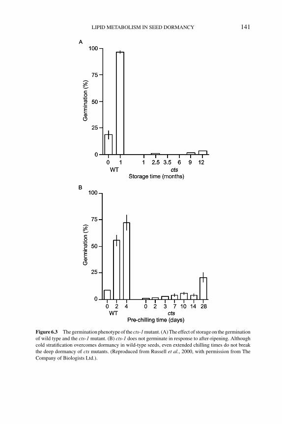
P1: PJM
BLUK053-Bradford August 27, 2006 17:19
LIPID METABOLISM IN SEED DORMANCY 141
Figure 6.3 The germination phenotype of the cts-1 mutant. (A) The effect of storage on the germination
of wild type and the cts-1 mutant. (B) cts-1 does not germinate in response to after-ripening. Although
cold stratification overcomes dormancy in wild-type seeds, even extended chilling times do not break
the deep dormancy of cts mutants. (Reproduced from Russell et al., 2000, with permission from The
Company of Biologists Ltd.).
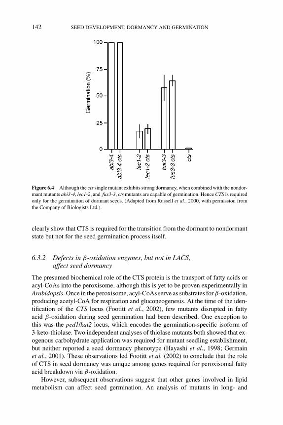
P1: PJM
BLUK053-Bradford August 27, 2006 17:19
142 SEED DEVELOPMENT, DORMANCY AND GERMINATION
Figure 6.4 Although the cts single mutant exhibits strong dormancy, when combined with the nondor-
mant mutants abi3-4, lec1-2, and fus3-3, cts mutants are capable of germination. Hence CTS is required
only for the germination of dormant seeds. (Adapted from Russell et al., 2000, with permission from
the Company of Biologists Ltd.).
clearly show that CTS is required for the transition from the dormant to nondormantstate but not for the seed germination process itself.
6.3.2 Defects in β-oxidation enzymes, but not in LACS,affect seed dormancy
The presumed biochemical role of the CTS protein is the transport of fatty acids oracyl-CoAs into the peroxisome, although this is yet to be proven experimentally inArabidopsis. Once in the peroxisome, acyl-CoAs serve as substrates for β-oxidation,producing acetyl-CoA for respiration and gluconeogenesis. At the time of the iden-tification of the CTS locus (Footitt et al., 2002), few mutants disrupted in fattyacid β-oxidation during seed germination had been described. One exception tothis was the ped1/kat2 locus, which encodes the germination-specific isoform of3-keto-thiolase. Two independent analyses of thiolase mutants both showed that ex-ogenous carbohydrate application was required for mutant seedling establishment,but neither reported a seed dormancy phenotype (Hayashi et al., 1998; Germainet al., 2001). These observations led Footitt et al. (2002) to conclude that the roleof CTS in seed dormancy was unique among genes required for peroxisomal fattyacid breakdown via β-oxidation.
However, subsequent observations suggest that other genes involved in lipidmetabolism can affect seed germination. An analysis of mutants in long- and
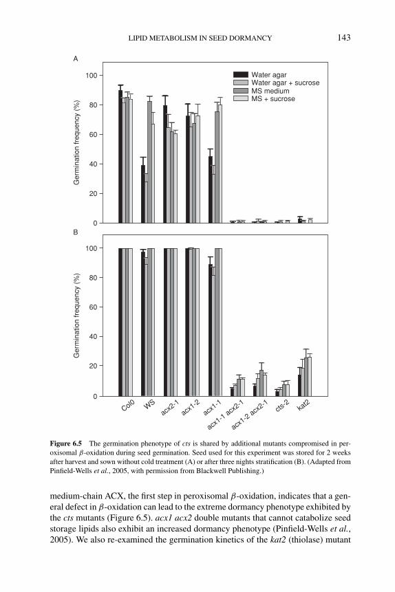
P1: PJM
BLUK053-Bradford August 27, 2006 17:19
LIPID METABOLISM IN SEED DORMANCY 143
100
80
60
40
20
0
100
80
60
40
20
0
Ger
min
atio
n fr
eque
ncy
(%)
Ger
min
atio
n fr
eque
ncy
(%)
Col0 WSacx
2-1
acx1-2
acx1-1
acx1-1 acx
2-1
acx1-2 acx
2-1cts
-2ka
t2
Water agarWater agar + sucroseMS mediumMS + sucrose
A
B
Figure 6.5 The germination phenotype of cts is shared by additional mutants compromised in per-
oxisomal β-oxidation during seed germination. Seed used for this experiment was stored for 2 weeks
after harvest and sown without cold treatment (A) or after three nights stratification (B). (Adapted from
Pinfield-Wells et al., 2005, with permission from Blackwell Publishing.)
medium-chain ACX, the first step in peroxisomal β-oxidation, indicates that a gen-eral defect in β-oxidation can lead to the extreme dormancy phenotype exhibited bythe cts mutants (Figure 6.5). acx1 acx2 double mutants that cannot catabolize seedstorage lipids also exhibit an increased dormancy phenotype (Pinfield-Wells et al.,2005). We also re-examined the germination kinetics of the kat2 (thiolase) mutant
P1: PJM
BLUK053-Bradford August 27, 2006 17:19
144 SEED DEVELOPMENT, DORMANCY AND GERMINATION
and found that it also exhibited a cts-like dormancy phenotype that had been over-looked in both previous mutant analyses. This was not surprising, as no dormancyphenotype had been described for two further alleles of the CTS locus, ped3 andpxa1, prior to the discovery that these are allelic to cts (Zolman et al., 2001; Hayashiet al., 2002). In addition, Pracharoenwattana et al. (2005) noted that the csy2 csy3double mutant seeds that were defective in β-oxidation (Figure 6.1) also germinatedvery poorly and demonstrated a cts-like ‘forever dormant’ phenotype. Hence, thecsy2 csy3 double mutant represents a fourth lesion in lipid metabolism that leadsto an increased dormancy phenotype. These examples underline the importance ofincluding seed germination and dormancy among the phenotypes examined whencharacterizing mutants in lipid metabolism.
For storage lipid breakdown, fatty acids must be activated to the correspondingacyl-CoA ester to enter the β-oxidation pathway. In the case of long-chain fatty acids,of which the majority of TAG is comprised, this reaction is performed by peroxiso-mally localized LACS (Figure 6.2). In common with the β-oxidation mutants, lacs6lacs7 double mutants exhibit a sugar-dependent seedling establishment phenotypeand impaired fatty acid breakdown. Nonetheless, seed germination is reported to benormal in the lacs6 lacs7 double mutants (Fulda et al., 2004; Pinfield-Wells et al.,2005). In the presence of exogenously applied sucrose, lipid breakdown will takeplace in lacs6 lacs7 double mutant seedlings, albeit at an extremely slow rate (Fuldaet al., 2004). This has not been observed in kat2 mutants or in the acx1 acx2 doublemutant, but importantly has been reported in the cts mutants (Footitt et al., 2002).The fact that this slow fatty acid breakdown phenotype is also exhibited by dormantcts seeds suggests that this cannot be responsible for the normal germination oflacs6 lacs7 double mutants. The lacs mutants are therefore particularly instructivebecause they appear to show that the β-oxidation of LACS substrates is not requiredfor germination, even though these substrates comprise the majority of TAG and arerequired for seedling establishment.
6.3.3 Storage lipid mobilization (glyoxylate cycle and gluconeogenesis) isnot required for seed dormancy release
At this point it is instructive to stop and briefly consider pathways and enzymes thatare required for normal storage lipid mobilization. The best characterized of these arethe glyoxylate cycle and gluconeogenesis. These were shown in germinating castorbean seeds to be part of the mechanism for the metabolism and export of carbonfrom the endosperm to the germinating embryo (Kornberg and Beevers, 1957).Mutants in two glyoxylate cycle enzymes, ICL and MLS, have been described inArabidopsis. Unlike pCSY, ICL and MLS have no role in β-oxidation (Eastmondet al., 2000a; Cornah et al., 2004) and mutants defective in these enzymes do notexhibit enhanced levels of seed dormancy. The same is true for the mutant alleles ofthe gluconeogenic enzyme PCK (Penfield et al., 2004). pck1 mutants show abnormalseedling establishment and defects in the mobilization of endospermic reserves andtheir transport to the embryo, but like the icl and mls mutants, catabolize fattyacids and germinate in a wild-type fashion. Therefore, it seems that altered seed
P1: PJM
BLUK053-Bradford August 27, 2006 17:19
LIPID METABOLISM IN SEED DORMANCY 145
dormancy is not a general result of compromising storage lipid conversion to solublecarbohydrate; rather it appears to be restricted to mutants affecting fatty acid β-oxidation in the peroxisome.
6.4 Mechanisms for the involvement of β-oxidation indormancy release
6.4.1 β-Oxidation does not fuel seed germination
As we have seen previously, lesions in genes vital for the peroxisomal β-oxidation offatty acids during seed germination lead to the failure of seedling establishment andthe retention of oil bodies in the cotyledons. In all known cases to date, the provisionof an alternative carbon source rescues affected seedlings and allows them to attainphotosynthetic competence, albeit at a slower rate than the corresponding wild-typeseedlings. This is consistent with the role of β-oxidation in providing acetyl-CoAfrom seed storage reserves as a substrate for respiration, the glyoxylate cycle, andgluconeogenesis. So can the dormancy phenotype of cts and related mutants also beexplained by the same metabolic block imposed on fatty acid β-oxidation and thelack of carbon availability to fuel germination? So far the weight of results suggeststhat limited carbon availability is not the cause of the cts phenotype. Firstly, theapplication of exogenous sugars does not rescue the germination of cts-1 singlemutants, even following a period of after-ripening (Footitt et al., 2002). The germi-nation of cts-1 seeds is improved by the weak acids propionate and butyrate, whichcould conceivably be used as a carbon source in the dormant seeds. However, thiseffect could not be demonstrated on dormant csy2 csy3 (Pracharoenwattana et al.,2005), acx1 acx2, kat2, or cts-2 seeds (H. Pinfield-Wells and I.A. Graham, unpub-lished results). csy2 csy3 seed germination also was unaffected by the presence ofsugars. The observation that exogenous sugars do not promote the germination ofmutants defective in peroxisomal β-oxidation was confirmed by the parallel analy-sis performed by Pinfield-Wells et al. (2005). The germination percentages of cts2,kat2, or acx1 acx2 seeds were not improved by sugars, yet small promotive effects ofnitrogen-containing media or cold stratification were observed. From these resultsit seems clear that the germination phenotype of β-oxidation mutants is separablefrom the seedling establishment phenotypes in terms of the effect of sugar. These re-sults suggest that although seedling establishment requires carbon from β-oxidation,seed germination does not.
6.4.2 β-Oxidation and hormonal signaling
Gibberellin biosynthesis and perception are essential for seed germination inArabidopsis (see Chapters 5, 8, and 9). This is clear because strong gibberellin-deficient mutants such as ga1-3 display a complete nongerminating phenotype.This phenotype can be fully restored by an exogenous supply of gibberellins suchas GA3 (Koornneef and Van der Veen, 1980). In addition, the F-BOX protein
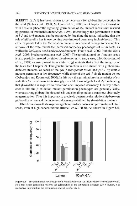
P1: PJM
BLUK053-Bradford August 27, 2006 17:19
146 SEED DEVELOPMENT, DORMANCY AND GERMINATION
SLEEPY1 (SLY1) has been shown to be necessary for gibberellin perception inthe seed (Steber et al., 1998; McGinnis et al., 2003; see Chapter 10). Consistentwith a role in gibberellin signaling, germination of sly1 mutant seeds is not rescuedby gibberellin treatment (Steber et al., 1998). Interestingly, the germination of bothga1-3 and sly1 mutants can be promoted by breaking the testa, indicating that therole of gibberellin lies in overcoming coat-imposed dormancy in Arabidopsis. Thiseffect is paralleled in the β-oxidation mutants; mechanical damage to or completeremoval of the testa reverts the increased dormancy phenotypes of cts mutants, aswell as the kat2, acx1 acx2, and csy2 csy3 mutants (Footitt et al., 2002; Pinfield-Wellset al., 2005; Pracharoenwattana et al., 2005). The germination of cts-1 mutant seedsis also partially restored by either the aberrant testa shape (ats; Leon-Kloosterzielet al., 1994) or transparent testa glabra (ttg) mutants that affect the integrity ofthe testa (see Chapter 2). This genetic interaction is also shared with gibberellin-deficient mutants, as seeds of the ga1-1 transparent testa4 and ga1-1 ttg doublemutants germinate at low frequency, while those of the ga1-1 single mutant do not(Debeaujon and Koornneef, 2000). In this way, the germination characteristics of ctsand other β-oxidation mutants strongly resemble those of ga1-3 and sly1, indicatingthat β-oxidation is required to overcome coat-imposed dormancy. One key differ-ence is that the β-oxidation mutant germination phenotypes are generally leaky,whereas strong gibberellin biosynthesis and signaling mutants can show absolutelyno germination. Thus it is important to precisely determine the relationship betweengibberellin action and the increased dormancy exhibited by β-oxidation mutants.
It has been shown that exogenous gibberellin does not rescue germination of cts-1seeds, even at high concentrations (Russell et al., 2000). As shown in Figure 6.6,
Figure 6.6 The germination of wild type andβ-oxidation mutants on media with or without gibberellin.
Note that while gibberellin restores the germination of the gibberellin-deficient ga1-3 mutant, it is
ineffective in promoting the germination of acx1 acx2 or cts-2.
P1: PJM
BLUK053-Bradford August 27, 2006 17:19
LIPID METABOLISM IN SEED DORMANCY 147
exogenous gibberellin does not rescue the germination of seeds of cts-2 or acx1acx2 mutants (similar data for the kat2 mutant not shown). Thus, the action ofβ-oxidation must lie genetically downstream of gibberellin biosynthesis, and mustbe either in the perception of or reaction to the gibberellin signal that overcomescoat-imposed dormancy. This is also consistent with the finding that signals thatpromote germination largely by upregulating gibberellin biosynthesis, such as lightand cold, have only minor effects on the germination of β-oxidation mutants.
6.4.3 Possible biosynthetic roles for β-oxidation in regulating germination
Besides the well-known function of peroxisomal β-oxidation during reserve mo-bilization in germinating oilseeds, there are numerous other potential roles forβ-oxidation in plants. Indeed, an analysis of acx3 acx4 double mutants has shownthem to be embryo lethal, demonstrating a constitutive function for β-oxidationduring embryo development (Rylott et al., 2003). csy2 csy3 double mutants cannotcomplete the vegetative stage of development and senesce and die before the onsetof flowering, while the aim1 mutant exhibits a dwarf phenotype and abnormal flowerformation (Richmond and Bleeker, 1999; Pracharoenwattana et al., 2005). Thesephenotypes point to the multiple roles of fatty acid β-oxidation in plants.
Perhaps the best understood of these is the requirement of β-oxidation for thesynthesis of the oxylipin signal molecule methyl jasmonate. Jasmonate synthesisbegins with the peroxidation of linolenic acid in the chloroplast and the subsequentproduction of 12-oxo-phytodienoic acid (OPDA), which can be stored in the chloro-plast membranes. OPDA is transported to the peroxisomes where reduction and tworounds of β-oxidation results in the production of jasmonate. It has recently beenshown that the ACX1 isoform is specifically required for jasmonate biosynthesisin both Arabidopsis and tomato (Cruz Castillo et al., 2004; Li et al., 2005). It isalso known that both KAT2 and CTS are required for normal jasmonate synthesis inArabidopsis (Cruz Castillo et al., 2004; Theodoulou et al., 2005). The three mutantsacx1, cts-2, and kat2 all exhibit highly reduced levels of jasmonate in control andwounded plants, yet it is likely that some residual jasmonate synthesis remains asthey exhibit none of the strong phenotypes evident in jasmonate signaling mutants.For instance, coronatine insensitive1 (coi1) and jasmonate resistant1 (jar1) are malesterile, while cts, kat2, and acx1 all produce viable pollen. So could jasmonate be thedormancy-breaking signal molecule? Germination experiments with the acx1 andacx1acx2 double mutants suggest that this is unlikely. Applied jasmonate does notrestore the germination of β-oxidation mutants (H. Pinfield-Wells and I.A. Graham,unpublished results). Furthermore, while the acx1 mutant is impaired in jasmonatebiosynthesis, seed dormancy is unaffected (Pinfield-Wells et al., 2005). Only aftercombining acx1 with acx2 is a seed dormancy phenotype observed. Furthermore,strong jasmonate-insensitive mutants such as coi1 do not exhibit a cts-like ‘for-ever dormant’ phenotype. However, it is quite possible that an alternative unknownoxylipin signal is required for seed germination in Arabidopsis. For example, sev-eral intermediates in the jasmonate biosynthetic pathway have biological activity(Blechert et al., 1995). Plants produce a wide range of volatile oxylipin-derived
P1: PJM
BLUK053-Bradford August 27, 2006 17:19
148 SEED DEVELOPMENT, DORMANCY AND GERMINATION
compounds and the functions of most of these remain unknown (Weber et al.,1997).
It is also possible that β-oxidation metabolizes a non-oxylipin-derived signal.Here a precedent exists in the form of IBA. IBA is a naturally occurring auxin thatrequires the action of β-oxidation to convert it to the more biologically active IAA(Epsteinand Ludwig-Muller, 1993). This conversion to IAA requires two roundsof peroxisomal β-oxidation and is dependent on the action of CTS and KAT2 inyoung seedlings (Hayashi et al., 1998; Zolman et al., 2001). Indeed, this is thebasis of the 2,4-DB resistance genetic screens that first isolated the ped1/kat2 andpxa1/ped3/cts mutants. No systematic attempt to identify all coenzyme A esters ingerminating seeds has ever been attempted, partly due to the technical difficultiesin quantifying and identifying molecules present at very low abundances in seedsamples. However, further characterization of coenzyme A esters in germinatingseeds may lead to the identification of a signal molecule important for seed ger-mination that requires β-oxidation for its production. Alternatively, a product ofβ-oxidation may be important for the synthesis of a signal molecule. One candidatewould be acetyl-CoA, which is an essential substrate for many secondary metabolicpathways.
6.4.4 β-Oxidation, reactive oxygen species, and redox control
The process of fatty acid β-oxidation impacts greatly on the redox balance of thecell. The action of ACX produces reactive oxygen intermediates that are believed tobe oxidized to molecular oxygen by catalase or by a peroxisomal ascorbate perox-idase system. It has been hypothesized that the peroxisome could be an importantsource of reactive oxygen signals, including nitric oxide (Corpas et al., 2001; seeChapter 7). Reactive oxygen species (ROS) produced during germination have beenshown to modulate seed dormancy in some species (Bailly, 2004). The preciseextent of the oxidative load produced during germination and seedling establish-ment by fatty acid β-oxidation has been revealed by the characterization of theArabidopsis vitaminE2 (vte2) mutant (Sattler et al., 2004). The vte2 mutant is defi-cient in tocopherol biosynthesis in the seed and exhibits a sugar-dependent seedlingestablishment phenotype consistent with a role for seed tocopherols in peroxisomalfatty acid β-oxidation. Germinated vte2 mutants accumulate lipid hydroperoxidesand hydroxy fatty acids that cannot proceed through β-oxidation, leading to theblock in seedling establishment. Oxidative damage to seed proteins during germi-nation has also been documented (Job et al., 2005). These observations suggestthat sufficient reactive oxygen exists to cause extensive protein damage even inwild-type seedlings. Much of this could be a by-product of fatty acid β-oxidation,although other sources of ROS do exist in germinating seeds, notably the NADPHoxidases (Kwak et al., 2003). Perhaps more interestingly, tocopherol-deficient seedsexhibit reduced longevity (Sattler et al., 2004). This finding is of particular signifi-cance, for it suggests the presence of ROS in seeds prior to germination that couldfunction in dormancy control. ROS generated through the action of the NADPHoxidases have been shown to modulate abscisic acid signaling in germinating seeds
P1: PJM
BLUK053-Bradford August 27, 2006 17:19
LIPID METABOLISM IN SEED DORMANCY 149
(Kwak et al., 2003). However, there is no evidence that this affects seed dormancy.Although there is ample scope for the production of ROS by fatty acid β-oxidation,there is as yet no direct evidence that this has any regulatory roles during seeddormancy and germination in Arabidopsis.
In addition to acetyl-CoA and hydrogen peroxide, the β-oxidation pathway alsogenerates NADH through the action of the MFP. Shuttle mechanisms that involvetransport of metabolites between the peroxisome, mitochondria, and cytosol to en-able reoxidation of NADH have been proposed (Mettler and Beevers, 1980; Cornahand Smith, 2002). These ultimately result in reducing equivalents being transferredfrom the peroxisome and production of NADH from NAD in the cytosol and mito-chondria. Germination studies in nondormant and dormant caryopses of oat (Avenasativa) have shown that there is an increase in NADH content and in the ratioNADH/(NADH + NAD+) (catabolic redox charge [CRC]) during early germina-tion of nondormant caryopses, which differed significantly from the NADH contentand CRC of nongerminating dormant caryopses (Gallais et al., 1998). These stud-ies also showed that ethanol provokes increases in NADH and consequently inCRC, suggesting that germination is enhanced or accelerated when the NADH con-tent increases. Arabidopsis mutants compromised in peroxisomal β-oxidation offatty acids derived from storage lipids during germination could therefore be com-promised in reducing equivalents such as NADH, which in turn could impact ongermination. However, the fact that the lacs6 lacs7 double mutant shows normalgermination despite the fact that it is as compromised as the other mutants in fattyacid breakdown argues against CRC being the cause of the reduced germination incts, kat2, and acx1 acx2.
6.5 Conclusions
Using molecular genetic approaches, it has been shown that peroxisomalβ-oxidationis required for the termination of dormancy and initiation of germination inArabidopsis seeds. All the evidence suggests that this effect is independent of thewell-known role of β-oxidation in the catabolism of seed oil reserves. Further workto determine the precise function of β-oxidation in seeds will provide key insightsinto the control of seed dormancy and germination.
References
A.R. Adham, B.K. Zolman, A. Millius and B. Bartel (2005) Mutations in Arabidopsis acyl-CoA
oxidase genes reveal distinct and overlapping roles in β-oxidation. The Plant Journal 41, 859–
874.
C. Bailly (2004) Active oxygen species and antioxidants in seed biology. Seed Science Research 14,
93–107.
S. Baud, J.-P. Boutin, M. Miquel, L. Lepiniec and C. Rochat (2002) An integrated overview of seed
development in Arabidopsis thaliana ecotype Ws. Plant Physiology and Biochemistry 40, 151–
160.
H. Baumlein, S. Misera, H. Luerssen, et al. (1994) The fus3 gene of Arabidopsis thaliana is a regulator
of gene expression during late embryogenesis. The Plant Journal 6, 379–387.
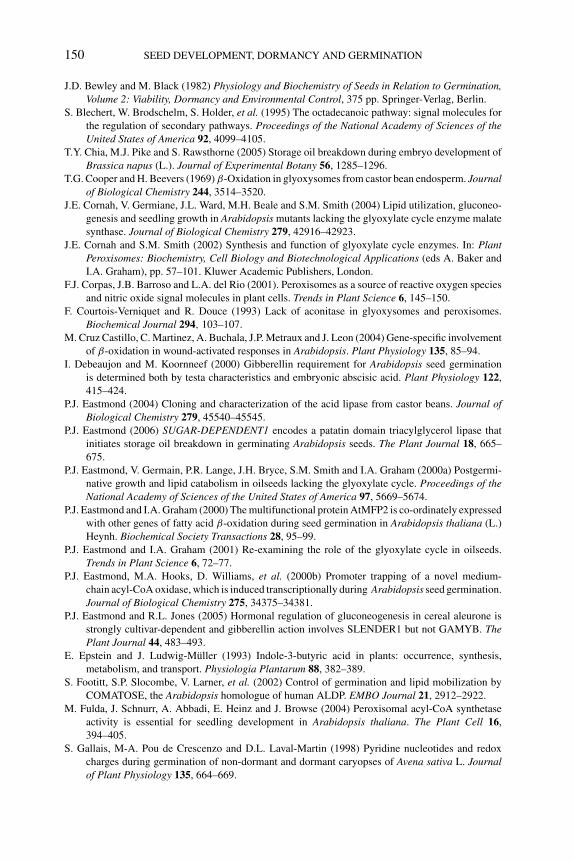
P1: PJM
BLUK053-Bradford August 27, 2006 17:19
150 SEED DEVELOPMENT, DORMANCY AND GERMINATION
J.D. Bewley and M. Black (1982) Physiology and Biochemistry of Seeds in Relation to Germination,Volume 2: Viability, Dormancy and Environmental Control, 375 pp. Springer-Verlag, Berlin.
S. Blechert, W. Brodschelm, S. Holder, et al. (1995) The octadecanoic pathway: signal molecules for
the regulation of secondary pathways. Proceedings of the National Academy of Sciences of theUnited States of America 92, 4099–4105.
T.Y. Chia, M.J. Pike and S. Rawsthorne (2005) Storage oil breakdown during embryo development of
Brassica napus (L.). Journal of Experimental Botany 56, 1285–1296.
T.G. Cooper and H. Beevers (1969) β-Oxidation in glyoxysomes from castor bean endosperm. Journalof Biological Chemistry 244, 3514–3520.
J.E. Cornah, V. Germiane, J.L. Ward, M.H. Beale and S.M. Smith (2004) Lipid utilization, gluconeo-
genesis and seedling growth in Arabidopsis mutants lacking the glyoxylate cycle enzyme malate
synthase. Journal of Biological Chemistry 279, 42916–42923.
J.E. Cornah and S.M. Smith (2002) Synthesis and function of glyoxylate cycle enzymes. In: PlantPeroxisomes: Biochemistry, Cell Biology and Biotechnological Applications (eds A. Baker and
I.A. Graham), pp. 57–101. Kluwer Academic Publishers, London.
F.J. Corpas, J.B. Barroso and L.A. del Rio (2001). Peroxisomes as a source of reactive oxygen species
and nitric oxide signal molecules in plant cells. Trends in Plant Science 6, 145–150.
F. Courtois-Verniquet and R. Douce (1993) Lack of aconitase in glyoxysomes and peroxisomes.
Biochemical Journal 294, 103–107.
M. Cruz Castillo, C. Martinez, A. Buchala, J.P. Metraux and J. Leon (2004) Gene-specific involvement
of β-oxidation in wound-activated responses in Arabidopsis. Plant Physiology 135, 85–94.
I. Debeaujon and M. Koornneef (2000) Gibberellin requirement for Arabidopsis seed germination
is determined both by testa characteristics and embryonic abscisic acid. Plant Physiology 122,
415–424.
P.J. Eastmond (2004) Cloning and characterization of the acid lipase from castor beans. Journal ofBiological Chemistry 279, 45540–45545.
P.J. Eastmond (2006) SUGAR-DEPENDENT1 encodes a patatin domain triacylglycerol lipase that
initiates storage oil breakdown in germinating Arabidopsis seeds. The Plant Journal 18, 665–
675.
P.J. Eastmond, V. Germain, P.R. Lange, J.H. Bryce, S.M. Smith and I.A. Graham (2000a) Postgermi-
native growth and lipid catabolism in oilseeds lacking the glyoxylate cycle. Proceedings of theNational Academy of Sciences of the United States of America 97, 5669–5674.
P.J. Eastmond and I.A. Graham (2000) The multifunctional protein AtMFP2 is co-ordinately expressed
with other genes of fatty acid β-oxidation during seed germination in Arabidopsis thaliana (L.)
Heynh. Biochemical Society Transactions 28, 95–99.
P.J. Eastmond and I.A. Graham (2001) Re-examining the role of the glyoxylate cycle in oilseeds.
Trends in Plant Science 6, 72–77.
P.J. Eastmond, M.A. Hooks, D. Williams, et al. (2000b) Promoter trapping of a novel medium-
chain acyl-CoA oxidase, which is induced transcriptionally during Arabidopsis seed germination.
Journal of Biological Chemistry 275, 34375–34381.
P.J. Eastmond and R.L. Jones (2005) Hormonal regulation of gluconeogenesis in cereal aleurone is
strongly cultivar-dependent and gibberellin action involves SLENDER1 but not GAMYB. ThePlant Journal 44, 483–493.
E. Epstein and J. Ludwig-Muller (1993) Indole-3-butyric acid in plants: occurrence, synthesis,
metabolism, and transport. Physiologia Plantarum 88, 382–389.
S. Footitt, S.P. Slocombe, V. Larner, et al. (2002) Control of germination and lipid mobilization by
COMATOSE, the Arabidopsis homologue of human ALDP. EMBO Journal 21, 2912–2922.
M. Fulda, J. Schnurr, A. Abbadi, E. Heinz and J. Browse (2004) Peroxisomal acyl-CoA synthetase
activity is essential for seedling development in Arabidopsis thaliana. The Plant Cell 16,
394–405.
S. Gallais, M-A. Pou de Crescenzo and D.L. Laval-Martin (1998) Pyridine nucleotides and redox
charges during germination of non-dormant and dormant caryopses of Avena sativa L. Journalof Plant Physiology 135, 664–669.
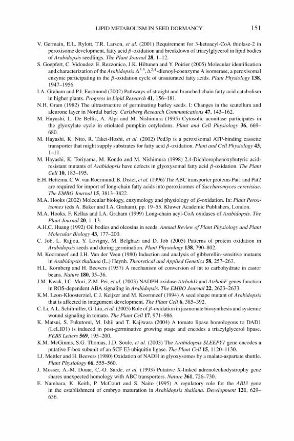
P1: PJM
BLUK053-Bradford August 27, 2006 17:19
LIPID METABOLISM IN SEED DORMANCY 151
V. Germain, E.L. Rylott, T.R. Larson, et al. (2001) Requirement for 3-ketoacyl-CoA thiolase-2 in
peroxisome development, fatty acid β-oxidation and breakdown of triacylglycerol in lipid bodies
of Arabidopsis seedlings. The Plant Journal 28, 1–12.
S. Goepfert, C. Vidoudez, E. Rezzonico, J.K. Hiltunen and Y. Poirier (2005) Molecular identification
and characterization of the Arabidopsis �3,5,�2,4-dienoyl-coenzyme A isomerase, a peroxisomal
enzyme participating in the β-oxidation cycle of unsaturated fatty acids. Plant Physiology 138,
1947–1956.
I.A. Graham and P.J. Eastmond (2002) Pathways of straight and branched chain fatty acid catabolism
in higher plants. Progress in Lipid Research 41, 156–181.
N.H. Gram (1982) The ultrastructure of germinating barley seeds. I: Changes in the scutellum and
aleurone layer in Nordal barley. Carlsberg Research Communications 47, 143–162.
M. Hayashi, L. De Bellis, A. Alpi and M. Nishimura (1995) Cytosolic aconitase participates in
the glyoxylate cycle in etiolated pumpkin cotyledons. Plant and Cell Physiology 36, 669–
680.
M. Hayashi, K. Nito, R. Takei-Hoshi, et al. (2002) Ped3p is a peroxisomal ATP-binding cassette
transporter that might supply substrates for fatty acid β-oxidation. Plant and Cell Physiology 43,
1–11.
M. Hayashi, K. Toriyama, M. Kondo and M. Nishimura (1998) 2,4-Dichlorophenoxybutyric acid-
resistant mutants of Arabidopsis have defects in glyoxysomal fatty acid β-oxidation. The PlantCell 10, 183–195.
E.H. Hettema, C.W. van Roermund, B. Distel, et al. (1996) The ABC transporter proteins Pat1 and Pat2
are required for import of long-chain fatty acids into peroxisomes of Saccharomyces cerevisiae.
The EMBO Journal 15, 3813–3822.
M.A. Hooks (2002) Molecular biology, enzymology and physiology of β-oxidation. In: Plant Perox-isomes (eds A. Baker and I.A. Graham), pp. 19–55. Kluwer Academic Publishers, London.
M.A. Hooks, F. Kellas and I.A. Graham (1999) Long-chain acyl-CoA oxidases of Arabidopsis. ThePlant Journal 20, 1–13.
A.H.C. Huang (1992) Oil bodies and oleosins in seeds. Annual Review of Plant Physiology and PlantMolecular Biology 43, 177–200.
C. Job, L. Rajjou, Y. Lovigny, M. Belghazi and D. Job (2005) Patterns of protein oxidation in
Arabidopsis seeds and during germination. Plant Physiology 138, 790–802.
M. Koornneef and J.H. Van der Veen (1980) Induction and analysis of gibberellin-sensitive mutants
in Arabidopsis thaliana (L.) Heynh. Theoretical and Applied Genetics 58, 257–263.
H.L. Kornberg and H. Beevers (1957) A mechanism of conversion of fat to carbohydrate in castor
beans. Nature 180, 35–36.
J.M. Kwak, I.C. Mori, Z.M. Pei, et al. (2003) NADPH oxidase AtrbohD and AtrbohF genes function
in ROS-dependent ABA signaling in Arabidopsis. The EMBO Journal 22, 2623–2633.
K.M. Leon-Kloosterziel, C.J. Keijzer and M. Koornneef (1994) A seed shape mutant of Arabidopsisthat is affected in integument development. The Plant Cell 6, 385–392.
C. Li, A.L. Schilmiller, G. Liu, et al. (2005) Role of β-oxidation in jasmonate biosynthesis and systemic
wound signaling in tomato. The Plant Cell 17, 971–986.
K. Matsui, S. Fukutomi, M. Ishii and T. Kajiwara (2004) A tomato lipase homologous to DAD1
(LeLID1) is induced in post-germinative growing stage and encodes a triacylglycerol lipase.
FEBS Letters 569, 195–200.
K.M. McGinnis, S.G. Thomas, J.D. Soule, et al. (2003) The Arabidopsis SLEEPY1 gene encodes a
putative F-box subunit of an SCF E3 ubiquitin ligase. The Plant Cell 15, 1120–1130.
I.J. Mettler and H. Beevers (1980) Oxidation of NADH in glyoxysomes by a malate-aspartate shuttle.
Plant Physiology 66, 555–560.
J. Mosser, A.-M. Douar, C.-O. Sarde, et al. (1993) Putative X-linked adrenoleukodystrophy gene
shares unexpected homology with ABC transporters. Nature 361, 726–730.
E. Nambara, K. Keith, P. McCourt and S. Naito (1995) A regulatory role for the ABI3 gene
in the establishment of embryo maturation in Arabidopsis thaliana. Development 121, 629–
636.
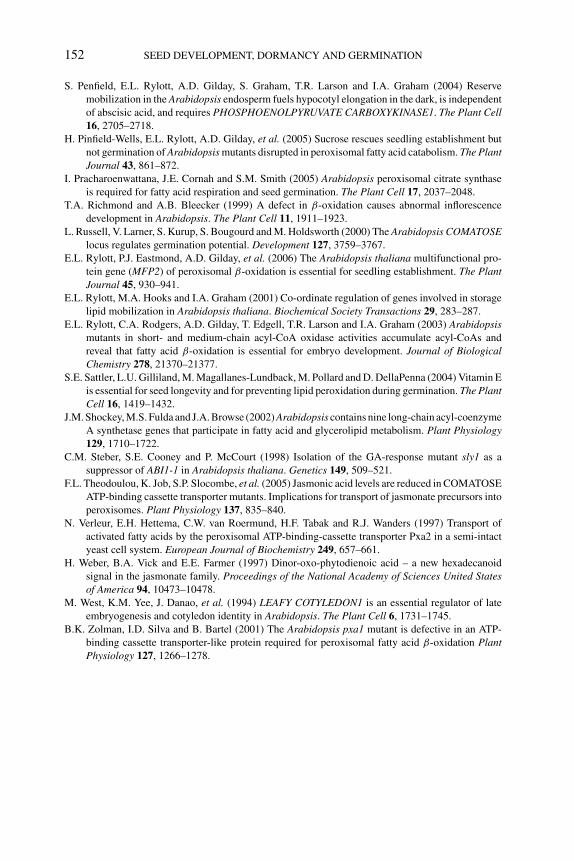
P1: PJM
BLUK053-Bradford August 27, 2006 17:19
152 SEED DEVELOPMENT, DORMANCY AND GERMINATION
S. Penfield, E.L. Rylott, A.D. Gilday, S. Graham, T.R. Larson and I.A. Graham (2004) Reserve
mobilization in the Arabidopsis endosperm fuels hypocotyl elongation in the dark, is independent
of abscisic acid, and requires PHOSPHOENOLPYRUVATE CARBOXYKINASE1. The Plant Cell16, 2705–2718.
H. Pinfield-Wells, E.L. Rylott, A.D. Gilday, et al. (2005) Sucrose rescues seedling establishment but
not germination of Arabidopsis mutants disrupted in peroxisomal fatty acid catabolism. The PlantJournal 43, 861–872.
I. Pracharoenwattana, J.E. Cornah and S.M. Smith (2005) Arabidopsis peroxisomal citrate synthase
is required for fatty acid respiration and seed germination. The Plant Cell 17, 2037–2048.
T.A. Richmond and A.B. Bleecker (1999) A defect in β-oxidation causes abnormal inflorescence
development in Arabidopsis. The Plant Cell 11, 1911–1923.
L. Russell, V. Larner, S. Kurup, S. Bougourd and M. Holdsworth (2000) The Arabidopsis COMATOSElocus regulates germination potential. Development 127, 3759–3767.
E.L. Rylott, P.J. Eastmond, A.D. Gilday, et al. (2006) The Arabidopsis thaliana multifunctional pro-
tein gene (MFP2) of peroxisomal β-oxidation is essential for seedling establishment. The PlantJournal 45, 930–941.
E.L. Rylott, M.A. Hooks and I.A. Graham (2001) Co-ordinate regulation of genes involved in storage
lipid mobilization in Arabidopsis thaliana. Biochemical Society Transactions 29, 283–287.
E.L. Rylott, C.A. Rodgers, A.D. Gilday, T. Edgell, T.R. Larson and I.A. Graham (2003) Arabidopsismutants in short- and medium-chain acyl-CoA oxidase activities accumulate acyl-CoAs and
reveal that fatty acid β-oxidation is essential for embryo development. Journal of BiologicalChemistry 278, 21370–21377.
S.E. Sattler, L.U. Gilliland, M. Magallanes-Lundback, M. Pollard and D. DellaPenna (2004) Vitamin E
is essential for seed longevity and for preventing lipid peroxidation during germination. The PlantCell 16, 1419–1432.
J.M. Shockey, M.S. Fulda and J.A. Browse (2002) Arabidopsis contains nine long-chain acyl-coenzyme
A synthetase genes that participate in fatty acid and glycerolipid metabolism. Plant Physiology129, 1710–1722.
C.M. Steber, S.E. Cooney and P. McCourt (1998) Isolation of the GA-response mutant sly1 as a
suppressor of ABI1-1 in Arabidopsis thaliana. Genetics 149, 509–521.
F.L. Theodoulou, K. Job, S.P. Slocombe, et al. (2005) Jasmonic acid levels are reduced in COMATOSE
ATP-binding cassette transporter mutants. Implications for transport of jasmonate precursors into
peroxisomes. Plant Physiology 137, 835–840.
N. Verleur, E.H. Hettema, C.W. van Roermund, H.F. Tabak and R.J. Wanders (1997) Transport of
activated fatty acids by the peroxisomal ATP-binding-cassette transporter Pxa2 in a semi-intact
yeast cell system. European Journal of Biochemistry 249, 657–661.
H. Weber, B.A. Vick and E.E. Farmer (1997) Dinor-oxo-phytodienoic acid – a new hexadecanoid
signal in the jasmonate family. Proceedings of the National Academy of Sciences United Statesof America 94, 10473–10478.
M. West, K.M. Yee, J. Danao, et al. (1994) LEAFY COTYLEDON1 is an essential regulator of late
embryogenesis and cotyledon identity in Arabidopsis. The Plant Cell 6, 1731–1745.
B.K. Zolman, I.D. Silva and B. Bartel (2001) The Arabidopsis pxa1 mutant is defective in an ATP-
binding cassette transporter-like protein required for peroxisomal fatty acid β-oxidation PlantPhysiology 127, 1266–1278.
P1: OIY
BLUK053-Bradford August 25, 2006 14:49
7 Nitric oxide in seed dormancyand germinationPaul C. Bethke, Igor G.L. Libourel and Russell L. Jones
7.1 Nitric oxide in plant growth and development
Nitric oxide (NO) is a reactive, gaseous, free radical that functions as a potent sig-naling molecule in plants and animals. Numerous roles for NO in plant growthand development have been uncovered during the past decade (Lamattina et al.,2003; Neill et al., 2003; Wendehenne et al., 2004; Delledonne, 2005; Shapiro,2005). In early reports it was shown that NO acts during the hypersensitive responseto protect plants from pathogens. NO has subsequently been shown to inhibit mat-uration and senescence, and to function as an antioxidant that slows the rate ofprogrammed cell death. NO promotes vigorous seedling development, represses thetransition to flowering, facilitates abscisic acid (ABA)-induced stomatal closure,and may be required for the adaptation of roots to low oxygen concentrations. Thenumber of processes in which NO participates is increasing rapidly, and additionalNO-dependent events in plants are likely to be discovered. In this chapter we reviewdata that indicate that NO plays important roles in seed dormancy and germination.Some of the literature discussed here was written prior to the recognition that NOfunctions as a signaling molecule, and we discuss it with the benefit of hindsight. Therest of the data summarized here come from experiments devised to test directly foran involvement of NO in seed dormancy or germination. When taken together, thesedata support the hypothesis that NO promotes the germination of seeds, either byreducing seed dormancy or by minimizing the effects of environmental conditionsthat inhibit germination.
7.2 Challenges in NO chemistry and biology
NO is a challenging molecule to work with for both chemists and biologists. Thesechallenges derive from the nature of the NO molecule itself. NO is a highly reac-tive, free radical gas that dissolves in aqueous solution but preferentially partitionsinto hydrophobic environments such as biomembranes. NO is not stable in aero-bic environments. NO in the gas phase reacts with itself and molecular oxygen toproduce nitrogen dioxide (NO2), as indicated in Equation 7.1, and NO2 that dif-fuses into an aqueous environment is converted to equal molar concentrations of
153
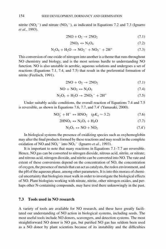
P1: OIY
BLUK053-Bradford August 25, 2006 14:49
154 SEED DEVELOPMENT, DORMANCY AND GERMINATION
nitrite (NO2−) and nitrate (NO3
−), as indicated in Equations 7.2 and 7.3 (Ignarroet al., 1993).
2NO + O2 → 2NO2 (7.1)
2NO2 ↔ N2O4 (7.2)
N2O4 + H2O → NO2− + NO3
− + 2H+ (7.3)
This conversion of one oxide of nitrogen into another is a theme that runs throughoutNO chemistry and biology, and is the most serious hurdle to understanding NOfunction. NO is also unstable in aerobic, aqueous solutions and undergoes a set ofreactions (Equations 7.1, 7.4, and 7.5) that result in the preferential formation ofnitrite (Feelisch, 1991).
2NO + O2 → 2NO2 (7.1)
NO + NO2 ↔ N2O3 (7.4)
N2O3 + H2O → 2NO2− + 2H+ (7.5)
Under suitably acidic conditions, the overall reaction of Equations 7.4 and 7.5is reversible, as shown in Equations 7.6, 7.7, and 7.4′ (Yamasaki, 2000).
NO−2 + H+ ↔ HNO2 (pKa ∼ 3.2) (7.6)
2HNO2 ↔ N2O3 + H2O (7.7)
N2O3 ↔ NO + NO2 (7.4′)
In biological systems the presence of oxidizing species such as oxyhemoglobinmay alter the final products formed by these reactions and may result in the completeoxidation of NO and NO2
− into NO3− (Ignarro et al., 1993).
It is important to note that many reactions in Equations 7.1–7.7 are reversible.Hence, NO gas can be converted to nitrogen dioxide, nitrous acid, nitrite, or nitrate;and nitrous acid, nitrogen dioxide, and nitrite can be converted into NO. The rate andextent of these conversions depend on the concentration of NO, the concentrationof oxygen, the presence of metals that can act as catalysts, the redox environment, andthe pH of the aqueous phase, among other parameters. It is into this morass of chemi-cal uncertainty that biologists must walk in order to investigate the biological effectsof NO. Plant biologists working with nitrate, nitrite, other nitrogen oxides, and per-haps other N-containing compounds, may have trod there unknowingly in the past.
7.3 Tools used in NO research
A variety of tools are available for NO research, and these have greatly facili-tated our understanding of NO action in biological systems, including seeds. Themost useful tools include NO donors, scavengers, and detection systems. The moststraightforward NO donor is NO gas, but purified NO gas has seldom been usedas a NO donor by plant scientists because of its instability and the difficulties
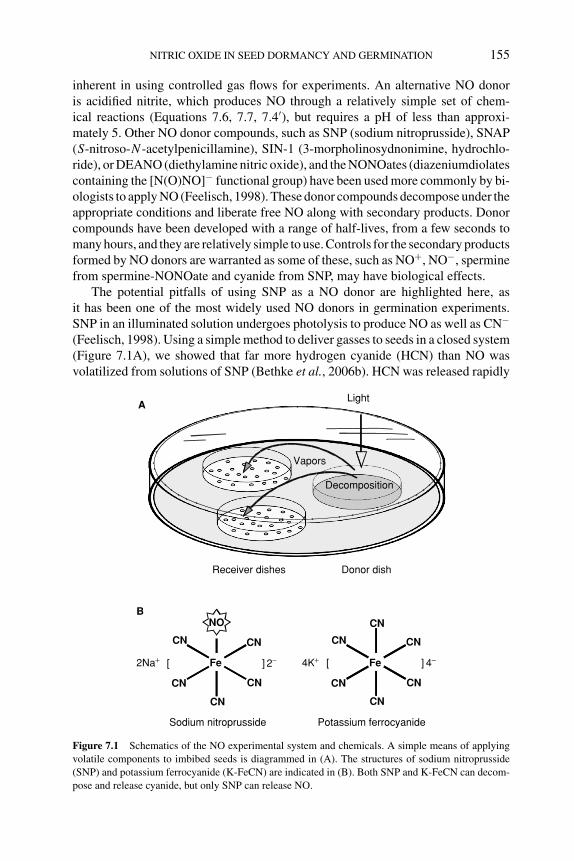
P1: OIY
BLUK053-Bradford August 25, 2006 14:49
NITRIC OXIDE IN SEED DORMANCY AND GERMINATION 155
inherent in using controlled gas flows for experiments. An alternative NO donoris acidified nitrite, which produces NO through a relatively simple set of chem-ical reactions (Equations 7.6, 7.7, 7.4′), but requires a pH of less than approxi-mately 5. Other NO donor compounds, such as SNP (sodium nitroprusside), SNAP(S-nitroso-N -acetylpenicillamine), SIN-1 (3-morpholinosydnonimine, hydrochlo-ride), or DEANO (diethylamine nitric oxide), and the NONOates (diazeniumdiolatescontaining the [N(O)NO]− functional group) have been used more commonly by bi-ologists to apply NO (Feelisch, 1998). These donor compounds decompose under theappropriate conditions and liberate free NO along with secondary products. Donorcompounds have been developed with a range of half-lives, from a few seconds tomany hours, and they are relatively simple to use. Controls for the secondary productsformed by NO donors are warranted as some of these, such as NO+, NO−, sperminefrom spermine-NONOate and cyanide from SNP, may have biological effects.
The potential pitfalls of using SNP as a NO donor are highlighted here, asit has been one of the most widely used NO donors in germination experiments.SNP in an illuminated solution undergoes photolysis to produce NO as well as CN−
(Feelisch, 1998). Using a simple method to deliver gasses to seeds in a closed system(Figure 7.1A), we showed that far more hydrogen cyanide (HCN) than NO wasvolatilized from solutions of SNP (Bethke et al., 2006b). HCN was released rapidly
Donor dishReceiver dishes
A
Fe
CN CN
CN
CN
CN
CN
[ ] 4−4K+
Potassium ferrocyanide
Fe
CN CN
CN
CN
CN
NO
[ ] 2−2Na+
Sodium nitroprusside
B
Vapors
Light
Decomposition
Figure 7.1 Schematics of the NO experimental system and chemicals. A simple means of applying
volatile components to imbibed seeds is diagrammed in (A). The structures of sodium nitroprusside
(SNP) and potassium ferrocyanide (K-FeCN) are indicated in (B). Both SNP and K-FeCN can decom-
pose and release cyanide, but only SNP can release NO.
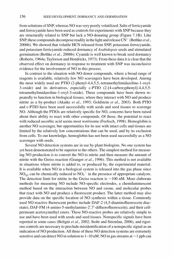
P1: OIY
BLUK053-Bradford August 25, 2006 14:49
156 SEED DEVELOPMENT, DORMANCY AND GERMINATION
from solutions of SNP, whereas NO was very poorly volatilized. Salts of ferricyanideand ferrocyanide have been used as controls for experiments with SNP because theyare structurally related to SNP but lack a NO-donating group (Figure 7.1B). LikeSNP, these compounds decompose readily in the light and release CN− (Bethke et al.,2006b). We showed that volatile HCN released from SNP, potassium ferrocyanide,and potassium ferricyanide reduced dormancy of Arabidopsis seeds and stimulatedgermination (Bethke et al., 2006b). Cyanide is well known to break seed dormancy(Roberts, 1964a; Taylorson and Hendricks, 1973). From these data it is clear that theobserved effect on dormancy in response to treatment with SNP was inconclusiveevidence for the involvement of NO in this process.
In contrast to the situation with NO donor compounds, where a broad range ofreagents is available, relatively few NO scavengers have been developed. Amongthe most widely used are PTIO (2-phenyl-4,4,5,5,-tetramethylimidazoline-1-oxyl-3-oxide) and its derivatives, especially c-PTIO (2-[4-carboxyphenyl]-4,4,5,5-tetramethylimidazoline-1-oxyl-3-oxide). These compounds have been shown re-peatedly to function in biological tissues, where they interact with NO and producenitrite as a by-product (Akaike et al., 1993; Goldstein et al., 2003). Both PTIOand c-PTIO have been used successfully with seeds and seed tissues to scavengeNO. Although the PTIOs are relatively specific for NO, concerns have been raisedabout their ability to react with other compounds. Of those, the potential to reactwith reduced ascorbic acid seems most worrisome (Feelisch, 1998). Hemoglobin isanother NO scavenger, but opportunities for its use with intact cells and tissues arelimited by the relatively low concentrations that can be used, and by its exclusionfrom cells. To our knowledge, hemoglobin has not been used successfully as a NOscavenger with seeds.
Several NO detection systems are in use by plant biologists. No one system hasyet been demonstrated to be superior to the others. The simplest method for measur-ing NO production is to convert the NO to nitrite and then measure the amount ofnitrite with the Greiss reaction (Granger et al., 1996). This method is not availablein situations where nitrite is added to, or produced by, the experimental material.It is available when NO in a biological system is released into the gas phase sinceNO(g) can be chemically reduced to NO2
− in the presence of appropriate catalysts.The detection limit for nitrite in the Greiss reaction is ∼100 nM. More elaboratemethods for measuring NO include NO-specific electrodes, a chemiluminescentmethod based on the interaction between NO and ozone, and molecular probesthat react with NO and produce a fluorescent product. The latter method may alsoprovide data on the specific location of NO synthesis within a tissue. Commonlyused NO-reactive fluorescent probes include DAF-2 (4,5-diaminofluorescein diac-etate), DAF-FM (4-amino-5-methylamino-2′,7′-difluorofluorescein), and their cell-permeant acetoxymethyl esters. These NO-reactive probes are relatively simple touse and have been used with seeds and seed tissues. Nonspecific signals have beenreported in some cases (Beligni et al., 2002; Stohr and Stremlau, 2006), and rigor-ous controls are necessary to preclude misidentification of a nonspecific signal as anindication of NO production. All three of these NO detection systems are extremelysensitive and can detect NO in solution to 1–10 nM. NO in gas streams at ∼1 ppb can
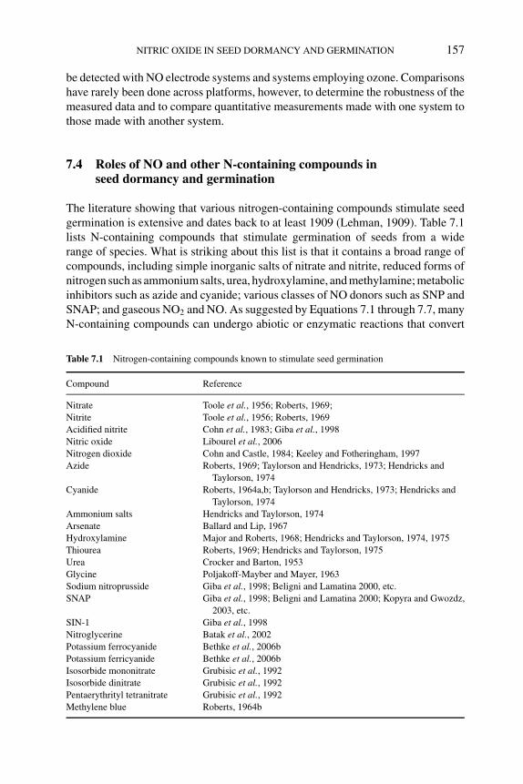
P1: OIY
BLUK053-Bradford August 25, 2006 14:49
NITRIC OXIDE IN SEED DORMANCY AND GERMINATION 157
be detected with NO electrode systems and systems employing ozone. Comparisonshave rarely been done across platforms, however, to determine the robustness of themeasured data and to compare quantitative measurements made with one system tothose made with another system.
7.4 Roles of NO and other N-containing compounds inseed dormancy and germination
The literature showing that various nitrogen-containing compounds stimulate seedgermination is extensive and dates back to at least 1909 (Lehman, 1909). Table 7.1lists N-containing compounds that stimulate germination of seeds from a widerange of species. What is striking about this list is that it contains a broad range ofcompounds, including simple inorganic salts of nitrate and nitrite, reduced forms ofnitrogen such as ammonium salts, urea, hydroxylamine, and methylamine; metabolicinhibitors such as azide and cyanide; various classes of NO donors such as SNP andSNAP; and gaseous NO2 and NO. As suggested by Equations 7.1 through 7.7, manyN-containing compounds can undergo abiotic or enzymatic reactions that convert
Table 7.1 Nitrogen-containing compounds known to stimulate seed germination
Compound Reference
Nitrate Toole et al., 1956; Roberts, 1969;
Nitrite Toole et al., 1956; Roberts, 1969
Acidified nitrite Cohn et al., 1983; Giba et al., 1998
Nitric oxide Libourel et al., 2006
Nitrogen dioxide Cohn and Castle, 1984; Keeley and Fotheringham, 1997
Azide Roberts, 1969; Taylorson and Hendricks, 1973; Hendricks and
Taylorson, 1974
Cyanide Roberts, 1964a,b; Taylorson and Hendricks, 1973; Hendricks and
Taylorson, 1974
Ammonium salts Hendricks and Taylorson, 1974
Arsenate Ballard and Lip, 1967
Hydroxylamine Major and Roberts, 1968; Hendricks and Taylorson, 1974, 1975
Thiourea Roberts, 1969; Hendricks and Taylorson, 1975
Urea Crocker and Barton, 1953
Glycine Poljakoff-Mayber and Mayer, 1963
Sodium nitroprusside Giba et al., 1998; Beligni and Lamatina 2000, etc.
SNAP Giba et al., 1998; Beligni and Lamatina 2000; Kopyra and Gwozdz,
2003, etc.
SIN-1 Giba et al., 1998
Nitroglycerine Batak et al., 2002
Potassium ferrocyanide Bethke et al., 2006b
Potassium ferricyanide Bethke et al., 2006b
Isosorbide mononitrate Grubisic et al., 1992
Isosorbide dinitrate Grubisic et al., 1992
Pentaerythrityl tetranitrate Grubisic et al., 1992
Methylene blue Roberts, 1964b
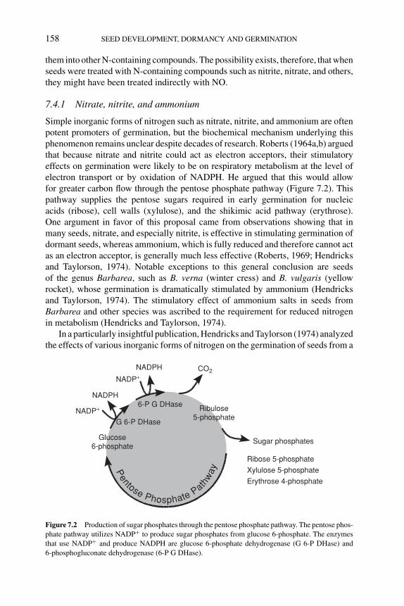
P1: OIY
BLUK053-Bradford August 25, 2006 14:49
158 SEED DEVELOPMENT, DORMANCY AND GERMINATION
them into other N-containing compounds. The possibility exists, therefore, that whenseeds were treated with N-containing compounds such as nitrite, nitrate, and others,they might have been treated indirectly with NO.
7.4.1 Nitrate, nitrite, and ammonium
Simple inorganic forms of nitrogen such as nitrate, nitrite, and ammonium are oftenpotent promoters of germination, but the biochemical mechanism underlying thisphenomenon remains unclear despite decades of research. Roberts (1964a,b) arguedthat because nitrate and nitrite could act as electron acceptors, their stimulatoryeffects on germination were likely to be on respiratory metabolism at the level ofelectron transport or by oxidation of NADPH. He argued that this would allowfor greater carbon flow through the pentose phosphate pathway (Figure 7.2). Thispathway supplies the pentose sugars required in early germination for nucleicacids (ribose), cell walls (xylulose), and the shikimic acid pathway (erythrose).One argument in favor of this proposal came from observations showing that inmany seeds, nitrate, and especially nitrite, is effective in stimulating germination ofdormant seeds, whereas ammonium, which is fully reduced and therefore cannot actas an electron acceptor, is generally much less effective (Roberts, 1969; Hendricksand Taylorson, 1974). Notable exceptions to this general conclusion are seedsof the genus Barbarea, such as B. verna (winter cress) and B. vulgaris (yellowrocket), whose germination is dramatically stimulated by ammonium (Hendricksand Taylorson, 1974). The stimulatory effect of ammonium salts in seeds fromBarbarea and other species was ascribed to the requirement for reduced nitrogenin metabolism (Hendricks and Taylorson, 1974).
In a particularly insightful publication, Hendricks and Taylorson (1974) analyzedthe effects of various inorganic forms of nitrogen on the germination of seeds from a
Sugar phosphates
Ribose 5-phosphate
Xylulose 5-phosphate
Erythrose 4-phosphate
CO2
NADP+
NADP+
NADPH
NADPH
G 6-P DHase
6-P G DHase Ribulose5-phosphate
Glucose6-phosphate
Pentose Phosphate Path
way
Figure 7.2 Production of sugar phosphates through the pentose phosphate pathway. The pentose phos-
phate pathway utilizes NADP+ to produce sugar phosphates from glucose 6-phosphate. The enzymes
that use NADP+ and produce NADPH are glucose 6-phosphate dehydrogenase (G 6-P DHase) and
6-phosphogluconate dehydrogenase (6-P G DHase).
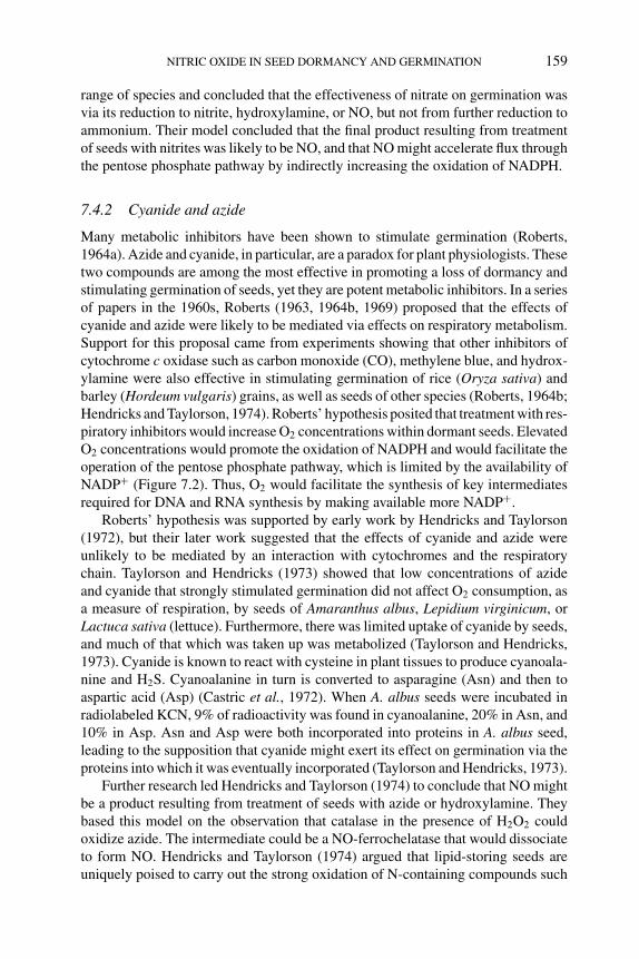
P1: OIY
BLUK053-Bradford August 25, 2006 14:49
NITRIC OXIDE IN SEED DORMANCY AND GERMINATION 159
range of species and concluded that the effectiveness of nitrate on germination wasvia its reduction to nitrite, hydroxylamine, or NO, but not from further reduction toammonium. Their model concluded that the final product resulting from treatmentof seeds with nitrites was likely to be NO, and that NO might accelerate flux throughthe pentose phosphate pathway by indirectly increasing the oxidation of NADPH.
7.4.2 Cyanide and azide
Many metabolic inhibitors have been shown to stimulate germination (Roberts,1964a). Azide and cyanide, in particular, are a paradox for plant physiologists. Thesetwo compounds are among the most effective in promoting a loss of dormancy andstimulating germination of seeds, yet they are potent metabolic inhibitors. In a seriesof papers in the 1960s, Roberts (1963, 1964b, 1969) proposed that the effects ofcyanide and azide were likely to be mediated via effects on respiratory metabolism.Support for this proposal came from experiments showing that other inhibitors ofcytochrome c oxidase such as carbon monoxide (CO), methylene blue, and hydrox-ylamine were also effective in stimulating germination of rice (Oryza sativa) andbarley (Hordeum vulgaris) grains, as well as seeds of other species (Roberts, 1964b;Hendricks and Taylorson, 1974). Roberts’ hypothesis posited that treatment with res-piratory inhibitors would increase O2 concentrations within dormant seeds. ElevatedO2 concentrations would promote the oxidation of NADPH and would facilitate theoperation of the pentose phosphate pathway, which is limited by the availability ofNADP+ (Figure 7.2). Thus, O2 would facilitate the synthesis of key intermediatesrequired for DNA and RNA synthesis by making available more NADP+.
Roberts’ hypothesis was supported by early work by Hendricks and Taylorson(1972), but their later work suggested that the effects of cyanide and azide wereunlikely to be mediated by an interaction with cytochromes and the respiratorychain. Taylorson and Hendricks (1973) showed that low concentrations of azideand cyanide that strongly stimulated germination did not affect O2 consumption, asa measure of respiration, by seeds of Amaranthus albus, Lepidium virginicum, orLactuca sativa (lettuce). Furthermore, there was limited uptake of cyanide by seeds,and much of that which was taken up was metabolized (Taylorson and Hendricks,1973). Cyanide is known to react with cysteine in plant tissues to produce cyanoala-nine and H2S. Cyanoalanine in turn is converted to asparagine (Asn) and then toaspartic acid (Asp) (Castric et al., 1972). When A. albus seeds were incubated inradiolabeled KCN, 9% of radioactivity was found in cyanoalanine, 20% in Asn, and10% in Asp. Asn and Asp were both incorporated into proteins in A. albus seed,leading to the supposition that cyanide might exert its effect on germination via theproteins into which it was eventually incorporated (Taylorson and Hendricks, 1973).
Further research led Hendricks and Taylorson (1974) to conclude that NO mightbe a product resulting from treatment of seeds with azide or hydroxylamine. Theybased this model on the observation that catalase in the presence of H2O2 couldoxidize azide. The intermediate could be a NO-ferrochelatase that would dissociateto form NO. Hendricks and Taylorson (1974) argued that lipid-storing seeds areuniquely poised to carry out the strong oxidation of N-containing compounds such
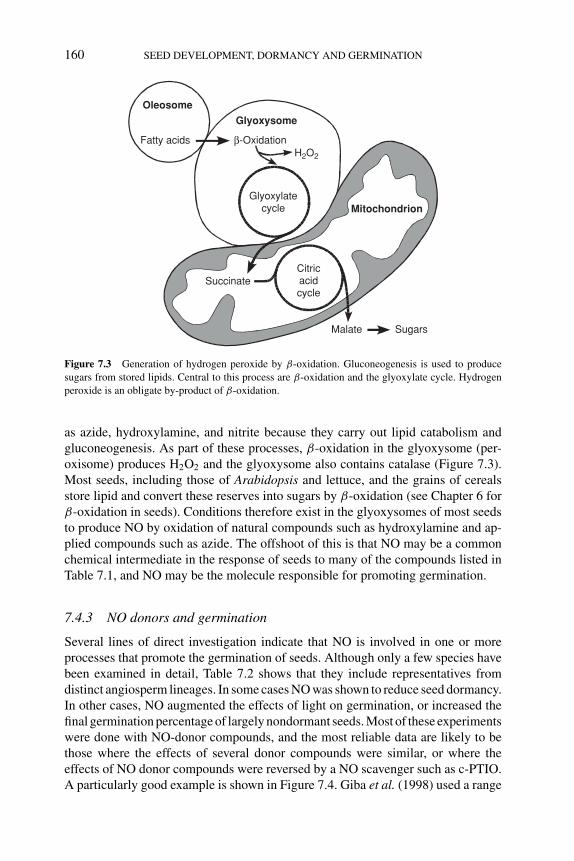
P1: OIY
BLUK053-Bradford August 25, 2006 14:49
160 SEED DEVELOPMENT, DORMANCY AND GERMINATION
Oleosome
Fatty acidsH2O2
Glyoxysome
β-Oxidation
Malate Sugars
Citricacidcycle
Glyoxylatecycle
Succinate
Mitochondrion
Figure 7.3 Generation of hydrogen peroxide by β-oxidation. Gluconeogenesis is used to produce
sugars from stored lipids. Central to this process are β-oxidation and the glyoxylate cycle. Hydrogen
peroxide is an obligate by-product of β-oxidation.
as azide, hydroxylamine, and nitrite because they carry out lipid catabolism andgluconeogenesis. As part of these processes, β-oxidation in the glyoxysome (per-oxisome) produces H2O2 and the glyoxysome also contains catalase (Figure 7.3).Most seeds, including those of Arabidopsis and lettuce, and the grains of cerealsstore lipid and convert these reserves into sugars by β-oxidation (see Chapter 6 forβ-oxidation in seeds). Conditions therefore exist in the glyoxysomes of most seedsto produce NO by oxidation of natural compounds such as hydroxylamine and ap-plied compounds such as azide. The offshoot of this is that NO may be a commonchemical intermediate in the response of seeds to many of the compounds listed inTable 7.1, and NO may be the molecule responsible for promoting germination.
7.4.3 NO donors and germination
Several lines of direct investigation indicate that NO is involved in one or moreprocesses that promote the germination of seeds. Although only a few species havebeen examined in detail, Table 7.2 shows that they include representatives fromdistinct angiosperm lineages. In some cases NO was shown to reduce seed dormancy.In other cases, NO augmented the effects of light on germination, or increased thefinal germination percentage of largely nondormant seeds. Most of these experimentswere done with NO-donor compounds, and the most reliable data are likely to bethose where the effects of several donor compounds were similar, or where theeffects of NO donor compounds were reversed by a NO scavenger such as c-PTIO.A particularly good example is shown in Figure 7.4. Giba et al. (1998) used a range
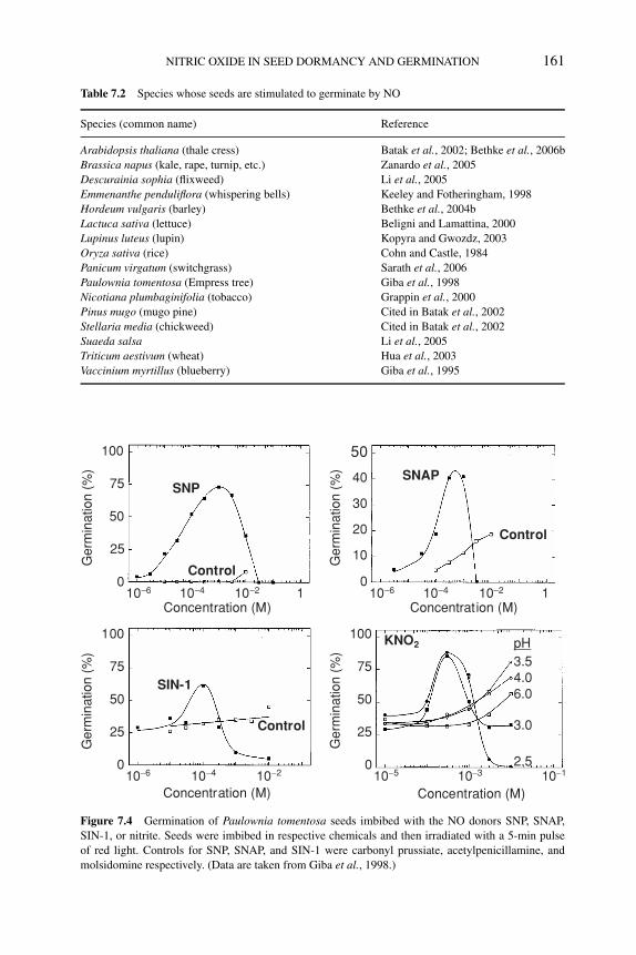
P1: OIY
BLUK053-Bradford August 25, 2006 14:49
NITRIC OXIDE IN SEED DORMANCY AND GERMINATION 161
Table 7.2 Species whose seeds are stimulated to germinate by NO
Species (common name) Reference
Arabidopsis thaliana (thale cress) Batak et al., 2002; Bethke et al., 2006b
Brassica napus (kale, rape, turnip, etc.) Zanardo et al., 2005
Descurainia sophia (flixweed) Li et al., 2005
Emmenanthe penduliflora (whispering bells) Keeley and Fotheringham, 1998
Hordeum vulgaris (barley) Bethke et al., 2004b
Lactuca sativa (lettuce) Beligni and Lamattina, 2000
Lupinus luteus (lupin) Kopyra and Gwozdz, 2003
Oryza sativa (rice) Cohn and Castle, 1984
Panicum virgatum (switchgrass) Sarath et al., 2006
Paulownia tomentosa (Empress tree) Giba et al., 1998
Nicotiana plumbaginifolia (tobacco) Grappin et al., 2000
Pinus mugo (mugo pine) Cited in Batak et al., 2002
Stellaria media (chickweed) Cited in Batak et al., 2002
Suaeda salsa Li et al., 2005
Triticum aestivum (wheat) Hua et al., 2003
Vaccinium myrtillus (blueberry) Giba et al., 1995
100
75
50
25
0
SNP
10−6 10−4 10−2 1Concentration (M)
Control
40
30
20
10
0
50
SNAP
10−6 10−4 10−2 1Concentration (M)
Control
100
75
50
25
0
SIN-1
10−6 10−4 10−2
Concentration (M)
Control
100
75
50
25
0
KNO2
10−5 10−3 10−1
Concentration (M)
2.5
3.0
3.5
6.04.0
pH
Ger
min
atio
n (%
)G
erm
inat
ion
(%)
Ger
min
atio
n (%
)G
erm
inat
ion
(%)
Figure 7.4 Germination of Paulownia tomentosa seeds imbibed with the NO donors SNP, SNAP,
SIN-1, or nitrite. Seeds were imbibed in respective chemicals and then irradiated with a 5-min pulse
of red light. Controls for SNP, SNAP, and SIN-1 were carbonyl prussiate, acetylpenicillamine, and
molsidomine respectively. (Data are taken from Giba et al., 1998.)
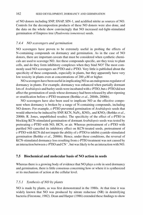
P1: OIY
BLUK053-Bradford August 25, 2006 14:49
162 SEED DEVELOPMENT, DORMANCY AND GERMINATION
of NO donors including SNP, SNAP, SIN-1, and acidified nitrite as sources of NO.Controls for the decomposition products of these NO donors were also done, andthe data on the whole show convincingly that NO increased red-light-stimulatedgermination of Empress tree (Paulownia tomentosa) seeds.
7.4.4 NO scavengers and germination
NO scavengers have proven to be extremely useful in probing the effects ofN-containing compounds on dormancy and germination. As in the case of NOdonors, there are important caveats that must be considered when synthetic chemi-cals are used to scavenge NO. Are these compounds specific, are they toxic to plantcells, and do they form inhibitory complexes when they bind NO? The most com-monly used NO scavengers are PTIO and c-PTIO. Very little is published about thespecificity of these compounds, especially in plants, but they apparently have verylow toxicity in plants even at concentrations of 200 μM or higher.
NO scavengers have been useful in implicating NO as an endogenous regulator ofdormancy in plants. For example, dormancy was enhanced when partially dormantlots of Arabidopsis and barley seeds were incubated with c-PTIO, but c-PTIO did notaffect the germination of seeds whose dormancy had been released by after-ripeningor stratification before c-PTIO treatment (Bethke et al., 2004b, 2006b).
NO scavengers have also been used to implicate NO as the effective compo-nent when dormancy is broken by a range of N-containing compounds, includingNO donors. For example, c-PTIO prevented germination of Arabidopsis seeds thatwould otherwise be induced by SNP, KCN, NaN3, KNO3, and KNO2 (Bethke et al.,2006b; R. Jones, unpublished results). The specificity of the effect of c-PTIO inblocking KCN-stimulated germination of dormant Arabidopsis seeds was tested bypretreating c-PTIO with NO, HCN, or air. Whereas pretreatment of c-PTIO withpurified NO canceled its inhibitory effect on KCN-treated seeds, pretreatment ofc-PTIO with KCN did not impair the ability of c-PTIO to inhibit cyanide-stimulatedgermination (Bethke et al., 2006b). Hence, under these conditions, the reversal ofKCN-stimulated dormancy loss resulting from c-PTIO treatment was not caused byan interaction between c-PTIO and CN−, but was likely to be an interaction with NO.
7.5 Biochemical and molecular basis of NO action in seeds
Whereas there is a growing body of evidence that NO plays a role in seed dormancyand germination, there is little consensus concerning how or where it is synthesizedor its mechanism of action at the cellular level.
7.5.1 Synthesis of NO by plants
NO is made by plants, as was first demonstrated in the 1980s. At that time it waswidely known that NO was produced by nitrate reductase (NR) in denitrifyingbacteria (Firestone, 1982). Dean and Harper (1986) extended these findings to show
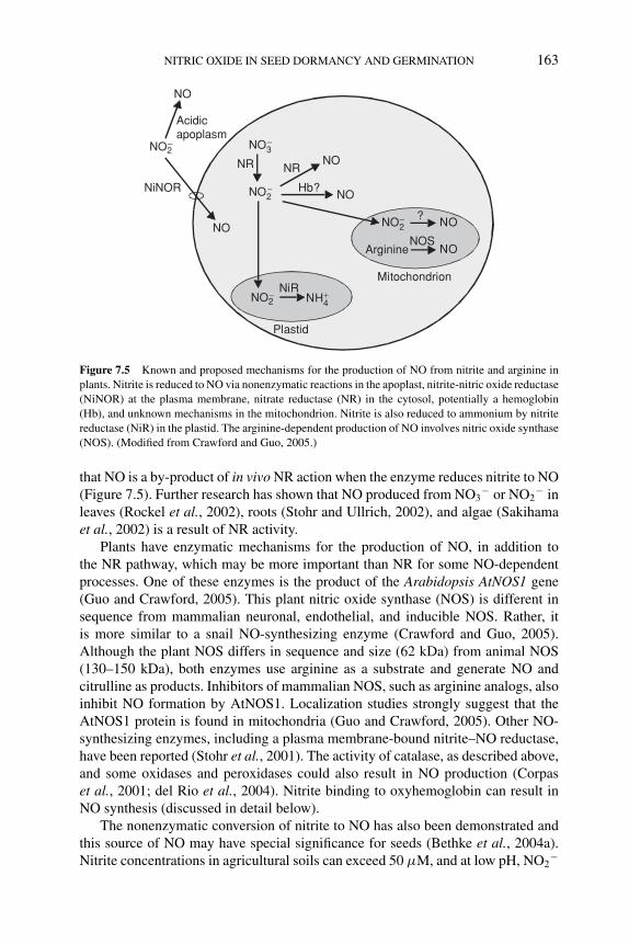
P1: OIY
BLUK053-Bradford August 25, 2006 14:49
NITRIC OXIDE IN SEED DORMANCY AND GERMINATION 163
NO2− NO?
Arginine NONOS
Mitochondrion
NO2− NiR
NH4+
Plastid
NO2−
NO3−
NR NR
Hb?
NO
NO
NO
Acidicapoplasm
NO2−
NiNOR
NO
Figure 7.5 Known and proposed mechanisms for the production of NO from nitrite and arginine in
plants. Nitrite is reduced to NO via nonenzymatic reactions in the apoplast, nitrite-nitric oxide reductase
(NiNOR) at the plasma membrane, nitrate reductase (NR) in the cytosol, potentially a hemoglobin
(Hb), and unknown mechanisms in the mitochondrion. Nitrite is also reduced to ammonium by nitrite
reductase (NiR) in the plastid. The arginine-dependent production of NO involves nitric oxide synthase
(NOS). (Modified from Crawford and Guo, 2005.)
that NO is a by-product of in vivo NR action when the enzyme reduces nitrite to NO(Figure 7.5). Further research has shown that NO produced from NO3
− or NO2− in
leaves (Rockel et al., 2002), roots (Stohr and Ullrich, 2002), and algae (Sakihamaet al., 2002) is a result of NR activity.
Plants have enzymatic mechanisms for the production of NO, in addition tothe NR pathway, which may be more important than NR for some NO-dependentprocesses. One of these enzymes is the product of the Arabidopsis AtNOS1 gene(Guo and Crawford, 2005). This plant nitric oxide synthase (NOS) is different insequence from mammalian neuronal, endothelial, and inducible NOS. Rather, itis more similar to a snail NO-synthesizing enzyme (Crawford and Guo, 2005).Although the plant NOS differs in sequence and size (62 kDa) from animal NOS(130–150 kDa), both enzymes use arginine as a substrate and generate NO andcitrulline as products. Inhibitors of mammalian NOS, such as arginine analogs, alsoinhibit NO formation by AtNOS1. Localization studies strongly suggest that theAtNOS1 protein is found in mitochondria (Guo and Crawford, 2005). Other NO-synthesizing enzymes, including a plasma membrane-bound nitrite–NO reductase,have been reported (Stohr et al., 2001). The activity of catalase, as described above,and some oxidases and peroxidases could also result in NO production (Corpaset al., 2001; del Rio et al., 2004). Nitrite binding to oxyhemoglobin can result inNO synthesis (discussed in detail below).
The nonenzymatic conversion of nitrite to NO has also been demonstrated andthis source of NO may have special significance for seeds (Bethke et al., 2004a).Nitrite concentrations in agricultural soils can exceed 50 μM, and at low pH, NO2
−
P1: OIY
BLUK053-Bradford August 25, 2006 14:49
164 SEED DEVELOPMENT, DORMANCY AND GERMINATION
can be reduced to NO. The pKa of this reaction is 3.2 and it is dramatically ac-celerated by reducing agents. Seeds and grains often maintain a low pH in theapoplast. For example, the cereal aleurone layer secretes phosphoric and organicacids and acidifies the endosperm to between pH 3.5 and 4 (Drozdowicz and Jones,1995). Furthermore, the testa/pericarp that surrounds the aleurone layer containsproanthocyanidins, which are strong antioxidants/reductants (Bethke et al., 2004a).Proanthocyanidins are solubilized from the cell wall of the grain and can contributeto the reduction of NO2
− in the cereal endosperm (see Chapter 2 for proanthocyani-din function in seed dormancy). When NO2
− was added to the incubation medium inwhich barley aleurone layers were incubated, copious amounts of NO were produced(Bethke et al., 2004a). Barley aleurone layers also have a nitrate-inducible NR ac-tivity. When nitrate is supplied to aleurone layers under anaerobic conditions, nitriteis released from the aleurone into the apoplast and starchy endosperm (Ferrari andVarner, 1969). The interior of the cereal grain is likely to have reduced O2 concentra-tions during germination, allowing NO3
− to be reduced to NO2− and released into
the acidic apoplast where it can be readily converted to NO (Bethke et al., 2004a).There is increasing recognition of the importance that nonenzymatic formation
of NO from NO2− has for human biology as well. In fact, NO2
− ‘may represent thelargest intravascular and tissue storage form of nitric oxide’, playing a central rolein the ‘human nitrogen cycle’ where nitrite is converted to NO via reactions withhemoglobin, myoglobin, and xanthine oxidoreductase (Gladwin et al., 2005). Thepresence of nitrate or nitrite in the soil, the ability of plants to synthesize nitrite fromnitrate, and the existence of acidic compartments in plants that are accessible tonitrite are strong indications that nitrate or nitrite may be equally important storageforms of NO for plant growth and development.
7.5.2 NO binding to metal-containing proteins
NO has a high affinity for metal-containing proteins, and proteins containing hemesare attractive targets for NO action (Shapiro, 2005). In mammalian systems, muchof NO action is transduced through the heme-containing enzyme guanalyl cyclase.Guanalyl cyclase may be a target for NO as part of plant responses to pathogens(Durner et al., 1998), but an involvement of this enzyme in NO signaling in seedsis unclear.
Cytochrome c oxidase is a heme-binding protein that is part of the mitochondrialelectron transport chain. It has been shown that NO prevents respiration throughthe cytochrome pathway, and that this prevents ATP synthesis when the alternateoxidase pathway is inhibited (Yamasaki et al., 2001). NO is likely to be produced inmitochondria by AtNOS1 (Guo and Crawford, 2005), making cytochrome c oxidasea potential target for NO binding.
Catalase is a heme-containing protein that is inhibited by NO. Hendricks andTaylorson (1974) hypothesized that NO derived from N-containing compoundsmight function to stimulate germination by inhibiting catalase activity. They pos-tulated that by inhibiting catalase, NO would increase intracellular H2O2 concen-trations, and that H2O2 might act as a substrate for enzymes such as peroxidases.
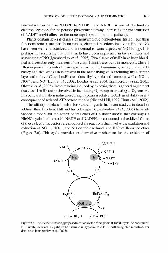
P1: OIY
BLUK053-Bradford August 25, 2006 14:49
NITRIC OXIDE IN SEED DORMANCY AND GERMINATION 165
Peroxidase can oxidize NADPH to NADP+, and NADP+ is one of the limitingelectron acceptors for the pentose phosphate pathway. Increasing the concentrationof NADP+ might allow for the more rapid operation of this pathway.
Plants contain several classes of nonsymbiotic hemoglobins (nsHb), but theirfunctions remain unclear. In mammals, chemical reactions involving Hb and NOhave been well characterized and are central to some aspects of NO biology. It isperhaps not surprising that plant nsHb have been implicated in the synthesis andscavenging of NO (Igamberdiev et al., 2005). Two classes of nsHb have been identi-fied in dicots, but only members of the class-1 family are found in monocots. Class-1Hb is expressed in seeds of many species including Arabidopsis, barley, and rice. Inbarley and rice seeds Hb is present in the outer living cells including the aleuronelayer and embryo. Class-1 nsHb are induced by hypoxia and sucrose as well as NO3
−,NO2
−, and NO (Hunt et al., 2002; Dordas et al., 2004; Igamberdiev et al., 2005;Ohwaki et al., 2005). Despite being induced by hypoxia, there is general agreementthat class-1 nsHb are not involved in facilitating O2 transport or acting as O2 sensors.It is believed that their induction during hypoxia is related to ATP availability or is aconsequence of reduced ATP concentrations (Nie and Hill, 1997; Hunt et al., 2002).
The affinity of class-1 nsHb for various ligands has been studied in detail toaddress their function. Hill and his colleagues (Igamberdiev et al., 2005) have ad-vanced a model for the action of this class of Hb under anoxia that envisages aHb/NO cycle. In this model, NADH and NADPH are consumed and oxidized formsof these electron acceptors are produced via reactions that involve the oxidation andreduction of NO3
−, NO2−, and NO on the one hand, and Hb/metHb on the other
(Figure 7.6). This cycle provides an alternative mechanism for the oxidation of
Figure 7.6 A schematic showing proposed reactions of the hemoglobin (Hb)/NO cycle. Abbreviations:
NR, nitrate reductase; E, putative NO sources in hypoxia; MetHb-R, methemoglobin reductase. For
details see Igamberdiev et al. (2005).
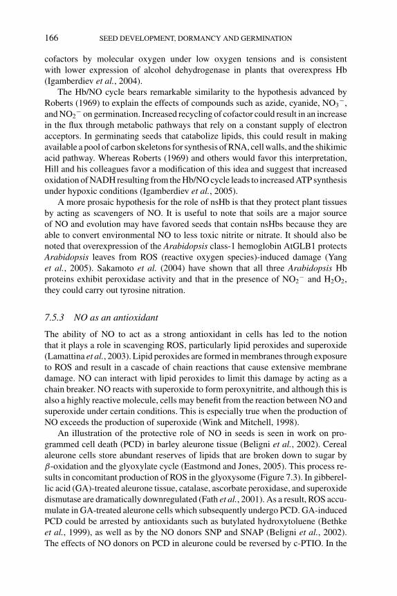
P1: OIY
BLUK053-Bradford August 25, 2006 14:49
166 SEED DEVELOPMENT, DORMANCY AND GERMINATION
cofactors by molecular oxygen under low oxygen tensions and is consistentwith lower expression of alcohol dehydrogenase in plants that overexpress Hb(Igamberdiev et al., 2004).
The Hb/NO cycle bears remarkable similarity to the hypothesis advanced byRoberts (1969) to explain the effects of compounds such as azide, cyanide, NO3
−,and NO2
− on germination. Increased recycling of cofactor could result in an increasein the flux through metabolic pathways that rely on a constant supply of electronacceptors. In germinating seeds that catabolize lipids, this could result in makingavailable a pool of carbon skeletons for synthesis of RNA, cell walls, and the shikimicacid pathway. Whereas Roberts (1969) and others would favor this interpretation,Hill and his colleagues favor a modification of this idea and suggest that increasedoxidation of NADH resulting from the Hb/NO cycle leads to increased ATP synthesisunder hypoxic conditions (Igamberdiev et al., 2005).
A more prosaic hypothesis for the role of nsHb is that they protect plant tissuesby acting as scavengers of NO. It is useful to note that soils are a major sourceof NO and evolution may have favored seeds that contain nsHbs because they areable to convert environmental NO to less toxic nitrite or nitrate. It should also benoted that overexpression of the Arabidopsis class-1 hemoglobin AtGLB1 protectsArabidopsis leaves from ROS (reactive oxygen species)-induced damage (Yanget al., 2005). Sakamoto et al. (2004) have shown that all three Arabidopsis Hbproteins exhibit peroxidase activity and that in the presence of NO2
− and H2O2,they could carry out tyrosine nitration.
7.5.3 NO as an antioxidant
The ability of NO to act as a strong antioxidant in cells has led to the notionthat it plays a role in scavenging ROS, particularly lipid peroxides and superoxide(Lamattina et al., 2003). Lipid peroxides are formed in membranes through exposureto ROS and result in a cascade of chain reactions that cause extensive membranedamage. NO can interact with lipid peroxides to limit this damage by acting as achain breaker. NO reacts with superoxide to form peroxynitrite, and although this isalso a highly reactive molecule, cells may benefit from the reaction between NO andsuperoxide under certain conditions. This is especially true when the production ofNO exceeds the production of superoxide (Wink and Mitchell, 1998).
An illustration of the protective role of NO in seeds is seen in work on pro-grammed cell death (PCD) in barley aleurone tissue (Beligni et al., 2002). Cerealaleurone cells store abundant reserves of lipids that are broken down to sugar byβ-oxidation and the glyoxylate cycle (Eastmond and Jones, 2005). This process re-sults in concomitant production of ROS in the glyoxysome (Figure 7.3). In gibberel-lic acid (GA)-treated aleurone tissue, catalase, ascorbate peroxidase, and superoxidedismutase are dramatically downregulated (Fath et al., 2001). As a result, ROS accu-mulate in GA-treated aleurone cells which subsequently undergo PCD. GA-inducedPCD could be arrested by antioxidants such as butylated hydroxytoluene (Bethkeet al., 1999), as well as by the NO donors SNP and SNAP (Beligni et al., 2002).The effects of NO donors on PCD in aleurone could be reversed by c-PTIO. In the
P1: OIY
BLUK053-Bradford August 25, 2006 14:49
NITRIC OXIDE IN SEED DORMANCY AND GERMINATION 167
absence of NO donors, PCD was even more pronounced in GA-treated aleuronelayers that were treated with c-PTIO than in controls without c-PTIO (Bethke et al.,2004b). These data indicate that endogenous NO reduced the rate of PCD in thistissue.
7.6 Interactions between NO and phytochrome or ABA
Light is a ubiquitous signal for plant growth and development that has profoundeffects on seed dormancy and germination. Many cases of interactions betweenlight and N-containing compounds, especially nitrate and nitrite, have been reported(see Chapter 3). In general, the germination response of seeds to light is greater inthe presence of N-containing compounds than in their absence. Phytochrome is thephotoreceptor that influences germination. Accumulating data (described below)suggest that NO derived from N-containing compounds may be a signaling moleculethat acts in conjunction with phytochrome.
Red light acting through phytochrome promotes germination of Grand Rapidslettuce seeds, while seeds imbibed in the dark do not germinate. Beligni andLamattina (2000) showed that germination of Grand Rapids lettuce seeds was greaterthan 95% when seeds were imbibed in the dark with 100 μM SNP or 100 μM SNAP.SNP- or SNAP-induced germination in the dark was reversed (<5% germination) byco-incubation with c-PTIO. These data provide strong evidence for the involvementof NO in photo-induced seed germination. Light and Van Staden (2003), however,did not observe increased germination of Grand Rapids lettuce seeds imbibed in thedark with SNP or PBN (N-tert-butyl-α-phenylnitrone). The reason for this discrep-ancy is unclear, but the authors suggested that it could be attributed to differencesin seed batches. Alternatively, there may have been differences in the uptake ofthe applied compounds. Germination of moderately dormant Arabidopsis seeds ex-posed to a pulse of red light is promoted by KNO3 application. The involvementof NO in light-stimulated germination in Arabidopsis seeds is also supported bythe observation that KNO3, nitrogylcerine, and SNP all augmented light-stimulatedgermination (Batak et al., 2002). This effect was greatest in imbibed seeds thatcontained primarily PhyA rather than PhyB. In this case, SNP and KNO3 wereequally effective, while nitroglycerine was approximately 10 times more effectivethan either of these compounds in promoting germination.
Seeds of Paulownia tomentosa require extended periods of illumination in orderto germinate, but this light requirement can be dispensed with if seeds are imbibedwith one of several N-containing compounds and given a brief period of red light.Grubisic (1992) showed that KNO3, nitroglycerine, and isosorbidine dinitrate allincreased red-light-stimulated germination of P. tomentosa seeds. The optimal con-centration for KNO3 was 1–10 mM, while the optimal concentrations for SNP andnitroglycerin were ∼100 μM. In a later report it was shown that nitroglycerine appli-cation resulted in the production of NO as measured by electron paramagnetic reso-nance, and that the NO donors SNAP and SIN-1 also promoted red-light-stimulatedgermination (Figure 7.4; Giba et al., 1998). In the same report, the effectiveness of
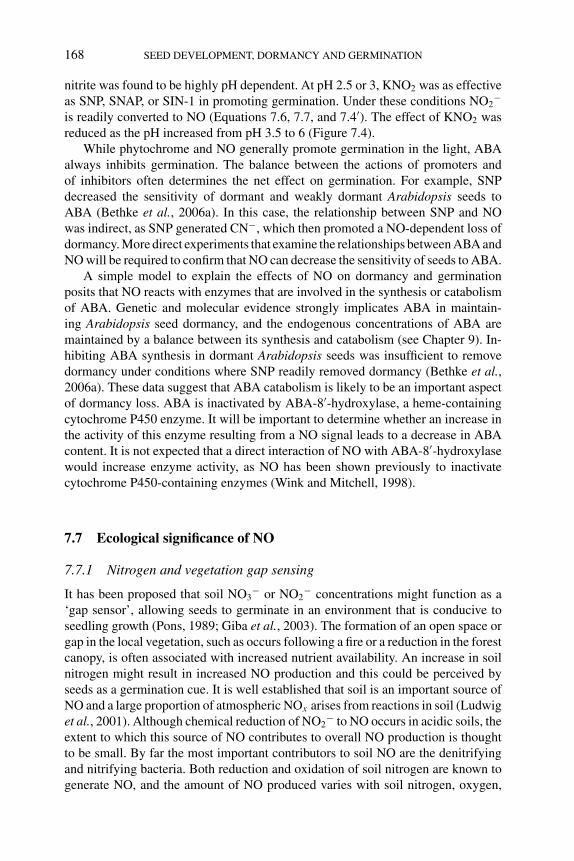
P1: OIY
BLUK053-Bradford August 25, 2006 14:49
168 SEED DEVELOPMENT, DORMANCY AND GERMINATION
nitrite was found to be highly pH dependent. At pH 2.5 or 3, KNO2 was as effectiveas SNP, SNAP, or SIN-1 in promoting germination. Under these conditions NO2
−
is readily converted to NO (Equations 7.6, 7.7, and 7.4′). The effect of KNO2 wasreduced as the pH increased from pH 3.5 to 6 (Figure 7.4).
While phytochrome and NO generally promote germination in the light, ABAalways inhibits germination. The balance between the actions of promoters andof inhibitors often determines the net effect on germination. For example, SNPdecreased the sensitivity of dormant and weakly dormant Arabidopsis seeds toABA (Bethke et al., 2006a). In this case, the relationship between SNP and NOwas indirect, as SNP generated CN−, which then promoted a NO-dependent loss ofdormancy. More direct experiments that examine the relationships between ABA andNO will be required to confirm that NO can decrease the sensitivity of seeds to ABA.
A simple model to explain the effects of NO on dormancy and germinationposits that NO reacts with enzymes that are involved in the synthesis or catabolismof ABA. Genetic and molecular evidence strongly implicates ABA in maintain-ing Arabidopsis seed dormancy, and the endogenous concentrations of ABA aremaintained by a balance between its synthesis and catabolism (see Chapter 9). In-hibiting ABA synthesis in dormant Arabidopsis seeds was insufficient to removedormancy under conditions where SNP readily removed dormancy (Bethke et al.,2006a). These data suggest that ABA catabolism is likely to be an important aspectof dormancy loss. ABA is inactivated by ABA-8′-hydroxylase, a heme-containingcytochrome P450 enzyme. It will be important to determine whether an increase inthe activity of this enzyme resulting from a NO signal leads to a decrease in ABAcontent. It is not expected that a direct interaction of NO with ABA-8′-hydroxylasewould increase enzyme activity, as NO has been shown previously to inactivatecytochrome P450-containing enzymes (Wink and Mitchell, 1998).
7.7 Ecological significance of NO
7.7.1 Nitrogen and vegetation gap sensing
It has been proposed that soil NO3− or NO2
− concentrations might function as a‘gap sensor’, allowing seeds to germinate in an environment that is conducive toseedling growth (Pons, 1989; Giba et al., 2003). The formation of an open space orgap in the local vegetation, such as occurs following a fire or a reduction in the forestcanopy, is often associated with increased nutrient availability. An increase in soilnitrogen might result in increased NO production and this could be perceived byseeds as a germination cue. It is well established that soil is an important source ofNO and a large proportion of atmospheric NOx arises from reactions in soil (Ludwiget al., 2001). Although chemical reduction of NO2
− to NO occurs in acidic soils, theextent to which this source of NO contributes to overall NO production is thoughtto be small. By far the most important contributors to soil NO are the denitrifyingand nitrifying bacteria. Both reduction and oxidation of soil nitrogen are known togenerate NO, and the amount of NO produced varies with soil nitrogen, oxygen,
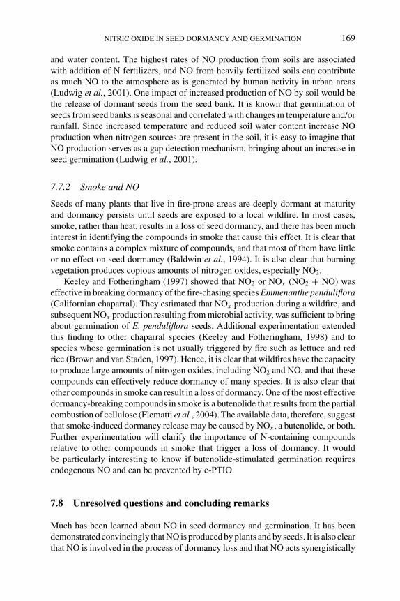
P1: OIY
BLUK053-Bradford August 25, 2006 14:49
NITRIC OXIDE IN SEED DORMANCY AND GERMINATION 169
and water content. The highest rates of NO production from soils are associatedwith addition of N fertilizers, and NO from heavily fertilized soils can contributeas much NO to the atmosphere as is generated by human activity in urban areas(Ludwig et al., 2001). One impact of increased production of NO by soil would bethe release of dormant seeds from the seed bank. It is known that germination ofseeds from seed banks is seasonal and correlated with changes in temperature and/orrainfall. Since increased temperature and reduced soil water content increase NOproduction when nitrogen sources are present in the soil, it is easy to imagine thatNO production serves as a gap detection mechanism, bringing about an increase inseed germination (Ludwig et al., 2001).
7.7.2 Smoke and NO
Seeds of many plants that live in fire-prone areas are deeply dormant at maturityand dormancy persists until seeds are exposed to a local wildfire. In most cases,smoke, rather than heat, results in a loss of seed dormancy, and there has been muchinterest in identifying the compounds in smoke that cause this effect. It is clear thatsmoke contains a complex mixture of compounds, and that most of them have littleor no effect on seed dormancy (Baldwin et al., 1994). It is also clear that burningvegetation produces copious amounts of nitrogen oxides, especially NO2.
Keeley and Fotheringham (1997) showed that NO2 or NOx (NO2 + NO) waseffective in breaking dormancy of the fire-chasing species Emmenanthe penduliflora(Californian chaparral). They estimated that NOx production during a wildfire, andsubsequent NOx production resulting from microbial activity, was sufficient to bringabout germination of E. penduliflora seeds. Additional experimentation extendedthis finding to other chaparral species (Keeley and Fotheringham, 1998) and tospecies whose germination is not usually triggered by fire such as lettuce and redrice (Brown and van Staden, 1997). Hence, it is clear that wildfires have the capacityto produce large amounts of nitrogen oxides, including NO2 and NO, and that thesecompounds can effectively reduce dormancy of many species. It is also clear thatother compounds in smoke can result in a loss of dormancy. One of the most effectivedormancy-breaking compounds in smoke is a butenolide that results from the partialcombustion of cellulose (Flematti et al., 2004). The available data, therefore, suggestthat smoke-induced dormancy release may be caused by NOx , a butenolide, or both.Further experimentation will clarify the importance of N-containing compoundsrelative to other compounds in smoke that trigger a loss of dormancy. It wouldbe particularly interesting to know if butenolide-stimulated germination requiresendogenous NO and can be prevented by c-PTIO.
7.8 Unresolved questions and concluding remarks
Much has been learned about NO in seed dormancy and germination. It has beendemonstrated convincingly that NO is produced by plants and by seeds. It is also clearthat NO is involved in the process of dormancy loss and that NO acts synergistically
P1: OIY
BLUK053-Bradford August 25, 2006 14:49
170 SEED DEVELOPMENT, DORMANCY AND GERMINATION
with light to promote germination of seeds that are not strictly dormant. NO mayhave additional roles in seeds. For example, NO may function as an antimicrobialcompound, a sensor for soil nutrient status, or a gap detector. The molecular interac-tions that allow NO to perform any of its roles, however, are completely unknown.For our understanding of NO in seed dormancy and germination to advance muchfarther it will be essential to establish where, when, and how NO is synthesized inseeds, the identity of the molecular targets for NO binding or covalent modifica-tion, and how NO signaling pathways interact with the phytochrome, GA, or ABAsignaling pathways.
NO is reactive and volatile, and thus must be produced when it is needed. Defini-tive information on the details of NO production in seeds is lacking. NO can be syn-thesized both enzymatically and nonenzymatically in seeds. Arabidopsis mutantslacking AtNOS1 are not defective in germination, making it clear that NO producedby AtNOS1 is not required for germination. Until NO production by seeds is pre-cisely quantified, it will be difficult to determine how its production is regulated bydevelopmental or environmental cues. Likewise, it will be important to identify thesites of NO synthesis in seeds. All tissues in the embryo or endosperm may have thecapacity to produce NO, but that does not preclude the possibility that some tissuesmake NO as a local or long-distance signal, and other tissues respond to NO butdo not synthesize it themselves. In many seeds the activities of the embryo and theendosperm must be coordinated, and an attractive possibility is that NO plays a rolein this coordination.
Molecular targets for NO in seeds have been proposed, but none have been shownto be required for NO activity in seed dormancy or germination. Among the mostcommonly mentioned are catalase, hemoglobin, cytochrome c oxidase, guanalylcyclase, and other metal-containing enzymes including superoxide dismutase andperoxidases. Hemoglobin is an intriguing target for NO, if for no other reason thanthe observation that seeds contain nonsymbiotic hemoglobin, the function of whichin seeds is unknown. Since some sites of NO production are in mitochondria, it islikely that NO binds to cytochrome c oxidase and prevents or reduces the electrontransport capacity of this protein. Numerous proposals have been made over theyears as to how redirecting electron transport through the alternative oxidase mightpromote seed germination. Perhaps some of this earlier literature should be revisitedwith NO binding to cytochrome c oxidase in mind.
Guanalyl cyclase is one of the principal targets for NO binding in mammals,and data from plants indicate that it is a target for NO produced during the hy-persensitive response to pathogens. In seeds, NO-activated guanalyl cyclases havebeen proposed to be a node for cross-talk between NO signaling and phytochromesignaling (Giba et al., 1998), but data supporting this hypothesis are lacking. NOworks in concert with ROS in plant disease resistance and in some forms of plantPCD. Given this, and that NO has a very high affinity for hemes such as those foundin superoxide dismutase and catalase, it is worth considering the possibility that NOpotentiates its own activity by changing the rate of ROS production. Finally, NO cancovalently modify proteins and thereby modify their activities. To date, nitrosylatedand nitrosated proteins have not been reported to occur in seeds.
P1: OIY
BLUK053-Bradford August 25, 2006 14:49
NITRIC OXIDE IN SEED DORMANCY AND GERMINATION 171
Research with seeds from lettuce, Empress tree, Arabidopsis, and other speciesindicates that there are interactions between NO and the phytochrome and ABAsignaling pathways. This is expected given that ABA and phytochrome play signif-icant roles in seed dormancy and germination. Importantly, it opens up a promisingavenue for further exploration of NO signaling in seeds. Mutants in phytochromeand ABA biosynthesis and signaling exist, especially in Arabidopsis. These shouldbe employed to full advantage in experiments designed to understand more about themolecular interactions between these signaling pathways. Mutant screens designedto identify components of the NO signaling network might add further depth to ourunderstanding of NO action in seeds.
Finally, the relationships among plant hormones, NO, and simple nitrogen-containing compounds such as NO2
−, NO3−, CN−, and azide need further investi-
gation. These nitrogen-containing compounds reduce seed dormancy across a widerange of species, but their mechanism of action remains unclear despite a century ofresearch. Are they triggers for increased NO synthesis or NO availability, or are theysubstrates for NO production as we and others have suggested? Careful measure-ments of NO production by seeds following exposure to nitrate, nitrite, cyanide, andazide may at last reveal part of the picture as to how these compounds reduce dor-mancy and promote germination. Perhaps the central question at this time, however,is how NO signaling, ABA signaling, and the operation of key metabolic pathwayswork together to control dormancy and germination. For example, is there an in-teraction between NO or NO signaling, on the one hand, and carbon metabolismon the other hand? Numerous proposals have been made over the years suggestingthat limitations to lipid metabolism or to the operation of the pentose phosphatepathway can inhibit germination. Yet much of the evidence for this is indirect orsubject to alternative interpretations. Recent advances in metabolite profiling andthe availability of mutants with defects in ABA signaling provide an entree for de-tailed investigations in this area. Coupling these to in vivo measurements of NOproduction under experimental conditions that promote or prevent germination mayhelp to explain more fully a century of experimentation with seeds.
References
T. Akaike, M. Yoshida, Y. Miyamoto, et al. (1993) Antagonistic action of imidazolineoxyl N -oxides
against endothelium-derived relaxing factor NO (nitric oxide) through a radical reaction. Bio-chemistry 32, 827–832.
I.T. Baldwin, L. Staszakkozinski and R. Davidson (1994) Up in smoke. 1: Smoke-derived germination
cues for postfire annual, Nicotiana attenuata Torr Ex Watson. Journal of Chemical Ecology 20,
2345–2371.
L.A.T. Ballard and A.E.G. Lipp (1967) Seed dormancy-breaking by uncouplers and inhibitors of
oxidative phosphorylation. Science 156, 398–400.
I. Batak, M. Devic, Z. Giba, D. Grubisic, K.L. Poff and R. Konjevic (2002) The effects of potassium
nitrate and NO-donors on phytochrome A- and phytochrome B-specific induced germination of
Arabidopsis thaliana seeds. Seed Science Research 12, 253–259.
M. Beligni, A. Fath, P.C. Bethke, L. Lamattina and R.L. Jones (2002) Nitric oxide acts as an antioxidant
and delays programmed cell death in barley aleurone layers. Plant Physiology 129, 1642–1650.
P1: OIY
BLUK053-Bradford August 25, 2006 14:49
172 SEED DEVELOPMENT, DORMANCY AND GERMINATION
M.V. Beligni and L. Lamattina (2000) Nitric oxide stimulates seed germination and de-etiolation, and
inhibits hypocotyl elongation, three light-inducible responses in plants. Planta 210, 215–221.
P.C. Bethke, M.R. Badger and R.L. Jones (2004a) Apoplastic synthesis of nitric oxide by plant tissues.
The Plant Cell 16, 332–341.
P.C. Bethke, F. Gubler, J.V. Jacobsen and R.L. Jones (2004b) Dormancy of Arabidopsis seeds and
barley grains can be broken by nitric oxide. Planta 219, 847–855.
P.C. Bethke, I. Libourel and R. Jones (2006a) Nitric oxide reduces seed dormancy in Arabidopsis.
Journal of Experimental Botany 57, 517–526.
P.C. Bethke, I.G.L. Libourel, V. Reinohl and R.L. Jones (2006b) Sodium nitroprusside, cyanide, nitrite,
and nitrate break Arabidopsis seed dormancy in a nitric oxide-dependent manner. Planta 223,
805–812.
P.C. Bethke, J.E. Lonsdale, A. Fath and R.L. Jones (1999) Hormonally regulated programmed cell
death in barley aleurone cells. The Plant Cell 11, 1033–1045.
N.A.C. Brown and J. van Staden (1997) Smoke as a germination cue: a review. Plant Growth Regulation22, 115–124.
P.A. Castric, K.J.F. Farnden and E.E. Conn (1972) Cyanide metaboism in higher plants. V: The
formation of asparagine from β-cyanoline. Archives of Biochemistry and Biophysics 152,
62–69.
M. Cohn, D. Butera and J. Hughes (1983) Seed dormancy in red rice. Plant Physiology 73, 381–384.
M.A. Cohn and L. Castle (1984) Dormancy in red rice. IV: Response of unimbibed and imbibing seeds
to nitrogen dioxide. Physiologia Plantarum 60, 552–556.
F.J. Corpas, J.B. Barroso and L.A. del Rio (2001) Peroxisomes as a source of reactive oxygen species
and nitric oxide signal molecules in plant cells. Trends in Plant Science 6, 145–150.
N.M. Crawford and F.-Q. Guo (2005) New insights into nitric oxide metabolism and regulatory
functions. Trends in Plant Science 10, 195–200.
W. Crocker and L. Barton (eds) (1953) Physiology of Seeds. Chronica Botanica, Waltham, MA.
J. Dean and J.E. Harper (1986) Nitric oxide and nitrous oxide production by soybean and winged bean
during the in vivo nitrate reductase assay. Plant Physiology 82, 718–723.
L.A. del Rio, F.J. Corpas, A.M. Leon, et al. (2004) The nitric oxide synthase activity of plant peroxi-
somes. Free Radical Biology and Medicine 36, S41–S42.
M. Delledonne (2005) NO news is good news for plants. Current Opinion in Plant Biology 8, 390–
396.
C. Dordas, B.B. Hasinoff, J. Rivoal and R.D. Hill (2004) Class-1 hemoglobins, nitrate and NO levels
in anoxic maize cell-suspension cultures. Planta 219, 66–72.
Y.M. Drozdowicz and R.L. Jones (1995) Hormonal regulation of organic and phosphoric acid release
by barley aleurone layers and scutella. Plant Physiology 108, 769–776.
J. Durner, D. Wendehenne and D.F. Klessig (1998) Defense gene induction in tobacco by nitric oxide,
cyclic GMP, and cyclic ADP-ribose. Proceedings of the National Academy of Sciences of theUnited States of America 95, 10328–10333.
P.J. Eastmond and R.L. Jones (2005) Hormonal regulation of gluconeogenesis in cereal aleurone is
strongly cultivar-dependent and gibberellin action involves SLENDER1 but not GAMYB. ThePlant Journal 44, 483–493.
A. Fath, P.C. Bethke and R.L. Jones (2001) Enzymes that scavenge reactive oxygen species are down-
regulated prior to gibberellic acid-induced programmed cell death in barley aleurone. PlantPhysiology 126, 156–166.
M. Feelisch (1991) The biochemical pathways of nitric oxide formation from nitrovasodilators: ap-
propriate choice of exogenous NO donors and aspects of preparation and handling of aqueous
NO solutions. Journal of Cardiovascular Pharmacology 17(Suppl. 3), S25–S33.
M. Feelisch (1998) The use of nitric oxide donors in pharmacological studies. Naunyn-SchmiedebergsArchives of Pharmacology 358, 113–122.
T.E. Ferrari and J.E. Varner (1969) Substrate induction of nitrate reductase in barley aleurone layers.
Plant Physiology 44, 85–88.
M.K. Firestone (1982) Biological dentrification. Agronomy Journal 22, 289–336.
P1: OIY
BLUK053-Bradford August 25, 2006 14:49
NITRIC OXIDE IN SEED DORMANCY AND GERMINATION 173
G.R. Flematti, E.L. Ghisalberti, K.W. Dixon and R.D. Trengove (2004) A compound from smoke that
promotes seed germination. Science 305, 977.
Z. Giba, D. Grubisic and R. Konjevic (1995) The involvement of phytochrome in light-induced ger-
mination of blueberry (Vaccinium myrtillus L.) seeds. Seed Science and Technology 23, 11–19.
Z. Giba, D. Grubisic and R. Konjevic (2003) Nitrogen oxides as environmental sensors for seeds. SeedScience Research 13, 187–196.
Z. Giba, D. Grubisic, S. Todorovic, L. Sajc, D. Stojakovic and R. Konjevic (1998) Effect of nitric
oxide-releasing compounds on phytochrome-controlled germination of Empress tree seeds. PlantGrowth Regulation 26, 175–181.
M.T. Gladwin, A.N. Schechter, D.B. Kim-Shapiro, et al. (2005) The emerging biology of the nitrite
anion. Nature Chemical Biology 1, 308–314.
S. Goldstein, A. Russo and A. Samuni (2003) Reactions of PTIO and carboxy-PTIO with NO, NO2,
and O2. Journal of Biological Chemistry 278, 50949–50955.
D.L. Granger, R.R. Taintor, K.S. Boockvar and J.B. Hibbs (1996) Measurement of nitrate and nitrite
in biological samples using nitrate reductase and Griess reaction. Methods in Enzymology 268,
142–151.
P. Grappin, D. Bouinot, B. Sotta, E. Miginiac and M. Jullien (2000) Control of seed dormancy in Nico-tiana plumbaginifolia: post-imbibition abscisic acid synthesis imposes dormancy maintenance.
Planta 210, 279–285.
D. Grubisic, Z. Giba and R. Konjevic (1992) The effect of organic nitrates in phytochrome-controlled
germination of Paulownia tomentosa seeds. Photochemistry and Photobiology 56, 629–632.
F.-G. Guo and N.M. Crawford (2005) The Arabidopsis nitric oxide synthase protein AtNOS1 is targeted
to mitochondria and protects against oxidative damage and dark-induced senescence. The PlantCell 17, 3436–3450.
S.B. Hendricks and R.B. Taylorson (1972) Promotion of seed germination by nitrates and cyanide.
Nature 237, 169–170.
S. Hendricks and R. Taylorson (1974) Promotion of seed germination by nitrate, nitrite, hydroxylamine
and ammonium salts. Plant Physiology 54, 304–309.
S.B. Hendricks and R.B. Taylorson (1975) Breaking of seed dormancy by catalase inhibition. Pro-ceedings of the National Academy of Sciences of the United States of America 72, 306–309.
Z. Hua, W.-B. Shen and L.-L. Xu (2003) Effects of nitric oxide on the germination of wheat seeds and
its reactive oxygen species metabolisms under osmotic stress. Acta Botanica Sinica 45, 901–905.
P.W. Hunt, E.J. Klok, B. Trevaskis, et al. (2002) Increased level of hemoglobin 1 enhances survival of
hypoxic stress and promotes early growth in Arabidopsis thaliana. Proceedings of the NationalAcademy of Sciences of the United States of America 99, 17197–17202.
A.U. Igamberdiev, K. Baron, N. Manac’h-Little, M. Stoimenova and R.D. Hill (2005) The
haemoglobin/nitric oxide cycle: involvement in flooding stress and effects on hormone signaling.
Annals of Botany 96, 557–564.
A.U. Igamberdiev, C. Seregelyes, N. Manach and R.D. Hill (2004) NADH-dependent metabolism of
nitric oxide in alfalfa root cultures expressing barley hemoglobin. Planta 219, 95–102.
L.J. Ignarro, J.M. Fukuto, J.M. Griscavage, N.E. Rogers and R.E. Byrns (1993) Oxidation of nitric
oxide in aqueous solution to nitrite but not nitrate – comparison with enzymatically formed nitric
oxide from l-arginine. Proceedings of the National Academy of Sciences of the United States ofAmerica 90, 8103–8107.
J.E. Keeley and C.J. Fotheringham (1997) Trace gas emissions and smoke-induced seed germination.
Science 276, 1248–1250.
J.E. Keeley and C.J. Fotheringham (1998) Smoke-induced seed germination in California chaparral.
Ecology 79, 2320–2336.
M. Kopyra and E.A. Gwozdz (2003) Nitric oxide stimulates seed germination and counteracts the
inhibitory effect of heavy metals and salinity on root growth of Lupinus luteus. Plant Physiologyand Biochemistry 41, 1011–1017.
L. Lamattina, C. Garcia-Mata, M. Graziano and G. Pagnussat (2003) Nitric oxide: the versatility of
an extensive signal molecule. Annual Review of Plant Biology 54, 109–136.
P1: OIY
BLUK053-Bradford August 25, 2006 14:49
174 SEED DEVELOPMENT, DORMANCY AND GERMINATION
E. Lehman (1909) Zur Keimungsphysiologie and biologie von Ranunculus sclereatus L. und einigen
anderen Samen. Berichte Deutschen Botanischen Gesellschaft 27, 476–494.
W.Q. Li, X.J. Liu, M.A. Khan, Y. Kamiya and S. Yamaguchi (2005) Hormonal and environmental
regulation of seed germination in flixweed (Descurainia sophia). Plant Growth Regulation 45,
199–207.
I.G.L. Libourel, P.C. Bethke, R. De Michele and R.L. Jones (2006) Nitric oxide gas stimulates ger-
mination of dormant Arabidopsis seeds: Use of a flow-through apparatus for delivery of nitric
oxide. Planta 223, 813–820.
M.E. Light and J. van Staden (2003) The nitric oxide-specific scavenger carboxy-PTIO does not inhibit
smoke-stimulated germination of Grand Rapids lettuce seeds. South African Journal of Botany69, 217–219.
J. Ludwig, F.X. Meixner, B. Vogel and J. Forstner (2001) Soil–air exchange of nitric oxide: an
overview of processes, environmental factors, and modeling studies. Biogeochemistry 52, 225–
257.
W. Major and E.H. Roberts (1968) Dormancy in cereal seeds. I: Effects of oxygen and respiratory
inhibitors. Journal of Experimental Botany 19, 77–89.
S. Neill, R. Desikan and J.T. Hancock (2003) Nitric oxide signaling in plants. New Phytologist 159,
11–35.
X. Nie and R.D. Hill (1997) Mitochondrial respiration and hemoglobin gene expression in barley
aleurone tissue. Plant Physiology 114, 835–840.
Y. Ohwaki, M. Kawagishi-Kobayashi, K. Wakasa, S. Fujihara and T. Yoneyama (2005) Induction of
class-1 non-symbiotic hemoglobin genes by nitrate, nitrite and nitric oxide in cultured rice cells.
Plant and Cell Physiology 46, 324–331.
A. Poljakoff-Mayber and A.M. Mayer (eds) (1963) The Germination of Seeds. Pergammon, Oxford.
T.L. Pons (1989) Breaking of seed dormancy by nitrate as a gap detection mechanism. Annals ofBotany 63, 139–143.
E.H. Roberts (1963) The effects of inorganic ions on dormancy in rice seed. Physiologia Plantarum16, 732–744.
E.H. Roberts (1964a) A survey of effects of chemical treatments on dormancy in rice seed. PhysiologiaPlantarum 17, 30–43.
E.H. Roberts (1964b) Distribution of oxidation–reduction enzymes: effects of respiratory inhibitors,
oxidising agents on dormancy in rice seed. Physiologia Plantarum 17, 14–29.
E.H. Roberts (1969) Seed dormancy and oxidation processes. Symposia of the Society of ExperimentalBiology 23, 161–192.
P. Rockel, F. Strube, A. Rockel, J. Wildt and W.M. Kaiser (2002) Regulation of nitric oxide (NO)
production by plant nitrate reductase in vivo and in vitro. Journal of Experimental Botany 53,
103–110.
A. Sakamoto, S. Sakurao, K. Fukunaga, et al. (2004) Three distinct Arabidopsis hemoglobins ex-
hibit peroxidase-like activity and differentially mediate nitrite-dependent protein nitration. FEBSLetters 572, 27–32.
Y. Sakihama, S. Nakamura and H. Yamasaki (2002) Nitric oxide production mediated by nitrate
reductase in the green alga Chlamydomonas reinhardtii: an alternative NO production pathway
in photosynthetic organisms. Plant and Cell Physiology 43, 290–297.
G. Sarath, P.C. Bethke, R.L. Jones, L.M. Baird, G. Hou and R.B. Mitchell (2006) Nitric oxide accel-
erates seed germination in warm-season grasses Planta 223, 1154–1164.
A.D. Shapiro (2005) Nitric oxide signaling in plants. Advances in Research and Applications: PlantHormones and Vitamins 72, 339–398.
C. Stohr and S. Stremlau (2006) Formation and possible roles of nitric oxide in plant roots. Journalof Experimental Botany 57, 463–470.
C. Stohr, F. Strube, G. Marx, W.R. Ullrich and P. Rockel (2001) A plasma membrane-bound enzyme
of tobacco roots catalyses the formation of nitric oxide from nitrite. Planta 212, 835–841.
C. Stohr and W.R. Ullrich (2002) Generation and possible roles of NO in plant roots and their apoplastic
space. Journal of Experimental Botany 53, 2293–2303.
P1: OIY
BLUK053-Bradford August 25, 2006 14:49
NITRIC OXIDE IN SEED DORMANCY AND GERMINATION 175
R.B. Taylorson and S.B. Hendricks (1973) Promotion of seed germination by cyanide. Plant Physiology52, 23–27.
E.H. Toole, S.B. Hendricks, H.A. Borthwick and V.K. Toole (1956) Physiology of seed germination.
Annual Review of Plant Physiology 7, 299–324.
D. Wendehenne, J. Durner and D.F. Klessig (2004) Nitric oxide: a new player in plant signalling and
defence responses. Current Opinion in Plant Biology 7, 449–455.
D.A. Wink and J.B. Mitchell (1998) Chemical biology of nitric oxide: insights into regulatory,
cytotoxic, and cytoprotective mechanisms of nitric oxide. Free Radical Biology and Medicine25, 434–456.
H. Yamasaki (2000) Nitrite-dependent nitric oxide production pathway: implications for involvement
of active nitrogen species in photoinhibition in vivo. Philosophical Transactions of the RoyalSociety London. Series B: Biological Sciences 355, 1477–1488.
H. Yamasaki, H. Shimoji, Y. Ohshiro and Y. Sakihama (2001) Inhibitory effects of nitric oxide on
oxidative phosphorylation in plant mitochondria. Nitric Oxide 5, 261–270.
L.X. Yang, R.Y. Wang, F. Ren, J. Liu, J. Cheng and Y.T. Lu (2005) AtGLB1 enhances the tolerance
of Arabidopsis to hydrogen peroxide stress. Plant and Cell Physiology 46, 1309–1316.
D. Zanardo, F. Zanardo, M. Ferrarese, J. Magalhaes and O. Ferrarese-Filho (2005) Nitric oxide affecting
seed germination and peroxidase activity in canola (Brassica napus L.). Physiology and Molec-ular Biology of Plants 11, 81–86.
P1: OTE/SPH P2: OTE
BLUK053-Bradford September 22, 2006 15:36
8 A merging of paths: abscisic acid andhormonal cross-talk in the control of seeddormancy maintenance and alleviationJ. Allan Feurtado and Allison R. Kermode
8.1 Introduction
The sesquiterpene phytohormone abscisic acid (ABA) regulates key events duringseed formation, such as the deposition of storage reserves, prevention of precociousgermination, acquisition of desiccation tolerance, and induction of primary dor-mancy (Finkelstein and Rock, 2002; Kermode, 2005). Dynamic changes in ABAbiosynthesis and catabolism elicit hormone-signaling changes that affect down-stream gene expression, and thereby regulate critical checkpoints during the plantlife cycle, including the transitions from dormancy to germination and from germi-nation to growth (Nambara and Marion-Poll, 2005). However, the regulatory roleof ABA is in part achieved through interactions with other hormones and their as-sociated signaling networks, by mechanisms that remain largely unknown. Thus,in many instances, a linear representation of hormone signaling pathways is nolonger a viable model to explain downstream physiological events. Instead, cross-talk among hormones such as ABA, gibberellic acid (GA), ethylene, brassinos-teroids (BRs), cytokinin, and auxin forms a complex web whereby key regula-tory factors become nodes or hubs under combinatorial control. There is furtherintricacy when we consider that the hormone levels themselves are under regula-tion in this complex web and that environmental cues, in turn, modulate hormonalresponses.
Quantitative genetics and functional genomics approaches have and will continueto contribute to the identification of genes and proteins that control seed dormancyand germination, including components of the ABA signal transduction pathway andelements that link this pathway to other hormonal networks. Interactions betweenABA and other hormones during the control of key processes, from dormancyinduction to seedling growth, include:
1. dormancy inception, in which GA and ethylene play antagonistic roles toABA during seed development;
2. the maintenance and termination of primary dormancy, in which other hor-mones or hormone-signaling components, such as GA, may alter ABA home-ostasis (e.g., de novo ABA synthesis or ABA catabolism) and/or the stabilityof ABA signal transduction components;
176
P1: OTE/SPH P2: OTE
BLUK053-Bradford September 22, 2006 15:36
ABSCISIC ACID AND HORMONAL CROSS-TALK IN SEED DORMANCY 177
3. the initiation of germination in which ethylene and other hormones such asBRs may alter ABA or GA homeostasis and associated signal transductioncomponents;
4. the developmental arrest during the early post-germinative phase, a stateinduced by conditions that are not optimal for a transition from germinationto seedling growth. Here seeds may respond by increasing ABA biosynthesisand the expression of genes that impose a transient ‘quiescence’ until thoseconditions become more favorable.
Recent evidence for interactions among the different hormonal networks duringthese dormancy/growth-related events of the plant life cycle is discussed, and wherepossible, the potential mechanisms that form the basis of these interactions arespeculated upon. We first outline the role of ABA in controlling these processes,followed by a discussion of the roles of GA, ethylene, auxin, cytokinin, and BRs indormancy induction, maintenance, and termination. Emphasis will also be drawn tocross-talk among the hormones and interactions with environmental signals such aslight and cold.
8.2 Abscisic acid
8.2.1 ABA in seed maturation and the induction of primary dormancy
ABA is involved in several specific processes during seed development and, ingeneral, is thought to maintain seeds in a developmental program while on the parentplant (for reviews see Kermode, 1995, 2005; Holdsworth et al., 1999; Finkelsteinand Rock, 2002). Evidence for the role of ABA in such processes as the depositionof storage reserves, prevention of precocious germination, acquisition of desiccationtolerance, and dormancy induction has come from work with ABA-deficiency or-response mutants of Arabidopsis thaliana or Zea mays (maize) (Finkelstein andRock, 2002). For example, many of the viviparous (vp) mutants of maize (whichexhibit precocious germination of kernels on the ear) are linked to deficiencies inABA synthesis or signaling (McCarty, 1995; Suzuki et al., 2006; see Chapter 1).Recently, McCourt et al. (2005) have proposed a model in which ABA functions asa ‘status quo’ hormone, preserving or maintaining the present state and preventinglife cycle transitions (e.g., the dormancy-to-germination transition). In addition tothis ascribed role, ABA also seems to prevent seed abortion and promote embryogrowth during embryogenesis (Cheng et al., 2002; Frey et al., 2004).
Seeds of different species show differences in the timing of dormancy inceptionduring development; the role of ABA in this process is evident from geneticand physiological studies (reviewed in Bewley, 1997; Koornneef et al., 2002).Typically, the ABA content in seeds is low during early development (i.e., duringhistodifferentiation and early pattern formation) and thereafter increases, usuallypeaking around mid-maturation. ABA levels usually decline precipitously duringlate development, particularly during the maturation drying phase (Bewley and
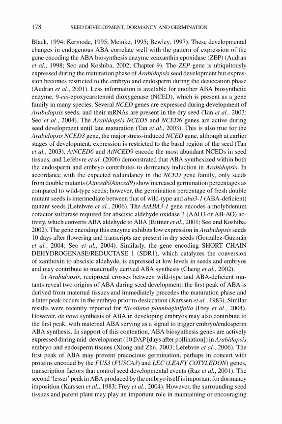
P1: OTE/SPH P2: OTE
BLUK053-Bradford September 22, 2006 15:36
178 SEED DEVELOPMENT, DORMANCY AND GERMINATION
Black, 1994; Kermode, 1995; Meinke, 1995; Bewley, 1997). These developmentalchanges in endogenous ABA correlate well with the pattern of expression of thegene encoding the ABA biosynthesis enzyme zeaxanthin epoxidase (ZEP) (Audranet al., 1998; Seo and Koshiba, 2002; Chapter 9). The ZEP gene is ubiquitouslyexpressed during the maturation phase of Arabidopsis seed development but expres-sion becomes restricted to the embryo and endosperm during the desiccation phase(Audran et al., 2001). Less information is available for another ABA biosyntheticenzyme, 9-cis-epoxycarotenoid dioxygenase (NCED), which is present as a genefamily in many species. Several NCED genes are expressed during development ofArabidopsis seeds, and their mRNAs are present in the dry seed (Tan et al., 2003;Seo et al., 2004). The Arabidopsis NCED5 and NCED6 genes are active duringseed development until late maturation (Tan et al., 2003). This is also true for theArabidopsis NCED3 gene, the major stress-induced NCED gene, although at earlierstages of development, expression is restricted to the basal region of the seed (Tanet al., 2003). AtNCED6 and AtNCED9 encode the most abundant NCEDs in seedtissues, and Lefebvre et al. (2006) demonstrated that ABA synthesized within boththe endosperm and embryo contributes to dormancy induction in Arabidopsis. Inaccordance with the expected redundancy in the NCED gene family, only seedsfrom double mutants (Atnced6/Atnced9) show increased germination percentages ascompared to wild-type seeds; however, the germination percentage of fresh doublemutant seeds is intermediate between that of wild-type and aba3-1 (ABA-deficient)mutant seeds (Lefebvre et al., 2006). The AtABA3-1 gene encodes a molybdenumcofactor sulfurase required for abscisic aldehyde oxidase 3 (AAO3 or AB-AO) ac-tivity, which converts ABA aldehyde to ABA (Bittner et al., 2001; Seo and Koshiba,2002). The gene encoding this enzyme exhibits low expression in Arabidopsis seeds10 days after flowering and transcripts are present in dry seeds (Gonzalez-Guzmanet al., 2004; Seo et al., 2004). Similarly, the gene encoding SHORT CHAINDEHYDROGENASE/REDUCTASE 1 (SDR1), which catalyzes the conversionof xanthoxin to abscisic aldehyde, is expressed at low levels in seeds and embryosand may contribute to maternally derived ABA synthesis (Cheng et al., 2002).
In Arabidopsis, reciprocal crosses between wild-type and ABA-deficient mu-tants reveal two origins of ABA during seed development: the first peak of ABA isderived from maternal tissues and immediately precedes the maturation phase anda later peak occurs in the embryo prior to desiccation (Karssen et al., 1983). Similarresults were recently reported for Nicotiana plumbaginifolia (Frey et al., 2004).However, de novo synthesis of ABA in developing embryos may also contribute tothe first peak, with maternal ABA serving as a signal to trigger embryo/endospermABA synthesis. In support of this contention, ABA biosynthesis genes are activelyexpressed during mid-development (10 DAP [days after pollination]) in Arabidopsisembryo and endosperm tissues (Xiong and Zhu, 2003; Lefebvre et al., 2006). Thefirst peak of ABA may prevent precocious germination, perhaps in concert withproteins encoded by the FUS3 (FUSCA3) and LEC (LEAFY COTYLEDON) genes,transcription factors that control seed developmental events (Raz et al., 2001). Thesecond ‘lesser’ peak in ABA produced by the embryo itself is important for dormancyimposition (Karssen et al., 1983; Frey et al., 2004). However, the surrounding seedtissues and parent plant may play an important role in maintaining or encouraging
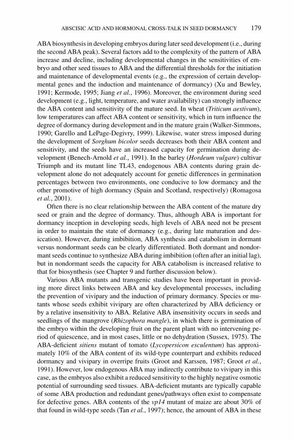
P1: OTE/SPH P2: OTE
BLUK053-Bradford September 22, 2006 15:36
ABSCISIC ACID AND HORMONAL CROSS-TALK IN SEED DORMANCY 179
ABA biosynthesis in developing embryos during later seed development (i.e., duringthe second ABA peak). Several factors add to the complexity of the pattern of ABAincrease and decline, including developmental changes in the sensitivities of em-bryo and other seed tissues to ABA and the differential thresholds for the initiationand maintenance of developmental events (e.g., the expression of certain develop-mental genes and the induction and maintenance of dormancy) (Xu and Bewley,1991; Kermode, 1995; Jiang et al., 1996). Moreover, the environment during seeddevelopment (e.g., light, temperature, and water availability) can strongly influencethe ABA content and sensitivity of the mature seed. In wheat (Triticum aestivum),low temperatures can affect ABA content or sensitivity, which in turn influence thedegree of dormancy during development and in the mature grain (Walker-Simmons,1990; Garello and LePage-Degivry, 1999). Likewise, water stress imposed duringthe development of Sorghum bicolor seeds decreases both their ABA content andsensitivity, and the seeds have an increased capacity for germination during de-velopment (Benech-Arnold et al., 1991). In the barley (Hordeum vulgare) cultivarTriumph and its mutant line TL43, endogenous ABA contents during grain de-velopment alone do not adequately account for genetic differences in germinationpercentages between two environments, one conducive to low dormancy and theother promotive of high dormancy (Spain and Scotland, respectively) (Romagosaet al., 2001).
Often there is no clear relationship between the ABA content of the mature dryseed or grain and the degree of dormancy. Thus, although ABA is important fordormancy inception in developing seeds, high levels of ABA need not be presentin order to maintain the state of dormancy (e.g., during late maturation and des-iccation). However, during imbibition, ABA synthesis and catabolism in dormantversus nondormant seeds can be clearly differentiated. Both dormant and nondor-mant seeds continue to synthesize ABA during imbibition (often after an initial lag),but in nondormant seeds the capacity for ABA catabolism is increased relative tothat for biosynthesis (see Chapter 9 and further discussion below).
Various ABA mutants and transgenic studies have been important in provid-ing more direct links between ABA and key developmental processes, includingthe prevention of vivipary and the induction of primary dormancy. Species or mu-tants whose seeds exhibit vivipary are often characterized by ABA deficiency orby a relative insensitivity to ABA. Relative ABA insensitivity occurs in seeds andseedlings of the mangrove (Rhizophora mangle), in which there is germination ofthe embryo within the developing fruit on the parent plant with no intervening pe-riod of quiescence, and in most cases, little or no dehydration (Sussex, 1975). TheABA-deficient sitiens mutant of tomato (Lycopersicon esculentum) has approxi-mately 10% of the ABA content of its wild-type counterpart and exhibits reduceddormancy and vivipary in overripe fruits (Groot and Karssen, 1987; Groot et al.,1991). However, low endogenous ABA may indirectly contribute to vivipary in thiscase, as the embryos also exhibit a reduced sensitivity to the highly negative osmoticpotential of surrounding seed tissues. ABA-deficient mutants are typically capableof some ABA production and redundant genes/pathways often exist to compensatefor defective genes. ABA contents of the vp14 mutant of maize are about 30% ofthat found in wild-type seeds (Tan et al., 1997); hence, the amount of ABA in these
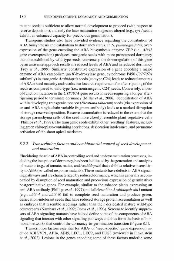
P1: OTE/SPH P2: OTE
BLUK053-Bradford September 22, 2006 15:36
180 SEED DEVELOPMENT, DORMANCY AND GERMINATION
mutant seeds is sufficient to allow normal development to proceed (with respect toreserve deposition), and only the later maturation stages are altered (e.g., vp14 seedsexhibit an enhanced capacity for precocious germination).
Transgenic studies also have provided evidence regarding the contribution ofABA biosynthesis and catabolism to dormancy status. In N. plumbaginifolia, over-expression of the gene encoding the ABA biosynthesis enzyme ZEP (i.e., ABA2gene overexpression) produces transgenic seeds with more pronounced dormancythan that exhibited by wild-type seeds; conversely, the downregulation of this geneby an antisense approach results in reduced levels of ABA and in reduced dormancy(Frey et al., 1999). Similarly, constitutive expression of a gene encoding a majorenzyme of ABA catabolism (an 8′-hydroxylase gene, cytochrome P450 CYP707Asubfamily) in transgenic Arabidopsis seeds (ecotype C24) leads to reduced amountsof ABA at seed maturity and results in a lowered requirement for after-ripening of theseeds as compared to wild-type (i.e., nontransgenic C24) seeds. Conversely, a loss-of-function mutation in the CYP707A gene results in seeds requiring a longer after-ripening period to terminate dormancy (Millar et al., 2006). Sequestration of ABAwithin developing transgenic tobacco (Nicotiana tabacum) seeds (via expression ofan anti-ABA single chain variable fragment antibody) leads to a marked disruptionof storage reserve deposition. Reserve accumulation is reduced to the extent that thestorage parenchyma cells of the seed more closely resemble plant vegetative cells(Phillips et al., 1997). The transgenic seeds exhibit other ‘seedling’ features, includ-ing green chloroplast-containing cotyledons, desiccation intolerance, and prematureactivation of the shoot apical meristem.
8.2.2 Transcription factors and combinatorial control of seed developmentand maturation
Elucidating the role of ABA in controlling seed and embryo maturation processes, in-cluding the inception of dormancy, has been facilitated by the generation and analysisof mutants (e.g., of tomato, maize, and Arabidopsis) that exhibit a relative insensitiv-ity to ABA (so-called response mutants). These mutants have defects in ABA signal-ing pathways and are characterized by reduced dormancy, which is generally accom-panied by disruption of seed maturation and precocious expression of germinative/postgerminative genes. For example, similar to the tobacco plants expressing ananti-ABA antibody (Phillips et al., 1997), null alleles of the Arabidopsis abi3 mutant(e.g., abi3-4 and abi3-6) fail to complete seed maturation and produce green,desiccation-intolerant seeds that have reduced storage protein accumulation as wellas embryos that resemble seedlings rather than their desiccated mature wild-typecounterparts (Nambara et al., 1992; Ooms et al., 1993). Screens to identify suppres-sors of ABA signaling mutants have helped define some of the components of ABAsignaling that interact with other signaling pathways and thus form the basis of hor-monal networks that control the dormancy-to-germination transition (Figure 8.1).
Transcription factors essential for ABA- or ‘seed-specific’ gene expression in-clude ABI3/VP1, ABI4, ABI5, LEC1, LEC2, and FUS3 (reviewed in Finkelsteinet al., 2002). Lesions in the genes encoding some of these factors underlie some
P1: OTE/SPH P2: OTE
BLUK053-Bradford September 22, 2006 15:36
ABSCISIC ACID AND HORMONAL CROSS-TALK IN SEED DORMANCY 181
Figure 8.1 Genetic interactions between ABA, GA, and ethylene pathways. Interactions are based on
(i) enhancer or suppressor screens (e.g., of abi1-1) or (ii) epistasis in double mutant analysis (e.g., ein2ctr1) (Koornneef et al., 1982; Kieber et al., 1993; Jacobsen and Olszewski, 1993; Finkelstein, 1994;
Steber et al., 1998; Beaudoin et al., 2000; Brady et al., 2003; Fu et al., 2004). Loci positively affecting
each hormone response are shown in solid circles; those with a negative effect are shown in dashed
circles. Suppressor interactions are shown by black lines and enhancer interactions by dashed lines.
The abi1-1 mutant phenotype is irregular; it is ABA insensitive but is actually a negative regulator of
ABA signaling (Yoshida et al., 2006). Abbreviations: aba, abscisic acid deficient; abi, abscisic acidinsensitive; ctr, constitutive triple response; ein, ethylene insensitive; era, enhanced response to abscisicacid; ga, gibberellic acid requiring; gai, gibberellic acid insensitive; sly, sleepy; spy, spindly.
of the phenotypic characteristics of mutants that are disrupted in seed maturation,including dormancy inception. For example, the various proposed functions of someof the transcription factors belonging to the B3 domain family (e.g., VP1 and ABI3)include (a) regulation of the expression of ABA-responsive genes during seed de-velopment (e.g., storage-protein genes and genes encoding putative desiccation pro-tectants) (McCarty et al., 1991; Giraudat et al., 1992; Parcy et al., 1997; Zeng et al.,2003; Zeng and Kermode, 2004; Kagaya et al., 2005); (b) repression of postgermi-native genes (Nambara et al., 2000; Gazzarrini et al., 2004); and (c) participation indormancy inception during seed development and/or the maintenance of a dormantstate in mature dispersed seeds (Jones et al., 1997; Fukuhara et al., 1999; McKibbinet al., 2002; Zeng et al., 2003).
Combinatorial control involving several transcription factors is a key theme inthe regulation of seed and embryo developmental events, including dormancy in-ception. Superimposed on this is the capacity for other components of hormonalsignal transduction to effect posttranslational modifications to proteins, thus modi-fying the actions of transcription factors and/or promoting their degradation. For ex-ample, there is strong evidence that the various functions of ABI3/VP1 are achievedthrough interactions with other proteins (Finkelstein et al., 2002; see Chapter 1). Inconcert, these may activate or repress target genes, including those important formaintaining/terminating the dormant state.
P1: OTE/SPH P2: OTE
BLUK053-Bradford September 22, 2006 15:36
182 SEED DEVELOPMENT, DORMANCY AND GERMINATION
ABI3 together with ABI4 and ABI5 participate in combinatorial control ofgene expression, possibly by forming a regulatory complex mediating seed-specificand/or ABA-inducible expression (Finkelstein et al., 2002). There appears tobe extensive cross-regulation of expression among ABI3, ABI4, and ABI5 genes(Soderman et al., 2000). Mutations in the ABI4 and ABI5 gene loci have similareffects on seed development and ABA sensitivity to those associated with the abi3mutant; however, null mutations in the ABI3 gene locus result in more severe pheno-types than those associated with defects in ABI4 or ABI5 (Finkelstein et al., 2002).The ABI4 and ABI5 proteins contain putative DNA-binding and protein-interactiondomains. ABI4 contains the AP2 domain characteristic of the APETALA2 familyof transcription factors (Finkelstein et al., 2002). The ABI5 family of proteins allshare three conserved charged domains in the N-terminal half of the protein thatcontain potential phosphorylation sites and a bZIP domain at the C-terminus. LikeVP1/ABI3, ABI5 can either activate or repress gene expression. ABI3 and ABI5interact physically (Nakamura et al., 2001) and may have antagonistic or synergisticeffects on gene expression, depending on the targeted gene (Finkelstein and Lynch,2000; Delseny et al., 2001).
ABA (and the signal it transduces) is not the only regulator of the middle and latestages of seed development. For example, the lec class mutants (lec1, lec2, and fus3)act in a concerted fashion to promote events that are critical to the later stages ofseed development, including the acquisition of desiccation tolerance and inductionof dormancy, and to repress germinative and postgerminative functions (Keith et al.,1994; Meinke et al., 1994; Holdsworth et al., 1999). Similar to ABI3, these three lociencode transcription factors regulating genes expressed during seed developmentand are thought to exert their effects by protein–DNA and/or protein–protein inter-actions. FUS3 and LEC2 belong to the B3-domain protein family of transcriptionfactors that includes ABI3 (Luerßen et al., 1998, Stone et al., 2001); the LEC1 geneencodes a protein with sequence similarity to the HAP3 subunit of CCAAT bindingfactors (Lotan et al., 1998). In mammalian cells, these heterotrimeric binding factorsenhance the transcription of a large number of genes (reviewed in Harada, 2001;see Chapter 1). FUS3 and LEC1 can modulate the abundance of ABI3 protein inseeds. Synergistic interactions among the three proteins (ABI3, FUS3, and LEC1)are thought to control the accumulation of chlorophyll and anthocyanins, sensitiv-ity to ABA and expression of individual members of the 12S storage protein genefamily (Parcy et al., 1997). FUS3 and LEC2 also appear to control ABA and GAhomeostasis (see below).
8.2.3 ABA in dormancy maintenance and termination
8.2.3.1 ABA synthesis and homeostasis during dormancy maintenanceand termination
De novo synthesis of ABA appears to be necessary for dormancy maintenancefollowing imbibition in seeds of sunflower (Helianthus annuus), barley, beechnut(Fagus sylvatica), lettuce (Lactica sativa), tobacco, and Douglas fir (Pseudotsugamenziesii) (Le Page-Degivry and Garello, 1992; Wang et al., 1995; Bianco et al.,
P1: OTE/SPH P2: OTE
BLUK053-Bradford September 22, 2006 15:36
ABSCISIC ACID AND HORMONAL CROSS-TALK IN SEED DORMANCY 183
1997; Le Page-Degivry et al., 1997; Yoshioka et al., 1998; Grappin et al., 2000).For instance, when tobacco seeds are imbibed, there is an accumulation of ABAin dormant seeds but not in seeds that have been after-ripened. In some species,there is a lag before ABA begins to accumulate in the dormant-imbibed seed. Thecarotenoid- and ABA-biosynthesis inhibitor fluridone, when used in conjunctionwith gibberellic acid (GA3), is effective in releasing dormancy; exogenous appli-cation of both chemicals to seeds inhibits accumulation of ABA during imbibition(Grappin et al., 2000). Similar to tobacco, GA3 and fluridone are effective in releas-ing dormancy of yellow-cedar (Chamaecyparis nootkatensis) seeds in the absenceof any additional treatment (Schmitz et al., 2001). These seeds normally require a3-month dormancy-releasing treatment consisting of 1 month of warm, moist con-ditions followed by 2 months of moist chilling (Ren and Kermode, 1999). However,fluridone alone (i.e., with no GA3) is much less effective in eliciting germination,indicating that a decline in ABA amount alone is not sufficient to release dormancyand other changes (e.g., synthesis of gibberellins) may also be necessary.
The termination of dormancy (through after-ripening, moist chilling, or smoke)is strongly associated with a decline in ABA content upon transfer to germinationconditions, and the decline in ABA corresponds well with an increase in germinationcapacity. After-ripening itself has little effect on ABA levels within embryos of drybarley grains (Jacobsen et al., 2002). Upon transfer of the after-ripened grain togermination conditions, the ABA content of the embryo declines markedly overthe first 12 h, and low ABA concentrations are maintained up to 30 h, by whichtime 90% of the grains have germinated. Dormant grains placed immediately ingermination conditions with no previous after-ripening period show some reductionof embryo ABA content over the first 12 h, but ABA content subsequently increasesbetween 12 and 30 h of imbibtion. A similar transient reduction of ABA, followed byits accumulation, occurs in imbibed dormant Arabidopsis seeds of the Cape VerdeIslands (Cvi) ecotype. This is in marked contrast to imbibed seeds that have receiveda previous dormancy-releasing treatment (moist chilling, nitrate or fluridone), inwhich ABA levels continue to decline during imbibition (Ali-Rachedi et al., 2004).Seeds of the commonly used accessions of Arabidopsis such as Columbia (Col)(which are far less dormant than seeds of the Cvi ecotype) show a decline in ABAlevels upon imbibition in the absence of a previous dormancy-releasing treatment;however, the seeds do not show the large increase in ABA glucosylester, an ABAmetabolite which is characteristic of their nondormant (moist-chilled and imbibed)counterparts (Chiwocha et al., 2005).
In Scots pine (Pinus sylvestris) seed, dormancy-releasing treatments that includeeither white or red light decrease ABA levels prior to radicle protrusion; seedssubjected to a far-red light pulse after red light do not exhibit as great a decline inABA, nor is dormancy relieved (Tillberg, 1992). During dormancy release of yellow-cedar seeds, there is about a twofold reduction of ABA in the embryo, but ABAcontent does not change in the megagametophyte; however, the embryos exhibit achange both in ABA turnover and in their sensitivity to ABA (Schmitz et al., 2000,2002). ABA decreases in both the megagametophyte and embryo during moistchilling of Douglas-fir seeds; in the former, ABA declines fourfold during 7 weeks
P1: OTE/SPH P2: OTE
BLUK053-Bradford September 22, 2006 15:36
184 SEED DEVELOPMENT, DORMANCY AND GERMINATION
of moist chilling (Corbineau et al., 2002). In addition, the longer the duration ofmoist chilling, the faster the rate of ABA decline during subsequent germination(Corbineau et al., 2002).
In postfire environments, smoke can promote the germination of many speciesthat otherwise remain dormant for long periods (reviewed in Gubler et al., 2005;see Chapter 7). Dormancy release of Nicotiana attenuata seeds induced by smoke-water, in which the active stimulator is likely the butenolide 3-methyl-2H -furo[2,3-c]pyran-2-one (Flematti et al., 2004), is accompanied by an eightfold decreasein seed ABA content. This is in marked contrast to seeds subjected to a control (water)treatment for an equivalent period (22 h), in which ABA contents increase shortlyafter imbibition and remain stable thereafter (Schwachtje and Baldwin, 2004).
Changes in ABA content are only part of the story, as the capacity for ABAbiosynthesis versus catabolism (metabolic flux) is a superior indicator of whether aseed will or will not terminate dormancy and germinate. Increased ABA catabolismis associated with dormancy termination of seeds of yellow-cedar, beechnut,Douglas-fir, and barley (Le Page-Degivry et al., 1997; Schmitz et al., 2000, 2002;Corbineau et al., 2002; Jacobsen et al., 2002). The degradation or inactivation ofABA occurs via oxidation and conjugation (Zaharia et al., 2005; see Chapter 9).The major pathway by which ABA is catabolized is through hydroxylation to form8′-hydroxy-ABA, which reversibly cyclizes to phaseic acid (PA). Further reduc-tion of PA can take place at the 4′ position to form dihydrophaseic acid (DPA).ABA and ABA metabolites (PA and DPA) can also become conjugated with glu-cose to form esters or glucosides. Other minor pathways include formation of7′-hydroxy-ABA (7′OH-ABA) and ABA 1′,4′-diols (Cutler and Krochko, 1999;Zeevaart, 1999).
Effective dormancy release likely requires ABA catabolism, but at the sametime may require a reduction in ABA biosynthesis as well. Moist chilling of west-ern white pine (Pinus monticola) seeds is accompanied by a significant decreasein ABA in both the embryo and the megagametophyte (Feurtado et al., 2004).More importantly, the decline in ABA contents after different durations of moistchilling correlate well with an increased capacity of seeds to germinate follow-ing their transfer to germination conditions. The decline of ABA (due to enhancedcatabolism and/or reduced synthesis) continues during germination. In conditionsthat are not effective for releasing dormancy, ABA increases in western white pineseeds but there is a decrease of PA, 7’OH ABA, and DPA (or maintenance atsteady-state amounts), suggesting a slower rate of ABA catabolism. Thus, moistchilling not only causes a decline in ABA by stimulating ABA catabolism as chill-ing proceeds, but, perhaps equally important, increases the capacity (or compe-tence) for ABA catabolism, when the seeds are subsequently placed in germinationconditions.
When barley grains are subjected to after-ripening and then imbibed, ABA inthe embryo is rapidly metabolized to PA, which is not released into the incuba-tion medium or into the endosperm (Jacobsen et al., 2002). The products of 8′-hydroxylation of ABA (PA and DPA) also accumulate in after-ripened Arabidopsis(Col) seeds over the first 24 h of imbibition (Kushiro et al., 2004). In previously
P1: OTE/SPH P2: OTE
BLUK053-Bradford September 22, 2006 15:36
ABSCISIC ACID AND HORMONAL CROSS-TALK IN SEED DORMANCY 185
moist-chilled Col seeds, the major product of ABA metabolism is ABA glucoseester (ABA-GE); PA and DPA remain at low levels over the first 48 h of imbibition(Chiwocha et al., 2005).
Thermodormancy of lettuce seeds, induced by imbibing seeds at a supraoptimaltemperature for germination (33◦C instead of 23◦C), is associated with surprisinglyactive hormone fluxes (Chiwocha et al., 2003). Moreover, the hormone and hormonemetabolite profiles of germinating and thermodormant lettuce seeds are distinct. Thisis particularly true for ABA and its metabolites; thermodormant seeds accumulatehigh levels of DPA, while germinating seeds accumulate high amounts of ABA-GE.Thermodormant seeds are further distinguished from germinating seeds by exhibit-ing accumulations of indole-3-acetic acid (IAA) and zeatin that are not accompaniedby any significant increases in the levels of their conjugates indole-3-aspartate andzeatin riboside, respectively. The most striking changes potentially reflective of hor-monal cross-talk include a marked accumulation of IAA levels in thermodormantseeds coincident with a major increase in the level of DPA; the significance of therise in IAA and DPA is unknown as this occurs well after the induction of ther-modormancy (Chiwocha et al., 2003). Whether there is a mechanistic connectionbetween auxin biosynthesis and ABA catabolism remains to be determined.
The cytochrome P450 CYP707A subfamily functions as ABA 8′-hydroxylasesin Arabidopsis (Kushiro et al., 2004; Saito et al., 2004; Nambara and Marion-Poll,2005; see Chapter 9). There are four CYP707A genes in Arabidopsis that exhibittissue-specific regulation (Kushiro et al., 2004; Saito et al., 2004). CYP707A2 isthe major 8′-hydroxylase gene expressed during seed imbibition, with its transcriptsreaching a peak at approximately 6 h after the start of imbibition in seeds receiv-ing a previous after-ripening period. This timing of expression coincides with thedecline in seed ABA and increase in PA contents. Expression of the CYP707A2gene is also induced by 30 μM ABA after 12 h of imbibition (Kushiro et al.,2004). In contrast, the expression of the CYP707A1 andA3 genes remains low dur-ing early imbibition and increases only after 12 h of imbibition. T-DNA insertionalmutants of CYP707A2 have higher seed ABA content and exhibit increased dor-mancy as compared to their wild-type counterparts. Thus, CYP707A2 contributesto the reduction of ABA content in imbibed after-ripened seeds, a process importantfor dormancy release. However, these analyses were conducted in Col seeds thatshow weak primary dormancy and it will be important to extend these analysesto a more dormant ecotype or species. In this respect, the gene encoding a barley8′-hydroxylase (CYP707A2) is induced to a much higher level in embryos of im-bibed nondormant grains (previously subjected to after-ripening) than in embryosof dormant imbibed grains and the transcripts (and hence, possibly 8′- hydroxylaseaction) are localized to the coleorhiza in the region of primary root tip (Millar et al.,2006).
Mechanisms that underlie the fine-tuned balance between ABA biosynthesisand catabolism and its potential transport within and between seed tissues need tobe elucidated. Changes in ABA sensitivity (and sensitivity to certain metabolites)undoubtedly also play a role in the transition of seeds from a dormant to a nondormantstate.
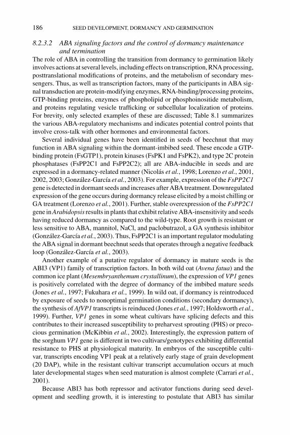
P1: OTE/SPH P2: OTE
BLUK053-Bradford September 22, 2006 15:36
186 SEED DEVELOPMENT, DORMANCY AND GERMINATION
8.2.3.2 ABA signaling factors and the control of dormancy maintenanceand termination
The role of ABA in controlling the transition from dormancy to germination likelyinvolves actions at several levels, including effects on transcription, RNA processing,posttranslational modifications of proteins, and the metabolism of secondary mes-sengers. Thus, as well as transcription factors, many of the participants in ABA sig-nal transduction are protein-modifying enzymes, RNA-binding/processing proteins,GTP-binding proteins, enzymes of phospholipid or phosphoinositide metabolism,and proteins regulating vesicle trafficking or subcellular localization of proteins.For brevity, only selected examples of these are discussed; Table 8.1 summarizesthe various ABA-regulatory mechanisms and indicates potential control points thatinvolve cross-talk with other hormones and environmental factors.
Several individual genes have been identified in seeds of beechnut that mayfunction in ABA signaling within the dormant-imbibed seed. These encode a GTP-binding protein (FsGTP1), protein kinases (FsPK1 and FsPK2), and type 2C proteinphosphatases (FsPP2C1 and FsPP2C2); all are ABA-inducible in seeds and areexpressed in a dormancy-related manner (Nicolas et al., 1998; Lorenzo et al., 2001,2002, 2003; Gonzalez-Garcıa et al., 2003). For example, expression of the FsPP2C1gene is detected in dormant seeds and increases after ABA treatment. Downregulatedexpression of the gene occurs during dormancy release elicited by a moist chilling orGA treatment (Lorenzo et al., 2001). Further, stable overexpression of the FsPP2C1gene in Arabidopsis results in plants that exhibit relative ABA-insensitivity and seedshaving reduced dormancy as compared to the wild-type. Root growth is resistant orless sensitive to ABA, mannitol, NaCl, and paclobutrazol, a GA synthesis inhibitor(Gonzalez-Garcıa et al., 2003). Thus, FsPP2C1 is an important regulator modulatingthe ABA signal in dormant beechnut seeds that operates through a negative feedbackloop (Gonzalez-Garcıa et al., 2003).
Another example of a putative regulator of dormancy in mature seeds is theABI3 (VP1) family of transcription factors. In both wild oat (Avena fatua) and thecommon ice plant (Mesembryanthemum crystallinum), the expression of VP1 genesis positively correlated with the degree of dormancy of the imbibed mature seeds(Jones et al., 1997; Fukuhara et al., 1999). In wild oat, if dormancy is reintroducedby exposure of seeds to nonoptimal germination conditions (secondary dormancy),the synthesis of AfVP1 transcripts is reinduced (Jones et al., 1997; Holdsworth et al.,1999). Further, VP1 genes in some wheat cultivars have splicing defects and thiscontributes to their increased susceptibility to preharvest sprouting (PHS) or preco-cious germination (McKibbin et al., 2002). Interestingly, the expression pattern ofthe sorghum VP1 gene is different in two cultivars/genotypes exhibiting differentialresistance to PHS at physiological maturity. In embryos of the susceptible culti-var, transcripts encoding VP1 peak at a relatively early stage of grain development(20 DAP), while in the resistant cultivar transcript accumulation occurs at muchlater developmental stages when seed maturation is almost complete (Carrari et al.,2001).
Because ABI3 has both repressor and activator functions during seed devel-opment and seedling growth, it is interesting to postulate that ABI3 has similar
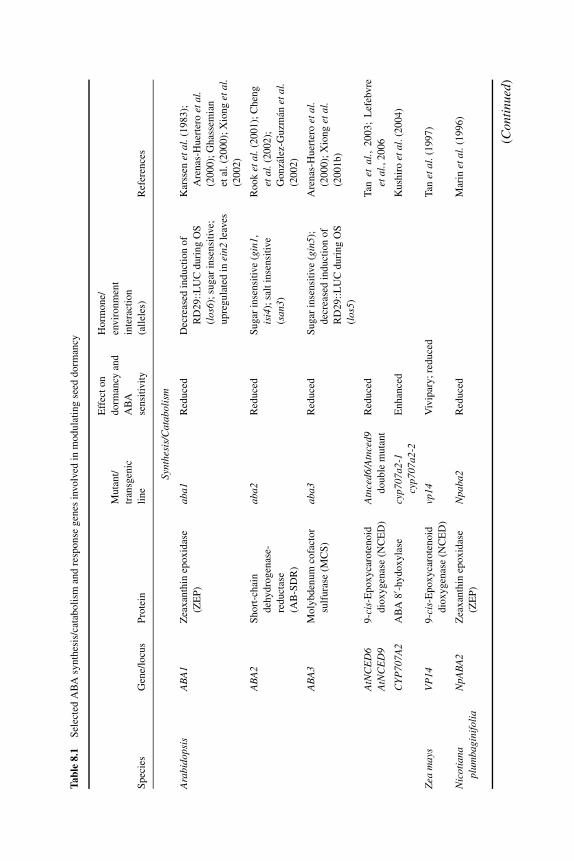
P1: OTE/SPH P2: OTE
BLUK053-Bradford September 22, 2006 15:36
Tabl
e8.
1S
elec
ted
AB
Asy
nth
esis
/cat
abo
lism
and
resp
on
seg
enes
invo
lved
inm
od
ula
tin
gse
edd
orm
ancy
Eff
ect
on
Ho
rmo
ne/
Mu
tan
t/d
orm
ancy
and
env
iro
nm
ent
tran
sgen
icA
BA
inte
ract
ion
Sp
ecie
sG
ene/
locu
sP
rote
inli
ne
sen
siti
vit
y(a
llel
es)
Ref
eren
ces
Synt
hesi
s/C
atab
olis
m
Ara
bido
psis
AB
A1
Zea
xan
thin
epo
xid
ase
(ZE
P)
aba1
Red
uce
dD
ecre
ased
ind
uct
ion
of
RD
29
::L
UC
du
rin
gO
S
(los
6);
sugar
inse
nsi
tive;
up
reg
ula
ted
inei
n2le
aves
Kar
ssen
etal
.(1
98
3);
Are
nas
-Hu
erte
roet
al.
(20
00
);G
has
sem
ian
etal
.(2
00
0);
Xio
ng
etal
.(2
00
2)
AB
A2
Sh
ort
-ch
ain
deh
yd
rog
enas
e-
red
uct
ase
(AB
-SD
R)
aba2
Red
uce
dS
ugar
inse
nsi
tive
(gin
1,is
i4);
salt
inse
nsi
tive
(san
3)
Ro
ok
etal
.(2
00
1);
Ch
eng
etal
.(2
00
2);
Go
nza
lez-
Gu
zman
etal
.(2
00
2)
AB
A3
Mo
lyb
den
um
cofa
cto
r
sulf
ura
se(M
CS
)
aba3
Red
uce
dS
ugar
inse
nsi
tive
(gin
5);
dec
reas
edin
du
ctio
no
f
RD
29
::L
UC
du
rin
gO
S
(los
5)
Are
nas
-Hu
erte
roet
al.
(20
00
);X
ion
get
al.
(20
01
b)
AtN
CE
D6
AtN
CE
D9
9-c
is-E
po
xy
caro
ten
oid
dio
xy
gen
ase
(NC
ED
)
Atn
ced6
/Atn
ced9
do
ub
lem
uta
nt
Red
uce
dT
anet
al.,
20
03
;L
efeb
vre
etal
.,2
00
6
CY
P70
7A2
AB
A8
′ -hydoxyla
secy
p707
a2-1
cyp7
07a2
-2E
nh
ance
dK
ush
iro
etal
.(2
00
4)
Zea
may
sV
P14
9-c
is-E
po
xy
caro
ten
oid
dio
xy
gen
ase
(NC
ED
)
vp14
Viv
ipar
y;
red
uce
dT
anet
al.(
19
97
)
Nic
otia
napl
umba
gini
foli
aN
pAB
A2
Zea
xan
thin
epo
xid
ase
(ZE
P)
Npa
ba2
Red
uce
dM
arin
etal
.(1
99
6)
(Con
tinu
ed)
187
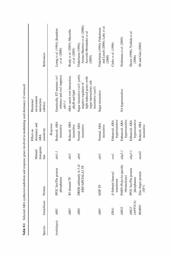
P1: OTE/SPH P2: OTE
BLUK053-Bradford September 22, 2006 15:36
Tabl
e8.
1S
elec
ted
AB
Asy
nth
esis
/cat
abo
lism
and
resp
on
seg
enes
invo
lved
inm
od
ula
tin
gse
edd
orm
ancy
(Con
tinu
ed)
Eff
ect
on
Ho
rmo
ne/
Mu
tan
t/d
orm
ancy
and
env
iro
nm
ent
tran
sgen
icA
BA
inte
ract
ion
Sp
ecie
sG
ene/
locu
sP
rote
inli
ne
sen
siti
vit
y(a
llel
es)
Ref
eren
ces
Res
pons
e
Ara
bido
psis
AB
I1P
P2
CS
er/T
hr
pro
tein
ph
osp
hat
ase
abi1
-1R
edu
ced
;A
BA
inse
nsi
tive
Gen
etic
ally
,E
Tm
uta
nts
ctr1
enh
ance
and
ein2
sup
pre
ss
abi1
-1
Leu
ng
etal
.(1
99
4);
Bea
ud
oin
etal
.(2
00
0)
AB
I3B
3d
om
ain
TF
abi3
Red
uce
d;
AB
A
inse
nsi
tive
AB
A–
aux
inro
ot
inte
ract
ion
;
phyB
and
light
Bra
dy
etal
.(2
00
3);
Maz
zell
a
etal
.(2
00
5)
AB
I4D
RE
Bsu
bfa
mil
yA
-3o
f
ER
F/A
PE
TA
LA
2T
F
abi4
No
rmal
;A
BA
inse
nsi
tive
Su
gar
inse
nsi
tive
(sis
5,gi
n6);
neg
ativ
ere
gula
tor
of
lig
ht-
ind
uce
dg
enes
(wit
h
sugar
inte
ract
ion
);sa
lt
inse
nsi
tive
(san
5)
Fin
kel
stei
n(1
99
4);
Are
nas
-Hu
erte
roet
al.(
20
00
);
Ace
ved
o-H
ern
and
ezet
al.
(20
05
)
AB
I5b
ZIP
TF
abi5
No
rmal
;A
BA
inse
nsi
tive
Su
gar
inse
nsi
tive
Fin
kel
stei
n(1
99
4);
Fin
kel
stei
n
and
Ly
nch
(20
00
);L
aby
etal
.(2
00
0)
ER
A1
β-S
ubu
nit
farn
esy
l
tran
sfer
ase
era1
En
han
ced
;A
BA
hyper
sensi
tive
Cu
tler
etal
.(1
99
6)
AH
G2
PAR
N(P
oly
(A)-
spec
ific
rib
on
ucl
ease
)
ahg2
-1E
nh
ance
d;
AB
A
hyper
sensi
tive
SA
hyper
sensi
tive
Nis
him
ura
etal
.(2
00
5)
AH
G3
(AtP
P2C
A)
PP
2C
Ser
/Th
rp
rote
in
ph
osp
hat
ase
ahg3
-1E
nh
ance
d;
AB
A
hyper
sensi
tive
Sh
een
(19
98
);Y
osh
ida
etal
.(2
00
6)
MA
RD
1Z
inc-
fin
ger
pro
tein
(TF
?)
mar
d1R
edu
ced
;A
BA
inse
nsi
tive
He
and
Gan
(20
04
)
188

P1: OTE/SPH P2: OTE
BLUK053-Bradford September 22, 2006 15:36
RG
S1R
egu
lato
ro
f
G-p
rote
in
sig
nal
ing
rgs1
-2R
edu
ced
;A
BA
inse
nsi
tive
Po
siti
ve
reg
ula
tor
of
AtN
CE
D3,
AtA
BA
2(e
sp.d
uri
ng
sugar
stre
ss)
Ch
enet
al.(
20
03
);C
hen
etal
.(2
00
6)
RP
N10
26
SP
rote
aso
me
subu
nit
rpn1
0-1
En
han
ced
(?);
AB
A
hyper
sensi
tive
See
dli
ngs
hyper
sensi
tive
toN
aCl,
sugar
;C
Kin
sen
siti
ve;
Au
x
inse
nsi
tive
roo
ts
Sm
alle
etal
.(2
00
3)
RO
P2
Ro
pG
TP
ase
CA
-rop
2(c
on
stit
uti
vel
y
acti
ve
GT
P-b
ou
nd
)
Red
uce
d;sl
igh
tly
red
uce
d
AB
Ase
nsi
tiv
ity
hypoco
tyl
elongat
ion
pro
mote
dby
BR
;in
crea
sed
LR
form
atio
nb
y
Au
xco
mp
ared
toW
T
Li
etal
.(2
00
1)
DN
-rop
2(d
om
inan
t
neg
ativ
eG
DP
-bound)
En
han
ced
;A
BA
hyper
sensi
tive
Dec
reas
edL
Rfo
rmat
ion
by
Au
x
com
par
edto
WT
SAD
1S
m-l
ike
snR
NP
pro
tein
sad1
En
han
ced
;A
BA
hyper
sensi
tive
Xio
ng
etal
.(2
00
1a)
Z.m
ays
VP
1B
3d
om
ain
TF
vp1
Viv
ipar
yo
rre
du
ced
do
rman
cy;
AB
A
inse
nsi
tive
AB
A–
aux
inro
ot
inte
ract
ion
;
incr
ease
det
hyle
ne
pro
duct
ion
du
rin
gse
edd
evel
op
men
t
McC
arty
etal
.(1
99
1);
Yo
un
gan
dG
alli
e
(20
00
);S
uzu
ki
etal
.(2
00
1)
Abbre
via
tions:
Aux,au
xin
;C
K,cy
tokin
in;
BL
,bra
ssin
oli
de
(bra
ssin
ost
eroid
);E
T,et
hyle
ne;
LR
,la
tera
lro
ot;
OS
,osm
oti
cst
ress
;S
A,sa
licy
lic
acid
;T
F,tr
ansc
ripti
on
fact
or.
189
P1: OTE/SPH P2: OTE
BLUK053-Bradford September 22, 2006 15:36
190 SEED DEVELOPMENT, DORMANCY AND GERMINATION
functions during dormancy maintenance in the mature seed to regulate postimbi-bition dormancy-related genes/processes. However, conclusive evidence of a directrole for ABI3 awaits and will be difficult to demonstrate experimentally because ofthe more global role of this transcription factor in seed developmental processes.Stable overexpression in a transgenic system influences several processes duringseed development (Zeng and Kermode, 2004); an inducible expression system (e.g.,via use of a glucocorticoid receptor/DEX [dexamethasone]-inducible system) maypose a more viable alternative to test for a direct role of ABI3/VP1 specifically indormancy maintenance. Maintenance of dormancy in yellow-cedar seeds appears toinvolve ABI3. A decline in CnABI3 transcripts and in CnABI3 protein abundanceis correlated with dormancy termination, and their synthesis is also downregulatedduring germination (Zeng et al., 2003). Further, it has been suggested that regula-tion of CnABI3 is not restricted to the transcriptional level and may also involveprotein stability (Zeng et al., 2003). Indeed, a key control over the dormancy-to-germination transition may occur at the level of regulation of proteasome compo-nents (Bove et al., 2005; see Chapter 10). Interestingly, AIP2 (ABI3-INTERACTINGPROTEIN 2), which was originally uncovered in a yeast 2-hybrid screen for proteinsthat interact with ABI3, encodes an E3-ubiquitin ligase (Kurup et al., 2000; Zhanget al., 2005), which targets proteins for degradation via the 26S proteasome path-way (Vierstra, 2003). AIP2 can polyubiquitinate ABI3 in vitro and, in transgenicplants, induced AIP2 gene expression leads to decreased ABI3 protein levels. Fur-ther, Arabidopsis aip2-1 null mutants show higher ABI3 protein levels compared tothe wild-type seeds after moist chilling, are hypersensitive to ABA, and mimic anABI3-overexpression phenotype. Conversely, AIP2 overexpressors contain lowerlevels of ABI3 protein compared to wild type and are more resistant to ABA, phe-nocopying the abi3 mutant (Zhang et al., 2005). Thus, AIP2 is a negative regulatorof ABA responses via its modulation of ABI3 protein levels.
8.3 Gibberellin
8.3.1 GA is antagonistic to ABA during seed development
During development, GA plays a multitude of roles, participating in fertilization, em-bryo growth, nutrient uptake, and the prevention of seed abortion (Hays et al., 2002;Singh et al., 2002; Finkelstein, 2004). GA levels are usually high during embryodevelopment, in which there may be two peaks of GA content; as seeds approachmaturity, most active GAs are deactivated, for example via 2-β-hydroxylation orconjugate formation (Hedden, 1999; see Chapter 9). An accumulation of active GAsduring later seed development would likely promote premature germination. Indeed,GA appears to act as an antagonist to ABA during seed development, especially withrespect to dormancy induction (White et al., 2000). When an ABA-deficient mutantof maize (vp5) is manipulated either genetically or via biosynthesis inhibitors toinduce GA deficiency during early seed development, vivipary is suppressed in de-veloping kernels and the seeds acquire desiccation tolerance and storage longevity. In
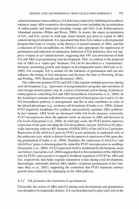
P1: OTE/SPH P2: OTE
BLUK053-Bradford September 22, 2006 15:36
ABSCISIC ACID AND HORMONAL CROSS-TALK IN SEED DORMANCY 191
cultured immature maize embryos, GA deficiency induced by inhibiting biosynthesisenhances many ABA-responsive developmental events including the accumulationof anthocyanins and transcripts encoding storage and LEA (Late EmbryogenesisAbundant) proteins (White and Riven, 2000). In planta, the major accumulationof GA1 and GA3 occurs in wild-type maize kernels just prior to a peak in ABAcontent during development. It is speculated that these GAs induce a developmentalprogram that leads to vivipary in the absence of normal amounts of ABA, and thata reduction of GAs reestablishes an ABA/GA ratio appropriate for suppression ofgermination and induction of maturation. Induction of GA deficiency does not sup-press vivipary in vp1 mutant kernels, suggesting that VP1 acts downstream of bothGA and ABA in programming seed development. Thus, in contrast to the proposedrole of ABA as a ‘status quo’ hormone, GA can be described as a ‘transitionary’hormone, promoting growth and changes in developmental status (McCourt et al.,2005). For example, GA is needed for the initiation of germination and can alsoinfluence the timing of leaf emergence and decrease the time to flowering (Evansand Poethig, 1995; Bentsink and Koornneef, 2002).
The embryonic proteins FUS3 and LEC2 that regulate multiple processes duringseed development (e.g., repression of postgerminative programs and activation ofseed storage protein genes) may be a nexus of hormone action during Arabidopsisembryogenesis controlling GA and ABA biosynthesis (Gazzarrini et al., 2004). Inthe Arabidopsis mutants with defective expression of these genes (fus3 and lec2), theGA biosynthesis pathway is misregulated, and this in turn contributes to some ofthe altered phenotypes (e.g., trichome cell formation) (Curaba et al., 2004). Indeed,FUS3 negatively modulates GA synthesis and positively regulates ABA synthesis.In fus3 mutants, ABA levels are decreased while GA levels increase; conversely,FUS3 misexpressors show the opposite trend: an increase in ABA and decrease inGA levels (Gazzarrini et al., 2004). In wild-type seeds, the FUS3 protein repressesexpression of the gene encoding the GA biosynthetic enzyme AtGA3ox2 by phys-ically interacting with two RY elements (GATGCATG) of the AtGA3ox2 promoter.Repression of the AtGA3ox2 gene by FUS3 occurs primarily in epidermal cells ofthe embryonic axis, which is distinct from the pattern of expression of this gene dur-ing germination (Curaba et al., 2004). Similarly, the expression of AtGA3ox1 andAtGA20ox1 genes is downregulated by inducible FUS3 misexpression in seedlings(Gazzarrini et al., 2004). FUS3 expression itself is modulated by the hormone auxin(see below). Gazzarrini et al. (2004) suggest that it is this modulation of the ABA/GAratio by FUS3, and a positive or negative feedback regulation of FUS3 by ABA andGA, respectively, that helps regulate maturation events during seed development.Interestingly, maternally derived ABA inhibits viviparous germination of fus3 mu-tants (Raz et al., 2001), supporting the contention that FUS3 maintains embryogrowth arrest indirectly by impinging on the ABA pathway.
8.3.2 GA promotes the transition to germination
Classically, the action of ABA and GA during seed development and germinationwas thought to be temporally distinct. GA was hypothesized to play a key role in the
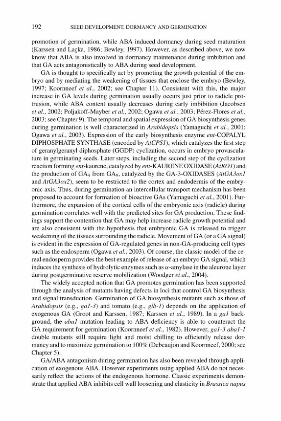
P1: OTE/SPH P2: OTE
BLUK053-Bradford September 22, 2006 15:36
192 SEED DEVELOPMENT, DORMANCY AND GERMINATION
promotion of germination, while ABA induced dormancy during seed maturation(Karssen and Lacka, 1986; Bewley, 1997). However, as described above, we nowknow that ABA is also involved in dormancy maintenance during imbibition andthat GA acts antagonistically to ABA during seed development.
GA is thought to specifically act by promoting the growth potential of the em-bryo and by mediating the weakening of tissues that enclose the embryo (Bewley,1997; Koornneef et al., 2002; see Chapter 11). Consistent with this, the majorincrease in GA levels during germination usually occurs just prior to radicle pro-trusion, while ABA content usually decreases during early imbibition (Jacobsenet al., 2002; Poljakoff-Mayber et al., 2002; Ogawa et al., 2003; Perez-Flores et al.,2003; see Chapter 9). The temporal and spatial expression of GA biosynthesis genesduring germination is well characterized in Arabidopsis (Yamaguchi et al., 2001;Ogawa et al., 2003). Expression of the early biosynthesis enzyme ent-COPALYLDIPHOSPHATE SYNTHASE (encoded by AtCPS1), which catalyzes the first stepof geranylgeranyl diphosphate (GGDP) cyclization, occurs in embryo provascula-ture in germinating seeds. Later steps, including the second step of the cyclizationreaction forming ent-kaurene, catalyzed by ent-KAURENE OXIDASE (AtKO1) andthe production of GA4 from GA9, catalyzed by the GA-3-OXIDASES (AtGA3ox1and AtGA3ox2), seem to be restricted to the cortex and endodermis of the embry-onic axis. Thus, during germination an intercellular transport mechanism has beenproposed to account for formation of bioactive GAs (Yamaguchi et al., 2001). Fur-thermore, the expansion of the cortical cells of the embryonic axis (radicle) duringgermination correlates well with the predicted sites for GA production. These find-ings support the contention that GA may help increase radicle growth potential andare also consistent with the hypothesis that embryonic GA is released to triggerweakening of the tissues surrounding the radicle. Movement of GA (or a GA signal)is evident in the expression of GA-regulated genes in non-GA-producing cell typessuch as the endosperm (Ogawa et al., 2003). Of course, the classic model of the ce-real endosperm provides the best example of release of an embryo GA signal, whichinduces the synthesis of hydrolytic enzymes such as α-amylase in the aleurone layerduring postgerminative reserve mobilization (Woodger et al., 2004).
The widely accepted notion that GA promotes germination has been supportedthrough the analysis of mutants having defects in loci that control GA biosynthesisand signal transduction. Germination of GA biosynthesis mutants such as those ofArabidopsis (e.g., ga1-3) and tomato (e.g., gib-1) depends on the application ofexogenous GA (Groot and Karssen, 1987; Karssen et al., 1989). In a ga1 back-ground, the aba1 mutation leading to ABA deficiency is able to counteract theGA requirement for germination (Koornneef et al., 1982). However, ga1-3 aba1-1double mutants still require light and moist chilling to efficiently release dor-mancy and to maximize germination to 100% (Debeaujon and Koornneef, 2000; seeChapter 5).
GA/ABA antagonism during germination has also been revealed through appli-cation of exogenous ABA. However experiments using applied ABA do not neces-sarily reflect the actions of the endogenous hormone. Classic experiments demon-strate that applied ABA inhibits cell wall loosening and elasticity in Brassica napus
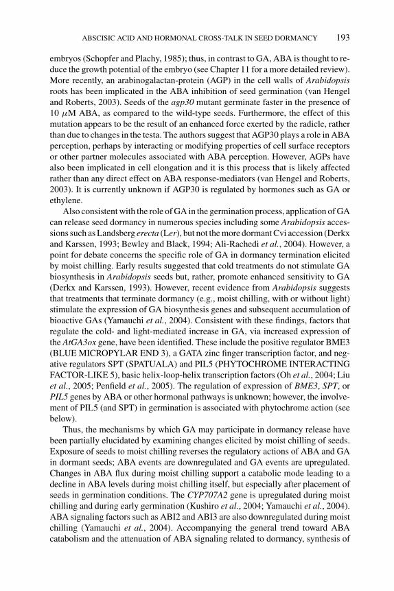
P1: OTE/SPH P2: OTE
BLUK053-Bradford September 22, 2006 15:36
ABSCISIC ACID AND HORMONAL CROSS-TALK IN SEED DORMANCY 193
embryos (Schopfer and Plachy, 1985); thus, in contrast to GA, ABA is thought to re-duce the growth potential of the embryo (see Chapter 11 for a more detailed review).More recently, an arabinogalactan-protein (AGP) in the cell walls of Arabidopsisroots has been implicated in the ABA inhibition of seed germination (van Hengeland Roberts, 2003). Seeds of the agp30 mutant germinate faster in the presence of10 μM ABA, as compared to the wild-type seeds. Furthermore, the effect of thismutation appears to be the result of an enhanced force exerted by the radicle, ratherthan due to changes in the testa. The authors suggest that AGP30 plays a role in ABAperception, perhaps by interacting or modifying properties of cell surface receptorsor other partner molecules associated with ABA perception. However, AGPs havealso been implicated in cell elongation and it is this process that is likely affectedrather than any direct effect on ABA response-mediators (van Hengel and Roberts,2003). It is currently unknown if AGP30 is regulated by hormones such as GA orethylene.
Also consistent with the role of GA in the germination process, application of GAcan release seed dormancy in numerous species including some Arabidopsis acces-sions such as Landsberg erecta (Ler), but not the more dormant Cvi accession (Derkxand Karssen, 1993; Bewley and Black, 1994; Ali-Rachedi et al., 2004). However, apoint for debate concerns the specific role of GA in dormancy termination elicitedby moist chilling. Early results suggested that cold treatments do not stimulate GAbiosynthesis in Arabidopsis seeds but, rather, promote enhanced sensitivity to GA(Derkx and Karssen, 1993). However, recent evidence from Arabidopsis suggeststhat treatments that terminate dormancy (e.g., moist chilling, with or without light)stimulate the expression of GA biosynthesis genes and subsequent accumulation ofbioactive GAs (Yamauchi et al., 2004). Consistent with these findings, factors thatregulate the cold- and light-mediated increase in GA, via increased expression ofthe AtGA3ox gene, have been identified. These include the positive regulator BME3(BLUE MICROPYLAR END 3), a GATA zinc finger transcription factor, and neg-ative regulators SPT (SPATUALA) and PIL5 (PHYTOCHROME INTERACTINGFACTOR-LIKE 5), basic helix-loop-helix transcription factors (Oh et al., 2004; Liuet al., 2005; Penfield et al., 2005). The regulation of expression of BME3, SPT, orPIL5 genes by ABA or other hormonal pathways is unknown; however, the involve-ment of PIL5 (and SPT) in germination is associated with phytochrome action (seebelow).
Thus, the mechanisms by which GA may participate in dormancy release havebeen partially elucidated by examining changes elicited by moist chilling of seeds.Exposure of seeds to moist chilling reverses the regulatory actions of ABA and GAin dormant seeds; ABA events are downregulated and GA events are upregulated.Changes in ABA flux during moist chilling support a catabolic mode leading to adecline in ABA levels during moist chilling itself, but especially after placement ofseeds in germination conditions. The CYP707A2 gene is upregulated during moistchilling and during early germination (Kushiro et al., 2004; Yamauchi et al., 2004).ABA signaling factors such as ABI2 and ABI3 are also downregulated during moistchilling (Yamauchi et al., 2004). Accompanying the general trend toward ABAcatabolism and the attenuation of ABA signaling related to dormancy, synthesis of
P1: OTE/SPH P2: OTE
BLUK053-Bradford September 22, 2006 15:36
194 SEED DEVELOPMENT, DORMANCY AND GERMINATION
bioactive GAs is upregulated during moist chilling. As such, selected GA-induciblegenes are upregulated while selected GA-repressible genes are downregulated dur-ing chilling (Yamauchi et al., 2004). One example of a gene downregulated byGA and moist chilling is the ABI5-homologue EEL (ENHANCED Em LEVEL;Bensmihen et al., 2002; Ogawa et al., 2003; Yamauchi et al., 2004). Interestingly,the expression of the EEL gene is dormancy-related, being upregulated in dormant-imbibed Cvi seeds, but downregulated during after-ripening of Cvi seeds (Finch-Savage, 2004). However, the participation of EEL in dormancy and germinationremains speculative, especially since eel mutants do not display ABA-insensitivephenotypes during germination (Bensmihen et al., 2002).
Additional mechanisms of GA-signaling in seed dormancy release and germi-nation are emerging and have been linked to hormone cross-talk. One of the mostprominent examples of repressors of GA action during germination is the DELLA(motif) transcription factors, GAI (GA INSENSITIVE), RGA (REPRESSOR OFga1-3), RGL1 (RGA-LIKE 1), RGL2, and RGL3 – a subfamily of GRAS (for GAI,RGA, and SCARECROW) regulatory proteins (Cao et al., 2005; see Chapter 10).In addition to modulating GA response, DELLA proteins appear also to mediateethylene and auxin responses and are suggested to play an integrative role in thephytohormone signal response network (Achard et al., 2003). Further, the ‘growth-repressing’ DELLA proteins have been linked to ABA signaling as well. The rootsof a quadruple-DELLA mutant (gai, rga, rgl1, rgl2) are relatively insensitive to ABAgrowth inhibition; in addition, in transgenic wild-type and abi1-1 mutant plants ex-pressing a reporter (green fluorescent protein)-RGA fusion driven by the native RGApromoter (pRGA::GFP-RGA), GFP-RGA accumulation in the roots is responsiveto ABA only in the former (Achard et al., 2006).
An additional factor that seems to interplay with the GA pathway is thePICKLE (PKL) locus. The PKL gene encodes an SWI (switch)/SNF (SucroseNon-Fermenting) class chromatin remodeling factor that belongs to the CHD3(Chromodomain Helicase DNA-binding 3) group that generally are repressors oftranscription (Ogas et al., 1999). PKL helps modulate the phase transition betweenseed development and dormancy/germination and is necessary for the repression ofembryonic traits from germination onward (Ogas et al., 1997; Li et al., 2005). Inmutant pkl plants, the embryonic identity genes, LEC1 and LEC2, are de-repressedduring germination and LEC1, LEC2, and FUS3 are upregulated more than 100-fold in pkl roots (Ogas et al., 1999; Rider et al., 2003). Expression of these LECfamily genes in pkl seedlings allows expression of seed developmental traits af-ter germination, such as accumulation of seed storage reserves and the ability toundergo somatic embryogenesis (Ogas et al., 1999; Stone et al., 2001; Hendersonet al., 2004; Rider et al., 2004). GA interacts with pkl to repress embryonic identity;decreasing the level of GA in germinating seeds increases the penetrance of thepkl root phenotype (swollen green appearance) from ∼1–5% to 80% (Ogas et al.,1997). Further, pkl plants are reminiscent of GA-response mutants; they are dwarfed,dark-green, and exhibit increased time to flowering. Exogenous GA can partiallyrescue pkl phenotypes but plants are not deficient in GA (Henderson et al., 2004).Henderson et al. (2004) suggested that PKL may mediate a subset of GA-dependent
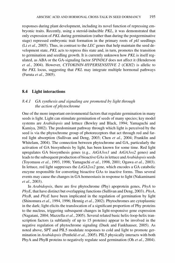
P1: OTE/SPH P2: OTE
BLUK053-Bradford September 22, 2006 15:36
ABSCISIC ACID AND HORMONAL CROSS-TALK IN SEED DORMANCY 195
responses during plant development, including its novel function of repressing em-bryonic traits. Recently, using a steroid-inducible PKL, it was demonstrated thatonly expression of PKL during germination (rather than during the postgerminativestage) repressed embryonic trait formation in the primary roots of pkl seedlings(Li et al., 2005). Thus, in contrast to the LEC genes that help maintain the seed de-velopment state, PKL acts to repress this state and, in turn, promotes the transitionto germination and seedling growth. It is currently unknown how PKL is itself reg-ulated, as ABA or the GA-signaling factor SPINDLY does not affect it (Hendersonet al., 2004). However, CYTOKININ-HYPERSENSITIVE 2 (CKH2) is allelic tothe PKL locus, suggesting that PKL may integrate multiple hormonal pathways(Furuta et al., 2005).
8.4 Light interactions
8.4.1 GA synthesis and signaling are promoted by light throughthe action of phytochrome
One of the more important environmental factors that regulate germination in manyseeds is light. Light can stimulate germination of seeds of many species; key modelsystems are Arabidopsis and lettuce (Bewley and Black, 1994; Yamaguchi andKamiya, 2002). The predominant pathway through which light is perceived by theseed is via the phytochrome group of photoreceptors that act through red and far-red light absorption (Sullivan and Deng, 2003; Chen et al., 2004; Franklin andWhitelam, 2004). The connection between phytochrome and GA, particularly theactivation of GA biosynthesis by light, has been known for some time. Red lightupregulates GA biosynthesis genes (e.g., AtGA3ox1 and AtGA3ox2 genes) andleads to the subsequent production of bioactive GAs in lettuce and Arabidopsis seeds(Toyomasu et al., 1993, 1998; Yamaguchi et al., 1998, 2001; Ogawa et al., 2003).In lettuce, red light suppresses the LsGA2ox2 gene, which encodes a GA catabolicenzyme responsible for converting bioactive GAs to inactive forms. Thus severalevents may cause the changes in GA homeostasis in response to light (Nakaminamiet al., 2003).
In Arabidopsis, there are five phytochrome (Phy) apoprotein genes, PhyA toPhyE, that have distinct but overlapping functions (Sullivan and Deng, 2003). PhyA,PhyB, and PhyE have been implicated in the regulation of germination by light(Shinomura et al., 1994, 1996; Hennig et al., 2002). Phytochromes are cytoplasmicin the dark; light elicits the translocation of a significant proportion of Phy proteinsto the nucleus, triggering subsequent changes in light-responsive gene expression(Nagatani, 2004; Mazzella et al., 2005). Several related basic helix-loop-helix tran-scription factors (a subfamily of up to 15 proteins) appear to be involved in thenegative regulation of phytochrome signaling (Duek and Fankhauser, 2005). Asnoted above, SPT and PIL5 modulate responses to cold and light to promote ger-mination in Arabidopsis (Penfield et al., 2005). PIL5 physically interacts with bothPhyA and PhyB proteins to negatively regulate seed germination (Oh et al., 2004).
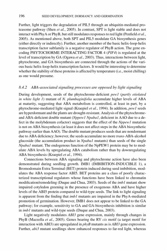
P1: OTE/SPH P2: OTE
BLUK053-Bradford September 22, 2006 15:36
196 SEED DEVELOPMENT, DORMANCY AND GERMINATION
Further, light triggers the degradation of PIL5 through an ubiquitin-mediated pro-teasome pathway (Shen et al., 2005). In contrast, SPT is light stable and does notinteract with PhyA or PhyB, but still modulates responses to red light (Penfield et al.,2005). As mentioned above, both SPT and PIL5 modulate GA biosynthesis genes(either directly or indirectly). Further, another member of the basic helix-loop-helixtranscription factor subfamily is a negative regulator of PhyB action. The gene en-coding PHYTOCHORME INTERACTING FACTOR 4 (PIF4) is regulated at thelevel of transcription by GA (Ogawa et al., 2003). Thus, interactions between light,phytochrome, and GA biosynthesis are connected through the actions of the vari-ous basic helix-loop-helix transcription factors. It would be interesting to determinewhether the stability of these proteins is affected by temperature (i.e., moist chilling)as one would presume.
8.4.2 ABA-associated signaling processes are opposed by light signaling
During development, seeds of the phytochrome-deficient pew1 (partly etiolatedin white light 1) mutant of N. plumbaginifolia accumulate higher levels of ABAat maturity, suggesting that ABA metabolism is controlled, at least in part, by aphytochrome-mediated light signal (Kraepiel et al., 1994). In addition, pew1 seedsare hyperdormant and the plants are drought-resistant. Analysis of the phytochrome-and ABA-deficient double mutant (Nppew1 Npaba1, deficient in AAO due to a de-fect in the molybdenum cofactor) suggests that the effect of the Nppew1 mutationis not on ABA biosynthesis (at least it does not affect steps in the ABA biosyntheticpathway earlier than AAO). The double mutant produces seeds that are nondormantdue to ABA deficiency; however, the seeds accumulate no more trans-ABA-alcoholglucoside (the accumulation product in Npaba1 mutants) than seeds of the singleNpaba1 mutant. The endogenous function of the NpPEW1 protein may be to mod-ulate ABA levels by upregulating ABA catabolism rather than by downregulatingABA biosynthesis (Kraepiel et al., 1994).
Connections between ABA signaling and phytochrome action have also beendemonstrated during seedling growth. IMB1 (IMBIBITION-INDUCIBLE 1), aBromodomain Extra Terminal (BET) protein induced during seed imbibition, mod-ulates the ABA response factor ABI5. BET proteins are a class of poorly charac-terized transcriptional regulators whose functions have been linked to chromatinmodification/remodeling (Duque and Chua, 2003). Seeds of the imb1 mutant showimpaired cotyledon greening in the presence of exogenous ABA and have higherlevels of the ABI5 protein compared to wild-type seeds. The link to light signalingis apparent from the finding that imb1 mutants are impaired in the PhyA-mediatedpromotion of germination. However, IMB1 does not appear to be linked to the GApathway; for example, sensitivity to GA and GA-biosynthesis inhibition is similarin imb1 mutants and wild-type seeds (Duque and Chua, 2003).
Light negatively modulates ABI3 gene expression, mainly through changes inPhyB (Mazzella et al., 2005). Genes bearing the RY cis motif (a target motif forinteraction with ABI3) are upregulated in phyB mutants as is ABI3 gene expression.Further, abi3 mutant seedlings show enhanced responses to far-red light, whereas
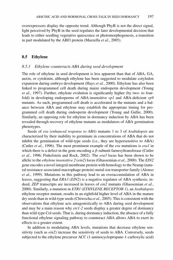
P1: OTE/SPH P2: OTE
BLUK053-Bradford September 22, 2006 15:36
ABSCISIC ACID AND HORMONAL CROSS-TALK IN SEED DORMANCY 197
overexpressors display the opposite trend. Although PhyB is not the direct signal,light perceived by PhyB in the seed regulates the later developmental decision thatleads to either seedling vegetative quiescence or photomorphogenesis, a transitionin part modulated by the ABI3 protein (Mazzella et al., 2005).
8.5 Ethylene
8.5.1 Ethylene counteracts ABA during seed development
The role of ethylene in seed development is less apparent than that of ABA, GA,auxin, or cytokinin, although ethylene has been suggested to modulate cotyledonexpansion during embryo development (Hays et al., 2000). Ethylene has also beenlinked to programmed cell death during maize endosperm development (Younget al., 1997). Further, ethylene evolution is significantly higher (by two- to four-fold) in developing endosperms of ABA-insensitive vp1 and ABA-deficient vp9mutants. As such, programmed cell death is accelerated in the mutants and a bal-ance between ABA and ethylene may establish the appropriate timing for pro-grammed cell death during endosperm development (Young and Gallie, 2000).Similarly, an opposing role for ethylene in dormancy induction by ABA has beenrevealed through recovery of ethylene mutants as modulators of ABA germinationphenotypes.
Seeds of era (enhanced response to ABA) mutants 1 to 3 of Arabidopsis arecharacterized by their inability to germinate in concentrations of ABA that do notinhibit the germination of wild-type seeds (i.e., they are hypersensitive to ABA)(Cutler et al., 1996). The most prominent example of the era mutations is era1 inwhich there is a defect in the gene encoding a β-subunit farnesyltransferase (Cutleret al., 1996; Finkelstein and Rock, 2002). The era3 locus has been shown to beallelic to the ethylene insensitive 2 (ein2) locus (Ghassemian et al., 2000). The EIN2gene encodes a novel integral membrane protein with homology to the Nramp (natu-ral resistance-associated macrophage protein) metal-ion transporter family (Alonsoet al., 1999). Mutations in this pathway lead to an overaccumulation of ABA inleaves, suggesting that ERA3 (EIN2) is a negative regulator of ABA synthesis; in-deed, ZEP transcripts are increased in leaves of ein2 mutants (Ghassemian et al.,2000). Similarly, a mutation in ETR1 (ETHYLENE RECEPTOR 1), an Arabidopsisethylene receptor mutant, results in an eightfold higher level of ABA in the maturedry seeds than in wild-type seeds (Chiwocha et al., 2005). This is consistent with theobservations that ethylene acts antagonistically to ABA during seed developmentand may be a main reason why etr1-2 seeds display a greater degree of dormancythan wild-type Col seeds. That is, during dormancy induction, the absence of a fullyfunctional ethylene signaling pathway to counteract ABA allows ABA to exert itseffects to a greater extent.
In addition to modulating ABA levels, mutations that decrease ethylene sen-sitivity (such as ein2) increase the sensitivity of seeds to ABA. Conversely, seedssubjected to the ethylene precursor ACC (1-aminocyclopropane-1-carboxylic acid)
P1: OTE/SPH P2: OTE
BLUK053-Bradford September 22, 2006 15:36
198 SEED DEVELOPMENT, DORMANCY AND GERMINATION
exhibit decreased ABA sensitivity, as do seeds of constitutive ethylene response(ctr1, constitutive triple response 1) mutants. CTR1, a Raf-like serine/threonineprotein kinase suggested to be a mitogen-activated protein kinase kinase kinase, isa negative regulator of ethylene signaling (Ouaked et al., 2003). In a similar study,Beaudoin et al. (2000) screened for mutations that either enhanced or suppressedthe ABA-resistant seed germination phenotype of Arabidopsis abi1-1 seeds. Allelesof ctr1 and ein2 were recovered as enhancer and suppressor mutations, respectively(Beaudoin et al., 2000).
One particular point of contention is that we do not know whether ethyleneexerts its antagonistic actions both during seed development (dormancy induction)and during dormancy termination and germination. From the discussion above, itseems clear that ethylene counteracts ABA during seed development and thereforemodulates dormancy induction (i.e., the degree of dormancy in the mature seed).Reciprocal crosses between wild-type tomato plants and Nr (Never-ripe) mutants(an ethylene receptor mutant) show that ethylene insensitivity during seed devel-opment plays a role in determining the subsequent time to complete germination(Siriwitayawan et al., 2003). However, ethylene is involved in dormancy release andgermination in many species, and the relative contributions of ethylene to dormancyinduction and release are still debatable. The existence of multiple ethylene recep-tors (e.g., in Arabidopsis) allowing for functional redundancies only compounds theproblem.
8.5.2 Ethylene promotes the transition from dormancy to germination
Ethylene acts antagonistically to ABA during seed development and during dor-mancy termination and germination and acts in concert with GA to promotethese transitional changes. Ethylene promotes germination of many species andin others, such as peanut (Arachis hypogaea), apple (Malus domestica), cocklebur(Xanthium strumarium) and sunflower, participates in the release from seed dor-mancy (Kepczynski and Kepczynski, 1997; Matilla, 2000). In beechnut, ethephon,an ethylene-releasing compound, can accelerate dormancy release (as can GA3)and the GA and ethylene pathways likely interact (Calvo et al., 2003, 2004a,b).FsGA20ox1 gene expression is stimulated by moist chilling, GA or ethephon treat-ment (all effective in releasing dormancy) (Calvo et al., 2004b). Similarly, FsACO1(ACC OXIDASE 1) gene expression, ACC content, and ethylene evolution all in-crease when dormant seeds are treated with GA3 (ACO catalyzes the last step inethylene biosynthesis, the conversion of ACC to ethylene) (Calvo et al., 2004a).Indeed, ethylene can replace the requirement for GA and restore germination ofArabidopsis ga1 seeds in the light or promote germination of wild-type seeds in thedark (Karssen et al., 1989). Conversely, GA3 can stimulate germination of seeds ofthe ethylene receptor mutant etr1 (Bleecker et al., 1988).
In many species, a burst of ethylene evolution occurs around the time of radicleprotrusion coincident with the major increase in bioactive GA in seeds (Kepczynskiand Kepczynski, 1997; Lashbrook et al., 1998; Kucera et al., 2005). However,the role of ethylene in germination is still under debate; ethylene production may
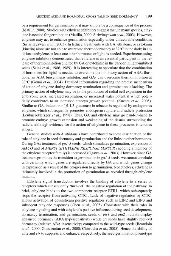
P1: OTE/SPH P2: OTE
BLUK053-Bradford September 22, 2006 15:36
ABSCISIC ACID AND HORMONAL CROSS-TALK IN SEED DORMANCY 199
be a requirement for germination or it may simply be a consequence of the process(Matilla, 2000). Studies with ethylene inhibitors suggest that, in many species, ethy-lene is needed for germination (Matilla, 2000; Siriwitayawan et al., 2003). However,ethylene may act to enhance germination especially under unfavorable conditions(Siriwitayawan et al., 2003). In lettuce, treatments with GA, ethylene, or cytokinin(kinetin) alone are not able to overcome thermodormancy at 32◦C in the dark; in ad-dition to ethylene, at least one other hormone, or light, is needed. Experiments usingethylene inhibitors demonstrated that ethylene is an essential participant in the re-lease of thermoinhibition elicited by GA or cytokinin in the dark or in light-imbibedseeds (Saini et al., 1986, 1989). It is interesting to speculate that the combinationof hormones (or light) is needed to overcome the inhibitory action of ABA; fluri-done, an ABA biosynthesis inhibitor, and GA3 can overcome thermoinhibition at33◦C (Gonai et al., 2004). Detailed information regarding the precise mechanismof action of ethylene during dormancy termination and germination is lacking. Theprimary action of ethylene may be in the promotion of radial cell expansion in theembryonic axis, increased respiration, or increased water potential which poten-tially contributes to an increased embryo growth potential (Kucera et al., 2005).Similar to GA, induction of β-1,3-glucanase in tobacco is regulated by endogenousethylene, which subsequently promotes endosperm rupture and radicle protrusion(Leubner-Metzger et al., 1998). Thus, GA and ethylene may go hand-in-hand topromote embryo growth extension and weakening of the tissues surrounding theradicle, although evidence for the action of ethylene in these processes is tenuousat best.
Genetic studies with Arabidopsis have contributed to some clarification of therole of ethylene in seed dormancy and germination and the links to other hormones.During GA4 treatment of ga1-3 seeds, which stimulates germination, expression ofAtACO and of AtERS1 (ETHYLENE RESPONSE SENSOR encoding a member ofthe ethylene receptor family) is increased (Ogawa et al., 2003). However, since GAtreatment promotes the transition to germination in ga1-3 seeds, we cannot concludewith certainty which genes are regulated directly by GA and which genes changein expression as a result of the progression to germination. Nonetheless, ethylene isintimately involved in the promotion of germination as revealed through ethylenemutants.
Ethylene signal transduction involves the binding of ethylene to a series ofreceptors which subsequently ‘turn-off’ the negative regulation of the pathway. Inbrief, ethylene binds to the two-component receptor ETR1, which subsequentlystops the receptor from activating CTR1. Lack of negative regulation by CTR1allows activation of downstream positive regulators such as EIN2 and EIN3 andsubseqent ethylene responses (Chen et al., 2005). Consistent with their roles inethylene signaling and with ethylene’s positive influence during seed development,dormancy termination, and germination, seeds of etr1 and ein2 mutants displayenhanced dormancy (ABA hypersensitivity) while ctr seeds have slightly reduceddormancy (relative ABA insensitivity) compared to the wild-type seeds (Beaudoinet al., 2000; Ghassemian et al., 2000; Chiwocha et al., 2005). Hence the ability ofein2 and ctr to suppress and enhance, respectively, the seed germination phenotype
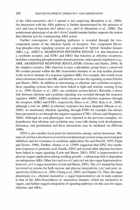
P1: OTE/SPH P2: OTE
BLUK053-Bradford September 22, 2006 15:36
200 SEED DEVELOPMENT, DORMANCY AND GERMINATION
of the ABA-insensitive abi1-1 mutant is not surprising (Beaudoin et al., 2000).An interaction with the ABA pathway is further demonstrated by the epistasis ofein2 and loss-of-function abi3 alleles (i.e., abi3-4) (Beaudoin et al., 2000). Thenondormant phenotype of an abi3-4/ein2 double mutant further supports the notionthat ethylene acts by counteracting ABA action.
Further convergence of signaling pathways is revealed through the two-component nature of the ethylene family of receptors. Two component His-to-Asp phospho-relay signaling systems are composed of ‘hybrid’ histidine kinases(HKs, e.g., AHK2-4, ARABIDOPSIS HISTIDINE KINASE 2-4, that functions asa cytokinin receptor, and ETR1 and ERS1 that function as ethylene receptors),histidine-containing phosphotransfer domain proteins, and response regulators (e.g.,ARR, ARABIDOPSIS RESPONSE REGULATOR) (Grefen and Harter, 2004). Inthe simplest scenario, HKs function to pass a phosphate group from a conservedHis residue present within the HK (transmitter domain) to an Asp residue presentin the receiver domain of a response regulator (RR). For example, this would occurwhen a hormone binds to the HK, and thereby activates the signaling system (Grefenand Harter, 2004). In addition to functioning as ethylene and cytokinin receptors,these signaling systems have also been linked to light and osmotic sensing (Uraoet al., 1999; Sweere et al., 2001; see cytokinin section below). Recently, a directlink between ethylene and cytokinin signaling was revealed through the responseregulator ARR2. ARR2 modulates both cytokinin and ethylene responses throughthe receptors AHK3 and ETR1, respectively (Hass et al., 2004; Kim et al., 2006),although a role for ARR2 in ethylene responses has been disputed (Mason et al.,2005). As mentioned, ethylene signaling, through ETR1 for example, has alwaysbeen presumed to act through the negative regulator CTR1 (Alonso and Stepanova,2004). Although no seed phenotypes were reported in the previous examples, wehypothesize that ethylene and cytokinin may cross-talk during seed development,dormancy, and germination and these interactions may be mediated via differentARRs.
EIN2 is also another focal point for interactions among various hormones. Mu-tants of ein2 have also been recovered in root phenotype screens using auxin transportinhibitors and for resistance to cytokinin application (Su and Howell, 1992; Fujitaand Syono, 1996). Further, Alonso et al. (1999) suggested that EIN2 also modu-lates responses to jasmonic acid. Finally, EIN2 and several other ethylene loci havebeen linked to sugar signaling (Leon and Sheen, 2003). EIN2 is hypersensitive toglucose (sugar) application during seedling growth – a phenotype that is dependenton endogenous ABA. Other loci such as etr1 and ein3 are also sugar hypersensitive;however, ctr1 is sugar insensitive (Leon and Sheen, 2003). The ctr1 mutant has beenrecovered in screens for both sucrose and glucose insensitivity (sis1 and gin4, re-spectively) (Gibson et al., 2001; Cheng et al., 2002; see Chapter 12). Thus, the sugarphenotypes (i.e., ethylene insensitive = sugar hypersensitive) are in stark contrastto that of the ABA-biosynthesis or -insensitive mutants, which are insensitive tosugars, and further suggest integration of signaling pathways (in this case for sugar,ethylene, and ABA).
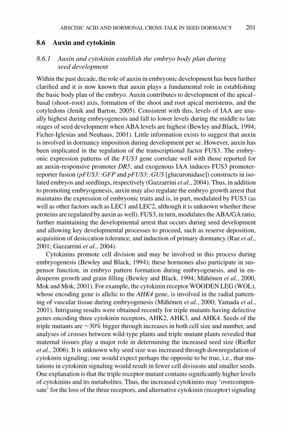
P1: OTE/SPH P2: OTE
BLUK053-Bradford September 22, 2006 15:36
ABSCISIC ACID AND HORMONAL CROSS-TALK IN SEED DORMANCY 201
8.6 Auxin and cytokinin
8.6.1 Auxin and cytokinin establish the embryo body plan duringseed development
Within the past decade, the role of auxin in embryonic development has been furtherclarified and it is now known that auxin plays a fundamental role in establishingthe basic body plan of the embryo. Auxin contributes to development of the apical–basal (shoot–root) axis, formation of the shoot and root apical meristems, and thecotyledons (Jenik and Barton, 2005). Consistent with this, levels of IAA are usu-ally highest during embryogenesis and fall to lower levels during the middle to latestages of seed development when ABA levels are highest (Bewley and Black, 1994;Ficher-Iglesias and Neuhaus, 2001). Little information exists to suggest that auxinis involved in dormancy imposition during development per se. However, auxin hasbeen implicated in the regulation of the transcriptional factor FUS3. The embry-onic expression patterns of the FUS3 gene correlate well with those reported foran auxin-responsive promoter DR5, and exogenous IAA induces FUS3 promoter-reporter fusion (pFUS3::GFP and pFUS3::GUS [glucuronidase]) constructs in iso-lated embryos and seedlings, respectively (Gazzarrini et al., 2004). Thus, in additionto promoting embryogenesis, auxin may also regulate the embryo growth arrest thatmaintains the expression of embryonic traits and is, in part, modulated by FUS3 (aswell as other factors such as LEC1 and LEC2, although it is unknown whether theseproteins are regulated by auxin as well). FUS3, in turn, modulates the ABA/GA ratio,further maintaining the developmental arrest that occurs during seed developmentand allowing key developmental processes to proceed, such as reserve deposition,acquisition of desiccation tolerance, and induction of primary dormancy (Raz et al.,2001; Gazzarrini et al., 2004).
Cytokinins promote cell division and may be involved in this process duringembryogenesis (Bewley and Black, 1994); these hormones also participate in sus-pensor function, in embryo pattern formation during embryogenesis, and in en-dosperm growth and grain filling (Bewley and Black, 1994; Mahonen et al., 2000,Mok and Mok, 2001). For example, the cytokinin receptor WOODEN LEG (WOL),whose encoding gene is allelic to the AHK4 gene, is involved in the radial pattern-ing of vascular tissue during embryogenesis (Mahonen et al., 2000; Yamada et al.,2001). Intriguing results were obtained recently for triple mutants having defectivegenes encoding three cytokinin receptors, AHK2, AHK3, and AHK4. Seeds of thetriple mutants are ∼30% bigger through increases in both cell size and number, andanalyses of crosses between wild-type plants and triple mutant plants revealed thatmaternal tissues play a major role in determining the increased seed size (Riefleret al., 2006). It is unknown why seed size was increased through downregulation ofcytokinin signaling; one would expect perhaps the opposite to be true, i.e., that mu-tations in cytokinin signaling would result in fewer cell divisions and smaller seeds.One explanation is that the triple receptor mutant contains significantly higher levelsof cytokinins and its metabolites. Thus, the increased cytokinins may ‘overcompen-sate’ for the loss of the three receptors, and alternative cytokinin (receptor) signaling
P1: OTE/SPH P2: OTE
BLUK053-Bradford September 22, 2006 15:36
202 SEED DEVELOPMENT, DORMANCY AND GERMINATION
paths may be invoked. Alternatively, complex interactions with other hormonal path-ways could account for the increase in seed size. As with auxin, no specific role forcytokinins in dormancy induction has been ascribed, although seeds of the triplecytokinin receptor mutants germinate faster than wild-type (Riefler et al., 2006).
8.6.2 Auxin and cytokinins have not been intimately linked to dormancymaintenance or termination
Recently, IAA was suggested to participate in the dormancy of wheat caryopses(Ramaih et al., 2003). IAA and its precursors tryptophan and indole-acetaldehydeinhibit germination of excised embryos from a dormant cultivar but not from anondormant one. Furthermore, embryos from dormant caryopses gradually losesensitivity to IAA throughout after-ripening (Ramaih et al., 2003). Thus, a link hasbeen drawn between exogenous auxin and embryo germinability but there is still nocompelling evidence to suggest that auxin is involved in dormancy maintenance ortermination per se. Auxin is linked to the resumption of growth; however, this roleis especially prominent in the germinated seedling. IAA is released from conjugates(with amino acids or peptides) stored in seeds of Scots pine and Arabidopsis duringgermination (Ljung et al., 2001; Rampey et al., 2004). This IAA may be neededfor germination and/or subsequent seedling growth. Postgerminative, shoot-derivedauxin may be essential for the initiation of lateral roots in Arabidopsis seedlings(Bhalerao et al., 2002). Interestingly, the first observable phenotypes resulting fromdeficiencies in auxin amount or response are often the presence of fewer lateral rootsand shorter hypocotyls in the light. An auxin-conjugate hydrolase triple mutant (ilr1[IAA-leucine resistant 1] iar3 [IAA-alanine resistant 3]ill2 [ilr-like2]) displays theselow-auxin phenotypes (Rampey et al., 2004). In addition, these phenotypes are as-sociated with the auxin response mutants axr1 (auxin resistant 1), axr4, and ibr5(IBA [indole-3-butyric acid] response 5) (Estelle and Somerville, 1987; Hobbieand Estelle, 1995; Monroe-Augustus et al., 2003). No significant seed germinationphenotypes have been reported for auxin-related mutants, so whether auxin playsa role in germination is unclear. However, germination of seeds of axr1 and ibr5mutants is slightly more insensitive to low ABA concentrations (0.5 to 1 μM) thanthat exhibited by wild-type seeds (Tiryaki and Staswick, 2002; Monroe-Augustuset al., 2003). Indirect evidence indicating the involvement of auxin in the germina-tion process comes from a microarray analysis of germinating Arabidopsis seeds.A number of auxin biosynthesis genes and genes encoding putative auxin carrierproteins are upregulated during germination in wild-type seeds and in ga1-3 mutantseeds treated with exogenous GA4 (Ogawa et al., 2003).
Cytokinins can release the dormancy of seeds of numerous species such as se-lected species of maple (Acer), apple, beech, and peanut (Cohn and Butera, 1982).Kinetin together with at least one other hormone or light can release the thermodor-mancy of lettuce seeds (Saini et al., 1986). Given the recent advances in hormonemetabolite profiling, it would be interesting to revisit the ability of kinetin and light,for example, to stimulate the germination of thermodormant seeds, examining theeffects of this treatment on the profiles of hormones and hormone metabolites (e.g.,
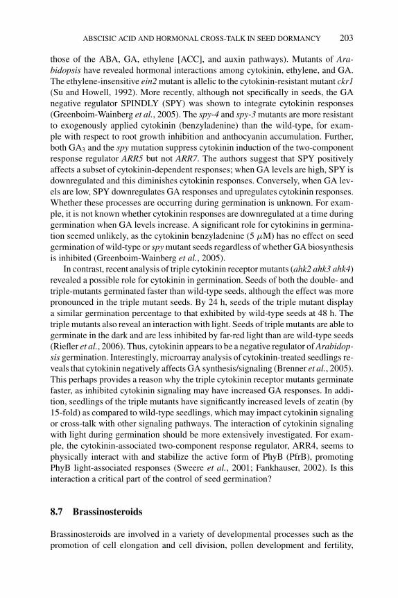
P1: OTE/SPH P2: OTE
BLUK053-Bradford September 22, 2006 15:36
ABSCISIC ACID AND HORMONAL CROSS-TALK IN SEED DORMANCY 203
those of the ABA, GA, ethylene [ACC], and auxin pathways). Mutants of Ara-bidopsis have revealed hormonal interactions among cytokinin, ethylene, and GA.The ethylene-insensitive ein2 mutant is allelic to the cytokinin-resistant mutant ckr1(Su and Howell, 1992). More recently, although not specifically in seeds, the GAnegative regulator SPINDLY (SPY) was shown to integrate cytokinin responses(Greenboim-Wainberg et al., 2005). The spy-4 and spy-3 mutants are more resistantto exogenously applied cytokinin (benzyladenine) than the wild-type, for exam-ple with respect to root growth inhibition and anthocyanin accumulation. Further,both GA3 and the spy mutation suppress cytokinin induction of the two-componentresponse regulator ARR5 but not ARR7. The authors suggest that SPY positivelyaffects a subset of cytokinin-dependent responses; when GA levels are high, SPY isdownregulated and this diminishes cytokinin responses. Conversely, when GA lev-els are low, SPY downregulates GA responses and upregulates cytokinin responses.Whether these processes are occurring during germination is unknown. For exam-ple, it is not known whether cytokinin responses are downregulated at a time duringgermination when GA levels increase. A significant role for cytokinins in germina-tion seemed unlikely, as the cytokinin benzyladenine (5 μM) has no effect on seedgermination of wild-type or spy mutant seeds regardless of whether GA biosynthesisis inhibited (Greenboim-Wainberg et al., 2005).
In contrast, recent analysis of triple cytokinin receptor mutants (ahk2 ahk3 ahk4)revealed a possible role for cytokinin in germination. Seeds of both the double- andtriple-mutants germinated faster than wild-type seeds, although the effect was morepronounced in the triple mutant seeds. By 24 h, seeds of the triple mutant displaya similar germination percentage to that exhibited by wild-type seeds at 48 h. Thetriple mutants also reveal an interaction with light. Seeds of triple mutants are able togerminate in the dark and are less inhibited by far-red light than are wild-type seeds(Riefler et al., 2006). Thus, cytokinin appears to be a negative regulator of Arabidop-sis germination. Interestingly, microarray analysis of cytokinin-treated seedlings re-veals that cytokinin negatively affects GA synthesis/signaling (Brenner et al., 2005).This perhaps provides a reason why the triple cytokinin receptor mutants germinatefaster, as inhibited cytokinin signaling may have increased GA responses. In addi-tion, seedlings of the triple mutants have significantly increased levels of zeatin (by15-fold) as compared to wild-type seedlings, which may impact cytokinin signalingor cross-talk with other signaling pathways. The interaction of cytokinin signalingwith light during germination should be more extensively investigated. For exam-ple, the cytokinin-associated two-component response regulator, ARR4, seems tophysically interact with and stabilize the active form of PhyB (PfrB), promotingPhyB light-associated responses (Sweere et al., 2001; Fankhauser, 2002). Is thisinteraction a critical part of the control of seed germination?
8.7 Brassinosteroids
Brassinosteroids are involved in a variety of developmental processes such as thepromotion of cell elongation and cell division, pollen development and fertility,
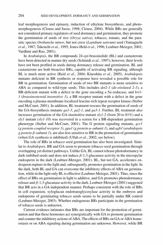
P1: OTE/SPH P2: OTE
BLUK053-Bradford September 22, 2006 15:36
204 SEED DEVELOPMENT, DORMANCY AND GERMINATION
leaf morphogenesis and epinasty, induction of ethylene biosynthesis, and photo-morphogenesis (Clouse and Sasse, 1998; Clouse, 2004). While BRs are generallynot considered primary regulators of seed dormancy and germination, they promotethe germination of seeds of rice (Oryza sativa), tobacco, tomato, and the para-sitic species Orobanche minor, but not cress (Lepidium sativum) seed (Yamaguchiet al., 1987; Takeuchi et al., 1995; Jones-Held et al., 1996; Leubner-Metzger, 2001;Vardhini and Rao, 2001).
In Arabidopsis, the BR compounds 24-epi-brassinolide (BL) and castasteronehave been detected in mature dry seeds (Schmidt et al., 1997); however, their levelshave not been profiled in seeds during dormancy release and germination. BL andcastasterone are both bioactive BRs, capable of activating BR signaling, althoughBL is much more active (Reid et al., 2004; Kinoshita et al., 2005). Arabidopsismutants deficient in BR synthesis or response have revealed a possible role forBR in germination. Germination of seeds of two BR mutants is more sensitive toABA as compared to wild-type seeds. This includes det2-1 (de-etiolated 2-1), aBR-deficient mutant with a defect in the gene encoding a 5α-reducase, and bri1-1 (brassinosteroid insensitive 1), a BR receptor mutant with a defect in the geneencoding a plasma-membrane-localized leucine-rich repeat receptor kinase (Steberand McCourt, 2001). In addition, BL treatment rescues the germination of seeds ofthe GA-biosynthesis mutants ga1-3, ga2-1, and ga3-1. Further, BL treatment alsoincreases germination of the GA-insensitive mutant sly1-2 (from 20 to 81%) and asly1 mutant (sly1-10) was recovered in a screen for a BR-dependent germinationphenotype (Steber and McCourt, 2001). The G protein signaling mutants, gcr1(g protein-coupled receptor 1), gpa1 (g protein α-subunit 1), and agb1 (arabidopsisg protein β-subunit 1), are also less sensitive to BR in the promotion of germination(when GA synthesis is inhibited) (Ullah et al., 2002; see below).
The role of BRs in tobacco seed germination has also been investigated. Simi-lar to Arabidopsis, BR and GA seem to promote tobacco seed germination throughoverlapping yet distinct pathways. Unlike GA, BL cannot release photodormancy indark-imbibed seeds and does not induce β-1,3-glucanase activity in the micropylarendosperm in the dark (Leubner-Metzger, 2001). BL, but not GA, accelerates en-dosperm rupture in the light and, subsequently, promotes germination in the light. Inthe dark, both BL and GA can overcome the inhibitory effects of ABA on germina-tion, while in the light only BL is effective (Leubner-Metzger, 2001). Thus, since theeffect of BRs on germination in light is additive, and GA promotes photodormancyrelease and β-1,3-glucanase activity in the dark, Leubner-Metzger (2001) suggestedthat BR acts in a GA-independent manner. Perhaps consistent with the role of BRsin cell expansion, xyloglucan endotransglycosylase activity in the embryos andendoperms of germinating tobacco seeds seems to be partially under BR control(Leubner-Metzger, 2003). Whether endogenous BRs participate in the germinationof tobacco seeds is unknown.
Current evidence intimates that BRs are important for the promotion of germi-nation and that these hormones act synergistically with GA to promote germinationand counter the inhibitory actions of ABA. The effects of BRs on GA or ABA home-ostasis or on ABA signaling during germination are unknown. However, while BR
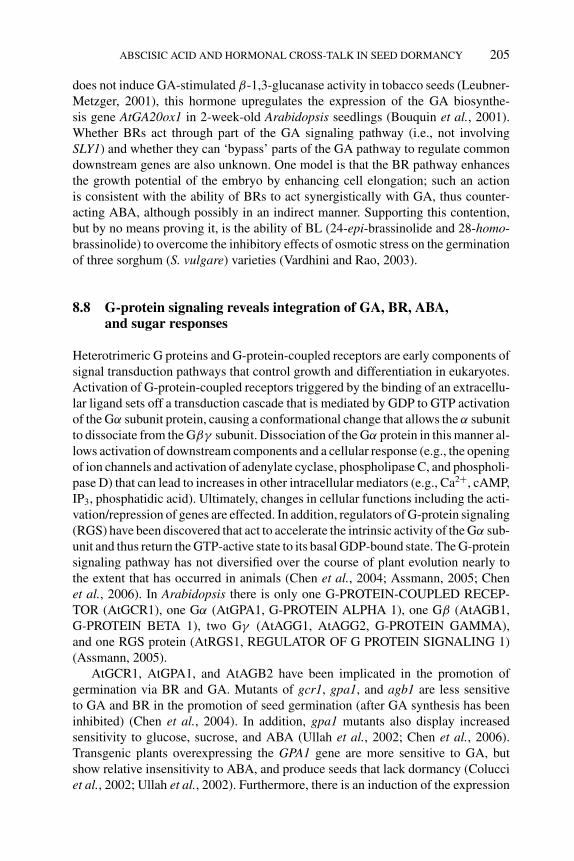
P1: OTE/SPH P2: OTE
BLUK053-Bradford September 22, 2006 15:36
ABSCISIC ACID AND HORMONAL CROSS-TALK IN SEED DORMANCY 205
does not induce GA-stimulated β-1,3-glucanase activity in tobacco seeds (Leubner-Metzger, 2001), this hormone upregulates the expression of the GA biosynthe-sis gene AtGA20ox1 in 2-week-old Arabidopsis seedlings (Bouquin et al., 2001).Whether BRs act through part of the GA signaling pathway (i.e., not involvingSLY1) and whether they can ‘bypass’ parts of the GA pathway to regulate commondownstream genes are also unknown. One model is that the BR pathway enhancesthe growth potential of the embryo by enhancing cell elongation; such an actionis consistent with the ability of BRs to act synergistically with GA, thus counter-acting ABA, although possibly in an indirect manner. Supporting this contention,but by no means proving it, is the ability of BL (24-epi-brassinolide and 28-homo-brassinolide) to overcome the inhibitory effects of osmotic stress on the germinationof three sorghum (S. vulgare) varieties (Vardhini and Rao, 2003).
8.8 G-protein signaling reveals integration of GA, BR, ABA,and sugar responses
Heterotrimeric G proteins and G-protein-coupled receptors are early components ofsignal transduction pathways that control growth and differentiation in eukaryotes.Activation of G-protein-coupled receptors triggered by the binding of an extracellu-lar ligand sets off a transduction cascade that is mediated by GDP to GTP activationof the Gα subunit protein, causing a conformational change that allows the α subunitto dissociate from the Gβγ subunit. Dissociation of the Gα protein in this manner al-lows activation of downstream components and a cellular response (e.g., the openingof ion channels and activation of adenylate cyclase, phospholipase C, and phospholi-pase D) that can lead to increases in other intracellular mediators (e.g., Ca2+, cAMP,IP3, phosphatidic acid). Ultimately, changes in cellular functions including the acti-vation/repression of genes are effected. In addition, regulators of G-protein signaling(RGS) have been discovered that act to accelerate the intrinsic activity of the Gα sub-unit and thus return the GTP-active state to its basal GDP-bound state. The G-proteinsignaling pathway has not diversified over the course of plant evolution nearly tothe extent that has occurred in animals (Chen et al., 2004; Assmann, 2005; Chenet al., 2006). In Arabidopsis there is only one G-PROTEIN-COUPLED RECEP-TOR (AtGCR1), one Gα (AtGPA1, G-PROTEIN ALPHA 1), one Gβ (AtAGB1,G-PROTEIN BETA 1), two Gγ (AtAGG1, AtAGG2, G-PROTEIN GAMMA),and one RGS protein (AtRGS1, REGULATOR OF G PROTEIN SIGNALING 1)(Assmann, 2005).
AtGCR1, AtGPA1, and AtAGB2 have been implicated in the promotion ofgermination via BR and GA. Mutants of gcr1, gpa1, and agb1 are less sensitiveto GA and BR in the promotion of seed germination (after GA synthesis has beeninhibited) (Chen et al., 2004). In addition, gpa1 mutants also display increasedsensitivity to glucose, sucrose, and ABA (Ullah et al., 2002; Chen et al., 2006).Transgenic plants overexpressing the GPA1 gene are more sensitive to GA, butshow relative insensitivity to ABA, and produce seeds that lack dormancy (Colucciet al., 2002; Ullah et al., 2002). Furthermore, there is an induction of the expression
P1: OTE/SPH P2: OTE
BLUK053-Bradford September 22, 2006 15:36
206 SEED DEVELOPMENT, DORMANCY AND GERMINATION
of a MYB65 gene, an Arabidopsis homologue of the GAMYB gene of barley, whoseencoded product regulates GA-mediated α-amylase induction in aleurone layer cells(Colucci et al., 2002). Ullah et al. (2002) suggested that the promotion of germinationmediated by G-protein signaling and GPA1 is through BR action; however, theevidence for this is currently circumstantial.
More recently, AtRGS1 has been identified as a regulator of sugar and ABA re-sponses during germination (Chen et al., 2006). In contrast to gpa1, mutant seeds ofrgs1 are less sensitive to ABA, glucose, and sucrose during germination. Overexpres-sors of AtRGS1 are hypersensitive to ABA and sugar. Thus, AtRGS1 may operate tooppose the actions of AtGCR1, AtGPA1, and AtAGB2. It is unknown whether theaction is a direct one, although the RGS domain of AtRGS1 can quench AtGPA1activity in vitro (Chen et al., 2003). AtRGS1 also has a membrane ligand-bindingdomain (similar to a GPCR [G-Protein Coupled Receptor] domain) and may beable to direct G-protein signaling itself, rather than simply opposing it (Chen et al.,2003). Supporting this, ABA levels and the expression of the genes AtNCED3 andAtABA2 are increased in wild-type seeds but not in rgs1 mutant seeds followingglucose application (Chen et al., 2006). It is unknown whether sugars are the lig-ands that bind to the GPCR domain of AtRGS1, but sugars do bind to GPCRs inyeast and mammalian systems (Assmann, 2005). Thus, sugar signaling, directly orupstream of AtRGS1, seems to modulate ABA biosynthesis, which may ultimatelybe the reason for induced quiescence during germination (see Chapter 12). Chenet al. (2006) provided the first evidence that this quiescence may operate throughG-protein signaling. Determination of the true nature of G-protein signaling in therealm of germination promotion and opposition is awaited with interest.
8.9 Profiling of hormone metabolic pathways in Arabidopsis mutantsreveals cross-talk
The ability of a specific mutation in a hormonal pathway to affect other hormonenetworks was recently demonstrated in the Arabidopsis etr1-2 mutant (Figure 8.2).The etr1-2 mutation confers dominant ethylene-insensitivity and as a consequenceresults in mature seed populations that exhibit more pronounced primary dormancy(Hall et al., 1999; Chiwocha et al., 2005). As discussed above, the inability ofseeds to perceive ethylene results in seeds with higher ABA contents at maturity(∼10-fold higher than in mature dry wild-type Col seeds), consistent with ethy-lene’s antagonism of ABA action during seed development (Beaudoin et al., 2000;Ghassemian et al., 2000). The higher ABA levels in the mature etr1-2 seeds suggestthat ABA response may also be upregulated during dormancy induction; indeed,ABA sensitivity is greater in mature etr1 seeds (Ghassemian et al., 2000). Thus,the lack of ethylene signaling (due to the etr1-2 mutation) likely results in a failureof ethylene to counteract both ABA synthesis and signaling. During imbibition ofdormant and nondormant seeds, there is a striking difference in ABA metabolismin etr1-2 mutant versus wild-type seeds; in particular, ABA-GE is decreased inthe mutant. This is especially evident in germinating seeds, for example, where a
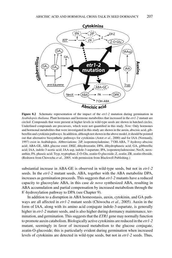
P1: OTE/SPH P2: OTE
BLUK053-Bradford September 22, 2006 15:36
ABSCISIC ACID AND HORMONAL CROSS-TALK IN SEED DORMANCY 207
Figure 8.2 Schematic representation of the impact of the etr1-2 mutation during germination in
Arabidopsis thaliana. Plant hormones and hormone metabolites that increased in the etr1-2 mutant are
circled. Compounds that were present at higher levels in wild-type seeds are shown in hatched circles.
Underlined compounds are precursors, which were not quantified in this study. Note: Only hormones
and hormonal metabolites that were investigated in this study are shown in the auxin, abscisic acid, gib-
berellin and cytokinin pathways. In addition, although not shown in the above model, it should be pointed
out that alternative biosynthetic pathways for cytokinins (Astot et al., 2000) and for IAA (Normanly,
1997) exist in Arabidopsis. Abbreviations: 2iP, isopentenyladenine; 7′OH-ABA, 7′-hydroxy-abscisic
acid; ABA-GE, ABA glucose ester; DHZ, dihydrozeatin; DPA, dihydrophaseic acid; GA, gibberellic
acid; IAA, indole-3-acetic acid; IAA-asp, indole-3-aspartate; IPA, isopentenyladenosine; NeoX, neox-
anthin; PA, phaseic acid; Tryp, tryptophan; Z-O-Glu, zeatin-O-glucoside; Z, zeatin; ZR, zeatin riboside.
(Redrawn from Chiwocha et al., 2005, with permission from Blackwell Publishing.)
substantial increase in ABA-GE is observed in wild-type seeds, but not in etr1-2seeds. In the etr1-2 mutant seeds, ABA, together with the ABA metabolite DPA,increases as germination proceeds. This suggests that etr1-2 mutants have a reducedcapacity to glucosylate ABA, in this case de novo synthesized ABA, resulting inABA accumulation and partial compensation by increased metabolism through the8′-hydroxylation pathway to DPA (see Chapter 9).
In addition to a disruption in ABA homeostasis, auxin, cytokinin, and GA path-ways are all affected in etr1-2 mutant seeds (Chiwocha et al., 2005). Auxin in theform of IAA, along with its amino acid conjugate indole-3-aspartate, is generallyhigher in etr1-2 mature seeds, and is also higher during dormancy maintenance, ter-mination, and germination. This suggests that the ETR1 gene may normally functionto promote auxin catabolism. Biologically active cytokinins are reduced in the etr1-2mutant, seemingly in favor of increased metabolism to the glucose conjugate,zeatin-O-glucoside; this is particularly evident during germination when increasedlevels of cytokinins are detected in wild-type seeds, but not in etr1-2 seeds. Thus,
P1: OTE/SPH P2: OTE
BLUK053-Bradford September 22, 2006 15:36
208 SEED DEVELOPMENT, DORMANCY AND GERMINATION
ethylene signaling through ETR1 may help to maintain bioactive cytokinins by shift-ing the flux away from cytokinin catabolism toward biosynthesis. The ethylene andcytokinin pathways may converge – the ein2 mutant is insensitive to both hormones.Levels of GAs (GA1, GA4, and GA7) are significantly increased in mature seeds ofetr1-2 and increased levels are detected through dormancy maintenance, termina-tion, and germination as compared to those found in wild-type seeds. The changesin the various hormonal pathways due to lack of ETR1 can be attributed to twopossibilities: (1) ethylene signaling normally regulates these hormonal pathwaysin a positive or negative way, either directly or indirectly (e.g., ethylene signal-ing antagonizes ABA response); or (2) the lack of a functional ethylene signalingsystem causes compensatory responses in other hormonal pathways (e.g., ethyleneinsensitivity causes an increase in GA to further counter increased ABA response).
We have recently compared the hormonal metabolic pathways in seeds of theArabidopsis ABA-insensitive abi1-1 mutant to those in Ler wild-type seeds. Theabi1-1 mutant produces seed populations that are less dormant than that of the wild-type (S.D.S. Chiwocha, A.R. Kermode et al., unpublished results), even thoughthe ABI1 protein seems to be a negative regulator of ABA-signaling (Gosti et al.,1999; Yoshida et al., 2006). In contrast to the etr1-2 mutation described above, theabi1-1 mutation did not significantly affect any hormonal pathway – hormones andhormone metabolites belonging to the ABA, GA, auxin, cytokinin, jasmonic acid,and salicylic acid classes were all similar in abundance in the mutant and wild-typeseeds. Subtle differences in the abi1-1 mutant included slightly higher levels of ABAand IAA and of the ABA metabolite DPA. Most of the differences between abi1-1and wild-type seeds occurred during imbibition of seeds that had not undergone adormancy-releasing moist-chilling period and could be attributable to differencesin germination capacity (i.e., abi1-1 mutant seeds exhibited 98% germination by2 days, while wild-type seeds exhibited only 13%). For example, the increase in GA4
in nonchilled abi1-1 seeds was virtually identical to that of moist-chilled wild-typeseeds (S.D.S. Chiwocha, A.R. Kermode et al., unpublished results).
The above examples illustrate the importance of integrating genomic (microar-ray) and proteomic data with hormone profiling to give a more comprehensive pictureof hormone response and signaling. In the etr1-2 mutant, for example, we cannotascribe all of the effects of the mutation on gene expression and protein synthesissolely to the ETR1 protein or ethylene; results must take into account the effectsof the mutation on all hormone pathways. Similarly, in experiments using hormonetreatments, we must be cautious in assigning a regulatory function to a gene basedon one hormone (i.e., gene X is GA-inducible); changes in developmental state andother hormones may also be involved.
8.10 Summary and future directions
The ability of mature desiccated seeds to withstand extremes in the environmentand remain viable is a remarkable phenomenon. Even more so is the capacity forthe imbibed seed to modulate its developmental schedule to germinate only when
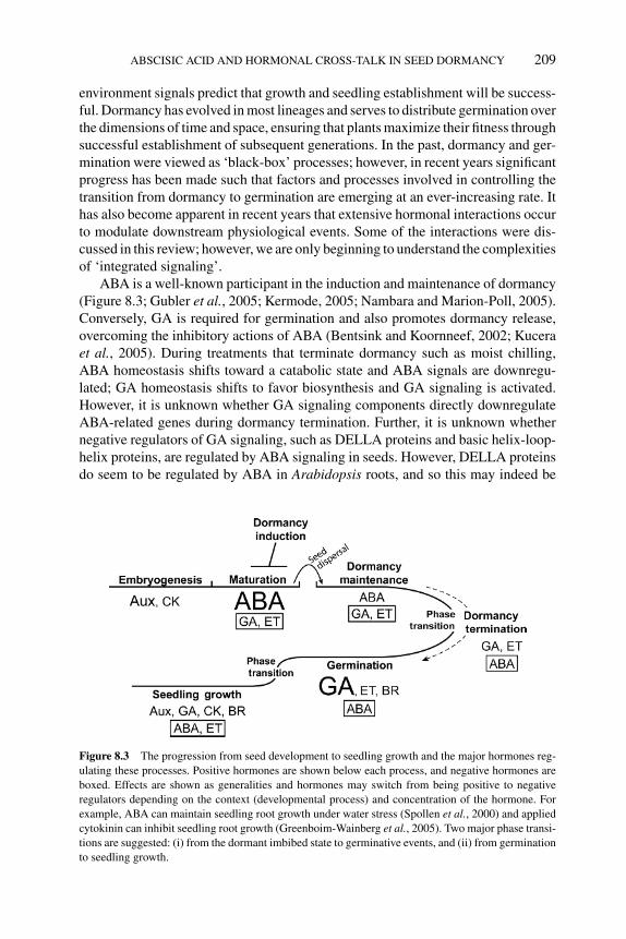
P1: OTE/SPH P2: OTE
BLUK053-Bradford September 22, 2006 15:36
ABSCISIC ACID AND HORMONAL CROSS-TALK IN SEED DORMANCY 209
environment signals predict that growth and seedling establishment will be success-ful. Dormancy has evolved in most lineages and serves to distribute germination overthe dimensions of time and space, ensuring that plants maximize their fitness throughsuccessful establishment of subsequent generations. In the past, dormancy and ger-mination were viewed as ‘black-box’ processes; however, in recent years significantprogress has been made such that factors and processes involved in controlling thetransition from dormancy to germination are emerging at an ever-increasing rate. Ithas also become apparent in recent years that extensive hormonal interactions occurto modulate downstream physiological events. Some of the interactions were dis-cussed in this review; however, we are only beginning to understand the complexitiesof ‘integrated signaling’.
ABA is a well-known participant in the induction and maintenance of dormancy(Figure 8.3; Gubler et al., 2005; Kermode, 2005; Nambara and Marion-Poll, 2005).Conversely, GA is required for germination and also promotes dormancy release,overcoming the inhibitory actions of ABA (Bentsink and Koornneef, 2002; Kuceraet al., 2005). During treatments that terminate dormancy such as moist chilling,ABA homeostasis shifts toward a catabolic state and ABA signals are downregu-lated; GA homeostasis shifts to favor biosynthesis and GA signaling is activated.However, it is unknown whether GA signaling components directly downregulateABA-related genes during dormancy termination. Further, it is unknown whethernegative regulators of GA signaling, such as DELLA proteins and basic helix-loop-helix proteins, are regulated by ABA signaling in seeds. However, DELLA proteinsdo seem to be regulated by ABA in Arabidopsis roots, and so this may indeed be
Figure 8.3 The progression from seed development to seedling growth and the major hormones reg-
ulating these processes. Positive hormones are shown below each process, and negative hormones are
boxed. Effects are shown as generalities and hormones may switch from being positive to negative
regulators depending on the context (developmental process) and concentration of the hormone. For
example, ABA can maintain seedling root growth under water stress (Spollen et al., 2000) and applied
cytokinin can inhibit seedling root growth (Greenboim-Wainberg et al., 2005). Two major phase transi-
tions are suggested: (i) from the dormant imbibed state to germinative events, and (ii) from germination
to seedling growth.
P1: OTE/SPH P2: OTE
BLUK053-Bradford September 22, 2006 15:36
210 SEED DEVELOPMENT, DORMANCY AND GERMINATION
the case (Achard et al., 2006). The role for ethylene and BRs in dormancy andgermination is less obvious than that of ABA and GA. Ethylene, together with GA,antagonizes ABA actions during dormancy induction/termination and germination.Moreover, ethylene and GA can each activate the synthesis of the other. BRs seemto play a synergistic role with GA in germination, perhaps through their role in pro-moting cell elongation in the embryo. However, no specific BR mutants have beenrecovered in germination screens, leading us to speculate upon the true functionof BRs during germination. Furthermore, identification of downstream ‘effector’genes for both ethylene and BR signaling in seeds is lacking.
Genetic redundancy within a plant’s genome appears to be commonplace.Arabidopsis is a key example: there are multiple receptors for hormones like ethy-lene, cytokinin, and auxin and there are large gene families that encode enzymes ofhormone biosynthesis (e.g., the NCED enzymes involved in ABA synthesis). Like-wise, signaling factors and transcription factors show redundancy (e.g., the proteinphosphatase PP2C subfamily A involved in ABA signaling and subfamily 15 of thebasic helix-loop-helix transcription factors involved in light/GA signaling). The listseems never-ending (Schwartz et al., 2003; Grefen and Harter, 2004; Schweighoferet al., 2004; Dharmasiri et al., 2005; Duek and Fankhauser, 2005). With this highdegree of redundancy, one may question why hormonal cross-talk/signal integrationis needed. But when one considers the complexity of the environment surroundingthe seed (and the parent plant), we can perhaps rationalize the need for an integratedhormonal web. Hormonal webs, akin to those of spiders, are stronger and more ro-bust and give the seed the flexibility to relay complex information within the embryoitself and between the seed and its surroundings.
Additional complexity is added to the role of hormones and their interactions indormancy and germination when we consider that signaling factors are not necessar-ily directly ‘homologous’ or have identical roles between species. For example, theABA-binding protein, ABAP1, from barley was isolated from ABA-treated aleuronelayers (Razem et al., 2004). Its closest homologue in Arabidopsis is FCA, a nuclearRNA-binding protein that promotes flowering, and it too binds ABA. ABA bindingto FCA disrupts FCA binding to FY (an mRNA 3′ end processing factor); togetherFCA–FY inhibit the accumulation of mRNAs encoding FLOWERING LOCUS C(FLC), a potent inhibitor of the transition to flowering. Thus, ABA binding to FCAleads to an increase in FLC activity and a delay in flowering. However, no seed ger-mination phenotypes are present in fca-1 mutants and seeds do not differ in ABAsensitivity as compared to wild-type (Razem et al., 2006). Thus, the roles of AtFCAand ABAP1 appear to be distinct; however, the precise role of ABAP1 in barleyseeds merits further investigation.
Other intricacies of the hormonal signaling web include the fact that signalingfactors may behave differently in different tissues or at different developmentalstages and may be dependent on hormone concentration (i.e., they are context de-pendent). The gpa1-1 and gpa1-2 mutants of Arabidopsis, encoding the G-proteinGα subunit, exhibit insensitivity to ABA, inhibition of stomatalopening, and alteredABA responsiveness of guard-cell inward K+ channels and slow anion channels(Wang et al., 2001). However, during seed germination, gpa1-1 and gpa1-2 mutants
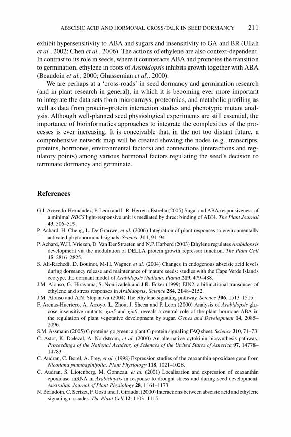
P1: OTE/SPH P2: OTE
BLUK053-Bradford September 22, 2006 15:36
ABSCISIC ACID AND HORMONAL CROSS-TALK IN SEED DORMANCY 211
exhibit hypersensitivity to ABA and sugars and insensitivity to GA and BR (Ullahet al., 2002; Chen et al., 2006). The actions of ethylene are also context-dependent.In contrast to its role in seeds, where it counteracts ABA and promotes the transitionto germination, ethylene in roots of Arabidopsis inhibits growth together with ABA(Beaudoin et al., 2000; Ghassemian et al., 2000).
We are perhaps at a ‘cross-roads’ in seed dormancy and germination research(and in plant research in general), in which it is becoming ever more importantto integrate the data sets from microarrays, proteomics, and metabolic profiling aswell as data from protein–protein interaction studies and phenotypic mutant anal-ysis. Although well-planned seed physiological experiments are still essential, theimportance of bioinformatics approaches to integrate the complexities of the pro-cesses is ever increasing. It is conceivable that, in the not too distant future, acomprehensive network map will be created showing the nodes (e.g., transcripts,proteins, hormones, environmental factors) and connections (interactions and reg-ulatory points) among various hormonal factors regulating the seed’s decision toterminate dormancy and germinate.
References
G.J. Acevedo-Hernandez, P. Leon and L.R. Herrera-Estrella (2005) Sugar and ABA responsiveness of
a minimal RBCS light-responsive unit is mediated by direct binding of ABI4. The Plant Journal43, 506–519.
P. Achard, H. Cheng, L. De Grauwe, et al. (2006) Integration of plant responses to environmentally
activated phytohormonal signals. Science 311, 91–94.
P. Achard, W.H. Vriezen, D. Van Der Straeten and N.P. Harberd (2003) Ethylene regulates Arabidopsisdevelopment via the modulation of DELLA protein growth repressor function. The Plant Cell15, 2816–2825.
S. Ali-Rachedi, D. Bouinot, M-H. Wagner, et al. (2004) Changes in endogenous abscisic acid levels
during dormancy release and maintenance of mature seeds: studies with the Cape Verde Islands
ecotype, the dormant model of Arabidopsis thaliana. Planta 219, 479–488.
J.M. Alonso, G. Hirayama, S. Nourizadeh and J.R. Ecker (1999) EIN2, a bifunctional transducer of
ethylene and stress responses in Arabidopsis. Science 284, 2148–2152.
J.M. Alonso and A.N. Stepanova (2004) The ethylene signaling pathway. Science 306, 1513–1515.
F. Arenas-Huertero, A. Arroyo, L. Zhou, J. Sheen and P. Leon (2000) Analysis of Arabidopsis glu-
cose insensitive mutants, gin5 and gin6, reveals a central role of the plant hormone ABA in
the regulation of plant vegetative development by sugar. Genes and Development 14, 2085–
2096.
S.M. Assmann (2005) G proteins go green: a plant G protein signaling FAQ sheet. Science 310, 71–73.
C. Astot, K. Dolezal, A. Nordstrom, et al. (2000) An alternative cytokinin biosynthesis pathway.
Proceedings of the National Academy of Sciences of the United States of America 97, 14778–
14783.
C. Audran, C. Borel, A. Frey, et al. (1998) Expression studies of the zeaxanthin epoxidase gene from
Nicotiana plumbaginifolia. Plant Physiology 118, 1021–1028.
C. Audran, S. Liotenberg, M. Gonneau, et al. (2001) Localisation and expression of zeaxanthin
epoxidase mRNA in Arabidopsis in response to drought stress and during seed development.
Australian Journal of Plant Physiology 28, 1161–1173.
N. Beaudoin, C. Serizet, F. Gosti and J. Giraudat (2000) Interactions between abscisic acid and ethylene
signaling cascades. The Plant Cell 12, 1103–1115.
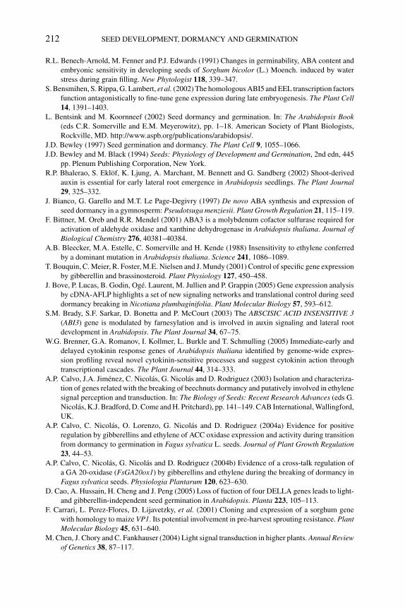
P1: OTE/SPH P2: OTE
BLUK053-Bradford September 22, 2006 15:36
212 SEED DEVELOPMENT, DORMANCY AND GERMINATION
R.L. Benech-Arnold, M. Fenner and P.J. Edwards (1991) Changes in germinability, ABA content and
embryonic sensitivity in developing seeds of Sorghum bicolor (L.) Moench. induced by water
stress during grain filling. New Phytologist 118, 339–347.
S. Bensmihen, S. Rippa, G. Lambert, et al. (2002) The homologous ABI5 and EEL transcription factors
function antagonistically to fine-tune gene expression during late embryogenesis. The Plant Cell14, 1391–1403.
L. Bentsink and M. Koornneef (2002) Seed dormancy and germination. In: The Arabidopsis Book(eds C.R. Somerville and E.M. Meyerowitz), pp. 1–18. American Society of Plant Biologists,
Rockville, MD. http://www.aspb.org/publications/arabidopsis/.
J.D. Bewley (1997) Seed germination and dormancy. The Plant Cell 9, 1055–1066.
J.D. Bewley and M. Black (1994) Seeds: Physiology of Development and Germination, 2nd edn, 445
pp. Plenum Publishing Corporation, New York.
R.P. Bhalerao, S. Eklof, K. Ljung, A. Marchant, M. Bennett and G. Sandberg (2002) Shoot-derived
auxin is essential for early lateral root emergence in Arabidopsis seedlings. The Plant Journal29, 325–332.
J. Bianco, G. Garello and M.T. Le Page-Degivry (1997) De novo ABA synthesis and expression of
seed dormancy in a gymnosperm: Pseudotsuga menziesii. Plant Growth Regulation 21, 115–119.
F. Bittner, M. Oreb and R.R. Mendel (2001) ABA3 is a molybdenum cofactor sulfurase required for
activation of aldehyde oxidase and xanthine dehydrogenase in Arabidopsis thaliana. Journal ofBiological Chemistry 276, 40381–40384.
A.B. Bleecker, M.A. Estelle, C. Somerville and H. Kende (1988) Insensitivity to ethylene conferred
by a dominant mutation in Arabidopsis thaliana. Science 241, 1086–1089.
T. Bouquin, C. Meier, R. Foster, M.E. Nielsen and J. Mundy (2001) Control of specific gene expression
by gibberellin and brassinosteroid. Plant Physiology 127, 450–458.
J. Bove, P. Lucas, B. Godin, Oge. Laurent, M. Jullien and P. Grappin (2005) Gene expression analysis
by cDNA-AFLP highlights a set of new signaling networks and translational control during seed
dormancy breaking in Nicotiana plumbaginifolia. Plant Molecular Biology 57, 593–612.
S.M. Brady, S.F. Sarkar, D. Bonetta and P. McCourt (2003) The ABSCISIC ACID INSENSITIVE 3(ABI3) gene is modulated by farnesylation and is involved in auxin signaling and lateral root
development in Arabidopsis. The Plant Journal 34, 67–75.
W.G. Brenner, G.A. Romanov, I. Kollmer, L. Burkle and T. Schmulling (2005) Immediate-early and
delayed cytokinin response genes of Arabidopsis thaliana identified by genome-wide expres-
sion profiling reveal novel cytokinin-sensitive processes and suggest cytokinin action through
transcriptional cascades. The Plant Journal 44, 314–333.
A.P. Calvo, J.A. Jimenez, C. Nicolas, G. Nicolas and D. Rodriguez (2003) Isolation and characteriza-
tion of genes related with the breaking of beechnuts dormancy and putatively involved in ethylene
signal perception and transduction. In: The Biology of Seeds: Recent Research Advances (eds G.
Nicolas, K.J. Bradford, D. Come and H. Pritchard), pp. 141–149. CAB International, Wallingford,
UK.
A.P. Calvo, C. Nicolas, O. Lorenzo, G. Nicolas and D. Rodriguez (2004a) Evidence for positive
regulation by gibberellins and ethylene of ACC oxidase expression and activity during transition
from dormancy to germination in Fagus sylvatica L. seeds. Journal of Plant Growth Regulation23, 44–53.
A.P. Calvo, C. Nicolas, G. Nicolas and D. Rodriguez (2004b) Evidence of a cross-talk regulation of
a GA 20-oxidase (FsGA20ox1) by gibberellins and ethylene during the breaking of dormancy in
Fagus sylvatica seeds. Physiologia Plantarum 120, 623–630.
D. Cao, A. Hussain, H. Cheng and J. Peng (2005) Loss of fuction of four DELLA genes leads to light-
and gibberellin-independent seed germination in Arabidopsis. Planta 223, 105–113.
F. Carrari, L. Perez-Flores, D. Lijavetzky, et al. (2001) Cloning and expression of a sorghum gene
with homology to maize VP1. Its potential involvement in pre-harvest sprouting resistance. PlantMolecular Biology 45, 631–640.
M. Chen, J. Chory and C. Fankhauser (2004) Light signal transduction in higher plants. Annual Reviewof Genetics 38, 87–117.
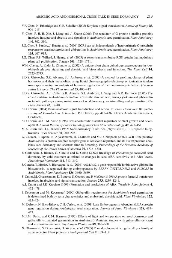
P1: OTE/SPH P2: OTE
BLUK053-Bradford September 22, 2006 15:36
ABSCISIC ACID AND HORMONAL CROSS-TALK IN SEED DORMANCY 213
Y.F. Chen, N. Etheridge and G.E. Schaller (2005) Ethylene signal transduction. Annals of Botany 95,
901–915.
Y. Chen, F. Ji, H. Xie, J. Liang and J. Zhang (2006) The regulator of G-protein signaling proteins
involved in sugar and abscisic acid signaling in Arabidopsis seed germination. Plant Physiology140, 302–310.
J.G. Chen, S. Pandey, J. Huang, et al. (2004) GCR1 can act independently of heterotrimeric G-protein in
response to brassinosteroids and gibberellins in Arabidopsis seed germination. Plant Physiology135, 907–915.
J.G. Chen, F.S. Willard, J. Huang, et al. (2003) A seven-transmembrane RGS protein that modulates
plant cell proliferation. Science 301, 1728–1731.
W.H. Cheng, A. Endo, L. Zhou, et al. (2002) A unique short chain dehydrogenase/reductase in Ara-bidopsis glucose signaling and abscisic acid biosynthesis and functions. The Plant Cell 14,
2723–2743.
S.D.S. Chiwocha, S.R. Abrams, S.J. Ambrose, et al. (2003) A method for profiling classes of plant
hormones and their metabolites using liquid chromatography–electrospray ionization tandem
mass spectrometry: an analysis of hormone regulation of thermodormancy in lettuce (Lactucasativa L.) seeds. The Plant Journal 35, 405–417.
S.D.S. Chiwocha, A.J. Cutler, S.R. Abrams, S.J. Ambrose, J. Yang and A.R. Kermode (2005) The
etr1-2 mutation in Arabidopsis thaliana affects the abscisic acid, auxin, cytokinin and gibberellin
metabolic pathways during maintenance of seed dormancy, moist-chilling and germination. ThePlant Journal 42, 35–48.
S.D. Clouse (2004) Brassinosteroid signal transduction and action. In: Plant Hormones: Biosynthe-sis, Signal Transduction, Action! (ed. P.J. Davies), pp. 413–436. Kluwer Academic Publishers,
Dordrecht.
S.D. Clouse and J.M. Sasse (1998) Brassinosteroids: essential regulators of plant growth and devel-
opment. Annual Review of Plant Physiology and Plant Molecular Biology 49, 427–451.
M.A. Cohn and D.L. Butera (1982) Seed dormancy in red rice (Oryza sativa). II. Response to cy-
tokinins. Weed Science 30, 200–205.
G. Colucci, F. Apone, N. Alyeshmerni, D. Chalmers and M.J. Chrispeels (2002) GCR1, the putative
Arabidopsis G protein-coupled receptor gene is cell cycle-regulated, and its overexpression abol-
ishes seed dormancy and shortens time to flowering. Proceedings of the National Academy ofSciences of the United States of America 99, 4736–4741.
F. Corbineau, J. Bianco, G. Garello and D. Come (2002) Breakage of Pseudotsuga menziesii seed
dormancy by cold treatment as related to changes in seed ABA sensitivity and ABA levels.
Physiologia Plantarum 114, 313–319.
J. Curaba, T. Moritz, R. Blervaque, et al. (2004) AtGA3ox2, a gene responsible for bioactive gibberellin
biosynthesis, is regulated during embryogenesis by LEAFY COTYLEDON2 and FUSCA3 in
Arabidopsis. Plant Physiology 136, 3660–3669.
S. Cutler, M. Ghassesmian, D. Bonetta, S. Cooney and P. McCourt (1996) A protein farnesyl transferase
involved in abscisic acid signal transduction. Science 273, 1239–1241.
A.J. Cutler and J.E. Krochko (1999) Formation and breakdown of ABA. Trends in Plant Science 4,
472–478.
I. Debeaujon and M. Koornneef (2000) Gibberellin requirement for Arabidopsis seed germination
is determined both by testa characteristics and embryonic abscisic acid. Plant Physiology 122,
415–424.
M. Delseny, N. Bies-Etheve, C.H. Carles, et al. (2001) Late Embryogenesis Abundant (LEA) protein
gene regulation during Arabidopsis seed maturation. Journal of Plant Physiology 158, 419–
427.
M.P.M. Derkx and C.M. Karssen (1993) Effects of light and temperature on seed dormancy and
gibberellin-stimulated germination in Arabidopsis thaliana: studies with gibberellin-deficient
and -insensitive mutants. Physiologia Plantarum 89, 360–368.
N. Dharmasiri, S. Dharmasiri, D. Weijers, et al. (2005) Plant development is regulated by a family of
auxin receptor F box proteins. Developmental Cell 9, 109–119.
P1: OTE/SPH P2: OTE
BLUK053-Bradford September 22, 2006 15:36
214 SEED DEVELOPMENT, DORMANCY AND GERMINATION
P.D. Duek and C. Fankhauser (2005) bHLH class transcription factors take centre stage in phytochrome
signaling. Trends in Plant Science 10, 51–54.
P. Duque and N.H. Chua (2003) IMB1, a bromodomain protein induced during seed imbibition,
regulates ABA- and phyA-mediated responses of germination in Arabidopsis. The Plant Journal35, 787–799.
M.A. Estelle and C. Somerville (1987) Auxin-resistant mutants of Arabidopsis thaliana with an altered
morphology. Molecular and General Genetics 206, 200–206.
M.M. Evans and R.S. Poethig (1995) Gibberellins promote vegetative phase change and reproductive
maturity in maize. Plant Physiology 108, 475–487.
C. Fankhauser (2002) Light perception in plants: cytokinins and red light join forces to keep phy-
tochrome B active. Trends in Plant Science 7, 143–145.
J.A. Feurtado, S.J. Ambrose, A.J. Cutler, A.R.S. Ross, S.R. Abrams and A.R. Kermode (2004) Dor-
mancy termination of western white pine (Pinus monticola Dougl. Ex D. Don) seeds is associated
with changes in abscisic acid metabolism. Planta 218, 630–639.
C. Ficher-Iglesias and G. Neuhaus (2001) Zygotic embryogenesis: hormonal control of embryo devel-
opment. In: Current Trends in the Embryology of Angiosperms (ed. S.S. Bhojwani), pp. 223–247.
Kluwer Academic Publishers, Dordrecht.
W. Finch-Savage (2004) A genomic approach to understanding seed dormancy. Nottingham Ara-
bidopsis Stock Centre Microarray Database, NASCARRAY-69, http://affymetrix.arabidopsis
.info/narrays/experimentbrowse.pl.
R.R. Finkelstein (1994) Mutations at two new Arabidopsis ABA response loci are similar to the abi3mutations. The Plant Journal 5, 765–771.
R.R. Finkelstein (2004) The role of hormones during seed development and germination. In: PlantHormones: Biosynthesis, Signal Transduction, Action! (ed. P.J. Davies), pp. 513–537. Kluwer
Academic Publishers, Dordrecht.
R.R. Finkelstein, S.S. Gampala and C.D. Rock (2002) Abscisic acid signaling in seeds and seedlings.
The Plant Cell 14(Suppl), S15–S45.
R.R. Finkelstein and T.J. Lynch (2000) The Arabidopsis abscisic acid response gene ABI5 encodes a
basic leucine zipper transcription factor. The Plant Cell 12, 599–609.
R.R. Finkelstein and C.D. Rock (2002) Abscisic acid biosynthesis and response. In: The ArabidopsisBook (eds C.R. Somerville and E.M. Meyerowitz), pp. 1–52. American Society of Plant Biologists,
Rockville, MD. http://www.aspb.org/publications/arabidopsis/.
G.R. Flematti, E.L. Ghisalberti, K.W. Dixon and R.D. Trengove (2004) A compound from smoke that
promotes seed germination. Science 305, 977.
K.A. Franklin and G.C. Whitelam (2004) Light signals, phytochromes and cross-talk with other
environmental cues. Journal of Experimental Botany 395, 271–276.
A. Frey, C. Audran, E. Marin, B. Sotta and A. Marion-Poll (1999) Engineering seed dormancy
by modification of zeaxanthin epoxidase gene expression. Plant Molecular Biology 39, 1267–
1274.
A. Frey, B. Godin, M. Bonnet, B. Sotta and A. Marion-Poll (2004) Maternal synthesis of abscisic acid
controls seed development and yield in Nicotiana plumbaginifolia. Planta 218, 958–964.
X. Fu, D.E. Richards, B. Fleck, D. Xie, N. Burton and N.P. Harberd (2004) The Arabidopsis mutant
sleepy1gar2−1 protein promotes plant growth by increasing the affinity of the SCFSLY1 E3 ubiquitin
ligase for DELLA protein substrates. The Plant Cell 16, 1406–1418.
H. Fujita and K. Syono (1996) Genetic analysis of the effects of polar auxin transport inhibitors on
root growth in Arabidopsis thaliana. Plant and Cell Physiology 37, 1094–1101.
T. Fukuhara, H.-H. Kirch and H.J. Bohnert (1999) Expression of Vp1 and water channel proteins
during seed germination. Plant, Cell and Environment 22, 417–424.
K. Furuta, K. Minoru, Y-G. Liu, D. Shibata and T. Kakimoto (2005) CKH2/PICKLE negatively
regulates a set of cytokinin responses. In : Abstract, Arabidopsis 2005, Madison, WI.
G. Garello and M.T. LePage-Degivry (1999) Evidence for the role of abscisic acid in the genetic and
environmental control of dormancy in wheat (Triticum aestivum L.). Seed Science Research 9,
219–226.
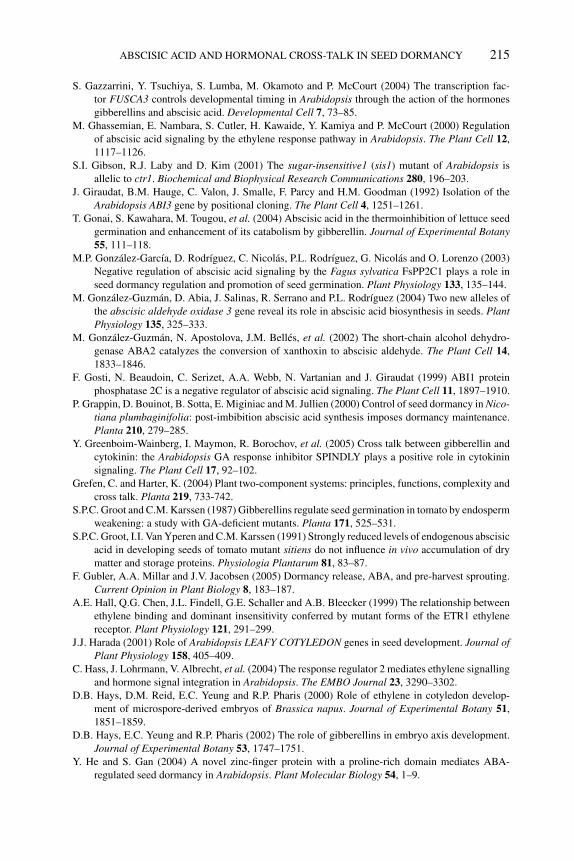
P1: OTE/SPH P2: OTE
BLUK053-Bradford September 22, 2006 15:36
ABSCISIC ACID AND HORMONAL CROSS-TALK IN SEED DORMANCY 215
S. Gazzarrini, Y. Tsuchiya, S. Lumba, M. Okamoto and P. McCourt (2004) The transcription fac-
tor FUSCA3 controls developmental timing in Arabidopsis through the action of the hormones
gibberellins and abscisic acid. Developmental Cell 7, 73–85.
M. Ghassemian, E. Nambara, S. Cutler, H. Kawaide, Y. Kamiya and P. McCourt (2000) Regulation
of abscisic acid signaling by the ethylene response pathway in Arabidopsis. The Plant Cell 12,
1117–1126.
S.I. Gibson, R.J. Laby and D. Kim (2001) The sugar-insensitive1 (sis1) mutant of Arabidopsis is
allelic to ctr1. Biochemical and Biophysical Research Communications 280, 196–203.
J. Giraudat, B.M. Hauge, C. Valon, J. Smalle, F. Parcy and H.M. Goodman (1992) Isolation of the
Arabidopsis ABI3 gene by positional cloning. The Plant Cell 4, 1251–1261.
T. Gonai, S. Kawahara, M. Tougou, et al. (2004) Abscisic acid in the thermoinhibition of lettuce seed
germination and enhancement of its catabolism by gibberellin. Journal of Experimental Botany55, 111–118.
M.P. Gonzalez-Garcıa, D. Rodrıguez, C. Nicolas, P.L. Rodrıguez, G. Nicolas and O. Lorenzo (2003)
Negative regulation of abscisic acid signaling by the Fagus sylvatica FsPP2C1 plays a role in
seed dormancy regulation and promotion of seed germination. Plant Physiology 133, 135–144.
M. Gonzalez-Guzman, D. Abia, J. Salinas, R. Serrano and P.L. Rodrıguez (2004) Two new alleles of
the abscisic aldehyde oxidase 3 gene reveal its role in abscisic acid biosynthesis in seeds. PlantPhysiology 135, 325–333.
M. Gonzalez-Guzman, N. Apostolova, J.M. Belles, et al. (2002) The short-chain alcohol dehydro-
genase ABA2 catalyzes the conversion of xanthoxin to abscisic aldehyde. The Plant Cell 14,
1833–1846.
F. Gosti, N. Beaudoin, C. Serizet, A.A. Webb, N. Vartanian and J. Giraudat (1999) ABI1 protein
phosphatase 2C is a negative regulator of abscisic acid signaling. The Plant Cell 11, 1897–1910.
P. Grappin, D. Bouinot, B. Sotta, E. Miginiac and M. Jullien (2000) Control of seed dormancy in Nico-tiana plumbaginifolia: post-imbibition abscisic acid synthesis imposes dormancy maintenance.
Planta 210, 279–285.
Y. Greenboim-Wainberg, I. Maymon, R. Borochov, et al. (2005) Cross talk between gibberellin and
cytokinin: the Arabidopsis GA response inhibitor SPINDLY plays a positive role in cytokinin
signaling. The Plant Cell 17, 92–102.
Grefen, C. and Harter, K. (2004) Plant two-component systems: principles, functions, complexity and
cross talk. Planta 219, 733-742.
S.P.C. Groot and C.M. Karssen (1987) Gibberellins regulate seed germination in tomato by endosperm
weakening: a study with GA-deficient mutants. Planta 171, 525–531.
S.P.C. Groot, I.I. Van Yperen and C.M. Karssen (1991) Strongly reduced levels of endogenous abscisic
acid in developing seeds of tomato mutant sitiens do not influence in vivo accumulation of dry
matter and storage proteins. Physiologia Plantarum 81, 83–87.
F. Gubler, A.A. Millar and J.V. Jacobsen (2005) Dormancy release, ABA, and pre-harvest sprouting.
Current Opinion in Plant Biology 8, 183–187.
A.E. Hall, Q.G. Chen, J.L. Findell, G.E. Schaller and A.B. Bleecker (1999) The relationship between
ethylene binding and dominant insensitivity conferred by mutant forms of the ETR1 ethylene
receptor. Plant Physiology 121, 291–299.
J.J. Harada (2001) Role of Arabidopsis LEAFY COTYLEDON genes in seed development. Journal ofPlant Physiology 158, 405–409.
C. Hass, J. Lohrmann, V. Albrecht, et al. (2004) The response regulator 2 mediates ethylene signalling
and hormone signal integration in Arabidopsis. The EMBO Journal 23, 3290–3302.
D.B. Hays, D.M. Reid, E.C. Yeung and R.P. Pharis (2000) Role of ethylene in cotyledon develop-
ment of microspore-derived embryos of Brassica napus. Journal of Experimental Botany 51,
1851–1859.
D.B. Hays, E.C. Yeung and R.P. Pharis (2002) The role of gibberellins in embryo axis development.
Journal of Experimental Botany 53, 1747–1751.
Y. He and S. Gan (2004) A novel zinc-finger protein with a proline-rich domain mediates ABA-
regulated seed dormancy in Arabidopsis. Plant Molecular Biology 54, 1–9.
P1: OTE/SPH P2: OTE
BLUK053-Bradford September 22, 2006 15:36
216 SEED DEVELOPMENT, DORMANCY AND GERMINATION
P. Hedden (1999) Regulation of gibberellin biosynthesis. In: Biochemistry and Molecular Biologyof Plant Hormones (eds P.J.J. Hooykaas, M.A. Hall and K.R. Libbenga), pp. 161–188. Elsevier
Science BV, Amsterdam.
J.T. Henderson, H.C. Li, S.D. Rider, et al. (2004) PICKLE acts throughout the plant to repress expres-
sion of embryonic traits and may play a role in gibberellin dependent responses. Plant Physiology134, 995–1005.
L. Hennig, W.M. Stoddart, M. Dieterle, G.C. Whitelam and E. Schafer (2002) Phytochrome E controls
light-induced germination of Arabidopsis. Plant Physiology 128, 194–200.
L. Hobbie and M. Estelle (1995) The axr4 auxin-resistant mutants of Arabidopsis thaliana define a
gene important for root gravitropism and lateral root initiation. The Plant Journal 7, 211–220.
M. Holdsworth, S. Kurup and R. McKibbin (1999) Molecular and genetic mechanisms regulating the
transition from embryo development to germination. Trends in Plant Science 4, 275–280.
S.E. Jacobsen and N.E. Olszewski (1993) Mutations at the SPINDLY locus of Arabidopsis alter
gibberellin signal transduction. The Plant Cell 5, 887–896.
J.V. Jacobsen, D.W. Pearce, A.T. Poole, R.P. Pharis and L.N. Mander (2002) Abscisic acid, phaseic
acid and gibberellin contents associated with dormancy and germination in barley. PhysiologiaPlantarum 115, 428–441.
P.D. Jenik and M.K. Barton (2005) Surge and destroy: the role of auxin in plant embryogenesis.
Development 132, 3577–3585.
L. Jiang, S. Abrams and A.R. Kermode (1996) Vicilin and napin storage protein gene promoters are
responsive to abscisic acid in developing transgenic tobacco seed but lose sensitivity following
premature desiccation. Plant Physiology 110, 1135–1144.
H.D. Jones, N.C. Peters and M.J. Holdsworth (1997) Genotype and environment interact to control
dormancy and differential expression of the VIVIPAROUS 1 homologue in embryos of Avenafatua. The Plant Journal 12, 911–920.
S. Jones-Held, M. Vandoren and T. Lockwood (1996) Brassinolide application to Lepidium sativumseeds and the effects on seedling growth. Journal of Plant Growth Regulation 15, 63–67.
Y. Kagaya, R. Toyoshima, R. Okuda, H. Usui, A. Yamamoto and T. Hattori (2005) LEAFY COTYLE-
DON1 controls seed storage protein genes through its regulation of FUSCA3 and ABSCISICACID INSENSITIVE3. Plant and Cell Physiology 46, 399–406.
C.M. Karssen, D.L.C. Brinkhorst-van der Swan, A.E. Breekland and M. Koornneef (1983) Induction
of dormancy during seed development by endogenous abscisic acid: studies on abscisic acid-
deficient genotypes of Arabidopsis thaliana (L.) Heynh. Planta 157, 158–165.
C.M. Karssen and E. Lacka (1986) A revision of the hormone balance theory of seed dormancy: studies
on gibberellin and/or abscisic acid deficient mutants of Arabidopsis thaliana. In: Plant GrowthSubstances (ed. M. Bopp), pp. 315–323. Springer-Verlag, Berlin.
C.M. Karssen, S. Zagorsky, J. Kepczynski and S.P.C. Groot (1989) Key role for endogenous gib-
berellins in the control of seed germination. Annals of Botany 63, 71–80.
K. Keith, M. Kraml, N.G. Dengler and P. McCourt (1994) fusca 3: a heterochronic mutation affecting
late embryo development in Arabidopsis. The Plant Cell 6, 589–600.
J. Kepczynski and E. Kepczynski (1997) Ethylene in seed dormancy and germination. PhysiologiaPlantarum 101, 720–726.
A.R. Kermode (1995) Regulatory mechanisms in the transition from seed development to germi-
nation: interactions between the embryo and the seed environment. In: Seed Development andGermination (eds J. Kigel and G. Galili), pp. 273–332. Marcel Dekker, New York.
A.R. Kermode (2005) Role of abscisic acid in seed dormancy. Journal of Plant Growth Regulation24, 319–344.
J.J. Kieber, M. Rothenberg, G. Roman, K.A. Feldmann and J.R. Ecker (1993) CTR1, a negative
regulator of the ethylene response pathway in Arabidopsis, encodes a member of the raf family
of protein kinases. Cell 72, 427–441.
H.J. Kim, H. Ryu, S.H. Hong, et al. (2006) Cytokinin-mediated control of leaf longevity by AHK3
through phosphorylation of ARR2 in Arabidopsis. Proceedings of the National Academy ofSciences of the United States of America 103, 814–819.
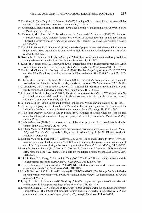
P1: OTE/SPH P2: OTE
BLUK053-Bradford September 22, 2006 15:36
ABSCISIC ACID AND HORMONAL CROSS-TALK IN SEED DORMANCY 217
T. Kinoshita, A. Cano-Delgado, H. Seto, et al. (2005) Binding of brassinosteroids to the extracellular
domain of plant receptor kinase BRI1. Nature 433, 167–171.
M. Koornneef, L. Bentsink and H. Hilhorst (2002) Seed dormancy and germination. Current Opinionin Plant Biology 5, 33–36.
M. Koornneef, M.L. Jorna, D.L.C. Brinkhorst-van der Swan and C.M. Karssen (1982) The isolation
of abscisic acid (ABA) deficient mutants by selection of induced revertants in non-germinating
gibberellin sensitive lines of Arabidopsis thaliana (L.) Heynh. Theoretical and Applied Genetics61, 385–393.
Y. Kraepiel, P. Rousselin, B. Sotta, et al. (1994) Analysis of phytochrome- and ABA-deficient mutants
suggests that ABA degradation is controlled by light in Nicotiana plumbaginifolia. The PlantJournal 6, 665–672.
B. Kucera, M.A. Cohn and G. Leubner-Metzger (2005) Plant hormone interactions during seed dor-
mancy release and germination. Seed Science Research 15, 281–307.
S. Kurup, H.D. Jones and M.J. Holdsworth (2000) Interactions of the developmental regulator ABI3
with proteins identified from developing Arabidopsis seeds. The Plant Journal 21, 143–155.
T. Kushiro, M. Okamoto, K. Nakabayashi, et al. (2004) The Arabidopsis cytochrome P450 CYP707Aencodes ABA 8′-hydroxylases: key enzymes in ABA catabolism. The EMBO Journal 23, 1647–
1656.
R.J. Laby, M.S. Kincaid, D. Kim and S.I. Gibson (2000) The Arabidopsis sugar-insensitive mutants
sis4 and sis5 are defective in abscisic acid synthesis and response. The Plant Journal 23, 587–596.
C.C. Lashbrook, D.M. Tieman and H.J. Klee (1998) Differential regulation of the tomato ETR gene
family throughout plant development. The Plant Journal 15, 243–252.
V. Lefebvre, H. North, A. Frey, et al. (2006) Functional analysis of Arabidopsis NCED6 and NCED9genes indicates that ABA synthesised in the endosperm is involved in the induction of seed
dormancy. The Plant Journal 45, 309–319.
P. Leon and J. Sheen (2003) Sugar and hormone connections. Trends in Plant Science 8, 110–116.
M.T. Le Page-Degivry and G. Garello (1992) In situ abscisic acid synthesis. A requirement for
induction of embryo dormancy in Helianthus annuus. Plant Physiology 98, 1386–1390.
M.T. Le Page-Degivry, G. Garello and P. Barthe (1997) Changes in abscisic acid biosynthesis and
catabolism during dormancy breaking in Fagus sylvatica embryo. Journal of Plant Growth Reg-ulation 16, 57–61.
G. Leubner-Metzger (2001) Brassinosteroids and gibberellins promote tobacco seed germination by
distinct pathways. Planta 213, 758–763.
G. Leubner-Metzger (2003) Brassinosteroids promote seed germination. In: Brassinosteroids: Bioac-tivity and Crop Productivity (eds S. Hayat and A. Ahmad), pp. 119–128. Kluwer Academic
Publishers, Dordrecht.
G. Leubner-Metzger, L. Petruzzelli, R. Waldvogel, R. Vogeli-Lange and F. Meins Jr. (1998) Ethylene-
responsive element binding protein (EREBP) expression and the transcriptional regulation of
class I β-1,3-glucanase during tobacco seed germination. Plant Molecular Biology 38, 785–795.
J. Leung, M. Bouvier-Durand, P.-C. Morris, D. Guerrier, F. Chefdor and J. Giraudat (1994) ArabidopsisABA-response gene ABI1: features of a calcium-modulated protein phosphatase. Science 264,
1448–1452.
H. Li, J.J. Shen, Z.L. Zheng, Y. Lin and Z. Yang (2001) The Rop GTPase switch controls multiple
developmental processes in Arabidopsis. Plant Physiology 126, 670–684.
H.C. Li, K. Chuang, J.T. Henderson, et al. (2005) PICKLE acts during germination to repress expression
of embryonic traits. The Plant Journal 44, 1010–1022.
P.P. Liu, N. Koizuka, R.C. Martin and H. Nonogaki (2005) The BME3 (Blue Micropylar End 3) GATA
zinc finger transcription factor is a positive regulator of Arabidopsis seed germination. The PlantJournal 44, 960–971.
K. Ljung, A. Ostin, L. Lioussanne and G. Sandberg (2001) Developmental regulation of indole-3-acetic
acid turnover in Scots pine seedlings. Plant Physiology 125, 464–475.
O. Lorenzo, C. Nicolas, G. Nicolas and D. Rodrıguez (2002) Molecular cloning of a functional protein
phosphatase 2C (FsPP2C2) with unusual features and synergistically upregulated by ABA and
calcium in dormant seeds of Fagus sylvatica. Physiologia Plantarum 114, 482–490.
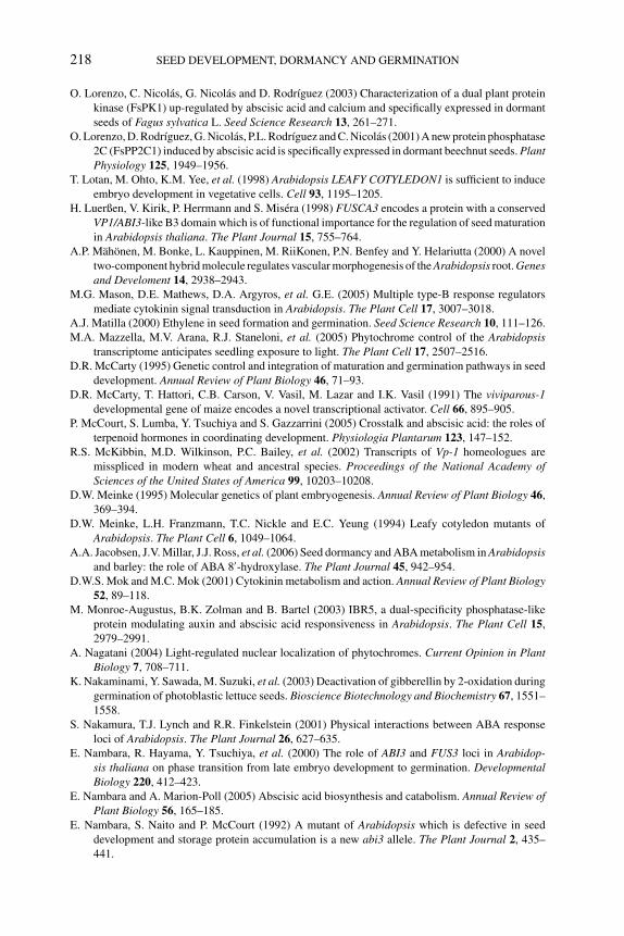
P1: OTE/SPH P2: OTE
BLUK053-Bradford September 22, 2006 15:36
218 SEED DEVELOPMENT, DORMANCY AND GERMINATION
O. Lorenzo, C. Nicolas, G. Nicolas and D. Rodrıguez (2003) Characterization of a dual plant protein
kinase (FsPK1) up-regulated by abscisic acid and calcium and specifically expressed in dormant
seeds of Fagus sylvatica L. Seed Science Research 13, 261–271.
O. Lorenzo, D. Rodrıguez, G. Nicolas, P.L. Rodrıguez and C. Nicolas (2001) A new protein phosphatase
2C (FsPP2C1) induced by abscisic acid is specifically expressed in dormant beechnut seeds. PlantPhysiology 125, 1949–1956.
T. Lotan, M. Ohto, K.M. Yee, et al. (1998) Arabidopsis LEAFY COTYLEDON1 is sufficient to induce
embryo development in vegetative cells. Cell 93, 1195–1205.
H. Luerßen, V. Kirik, P. Herrmann and S. Misera (1998) FUSCA3 encodes a protein with a conserved
VP1/ABI3-like B3 domain which is of functional importance for the regulation of seed maturation
in Arabidopsis thaliana. The Plant Journal 15, 755–764.
A.P. Mahonen, M. Bonke, L. Kauppinen, M. RiiKonen, P.N. Benfey and Y. Helariutta (2000) A novel
two-component hybrid molecule regulates vascular morphogenesis of the Arabidopsis root. Genesand Develoment 14, 2938–2943.
M.G. Mason, D.E. Mathews, D.A. Argyros, et al. G.E. (2005) Multiple type-B response regulators
mediate cytokinin signal transduction in Arabidopsis. The Plant Cell 17, 3007–3018.
A.J. Matilla (2000) Ethylene in seed formation and germination. Seed Science Research 10, 111–126.
M.A. Mazzella, M.V. Arana, R.J. Staneloni, et al. (2005) Phytochrome control of the Arabidopsistranscriptome anticipates seedling exposure to light. The Plant Cell 17, 2507–2516.
D.R. McCarty (1995) Genetic control and integration of maturation and germination pathways in seed
development. Annual Review of Plant Biology 46, 71–93.
D.R. McCarty, T. Hattori, C.B. Carson, V. Vasil, M. Lazar and I.K. Vasil (1991) The viviparous-1developmental gene of maize encodes a novel transcriptional activator. Cell 66, 895–905.
P. McCourt, S. Lumba, Y. Tsuchiya and S. Gazzarrini (2005) Crosstalk and abscisic acid: the roles of
terpenoid hormones in coordinating development. Physiologia Plantarum 123, 147–152.
R.S. McKibbin, M.D. Wilkinson, P.C. Bailey, et al. (2002) Transcripts of Vp-1 homeologues are
misspliced in modern wheat and ancestral species. Proceedings of the National Academy ofSciences of the United States of America 99, 10203–10208.
D.W. Meinke (1995) Molecular genetics of plant embryogenesis. Annual Review of Plant Biology 46,
369–394.
D.W. Meinke, L.H. Franzmann, T.C. Nickle and E.C. Yeung (1994) Leafy cotyledon mutants of
Arabidopsis. The Plant Cell 6, 1049–1064.
A.A. Jacobsen, J.V. Millar, J.J. Ross, et al. (2006) Seed dormancy and ABA metabolism in Arabidopsisand barley: the role of ABA 8′-hydroxylase. The Plant Journal 45, 942–954.
D.W.S. Mok and M.C. Mok (2001) Cytokinin metabolism and action. Annual Review of Plant Biology52, 89–118.
M. Monroe-Augustus, B.K. Zolman and B. Bartel (2003) IBR5, a dual-specificity phosphatase-like
protein modulating auxin and abscisic acid responsiveness in Arabidopsis. The Plant Cell 15,
2979–2991.
A. Nagatani (2004) Light-regulated nuclear localization of phytochromes. Current Opinion in PlantBiology 7, 708–711.
K. Nakaminami, Y. Sawada, M. Suzuki, et al. (2003) Deactivation of gibberellin by 2-oxidation during
germination of photoblastic lettuce seeds. Bioscience Biotechnology and Biochemistry 67, 1551–
1558.
S. Nakamura, T.J. Lynch and R.R. Finkelstein (2001) Physical interactions between ABA response
loci of Arabidopsis. The Plant Journal 26, 627–635.
E. Nambara, R. Hayama, Y. Tsuchiya, et al. (2000) The role of ABI3 and FUS3 loci in Arabidop-sis thaliana on phase transition from late embryo development to germination. DevelopmentalBiology 220, 412–423.
E. Nambara and A. Marion-Poll (2005) Abscisic acid biosynthesis and catabolism. Annual Review ofPlant Biology 56, 165–185.
E. Nambara, S. Naito and P. McCourt (1992) A mutant of Arabidopsis which is defective in seed
development and storage protein accumulation is a new abi3 allele. The Plant Journal 2, 435–
441.
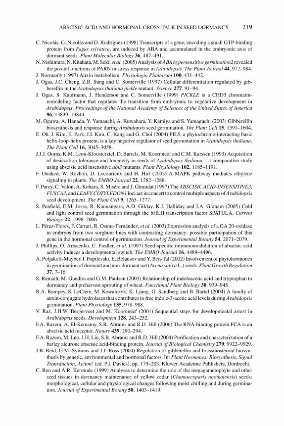
P1: OTE/SPH P2: OTE
BLUK053-Bradford September 22, 2006 15:36
ABSCISIC ACID AND HORMONAL CROSS-TALK IN SEED DORMANCY 219
C. Nicolas, G. Nicolas and D. Rodrıguez (1998) Transcripts of a gene, encoding a small GTP-binding
protein from Fagus sylvatica, are induced by ABA and accumulated in the embryonic axis of
dormant seeds. Plant Molecular Biology 36, 487–491.
N. Nishimura, N. Kitahata, M. Seki, et al. (2005) Analysis of ABA hypersensitive germination2 revealed
the pivotal functions of PARN in stress response in Arabidopsis. The Plant Journal 44, 972–984.
J. Normanly (1997) Auxin metabolism. Physiologia Plantarum 100, 431–442.
J. Ogas, J.C. Cheng, Z.R. Sung and C. Somerville (1997) Cellular differentiation regulated by gib-
berellin in the Arabidopsis thaliana pickle mutant. Science 277, 91–94.
J. Ogas, S. Kaufmann, J. Henderson and C. Somerville (1999) PICKLE is a CHD3 chromatin-
remodeling factor that regulates the transition from embryonic to vegetative development in
Arabidopsis. Proceedings of the National Academy of Sciences of the United States of America96, 13839–13844.
M. Ogawa, A. Hanada, Y. Yamauchi, A. Kuwahara, Y. Kamiya and S. Yamaguchi (2003) Gibberellin
biosynthesis and response during Arabidopsis seed germination. The Plant Cell 15, 1591–1604.
E. Oh, J. Kim, E. Park, J.I. Kim, C. Kang and G. Choi (2004) PIL5, a phytochrome-interacting basic
helix-loop-helix protein, is a key negative regulator of seed germination in Arabidopsis thaliana.
The Plant Cell 16, 3045–3058.
J.J.J. Ooms, K.M. Leon-Kloosterziel, D. Bartels, M. Koornneef and C.M. Karssen (1993) Acquisition
of desiccation tolerance and longevity in seeds of Arabidopsis thaliana – a comparative study
using abscisic acid insensitive abi3 mutants. Plant Physiology 102, 1185–1191.
F. Ouaked, W. Rozhon, D. Lecourieux and H. Hirt (2003) A MAPK pathway mediates ethylene
signaling in plants. The EMBO Journal 22, 1282–1288.
F. Parcy, C. Valon, A. Kohara, S. Misera and J. Giraudat (1997) The ABSCISIC ACID-INSENSITIVE3,
FUSCA3, and LEAFY COTYLEDON1 loci act in concert to control multiple aspects of Arabidopsisseed development. The Plant Cell 9, 1265–1277.
S. Penfield, E.M. Josse, R. Kannangara, A.D. Gilday, K.J. Halliday and I.A. Graham (2005) Cold
and light control seed germination through the bHLH transcription factor SPATULA. CurrentBiology 22, 1998–2006.
L. Perez-Flores, F. Carrari, R. Osuna-Fernandez, et al. (2003) Expression analysis of a GA 20-oxidase
in embryos from two sorghum lines with contrasting dormancy: possible participation of this
gene in the hormonal control of germination. Journal of Experimental Botany 54, 2071–2079.
J. Phillips, O. Artsaenko, U. Fiedler, et al. (1997) Seed-specific immunomodulation of abscisic acid
activity induces a developmental switch. The EMBO Journal 16, 4489–4496.
A. Poljakoff-Mayber, I. Popilevski, E. Belausov and Y. Ben-Tal (2002) Involvement of phytohormones
in germination of dormant and non-dormant oat (Avena sativa L.) seeds. Plant Growth Regulation37, 7–16.
S. Ramaih, M. Guedira and G.M. Paulsen (2003) Relationship of indoleacetic acid and tryptophan to
dormancy and preharvest sprouting of wheat. Functional Plant Biology 30, 939–945.
R.A. Rampey, S. LeClere, M. Kowalczyk, K. Ljung, G. Sandberg and B. Bartel (2004) A family of
auxin-conjugate hydrolases that contributes to free indole-3-acetic acid levels during Arabidopsisgermination. Plant Physiology 135, 978–988.
V. Raz, J.H.W. Bergervoet and M. Koornneef (2001) Sequential steps for developmental arrest in
Arabidopsis seeds. Development 128, 243–252.
F.A. Razem, A. El-Kereamy, S.R. Abrams and R.D. Hill (2006) The RNA-binding protein FCA is an
abscisic acid receptor. Nature 439, 290–294.
F.A. Razem, M. Luo, J.H. Liu, S.R. Abrams and R.D. Hill (2004) Purification and characterization of a
barley aleurone abscisic acid-binding protein. Journal of Biological Chemistry 279, 9922–9929.
J.B. Reid, G.M. Symons and J.J. Ross (2004) Regulation of gibberellin and brassinosteroid biosyn-
thesis by genetic, environmental and hormonal factors. In: Plant Hormones: Biosynthesis, SignalTransduction, Action! (ed. P.J. Davies), pp. 179–203. Kluwer Academic Publishers, Dordrecht.
C. Ren and A.R. Kermode (1999) Analyses to determine the role of the megagametophyte and other
seed tissues in dormancy maintenance of yellow cedar (Chamaecyparis nootkatensis) seeds:
morphological, cellular and physiological changes following moist chilling and during germina-
tion. Journal of Experimental Botany 50, 1403–1419.
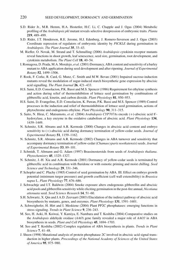
P1: OTE/SPH P2: OTE
BLUK053-Bradford September 22, 2006 15:36
220 SEED DEVELOPMENT, DORMANCY AND GERMINATION
S.D. Rider Jr., M.R. Hemm, H.A. Hostetler, H.C. Li, C. Chapple and J. Ogas (2004) Metabolic
profiling of the Arabidopsis pkl mutant reveals selective derepression of embryonic traits. Planta219, 489–499.
S.D. Rider, J.T. Henderson, R.E. Jerome, H.J. Edenberg, J. Romero-Severson and J. Ogas (2003)
Coordinate repression of regulators of embryonic identity by PICKLE during germination in
Arabidopsis. The Plant Journal 35, 33–43.
M. Riefler, O. Novak, M. Strand and T. Schmulling (2006) Arabidopsis cytokinin receptor mutants
reveal functions in shoot growth, leaf senescence, seed size, germination, root development, and
cytokinin metabolism. The Plant Cell 18, 40–54.
I. Romagosa, D. Prada, M.A. Moralejo, et al. (2001) Dormancy, ABA content and sensitivity of a barley
mutant to ABA application during seed development and after ripening. Journal of ExperimentalBotany 52, 1499–1506.
F. Rook, F. Corke, R. Card, G. Munz, C. Smith and M.W. Bevan (2001) Impaired sucrose-induction
mutants reveal the modulation of sugar-induced starch biosynthetic gene expression by abscisic
acid signalling. The Plant Journal 26, 421–433.
H.S. Saini, E.D. Consolacion, P.K. Bassi and M.S. Spencer (1986) Requirement for ethylene synthesis
and action during relief of thermoinhibition of lettuce seed germination by combinations of
gibberellic acid, kinetin, and carbon dioxide. Plant Physiology 81, 950–953.
H.S. Saini, D. Evangeline, E.D. Consolacion, K. Pawan, P.K. Bassi and M.S. Spencer (1989) Control
processes in the induction and relief of thermoinhibition of lettuce seed germination, actions of
phytochrome and endogenous ethylene. Plant Physiology 90, 311–315.
S. Saito, N. Hirai, C. Matsumoto, et al. (2004) Arabidopsis CYP707As encode (+)-abscisic acid 8′-hydoxylase, a key enzyme in the oxidative catabolism of abscisic acid. Plant Physiology 134,
1439–1449.
N. Schmitz, S.R. Abrams and A.R. Kermode (2000) Changes in abscisic acid content and embryo
sensitivity to (+)-abscisic acid during dormancy termination of yellow-cedar seeds. Journal ofExperimental Botany 51, 1159–1162.
N. Schmitz, S.R. Abrams and A.R. Kermode (2002) Changes in ABA turnover and sensitivity that
accompany dormancy termination of yellow-cedar (Chamaecyparis nootkatensis) seeds. Journalof Experimental Botany 53, 89–101.
J. Schmidt, T. Altmann and G. Adam (1997) Brassinosteroids from seeds of Arabidopsis thaliana.
Phytochemistry 45, 1325–1327.
N. Schmitz, J.-H. Xia and A.R. Kermode (2001) Dormancy of yellow-cedar seeds is terminated by
gibberellic acid in combination with fluridone or with osmotic priming and moist chilling. SeedScience and Technology 29, 331–346.
P. Schopfer and C. Plachy (1985) Control of seed germination by ABA. III. Effect on embryo growth
potential (minimum turgor pressure) and growth coefficient (cell wall extensibility) in Brassicanapus L. Plant Physiology 77, 676–686.
J. Schwachtje and I.T. Baldwin (2004) Smoke exposure alters endogenous gibberellin and abscisic
acid pools and gibberellin sensitivity while eliciting germination in the post-fire annual, Nicotianaattenuata seed. Seed Science Research 14, 51–60.
S.H. Schwartz, X. Qin and J.A.D. Zeevaart (2003) Elucidation of the indirect pathway of abscisic acid
biosynthesis by mutants, genes, and enzymes. Plant Physiology 131, 1591–1601.
A. Schweighofer, H. Hirt and I. Meskiene (2004) Plant PP2C phosphatases: emerging functions in
stress signaling. Trends in Plant Science 9, 236–243.
M. Seo, H. Aoki, H. Koiwai, Y. Kamiya, E. Nambara and T. Koshiba (2004) Comparative studies on
the Arabidopsis aldehyde oxidase (AAO) gene family revealed a major role of AAO3 in ABA
biosynthesis in seeds. Plant and Cell Physiology 45, 1694–1703.
M. Seo and T. Koshiba (2002) Complex regulation of ABA biosynthesis in plants. Trends in PlantScience 7, 41–48.
J. Sheen (1998) Mutational analysis of protein phosphatase 2C involved in abscisic acid signal trans-
duction in higher plants. Proceedings of the National Academy of Sciences of the United Statesof America 95, 975–980.
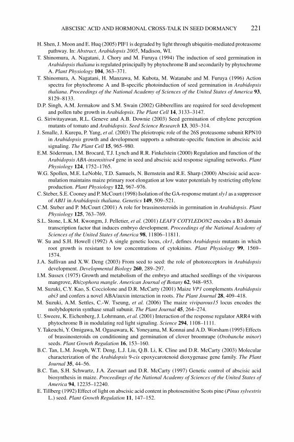
P1: OTE/SPH P2: OTE
BLUK053-Bradford September 22, 2006 15:36
ABSCISIC ACID AND HORMONAL CROSS-TALK IN SEED DORMANCY 221
H. Shen, J. Moon and E. Huq (2005) PIF1 is degraded by light through ubiquitin-mediated proteasome
pathway. In: Abstract, Arabidopsis 2005, Madison, WI.
T. Shinomura, A. Nagatani, J. Chory and M. Furuya (1994) The induction of seed germination in
Arabidopsis thaliana is regulated principally by phytochrome B and secondarily by phytochrome
A. Plant Physiology 104, 363–371.
T. Shinomura, A. Nagatani, H. Manzawa, M. Kubota, M. Watanabe and M. Furuya (1996) Action
spectra for phytochrome A and B-specific photoinduction of seed germination in Arabidopsisthaliana. Proceedings of the National Academy of Sciences of the United States of America 93,
8129–8133.
D.P. Singh, A.M. Jermakow and S.M. Swain (2002) Gibberellins are required for seed development
and pollen tube growth in Arabidopsis. The Plant Cell 14, 3133–3147.
G. Siriwitayawan, R.L. Geneve and A.B. Downie (2003) Seed germination of ethylene perception
mutants of tomato and Arabidopsis. Seed Science Research 13, 303–314.
J. Smalle, J. Kurepa, P. Yang, et al. (2003) The pleiotropic role of the 26S proteasome subunit RPN10
in Arabidopsis growth and development supports a substrate-specific function in abscisic acid
signaling. The Plant Cell 15, 965–980.
E.M. Soderman, I.M. Brocard, T.J. Lynch and R.R. Finkelstein (2000) Regulation and function of the
Arabidopsis ABA-insensitive4 gene in seed and abscisic acid response signaling networks. PlantPhysiology 124, 1752–1765.
W.G. Spollen, M.E. LeNoble, T.D. Samuels, N. Bernstein and R.E. Sharp (2000) Abscisic acid accu-
mulation maintains maize primary root elongation at low water potentials by restricting ethylene
production. Plant Physiology 122, 967–976.
C. Steber, S.E. Cooney and P. McCourt (1998) Isolation of the GA-response mutant sly1 as a suppressor
of ABI1 in Arabidopsis thaliana. Genetics 149, 509–521.
C.M. Steber and P. McCourt (2001) A role for brassinosteroids in germination in Arabidopsis. PlantPhysiology 125, 763–769.
S.L. Stone, L.K.M. Kwongm, J. Pelletier, et al. (2001) LEAFY COTYLEDON2 encodes a B3 domain
transcription factor that induces embryo development. Proceedings of the National Academy ofSciences of the United States of America 98, 11806–11811.
W. Su and S.H. Howell (1992) A single genetic locus, ckr1, defines Arabidopsis mutants in which
root growth is resistant to low concentrations of cytokinins. Plant Physiology 99, 1569–
1574.
J.A. Sullivan and X.W. Deng (2003) From seed to seed: the role of photoreceptors in Arabidopsisdevelopment. Developmental Biology 260, 289–297.
I.M. Sussex (1975) Growth and metabolism of the embryo and attached seedlings of the viviparous
mangrove, Rhizophora mangle. American Journal of Botany 62, 948–953.
M. Suzuki, C.Y. Kao, S. Cocciolone and D.R. McCarty (2001) Maize VP1 complements Arabidopsisabi3 and confers a novel ABA/auxin interaction in roots. The Plant Journal 28, 409–418.
M. Suzuki, A.M. Settles, C.-W. Tseung, et al. (2006) The maize viviparous15 locus encodes the
molybdopterin synthase small subunit. The Plant Journal 45, 264–274.
U. Sweere, K. Eichenberg, J. Lohrmann, et al. (2001) Interaction of the response regulator ARR4 with
phytochrome B in modulating red light signaling. Science 294, 1108–1111.
Y. Takeuchi, Y. Omigawa, M. Ogasawara, K. Yoneyama, M. Konnai and A.D. Worsham (1995) Effects
of brassinosteroids on conditioning and germination of clover broomrape (Orobanche minor)
seeds. Plant Growth Regulation 16, 153–160.
B.C. Tan, L.M. Joseph, W.T. Deng, L.J. Liu, Q.B. Li, K. Cline and D.R. McCarty (2003) Molecular
characterization of the Arabidopsis 9-cis epoxycarotenoid dioxygenase gene family. The PlantJournal 35, 44–56.
B.C. Tan, S.H. Schwartz, J.A. Zeevaart and D.R. McCarty (1997) Genetic control of abscisic acid
biosynthesis in maize. Proceedings of the National Academy of Sciences of the United States ofAmerica 94, 12235–12240.
E. Tillberg (1992) Effect of light on abscisic acid content in photosensitive Scots pine (Pinus sylvestrisL.) seed. Plant Growth Regulation 11, 147–152.
P1: OTE/SPH P2: OTE
BLUK053-Bradford September 22, 2006 15:36
222 SEED DEVELOPMENT, DORMANCY AND GERMINATION
I. Tiryaki and P.E. Staswick (2002) An Arabidopsis mutant defective in jasmonate response is allelic
to the auxin-signaling mutant axr1. Plant Physiology 130, 887–894.
T. Toyomasu, H. Kawaide, W. Mitsuhashi, Y. Inoue and Y. Kamiya (1998) Phytochrome regulates
gibberellin biosynthesis during germination of photoblastic lettuce seeds. Plant Physiology 118,
1517–1523.
T. Toyomasu, H. Tsuji, H. Yamane, et al. (1993) Light effects on endogenous levels of gibberellins in
photoblastic lettuce seeds. Journal of Plant Growth Regulation 12, 85–90.
H. Ullah, J.G. Chen, S. Wang and A.M. Jones (2002). Role of a heterotrimeric G protein in regulation
of Arabidopsis seed germination. Plant Physiology 129, 897–907.
T. Urao, B. Yakubov, R. Satoh, et al. (1999) A transmembrane hybrid-type histidine kinase in Ara-bidopsis functions as an osmosensor. The Plant Cell 11, 1743–1754.
A.J. van Hengel and K. Roberts (2003) AtAGP30, an arabinogalactan-protein in the cell walls of
the primary root, plays a role in root regeneration and seed germination. The Plant Journal 36,
256–270.
B.V. Vardhini and S.S.R. Rao (2001) Effect of brassinosteroids on the seed germination and seedling
growth of tomato (Lycopersicon escultentum Mill.). Journal of Plant Biology 27, 303–305.
B.V. Vardhini and S.S.R. Rao (2003) Amelioration of osmotic stress by brassinosteroids on seed
germination and seedling growth of three varieties of sorghum. Plant Growth Regulation 41,
25–31.
R.D. Vierstra (2003) The ubiquitin/26S proteasome pathway, the complex last chapter in the life of
many plant proteins. Trends in Plant Science 8, 135–142.
M. Walker-Simmons (1990) Dormancy in cereals – levels of and response to abscisic acid. In: PlantGrowth Substances (eds R.P. Pharis and S.B. Rood), pp. 400–406. Springer-Verlag, New York.
M. Wang, S. Heimovaara-Dijkstra and B. Van Duijn (1995) Modulation of germination of embryos
isolated from dormant and non-dormant barley grains by manipulation of endogenous abscisic
acid. Planta 195, 586–592.
X.Q. Wang, H. Ullah, A.M. Jones and S.M. Assmann (2001). G protein regulation of ion channels and
abscisic acid signaling in Arabidopsis guard cells. Science 292, 2070–2072.
C.N. White, W.M. Proebsting, P. Hedden and C.J. Riven (2000) Gibberellins and seed development in
maize. I. Evidence that gibberellin/abscisic acid balance governs germination versus maturation
pathways. Plant Physiology 122, 1081–1088.
C.N. White and C.J. Riven (2000) Gibberellins and seed development in maize. II. Gibberellin synthesis
inhibition enhances abscisic acid signaling in cultured embryos. Plant Physiology 122, 1089–
1097.
F. Woodger, J.V. Jacobsen and F. Gubler (2004) Gibberellin action in germinating cereal grains.
In: Plant Hormones: Biosynthesis, Signal Transduction, Action! (ed. P.J. Davies), pp. 221–240.
Kluwer Academic Publishers, Dordrecht.
L. Xiong, Z. Gong, C. Rock, et al. (2001a) Modulation of abscisic acid signal transduction and
biosynthesis by an Sm-like protein in Arabidopsis. Developmental Cell 1, 771–781.
L. Xiong, H. Lee, M. Ishitani and J.K. Zhu (2002) Regulation of osmotic stress-responsive gene
expression by the LOS6/ABA1 locus in Arabidopsis. Journal of Biological Chemistry 277, 8588–
8596.
L. Xiong and J.K. Zhu (2003) Regulation of abscisic acid biosynthesis. Plant Physiology 133, 29–36.
L.M. Xiong, M. Ishitani, H. Lee and J.K. Zhu (2001b) The Arabidopsis LOS5/ABA3 locus encodes a
molybdenum cofactor sulfurase and modulates cold stress- and osmotic stress-responsive gene
expression. The Plant Cell 13, 2063–2083.
N. Xu and J.D. Bewley (1991) Sensitivity to abscisic acid and osmoticum changes during embryoge-
nesis of alfalfa (Medicago sativa). Journal of Experimental Botany 42, 821–826.
H. Yamada, T. Suzuki, K. Terada, et al. (2001) The Arabidopsis AHK4 histidine kinase is a cytokinin-
binding receptor that transduces cytokinin signals across the membrane. Plant and Cell Physiology42, 1017–1023.
S. Yamaguchi and Y. Kamiya (2002) Gibberellins and light-stimulated seed germination. Journal ofPlant Growth Regulation 20, 369–376.
P1: OTE/SPH P2: OTE
BLUK053-Bradford September 22, 2006 15:36
ABSCISIC ACID AND HORMONAL CROSS-TALK IN SEED DORMANCY 223
S. Yamaguchi, Y. Kamiya and T-P. Sun (2001) Distinct cell-specific expression patterns of early and
late gibberellin biosynthetic genes during Arabidopsis seed germination. The Plant Journal 28,
443–453.
S. Yamaguchi, M.W. Smith, R.G. Brown, Y. Kamiya and T-P. Sun (1998) Phytochrome regulation and
differential expression of gibberellin 3β-hydroxylase genes in germinating Arabidopsis seeds.
The Plant Cell 10, 2115–2126.
T. Yamaguchi, T. Wakizuka, K. Hirai, S. Fujii and A. Fujita (1987) Stimulation of germination in aged
rice seeds by pretreatment with brassinolide. Proceedings of the Plant Growth Regulation Societyof America 14, 26–27.
Y. Yamauchi, M. Ogawa, A. Kuwahara, A. Hanada, Y. Kamiya and S. Yamaguchi (2004) Activation
of gibberellin biosynthesis and response pathways by low temperature during imbibition of
Arabidopsis thaliana seeds. The Plant Cell 16, 367–378.
T. Yoshioka, T. Endo and S. Satoh (1998) Restoration of seed germination at supraoptimal temperature
by fluridone, an inhibitor of abscisic acid biosynthesis. Plant and Cell Physiology 39, 307–312.
T. Yoshida, N. Nishimura, N. Kitahata, et al. (2006) ABA-Hypersensitive Germination3 encodes a pro-
tein phosphatase 2C (AtPP2CA) that strongly regulates abscisic acid signaling during germination
among Arabidopsis protein phosphatase 2Cs. Plant Physiology 140, 115–126.
T.E. Young and D.R. Gallie (2000) Programmed cell death during endosperm development. PlantMolecular Biology 44, 283–301.
T.E. Young, D.R. Gallie and D.A. DeMason (1997) Ethylene mediated programmed cell death during
maize endosperm development of Su and sh2 genotypes. Plant Physiology 115, 737–751.
L.I. Zaharia, M.K. Walker-Simmons, C.N. Rodriguez and S.R. Abrams (2005) Chemistry of abscisic
acid and abscisic acid catabolites and analogs. Journal of Plant Growth Regulation 24, 274–284.
J.A.D. Zeevaart (1999) Abscisic acid metabolism and its regulation. In: Biochemistry and MolecularBiology of Plant Hormones (eds P.J.J. Hooykaas, K.R. Hall and K.R. Libbenga), pp. 189–207.
Elsevier Science BV, Amsterdam.
Y. Zeng and A.R. Kermode (2004) A gymnosperm ABI3 gene functions in a severe abscisic acid-
insensitive mutant of Arabidopsis (abi3-6) to restore the wild-type phenotype and demonstrates a
strong synergistic effect with sugar in the inhibition of post-germinative growth. Plant MolecularBiology 56, 731–746.
Y. Zeng, N. Raimondi and A.R. Kermode (2003) Role of an ABI3 homologue in dormancy maintenance
of yellow-cedar seeds and in the activation of storage protein and Em gene promoters. PlantMolecular Biology 51, 39–49.
X. Zhang, V. Garreton and N.-H. Chua (2005) The AIP2 E3 ligase acts as a novel negative regulator
of ABA signaling by promoting ABI3 degradation. Genes and Development 19, 1532–1543.
P1: JYS
BLUK053-Bradford September 22, 2006 14:30
9 Regulation of ABA and GA levels during seeddevelopment and germination in ArabidopsisShinjiro Yamaguchi, Yuji Kamiya and Eiji Nambara
9.1 Introduction
Seed germination is regulated by both developmental and environmental factors.Plant hormones are important signaling molecules that transfer the changes of theenvironment to seeds during germination. Abscisic acid (ABA) and gibberellin (GA)are considered to be major hormones in germinating Arabidopsis thaliana seeds.ABA is a sesquiterpene hormone that is necessary during late embryogenesis andinhibits precocious seed germination. ABA also increases the tolerance of plantsto stresses such as drought, salinity, and temperature, and induces stomatal closurein leaves (Davies, 2004). Elevated ABA levels are important during mid- and late-embryogenesis to maintain normal seed development. Relatively high levels of ABAin maturing seeds maintain their dormancy and avoid precocious germination. GAis a diterpene hormone that regulates seed development and germination and otheraspects of plant growth and development, such as leaf expansion, stem elongation,and flowering. Although some reports suggest that ethylene and brassinosteroidsalso affect seed germination of Arabidopsis (Kucera et al., 2005), we focus here onregulation of ABA and GA levels in seeds. Endogenous levels of active hormonesare determined by the relative rates of their formation and conversion into inactiveforms. In this chapter, we refer to biosynthesis as production of bioactive forms ofa hormone from its precursors, deactivation as conversion of bioactive forms or itsprecursors to the inactive (or less active) forms, and metabolism as both biosynthesisand deactivation.
Biosynthesis of ABA and GA starts in plastids using a common precursor,geranylgeranyldiphosphate (GGDP), which is synthesized via the 2-C-methyl-d-erythritol phosphate (MEP) pathway (Milborrow and Lee, 1998; Hirai et al., 2000;Kasahara et al., 2004). The mevalonate pathway in the cytosol is important forsteroid and triterpene biosynthesis, but is not principally involved in the biosynthe-sis of ABA and GA. Severe alleles of GA-deficient mutants of Arabidopsis cannotgerminate without exogenous bioactive GAs (Koornneef and van der Veen, 1980).Therefore, GA is an essential hormone leading to germination in Arabidopsis. How-ever, some ABA-deficient and -insensitive mutants can germinate in the presence ofpaclobutrazol (PAC), a GA biosynthesis inhibitor, at the concentration that is suffi-cient to inhibit germination of wild-type seeds. This suggests that a low level of GAis enough to cause germination when ABA biosynthesis or signaling is impaired.
224
P1: JYS
BLUK053-Bradford September 22, 2006 14:30
REGULATION OF ABA AND GA LEVELS 225
These observations support the idea that the balance of endogenous ABA and GAlevels plays an important role in controlling seed germination.
In this chapter, we first describe the outline of ABA and GA metabolism andthe genes involved in the respective pathways. Chemical inhibitors for ABA andGA metabolism enzymes will also be described, because they have contributedsignificantly to understanding the roles of ABA and GA in seed biology. Finally, wewill highlight recent findings on the regulation of ABA and GA levels in developingand germinating seeds. Throughout this chapter, we will focus on Arabidopsis,because the regulation of ABA and GA metabolism has been best understood in thismodel species.
9.2 Biosynthetic and deactivation pathways of ABA and GA
Microarrays have been used to analyze the changes in the transcriptome in Arabidop-sis seeds (Ogawa et al., 2003; Yamauchi et al., 2004; Nakabayashi et al., 2005). Inaddition to these published results, a large set of GeneChip data has been made avail-able as public databases such as the AtGenExpress project (http://www.arabidopsis.org/info/expression/ATGenExpress.jsp). We summarized these data to provide avisual overview of the expression profiles of ABA and GA metabolism genes dur-ing seed development and germination (Figures 9.1 and 9.2). The identities (AGI[Arabidopsis Genome Initiative] codes) of all ABA and GA metabolism genes arelisted in Tables 9.1 and 9.2. Some of these genes and the corresponding loss-of-function mutants are selected to discuss the specific aspects of ABA and GA regu-lation and to highlight recent findings in the following sections.
9.2.1 ABA biosynthesis
In plants, ABA is synthesized from C40 carotenoids via oxidative cleavage (Nambaraand Marion-Poll, 2005). Earlier steps in ABA biosynthesis occur in plastids, andthe last two steps take place in the cytosol. Zeaxanthin epoxidase (ZEP/AtABA1)is a xanthophyll cycle enzyme that catalyzes two-step epoxidation of zeaxanthin toviolaxanthin (Audran et al., 2001; Xiong et al., 2002) (Figure 9.1). Violaxanthinis converted to 9′-cis-neoxanthin or 9-cis-violaxanthin by unknown mechanisms.9-cis-Epoxycarotenoid dioxygenase (NCED) cleaves the 9-cis isomers of neox-anthin and violaxanthin to produce xanthoxin, a C15 precursor of ABA (Qinand Zeevaart, 1999; Chernys and Zeevaart, 2000; Iuchi et al., 2001; Schwartzet al., 2003). This reaction is thought to be the most important regulatory step inABA biosynthesis. In Arabidopsis, five NCEDs (AtNCED2, AtNCED3, AtNCED5,AtNCED6, and AtNCED9) are likely to be involved in this reaction (Iuchi et al.,2001; Tan et al., 2003). AtNCEDs have been shown to be chloroplast-targeted invitro (Tan et al., 2003). Xanthoxin is subsequently converted to abscisic aldehyde byshort-chain alcohol dehydrogenase/reductase (SDR/AtABA2) and then to abscisicacid by abscisic aldehyde oxidase (AAO3), two cytosolic enzymes (Figure 9.1).Considering the plastid localization of NCEDs, xanthoxin should be transported
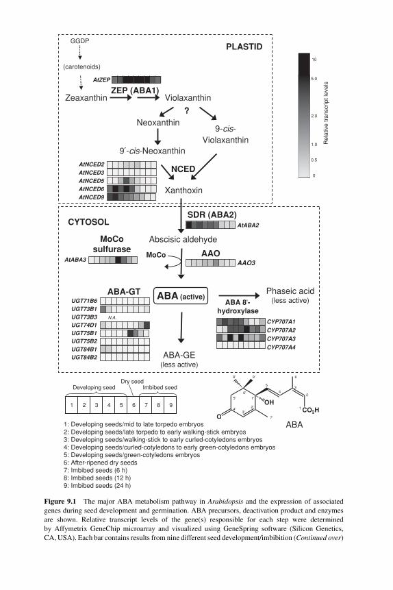
P1: JYS
BLUK053-Bradford September 22, 2006 14:30
ZEP (ABA1)
SDR (ABA2)
MoCosulfurase
ViolaxanthinZeaxanthin
Neoxanthin
Xanthoxin
NCED
Phaseic acid(less active)ABA 8'-
hydroxylase
ABA (active)
Abscisic aldehyde
9 -cis-Neoxanthin
9-cis-Violaxanthin
AAOAAO3
ABA-GE(less active)
UGT84B1
MoCo
247025_atZEP/ABA1
_
_
_
AtABA2
AtZEP
AtNCED2
AtNCED3AtNCED5AtNCED6AtNCED9
AtABA3
CYP707A1
CYP707A2
CYP707A3
CYP707A4
ABA-GT
Developing seedDry seed
Imbibed seed
1 2 3 4 5 6 8 97
1: Developing seeds/mid to late torpedo embryos2: Developing seeds/late torpedo to early walking-stick embryos3: Developing seeds/walking-stick to early curled-cotyledons embryos4: Developing seeds/curled-cotyledons to early green-cotyledons embryos5: Developing seeds/green-cotyledons embryos6: After-ripened dry seeds7: Imbibed seeds (6 h)8: Imbibed seeds (12 h)9: Imbibed seeds (24 h)
10
5.0
2.0
1.0
0.5
0
UGT75B1
UGT84B2
UGT71B6
UGT75B2
UGT74D1UGT73B3 N.A.
UGT73B1
GGDP
(carotenoids)
OCO2H
OH
7'
8' 9'
1'
2'3'
4'
5'
6'
1
2
3
6
4
5
ABA
?
CYTOSOL
PLASTID
Rel
ativ
e tr
ansc
ript l
evel
s
Figure 9.1 The major ABA metabolism pathway in Arabidopsis and the expression of associated
genes during seed development and germination. ABA precursors, deactivation product and enzymes
are shown. Relative transcript levels of the gene(s) responsible for each step were determined
by Affymetrix GeneChip microarray and visualized using GeneSpring software (Silicon Genetics,
CA, USA). Each bar contains results from nine different seed development/imbibition (Continued over)
226
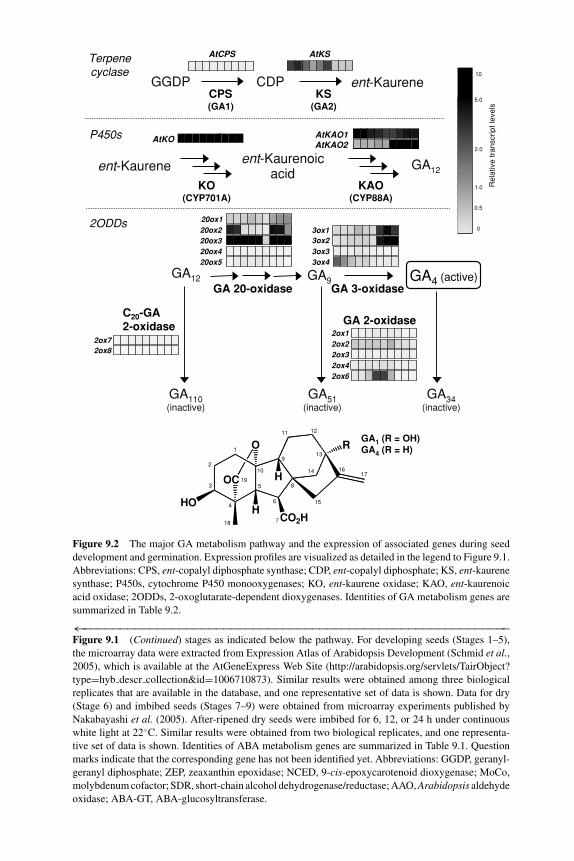
P1: JYS
BLUK053-Bradford September 22, 2006 14:30
CPS(GA1)
KS(GA2)
GA 20-oxidase GA 3-oxidase
ent-KaureneCDPGGDP
ent-Kaurenoicacid
GA12
Terpenecyclase
P450s
2ODDs
KAO(CYP88A)
GA34(inactive)
GA51(inactive)
GA 2-oxidase
GA4 (active)GA9
GA110(inactive)
KO(CYP701A)
C20-GA 2-oxidase
2ox12ox22ox32ox42ox6
AtKSAtCPS
3ox43ox33ox23ox1
2ox72ox8
20ox420ox5
20ox120ox220ox3
GA12ent-Kaurene
AtKAO1AtKAO2
AtKO_
_
GA1 (R = OH)GA4 (R = H)
CO2H
OC
O
HO
R
H
H
1
2
3
4
5
6
7
8
9
10
11 12
13
14
15
1617
18
19
10
5.0
2.0
1.0
0.5
0
Rel
ativ
e tr
ansc
ript l
evel
s
Figure 9.2 The major GA metabolism pathway and the expression of associated genes during seed
development and germination. Expression profiles are visualized as detailed in the legend to Figure 9.1.
Abbreviations: CPS, ent-copalyl diphosphate synthase; CDP, ent-copalyl diphosphate; KS, ent-kaurene
synthase; P450s, cytochrome P450 monooxygenases; KO, ent-kaurene oxidase; KAO, ent-kaurenoic
acid oxidase; 2ODDs, 2-oxoglutarate-dependent dioxygenases. Identities of GA metabolism genes are
summarized in Table 9.2.
←−−−−−−−−−−−−−−−−−−−−−−−−−−−−−−−−−−−−−−−−−−−−−−−−−−−−−−−−−−Figure 9.1 (Continued) stages as indicated below the pathway. For developing seeds (Stages 1–5),
the microarray data were extracted from Expression Atlas of Arabidopsis Development (Schmid et al.,2005), which is available at the AtGeneExpress Web Site (http://arabidopsis.org/servlets/TairObject?
type=hyb descr collection&id=1006710873). Similar results were obtained among three biological
replicates that are available in the database, and one representative set of data is shown. Data for dry
(Stage 6) and imbibed seeds (Stages 7–9) were obtained from microarray experiments published by
Nakabayashi et al. (2005). After-ripened dry seeds were imbibed for 6, 12, or 24 h under continuous
white light at 22◦C. Similar results were obtained from two biological replicates, and one representa-
tive set of data is shown. Identities of ABA metabolism genes are summarized in Table 9.1. Question
marks indicate that the corresponding gene has not been identified yet. Abbreviations: GGDP, geranyl-
geranyl diphosphate; ZEP, zeaxanthin epoxidase; NCED, 9-cis-epoxycarotenoid dioxygenase; MoCo,
molybdenum cofactor; SDR, short-chain alcohol dehydrogenase/reductase; AAO, Arabidopsis aldehyde
oxidase; ABA-GT, ABA-glucosyltransferase.
227
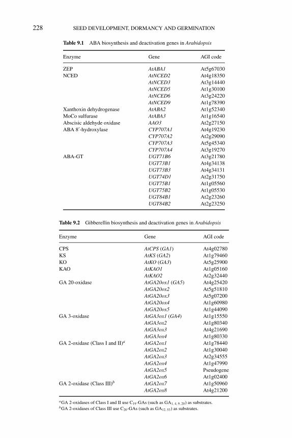
P1: JYS
BLUK053-Bradford September 22, 2006 14:30
228 SEED DEVELOPMENT, DORMANCY AND GERMINATION
Table 9.1 ABA biosynthesis and deactivation genes in Arabidopsis
Enzyme Gene AGI code
ZEP AtABA1 At5g67030
NCED AtNCED2 At4g18350
AtNCED3 At3g14440
AtNCED5 At1g30100
AtNCED6 At3g24220
AtNCED9 At1g78390
Xanthoxin dehydrogenase AtABA2 At1g52340
MoCo sulfurase AtABA3 At1g16540
Abscisic aldehyde oxidase AAO3 At2g27150
ABA 8′-hydroxylase CYP707A1 At4g19230
CYP707A2 At2g29090
CYP707A3 At5g45340
CYP707A4 At3g19270
ABA-GT UGT71B6 At3g21780
UGT73B1 At4g34138
UGT73B3 At4g34131
UGT74D1 At2g31750
UGT75B1 At1g05560
UGT75B2 At1g05530
UGT84B1 At2g23260
UGT84B2 At2g23250
Table 9.2 Gibberellin biosynthesis and deactivation genes in Arabidopsis
Enzyme Gene AGI code
CPS AtCPS (GA1) At4g02780
KS AtKS (GA2) At1g79460
KO AtKO (GA3) At5g25900
KAO AtKAO1 At1g05160
AtKAO2 At2g32440
GA 20-oxidase AtGA20ox1 (GA5) At4g25420
AtGA20ox2 At5g51810
AtGA20ox3 At5g07200
AtGA20ox4 At1g60980
AtGA20ox5 At1g44090
GA 3-oxidase AtGA3ox1 (GA4) At1g15550
AtGA3ox2 At1g80340
AtGA3ox3 At4g21690
AtGA3ox4 At1g80330
GA 2-oxidase (Class I and II)a AtGA2ox1 At1g78440
AtGA2ox2 At1g30040
AtGA2ox3 At2g34555
AtGA2ox4 At1g47990
AtGA2ox5 Pseudogene
AtGA2ox6 At1g02400
GA 2-oxidase (Class III)b AtGA2ox7 At1g50960
AtGA2ox8 At4g21200
aGA 2-oxidases of Class I and II use C19-GAs (such as GA1, 4, 9, 20) as substrates.bGA 2-oxidases of Class III use C20-GAs (such as GA12, 53) as substrates.
P1: JYS
BLUK053-Bradford September 22, 2006 14:30
REGULATION OF ABA AND GA LEVELS 229
from plastid to cytosol. To date, the genes encoding these cytosolic enzymes havebeen identified only from Arabidopsis. The AtABA2 gene encodes a member of theSDR family that catalyzes the conversion of xanthoxin to abscisic aldehyde (Rooket al., 2001; Cheng et al., 2002; Gonzalez-Guzman et al., 2002). The final step ofABA biosynthesis is oxidation of abscisic aldehyde to ABA, which is catalyzed byaldehyde oxidase (Figure 9.1). Among four Arabidopsis aldehyde oxidases (AAO1to AAO4), AAO3 plays a specific role in ABA biosynthesis in vivo (Seo et al., 2000).Aldehyde oxidase is a molybdenum cofactor (MoCo)-requiring enzyme. Therefore,mutations in the genes for MoCo biosynthesis also lead to ABA deficiency. Con-sistently, mutations in the AtABA3 gene, which encodes a MoCo sulfurase, showtypical ABA-deficient phenotypes, such as reduced seed dormancy and wiltiness(Figure 9.1 and Table 9.1; Bittner et al., 2001; Xiong et al., 2001).
9.2.2 ABA deactivation
ABA is deactivated through several pathways that are triggered by either hydroxyla-tion or conjugation (Nambara and Marion-Poll, 2005). It has been shown that thereare three different ABA hydroxylation pathways that oxidize one of the methylgroups (C-7′, C-8′, and C-9′) of the ring structure. The hydroxylation at positionC-8′ is thought to be the predominant ABA deactivation pathway in many phys-iological processes (Cutler and Krochko, 1999; Zeevaart, 1999) (Figure 9.1), andis catalyzed by a cytochrome P450 monooxygenase (P450) (Krochko et al., 1998).The 8′-hydroxy-ABA isomerizes spontaneously to phaseic acid (PA) and is furthercatabolized to dihydrophaseic acid (DPA) by an unknown soluble reductase (Gillardand Walton, 1976). Recent studies demonstrated that ABA 8′-hydroxylases are en-coded by CYP707A genes (Kushiro et al., 2004; Saito et al., 2004). It has been shownthat 8′-hydroxy-ABA is also oxidized further to 8′-hydroxy-PA or 8′-oxo-PA viaseveral possible intermediates (Zaharia et al., 2004). In addition, ABA is deactivatedthrough conjugation pathways; the most predominant and widespread conjugate isABA glucose ester (ABA-GE) (Figure 9.1). Conjugated forms of PA and DPA havealso been identified. This makes it complicated to evaluate the physiological rolesof these pathways based on the levels of the catabolites alone. Loss-of-function mu-tants of ABA deactivation genes might help elucidate the physiological role of eachpathway.
An ABA glucosyltransferase gene (AOG) has been identified from adzuki bean(Vigna angularis) (Xu et al., 2002). AOG was shown to catalyze the conjugationof UDP-d-glucose to ABA, as well as to 2-trans-ABA, (−)-R-ABA, and cinnamicacid, but not to PA. Recently, Lim et al. (2005) carried out a comprehensive analysison 105 Arabidopsis glycosyltransferases (GTs) using a heterologous expressionsystem to find those that can attach UDP-d-glucose to ABA (Lim et al., 2005). OneGT, UGT71B6, showed conjugation activity specific to natural ABA ((+)-ABA),whereas seven other GTs showed ABA glucosylation activity both with ABA andits analogue (−)-ABA. UGT84B1, which shows the highest activity in vitro towardracemic ABA, is known to glucosylate indole acetic acid. Nevertheless, it is stillunclear which GTs are involved in ABA deactivation in vivo or which GTs catalyzethe glycosylation of ABA catabolites, such as hydroxylated ABAs, PA, and DPA.
P1: JYS
BLUK053-Bradford September 22, 2006 14:30
230 SEED DEVELOPMENT, DORMANCY AND GERMINATION
9.2.3 ABA-deficient mutants and seed germination
Genes involved in ABA biosynthesis and deactivation are summarized in Table 9.1.To date, most ABA biosynthetic genes have been identified in Arabidopsis(Figure 9.1). Except for NCED, other known ABA biosynthesis enzymes are en-coded by single genes in the Arabidopsis genome. Loss of function of such singlegenes (i.e., AtABA1/ZEP, AtABA2/SDR, and AtABA3) causes severe ABA-deficientphenotypes, such as enhanced germination ability under suboptimal conditions andreduced seed dormancy. Null alleles of these genes are still viable, and so other un-known minor ABA biosynthesis pathways may exist in plants (Barrero et al., 2005).The mild phenotypes of aao3 null alleles in seeds are attributed to the broad substratespecificity of other AAOs (Seo et al., 2004). In contrast, mutants of AtNCED genesdisplay mild and distinct phenotypes, supporting the idea that each member of thisgene family regulates ABA biosynthesis in distinct physiological and developmentalprocesses.
During seed development, AtNCED6 and AtNCED9 are expressed abundantly inimmature seeds (Lefebvre et al., 2006) (Figure 9.1). AtNCED6 is expressed specif-ically in immature endosperm, whereas AtNCED9 is expressed in the embryo andendosperm during seed development. The nced6 and nced9 mutants show reducedABA levels in dry seeds. Germination of these mutants is resistant to PAC. On theother hand, other AtNCEDs are also expressed weakly or in a particular organ in de-veloping siliques (Tan et al., 2003; Seo et al., 2004). The nced3 mutant was identifiedin a screen for enhanced germination on hypertonic media (Ruggiero et al., 2004).This suggests that each AtNCED is involved in the regulation of ABA biosyn-thesis in seeds under particular conditions. In contrast, ABA deactivation genesCYP707A1 and CYP707A2 are abundantly expressed during seed development andafter imbibition, respectively (Kushiro et al., 2004). Consistently, the cyp707a1and cyp707a2 mutants exhibited enhanced seed dormancy (Okamoto et al.,2006).
9.2.4 GA biosynthesis
ABA and GAs share GGDP as a common C20 biosynthetic precursor (Figures 9.1and 9.2). Among over one hundred GAs identified from plants (MacMillan, 2002),only a small number of them, such as GA1 and GA4, are thought to act as growthhormones (bioactive GAs). Many non-bioactive GAs are precursors for the bioactiveforms or deactivated metabolites. The conversions of GGDP into bioactive GAs canbe classified into three stages, based on the types of enzymes and their subcellularcompartments (Figure 9.2; Hedden et al., 2002; Olszewski et al., 2002).
In the first stage of the pathway, two terpene cyclases, ent-copalyl diphosphatesynthase (CPS) and ent-kaurene synthase (KS), are involved in the conversion ofGGDP to ent-kaurene, a tetracyclic hydrocarbon (Figure 9.2). These reactions arethought to occur in the plastid, because CPS and KS are localized to this organelle.In the second stage of the pathway, ent-kaurene is oxidized to form GA12 by twoP450s, CYP701A and CYP88A, which are located on the plastid envelope and
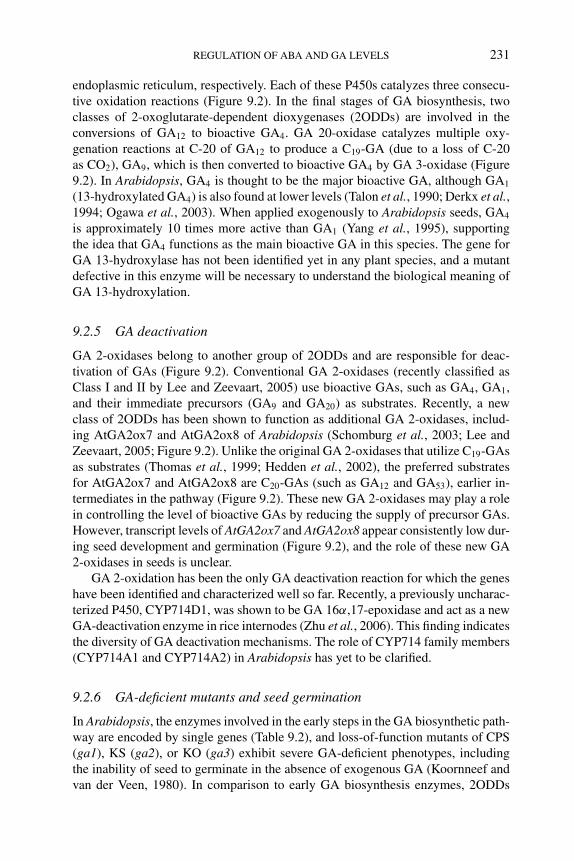
P1: JYS
BLUK053-Bradford September 22, 2006 14:30
REGULATION OF ABA AND GA LEVELS 231
endoplasmic reticulum, respectively. Each of these P450s catalyzes three consecu-tive oxidation reactions (Figure 9.2). In the final stages of GA biosynthesis, twoclasses of 2-oxoglutarate-dependent dioxygenases (2ODDs) are involved in theconversions of GA12 to bioactive GA4. GA 20-oxidase catalyzes multiple oxy-genation reactions at C-20 of GA12 to produce a C19-GA (due to a loss of C-20as CO2), GA9, which is then converted to bioactive GA4 by GA 3-oxidase (Figure9.2). In Arabidopsis, GA4 is thought to be the major bioactive GA, although GA1
(13-hydroxylated GA4) is also found at lower levels (Talon et al., 1990; Derkx et al.,1994; Ogawa et al., 2003). When applied exogenously to Arabidopsis seeds, GA4
is approximately 10 times more active than GA1 (Yang et al., 1995), supportingthe idea that GA4 functions as the main bioactive GA in this species. The gene forGA 13-hydroxylase has not been identified yet in any plant species, and a mutantdefective in this enzyme will be necessary to understand the biological meaning ofGA 13-hydroxylation.
9.2.5 GA deactivation
GA 2-oxidases belong to another group of 2ODDs and are responsible for deac-tivation of GAs (Figure 9.2). Conventional GA 2-oxidases (recently classified asClass I and II by Lee and Zeevaart, 2005) use bioactive GAs, such as GA4, GA1,and their immediate precursors (GA9 and GA20) as substrates. Recently, a newclass of 2ODDs has been shown to function as additional GA 2-oxidases, includ-ing AtGA2ox7 and AtGA2ox8 of Arabidopsis (Schomburg et al., 2003; Lee andZeevaart, 2005; Figure 9.2). Unlike the original GA 2-oxidases that utilize C19-GAsas substrates (Thomas et al., 1999; Hedden et al., 2002), the preferred substratesfor AtGA2ox7 and AtGA2ox8 are C20-GAs (such as GA12 and GA53), earlier in-termediates in the pathway (Figure 9.2). These new GA 2-oxidases may play a rolein controlling the level of bioactive GAs by reducing the supply of precursor GAs.However, transcript levels of AtGA2ox7 and AtGA2ox8 appear consistently low dur-ing seed development and germination (Figure 9.2), and the role of these new GA2-oxidases in seeds is unclear.
GA 2-oxidation has been the only GA deactivation reaction for which the geneshave been identified and characterized well so far. Recently, a previously uncharac-terized P450, CYP714D1, was shown to be GA 16α,17-epoxidase and act as a newGA-deactivation enzyme in rice internodes (Zhu et al., 2006). This finding indicatesthe diversity of GA deactivation mechanisms. The role of CYP714 family members(CYP714A1 and CYP714A2) in Arabidopsis has yet to be clarified.
9.2.6 GA-deficient mutants and seed germination
In Arabidopsis, the enzymes involved in the early steps in the GA biosynthetic path-way are encoded by single genes (Table 9.2), and loss-of-function mutants of CPS(ga1), KS (ga2), or KO (ga3) exhibit severe GA-deficient phenotypes, includingthe inability of seed to germinate in the absence of exogenous GA (Koornneef andvan der Veen, 1980). In comparison to early GA biosynthesis enzymes, 2ODDs
P1: JYS
BLUK053-Bradford September 22, 2006 14:30
232 SEED DEVELOPMENT, DORMANCY AND GERMINATION
that catalyze the late steps in the pathway are encoded by small gene familiesin Arabidopsis (Table 9.2). The relative transcript levels of these genes indicatethat they are differentially regulated during seed development and germination(Figure 9.2). Among four AtGA3ox genes, AtGA3ox1 and AtGA3ox2 are expressedat relatively high levels in seeds imbibed in the light. Both ga3ox1 and ga3ox2 singlemutants germinate normally under continuous white light, but the ga3ox1 ga3ox2double mutant fails to germinate (Mitchum et al., 2006). These results indicate thatAtGA3ox1 and AtGA3ox2 encode major GA 3-oxidases that function redundantlyduring germination in the light. This is supported by the observation that these twoAtGA3ox genes show similar cell type-specific expression patterns in seeds imbibedin the light (Yamaguchi et al., 2001). The roles of AtGA3ox1 and AtGA3ox2 genes inimbibed seeds will be discussed again below in relation to environmental regulationof seed germination.
9.3 Inhibitors of ABA and GA metabolism: efficacy andside effects of drugs
Chemical inhibitors that block the ABA or GA metabolism pathways have beenuseful to elucidate the role of these hormones in seeds and to identify important sig-naling components in hormone-regulated seed germination through genetic screens.We will first describe chemicals that inhibit ABA metabolism enzymes, includingthose recently designed. Then, we will briefly describe the GA biosynthesis in-hibitors that have been commonly used for seed biology research.
Many chemicals are known to block ABA biosynthesis and deactivation in plants.Carotenoid biosynthesis inhibitors are used most frequently in order to reduce en-dogenous ABA levels (Figure 9.3). In addition, inhibitors of AOs, such as tungstate,have been reported to reduce ABA levels (Figure 9.3; Lee and Milborrow, 1997). Onthe other hand, some P450 inhibitors are known to increase ABA levels by inhibit-ing ABA 8′-hydroxylases. Moreover, some ABA analogues have been postulated asspecific inhibitors of ABA deactivation enzymes, such as ABA 8′-hydroxylases orABA-GTs. Cloning of ABA biosynthesis and deactivation enzyme genes allows usto search for better inhibitors or evaluate the modes of action of these chemicals byusing heterologous expression systems.
9.3.1 Drugs to reduce endogenous ABA levels
The herbicides fluridone and norflurazon have been widely used to reduce endoge-nous ABA levels in plants. These chemicals block the activity of phytoene desat-urase in carotenoid biosynthesis (Figure 9.3). Because of their high competenceof penetration and inhibitory effect on the enzyme activity, these chemicals havebeen commonly used in seed biology research. Fluridone is known to alleviateprimary and secondary dormancy in seeds, such as primary dormancy of tobacco(Nicotiana plumbaginifolia) (Grappin et al., 2000), Arabidopsis (Ali-Rachedi et al.,2004), and potato (Solanum tuberosum) seeds (Alvarado and Bradford, 2005), and
P1: JYS
BLUK053-Bradford September 22, 2006 14:30
REGULATION OF ABA AND GA LEVELS 233
Zeaxanthin
Violaxanthin
Neoxanthin
9 -cis-Neoxanthin 9-cis-Violaxanthin
Xanthoxin
Abscisic aldehyde
ABA
ABA-GE Phaseic acid
7´´-Hydroxy-ABA
9 -Hydroxy-ABA
β-Carotene
Lutein
Lycopene
Phytoene GGDP
CYP707A Diniconazole, Uniconazole-PABA analogues
TungstateAO
AbamineNCED
FluridoneNorflurazon
PDS
GTABA analogues
ent-kaurene
ent-kaurenoic acid
GA12
GA9
GA4
GA34
Prohexadione
PaclobutrazolUniconazole-PTetcyclacisAncymidol
AMO-1618Chlormequat
CPS KS
KO
GA 20-oxidase
GA 3-oxidase
GA 2-oxidase
α-Carotene
ζ-Carotene
Figure 9.3 Inhibitors and their target steps in ABA and GA metabolic pathways. Target sites of the
inhibitors (boxed) used for reducing and increasing ABA and GA levels are shown. Abbreviations:
PDS, phytoene desaturase; NCED, 9-cis-epoxycarotenoid dioxygenase; AO, aldehyde oxidase; GT,
glucosyltransferase; CPS, ent-copalyl diphosphate synthase; KS, ent-kaurene synthase; KO, ent-kaurene
oxidase; KAO, ent-kaurenoic acid oxidase.
secondary dormancy induced by supraoptimal temperature in lettuce (Lactucasativa) seeds (Yoshioka et al., 1998) and by osmotic stress in oilseeds (Brassicanapus) (Gulden et al., 2004). Carotenoids are also considered to be precursors ofseveral signal compounds, such as strigolactones (Matusova et al., 2005) and MAX(more axillary)-dependent substance (Sorefan et al., 2003; Booker et al., 2004), anunknown mobile signal produced in roots (Van Norman et al., 2004). Although itremains unknown whether these chemicals are involved in seed physiology, it isrecommended to design experiments using several different inhibitors that blockdifferent steps in the metabolic pathway.
Fluridone application leads to both reduction in ABA levels and bleaching. Be-cause carotenoids play an essential role in protecting against photooxidative damage,
P1: JYS
BLUK053-Bradford September 22, 2006 14:30
234 SEED DEVELOPMENT, DORMANCY AND GERMINATION
fluridone is lethal to green plants in the light. Recently, an ABA biosynthesis in-hibitor that blocks NCED activity was developed (Figure 9.3; Han et al., 2004b).Abamine (ABA biosynthesis inhibitor with an amine moiety) was identified throughcomprehensive analysis of the derivatives of an uncharacterized ABA biosynthesisinhibitor nordihydroguaiaretic acid, which structurally resembles lignostilbene-α,β-dioxygenase (LSD) inhibitors (Han et al., 2004a,b). LSD catalyzes oxidativecleavage of the central double bond of stilbene in a manner similar to the oxidativecleavage by NCEDs. Abamine-treated spinach (Spinacia oleracea), cress (Lepidiumsativum), and Arabidopsis show a reduction in osmotic stress-induced ABA synthe-sis without bleaching (Han et al., 2004b). This chemical might be a useful tool toinvestigate ABA physiology, especially in plant species where genetic analysis isdifficult.
9.3.2 Drugs to increase endogenous ABA levels
P450 inhibitors have been used as plant growth regulators (Rademacher, 2000).These are largely categorized into two types: ones known as plant growth retardantsthat inhibit GA biosynthesis and others that act as fungicides by blocking sterolbiosynthesis. It is also known that application of some P450 inhibitors leads to en-hanced stress tolerance (Fletcher et al., 2000). The inhibitor treatment often causesthe alteration of endogenous ABA levels, although it depends on the plant species,organs, and timing of inhibitor applications. The mechanism of ABA accumulationhas been extensively examined in the case of tetcyclacis, a norbornanodiazetine-typeP450 inhibitor. Radiotracer experiments demonstrated that the conversion of ABAto PA is impaired in tetcyclacis-treated Xanthium strumarium leaves (Zeevaart et al.,1990). Likewise, tetcyclacis and a triazole BAS111.W appear to block ABA deac-tivation in suspension cultures and seedlings of oilseed (Brassica napus) (Hauseret al., 1990). Tetcyclacis is shown to inhibit ABA 8′-hydroxylase activity of mi-crosomal fractions from maize (Zea mays) suspension cells (Krochko et al., 1998)or from yeast expressing Arabidopsis CYP707A1 (Kushiro et al., 2004). Recently,comprehensive analysis was conducted on known P450 inhibitors to investigate theirinhibitory effects on ABA deactivation (Kitahata et al., 2005). A fungicide dinicona-zole was found to inhibit ABA 8′-hydroxylase more effectively than the inhibitorsknown as GA biosynthesis inhibitors, such as uniconazole, PAC, and tetcyclacis(Figure 9.3). Interestingly, the S-form is more effective as an ABA 8′-hydroxylaseinhibitor than the R-form, which is a more potent fungicide. This indicates that theS-form of diniconazole is a useful tool to increase endogenous ABA levels.
Structural analogues of ABA have been synthesized and studied for theirstructure–activity relationships (Todoroki and Hirai, 2002; Zaharia et al., 2005).These analogues include those exhibiting reduced or increased ABA-like activityand persistent analogues that are resistant to ABA deactivation. Cutler et al. (2000)demonstrated that ABA analogues modified at positions C-8′ or C-9′ inhibit ABA8′-hydroxylase activity in microsomal fractions of maize suspension cells (Cutleret al., 2000). Among them, (+)-8′-acetylene-ABA and (+)-9′-propargyl-ABA wereshown to act as suicide substrates and inhibit Arabidopsis seed germination more
P1: JYS
BLUK053-Bradford September 22, 2006 14:30
REGULATION OF ABA AND GA LEVELS 235
effectively than natural ABA. This suggests that these analogues contain both in-hibitory activity toward ABA 8′-hydroxylase and ABA-like biological activity. Uenoet al. (2005) conducted a comprehensive analysis of ABA analogues concerning theirinhibitory effects on both ABA 8′-hydroxylase and hormone activities (Ueno et al.,2005). Elimination of the 6-methyl and 1′-hydroxy groups had little effect on com-petitive inhibition of ABA 8′-hydroxylase, but abolished ABA-like biological activ-ity. The authors suggested that it is possible to design specific ABA 8′-hydroxylaseinhibitors that do not have ABA-like activity. On the other hand, Priest et al. (2005)reported that recognition of ABA by UGT71B6, an Arabidopsis GT, does not re-quire the ketone at C-4 and is unaffected by addition of bulky substitution groupsat the double bond of the ABA ring (Priest et al., 2005). These designed inhibitorswill be powerful tools in the future research on ABA physiology in various plantspecies.
9.3.3 Drugs to reduce GA levels
As mentioned above, three different classes of enzymes are involved in the con-version of GGDP to bioactive GAs (Figure 9.2). Chemical inhibitors are known foreach enzyme class in the GA biosynthesis pathway (Figure 9.3; Rademacher, 2000).The most commonly used GA biosynthesis inhibitors for seed biology are PAC anduniconazole-P, both of which inhibit KO/CYP701A (GA3) and belong to a groupof triazole-containing heterocycles. These chemicals also exhibit an inhibitory ef-fect on ABA deactivation enzymes and reduce brassinosteroid levels (Rademacher,2000). Therefore, it is important to evaluate the specificity as a GA biosynthesisinhibitor by including a control sample to test whether the effect of the chemical isreversed by simultaneous application of exogenous GA. Prohexadione, a structuralmimic of 2-oxoglutarate, also inhibits Arabidopsis seed germination (Yang et al.,1995). The inhibitory effect of prohexadione on Arabidopsis seed germination isefficiently restored by exogenous GA4, but not by GA9, consistent with the idea thatGA 3-oxidase is a target of this inhibitor (Figure 9.3).
9.3.4 Side effects of drugs
Inhibitors of ABA/GA biosynthesis and deactivation are powerful tools to investi-gate physiological roles of these hormones in various plant species in which geneticapproaches cannot be easily employed. However, we should also consider that manyso-called specific inhibitors are not necessarily specific. In addition, the effects andside effects of inhibitors vary when applied by different methods, to different or-gans or at different concentrations, even in the same plant species. Debeaujon andKoornneef (2000) reported an example of inconsistent results from similar inhibitors.The testa (seed coat) plays an important role in maintaining seed dormancy by pro-tecting embryos both physically and chemically. Thus, Arabidopsis mutants havingdefective testae often show reduced seed dormancy and increased ability to takeup chemicals from media (Debeaujon et al., 2000). PAC inhibits germination ofall testa mutants more effectively than it inhibits wild-type seeds, but only some of
P1: JYS
BLUK053-Bradford September 22, 2006 14:30
236 SEED DEVELOPMENT, DORMANCY AND GERMINATION
the testa mutants show hypersensitivity to tetcyclacis. This suggests that wild-typeseed coats inhibit uptake of such inhibitors. In addition, different effects of PAC andtetcyclacis may be attributed to differences in inhibitory effects on GA biosynthesisor ABA deactivation. Nevertheless, our research is often dependent on such drugs,even though special caution is needed to design such experiments.
9.4 Regulation of ABA and GA levels in Arabidopsis seeds
9.4.1 Regulation of ABA and GA levels during seed development
Recent molecular genetic analyses have revealed that the actions of ABA and GAsare modulated directly by developmental regulators that define the ‘space’ or ‘tim-ing’ of embryo development. These regulators act as ‘balancers’ of the metabolismof and sensitivities to these two hormones, which, at least in part, explains theantagonistic actions of ABA and GAs.
9.4.1.1 Roles of ABA and GA during seed developmentBoth ABA and GA are necessary for embryo growth during seed development. Over-expression of a GA deactivation enzyme gene (pea GA 2-oxidase2) in Arabidopsisincreases the proportion of seed abortion (Singh et al., 2002). Severe ABA-deficientaba2 mutants also show an increase in seed abortion compared to the wild type(Cheng et al., 2002). In addition to their roles in embryo growth, ABA and GAantagonistically regulate the degree of seed dormancy during seed development.Precocious germination of Arabidopsis lec (leafy cotyledon) and fus (fusca) mutantsis suppressed by a GA-deficient mutation, supporting the idea that GA biosynthesisis regulated negatively during seed development to prevent precocious germina-tion (Raz et al., 2001). In contrast, ABA accumulation during seed development isimportant to induce seed maturation processes and impose seed dormancy. Bipha-sic ABA accumulation has been observed during seed development (Figure 9.4;Karssen et al., 1983). The first peak of ABA is derived from both zygotic and ma-ternal tissues, while the second peak is derived only from the zygotic tissues. Thiszygotic-derived ABA is thought to be essential for the induction and maintenance ofseed dormancy. In contrast, the maternal-derived ABA is involved in the inhibitionof precocious germination and processes of seed maturation (Koornneef et al., 1989;Raz et al., 2001).
9.4.1.2 FUS3, a balancer of ABA and GA levelsThe levels of ABA and bioactive GAs are thought to be correlated negatively dur-ing seed development (Jacobsen and Chandler, 1987; Batge et al., 1999; Whiteet al., 2000). Recent reports have demonstrated that ABA and GA levels are regu-lated oppositely by the same transcription factors during Arabidopsis seed develop-ment. FUS3 and LEC2 encode B3 transcription factors (Luerßen et al., 1998; Stoneet al., 2001). Loss of function of these genes causes severe defects in seed matu-ration processes, such as reduced reserve accumulation and desiccation intolerance
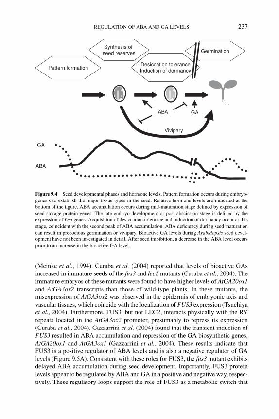
P1: JYS
BLUK053-Bradford September 22, 2006 14:30
REGULATION OF ABA AND GA LEVELS 237
Synthesis of seed reserves
Pattern formationDesiccation toleranceInduction of dormancy
Germination
ABA GA
GA
ABA
Vivipary
Figure 9.4 Seed developmental phases and hormone levels. Pattern formation occurs during embryo-
genesis to establish the major tissue types in the seed. Relative hormone levels are indicated at the
bottom of the figure. ABA accumulation occurs during mid-maturation stage defined by expression of
seed storage protein genes. The late embryo development or post-abscission stage is defined by the
expression of Lea genes. Acquisition of desiccation tolerance and induction of dormancy occur at this
stage, coincident with the second peak of ABA accumulation. ABA deficiency during seed maturation
can result in precocious germination or vivipary. Bioactive GA levels during Arabidopsis seed devel-
opment have not been investigated in detail. After seed imbibition, a decrease in the ABA level occurs
prior to an increase in the bioactive GA level.
(Meinke et al., 1994). Curaba et al. (2004) reported that levels of bioactive GAsincreased in immature seeds of the fus3 and lec2 mutants (Curaba et al., 2004). Theimmature embryos of these mutants were found to have higher levels of AtGA20ox1and AtGA3ox2 transcripts than those of wild-type plants. In these mutants, themisexpression of AtGA3ox2 was observed in the epidermis of embryonic axis andvascular tissues, which coincide with the localization of FUS3 expression (Tsuchiyaet al., 2004). Furthermore, FUS3, but not LEC2, interacts physically with the RYrepeats located in the AtGA3ox2 promoter, presumably to repress its expression(Curaba et al., 2004). Gazzarrini et al. (2004) found that the transient induction ofFUS3 resulted in ABA accumulation and repression of the GA biosynthetic genes,AtGA20ox1 and AtGA3ox1 (Gazzarrini et al., 2004). These results indicate thatFUS3 is a positive regulator of ABA levels and is also a negative regulator of GAlevels (Figure 9.5A). Consistent with these roles for FUS3, the fus3 mutant exhibitsdelayed ABA accumulation during seed development. Importantly, FUS3 proteinlevels appear to be regulated by ABA and GA in a positive and negative way, respec-tively. These regulatory loops support the role of FUS3 as a metabolic switch that
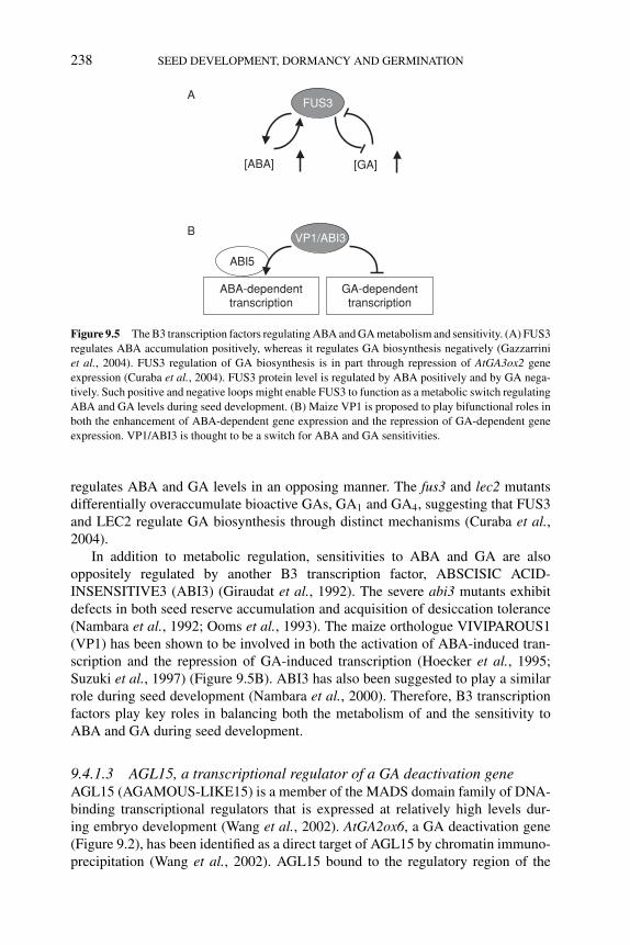
P1: JYS
BLUK053-Bradford September 22, 2006 14:30
238 SEED DEVELOPMENT, DORMANCY AND GERMINATION
A
B
FUS3
[ABA] [GA]
VP1/ABI3
ABA-dependenttranscription
GA-dependenttranscription
ABI5
Figure 9.5 The B3 transcription factors regulating ABA and GA metabolism and sensitivity. (A) FUS3
regulates ABA accumulation positively, whereas it regulates GA biosynthesis negatively (Gazzarrini
et al., 2004). FUS3 regulation of GA biosynthesis is in part through repression of AtGA3ox2 gene
expression (Curaba et al., 2004). FUS3 protein level is regulated by ABA positively and by GA nega-
tively. Such positive and negative loops might enable FUS3 to function as a metabolic switch regulating
ABA and GA levels during seed development. (B) Maize VP1 is proposed to play bifunctional roles in
both the enhancement of ABA-dependent gene expression and the repression of GA-dependent gene
expression. VP1/ABI3 is thought to be a switch for ABA and GA sensitivities.
regulates ABA and GA levels in an opposing manner. The fus3 and lec2 mutantsdifferentially overaccumulate bioactive GAs, GA1 and GA4, suggesting that FUS3and LEC2 regulate GA biosynthesis through distinct mechanisms (Curaba et al.,2004).
In addition to metabolic regulation, sensitivities to ABA and GA are alsooppositely regulated by another B3 transcription factor, ABSCISIC ACID-INSENSITIVE3 (ABI3) (Giraudat et al., 1992). The severe abi3 mutants exhibitdefects in both seed reserve accumulation and acquisition of desiccation tolerance(Nambara et al., 1992; Ooms et al., 1993). The maize orthologue VIVIPAROUS1(VP1) has been shown to be involved in both the activation of ABA-induced tran-scription and the repression of GA-induced transcription (Hoecker et al., 1995;Suzuki et al., 1997) (Figure 9.5B). ABI3 has also been suggested to play a similarrole during seed development (Nambara et al., 2000). Therefore, B3 transcriptionfactors play key roles in balancing both the metabolism of and the sensitivity toABA and GA during seed development.
9.4.1.3 AGL15, a transcriptional regulator of a GA deactivation geneAGL15 (AGAMOUS-LIKE15) is a member of the MADS domain family of DNA-binding transcriptional regulators that is expressed at relatively high levels dur-ing embryo development (Wang et al., 2002). AtGA2ox6, a GA deactivation gene(Figure 9.2), has been identified as a direct target of AGL15 by chromatin immuno-precipitation (Wang et al., 2002). AGL15 bound to the regulatory region of the
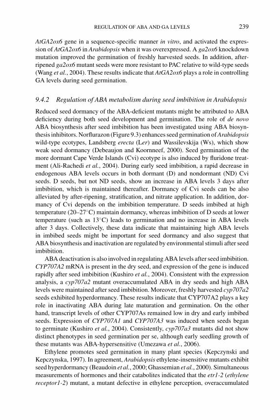
P1: JYS
BLUK053-Bradford September 22, 2006 14:30
REGULATION OF ABA AND GA LEVELS 239
AtGA2ox6 gene in a sequence-specific manner in vitro, and activated the expres-sion of AtGA2ox6 in Arabidopsis when it was overexpressed. A ga2ox6 knockdownmutation improved the germination of freshly harvested seeds. In addition, after-ripened ga2ox6 mutant seeds were more resistant to PAC relative to wild-type seeds(Wang et al., 2004). These results indicate that AtGA2ox6 plays a role in controllingGA levels during seed germination.
9.4.2 Regulation of ABA metabolism during seed imbibition in Arabidopsis
Reduced seed dormancy of the ABA-deficient mutants might be attributed to ABAdeficiency during both seed development and germination. The role of de novoABA biosynthesis after seed imbibition has been investigated using ABA biosyn-thesis inhibitors. Norflurazon (Figure 9.3) enhances seed germination of Arabidopsiswild-type ecotypes, Landsberg erecta (Ler) and Wassilevskija (Ws), which showweak seed dormancy (Debeaujon and Koornneef, 2000). Seed germination of themore dormant Cape Verde Islands (Cvi) ecotype is also induced by fluridone treat-ment (Ali-Rachedi et al., 2004). During early seed imbibition, a rapid decrease inendogenous ABA levels occurs in both dormant (D) and nondormant (ND) Cviseeds. D seeds, but not ND seeds, show an increase in ABA levels 3 days afterimbibition, which is maintained thereafter. Dormancy of Cvi seeds can be alsoalleviated by after-ripening, stratification, and nitrate application. In addition, dor-mancy of Cvi depends on the imbibition temperature. D seeds imbibed at hightemperature (20–27◦C) maintain dormancy, whereas imbibition of D seeds at lowertemperature (such as 13◦C) leads to germination and no increase in ABA levelsafter 3 days. Collectively, these data indicate that maintaining high ABA levelsin imbibed seeds might be important for seed dormancy and also suggest thatABA biosynthesis and inactivation are regulated by environmental stimuli after seedimbibition.
ABA deactivation is also involved in regulating ABA levels after seed imbibition.CYP707A2 mRNA is present in the dry seed, and expression of the gene is inducedrapidly after seed imbibition (Kushiro et al., 2004). Consistent with the expressionanalysis, a cyp707a2 mutant overaccumulated ABA in dry seeds and high ABAlevels were maintained after seed imbibition. Moreover, freshly harvested cyp707a2seeds exhibited hyperdormancy. These results indicate that CYP707A2 plays a keyrole in inactivating ABA during late maturation and germination. On the otherhand, transcript levels of other CYP707As remained low in dry and early imbibedseeds. Expression of CYP707A1 and CYP707A3 was induced when seeds beganto germinate (Kushiro et al., 2004). Consistently, cyp707a3 mutants did not showdistinct phenotypes in seed germination per se, although early seedling growth ofthese mutants was ABA-hypersensitive (Umezawa et al., 2006).
Ethylene promotes seed germination in many plant species (Kepczynski andKepczynska, 1997). In agreement, Arabidopsis ethylene-insensitive mutants exhibitseed hyperdormancy (Beaudoin et al., 2000; Ghassemian et al., 2000). Simultaneousmeasurements of hormones and their catabolites indicated that the etr1-2 (ethylenereceptor1-2) mutant, a mutant defective in ethylene perception, overaccumulated
P1: JYS
BLUK053-Bradford September 22, 2006 14:30
240 SEED DEVELOPMENT, DORMANCY AND GERMINATION
ABA in dry seeds (Chiwocha et al., 2005; see Chapter 8). ABA levels of etr1-2imbibed seeds decreased immediately after seed imbibition, as occurred in the wild-type seeds, but were then maintained at higher level than in wild type. In contrast,ABA levels in the etr1-2 imbibed seeds were equivalent to the wild type when seedswere stratified. This stratification-dependent change in ABA levels is consistent withgermination phenotypes of the etr1-2 seed, in which seed hyperdormancy is canceledby the stratification treatment. Moreover, stratified wild-type seeds accumulated asignificant amount of ABA-GE, suggesting that the ABA glucosylation pathway isinvolved in regulating ABA levels during stratification. Recent identification of theputative ABA GT genes might facilitate elucidation of the physiological roles ofeach ABA inactivation pathway.
9.4.3 Regulation of GA metabolism during seed imbibition in Arabidopsis
GA biosynthesis in imbibed Arabidopsis seeds is strictly regulated by environmentalconditions. When after-ripened Arabidopsis seeds were imbibed under continuouswhite light at optimal temperature (22◦C), the level of bioactive GA4 increasedjust prior to radicle emergence (Ogawa et al., 2003). This increase in GA4 contentappears to be achieved through the coordinated regulation of GA biosynthesis anddeactivation genes; several GA biosynthesis genes, such as AtGA20ox3, AtGA3ox1,and AtGA3ox2, are upregulated upon imbibition under continuous light, whereasexpression of all known GA 2-oxidase genes remains at low levels (Figure 9.2;Ogawa et al., 2003). In the following sections, we will briefly describe how GAbiosynthesis and deactivation pathways are regulated by light and temperature inimbibed Arabidopsis seeds.
9.4.3.1 Regulation of GA biosynthesis by lightPhytochromes play an important role in light-dependent germination of Arabidop-sis seeds (Shinomura, 1997). Among five phytochromes, PHYB is the major familymember that is stored in dry seeds and is responsible for the typical photoreversibleresponse to red (R) and far-red (FR) light shortly after the onset of imbibition(Shinomura et al., 1994, 1996). When imbibed seeds are irradiated with an FR-lightpulse to inactivate phytochrome and incubated in the dark, expression of AtGA3ox1and AtGA3ox2 remains at low levels. In such dark-imbibed seeds, transcript levelsof AtGA3ox1 and AtGA3ox2 are highly elevated by an R pulse, and the effect of Ris reversed by subsequent exposure to FR (Yamaguchi et al., 1998). These resultsindicate that AtGA3ox1 and AtGA3ox2 genes are regulated by phytochrome. In thephyB mutant, AtGA3ox2 expression is not increased by R, indicating the primaryrole of PHYB in mediating the induction of this gene (Yamaguchi et al., 1998).Expression of all AtGA2ox genes remained at low levels in imbibed seeds undercontinuous white light (Figure 9.2; Ogawa et al., 2003). AtGA2ox2 transcript accu-mulated at high levels during imbibition in the dark after an FR pulse (Yamauchiet al., 2004). These results suggest that GA deactivation also is modulated by lightthrough regulation of the AtGA2ox2 gene.
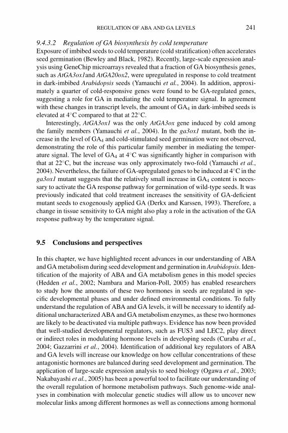
P1: JYS
BLUK053-Bradford September 22, 2006 14:30
REGULATION OF ABA AND GA LEVELS 241
9.4.3.2 Regulation of GA biosynthesis by cold temperatureExposure of imbibed seeds to cold temperature (cold stratification) often acceleratesseed germination (Bewley and Black, 1982). Recently, large-scale expression anal-ysis using GeneChip microarrays revealed that a fraction of GA biosynthesis genes,such as AtGA3ox1and AtGA20ox2, were upregulated in response to cold treatmentin dark-imbibed Arabidopsis seeds (Yamauchi et al., 2004). In addition, approxi-mately a quarter of cold-responsive genes were found to be GA-regulated genes,suggesting a role for GA in mediating the cold temperature signal. In agreementwith these changes in transcript levels, the amount of GA4 in dark-imbibed seeds iselevated at 4◦C compared to that at 22◦C.
Interestingly, AtGA3ox1 was the only AtGA3ox gene induced by cold amongthe family members (Yamauchi et al., 2004). In the ga3ox1 mutant, both the in-crease in the level of GA4 and cold-stimulated seed germination were not observed,demonstrating the role of this particular family member in mediating the temper-ature signal. The level of GA4 at 4◦C was significantly higher in comparison withthat at 22◦C, but the increase was only approximately two-fold (Yamauchi et al.,2004). Nevertheless, the failure of GA-upregulated genes to be induced at 4◦C in thega3ox1 mutant suggests that the relatively small increase in GA4 content is neces-sary to activate the GA response pathway for germination of wild-type seeds. It waspreviously indicated that cold treatment increases the sensitivity of GA-deficientmutant seeds to exogenously applied GA (Derkx and Karssen, 1993). Therefore, achange in tissue sensitivity to GA might also play a role in the activation of the GAresponse pathway by the temperature signal.
9.5 Conclusions and perspectives
In this chapter, we have highlighted recent advances in our understanding of ABAand GA metabolism during seed development and germination in Arabidopsis. Iden-tification of the majority of ABA and GA metabolism genes in this model species(Hedden et al., 2002; Nambara and Marion-Poll, 2005) has enabled researchersto study how the amounts of these two hormones in seeds are regulated in spe-cific developmental phases and under defined environmental conditions. To fullyunderstand the regulation of ABA and GA levels, it will be necessary to identify ad-ditional uncharacterized ABA and GA metabolism enzymes, as these two hormonesare likely to be deactivated via multiple pathways. Evidence has now been providedthat well-studied developmental regulators, such as FUS3 and LEC2, play director indirect roles in modulating hormone levels in developing seeds (Curaba et al.,2004; Gazzarrini et al., 2004). Identification of additional key regulators of ABAand GA levels will increase our knowledge on how cellular concentrations of theseantagonistic hormones are balanced during seed development and germination. Theapplication of large-scale expression analysis to seed biology (Ogawa et al., 2003;Nakabayashi et al., 2005) has been a powerful tool to facilitate our understanding ofthe overall regulation of hormone metabolism pathways. Such genome-wide anal-yses in combination with molecular genetic studies will allow us to uncover newmolecular links among different hormones as well as connections among hormonal
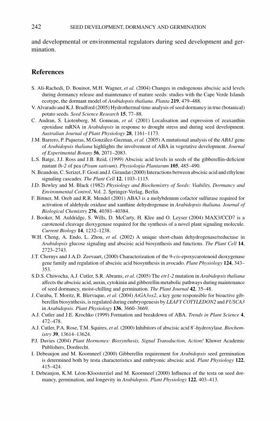
P1: JYS
BLUK053-Bradford September 22, 2006 14:30
242 SEED DEVELOPMENT, DORMANCY AND GERMINATION
and developmental or environmental regulators during seed development and ger-mination.
References
S. Ali-Rachedi, D. Bouinot, M.H. Wagner, et al. (2004) Changes in endogenous abscisic acid levels
during dormancy release and maintenance of mature seeds: studies with the Cape Verde Islands
ecotype, the dormant model of Arabidopsis thaliana. Planta 219, 479–488.
V. Alvarado and K.J. Bradford (2005) Hydrothermal time analysis of seed dormancy in true (botanical)
potato seeds. Seed Science Research 15, 77–88.
C. Audran, S. Liotenberg, M. Gonneau, et al. (2001) Localisation and expression of zeaxanthin
epoxidase mRNA in Arabidopsis in response to drought stress and during seed development.
Australian Journal of Plant Physiology 28, 1161–1173.
J.M. Barrero, P. Piqueras, M.Gonzalez-Guzman, et al. (2005) A mutational analysis of the ABA1 gene
of Arabidopsis thaliana highlights the involvement of ABA in vegetative development. Journalof Experimental Botany 56, 2071–2083.
L.S. Batge, J.J. Ross and J.B. Reid, (1999) Abscisic acid levels in seeds of the gibberellin-deficient
mutant lh-2 of pea (Pisum sativum). Physiologia Plantarum 105, 485–490.
N. Beaudoin, C. Serizet, F. Gosti and J. Giraudat (2000) Interactions between abscisic acid and ethylene
signaling cascades. The Plant Cell 12, 1103–1115.
J.D. Bewley and M. Black (1982) Physiology and Biochemistry of Seeds: Viability, Dormancy andEnvironmental Control, Vol. 2. Springer-Verlag, Berlin.
F. Bittner, M. Oreb and R.R. Mendel (2001) ABA3 is a molybdenum cofactor sulfurase required for
activation of aldehyde oxidase and xanthine dehydrogenase in Arabidopsis thaliana. Journal ofBiological Chemistry 276, 40381–40384.
J. Booker, M. Auldridge, S. Wills, D. McCarty, H. Klee and O. Leyser (2004) MAX3/CCD7 is a
carotenoid cleavage dioxygenase required for the synthesis of a novel plant signaling molecule.
Current Biology 14, 1232–1238.
W.H. Cheng, A. Endo, L. Zhou, et al. (2002) A unique short-chain dehydrogenase/reductase in
Arabidopsis glucose signaling and abscisic acid biosynthesis and functions. The Plant Cell 14,
2723–2743.
J.T. Chernys and J.A.D. Zeevaart, (2000) Characterization of the 9-cis-epoxycarotenoid dioxygenase
gene family and regulation of abscisic acid biosynthesis in avocado. Plant Physiology 124, 343–
353.
S.D.S. Chiwocha, A.J. Cutler, S.R. Abrams, et al. (2005) The etr1-2 mutation in Arabidopsis thalianaaffects the abscisic acid, auxin, cytokinin and gibberellin metabolic pathways during maintenance
of seed dormancy, moist-chilling and germination. The Plant Journal 42, 35–48.
J. Curaba, T. Moritz, R. Blervaque, et al. (2004) AtGA3ox2, a key gene responsible for bioactive gib-
berellin biosynthesis, is regulated during embryogenesis by LEAFY COTYLEDON2 and FUSCA3in Arabidopsis. Plant Physiology 136, 3660–3669.
A.J. Cutler and J.E. Krochko (1999) Formation and breakdown of ABA. Trends in Plant Science 4,
472–478.
A.J. Cutler, P.A. Rose, T.M. Squires, et al. (2000) Inhibitors of abscisic acid 8′-hydroxylase. Biochem-istry 39, 13614–13624.
P.J. Davies (2004) Plant Hormones: Biosynthesis, Signal Transduction, Action! Kluwer Academic
Publishers, Dordrecht.
I. Debeaujon and M. Koornneef (2000) Gibberellin requirement for Arabidopsis seed germination
is determined both by testa characteristics and embryonic abscisic acid. Plant Physiology 122,
415–424.
I. Debeaujon, K.M. Leon-Kloosterziel and M. Koornneef (2000) Influence of the testa on seed dor-
mancy, germination, and longevity in Arabidopsis. Plant Physiology 122, 403–413.
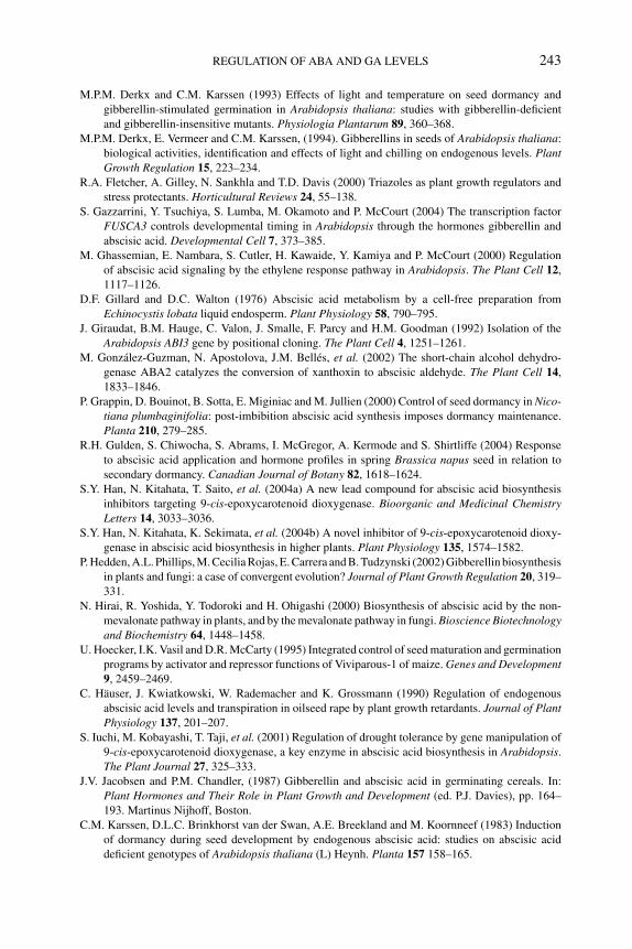
P1: JYS
BLUK053-Bradford September 22, 2006 14:30
REGULATION OF ABA AND GA LEVELS 243
M.P.M. Derkx and C.M. Karssen (1993) Effects of light and temperature on seed dormancy and
gibberellin-stimulated germination in Arabidopsis thaliana: studies with gibberellin-deficient
and gibberellin-insensitive mutants. Physiologia Plantarum 89, 360–368.
M.P.M. Derkx, E. Vermeer and C.M. Karssen, (1994). Gibberellins in seeds of Arabidopsis thaliana:
biological activities, identification and effects of light and chilling on endogenous levels. PlantGrowth Regulation 15, 223–234.
R.A. Fletcher, A. Gilley, N. Sankhla and T.D. Davis (2000) Triazoles as plant growth regulators and
stress protectants. Horticultural Reviews 24, 55–138.
S. Gazzarrini, Y. Tsuchiya, S. Lumba, M. Okamoto and P. McCourt (2004) The transcription factor
FUSCA3 controls developmental timing in Arabidopsis through the hormones gibberellin and
abscisic acid. Developmental Cell 7, 373–385.
M. Ghassemian, E. Nambara, S. Cutler, H. Kawaide, Y. Kamiya and P. McCourt (2000) Regulation
of abscisic acid signaling by the ethylene response pathway in Arabidopsis. The Plant Cell 12,
1117–1126.
D.F. Gillard and D.C. Walton (1976) Abscisic acid metabolism by a cell-free preparation from
Echinocystis lobata liquid endosperm. Plant Physiology 58, 790–795.
J. Giraudat, B.M. Hauge, C. Valon, J. Smalle, F. Parcy and H.M. Goodman (1992) Isolation of the
Arabidopsis ABI3 gene by positional cloning. The Plant Cell 4, 1251–1261.
M. Gonzalez-Guzman, N. Apostolova, J.M. Belles, et al. (2002) The short-chain alcohol dehydro-
genase ABA2 catalyzes the conversion of xanthoxin to abscisic aldehyde. The Plant Cell 14,
1833–1846.
P. Grappin, D. Bouinot, B. Sotta, E. Miginiac and M. Jullien (2000) Control of seed dormancy in Nico-tiana plumbaginifolia: post-imbibition abscisic acid synthesis imposes dormancy maintenance.
Planta 210, 279–285.
R.H. Gulden, S. Chiwocha, S. Abrams, I. McGregor, A. Kermode and S. Shirtliffe (2004) Response
to abscisic acid application and hormone profiles in spring Brassica napus seed in relation to
secondary dormancy. Canadian Journal of Botany 82, 1618–1624.
S.Y. Han, N. Kitahata, T. Saito, et al. (2004a) A new lead compound for abscisic acid biosynthesis
inhibitors targeting 9-cis-epoxycarotenoid dioxygenase. Bioorganic and Medicinal ChemistryLetters 14, 3033–3036.
S.Y. Han, N. Kitahata, K. Sekimata, et al. (2004b) A novel inhibitor of 9-cis-epoxycarotenoid dioxy-
genase in abscisic acid biosynthesis in higher plants. Plant Physiology 135, 1574–1582.
P. Hedden, A.L. Phillips, M. Cecilia Rojas, E. Carrera and B. Tudzynski (2002) Gibberellin biosynthesis
in plants and fungi: a case of convergent evolution? Journal of Plant Growth Regulation 20, 319–
331.
N. Hirai, R. Yoshida, Y. Todoroki and H. Ohigashi (2000) Biosynthesis of abscisic acid by the non-
mevalonate pathway in plants, and by the mevalonate pathway in fungi. Bioscience Biotechnologyand Biochemistry 64, 1448–1458.
U. Hoecker, I.K. Vasil and D.R. McCarty (1995) Integrated control of seed maturation and germination
programs by activator and repressor functions of Viviparous-1 of maize. Genes and Development9, 2459–2469.
C. Hauser, J. Kwiatkowski, W. Rademacher and K. Grossmann (1990) Regulation of endogenous
abscisic acid levels and transpiration in oilseed rape by plant growth retardants. Journal of PlantPhysiology 137, 201–207.
S. Iuchi, M. Kobayashi, T. Taji, et al. (2001) Regulation of drought tolerance by gene manipulation of
9-cis-epoxycarotenoid dioxygenase, a key enzyme in abscisic acid biosynthesis in Arabidopsis.
The Plant Journal 27, 325–333.
J.V. Jacobsen and P.M. Chandler, (1987) Gibberellin and abscisic acid in germinating cereals. In:
Plant Hormones and Their Role in Plant Growth and Development (ed. P.J. Davies), pp. 164–
193. Martinus Nijhoff, Boston.
C.M. Karssen, D.L.C. Brinkhorst van der Swan, A.E. Breekland and M. Koornneef (1983) Induction
of dormancy during seed development by endogenous abscisic acid: studies on abscisic acid
deficient genotypes of Arabidopsis thaliana (L) Heynh. Planta 157 158–165.
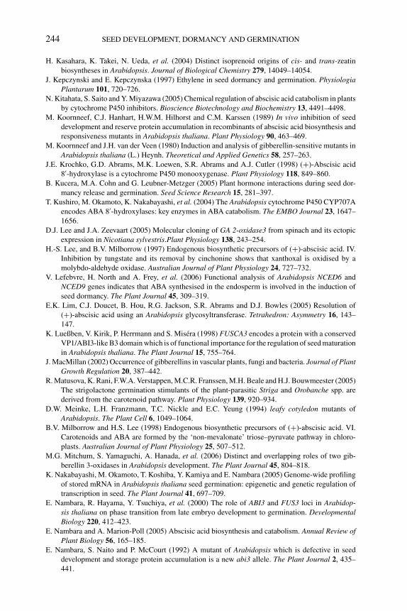
P1: JYS
BLUK053-Bradford September 22, 2006 14:30
244 SEED DEVELOPMENT, DORMANCY AND GERMINATION
H. Kasahara, K. Takei, N. Ueda, et al. (2004) Distinct isoprenoid origins of cis- and trans-zeatin
biosyntheses in Arabidopsis. Journal of Biological Chemistry 279, 14049–14054.
J. Kepczynski and E. Kepczynska (1997) Ethylene in seed dormancy and germination. PhysiologiaPlantarum 101, 720–726.
N. Kitahata, S. Saito and Y. Miyazawa (2005) Chemical regulation of abscisic acid catabolism in plants
by cytochrome P450 inhibitors. Bioscience Biotechnology and Biochemistry 13, 4491–4498.
M. Koornneef, C.J. Hanhart, H.W.M. Hilhorst and C.M. Karssen (1989) In vivo inhibition of seed
development and reserve protein accumulation in recombinants of abscisic acid biosynthesis and
responsiveness mutants in Arabidopsis thaliana. Plant Physiology 90, 463–469.
M. Koornneef and J.H. van der Veen (1980) Induction and analysis of gibberellin-sensitive mutants in
Arabidopsis thaliana (L.) Heynh. Theoretical and Applied Genetics 58, 257–263.
J.E. Krochko, G.D. Abrams, M.K. Loewen, S.R. Abrams and A.J. Cutler (1998) (+)-Abscisic acid
8′-hydroxylase is a cytochrome P450 monooxygenase. Plant Physiology 118, 849–860.
B. Kucera, M.A. Cohn and G. Leubner-Metzger (2005) Plant hormone interactions during seed dor-
mancy release and germination. Seed Science Research 15, 281–397.
T. Kushiro, M. Okamoto, K. Nakabayashi, et al. (2004) The Arabidopsis cytochrome P450 CYP707A
encodes ABA 8′-hydroxylases: key enzymes in ABA catabolism. The EMBO Journal 23, 1647–
1656.
D.J. Lee and J.A. Zeevaart (2005) Molecular cloning of GA 2-oxidase3 from spinach and its ectopic
expression in Nicotiana sylvestris.Plant Physiology 138, 243–254.
H.-S. Lee, and B.V. Milborrow (1997) Endogenous biosynthetic precursors of (+)-abscisic acid. IV.
Inhibition by tungstate and its removal by cinchonine shows that xanthoxal is oxidised by a
molybdo-aldehyde oxidase. Australian Journal of Plant Physiology 24, 727–732.
V. Lefebvre, H. North and A. Frey, et al. (2006) Functional analysis of Arabidopsis NCED6 and
NCED9 genes indicates that ABA synthesised in the endosperm is involved in the induction of
seed dormancy. The Plant Journal 45, 309–319.
E.K. Lim, C.J. Doucet, B. Hou, R.G. Jackson, S.R. Abrams and D.J. Bowles (2005) Resolution of
(+)-abscisic acid using an Arabidopsis glycosyltransferase. Tetrahedron: Asymmetry 16, 143–
147.
K. Lueßben, V. Kirik, P. Herrmann and S. Misera (1998) FUSCA3 encodes a protein with a conserved
VP1/ABI3-like B3 domain which is of functional importance for the regulation of seed maturation
in Arabidopsis thaliana. The Plant Journal 15, 755–764.
J. MacMillan (2002) Occurrence of gibberellins in vascular plants, fungi and bacteria. Journal of PlantGrowth Regulation 20, 387–442.
R. Matusova, K. Rani, F.W.A. Verstappen, M.C.R. Franssen, M.H. Beale and H.J. Bouwmeester (2005)
The strigolactone germination stimulants of the plant-parasitic Striga and Orobanche spp. are
derived from the carotenoid pathway. Plant Physiology 139, 920–934.
D.W. Meinke, L.H. Franzmann, T.C. Nickle and E.C. Yeung (1994) leafy cotyledon mutants of
Arabidopsis. The Plant Cell 6, 1049–1064.
B.V. Milborrow and H.S. Lee (1998) Endogenous biosynthetic precursors of (+)-abscisic acid. VI.
Carotenoids and ABA are formed by the ‘non-mevalonate’ triose–pyruvate pathway in chloro-
plasts. Australian Journal of Plant Physiology 25, 507–512.
M.G. Mitchum, S. Yamaguchi, A. Hanada, et al. (2006) Distinct and overlapping roles of two gib-
berellin 3-oxidases in Arabidopsis development. The Plant Journal 45, 804–818.
K. Nakabayashi, M. Okamoto, T. Koshiba, Y. Kamiya and E. Nambara (2005) Genome-wide profiling
of stored mRNA in Arabidopsis thaliana seed germination: epigenetic and genetic regulation of
transcription in seed. The Plant Journal 41, 697–709.
E. Nambara, R. Hayama, Y. Tsuchiya, et al. (2000) The role of ABI3 and FUS3 loci in Arabidop-sis thaliana on phase transition from late embryo development to germination. DevelopmentalBiology 220, 412–423.
E. Nambara and A. Marion-Poll (2005) Abscisic acid biosynthesis and catabolism. Annual Review ofPlant Biology 56, 165–185.
E. Nambara, S. Naito and P. McCourt (1992) A mutant of Arabidopsis which is defective in seed
development and storage protein accumulation is a new abi3 allele. The Plant Journal 2, 435–
441.
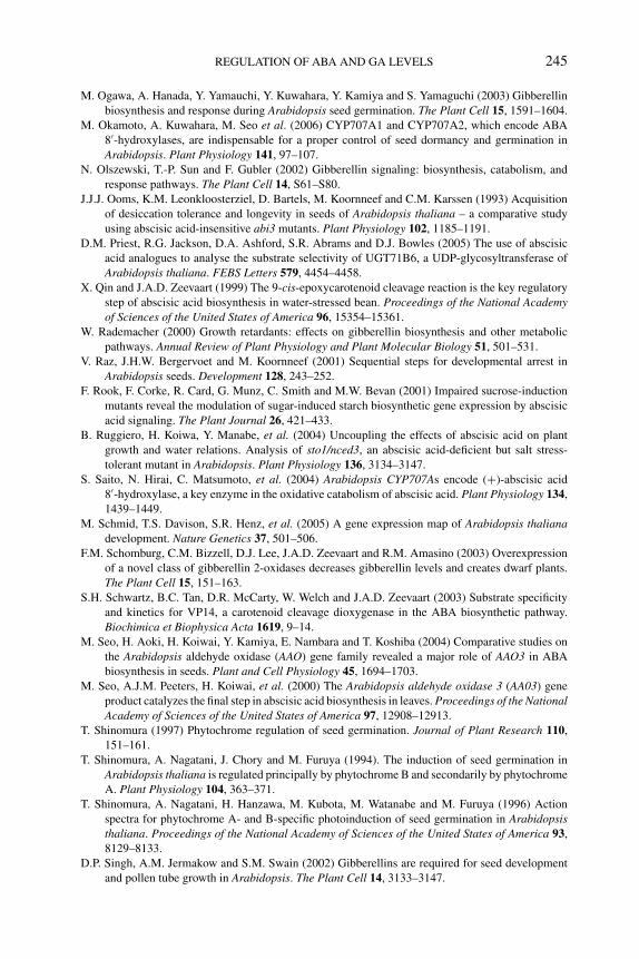
P1: JYS
BLUK053-Bradford September 22, 2006 14:30
REGULATION OF ABA AND GA LEVELS 245
M. Ogawa, A. Hanada, Y. Yamauchi, Y. Kuwahara, Y. Kamiya and S. Yamaguchi (2003) Gibberellin
biosynthesis and response during Arabidopsis seed germination. The Plant Cell 15, 1591–1604.
M. Okamoto, A. Kuwahara, M. Seo et al. (2006) CYP707A1 and CYP707A2, which encode ABA
8′-hydroxylases, are indispensable for a proper control of seed dormancy and germination in
Arabidopsis. Plant Physiology 141, 97–107.
N. Olszewski, T.-P. Sun and F. Gubler (2002) Gibberellin signaling: biosynthesis, catabolism, and
response pathways. The Plant Cell 14, S61–S80.
J.J.J. Ooms, K.M. Leonkloosterziel, D. Bartels, M. Koornneef and C.M. Karssen (1993) Acquisition
of desiccation tolerance and longevity in seeds of Arabidopsis thaliana – a comparative study
using abscisic acid-insensitive abi3 mutants. Plant Physiology 102, 1185–1191.
D.M. Priest, R.G. Jackson, D.A. Ashford, S.R. Abrams and D.J. Bowles (2005) The use of abscisic
acid analogues to analyse the substrate selectivity of UGT71B6, a UDP-glycosyltransferase of
Arabidopsis thaliana. FEBS Letters 579, 4454–4458.
X. Qin and J.A.D. Zeevaart (1999) The 9-cis-epoxycarotenoid cleavage reaction is the key regulatory
step of abscisic acid biosynthesis in water-stressed bean. Proceedings of the National Academyof Sciences of the United States of America 96, 15354–15361.
W. Rademacher (2000) Growth retardants: effects on gibberellin biosynthesis and other metabolic
pathways. Annual Review of Plant Physiology and Plant Molecular Biology 51, 501–531.
V. Raz, J.H.W. Bergervoet and M. Koornneef (2001) Sequential steps for developmental arrest in
Arabidopsis seeds. Development 128, 243–252.
F. Rook, F. Corke, R. Card, G. Munz, C. Smith and M.W. Bevan (2001) Impaired sucrose-induction
mutants reveal the modulation of sugar-induced starch biosynthetic gene expression by abscisic
acid signaling. The Plant Journal 26, 421–433.
B. Ruggiero, H. Koiwa, Y. Manabe, et al. (2004) Uncoupling the effects of abscisic acid on plant
growth and water relations. Analysis of sto1/nced3, an abscisic acid-deficient but salt stress-
tolerant mutant in Arabidopsis. Plant Physiology 136, 3134–3147.
S. Saito, N. Hirai, C. Matsumoto, et al. (2004) Arabidopsis CYP707As encode (+)-abscisic acid
8′-hydroxylase, a key enzyme in the oxidative catabolism of abscisic acid. Plant Physiology 134,
1439–1449.
M. Schmid, T.S. Davison, S.R. Henz, et al. (2005) A gene expression map of Arabidopsis thalianadevelopment. Nature Genetics 37, 501–506.
F.M. Schomburg, C.M. Bizzell, D.J. Lee, J.A.D. Zeevaart and R.M. Amasino (2003) Overexpression
of a novel class of gibberellin 2-oxidases decreases gibberellin levels and creates dwarf plants.
The Plant Cell 15, 151–163.
S.H. Schwartz, B.C. Tan, D.R. McCarty, W. Welch and J.A.D. Zeevaart (2003) Substrate specificity
and kinetics for VP14, a carotenoid cleavage dioxygenase in the ABA biosynthetic pathway.
Biochimica et Biophysica Acta 1619, 9–14.
M. Seo, H. Aoki, H. Koiwai, Y. Kamiya, E. Nambara and T. Koshiba (2004) Comparative studies on
the Arabidopsis aldehyde oxidase (AAO) gene family revealed a major role of AAO3 in ABA
biosynthesis in seeds. Plant and Cell Physiology 45, 1694–1703.
M. Seo, A.J.M. Peeters, H. Koiwai, et al. (2000) The Arabidopsis aldehyde oxidase 3 (AA03) gene
product catalyzes the final step in abscisic acid biosynthesis in leaves. Proceedings of the NationalAcademy of Sciences of the United States of America 97, 12908–12913.
T. Shinomura (1997) Phytochrome regulation of seed germination. Journal of Plant Research 110,
151–161.
T. Shinomura, A. Nagatani, J. Chory and M. Furuya (1994). The induction of seed germination in
Arabidopsis thaliana is regulated principally by phytochrome B and secondarily by phytochrome
A. Plant Physiology 104, 363–371.
T. Shinomura, A. Nagatani, H. Hanzawa, M. Kubota, M. Watanabe and M. Furuya (1996) Action
spectra for phytochrome A- and B-specific photoinduction of seed germination in Arabidopsisthaliana. Proceedings of the National Academy of Sciences of the United States of America 93,
8129–8133.
D.P. Singh, A.M. Jermakow and S.M. Swain (2002) Gibberellins are required for seed development
and pollen tube growth in Arabidopsis. The Plant Cell 14, 3133–3147.
P1: JYS
BLUK053-Bradford September 22, 2006 14:30
246 SEED DEVELOPMENT, DORMANCY AND GERMINATION
K. Sorefan, J. Booker, K. Haurogne, et al. (2003) MAX4 and RMS1 are orthologous dioxygenase-
like genes that regulate shoot branching in Arabidopsis and pea. Genes and Development 17,
1469–1474.
S.L. Stone, L.W. Kwong, K.M. Yee, et al. (2001) LEAFY COTYLEDON2 encodes a B3 domain
transcription factor that induces embryo development. Proceedings of the National Academy ofSciences of the United States of America 98, 11806–11811.
M. Suzuki, C.Y. Kao and D.R. McCarty (1997) The conserved B3 domain of VIVIPAROUS1 has a
cooperative DNA binding activity. The Plant Cell 9, 799–807.
M. Talon, M. Koornneef and J.A.D. Zeevaart (1990) Endogenous gibberellins in Arabidopsis thalianaand possible steps blocked in the biosynthetic pathways of the semidwarf ga4 and ga5 mu-
tants. Proceedings of the National Academy of Sciences of the United States of America 87,
7983–7987.
B.C. Tan, L.M. Joseph, W.T. Deng, et al. (2003) Molecular characterization of the Arabidopsis 9-cis-
epoxycarotenoid dioxygenase gene family. The Plant Journal 35, 44–56.
S.G. Thomas, A.L. Phillips and P. Hedden (1999) Molecular cloning and functional expression of
gibberellin 2-oxidases, multifunctional enzymes involved in gibberellin deactivation. Proceedingsof the National Academy of Sciences of the United States of America 96, 4698–4703.
Y. Todoroki and N. Hirai (2002) Abscisic acid analogs for probing the mechanism of abscisic acid
perception and inactivation. Studies in Natural Products Chemistry 27, 321–360.
Y. Tsuchiya, E. Nambara, S. Naito and P. McCourt (2004) The FUS3 transcription factor functions
through the epidermal regulator TTG1 during embryogenesis in Arabidopsis. The Plant Journal37, 73–81.
K. Ueno, Y. Araki, N. Hirai, et al. (2005) Differences between the structural requirements for ABA
8′-hydroxylase inhibition and for ABA activity. Bioorganic and Medicinal Chemistry 13, 3359–
3370.
T. Umezawa, M. Okamoto, T. Kushiro, et al. (2006) CYP707A3, a major ABA 8′-hydroxylase in-
volved in dehydration and rehydration response in Arabidopsis thaliana. The Plant Journal 46,
171–182.
J.M. Van Norman, R.L. Frederick and L.E. Sieburth (2004) BYPASS1 negatively regulates a root-
derived signal that controls plant architecture. Current Biology 14, 1739–1746.
H. Wang, L.V. Caruso, B. Downie and S.E. Perry (2004) The embryo MADS domain protein
AGAMOUS-like 15 directly regulates expression of a gene encoding an enzyme involved in
gibberellin metabolism. The Plant Cell 16, 1206–1219.
H. Wang, W. Tang, C. Zhu and S.E. Perry (2002) A chromatin immunoprecipitation (ChIP) approach
to isolate genes regulated by AGL15, a MADS domain protein that preferentially accumulates in
embryos. The Plant Journal 32, 831–843.
C.N. White, W.M. Proebsting, P. Hedden and C.J. Rivin (2000) Gibberellins and seed development in
maize. I: Evidence that gibberellin/abscisic acid balance governs germination versus maturation
pathways. Plant Physiology 122, 1081–1088.
L.M. Xiong, M. Ishitani, H. Lee and J.K. Zhu (2001) The Arabidopsis LOS5/ABA3 locus encodes a
molybdenum cofactor sulfurase and modulates cold stress- and osmotic stress-responsive gene
expression. The Plant Cell 13, 2063–2083.
L.M. Xiong, H.J. Lee, M. Ishitani and J.K. Zhu (2002) Regulation of osmotic stress-responsive gene
expression by the LOS6/ABA1 locus in Arabidopsis. Journal of Biological Chemistry 277, 8588–
8596.
Z.-J. Xu, M. Nakajima, Y. Suzuki and I. Yamaguchi (2002) Cloning and characterization of abscisic
acid-specific glucosyltransferase gene from Adzuki bean seedlings. Plant Physiology 129, 1285–
1295.
S. Yamaguchi, Y. Kamiya and T.-P. Sun (2001) Distinct cell-specific expression patterns of early and
late gibberellin biosynthetic genes during Arabidopsis seed germination. The Plant Journal 28,
443–454.
S. Yamaguchi, M.W. Smith, R.G.S. Brown, Y. Kamiya and T.-P. Sun (1998) Phytochrome regulation
and differential expression of gibberellin 3β-hydroxylase genes in germinating Arabidopsis seeds.
The Plant Cell 10, 2115–2126.
P1: JYS
BLUK053-Bradford September 22, 2006 14:30
REGULATION OF ABA AND GA LEVELS 247
Y. Yamauchi, M. Ogawa, A. Kuwahara, A. Hanada, Y. Kamiya and S. Yamaguchi (2004) Activation
of gibberellin biosynthesis and response pathways by low temperature during imbibition of
Arabidopsis seeds. The Plant Cell 16, 367–378.
Y.Y. Yang, A. Nagatani, Y.J. Zhao, B.J. Kang, R.E. Kentrick and Y. Kamiya (1995) Effects of gib-
berellins on seed germination of phytochrome-deficient mutants of Arabidopsis thaliana. Plantand Cell Physiology 36, 1205–1211.
T. Yoshioka, T. Endo and S. Satoh (1998) Restoration of seed germination at supraoptimal temperatures
by fluridone, an inhibitor of abscisic acid biosynthesis. Plant and Cell Physiology 39, 307–312.
L.I. Zaharia, Y. Gai, K.M. Nelson, S.J. Ambrose and S.R. Abrams (2004) Oxidation of 8′-hydroxy
abscisic acid in Black Mexican Sweet maize cell suspension cultures. Phytochemistry 65, 3199–
3209.
L.I. Zaharia, M.K. Walker-Simmons, C.N. Rodrıguez and S.R. Abrams (2005) Chemistry of abscisic
acid, abscisic acid catabolites and analogues. Journal of Plant Growth Regulation 24, 274–284.
J.A.D. Zeevaart (1999) Abscisic acid metabolism and its regulation. In: Biochemistry and MolecularBiology of Plant Hormones (eds P.J.J. Hooykaas, M.A. Hall and K.R. Libbenga), pp. 189–207.
Elsevier, New York.
J.A.D. Zeevaart, D.A. Gage and R.A. Creelman (1990) Recent studies of the metabolism of abscisic
acid. In: Plant Growth Substances 1988 (eds R.P. Pharis and S.B. Rood), pp. 232–240. Springer-
Verlag, New York.
Y. Zhu, T. Nomura, Y. Xu, et al. (2006) ELONGATED UPPERMOST INTERNODE encodes a cy-
tochrome P450 monooxygenase that epoxidizes gibberellins in a novel deactivation reaction in
rice. The Plant Cell 18, 442–456.
P1: PIB
BLUK053-Bradford August 27, 2006 17:37
10 De-repression of seed germinationby GA signalingCamille M. Steber
10.1 Introduction
This chapter explores evidence that the ubiquitin–proteasome pathway plays arole in gibberellin (GA) stimulation of germination via proteolysis of DELLAproteins (named after conserved amino acids). GA stimulates germination,stem elongation, transition to flowering, and fertility. DELLA proteins arerepressors of GA responses defined by the presence of conserved DELLAand GRAS (named after GAI [GA-INSENSITIVE], RGA [REPRESSOR OFGA-INSENSITIVE], and SCARECROW proteins) domain amino acid sequences. Itis clear that the ubiquitin–proteasome pathway relieves DELLA repression of stemelongation in response to GA signaling. The paradigm is that GA stimulates an SCF(named after the Skp1, Cullin, and F-box subunits) E3 ubiquitin ligase complex,which in turn stimulates degradation of the DELLA proteins, negative regulatorsof GA response, by the proteasome pathway. This induces the downstream eventsnecessary for seed germination, which were under repression in the absence of GA.However, interpretation of the evidence for DELLA regulation of seed germina-tion has been somewhat contentious. Work in Arabidopsis thaliana suggests thatthe DELLA protein RGL2 (RGA-LIKE2) is the main negative regulator of GA re-sponse in germination, and that the SCFSLY1 E3 ubiquitin ligase complex is requiredfor GA-stimulated disappearance of RGL2.
10.2 Control of germination by GA signaling
The plant hormone GA was first identified in 1926 based on its role in stem elonga-tion (Tamura, 1991; Sun and Gubler, 2004; Thomas et al., 2005). Eiichi Kurosawaidentified gibberellin as the agent causing the excessive stem elongation in rice(Oryza sativa) seedlings infected with bakanae disease. Subsequent work demon-strated that gibberellins are growth regulators in a wide range of plant, algal, andfungal species. GA is not a single plant hormone, but actually a large family oftetracyclic diterpenes. Thus far, 136 naturally occurring GA molecules have beenidentified in plants and fungi (Thomas et al., 2005). GA acts on many physiologicalevents during plant growth and development (reviewed by Sun and Gubler, 2004;Thomas et al., 2005). GA promotes stem elongation by stimulating both cell elon-gation and cell division. GA stimulates the transition to flowering in most plant
248
P1: PIB
BLUK053-Bradford August 27, 2006 17:37
DE-REPRESSION OF SEED GERMINATION BY GA SIGNALING 249
species and appears to be required for normal fertility and embryo development. Awide range of evidence has shown that GA plays a role in seed germination.
By far the strongest evidence for the role of GA in germination comes fromstudies of mutants defective in GA biosynthesis in dicots. In Arabidopsis, mutationsin early GA biosynthesis genes result in a requirement for GA application to germi-nate (Koornneef and van der Veen, 1980). Mutations in the biosynthesis genes GA1(copalyl synthase), GA2 (ent-kaurene synthase), and GA3 (ent-kaurene oxidase) re-sult in failure to germinate. Mutations in genes acting late in GA biosynthesis suchas ga4-1 (GA 3-oxidase or GA3ox) and ga5-1 (GA 20-oxidase or GA20ox) do notrequire GA application to germinate. The explanation may lie in the fact that bothof these genes are part of multigene families in higher plants. These genes showtissue-specific expression. For example, the Arabidopsis GA3ox2 gene was foundto be expressed primarily in germinating seeds and seedlings, while GA3ox1 wasexpressed in all growing tissues (Yamaguchi et al., 1998). Future work may usestudies of multiple mutants to determine whether these GA biosynthesis genes arealso required for germination in Arabidopsis. A mutation in ent-kaurene synthasecalled gib-1 has been identified in tomato (Lycopersicon esculentum) and gib-1 seedsrequire GA application for germination (Karssen et al., 1989; Benson and Zeevaart,1990). Thus, the requirement for GA in germination is not unique to Arabidopsis.
The plant hormones ABA (abscisic acid) and GA have tightly interwoven rolesin the decision to germinate. While GA is often required for germination and forpostgerminative reserve mobilization, ABA inhibits germination, promotes nutrientstorage, and is required for dormancy and desiccation tolerance during embryo mat-uration (reviewed by Bentsink and Koornneef, 2002). Seeds are said to be dormantwhen they are unable to germinate even under favorable conditions. Mutants unableto synthesize ABA have nondormant seeds and a vegetative wilty phenotype. Thewilty phenotype results from an inability to close stomates and conserve water inresponse to drought stress. Thus, in adult tissues ABA is the stress hormone neededfor resistance to drought, and is also involved in cold and salt stress. The hormonebalance theory postulates that the decision to germinate is a balancing act betweenABA and GA signals (Karssen and Lacka, 1986). If ABA biosynthesis or signalingis reduced, a seed is more likely to germinate. If GA biosynthesis or signaling is re-duced, a seed is less likely to germinate. A mutation in one pathway can compensatefor a mutation in the other. For example, germination of a strong GA biosynthe-sis mutant can be rescued by mutants causing either reduced ABA biosynthesis orsensitivity (reviewed by Bentsink and Koornneef, 2002). Conversely, the ability ofABA-insensitive mutants to germinate in the presence of high concentrations ofABA is suppressed by reduced GA biosynthesis or signaling (Steber et al., 1998).
By definition, seed germination is complete when the embryo radicle emergesfrom the seed coat. GA is believed to stimulate germination by inducing hydrolyticenzymes that weaken the seed coat or endosperm cap, by inducing mobilizationof seed nutrient storage compounds, and by stimulating expansion of the embryo(Bewley and Black, 1994; see Chapter 11). In tomato, it has been shown that GAstimulates expression of endo-β-mannanase, an enzyme that breaks down cell wallreserves in the endosperm cap (Still and Bradford, 1997; Nonogaki et al., 2000).
P1: PIB
BLUK053-Bradford August 27, 2006 17:37
250 SEED DEVELOPMENT, DORMANCY AND GERMINATION
During Arabidopsis germination, genes expected to be involved in cell wall degra-dation and induction of cell division are induced by GA (Ogawa et al., 2003). Inthe cereal grains of rice, barley (Hordeum vulgare), and wheat (Triticum aestivum),GA induces the expression of genes encoding α-amylase, an enzyme needed for thebreakdown of the starchy endosperm (Sun and Gubler, 2004). The mobilization ofthis nutrient source doubtlessly contributes to embryo growth after germination.
The role of GA biosynthesis and signaling genes in rice germination is currentlyunclear. An apparent knockout mutation in an ent-kaurene synthase gene of rice(OsKS1) results in the expected dwarfism and infertility phenotypes, but in contrastto Arabidopsis and tomato it does not result in failure to germinate (Sakamoto et al.,2004; Margis-Pinheiro et al., 2005). The fact that OsKS1 is not expressed in imbibingseeds, however, raises the possibility that one of the five to seven homologues ofKS may function in rice seed germination (Margis-Pinheiro et al., 2005). GA-insensitive mutations in the rice F-box gene GID2 (GIBBERELLIN-INSENSITIVEDWARF2) and in the GA receptor GID1 also result in dwarfism and infertilitywithout compromising germination (Itoh et al., 2003). While the gid1 mutants failto induce α-amylase during seedling establishment, the mutants germinate well(Ueguchi-Tanaka et al., 2005; M. Matsuoka, personal communication). There are anumber of explanations for these observations. Cereal seeds (‘caryopses’) includingrice have highly specialized seed morphology where the embryo is not entirelyenclosed by the endosperm. Tissues covering the embryo may offer relatively littleresistance to penetration by the coleorhiza and coleoptile. It is possible that inrice, GA is not required for germination per se but is required only for reservemobilization. It is also possible that the rice variety in which the gid1 and gid2mutants were identified does not require GA signaling in germination either dueto lack of seed dormancy or due to a defect in ABA signaling in that background.The ability of GA mutant rice seeds (caryopses), which have pale testa color togerminate, may be due to reduced dormancy resulting from lack of red pigmentsin the testa. Red rice has strong seed dormancy compared to pigmentless cultivars(Footitt and Cohn, 1995). Loss of red testa color causes reduced seed dormancy. Forexample, the reduced dormancy caused by the Arabidopsis transparent testa mutanttt4 suppresses the requirement for GA in germination, allowing the ga1-1 mutantto germinate (Debeaujon and Koornneef, 2000; Chapter 2). Another possibilityis that rice varieties used in these studies have a defect in ABA signaling. Forexample, it has been shown that all tested wheat cultivars are unable to properly splicethe ZmVP1/AtABI3 (Zea mays VIVIPAROUS1/ABA-INSENSITIVE3) homologue ofwheat TaVP1 (McKibbin et al., 2002). Thus, bread wheat has reduced seed dormancydue to reduced ABA signaling. In Arabidopsis, mutations causing ABA insensitivityallow both GA biosynthesis and GA-insensitive mutants to germinate. A similardefect in rice ABA signaling might obviate the requirement for GA in germination.The fact that GA biosynthesis is required for germination of the ABA-insensitivevp5 mutants on the mother plant in maize (Zea mays) (White et al., 2000; White andRivin, 2000) suggests that GA is important for cereal germination during embryodevelopment. Future research will need to address the role of GA in cereal graingermination.
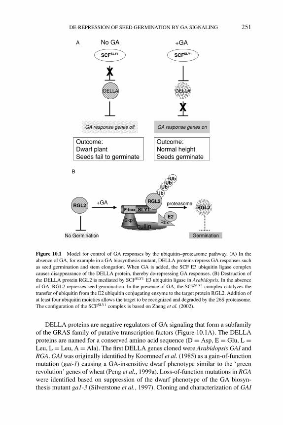
P1: PIB
BLUK053-Bradford August 27, 2006 17:37
DE-REPRESSION OF SEED GERMINATION BY GA SIGNALING 251
DELLA
No GA
GA response genes off
+GA
GA response genes on
DELLA
SCFSLY1
X
Outcome:Dwarf plantSeeds fail to germinate
SCFSLY1
X
Outcome:Normal heightSeeds germinate
RGL2F-box
cullinRbxSkp1
No Germination
UbUb
UbUb
RGL2RGL2
Germination
proteasome+GA
A
B
SLY1
E2
Figure 10.1 Model for control of GA responses by the ubiquitin–proteasome pathway. (A) In theabsence of GA, for example in a GA biosynthesis mutant, DELLA proteins repress GA responses suchas seed germination and stem elongation. When GA is added, the SCF E3 ubiquitin ligase complexcauses disappearance of the DELLA protein, thereby de-repressing GA responses. (B) Destruction ofthe DELLA protein RGL2 is mediated by SCFSLY1 E3 ubiquitin ligase in Arabidopsis. In the absenceof GA, RGL2 represses seed germination. In the presence of GA, the SCFSLY1 complex catalyzes thetransfer of ubiquitin from the E2 ubiquitin conjugating enzyme to the target protein RGL2. Addition ofat least four ubiquitin moieties allows the target to be recognized and degraded by the 26S proteasome.The configuration of the SCFSLY1 complex is based on Zheng et al. (2002).
DELLA proteins are negative regulators of GA signaling that form a subfamilyof the GRAS family of putative transcription factors (Figure 10.1A). The DELLAproteins are named for a conserved amino acid sequence (D = Asp, E = Glu, L =Leu, L = Leu, A = Ala). The first DELLA genes cloned were Arabidopsis GAI andRGA. GAI was originally identified by Koornneef et al. (1985) as a gain-of-functionmutation (gai-1) causing a GA-insensitive dwarf phenotype similar to the ‘greenrevolution’ genes of wheat (Peng et al., 1999a). Loss-of-function mutations in RGAwere identified based on suppression of the dwarf phenotype of the GA biosyn-thesis mutant ga1-3 (Silverstone et al., 1997). Cloning and characterization of GAI
P1: PIB
BLUK053-Bradford August 27, 2006 17:37
252 SEED DEVELOPMENT, DORMANCY AND GERMINATION
and RGA revealed that they are homologous genes (Peng et al., 1997; Silverstorneet al., 1998). While loss of neither gene alone gives increased stature, the gai-t6rga-24 double mutant is taller than wild-type plants, indicating that together theyrepress stem elongation of Arabidopsis (Dill and Sun, 2001; King et al., 2001).This increased height phenotype is reminiscent of the slender phenotype of loss-of-function mutants in DELLA genes of barley (SLN1, SLENDER1) and rice (SLR1,SLENDER RICE1) (Ikeda et al., 2001; Chandler et al., 2002). A DELLA proteinhas been shown to inhibit stem elongation in species as diverse as barley, rice,wheat, maize, grape (Vitis vinifera), tobacco (Nicotiana tabacum), Brassica rapa,and Arabidopsis (Peng et al., 1999a; Silverstone et al., 2001; Boss and Thomas,2002; Gubler et al., 2002; Itoh et al., 2002; Hynes et al., 2003; Dill et al., 2004;Fu et al., 2004; Muangprom et al., 2005).
The current model for GA signaling in stem elongation is widely conserved inthe plant kingdom. If the plant never produced GA, as in a GA biosynthesis mutant,repression of stem elongation by DELLA would result in a dwarf phenotype (Figure10.1A). Addition of GA stimulates cell division and expansion within the meristemby causing disappearance of the DELLA protein. GA seems to cause DELLA pro-tein disappearance by triggering DELLA protein polyubiquitination by an SCF E3ubiquitin ligase (Itoh et al., 2003). Addition of four or more ubiquitin moieties to aDELLA protein targets it for destruction by the 26S proteasome (Sasaki et al., 2003).The interaction between the DELLA protein and SCF complex may be regulated byDELLA phosphorylation or by interaction between the DELLA protein and the GAreceptor GID1 (Fu et al., 2004; Gomi et al., 2004; Ueguchi-Tanaka et al., 2005).
Evidence from work in Arabidopsis suggests that GA signaling also stimulatesgermination via DELLA protein disappearance. There are five DELLA genes inArabidopsis, RGA, GAI, RGL1, RGL2, and RGL3. RGA and GAI repress stem elon-gation, while RGL1, RGA, and RGL2 repress the transition to flowering (Itoh et al.,2003). RGL2 is the main repressor of germination, although RGL1, RGA, and GAIalso appear to contribute (Lee et al., 2002; Tyler et al., 2004; Cao et al., 2005).Two questions must be answered in order to understand the role of GA and DELLAproteins in seed germination: (1) Is RGL2 a master negative regulator of seed ger-mination?; and (2) How widely conserved is this mechanism for controlling seedgermination? The evidence that DELLA and the SCF E3 ubiquitin ligase complexcontrol germination in Arabidopsis will be discussed first, and then evidence inadditional plant species will be considered.
10.3 The role of the ubiquitin–proteasome pathway in GA signaling
Several lines of evidence indicate that DELLA proteins are regulated by theubiquitin–proteasome pathway (Figure 10.1). First, GA treatment causes thedisappearance of DELLA proteins in Arabidopsis, rice, and barley (Silverstorne etal., 2001; Itoh et al., 2002; Gubler et al., 2002; Dill et al., 2004; Fu et al., 2004).This disappearance requires the F-box subunit of an SCF E3 ubiquitin ligase andthe 26S proteasome. The disappearance of DELLA protein in response to GA is
P1: PIB
BLUK053-Bradford August 27, 2006 17:37
DE-REPRESSION OF SEED GERMINATION BY GA SIGNALING 253
blocked by inhibitors of the 26S proteasome (Fu et al., 2002; Hussain et al., 2005)and by mutations in the F-box genes rice GID2 and Arabidopsis SLY1 (SLEEPY1)(McGinnis et al., 2003; Sasaki et al., 2003). This suggests that GA causes theSCFSLY1/GID2 E3 ubiquitin ligase to target the DELLA protein for destruction viathe 26S proteasome. Mutations in GID2 and SLY1 cause a range of GA-insensitivephenotypes including severe dwarfism, dark green color, and reduced fertility (Itohet al., 2003). Mutations in SLY1 also cause increased seed dormancy. SLY1 andGID2 proteins have been shown to interact with DELLA proteins, and ubiquitinatedDELLA protein has been detected in rice (Sasaki et al., 2003; Dill et al., 2004; Fuet al., 2004; Gomi et al., 2004).
DELLA proteins are regulated by Arabidopsis SCFSLY1 and rice SCFGID2 E3ubiquitin ligase complexes (Figure 10.1B; McGinnis et al., 2003; Sasaki et al.,2003). SCF complexes are one of several types of E3 ubiquitin ligases found inplants and in animals (Smalle and Vierstra, 2004). An SCF complex is identifiedbased on the F-box subunit because it is the F-box subunit that binds to a specifictarget or substrate protein. Based on the crystal structure of mammalian SCFSkp2,the backbone of the SCF complex is cullin (Zheng et al., 2002). Cullin binds ahomologue of the ring-finger protein Rbx1 at its C-terminus, and Rbx1 binds theE2 ubiquitin conjugating enzyme. At the N-terminus, cullin binds a homologue ofSkp1. Skp1 binds the F-box protein via the conserved F-box domain and tethers theF-box protein to the rest of the complex. The F-box protein is generally composedof an F-box domain at the N-terminus and a protein–protein interaction domain atthe C-terminus that binds to a substrate protein such as DELLA. The SCF complexcatalyzes transfer of ubiquitin from E2 to the substrate, in this case a DELLA protein.
Ubiquitin is a 76-amino acid protein used in plants and animals as a tag to signalfor proteolytic processing, cleavage, or destruction (Smalle and Vierstra, 2004).DELLA proteins appear to be targeted by ubiquitination for destruction by the 26Sproteasome. Generally, ubiquitin is first covalently bound to a cysteine of an E1ubiquitin-activating enzyme via a thioester bond and then transferred to a cysteineof an E2 ubiquitin-conjugating enzyme. Finally, an E3 ubiquitin ligase catalyzes thetransfer of ubiquitin from E2 to a lysine residue of the substrate protein. Formation ofa chain of four or more ubiquitin moieties on the substrate allows its recognition andproteolysis by the 26S proteasome. The 26S proteasome is a multi-subunit proteincomplex that can be found both in the nucleus and in the cytosol. It is surmised thatDELLA proteins meet their end in the 26S proteasome because the DELLA proteinsbarley SLN1 and Arabidopsis RGL2 were shown to be stabilized by compoundsknown to inhibit the 26S proteasome (Fu et al., 2002; Hussain et al., 2005). Therice DELLA SLR1 was shown to be subjected to ubiquitination by western analysisusing an antibody to ubiquitin (Sasaki et al., 2003). Finally, DELLA proteins in awide range of plant species have been shown to be regulated by a highly conservedSCF E3 ubiquitin ligase (Itoh et al., 2003).
Protein destruction is a recurrent theme in plant signal transduction (Smalle andVierstra, 2004). There are almost 700 F-box proteins in the Arabidopsis genome.It is already clear that F-box proteins and the ubiquitin–proteasome pathway playa crucial role in plant hormone signaling. The ubiquitin–proteasome pathway was
P1: PIB
BLUK053-Bradford August 27, 2006 17:37
254 SEED DEVELOPMENT, DORMANCY AND GERMINATION
implicated in ABA and cytokinin signaling when mutations in subunits of the 26Sproteasome were shown to result in changes in hormone sensitivity (Smalle et al.,2002, 2003). Recently, the ABA signaling protein ABI3 was found to be regulatedby the AIP2 (AB13-INTERACTING PROTEIN2) E3 ubiquitin ligase (Zhang et al.,2005). In ethylene signaling, the transcription factors EIN3 (ETHYLENE INSEN-SITIVE3) and EIL1 (EIN3-LIKE) are regulated by the homologous F-box proteinsEBF1 (EIN3-BINDING F-BOX) and EBF2 (Guo and Ecker, 2003; Potuschak et al.,2003; Gagne et al., 2004). EBF1 and EBF2 F-box proteins contain the leucine-richrepeat (LRR) type of protein–protein interaction domain at the C-terminus. TheSLY1 F-box protein contains no consensus protein–protein interaction domain atthe C-terminus. However, based on mutation studies, the C-terminus is required bothfor function and for interaction with DELLA proteins (McGinnis et al., 2003; Dillet al., 2004; Fu et al., 2004). The COI1 (CORONATINE INSENSITIVE1) F-boxprotein required for jasmonic acid signaling contains an LRR protein–protein in-teraction domain at the C-terminus (Xu et al., 2002). This is similar to the LRRsfound in the C-terminus of the TIR1 (TRANSPORT INHIBITOR RESPONSE1)F-box protein (Gray et al., 1999). TIR1 was recently shown to be an auxin receptor(Dharmasiri et al., 2005; Kepinski and Leyser, 2005).
Auxin signaling has a number of similarities to GA signaling. Auxin triggersauxin responses by targeting the IAA/AUX family of transcriptional repressors fordestruction via the 26S proteasome (Gray et al., 2001). The IAA/AUX proteins re-press auxin responses by binding to the ARF (AUXIN RESPONSE FACTOR) tran-scription factors. DELLA proteins repress GA responses by an unknown mechanism.Auxin binds the F-box protein TIR1, thus enabling TIR1 to bind to the IAA/AUXproteins and polyubiquitinate them, thereby targeting them for destruction by the26S proteasome (Dharmasiri et al., 2005, Kepinski and Leyser, 2005). The DELLAproteins have been shown to be regulated by the F-box proteins Arabidopsis SLY1or rice GID2. However, it is still unclear precisely how GA controls the interactionbetween the F-box protein and the DELLA protein. While interactions have beendetected between F-box proteins SLY1/GID2 and DELLA proteins, no interactionbetween GA and SLY1/GID2 has yet been reported. However, it has been reportedthat GA binds to the rice GID1 protein, a GA receptor (Ueguchi-Tanaka et al.,2005). GID1 shows GA-dependent interaction with the rice DELLA protein SLR1.Mutations in GID1 lead to overaccumulation of DELLA protein, suggesting thatGID1 is required for destruction of the DELLA protein by SCFGID2. Whether GID1controls the proteolysis of DELLA proteins via direct protein–protein interactionbetween GID1 and GID2 F-box protein needs to be investigated.
While DELLA proteins are known to be negative regulators of GA responses,their precise function remains unknown. They are considered likely transcriptionfactors based on amino acid homology and on yeast two-hybrid data showing thatthey can activate transcription (Peng et al., 1999a; Itoh et al., 2002). The DELLAprotein family is defined by conserved amino acid sequences (Sun and Gubler,2004). The C-terminus of DELLA proteins contains homology to the GRAS familyof proteins, including a nuclear localization sequence, the VHIID domain, leucineheptad repeats (LHR), and SH2 domains. The SH2 domain of metazoan STAT
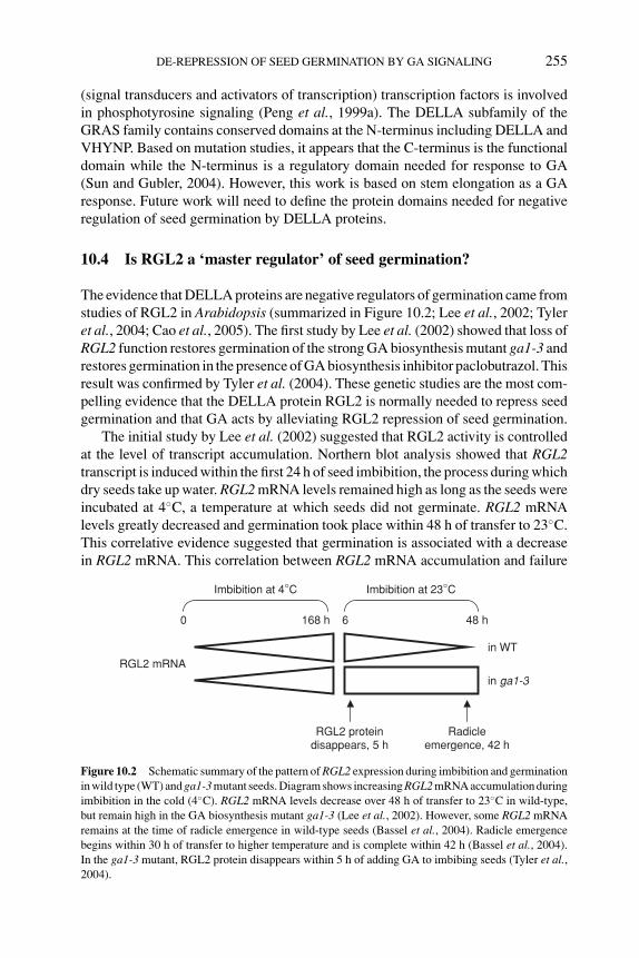
P1: PIB
BLUK053-Bradford August 27, 2006 17:37
DE-REPRESSION OF SEED GERMINATION BY GA SIGNALING 255
(signal transducers and activators of transcription) transcription factors is involvedin phosphotyrosine signaling (Peng et al., 1999a). The DELLA subfamily of theGRAS family contains conserved domains at the N-terminus including DELLA andVHYNP. Based on mutation studies, it appears that the C-terminus is the functionaldomain while the N-terminus is a regulatory domain needed for response to GA(Sun and Gubler, 2004). However, this work is based on stem elongation as a GAresponse. Future work will need to define the protein domains needed for negativeregulation of seed germination by DELLA proteins.
10.4 Is RGL2 a ‘master regulator’ of seed germination?
The evidence that DELLA proteins are negative regulators of germination came fromstudies of RGL2 in Arabidopsis (summarized in Figure 10.2; Lee et al., 2002; Tyleret al., 2004; Cao et al., 2005). The first study by Lee et al. (2002) showed that loss ofRGL2 function restores germination of the strong GA biosynthesis mutant ga1-3 andrestores germination in the presence of GA biosynthesis inhibitor paclobutrazol. Thisresult was confirmed by Tyler et al. (2004). These genetic studies are the most com-pelling evidence that the DELLA protein RGL2 is normally needed to repress seedgermination and that GA acts by alleviating RGL2 repression of seed germination.
The initial study by Lee et al. (2002) suggested that RGL2 activity is controlledat the level of transcript accumulation. Northern blot analysis showed that RGL2transcript is induced within the first 24 h of seed imbibition, the process during whichdry seeds take up water. RGL2 mRNA levels remained high as long as the seeds wereincubated at 4◦C, a temperature at which seeds did not germinate. RGL2 mRNAlevels greatly decreased and germination took place within 48 h of transfer to 23◦C.This correlative evidence suggested that germination is associated with a decreasein RGL2 mRNA. This correlation between RGL2 mRNA accumulation and failure
RGL2 mRNA
Radicleemergence, 42 h
RGL2 proteindisappears, 5 h
Imbibition at 4°C Imbibition at 23°C
168 h0 6 48 h
in WT
in ga1-3
Figure 10.2 Schematic summary of the pattern of RGL2 expression during imbibition and germinationin wild type (WT) and ga1-3 mutant seeds. Diagram shows increasing RGL2 mRNA accumulation duringimbibition in the cold (4◦C). RGL2 mRNA levels decrease over 48 h of transfer to 23◦C in wild-type,but remain high in the GA biosynthesis mutant ga1-3 (Lee et al., 2002). However, some RGL2 mRNAremains at the time of radicle emergence in wild-type seeds (Bassel et al., 2004). Radicle emergencebegins within 30 h of transfer to higher temperature and is complete within 42 h (Bassel et al., 2004).In the ga1-3 mutant, RGL2 protein disappears within 5 h of adding GA to imbibing seeds (Tyler et al.,2004).
P1: PIB
BLUK053-Bradford August 27, 2006 17:37
256 SEED DEVELOPMENT, DORMANCY AND GERMINATION
to germinate was further supported by data showing that the strong GA biosynthesismutant ga1-3 accumulates high levels of RGL2 mRNA during imbibition both at4◦C and at 23◦C. RGL2 mRNA abundance decreases within 48 h of addition of GAto imbibing ga1-3 seeds. This suggests that one way that GA stimulates germinationof ga1-3 seeds is by decreasing the level of RGL2 transcript. It is important to note,however, that the RGL2 transcript did not completely disappear within 48 h of addingGA. Thus Lee et al. (2002) concluded that downregulation of RGL2 mRNA level isconsistent with the notion that it is a negative regulator of seed germination, but couldnot conclude that complete absence of RGL2 mRNA is required for germination tooccur.
A subsequent study by Bassel et al. (2004) sought to determine whether down-regulation of DELLA transcript is required for seed germination in Arabidopsis.This study very carefully examined the level of RGL2 mRNA relative to percentgermination. They found that even after Arabidopsis seed germination is complete,RGL2 transcript can still be detected (Bassel et al., 2004). This study clearly indicatesthat disappearance of RGL2 mRNA cannot be the sole mechanism for stimulatinggermination. However, it does leave open the possibility that RGL2 is a negative reg-ulator of germination subject to posttranscriptional regulation, such as translation,nuclear localization, protein stability, or posttranslational modification of RGL2protein.
A study by Tyler et al. (2004) suggests that RGL2 function is regulated byproteolysis and that GA-dependent disappearance of RGL2 protein is regulated bythe SLY1 F-box protein. Western analysis of RGL2 protein accumulation in thega1-3 mutant background showed that RGL2 protein disappears within 5 h afterGA application to the ga1-3 seeds, but does not disappear in the absence of GA.This correlation between GA-triggered disappearance of RGL2 protein and GAstimulation of germination suggests that disappearance of RGL2 protein is requiredfor germination in Arabidopsis. The rapid disappearance of RGL2 protein wouldbe consistent with a role during germination proper (prior to radicle emergence),rather than a role in postgermination events such as seedling establishment. Takentogether with the fact that loss of RGL2 function rescues ga1-3 seed germina-tion, this study strongly suggests that RGL2 is a negative regulator of germinationin Arabidopsis. Proof that RGL2 is a negative regulator of seed germination re-quires demonstration that germination fails to occur if RGL2 protein persists duringgermination.
RGL2 protein does persist after GA treatment of sly1 mutant seeds (Tyler et al.,2004). This suggests that the SCFSLY1 E3 ubiquitin ligase normally targets RGL2 fordestruction in response to GA. Mutations in the SLY1 gene result in an increase inseed dormancy and in an increase in sensitivity to ABA in germination (Steber et al.,1998; Steber and McCourt, 2001; Strader et al., 2004). However, sly1 mutants doeventually after-ripen, and some seed lots do germinate (C.M. Steber, unpublishedresults). This suggests that the disappearance of RGL2 protein is important for ger-mination, but is not the sole mechanism regulating germination in Arabidopsis. Itis possible that there are additional regulators of seed germination in Arabidopsis,or that there are additional mechanisms for posttranslational regulation of RGL2.
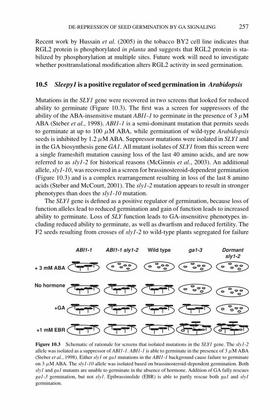
P1: PIB
BLUK053-Bradford August 27, 2006 17:37
DE-REPRESSION OF SEED GERMINATION BY GA SIGNALING 257
Recent work by Hussain et al. (2005) in the tobacco BY2 cell line indicates thatRGL2 protein is phosphorylated in planta and suggests that RGL2 protein is sta-bilized by phosphorylation at multiple sites. Future work will need to investigatewhether posttranslational modification alters RGL2 activity in seed germination.
10.5 Sleepy1 is a positive regulator of seed germination in Arabidopsis
Mutations in the SLY1 gene were recovered in two screens that looked for reducedability to germinate (Figure 10.3). The first was a screen for suppressors of theability of the ABA-insensitive mutant ABI1-1 to germinate in the presence of 3 μMABA (Steber et al., 1998). ABI1-1 is a semi-dominant mutation that permits seedsto germinate at up to 100 μM ABA, while germination of wild-type Arabidopsisseeds is inhibited by 1.2 μM ABA. Suppressor mutations were isolated in SLY1 andin the GA biosynthesis gene GA1. All mutant isolates of SLY1 from this screen werea single frameshift mutation causing loss of the last 40 amino acids, and are nowreferred to as sly1-2 for historical reasons (McGinnis et al., 2003). An additionalallele, sly1-10, was recovered in a screen for brassinosteroid-dependent germination(Figure 10.3) and is a complex rearrangement resulting in loss of the last 8 aminoacids (Steber and McCourt, 2001). The sly1-2 mutation appears to result in strongerphenotypes than does the sly1-10 mutation.
The SLY1 gene is defined as a positive regulator of germination, because loss offunction alleles lead to reduced germination and gain of function leads to increasedability to germinate. Loss of SLY function leads to GA-insensitive phenotypes in-cluding reduced ability to germinate, as well as dwarfism and reduced fertility. TheF2 seeds resulting from crosses of sly1-2 to wild-type plants segregated for failure
ABI1-1 sly1-2ABI1-1 Wild type ga1-3
No hormone
+GA
+ 3 mM ABA
+1 mM EBR
Dormantsly1-2
Figure 10.3 Schematic of rationale for screens that isolated mutations in the SLY1 gene. The sly1-2allele was isolated as a suppressor of ABI1-1. ABI1-1 is able to germinate in the presence of 3 μM ABA(Steber et al., 1998). Either sly1 or ga1 mutations in the ABI1-1 background cause failure to germinateon 3 μM ABA. The sly1-10 allele was isolated based on brassinosteroid-dependent germination. Bothsly1 and ga1 mutants are unable to germinate in the absence of hormone. Addition of GA fully rescuesga1-3 germination, but not sly1. Epibrassinolide (EBR) is able to partly rescue both ga1 and sly1germination.
P1: PIB
BLUK053-Bradford August 27, 2006 17:37
258 SEED DEVELOPMENT, DORMANCY AND GERMINATION
to germinate. This suggested that even in the absence of the ABI1-1 mutation, therecessive sly1-2 mutation has a strong germination phenotype (Steber et al., 1998).Most fresh (unaged) sly1-2 seed lots germinate between 0 and 20% (Steber andMcCourt, 2001; C.M. Steber unpublished results). Such seed stocks can requireup to 3 years to after-ripen. Some rare seed lots germinate 80–100% soon afterharvest. However, such seed lots show increased sensitivity to ABA in germina-tion, suggesting that they also have a reduced capacity to germinate (Strader et al.,2004). Further work is needed to determine how environmental conditions duringseed development and during seed storage influence the degree of seed dormancyin sly1 mutants. Germination of sly1 mutants can be rescued by cutting the seedcoat, suggesting that the germination phenotype is the result of seed coat-imposeddormancy (McGinnis et al., 2003; see Chapters 2 and 11). The fact that sly1-10mutants also show reduced capacity to germinate indicates that multiple alleles ofsly1 decrease the germination efficiency of Arabidopsis seeds. The notion that SLY1is a positive regulator of seed germination is further supported by the fact that SLY1overexpression and a gain-of-function mutation in SLY1, sly1-gar2 (gai revertant2),both cause increased resistance to the GA biosynthesis inhibitor paclobutrazol ingermination (Peng et al., 1999b; Fu et al., 2004).
Evidence that the reduced germination in the sly1 mutant is caused by failureof RGL2 protein to disappear in response to GA is strictly correlative at this point.It is not yet known whether a mutation in RGL2 can suppress the germination phe-notype of the sly1 mutant in the same way that it suppresses the ga1-3 germinationphenotype. As described above, it is known that whereas RGL2 protein disappearswithin 5 h of adding GA to ga1-3 mutant seeds, it does not disappear within 5 hof adding GA to imbibing seeds of the GA-insensitive mutant sly1-10 (Tyler et al.,2004). Thus SLY1 is required for the GA-induced disappearance of RGL2 proteinin seed germination. Since the sly1-10 mutant has reduced ability to germinate, itis possible that this germination phenotype is due to over-accumulation of RGL2protein. This suggests that the SCFSLY1 complex is responsible for relieving RGL2repression of germination, just as it relieves DELLA repression of stem elongation(Figure 10.1). It is not yet known, however, whether sly1 mutant seed lots that ger-minate well accumulate less RGL2 protein than sly1 seed lots that do not germinatewell. If RGL2 protein levels correlate with the severity of the sly1 germinationphenotype, it is possible that other ubiquitin ligases can target RGL2 protein fordestruction in the absence of SLY1. Candidates for ubiquitin ligases that might haveoverlapping function with SLY1 protein in germination include the U-box proteinPHOR1 (PHOTOPERIOD-RESPONSIVE1) that acts in GA signaling in potato andthe F-box protein SNEEZY (SNE), which is a homologue of SLY1 in Arabidopsis(Amador et al., 2001; Fu et al., 2004; Strader et al., 2004).
10.6 Do DELLA proteins have a conserved role in seed germination?
Both the SLY1 gene and DELLA gene family are highly conserved among plantspecies. Homologues of SLY1 are known in soybean (Glycine max), Medicago
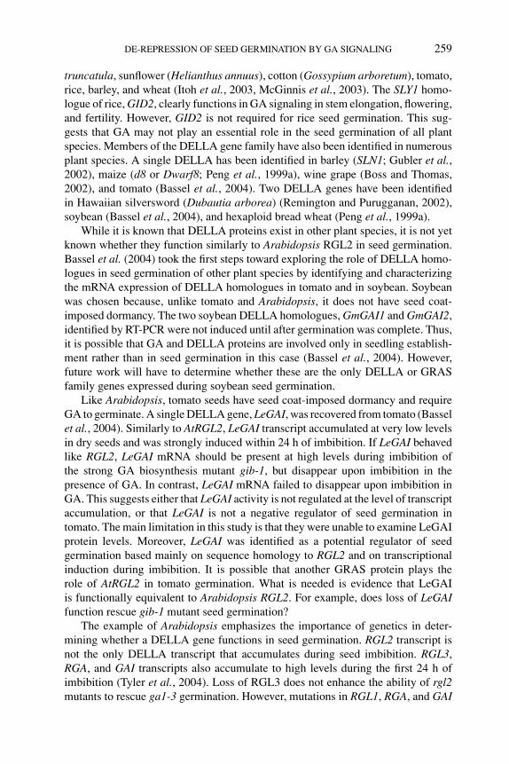
P1: PIB
BLUK053-Bradford August 27, 2006 17:37
DE-REPRESSION OF SEED GERMINATION BY GA SIGNALING 259
truncatula, sunflower (Helianthus annuus), cotton (Gossypium arboretum), tomato,rice, barley, and wheat (Itoh et al., 2003, McGinnis et al., 2003). The SLY1 homo-logue of rice, GID2, clearly functions in GA signaling in stem elongation, flowering,and fertility. However, GID2 is not required for rice seed germination. This sug-gests that GA may not play an essential role in the seed germination of all plantspecies. Members of the DELLA gene family have also been identified in numerousplant species. A single DELLA has been identified in barley (SLN1; Gubler et al.,2002), maize (d8 or Dwarf8; Peng et al., 1999a), wine grape (Boss and Thomas,2002), and tomato (Bassel et al., 2004). Two DELLA genes have been identifiedin Hawaiian silversword (Dubautia arborea) (Remington and Purugganan, 2002),soybean (Bassel et al., 2004), and hexaploid bread wheat (Peng et al., 1999a).
While it is known that DELLA proteins exist in other plant species, it is not yetknown whether they function similarly to Arabidopsis RGL2 in seed germination.Bassel et al. (2004) took the first steps toward exploring the role of DELLA homo-logues in seed germination of other plant species by identifying and characterizingthe mRNA expression of DELLA homologues in tomato and in soybean. Soybeanwas chosen because, unlike tomato and Arabidopsis, it does not have seed coat-imposed dormancy. The two soybean DELLA homologues, GmGAI1 and GmGAI2,identified by RT-PCR were not induced until after germination was complete. Thus,it is possible that GA and DELLA proteins are involved only in seedling establish-ment rather than in seed germination in this case (Bassel et al., 2004). However,future work will have to determine whether these are the only DELLA or GRASfamily genes expressed during soybean seed germination.
Like Arabidopsis, tomato seeds have seed coat-imposed dormancy and requireGA to germinate. A single DELLA gene, LeGAI, was recovered from tomato (Basselet al., 2004). Similarly to AtRGL2, LeGAI transcript accumulated at very low levelsin dry seeds and was strongly induced within 24 h of imbibition. If LeGAI behavedlike RGL2, LeGAI mRNA should be present at high levels during imbibition ofthe strong GA biosynthesis mutant gib-1, but disappear upon imbibition in thepresence of GA. In contrast, LeGAI mRNA failed to disappear upon imbibition inGA. This suggests either that LeGAI activity is not regulated at the level of transcriptaccumulation, or that LeGAI is not a negative regulator of seed germination intomato. The main limitation in this study is that they were unable to examine LeGAIprotein levels. Moreover, LeGAI was identified as a potential regulator of seedgermination based mainly on sequence homology to RGL2 and on transcriptionalinduction during imbibition. It is possible that another GRAS protein plays therole of AtRGL2 in tomato germination. What is needed is evidence that LeGAIis functionally equivalent to Arabidopsis RGL2. For example, does loss of LeGAIfunction rescue gib-1 mutant seed germination?
The example of Arabidopsis emphasizes the importance of genetics in deter-mining whether a DELLA gene functions in seed germination. RGL2 transcript isnot the only DELLA transcript that accumulates during seed imbibition. RGL3,RGA, and GAI transcripts also accumulate to high levels during the first 24 h ofimbibition (Tyler et al., 2004). Loss of RGL3 does not enhance the ability of rgl2mutants to rescue ga1-3 germination. However, mutations in RGL1, RGA, and GAI
P1: PIB
BLUK053-Bradford August 27, 2006 17:37
260 SEED DEVELOPMENT, DORMANCY AND GERMINATION
appear to enhance rgl2 mutant suppression of the ga1-3 germination phenotype (Caoet al., 2005). Thus, induction during imbibition alone is not a sufficient criterion fordetermining whether homologues of RGL2 share RGL2 function. Future researchon DELLA or GRAS gene function will need to determine whether homologuesare expressed during seed imbibition and whether loss of function causes reducedrequirement for GA in germination.
10.7 Future directions
It is clear that GA stimulated de-repression of germination via proteolysis of theDELLA protein RGL2 is an important component of the decision to germinatein Arabidopsis seeds. Further work is needed to determine whether RGL2 is a‘master regulator’ of seed germination in Arabidopsis. Future work will also needto clarify whether GA is required for germination in other plant species, and if so,whether DELLA repression of germination is conserved within the plant kingdom.Some plant species may use other plant hormones known to stimulate germination,such as ethylene or brassinosteroids, to control seed germination (Karssen et al.,1989; Leubner-Metzger, 2001; Steber and McCourt, 2001). It will be important todetermine whether plant hormones that stimulate germination destabilize DELLAproteins, while hormones that inhibit germination stabilize DELLA proteins. It isknown that ethylene, a hormone that stimulates germination, can delay GA-induceddisappearance of DELLA proteins in seedlings (Achard et al., 2003). While this isthe reverse of what would be expected, the experiment needs to be performed withgerminating seeds. It is possible that the role of DELLA proteins in seed germinationis not as highly conserved as their role in stem elongation. It is also possible thatspecies with seed coat-imposed dormancy like tomato and Arabidopsis use DELLAproteins, while species that have different mechanisms for seed dormancy suchas embryo dormancy use a completely different mechanism for controlling seedgermination (Bassel et al., 2004).
References
P. Achard, W.H. Vriezen, D. Van Der Straeten and N.P. Harberd (2003) Ethylene regulates Arabidopsisdevelopment via the modulation of DELLA protein growth repressor function. The Plant Cell15, 2816–2825.
V. Amador, E. Monte, J.L. Garcia-Martinez and S. Prat (2001) Gibberellins signal nuclear import ofPHOR1, a photoperiod-responsive protein with homology to Drosophila armadillo. Cell 106,343–354.
G.W. Bassel, E. Zielinska, R.T. Mullen and J.D. Bewley (2004) Down-regulation of DELLA genes isnot essential for germination in tomato, soybean, and Arabidopsis seeds. Plant Physiology 136,2782–2789.
J. Benson and J.A. Zeevaart (1990) Comparison of ent-kaurene synthase A and B activities in cell-freeextracts from young tomato fruits of wild-type and gib-1, gib-2, and gib-3 tomato plants. Journalof Plant Growth Regulation 9, 237–242.
L. Bentsink and M. Koornneef (2002) Seed dormancy and germination. In: The Arabidopsis Book (edsC.R. Somerville and E.M. Meyerowitz). American Society of Plant Biologists, Rockville, MD.http://www.aspb.org/publications/arabidopsis/, pp. 1–18.
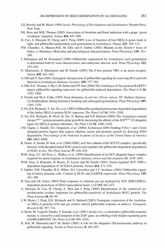
P1: PIB
BLUK053-Bradford August 27, 2006 17:37
DE-REPRESSION OF SEED GERMINATION BY GA SIGNALING 261
J.D. Bewley and M. Black (1994) Seeds: Physiology of Development and Germination. Plenum Press,New York.
P.K. Boss and M.R. Thomas (2002) Association of dwarfism and floral induction with a grape ‘greenrevolution’ mutation. Nature 416, 847–850.
D. Cao, A. Hussain, H. Cheng and J. Peng (2005) Loss of function of four DELLA genes leads tolight- and gibberellin-independent seed germination in Arabidopsis. Planta, 223, 105–113.
P.M. Chandler, A. Marion-Poll, M. Ellis and F. Gubler (2002) Mutants at the Slender1 locus ofbarley cv Himalaya. Molecular and physiological characterization. Plant Physiology 129, 181–190.
I. Debeaujon and M. Koornneef (2000) Gibberellin requirement for Arabidopsis seed germinationis determined both by testa characteristics and embryonic abscisic acid. Plant Physiology 122,415–424.
N. Dharmasiri, S. Dharmasiri and M. Estelle (2005) The F-box protein TIR1 is an auxin receptor.Nature 435, 441–445.
A. Dill and T. Sun (2001) Synergistic derepression of gibberellin signaling by removing RGA and GAIfunction in Arabidopsis thaliana. Genetics 159, 777–785.
A. Dill, S.G. Thomas, J. Hu, C.M. Steber and T.P. Sun (2004) The Arabidopsis F-box protein SLEEPY1targets gibberellin signaling repressors for gibberellin-induced degradation. The Plant Cell 16,1392–1405.
S. Footitt and M.A. Cohn (1995) Seed dormancy in red rice (Oryza sativa). IX: Embryo fructose-2,6-bisphosphate during dormancy breaking and subsequent germination. Plant Physiology 107,1365–1370.
X. Fu, D.E. Richards, T. Ait-Ali, et al. (2002) Gibberellin-mediated proteasome-dependent degradationof the barley DELLA protein SLN1 repressor. The Plant Cell 14, 3191–3200.
X. Fu, D.E. Richards, B. Fleck, D. Xie, N. Burton and N.P. Harberd (2004) The Arabidopsis mutantsleepy1gar2-1 protein promotes plant growth by increasing the affinity of the SCFSLY1 E3 ubiquitinligase for DELLA protein substrates. The Plant Cell 16, 1406–1418.
J.M. Gagne, J. Smalle, D.J. Gingerich, et al. (2004) Arabidopsis EIN3-binding F-box 1 and 2 formubiquitin-protein ligases that repress ethylene action and promote growth by directing EIN3degradation. Proceedings of the National Academy of Sciences of the United States of America101, 6803–6808.
K. Gomi, A. Sasaki, H. Itoh, et al. (2004) GID2, an F-box subunit of the SCF E3 complex, specificallyinteracts with phosphorylated SLR1 protein and regulates the gibberellin-dependent degradationof SLR1 in rice. The Plant Journal 37, 626–634.
W.M. Gray, J.C. del Pozo, L. Walker, et al. (1999) Identification of an SCF ubiquitin-ligase complexrequired for auxin response in Arabidopsis thaliana. Genes and Development 13, 1678–1691.
W.M. Gray, S. Kepinski, D. Rouse, O. Leyser and M. Estelle (2001) Auxin regulates SCF (TIR1)-dependent degradation of AUX/IAA proteins. Nature 414, 271–276.
F. Gubler, P.M. Chandler, R.G. White, D.J. Llewellyn and J.V. Jacobsen (2002) Gibberellin signal-ing in barley aleurone cells. Control of SLN1 and GAMYB expression. Plant Physiology 129,191–200.
H. Guo and J.R. Ecker (2003) Plant responses to ethylene gas are mediated by SCF (EBF1/EBF2)-dependent proteolysis of EIN3 transcription factor. Cell 115, 667–677.
A. Hussain, D. Cao, H. Cheng, Z. Wen and J. Peng (2005) Identification of the conserved ser-ine/threonine residues important for gibberellin-sensitivity of Arabidopsis RGL2 protein. ThePlant Journal 44, 88–99.
L.W. Hynes, J. Peng, D.E. Richards and N. Harberd (2003) Transgenic expression of the Arabidop-sis DELLA proteins GAI and gai confers altered gibberellin response in tobacco. TransgenicResearch 12, 707–714.
A. Ikeda, M. Ueguchi-Tanaka, Y. Sonoda, et al. (2001) slender rice, a constitutive gibberellin responsemutant, is caused by a null mutation of the SLR1 gene, an ortholog of the height-regulating geneGAI/RGA/RHT/D8. The Plant Cell 13, 999–1010.
H. Itoh, M. Matsuoka and C.M. Steber (2003) A role for the ubiquitin–26S-proteasome pathway ingibberellin signaling. Trends in Plant Science 8, 492–497.
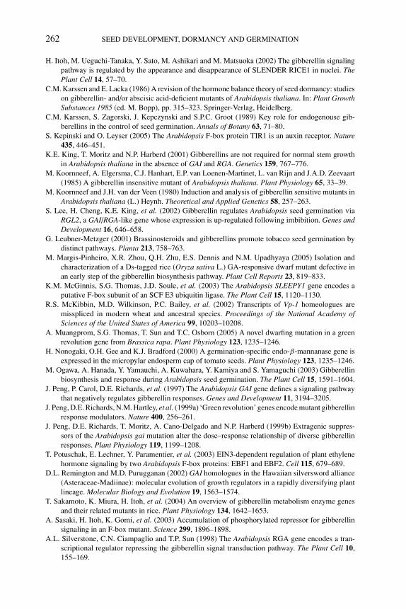
P1: PIB
BLUK053-Bradford August 27, 2006 17:37
262 SEED DEVELOPMENT, DORMANCY AND GERMINATION
H. Itoh, M. Ueguchi-Tanaka, Y. Sato, M. Ashikari and M. Matsuoka (2002) The gibberellin signalingpathway is regulated by the appearance and disappearance of SLENDER RICE1 in nuclei. ThePlant Cell 14, 57–70.
C.M. Karssen and E. Lacka (1986) A revision of the hormone balance theory of seed dormancy: studieson gibberellin- and/or abscisic acid-deficient mutants of Arabidopsis thaliana. In: Plant GrowthSubstances 1985 (ed. M. Bopp), pp. 315–323. Springer-Verlag, Heidelberg.
C.M. Karssen, S. Zagorski, J. Kepczynski and S.P.C. Groot (1989) Key role for endogenouse gib-berellins in the control of seed germination. Annals of Botany 63, 71–80.
S. Kepinski and O. Leyser (2005) The Arabidopsis F-box protein TIR1 is an auxin receptor. Nature435, 446–451.
K.E. King, T. Moritz and N.P. Harberd (2001) Gibberellins are not required for normal stem growthin Arabidopsis thaliana in the absence of GAI and RGA. Genetics 159, 767–776.
M. Koornneef, A. Elgersma, C.J. Hanhart, E.P. van Loenen-Martinet, L. van Rijn and J.A.D. Zeevaart(1985) A gibberellin insensitive mutant of Arabidopsis thaliana. Plant Physiology 65, 33–39.
M. Koornneef and J.H. van der Veen (1980) Induction and analysis of gibberellin sensitive mutants inArabidopsis thaliana (L.) Heynh. Theoretical and Applied Genetics 58, 257–263.
S. Lee, H. Cheng, K.E. King, et al. (2002) Gibberellin regulates Arabidopsis seed germination viaRGL2, a GAI/RGA-like gene whose expression is up-regulated following imbibition. Genes andDevelopment 16, 646–658.
G. Leubner-Metzger (2001) Brassinosteroids and gibberellins promote tobacco seed germination bydistinct pathways. Planta 213, 758–763.
M. Margis-Pinheiro, X.R. Zhou, Q.H. Zhu, E.S. Dennis and N.M. Upadhyaya (2005) Isolation andcharacterization of a Ds-tagged rice (Oryza sativa L.) GA-responsive dwarf mutant defective inan early step of the gibberellin biosynthesis pathway. Plant Cell Reports 23, 819–833.
K.M. McGinnis, S.G. Thomas, J.D. Soule, et al. (2003) The Arabidopsis SLEEPY1 gene encodes aputative F-box subunit of an SCF E3 ubiquitin ligase. The Plant Cell 15, 1120–1130.
R.S. McKibbin, M.D. Wilkinson, P.C. Bailey, et al. (2002) Transcripts of Vp-1 homeologues aremisspliced in modern wheat and ancestral species. Proceedings of the National Academy ofSciences of the United States of America 99, 10203–10208.
A. Muangprom, S.G. Thomas, T. Sun and T.C. Osborn (2005) A novel dwarfing mutation in a greenrevolution gene from Brassica rapa. Plant Physiology 123, 1235–1246.
H. Nonogaki, O.H. Gee and K.J. Bradford (2000) A germination-specific endo-β-mannanase gene isexpressed in the micropylar endosperm cap of tomato seeds. Plant Physiology 123, 1235–1246.
M. Ogawa, A. Hanada, Y. Yamauchi, A. Kuwahara, Y. Kamiya and S. Yamaguchi (2003) Gibberellinbiosynthesis and response during Arabidopsis seed germination. The Plant Cell 15, 1591–1604.
J. Peng, P. Carol, D.E. Richards, et al. (1997) The Arabidopsis GAI gene defines a signaling pathwaythat negatively regulates gibberellin responses. Genes and Development 11, 3194–3205.
J. Peng, D.E. Richards, N.M. Hartley, et al. (1999a) ‘Green revolution’ genes encode mutant gibberellinresponse modulators. Nature 400, 256–261.
J. Peng, D.E. Richards, T. Moritz, A. Cano-Delgado and N.P. Harberd (1999b) Extragenic suppres-sors of the Arabidopsis gai mutation alter the dose–response relationship of diverse gibberellinresponses. Plant Physiology 119, 1199–1208.
T. Potuschak, E. Lechner, Y. Paramentier, et al. (2003) EIN3-dependent regulation of plant ethylenehormone signaling by two Arabidopsis F-box proteins: EBF1 and EBF2. Cell 115, 679–689.
D.L. Remington and M.D. Purugganan (2002) GAI homologues in the Hawaiian silversword alliance(Asteraceae-Madiinae): molecular evolution of growth regulators in a rapidly diversifying plantlineage. Molecular Biology and Evolution 19, 1563–1574.
T. Sakamoto, K. Miura, H. Itoh, et al. (2004) An overview of gibberellin metabolism enzyme genesand their related mutants in rice. Plant Physiology 134, 1642–1653.
A. Sasaki, H. Itoh, K. Gomi, et al. (2003) Accumulation of phosphorylated repressor for gibberellinsignaling in an F-box mutant. Science 299, 1896–1898.
A.L. Silverstone, C.N. Ciampaglio and T.P. Sun (1998) The Arabidopsis RGA gene encodes a tran-scriptional regulator repressing the gibberellin signal transduction pathway. The Plant Cell 10,155–169.
P1: PIB
BLUK053-Bradford August 27, 2006 17:37
DE-REPRESSION OF SEED GERMINATION BY GA SIGNALING 263
A.L. Silverstone, H.S. Jung, A. Dill, H. Kawaide, Y. Kamiya and T.P. Sun (2001) Repressing arepressor: gibberellin-induced rapid reduction of the RGA protein in Arabidopsis. The Plant Cell13, 1555–1566.
A.L. Silverstone, P.Y.A. Mak, E.C. Martinez and T.-P. Sun (1997) The new RGA locus encodes anegative regulator of gibberellin response in Arabidopsis thaliana. Genetics 146, 1087–1099.
J. Smalle, J. Kurepa, P. Yang, E. Babiychuk, S. Kushnir and R.D. Vierstra (2002) Cytokinin growthresponses in Arabidopsis involve the 26S proteasome subunit RPN12. The Plant Cell 14, 17–32.
J. Smalle, J. Kurepa, P. Yang et al. (2003) The pleiotropic role of the 26S proteasome subunit RPN10in Arabidopsis growth and development supports a substrate-specific function in abscisic acidsignaling. The Plant Cell 15, 965–980.
J. Smalle and R.D. Vierstra (2004) The ubiquitin 26S proteasome pathway. Annual Review of PlantBiology 55, 555–590.
C.M. Steber, S.E. Cooney and P. McCourt (1998) Isolation of the GA-response mutant sly1 as asuppressor of ABI1-1 in Arabidopsis thaliana. Genetics 149, 509–521.
C.M. Steber and P. McCourt (2001) A role for brassinosteroids in germination in Arabidopsis. PlantPhysiology 125, 763–769.
D.W. Still and K.J. Bradford (1997) Endo-β-mannanase activity from individual tomato endospermcaps and radicle tips in relation to germination rates. Plant Physiology 113, 21–29.
L.C. Strader, S. Ritchie, J.D. Soule, K.M. McGinnis and C.M. Steber (2004) Recessive-interferingmutations in the gibberellin signaling gene SLEEPY1 are rescued by overexpression of its ho-mologue, SNEEZY. Proceedings of the National Academy of Sciences of the United States ofAmerica 101, 12771–12776.
T.P. Sun and F. Gubler (2004) Molecular mechanism of gibberellin signaling in plants. Annual Reviewof Plant Biology 55, 197–223.
S. Tamura (1991) Historical aspects of gibberellins. In: Gibberellins (eds N. Takahashi, B.O. Phinneyand J. MacMillan), pp. 1–8. Springer-Verlag, New York.
S.G. Thomas, I. Rieu and C.M. Steber (2005) Gibberellin metabolism and signaling. In: Vitamins andHormones, Vol. 72 (ed. G. Litwack), pp. 289–337. Elsevier, London.
L. Tyler, S.G. Thomas, J. Hu, et al. (2004) DELLA Proteins and gibberellin-regulated seed germinationand floral development in Arabidopsis. Plant Physiology 135, 1008–1019.
M. Ueguchi-Tanaka, M. Ashikari, M. Nakajima, et al. (2005) GIBBERELLIN INSENSITIVE DWARF1encodes a soluble receptor for gibberellin. Nature 437, 693–698.
C.N. White, W.M. Proebsting, P. Hedden and C.J. Rivin (2000) Gibberellins and seed development inmaize. I. Evidence that gibberellin/abscisic acid balance governs germination versus maturationpathways. Plant Physiology 122, 1081–1088.
C.N. White and C.J. Rivin (2000) Gibberellins and seed development in maize. II. Gibberellin synthesisinhibition enhances abscisic acid signaling in cultured embryos. Plant Physiology 122, 1089–1097.
L. Xu, F. Liu, E. Lechner, et al. (2002) The SCF (COI1) ubiquitin-ligase complexes are required forjasmonate response in Arabidopsis. The Plant Cell 14, 1919–1935.
S. Yamaguchi, M.W. Smith, R.G. Brown, Y. Kamiya and T. Sun (1998) Phytochrome regulation anddifferential expression of gibberellin 3β-hydroxylase genes in germinating Arabidopsis seeds.The Plant Cell 10, 2115–2126.
X. Zhang, V. Garreton and N.H. Chua (2005) The AIP2 E3 ligase acts as a novel negative regulator ofABA signaling by promoting ABI3 degradation. Genes and Development 19, 1532–1543.
N. Zheng, B.A. Schulman, L. Song, et al. (2002) Structure of the Cul1-Rbx1-Skp1-F boxSkp2 SCFubiquitin ligase complex. Nature 416, 703–709.
P1: OTE/SPH P2: OTE
BLUK053-Bradford September 27, 2006 15:21
11 Mechanisms and genes involvedin germination sensu strictoHiroyuki Nonogaki, Feng Chen and Kent J. Bradford
11.1 Introduction
The term ‘germination’ has a surprisingly large number of meanings. In general,the term is applied to seeds, spores, and pollen to indicate when these quiescentstructures reinitiate growth. Physiologists often consider seed germination to bethe period from imbibition up to the point that embryo growth is initiated and theembryo protrudes through any covering tissues. Seed quality analysts, on the otherhand, do not consider germination to be complete until the seedling has grownsufficiently to be able to observe and evaluate the root, hypocotyl, and cotyledons(ISTA, 1999). Growers often consider germination to be completed only when theseedling emerges through the soil. For this chapter, we will consider germination inthe strict sense (sensu stricto; Perino and Come, 1991) to be the period from the startof imbibition of a dry seed until the embryo (usually the radicle) first emerges fromany tissues enclosing it. Processes that are associated uniquely with germination,such as storage reserve mobilization and de-etiolation, still occur after this point, butmost physiological processes in seedlings are similar to those occurring during plantgrowth. Gene expression patterns during germination, however, are quite distinctfrom those in other stages of the plant life cycle (Czechowski et al., 2005; Ma et al.,2005).
In addition to restricting discussion to germination sensu stricto, we will alsofocus primarily on processes associated mechanistically with the initiation and com-pletion of embryo emergence through any covering tissues. Other chapters in thisbook look in depth at the regulatory and signaling pathways involved in germinationand dormancy, so we will not repeat that material except to discuss the regulationof the genes and processes directly involved in embryo emergence. We also will notaddress reserve mobilization in any depth, as the majority of that process occursafter germination sensu stricto. Instead, our focus will be on the mechanisms andgenes that are involved in the progress of a seed from imbibition through radicleemergence.
11.2 Imbibition and water relations of seed germination
Beginning in most cases from an air-dry state, seeds must first imbibe water inorder to activate metabolic processes. Seeds having impermeable seed coats that
264
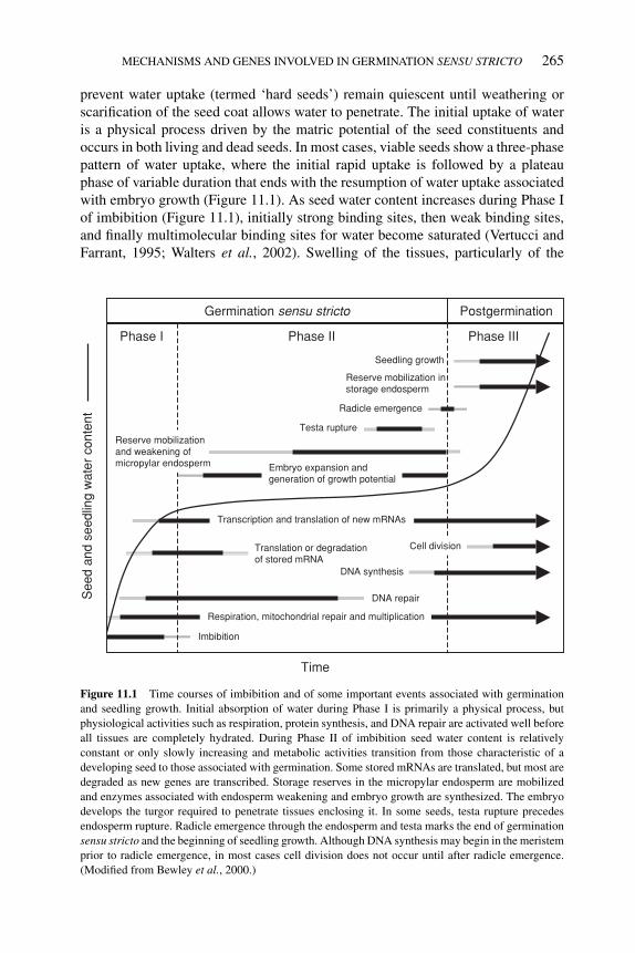
P1: OTE/SPH P2: OTE
BLUK053-Bradford September 27, 2006 15:21
MECHANISMS AND GENES INVOLVED IN GERMINATION SENSU STRICTO 265
prevent water uptake (termed ‘hard seeds’) remain quiescent until weathering orscarification of the seed coat allows water to penetrate. The initial uptake of wateris a physical process driven by the matric potential of the seed constituents andoccurs in both living and dead seeds. In most cases, viable seeds show a three-phasepattern of water uptake, where the initial rapid uptake is followed by a plateauphase of variable duration that ends with the resumption of water uptake associatedwith embryo growth (Figure 11.1). As seed water content increases during Phase Iof imbibition (Figure 11.1), initially strong binding sites, then weak binding sites,and finally multimolecular binding sites for water become saturated (Vertucci andFarrant, 1995; Walters et al., 2002). Swelling of the tissues, particularly of the
See
d an
d se
edlin
g w
ater
con
tent
Germination sensu stricto Postgermination
Phase I Phase II Phase III
DNA repair
DNA synthesis
Cell divisionTranslation or degradation of stored mRNA
Reserve mobilization and weakening of micropylar endosperm
Reserve mobilization in storage endosperm
Radicle emergence
Seedling growth
Imbibition
Testa rupture
Time
Transcription and translation of new mRNAs
Respiration, mitochondrial repair and multiplication
Embryo expansion and generation of growth potential
Figure 11.1 Time courses of imbibition and of some important events associated with germination
and seedling growth. Initial absorption of water during Phase I is primarily a physical process, but
physiological activities such as respiration, protein synthesis, and DNA repair are activated well before
all tissues are completely hydrated. During Phase II of imbibition seed water content is relatively
constant or only slowly increasing and metabolic activities transition from those characteristic of a
developing seed to those associated with germination. Some stored mRNAs are translated, but most are
degraded as new genes are transcribed. Storage reserves in the micropylar endosperm are mobilized
and enzymes associated with endosperm weakening and embryo growth are synthesized. The embryo
develops the turgor required to penetrate tissues enclosing it. In some seeds, testa rupture precedes
endosperm rupture. Radicle emergence through the endosperm and testa marks the end of germination
sensu stricto and the beginning of seedling growth. Although DNA synthesis may begin in the meristem
prior to radicle emergence, in most cases cell division does not occur until after radicle emergence.
(Modified from Bewley et al., 2000.)
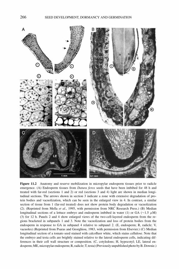
P1: OTE/SPH P2: OTE
BLUK053-Bradford September 27, 2006 15:21
266 SEED DEVELOPMENT, DORMANCY AND GERMINATION
Figure 11.2 Anatomy and reserve mobilization in micropylar endosperm tissues prior to radicle
emergence. (A) Endosperm tissues from Datura ferox seeds that have been imbibed for 48 h and
treated with far-red (sections 1 and 2) or red (sections 3 and 4) light are shown in median longi-
tudinal sections. The arrows shown in section 3 indicate a zone with extensive degradation of pro-
tein bodies and vacuolization, which can be seen in the enlarged view in 4. In contrast, a similar
section of tissue from 1 (far-red treated) does not show protein body degradation or vacuolization
(2). (Reprinted from Mella et al., 1995, with permission from NRC Research Press.) (B) Median
longitudinal sections of a lettuce embryo and endosperm imbibed in water (1) or GA (∼1.5 μM)
(3) for 12 h. Panels 2 and 4 show enlarged views of the two-cell-layered endosperm from the re-
gions bracketed in subpanels 1 and 3. Note the vacuolization and loss of protein bodies from the
endosperm in response to GA in subpanel 4 relative to subpanel 2. (E, endosperm; R, radicle; V,
vacuoles) (Reprinted from Psaras and Georghiou, 1983, with permission from Elsevier.) (C) Median
longitudinal section of a tomato seed stained with calcofluor white, which stains cellulose. Note that
the embryo and testa cells are brightly stained relative to the lateral endosperm cells, indicating dif-
ferences in their cell wall structure or composition. (C, cotyledons; H, hypocotyl; LE, lateral en-
dosperm; ME, micropylar endosperm; R, radicle; T, testa) (Previously unpublished photo by B. Downie.)
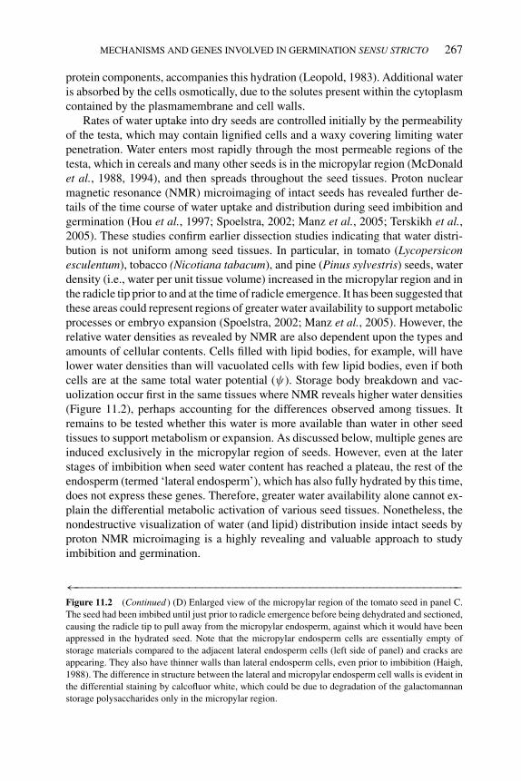
P1: OTE/SPH P2: OTE
BLUK053-Bradford September 27, 2006 15:21
MECHANISMS AND GENES INVOLVED IN GERMINATION SENSU STRICTO 267
protein components, accompanies this hydration (Leopold, 1983). Additional wateris absorbed by the cells osmotically, due to the solutes present within the cytoplasmcontained by the plasmamembrane and cell walls.
Rates of water uptake into dry seeds are controlled initially by the permeabilityof the testa, which may contain lignified cells and a waxy covering limiting waterpenetration. Water enters most rapidly through the most permeable regions of thetesta, which in cereals and many other seeds is in the micropylar region (McDonaldet al., 1988, 1994), and then spreads throughout the seed tissues. Proton nuclearmagnetic resonance (NMR) microimaging of intact seeds has revealed further de-tails of the time course of water uptake and distribution during seed imbibition andgermination (Hou et al., 1997; Spoelstra, 2002; Manz et al., 2005; Terskikh et al.,2005). These studies confirm earlier dissection studies indicating that water distri-bution is not uniform among seed tissues. In particular, in tomato (Lycopersiconesculentum), tobacco (Nicotiana tabacum), and pine (Pinus sylvestris) seeds, waterdensity (i.e., water per unit tissue volume) increased in the micropylar region and inthe radicle tip prior to and at the time of radicle emergence. It has been suggested thatthese areas could represent regions of greater water availability to support metabolicprocesses or embryo expansion (Spoelstra, 2002; Manz et al., 2005). However, therelative water densities as revealed by NMR are also dependent upon the types andamounts of cellular contents. Cells filled with lipid bodies, for example, will havelower water densities than will vacuolated cells with few lipid bodies, even if bothcells are at the same total water potential (ψ). Storage body breakdown and vac-uolization occur first in the same tissues where NMR reveals higher water densities(Figure 11.2), perhaps accounting for the differences observed among tissues. Itremains to be tested whether this water is more available than water in other seedtissues to support metabolism or expansion. As discussed below, multiple genes areinduced exclusively in the micropylar region of seeds. However, even at the laterstages of imbibition when seed water content has reached a plateau, the rest of theendosperm (termed ‘lateral endosperm’), which has also fully hydrated by this time,does not express these genes. Therefore, greater water availability alone cannot ex-plain the differential metabolic activation of various seed tissues. Nonetheless, thenondestructive visualization of water (and lipid) distribution inside intact seeds byproton NMR microimaging is a highly revealing and valuable approach to studyimbibition and germination.
←−−−−−−−−−−−−−−−−−−−−−−−−−−−−−−−−−−−−−−−−−−−−−−−−−−−−−−−−−−Figure 11.2 (Continued ) (D) Enlarged view of the micropylar region of the tomato seed in panel C.
The seed had been imbibed until just prior to radicle emergence before being dehydrated and sectioned,
causing the radicle tip to pull away from the micropylar endosperm, against which it would have been
appressed in the hydrated seed. Note that the micropylar endosperm cells are essentially empty of
storage materials compared to the adjacent lateral endosperm cells (left side of panel) and cracks are
appearing. They also have thinner walls than lateral endosperm cells, even prior to imbibition (Haigh,
1988). The difference in structure between the lateral and micropylar endosperm cell walls is evident in
the differential staining by calcofluor white, which could be due to degradation of the galactomannan
storage polysaccharides only in the micropylar region.
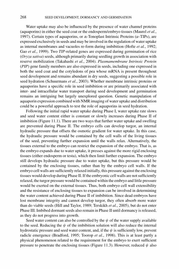
P1: OTE/SPH P2: OTE
BLUK053-Bradford September 27, 2006 15:21
268 SEED DEVELOPMENT, DORMANCY AND GERMINATION
Water uptake may also be influenced by the presence of water channel proteins(aquaporins) in either the seed coat or the endosperm/embryo tissues (Maurel et al.,1997). Certain types of aquaporins, or α-Tonoplast Intrinsic Proteins (α-TIPs), areexpressed exclusively in seeds and may be involved in the regulation of water uptakeas internal membranes and vacuoles re-form during imbibition (Hofte et al., 1992;Gao et al., 1999). Two TIP-related genes are expressed during germination of rice(Oryza sativa) seeds, although primarily during seedling growth in association withreserve mobilization (Takahashi et al., 2004). Plasmamembrane Intrinsic Protein(PIP) gene family members are also expressed in seeds, including one expressed inboth the seed coat and the cotyledons of pea whose mRNA is present throughoutseed development and remains abundant in dry seeds, suggesting a possible role inseed hydration (Schuurmans et al., 2003). Whether membrane intrinsic proteins oraquaporins have a specific role in seed imbibition or are primarily associated withinter- and intracellular water transport during seed development and germinationremains an intriguing but largely unexplored question. Genetic manipulation ofaquaporin expression combined with NMR imaging of water uptake and distributioncould be a powerful approach to test the role of aquaporins in seed hydration.
Following the initial rapid water uptake during Phase I, water uptake rate slowsand seed water content either is constant or slowly increases during Phase II ofimbibition (Figure 11.1). There are two ways that further water uptake and swellingare prevented during Phase II. The embryo cells can develop turgor, an internalhydraulic pressure that offsets the osmotic gradient for water uptake. In this case,the hydraulic pressure would be contained by the cell walls of the living tissuesof the seed, preventing further expansion until the walls relax. Alternatively, thetissues external to the embryo can restrict the expansion of the embryo. That is, asthe embryo expands due to water uptake, it presses against the more rigid enclosingtissues (either endosperm or testa), which then limit further expansion. The embryostill develops hydraulic pressure due to water uptake, but this pressure would becontained by the enclosing tissues, rather than by the embryo cell walls. If theembryo cell walls are sufficiently relaxed initially, this pressure against the enclosingtissues would develop during Phase II. If the embryonic cell walls are not sufficientlyrelaxed, the turgor pressure would be contained within the embryo and little pressurewould be exerted on the external tissues. Thus, both embryo cell wall extensibilityand the resistance of enclosing tissues to expansion can be involved in determiningthe water content achieved during Phase II of imbibition. Since dead embryos havelost membrane integrity and cannot develop turgor, they often absorb more waterthan do viable seeds (Hill and Taylor, 1989; Terskikh et al., 2005), but do not enterPhase III. Imbibed dormant seeds also remain in Phase II until dormancy is released,as they do not progress into growth.
Seed water content can also be controlled by the ψ of the water supply availableto the seed. Reducing the ψ of the imbibition solution will also reduce the internalhydrostatic pressure and seed water content, and, if the ψ is sufficiently low, preventradicle emergence (Bradford, 1995; Toorop et al., 1998). This is at least partly aphysical phenomenon related to the requirement for the embryo to exert sufficientpressure to penetrate the enclosing tissues (Figure 11.3). However, reduced ψ also
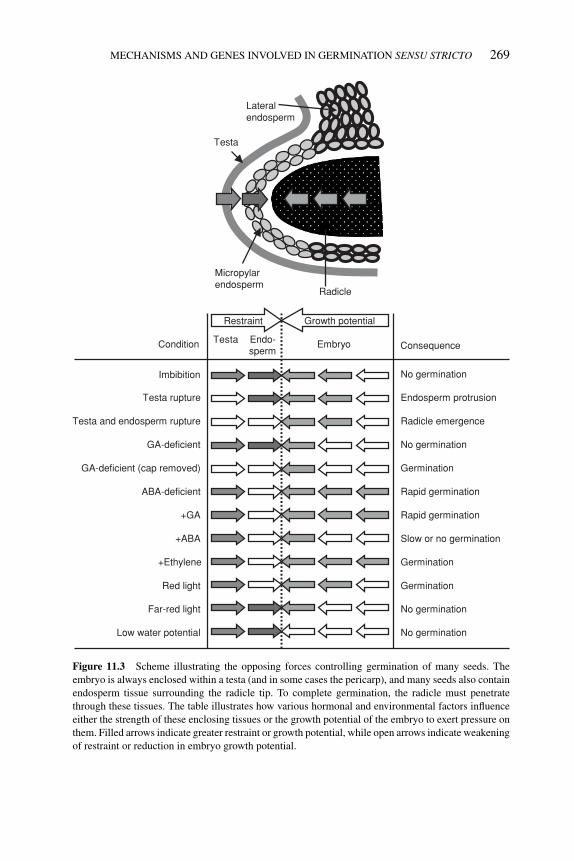
P1: OTE/SPH P2: OTE
BLUK053-Bradford September 27, 2006 15:21
MECHANISMS AND GENES INVOLVED IN GERMINATION SENSU STRICTO 269
Testa
Radicle
Micropylar endosperm
Lateral endosperm
sperm
Imbibition
+ABA
Testa Endo- Embryo
No germination
Slow or no germination
Condition Consequence
Testa rupture Endosperm protrusion
Testa and endosperm rupture Radicle emergence
GA-deficient No germination
ABA-deficient Rapid germination
+GA Rapid germination
Far-red light No germination
Red light Germination
Low water potential No germination
+Ethylene Germination
GA-deficient (cap removed) Germination
Restraint Growth potential
Figure 11.3 Scheme illustrating the opposing forces controlling germination of many seeds. The
embryo is always enclosed within a testa (and in some cases the pericarp), and many seeds also contain
endosperm tissue surrounding the radicle tip. To complete germination, the radicle must penetrate
through these tissues. The table illustrates how various hormonal and environmental factors influence
either the strength of these enclosing tissues or the growth potential of the embryo to exert pressure on
them. Filled arrows indicate greater restraint or growth potential, while open arrows indicate weakening
of restraint or reduction in embryo growth potential.
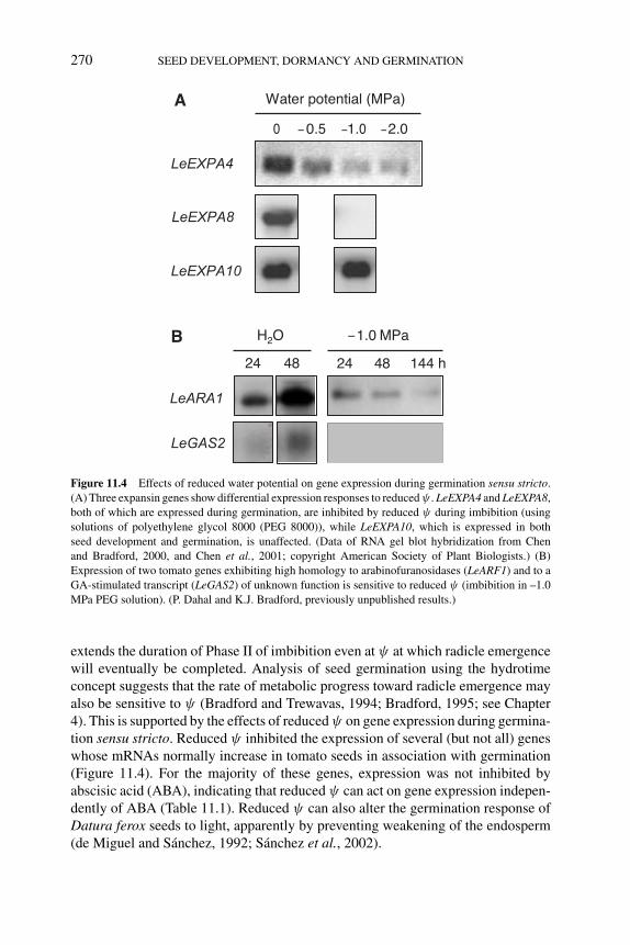
P1: OTE/SPH P2: OTE
BLUK053-Bradford September 27, 2006 15:21
270 SEED DEVELOPMENT, DORMANCY AND GERMINATION
24 48
H2O 1.0 MPa
LeARA1
24 48 144 h
LeGAS2
Water potential (MPa)
0 −0.5 −1.0 −2.0
LeEXPA4
LeEXPA8
LeEXPA10
A
B −
Figure 11.4 Effects of reduced water potential on gene expression during germination sensu stricto.
(A) Three expansin genes show differential expression responses to reduced ψ . LeEXPA4 and LeEXPA8,
both of which are expressed during germination, are inhibited by reduced ψ during imbibition (using
solutions of polyethylene glycol 8000 (PEG 8000)), while LeEXPA10, which is expressed in both
seed development and germination, is unaffected. (Data of RNA gel blot hybridization from Chen
and Bradford, 2000, and Chen et al., 2001; copyright American Society of Plant Biologists.) (B)
Expression of two tomato genes exhibiting high homology to arabinofuranosidases (LeARF1) and to a
GA-stimulated transcript (LeGAS2) of unknown function is sensitive to reduced ψ (imbibition in –1.0
MPa PEG solution). (P. Dahal and K.J. Bradford, previously unpublished results.)
extends the duration of Phase II of imbibition even at ψ at which radicle emergencewill eventually be completed. Analysis of seed germination using the hydrotimeconcept suggests that the rate of metabolic progress toward radicle emergence mayalso be sensitive to ψ (Bradford and Trewavas, 1994; Bradford, 1995; see Chapter4). This is supported by the effects of reduced ψ on gene expression during germina-tion sensu stricto. Reduced ψ inhibited the expression of several (but not all) geneswhose mRNAs normally increase in tomato seeds in association with germination(Figure 11.4). For the majority of these genes, expression was not inhibited byabscisic acid (ABA), indicating that reduced ψ can act on gene expression indepen-dently of ABA (Table 11.1). Reduced ψ can also alter the germination response ofDatura ferox seeds to light, apparently by preventing weakening of the endosperm(de Miguel and Sanchez, 1992; Sanchez et al., 2002).
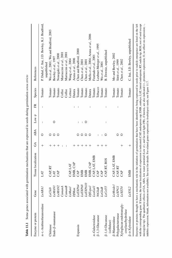
P1: OTE/SPH P2: OTE
BLUK053-Bradford September 27, 2006 15:21
Tabl
e11
.1S
om
eg
enes
asso
ciat
edw
ith
ger
min
atio
nm
ech
anis
ms
that
are
exp
ress
edin
seed
sd
uri
ng
ger
min
atio
nse
nsu
stri
cto
En
zym
eo
rp
rote
inG
ene
Tis
sue
loca
liza
tio
nG
AA
BA
Low
ψF
RS
pec
ies
Ref
eren
ces
α–
l-A
rab
ino
fura
no
sid
ase
LeA
RA
1L
AT
+O
––
To
mat
oP.
Dah
al,
C.
Itai
,J.
D.
Bew
ley,
K.J
.B
rad
ford
,
un
pu
bli
shed
Ch
itin
ase
LeC
hi9
CA
P,R
T+
OT
om
ato
Wu
etal
.,2
00
1;
Wu
and
Bra
dfo
rd,2
00
3
En
do
-β-m
ann
anas
eL
eMA
N1
LA
T+
OO
To
mat
oB
ewle
yet
al.,
19
97
LeM
AN
2C
AP
+O
To
mat
oN
on
ogak
iet
al.,
20
00
Cam
anA
–C
off
eeM
arra
ccin
iet
al.,
20
01
Cam
anB
–C
off
eeM
arra
ccin
iet
al.,
20
01
LsM
an1
CA
P,L
AT
Let
tuce
Wan
get
al.,
20
04
DfM
anE
MB
,C
AP
+–
Dat
ura
Ara
na
etal
.,2
00
6
Ex
pan
sin
LeE
XPA
4C
AP
+O
––
To
mat
oC
hen
and
Bra
dfo
rd,2
00
0
LeE
XPA
8E
MB
+O
–T
om
ato
Ch
enet
al.,
20
01
LeE
XPA
10E
MB
+O
OT
om
ato
Ch
enet
al.,
20
01
DfE
xpa1
EM
B,C
AP
+O
–D
atu
raM
ella
etal
.,2
00
4;
Ara
na
etal
.,2
00
6
α–
Gal
acto
sid
ase
Lea
Gal
1C
AP,
LA
T,
EM
BT
om
ato
Feu
rtad
oet
al.,
20
01
β-1
,3-G
luca
nas
eN
tGlu
BC
AP
+–
To
bac
coL
eub
ner
-Met
zger
etal
.,1
99
5
LeG
luB
CA
P+
–T
om
ato
Wu
etal
.,2
00
1
β-1
,4-G
luca
nas
e
(cel
lula
se)
LeC
el55
CA
P,R
T,
RO
S+
O–
–T
om
ato
B.
Dow
nie
,u
np
ub
lish
ed
β-M
ann
osi
das
eL
eMsi
de1
CA
P,L
AT
,E
MB
To
mat
oM
oan
dB
ewle
y,2
00
2
Po
lygal
actu
ron
ase
LeX
PG
1C
AP,
RT
O–
To
mat
oS
itri
tet
al.,
19
99
Xy
log
luca
nen
do
tran
sgly
-
cosy
lase
/hy
dro
lase
LeX
ET
4C
AP
+O
To
mat
oC
hen
etal
.,2
00
2
β-d
-Xy
losi
das
eL
eXY
L2
EM
B+
To
mat
oC
.It
ai,J.
D.B
ewle
y,u
np
ub
lish
ed
Enzy
mes
or
pro
tein
sth
ought
tohav
ea
mec
han
isti
cro
lein
the
init
iati
on
of
ger
min
atio
nth
athav
ebee
nid
enti
fied
asbei
ng
expre
ssed
inse
eds
pri
or
tora
dic
leem
ergen
cear
eli
sted
on
the
left
wit
hth
eco
rres
pondin
ggen
e.If
know
n,th
eti
ssue
loca
liza
tion
of
expre
ssio
nis
indic
ated
(CA
P,en
dosp
erm
cap;
EM
B,em
bry
o;
LA
T,la
tera
len
dosp
erm
;R
T,ra
dic
leti
p;
RO
S,re
stof
seed
exce
pt
mic
ropyla
rti
p).
The
qual
itat
ive
effe
cts
of
GA
,A
BA
,lo
ww
ater
pote
nti
al(L
owψ
),an
dfa
r-re
dli
ght
(FR
),if
know
n,
are
also
indic
ated
(+,
pro
mote
sex
pre
ssio
n;
O,
no
effe
cton
expre
ssio
n;
–,
inhib
its
expre
ssio
n;
bla
nk,in
form
atio
nnot
avai
lable
).S
eete
xt
for
det
ails
.F
or
rela
ted
gen
esex
pre
ssed
inA
rabi
dops
isse
eds,
see
Fig
ure
11.7
.
271
P1: OTE/SPH P2: OTE
BLUK053-Bradford September 27, 2006 15:21
272 SEED DEVELOPMENT, DORMANCY AND GERMINATION
Imbibing seeds at reduced ψ is the basis of seed priming, a process utilized in theseed industry to enhance germination speed and break thermo- and photodormancy(Halmer, 2004). Microarray and proteomic studies in Arabidopsis and Brassicaoleracea have found that the majority of genes whose expression is altered (ei-ther increased or decreased) during germination sensu stricto show similar changesduring priming at reduced ψ (Gallardo et al., 2001; Soeda et al., 2005). However,specific subsets of genes whose expression increases during germination increasedless or not at all during priming, consistent with the results discussed above. Anumber of genes associated with stress tolerance or pathogen defense were amongthis group, suggesting that these genes may be expressed only when radicle emer-gence is imminent (Soeda et al., 2005). Thus, reduced ψ has specific effects on geneexpression and protein synthesis in addition to lowering seed water content.
11.3 Testa/endosperm restraint and embryo growth potential
Testa mutants exhibiting reduced or enhanced dormancy characteristics demonstratethat the presence and/or properties of tissues external to the embryo can affect thetiming and completion of germination (Debeaujon et al., 2000; Downie et al., 2004;see Chapter 2). In some seeds, such as those of the the Brassicaceae, the testa andendosperm tissues enclosing the embryo are thin and relatively weak, and maysplit during imbibition, therefore presenting little resistance to penetration by theembryo (Figure 11.3; Schopfer et al., 1979). In other cases, such as tobacco, thetesta may rupture during imbibition, but germination may still be controlled byan endosperm layer enclosing the embryo (Manz et al., 2005). In tomato, pepper(Capsicum annuum), Datura, or lettuce (Lactuca sativa), the primary regulator ofgermination appears to be the endosperm tissues enclosing the embryo (Watkinsand Cantliffe, 1983b; Groot and Karssen, 1987; Sanchez et al., 1990; Sung et al.,1998), although the testa may still contribute to a lesser extent (Hilhorst and Downie,1996). Even in the absence of an external restraint, the embryo tissues must expandin order to protrude, so processes related to cell growth (embryo growth potential)can also regulate germination (de Miguel and Sanchez, 1992; Toorop et al., 1998).It is therefore the balance between the restraint exerted by enclosing tissues andthe force or growth potential that can be generated by the embryo that in the enddetermines whether radicle emergence will occur (Figure 11.3). Here we will reviewthe roles of each of these components and possible mechanisms underlying theirinvolvement in germination.
11.3.1 Testa and pericarp
Seeds are enclosed in a testa that develops from the integuments of the ovule.The testa is a multifunctional organ. During seed development, the testa plays animportant role in embryo nutrition, as the phloem ends in the testa, and all sugars,amino acids, and mineral ions transported to the developing seed are unloaded inthe testa and move apoplastically to the embryo and endosperm (Patrick and Offler,
P1: OTE/SPH P2: OTE
BLUK053-Bradford September 27, 2006 15:21
MECHANISMS AND GENES INVOLVED IN GERMINATION SENSU STRICTO 273
2001). In mature seeds, the testa protects the seed against detrimental agents from theenvironment. As a result of this protective function, the testa may impose dormancyon the seeds due to its impermeability to water and/or oxygen, its mechanical strengththat resists radicle protrusion, or both. The function of the testa in seed dormancyhas been extensively studied using mutants in Arabidopsis and tomato (Hilhorst andDownie, 1996; Debeaujon et al., 2000; Downie et al., 2003, 2004) and transgenicmodification in tobacco (Leubner-Metzger, 2002, 2005; see Chapter 2). Some seeds(i.e., dispersal units) also contain pericarp tissues, which develop from the ovary wall.Such seeds are actually dry fruits in which the pericarp and testa are fused, as in grass(‘caryopses’), Compositae (‘achenes’), and Umbelliferae (‘schizocarps’) species.As an embryo-covering tissue, the pericarp may also regulate seed germination byrestricting radicle penetration (Ogawa and Iwabuchi, 2001).
Because of the integumental origin of the testa, its properties are mainly con-trolled by the genetics of the maternal plant. However, communication between thematernal testa and the embryo and/or endosperm can also affect testa attributes intomato (Downie et al., 2003). After seed maturity and desiccation, the testa is gen-erally dead and considered to be physiologically inactive, although in some seeds,such as tobacco, a living cell layer that is interposed between the dead outer testaand the endosperm has been identified (Matzke et al., 1993). Testa rupture is asso-ciated, at least partly, with an increase of water uptake during imbibition that leadsto embryo swelling and initial growth. In some seeds, splitting or cracking of thetesta can occur prior to, and distinct from, radicle emergence through endospermenvelopes inside the testa (see below). Interestingly, such testa cracking generallydoes not occur when dormant seeds are imbibed (Leubner-Metzger, 2002; Penfieldet al., 2004; Liu et al., 2005a), indicating that release of dormancy is associated witheither weakening of the testa or relaxation of the constraints on expansion of the em-bryo. Such after-ripening-mediated testa rupture can be replaced by overexpressionof a class I β-1,3-glucanase gene in the seed-covering layers (Leubner-Metzger,2002). Thus, the extent of embryo swelling and/or testa strength during Phase II ofimbibition seems to be under physiological control.
11.3.2 Endosperm
Endosperm is formed from fusion of two haploid maternal nuclei and one haploidpaternal nucleus during double fertilization. Endosperm cells are rich in food re-serves such as carbohydrates (polysaccharides), oils, and proteins. In some seeds,absorption of the food reserves from the endosperm by the embryo is completedearly in seed development. In these seeds, such as bean or pea, the endosperm hasdisappeared or is reduced to a few cell layers at seed maturity. In contrast, someseeds, such as cereals, contain a large dead endosperm and a few living cell layers ofendosperm (called the aleurone layer) at maturity. In these seeds, the embryo is notcovered by the endosperm tissue. The dead endosperm in the latter types of seedsserves solely as a food reserve for early seedling growth. The degradation of the ce-real endosperm has been studied intensively, especially the synthesis and secretionof amylases and proteases from the aleurone layer and regulation of their synthesis
P1: OTE/SPH P2: OTE
BLUK053-Bradford September 27, 2006 15:21
274 SEED DEVELOPMENT, DORMANCY AND GERMINATION
by gibberellins (GA) and abcisic acid (ABA) (Jacobsen et al., 2002; Gubler et al.,2005).
In contrast, the seeds of many plant species at maturity contain living endospermtissues that completely enclose the embryo. Examples of such seeds include tomato,tobacco, pepper, and Datura (all of which are in the Solanaceae family); lettuce(Compositae); muskmelon (Cucumis melo, Cucurbitaceae); and coffee (Coffea ara-bica, Rubiaceae). In these seeds, the endosperm possesses two biological functions:first, as in other types of seeds, to provide reserves for seed germination and seedlinggrowth; and second, to control the timing of germination. Because of its completeenclosure of the embryo, the endosperm serves as a physical constraint that has tobe overcome in order for the radicle tip to emerge. The endosperm in such seedsfunctions as a major determinant of ‘coat-imposed’ dormancy.
The regulatory role of the endosperm in germination is realized mainly throughthe micropylar endosperm. In tomato and other endospermic seeds, the micropylarendosperm forms a cone that encloses the radicle tip. It has therefore been calledthe ‘endosperm cap’. The micropylar endosperm can be anatomically distinguishedfrom the remainder of the endosperm, or the lateral endosperm. In tomato seeds, forexample, the micropylar endosperm is composed of smaller cells with thinner cellwalls compared to the lateral endosperm (Figure 11.2; Haigh, 1988).
The micropylar endosperm undergoes dramatic changes during germinationsensu stricto. The inner surface of the endosperm cell walls is initially smoothand becomes increasingly degraded following imbibition (Sanchez et al., 1990;Nonogaki et al., 1992; Welbaum et al., 1995). Similar changes occur in the mi-cropylar region of the megagametophyte that encloses the embryo of spruce (Piceaglauca) seeds (Downie et al., 1997a). The storage reserves in the micropylar cells aredegraded and the cells become highly vacuolated prior to radicle emergence, whilethese changes do not occur in adjacent cells of the lateral endosperm until after radi-cle emergence (Figure 11.2). The division between the two types of endosperm issharp, with adjacent cells exhibiting either a micropylar or a lateral endosperm typeof response. These cellular changes are associated with physical weakening of theendosperm cap tissue. Using radicle-sized probes and force analyzers, several groupshave measured the physical force that is needed to break through the isolated mi-cropylar endosperms (or megagametophyte/nucellar tissue) of tomato, muskmelon,pepper, coffee, and spruce seeds (Watkins and Cantliffe, 1983b; Groot and Karssen,1987; Welbaum et al., 1995; Downie et al., 1997b; Chen and Bradford, 2000; Wuet al., 2001; da Silva et al., 2004). In all cases, the micropylar endosperm physicallyweakened following imbibition and prior to radicle emergence (Figure 11.5).
Endosperm weakening is a prerequisite for radicle protrusion in such seeds.In tomato, this conclusion was drawn based on a study with GA-deficient mutant(gib-1) seeds that fail to complete germination without exogenous GA. When themicropylar endosperm of gib-1 seeds is removed, however, embryo expansion canoccur without GA (Figure 11.4) (Groot and Karssen, 1987). Application of GA re-sults in weakening of the endosperm cap and allows radicle protrusion (Figure 11.5).In some seeds, the endosperm cap region stretches and protrudes through the testa,forming a protuberance as the embryo expands until the cap is penetrated by theradicle tip (Watkins and Cantliffe, 1983a; de Miguel and Sanchez, 1992; da Silva
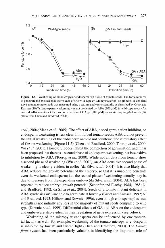
P1: OTE/SPH P2: OTE
BLUK053-Bradford September 27, 2006 15:21
MECHANISMS AND GENES INVOLVED IN GERMINATION SENSU STRICTO 275
1.2(A) (B)
1
0.8
0.6
0.40 12 24 36 48
ABA GA + ABA
GA
Wild-type seeds
Pun
ctur
e fo
rce
(N)
Imbibition time (h) Imbibition time (h)
gib-1 mutant seeds
H2O
H2O
60 0 12 24 36 48 60
Figure 11.5 Weakening of the micropylar endosperm cap tissue of tomato seeds. The force required
to penetrate the excised endosperm caps of (A) wild-type cv. Moneymaker or (B) gibberellin-deficient
gib-1 mutant tomato seeds was measured using a texture analyzer essentially as described by Groot and
Karssen (1987). Endosperm weakening was not prevented by ABA (100 μM) in wild-type seeds (A);
nor did ABA counteract the promotive action of GA4+7 (100 μM) on weakening in gib-1 seeds (B).
(Data from Chen and Bradford, 2000.)
et al., 2004; Manz et al., 2005). The effect of ABA, a seed germination inhibitor, onendosperm weakening is less clear. In imbibed tomato seeds, ABA did not preventthe initial weakening of the endosperm and did not counteract the stimulatory effectof GA on weakening (Figure 11.5) (Chen and Bradford, 2000; Toorop et al., 2000;Wu et al., 2001). However, it does inhibit the completion of germination, and it hasbeen proposed that there is a second phase of endosperm weakening that is sensitiveto inhibition by ABA (Toorop et al., 2000). While not all data from tomato showa second phase of weakening (Wu et al., 2001), an ABA-sensitive second phase ofweakening is clearly evident in coffee (da Silva et al., 2004). It is also likely thatABA reduces the growth potential of the embryo, so that it is unable to penetrateeven the weakened endosperm; i.e., the second phase of weakening actually may bedue to pressure from the expanding embryo (da Silva et al., 2004). ABA has beenreported to reduce embryo growth potential (Schopfer and Plachy, 1984, 1985; Niand Bradford, 1992; da Silva et al., 2004). Seeds of a tomato mutant deficient inABA synthesis (sitw) are able to germinate at lower ψ (Groot and Karssen, 1992; Niand Bradford, 1993; Hilhorst and Downie, 1996), even though endosperm plus testastrength is not initially any less in the majority of mutant seeds compared to wildtype (Downie et al., 1999). Differential effects of GA and ABA on the endospermand embryo are also evident in their regulation of gene expression (see below).
Weakening of the micropylar endosperm can be influenced by environmen-tal factors as well. For example, weakening of the tomato micropylar endospermis inhibited by low ψ and far-red light (Chen and Bradford, 2000). The Daturaferox system has been particularly valuable in identifying the important role of
P1: OTE/SPH P2: OTE
BLUK053-Bradford September 27, 2006 15:21
276 SEED DEVELOPMENT, DORMANCY AND GERMINATION
phytochrome in regulating endosperm weakening in that species (de Miguel andSanchez, 1992; de Miguel et al., 2000; Sanchez et al., 2002; Mella et al., 2004;Arana et al., 2006).
The effects of the various factors discussed above on the restraint offered by thetesta and endosperm or on the growth potential of the embryo are summarized inFigure 11.3. Not all of these situations are applicable to all species, but the overallscheme illustrates how different factors can affect either the envelope restraint orthe ability of the embryo to exert the pressure required for emergence, resulting inrapid, normal, slow, or no germination.
11.3.3 Cell wall proteins and hydrolases involved in weakeningof covering tissues
The mechanisms leading to endosperm weakening have been of interest to seed bi-ologists since at least the early 1960s, when Ikuma and Thimann (1963) suggested,based on their studies of lettuce seed germination, that the endosperm is weakenedthrough enzyme-mediated degradation. Because cell walls determine the shape andstrength of cells, cell wall hydrolases have been the focus of many subsequent stud-ies. In the past two decades, the biochemical and molecular mechanisms responsiblefor endosperm cell wall modification have been extensively studied. A number ofcell wall proteins and hydrolases and the genes encoding them have been identifiedthat are likely to have a mechanistic role in endosperm weakening (summarized inTable 11.1 and Figure 11.6).
11.3.3.1 ExpansinsExpansins are novel cell wall proteins that have the ability to cause extension ofisolated cell walls. Expansin genes are highly conserved in higher plants and are oftenpresent as gene families with multiple members (Cosgrove et al., 2002). In silicoanalyses of the Arabidopsis and rice genomes revealed the existence of 26 and 33expansin genes (α-type) in the two genomes, respectively, and 14 expansin geneshave been reported in tomato (Cosgrove, 2005). Multiple expansin genes often areexpressed in association with developmental events such as root hair initiation orfruit growth (Brummell et al., 1999; Harrison et al., 2001; Cho and Cosgrove, 2002).They are also involved in processes such as fruit ripening and abscission where cellwall modification occurs without expansion (Rose et al., 1997; Cho and Cosgrove,2000). Thus, it is likely that expansins may be involved in both embryo growth andendosperm weakening during germination.
At least three expansin genes are expressed in tomato seeds during germination.One of them, LeEXPA4 (previously termed LeEXP4; Kende et al., 2004), was specif-ically expressed in the micropylar endosperm cap region within 12 h of imbibition(Figure 11.6), a time when endosperm weakening had just begun (Figure 11.5; Chenand Bradford, 2000). GA induced the expression of LeEXPA4 in GA-deficient gib-1mutant seeds, and in wild-type seeds, LeEXPA4 mRNA accumulation was blockedby far-red light and decreased by low water potential. Although ABA preventedradicle emergence, it did not inhibit LeEXPA4 expression in wild-type seeds or ingib-1 seeds in the presence of GA, consistent with the inability of ABA to block
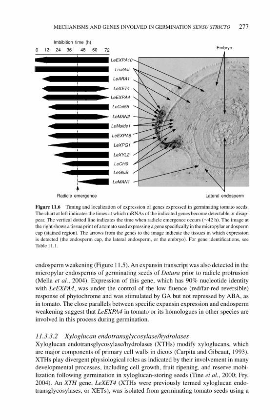
P1: OTE/SPH P2: OTE
BLUK053-Bradford September 27, 2006 15:21
MECHANISMS AND GENES INVOLVED IN GERMINATION SENSU STRICTO 277
Imbibition time (h)
LeEXPA10
Radicle emergence
LeaGal
Lateral endosperm
Embryo
LeEXPA8
LeXET4
LeMAN2
LeGluB
LeChi9
LeARA1
LeMside1
LeXPG1
LeMAN1
LeXYL2
LeCel55
LeEXPA4
6048 723624120
Figure 11.6 Timing and localization of expression of genes expressed in germinating tomato seeds.
The chart at left indicates the times at which mRNAs of the indicated genes become detectable or disap-
pear. The vertical dotted line indicates the time when radicle emergence occurs (∼42 h). The image at
the right shows a tissue print of a tomato seed expressing a gene specifically in the micropylar endosperm
cap (stained region). The arrows from the genes to the image indicate the tissues in which expression
is detected (the endosperm cap, the lateral endosperm, or the embryo). For gene identifications, see
Table 11.1.
endosperm weakening (Figure 11.5). An expansin transcript was also detected in themicropylar endosperms of germinating seeds of Datura prior to radicle protrusion(Mella et al., 2004). Expression of this gene, which has 90% nucleotide identitywith LeEXPA4, was under the control of the low fluence (red/far-red reversible)response of phytochrome and was stimulated by GA but not repressed by ABA, asin tomato. The close parallels between specific expansin expression and endospermweakening suggest that LeEXPA4 in tomato or its homologues in other species areinvolved in this process during germination.
11.3.3.2 Xyloglucan endotransglycosylase/hydrolasesXyloglucan endotransglycosylase/hydrolases (XTHs) modify xyloglucans, whichare major components of primary cell walls in dicots (Carpita and Gibeaut, 1993).XTHs play divergent physiological roles as indicated by their involvement in manydevelopmental processes, including cell growth, fruit ripening, and reserve mobi-lization following germination in xyloglucan-storing seeds (Tine et al., 2000; Fry,2004). An XTH gene, LeXET4 (XTHs were previously termed xyloglucan endo-transglycosylases, or XETs), was isolated from germinating tomato seeds using a
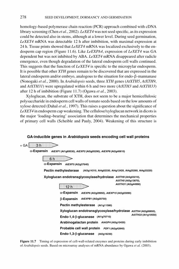
P1: OTE/SPH P2: OTE
BLUK053-Bradford September 27, 2006 15:21
278 SEED DEVELOPMENT, DORMANCY AND GERMINATION
homology-based polymerase chain reaction (PCR) approach combined with cDNAlibrary screening (Chen et al., 2002). LeXET4 was not seed specific, as its expressioncould be detected also in stems, although at a lower level. During seed germination,LeXET4 mRNA was detectable 12 h after imbibition, with maximal expression at24 h. Tissue prints showed that LeXET4 mRNA was localized exclusively to the en-dosperm cap region (Figure 11.6). Like LeEXPA4, expression of LeXET4 was GAdependent but was not inhibited by ABA. LeXET4 mRNA disappeared after radicleemergence, even though degradation of the lateral endosperm cell walls continued.This suggests that the function of LeXET4 is specific to the micropylar endosperm.It is possible that other XTH genes remain to be discovered that are expressed in thelateral endosperm and/or embryo, analogous to the situation for endo-β-mannanase(Nonogaki et al., 2000). In Arabidopsis seeds, three XTH genes (AtXTH5, AtXTH9,and AtXTH31) were upregulated within 6 h and two more (AtXTH3 and AtXTH33)after 12 h of imbibition (Figure 11.7) (Ogawa et al., 2003).
Xyloglucan, the substrate of XTH, does not seem to be a major hemicellulosicpolysaccharide in endosperm cell walls of tomato seeds based on the low amounts ofxylose detected (Dahal et al., 1997). This raises a question about the significance ofLeXET4 in endosperm cap weakening. The cellulose/xyloglucan network in dicots isthe major ‘loading–bearing’ association that determines the mechanical propertiesof primary cell walls (Scheible and Pauly, 2004). Weakening of this structure is
GA-inducible genes in Arabidopsis seeds encoding cell wall proteins
6 h
α-Expansin
3 h
12 h
α-Expansin AtEXP3 (At2g37640)
Pectin methylesterase
Xyloglucan endotransglycosylase/hydrolase
(At3g14310, At4g02330, At4g12420, At4g25260, At4g33220)
AtXTH9 (At4g03210), AtXTH5 (At5g13870), AtXTH31 (At3g44990)
Arabinogalactan protein
AtEXP1 (At1g69530), AtEXP2 (At5g05290), AtEXP8 (At2g40610)
Probable cell wall protein
α-Expansin
Pectin methylesterase
β-Expansin
AtEXP6 (At2g28950), AtEXP15 (At2g03090)
Endo-1,4-β-glucanase (At1g70710)
AtEXPB1 (At2g20750)
+ GA
(At1g11580)
Xyloglucan endotransglycosylase/hydrolase AtXTH4 (At2g06850), AtXTH33 (At1g10550)
AtAGP4 (At5g10430)
PDF1 (At2g42840)
Endo-1,3-β-glucanase (At2g16230)
Figure 11.7 Timing of expression of cell-wall-related enzymes and proteins during early imbibition
of Arabidopsis seeds. Based on microarray analyses of mRNA abundance by Ogawa et al. (2003).
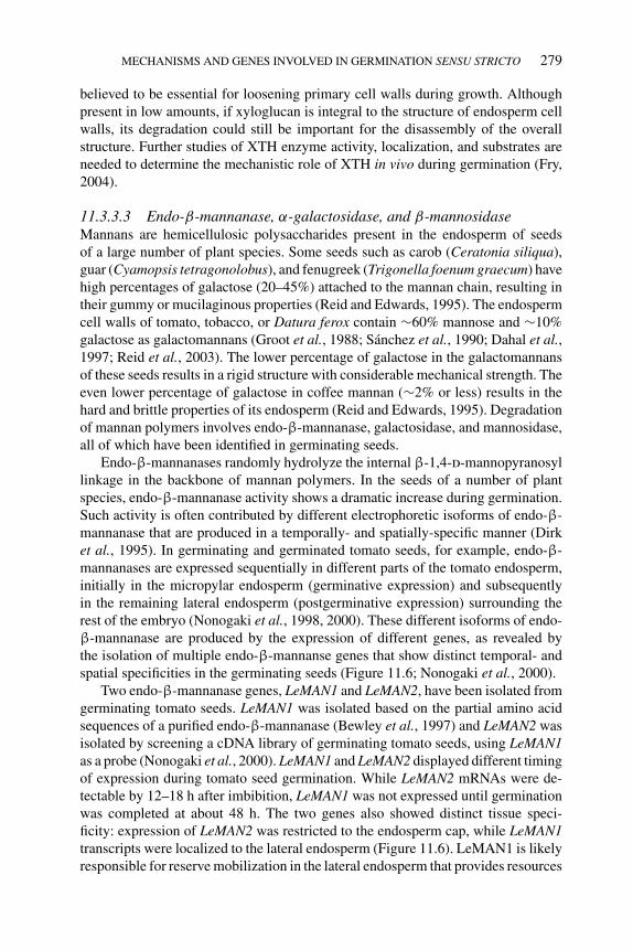
P1: OTE/SPH P2: OTE
BLUK053-Bradford September 27, 2006 15:21
MECHANISMS AND GENES INVOLVED IN GERMINATION SENSU STRICTO 279
believed to be essential for loosening primary cell walls during growth. Althoughpresent in low amounts, if xyloglucan is integral to the structure of endosperm cellwalls, its degradation could still be important for the disassembly of the overallstructure. Further studies of XTH enzyme activity, localization, and substrates areneeded to determine the mechanistic role of XTH in vivo during germination (Fry,2004).
11.3.3.3 Endo-β-mannanase, α-galactosidase, and β-mannosidaseMannans are hemicellulosic polysaccharides present in the endosperm of seedsof a large number of plant species. Some seeds such as carob (Ceratonia siliqua),guar (Cyamopsis tetragonolobus), and fenugreek (Trigonella foenum graecum) havehigh percentages of galactose (20–45%) attached to the mannan chain, resulting intheir gummy or mucilaginous properties (Reid and Edwards, 1995). The endospermcell walls of tomato, tobacco, or Datura ferox contain ∼60% mannose and ∼10%galactose as galactomannans (Groot et al., 1988; Sanchez et al., 1990; Dahal et al.,1997; Reid et al., 2003). The lower percentage of galactose in the galactomannansof these seeds results in a rigid structure with considerable mechanical strength. Theeven lower percentage of galactose in coffee mannan (∼2% or less) results in thehard and brittle properties of its endosperm (Reid and Edwards, 1995). Degradationof mannan polymers involves endo-β-mannanase, galactosidase, and mannosidase,all of which have been identified in germinating seeds.
Endo-β-mannanases randomly hydrolyze the internal β-1,4-d-mannopyranosyllinkage in the backbone of mannan polymers. In the seeds of a number of plantspecies, endo-β-mannanase activity shows a dramatic increase during germination.Such activity is often contributed by different electrophoretic isoforms of endo-β-mannanase that are produced in a temporally- and spatially-specific manner (Dirket al., 1995). In germinating and germinated tomato seeds, for example, endo-β-mannanases are expressed sequentially in different parts of the tomato endosperm,initially in the micropylar endosperm (germinative expression) and subsequentlyin the remaining lateral endosperm (postgerminative expression) surrounding therest of the embryo (Nonogaki et al., 1998, 2000). These different isoforms of endo-β-mannanase are produced by the expression of different genes, as revealed bythe isolation of multiple endo-β-mannanse genes that show distinct temporal- andspatial specificities in the germinating seeds (Figure 11.6; Nonogaki et al., 2000).
Two endo-β-mannanase genes, LeMAN1 and LeMAN2, have been isolated fromgerminating tomato seeds. LeMAN1 was isolated based on the partial amino acidsequences of a purified endo-β-mannanase (Bewley et al., 1997) and LeMAN2 wasisolated by screening a cDNA library of germinating tomato seeds, using LeMAN1as a probe (Nonogaki et al., 2000). LeMAN1 and LeMAN2 displayed different timingof expression during tomato seed germination. While LeMAN2 mRNAs were de-tectable by 12–18 h after imbibition, LeMAN1 was not expressed until germinationwas completed at about 48 h. The two genes also showed distinct tissue speci-ficity: expression of LeMAN2 was restricted to the endosperm cap, while LeMAN1transcripts were localized to the lateral endosperm (Figure 11.6). LeMAN1 is likelyresponsible for reserve mobilization in the lateral endosperm that provides resources
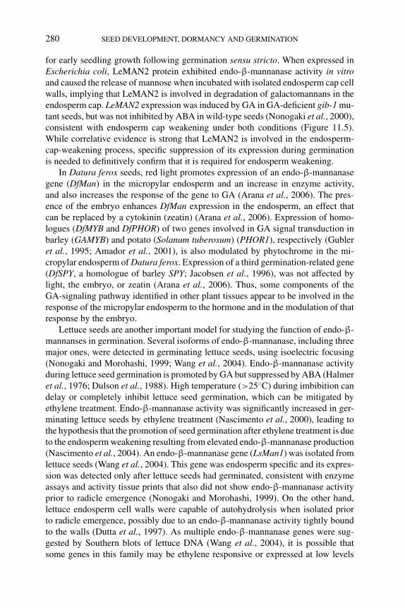
P1: OTE/SPH P2: OTE
BLUK053-Bradford September 27, 2006 15:21
280 SEED DEVELOPMENT, DORMANCY AND GERMINATION
for early seedling growth following germination sensu stricto. When expressed inEscherichia coli, LeMAN2 protein exhibited endo-β-mannanase activity in vitroand caused the release of mannose when incubated with isolated endosperm cap cellwalls, implying that LeMAN2 is involved in degradation of galactomannans in theendosperm cap. LeMAN2 expression was induced by GA in GA-deficient gib-1 mu-tant seeds, but was not inhibited by ABA in wild-type seeds (Nonogaki et al., 2000),consistent with endosperm cap weakening under both conditions (Figure 11.5).While correlative evidence is strong that LeMAN2 is involved in the endosperm-cap-weakening process, specific suppression of its expression during germinationis needed to definitively confirm that it is required for endosperm weakening.
In Datura ferox seeds, red light promotes expression of an endo-β-mannanasegene (DfMan) in the micropylar endosperm and an increase in enzyme activity,and also increases the response of the gene to GA (Arana et al., 2006). The pres-ence of the embryo enhances DfMan expression in the endosperm, an effect thatcan be replaced by a cytokinin (zeatin) (Arana et al., 2006). Expression of homo-logues (DfMYB and DfPHOR) of two genes involved in GA signal transduction inbarley (GAMYB) and potato (Solanum tuberosum) (PHOR1), respectively (Gubleret al., 1995; Amador et al., 2001), is also modulated by phytochrome in the mi-cropylar endosperm of Datura ferox. Expression of a third germination-related gene(DfSPY, a homologue of barley SPY; Jacobsen et al., 1996), was not affected bylight, the embryo, or zeatin (Arana et al., 2006). Thus, some components of theGA-signaling pathway identified in other plant tissues appear to be involved in theresponse of the micropylar endosperm to the hormone and in the modulation of thatresponse by the embryo.
Lettuce seeds are another important model for studying the function of endo-β-mannanses in germination. Several isoforms of endo-β-mannanase, including threemajor ones, were detected in germinating lettuce seeds, using isoelectric focusing(Nonogaki and Morohashi, 1999; Wang et al., 2004). Endo-β-mannanase activityduring lettuce seed germination is promoted by GA but suppressed by ABA (Halmeret al., 1976; Dulson et al., 1988). High temperature (>25◦C) during imbibition candelay or completely inhibit lettuce seed germination, which can be mitigated byethylene treatment. Endo-β-mannanase activity was significantly increased in ger-minating lettuce seeds by ethylene treatment (Nascimento et al., 2000), leading tothe hypothesis that the promotion of seed germination after ethylene treatment is dueto the endosperm weakening resulting from elevated endo-β-mannanase production(Nascimento et al., 2004). An endo-β-mannanase gene (LsMan1) was isolated fromlettuce seeds (Wang et al., 2004). This gene was endosperm specific and its expres-sion was detected only after lettuce seeds had germinated, consistent with enzymeassays and activity tissue prints that also did not show endo-β-mannanase activityprior to radicle emergence (Nonogaki and Morohashi, 1999). On the other hand,lettuce endosperm cell walls were capable of autohydrolysis when isolated priorto radicle emergence, possibly due to an endo-β-mannanase activity tightly boundto the walls (Dutta et al., 1997). As multiple endo-β-mannanase genes were sug-gested by Southern blots of lettuce DNA (Wang et al., 2004), it is possible thatsome genes in this family may be ethylene responsive or expressed at low levels
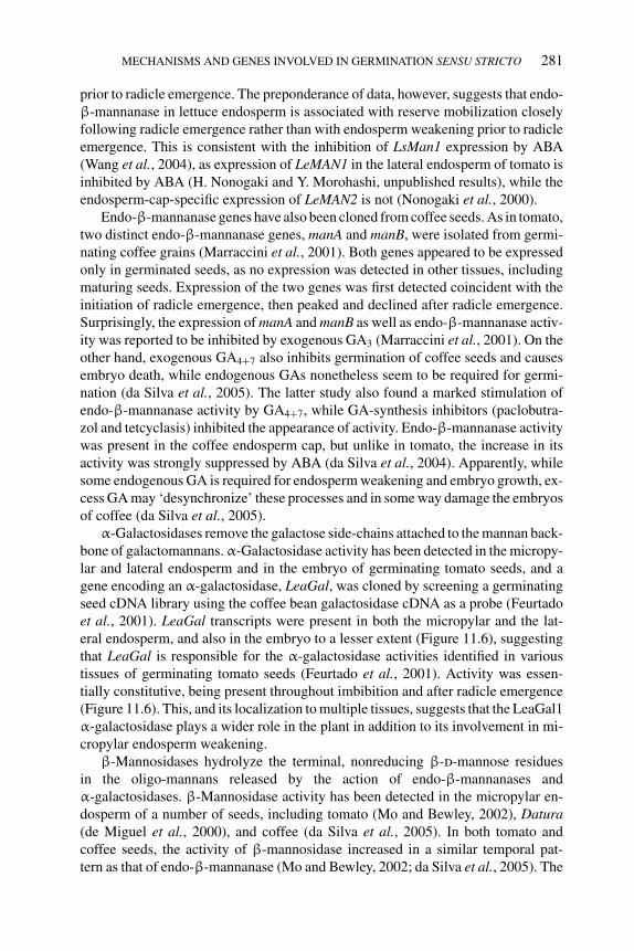
P1: OTE/SPH P2: OTE
BLUK053-Bradford September 27, 2006 15:21
MECHANISMS AND GENES INVOLVED IN GERMINATION SENSU STRICTO 281
prior to radicle emergence. The preponderance of data, however, suggests that endo-β-mannanase in lettuce endosperm is associated with reserve mobilization closelyfollowing radicle emergence rather than with endosperm weakening prior to radicleemergence. This is consistent with the inhibition of LsMan1 expression by ABA(Wang et al., 2004), as expression of LeMAN1 in the lateral endosperm of tomato isinhibited by ABA (H. Nonogaki and Y. Morohashi, unpublished results), while theendosperm-cap-specific expression of LeMAN2 is not (Nonogaki et al., 2000).
Endo-β-mannanase genes have also been cloned from coffee seeds. As in tomato,two distinct endo-β-mannanase genes, manA and manB, were isolated from germi-nating coffee grains (Marraccini et al., 2001). Both genes appeared to be expressedonly in germinated seeds, as no expression was detected in other tissues, includingmaturing seeds. Expression of the two genes was first detected coincident with theinitiation of radicle emergence, then peaked and declined after radicle emergence.Surprisingly, the expression of manA and manB as well as endo-β-mannanase activ-ity was reported to be inhibited by exogenous GA3 (Marraccini et al., 2001). On theother hand, exogenous GA4+7 also inhibits germination of coffee seeds and causesembryo death, while endogenous GAs nonetheless seem to be required for germi-nation (da Silva et al., 2005). The latter study also found a marked stimulation ofendo-β-mannanase activity by GA4+7, while GA-synthesis inhibitors (paclobutra-zol and tetcyclasis) inhibited the appearance of activity. Endo-β-mannanase activitywas present in the coffee endosperm cap, but unlike in tomato, the increase in itsactivity was strongly suppressed by ABA (da Silva et al., 2004). Apparently, whilesome endogenous GA is required for endosperm weakening and embryo growth, ex-cess GA may ‘desynchronize’ these processes and in some way damage the embryosof coffee (da Silva et al., 2005).
α-Galactosidases remove the galactose side-chains attached to the mannan back-bone of galactomannans. α-Galactosidase activity has been detected in the micropy-lar and lateral endosperm and in the embryo of germinating tomato seeds, and agene encoding an α-galactosidase, LeaGal, was cloned by screening a germinatingseed cDNA library using the coffee bean galactosidase cDNA as a probe (Feurtadoet al., 2001). LeaGal transcripts were present in both the micropylar and the lat-eral endosperm, and also in the embryo to a lesser extent (Figure 11.6), suggestingthat LeaGal is responsible for the α-galactosidase activities identified in varioustissues of germinating tomato seeds (Feurtado et al., 2001). Activity was essen-tially constitutive, being present throughout imbibition and after radicle emergence(Figure 11.6). This, and its localization to multiple tissues, suggests that the LeaGal1α-galactosidase plays a wider role in the plant in addition to its involvement in mi-cropylar endosperm weakening.
β-Mannosidases hydrolyze the terminal, nonreducing β-d-mannose residuesin the oligo-mannans released by the action of endo-β-mannanases andα-galactosidases. β-Mannosidase activity has been detected in the micropylar en-dosperm of a number of seeds, including tomato (Mo and Bewley, 2002), Datura(de Miguel et al., 2000), and coffee (da Silva et al., 2005). In both tomato andcoffee seeds, the activity of β-mannosidase increased in a similar temporal pat-tern as that of endo-β-mannanase (Mo and Bewley, 2002; da Silva et al., 2005). The
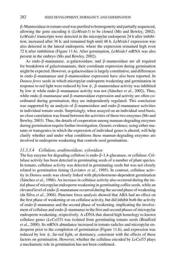
P1: OTE/SPH P2: OTE
BLUK053-Bradford September 27, 2006 15:21
282 SEED DEVELOPMENT, DORMANCY AND GERMINATION
β-Mannosidase in tomato seed was purified to homogeneity and partially sequenced,allowing the gene encoding it (LeMside1) to be cloned (Mo and Bewley, 2002).LeMside1 transcripts were detected in the micropylar endosperm 24 h after imbibi-tion, increased after 36 h, and remained high until 48 h. LeMside1 expression wasalso detected in the lateral endosperm, where the expression remained high even72 h after imbibition (Figure 11.6). After germination, LeMside1 mRNA was alsopresent in the embryo (Mo and Bewley, 2002).
As endo-β-mannanase, α-galactosidase, and β-mannosidase are all requiredfor breakdown of galactomannans, their coordinate expression during germinationmight be expected. However, α-galactosidase is largely constitutive, and differencesin endo-β-mannanase and β-mannosidase expression have also been reported. InDatura ferox seeds in which micropylar endosperm weakening and germination inresponse to red light were reduced by low ψ , β-mannosidase activity was inhibitedby low ψ while endo-β-mannanase activity was not (Sanchez et al., 2002). Thus,while endo-β-mannanase and β-mannosidase expression and activity are often co-ordinated during germination, they are independently regulated. This conclusionwas supported by an analysis of β-mannosidase and endo-β-mannanase activitiesin individual tomato seeds. Surprisingly, when assayed on an individual seed basis,no close correlation was found between the activities of these two enzymes (Mo andBewley, 2003). Thus, the details of cooperation among mannan-degrading enzymesduring germination require further investigation. Genetic evidence, using either mu-tants or transgenics in which the expression of individual genes is altered, will helpclarify whether and under what conditions these mannan-degrading enzymes areinvolved in endosperm weakening that controls seed germination.
11.3.3.4 Cellulase, arabinosidase, xylosidaseThe key enzyme for degrading cellulose is endo-β-1,4-glucanase, or cellulase. Cel-lulase activity has been detected in germinating seeds of a number of plant species.In tomato, cellulase activity was detected in germinating seeds but was not closelyrelated to germination timing (Leviatov et al., 1995). In contrast, cellulase activ-ity in Datura seeds was closely linked with phytohormone-dependent germination(Sanchez et al., 1986). An increase in cellulase activity also occurred during the ini-tial phase of micropylar endosperm weakening in germinating coffee seeds, while anelevated level of endo-β-mannanase occurred during the second phase of weakening(da Silva et al., 2004). Puncture force analysis showed that ABA had no effect onthe first phase of weakening or on cellulase activity, but did inhibit both the activityof endo-β-mannanse and the second phase of weakening, implicating the involve-ment of cellulase and endo-β-mannanse in the first and second phases of micropylarendosperm weakening, respectively. A cDNA that shared high homology to knowncellulase genes (LeCel55) was isolated from germinating tomato seeds (Bradfordet al., 2000). Its mRNA abundance increased in tomato radicles and micropylar en-dosperm prior to the completion of germination (Figure 11.6), and expression wasreduced by low ψ , far-red light, or dormancy, consistent with the effects of thesefactors on germination. However, whether the cellulase encoded by LeCel55 playsa mechanistic role in germination has not been confirmed.
P1: OTE/SPH P2: OTE
BLUK053-Bradford September 27, 2006 15:21
MECHANISMS AND GENES INVOLVED IN GERMINATION SENSU STRICTO 283
Tomato endosperm cell walls contain up to 10% arabinose but little xylose,while the reverse is true for embryo cell walls (Groot et al., 1988; Dahal et al.,1997). A cDNA encoding a putativeα-l-arabinofuranosidase (LeARA1) was isolatedby differential display screening from germinating tomato seeds (Bradford et al.,2000), and the same gene was also isolated from developing and ripening tomatofruit (termed LeARF1) (Itai et al., 2003). Although its mRNA appeared within12 h after imbibition, expression of LeARA1 was restricted to the lateral endospermof tomato seeds (Figure 11.6; P. Dahal and K.J. Bradford, unpublished results).A β-d-xylosidase gene (LeXYL2) was also cloned from tomato fruits (Itai et al.,2003), and it was expressed almost exclusively in the embryos of imbibed tomatoseeds (Figure11.6; A. Itai and J.D. Bewley, personal communication, 12 May 2004).Although both genes are expressed well prior to radicle emergence, their localizationsuggests that they may primarily be involved in postgermination events.
11.3.3.5 Polygalacturonase and pectin methylesteraseAccording to a widely accepted model (Carpita and Gibeaut, 1993), pectin func-tions as a matrix in which cellulose microfibrils and hemicellulosic polymers areembedded in plant primary cell walls. Pectin is especially abundant in the middlelamella and is important for maintaining cell-to-cell cohesion. Disassembly of pectin(polygalacturonic acid) by polygalacturonases (PGs) is associated with many devel-opmental processes and has been particularly well studied in fruit ripening (Hadfieldand Bennett, 1998). In germinating tomato seeds, a calcium-dependent exo-PG ac-tivity was detected and a gene (LeXPG1) was cloned from germinating seeds usingdegenerate primers corresponding to two conserved regions of known PG genes(Sitrit et al., 1999). LeXPG1 mRNA abundance was low during seed development,increased during imbibition, and was high in seeds that completed germination.Expression of LeXPG1 during germination was detected in both the micropylar en-dosperm and the radicle tip (Figure 11.6), suggesting a role for LeXPG1 in bothtissues.
The degree of esterification of pectin, regulated by pectin methylesterase (PME),plays an important role in its rigidity. De-esterification of pectin catalyzed by PMEhas been suggested to be involved in cell wall weakening of the megagametophyteof yellow cedar (Chamaecyparis nootkatensis) seeds during dormancy breakage andgermination (Ren and Kermode, 2000). In germinating tomato seeds, PME activitywas detected in the micropylar endosperm, although the majority of activity was inthe embryo (Downie et al., 1998). A number of PME genes were upregulated byGA within 6–12 h of imbibition in Arabidopsis seeds as well (Figure 11.7; Ogawaet al., 2003).
11.3.3.6 β-1,3-Glucanase and chitinaseClass I β-1,3-glucanases (βGLU) are well-known pathogenesis-related (PR) pro-teins involved in plant defense against fungal infection and other physiological pro-cesses (Leubner-Metzger, 2003). The involvement ofβGLU in seed germination wasfirst revealed in transgenic tobacco seeds containing a chimeric GUS (glucuronidase)reporter gene fused with the promoter of a tobacco βGLU (GluB) (Vogeli-Lange
P1: OTE/SPH P2: OTE
BLUK053-Bradford September 27, 2006 15:21
284 SEED DEVELOPMENT, DORMANCY AND GERMINATION
et al., 1994). During germination, GUS activity was highly induced and specificallylocalized to the micropylar endosperm tissue prior to radicle emergence (Leubner-Metzger et al., 1995). Expression analysis showed that Gluβ mRNA accumulationand enzyme activity increased just prior to endosperm rupture. Chitinases are an-other group of PR proteins whose expression often accompanies the expression ofβGLU during plant defense responses (van Loon, 1997). In germinating tobaccoseeds, chitinase activity is very low (Leubner-Metzger et al., 1995), but in tomatoseeds, genes encoding both βGLU and chitinase are expressed in the micropylarendosperm just prior to radicle emergence (Figure 11.6; Wu et al., 2001).
The role of βGLU in germinating tobacco seeds has been studied using a trans-genic approach. Overexpression of βGLU in tobacco seeds was achieved by trans-forming a βGLU gene under the control of an ABA-inducible Cat1 promoter fromcastor bean (Ricinus communis) into tobacco (Leubner-Metzger and Meins, 2000).Thus, in addition to expression during seed development and higher initial levels ofβGLU, seeds containing Cat1::βGLU also increase their expression of βGLU inresponse to ABA, in contrast to the inhibition (delay in expression) of the endoge-nous βGLU gene by ABA (Leubner-Metzger and Meins, 2000). Immunolocalizationanalysis showed that βGLU in transgenic tobacco seeds is mainly localized in themicropylar endosperm and in an inner layer of living cells in the testa (Leubner-Metzger, 2002). Overexpression of βGLU had no effect on the rate of testa rupturein either the presence or absence of ABA or on the time to endosperm rupture in theabsence of ABA, but reduced the time to endosperm rupture by about 10–20% inthe presence of ABA (Leubner-Metzger and Meins, 2000). Expression of antisenseconstructs of βGLU, on the other hand, had a delaying effect on the time to testarupture, even though there was no effect on the expression of βGLU associated withendosperm rupture (Leubner-Metzger and Meins, 2001). Freshly harvested seedsare normally dormant when imbibed in the dark and lose this dormancy during drystorage (after-ripening). Sense expression of βGLU was able to partially replacethis requirement for after-ripening (Leubner-Metzger and Meins, 2000), while an-tisense expression extended the dormancy period (Leubner-Metzger and Meins,2001). Reciprocal crosses confirmed the maternal inheritance of the effect of sense-βGLU expression on germination speed, indicating that it was due to expressionin the testa (Leubner-Metzger, 2002). These studies led to the conclusion that intobacco, βGLU expression promotes both testa and endosperm rupture, with onlythe latter being sensitive to ABA (Leubner-Metzger, 2002).
Somewhat contrasting results were obtained for βGLU and chitinase expressionin germinating tomato seeds. As in tobacco, one gene encoding a basic (type I)form of βGLU was expressed specifically in the micropylar endosperm in tomatojust prior to radicle emergence (Figure 11.6; Wu et al., 2001), and its expression isGA-dependent but suppressed by ABA. In contrast to tobacco, a chitinase gene wasexpressed coordinately with βGLU, and was also localized to the tomato endospermcap (with lower expression in the radicle tip) but was not inhibited by ABA (Wuet al., 2001). Interestingly, chitinase (but not βGLU) expression in the endospermcap was also regulated by wounding and jasmonic acid (Wu and Bradford, 2003).Thus, while chitinase and βGLU genes are normally expressed simultaneously andare co-localized in the endosperm cap tissue of tomato seeds (Figure 11.6), they
P1: OTE/SPH P2: OTE
BLUK053-Bradford September 27, 2006 15:21
MECHANISMS AND GENES INVOLVED IN GERMINATION SENSU STRICTO 285
are differentially regulated by ABA, jasmonate and wounding. In addition, a differ-ent βGLU enzyme was reported to increase in tomato seeds following germination(Morohashi and Matsushima, 2000). βGLU hydrolyzes β-1,3-glucan polymers, orcallose, which is present in the covering tissues of some seeds, such as muskmelon(Yim and Bradford, 1998). However, callose was not detected by staining in tomatoseeds (Beresniewicz et al., 1995), and when βGLU expressed in E. coli was incu-bated with isolated tomato endosperm cell walls, no hydrolytic activity was detected,suggesting that substrates for βGLU may not be present in the endosperm cell walls(Wu et al., 2001). Similarly, chitins are not known to be present in the endosperm,although other substrates for chitinase, such as glucosamine-containing arabino-galactan proteins, could be present (Gomez et al., 2002). Thus, it remains an openquestion whether βGLU and chitinase are involved in the mechanism of germinationper se in tomato or whether they are components of a defense response to delay orprevent entry of fungi into the lateral endosperm following radicle emergence (Wuand Bradford, 2003). βGLU could be playing a role other than in endosperm weak-ening, as synthesis and degradation of callose is associated with closing and openingof plasmodesmata that are correlated with imposition and loss of dormancy and inintercellular signaling in shoot apical meristems (Rinne and van der Schoot, 2003).
11.3.3.7 Concerted action of cell wall hydrolases and expansinsThe presence of multiple cell wall hydrolases and expansins in the endosperm dur-ing seed germination suggests that these enzymes and proteins work together tocause endosperm cell wall disassembly. Potential cooperation between expansinsand XTHs has been studied in several biological processes. Whitney et al. (2000)found that expansin protein was active on polymer composites containing xyloglucanbut not on those made with mannan polymers. From this and related studies they con-cluded that the cellulose/xyloglucan matrix is the site of expansin action (Whitneyet al., 1999, 2000). The concerted expression of an expansin gene (LeEXP2) and anXTH gene (LeEXT1) in early tomato fruit development and in auxin-treated tomatohypocotyl elongation supports this proposal (Catala et al., 1997, 2000). Similarly,LeEXP4 and LeXET4 showed concerted expression in the endosperm cap duringtomato seed germination and displayed similar responses to environmental and hor-monal factors (Figure 11.6; Table 11.1), suggesting that the enzymes encoded byLeEXPA4 and LeXET4 may work cooperatively towards endosperm weakening.
Such a cooperative action may be extended to other cell wall hydrolases duringdifferent stages of cell wall disassembly. During fruit ripening, two stages of wallstructure alteration are apparent (Rose et al., 1998; Bennett, 2002). The first stageis associated with softening, in which expansins and XTHs are likely essential. Thesecond stage is associated with tissue disruption and decay, for which other cellwall hydrolases such as PGs are key players. A two-stage process may also be thecase for micropylar endosperm weakening. LeEXPA4, LeXET4 and LeMAN2 are allexpressed specifically in the micropylar endosperm, but LeMAN2 mRNA appearssomewhat later than that of the other two (Figure 11.6). One hypothesis is that theaction of LeEXP4 and LeXET4, expressed at an early stage of imbibition, causesloosening of the cellulose-xyloglucan network, which in turn makes the wall ma-trix accessible to other cell wall hydrolases, such as endo-β-mannanase. However,
P1: OTE/SPH P2: OTE
BLUK053-Bradford September 27, 2006 15:21
286 SEED DEVELOPMENT, DORMANCY AND GERMINATION
it should be noted that this two-stage cell wall modification does not explain thesecondary weakening hypothesis, since all above mentioned expansin and hemicel-lulase genes are associated with the drastic reduction in the mechanical resistanceof the endosperm cap during the first (ABA-insensitive) stage (Figure 11.5).
Hypotheses concerning the functions of the individual cell wall hydrolases inendosperm weakening are mainly based on the association between enzyme activ-ity/gene expression and endosperm weakening/seed germination. While the correl-ative and circumstantial evidence is quite strong, only for βGLU is there geneticevidence supporting a specific biological function for cell wall enzymes in germi-nation. Similar evidence is desired for all other cell wall hydrolases and expansins.With antisense or RNAi strategies, the expression of individual genes can be se-lectively eliminated in the seed to test whether they play essential roles, and ifso, what those roles are. GA appears to be a key regulator of most of these genes(Table 11.1). Interestingly, many of these genes expressed during germination sensustricto are insensitive to exogenous ABA, while other typical GA-inducible genes inpostgermination, such as α-amylase or LeMAN1, are repressed by ABA. This sug-gests that regulatory mechanisms differ between germinative and postgerminativegene expression patterns. The identification of the regulatory genes and transcriptionfactors that connect hormonal signals to their targeted genes will help understandthe concerted action of cell wall hydrolases.
11.3.4 Embryo growth potential
While weakening of enclosing tissues is a prerequisite for germination in many seeds,embryo growth is essential for germination of all seeds. Seeds display a wide rangeof embryo morphologies at seed maturity, from rudimentary to fully developed. Insome seeds, such as celery (Apium graveolens), considerable embryo growth anddevelopment must occur within the seed prior to completion of germination (van derToorn and Karssen, 1992). In others, only expansion of the existing embryonicstructures is required. In the latter cases, DNA replication and cell cycle activitymay commence prior to radicle emergence, but cell division in the meristematictissues is generally delayed until at or after radicle emergence (de Castro et al., 2000;Barroco et al., 2005). Cell cycle initiation may play an additive role in Arabidopsisseed germination, but its effect was relatively small and not essential, as germinationwas delayed but not prevented by cell cycle inhibitors or mutations in some cyclinD genes (Masubelele et al., 2005). Here, we will address only the expansion of theembryo that occurs during Phase II and transition to Phase III of imbibition (Figure11.1) and results in completion of germination sensu stricto.
11.3.4.1 Generation of embryo growth potentialPlant growth is dependent upon cell expansion due to water uptake. The drivingforce for water uptake is the osmotic or solute potential (ψs) of the cytoplasm andvacuole, reduced by the hydrostatic pressure or turgor (ψp) generated by resistanceto expansion by the cell walls. Thus, at ψ equilibrium for a non-expanding cell inwater the values of ψs and ψp are equal and opposite in value and net water flux
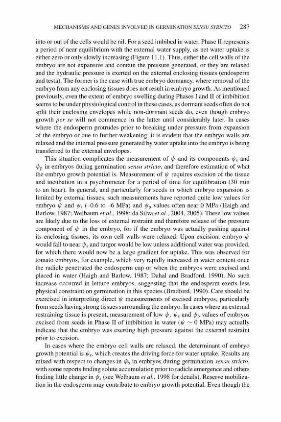
P1: OTE/SPH P2: OTE
BLUK053-Bradford September 27, 2006 15:21
MECHANISMS AND GENES INVOLVED IN GERMINATION SENSU STRICTO 287
into or out of the cells would be nil. For a seed imbibed in water, Phase II representsa period of near equilibrium with the external water supply, as net water uptake iseither zero or only slowly increasing (Figure 11.1). Thus, either the cell walls of theembryo are not expansive and contain the pressure generated, or they are relaxedand the hydraulic pressure is exerted on the external enclosing tissues (endospermand testa). The former is the case with true embryo dormancy, where removal of theembryo from any enclosing tissues does not result in embryo growth. As mentionedpreviously, even the extent of embryo swelling during Phases I and II of imbibitionseems to be under physiological control in these cases, as dormant seeds often do notsplit their enclosing envelopes while non-dormant seeds do, even though embryogrowth per se will not commence in the latter until considerably later. In caseswhere the endosperm protrudes prior to breaking under pressure from expansionof the embryo or due to further weakening, it is evident that the embryo walls arerelaxed and the internal pressure generated by water uptake into the embryo is beingtransferred to the external envelopes.
This situation complicates the measurement of ψ and its components ψs andψp in embryos during germination sensu stricto, and therefore estimation of whatthe embryo growth potential is. Measurement of ψ requires excision of the tissueand incubation in a psychrometer for a period of time for equilibration (30 minto an hour). In general, and particularly for seeds in which embryo expansion islimited by external tissues, such measurements have reported quite low values forembryo ψ and ψs (–0.6 to –6 MPa) and ψp values often near 0 MPa (Haigh andBarlow, 1987; Welbaum et al., 1998; da Silva et al., 2004, 2005). These low valuesare likely due to the loss of external restraint and therefore release of the pressurecomponent of ψ in the embryo, for if the embryo was actually pushing againstits enclosing tissues, its own cell walls were relaxed. Upon excision, embryo ψ
would fall to near ψs and turgor would be low unless additional water was provided,for which there would now be a large gradient for uptake. This was observed fortomato embryos, for example, which very rapidly increased in water content oncethe radicle penetrated the endosperm cap or when the embryos were excised andplaced in water (Haigh and Barlow, 1987; Dahal and Bradford, 1990). No suchincrease occurred in lettuce embryos, suggesting that the endosperm exerts lessphysical constraint on germination in this species (Bradford, 1990). Care should beexercised in interpreting direct ψ measurements of excised embryos, particularlyfrom seeds having strong tissues surrounding the embryo. In cases where an externalrestraining tissue is present, measurement of low ψ , ψs and ψp values of embryosexcised from seeds in Phase II of imbibition in water (ψ ∼ 0 MPa) may actuallyindicate that the embryo was exerting high pressure against the external restraintprior to excision.
In cases where the embryo cell walls are relaxed, the determinant of embryogrowth potential is ψs, which creates the driving force for water uptake. Results aremixed with respect to changes in ψs in embryos during germination sensu stricto,with some reports finding solute accumulation prior to radicle emergence and othersfinding little change in ψs (see Welbaum et al., 1998 for details). Reserve mobiliza-tion in the endosperm may contribute to embryo growth potential. Even though the
P1: OTE/SPH P2: OTE
BLUK053-Bradford September 27, 2006 15:21
288 SEED DEVELOPMENT, DORMANCY AND GERMINATION
Arabidopsis endosperm is only a single cell layer, removing it or blocking reservemobilization in it reduced subsequent hypocotyl growth (Penfield et al., 2004).Thus, the early mobilization of reserves, as is evident in micropylar endosperms(Figure 11.2) may provide solutes to the embryo to increase its growth potential.
Due to the difficulties in measuring the actual pressure that the embryo can exertto penetrate any covering tissues, embryo growth potential is often measured as theability of the embryo to either complete germination or elongate at a range of externalwater potentials when intact or with the endosperm excised. One approach to thisis to determine the base or minimum water potential (ψb) that allows germinationof a given fraction of the seed population (Dahal and Bradford, 1990; Bradford andSomasco, 1994). This approach is described in detail in Chapter 4, and integratesboth embryo growth potential and the restraint of external tissues. Alternatively,the ability of excised embryos to grow in water or at a range of ψ after excisioncan give an indication of the embryo growth potential. This approach has shown,for example, that red light and GA increase the growth potential of the embryo ofDatura ferox while also stimulating endosperm weakening (de Miguel and Sanchez,1992; Arana et al., 2006). The Datura embryo also produces a signal (possibly acytokinin) in response to red light that enhances the sensitivity of the micropylarendosperm to GA (Arana et al., 2006).
11.3.4.2 Gene expression associated with embryo growthIn comparison with the number of genes known to be expressed in endospermtissues during germination sensu stricto, relatively few genes have been shown tobe specifically expressed in the embryo in association with germination sensu stricto.Two expansin genes (LeEXPA8 and LeEXPA10) were expressed in tomato embryosprior to radicle emergence (Figure 11.6; Chen et al., 2001). LeEXPA10 was expressedthroughout the embryo during both embryogenesis and germination, while LeEXPA8was specific to germination and was expressed only in the elongation zone of theradicle. Lack of expression in GA-deficient gib-1 mutant seeds unless supplied withGA indicates that expression of both genes is dependent on the hormone. ABA didnot prevent the initial elevation of transcript abundance of both genes in wild-typeseeds, although with extended incubation in ABA without germination, transcriptabundance declined to low levels by 96 h of imbibition. Lowψ had differential effectson expression: expression of LeEXPA8 was prevented, while that of LeEXPA10 wasunaffected initially, then declined with extended incubation. This pattern, alongwith the tissue localization, suggests that LeEXPA8 may be specifically associatedwith expansion of the elongation zone of the radicle during germination, whileLeEXPA10 may be involved more generally in growth throughout embryogenesisand during seedling growth. Expansin transcripts were also expressed in the embryoduring germination of Datura ferox seeds (Mella et al., 2004). In addition to similarregulation by GA and ABA as described above, expression was also reduced byfar-red light, which is known to lower embryo growth potential in this species (deMiguel and Sanchez, 1992).
In Arabidopsis, expansins were among the earliest genes up-regulated by GAin GA-deficient (ga1-3) seeds following imbibition (Figure 11.7; Ogawa et al.,2003). Transcripts for three α-expansins (AtEXP1, AtEXP2 and AtEXP8) were more
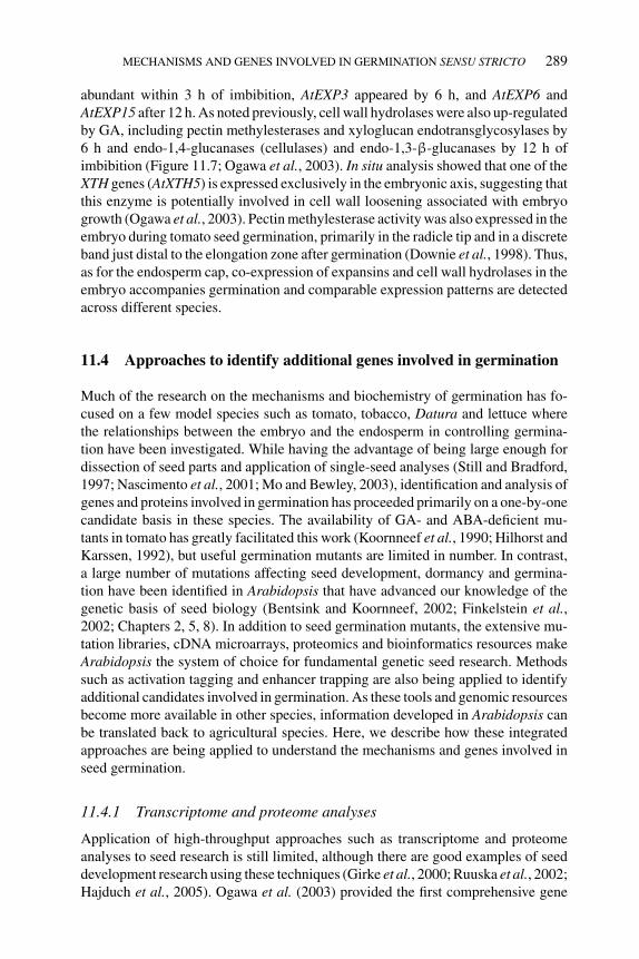
P1: OTE/SPH P2: OTE
BLUK053-Bradford September 27, 2006 15:21
MECHANISMS AND GENES INVOLVED IN GERMINATION SENSU STRICTO 289
abundant within 3 h of imbibition, AtEXP3 appeared by 6 h, and AtEXP6 andAtEXP15 after 12 h. As noted previously, cell wall hydrolases were also up-regulatedby GA, including pectin methylesterases and xyloglucan endotransglycosylases by6 h and endo-1,4-glucanases (cellulases) and endo-1,3-β-glucanases by 12 h ofimbibition (Figure 11.7; Ogawa et al., 2003). In situ analysis showed that one of theXTH genes (AtXTH5) is expressed exclusively in the embryonic axis, suggesting thatthis enzyme is potentially involved in cell wall loosening associated with embryogrowth (Ogawa et al., 2003). Pectin methylesterase activity was also expressed in theembryo during tomato seed germination, primarily in the radicle tip and in a discreteband just distal to the elongation zone after germination (Downie et al., 1998). Thus,as for the endosperm cap, co-expression of expansins and cell wall hydrolases in theembryo accompanies germination and comparable expression patterns are detectedacross different species.
11.4 Approaches to identify additional genes involved in germination
Much of the research on the mechanisms and biochemistry of germination has fo-cused on a few model species such as tomato, tobacco, Datura and lettuce wherethe relationships between the embryo and the endosperm in controlling germina-tion have been investigated. While having the advantage of being large enough fordissection of seed parts and application of single-seed analyses (Still and Bradford,1997; Nascimento et al., 2001; Mo and Bewley, 2003), identification and analysis ofgenes and proteins involved in germination has proceeded primarily on a one-by-onecandidate basis in these species. The availability of GA- and ABA-deficient mu-tants in tomato has greatly facilitated this work (Koornneef et al., 1990; Hilhorst andKarssen, 1992), but useful germination mutants are limited in number. In contrast,a large number of mutations affecting seed development, dormancy and germina-tion have been identified in Arabidopsis that have advanced our knowledge of thegenetic basis of seed biology (Bentsink and Koornneef, 2002; Finkelstein et al.,2002; Chapters 2, 5, 8). In addition to seed germination mutants, the extensive mu-tation libraries, cDNA microarrays, proteomics and bioinformatics resources makeArabidopsis the system of choice for fundamental genetic seed research. Methodssuch as activation tagging and enhancer trapping are also being applied to identifyadditional candidates involved in germination. As these tools and genomic resourcesbecome more available in other species, information developed in Arabidopsis canbe translated back to agricultural species. Here, we describe how these integratedapproaches are being applied to understand the mechanisms and genes involved inseed germination.
11.4.1 Transcriptome and proteome analyses
Application of high-throughput approaches such as transcriptome and proteomeanalyses to seed research is still limited, although there are good examples of seeddevelopment research using these techniques (Girke et al., 2000; Ruuska et al., 2002;Hajduch et al., 2005). Ogawa et al. (2003) provided the first comprehensive gene
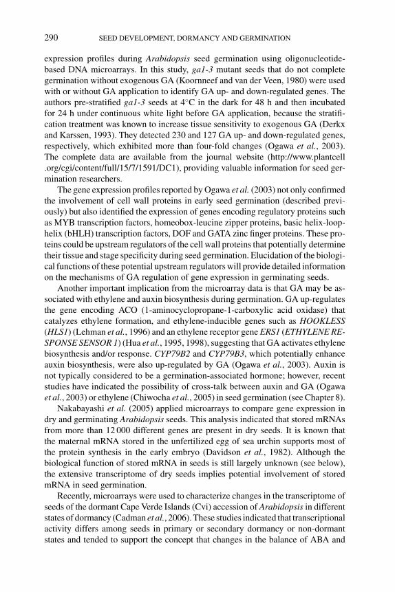
P1: OTE/SPH P2: OTE
BLUK053-Bradford September 27, 2006 15:21
290 SEED DEVELOPMENT, DORMANCY AND GERMINATION
expression profiles during Arabidopsis seed germination using oligonucleotide-based DNA microarrays. In this study, ga1-3 mutant seeds that do not completegermination without exogenous GA (Koornneef and van der Veen, 1980) were usedwith or without GA application to identify GA up- and down-regulated genes. Theauthors pre-stratified ga1-3 seeds at 4◦C in the dark for 48 h and then incubatedfor 24 h under continuous white light before GA application, because the stratifi-cation treatment was known to increase tissue sensitivity to exogenous GA (Derkxand Karssen, 1993). They detected 230 and 127 GA up- and down-regulated genes,respectively, which exhibited more than four-fold changes (Ogawa et al., 2003).The complete data are available from the journal website (http://www.plantcell.org/cgi/content/full/15/7/1591/DC1), providing valuable information for seed ger-mination researchers.
The gene expression profiles reported by Ogawa et al. (2003) not only confirmedthe involvement of cell wall proteins in early seed germination (described previ-ously) but also identified the expression of genes encoding regulatory proteins suchas MYB transcription factors, homeobox-leucine zipper proteins, basic helix-loop-helix (bHLH) transcription factors, DOF and GATA zinc finger proteins. These pro-teins could be upstream regulators of the cell wall proteins that potentially determinetheir tissue and stage specificity during seed germination. Elucidation of the biologi-cal functions of these potential upstream regulators will provide detailed informationon the mechanisms of GA regulation of gene expression in germinating seeds.
Another important implication from the microarray data is that GA may be as-sociated with ethylene and auxin biosynthesis during germination. GA up-regulatesthe gene encoding ACO (1-aminocyclopropane-1-carboxylic acid oxidase) thatcatalyzes ethylene formation, and ethylene-inducible genes such as HOOKLESS(HLS1) (Lehman et al., 1996) and an ethylene receptor gene ERS1 (ETHYLENE RE-SPONSE SENSOR 1) (Hua et al., 1995, 1998), suggesting that GA activates ethylenebiosynthesis and/or response. CYP79B2 and CYP79B3, which potentially enhanceauxin biosynthesis, were also up-regulated by GA (Ogawa et al., 2003). Auxin isnot typically considered to be a germination-associated hormone; however, recentstudies have indicated the possibility of cross-talk between auxin and GA (Ogawaet al., 2003) or ethylene (Chiwocha et al., 2005) in seed germination (see Chapter 8).
Nakabayashi et al. (2005) applied microarrays to compare gene expression indry and germinating Arabidopsis seeds. This analysis indicated that stored mRNAsfrom more than 12 000 different genes are present in dry seeds. It is known thatthe maternal mRNA stored in the unfertilized egg of sea urchin supports most ofthe protein synthesis in the early embryo (Davidson et al., 1982). Although thebiological function of stored mRNA in seeds is still largely unknown (see below),the extensive transcriptome of dry seeds implies potential involvement of storedmRNA in seed germination.
Recently, microarrays were used to characterize changes in the transcriptome ofseeds of the dormant Cape Verde Islands (Cvi) accession of Arabidopsis in differentstates of dormancy (Cadman et al., 2006). These studies indicated that transcriptionalactivity differs among seeds in primary or secondary dormancy or non-dormantstates and tended to support the concept that changes in the balance of ABA and
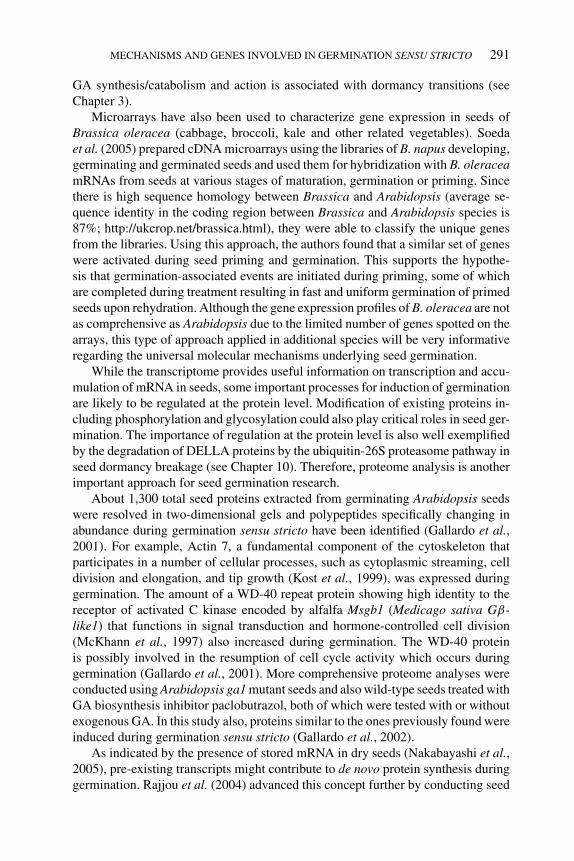
P1: OTE/SPH P2: OTE
BLUK053-Bradford September 27, 2006 15:21
MECHANISMS AND GENES INVOLVED IN GERMINATION SENSU STRICTO 291
GA synthesis/catabolism and action is associated with dormancy transitions (seeChapter 3).
Microarrays have also been used to characterize gene expression in seeds ofBrassica oleracea (cabbage, broccoli, kale and other related vegetables). Soedaet al. (2005) prepared cDNA microarrays using the libraries of B. napus developing,germinating and germinated seeds and used them for hybridization with B. oleraceamRNAs from seeds at various stages of maturation, germination or priming. Sincethere is high sequence homology between Brassica and Arabidopsis (average se-quence identity in the coding region between Brassica and Arabidopsis species is87%; http://ukcrop.net/brassica.html), they were able to classify the unique genesfrom the libraries. Using this approach, the authors found that a similar set of geneswere activated during seed priming and germination. This supports the hypothe-sis that germination-associated events are initiated during priming, some of whichare completed during treatment resulting in fast and uniform germination of primedseeds upon rehydration. Although the gene expression profiles of B. oleracea are notas comprehensive as Arabidopsis due to the limited number of genes spotted on thearrays, this type of approach applied in additional species will be very informativeregarding the universal molecular mechanisms underlying seed germination.
While the transcriptome provides useful information on transcription and accu-mulation of mRNA in seeds, some important processes for induction of germinationare likely to be regulated at the protein level. Modification of existing proteins in-cluding phosphorylation and glycosylation could also play critical roles in seed ger-mination. The importance of regulation at the protein level is also well exemplifiedby the degradation of DELLA proteins by the ubiquitin-26S proteasome pathway inseed dormancy breakage (see Chapter 10). Therefore, proteome analysis is anotherimportant approach for seed germination research.
About 1,300 total seed proteins extracted from germinating Arabidopsis seedswere resolved in two-dimensional gels and polypeptides specifically changing inabundance during germination sensu stricto have been identified (Gallardo et al.,2001). For example, Actin 7, a fundamental component of the cytoskeleton thatparticipates in a number of cellular processes, such as cytoplasmic streaming, celldivision and elongation, and tip growth (Kost et al., 1999), was expressed duringgermination. The amount of a WD-40 repeat protein showing high identity to thereceptor of activated C kinase encoded by alfalfa Msgb1 (Medicago sativa Gβ-like1) that functions in signal transduction and hormone-controlled cell division(McKhann et al., 1997) also increased during germination. The WD-40 proteinis possibly involved in the resumption of cell cycle activity which occurs duringgermination (Gallardo et al., 2001). More comprehensive proteome analyses wereconducted using Arabidopsis ga1 mutant seeds and also wild-type seeds treated withGA biosynthesis inhibitor paclobutrazol, both of which were tested with or withoutexogenous GA. In this study also, proteins similar to the ones previously found wereinduced during germination sensu stricto (Gallardo et al., 2002).
As indicated by the presence of stored mRNA in dry seeds (Nakabayashi et al.,2005), pre-existing transcripts might contribute to de novo protein synthesis duringgermination. Rajjou et al. (2004) advanced this concept further by conducting seed
P1: OTE/SPH P2: OTE
BLUK053-Bradford September 27, 2006 15:21
292 SEED DEVELOPMENT, DORMANCY AND GERMINATION
germination and proteome analyses using α-amanitin, a transcriptional inhibitor ofRNA polymerase II. The authors showed that radicle protrusion was not inhibitedby α-amanitin, while it did block subsequent seedling growth. This was notdue to impermeability of the testa, since transparent testa 2-1 seeds that have ahighly permeable testa were used for this experiment. In contrast, the translationalinhibitor cycloheximide completely blocked radicle protrusion, indicating that denovo protein synthesis is indispensable for germination. Although there is generalagreement about the importance of protein synthesis for seed germination, it seemsstill controversial whether germination can be completed without transcription ofany genes. Certainly, many genes are normally expressed early in seed germination(Ogawa et al., 2003). As Rajjou et al. (2004) proposed, microarray analyses of seedgermination in the presence or absence of α-amanitin would be very informative andcould lead to integration of the information from both transcriptome and proteomeanalyses.
11.4.2 Activation tagging and enhancer trapping
Activation tagging is an efficient approach in functional genomics (Walden et al.,1994). An activation tagging vector uses four copies of an enhancer element from theconstitutively active cauliflower mosaic virus 35S (CaMV 35S) promoter. When theT-DNA is inserted into the plant genome, the multiple copies of enhancers activatethe transcription of nearby genes, which results in a gain-of-function mutation.This is a powerful approach, especially for large gene families with redundancyin their function. A large population of Arabidopsis activation tagging lines hasbeen created. In this population, the majority of over-expressed genes were foundto be immediately adjacent to the enhancers. The CaMV 35S enhancers primarilyenhance an endogenous expression pattern and do not induce constitutive ectopicexpression (Weigel et al., 2000). Activation tagging has been used successfully toidentify genes involved in flowering (Kardailsky et al., 1999; Lee et al., 2000),phenylpropanoid biosynthesis (Borevitz et al., 2000), disease resistance (Xia et al.,2004), and hormone biosynthesis (Zhao et al., 2001), catabolism (Busov et al., 2003)and signaling (Neff et al., 1999). Interesting mutants that exhibited seedless fruitdevelopment were also isolated using this approach (Ito and Meyerowitz, 2000).Recent successful outcomes from activation tagging are the discovery of mutantsover-expressing microRNA genes (Aukerman and Sakai, 2003; Palatnik et al., 2003).
Salaita et al. (2005) successfully applied the activation tagging approach for seedgermination research. The authors screened 52 000 independent activation tagginglines for seeds that germinated faster than wildtype at 10◦C and recovered the threedominant mutants termed ctg (cold temperature germination) 10, ctg41, and ctg144.The recessive mutants ctg156 and ctg225 were also isolated from this screen, whichprobably result simply from the disruption of genes responsible for germinationby the activation tagging T-DNA (Salaita et al., 2005). This powerful approach, asshown by research on other aspects of plant development, has not been extensivelyused for seed germination research and could be applied to screen for genes involvedin other traits such as high temperature germination or secondary dormancy.
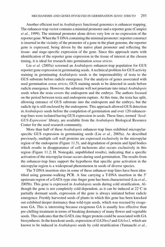
P1: OTE/SPH P2: OTE
BLUK053-Bradford September 27, 2006 15:21
MECHANISMS AND GENES INVOLVED IN GERMINATION SENSU STRICTO 293
Another efficient tool in Arabidopsis functional genomics is enhancer trapping.The enhancer-trap vector contains a minimal promoter and a reporter gene (Campisiet al., 1999). The minimal promoter alone drives very low or no expression of thereporter gene. When the T-DNA containing the minimal promoter::reporter constructis inserted in the vicinity of the promoter of a gene in the plant genome, the reportergene is expressed, being driven by the native plant promoter and reflecting thetissue- and stage-specific expression of the gene. Since this approach starts withidentification of the reporter gene expression in the tissue of interest at the chosentiming, it is ideal for research into germination sensu stricto.
Liu et al. (2005a) screened an Arabidopsis enhancer-trap population for GUSreporter gene expression in germinating seeds. A technical problem for GUS activitystaining in germinating Arabidopsis seeds is the impermeability of testa to theGUS substrate before radicle emergence. For the analysis of genes associated withseed germination sensu stricto, GUS staining needs to be detected in seeds beforeradicle emergence. However, the substrate will not penetrate into intact Arabidopsisseeds when the testa covers the endosperm and the embryo. The authors focusedon the period between testa and endosperm rupture. At this time, the testa has split,allowing entrance of GUS substrate into the endosperm and the embryo, but theradicle tip is still enclosed by the endosperm. This approach allowed GUS detectionin Arabidopsis seeds before the completion of germination and over 120 enhancer-trap lines were isolated having GUS expression in seeds. These lines, termed ‘Seed-GUS-Expression’ library, are available from the Arabidopsis Biological ResourceCenter for the seed research community.
More than half of these Arabidopsis enhancer-trap lines exhibited micropylar-specific GUS expression in germinating seeds (Liu et al., 2005a). As describedpreviously, multiple cell wall proteins are expressed exclusively in the micropylarregion of the endosperm (Figure 11.5), and degradation of protein and lipid bodieswhich results in disappearance of cell inclusions also occurs exclusively in thisregion (Figure 11.2; H. Nonogaki, unpublished results), indicating that a specificactivation of the micropylar tissue occurs during seed germination. The results fromthe enhancer-trap lines support the hypothesis that specific gene activation in themicropylar region is a widespread phenomenon in seeds of diverse species.
The T-DNA insertion sites in some of these enhancer-trap lines have been iden-tified using genome-walking PCR. A line carrying a T-DNA insertion in the 5′
upstream region of a GATA-type zinc finger gene has been characterized (Liu et al.,2005b). This gene is expressed in Arabidopsis seeds during cold stratification. Al-though the gene is not completely cold-dependent, as it can be induced at 22◦C inpartially dormant seeds, expression of the gene is always initiated before radicleemergence. Freshly harvested seeds of plants in which this gene has been knockedout exhibited deeper dormancy than wild-type seeds, which was rescued by exoge-nous GA. This is interesting because exogenous GA is usually less effective thanpre-chilling treatment in terms of breaking dormancy of many flower and vegetableseeds. This indicates that the GATA zinc finger protein could be associated with GAbiosynthesis. In the knockout seeds, expression of GA20ox3 and GA3ox1, which areknown to be induced in Arabidopsis seeds by cold stratification (Yamauchi et al.,
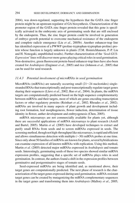
P1: OTE/SPH P2: OTE
BLUK053-Bradford September 27, 2006 15:21
294 SEED DEVELOPMENT, DORMANCY AND GERMINATION
2004), was down-regulated, supporting the hypothesis that the GATA zinc fingerprotein might be an upstream regulator of GA biosynthesis. Characterization of thepromoter region of the GATA zinc finger protein revealed that this gene is specif-ically activated in the embryonic axis of germinating seeds that are still enclosedby the endosperm. Thus, the zinc finger protein could be involved in generationof embryo growth potential to overcome mechanical resistance of the endospermand complete radicle emergence (Liu et al., 2005b). Another enhancer-trap linehas identified expression of a PWWP (proline-tryptophan-tryptophan-proline) pro-tein whose function is largely unknown in plants (T.M. Homrichhausen, P.-P. Liuand H. Nonogaki, unpublished results). Further characterization of the ‘Seed-GUS-Expression’ lines will discover more genes playing critical roles in seed germination.Non-destructive, green fluorescent protein-based enhancer-trap lines have also beencreated for Arabidopsis (Engineer et al., 2005) and rice (Johnson et al., 2005) thatcan be used for seed research.
11.4.3 Potential involvement of microRNAs in seed germination
MicroRNAs (miRNAs) are naturally occurring small (21∼24 nucleotides) single-stranded RNAs that transcriptionally and post-transcriptionally regulate target genessharing their sequences (Llave et al., 2002; Bao et al., 2004). In plants, the miRNAtargets are computationally predicted based on the high sequence complementaritybetween miRNAs and their targets, most of which are genes encoding transcriptionfactors or other regulatory proteins (Reinhart et al., 2002; Rhoades et al., 2002).miRNAs are involved in many aspects of plant growth and development includ-ing root formation, leaf morphogenesis, flower induction, determination of tissueidentity in flower, anther development and embryogenesis (Chen, 2005).
miRNA microarrays are not commercially available for plants yet, althoughthere are successful applications of miRNA microarrays to plant research (Axtelland Bartel, 2005). Martin et al. (2005) have developed techniques to extract andpurify small RNAs from seeds and to screen miRNAs expressed in seeds. Thescreening method, though not high-throughput like microarrays, is rapid and efficientand allows simultaneous detection with multiple (∼30) miRNA probes at one time.Since only about 50 families of miRNAs are known for plants, several hybridizationscan examine expression of all known miRNAs with replication. Using this method,Martin et al. (2005) detected major miRNAs expressed in Arabidopsis and tomatoseeds. Interestingly, germinating seeds of these two species showed similar miRNAexpression profiles, suggesting that a specific set of miRNAs plays roles in seedgermination. In contrast, the authors found a shift in the expression profiles betweengerminative and postgerminative stages of tomato seeds.
Seed-expressed miRNAs are being identified and, as mentioned above, theirtarget genes are computationally predicted. The next phase of research is the char-acterization of the target genes expressed during seed germination. miRNA-resistanttarget genes can be created by mutagenizing the miRNA complementary sequencesin the target genes and transforming them into Arabidopsis (Mallory et al., 2005;
P1: OTE/SPH P2: OTE
BLUK053-Bradford September 27, 2006 15:21
MECHANISMS AND GENES INVOLVED IN GERMINATION SENSU STRICTO 295
Millar and Gubler, 2005). This will cause over-accumulation of the target genes thatare usually repressed by miRNAs. Using this ‘de-repression’ approach, the biologi-cal functions of miRNA and their target genes in seed germination will be elucidated.
References
V. Amador, E. Monte, J.-L. Garcia-Martinez and S. Prat (2001) Gibberellins signal nuclear import
of PHOR1, a photoperiod-responsive protein with homology to Drosophila armadillo. Cell 106,
343–354.
M.V. Arana, L.C. de Miguel and R.A. Sanchez (2006) A phytochrome-dependent embryonic factor
modulates gibberellin responses in the embryo and micropylar endosperm of Datura ferox seeds.
Planta 223, 847–857.
M.J. Aukerman and H. Sakai (2003) Regulation of flowering time and floral organ identity by a
microRNA and its APETALA2-like target genes. The Plant Cell 15, 2730–2741.
M.J. Axtell and D.P. Bartel (2005) Antiquity of microRNAs and their targets in land plants. The PlantCell 17, 1658–1673.
N. Bao, K.W. Lye and M.K. Barton (2004) MicroRNA binding sites in Arabidopsis class III
HD-ZIP mRNAs are required for methylation of the template chromosome. Developmental Cell7, 653–662.
R.M. Barroco, K. Van Pounke, J.H.W. Bergervoet, et al. (2005) The role of the cell cycle machinery
in resumption of postembryonic development. Plant Physiology 137, 127–140.
A.B. Bennett (2002) Biochemical and genetic determinants of cell wall disassembly in ripening fruit:
a general model. Hortscience 37, 447–450.
L. Bentsink and M. Koornneef (2002) Seed dormancy and germination. In: The Arabidopsis Book (eds
C.R. Somerville and E.M., Meyerowitz). American Society of Plant Biologists, Rockville, MD.
(http://www.aspb.org/publications/arabidopsis/), pp. 1–18.
M.M. Beresniewicz, A.G. Taylor, M.C. Goffinet and W.D. Koeller (1995) Chemical nature of a semiper-
meable layer in seed coats of leek, onion (Liliaceae), tomato and pepper (Solanaceae). SeedScience and Technology 23, 135–145.
J.D. Bewley, R.A. Burton, Y. Morohashi and G.B. Fincher (1997) Molecular cloning of a cDNA
encoding a (1→4)-β-mannan endohydrolase from the seeds of germinated tomato (Lycopersiconesculentum). Planta 203, 454–459.
J.D. Bewley, F.D. Hempel, S. McCormick and P. Zambryski (2000) Reproductive development. In:
Biochemistry & Molecular Biology of Plants (eds B. Buchanan, W. Gruissem and R. Jones),
pp. 988–1043. American Society of Plant Biologists, Rockville, MD.
J.O. Borevitz, Y. Xia, J. Blount, R.A. Dixon and C. Lamb (2000) Activation tagging identifies a
conserved MYB regulator of phenylpropanoid biosynthesis. The Plant Cell 12, 2383–2394.
K.J. Bradford (1990) A water relations analysis of seed germination rates. Plant Physiology 94, 840–
849.
K.J. Bradford (1995) Water relations in seed germination. In: Seed Development and Germination(eds J. Kigel and G. Galili), pp. 351–396. Marcel Dekker, Inc., New York.
K.J. Bradford, F. Chen, M.B. Cooley, et al. (2000) Gene expression prior to radicle emergence in
imbibed tomato seeds. In: Seed Biology: Advances and Applications (eds M. Black, K.J. Bradford
and J. Vazquez-Ramos), pp. 231–251. CABI, Wallingford, U.K.
K.J. Bradford and O.A. Somasco (1994) Water relations of lettuce seed thermoinhibition. I. Priming
and endosperm effects on base water potential. Seed Science Research 4, 1–10.
K.J. Bradford and A.J. Trewavas (1994) Sensitivity thresholds and variable time scales in plant hormone
action. Plant Physiology 105, 1029–1036.
D.A. Brummell, M.H. Harpster and P. Dunsmuir (1999) Differential expression of expansin gene family
members during growth and ripening of tomato fruit. Plant Molecular Biology 39, 161–169.
P1: OTE/SPH P2: OTE
BLUK053-Bradford September 27, 2006 15:21
296 SEED DEVELOPMENT, DORMANCY AND GERMINATION
V.B. Busov, R. Meilan, D.W. Pearce, C. Ma, S.B. Rood and S.H. Strauss (2003) Activation tagging of
a dominant gibberellin catabolism gene (GA 2-oxidase) from poplar that regulates tree stature.
Plant Physiology 132, 1283–1291.
C.S.C. Cadman, P.E. Toorop, H.W.M. Hilhorst and W.E. Finch-Savage (2006) Gene expression profiles
of Arabidopsis Cvi seeds during cycling through dormant and non-dormant states indicate a
common underlying dormancy control mechanism. The Plant Journal 46, 805–822.
L. Campisi, Y. Yang, Y. Yi, et al. (1999) Generation of enhancer trap lines in Arabidopsis and charac-
terization of expression patterns in the inflorescence. The Plant Journal 17, 699–707.
N.C. Carpita and D.M. Gibeaut (1993) Structural models of primary cell walls in flowering plants -
consistency of molecular structure with the physical properties of the walls during growth. ThePlant Journal 3, 1–30.
C. Catala, J.K.C. Rose and A.B. Bennett (1997) Auxin regulation and spatial localization of an endo-
1,4-β-d-glucanase and a xyloglucan endotransglycosylase in expanding tomato hypocotyls. ThePlant Journal 12, 417–426.
C. Catala, J.K.C. Rose and A.B. Bennett (2000) Auxin-regulated genes encoding cell wall-modifying
proteins are expressed during early tomato fruit growth. Plant Physiology 122, 527–534.
F. Chen and K.J. Bradford (2000) Expression of an expansin is associated with endosperm weakening
during tomato seed germination. Plant Physiology 124, 1265–1274.
F. Chen, P. Dahal and K.J. Bradford (2001) Two tomato expansin genes show divergent expression
and localization in embryos during seed development and germination. Plant Physiology 127,
928–936.
F. Chen, H. Nonogaki and K.J. Bradford (2002) A gibberellin-regulated xyloglucan endotransgly-
cosylase gene is expressed in the endosperm cap during tomato seed germination. Journal ofExperimental Botany 53, 215–223.
X. Chen (2005) microRNA biogenesis and function in plants. FEBS Letters 579, 5923–5931.
S.D. Chiwocha, A.J. Cutler, S.R. Abrams, et al. (2005) The etr1-2 mutation in Arabidopsis thalianaaffects the abscisic acid, auxin, cytokinin and gibberellin metabolic pathways during maintenance
of seed dormancy, moist-chilling and germination. The Plant Journal 42, 35–48.
H.T. Cho and D.J. Cosgrove (2000) Altered expression of expansin modulates leaf growth and pedicel
abscission in Arabidopsis thaliana. Proceedings of the National Academy of Sciences UnitedStates of America 97, 9783–9788.
H.T. Cho and D.J. Cosgrove (2002) Regulation of root hair initiation and expansin gene expression in
Arabidopsis. The Plant Cell 14, 3237–3253.
D.J. Cosgrove (2005) Expansin web page. (http://www.bio.psu.edu/expansins/), date of access
1 October 2005.
D.J. Cosgrove, L.C. Li, H.T. Cho, S. Hoffmann-Benning, R.C. Moore and D. Blecker (2002) The
growing world of expansins. Plant and Cell Physiology 43, 1436–1444.
T. Czechowski, M. Stitt, T. Altmann, M.K. Udvardi and W.-R. Scheible (2005) Genome-wide identi-
fication and testing of superior reference genes for transcript normalization in Arabidopsis. PlantPhysiology 139, 5–17.
E.A.A. da Silva, P.E. Toorop, J. Nijsse, J.D. Bewley and H.W.M. Hilhorst (2005) Exogenous gib-
berellins inhibit coffee (Coffea arabica cv. Rubi) seed germination and cause cell death in the
embryo. Journal of Experimental Botany 56, 1029–1038.
E.A.A. da Silva, P.E. Toorop, A.C. van Aelst and H.W.M. Hilhorst (2004) Abscisic acid controls
embryo growth potential and endosperm cap weakening during coffee (Coffea arabica cv. Rubi)
seed germination. Planta 220, 251–261.
P. Dahal and K.J. Bradford (1990) Effects of priming and endosperm integrity on seed germination
rates of tomato genotypes. II. Germination at reduced water potential. Journal of ExperimentalBotany 41, 1441–1453.
P. Dahal, D.J. Nevins and K.J. Bradford (1997) Relationship of endo-β-d-mannanase activity and cell
wall hydrolysis in tomato endosperm to germination rates. Plant Physiology 113, 1243–1252.
E.H. Davidson, B.R. Hough-Evans and R.J. Britten (1982) Molecular biology of the sea urchin embryo.
Science 217, 17–26.
P1: OTE/SPH P2: OTE
BLUK053-Bradford September 27, 2006 15:21
MECHANISMS AND GENES INVOLVED IN GERMINATION SENSU STRICTO 297
I. Debeaujon, K.M. Leon-Kloosterziel and M. Koornneef (2000) Influence of the testa on seed dor-
mancy, germination, and longevity in Arabidopsis. Plant Physiology 122, 403–413.
R.D. de Castro, A.A.M. van Lammeren, S.P.C. Groot, R.J. Bino and H.W.M. Hilhorst (2000) Cell
division and subsequent radicle protrusion in tomato seeds are inhibited by osmotic stress but
DNA synthesis and formation of microtubular cytoskeleton are not. Plant Physiology 122, 327–
335.
L. de Miguel, M.J. Burgin, J.J. Casal and R.A. Sanchez (2000) Antagonistic action of low-fluence and
high-irradiance modes of response of phytochrome on germination and β-mannanase activity in
Datura ferox seeds. Journal of Experimental Botany 51, 1127–1133.
L. de Miguel and R.A. Sanchez (1992) Phytochrome-induced germination, endosperm softening and
embryo growth-potential in Datura ferox seeds. Sensitivity to low water potential and time to
escape to Fr reversal. Journal of Experimental Botany 43, 969–974.
M.P.M. Derkx and C.M. Karssen (1993) Effects of light and temperature on seed dormancy and
gibberellin stimulated germination in Arabidopsis thaliana: studies with gibberellin-deficient
and -insensitive mutants. Physiologia Plantarum 89, 360–368.
L.M.A. Dirk, A.M. Griffen, B. Downie and J.D. Bewley (1995) Multiple isozymes of endo-β-d-
mannanase in dry and imbibed seeds. Phytochemistry 40, 1045–1056.
B. Downie, L.M.A. Dirk, K.A. Hadfield, T.A. Wilkins, A.B. Bennett and K.J. Bradford (1998) A gel
diffusion assay for quantification of pectin methylesterase activity. Analytical Biochemistry 264,
149–157.
A.B. Downie, L.M.A. Dirk, Q.L. Xu, et al. (2004) A physical, enzymatic, and genetic characterization
of perturbations in the seeds of the brownseed tomato mutants. Journal of Experimental Botany55, 961–973.
B. Downie, S. Gurusinghe and K.J. Bradford (1999) Internal anatomy of individual tomato seeds:
relationship to abscisic acid and germination physiology. Seed Science Research 9, 117–128.
B. Downie, S. Gurusinghe, K.J. Bradford, C.G. Plopper, J.S. Greenwood and J.D. Bewley (1997a)
Embryo root cap cells adhere to the megagametophyte and sheathe the radicle of white spruce
(Picea glauca [Moench.] Voss.) seeds following germination. International Journal of PlantSciences 158, 738–746.
B. Downie, H.W.M. Hilhorst and J.D. Bewley (1997b) Endo-β-mannanase activity during dormancy
alleviation and germination of white spruce (Picea glauca) seeds. Physiologia Plantarum 101,
405–415.
A.B. Downie, D.Q. Zhang, L.M.A. Dirk, et al. (2003) Communication between the maternal testa and
the embryo and/or endosperm affect testa attributes in tomato. Plant Physiology 133, 145–160.
J. Dulson, J.D. Bewley and R.N. Johnston (1988) Abscisic acid is an endogenous inhibitor in the regu-
lation of mannanase production by isolated lettuce (Lactuca sativa cv Grand Rapids) endosperms.
Plant Physiology 87, 660–665.
S. Dutta, K.J. Bradford and D.J. Nevins (1997) Endo-β-mannanase activity present in cell wall extracts
of lettuce endosperm prior to radicle emergence. Plant Physiology 113,155–161.
C.B. Engineer, K.C. Fitzsimmons, J.J. Schmuke, S.B. Dotson and R.G. Kranz (2005) Development
and evaluation of a Gal4-mediated LUC/GFP/GUS enhancer trap system in Arabidopsis. BMCPlant Biology 2005, 5:9 doi:10.1186/1471-2229-5-9.
J.A. Feurtado, M. Banik and J.D. Bewley (2001) The cloning and characterization of α-galactosidase
present during and following germination of tomato (Lycopersicon esculentum Mill.) seed. Jour-nal of Experimental Botany 52, 1239–1249.
R.R. Finkelstein, S.S.L. Gampala and C.D. Rock (2002) Abscisic acid signaling in seeds and seedlings.
The Plant Cell 14, S15–S45.
S.C. Fry (2004) Primary cell wall metabolism: tracking the careers of wall polymers in living plant
cells. New Phytologist 161, 641–675.
K. Gallardo, C. Job, S.P. Groot, et al. (2002) Proteomics of Arabidopsis seed germination. A compar-
ative study of wild-type and gibberellin-deficient seeds. Plant Physiology 129, 823–837.
K. Gallardo, C. Job, S.P.C. Groot, et al. (2001) Proteomic analysis of Arabidopsis seed germination
and priming. Plant Physiology 126, 835–848.
P1: OTE/SPH P2: OTE
BLUK053-Bradford September 27, 2006 15:21
298 SEED DEVELOPMENT, DORMANCY AND GERMINATION
Y.P. Gao, L. Young, P. Bonham-Smith and L.V. Gusta (1999) Characterization and expression of plasma
and tonoplast membrane aquaporins in primed seed of Brassica napus during germination under
stress conditions. Plant Molecular Biology 40, 635–644.
T. Girke, J. Todd, S. Ruuska, J. White, C. Benning and J. Ohlrogge (2000) Microarray analysis of
developing Arabidopsis seeds. Plant Physiology 124, 1570–1581.
L. Gomez, I. Allona, R. Casado and C. Aragoncillo (2002) Seed chitinases. Seed Science Research12, 217–230.
S.P.C. Groot and C.M. Karssen (1987) Gibberellins regulate seed germination in tomato by endosperm
weakening–a study with gibberellin-deficient mutants. Planta 171, 525–531.
S.P.C. Groot and C.M. Karssen (1992) Dormancy and germination of abscisic acid-deficient tomato
seeds. Studies with the sitiens mutant. Plant Physiology 99, 952–958.
S.P.C. Groot, B. Kieliszewskarokicka, E. Vermeer and C.M. Karssen (1988) Gibberellin-induced
hydrolysis of endosperm cell walls in gibberellin-deficient tomato seeds prior to radicle protrusion.
Planta 174, 500–504.
F. Gubler, R. Kalla, J.K. Roberts and J.V. Jacobsen (1995) Gibberellin-regulated expression of a mybgene in barley aleurone cells: evidence for myb transactivation of a high-pI α-amylase gene
promoter. The Plant Cell 7, 1879–1891.
F. Gubler, A.A. Millar and J.V. Jacobsen (2005) Dormancy release, ABA and pre-harvest sprouting.
Current Opinion in Plant Biology 8, 183–187.
K.A. Hadfield and A.B. Bennett (1998) Polygalacturonases: many genes in search of a function. PlantPhysiology 117, 337–343.
A.M. Haigh (1988) Why do tomato seeds prime? Physiological Investigations into the Control ofTomato Seed Germination and Priming. Ph.D. Dissertation, School of Biological Sciences, Mac-
quarie University, North Ryde, NSW, Australia.
A.M. Haigh and E.W.R. Barlow (1987) Water relations of tomato seed germination. Australian Journalof Plant Physiology 14, 485–492.
M. Hajduch, A. Ganapathy, J.W. Stein and J.J. Thelen (2005) A systematic proteomic study of seed
filling in soybean. Establishment of high-resolution two-dimensional reference maps, expression
profiles, and an interactive proteome database. Plant Physiology 137, 1397–1419.
P. Halmer (2004) Methods to improve seed performance in the field. In: Handbook of Seed Physiology.Applications to Agriculture (eds R.L. Bench-Arnold and R.A. Sanchez), pp. 125–166. Food
Products Press, New York.
P. Halmer, J.D. Bewley and T.A. Thorpe (1976) Enzyme to degrade lettuce endosperm cell walls – ap-
pearance of a mannanase following phytochrome-induced and gibberellin-induced germination.
Planta 130, 189–196.
E.P. Harrison, S.J. McQueen-Mason and K. Manning (2001) Expression of six expansin genes in
relation to extension activity in developing strawberry fruit. Journal of Experimental Botany 52,
1437–1446.
H.W.M. Hilhorst and B. Downie (1996) Primary dormancy in tomato (Lycopersicon esculentum cv
Moneymaker): studies with the sitiens mutant. Journal of Experimental Botany 47, 89–97.
H.W.M. Hilhorst and C.M. Karssen (1992) Seed dormancy and germination – the role of abscisic acid
and gibberellins and the importance of hormone mutants. Plant Growth Regulation 11, 225–238.
H.J. Hill and A.G. Taylor (1989) Relationship between viability, endosperm integrity, and imbibed
lettuce seed density and leakage. Hortscience 24, 814–816.
H. Hofte, L. Hubbard, J. Reizer, D. Ludevid, E.M. Herman and M.J. Chrispeels (1992) Vegetative
and seed-specific forms of tonoplast intrinsic protein in the vacuolar membrane of Arabidopsisthaliana. Plant Physiology 99, 561–570.
J. Hou, E. Kendall and G. Simpson (1997) Water uptake and distribution in non-dormant and dormant
wild oat (Avena fatua L.) caryopses. Journal of Experimental Botany 48, 683–692.
J. Hua, C. Chang, Q. Sun and E.M. Meyerowitz (1995) Ethylene insensitivity conferred by ArabidopsisERS gene. Science 269, 1712–1714.
J. Hua, H. Sakai, S. Nourizadeh, et al. (1998) EIN4 and ERS2 are members of the putative ethylene
receptor gene family in Arabidopsis. The Plant Cell 10, 1321–1332.
P1: OTE/SPH P2: OTE
BLUK053-Bradford September 27, 2006 15:21
MECHANISMS AND GENES INVOLVED IN GERMINATION SENSU STRICTO 299
H. Ikuma and K.V. Thimann (1963) The role of the seed-coats in germination of photosensitive lettuce
seeds. Plant and Cell Physiology 4, 169–185.
ISTA (1999) International rules for seed testing. Seed Science and Technology 27, supplement.
A. Itai, K. Ishihara and J.D. Bewley (2003) Characterization of expression, and cloning, of β-d-
xylosidase and α-l-arabinofuranosidase in developing and ripening tomato (Lycopersicon escu-lentum Mill.) fruit. Journal of Experimental Botany 54, 2615–2622.
T. Ito and E.M. Meyerowitz (2000) Overexpression of a gene encoding a cytochrome P450, CYP78A9,
induces large and seedless fruit in Arabidopsis. The Plant Cell 12, 1541–1550.
S.E. Jacobsen, K.A. Binkowski and N.E. Olszewski (1996) SPINDLY, a tetratricopeptide repeat protein
involved in gibberellin signal transduction in Arabidopsis. Proceedings of the National Academyof Sciences United States of America 93, 9292–9296.
J.V. Jacobsen, D.W. Pearce, A.T. Poole, R.P. Pharis and L.N. Mander (2002) Abscisic acid, phaseic
acid and gibberellin contents associated with dormancy and germination in barley. PhysiologiaPlantarum 115, 428–441.
A.A. Johnson, J.M. Hibberd, C. Gay, et al. (2005) Spatial control of transgene expression in rice
(Oryza sativa L.) using the GAL4 enhancer trapping system. The Plant Journal 41, 779–789.
I. Kardailsky, V.K. Shukla, J.H. Ahn, et al. (1999) Activation tagging of the floral inducer FT. Science286, 1962–1965.
H. Kende, K.J. Bradford, D.A. Brummell, et al. (2004) Nomenclature for members of the expansin
superfamily of genes and proteins. Plant Molecular Biology 55, 311–314.
M. Koornneef, T.D.G. Bosma, C.J. Hanhart, J.H. Vanderveen and J.A.D. Zeevaart (1990) The isolation
and characterization of gibberellin-deficient mutants in tomato. Theoretical and Applied Genetics80, 852–857.
M. Koornneef and J.H. van der Veen (1980) Induction and analysis of gibberellin sensitive mutants in
Arabidopsis thaliana (L.) Heynh. Theoretical Applied Genetics 58, 257–263.
B. Kost, J. Mathur and N.H. Chua (1999) Cytoskeleton in plant development. Current Opinion inPlant Biology 2, 462–470.
H. Lee, S.S. Suh, E. Park, et al. (2000) The AGAMOUS-LIKE 20 MADS domain protein integrates
floral inductive pathways in Arabidopsis. Genes and Development 14, 2366–2376.
A. Lehman, R. Black and J.R. Ecker (1996) HOOKLESS1, an ethylene response gene, is required for
differential cell elongation in the Arabidopsis hypocotyl. Cell 85, 183–194.
A.C. Leopold (1983) Volumetric components of seed imbibition. Plant Physiology 73, 677–680.
G. Leubner-Metzger (2002) Seed after-ripening and over-expression of class I β-1,3-glucanase confer
maternal effects on tobacco testa rupture and dormancy release. Planta 215, 959–968.
G. Leubner-Metzger (2003) Functions and regulation of β-1,3-glucanases during seed germination,
dormancy release and after-ripening. Seed Science Research 13, 17–34.
G. Leubner-Metzger (2005) β-1,3-glucanase gene expression in low-hydrated seeds as a mechanism
for dormancy release during tobacco after-ripening. Plant Journal 41, 133–145.
G. Leubner-Metzger, C. Frundt, R. Vogeli-Lange and F. Meins (1995) Class-I β-1,3-glucanases in the
endosperm of tobacco during germination. Plant Physiology 109, 751–759.
G. Leubner-Metzger and F. Meins (2000) Sense transformation reveals a novel role for class I β-1,3-
glucanase in tobacco seed germination. Plant Journal 23, 215–221.
G. Leubner-Metzger and F. Meins (2001) Antisense-transformation reveals novel roles for class I
β-1,3-glucanase in tobacco seed after-ripening and photodormancy. Journal of ExperimentalBotany 52, 1753–1759.
S. Leviatov, O. Shoseyov and S. Wolf (1995) Involvement of endomannanase in the control of tomato
seed germination under low temperature conditions. Annals of Botany 76, 1–6.
P.-P. Liu, N. Koizuka, T.M. Homrichhausen, J.R. Hewitt, R.C. Martin and H. Nonogaki (2005a) Large-
scale screening of Arabidopsis enhancer-trap lines for seed germination-associated genes. PlantJournal 41, 936–944.
P.-P. Liu, N. Koizuka, R.C. Martin and H. Nonogaki (2005b) The BME3 (Blue Micropylar End 3)
GATA zinc finger transcription factor is a positive regulator of Arabidopsis seed germination.
Plant Journal 44, 960–971.

P1: OTE/SPH P2: OTE
BLUK053-Bradford September 27, 2006 15:21
300 SEED DEVELOPMENT, DORMANCY AND GERMINATION
C. Llave, Z. Xie, K.D. Kasschau and J.C. Carrington (2002) Cleavage of Scarecrow-like mRNA targets
directed by a class of Arabidopsis miRNA. Science 297, 2053–2056.
L. Ma, N. Sun, X. Liu, Y. Jiao, H. Zhao and X.W. Deng (2005) Organ-specific expression of Arabidopsisgenome during development. Plant Physiology 138, 80–91.
A.C. Mallory, D.P. Bartel and B. Bartel (2005) MicroRNA-directed regulation of Arabidopsis AUXINRESPONSE FACTOR17 is essential for proper development and modulates expression of early
auxin response genes. The Plant Cell 17, 1360–1375.
B. Manz, K. Muller, B. Kucera, F. Volke and G. Leubner-Metzger (2005) Water uptake and distribution
in germinating tobacco seeds investigated in vivo by nuclear magnetic resonance imaging. PlantPhysiology 138, 1538–1551.
P. Marraccini, W.J. Rogers, C. Allard, et al. (2001) Molecular and biochemical characterization
of endo-β-mannanases from germinating coffee (Coffea arabica) grains. Planta 213, 296–
308.
R.C. Martin, P.-P. Liu and H. Nonogaki (2005) Simple purification of small RNAs from seeds and
efficient detection of multiple microRNAs expressed in Arabidopsis thaliana and tomato (Lycop-ersicon esculentum) seeds. Seed Science Research 15, 319–328.
R.C. Martin, P.-P. Liu and H. Nonogaki (2006) MicroRNAs in seeds – modified detection techniques
and potential applications. Canadian Journal of Botany 84, 189–198.
N.H. Masubelele, W. Dewitte, M. Menges, et al. (2005) D-type cyclins activate division in the root apex
to promote seed germination in Arabidopsis. Proceedings of the National Academy of SciencesUnited States of America 102, 15694–15699.
A.J.M. Matzke, E.M. Stoger and M.A. Matzke (1993) A zein gene promoter fragment drives Gus
expression in a cell layer that is interposed between the endosperm and the seed coat. PlantMolecular Biology 22, 553–554.
C. Maurel, M. Chrispeels, C. Lurin, et al. (1997) Function and regulation of seed aquaporins. Journalof Experimental Botany 48, 421–430.
M.B. McDonald, J. Sullivan and M.J. Lauer (1994) The pathway of water-uptake in maize seeds. SeedScience and Technology 22, 79–90.
M.B. McDonald, C.W. Vertucci and E.E. Roos (1988) Soybean seed imbibition – water-absorption by
seed parts. Crop Science 28, 993–997.
H.I. McKhann, F. Frugier, G. Petrovics, et al. (1997) Cloning of a WD-repeat-containing gene from
alfalfa (Medicago sativa): a role in hormone-mediated cell division? Plant Molecular Biology34, 771–780.
R.A. Mella, M.J. Burgin and R.A. Sanchez (2004) Expansin gene expression in Datura ferox L. seeds is
regulated by the low-fluence response, but not by the high-irradiance response, of phytochromes.
Seed Science Research 14, 61–71.
R.A. Mella, S. Maldonado and R.A. Sanchez (1995) Phytochrome-induced structural changes and
protein degradation prior to radicle protrusion in Datura ferox seeds. Canadian Journal of Botany73, 1371–1378.
A.A. Millar and F. Gubler (2005) The Arabidopsis GAMYB-like genes, MYB33 and MYB65, are
microRNA-regulated genes that redundantly facilitate anther development. The Plant Cell 17,
705–721.
B.X. Mo and J.D. Bewley (2002) β-Mannosidase (EC 3.2.1.25) activity during and following germina-
tion of tomato (Lycopersicon esculentum Mill.) seeds. Purification, cloning and characterization.
Planta 215, 141–152.
B. Mo and J.D. Bewley (2003) The relationship between β-mannosidase and endo-β-mannanase
activities in tomato seeds during and following germination: a comparison of seed populations
and individual seeds. Journal of Experimental Botany 54, 2503–2510.
Y. Morohashi and H. Matsushima (2000) Development of β-1,3-glucanase activity in germinated
tomato seeds. Journal of Experimental Botany 51, 1381–1387.
K. Nakabayashi, M. Okamoto, T. Koshiba, Y. Kamiya and E. Nambara (2005) Genome-wide profiling
of stored mRNA in Arabidopsis thaliana seed germination: epigenetic and genetic regulation of
transcription in seed. The Plant Journal 41, 697–709.
P1: OTE/SPH P2: OTE
BLUK053-Bradford September 27, 2006 15:21
MECHANISMS AND GENES INVOLVED IN GERMINATION SENSU STRICTO 301
W.M. Nascimento, D.J. Cantliffe and D.J. Huber (2000) Thermotolerance in lettuce seeds: association
with ethylene and endo-β-mannanase. Journal of the American Society for Horticultural Science125, 518–524.
W.M. Nascimento, D.J. Cantliffe and D.J. Huber (2001) Endo-β-mannanase activity and seed germi-
nation of thermosensitive and thermotolerant lettuce genotypes in response to seed priming. SeedScience Research 11, 255–264.
W.M. Nascimento, D.J. Cantliffe and D.J. Huber (2004) Ethylene evolution and endo-β-mannanase
activity during lettuce seed germination at high temperature. Scientia Agricola 61, 156–163.
M.M. Neff, S.M. Nguyen, E.J. Malancharuvil, et al. (1999) BAS1: A gene regulating brassinosteroid
levels and light responsiveness in Arabidopsis. Proceedings of the National Academy of SciencesUnited States of America 96, 15316–15323.
B.R. Ni and K.J. Bradford (1992) Quantitative models characterizing seed germination responses to
abscisic acid and osmoticum. Plant Physiology 98, 1057–1068.
B.R. Ni and K.J. Bradford (1993) Germination and dormancy of abscisic acid-deficient and gibberellin-
deficient mutant tomato seeds. Sensitivity of germination to abscisic acid, gibberellin, and water
potential. Plant Physiology 101, 607–617.
H. Nonogaki, O.H. Gee and K.J. Bradford (2000) A germination-specific endo-β-mannanase gene is
expressed in the micropylar endosperm cap of tomato seeds. Plant Physiology 123, 1235–1245.
H. Nonogaki, H. Matsushima and Y. Morohashi (1992) Galactomannan hydrolyzing activity develops
during priming in the micropylar endosperm tip of tomato seeds. Physiologia Plantarum 85,
167–172.
H. Nonogaki and Y. Morohashi (1999) Temporal and spatial pattern of the development of endo-β-
mannanase activity in germinating and germinated lettuce seeds. Journal of Experimental Botany50, 1307–1313.
H. Nonogaki, M. Nomaguchi, N. Okumoto, Y. Kaneko, H. Matsushima and Y. Morohashi (1998)
Temporal and spatial pattern of the biochemical activation of the endosperm during and following
imbibition of tomato seeds. Physiologia Plantarum 102, 236–242.
M. Ogawa, A. Hanada, Y. Yamauchi, A. Kuwalhara, Y. Kamiya and S. Yamaguchi (2003) Gib-
berellin biosynthesis and response during Arabidopsis seed germination. The Plant Cell 15, 1591–
1604.
K. Ogawa and M. Iwabuchi (2001) A mechanism for promoting the germination of Zinnia elegansseeds by hydrogen peroxide. Plant and Cell Physiology 42, 286–291.
J.F. Palatnik, E. Allen, X. Wu, et al. (2003) Control of leaf morphogenesis by microRNAs. Nature425, 257–263.
J.W. Patrick and C.E. Offler (2001) Compartmentation of transport and transfer events in developing
seeds. Journal of Experimental Botany 52, 551–564.
S. Penfield, E.L. Rylott, A.D. Gilday, S. Graham, T.R. Larson and I.A. Graham (2004) Reserve
mobilization in the Arabidopsis endosperm fuels hypocotyl elongation in the dark, is independent
of abscisic acid, and requires PHOSPHOENOLPYRUVATE CARBOXYKINASE1. The Plant Cell16, 2705–2718.
C. Perino and D. Come (1991) Physiological and metabolic study of the germination phases in apple
embryo. Seed Science and Technology 19, 1–14.
G. Psaras and K. Georghiou (1983) Gibberellic acid-induced structural alterations in the endosperm
of germinating Lactuca sativa L. achenes. Zeitschrift Fur Pflanzenphysiologie 112, 14–19.
L. Rajjou, K. Gallardo, I. Debeaujon, J. Vandekerckhove, C. Job and D. Job (2004) The effect of
α-amanitin on the Arabidopsis seed proteome highlights the distinct roles of stored and neosyn-
thesized mRNAs during germination. Plant Physiology 134, 1598–1613.
J.S.G. Reid and M. Edwards (1995) Galactomannans and other cell wall storage polysaccharides in
seeds. In: Food Polysaccharides and Their Applications (ed. A.M. Stephen), pp. 155–186. Marcel
Dekker, New York.
J.S.G. Reid, M.E. Edwards, C.A. Dickson, C. Scott and M.J. Gidley (2003) Tobacco transgenic lines
that express fenugreek galactomannan galactosyltransferase constitutively have structurally al-
tered galactomannans in their seed endosperm cell walls. Plant Physiology 131, 1487–1495.
P1: OTE/SPH P2: OTE
BLUK053-Bradford September 27, 2006 15:21
302 SEED DEVELOPMENT, DORMANCY AND GERMINATION
B.J. Reinhart, E.G. Weinstein, M.W. Rhoades, B. Bartel and D.P. Bartel (2002) MicroRNAs in plants.
Genes and Development 16, 1616–1626.
C.W. Ren and A.R. Kermode (2000) An increase in pectin methyl esterase activity accompa-
nies dormancy breakage and germination of yellow cedar seeds. Plant Physiology 124, 231–
242.
M.W. Rhoades, B.J. Reinhart, L.P. Lim, C.B. Burge, B. Bartel and D.P. Bartel (2002) Prediction of
plant microRNA targets. Cell 110, 513–520.
P.L.H. Rinne and C. van der Schoot (2003) Plasmodesmata at the crossroads between development,
dormancy, and defense. Canadian Journal of Botany 81, 1182–1197.
J.K.C. Rose, K.A. Hadfield, J.M. Labavitch and A.B. Bennett (1998) Temporal sequence of cell wall
disassembly in rapidly ripening melon fruit. Plant Physiology 117, 345–361.
J.K.C. Rose, H.H. Lee and A.B. Bennett (1997) Expression of a divergent expansin gene is fruit-
specific and ripening-regulated. Proceedings of the National Academy of Sciences United Statesof America 94, 5955–5960.
S.A. Ruuska, T. Girke, C. Benning and J.B. Ohlrogge (2002) Contrapuntal networks of gene expression
during Arabidopsis seed filling. The Plant Cell 14, 1191–206.
L. Salaita, R.K. Kar, M. Majee and A.B. Downie (2005) Identification and characterization of mutants
capable of rapid seed germination at 10◦C from activation-tagged lines of Arabidopsis thaliana.
Journal of Experimental Botany 56, 2059–2069.
R.A. Sanchez, L. de Miguel, C. Lima and R.M. de Lederkremer (2002) Effect of low water potential
on phytochrome-induced germination, endosperm softening and cell-wall mannan degradation
in Datura ferox seeds. Seed Science Research 12, 155–163.
R.A. Sanchez, L. de Miguel and O. Mercuri (1986) Phytochrome control of cellulase activity in
Datura ferox L. seeds and its relationship with germination. Journal of Experimental Botany 37,
1574–1580.
R.A. Sanchez, L. Sunell, J.M. Labavitch and B.A. Bonner (1990) Changes in the endosperm cell walls
of two Datura species before radicle protrusion. Plant Physiology 93, 89–97.
W.R. Scheible and M. Pauly (2004) Glycosyltransferases and cell wall biosynthesis: novel players and
insights. Current Opinion in Plant Biology 7, 285–295.
P. Schopfer, D. Bajracharya and C. Plachy (1979) Control of seed germination by abscisic acid. I.
Time course of action in Sinapis alba L. Plant Physiology 64, 822–827.
P. Schopfer and C. Plachy (1984) Control of seed germination by abscisic acid. II. Effect on embryo
water uptake in Brassica napus L. Plant Physiology 76, 155–160.
P. Schopfer and C. Plachy (1985) Control of seed germination by abscisic acid. III. Effect on embryo
growth potential (minimum turgor pressure) and growth coefficient (cell wall extensibility) in
Brassica napus L. Plant Physiology 77, 676–686.
J. Schuurmans, J.T. van Dongen, B.P.W. Rutjens, A. Boonman, C.M.J. Pieterse and A.C. Borstlap
(2003) Members of the aquaporin family in the developing pea seed coat include representatives
of the PIP, TIP, and NIP subfamilies. Plant Molecular Biology 53, 655–667.
Y. Sitrit, K.A. Hadfield, A.B. Bennett, K.J. Bradford and A.B. Downie (1999) Expression of a poly-
galacturonase associated with tomato seed germination. Plant Physiology 121, 419–428.
Y. Soeda, M. Konings, O. Vorst, et al. (2005) Gene expression programs during Brassica oleraceaseed maturation, osmopriming, and germination are indicators of progression of the germination
process and the stress tolerance level. Plant Physiology 137, 354–368.
P. Spoelstra (2002) Germination and dormancy of single tomato seeds. A Study Using Non-Invasive Molecular and Biophysical Techniques. Ph.D. Disseration, Wageningen University, The
Netherlands.
D.W. Still and K.J. Bradford (1997) Endo-β-mannanase activity from individual tomato endosperm
caps and radicle tips in relation to germination rates. Plant Physiology 113, 21–29.
Y. Sung, D.J. Cantliffe and R. Nagata (1998) Using a puncture test to identify the role of seed coverings
on thermotolerant lettuce seed germination. Journal of the American Society for HorticulturalScience 123, 1102–1106.
P1: OTE/SPH P2: OTE
BLUK053-Bradford September 27, 2006 15:21
MECHANISMS AND GENES INVOLVED IN GERMINATION SENSU STRICTO 303
H. Takahashi, M. Rai, T. Kitagawa, S. Morita, T. Masumura and K. Tanaka (2004) Differential
localization of tonoplast intrinsic proteins on the membrane of protein body type II and aleurone
grain in rice seeds. Bioscience Biotechnology and Biochemistry 68, 1728–1736.
V.V. Terskikh, J.A. Feurtado, C. Ren, S.R. Abrams and A.R. Kermode (2005) Water uptake and oil
distribution during imbibition of seeds of western white pine (Pinus monticola Dougl. ex D. Don)
monitored in vivo using magnetic resonance imaging. Planta 221, 17–27.
M.A.S. Tine, A.L. Cortelazzo and M.S. Buckeridge (2000) Xyloglucan mobilization in cotyledons
of developing plantlets of Hymenaea courbaril L. (Leguminosae-Cesalpinoideae). Plant Science154, 117–126.
P.E. Toorop, A.C. van Aelst and H.W.M. Hilhorst (1998) Endosperm cap weakening and endo-β-
mannanase activity during priming of tomato (Lycopersicon esculentum cv. Moneymaker) seeds
are initiated upon crossing a threshold water potential. Seed Science Research 8, 483–491.
P.E. Toorop, A.C. van Aelst and H.W.M. Hilhorst (2000) The second step of the biphasic endosperm
cap weakening that mediates tomato (Lycopersicon esculentum) seed germination is under control
of ABA. Journal of Experimental Botany 51, 1371–1379.
P. van der Toorn and C.M. Karssen (1992) Analysis of embryo growth in mature fruits of celery (Apiumgraveolens). Physiologia Plantarum 84, 593–599.
L.C. van Loon (1997) Induced resistance in plants and the role of pathogenesis-related proteins.
European Journal of Plant Pathology 103, 753–765.
C.W. Vertucci and J.M. Farrant (1995) Acquisition and loss of desiccation tolerance. In: Seed Devel-opment and Germination (eds J. Kigel and G. Galili), pp. 237–271. Marcel Dekker, Inc., New
York.
R. Vogeli-Lange, C. Frundt, C.M. Hart, R. Beffa, F. Nagy and F. Meins (1994) Evidence for a role of
β-1,3-glucanase in dicot seed germination. The Plant Journal 5, 273–278.
R. Walden, K. Fritze, H. Hayashi, E. Miklashevichs, H. Harling and J. Schell (1994) Activation tagging:
a means of isolating genes implicated as playing a role in plant growth and development. PlantMolecular Biology 26, 1521–1528.
C. Walters, J.M. Farrant, N.W. Pammenter and P. Berjak (2002) Desiccation stress and damage. In:
Desiccation and Survival in Plants. Drying without Dying (eds M. Black and H.W. Pritchard),
pp. 263–291. CABI Publishing, Wallingford, Oxon, UK.
A.X. Wang, J.R. Li and J.D. Bewley (2004) Molecular cloning and characterization of an endo-β-
mannanase gene expressed in the lettuce endosperm following radicle emergence. Seed ScienceResearch 14, 267–276.
J.T. Watkins and D.J. Cantliffe (1983a) Hormonal control of pepper seed germination. Hortscience18, 342–343.
J.T. Watkins and D.J. Cantliffe (1983b) Mechanical resistance of the seed coat and endosperm during
germination of Capsicum annuum at low temperature. Plant Physiology 72, 146–150.
D. Weigel, J.H. Ahn, M.A. Blazquez, et al. (2000) Activation tagging in Arabidopsis. Plant Physiology122, 1003–1013.
G.E. Welbaum, K.J. Bradford, K.O. Yim, D.T. Booth and M.O. Oluoch (1998) Biophysical, phys-
iological and biochemical processes regulating seed germination. Seed Science Research 8,
161–172.
G.E. Welbaum, W.J. Muthui, J.H. Wilson, R.L. Grayson and R.D. Fell (1995) Weakening of muskmelon
perisperm envelope tissue during germination. Journal of Experimental Botany 46, 391–400.
S.E.C. Whitney, M.J. Gidley and S.J. McQueen-Mason (2000) Probing expansin action using cellu-
lose/hemicellulose composites. The Plant Journal 22, 327–334.
S.E.C. Whitney, M.G.E. Gothard, J.T. Mitchell and M.J. Gidley (1999) Roles of cellulose and xy-
loglucan in determining the mechanical properties of primary plant cell walls. Plant Physiology121, 657–663.
C.T. Wu and K.J. Bradford (2003) Class I chitinase and β-1,3-glucanase are differentially regulated
by wounding, methyl jasmonate, ethylene, and gibberellin in tomato seeds and leaves. PlantPhysiology 133, 263–273.

P1: OTE/SPH P2: OTE
BLUK053-Bradford September 27, 2006 15:21
304 SEED DEVELOPMENT, DORMANCY AND GERMINATION
C.T. Wu, G. Leubner-Metzger, F. Meins and K.J. Bradford (2001) Class I β-1,3-glucanase and chitinase
are expressed in the micropylar endosperm of tomato seeds prior to radicle emergence. PlantPhysiology 126, 1299–1313.
Y. Xia, H. Suzuki, J. Borevitz, et al. (2004) An extracellular aspartic protease functions in Arabidopsisdisease resistance signaling. EMBO Journal 23, 980–988.
Y. Yamauchi, M. Ogawa, A. Kuwahara, A. Hanada, Y. Kamiya and S. Yamaguchi (2004) Activation
of gibberellin biosynthesis and response pathways by low temperature during imbibition of
Arabidopsis thaliana seeds. The Plant Cell 16, 367–378.
K.O. Yim and K.J. Bradford (1998) Callose deposition is responsible for apoplastic semipermeability
of the endosperm envelope of muskmelon seeds. Plant Physiology 118, 83–90.
Y. Zhao, S.K. Christensen, C. Fankhauser, et al. (2001) A role for flavin monooxygenase-like enzymes
in auxin biosynthesis. Science 291, 306–309.
P1: OTE/SPH P2: OTE
BLUK053-Bradford September 27, 2006 15:22
12 Sugar and abscisic acid regulation ofgermination and transition to seedling growthBas J.W. Dekkers and Sjef C.M. Smeekens
12.1 Introduction
Carbohydrates serve diverse functions in plants ranging from energy sources, storagemolecules, and structural components to intermediates for the synthesis of otherorganic molecules. Sugars also act as signaling molecules in both prokaryotes andeukaryotes including plants. Plants are autotrophic organisms and it is importantto adjust sugar consumption to sugar production and to properly allocate theseresources between source and sink tissues. Sugars are potent regulators of theseprocesses and sugar-induced signal transduction has been shown to control geneexpression in plants via diverse mechanisms that include transcription, translation,and modification of mRNA and protein stability. Consequently, many physiologicaland developmental processes in plants are affected by sugars. Sugar signaling isintimately related with plant hormone signaling, in particular abscisic acid (ABA)signaling.
Here we review advances mainly in Arabidopsis research concerning sugar andABA signaling and their interactions with particular focus on two early develop-mental stages, namely seed germination and seedling establishment. Sugar and ABAsignaling affect both germination (i.e., radicle emergence) and the switch from em-bryonic to vegetative growth (i.e., seedling development).
12.2 ABA signaling during germination and early seedling growth
12.2.1 ABA response mutants isolated in germination-based screens
ABA is involved in several developmental processes including seed development andphase transitions from embryonic to germinative growth. Furthermore, ABA playsan integral part in responses to biotic and abiotic stresses, regulation of stomatal aper-ture, and nitrate and sugar signaling (Leung and Giraudat, 1998; Finkelstein et al.,2002). In Arabidopsis alone, about 50 genes are known to be involved in ABA re-sponses (Finkelstein et al., 2002). Different genetic approaches were used to identifymany mutants with aberrant responses to ABA. The ‘classic’ screen for mutants inABA signaling is a germination-based assay initially performed by Koornneef et al.(1984). The ABA-insensitive (abi) mutants are able to germinate in the presenceof otherwise inhibitory concentrations of ABA. Such germination-based screens
305
P1: OTE/SPH P2: OTE
BLUK053-Bradford September 27, 2006 15:22
306 SEED DEVELOPMENT, DORMANCY AND GERMINATION
proved to be remarkably successful and have identified many important mutants.These include mutants insensitive or hypersensitive to ABA and that are affectedin diverse ABA-regulated processes such as seed dormancy and stomatal closure.The genes affected in abi mutants were identified and found to encode protein phos-phatases (ABI1 and ABI2) and a B3 class transcription factor (ABI3) (Giraudat et al.,1992; Leung et al., 1994; Meyer et al., 1994; Leung et al., 1997). These three mutantslack normal seed dormancy, and abi1 and abi2 are defective in regulation of stomatalclosure as well (Koornneef et al., 1984). AtP2C-HA/HAB1 phosphatase 2C, whichis homologous to ABI1 and ABI2, was also shown to be involved in ABA signaling(Leonhardt et al., 2004; Saez et al., 2004). Three additional abi mutants, abi4, abi5,and abi8, were isolated. ABI4 and ABI5 encode AP2 (APETALA2) and bZIP familytranscription factors, respectively, whereas ABI8 encodes a protein of unknown func-tion that is also known as KOBITO1 or ELONGATION DEFECTIVE1 (Finkelstein,1994; Finkelstein et al., 1998; Finkelstein and Lynch, 2000a; Soderman et al., 2000;Brocard-Gifford et al., 2004). Using a nonnatural (−)-(R)-ABA analogue insteadof the natural (+)-(S)-ABA, Nambara et al. (2002) isolated many new alleles ofknown abi mutants but also two new loci named CHOTTO1 and CHOTTO2. Ge-netic analysis suggests that CHOTTO1 may act downstream of ABI4 in parallelwith a pathway containing ABI3 and ABI5 (E. Nambara, personal communication).
In addition to screens for ABA-insensitive mutants, germination assays havebeen used to search also for ABA-hypersensitive mutants, which resulted in iso-lation of the enhanced response to ABA (era) and ABA-hypersensitive (abh) mu-tants. Analysis of these mutants revealed the involvement of a subunit of a farnesyltransferase (ERA1), an ethylene signal transduction protein ETHYLENE INSENSI-TIVE2 (EIN2/ERA3), and an mRNA cap binding protein (ABH1) in ABA signaling(Cutler et al., 1996; Ghassemian et al., 2000; Hugouvieux et al., 2001). Recently,four novel ABA-hypersensitive germination (ahg) mutants have been characterized(Nishimura et al., 2004). These mutants also show enhanced sensitivity to salt andosmotic stress during germination. However, the responsiveness to sucrose and glu-cose during germination is different in each ahg mutant (Nishimura et al., 2004). Inaddition, screens for suppressors and enhancers of abi1-1 revealed the importanceof gibberellin (GA) and ethylene in the regulation of ABA responses (Steber et al.,1998; Beaudoin et al., 2000).
12.2.2 ABA inhibition of seed germination is suppressed by sugars
Exogenous ABA inhibits Arabidopsis seed germination, but this inhibition can berelieved by the addition of metabolizable sugars or peptone (Garciarrubio et al.,1997; Finkelstein and Lynch, 2000b). These studies showed that glucose or peptonedo not affect ABA uptake or repress ABA signaling in general since (i) mobiliza-tion of storage proteins in seeds, which is blocked by ABA treatment, still doesnot occur in the presence of both ABA and glucose; and (ii) ABA-induced geneexpression is not affected by glucose treatment. Thus, glucose (or peptone) rescuesradicle emergence of ABA-treated seeds but not seedling growth, indicating that thisantagonistic effect of sugar on ABA signaling is specific to the process of radicleemergence only.
P1: OTE/SPH P2: OTE
BLUK053-Bradford September 27, 2006 15:22
SUGAR AND ABSCISIC ACID REGULATION 307
Since ABA treatment inhibits the breakdown of storage proteins, Garciarrubioet al. (1997) hypothesized that ABA inhibits germination by restricting the avail-ability of nutrients and metabolites. This metabolic restriction could be relievedby sugar treatment. Finkelstein and Lynch (2000b) hypothesized that the glucoserescue of ABA inhibition is not solely nutritional but may involve sugar signalingevents. This is based on several observations that (i) sugar application suppressesonly ABA inhibition of radicle emergence and not of seedling growth; (ii) the sugarresponse was enhanced by light; and (iii) a relatively low concentration (35 mM) ofglucose is the optimum to suppress the ABA inhibition of germination while higherconcentrations of glucose are less effective. However, glucose also delays germi-nation at low concentrations (5–55 mM) (Price et al., 2003; Dekkers et al., 2004).This glucose-induced delay is dose dependent and is enhanced by ABA. Therefore,a separate process may be involved in the lower effectiveness of higher glucose con-centrations in the recovery of ABA-inhibited germination observed by Finkelsteinand Lynch (2000b). Using glucose analogues did not provide clear evidence forglucose signaling in this response (Finkelstein and Gibson, 2001), possibly becausesuch analogues inhibit seed germination as well (Pego et al., 1999; Dekkers et al.,2004).
Interestingly, ABA-treated Arabidopsis seeds are still able to mobilize their lipidstorage reserves, albeit at lower rates (Pritchard et al., 2002; see Chapter 6). Thismobilization of lipid storage reserves in ABA-treated seeds results in increasedlevels of sucrose and glucose. Puzzlingly, this internal increase in sugar concentra-tion by itself does not provoke the ABA-treated seeds to germinate, in contrast toexogenously supplied metabolizable sugars. However, lipid storage in Arabidopsisseeds is not restricted to the embryo. The endosperm tissue contains about 10% ofthe total seed storage lipids (Penfield et al., 2004). These lipid reserves in the en-dosperm are important to fuel hypocotyl elongation in the dark. GA is required forstorage lipid mobilization in both embryo and endosperm. Normally, endosperm-derived carbon is transported to the embryo to fuel growth. However, ABA blocksstorage lipid breakdown in the embryo but not in the endosperm tissues (Penfieldet al., 2004). Possibly, this differential regulation of storage lipid breakdown inthe endosperm and embryo by ABA results in carbon limitation in the embryoonly, which can somehow be overcome by exogenous sugar treatment (Penfieldet al., 2004). ABA might affect carbon allocation between these two seed tissuesas well. Thus far, however, it is not yet established whether this differential storagelipid breakdown indeed results in different sugar levels between endosperm andembryo.
12.2.3 ABA blocks the transition from embryonic to vegetative growth
Lopez-Molina and Chua (2000) used an adapted version of the classic germinationscreen in their search for ABA-insensitive mutants. Seeds were plated on ABA me-dia containing 1% sucrose. Such treatment allows seeds to germinate but seedlinggrowth (i.e., root growth, greening of the cotyledons, and leaf formation) is arrestedimmediately after germination. Mutants whose seedling growth was not blockedby ABA were selected. One such growth-insensitive to ABA mutant (gia1) was
P1: OTE/SPH P2: OTE
BLUK053-Bradford September 27, 2006 15:22
308 SEED DEVELOPMENT, DORMANCY AND GERMINATION
isolated and was shown to be allelic to abi5 (Lopez-Molina and Chua, 2000). ABI5mRNA and protein accumulate in seeds in response to ABA and also in responseto NaCl and drought stress (Lopez-Molina et al., 2001). Interestingly, seedling de-velopment can be blocked by exogenous ABA only in a restricted time frame aftergermination, and only during this ABA-sensitive phase does ABI5 protein accumu-late. Such a postgerminative arrested state can be maintained for at least a monthand protects the seedling against the effects of drought stress (Lopez-Molina et al.,2001). This arrested seedling state is relieved as soon as growth conditions becomefavorable. It has been hypothesized by Lopez-Molina et al. (2001) that seedlingsmay use this postgermination time frame to monitor the osmotic environment beforethe shift to vegetative growth. Collectively, different mechanisms controlling germi-nation and seedling establishment by developmental as well as environmental cuesoptimize seedling survival by allowing seed germination and seedling establishmentonly in favorable conditions.
Interestingly, ABI5 protein level is enhanced in the rpn10 (regulatory particlenon-ATPase subunit [of 265 proteasome]) mutant, which is ABA-, sucrose-, andNaCl-hypersensitive, among other phenotypes (Smalle et al., 2003). rpn10 is mu-tated in a locus encoding a 26S subunit of the proteasome, consistent with the findingthat an ABI5-interacting protein, AFP, acts as a negative regulator of ABA signalingby stimulating ABI5 protein degradation (Lopez-Molina et al., 2003). ABI5 appearsto be an important regulator of early seedling growth arrest due to ABA, with pro-tein turnover of ABI5 via the 26S proteasome pathway being involved in regulatingABI5 protein levels.
Another ABA-insensitive mutant, abi3-1, was also shown to have a gia phe-notype (Lopez-Molina et al., 2002). Interestingly, ABI3 protein levels are alsocontrolled by the ubiquitin–26S proteasome pathway via binding to an ABI3-INTERACTING PROTEIN, AIP2 (Zhang et al., 2005). Genetic analysis suggeststhat ABI5 acts downstream of ABI3 to arrest early seedling development in responseto ABA.
In addition, HYPONASTIC LEAVES1 (HYL1) is a dsRNA-binding protein in-volved in microRNA-mediated gene regulation that affects ABA signaling (Lu andFedoroff, 2000; Han et al., 2004; Vazquez et al., 2004). The hyl1 mutant is hyper-sensitive to ABA, glucose, NaCl, and osmotic stress (Lu and Fedoroff, 2000; Hanet al., 2004). Using the hyl1 mutant, Lu et al. (2002) showed that the ABA-inducedseedling arrest is mediated by MAPK (mitogen-activated protein kinase) signaling.In this mutant, MAPK activity and ABI5 protein accumulation are enhanced inresponse to ABA.
Both radicle emergence and seedling development are inhibited by ABA(Koornneef et al., 1984). Perhaps it is not surprising that common mutants areidentified in the abi and gia screens because the original abi mutants were screenedfor plantlets developing on ABA-containing media. Thus abi1-1, abi2-1, abi3-1,abi4, and abi5 are insensitive to ABA during radicle emergence as well as earlyseedling development (Koornneef et al., 1984; Finkelstein, 1994; Lopez-Molinaet al., 2002).
P1: OTE/SPH P2: OTE
BLUK053-Bradford September 27, 2006 15:22
SUGAR AND ABSCISIC ACID REGULATION 309
12.3 Sugar signaling represses germination and the transitionto vegetative growth
12.3.1 Plant sugar signaling and the identificationof sugar-response mutants
Carbohydrates have many important functions in plants. It is not surprising that theaddition of sugar to plant growth media affects many developmental processes andgene expression (Gibson, 2005). Microarray experiments show that glucose is a po-tent regulator of transcription and that hundreds of genes are responsive to glucosetreatment (Price et al., 2004; Villadsen and Smith, 2004). Responsive genes includethose involved in carbohydrate and nitrogen metabolism, signal transduction, andstress responses. Starvation conditions (famine response) upregulate genes associ-ated with photosynthesis and resource mobilization while genes related to storageand utilization functions are repressed. Sugar excess (feast response) induces theopposite effects on gene expression (Koch, 1996). Such sugar-regulated gene ex-pression adjusts cellular activity to nutrient availability. In this way sugars controlplant resource allocation and adaptive developmental changes resulting in enhancedsurvival and/or competitiveness (Koch, 1996). Potentially all sugars or intermediarymetabolites may serve as signaling molecules, but so far there is evidence for theexistence of separate glucose, sucrose, and trehalose signaling pathways (Smeekens,2000).
Factors involved in sugar signaling in plants have been identified using geneticapproaches in Arabidopsis. A large number of mutants in different sugar-regulatedprocesses were isolated and studied. Several review papers summarize and discussthe different screens performed and the mutants and genes identified (Smeekens,2000; Gazzarrini and McCourt, 2001; Finkelstein and Gibson, 2001; Rolland et al.,2002; Leon and Sheen, 2003; Rook and Bevan, 2003; Gibson, 2005). Four screensare of particular interest for this review since these were performed at germinationand early seedling growth and have resulted in a significant number of identifiedmutant genes (Table 12.1). These mutant screens can be divided into two classesthat are discussed in more detail below.
The first class of mutant screens used a sugar-regulated promoter fused to areporter gene. Mutants were identified that showed an aberrant regulation of thereporter gene when challenged with sugar. One such example is a screen for sucroseuncoupled (sun) mutants. In this screen, the promoter of the plastocyanin (PC)gene was used. When seeds are germinated in the dark, the seedlings show tran-siently increased expression of several photosynthesis genes including PC (Brusslanand Tobin, 1992; Dijkwel et al., 1996). This transient increase in expression ofphotosynthesis genes is developmentally orchestrated and is independent of light.Such increase in expression might be important for seedlings to be prepared forthe utilization of light for photosynthesis once it becomes available. Dijkwel et al.(1996) showed that this transient activation of PC could be repressed by additionof sucrose to the germination medium. Mutagenized transgenic plants containinga PC promoter::luciferase (LUC) fusion were screened for mutants that lack the
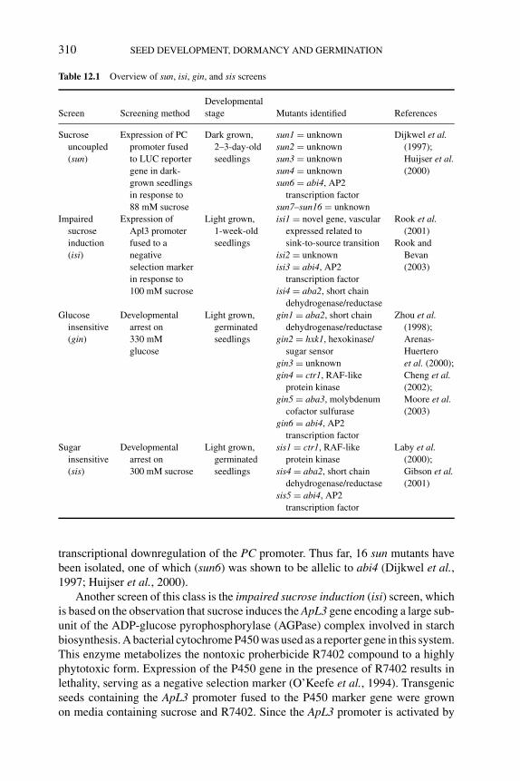
P1: OTE/SPH P2: OTE
BLUK053-Bradford September 27, 2006 15:22
310 SEED DEVELOPMENT, DORMANCY AND GERMINATION
Table 12.1 Overview of sun, isi, gin, and sis screens
Developmental
Screen Screening method stage Mutants identified References
Sucrose
uncoupled
(sun)
Expression of PC
promoter fused
to LUC reporter
gene in dark-
grown seedlings
in response to
88 mM sucrose
Dark grown,
2–3-day-old
seedlings
sun1 = unknown
sun2 = unknown
sun3 = unknown
sun4 = unknown
sun6 = abi4, AP2
transcription factor
sun7–sun16 = unknown
Dijkwel et al.(1997);
Huijser et al.(2000)
Impaired
sucrose
induction
(isi)
Expression of
Apl3 promoter
fused to a
negative
selection marker
in response to
100 mM sucrose
Light grown,
1-week-old
seedlings
isi1 = novel gene, vascular
expressed related to
sink-to-source transition
isi2 = unknown
isi3 = abi4, AP2
transcription factor
isi4 = aba2, short chain
dehydrogenase/reductase
Rook et al.(2001)
Rook and
Bevan
(2003)
Glucose
insensitive
(gin)
Developmental
arrest on
330 mM
glucose
Light grown,
germinated
seedlings
gin1 = aba2, short chain
dehydrogenase/reductase
gin2 = hxk1, hexokinase/
sugar sensor
gin3 = unknown
gin4 = ctr1, RAF-like
protein kinase
gin5 = aba3, molybdenum
cofactor sulfurase
gin6 = abi4, AP2
transcription factor
Zhou et al.(1998);
Arenas-
Huertero
et al. (2000);
Cheng et al.(2002);
Moore et al.(2003)
Sugar
insensitive
(sis)
Developmental
arrest on
300 mM sucrose
Light grown,
germinated
seedlings
sis1 = ctr1, RAF-like
protein kinase
sis4 = aba2, short chain
dehydrogenase/reductase
sis5 = abi4, AP2
transcription factor
Laby et al.(2000);
Gibson et al.(2001)
transcriptional downregulation of the PC promoter. Thus far, 16 sun mutants havebeen isolated, one of which (sun6) was shown to be allelic to abi4 (Dijkwel et al.,1997; Huijser et al., 2000).
Another screen of this class is the impaired sucrose induction (isi) screen, whichis based on the observation that sucrose induces the ApL3 gene encoding a large sub-unit of the ADP-glucose pyrophosphorylase (AGPase) complex involved in starchbiosynthesis. A bacterial cytochrome P450 was used as a reporter gene in this system.This enzyme metabolizes the nontoxic proherbicide R7402 compound to a highlyphytotoxic form. Expression of the P450 gene in the presence of R7402 results inlethality, serving as a negative selection marker (O’Keefe et al., 1994). Transgenicseeds containing the ApL3 promoter fused to the P450 marker gene were grownon media containing sucrose and R7402. Since the ApL3 promoter is activated by
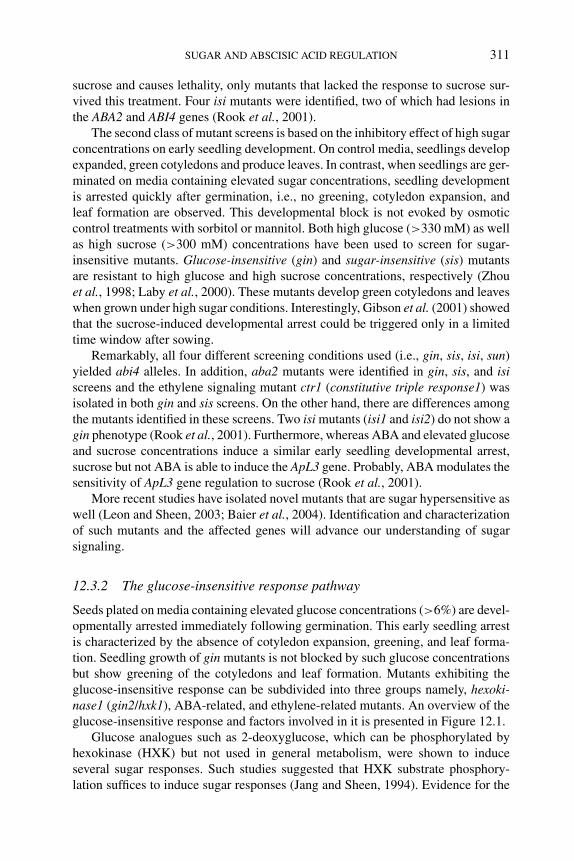
P1: OTE/SPH P2: OTE
BLUK053-Bradford September 27, 2006 15:22
SUGAR AND ABSCISIC ACID REGULATION 311
sucrose and causes lethality, only mutants that lacked the response to sucrose sur-vived this treatment. Four isi mutants were identified, two of which had lesions inthe ABA2 and ABI4 genes (Rook et al., 2001).
The second class of mutant screens is based on the inhibitory effect of high sugarconcentrations on early seedling development. On control media, seedlings developexpanded, green cotyledons and produce leaves. In contrast, when seedlings are ger-minated on media containing elevated sugar concentrations, seedling developmentis arrested quickly after germination, i.e., no greening, cotyledon expansion, andleaf formation are observed. This developmental block is not evoked by osmoticcontrol treatments with sorbitol or mannitol. Both high glucose (>330 mM) as wellas high sucrose (>300 mM) concentrations have been used to screen for sugar-insensitive mutants. Glucose-insensitive (gin) and sugar-insensitive (sis) mutantsare resistant to high glucose and high sucrose concentrations, respectively (Zhouet al., 1998; Laby et al., 2000). These mutants develop green cotyledons and leaveswhen grown under high sugar conditions. Interestingly, Gibson et al. (2001) showedthat the sucrose-induced developmental arrest could be triggered only in a limitedtime window after sowing.
Remarkably, all four different screening conditions used (i.e., gin, sis, isi, sun)yielded abi4 alleles. In addition, aba2 mutants were identified in gin, sis, and isiscreens and the ethylene signaling mutant ctr1 (constitutive triple response1) wasisolated in both gin and sis screens. On the other hand, there are differences amongthe mutants identified in these screens. Two isi mutants (isi1 and isi2) do not show agin phenotype (Rook et al., 2001). Furthermore, whereas ABA and elevated glucoseand sucrose concentrations induce a similar early seedling developmental arrest,sucrose but not ABA is able to induce the ApL3 gene. Probably, ABA modulates thesensitivity of ApL3 gene regulation to sucrose (Rook et al., 2001).
More recent studies have isolated novel mutants that are sugar hypersensitive aswell (Leon and Sheen, 2003; Baier et al., 2004). Identification and characterizationof such mutants and the affected genes will advance our understanding of sugarsignaling.
12.3.2 The glucose-insensitive response pathway
Seeds plated on media containing elevated glucose concentrations (>6%) are devel-opmentally arrested immediately following germination. This early seedling arrestis characterized by the absence of cotyledon expansion, greening, and leaf forma-tion. Seedling growth of gin mutants is not blocked by such glucose concentrationsbut show greening of the cotyledons and leaf formation. Mutants exhibiting theglucose-insensitive response can be subdivided into three groups namely, hexoki-nase1 (gin2/hxk1), ABA-related, and ethylene-related mutants. An overview of theglucose-insensitive response and factors involved in it is presented in Figure 12.1.
Glucose analogues such as 2-deoxyglucose, which can be phosphorylated byhexokinase (HXK) but not used in general metabolism, were shown to induceseveral sugar responses. Such studies suggested that HXK substrate phosphory-lation suffices to induce sugar responses (Jang and Sheen, 1994). Evidence for the
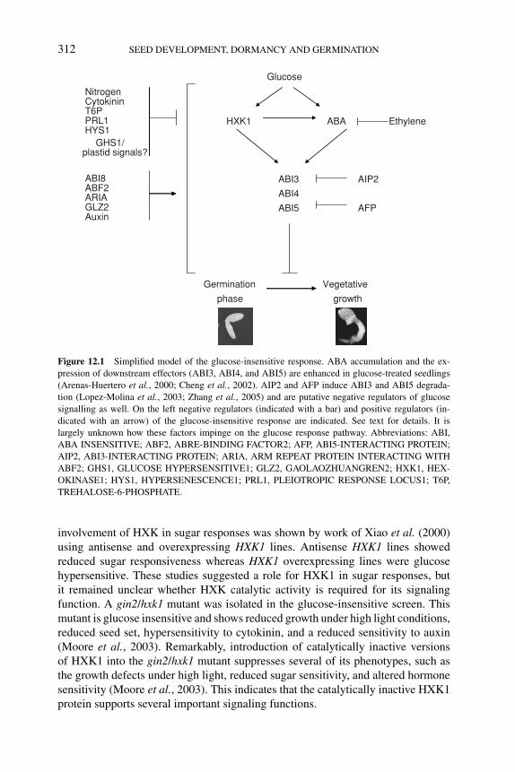
P1: OTE/SPH P2: OTE
BLUK053-Bradford September 27, 2006 15:22
312 SEED DEVELOPMENT, DORMANCY AND GERMINATION
Glucose
HXK1 ABA Ethylene
ABI3 AIP2
ABI4
ABI5 AFP
Germination Vegetative
phase growth
NitrogenCytokininT6PPRL1HYS1
GHS1/plastid signals?
ABI8ABF2ARIAGLZ2Auxin
Figure 12.1 Simplified model of the glucose-insensitive response. ABA accumulation and the ex-
pression of downstream effectors (ABI3, ABI4, and ABI5) are enhanced in glucose-treated seedlings
(Arenas-Huertero et al., 2000; Cheng et al., 2002). AIP2 and AFP induce ABI3 and ABI5 degrada-
tion (Lopez-Molina et al., 2003; Zhang et al., 2005) and are putative negative regulators of glucose
signalling as well. On the left negative regulators (indicated with a bar) and positive regulators (in-
dicated with an arrow) of the glucose-insensitive response are indicated. See text for details. It is
largely unknown how these factors impinge on the glucose response pathway. Abbreviations: ABI,
ABA INSENSITIVE; ABF2, ABRE-BINDING FACTOR2; AFP, ABI5-INTERACTING PROTEIN;
AIP2, ABI3-INTERACTING PROTEIN; ARIA, ARM REPEAT PROTEIN INTERACTING WITH
ABF2; GHS1, GLUCOSE HYPERSENSITIVE1; GLZ2, GAOLAOZHUANGREN2; HXK1, HEX-
OKINASE1; HYS1, HYPERSENESCENCE1; PRL1, PLEIOTROPIC RESPONSE LOCUS1; T6P,
TREHALOSE-6-PHOSPHATE.
involvement of HXK in sugar responses was shown by work of Xiao et al. (2000)using antisense and overexpressing HXK1 lines. Antisense HXK1 lines showedreduced sugar responsiveness whereas HXK1 overexpressing lines were glucosehypersensitive. These studies suggested a role for HXK1 in sugar responses, butit remained unclear whether HXK catalytic activity is required for its signalingfunction. A gin2/hxk1 mutant was isolated in the glucose-insensitive screen. Thismutant is glucose insensitive and shows reduced growth under high light conditions,reduced seed set, hypersensitivity to cytokinin, and a reduced sensitivity to auxin(Moore et al., 2003). Remarkably, introduction of catalytically inactive versionsof HXK1 into the gin2/hxk1 mutant suppresses several of its phenotypes, such asthe growth defects under high light, reduced sugar sensitivity, and altered hormonesensitivity (Moore et al., 2003). This indicates that the catalytically inactive HXK1protein supports several important signaling functions.
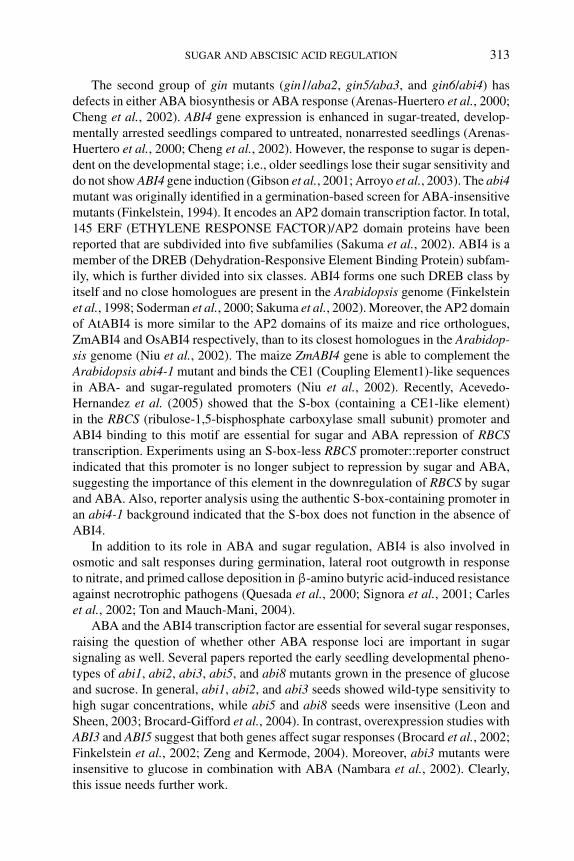
P1: OTE/SPH P2: OTE
BLUK053-Bradford September 27, 2006 15:22
SUGAR AND ABSCISIC ACID REGULATION 313
The second group of gin mutants (gin1/aba2, gin5/aba3, and gin6/abi4) hasdefects in either ABA biosynthesis or ABA response (Arenas-Huertero et al., 2000;Cheng et al., 2002). ABI4 gene expression is enhanced in sugar-treated, develop-mentally arrested seedlings compared to untreated, nonarrested seedlings (Arenas-Huertero et al., 2000; Cheng et al., 2002). However, the response to sugar is depen-dent on the developmental stage; i.e., older seedlings lose their sugar sensitivity anddo not show ABI4 gene induction (Gibson et al., 2001; Arroyo et al., 2003). The abi4mutant was originally identified in a germination-based screen for ABA-insensitivemutants (Finkelstein, 1994). It encodes an AP2 domain transcription factor. In total,145 ERF (ETHYLENE RESPONSE FACTOR)/AP2 domain proteins have beenreported that are subdivided into five subfamilies (Sakuma et al., 2002). ABI4 is amember of the DREB (Dehydration-Responsive Element Binding Protein) subfam-ily, which is further divided into six classes. ABI4 forms one such DREB class byitself and no close homologues are present in the Arabidopsis genome (Finkelsteinet al., 1998; Soderman et al., 2000; Sakuma et al., 2002). Moreover, the AP2 domainof AtABI4 is more similar to the AP2 domains of its maize and rice orthologues,ZmABI4 and OsABI4 respectively, than to its closest homologues in the Arabidop-sis genome (Niu et al., 2002). The maize ZmABI4 gene is able to complement theArabidopsis abi4-1 mutant and binds the CE1 (Coupling Element1)-like sequencesin ABA- and sugar-regulated promoters (Niu et al., 2002). Recently, Acevedo-Hernandez et al. (2005) showed that the S-box (containing a CE1-like element)in the RBCS (ribulose-1,5-bisphosphate carboxylase small subunit) promoter andABI4 binding to this motif are essential for sugar and ABA repression of RBCStranscription. Experiments using an S-box-less RBCS promoter::reporter constructindicated that this promoter is no longer subject to repression by sugar and ABA,suggesting the importance of this element in the downregulation of RBCS by sugarand ABA. Also, reporter analysis using the authentic S-box-containing promoter inan abi4-1 background indicated that the S-box does not function in the absence ofABI4.
In addition to its role in ABA and sugar regulation, ABI4 is also involved inosmotic and salt responses during germination, lateral root outgrowth in responseto nitrate, and primed callose deposition in β-amino butyric acid-induced resistanceagainst necrotrophic pathogens (Quesada et al., 2000; Signora et al., 2001; Carleset al., 2002; Ton and Mauch-Mani, 2004).
ABA and the ABI4 transcription factor are essential for several sugar responses,raising the question of whether other ABA response loci are important in sugarsignaling as well. Several papers reported the early seedling developmental pheno-types of abi1, abi2, abi3, abi5, and abi8 mutants grown in the presence of glucoseand sucrose. In general, abi1, abi2, and abi3 seeds showed wild-type sensitivity tohigh sugar concentrations, while abi5 and abi8 seeds were insensitive (Leon andSheen, 2003; Brocard-Gifford et al., 2004). In contrast, overexpression studies withABI3 and ABI5 suggest that both genes affect sugar responses (Brocard et al., 2002;Finkelstein et al., 2002; Zeng and Kermode, 2004). Moreover, abi3 mutants wereinsensitive to glucose in combination with ABA (Nambara et al., 2002). Clearly,this issue needs further work.
P1: OTE/SPH P2: OTE
BLUK053-Bradford September 27, 2006 15:22
314 SEED DEVELOPMENT, DORMANCY AND GERMINATION
The third group of gin mutants exhibits altered ethylene biosynthesis or re-sponses. Zhou et al. (1998) found that the glucose insensitivity of the gin1 mutantcould be phenocopied by treatment of wild-type seeds with the ethylene precursor1-aminocyclopropane carboxylic acid (ACC). Moreover, mutants that overproduceethylene or have a constitutive ethylene response show a gin phenotype. In twoscreens (gin and sis) alleles of constitutive ethylene response (ctr) mutants wereidentified as sugar insensitive (Gibson et al., 2001; Cheng et al., 2002; Leon andSheen, 2003). Vice versa, ethylene-insensitive (ein) mutants were found to be hy-persensitive to glucose-induced seedling arrest (Zhou et al., 1998; Leon and Sheen,2003). Interestingly, Yanagisawa et al. (2003) showed that glucose promotes EIN3turnover via a HXK1-dependent pathway. EIN3 regulation is an important step inethylene signaling. EIN3 is degraded via the ubiquitin–proteasome pathway whichinvolves the F-box proteins EBF1 (EIN3-BINDING F-BOX) and EBF2. Upon ethy-lene treatment, EIN3 protein accumulates, dependent on several genes of the ethy-lene response pathway including ETR1 (ETHYLENE RECEPTOR1), CTR1, EIN2,and EIN6 (Guo and Ecker, 2003; Potuschak et al., 2003).
Both ABA and ethylene are regulators of sugar responses and also interact intheir signaling pathways. Constitutive ethylene signaling decreases ABA sensitivityand ethylene-insensitive mutants are ABA hypersensitive (Beaudoin et al., 2000;Ghassemian et al., 2000). Genetic analysis of glucose-induced seedling arrest usingethylene signaling etr1 and ein2 mutants and gin1/aba2 demonstrated that gin1/aba2acts downstream of ethylene signaling (Zhou et al., 1998; Cheng et al., 2002). Pos-sibly, ethylene affects sugar responses by modifying ABA levels or ABA signaling,as was shown for germination (Beaudoin et al., 2000; Ghassemian et al., 2000) andfor stomatal closure (Tanaka et al., 2005).
12.3.3 Other factors affecting the glucose response during earlyseedling development
The analysis of gin mutants provides an important basis for our knowledge of glu-cose signaling during early seedling development. However, other nutrients, metabo-lites, hormones, and mutations are known to affect the sugar response during earlyseedling development. For example, the gin2/hxk1 mutant is hypersensitive to cy-tokinin and has a reduced sensitivity to auxin (Moore et al., 2003). Interestingly, addi-tion of cytokinin to developing seedlings results in a reduced sugar sensitivity as doesthe addition of ACC (Zhou et al., 1998; Moore et al., 2003). The effect of cytokininis independent from ethylene signaling. In addition, auxin signaling mutants, suchas tir1 (transport inhibitor response1), show a gin phenotype (Moore et al., 2003).
Both carbon (C) and nitrogen (N) are important nutrients for plant growth anddevelopment. C and N signaling intersect and several responses are more responsiveto C/N ratio rather than to C concentration alone (Martin et al., 2002; for reviewssee Coruzzi and Bush, 2001; Coruzzi and Zhou, 2001). Microarray analyses of C,N, or C + N responsive genes supported the hypothesis that plants possess suchC/N ratio-responsive mechanisms (Palenchar et al., 2004). Furthermore, this studyshowed that C/N ratio controls transcription via multiple mechanisms. Increasing Nconcentration in the growth medium decreases responsiveness of seedlings to sugar,
P1: OTE/SPH P2: OTE
BLUK053-Bradford September 27, 2006 15:22
SUGAR AND ABSCISIC ACID REGULATION 315
while lowering nitrogen level increases the sensitivity of seedlings to sugar (Martinet al., 2002; Moore et al., 2003). Photosynthetic gene expression and lipid mobi-lization in response to sugar are affected by C/N ratio as well (Martin et al., 2002).
Trehalose metabolism also affects sugar utilization during early seedling growth.Genes for trehalose metabolism were found in many plants, but with the exceptionof some resurrection plants, trehalose levels are very low to undetectable (Eastmondand Graham, 2003). Also in Arabidopsis the amount of trehalose is very low but,nevertheless, trehalose metabolism was found to be essential for Arabidopsis devel-opment and carbon metabolism (Eastmond et al., 2002; Schluepmann et al., 2003).Transgenic plants with enhanced levels of trehalose-6-phosphate (T6P) show im-proved growth on media supplemented with sugars, while transgenic lines withdecreased levels of T6P develop slower and exhibit reduced cotyledon expansionand greening (Schluepmann et al., 2003; Avonce et al., 2004). Thus, T6P levelsdetermine the responses of seedlings to exogenously supplied sugar.
Recently several mutants that have an altered gin response were described. Mu-tants in the ABRE-binding factor ABF2 and its interacting partner ARIA (ARMREPEAT PROTEIN INTERACTING WITH ABF2) result in a gin phenotype,and the opposite result was obtained when both genes were overexpressed (Kimet al., 2004a,b). Also a genetic lesion in GAOLAOZHUANGREN2 (GLZ2) has beenidentified as a gin mutant (Chen et al., 2004). On the other hand, mutations ingenes like PLEIOTROPIC RESPONSE LOCUS1 (PRL1), HYPERSENESCENCE1(HYS1)/CONSTITUTIVE EXPRESSOR OF PATHOGENESIS-RELATED GENES(CPR5), and GLUCOSE HYPERSENSITIVE1 (GHS1) result in an enhanced re-sponse to sugar during early seedling growth (Nemeth et al., 1998; Yoshida et al.,2002; Morito-Yamamuro et al., 2004). Interestingly, ghs1 has a lesion in plastid 30Sribosomal protein S21. This mutant has reduced levels of RBCL (RBC large subunit)and impaired chloroplast development (Morita-Yamamuro et al., 2004). Possibly,a translation-dependent plastidic signal is necessary for proper sugar signaling,although alternative chloroplast defects cannot be ruled out (Morita-Yamamuroet al., 2001). Interestingly, treatment of dark-grown seedlings with lincomycin, aninhibitor of plastid translation, inhibits the developmentally regulated PC expressionand mimics sugar treatment of dark-grown seedlings (Dijkwel, 1997).
12.3.4 Sugar delays seed germination in Arabidopsis
The storage reserves in seeds are consumed during germination and seedling growthto sustain cellular processes and growth. It is, therefore, remarkable that the additionof metabolizable sugars such as glucose delays seed germination in Arabidopsis (Toet al., 2002; Ullah et al., 2002; Price et al., 2003; Dekkers et al., 2004). An inhibitionof germination by sugars is also reported for tomato (Lycopersicon esculentum),squash (Cucurbita pepo), and watermelon (Citrullus lanatus) seeds (Martin andBlackburn, 2003). In contrast, addition of metabolizable sugar relieves the inhibitionof Arabidopsis seed germination by mannose (see below) and ABA (Garciarrubioet al., 1997; Pego et al., 1999; Finkelstein and Lynch, 2000a). In Arabidopsis thedelay of germination is provoked by low concentrations of glucose. This glucose-induced germination delay is dose dependent but independent of the osmotic stress,
P1: OTE/SPH P2: OTE
BLUK053-Bradford September 27, 2006 15:22
316 SEED DEVELOPMENT, DORMANCY AND GERMINATION
because equal concentrations of sorbitol or mannitol do not cause a similar delay ingermination (To et al., 2002; Price et al., 2003; Dekkers et al., 2004).
The glucose analog mannose is a potent inhibitor of Arabidopsis seed germi-nation and is effective at low concentrations (5–10 mM) (Pego et al., 1999). Themannose-induced inhibition of germination depends on a HXK-mediated step, andabi4 mutants have a reduced sensitivity for such treatment. Surprisingly, aba mu-tants show normal sensitivity to mannose during germination (Pego et al., 1999;Huijser et al., 2000; Laby et al., 2000). Glucose and mannose both inhibit seedgermination but this occurs via separate pathways, as the effect of mannose can becounteracted by the addition of 60 mM glucose (Pego et al., 1999; Dekkers et al.,2004). Originally, it was hypothesized that mannose signaling restricts carbon mo-bilization necessary for germination. Analysis of a mutant in AtSTP1 (Arabidopsisthaliana SUGAR TRANSPORT PROTEIN1), a monosaccharide/proton symporterin Arabidopsis, showed that this is a major sugar transporter in seeds and seedlings(Sherson et al., 2000). Glucose and mannose uptake is reduced up to 60% in thestp1 mutant seedlings. The stp1 seeds exhibit mannose-insensitive germination (mig;Pego et al., 1999), indicating that mannose transport is important for the germinationinhibition. Sherson et al. (2000) therefore suggested that the rescue of mannose-inhibited germination by glucose is possibly caused by reduced mannose uptakedue to competition with excess glucose.
As discussed earlier, the ability of elevated sugar concentrations to inhibit earlyseedling growth was used to screen for sugar-insensitive mutants. The hxk1, aba2,abi4, and ctr1 mutants lacked this postgerminative developmental arrest when chal-lenged with elevated sugar concentrations (Leon and Sheen, 2003). Such sugar-insensitive mutants were predicted to be insensitive also to the sugar-induced ger-mination delay. Strikingly, hxk1, abi4, and ctr1 mutants turned out to be sensitive tothe sugar-induced germination delay. Only ABA-deficient mutants were insensitiveto sugar to some extent. These results show that the sugar-induced delay of ger-mination and sugar-induced postgerminative developmental arrest are two distinctresponses (Price et al., 2003; Dekkers et al., 2004).
Other plant hormones were investigated for their roles in the sugar-inducedgermination delay. Sugar treatment delayed ABA degradation (Price et al., 2003);however, it is unclear whether this is the cause of the delay of germination or is aconsequence of the glucose-triggered delay of germination. Mutants with a reducedABA sensitivity such as abi1, abi2, abi4, abi5, and ctr1 exhibit normal sensitivityto glucose during germination (Price et al., 2003; Dekkers et al., 2004). Theseresults support the hypothesis that the delayed ABA degradation is a consequencerather than the cause of the germination delay. It is unclear why ABA-deficientmutants show a reduced sensitivity to glucose. Possibly, ABA levels are important forregulating the glucose sensitivity in seeds. ABA concentrations that are not inhibitoryto germination synergistically enhance the repression of germination by glucose(Dekkers et al., 2004). This enhancement of glucose repression of germination byABA is not due to osmotic signaling and is independent of ABI4 (Figure 12.2).Thus, ABI4 is involved neither in the glucose response during germination nor in
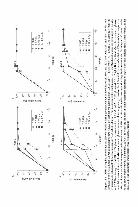
P1: OTE/SPH P2: OTE
BLUK053-Bradford September 27, 2006 15:22
Co
l-0
020406080100
0
Tim
e (h
)
Germination (%)
0.5M
S0.
5 A
BA
2.5%
sor
bA
+ 2
.5 s
orb
abi4
-1
020406080100
Germination (%)
0.5M
S0.
5 A
BA
2.5%
sor
bA
+ 2
.5 s
orb
Co
l-0
020406080100
Germination (%)
0.5M
S0.
5 A
BA
2.5
glc
0.5A
+ 2
.5gl
c
abi4
-1
020406080100
Germination (%)
0.5M
S0.
5 A
BA
2.5
glc
0.5A
+ 2
.5gl
c
A C
B D
1428
4256
700
Tim
e (h
)
1428
4256
70
0
Tim
e (h
)
1428
4256
700
Tim
e (h
)
1428
4256
70
Fig
ure
12.2
AB
I4is
requir
ednei
ther
for
the
glu
cose
resp
onse
duri
ng
ger
min
atio
nnor
its
modula
tion
by
AB
A.
(A,
B)
Col-
0(w
ild-t
ype)
and
abi4
-1se
eds
wer
e
ger
min
ated
on
0.5
MS
med
ia,0
.5M
Sco
nta
inin
g0.5
μM
AB
A,2
.5%
sorb
itolor
aco
mbin
atio
nof
0.5
μM
AB
A+
2.5
%so
rbit
ol(A
+2.5
sorb
).T
he
osm
oti
cre
sponse
is
syner
gis
tica
lly
enhan
ced
by
low
conce
ntr
atio
ns
of
AB
Ain
Col-
0,b
uta
bi4-
1is
inse
nsi
tive
toth
isco
mbin
atio
n.(
C,D
)C
ol-
0an
dab
i4-1
seed
sar
eger
min
ated
on
0.5
MS
,
or
0.5
MS
conta
inin
g0.5
μM
AB
A,
2.5
%glu
cose
or
aco
mbin
atio
nof
0.5
μM
AB
A+
2.5
%glu
cose
(0.5
A+
2.5
glc
).B
oth
Col-
0an
dab
i4-1
hav
een
han
ced
glu
cose
sensi
tivit
yin
com
bin
atio
nw
ith
AB
Aco
mpar
edto
glu
cose
trea
tmen
tal
one.
Appar
entl
y,bas
edon
the
dif
fere
nti
alre
sponse
of
abi4
-1to
AB
A+
sorb
itol
com
par
edto
AB
A+
glu
cose
,th
een
han
ced
resp
onse
toth
eco
mbin
atio
nof
AB
Aan
dglu
cose
isnot
due
toosm
oti
csi
gnal
ing.S
eeds
wer
est
rati
fied
for
3day
sat
4◦ C
bef
ore
tran
sfer
toth
egro
wth
cham
ber
(16
h/8
hli
ght/
dar
k,
22
◦ C).
The
resu
lts
are
the
mea
nof
single
exper
imen
tper
form
edin
dupl
o.E
rror
bar
sin
dic
ate
SE
(SE
smal
ler
than
2is
not
indic
ated
).T
he
exper
imen
tw
asre
pea
ted
twic
ew
ith
sim
ilar
resu
lts.
317
P1: OTE/SPH P2: OTE
BLUK053-Bradford September 27, 2006 15:22
318 SEED DEVELOPMENT, DORMANCY AND GERMINATION
the modulation of this response by ABA. Furthermore, the addition of hormonessuch as GA, brassinosteroids, and ethylene (via ACC) that are known to stimulategermination did not suppress the glucose-induced germination delay (Dekkers et al.,2004).
Another mutant with an enhanced sensitivity to sugar during germination isgpa1 (Ullah et al., 2002). GPA1 (G PROTEIN α-SUBUNIT) is a gene encoding thealpha subunit of the G protein in Arabidopsis. Inhibition of carotenoid synthesis byfluridone (which causes an inhibition of ABA synthesis) suppresses the hypersensi-tivity of gpa1 seeds to glucose (Ullah et al., 2002). The gpa1 mutants are also lesssensitive to GA and lack the response to brassinosteroids.
An impaired germination phenotype is also observed in mutants that have defectsin fatty acid metabolism, such as ketoacyl-CoA thiolase2 (kat2), comatose (cts), andacyl-CoA oxidase1 acyl-CoA oxidase2 (acx1 acx2) double mutant. A recent studyof Pinfield-Wells et al. (2005) showed that seed germination of fatty acid break-down mutants cannot be rescued by sucrose addition (see Chapter 6). This suggeststhat peroxisomal β-oxidation activity has another essential function in germina-tion in addition to the provision of carbon to support growth. Possibly peroxiso-mal β-oxidation activity is necessary for the generation of metabolites or signalingmolecules that induce germination or, alternatively, is involved in the degradation ofa germination inhibitor (Pinfield-Wells et al., 2005). Studies on the fatty acid trans-port mutant cts show that its ‘forever’ dormant phenotype is suppressed in certaingenetic backgrounds. Combining cts with mutations such as aba1, abi3, lec1 (leafycotyledon1), and fus3 (fusca3) rescues the germination phenotype (Russell et al.,2000). However, cts germination phenotype is not rescued by abi1-1 or reduced dor-mancy2 (rdo2), both of which show a reduced dormancy phenotype (Footitt et al.,2002). Notably, aba, abi3-5, lec1, and fus3 have reduced glucose sensitivity duringgermination but abi1-1 and rdo2 are sensitive to sugar during germination (Brocard-Gifford et al., 2003; Price et al., 2003; Dekkers et al., 2004; our unpublished results).Moreover, germination of the fatty acid breakdown mutants acx1, acx2 and cts isnot significantly improved by exogenous GA (Russell et al., 2000; Pinfield-Wellset al., 2005). Similarly, the glucose-induced delay of germination is not rescued byGA (Dekkers et al., 2004). A correlation might exist between different genotypesthat specify glucose sensitivity and their sensitivity to a dysfunctional peroxisomalβ-oxidation pathway during germination. Moreover, sugar addition delayed stor-age lipid breakdown in Arabidopsis seeds (Martin et al., 2002; To et al., 2002).One could suggest that glucose addition to seeds delays germination by transientlyrepressing peroxisomal β-oxidation activity, which is essential for proper germina-tion. However, this hypothesis seems unlikely, since the sugar-insensitive mutantabi4 is insensitive to glucose repression of storage lipid mobilization but is sensitiveto glucose-induced germination delay (To et al., 2002). Moreover, nitrogen concen-trations do not affect the glucose-induced delay of seed germination but C/N ratiodoes influence storage lipid breakdown (Martin et al., 2002). Finally, the glucoseanalogue 3-O-methylglucose delays germination but does not affect storage lipidbreakdown (To et al., 2002). These observations uncouple the inhibitory effects ofglucose on storage lipid mobilization and on germination.
P1: OTE/SPH P2: OTE
BLUK053-Bradford September 27, 2006 15:22
SUGAR AND ABSCISIC ACID REGULATION 319
12.3.5 Imbibed seeds rapidly lose sensitivity for the glucose-inducedgermination delay
The temporal window of seed glucose sensitivity was investigated by transferringnon-cold-stratified seeds from sugar-free media to glucose-containing media at dif-ferent times after imbibition. The sensitivity to glucose rapidly declined duringimbibition prior to radicle emergence, in contrast to the sensitivity to ABA, whichremained essentially constant during this time period (Figures 12.3A and 12.3B).Since cold stratification (imbibed chilling at 4◦C) promotes loss of dormancy inArabidopsis seeds, it was of interest to test whether the sensitivity to glucose wouldalso be lost during cold stratification. When Arabidopsis seeds were treated with2.5% glucose during cold stratification and subsequent incubation at 22◦C, germi-nation was significantly delayed (Dekkers et al., 2004). In contrast, when seeds wereexposed to 2.5% glucose only immediately after cold stratification, they had losttheir sensitivity to glucose and exhibited no delay in germination (B.J.W. Dekkersand J.C.M. Smeekens, unpublished results). Moreover, seeds were resistant to evenhigher glucose concentrations (6%) after cold stratification, while the response to6% sorbitol was not affected by cold stratification (Figures 12.3C and 12.3D). Ger-mination of seeds that were treated with glucose after cold stratification mimicsthe germination curves of sorbitol-treated seeds (Figures 12.3C and 12.3D). On theother hand, continuous glucose treatment during cold stratification and germinationresulted in a clear germination delay (Figures 12.3C and 12.3D).
As discussed previously, metabolizable sugars antagonize ABA signaling duringgermination and promote germination of ABA-treated seeds (Garciarrubio et al.,1997; Finkelstein and Lynch, 2000b). This rescue of germination of ABA-treatedseeds by glucose is more pronounced when glucose is added to the medium aftercold stratification (Figures 12.3E and 12.3F), which is consistent with the observa-tion that glucose is inhibitory when present during cold stratification. This findingshows that the stimulatory effect of glucose on germination is exerted after coldstratification. It appears that the inhibitory and stimulatory (in the case of ABAtreatment) actions of glucose during germination are separable in time. Mutantswith disrupted seed development such as lec1, fus3, and abi3-5 are insensitive tosugar during germination as well (Brocard-Gifford et al., 2003; B.J.W. Dekkers andJ.C.M. Smeekens, unpublished results). The temporal separation of glucose sensi-tivity of seeds between cold stratification and germination periods might explain theinsensitivity of these mutants; i.e., the developmental modes of lec1, fus3 and abi3-5may be already shifted to a sugar-insensitive phase due to their heterochronic nature.
Thus far, little is known about the physiological or molecular basis of the glucoseresponse during seed germination. In Arabidopsis many genes involved in dormancyand germination have been described using mutant or natural variation analyses(Bentsink and Koornneef, 2002; see Chapter 5). Genes such as IMBIBITION-INDUCIBLE1 (IMB1), PIF3-LIKE5 (PIL5), RGA-LIKE2 (RGL2), DELAY OFGERMINATION (DOG), and REDUCED DORMANCY (RDO1/4) are importantregulators of germination (Leon-Kloosterziel et al., 1996; Bentsink and Koornneef,2002; Lee et al., 2002; Alonso-Blanco et al., 2003, Duque and Chua, 2003; Oh et al.,
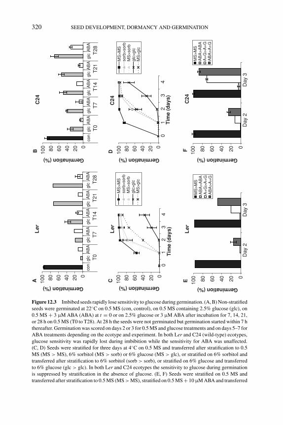
P1: OTE/SPH P2: OTE
BLUK053-Bradford September 27, 2006 15:22
320 SEED DEVELOPMENT, DORMANCY AND GERMINATION
Ler
020406080100
con
glc
AB
Agl
cA
BA
glc
AB
Agl
cA
BA
glc
AB
A
T0
T7
T14
T21
T28
Germination (%)
C24
020406080100
con
glc
AB
Agl
cA
BA
glc
AB
Agl
cA
BA
glc
AB
A
T0
T7
T14
T21
T28
Germination (%)
Ler
020406080100
Day
2D
ay 3
Germination (%)
MS
>M
SA
BA
>A
BA
A+
G>
A+
GA
BA
>A
+G
C24
020406080100
Day
2D
ay 3
Germination (%)
MS
>M
SA
BA
>A
BA
A+
G>
A+
GA
BA
>A
+G
Ler
020406080100
01
23
4T
ime
(day
s)
Germination (%)
MS
>M
Sso
rb>
sorb
MS
>so
rbgl
c>gl
c M
S>
glc
C24
020406080100
01
23
4T
ime
(day
s)
Germination (%)
MS
>M
Sso
rb>
sorb
MS
>so
rbgl
c>gl
c M
S>
glc
AB
C EFD
Figure 12.3 Imbibed seeds rapidly lose sensitivity to glucose during germination. (A, B) Non-stratified
seeds were germinated at 22◦C on 0.5 MS (con, control), on 0.5 MS containing 2.5% glucose (glc), on
0.5 MS + 3 μM ABA (ABA) at t = 0 or on 2.5% glucose or 3 μM ABA after incubation for 7, 14, 21,
or 28 h on 0.5 MS (T0 to T28). At 28 h the seeds were not germinated but germination started within 7 h
thereafter. Germination was scored on days 2 or 3 for 0.5 MS and glucose treatments and on days 5–7 for
ABA treatments depending on the ecotype and experiment. In both Ler and C24 (wild-type) ecotypes,
glucose sensitivity was rapidly lost during imbibition while the sensitivity for ABA was unaffected.
(C, D) Seeds were stratified for three days at 4◦C on 0.5 MS and transferred after stratification to 0.5
MS (MS > MS), 6% sorbitol (MS > sorb) or 6% glucose (MS > glc), or stratified on 6% sorbitol and
transferred after stratification to 6% sorbitol (sorb > sorb), or stratified on 6% glucose and transferred
to 6% glucose (glc > glc). In both Ler and C24 ecotypes the sensitivity to glucose during germination
is suppressed by stratification in the absence of glucose. (E, F) Seeds were stratified on 0.5 MS and
transferred after stratification to 0.5 MS (MS > MS), stratified on 0.5 MS + 10 μM ABA and transferred
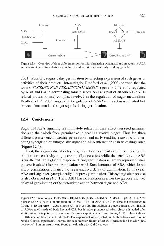
P1: OTE/SPH P2: OTE
BLUK053-Bradford September 27, 2006 15:22
SUGAR AND ABSCISIC ACID REGULATION 321
Germination Seedling growth
Glucose ABA Glucose
ABA
Stratification
GPA1Glucose
HXK1 ABA Ethylene
ABI3/4/5
ABI genes
Figure 12.4 Overview of three different responses with alternating synergistic and antagonistic ABA
and glucose interactions during Arabidopsis seed germination and early seedling growth.
2004). Possibly, sugars delay germination by affecting expression of such genes oractivities of their products. Interestingly, Bradford et al. (2003) showed that thetomato SUCROSE NON-FERMENTING4 (LeSNF4) gene is differently regulatedby ABA and GA in germinating tomato seeds. SNF4 is part of an SnRK1 (SNF1-related protein kinase) complex involved in the regulation of sugar metabolism.Bradford et al. (2003) suggest that regulation of LeSNF4 may act as a potential linkbetween hormonal and sugar signals during germination.
12.4 Conclusions
Sugar and ABA signaling are intimately related in their effects on seed germina-tion and the switch from germinative to seedling growth stages. Thus far, threedifferent phases encompassing germination and early seedling growth with alter-nating synergistic or antagonistic sugar and ABA interactions can be distinguished(Figure 12.4).
First, the sugar-induced delay of germination is an early response. During im-bibition the sensitivity to glucose rapidly decreases while the sensitivity to ABAis unaffected. This glucose response during germination is largely repressed whenglucose is added after the stratification period. Small amounts of ABA, which do notaffect germination, enhance the sugar-induced delay of germination. In this case,ABA and sugar act synergistically to repress germination. This synergistic responseis also observed in abi4. Thus, ABI4 has no function in either the glucose-induceddelay of germination or the synergistic action between sugar and ABA.
←−−−−−−−−−−−−−−−−−−−−−−−−−−−−−−−−−−−−−−−−−−−−−−−−−−−−−−−−−−Figure 12.3 (Continued ) to 0.5 MS + 10 μM ABA (ABA > ABA) or 0.5 MS + 10 μM ABA + 2.5%
glucose (ABA > A+G), or stratified on 0.5 MS + 10 μM ABA + 2.5% glucose and transferred to
0.5 MS + 10 μM ABA + 2.5% glucose (A+G > A+G). The addition of glucose rescues germination
of ABA-treated seeds of both Ler and C24, but is more pronounced when glucose is added after
stratification. Data points are the means of a single experiment performed in duplo. Error bars indicate
SE (SE smaller than 2 is not indicated). The experiment was repeated one to three times with similar
results. Control experiments showed that seed transfer did not affect their germination behavior (data
not shown). Similar results were found as well using the Col-0 ecotype.
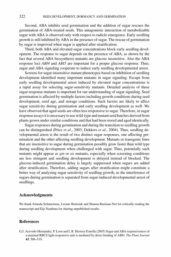
P1: OTE/SPH P2: OTE
BLUK053-Bradford September 27, 2006 15:22
322 SEED DEVELOPMENT, DORMANCY AND GERMINATION
Second, ABA inhibits seed germination and the addition of sugar rescues thegermination of ABA-treated seeds. This antagonistic interaction of metabolizablesugar with ABA is observed only with respect to radicle emergence. Early seedlinggrowth is still inhibited by ABA in the presence of sugar. The rescue of germinationby sugar is improved when sugar is applied after stratification.
Third, both ABA and elevated sugar concentrations block early seedling devel-opment. The response to sugar depends on the presence of ABA, as shown by thefact that several ABA biosynthesis mutants are glucose insensitive. Also the ABAresponse loci ABI4 and ABI5 are important for a proper glucose response. Thus,sugar and ABA signaling cooperate to induce early seedling developmental arrest.
Screens for sugar-insensitive mutant phenotypes based on inhibition of seedlingdevelopment identified many important mutants in sugar signaling. Escape fromearly seedling developmental arrest induced by elevated sugar concentrations isa rapid assay for selecting sugar-sensitivity mutants. Detailed analysis of thesesugar-response mutants is important for our understanding of sugar signaling. Seedgermination is affected by multiple factors including growth conditions during seeddevelopment, seed age, and storage conditions. Such factors are likely to affectsugar sensitivity during germination and early seedling development as well. Wehave observed that aged seeds are often less responsive to sugar. Therefore, in sugarresponse assays it is necessary to use wild-type and mutant seed batches derived fromplants grown under similar conditions and that had been stored and aged identically.
Sugar responses during germination and during the transition to seedling growthcan be distinguished (Price et al., 2003; Dekkers et al., 2004). Thus, seedling de-velopmental arrest is the result of two distinct sugar responses, one affecting ger-mination and the other affecting seedling development. Mutants or transgenic linesthat are insensitive to sugar during germination possibly grow faster than wild typeduring seedling development when challenged with sugar. Thus, potentially suchmutants might appear as gin or sis mutants, especially when screening conditionsare less stringent and seedling development is delayed instead of blocked. Theglucose-induced germination delay is largely suppressed when sugars are addedafter stratification. Therefore, adding sugars after stratification might constitute abetter way of analyzing sugar sensitivity of seedling growth, as the interference ofsugars during germination is separated from sugar-induced developmental arrest ofseedlings.
Acknowledgments
We thank Jolanda Schuurmans, Leonie Bentsink and Shanna Bastiaan-Net for critically reading the
manuscript and Eiji Nambara for sharing unpublished results.
References
G.J. Acevedo-Hernandez, P. Leon and L.R. Herrera-Estrella (2005) Sugar and ABA responsiveness of
a minimal RBCS light-responsive unit is mediated by direct binding of ABI4. The Plant Journal43, 506–519.
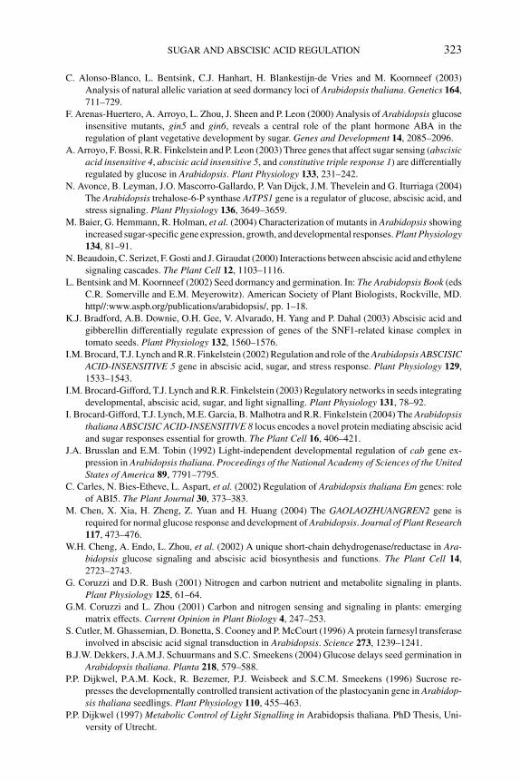
P1: OTE/SPH P2: OTE
BLUK053-Bradford September 27, 2006 15:22
SUGAR AND ABSCISIC ACID REGULATION 323
C. Alonso-Blanco, L. Bentsink, C.J. Hanhart, H. Blankestijn-de Vries and M. Koornneef (2003)
Analysis of natural allelic variation at seed dormancy loci of Arabidopsis thaliana. Genetics 164,
711–729.
F. Arenas-Huertero, A. Arroyo, L. Zhou, J. Sheen and P. Leon (2000) Analysis of Arabidopsis glucose
insensitive mutants, gin5 and gin6, reveals a central role of the plant hormone ABA in the
regulation of plant vegetative development by sugar. Genes and Development 14, 2085–2096.
A. Arroyo, F. Bossi, R.R. Finkelstein and P. Leon (2003) Three genes that affect sugar sensing (abscisicacid insensitive 4, abscisic acid insensitive 5, and constitutive triple response 1) are differentially
regulated by glucose in Arabidopsis. Plant Physiology 133, 231–242.
N. Avonce, B. Leyman, J.O. Mascorro-Gallardo, P. Van Dijck, J.M. Thevelein and G. Iturriaga (2004)
The Arabidopsis trehalose-6-P synthase AtTPS1 gene is a regulator of glucose, abscisic acid, and
stress signaling. Plant Physiology 136, 3649–3659.
M. Baier, G. Hemmann, R. Holman, et al. (2004) Characterization of mutants in Arabidopsis showing
increased sugar-specific gene expression, growth, and developmental responses. Plant Physiology134, 81–91.
N. Beaudoin, C. Serizet, F. Gosti and J. Giraudat (2000) Interactions between abscisic acid and ethylene
signaling cascades. The Plant Cell 12, 1103–1116.
L. Bentsink and M. Koornneef (2002) Seed dormancy and germination. In: The Arabidopsis Book (eds
C.R. Somerville and E.M. Meyerowitz). American Society of Plant Biologists, Rockville, MD.
http//:www.aspb.org/publications/arabidopsis/, pp. 1–18.
K.J. Bradford, A.B. Downie, O.H. Gee, V. Alvarado, H. Yang and P. Dahal (2003) Abscisic acid and
gibberellin differentially regulate expression of genes of the SNF1-related kinase complex in
tomato seeds. Plant Physiology 132, 1560–1576.
I.M. Brocard, T.J. Lynch and R.R. Finkelstein (2002) Regulation and role of the Arabidopsis ABSCISICACID-INSENSITIVE 5 gene in abscisic acid, sugar, and stress response. Plant Physiology 129,
1533–1543.
I.M. Brocard-Gifford, T.J. Lynch and R.R. Finkelstein (2003) Regulatory networks in seeds integrating
developmental, abscisic acid, sugar, and light signalling. Plant Physiology 131, 78–92.
I. Brocard-Gifford, T.J. Lynch, M.E. Garcia, B. Malhotra and R.R. Finkelstein (2004) The Arabidopsisthaliana ABSCISIC ACID-INSENSITIVE 8 locus encodes a novel protein mediating abscisic acid
and sugar responses essential for growth. The Plant Cell 16, 406–421.
J.A. Brusslan and E.M. Tobin (1992) Light-independent developmental regulation of cab gene ex-
pression in Arabidopsis thaliana. Proceedings of the National Academy of Sciences of the UnitedStates of America 89, 7791–7795.
C. Carles, N. Bies-Etheve, L. Aspart, et al. (2002) Regulation of Arabidopsis thaliana Em genes: role
of ABI5. The Plant Journal 30, 373–383.
M. Chen, X. Xia, H. Zheng, Z. Yuan and H. Huang (2004) The GAOLAOZHUANGREN2 gene is
required for normal glucose response and development of Arabidopsis. Journal of Plant Research117, 473–476.
W.H. Cheng, A. Endo, L. Zhou, et al. (2002) A unique short-chain dehydrogenase/reductase in Ara-bidopsis glucose signaling and abscisic acid biosynthesis and functions. The Plant Cell 14,
2723–2743.
G. Coruzzi and D.R. Bush (2001) Nitrogen and carbon nutrient and metabolite signaling in plants.
Plant Physiology 125, 61–64.
G.M. Coruzzi and L. Zhou (2001) Carbon and nitrogen sensing and signaling in plants: emerging
matrix effects. Current Opinion in Plant Biology 4, 247–253.
S. Cutler, M. Ghassemian, D. Bonetta, S. Cooney and P. McCourt (1996) A protein farnesyl transferase
involved in abscisic acid signal transduction in Arabidopsis. Science 273, 1239–1241.
B.J.W. Dekkers, J.A.M.J. Schuurmans and S.C. Smeekens (2004) Glucose delays seed germination in
Arabidopsis thaliana. Planta 218, 579–588.
P.P. Dijkwel, P.A.M. Kock, R. Bezemer, P.J. Weisbeek and S.C.M. Smeekens (1996) Sucrose re-
presses the developmentally controlled transient activation of the plastocyanin gene in Arabidop-sis thaliana seedlings. Plant Physiology 110, 455–463.
P.P. Dijkwel (1997) Metabolic Control of Light Signalling in Arabidopsis thaliana. PhD Thesis, Uni-
versity of Utrecht.
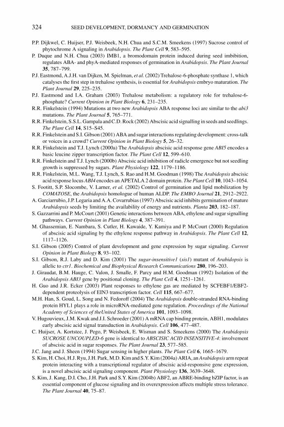
P1: OTE/SPH P2: OTE
BLUK053-Bradford September 27, 2006 15:22
324 SEED DEVELOPMENT, DORMANCY AND GERMINATION
P.P. Dijkwel, C. Huijser, P.J. Weisbeek, N.H. Chua and S.C.M. Smeekens (1997) Sucrose control of
phytochrome A signaling in Arabidopsis. The Plant Cell 9, 583–595.
P. Duque and N.H. Chua (2003) IMB1, a bromodomain protein induced during seed imbibition,
regulates ABA- and phyA-mediated responses of germination in Arabidopsis. The Plant Journal35, 787–799.
P.J. Eastmond, A.J.H. van Dijken, M. Spielman, et al. (2002) Trehalose-6-phosphate synthase 1, which
catalyses the first step in trehalose synthesis, is essential for Arabidopsis embryo maturation. ThePlant Journal 29, 225–235.
P.J. Eastmond and I.A. Graham (2003) Trehalose metabolism: a regulatory role for trehalose-6-
phosphate? Current Opinion in Plant Biology 6, 231–235.
R.R. Finkelstein (1994) Mutations at two new Arabidopsis ABA response loci are similar to the abi3mutations. The Plant Journal 5, 765–771.
R.R. Finkelstein, S.S.L. Gampala and C.D. Rock (2002) Abscisic acid signalling in seeds and seedlings.
The Plant Cell 14, S15–S45.
R.R. Finkelstein and S.I. Gibson (2001) ABA and sugar interactions regulating development: cross-talk
or voices in a crowd? Current Opinion in Plant Biology 5, 26–32.
R.R. Finkelstein and T.J. Lynch (2000a) The Arabidopsis abscisic acid response gene ABI5 encodes a
basic leucine zipper transcription factor. The Plant Cell 12, 599–610.
R.R. Finkelstein and T.J. Lynch (2000b) Abscisic acid inhibition of radicle emergence but not seedling
growth is suppressed by sugars. Plant Physiology 122, 1179–1186.
R.R. Finkelstein, M.L. Wang, T.J. Lynch, S. Rao and H.M. Goodman (1998) The Arabidopsis abscisic
acid response locus ABI4 encodes an APETALA 2 domain protein. The Plant Cell 10, 1043–1054.
S. Footitt, S.P. Slocombe, V. Larner, et al. (2002) Control of germination and lipid mobilization by
COMATOSE, the Arabidopsis homologue of human ALDP. The EMBO Journal 21, 2912–2922.
A. Garciarrubio, J.P. Legaria and A.A. Covarrubias (1997) Abscisic acid inhibits germination of mature
Arabidopsis seeds by limiting the availability of energy and nutrients. Planta 203, 182–187.
S. Gazzarrini and P. McCourt (2001) Genetic interactions between ABA, ethylene and sugar signalling
pathways. Current Opinion in Plant Biology 4, 387–391.
M. Ghassemian, E. Nambara, S. Cutler, H. Kawaide, Y. Kamiya and P. McCourt (2000) Regulation
of abscisic acid signaling by the ethylene response pathway in Arabidopsis. The Plant Cell 12,
1117–1126.
S.I. Gibson (2005) Control of plant development and gene expression by sugar signaling. CurrentOpinion in Plant Biology 8, 93–102.
S.I. Gibson, R.J. Laby and D. Kim (2001) The sugar-insensitive1 (sis1) mutant of Arabidopsis is
allelic to ctr1. Biochemical and Biophysical Research Communications 280, 196–203.
J. Giraudat, B.M. Hauge, C. Valon, J. Smalle, F. Parcy and H.M. Goodman (1992) Isolation of the
Arabidopsis ABI3 gene by positional cloning. The Plant Cell 4, 1251–1261.
H. Guo and J.R. Ecker (2003) Plant responses to ethylene gas are mediated by SCFEBF1/EBF2-
dependent proteolysis of EIN3 transcription factor. Cell 115, 667–677.
M.H. Han, S. Goud, L. Song and N. Fedoroff (2004) The Arabidopsis double-stranded RNA-binding
protein HYL1 plays a role in microRNA-mediated gene regulation. Proceedings of the NationalAcademy of Sciences of theUnited States of America 101, 1093–1098.
V. Hugouvieux, J.M. Kwak and J.I. Schroeder (2001) A mRNA cap binding protein, ABH1, modulates
early abscisic acid signal transduction in Arabidopsis. Cell 106, 477–487.
C. Huijser, A. Kortstee, J. Pego, P. Weisbeek, E. Wisman and S. Smeekens (2000) The ArabidopsisSUCROSE UNCOUPLED-6 gene is identical to ABSCISIC ACID INSENSITIVE-4: involvement
of abscisic acid in sugar responses. The Plant Journal 23, 577–585.
J.C. Jang and J. Sheen (1994) Sugar sensing in higher plants. The Plant Cell 6, 1665–1679.
S. Kim, H. Choi, H.J. Ryu, J.H. Park, M.D. Kim and S.Y. Kim (2004a) ARIA, an Arabidopsis arm repeat
protein interacting with a transcriptional regulator of abscisic acid-responsive gene expression,
is a novel abscisic acid signaling component. Plant Physiology 136, 3639–3648.
S. Kim, J. Kang, D.I. Cho, J.H. Park and S.Y. Kim (2004b) ABF2, an ABRE-binding bZIP factor, is an
essential component of glucose signaling and its overexpression affects multiple stress tolerance.
The Plant Journal 40, 75–87.
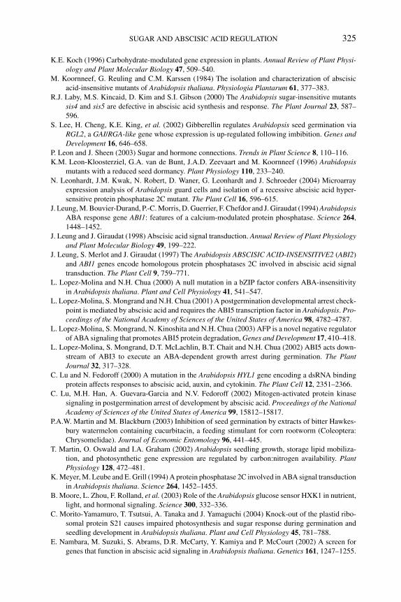
P1: OTE/SPH P2: OTE
BLUK053-Bradford September 27, 2006 15:22
SUGAR AND ABSCISIC ACID REGULATION 325
K.E. Koch (1996) Carbohydrate-modulated gene expression in plants. Annual Review of Plant Physi-ology and Plant Molecular Biology 47, 509–540.
M. Koornneef, G. Reuling and C.M. Karssen (1984) The isolation and characterization of abscisic
acid-insensitive mutants of Arabidopsis thaliana. Physiologia Plantarum 61, 377–383.
R.J. Laby, M.S. Kincaid, D. Kim and S.I. Gibson (2000) The Arabidopsis sugar-insensitive mutants
sis4 and sis5 are defective in abscisic acid synthesis and response. The Plant Journal 23, 587–
596.
S. Lee, H. Cheng, K.E. King, et al. (2002) Gibberellin regulates Arabidopsis seed germination via
RGL2, a GAI/RGA-like gene whose expression is up-regulated following imbibition. Genes andDevelopment 16, 646–658.
P. Leon and J. Sheen (2003) Sugar and hormone connections. Trends in Plant Science 8, 110–116.
K.M. Leon-Kloosterziel, G.A. van de Bunt, J.A.D. Zeevaart and M. Koornneef (1996) Arabidopsismutants with a reduced seed dormancy. Plant Physiology 110, 233–240.
N. Leonhardt, J.M. Kwak, N. Robert, D. Waner, G. Leonhardt and J. Schroeder (2004) Microarray
expression analysis of Arabidopsis guard cells and isolation of a recessive abscisic acid hyper-
sensitive protein phosphatase 2C mutant. The Plant Cell 16, 596–615.
J. Leung, M. Bouvier-Durand, P.-C. Morris, D. Guerrier, F. Chefdor and J. Giraudat (1994) ArabidopsisABA response gene ABI1: features of a calcium-modulated protein phosphatase. Science 264,
1448–1452.
J. Leung and J. Giraudat (1998) Abscisic acid signal transduction. Annual Review of Plant Physiologyand Plant Molecular Biology 49, 199–222.
J. Leung, S. Merlot and J. Giraudat (1997) The Arabidopsis ABSCISIC ACID-INSENSITIVE2 (ABI2)
and ABI1 genes encode homologous protein phosphatases 2C involved in abscisic acid signal
transduction. The Plant Cell 9, 759–771.
L. Lopez-Molina and N.H. Chua (2000) A null mutation in a bZIP factor confers ABA-insensitivity
in Arabidopsis thaliana. Plant and Cell Physiology 41, 541–547.
L. Lopez-Molina, S. Mongrand and N.H. Chua (2001) A postgermination developmental arrest check-
point is mediated by abscisic acid and requires the ABI5 transcription factor in Arabidopsis. Pro-ceedings of the National Academy of Sciences of the United States of America 98, 4782–4787.
L. Lopez-Molina, S. Mongrand, N. Kinoshita and N.H. Chua (2003) AFP is a novel negative regulator
of ABA signaling that promotes ABI5 protein degradation, Genes and Development 17, 410–418.
L. Lopez-Molina, S. Mongrand, D.T. McLachlin, B.T. Chait and N.H. Chua (2002) ABI5 acts down-
stream of ABI3 to execute an ABA-dependent growth arrest during germination. The PlantJournal 32, 317–328.
C. Lu and N. Fedoroff (2000) A mutation in the Arabidopsis HYL1 gene encoding a dsRNA binding
protein affects responses to abscisic acid, auxin, and cytokinin. The Plant Cell 12, 2351–2366.
C. Lu, M.H. Han, A. Guevara-Garcia and N.V. Fedoroff (2002) Mitogen-activated protein kinase
signaling in postgermination arrest of development by abscisic acid. Proceedings of the NationalAcademy of Sciences of the United States of America 99, 15812–15817.
P.A.W. Martin and M. Blackburn (2003) Inhibition of seed germination by extracts of bitter Hawkes-
bury watermelon containing cucurbitacin, a feeding stimulant for corn rootworm (Coleoptera:
Chrysomelidae). Journal of Economic Entomology 96, 441–445.
T. Martin, O. Oswald and I.A. Graham (2002) Arabidopsis seedling growth, storage lipid mobiliza-
tion, and photosynthetic gene expression are regulated by carbon:nitrogen availability. PlantPhysiology 128, 472–481.
K. Meyer, M. Leube and E. Grill (1994) A protein phosphatase 2C involved in ABA signal transduction
in Arabidopsis thaliana. Science 264, 1452–1455.
B. Moore, L. Zhou, F. Rolland, et al. (2003) Role of the Arabidopsis glucose sensor HXK1 in nutrient,
light, and hormonal signaling. Science 300, 332–336.
C. Morito-Yamamuro, T. Tsutsui, A. Tanaka and J. Yamaguchi (2004) Knock-out of the plastid ribo-
somal protein S21 causes impaired photosynthesis and sugar response during germination and
seedling development in Arabidopsis thaliana. Plant and Cell Physiology 45, 781–788.
E. Nambara, M. Suzuki, S. Abrams, D.R. McCarty, Y. Kamiya and P. McCourt (2002) A screen for
genes that function in abscisic acid signaling in Arabidopsis thaliana. Genetics 161, 1247–1255.
P1: OTE/SPH P2: OTE
BLUK053-Bradford September 27, 2006 15:22
326 SEED DEVELOPMENT, DORMANCY AND GERMINATION
K. Nemeth, K. Salchert, P. Putnoky, et al. (1998) Pleiotropic control of glucose and hormone responses
by PRL1, a nuclear WD protein, in Arabidopsis. Genes and Development 12, 3059–3073.
N. Nishimura, T. Yoshida, M. Murayama, T. Asami, K. Shinozaki and T. Hirayama (2004) Isola-
tion and characterization of novel mutants affecting the abscisic acid sensitivity of Arabidopsisgermination and seedling growth. Plant and Cell Physiology 45, 1485–1499.
X. Niu, T. Helentjaris and N.J. Bate (2002) Maize ABI4 binds Coupling Element1 in abscisic acid and
sugar response genes. The Plant Cell 14, 2565–2575.
E. Oh, J. Kim, E. Park, J.I. Kim, C. Kang and G. Choi (2004) PIL5, a phytochrome-interacting basic
helix-loop-helix protein, is a key negative regulator of seed germination in Arabidopsis thaliana.
The Plant Cell 16, 3045–3058.
D.P. O’Keefe, J.M. Tepperman, C. Dean, K.J. Leto, D.L. Erbes and J.T. Odell (1994) Plant expression
of a bacterial cytochrome P450 that catalyzes activation of a sulfonylurea pro-herbicide. PlantPhysiology 105, 473–482.
P. Palenchar, A. Kouranov, L. Lejay and G. Coruzzi (2004) Genome-wide patterns of carbon and
nitrogen regulation of gene expression validate the combined carbon and nitrogen (CN)-signaling
hypothesis in plants. Genome Biology 5, R91.
J.V. Pego, P.J. Weisbeek and S.C.M. Smeekens (1999) Mannose inhibits Arabidopsis germination via
a hexokinase-mediated step. Plant Physiology 119, 1017–1024.
S. Penfield, E.L. Rylott, A.D. Gilday, S. Graham, T.R. Larson and I.A. Graham (2004) Reserve
mobilization in the Arabidopsis endosperm fuels hypocotyl elongation in the dark, is independent
of abscisic acid, and requires PHOSPHOENOLPYRUVATE CARBOXYKINASE1. The Plant Cell16, 2705–2718.
H. Pinfield-Wells, E.L. Rylott, A.D. Gilday, et al. (2005) Sucrose rescues seedling establishment but
not germination of Arabidopsis mutants disrupted in peroxisomal fatty acid catabolism. The PlantJournal 43, 861–872.
T. Potuschak, E. Lechner, Y. Parmentier, et al. (2003) EIN3-dependent regulation of plant ethylene
hormone signaling by two Arabidopsis F box proteins: EBF1 and EBF2. Cell 115, 679–689.
J. Price, A. Laxmi, S.K. St. Martin and J.C. Jang (2004) Global transcription profiling reveals multiple
sugar signal transduction mechanisms in Arabidopsis. The Plant Cell 16, 2128–2150.
J. Price, T.C. Li, S.G. Kang, J.K. Na and J.C. Jang (2003) Mechanisms of glucose signaling during
germination of Arabidopsis. Plant Physiology 132, 1424–1438.
S.L. Pritchard, W.L. Charlton, A. Baker and I.A. Graham (2002) Germination and storage reserve
mobilization are regulated independently in Arabidopsis. The Plant Journal 31, 639–647.
V. Quesada, M.R. Ponce and J.L. Micol (2000) Genetic analysis of salt-tolerant mutants in Arabidopsisthaliana. Genetics 154, 421–436.
F. Rolland, B. Moore and J. Sheen (2002) Sugar sensing and signalling in plants. The Plant Cell 14,
S185–S205.
F. Rook and M.W. Bevan (2003) Genetic approaches to understanding sugar-response pathways.
Journal of Experimental Botany 54, 495–501.
F. Rook, F. Corke, R. Card, G. Munz, C. Smith and M.W. Bevan (2001) Impaired sucrose-induction
mutants reveal the modulation of sugar-induced starch biosynthetic gene expression by abscisic
acid signalling. The Plant Journal 26, 421–433.
L. Russell, V. Larner, S. Kurup, S. Bougourd and M. Holdsworth (2000) The Arabidopsis COMATOSElocus regulates germination potential. Development 127, 3759–3767.
A. Saez, N. Apostolova, M. Gonzalez-Guzman, et al. (2004) Gain-of-function and loss-of-function
phenotypes of the protein phophatase 2C HAB1 reveal its role as a negative regulator of abscisic
acid signalling. The Plant Journal 37, 354–369.
Y. Sakuma, Q. Liu, J.G. Dubouzet, H. Abe, K. Shinozaki and K. Yamaguchi-Shinozaki (2002) DNA-
binding specificity of the ERF/AP2 domain of Arabidopsis DREBs, transcription factors involved
in dehydration- and cold-inducible gene expression. Biochemical and Biophysical Research Com-munications 290, 998–1009.
H. Schluepmann, T. Pellny, A. van Dijken, S. Smeekens and M. Paul (2003) Trehalose 6-phosphate
is indispensable for carbohydrate utilization and growth in Arabidopsis thaliana. Proceedings ofthe National Academy of Sciences of the United States of America 100, 6849–6854.
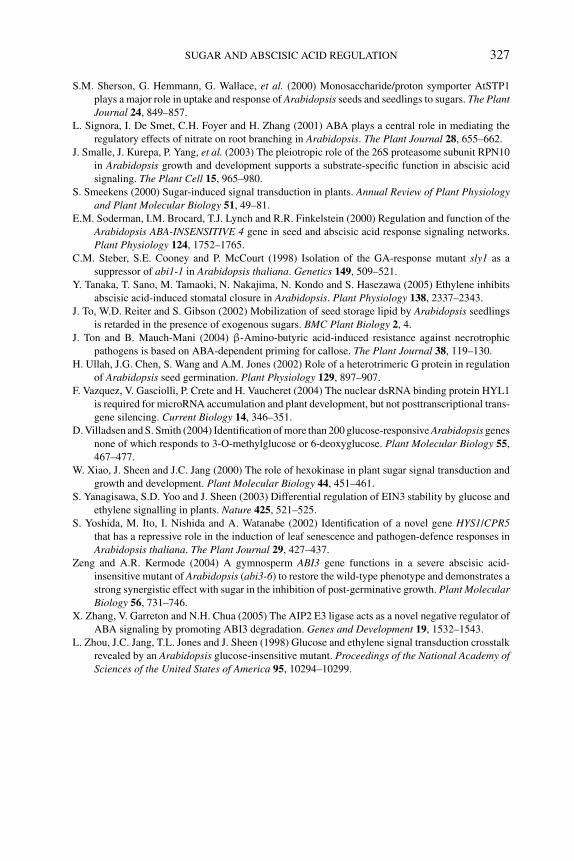
P1: OTE/SPH P2: OTE
BLUK053-Bradford September 27, 2006 15:22
SUGAR AND ABSCISIC ACID REGULATION 327
S.M. Sherson, G. Hemmann, G. Wallace, et al. (2000) Monosaccharide/proton symporter AtSTP1
plays a major role in uptake and response of Arabidopsis seeds and seedlings to sugars. The PlantJournal 24, 849–857.
L. Signora, I. De Smet, C.H. Foyer and H. Zhang (2001) ABA plays a central role in mediating the
regulatory effects of nitrate on root branching in Arabidopsis. The Plant Journal 28, 655–662.
J. Smalle, J. Kurepa, P. Yang, et al. (2003) The pleiotropic role of the 26S proteasome subunit RPN10
in Arabidopsis growth and development supports a substrate-specific function in abscisic acid
signaling. The Plant Cell 15, 965–980.
S. Smeekens (2000) Sugar-induced signal transduction in plants. Annual Review of Plant Physiologyand Plant Molecular Biology 51, 49–81.
E.M. Soderman, I.M. Brocard, T.J. Lynch and R.R. Finkelstein (2000) Regulation and function of the
Arabidopsis ABA-INSENSITIVE 4 gene in seed and abscisic acid response signaling networks.
Plant Physiology 124, 1752–1765.
C.M. Steber, S.E. Cooney and P. McCourt (1998) Isolation of the GA-response mutant sly1 as a
suppressor of abi1-1 in Arabidopsis thaliana. Genetics 149, 509–521.
Y. Tanaka, T. Sano, M. Tamaoki, N. Nakajima, N. Kondo and S. Hasezawa (2005) Ethylene inhibits
abscisic acid-induced stomatal closure in Arabidopsis. Plant Physiology 138, 2337–2343.
J. To, W.D. Reiter and S. Gibson (2002) Mobilization of seed storage lipid by Arabidopsis seedlings
is retarded in the presence of exogenous sugars. BMC Plant Biology 2, 4.
J. Ton and B. Mauch-Mani (2004) β-Amino-butyric acid-induced resistance against necrotrophic
pathogens is based on ABA-dependent priming for callose. The Plant Journal 38, 119–130.
H. Ullah, J.G. Chen, S. Wang and A.M. Jones (2002) Role of a heterotrimeric G protein in regulation
of Arabidopsis seed germination. Plant Physiology 129, 897–907.
F. Vazquez, V. Gasciolli, P. Crete and H. Vaucheret (2004) The nuclear dsRNA binding protein HYL1
is required for microRNA accumulation and plant development, but not posttranscriptional trans-
gene silencing. Current Biology 14, 346–351.
D. Villadsen and S. Smith (2004) Identification of more than 200 glucose-responsive Arabidopsis genes
none of which responds to 3-O-methylglucose or 6-deoxyglucose. Plant Molecular Biology 55,
467–477.
W. Xiao, J. Sheen and J.C. Jang (2000) The role of hexokinase in plant sugar signal transduction and
growth and development. Plant Molecular Biology 44, 451–461.
S. Yanagisawa, S.D. Yoo and J. Sheen (2003) Differential regulation of EIN3 stability by glucose and
ethylene signalling in plants. Nature 425, 521–525.
S. Yoshida, M. Ito, I. Nishida and A. Watanabe (2002) Identification of a novel gene HYS1/CPR5that has a repressive role in the induction of leaf senescence and pathogen-defence responses in
Arabidopsis thaliana. The Plant Journal 29, 427–437.
Zeng and A.R. Kermode (2004) A gymnosperm ABI3 gene functions in a severe abscisic acid-
insensitive mutant of Arabidopsis (abi3-6) to restore the wild-type phenotype and demonstrates a
strong synergistic effect with sugar in the inhibition of post-germinative growth. Plant MolecularBiology 56, 731–746.
X. Zhang, V. Garreton and N.H. Chua (2005) The AIP2 E3 ligase acts as a novel negative regulator of
ABA signaling by promoting ABI3 degradation. Genes and Development 19, 1532–1543.
L. Zhou, J.C. Jang, T.L. Jones and J. Sheen (1998) Glucose and ethylene signal transduction crosstalk
revealed by an Arabidopsis glucose-insensitive mutant. Proceedings of the National Academy ofSciences of the United States of America 95, 10294–10299.

P1: OTE/SPH P2: OTE
BLUK053-Bradford September 27, 2006 15:22
328
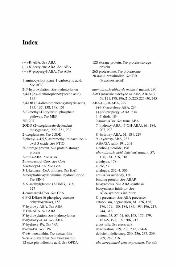
P1: PAE
BLUK053-Bradford September 22, 2006 21:0
Index
(−)-R-ABA. See ABA
(+)-8′-acetylene-ABA. See ABA
(+)-9′-propargyl-ABA. See ABA
1-aminocyclopropane-1-carboxylic acid.
See ACC
2-β-hydroxylation. See hydroxylation
2,4-D (2,4-dichlorophenoxyacetic acid),
135
2,4-DB (2,4-dichlorophenoxybutyric acid),
135, 137, 138, 148, 151
2-C-methyl-D-erythritol phosphate
pathway. See MEP
2iP, 207
2ODD (2-oxoglutarate-dependent
dioxygenase), 227, 231, 232
2-oxoglutarate. See 2ODD
2-phenyl-4,4,5,5,-tetramethylimidazoline-1-
oxyl 3-oxide. See PTIO
2S storage protein. See protein-storage
protein
2-trans-ABA. See ABA
2-trans-enoyl-CoA. See CoA
3-ketoacyl-CoA. See CoA
3-L-ketoacyl-CoA thiolase. See KAT
3-morpholinosydnonimine, hydrochloride.
See SIN-1
3-O-methylglucose (3-OMG), 318,
327
4-coumaroyl-CoA. See CoA
6-P G DHase (6-phosphogluconate
dehydrogenase), 158
7′ hydroxy-ABA. See ABA
7′ OH-ABA. See ABA
8′ hydroxylation. See hydroxylation
8′-hydroxy-ABA. See ABA
8′-hydroxy-PA. See 1PA
8′-oxo-PA. See 1PA
9′-cis-neoxanthin. See neoxanthin
9-cis-violaxanthin. See violaxanthin
12-oxo-phytodienoic acid. See OPDA
12S storage protein. See protein-storage
protein
26S proteasome. See proteasome
28-homo-brassinolide. See BR
(brassinosteroid)
aao (abscisic aldehyde oxidase) mutant, 230
AAO (abscisic aldehyde oxidase, AB-AO),
59,121,178,196,215,220,225–30,245
ABA-(−)-R-ABA, 229
(+)-8′-acetylene-ABA, 234
(+)-9′-propargyl-ABA, 234
1′,4′ diols, 184
2-trans-ABA. See trans ABA
7′ hydroxy-ABA, (7’OH-ABA), 61, 184,
207, 233
8′-hydroxy-ABA, 61, 184, 229
9′- hydroxy-ABA, 233
ABA/GA ratio, 191, 201
alcohol glucoside, 196
aba (abscisic acid deficient) mutant, 57,
126, 181, 316, 318
aldehyde, 178
allele, 57
analogue, 232–4, 306
anti-ABA antibody, 180
binding protein. See ABAP
biosynthesis. See ABA-synthesis
biosynthesis inhibitor. SeeABA-synthesis inhibitor
C15 precursor. See ABA precursor
catabolism, degradation, 61, 126, 168,
176, 179, 180, 184, 185, 193, 196, 217,
244, 316
content, 53, 57–61, 63, 168, 177, 179,
183–5, 191, 192, 206, 212
cross-talk. See cross-talk
deactivation, 229, 230, 232, 234–6
deficient, deficiency, 230, 236, 237, 239,
269, 289, 316
aba deregulated gene expression. See ade
329
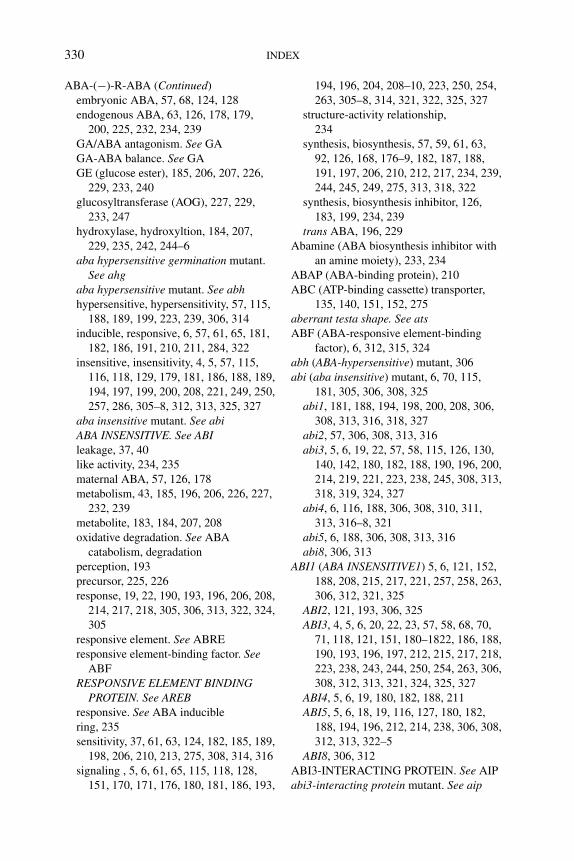
P1: PAE
BLUK053-Bradford September 22, 2006 21:0
330 INDEX
ABA-(−)-R-ABA (Continued)
embryonic ABA, 57, 68, 124, 128
endogenous ABA, 63, 126, 178, 179,
200, 225, 232, 234, 239
GA/ABA antagonism. See GA
GA-ABA balance. See GA
GE (glucose ester), 185, 206, 207, 226,
229, 233, 240
glucosyltransferase (AOG), 227, 229,
233, 247
hydroxylase, hydroxyltion, 184, 207,
229, 235, 242, 244–6
aba hypersensitive germination mutant.
See ahgaba hypersensitive mutant. See abhhypersensitive, hypersensitivity, 57, 115,
188, 189, 199, 223, 239, 306, 314
inducible, responsive, 6, 57, 61, 65, 181,
182, 186, 191, 210, 211, 284, 322
insensitive, insensitivity, 4, 5, 57, 115,
116, 118, 129, 179, 181, 186, 188, 189,
194, 197, 199, 200, 208, 221, 249, 250,
257, 286, 305–8, 312, 313, 325, 327
aba insensitive mutant. See abiABA INSENSITIVE. See ABIleakage, 37, 40
like activity, 234, 235
maternal ABA, 57, 126, 178
metabolism, 43, 185, 196, 206, 226, 227,
232, 239
metabolite, 183, 184, 207, 208
oxidative degradation. See ABA
catabolism, degradation
perception, 193
precursor, 225, 226
response, 19, 22, 190, 193, 196, 206, 208,
214, 217, 218, 305, 306, 313, 322, 324,
305
responsive element. See ABRE
responsive element-binding factor. SeeABF
RESPONSIVE ELEMENT BINDINGPROTEIN. See AREB
responsive. See ABA inducible
ring, 235
sensitivity, 37, 61, 63, 124, 182, 185, 189,
198, 206, 210, 213, 275, 308, 314, 316
signaling , 5, 6, 61, 65, 115, 118, 128,
151, 170, 171, 176, 180, 181, 186, 193,
194, 196, 204, 208–10, 223, 250, 254,
263, 305–8, 314, 321, 322, 325, 327
structure-activity relationship,
234
synthesis, biosynthesis, 57, 59, 61, 63,
92, 126, 168, 176–9, 182, 187, 188,
191, 197, 206, 210, 212, 217, 234, 239,
244, 245, 249, 275, 313, 318, 322
synthesis, biosynthesis inhibitor, 126,
183, 199, 234, 239
trans ABA, 196, 229
Abamine (ABA biosynthesis inhibitor with
an amine moiety), 233, 234
ABAP (ABA-binding protein), 210
ABC (ATP-binding cassette) transporter,
135, 140, 151, 152, 275
aberrant testa shape. See atsABF (ABA-responsive element-binding
factor), 6, 312, 315, 324
abh (ABA-hypersensitive) mutant, 306
abi (aba insensitive) mutant, 6, 70, 115,
181, 305, 306, 308, 325
abi1, 181, 188, 194, 198, 200, 208, 306,
308, 313, 316, 318, 327
abi2, 57, 306, 308, 313, 316
abi3, 5, 6, 19, 22, 57, 58, 115, 126, 130,
140, 142, 180, 182, 188, 190, 196, 200,
214, 219, 221, 223, 238, 245, 308, 313,
318, 319, 324, 327
abi4, 6, 116, 188, 306, 308, 310, 311,
313, 316–8, 321
abi5, 6, 188, 306, 308, 313, 316
abi8, 306, 313
ABI1 (ABA INSENSITIVE1) 5, 6, 121, 152,
188, 208, 215, 217, 221, 257, 258, 263,
306, 312, 321, 325
ABI2, 121, 193, 306, 325
ABI3, 4, 5, 6, 20, 22, 23, 57, 58, 68, 70,
71, 118, 121, 151, 180–1822, 186, 188,
190, 193, 196, 197, 212, 215, 217, 218,
223, 238, 243, 244, 250, 254, 263, 306,
308, 312, 313, 321, 324, 325, 327
ABI4, 5, 6, 19, 180, 182, 188, 211
ABI5, 5, 6, 18, 19, 116, 127, 180, 182,
188, 194, 196, 212, 214, 238, 306, 308,
312, 313, 322–5
ABI8, 306, 312
ABI3-INTERACTING PROTEIN. See AIP
abi3-interacting protein mutant. See aip
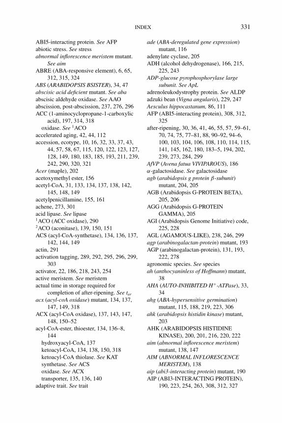
P1: PAE
BLUK053-Bradford September 22, 2006 21:0
INDEX 331
ABI5-interacting protein. See AFP
abiotic stress. See stress
abnormal inflorescence meristem mutant.
See aimABRE (ABA-responsive element), 6, 65,
312, 315, 324
ABS (ARABIDOPSIS BSISTER), 34, 47
abscisic acid deficient mutant. See abaabscisic aldehyde oxidase. See AAO
abscission, post-abscission, 237, 276, 296
ACC (1-aminocyclopropane-1-carboxylic
acid), 197, 314, 318
oxidase. See 1ACO
accelerated aging, 42, 44, 112
accession, ecotype, 10, 16, 32, 33, 37, 43,
44, 57, 58, 67, 115, 120, 122, 123, 127,
128, 149, 180, 183, 185, 193, 211, 239,
242, 290, 320, 321
Acer (maple), 202
acetoxymethyl ester, 156
acetyl-CoA, 31, 133, 134, 137, 138, 142,
145, 148, 149
acetylpenicillamine, 155, 161
achene, 273, 301
acid lipase. See lipase1ACO (ACC oxidase), 2902ACO (aconitase), 139, 150, 151
ACS (acyl-CoA-synthetase), 134, 136, 137,
142, 144, 149
actin, 291
activation tagging, 289, 292, 295, 296, 299,
303
activator, 22, 186, 218, 243, 254
active meristem. See meristem
actual time in storage required for
completion of after-ripening. See tar
acx (acyl-coA oxidase) mutant, 134, 137,
147, 149, 318
ACX (acyl-CoA oxidase), 137, 143, 147,
148, 150–52
acyl-CoA-ester, thioester, 134, 136–8,
144
hydroxyacyl-CoA, 137
ketoacyl-CoA, 134, 138, 150, 318
ketoacyl-CoA thiolase. See KAT
synthetase. See ACS
oxidase. See ACX
transporter, 135, 136, 140
adaptive trait. See trait
ade (ABA-deregulated gene expression)
mutant, 116
adenylate cyclase, 205
ADH (alcohol dehydrogenase), 166, 215,
225, 243
ADP-glucose pyrophosphorylase largesubunit. See ApL
adrenoleukodystrophy protein. See ALDP
adzuki bean (Vigna angularis), 229, 247
Aesculus hippocastanum, 86, 111
AFP (ABI5-interacting protein), 308, 312,
325
after-ripening, 30, 36, 41, 46, 55, 57, 59–61,
70, 74, 75, 77–81, 88, 90–92, 94–6,
100, 103, 104, 106, 108, 110, 114, 115,
141, 145, 162, 180, 183–5, 194, 202,
239, 273, 284, 299
AfVP (Avena fatua VIVIPAROUS), 186
α-galactosidase. See galactosidase
agb (arabidopsis g protein β-subunit)mutant, 204, 205
AGB (Arabidopsis G-PROTEIN BETA),
205, 206
AGG (Arabidopsis G-PROTEIN
GAMMA), 205
AGI (Arabidopsis Genome Initiative) code,
225, 228
AGL (AGAMOUS-LIKE), 238, 246, 299
agp (arabinogalactan-protein) mutant, 193
AGP (arabinogalactan-protein), 131, 193,
222, 278
agronomic species. See species
ah (anthocyaninless of Hoffmann) mutant,
38
AHA (AUTO-INHIBITED H+-ATPase), 33,
34
ahg (ABA-hypersensitive germination)
mutant, 115, 188, 219, 223, 306
ahk (arabidopsis histidin kinase) mutant,
203
AHK (ARABIDOPSIS HISTIDINE
KINASE), 200, 201, 216, 220, 222
aim (abnormal inflorescence meristem)
mutant, 138, 147
AIM (ABNORMAL INFLORESCENCEMERISTEM), 138
aip (abi3-interacting protein) mutant, 190
AIP (ABI3-INTERACTING PROTEIN),
190, 223, 254, 263, 308, 312, 327
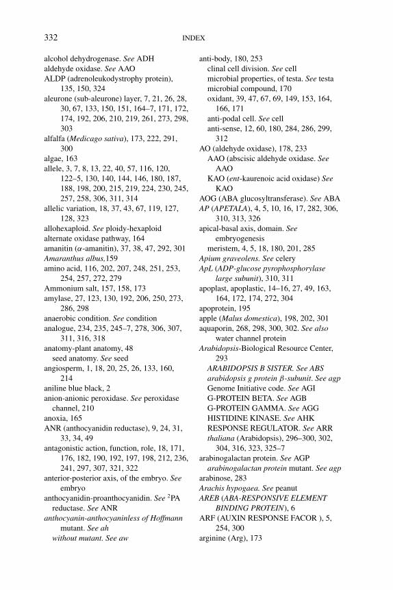
P1: PAE
BLUK053-Bradford September 22, 2006 21:0
332 INDEX
alcohol dehydrogenase. See ADH
aldehyde oxidase. See AAO
ALDP (adrenoleukodystrophy protein),
135, 150, 324
aleurone (sub-aleurone) layer, 7, 21, 26, 28,
30, 67, 133, 150, 151, 164–7, 171, 172,
174, 192, 206, 210, 219, 261, 273, 298,
303
alfalfa (Medicago sativa), 173, 222, 291,
300
algae, 163
allele, 3, 7, 8, 13, 22, 40, 57, 116, 120,
122–5, 130, 140, 144, 146, 180, 187,
188, 198, 200, 215, 219, 224, 230, 245,
257, 258, 306, 311, 314
allelic variation, 18, 37, 43, 67, 119, 127,
128, 323
allohexaploid. See ploidy-hexaploid
alternate oxidase pathway, 164
amanitin (α-amanitin), 37, 38, 47, 292, 301
Amaranthus albus,159
amino acid, 116, 202, 207, 248, 251, 253,
254, 257, 272, 279
Ammonium salt, 157, 158, 173
amylase, 27, 123, 130, 192, 206, 250, 273,
286, 298
anaerobic condition. See condition
analogue, 234, 235, 245–7, 278, 306, 307,
311, 316, 318
anatomy-plant anatomy, 48
seed anatomy. See seed
angiosperm, 1, 18, 20, 25, 26, 133, 160,
214
aniline blue black, 2
anion-anionic peroxidase. See peroxidase
channel, 210
anoxia, 165
ANR (anthocyanidin reductase), 9, 24, 31,
33, 34, 49
antagonistic action, function, role, 18, 171,
176, 182, 190, 192, 197, 198, 212, 236,
241, 297, 307, 321, 322
anterior-posterior axis, of the embryo. Seeembryo
anthocyanidin-proanthocyanidin. See 2PA
reductase. See ANR
anthocyanin-anthocyaninless of Hoffmannmutant. See ah
without mutant. See aw
anti-body, 180, 253
clinal cell division. See cell
microbial properties, of testa. See testa
microbial compound, 170
oxidant, 39, 47, 67, 69, 149, 153, 164,
166, 171
anti-podal cell. See cell
anti-sense, 12, 60, 180, 284, 286, 299,
312
AO (aldehyde oxidase), 178, 233
AAO (abscisic aldehyde oxidase. SeeAAO
KAO (ent-kaurenoic acid oxidase) SeeKAO
AOG (ABA glucosyltransferase). See ABA
AP (APETALA), 4, 5, 10, 16, 17, 282, 306,
310, 313, 326
apical-basal axis, domain. Seeembryogenesis
meristem, 4, 5, 18, 180, 201, 285
Apium graveolens. See celery
ApL (ADP-glucose pyrophosphorylaselarge subunit), 310, 311
apoplast, apoplastic, 14–16, 27, 49, 163,
164, 172, 174, 272, 304
apoprotein, 195
apple (Malus domestica), 198, 202, 301
aquaporin, 268, 298, 300, 302. See alsowater channel protein
Arabidopsis-Biological Resource Center,
293
ARABIDOPSIS B SISTER. See ABSarabidopsis g protein β-subunit. See agpGenome Initiative code. See AGI
G-PROTEIN BETA. See AGB
G-PROTEIN GAMMA. See AGG
HISTIDINE KINASE. See AHK
RESPONSE REGULATOR. See ARR
thaliana (Arabidopsis), 296–300, 302,
304, 316, 323, 325–7
arabinogalactan protein. See AGP
arabinogalactan protein mutant. See agparabinose, 283
Arachis hypogaea. See peanut
AREB (ABA-RESPONSIVE ELEMENTBINDING PROTEIN), 6
ARF (AUXIN RESPONSE FACOR ), 5,
254, 300
arginine (Arg), 173
P1: PAE
BLUK053-Bradford September 22, 2006 21:0
INDEX 333
ARIA (ARM REPEAT PROTEIN
INTERACTING WITH ABF2), 312,
315, 324
ARR (ARABIDOPSIS RESPONSE
REGULATOR), 200, 203, 221
ascorbic acid, ascorbate, 148, 156
asparagine (Asn), 159, 172
aspartic acid (Asp), 159, 200, 207, 251
Asteridae, 27
AtACO (Arabidopsis thaliana ACC oxidase)
See 1ACOAtbZIP (Arabidopsis thaliana bZIP67), 6
AtCPS (Arabidopsis thaliana ent-copalyl
diphosphate synthase). See CPSAtEM (Arabidopsis thaliana
EarlyMmethionine). See EmAtERS (Arabidopsis thaliana ETHYLENE
RESPONSE SENSOR. See ERSAtGA2ox (Arabidopsis thaliana GA
2-oxidase). See GA
AtGA3ox (Arabidopsis thaliana GA
3-oxidase). See GA
AtGenExpress project, 225
AtGLB1 (Arabidopsis thaliana class-1
hemoglobin), 166, 175
α-TIP (α-Tonoplast Intrinsic Protein). SeeTIP
AtKO (Arabidopsis thaliana ent-kaurene
oxidase). See KO
atmospheric humidity. See humidity
atmospheric NOx. See NO
Atnos (Arabidopsis thaliana nitric oxidesynthase). See nos
ATP-ATPase. See H+-ATPase
binding cassette transporter. See ABC
synthesis, 164, 166
AtPER (Arabidopsis thalianaperoxiredoxin). See PER
ats (aberrant testa shape) mutant, 42, 146
AtSTP (Arabidopsis thaliana SUGARTRANSPORT PROTEIN). See STP
AtXTH (Arabidopsis thaliana xyloglucanendotransglycosylase/hydrolase). SeeXTH
autohydrolysis, of cell wall, 280
AUTO-INHIBITED H+-ATPase. See AHAautotrophic, 305
autoxidation, 39
AUX-IAA protein, 5
auxin-biosynthesis, synthesis. Seeauxin-synthesis
catabolism, 207
conjugate, 202, 219
cross-talk. See cross-talk
insensitive, 20
receptor, 213, 254, 261, 262
auxin resistant mutant. See axrresponse, 20, 194, 254, 261, 300
AUXIN RESPONSE FACTOR. See ARF
shoot-derived auxin, 202, 212
signaling, 212, 222, 254, 314
synthesis, biosynthesis, 185, 202, 290,
304
transport inhibitor, 200, 214
AV10 (avenin), 60
Avena fatua. See oat
aw (anthocyanin without) mutant, 38
awn, 123
axis. See embryonic axis
axr (auxin resistant) mutant, 202, 214, 216,
222
azide, 157, 159, 160, 166, 171
B3-domain transcription factor. Seetranscription factor
baby lea syndrome mutant. See blsbacteria, 66, 70, 162, 168, 326
bakanae disease, 248
ban (banyuls) mutant, 9, 31, 32
BAN (BANYULS), 9, 17, 19, 24, 34, 35–7,
43, 44, 47, 49
Barbarea verna. See winter cress
Barbarea vulgaris. See yellow rocket
barley (Hordeum vulgare), 39, 40, 46, 48,
61, 64, 67, 69, 113, 119, 120, 122–4,
127–32, 151, 159, 161, 162, 164–6,
171–4, 179, 182–5, 206, 210, 216,
218–20, 222, 250, 252, 253, 259, 261,
280, 298, 299
BARNASE, 36, 37
BAS111.W, 234
base (or minimum) temperature. See Tb
(or minimum) water potential. See ψb
basic helix-loop-helix transcription factor.
See bHLH
BBM (BABYBOOM), 4
BDL (BODENLOS), 5, 20
bodenlos mutant, 20
P1: PAE
BLUK053-Bradford September 22, 2006 21:0
334 INDEX
bean (Phaseolus vulgaris), 26, 37, 45, 49,
245, 273
adzuki bean (Vigna angularis). Seeadzuki bean
castor bean (Ricinus communis). Seecastor bean
coffee bean (Cofea Arabica). See coffee
faba bean (Vicia faba). See faba bean
mungbean (Vigna radiate). See mungbean
soybean (Gycine max). See soybean
winged bean, 172
beechnut (Fagus sylvatica), 40, 43, 60, 61,
68, 70, 182, 184, 186, 198, 212, 215,
217–9
bent cotyledon stage. See embryogenesis
benzyladenine, 203
BET (BROMODOMAIN EXTRA
TERMINAL), 196
BGA (BORGIA), 7, 8
bHLH (basic helix-loop-helix) protein, 10,
34, 47, 193, 195, 196, 209, 210, 214,
219, 290, 326
bio (biotin synthase) mutant, 3
bioactive GA. See GA
biosynthesis. See synthesis
biotic stress. See stress
bitegmic ovule. See ovule
bks (blackseed) mutant, 38
BL (24-epi-brassinolide,
28-homo-brassinolide, epibrassinolide
[EBR]), 189, 204, 205, 216, 223, 257
bls (baby lea syndrome) mutant, 38
blueberry (Vaccinium myrtillus), 161, 173
BME (BLUE MICROPYLAR END), 193,
217, 299
BnCysP (Brassica napus cysteine
proteinase), 29, 48
BODENLOS. See BDL
body plan. See embryo-body plan
BORGIA. See BGAbotanical potato seed, 91, 106, 242
BOx (β-oxidation), 133–8, 140, 142–52,
160, 166, 318
BR (brassinosteroid) – ABA cross-talk. Seecross-talk
bioactive BR, 204
biosynthesis. See BR-synthesis
cell expansion, elongation, 204, 205
cross-talk. See cross-talk
deficient, 131, 204
dependent, 204, 257
dormancy, 177, 204, 210
GA cross-talk. See cross-talk
G-protein, 206
brassinosteroid insensitive mutant. Seebri
receptor, 204
response, 204
signaling, 118, 131, 204, 210, 213
synthesis, biosynthesis, 204, 219
Brassica napus. See rapeseed
Brassica rapa, 41, 47, 252, 262
Brassicaceae, 9, 29, 47, 56, 272
bri (brassinosteroid insensitive), 118, 204
BRI (BRASSINOSTEROID INSENSITIVE),
217
broccoli, 291
BROMODOMAIN EXTRA TERMINAL.
See BET
Bromus secalinus, 64
Bromus tectorum. See cheatgrass,
Junegrass, downy brome
brown pigment. See pigment
bruchid, 42, 46
bs (brownseed) mutant, 38, 44, 297
B-sister group MADS box protein, 34
buried seed. See seed
burning vegetation, 169
butenolide (3-methyl-2H -furo[2,3-c]pyran-
2-one), 169, 184
butylated hydroxytoluene, 166
BZIP transcription factor. See transcription
factor
C kinase. See kinase
C/N ratio, 314, 315, 318
C15 precursor. See ABA
C20 biosynthetic precursor. See GA, ABA
C40 carotenoid. See carotenoid
Ca2+, 205
cabbage, 291
caffeic acid, 31
calcofluor white, 266, 267
Californian chaparral (Emmenanthependuliflora), 161, 169
callose, 30, 285, 304, 313, 327
calpains, 7, 21
Caman (Coffea arabica mannanase), 271
P1: PAE
BLUK053-Bradford September 22, 2006 21:0
INDEX 335
cAMP, 205
CaMV 35S (cauliflower mosaic virus 35S )
promoter, 292
canopy density, 81
cap-endosperm cap. See endosperm
mRNA cap. See RNA
root cap. See root
capacitance. See soil capacitance
Cape Verde Island. See Cvi
Capsicum annuum. See pepper
carbohydrate, 1, 20, 24, 35, 53, 134, 142,
145, 151, 273, 305, 309, 325, 326
carbon-allocation, 307
availability, 145
dioxide. See CO2
metabolism, 171, 315
monoxide. See CO
skeleton, 133, 138, 139, 166
source, 136, 138, 139, 145
carbonyl prussiate, 161
cardinal temperature. See temperature
carob (Ceratonia siliqua), 279
carotenoid-biosynthesis. Seecarotenoid-synthesis
C40 carotenoid, 225
cleavage, 225, 242, 245
epoxycarotenoid dioxynase, 61, 178, 221,
225, 227, 242, 246
inhibitor. See carotenoid synthesis,
biosynthesis inhibitor pathway, 244
synthesis, biosynthesis, 37, 61, 232
synthesis, biosynthesis inhibitor, 37, 61,
183, 232, 318
caryopsis, 25, 49, 132
castasterone, 204
castor bean (Ricinus communis), 135, 139,
140, 144, 150, 151, 284
Cat1 promoter. See promoter
catabolic redox charge. See CRC
catalase, 38, 148, 159, 160, 163, 164, 166,
170, 173
catalyst, 154, 156
catechin (C, 2-3–trans), 32, 34, 37, 39, 44,
45
epicatechin (EC, 2-3-cis), 31–4, 37, 39
catechol oxidase, 39, 47
CCAAT binding transcription factor. Seetranscription factor
1CE (condensing enzyme), 31
2CE (coupling element), 65, 313, 326
Ceiling temperature, for germination. See Tc
celery (Apium graveolens), 52, 71, 286, 303
cell-inclusion, 293
anticlinal cell division, 8
antipodal cell, 2
cellular compartment, 133, 230
cellular function, 3, 127, 205
cellular organization, 27
central cell, 1, 2, 6, 8
cortical cell, 192
cycle, 8, 128, 213, 286, 291, 295
egg cell, 1
elongation, 11, 13, 20, 29, 45, 62, 114,
193, 203, 205, 210, 248, 299
endothelial cell, 28
expansion, 199, 204, 286
fate, 4, 7, 22, 27
guard cell, 210, 222, 325
L1 cell layer, 26
lignified cell, 267
nucellar cell, 3
number, 10, 15–17
parenchyma cell, 15, 180
permeant, 156
primary cell wall, 277–9, 283, 296, 297
radial cell expansion, 199
radial cell wall, 27
secondary cell wall, 10
size, 10, 15, 16, 201
sperm cell, 2, 6
stellar cell, 29
surface receptor, 193
suspension cell, 234
suspensor cell, 4
synergid cell, 2
tobacco BY2 cell, 257
to-cell cohesion, 283
vacuolated cell, 267
vegetative cell, 4, 22, 180, 218
wall disassembly, degradation, 250, 285,
295, 302
wall extensibility, 220, 268, 302
wall hydrolase, 276, 285, 286, 289
wall loosening, 192, 289
wall modification, 276, 286
cellulase (endo-β 1,4-glucanase), 271, 278,
282, 289, 296, 302
hemicellulase, 296
P1: PAE
BLUK053-Bradford September 22, 2006 21:0
336 INDEX
cellulose-hemicellulose, 53, 303
microfibril, 283
xyloglucan matrix, 285
central-cell. See cell
vacuole. See vacuole
centromere, 119, 122
Ceratonia siliqua. See carob
cereal, 2, 6, 7, 11, 15, 16, 23, 25, 26, 28, 39,
40, 45, 47, 53, 67, 69, 96, 113, 120,
124, 125, 128, 133, 150, 160, 164, 166,
172, 174, 192, 222, 243, 250, 267, 273
chain-breaker, 166
reaction, 166, 278
chalaza, 32
chalazal-endosperm. See endosperm
pole, 2, 28
proliferating tissue. See cpt
chalcone synthase. See CHS
Chamaecyparis nootkatensis. See yellow
cedar
chaperonin, 3, 18
CHD (Chromodomain Helicase
DNA-binding), 194, 219
cheatgrass (Bromus tectorum), 77, 92, 95,
96, 106–8, 110, 111
chemical barrier, of testa. See testa
Chenopodium album, 41, 81
CHI (chalcone isomerase), 31, 33
chickweed (Stellaria media), 109, 161
chilling, 30, 36, 47, 56, 59–62, 68, 80, 92,
128, 141, 183, 184, 186, 190, 192–4,
196, 198, 208, 209, 213, 220, 242, 243,
293, 296, 319. See also stratification
Chinese Spring, wheat, 124
chitinase, 42, 45, 271, 283-5, 298, 303, 304
chl (chlorate resistant) mutant, 66
chlorophyll, 32, 182
chloroplast, 147, 180, 225, 244, 315
CHOTTO, 306
chromatin modification, remodeling, 194,
196
chromodomain Helicase DNA-binding. SeeCHD
chromosome, 119, 121–4, 128–31, 295
CHS (chalcone synthase), 31, 33
cinnamic acid, 31, 229
cis-acting element, 65
cis-xanthophyll. See xanthophyll
citrate synthase. See CSY
citrate synthase mutant. See csycitrate, 134, 138, 139, 152
citrulline, 163
Citrullus lanatus. See watermelon
ckh (cytokinin hypersensitive) mutant, 195
CKH (CYTOKININ-HYPERSENSITIVE),
195, 214
ckr (cytokinin resistant) mutant, 203, 221
cleared embryo. See embryo
CnABI (Chamaecyparis nootkatensis ABI),
190
CO (carbon monoxide), 159
CO2 (carbon dioxide), 30, 39, 80, 139, 158,
220, 226, 227, 231
CoA (co-enzyme A)
2-trans-enoyl-CoA, 137
3-ketoacyl-CoA, 138
3-L-ketoacyl-CoA thiolase. See KAT
4-coumaroyl-CoA, 31
acetyl-CoA. See acetyl-CoA
acyl-CoA. See acyl-CoA
ester, 134–8, 144
transporter, 136, 140
yeast acyl-CoA synthase. See Faa2p
coat-imposed dormancy. See dormancy
cocklebur (Xanthium strumarium), 198
coenocyte, 6
coffee (Coffea arabica), 52, 54, 68, 271,
274, 275, 279, 281, 282, 296, 300
coi (coronatine insensitive) mutant, 147,
COI (CORONATINE INSENSITIVE), 254,
263
Col (Columbia), 183–5, 197, 206, 317, 321
cold-stratification. See stratification
stress. See stress
cold temperature germination mutant.
See ctgcoleoptile, 45, 250
coleorhiza, 185, 250
colorless compound, 27
columella, columellae. See testa
COMATOSE. See CTScomatose mutant. See ctscombinatorial-control, by hormones. See
hormone
dormancy. See dormancy
common ice plant (Mesembryanthemumcrystallinum), 186
complementation, 32
P1: PAE
BLUK053-Bradford September 22, 2006 21:0
INDEX 337
condensed tannin. See 2PA
(proanthocyanidin)
condensing enzyme. See 1CE
condition-anaerobic condition, 164
environmental condition, 29, 42, 73, 75,
76, 80, 83, 92, 103, 105, 114, 153, 240,
241, 258
field condition, 56, 73, 80, 96, 98
gaseous condition, 73
hydric condition, 73
stress condition, 38, 42, 43, 49, 55, 298
suboptimal condition, 230
thermal condition, 73
conformational change, 205
conjugate, conjugation, 184, 185, 190, 202,
207, 219, 229, 245, 251, 253
constitutive expression. See expression
CONSTITUTIVE EXPRESSOR OFPATHOGENESIS-RELATED GENE.See CPR
CONSTITUTIVE TRIPLE RESPONSE.See CTR
constitutive triple response. See ctrcontrolled deterioration, 42
copalyl diphosphate synthase. See CPS
CORONATINE INSENSITIVE. See COI
coronatine insensituive mutant. See coicortex, 192
cortical cell. See cell
cotton (Gossypium arboretum), 39, 259
cotyledon-development, 5, 23, 24, 215
identity, 4, 24, 152
impaired cotyledon, 196
stage. See embryogenesis
coumaric acid, 31, 39
coumaroyl-CoA. See CoA
Coupling Element. See CE
covalent, 12, 170, 253
covering structure, of seed, 25, 29, 40
cowpea (Vigna unguiculata), 42, 43, 46, 108
CPR (CONSTITUTIVE EXPRESSOR OFPATHOGENESIS-RELATED GENE),
315, 327
CPS (ent-copalyl diphosphate synthase),
192, 227, 228, 230, 231, 233
cpt (chalazal proliferating tissue), 26
c-PTIO (2-[4-carboxyphenyl]-4,4,5,5-
tetramethylimidazoline-1-oxyl-3-
oxide). See PTIO
CRC (catabolic redox charge), 149
Cress (Lepidium sativum), 158, 161, 204,
216, 234
CRINKLY, 7, 18
Crop species. See species
cross-talk, among hormones, 126, 176, 177,
185, 186, 194, 203, 206, 210, 214, 324
auxin and ABA, 185
auxin and cytokinin, 200, 201, 314
auxin and ethylene, 200, 207, 290
auxin and GA, 290
BR and GA, 118, 177, 205, 210
BR and ABA, 177, 204
cytokinin and ethylene, 200, 207, 208
cytokinin and GA, 203, 215
cytokinin and phytochrome, 203, 214,
288
cytokinin and sugar, 312, 314
ethylene and ABA, 206, 207
ethylene and GA, 207, 212
ethylene and sugar, 126
NO signaling and phytochrome, 170
crystal structure, 253
csy (citrate synthase) mutant, 134, 138,
144–7
CSY (citrate synthase), 138, 139, 144, 152
ct (cuticle), 26, 28
C-terminus, 182, 253–5
ctg (cold temperature germination) mutant,
292
ctr (constitutive triple response) mutant,
126, 128, 181, 188, 198–200, 215, 310,
311, 314, 316, 324
CTR (CONSTITUTIVE TRIPLERESPONSE), 198–200, 216, 314
cts (comatose) mutant, 134–6, 140–9, 318.
See also pxa and pedCTS (COMATOSE), 121, 135–7, 140, 142,
147, 148
Cucumis melo. See muskmelon
Cucurbita pepo. See squash
Cucurbitaceae, 274
cullin, 248, 251, 253
cuticle. See ct
cutin, 30
Cvi (Cape Verde Island), 10, 37, 57, 59–61,
63–5, 68, 115, 120, 183, 193, 194, 239,
290, 296
Cyamopsis tetragonolobus. See guar
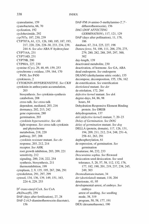
P1: PAE
BLUK053-Bradford September 22, 2006 21:0
338 INDEX
cyanoalanine, 159
cyanobacteria, 66, 70
cyclization, 192
cycloheximide, 292
cyp707a, 187, 230, 239
CYP707A, 61, 121, 126, 180, 185, 187, 193,
217, 220, 226, 228–30, 233, 234, 239,
244–6. See also ABA 8′-hydroxylase
CYP714A, 231
CYP714D, 231
CYP79B, 290
CYP88A, 227, 230
cysteine (Cys), 29, 48, 69, 159, 253
cytochrome c oxidase, 159, 164, 170
P450. See P450
cytokinesis, 2
CYTOKININ-HYPERSENSITIVE. See CKHcytokinin-in anthocyanin accumulation,
203
biosynthesis. See cytokinin-synthesis
catabolism, 208
cross-talk. See cross-talk
dependent, mediated, 203, 216
dormancy, 202, 213, 242
gene expression, 280
germination, 203
cytokinin hypersensitive. See ckhlight response. See cross-talk-cytokinin
and phytochrome
metabolism, 218, 220
pathway, 207, 208
cytokinin resistant mutant. See ckrresponse, 203, 212, 214
receptor. See AHK
root growth inhibition, 203, 209, 221
sensitivity, 212
signaling, 200, 218, 222, 254
synthesis, biosynthesis, 211
thermoinhibition, 199
cytoplasm, 2, 9, 135, 195, 267, 286, 291
cytoskeleton, 291, 297, 299
cytosol, 134, 136, 139, 149, 151, 163,
224–6, 229, 253
D2-trans-enoyl-CoA. See CoA
d8(Dwarf8), 259
DAF (days after fertilization), 27, 28
DAF-2 (4,5-diaminofluorescein diacetate),
156
DAF-FM (4-amino-5-methylamino-2′,7′-difluorofluorescein), 156
DAG (DOF AFFECTINGGERMINATION), 117, 121, 129
DAP (days after pollination), 11, 178,
186
database, 67, 214, 225, 227, 198
Datura ferox, 54, 106, 111, 266, 270, 275,
279, 280, 282, 288, 295, 297, 300,
302
day-length, 139
deactivated metabolite, 230
deactivation, of hormone. See GA, ABA
dead endosperm. See endosperm
DEANO (diethylamine nitric oxide), 155
decompose, decomposition, 155, 156, 162
de-esterification. See esterification
deetiolated mutant. See detde-etiolation, 172, 264
defective kernel mutant. See dekdegree-days, 84, 86, 98
hours, 84
Dehydration-Responsive Element Binding
protein. See DREB
dehydrogenation, 137
dek (defective kernel) mutant, 7, 20–23
Delay of Germination. See DOGdelay of germination mutant. See dogDELLA (protein, domain), 117, 126, 131,
194, 209, 211, 212, 214, 248, 251–6,
258–61, 263, 291
dephosphorylation, 61
de-repression, of germination. Seegermination
desaturase, 66, 232, 233
Descurainia sophia. See flixweed
desiccation-seed desiccation. See seed
tolerance, 5, 28, 57, 58, 112, 132, 176,
177, 182, 190, 201, 219, 237, 238, 245,
249, 303
Desmodiumuncinatum, 34
det (deetiolated) mutant, 118, 204
deteriorate, 41, 95
developmental-arrest, of embryo. Seeembryo
arrest of seedling. See seedling
mode, 38, 319
program, 50, 58, 177, 191
DEX (dexamethasone), 190
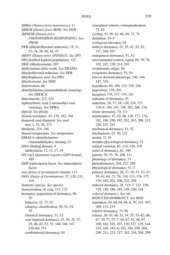
P1: PAE
BLUK053-Bradford September 22, 2006 21:0
INDEX 339
DfMan (Datura ferox mannanase), 11
DfMYB (Datura ferox MYB). See MYBDfPHOR (Datura ferox
PHOTOPERIOD-RESIPONSIVE). SeePHOR
DFR (dihydroflavonol reductase), 19, 31,
33, 34, 38, 40, 44, 47
DfSPY (Datura ferox SPINDLY). See SPYDH (doubled haploid population), 122
DHZ (dihydrozeatin), 207
diethylamine nitric oxide. See DEANO
dihydroflavonol reductase. See DFR
dihydrophaseic acid. See DPA
dihydrozeatin. See DHZ
dimerization, 66
dimethylamino-cinnamaldehyde (staining).
See DMACA
diniconazole, 233, 234
diphenylboric acid-2-aminoethyl ester
(staining). See DPBA
diploid. See ploidy
disease resistance, 45, 170, 292, 304
dispersal-seed dispersal. See seed
unit, 1, 53, 54, 273
diterpene, 224, 248
diurnal temperature. See temperature
DMACA (dimethylamino-
cinnamaldehyde), staining, 33
DNA-binding domain, 4
methylation, 12, 13, 17, 19
DN-rop2 (dominant negative GDP-bound),
189
DOF transcription factor. See transcription
factor
dog (delay of germination) mutant, 115
DOG (Delay of Germination), 37, 120, 121,
319
domestic species. See species
domestication, of crop, 113, 123
dormancy-acquisition of dormancy, 50,
59
behavior, 32, 73, 91
category, classification, 50, 52, 54,
55
chemical dormancy, 51, 52
coat-imposed dormancy, 25, 30, 35, 37,
39, 40–42, 52, 53, 140, 146, 147,
258–60, 274
combinatorial dormancy, 54
conceptual scheme, conceptualization,
73, 82
cycling, 53, 56, 63, 66, 69, 73, 78
definition, 53–5
ecological relevance, 64
embryo dormancy, 25, 35, 41, 52, 53,
217, 260, 287
endogenous dormancy, 51, 52
environmental control, signal, 65, 70, 76,
102, 107, 150, 214, 242
evolutionary origin, 50
exogenous dormancy, 53, 54
forever dormant phenotype, 140, 144,
147, 318
hypothesis, 69, 109, 153, 159, 166
imposition, 178, 201
inception, 176, 177, 179, 181
indicator of dormancy, 55
induction, 28, 57, 58, 120, 124, 127,
176–8, 190, 197, 198, 202, 206, 210
innate dormancy, 73, 111
maintenance, 47, 62, 68, 126, 173, 176,
182, 186, 190, 192, 202, 207, 208, 215,
220, 223, 243
mechanical dormancy, 51, 52
mechanisms, 25, 30, 122
model, 72, 91
morpho-physiological dormancy, 51
natural variation, 67, 114, 120, 319
onset of dormancy, 61, 140
pattern, 70, 73, 76, 108, 111
phenology of dormancy, 73
photodormancy, 204, 272, 299
physiological dormancy, 51–3
primary dormancy, 28, 37, 50, 51, 53, 57,
59, 63, 69, 73, 79, 110, 113, 176, 177,
179, 185, 201, 206, 232, 298
reduced dormancy, 38, 115–7, 123, 156,
179, 180, 186, 189, 199, 250, 318
reduced dormancy. See rdoREDUCED DORMANCY. See RDOregulation, 50, 60, 64, 66–8, 70, 103, 107,
109, 131, 215
relative dormancy, 75, 90
release, 36, 43, 46, 52, 54, 55, 55–62, 66,
67, 70, 73, 75–7, 80, 87, 91, 96, 97,
100, 103, 105, 107, 110, 127, 129, 144,
145, 169, 183–6, 193, 194, 198, 204,
209, 211, 215, 217, 242, 244, 298, 299
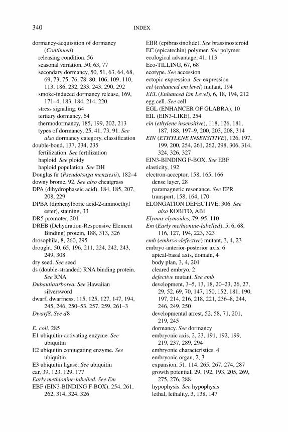
P1: PAE
BLUK053-Bradford September 22, 2006 21:0
340 INDEX
dormancy-acquisition of dormancy
(Continued)
releasing condition, 56
seasonal variation, 50, 63, 77
secondary dormancy, 50, 51, 63, 64, 68,
69, 73, 75, 76, 78, 80, 106, 109, 110,
113, 186, 232, 233, 243, 290, 292
smoke-induced dormancy release, 169,
171–4, 183, 184, 214, 220
stress signaling, 64
tertiary dormancy, 64
thermodormancy, 185, 199, 202, 213
types of dormancy, 25, 41, 73, 91. Seealso dormancy category, classification
double-bond, 137, 234, 235
fertilization. See fertilization
haploid. See ploidy
haploid population. See DH
Douglas fir (Pseudotsuga menziesii), 182–4
downy brome, 92. See also cheatgrass
DPA (dihydrophaseic acid), 184, 185, 207,
208, 229
DPBA (diphenylboric acid-2-aminoethyl
ester), staining, 33
DR5 promoter, 201
DREB (Dehydration-Responsive Element
Binding) protein, 188, 313, 326
drosophila, 8, 260, 295
drought, 50, 65, 196, 211, 224, 242, 243,
249, 308
dry seed. See seed
ds (double-stranded) RNA binding protein.
See RNA
Dubautiaarborea. See Hawaiian
silversword
dwarf, dwarfness, 115, 125, 127, 147, 194,
245, 246, 250–53, 257, 259, 261–3
Dwarf8. See d8
E. coli, 285
E1 ubiquitin-activating enzyme. Seeubiquitin
E2 ubiquitin conjugating enzyme. Seeubiquitin
E3 ubiquitin ligase. See ubiquitin
ear, 39, 123, 129, 177
Early methionine-labelled. See EmEBF (EIN3-BINDING F-BOX), 254, 261,
262, 314, 324, 326
EBR (epibrassinolide). See brassinosteroid
EC (epicatechin) polymer. See polymer
ecological advantage, 41, 113
Eco-TILLING, 67, 68
ecotype. See accession
ectopic expression. See expression
eel (enhanced em level) mutant, 194
EEL (Enhanced Em Level), 6, 18, 194, 212
egg cell. See cell
EGL (ENHANCER OF GLABRA), 10
EIL (EIN3-LIKE), 254
ein (ethylene insensitive), 118, 126, 181,
187, 188, 197–9, 200, 203, 208, 314
EIN (ETHYLENE INSENSITIVE), 126, 197,
199, 200, 254, 261, 262, 298, 306, 314,
324, 326, 327
EIN3-BINDING F-BOX. See EBF
elasticity, 192
electron-acceptor, 158, 165, 166
dense layer, 28
paramagnetic resonance. See EPR
transport, 158, 164, 170
ELONGATION DEFECTIVE, 306. Seealso KOBITO, ABI
Elymus elymoides, 79, 95, 110
Em (Early methionine-labelled), 5, 6, 68,
116, 127, 194, 223, 323
emb (embryo-defective) mutant, 3, 4, 23
embryo-anterior-posterior axis, 6
apical-basal axis, domain, 4
body plan, 3, 4, 201
cleared embryo, 2
defective mutant. See embdevelopment, 3–5, 13, 18, 20–23, 26, 27,
29, 52, 69, 70, 147, 150, 152, 181, 190,
197, 214, 216, 218, 221, 236–8, 244,
246, 249, 250
developmental arrest, 52, 58, 71, 201,
219, 245
dormancy. See dormancy
embryonic axis, 2, 23, 191, 192, 199,
219, 237, 289, 294
embryonic characteristics, 4
embryonic organ, 2, 3
expansion, 51, 114, 265, 267, 274, 287
growth potential, 29, 192, 193, 205, 269,
275, 276, 288
hypophysis. See hypophysis
lethal, lethality, 3, 138, 147
P1: PAE
BLUK053-Bradford September 22, 2006 21:0
INDEX 341
mature embryo, 2, 51, 64, 237
pattern formation, 18, 21, 177, 201, 237
polarity, 3
proliferation, 15, 16, 19, 29
proper, 2
rudimentary embryo, 52
sac, 2, 11, 25
secondary embryo, 3
shoot-root axis, 3, 201
somatic embryo, 4, 194
surface, 39
thrust, 52, 54
embryogenesis-bent cotyledon stage, 11
cotyledon stage, 2, 3, 11
globular stage, 2, 9, 13
heart stage, 2, 26, 27, 29
linear cotyledon stage, 2
mature green stage, 2
preglobular stage, 9
somatic embryogenesis, 4, 194
torpedo stage, 8, 9
embryonic-ABA. See ABA
axis, 2, 23, 191, 192, 199, 219, 237, 289,
294
identity, 194, 220
leaves, 26. See also cotyledons
trait, 194, 195, 201, 216, 217, 220
emergence-radicle emergence. See radicle
seedling emergence. See seedling
model. See model
Emmenanthe penduliflora. See Californian
chaparral
Empress tree (Paulownia tomentosa), 161,
162, 167, 171, 173
endo-1,4-glucanase. See glucanase
endodermis, 192
endogenous dormancy. See dormancy
endoreduplication, 7, 21
endosperm-aleurone layer. See aleurone
layer
basal endosperm, 6, 7, 16, 22
cap, 44, 68, 249, 262, 263, 271, 274–81,
284–7, 289, 296, 301–3
cellularization, 7, 11–3, 28
chalazal endosperm, 2, 7, 11, 12
coenocyte. See coenotyte
dead endosperm, 273
derived signal, for testa development,
29
development, 6–8, 11, 13, 21–3, 29, 47,
197, 223
endospermic seed. See seed
envelope, 273, 304
immature endosperm, 230
lateral endosperm, 266, 267, 269, 271,
274, 277–9, 281–3, 285
maternalization, 13
micropylar endosperm, 2, 7, 30, 53, 204,
262, 265–7, 269, 274–85, 288, 295,
301, 304
non-endospermic seed. See seed
free nucleus, nuclei, 2, 6, 11, 12, 273
paternalization, 13
peripheral endosperm, 2, 7, 26
persistent endosperm, 7
proliferation, 13
restraint, 269, 272
rupture, 199, 204, 265, 269, 284, 293
size, 12, 13
starchy endosperm, 7, 164, 250
sub-aleurone layer. See aleurone layer
transfer layer, 6, 7, 16
transient endosperm, 2
weakening, 68, 215, 265, 274–7,
280–282, 285, 286, 288, 296, 298
endothelial cell. See cell
endothelium, 9, 26, 27, 29, 34, 35, 38, 43,
44, 171
endo-β-mannanase. See mannanase
energy, 17, 41, 78, 124, 128, 133, 305, 324
enhanced-response to aba. See eraEnhanced Em Level. See EELenhanced em level mutant. See eelENHANCED RESPONSE to ABA. See
ERAenhancer-element, 292
ENHANCER OF GLABRA. See EGL
ENHANCER OF ZESTE, 8
trap, 130, 289, 292–4, 296, 297, 299
ENP (ENHANCER OF PINOID), 5
ent-copalyl diphosphate synthase. See CPS
ent-kaurene synthase. See KS
ent-kaurene, 192, 227, 230, 233, 249, 250,
260
ent-kaurenoic acid oxidase. See KAO
environmental-condition. See condition
parameter, 101, 102
stimulus 80, 239
P1: PAE
BLUK053-Bradford September 22, 2006 21:0
342 INDEX
enzyme secretion, 54
epibrassinolide (EBR). See BL
epicatechin. See catechin
epicuticular wax, 123, 129
epidermis, 7, 15, 19, 26, 37
subepidermal layer, 27
epinasty, 204
epistatic, epistases, epistasis, 34, 114, 129,
140, 181, 200
epoxidation, 225
epoxycarotenoid. See carotenoid
EPR (electron paramagnetic resonance),
167
equilibration, equilibrium, 60, 286, 287
era (enhanced response to ABA) mutant, 57,
115, 181, 197, 306
Ericales, 27
ERS (ETHYLENE RESPONSE SENSOR),
298
erythrose, 158
esterification, de-esterification, 283
ethephon, 198
ethylene-cross-talk. See cross-talk
evolution, 197, 198, 301
exogenous ethylene, 41
ethylene insensitive. See eininsensitive, insensitivity, 118, 181, 197,
198, 200, 203, 206, 208, 239, 298, 306,
314
precursor, 197, 314
receptor, 192–200, 215, 290, 298
ethylene receptor. See etrETHYLENE RECEPTOR. See ETRreleasing compound, 198
response, 128, 198–200, 215, 216, 243,
290, 299, 314, 324
signaling, 127, 132, 197–200, 206, 208,
211–3, 215, 219, 242, 254, 306, 311,
314, 323, 327
ETHYLENE INSENSITIVE. See EINetr (ethylene receptor) mutant, 118, 126,
128, 197, 198, 200, 206, 208, 213, 239,
240, 242, 296, 314
ETR (ETHYLENE RECEPTOR), 121, 197,
199, 200, 207, 208, 215, 217, 314
eukaryote, 205, 305
evolution, evolutionary, 23, 50, 67, 106,
108, 166, 205, 243, 251, 261, 262
exogenous dormancy. See dormancy
exo-PG. See PG
expansin, 270, 271, 276, 278, 285, 286, 288,
295, 296, 298–300, 302, 303
expression-constitutive expression, 180
ectopic expression, 4, 6, 19, 48, 244,
292
germinative expression, 279
heterologous expression, 229, 232
inducible expression, 182, 190
micropylar-specific expression, 293
misexpression, 4, 191, 237
overexpression, 57, 61, 64, 117, 128, 166,
180, 186, 190, 213, 236, 245, 258, 263,
273, 284, 299, 313, 324
post-germinative expression, 286
spatial expression, 192, 279
stage-specific expression, 293
temporal expression, 192, 279, 281
tissue-specific expression, 249
extensin, 44, 116, 128
EXTRA SEX COMB, 8
F3′H (flavanone 3′-hydroxylase), 31, 33
F3H (flavanone 3-hydroxylase), 31, 33
Faa2p (yeast acyl-CoA synthase), 135
faba bean (Vicia faba), 13–16, 46, 108
FAD, 137
FADH2, 137
Fagus sylvatica. See beechnut
famine response, starvation condition,
309
far red light. See FR
farnesyltransferase, 197
fatty acid-free fatty acid. See FFA
hydroxy fatty acid, 148
long-chain fatty acid. See LCFA
medium-chain fatty acid, 135
saturated fatty acid, 137
unsaturated fatty acid, 137, 151
F-box (gene, protein), 145, 151, 248,
250–254, 256, 258, 261, 262, 314
FCA, 210, 219
fca, 210
feast response, sugar excess, 309
fenugreek (Trigonella foenum-graecum),
279, 301
ferricyanide, 156, 157
ferrocyanide, 155–7
fertility, 16, 203, 248–50, 253, 257, 259
P1: PAE
BLUK053-Bradford September 22, 2006 21:0
INDEX 343
fertilization-double fertilization, 1, 26, 28,
273
fertilization independent seed mutant.
See fisFERTILIZATION INDEPENDENT
ENDOSPERM. See FIEfertilization independent endosperm
mutant. See fieFERTILIZATION INDEPENDENT
SEED. See FISfertilized soil. See soil
ferulic acid, 31
FFA (free fatty acid), 134
fie (fertilization independent endosperm)
mutant, 13, 23
FIE (FERTILIZATION INDEPENDENTENDOSPERM), 7, 8, 13, 21–4
field-condition, 56, 73, 80, 96, 98
temperature. See temperature
fine-mapping, 67, 123
FIS (FERTILIZATION INDEPENDENTSEED), 7, 8, 20
flavan-3-ol stereoisomers, 32, 34
flavanone-3-hydroxylase. See F3′H3′-hydroxylase. See F3H
flavonoid-biosynthesis. Seeflavonoid-synthesis
compartmentation, 34
composition, 33
Metabolism, 32, 33, 35, 38, 42
pathway, 31, 32
synthesis, biosynthesis, 9, 11, 17, 19, 24,
28, 32, 43, 45, 48, 49, 124
flavonol-glycoside, 31
producing cell. See cell
synthase. See FLS
flavoprotein dehydrogenase, 137
flax (Linum usitatissimum), 42, 44
flixweed (Descurainia sophia), 161, 174
flour, 21, 124
FLS (flavonol synthase), 31–3
fls1 (flavonol synthase) mutant, 31, 32
fluctuating temperature. See temperature
fluridone, 61, 92, 183, 199, 220, 223,
232–4, 239, 247, 318
force analyzer, 274
forest-canopy, 168
species. See species
forever dormant phenotype. See dormancy
FR (far red light), 30, 65, 74, 81, 107–9,
118, 183, 196, 203, 240, 266, 269, 271,
275–7, 282, 288, 297
frameshift, in sequence, 257
free-endosperm nucleus. See endosperm
fatty acid. See FFA
radical, 4, 153, 172, 175
fruit, 10, 26, 40, 53, 57, 68, 70, 179, 276,
277, 283, 285, 292, 295, 296, 298, 299,
302
FsACO (Fagus sylvatica ACC OXIDASE).
See ACOFsGTP (Fagus sylvatica GTP-binding
protein). See GTP-binding protein
FsPK (Fagus sylvatica protein kinase). Seekinase
FsPP2C (Fagus sylvatica protein
phosphatase 2C). See phosphatase
functional genomics, 46, 67, 176, 292, 293
redundancy. See redundant, redundancy
fungicide, 234
funiculus, 27, 28, 117
fus (fusca) mutant, 4, 21, 58, 140, 142, 149,
182, 191, 216, 236–8, 318, 319
FUS (FUSCA), 4, 6, 18, 20, 21–3, 58, 68,
70, 121, 178, 180, 182, 191, 194, 201,
213, 215, 216, 218, 219, 236–8, 241–4,
246
FY, 210
G-protein-Arabidopsis G-PROTEIN
GAMMA. See AGG
G 6-P (glucose 6-phosphate), 158
G 6-P Dhase (glucose 6-phosphate
dehydrogenase), 158
G protein-arabidopsis g protein α-subunitmutant. See agb
Arabidopsis G-PROTEIN BETA. SeeAGB
G PROTEIN-COUPLED RECEPTOR.
See GCR
G protein-coupled receptor. See gcrGA (gibberellin)-2-oxidase (GA2ox), 61,
227, 228, 231, 233, 236, 240, 244–6,
296
3-oxidase (GA3ox), 61, 62, 192, 227,
228, 231–3, 235, 244
12-aldehyde, 37
13-hydroxylase, 231
P1: PAE
BLUK053-Bradford September 22, 2006 21:0
344 INDEX
GA (gibberellin)-2-oxidase (GA2ox)
(Continued)
16α, 17-epoxidase, 231
20-oxidase (GA20ox), 122, 124, 130,
212, 228, 231, 233, 249
GA/ABA antagonism, 192
GA/ABA balance, 61
GA/ABA ratio. See ABA/GA ratio
bioactive GA, 62, 65, 192–5, 198, 224,
230, 231, 235–8, 240
biosynthesis. See GA-synthesis
biosynthesis inhibitor. See GA-synthesis
inhibitor
C20 biosynthetic precursor, 230
catabolism, degradation, 60, 62
cross-talk. See cross-talk
deactivation, 218, 224, 225, 228, 231,
235, 236, 238, 240, 246, 247
deficient, deficiency, 58, 62, 103, 115,
117, 118, 127, 190, 191, 215, 224, 231,
236, 241, 269, 274, 276, 280, 288
dependent, dependency, 58, 194, 238,
254, 256, 278, 284
gibberellic acid requiring, 181
insensitive, insensitivity, 194, 204, 248,
250, 251, 253, 257, 258
MYB. See MYB
receptor, 250, 254
response, 152, 194, 203, 215, 221, 241,
248, 251, 254, 255, 263, 327
sensitivity, 59, 62, 90, 117
signaling, 115, 117, 192, 194, 195, 205,
209, 210, 248–52, 254, 258, 280
synthesis, biosynthesis, 36, 37, 40, 60,
115, 119, 125, 191–3, 195, 196, 204,
205, 224, 230–232, 234–6, 238, 240,
241, 249–52, 255–9, 291, 293, 294
synthesis, biosynthesis inhibitor, 36, 115,
125, 224, 232–5, 255, 258, 291
up-regulated gene, 288–90, 325
Gα subunit protein, 205, 210, 318
gai (gibberellic acid insensitive) mutant,
181, 194, 251, 252, 258, 261, 262
GAI (GIBBERELLIC ACID INSENSITIVE),
117, 194, 248, 251, 252, 259, 261, 262,
325
gain-of-function mutant, mutation, 251,
258, 292
galactomannan. See mannan
galactose, 279, 281
galactosidase, 271, 279, 281, 282, 297
gametophyte, gametogenesis, 1, 3, 6, 8, 12,
13, 19, 25, 47, 183, 184, 220, 274, 283,
297
GAMYB. See MYB
GAOLAOZHUANGREN. See GLZgap-detection, detector, 169, 170, 174
sensor, 168
gar (gai revertant) mutant, 258
gas-permeability, 1
gaseous condition. See condition
GATA-type zinc finger, 193, 217, 290, 293,
294, 299
gaussian distribution, 87, 111
Gβγ subunit, 205
GC (glyoxylate cycle), 133, 134, 137–9,
144, 145, 150, 151, 160, 166
GCR (G PROTEIN-COUPLED
RECEPTOR), 117, 121, 128, 205, 206,
213
gcr (g protein-coupled receptor), 204, 205
GDP, 189, 205
gel, 27, 270, 297
Gene-Chip, 225, 226, 241
gene-homeotic gene, 21
silencing, 8, 57, 327
genetic-ablation, 36, 37
approach, 114, 126, 149, 235, 305, 309,
326
control, 1, 3, 6, 70, 120, 123, 130, 131,
218, 221
engineering, 42
screen, 3, 4, 7, 116, 135, 148, 232
genome-sequence, 67, 125
walking PCR, 293
wide analysis, 241
genotype, 1, 16, 58, 69, 95, 108, 113–5,
117, 119, 120, 122, 123, 126, 129, 186,
216, 223, 244, 296, 301, 318
geranylgeranyl diphosphate. See GGDP
germination-absence of germination, 54, 55
behavior, 36, 41, 53, 83, 84, 90, 91, 93,
98, 101, 103, 117, 321
completion of germination, 52, 53, 62,
86, 114, 272, 275, 282, 286, 293
de-repression of germination, 248, 260
determinants, 52
inhibitor, 39, 40, 45, 275, 318
P1: PAE
BLUK053-Bradford September 22, 2006 21:0
INDEX 345
initiation of germination, 55, 91, 149,
177, 191, 271
master regulator of germination, 255, 260
post-germination, 286
potential, 58, 59, 113, 114, 152, 326
precocious germination, 4, 38, 57, 58,
126, 176, 177, 186, 236. See also PHS
rate. See GRg
repression, 258, 260, 316
seasonal change, 53, 77, 82, 83, 107, 112
sensu stricto,114, 264, 265, 270–72, 274,
280, 286–8, 291, 293
speed, 55, 89, 118, 272, 284
stimulating factor, 74, 75, 80, 100, 102,
103
temperature. See temperature
time to germination. See tg
timing, 78, 89, 95, 106, 108, 128, 274,
282
U-shaped germination characteristics, 84
germinative-expression. See expression
mode, 38
germplasm, 123
GGDP (geranylgeranyl diphosphate), 192,
224, 226, 227, 230, 233, 235
ghs (glucose hypersensitive), 315
GHS (GLUCOSE HYPERSENSITIVE),
312, 315
gia (growth-insensitive to ABA) mutant, 308
gib-1 (tomato GA-deficient) mutant, 192,
249, 259, 260, 274–6, 280, 288
gibberellic acid insensitive mutant. See gaigid (gibberellin insensitive dwarf ) mutant,
250
GID (GIBBERELLIN-INSENSITIVEDWARF), 250, 252–4, 259, 261
gin (glucose insensitive) mutant, 18, 132,
187, 188, 200, 211, 310–15, 322, 323
globular stage. See embryogenesis
Glu (glucanase), 46, 60, 70, 110, 199, 204,
205, 217, 271, 273, 278, 283, 299, 300,
303, 304
glucan, 285
glucanase. See Glu for endo-β-1,3-
glucanase;cellulase for
endo-β-1,4-glucanase
glucocorticoid receptor, 190
gluconeogenesis. See GN
glucosamine, 285
glucose-6-phosphate. See G 6-P
6-phosphate dehydrogenase. See G 6-P
DHase
GLUCOSE HYPERSENSITIVE. See GHSglucose insensitive mutant. See gin
glucoside, 184, 196, 207
glucosylation, 229, 240
glucuronidase. See GUS
glumellae, 39
glutathione-S-transferase, 34
glycerol, 133, 135
glycine (Gly), 157
Glycine max. See soybean
glycoside derivatives, of flavonol, 31
glycosylation, 229, 291
glycosyltransferase. See GT
glyoxylate cycle. See GC
glyoxysome, 160, 166. See also peroxisome
GLZ (GAOLAOZHUANGREN), 312, 315,
323
GmGAI (Glycine max GAI). See GAIGN (gluconeogenesis), 134, 137–9, 142,
144, 145, 150, 160, 172
Gossypium arboretum. See cotton
Gossypium hirsutum, 39
gpa (g-protein alpha subunit) mutant, 117,
205, 206, 210, 318
GPA (G-PROTEIN ALPHA SUBUNIT),
117, 121, 205, 206, 318, 321
GPCR (G-Protein Coupled Receptor)
domain, 205, 206
G-protein-Coupled Receptor. See GPCR
G-PROTEIN ALPHA SUBUNIT. SeeGPA
REGULATION OF G-PROTEIN
SIGNALING. See RGS
signaling, 205, 206, 210, 213
grain filling, 7, 22, 201, 212
Gramineae, 25, 119, 120
Grand Rapids (lettuce), 167, 174, 297
grape (Vitis vinifera), 252, 259, 261
riverbank grape (Vitis riparia). Seeriverbank grape
GRAS (GAI, RGA, SCARECROW), 5,
194, 251, 254, 255, 259, 260
green fluorescent protein, 136, 194, 294
green revolution gene, 251, 261, 262
greening, 196, 308, 311, 315
Greiss reaction, 156
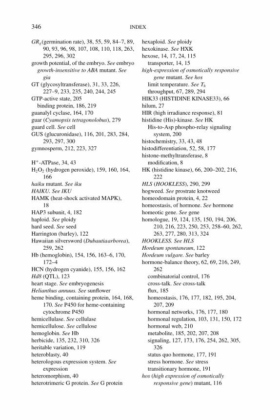
P1: PAE
BLUK053-Bradford September 22, 2006 21:0
346 INDEX
GRg(germination rate), 38, 55, 59, 84–7, 89,
90, 93, 96, 98, 107, 108, 110, 118, 263,
295, 296, 302
growth potential, of the embryo. See embryo
growth-insensitive to ABA mutant. Seegia
GT (glycosyltransferase), 31, 33, 226,
227–9, 233, 235, 240, 244, 245
GTP-active state, 205
binding protein, 186, 219
guanalyl cyclase, 164, 170
guar (Cyamopsis tetragonolobus), 279
guard cell. See cell
GUS (glucuronidase), 116, 201, 283, 284,
293, 297, 300
gymnosperm, 212, 223, 327
H+-ATPase, 34, 43
H2O2 (hydrogen peroxide), 159, 160, 164,
166
haiku mutant. See ikuHAIKU. See IKUHAMK (heat-shock activated MAPK),
18
HAP3 subunit, 4, 182
haploid. See ploidy
hard seed. See seed
Harrington (barley), 122
Hawaiian silversword (Dubautiaarborea),
259, 262
Hb (hemoglobin), 154, 156, 163–6, 170,
172–4
HCN (hydrogen cyanide), 155, 156, 162
Hd8 (QTL), 123
heart stage. See embryogenesis
Helianthus annuus. See sunflower
heme binding, containing protein, 164, 168,
170. See P450 for heme-containing
cytochrome P450
hemicellulase. See cellulase
hemicellulose. See cellulose
hemoglobin. See Hb
herbicide, 135, 232, 310, 326
heritable variation, 119
heteroblasty, 40
heterologous expression system. Seeexpression
heteromorphism, 40
heterotrimeric G protein. See G protein
hexaploid. See ploidy
hexokinase. See HXK
hexose, 14, 17, 24, 115
transporter, 14, 15
high-expression of osmotically responsivegene mutant. See hos
limit temperature. See Th
throughput, 67, 289, 294
HIK33 (HISTIDINE KINASE33), 66
hilum, 27
HIR (high irradiance response), 81
histidine (His)-kinase. See HK
His-to-Asp phospho-relay signaling
system, 200
histochemistry, 33, 43, 48
histodifferentiation, 52, 58, 177
histone-methyltransferase, 8
modification, 8
HK (histidine kinase), 66, 200–202, 216,
222
HLS (HOOKLESS), 290, 299
hogweed. See prostrate knotweed
homeodomain protein, 4, 22
homeostasis, of hormone. See hormone
homeotic gene. See gene
homologue, 19, 124, 135, 150, 194, 206,
210, 216, 223, 250, 253, 258–60, 262,
263, 277, 280, 313, 324
HOOKLESS. See HLSHordeum spontaneum, 122
Hordeum vulgare. See barley
hormone-balance theory, 62, 69, 216, 249,
262
combinatorial control, 176
cross-talk. See cross-talk
flux, 185
homeostasis, 176, 177, 182, 195, 204,
207, 209
hormonal networks, 176, 177, 180
hormonal regulation, 103, 131, 150, 172
hormonal web, 210
metabolite, 185, 202, 207, 208
signaling, 127, 173, 176, 254, 262, 305,
326
status quo hormone, 177, 191
stress hormone. See stress
transitionary hormone, 191
hos (high expression of osmoticallyresponsive gene) mutant, 116
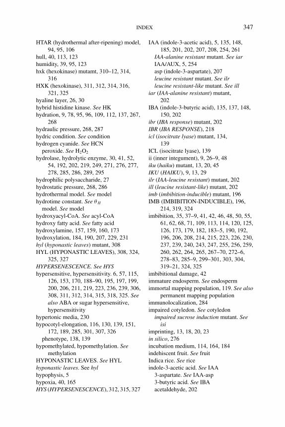
P1: PAE
BLUK053-Bradford September 22, 2006 21:0
INDEX 347
HTAR (hydrothermal after-ripening) model,
94, 95, 106
hull, 40, 113, 123
humidity, 39, 95, 123
hxk (hexokinase) mutamt, 310–12, 314,
316
HXK (hexokinase), 311, 312, 314, 316,
321, 325
hyaline layer, 26, 30
hybrid histidine kinase. See HK
hydration, 9, 78, 95, 96, 109, 112, 137, 267,
268
hydraulic pressure, 268, 287
hydric condition. See condition
hydrogen cyanide. See HCN
peroxide. See H2O2
hydrolase, hydrolytic enzyme, 30, 41, 52,
54, 192, 202, 219, 249, 271, 276, 277,
278, 285, 286, 289, 295
hydrophilic polysaccharide, 27
hydrostatic pressure, 268, 286
hydrothermal model. See model
hydrotime constant. See θ H
model. See model
hydroxyacyl-CoA. See acyl-CoA
hydroxy fatty acid. See fatty acid
hydroxylamine, 157, 159, 160, 173
hydroxylation, 184, 190, 207, 229, 231
hyl (hyponastic leaves) mutant, 308
HYL (HYPONASTIC LEAVES), 308, 324,
325, 327
HYPERSENESCENCE. See HYShypersensitive, hypersensitivity. 6, 57, 115,
126, 153, 170, 188–90, 195, 197, 199,
200, 206, 211, 219, 223, 236, 239, 306,
308, 311, 312, 314, 315, 318, 325. Seealso ABA or sugar hypersensitive,
hypersensitivity
hypertonic media, 230
hypocotyl-elongation, 116, 130, 139, 151,
172, 189, 285, 301, 307, 326
phenotype, 138, 139
hypomethylated, hypomethylation. Seemethylation
HYPONASTIC LEAVES. See HYL
hyponastic leaves. See hylhypophysis, 5
hypoxia, 40, 165
HYS (HYPERSENESCENCE), 312, 315, 327
IAA (indole-3-acetic acid), 5, 135, 148,
185, 201, 202, 207, 208, 254, 261
IAA-alanine resistant mutant. See iarIAA/AUX, 5, 254
asp (indole-3-aspartate), 207
leucine resistant mutant. See ilrleucine resistant-like mutant. See ill
iar (IAA-alanine resistant) mutant,
202
IBA (indole-3-butyric acid), 135, 137, 148,
150, 202
ibr (IBA response) mutant, 202
IBR (IBA RESPONSE), 218
icl (isocitrate lyase) mutant, 134,
139
ICL (isocitrate lyase), 139
ii (inner integument), 9, 26–9, 48
iku (haiku) mutant, 13, 20, 45
IKU (HAIKU), 9, 13, 29
ilr (IAA-leucine resistant) mutant, 202
ill (leucine resistant-like) mutant, 202
imb (imbibition-inducible) mutant, 196
IMB (IMBIBITION-INDUCIBLE), 196,
214, 319, 324
imbibition, 35, 37–9, 41, 42, 46, 48, 50, 55,
61, 62, 68, 71, 109, 113, 114, 120, 125,
126, 173, 179, 182, 183–5, 190, 192,
196, 206, 208, 214, 215, 223, 226, 230,
237, 239, 240, 243, 247, 255, 256, 259,
260, 262, 264, 265, 267–70, 272–6,
278–83, 285–9, 299–301, 303, 304,
319–21, 324, 325
imbibitional damage, 42
immature endosperm. See endosperm
immortal mapping population, 119. See alsopermanent mapping population
immunolocalization, 284
impaired cotyledon. See cotyledon
impaired sucrose induction mutant. Seeisi
imprinting, 13, 18, 20, 23
in silico, 276
incubation medium, 114, 164, 184
indehiscent fruit. See fruit
Indica rice. See rice
indole-3-acetic acid. See IAA
3-aspartate. See IAA-asp
3-butyric acid. See IBA
acetaldehyde, 202
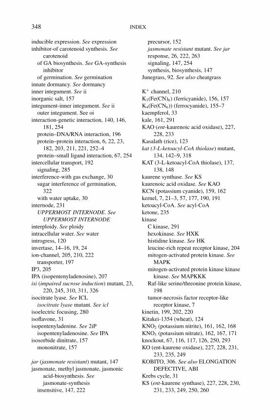
P1: PAE
BLUK053-Bradford September 22, 2006 21:0
348 INDEX
inducible expression. See expression
inhibitor-of carotenoid synthesis. Seecarotenoid
of GA biosynthesis. See GA-synthesis
inhibitor
of germination. See germination
innate dormancy. See dormancy
inner integument. See ii
inorganic salt, 157
integument-inner integument. See ii
outer integument. See oi
interaction-genetic interaction, 140, 146,
181, 254
protein–DNA/RNA interaction, 196
protein–protein interaction, 6, 22, 23,
182, 203, 211, 221, 252–4
protein–small ligand interaction, 67, 254
intercellular transport, 192
signaling, 285
interference-with gas exchange, 30
sugar interference of germination,
322
with water uptake, 30
internode, 231
UPPERMOST INTERNODE. SeeUPPERMOST INTERNODE
interploidy. See ploidy
intracellular water. See water
introgress, 120
invertase, 14–16, 19, 24
ion-channel, 205, 210, 222
transporter, 197
IP3, 205
IPA (isopentenyladenosine), 207
isi (impaired sucrose induction) mutant, 23,
220, 245, 310, 311, 326
isocitrate lyase. See ICL
isocitrate lyase mutant. See iclisoelectric focusing, 280
isoflavone, 31
isopentenyladenine. See 2iP
isopentenyladenosine. See IPA
isosorbide dinitrate, 157
mononitrate, 157
jar (jasmonate resistant) mutant, 147
jasmonate, methyl jasmonate, jasmonic
acid-biosynthesis. Seejasmonate-synthesis
insensitive, 147, 222
precursor, 152
jasmonate resistant mutant. See jarresponse, 26, 222, 263
signaling, 147, 254
synthesis, biosynthesis, 147
Junegrass, 92. See also cheatgrass
K+ channel, 210
K3(Fe(CN)6) (ferricyanide), 156, 157
K4(Fe(CN6)) (ferrocyanide), 155–7
kaempferol, 33
kale, 161, 291
KAO (ent-kaurenoic acid oxidase), 227,
228, 233
Kasalath (rice), 123
kat (3-L-ketoacyl-CoA thiolase) mutant,
134, 142–9, 318
KAT (3-L-ketoacyl-CoA thiolase), 137,
138, 148
kaurene synthase. See KS
kaurenoic acid oxidase. See KAO
KCN (potassium cyanide), 159, 162
kernel, 7, 21–3, 57, 177, 190, 191
ketoacyl-CoA. See acyl-CoA
ketone, 235
kinase
C kinase, 291
hexokinase. See HXK
histidine kinase. See HK
leucine-rich repeat receptor kinase, 204
mitogen-activated protein kinase. SeeMAPK
mitogen-activated protein kinase kinase
kinase. See MAPKKK
Raf-like serine/threonine protein kinase,
198
tumor-necrosis factor receptor-like
receptor kinase, 7
kinetin, 199, 202, 220
Kitakei-1354 (wheat), 124
KNO2 (potassium nitrite), 161, 162, 168
KNO3 (potassium nitrate), 162, 167, 171
knockout, 67, 116, 117, 126, 250, 293
KO (ent-kaurene oxidase), 227, 228, 231,
233, 235, 249
KOBITO, 306. See also ELONGATION
DEFECTIVE, ABI
Krebs cycle, 31
KS (ent-kaurene synthase), 227, 228, 230,
231, 233, 249, 250, 260
P1: PAE
BLUK053-Bradford September 22, 2006 21:0
INDEX 349
L1 cell layer. See cell
L-3-hydroxyacyl-CoA dehydrogenase, 137
LAC (laccase), 31, 33, 34, 38, 45, 47
lacs (long-chain acyl-CoA synthetase)
mutant, 134, 136, 144, 149
LACS (long-chain acyl-CoA synthetase),
136, 137, 142, 144
Lactuca sativa. See lettuce
Landsberg erecta. See LerLAR (leucoanthocyanidin reductase), 34,
48
LATE EMBRYOGENESIS ABUNDANT.
See LEA
late-flowering mutant, 10
lateral endosperm. See endosperm
root. See root
LCFA (long chain fatty acid), 136
LC-MS (liquid chromatography-tandem
mass spectrometry), 32, 33
LD (long day), 41
LDOX (leucoanthocyanidin dioxygenase),
31, 33, 34, 43
LEA (LATE EMBRYOGENESIS
ABUNDANT), 5, 57, 64, 191, 213, 237
leaf-canopy, 81, 107 168
expansion, 224
LeaGal (Lycopersicon esculentumα
-galactosidase), 271, 281
leakage, 30, 37, 40, 42, 60, 298
LeARA (Lycopersicon esculentuma-arabinofuranosidase), 270, 271, 283
LeARF (Lycopersicon esculentuma-arabinofuranosidase), 270, 283
LeCel (Lycopersicon esculentum cellulase),
271, 282
LeChi (Lycopersicon esculentum chitinase),
271
LeEXP (Lycopersicon esculentumExpansin), 270, 271, 276–8, 285, 288
LeGAI (Lycopersicon esculentum GAI), 259
LeGAS (Lycopersicon esculentumGA-stimulated transcript), 270
legume, 14, 18, 21, 46, 48, 108
Leguminosae, 31, 39, 303
LeMAN (Lycopersicon esculentummannanase), 271, 279–81, 285, 286
LeMside (Lycopersicon esculentummannosidase), 271, 282
Lepidium sativum. See cress
Lepidium virginicum, 159
Ler (Landsberg erecta), 10, 33, 34, 37, 115,
120, 128, 140, 193, 208, 239, 320,
321
LeSNF (Lycopersicon esculentumSUCROSE NON-FERMENTING), 321
Lespedeza, 37, 44
lettuce (Lactuca sativa), 61, 71, 86, 90, 103,
108, 110, 111, 159–61, 167, 169, 171,
174, 182, 185, 195, 199, 202, 213, 215,
218, 220, 222, 233, 266, 271, 272, 274,
276, 280, 281, 287, 289, 295, 297–9,
301–3
Leucine-Heptad Repeat. See LHR
leucine-rich repeat. See LRR
rich repeat receptor kinase. See kinase
leucoanthocyanidin dioxygenase. SeeLDOX
leucoanthocyanidin reductase. See LAR
LeXET (Lycopersicon esculentumxyloglucan endotransglycosylase),
271, 277, 278, 285
LeXPG (Lycopersicon esculentumexo-polygalacturonase), 271, 283
LeXYL (Lycopersicon esculentumb-D-xylosidase), 271, 283
LFR (low fluence response), 81, 90, 100,
101, 103, 105, 118, 300
LHR (Leucine Heptad Repeat), 254
ligand, 67, 165, 205, 206
light-filtration, 30
spectrum, 40
ultraviolet. See UV
lignan, 39, 44
lignified cell. See cell
lignin, 42, 44, 47, 48
lignostilbene-a,b-dioxygenase. See LSD
Linaceae, 9
lincomycin, 315
linear cotyledon stage. See embryogenesis
linkage map, 114, 119, 125
linolenic acid, 147
Linum usitatissimum, 42, 44
lipase-acid lipase, 135, 150
TAG lipase, 135
lipid-hydroperoxide, 148
body, 135, 150, 267, 293. See alsospherosome
breakdown, 135, 139, 144, 307, 318
mobilization, 134, 144, 150, 152, 307,
315, 318, 324, 325
P1: PAE
BLUK053-Bradford September 22, 2006 21:0
350 INDEX
lipid-hydroperoxide (Continued)
peroxide, 166
storage lipid, 138, 143–5, 149, 152, 307,
318, 325, 327
superoxide, 166
lipolysis, 134, 135
liquid chromatography-tandem mass
spectrometry. See LC-MS
loading-bearing, 278
locular tissue, 58
longevity, 19, 25, 41, 42, 44, 128, 130, 148,
152, 190, 216, 219, 243, 245, 297
long day. See LD
long-chain acyl-CoA synthetase. See LACS
long-chain acyl-CoA synthetase mutant.
See lacsdistance signal, 170
longitudinal section, 26, 266
los (low expressionof osmoticallyresponsive gene), 116
LOS (LOW EXPRESSION OFOSMOTICALLY RESPONSIVEGENE), 131, 132, 187, 222, 246
loss-of-function mutant, mutation, 5–7, 9,
120, 200, 225, 229, 231, 251, 326
low fluence response. See LFR
limit temperature. See Tl
LRR (leucine-rich repeat), 204, 254
LSD (lignostilbene-α,β-dioxygenase), 234
LsGA2ox2 (Lactuca sativa GA2ox2), 195
LsMan (Lactuca sativa Mannanase), 271,
280, 281
lupin (Lupinus luteus), 161, 173
Lycopersicon esculentum. See tomato
lysine (Lys), 19, 253
MADS box, domain, 21, 34, 43, 47, 238,
246, 299
maize (Zea mays), 3, 4, 6, 7, 16, 18–23, 25,
38, 40, 45, 57, 109, 118, 131, 172, 177,
179, 180, 187, 190, 191, 197, 212, 214,
218, 221–3, 234, 238, 243–7, 250, 252,
259, 263, 300, 313, 326
malate dehydrogenase. See MDH
malate synthase. See MLS
malate synthase mutant. See mlsmalting, 113, 122
Malus domestica. See apple
mangrove (Rhizophora mangle), 179, 221
mannan-degrading enzyme, 282
galactomannan, 267, 279–82, 301
oligo-mannan, 281
mannanase, 110, 249, 262, 263, 271,
278–82, 285, 296–303
mannitol, 186, 311, 316
mannopyranosyl linkage, 279
mannose, 279–81, 315, 316, 326
mannose-insensitive germination mutant.
See migmannosidase, 271, 279, 281, 282, 300
MAPK (mitogen-activated protein kinase),
66, 219, 308, 328
MAPKKK (mitogen-activated protein
kinase kinase kinase), 198
maple. See AcerMARD (MEDIATOR OF
ABA-REGULATED DORMACY), 116,
121, 188
marker-assisted breeding, 122
assisted selection, 125
molecular marker, 42, 47, 111, 125, 128,
130, 131
selection marker, 310
master regulator, of germination. Seegermination
MATE secondary transporter, 34, 44
maternal-genome, 1, 11, 12
ABA. See ABA
allele, 13
effect, 7, 13, 37, 45, 46, 74, 128, 299
genotype, 117
mRNA, 290
origin, 1, 3
tissue, 7, 11, 17, 26, 35, 51, 117, 178,
201, 236
maternalization, of endosperm. Seeendosperm
mathematical model. See model
matric potential, 265
maturation-drying, 177
phase, 3–6, 14–16, 28, 52, 178
seed maturation. See seed
mature-embryo. See embryo
green stage. See embryogenesis
maturity. See seed maturity
MAX (more axillary), 233, 242, 246
MCS (molybdenum cofactor sulfurase), 187
MDH (malate dehydrogenase), 139
P1: PAE
BLUK053-Bradford September 22, 2006 21:0
INDEX 351
mea (medea) mutant, 13, 23
MEA (MEDEA), 7, 8, 13, 20–24
mean temperature. See temperature
mechanical-damage, 42, 44, 146
dormancy. See dormancy
MEDIATOR OF ABA-REGULATEDDORMACY. See MARD
Medicago sativa. See alfalfa
Medicago truncatula, 34, 49
medium-chain fatty acid. See fatty acid
megagametophyte, 25, 47, 183, 184, 220,
274, 283, 297
melanic substance, 38
melon. See muskmelon
membrane-damage, 166
fluidity, 66, 70, 71
integrity, 268
intrinsic protein. See PIP, TIP
permeability, 66
phospholipid. See phospholipid
plasmamembrane, 267, 268
protein, 7, 21, 66, 197
receptor, 66
rigidification, 66, 69
unsaturation, 66
MEP (2-C-methyl-D-erythritol phosphate)
pathway, 224
meristem, 4, 5, 18, 21, 22, 26, 58, 138, 180,
201, 252, 265, 285, 286
meristematic L1 cell layer. See cell-L1 cell
Mesembryanthemum crystallinum. Seecommon ice plant
MET (METHYLTRANSFERASE), 8, 12,
19
metabolic-flux, 184
quiescence, 3
switch, 237, 238
metabolism-ABA metabolism. See ABA
carbon metabolism. See carbon
flavonoid metabolism. See flavonoid
proanthocyanidin metabolism. See 2PA
metabolizable sugar. See sugar
metabolome, 32
metal-containing protein, 164
ion transporter, 197
metHb (methemoglubin), 165
MetHb-R (methemoglobin reductase), 165
methyl group, 12, 229
methylamine, 157
methylation-hypomethylated,
hypomethylation, 12, 13, 17, 19, 23, 295
methyltransferase. See MET
methylene blue, 157, 159
methyl-jasmonate. See jasmonate
METHYLTRANSFERASE. See MET
mevalonate pathway, 224, 243
mfp (multifunctional protein) mutant, 134,
138
MFP (multifunctional protein), 137, 138,
149, 150, 152
microarray, 20, 127, 202, 203, 208, 211,
214, 225–7, 241, 272, 278, 289–92,
294, 298, 309, 314, 325
microbial activity, 169
microclimate, 74, 82, 83
microimaging, 267
micropylar-endosperm. See endosperm
pole, 2
region, 27, 119, 267, 274, 293
specific expression. See expression
micropyle, 26, 27, 32, 34
microRNA (miRNA). See RNA
microsomal fraction, 234
middle lamella, 283
mig (mannose-insensitive germination)
mutant, 316
mineral ion, 272
minimal promoter, 293
minimum (or base) temperature. See Tb
miRNA (microRNA). See RNA
misexpression. See expression
mitochondria, 133, 134, 149, 163, 164, 170,
173–5, 265
mitogen-activated protein kinase. SeeMAPK
activated protein kinase kinase kinase.
See MAPKKK
Mls (malate synthase) mutant, 139, 144
MLS (malate synthase), 134, 139, 144, 150
mobilze, mobilization, 116, 130, 133, 134,
144, 147, 150–52, 192, 249, 250, 264,
265, 266, 268, 277, 279, 281, 287, 288,
301, 303, 306, 307, 309, 315, 316, 318,
324–7
MoCo (molybdenum cofactor), 131, 178,
187, 196, 212, 222, 226–9, 242, 246,
310
sulfurase, 226, 228, 229
P1: PAE
BLUK053-Bradford September 22, 2006 21:0
352 INDEX
model-dormancy model. See dormancy
emergence model, 83
hydrothermal after-ripening model. SeeHTAR
hydrothermal model, 89
hydrotime model, 87–90, 92, 93, 102
mathematical model, 84, 105, 107, 111
physiologically based model, 83
population-based model,
population-based threshold model, 73,
90, 100, 107, 109
predictive model, 72, 83, 84, 97, 107
stratification thermal time model, 98
TAR (thermal after-ripening time) model.
See TAR
temperature response model, 83
VOP (virtual osmotic potential) model.
See VOP
moist chilling. See chilling
molecular genetics, 25, 32, 43, 70, 149, 218,
236, 241
oxygen. See oxygen
probe, 156
molsidomine, 161
molybdenum-cofactor. See MoCo
cofactor sulfrase. See MCS
Moneymaker (tomato), 58, 69, 275, 298,
303
monocot, monocotyledonous, 26, 165
monooxygenase. See P450
MONOPTEROS, 5, 18, 20
monosaccharide/proton symporter, 316,
327
Morex (barley), 122, 131
morphogenesis, 3–5, 9, 14, 15, 18, 110,
129, 197, 204, 218, 294, 301
morpho-physiological dormancy. Seedormancy
mother plant, 10, 11, 14, 37, 41, 73, 114,
126, 250
MPa, 87, 88, 92–5, 240, 270, 287
mRNA cap binding protein. See RNA
MS (Murashige and Skoog) media, 140,
143, 317, 320, 321
Msgb (Medicago sativa Gb-like1), 291
MSI (MULTICOPY SUPRESSOR OF IRA),
7, 8, 21
mucilage. See testa
MUCILAGE MODIFIED. See MUM
multi-functional protein. See MFP
nucleated cell. See cell
subunit protein, 253
molecular binding site (for water),
265
MUM (MUCILAGE MODIFIED), 10
mungbean (Vigna radiata), 109
muskmelon (Cucumis melo), 40, 274, 285,
303, 304
mutagenesis, 22
MYB, 10, 23, 34, 40, 45, 47, 124, 150,
172, 206, 261, 280, 290, 295, 298,
300
mycoheterotrophs, 52
myoglobin, 164
N fertilizer, 169
NaCl, 38, 186, 189, 308
NAD, 1NADH, NADP, NADPH,63,138,
148, 149, 151, 158, 159, 165, 166,
1732NADH (nitrate reductase), 63, 65, 69, 118,
162, 163, 165, 172–4. See also NR
NADPH oxidase, 148, 151
NaN3 (sodium azide), 162
native species. See species
natural resistance-associated macrophage
protein. See Nramp
variation. See dormancy
nced (9-cis-epoxycarotenoid dioxygenase),
230, 245
NCED (nine-cis-epoxycarotenoid
dioxygenase), 61, 119, 121, 178, 187,
189, 210, 217, 225–8, 230, 233, 234,
242–4
near-isogenic line. See NIL
necrotrophic pathogen. See pathogen
negative feedback, 186, 191
NeoX (neoxanthin), 207, 225, 226, 233
nia (nitrate reductase) mutant, 63, 118
Nicotiana attenuata, 171, 184, 220
Nicotiana plumbaginifolia, 59, 68, 161,
173, 178, 187, 211, 212, 214, 215, 217,
218, 232, 243
Nicotiana tabacum. See tobacco
Nikolaeva, 50, 70
NIL (near-isogenic lines), 120
NiNOR (nitrite-nitric oxide reductase),
163
Nipponbare (rice), 123
NiR (nitrite reductase), 163
P1: PAE
BLUK053-Bradford September 22, 2006 21:0
INDEX 353
nitrate-receptor, 65, 66
reductase. See NR
nitrate reductase mutant. See niatransporter. See NRT
nitric oxide. See NO
nitric oxide synthase. See NOS
nitrite-nitric oxide reductase. See NiNOR
reductase. See NiR
nitrogylcerine, 167
nitrosated protein, 170
nitrosylated protein, 170
nitrous acid, 154
NMR (nuclear magnetic resonance), 32, 35,
46, 267, 268, 300
NO (nitric oxide)-atmospheric NOx, 168
availability, 171
diazeniumdiolates containing the
[N(O)NO]− functional group. SeeNONOates
diethylamine nitric oxide. See DEANO
donor, 154–7, 160–162, 166, 167, 171,
172
ecological significance, 168
electrode, 157
ferrochelatase, 159
nitrite-nitric oxide reductase. See NiNOR
reactive fluorescent probe, 156
scavenger, 156, 160, 162
smoke and NO, 169
soil NO, 168
source of NO, 163, 166, 168
synthesis, 156, 163, 170, 171
NO2 (nitrogen dioxide), 153, 154, 157, 163,
169, 173
NO−2 (nitrite), 153, 154, 156, 163, 164–6,
168, 171
NO−3 (nitrate), 154, 163–6, 168, 171
Nomarski (microscope) image, 2
non-endospermic seed. See seed
NONOates (diazeniumdiolates containing
the [N(O)NO]− functional group), 155
non-symbiotic hemoglobins. See nsHb
norbornanodiazetine-type P450 inhibitor,
234
nordihydroguaiaretic acid, 234
norflurazon, 37, 232, 233, 239
Northern blot analysis, 255
nos (nitric oxide synthase) mutant, 163
NOS (nitric oxide synthase), 163, 164, 170,
172, 173
NOx, 168, 169
Npaba (Nicotiana plumbaginifolia aba),
187, 196,
Nppew (Nicotiana plumbaginifoliapartly etiolated in white light) mutant,
196
Nr (Never-ripe) mutant, 198
NR (nitrate reductase), 162–5. See also2NADH
Nramp (natural resistance-associated
macrophage protein), 197
NRT (nitrate transporter), 66
nsHb (non-symbiotic hemoglobin), 165,
166, 174
N-terminus, 253, 255
N-tert-butyl-alpha-phenylnitrone. See PBN
nucellar-cell. See cell
tissue, 274
nucellus, 1, 25, 26, 28, 53
nuclear division, 2, 6
nuclear magnetic resonance. See NMR
nucleic acid, 158
nutrient, 1, 7, 11, 128, 133, 168, 170, 190,
249, 250, 307, 309, 314, 323–5
O2 (oxygen) sensor, 165
transport, 165
oat (Avena fatua), 60, 70, 90, 109, 110,
122, 124, 128, 149, 186, 216, 219,
298
oi (outer integument), 8, 9, 24, 26, 27, 29,
35, 48
oleosin, 4, 67, 151
oleosome,135, 160. See also lipid body
oligo-mannan. See mannan
onset of dormancy. See dormancy
OPDA (12-oxo-phytodienoic acid),
147
optimum temperature. See To
Orchidaceae, 52, 71
organelle, 133, 135, 230
organic acid, 164
Orobanchaceae, 52
Orobanche minor, 204, 221
Oryza rufipogon, 123
Oryza sativa. See rice
OsABI (Oryza sativa ABA
INSENSITIVE), 313
OsKS (Oryza sativa ent-kaurene synthase),
250
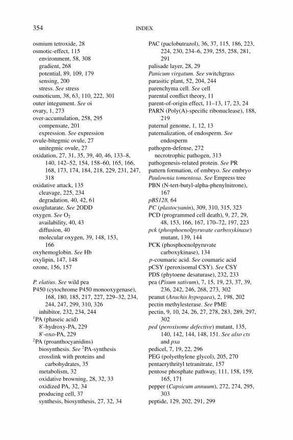
P1: PAE
BLUK053-Bradford September 22, 2006 21:0
354 INDEX
osmium tetroxide, 28
osmotic-effect, 115
environment, 58, 308
gradient, 268
potential, 89, 109, 179
sensing, 200
stress. See stress
osmoticum, 38, 63, 110, 222, 301
outer integument. See oi
ovary, 1, 273
over-accumulation, 258, 295
compensate, 201
expression. See expression
ovule-bitegmic ovule, 27
unitegmic ovule, 27
oxidation, 27, 31, 35, 39, 40, 46, 133–8,
140, 142–52, 154, 158–60, 165, 166,
168, 173, 174, 184, 218, 229, 231, 247,
318
oxidative attack, 135
cleavage, 225, 234
degradation, 40, 42, 61
oxoglutarate. See 2ODD
oxygen. See O2
availability, 40, 43
diffusion, 40
molecular oxygen, 39, 148, 153,
166
oxyhemoglobin. See Hb
oxylipin, 147, 148
ozone, 156, 157
P. elatius. See wild pea
P450 (cytochrome P450 monooxygenase),
168, 180, 185, 217, 227, 229–32, 234,
244, 247, 299, 310, 326
inhibitor, 232, 234, 2441PA (phaseic acid)
8′-hydroxy-PA, 229
8′-oxo-PA, 2292PA (proanthocyanidins)
biosynthesis. See 2PA-synthesis
crosslink with proteins and
carbohydrates, 35
metabolism, 32
oxidative browning, 28, 32, 33
oxidized PA, 32, 34
producing cell, 37
synthesis, biosynthesis, 27, 32, 34
PAC (paclobutrazol), 36, 37, 115, 186, 223,
224, 230, 234–6, 239, 255, 258, 281,
291
palisade layer, 28, 29
Panicum virgatum. See switchgrass
parasitic plant, 52, 204, 244
parenchyma cell. See cell
parental conflict theory, 11
parent-of-origin effect, 11–13, 17, 23, 24
PARN (Poly(A)-specific ribonuclease), 188,
219
paternal genome, 1, 12, 13
paternalization, of endosperm. Seeendosperm
pathogen-defense, 272
necrotrophic pathogen, 313
pathogenesis-related protein. See PR
pattern formation, of embryo. See embryo
Paulownia tomentosa. See Empress tree
PBN (N-tert-butyl-alpha-phenylnitrone),
167
pBS128, 64
PC (plastocyanin), 309, 310, 315, 323
PCD (programmed cell death), 9, 27, 29,
48, 153, 166, 167, 170–72, 197, 223
pck (phosphoenolpyruvate carboxykinase)
mutant, 139, 144
PCK (phosphoenolpyruvate
carboxykinase), 134
p-coumaric acid. See coumaric acid
pCSY (peroxisomal CSY). See CSY
PDS (phytoene desaturase), 232, 233
pea (Pisum sativum), 7, 15, 19, 23, 37, 39,
236, 242, 246, 268, 273, 302
peanut (Arachis hypogaea), 2, 198, 202
pectin methylesterase. See PME
pectin, 9, 10, 24, 26, 27, 278, 283, 289, 297,
302
ped (peroxisome defective) mutant, 135,
140, 142, 144, 148, 151. See also ctsand pxa
pedicel, 7, 19, 22, 296
PEG (polyethylene glycol), 205, 270
pentaerythrityl tetranitrate, 157
pentose phosphate pathway, 111, 158, 159,
165, 171
pepper (Capsicum annuum), 272, 274, 295,
303
peptide, 129, 202, 291, 299
P1: PAE
BLUK053-Bradford September 22, 2006 21:0
INDEX 355
peptone, 306
PER (peroxiredoxin), 64, 67, 69
pericarp, 26, 40, 53, 123, 124, 164, 269,
272, 273
periodic acid-Schiff, 2
peripheral endosperm. See endosperm
perisperm, 3, 26, 51, 53, 303
permanent mapping population, 119
peroxidase, 38, 39, 42, 45, 148, 163–6, 170,
174, 175
peroxidation, 147, 152
peroxiredoxin. See Prx
peroxisomal-ABC protein. See Pxa
peroxisomal ABC protein mutant. See pxaacyl-CoA synthetase. See ACS
acyl-CoA transporter. See acyl-CoA
ascorbate peroxidase. See ascorbate
peroxidase
lumen, 137
membrane, 135
peroxisome, 133–6, 139, 140, 142, 145,
147–52, 160, 172
peroxisome defective mutant. See pedperoxynitrite, 166
Petunia, 27
pew. See NppewPfr, 30, 56, 81, 100, 101, 103, 105, 203
PG (polygalacturonase), 271, 283, 285, 298,
302
phase transition, 8, 70, 194, 209, 218, 244,
305
phaseic acid. See 1PA
Phaseolus vulgaris. See bean
PHE (PHERES), 8, 21
phenolic compounds, phenolics, 29, 30, 39,
45–9, 52, 53
phenology of dormancy. See dormancy
phenotype, 6–8, 13, 23, 32, 37, 38, 49, 57,
58, 67, 115–20, 125–7, 135, 136,
138–48, 181, 182, 190, 191, 194,
197–200, 202, 204, 210, 223, 229–31,
239, 240, 249–53, 257, 258, 260, 308,
311, 312, 314, 315, 318, 322, 326,
327
phenylpropanoid pathway, 31
phlobaphenes, 31, 40, 124
phloem, 14, 272
PHOR (PHOTOPERIOD-RESPONSIVE),
258, 260, 280, 295
phosphatase, 61, 186, 188, 210, 215, 217,
218, 220, 221, 223, 306, 325
protein phosphatase type-2C. See PP2C
phosphate, 24, 111, 158, 159, 165, 171,
192, 200, 224, 227, 230, 233, 261, 312,
313, 315, 324, 326
phosphatidic acid, 205
phosphoenolpyruvate carboxykinase. SeePCK
phosphoenolpyruvate carboxykinasemutant. See pck
phosphoinositide, 186
phospholipase C, 205
D, 205
phospholipid, 66, 186
phosphoric acid, 172
phosphorylation, 61, 171, 175, 182, 216,
252, 257, 291, 311
phosphotransfer domain protein, 200
phosphotyrosine signaling, 255
photodormancy. See dormancy
photo-lysis, 155
morphogenesis, 110, 197, 204
oxidative damage, 233
periodic plants, 41
PHOTOPERIOD-RESPONSIVE. SeePHOR
receptor, 30, 70, 81, 118, 167, 195, 221
respiratory enzyme, 139
reversible regulation, of germination, 65
synthesis, 130, 133, 309, 325
PHS (pre-harvest sprouting), 39, 40, 113,
122–4, 128–30, 186, 212, 215, 298
Phs (Pre-Harvest Sprouting), 40
PHS (QTL), 124
PHY (PHYTOCHROME), 30, 65, 68, 69,
81, 90, 106, 108, 118, 121, 131, 167,
171, 195, 196, 216, 221, 240, 246, 280
phy (phytochrome-deficient) mutant, 117,
118, 132, 247. See also Nppewphytochrome-ABA, 171, 196, 217
action, 40, 193, 195, 196
control, 130, 173, 218, 302
deficient. See phydependent, 295
ethylene, 220
GA, 71, 195, 196, 222, 277
hypocotyl growth, 109
induced, 297, 298, 300, 302
P1: PAE
BLUK053-Bradford September 22, 2006 21:0
356 INDEX
phytochrome-ABA (Continued)
interacting protein, 219, 326. See alsoPIF, PIL
mediated, 81, 117, 196
NO, 167, 170
regulation, 49, 71, 195, 223, 245, 247,
263
physiological heterogeneity, 40, 41
physiologically based model. See model
PHYTOCHROME. See PHY
phytoene desaturase. See PDS
Picea glauca. See spruce
PID (PINOID), 5, 23
PIF (PHYTOCHRME INTERACTING
FACTOR), 196, 221, 319
pigment-brown pigment, 27
less seed. See seed
pigmentation of testa. See testa
strand. See ps
pigmentation mutants, 32
PIL (PHYTOCHROME INTERACTING
FACTOR3-LIKE), 193, 195, 196, 219,
319, 326
pine-mugo pine (Pinus mugo), 161
Scots pine (Pinus sylvestris), 183, 202,
217, 222, 267
western white pine (Pinus monticola), 35,
48, 184, 214, 303
Pinus sylvestris. See Scots pine
PIP (Plasmamembrane Intrinsic Protein),
268, 302
Pisum sativum. See pea
pKa,154
pkl (pickle), 194, 195, 220
PKL (PICKLE), 194, 195, 214, 216, 219,
220
plant growth regulator, 234, 243
plant hormone, 18, 52, 53, 70, 119, 125,
126, 171, 174, 207, 211, 213, 214, 216,
217, 219, 222–4, 242–4, 247–9, 254,
260, 295, 305, 316, 323
Plantaginaceae, 9
plantlet, 330, 308
plasmamembrane. See membrane
plasmamembrane Intrinsic Protein. See PIP
plasmodesmata, 14, 285, 302
plastid, 3, 163, 224–6, 229, 230, 312, 315,
325
plastocyanin. See PC
plateau phase, of water uptake, 265
PLEIOTROPIC RESPONSE LOCUS. SeePRL
pleiotropic, pleiotropism, 115, 119, 120,
123, 127, 221, 263, 312, 315, 326,
327
ploidy-triploid, 1, 20, 28
diploid, 1, 11, 12, 28, 53
haploid, 1, 122, 273
hexaploid, 123, 259
interploidy, 11, 12, 13
tetraploid, 11
PME (pectin methylesterase), 278, 283,
289, 297
polar nuclei, 12
polarity, of embryo. See embryo
pollen, 2, 13, 27, 147, 203, 209, 221, 246,
264
pollination, 10–12, 178
poly(A)-specific ribonuclease. See PARN
Polycomb, group, gene, 8, 19–24
polyethyleneglycol. See PEG
polygalacturonase. See PG
Polygenic inheritance, 113
Polygonum aviculare. See prostrate
knotweed
polymer, 32, 33, 47, 278, 279, 283, 285,
292, 297
polymerization, 32, 47
polyphenol oxidase, 33, 38, 39, 42
polysaccharide, 27, 45, 267, 273, 278, 279,
301
polyubiquitinate, polyubiquitination. Seeubiquitin
population-based model, population-based
threshold model. See model
post-abscission. See abscission
fire environment, 184
germination. See germination
germinative expression. See expression
translational modification, 67, 181, 186,
256, 257
potassium cyanide. See KCN
ferricyanide. See K3(Fe(CN)6)
ferrocyanide. See K4(FeCN)6)
nitrate. See KNO3
nitrite. See KNO2
potato (Solanum tuberosum), 88, 91–4, 106,
111, 139, 232, 242, 258, 280
P1: PAE
BLUK053-Bradford September 22, 2006 21:0
INDEX 357
PP2C (protein phosphatase type-2C), 61,
68, 186, 188, 210, 215, 217, 218, 220,
221, 223, 325
PR (pathogenesis-related) protein, 283, 284,
303
Pr, 81. See also phytochrome
pre-chilling. See chilling
precocious germination. See germination
precursor, 5, 9, 37, 152, 197, 202, 207,
224–6, 230, 231, 233, 244, 314
predation, 54
pre-existing transcript. See transcript
preglobular stage. See embryogenesis
Pre-Harvest Sprouting. See Phspreharvest sprouting. See PHS
premature drying, 38
pressure potential. See ψ p
primary cell wall. See cell
dormancy. See dormancy
PRL (PLEIOTROPIC RESPONSE LOCUS),
312, 315, 326
Proanthocyanidin, condensed tannin. See2PA
probit analysis, regression analysis, 84, 87
programmed cell death. See PCD
proherbicide R7402, 310
prohexadione, 233, 235
prokaryote, 305
proline-tryptophan-tryptophan-proline
protein. See PWWP
promoter-reporter, 34, 35, 201, 293, 313
activity, 33, 34
Cat1 promoter, 284
trap, 116, 150
propionate, 145
prostrate knotweed (Polygonum aviculare),
76, 96, 97, 99, 101, 102, 107, 110
proteasome (26S proteasome), 117, 189,
190, 196, 221, 222, 248, 251–4, 261–3,
291, 308, 314, 327
protein body, 266, 303
phosphatase type-2C. See PP2C
storage protein, 4, 16, 21, 22, 57, 130,
180–182, 191, 215, 216, 219, 223, 237,
245, 306, 307
proteolysis, 248, 253, 254, 256, 260, 261,
324
proteolytic processing, cleavage,
destruction, 253
proteome, proteomics, 47, 67, 125, 127,
128, 208, 211, 272, 289, 291, 292, 297,
298, 301
proton NMR. See NMR
protuberance, 274
Provasculature, 192
Prx (peroxiredoxin), 64, 67, 69
ps (pigment strand), 26, 28, 49
Pseudotsuga menziesii. See Douglas fir
psychrometer, 287
PTIO (2-phenyl-4,4,5,5,-
tetramethylimidazoline-1-oxyl
3-oxide), 156, 160, 162, 166, 167, 169,
173, 174
public database. See database
pumpkin (Cucurbita moschata), 127, 139,
151
puroindoline-like (AV1) protein, 60
PWWP (proline-tryptophan-tryptophan-
proline) protein, 294
Pxa (peroxisomal ABC [ATP-binding
cassette] protein), 135, 136, 152
pxa (peroxisomal ABC [ATP-bindingcassette] protein) mutant, 135, 140,
144, 148, 152. See also cts and ped
Q-3-O-R (quercetin-3-O-rhamnoside),
31–3
QTL (quantitative trait loci), 10, 37, 40, 67,
119, 120–25, 127–31
quadruple-DELLA (gai, rga, rgl1, rgl2)
mutant, 194
quantitative genetics, 119, 176
quercetin rhamnoside. See Q-3-O-R
quiescence, 3, 23, 55, 58, 177, 179, 197, 206
1 R (Red grain color), 40, 45, 1242R (red light), 30, 40, 41, 62, 64, 65, 69, 71,
74, 81, 109, 161, 162, 167, 183, 195,
196, 203, 214, 221, 240, 269, 271, 275,
276, 280, 282, 288
R2R3-MYB protein. See MYB
R7402. See proherbicie
radial-domain, in embryo embryogenesis, 3,
5
cell. See cell
cell wall. See cell
patterning of vascular tissues, 201
transport of ABA, 245
P1: PAE
BLUK053-Bradford September 22, 2006 21:0
358 INDEX
radiation damage, 40
radicle-emergence, 87, 103, 240, 255, 256,
264–74, 276–8, 280, 281, 283, 284–8,
293–5, 297, 303–8, 319, 322, 324
protrusion, 29, 30, 37, 38–41, 45, 52–4,
71, 114, 116, 125, 183, 192, 198, 199,
273, 274, 277, 292, 297, 298, 300, 302
sized probe, 274
tip, 263, 267, 269, 271, 274, 283, 284,
289, 293, 302
radiotracer experiment, 234
Raf-like serine/threonine protein kinase. Seekinase
rainfall, 78, 83, 96, 169
rapeseed (Brassica napus), 26, 29, 39, 42,
47
RBCL (ribulose-1,5-bisphosphate
carboxylase large subunit), 315
RBCS (ribulose-1,5-bisphosphate
carboxylase small subunit), 211, 313,
322
RBR (RETINOBLASTOMA RELATEDPROTEIN), 7, 8
Rbx, 251, 253, 263
Rc (red pericarp), 40, 45, 123
rdo (reduced dormancy) mutant, 115, 318
RDO (REDUCED DORMANCY), 319
reactive oxygen species. See ROS
reciprocal cross, 35, 36, 178
recombinant-inbred line. See RIL
recombinant protein, 34
red pericarp. See Rcred rice. See rice
redox, 148–50, 154
reduced dormancy. See dormancy
reduced dormancy. See rdoREDUCED DORMANCY. See RDOreduced grain filling mutant. See rgfreductant, 164
reductase-anthocyanidin reductase. SeeANR
dihydroflavonol reductase. See DFR
leucoanthocyanidin reductase. See LAR
methemoglobin reductase. See MetHb-R
nitrite-nitric oxide reductase. See NiNOR
nitrate reductase. See 2NADH and NR
nitrite reductase. See NiR
Short Chain Dehydrogenase/Reductase.
See SDR
xanthine oxidoreductase. See xanthine
oxidoreductase
reduction, 5, 6, 7, 12, 13, 16, 33, 38, 39, 57,
60, 61, 81, 89, 92, 111, 147, 159, 164,
165, 168, 174, 183, 185, 191, 233, 234,
263, 269, 286
redundant, redundancy, 116, 117, 136, 178,
179, 198, 210, 232, 292, 300
regulators of g-protein signaling mutant.
See rgsREGULATORS of G-PROTEIN
SIGNALING. See RGS
relative dormancy. See dormancy
reporter gene, 116, 283, 293, 309, 310
repression, 4, 181, 191, 194, 205, 220, 237,
238, 248, 249, 251–3, 255, 257,
258–61, 263, 295, 313, 316, 318
repressor, 117, 126, 186, 194, 211, 243,
248, 252, 254, 260–63
repressor of ga1-3. See rgaREPRESSOR OF ga1-3-LIKE. See RGL
reserve-accumulation, 28, 180, 236, 238
deposition, 180, 201
mobilization, 116, 130, 133, 134, 147,
151, 192, 249, 250, 264–6, 268, 277,
279, 281, 287, 288, 301, 326
resource allocation, 309
respiration, 39, 112, 133, 134, 138, 142,
145, 152, 159, 164, 174, 199, 265
response-mutant, 152, 177, 180, 194, 202,
221, 261, 263, 305, 309, 322, 327
response regulator. See RR
resurrection plant, 315
retinoblastoma binding protein, 8
reverse genetics, 32, 67, 138
revertant, 115, 129, 217, 258
rga (repressor of ga1-3), 194, 252
RGA (REPRESSOR OF GA1-3), 117, 194,
248, 251, 252, 259, 261–3, 319, 325
rgf (reduced grain filling) mutant, 7, 22
rgl (repressor of ga1-3-like) mutant, 194,
259, 260
RGL (REPRESSOR OF ga1-3-LIKE), 117,
121, 126, 194, 248, 251–3, 255–62,
319, 325
rgs (regulators of g-protein signaling)
mutant, 189, 206
RGS (REGULATORS of G-PROTEIN
SIGNALING), 189, 205, 206, 213
P1: PAE
BLUK053-Bradford September 22, 2006 21:0
INDEX 359
rhamnogalacturonan, 9
ribose, 158, 172
ribulose-1,5-bisphosphate carboxylase large
subunit. See RBCL
ribulose-1,5-bisphosphate carboxylase
small subunit. See RBCS
rice (Oryza sativa), 3, 21, 25, 31, 39, 40, 45,
47, 110, 119, 120, 122, 123, 125, 127,
129, 130, 159, 161, 165, 169, 172, 174,
204, 213, 223, 231, 247, 248, 250,
252–4, 259, 261, 262, 268, 276, 294,
299, 303, 313
Ricinus communis. See castor bean
RIL (recombinant inbred line), 120, 123,
128
ring-finger protein, 253
riverbank grape (Vitis riparia), 39, 49
RNA-double-stranded (ds) RNA binding
protein, 324
miRNA (microRNA), 294–6, 300–302,
308, 324, 327
mRNA cap binding protein, 306, 324
polymerase, 292
processing, 186
RNAi, 64, 286
stored mRNA, 70, 244, 265, 290, 291,
300
root-apical meristem, 4, 5, 201
cap, 297
lateral root, 189, 202, 212, 216,
313
shoot-root axis, 3, 201
tip, 185
rop (rop GTPase), 189
ROP (Rop GTPase), 189, 2171ROS (reactive oxygen species), 64, 148,
150, 166, 172, 1732ROS (rest of seed), 271
Rosidae, 27
rpn10 (rp[regulatory particle]non-ATPase
[RPN] subunit [of 26S proteasome])
mutant, 189, 308
RR (response regulator), 200, 203, 215,
218, 221. See also ARR
Rubiaceae, 274
rudimentary embryo. See embryo
RY-/Sph. seed-specific motif, 65
RY elements, motif, repeat, 191, 196,
237
Saccharomyces cerevisiae. See yeast
SAD (Sm-like SnRNP protein), 189
salicylic acid, 189, 208
salinity, 38, 173, 224
salt, 35, 157, 158, 173, 187, 188, 245, 249,
306, 313, 326
SAMK (stress-activated MAPK), 66
san (salt insensitive) mutant, 187, 188
saturated fatty acid. See fatty acid
S-box, in promoter, 313
scar, 27
scarification, 53, 265
SCF (Skp1, Cullin and F-box subunits),
151, 214, 248, 251–4, 256, 258, 261–3,
324. See also ubiquitin
schizocarp, 273
schleperless mutant. See slpScots pine (Pinus sylvestris), 183, 202, 217,
222, 267
SCR (SCARECROW), 5, 194, 248
SD (short day), 41
SD (QTL), 122–4
sdr (QTL), 1231SDR (Short Chain
Dehydrogenase/Reductase), 178, 187,
225, 226, 229, 2302SDR (red light to interrupt the long night
under short-day conditions), 41
sea urchin, 290, 296
seasonal change, 53, 77, 82, 83, 107, 112
seasonal variation, in dormancy. Seedormancy
secondary-cell wall. See cell
dormancy. See dormancy
embryo. See embryo
messenger, 62, 186
metabolite, 31, 63
seed-abortion, 13, 177, 190, 236
anatomy, 25, 26, 48, 266, 297
bank, 55, 63, 66, 72, 73, 76, 78, 81–3, 96,
98, 100, 105, 107, 169
botanical potato seed. See true potato
seed
buried seed, 80, 82, 83, 107, 111, 139
carbohydrate. See carbohydrate
coat, 1, 11, 14, 15–9, 22–5, 29, 32, 35,
37, 39–49, 52, 53, 74, 235, 236, 249,
258–60, 264, 265, 268, 295, 299, 300,
302, 303. See also testa
P1: PAE
BLUK053-Bradford September 22, 2006 21:0
360 INDEX
seed-abortion (Continued)
color, 28, 32, 39, 41, 118
coverings, 31, 273, 302
decay, 73, 285
desiccation, 3, 5, 27, 28, 38, 39, 57, 58,
64, 78, 112, 124, 132, 140, 176,
177–82, 190, 201, 216, 219, 236–8,
245, 249, 273, 303
detachment, 27
dispersal, 1, 8, 50, 53, 54, 273
dry seed, 29, 39, 60, 75, 70, 78, 110, 178,
179, 197, 204, 226, 227, 230, 239, 240,
255, 259, 264, 267, 268, 290, 291
endospermic seed, 2, 7, 11, 26, 274
envelop, 25, 37, 42
germination. See germination
Seed-GUS-Expression library, 293
hard seed, 39, 265
horticultural seed, 79
hydrated seed, 70, 80, 81, 110, 267, 299
imbibition. See imbibition
lipid. See lipid
longevity. See longevity
mass, 1, 8, 10, 11, 13, 15–17, 21, 22. Seealso seed size
maturation, 19, 20, 22, 23, 37, 45, 58, 61,
110, 113, 114, 116, 125, 126, 133, 177,
180, 181, 186, 192, 213, 218, 236, 237,
243, 244, 302
maturity, 26, 27, 180, 273, 286
moisture content. See seed water content
morphology, 50, 250
non-endospermic seed, 2, 7, 11, 26
number, 10, 11, 16
pigment-less seed, 37
population, 72, 73, 75–7, 80, 83–6, 88,
89, 91–8, 100–103, 107, 111, 206, 208,
288, 300
protein. See protein
quality, 41, 42, 264
reserve, 41, 106, 133, 145, 194, 237, 238
seedless fruit, 292, 299
shape, 46, 70, 151
shattering, 123, 127
size, 11, 12, 15, 16, 18, 20, 24, 29, 45, 53,
201, 202, 220
storage carbohydrate. See carbohydrate
storage condition, 114, 322
storage lipid. See lipid
storage protein. See protein
storage reserve. See seed reserve
true (or botanical) potato seed, 88, 93, 94,
111
viability, 1, 13, 47, 298
viable seed, 13, 29, 54, 105, 113, 265, 268
vigor (vigour), 41, 46, 78, 113
water, moisture, content, 35, 41, 57, 88,
265, 267, 268, 272
zone microclimate, 74, 82
zone temperature. See temperature
zone water potential. See ψ
seedling-death, 78
seedling defective mutant, 4
development, 4, 133, 150, 153, 305, 308,
311, 313, 314, 322, 325
developmental arrest, 177, 310, 311, 316,
322, 325
emergence, 56, 63, 72–5, 82–4, 105, 109
establishment, 10, 25, 72, 78–80, 82, 96,
113, 114, 127, 135, 137–40, 142, 144,
145, 148, 152, 209, 250, 256, 259, 305,
308, 326
survival, 73, 308
selection, 50, 108, 115, 122, 125, 128, 129,
217, 310
semi-arid habitat, 78
semidominant mutation, 257
senescence, 153, 173, 220, 312, 315, 327
serine proteinase inhibitor-like (Z1) protein,
60
sesquiterpene, 176, 224
SET-domain protein, 8
SH2 domain, 254
shattering. See seed
shikimic acid pathway, 158, 166
shoot-apical meristem. See apical meristem
derived auxin. See auxin
SHORT-ROOT. See SHRshoot-root axis. See embryo
Short Chain Dehydrogenase/Reductase. SeeSDR
short day. See SD
SHR (SHORT-ROOT), 5
side effect (of inhibitor), 232, 235
signal-endosperm-derived signal. Seeendosperm
local signal, 170
plastid signal. See plastid
P1: PAE
BLUK053-Bradford September 22, 2006 21:0
INDEX 361
signaling molecule, 63, 153, 167, 242
signaling, signal transduction. See ABA,
hormone, sugar or stress signaling,
signal transduction
silique, 38, 41, 47, 138, 230
SIN-1 (3-morpholinosydnonimine,
hydrochloride), 155, 157, 161, 162,
167, 168
sinapic acid, 31
single-seed analysis, 289
sink and source. See source and sink
sis (sugar-insensitive) mutant, 21, 116, 128,
129, 200, 215, 217, 310, 311, 314, 322,
324, 325
Sisymbrium officinal, 55, 69, 107–9
sitw (sitiens) mutant, 57, 58, 275
SLN (SLENDER), 150, 172, 252, 253, 259,
261
slp (schleperless) mutant, 3
SLR (SLENDER RICE), 252–4, 261, 262
sly (sleepy) mutant, 118, 146, 152, 181,
204, 214, 221, 256–8, 261, 263, 327
SLY (SLEEPY), 146, 205, 214, 248, 251,
253, 254, 256–9, 261–3
smoke-induced dormancy release. Seedormancy
smoke and NO, 169. See NO
water, 184
SNAP (S-nitroso-N -acetylpenicillamine),
155, 157, 161, 162, 166–8
SNE (SNEEZY), 258, 263
SNF (Sucrose Non-Fermenting), 194, 321,
323
SNP (sodium nitroprusside), 155–7, 161,
162, 166–8
SnRK1 (SNF1-related protein kinase), 321
sodium azide. See NaN3
soil-capacitance, 95
fertilized soil, 169
moisture content. See soil water content
nitrogen, 168
NO. See NO
seed bank. See seed
temperature. See temperature
water (or moisture) content, 76, 77, 83,
95, 169
Solanaceae, 9, 27, 38, 295
Solanum tuberosum. See potato
solute potential. See ψ s
somatic-embryo. See embryo
embryogenesis. See embryogenesis
sorbitol, sorb, 311, 316, 317, 319, 320
Sorghum bicolor, 40, 120, 124, 127, 130,
131, 179, 189, 212
Sorghum halepense, 107, 109
Sorghum vulgare, 205, 219, 222
source-carbon source, 53, 136, 138, 139,
145
energy source, 133
of mutant, 117
NO source. See NO
nutrient source, 1, 250
of reactive oxygen. See 1ROS
and sink, 11, 305, 310
Southern blot, 280
soybean (Glycine max), 2, 39, 42, 44, 45,
108, 172, 258–60, 298, 300
spatial expression. See expression
SPATUALA. See SPT
species-agronomic species, 72
crop species, 40, 55
domesticated species, 122
forestry species, 79
perpetuation, 80
weed species, 111, 113, 122
wild species, 80
sperm cell. See cell
spherosome, 135. See also lipid body
spinach (Spinacia oleracea), 234, 244
spore, 264
sporophyte, 1, 17
spruce (Picea glauca), 274, 297
SPT (SPATUALA), 193, 195, 196
spy (spindly) mutant, 115, 181, 203
SPY (SPINDLY), 121, 126, 129, 195, 203,
215, 216, 280, 299
squash (Cucurbita pepo), 315
stage-specific expression. See expression
starch-degradation, 27, 46
granule, 27, 53
storage starch, 16
starchy endosperm. See endosperm
starvation condition. See famine response
STAT (signal transducers and activators of
transcription), 254
status quo hormone. See hormone
stellar cell. See cell
Stellaria media. See chickweed
P1: PAE
BLUK053-Bradford September 22, 2006 21:0
362 INDEX
stem elongation, 224, 248, 251, 252, 255,
258–60
Steptoe (barley), 122
steroid-biosynthesis. See steroid-synthesis
hormone, 118. See also BR
inducible, 195. See also DEX,
glucocorticoid
synthesis, biosynthesis, 224
sterol synthesis, biosynthesis, 234
stilbene, 234
stm (shoot meristemless) mutant, 5, 18
STM (SHOOTMERISTEMLESS), 4,
22
Stomata-stomatal aperture, 305
stomatal closure, opening, 115, 153, 210,
224, 306, 314, 327
storage-carbohydrate. See carbohydrate
lipid. See lipid
organ, 2, 3
protein. See protein
reserves, 1, 5, 53, 133, 140, 145, 176,
177, 194, 265, 274, 307, 315
starch. See starch
temperature. See Ts
time. See ts
tissue, 53
stored mRNA. See RNA
stp (sugar transporter protein), 316
STP (SUGAR TRANSPORTER PROTEIN),
316, 327
stratification thermal model. See model
cold stratification, 36, 39, 59, 61–3, 140,
141, 145, 241, 293, 319. See alsochilling
thermal time. See Stt
stress-abiotic, biotic stress, 34, 75, 305
activated MAPK. See SAMK
cold stress, 65, 66, 71, 131, 222, 246
condition. See condition
dormancy. See dormancy
drought, desiccation stress, 3, 65, 211,
224, 242, 249, 303, 308
hormone, 249
hydrogen peroxide stress, 175
hypoxic stress, 173
flooding stress, 173
induced, activated, related, 64, 178, 234
osmotic stress, 3, 64, 71, 116, 131, 132,
173, 189, 222, 233, 234, 246, 297, 306,
308, 316
PEG stress. See osmotic stress
protectant, 243
response, 19, 64, 65, 211, 219, 309, 323
salt stress, 249
signaling, 64, 220, 323, 245
sugar stress, 189
temperature, heat, stress, 64, 71
tolerance, 89, 234, 245, 272, 302, 324
water stress, 109–11, 179, 209, 212, 245.
See also osmotic, drought stress
strigolactone, 233, 244
structure-activity relationship, of ABA. SeeABA
Stt (accumulated stratification thermal
time), 97, 98, 100–02
Suaeda salsa, 161
sub-aleurone layer. See aleurone layer
subcellular compartment, 230
subepidermal layer. See epidermis
suboptimal-condition. See condition
temperature. See temperature
substrate, 60, 116, 134, 136, 137, 142, 144,
145, 148, 151, 163, 164, 171, 214, 221,
228, 230, 231, 234, 245, 253, 261, 263,
278, 279, 285, 293, 311, 327
suicide substrate, 234
sucrose-import, 14
induced, 23, 220, 245, 310, 311, 326
Sucrose Non-Fermenting. See SNF
synthase, 14, 24
transporter, 14, 15
sucrose uncoupled mutant. See sunsugar-consumption, 305
cross-talk. See cross-talk
excess. See feast response
hypersensitive, hypersensitivity, 126,
200, 311
insensitive, insensitivity, 21, 116, 128,
129, 187, 200, 215, 217, 311, 314, 316,
318, 319, 322, 324, 325
sugar insensitive mutant. See sismetabolizable sugar, 306, 307, 315, 319,
322
phenotype, 200
phosphate, 158
production, 305
signaling, signal transduction, 6, 116,
118, 126, 200, 206, 305, 307, 309, 311,
313, 315, 321, 322, 324, 326, 327
stress. See stress
P1: PAE
BLUK053-Bradford September 22, 2006 21:0
INDEX 363
transporter, 24, 316
sugar transporter protein. See stpsuicide substrate. See substrate
summer annual, 56, 59, 66, 73, 76, 77,
96–8, 100, 104, 106
sun (sucrose uncoupled) mutant, 116, 129,
309–11
sunflower (Helianthus annuus), 182, 198,
259
superoxide, 166, 170
SUPPRESSOR OF ZESTE, 8
suppressor, 8, 152, 180, 181, 198, 221, 257,
262, 263, 306, 327
supraoptimal temperature. See temperature
suspension cell. See cell
culture, 66, 172, 234, 247
suspensor cell. See cell
swelling, 265, 268, 273, 287
SWI (SWITCH), 194
switch, 38, 58, 209, 217, 237, 238, 305, 321
switchgrass (Panicum virgatum), 161
syncytium, 2, 6
Synechocystis, 66, 69
synergid cell. See cell
synergistic role, 210
synteny, of genomes, 40, 125
synthesis-ABA synthesis. See ABA
ATP synthesis. See ATP
carotenoid synthesis. See carotenoid
flavonoid synthesis. See flavonoid
GA synthesis. See GA
NO synthesis. See NO
proanthocyanidin synthesis. See PA
sterol synthesis. See sterol
T-6-P (TREHALOSE-6-PHOSPHATE),
312, 324
T6P (trehalose-6-phosphate), 315
TAG (triacylglycerol), 133–8, 144, 150,
151
lipase. See lipase
tangential side, 27
tannin-deficient seed mutant. See tdstanning reaction, 39
TAR (thermal after-ripening time) model,
equation, 94, 95, 98
tar (actual time in storage required for
completion of after-ripening), 94
TaVP (Triticum aestivum VIVIPAROUS).
See VP
Tb (base temperature), 83–6, 89, 90, 92–4,
97, 111
Tc (ceiling or maximum temperature), 83–6,
90, 92, 93, 97, 103
TCA (tricarboxylic acid) cycle, 134, 138,
139
Tdc (dormancy release ceiling temperature),
97
T-DNA, 3, 116, 117, 185, 292, 293
tds (tannin-deficient seed) mutant,
31–3
temperature-atmospheric temperature,
72
base temperature. See Tb
cardinal temperature, 83, 87, 93, 106
ceiling (or maximum) temperature. See Tc
cycling, 103
dependent, dependency, 78, 111
diurnal temperature cycle, 80, 81
dormancy release ceiling temperature.
See Tdc
field temperature, 56
fluctuating temperature, 75, 78, 80, 82,
94, 100, 102–05, 107, 109
germination temperature, 54, 55, 60, 84,
91, 92
high limit temperature. See Th
low limit temperature. See Tl
lower limit storage temperature. See Tsl
mean temperature, 81
memory, of membrane, 66
minimum temperature. Seetemperature-base temperature
optimum temperature. See To
range, 55, 75, 76, 84, 86, 103, 110,
114
response model. See model
seed zone temperature, 72, 79, 94
soil temperature, 75–7, 83, 96, 98, 100,
107
storage temperature. See Ts
stress. See stress
sub-optimal temperature, 84, 85, 89, 91,
92, 108
supra-optimal temperature, 111
threshold (or base) temperature. See Tb
window, 54, 55
temporal expression. See expression
terpene cyclase, 230
tertiary dormancy. See dormancy
P1: PAE
BLUK053-Bradford September 22, 2006 21:0
364 INDEX
testa. See also seed coat
antimicrobial properties, 42
barrier, chemical barrier, 1
characteristics, 38, 44, 68, 128, 150, 213,
242, 261
color, coloration, 32, 250
columella, collumellae, 9, 10, 26,
27
cracking, 273. See also testa rupture
development, 8, 9, 11, 42, 44
hardening, 39
mature testa, 26, 27, 29, 35
mechanical strength, 35, 38, 273
mucilage, 9, 10, 17, 20, 23, 24, 26, 27,
29, 30, 35, 45, 48
permeability, 1, 35–9, 42, 46–8, 52, 53,
66, 74, 267, 273, 292, 304
phenolics, 29, 30, 39, 45–8, 53
pigment strand of testa. See ps
pigmentation, 32, 34, 40, 43, 46, 47
quality, 42
structure, 26, 27, 29, 35, 48
tanning reaction, 39
thickness, 35
TET (tetcyclacis), 36, 233, 234, 236,
281
tetracyclic diterpene. See diterpene
tetracyclic hydrocarbon, 230
tetraploid. See ploidy
tetrazolium salt, 35
texture analyzer, 275
tg (time to germination), 84–7, 89
Th (high limit temperature), 76, 77, 86, 87,
90, 97, 103–05
thermal-after ripening model, equation. SeeTAR
amplitude, 81
condition. See condition
history, 94
time. See θ T
time accumulated stratification thermal
time. See Stt
time required for after-ripening. See θ AT
thermo-dormancy. See dormancy
inhibition, 108, 199, 215, 220,
295
thiolase, 137, 138, 142, 143, 150
thiolytic cleavage, 137, 138
thiourea, 157
threshold-(or base) temperature. See Tb
(or base) water potential. See ψb
population-based threshold model. Seemodel
time to germination. See tg
TIP (Tonoplast Intrinsic Protein), 268, 298,
302
tip growth, 291
tir (transport inhibitor response), 314
TIR (TRANSPORT INHIBITOR
RESPONSE), 254, 261, 262
tissue-specific expression. See expression
Tl (low limit temperature), 76, 77, 86, 87,
90, 97, 98, 100, 103–05
TL43 mutant (barley), 179
To (optimum temperature), 84–6, 93, 97
tobacco (Nicotiana tabacum), 27, 35, 46,
54, 57, 59, 60, 70, 110, 161, 172, 174,
180, 182, 183, 199, 204, 216, 217, 252,
257, 261, 262, 267, 271–3, 279, 283,
284, 289, 299–301
tobacco BY2 cell. See cell
tocopherol, 148
toluidine blue O, 2
tomato (Lycopersicon esculentum), 27, 38,
41, 43–5, 54, 57–9, 62, 68–70, 90, 103,
108, 110, 111, 119, 125, 129, 131, 135,
147, 151, 179, 180, 192, 198, 204, 215,
217, 221, 222, 249, 250, 259, 260, 262,
263, 266, 267, 270–279, 281–5, 287–9,
294–304, 315, 321, 323
topography, 74
torpedo stage. See embryogenesis
Tos17 transposon. See transposon
trans-ABA-alcohol glucoside. SeeABA-alcohol glucoside
transcript, 62, 64, 124, 186, 226, 227, 231,
232, 239, 240, 241, 255, 256, 259, 270,
288, 296
pre-existing transcript, 291. See alsostored mRNA
transcription, 290–92, 294, 299, 300, 305,
306, 309, 310, 313, 314, 324–6
transcription factor-AP2 domain
transcription factor. See AP
ARF transcription factor. See ARF
B3 domain transcription factor, 4, 5, 23,
118, 182, 188, 189, 218, 221, 238,
246
P1: PAE
BLUK053-Bradford September 22, 2006 21:0
INDEX 365
basic helix-loop-helix transcription
factor. See bHLH
bZIP transcription factor, 5, 6, 18, 182,
188, 306, 324, 325
CCAAT binding transcription factor, 4,
182
DOF transcription factor, 117, 129,
220
GRAS family transcription factor. SeeGRAS
MYB transcription factor. See MYB
STAT transcription factor. See STAT
WRKY transcription factor, 34, 46
zinc-finger transcription factor, 8, 34,
129, 188, 215
transcriptome, 61, 218, 225, 289–92. Seealso microarray
transfer layer. See endosperm
trans-flavan-3-ols, 34
transitionary hormone. See hormone
translation, 60, 78, 256, 265, 305, 315
translocation, 195
transmitter domain, 200
TRANSPARENT TESTA. See TTTRANSPARENT TESTA GLABRA. See
TTGtransparent testa glabra mutant. See ttgtransparent testa mutant. See tttransport inhibitor response. See tirTRANSPORT INHIBITOR RESPONSE.
See TIR
transporter-ABC transporter. See ABC
CoA transporter. See CoA
hexose transporter. See hexose
ion transporter. See ion
nitrate transporter. See NRT
secondary transporter, 34, 44. See alsoMATE
sucrose transporter. See sucrose
sugar transporter. See sugar
transposon, 119, 127
trehalose-6-phosphate. See T6P
TREHALOSE-6-PHOSPHATE. SeeT-6-P
metabolism, 315, 324
signaling, 309
synthesis, 324
triacylglycerol. See TAG
triazole, 234, 235, 243
tricarboxylic acid cycle. See TCA cycle
trichome, 46, 48, 191
Trigonella foenum-graecum. See fenugreek
triploid. See ploidy
triterpene, 224
Triticum aestivum. See wheat
Triumph (barley), 119, 122, 131, 179
true potato seed. See seed
tryptophan (Trp), 202, 207, 219, 294
Ts (storage temperature), 94, 97
ts (storage time), 97
Tsl (lower limit storage temperature), 94
tt (transparent testa) mutant, 11, 31–8, 44,
45, 48, 146, 250, 292
TT (TRANSPARENT TESTA), 9, 24, 29, 35,
43, 44, 46–8, 121, 124
ttg (transparent testa glabra) mutant, 11,
31–6, 46, 146
TTG (TRANSPARENT TESTA GLABRA),
9–11, 24, 29, 34, 35, 43, 46, 48,
246
tumor-necrosis factor receptor-like receptor
kinase. See kinase
tungstate, 232, 233, 244
turgor, 220, 265, 268, 286, 287, 302
twn (twin) mutant, 3, 24
two-dimensional gel, 291
tyrosine nitration, 166
ubiquitin-E1 ubiquitin-activating enzyme,
253
E2 ubiquitin conjugating enzyme, 251,
253
E3 ubiquitin ligase, SCF E3 ubiquitin
ligase, 151, 190, 214, 248, 251–4, 256,
258, 261–3
moiety, 251–3
26 S proteasome. See proteasome
ubiquitination, polyubiquitinate,
polyubiquitination, 190, 252–4
U-box protein, 258
UDP-D-glucose, 229
UGT71B6, 226, 228, 229, 235, 245
UGT84B1, 226, 228, 229
uniconazole, 233–5
unitegmic ovule. See ovule
unsaturated fatty acid. See fatty acid
UPPERMOST INTERNODE, 247
urea, 157
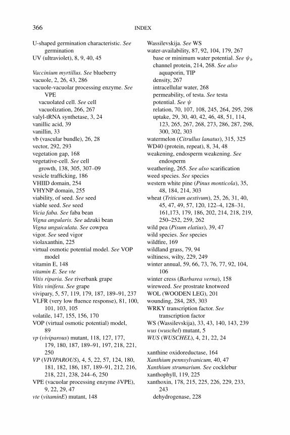
P1: PAE
BLUK053-Bradford September 22, 2006 21:0
366 INDEX
U-shaped germination characteristic. Seegermination
UV (ultraviolet), 8, 9, 40, 45
Vaccinium myrtillus. See blueberry
vacuole, 2, 26, 43, 286
vacuole-vacuolar processing enzyme. SeeVPE
vacuolated cell. See cell
vacuolization, 266, 267
valyl-tRNA synthetase, 3, 24
vanillic acid, 39
vanillin, 33
vb (vascular bundle), 26, 28
vector, 292, 293
vegetation gap, 168
vegetative-cell. See cell
growth, 138, 305, 307–09
vesicle trafficking, 186
VHIID domain, 254
VHYNP domain, 255
viability, of seed. See seed
viable seed. See seed
Vicia faba. See faba bean
Vigna angularis. See adzuki bean
Vigna unguiculata. See cowpea
vigor. See seed vigor
violaxanthin, 225
virtual osmotic potential model. See VOP
model
vitamin E, 148
vitamin E. See vteVitis riparia. See riverbank grape
Vitis vinifera. See grape
vivipary, 5, 57, 119, 179, 187, 189–91, 237
VLFR (very low fluence response), 81, 100,
101, 103, 105
volatile, 147, 155, 156, 170
VOP (virtual osmotic potential) model,
89
vp (viviparous) mutant, 118, 127, 177,
179, 180, 187, 189–91, 197, 218, 221,
250
VP (VIVIPAROUS), 4, 5, 22, 57, 124, 180,
181, 182, 186, 187, 189–91, 212, 216,
218, 221, 238, 244–6, 250
VPE (vacuolar processing enzyme δVPE),
9, 22, 29, 47
vte (vitaminE) mutant, 148
Wassilevskija. See WS
water-availability, 87, 92, 104, 179, 267
base or minimum water potential. See ψb
channel protein, 214, 268. See alsoaquaporin, TIP
density, 267
intracellular water, 268
permeability, of testa. See testa
potential. See ψ
relation, 70, 107, 108, 245, 264, 295, 298
uptake, 29, 30, 40, 42, 46, 48, 51, 114,
123, 265, 267, 268, 273, 286, 287, 298,
300, 302, 303
watermelon (Citrullus lanatus), 315, 325
WD40 (protein, repeat), 8, 34, 48
weakening, endosperm weakening. Seeendosperm
weathering, 265. See also scarification
weed species. See species
western white pine (Pinus monticola), 35,
48, 184, 214, 303
wheat (Triticum aestivum), 25, 26, 31, 40,
45, 47, 49, 57, 120, 122–4, 128–31,
161,173, 179, 186, 202, 214, 218, 219,
250–252, 259, 262
wild pea (Pisum elatius), 39, 47
wild species. See species
wildfire, 169
wildland grass, 79, 94
wiltiness, wilty, 229, 249
winter annual, 59, 66, 73, 76, 77, 92, 104,
106
winter cress (Barbarea verna), 158
wireweed. See prostrate knotweed
WOL (WOODEN LEG), 201
wounding, 284, 285, 303
WRKY transcription factor. Seetranscription factor
WS (Wassilevskija), 33, 43, 140, 143, 239
wus (wuschel) mutant, 5
WUS (WUSCHEL), 4, 21, 22, 24
xanthine oxidoreductase, 164
Xanthium pennsylvanicum, 40, 47
Xanthium strumarium. See cocklebur
xanthophyll, 119, 225
xanthoxin, 178, 215, 225, 226, 229, 233,
243
dehydrogenase, 228
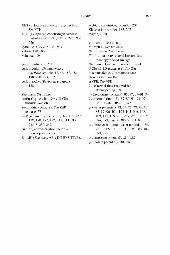
P1: PAE
BLUK053-Bradford September 22, 2006 21:0
INDEX 367
XET (xyloglucan endotransglycosylase).
See XTH
XTH (xyloglucan endotransglycosylase/
hydrolase), 44, 271, 277–9, 285, 289,
296
xyloglucan, 277–9, 285, 303
xylose, 278, 283
xylulose, 158
yeast two-hybrid, 254
yellow cedar (Chamaecyparisnootkatensis), 40, 47, 61, 183, 184,
190, 220, 223, 302
yellow rocket (Barbarea vulgaris),
158
Zea mays. See maize
zeatin-O-glucoside. See z-O-Glu
riboside. See ZR
zeaxanthin-epoxidase. See ZEP
oxidase, 57
ZEP (zeaxanthin epoxidase), 68, 119, 127,
178, 180, 187, 197, 211, 214, 218,
225–8, 230, 242
zinc-finger transcription factor. Seetranscription factor
ZmABI (Zea mays ABA INSENSITIVE),
313
z-O-Glu (zeatin-O-glucoside), 207
ZR (zeatin riboside), 185, 207
zygote, 2, 20
α-amanitin. See amanitin
α-amylase. See amylase
β-1,3-glucan. See glucan
β-1,4-d-mannopyranosyl linkage. Seemannopyranosyl linkage
β-amino butyric acid. See butric acid
β Glu (β-1,3-glucanase). See Glu
β-mannosidase. See mannosidase
β-oxidation. See Box
�VPE. See VPE
θ AT (thermal time required for
after-ripening), 94
θ H (hydrotime constant), 85, 87, 89–91, 94
θ T (thermal time), 83–87, 89–91, 94, 97,
98, 100–02, 105–11, 242
ψ (water potential), 72, 74, 75, 78, 79, 82,
85, 87–96, 101, 103, 105, 106, 108,
109, 111, 199, 221, 267, 268–72, 275,
276, 282, 286–8, 295–7, 301–03
ψb (base or minimum water potential), 74,
75, 79, 85, 87–96, 103, 105, 106, 108,
288, 295
ψ p (pressure potential), 286, 287
ψ s (solute potential), 286, 287

P1: PAE
BLUK053-Bradford September 22, 2006 21:0
368