anintrinsicmicrornatimerregulatesprogressivedeclinein ... · shoot regenerative capacity in plants...
TRANSCRIPT
An Intrinsic MicroRNA Timer Regulates Progressive Decline inShoot Regenerative Capacity in Plants
Tian-Qi Zhang,a,bHengLian,a,1 HongboTang,a,1 KarelDolezal,c,d,eChuan-MiaoZhou,a ShaYu,a,b Juan-HuaChen,a,b
Qi Chen,a,b Hongtao Liu,a Karin Ljung,c and Jia-Wei Wanga,2
a National Key Laboratory of Plant Molecular Genetics, Institute of Plant Physiology and Ecology, Shanghai Institutes for BiologicalSciences, Shanghai 200032, People’s Republic of ChinabUniversity of the Chinese Academy of Sciences, Shanghai 200032, People’s Republic of ChinacUmeå Plant Science Centre, Department of Forest Genetics and Plant Physiology, Swedish University of Agricultural Sciences, SE-901 83 Umeå, Swedend Laboratory of Growth Regulators, Institute of Experimental Botany, Academy of Sciences of the Czech Republic, 78371 Olomouc,Czech Republice Centre of the Region Haná for Biotechnological and Agricultural Research, Faculty of Science, Palacký University, 78371 Olomouc,Czech Republic
ORCID ID: 0000-0003-3885-6296 (J.-W.W.)
Plant cells are totipotent and competent to regenerate from differentiated organs. It has been shown that two phytohormones,auxin and cytokinin, play critical roles within this process. As in animals, the regenerative capacity declines with age in plants, butthe molecular basis for this phenomenon remains elusive. Here, we demonstrate that an age-regulated microRNA, miR156,regulates shoot regenerative capacity. As a plant ages, the gradual increase in miR156-targeted SQUAMOSA PROMOTERBINDING PROTEIN-LIKE (SPL) transcription factors leads to the progressive decline in shoot regenerative capacity. In old plants,SPL reduces shoot regenerative capacity by attenuating the cytokinin response through binding with the B-type ARABIDOPSISRESPONSE REGULATORs, which encode the transcriptional activators in the cytokinin signaling pathway. Consistently, theincreased amount of exogenous cytokinin complements the reduced shoot regenerative capacity in old plants. Therefore, therecruitment of age cues in response to cytokinin contributes to shoot regenerative competence.
INTRODUCTION
Regeneration of a multicellular organism from a piece of adult so-matic tissue is a prevalent phenomenon that occurs in both plantsand animals (Birnbaum and Sánchez Alvarado, 2008). In contrastwith animal cells, plant cells have been thought to maintain toti-potency, and most plant tissues from already differentiated organsare able to regenerate whole plants under proper in vitro cultureconditions (Duclercq et al., 2011; Sugimoto et al., 2011). It is wellknown that the ratio of two phytohormones, auxin and cytokinin,determines the developmental fate of regenerating tissue. A highcytokinin:auxin ratio directs regeneration of the shoot, whereasa low cytokinin:auxin ratio induces root differentiation (Skoog andMiller, 1957). However, little is understood concerning the mecha-nisms by which the auxin/cytokinin balance exerts these oppositeeffects. In addition, how cytokinin promotes the specification ofapical/shoot fate during shoot regeneration remains elusive.
Cytokinin signal transduction involves a multistep phosphorelaysignaling cascade from ligand perception at the cell membrane to
transcriptional activation in the nucleus (Hwang et al., 2012; Kieberand Schaller, 2014). In Arabidopsis thaliana, cytokinin is perceivedby the cytokinin receptors ARABIDOPSIS HISTIDINE KINASE2(AHK2), AHK3, and AHK4. Ligand binding triggers autophosphor-ylation at a conserved His residue in the receiver domain andsubsequent transfer of the phosphoryl group to a conserved Aspresidue in the attached transmitter domain. The phosphoryl groupon the Asp residue is then passed on to one of five ARABIDOPSISHISTIDINE PHOSPHOTRANSFER proteins and then to a group ofnucleus-localized B-type ARABIDOPSIS RESPONSE REGULATORs(ARRs). B-type ARRs activate the expression of cytokinin-responsivegenes and A-type ARRs. A-type ARRs, in turn, interfere with thefunction of B-type ARR proteins through protein-protein interaction,which establishes a negative feedback loop to the signaling pathway(Werner and Schmülling, 2009; Hwang et al., 2012).An interesting and common phenomenon in animals is the pro-
gressive reduction in regenerative capacity. For example, remyelination,a regenerative process that produces new myelin sheaths fromadult stem cells in the central nervous system, declines with in-creasing age (Ruckh et al., 2012). Similarly, the mammalian heartappears to have the capacity to regenerate only within a briefperiod after birth (Porrello et al., 2011). To what extent and bywhich means age contributes to plant regenerative capacity isunknown.miR156, which targets SQUAMOSA PROMOTER BINDING
PROTEIN-LIKE (SPL) transcription factors, governs the age path-way in plants. The level of miR156, in response to endogenous
1 These authors contributed equally to this work.2 Address correspondence to [email protected] author responsible for distribution of materials integral to the findingspresented in this article in accordance with the policy described in theInstructions for Authors (www.plantcell.org) is: Jia-Wei Wang ([email protected]).www.plantcell.org/cgi/doi/10.1105/tpc.114.135186
The Plant Cell, Vol. 27: 349–360, February 2015, www.plantcell.org ã 2015 American Society of Plant Biologists. All rights reserved.

sugar, gradually decreases with time (Wu and Poethig, 2006; Wanget al., 2009; Wu et al., 2009; Poethig, 2013; Yang et al., 2013; Yuet al., 2013). The onset of adult phase is defined by miR156 level:overexpression of miR156 prolongs the juvenile phase, whereasa reduction in miR156 activity leads to an accelerated expression ofadult traits (Wu et al., 2009). It has been shown that miR156-targeted SPLs regulate diverse age-related developmental pro-cesses, such as embryonic pattern formation, juvenile-to-adultphase transition, flowering time, inflorescence trichome initiation,and anthocyanin biosynthesis (Wang et al., 2009; Wu et al., 2009;Nodine and Bartel, 2010; Yu et al., 2010; Gou et al., 2011; Bergonziet al., 2013; Zhou et al., 2013; Rubio-Somoza et al., 2014).
Here, we show that old plants exhibit lower shoot regenerativecapacity than young plants, largely due to the reduced cytokininresponse. Our mutant characterizations, expression analyses,and protein-protein interaction assays further indicate that theincreased level of miR156-targeted SPLs in old plants dampensshoot regeneration by interfering with the function of B-type ARRs,thus establishing a molecular link between developmental timingand cytokinin-mediated shoot regeneration.
RESULTS
The Progressive Decline in Shoot Regenerative Capacitywith Age
To reveal whether the shoot regenerative capacity is changed asplants age, we performed in vitro regeneration assays using Ara-bidopsis and tobacco (Nicotiana tabacum) leaves. In Arabidopsis,shoot regeneration requires two steps: in the first step, callus, apluripotent cell mass, is formed from explants on auxin-rich callus-inducing medium (CIM). Subsequently, culture of the callus onshoot-inducing medium (SIM), which contains a high cytokinin:auxin ratio, induces the differentiation of callus into shoot (Duclercqet al., 2011). By contrast, tobacco can be regenerated directly fromleaf dics, which enables us to eliminate the effect of callus inductionon shoot regeneration.
We compared shoot regenerative rates of the first/second (early),fifth (mid), and ninth/tenth (late) tobacco leaves. To avoid the impactof leaf age on regenerative capacity, leaves of the same de-velopmental stage (1 cm in length) were used. In the absence of6-benzylaminopurine (6-BA), a synthetic cytokinin, none of the leafdics was competent to regenerate (Supplemental Figure 1A). Earlytobacco leaves exhibited a higher regenerative rate than late leaveson Murashige and Skoog (MS) medium supplemented with 6-BA(Figures 1A and 1B). In Arabidopsis, the callus inductive rate wasnot changed as plants aged (Supplemental Figure 1B). However,the same difference in regenerative rate between early and lateleaves was observed (Figures 1C and 1D). Thus, the shoot re-generative capacity was inversely correlated with plant age.
Shoot regeneration is directed by a high ratio of cytokinin toauxin. The regenerative rates were elevated as cytokinin levelincreased in both tobacco and Arabidopsis (Figures 1A to 1D).Notably, the regenerative rate of the late tobacco leaves on MSmedium supplemented with 0.5 mM 6-BA was 5-fold higherthan that on MS medium with 0.2 mM 6-BA (Figure 1C). Therefore,the increased amount of exogenous cytokinin complements the
reduced regenerative capacity in old plants. In agreement with thisobservation, the expression of ProTCS:GFP (for green fluorescentprotein), a synthetic cytokinin reporter (Müller and Sheen, 2008), waslower in late leaves than in early leaves on CIM and SIM (Figure 1E).
miR156 Contributes to Developmental Decline in ShootRegenerative Capacity
miR156, which targets SPL transcription factors, regulates thejuvenile-to-adult phase transition in many plant species, includingArabidopsis, Arabis alpina, Cardamine flexuosa, Populus 3canadensis, and Zea mays (Chuck et al., 2007; Wu et al., 2009;Wang et al., 2011; Bergonzi et al., 2013; Zhou et al., 2013). In to-bacco, miR156 level was correlated with age, being most abundantin seedlings (Supplemental Figure 2A). To determine whethermiR156 plays a role in shoot regeneration in tobacco, we generatedtransgenic plants that overexpressed either miR156 (Pro35S:MIR156) or a target mimic of miR156 (Pro35S:MIM156), in whichmiR156 is inactivated (Supplemental Figures 2B to 2D) (Franco-Zorrilla et al., 2007).We performed regeneration assays using the fifth (mid) tobacco
leaf. Compared with the wild type, Pro35S:MIR156 exhibited anincreased regenerative capacity, whereas Pro35S:MIM156 ex-hibited a reduced regenerative capacity (Figures 2A and 2B). Inaddition, overexpression of miR156 suppressed the developmentaldecline in shoot regeneration that was evident in the wild type(Figure 2E; Supplemental Figure 3A). In the fourth (mid) Arabidopsisleaf, there was no difference in callus inductive rate between thewild type and Pro35S:MIR156 or Pro35S:MIM156. However, weobserved a similar correlation between mR156 level and shootregenerative capacity (Figures 2C and 2D). It has been shown thatthe manipulation of mR156 level in Arabidopsis results in a dramaticchange in leaf morphology (Wang et al., 2008; Wu et al., 2009). Weobtained comparable results using wild-type, Pro35S:MIR156, andPro35S:MIM156 hypocotyls as explants, eliminating the effectof leaf morphology or leaf age on shoot regenerative capacity(Supplemental Figures 3B and 3C). Furthermore, elevation of cy-tokinin concentration in SIM rescued the low regenerative capacityof Pro35S:MIM156 plants (Figures 2B and 2D). Taken together,these results indicate that miR156 contributes to shoot re-generative competence.
SPL9-Group Genes, but Not SPL3-Group Genes, RegulateShoot Regeneration
miR156-targeted SPLs can be structurally divided into twogroups, represented by SPL3 (including SPL3, SPL4, and SPL5)and SPL9 (including SPL2, SPL6, SPL9, SPL10, SPL11, SPL13,and SPL15) (Xing et al., 2010). SPL9 differs from SPL3 becauseit harbors a C-terminal domain responsible for protein-proteininteraction (Yu et al., 2012). To determine whether both groupsof genes play a role in shoot regeneration, we examined theshoot regenerative rate of the transgenic plants that overexpressedmiR156-nontargetable SPL3 (rSPL3) and rSPL9 (Pro35S:rSPL3 andProSPL9:rSPL9). Pro35S:rSPL3 exhibited the same regenerativerate as the wild type, whereas ProSPL9:rSPL9 markedly impairedshoot regeneration (Figure 2F), indicating that SPL9 group genes,but not SPL3, regulate shoot regenerative capacity. Earlier work
350 The Plant Cell

Figure 1. The Developmental Decline in Shoot Regenerative Capacity.
(A) and (C) Shoot regeneration of tobacco (A) and Arabidopsis (C). The leaves from plants of different ages were used as explants for regenerationassays. For tobacco, shoots were induced on MS medium supplemented with 6-BA of different concentrations. For Arabidopsis, calli were induced fromexplants on CIM and shoots were then regenerated on MS medium supplemented with 0.9 mM indole-3-acetic acid (an auxin) and 2-isopentenyladenine(2-IP; a cytokinin) of different concentrations. Bar = 0.5 cm.(B) and (D) Quantitative analyses of regenerative capacity in tobacco (B) and Arabidopsis (D). The regenerative rate was represented by the number ofregenerated shoots. Nine (B) or eight (D) explants were examined. Asterisks indicate significant differences from leaf #1-2 (Student’s t test, P < 0.05).Data are means 6 SD of three biological replicates.(E) Visualization of the ProTCS:GFP reporter in explants cultured on CIM or SIM. Explants derived from early or late leaves were examined (n = 10). Thenumber of days after transfer to CIM or SIM is indicated. Bar = 400 mm.
Age and Shoot Regenerative Capacity 351

Figure 2. miR156 Regulates Shoot Regenerative Capacity.
(A) and (C) Shoot regeneration of wild-type, Pro35S:MIR156, and Pro35S:MIM156 plants. The fifth tobacco leaf (A) and the fourth Arabidopsis leaf (C)were used as explants for regeneration assays. 2-IP, 2-isopentenyladenine. Bars = 0.5 cm.(B) and (D) Quantitative analyses of regenerative capacity in tobacco (B) and Arabidopsis (D). Nine (B) or eight (D) explants were examined. Asterisksindicate significant differences from the wild type (Student’s t test, P < 0.05).(E) miR156 overexpression suppressed the progressive decline in shoot regeneration in tobacco. Leaves from plants of different ages were used as theexplants for regeneration assays. Shoots were induced on MS medium supplemented with 0.2 mM 6-BA. Nine explants were examined. Asterisksindicate significant differences from the wild type (Student’s t test, P < 0.05).(F) Roles of SPL3 and SPL9 group genes in shoot regeneration. Eight explants were examined. Asterisks indicate significant differences from the wildtype (Student’s t test, P < 0.05).
352 The Plant Cell
revealed that SPL9 and SPL15 play major but redundant roles withinthe SPL9 group (Schwarz et al., 2008; Wang et al., 2008). The shootregenerative capacity of the spl9-1 spl15-2 double mutant washigher than that of the wild type but lower than that of the miR156overexpressor (Figure 2F), suggesting the functional redundancy ofmiR156-targeted SPL9 group genes in shoot regeneration.
miR156 Regulates Shoot Regenerative Capacity throughModulating the Cytokinin Response
The high regenerative capacity of Pro35S:MIR156 plants can resultfrom a high concentration of cytokinin in vivo. Cytokinin measure-ment revealed that Pro35S:MIR156 accumulated the same amountof cytokinin as the wild type (Supplemental Table 1), suggestingthat miR156 does not regulate shoot regenerative capacity throughmodulating endogenous cytokinin levels.
Cytokinin signaling transduction involves a multistep phosphor-elay signaling cascade from ligand perception at the cell membraneto transcriptional activation in the nucleus. In Arabidopsis, fourB-type ARR transcription factors, ARR1, ARR2, ARR10, and ARR12,play essential roles in cytokinin signaling transduction (Hwang andSheen, 2001; Mason et al., 2005; Ishida et al., 2008). Compared withthe wild type, the regenerative capacity was markedly reduced inarr2-4 arr12-1 and completely lost in the arr1-3 arr10-5 arr12-1mutant. Pro35S:MIR156 did not restore the impaired shoot re-generative capacity of arr2-4 arr12-1 and arr1-3 arr10-5 arr12-1(Supplemental Figure 4), suggesting that miR156 and its target SPLsmodulate shoot regenerative capacity through B-type ARRs.
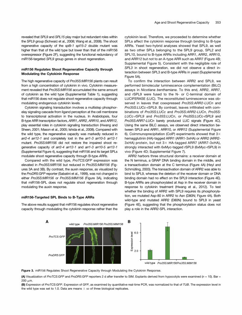
Compared with the wild type, ProTCS:GFP expression waselevated in Pro35S:MIR156 but reduced in Pro35S:MIM156 (Fig-ures 3A and 3B). By contrast, the auxin response, as visualized bythe ProDR5:GFP reporter (Sabatini et al., 1999), was not changed ineither Pro35S:MIR156 or Pro35S:MIM156 (Figure 3A), indicatingthat miR156-SPL does not regulate shoot regeneration throughmodulating the auxin response.
miR156-Targeted SPL Binds to B-Type ARRs
The above results suggest that miR156 regulates shoot regenerativecapacity through modulating the cytokinin response rather than the
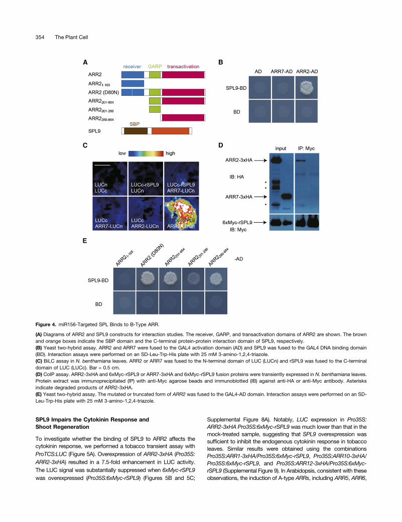
cytokinin level. Therefore, we proceeded to determine whetherSPLs affect the cytokinin response through binding to B-typeARRs. Yeast two-hybrid analyses showed that SPL9, as wellas two other SPLs belonging to the SPL9 group, SPL2 andSPL10, bound to B-type ARRs including ARR1, ARR2, ARR10,and ARR12 but not to an A-type ARR such as ARR7 (Figure 4B;Supplemental Figure 5). Consistent with the negligible role ofSPL3 in shoot regeneration, we did not observe a direct in-teraction between SPL3 and B-type ARRs in yeast (SupplementalFigure 5A).To confirm the interaction between ARR2 and SPL9, we
performed bimolecular luminescence complementation (BiLC)assays in Nicotiana benthamiana. To this end, ARR2, ARR7,and rSPL9 were fused to the N- or C-terminal domain ofLUCIFERASE (LUC). The reconstituted luminescence was ob-served in leaves that coexpressed Pro35S:ARR2-LUCn andPro35S:LUCc-rSPL9. By contrast, leaves infiltrated with com-binations of Pro35S:LUCc and Pro35S:ARR2-LUCn, Pro35S:LUCc-rSPL9 and Pro35S:LUCn, or Pro35S:LUCc-rSPL9 andPro35S:ARR7-LUCn barely produced LUC signals (Figure 4C).Using the same BiLC assays, we observed direct interaction be-tween SPL9 and ARR1, ARR10, or ARR12 (Supplemental Figure6). Coimmunoprecipitation (CoIP) experiments showed that 33hemagglutinin (HA)-tagged ARR1 (ARR1-3xHA) or ARR2 (ARR2-3xHA) protein, but not 33 HA-tagged ARR7 (ARR7-3xHA),strongly interacted with 6xMyc-tagged rSPL9 (6xMyc-rSPL9) invivo (Figure 4D; Supplemental Figure 7).ARR2 harbors three structural domains: a receiver domain at
the N terminus, a GRAP DNA binding domain in the middle, anda transactivation domain at the C terminus (Figure 4A) (Heyl andSchmülling, 2003). The transactivation domain of ARR2 was able tobind to SPL9, whereas the deletion of the receiver domain or DNAbinding domain had no effect on the SPL9 interaction (Figure 4E).B-type ARRs are phosphorylated at Asp in the receiver domain inresponse to cytokinin treatment (Hwang et al., 2012). To testwhether the binding of ARR2 with SPL9 requires its phosphoryla-tion, we mutated Asp-80 in ARR2 to Asn (D80N; Figure 4A). Bothwild-type and mutated ARR2 (D80N) bound to SPL9 in yeast(Figure 4E), suggesting that the phosphorylation status does notplay a role in the ARR2-SPL interaction.
Figure 3. miR156 Regulates Shoot Regenerative Capacity through Modulating the Cytokinin Response.
(A) Visualization of ProTCS:GFP and ProDR5:GFP reporters 2 d after transfer to SIM. Explants derived from hypocotyls were examined (n = 10). Bar =200 mm.(B) Expression of ProTCS:GFP. Expression of GFP, as examined by quantitative real-time PCR, was normalized to that of TUB. The expression level inthe wild type was set to 1.0. Data are means 6 SD of three biological replicates.
Age and Shoot Regenerative Capacity 353
SPL9 Impairs the Cytokinin Response andShoot Regeneration
To investigate whether the binding of SPL9 to ARR2 affects thecytokinin response, we performed a tobacco transient assay withProTCS:LUC (Figure 5A). Overexpression of ARR2-3xHA (Pro35S:ARR2-3xHA) resulted in a 7.5-fold enhancement in LUC activity.The LUC signal was substantially suppressed when 6xMyc-rSPL9was overexpressed (Pro35S:6xMyc-rSPL9) (Figures 5B and 5C;
Supplemental Figure 8A). Notably, LUC expression in Pro35S:ARR2-3xHA Pro35S:6xMyc-rSPL9was much lower than that in themock-treated sample, suggesting that SPL9 overexpression wassufficient to inhibit the endogenous cytokinin response in tobaccoleaves. Similar results were obtained using the combinationsPro35S:ARR1-3xHA/Pro35S:6xMyc-rSPL9, Pro35S:ARR10-3xHA/Pro35S:6xMyc-rSPL9, and Pro35S:ARR12-3xHA/Pro35S:6xMyc-rSPL9 (Supplemental Figure 9). In Arabidopsis, consistent with theseobservations, the induction of A-type ARRs, including ARR5, ARR6,
Figure 4. miR156-Targeted SPL Binds to B-Type ARR.
(A) Diagrams of ARR2 and SPL9 constructs for interaction studies. The receiver, GARP, and transactivation domains of ARR2 are shown. The brownand orange boxes indicate the SBP domain and the C-terminal protein-protein interaction domain of SPL9, respectively.(B) Yeast two-hybrid assay. ARR2 and ARR7 were fused to the GAL4 activation domain (AD) and SPL9 was fused to the GAL4 DNA binding domain(BD). Interaction assays were performed on an SD-Leu-Trp-His plate with 25 mM 3-amino-1,2,4-triazole.(C) BiLC assay in N. benthamiana leaves. ARR2 or ARR7 was fused to the N-terminal domain of LUC (LUCn) and rSPL9 was fused to the C-terminaldomain of LUC (LUCc). Bar = 0.5 cm.(D) CoIP assay. ARR2-3xHA and 6xMyc-rSPL9 or ARR7-3xHA and 6xMyc-rSPL9 fusion proteins were transiently expressed in N. benthamiana leaves.Protein extract was immunoprecipitated (IP) with anti-Myc agarose beads and immunoblotted (IB) against anti-HA or anti-Myc antibody. Asterisksindicate degraded products of ARR2-3xHA.(E) Yeast two-hybrid assay. The mutated or truncated form of ARR2 was fused to the GAL4-AD domain. Interaction assays were performed on an SD-Leu-Trp-His plate with 25 mM 3-amino-1,2,4-triazole.
354 The Plant Cell

and ARR15, by 6-BA was elevated in Pro35S:MIR156 but reduced inPro35S:MIM156 (Figure 5D; Supplemental Figures 8B and 8C).
We then performed the same experiment using ProDR5:LUCas reporter. In agreement with the above findings that the auxinresponse was not changed in either Pro35S:MIR156 or Pro35S:MIM156 explants (Figure 3A), overexpression of ARR2 and SPL9did not affect DR5 expression (Supplemental Figure 10).
To further investigate the repression of ARR2 transcriptionalactivity by SPL9, we generated an effector in which the trans-activation domain of ARR2 (DARR2) was fused to the DNA bindingdomain of yeast GAL4 (Pro35S:BD-DARR2) and a Pro6xUAS:LUCreporter in which LUC was expressed from six repeats of the GAL4target sequence (Supplemental Figure 11A). The LUC signal wasmarkedly elevated by Pro35S:BD-DARR2 but not by Pro35S:DARR2, which did not harbor GAL4-BD. The activation of LUCby Pro35S:BD-DARR2 was suppressed when 6xMyc-rSPL9 wascoexpressed (Supplemental Figures 11B and 11C).
To explore the role of SPL9 in shoot regeneration, we comparedthe regenerative capacity among the wild type, the spl9-1 spl15-2double mutant, and Pro35S:rSPL9-GR transgenic plants, in which
rSPL9 was fused to the hormone binding domain of rat glucocor-ticoid receptor (GR) and expressed from the 35S promoter (Wuet al., 2009). Treatment with the steroid hormone ligand dexa-methasone (DEX), which leads to a translocation of the rSPL9-GRfusion protein from the cytoplasm to the nucleus, resulted in thesame phenotype as the transgenic plants expressing rSPL9(ProSPL9:rSPL9) (Wu et al., 2009). The spl9-1 spl15-2 doublemutant exhibited higher regenerative capacity than the wild type.Notably, shoot regeneration of the spl9-1 spl15-2 double mu-tant was not affected by DEX treatment (Figures 6A and 6B;Supplemental Figure 12). We observed an inverse correlation be-tween the concentration of DEX (i.e., the level of rSPL9-GR fusionprotein in the nucleus) and shoot regenerative capacity (Figures 6Aand 6B). The addition of 10 mM DEX in SIM resulted in a completeloss of shoot regenerative capacity at all the hormone concen-trations tested. At lower concentrations of DEX, the increased levelof 6-BA complemented the reduced shoot regenerative capacitycaused by rSPL9-GR.To confirm the role of the ARR2-SPL9 interaction in shoot re-
generation, we performed regeneration assays using ProSPL9:
Figure 5. miR156-Targeted SPL Regulates the Cytokinin Response.
(A) Diagrams of effector and reporter constructs for transactivation studies. The green boxes in the ProTCS:LUC construct indicate B-type ARR bindingsites.(B) Transactivation assays. N. benthamiana leaves were infiltrated with different combinations of effector and reporter. Bar = 0.5 cm.(C) Quantitative analyses of LUC activity in N. benthamiana leaves. Pro35S:REN was used as an internal control. Quantification was performed bynormalizing LUC activity to that of REN. The LUC activity in ProTCS:LUC without effector was set to 1.0. Data are means 6 SD of three biologicalreplicates. Asterisks indicate significant differences from mock sample (Student’s t test, P < 0.05).(D) Expression of ARR5. Seven-day-old Arabidopsis seedlings were treated with DMSO (mock) or 5 mM 6-BA for 40 min. The expression of ARR5 inmock wild-type sample was set to 1.0. Data are means 6 SD of three biological replicates.
Age and Shoot Regenerative Capacity 355

Figure 6. miR156-Targeted SPL Regulates Regenerative Capacity.
(A) Shoot regeneration assay using the wild type (Columbia-0 [Col-0]) and Pro35S:rSPL9-GR. The hypocotyls were cultured on CIM and then trans-ferred to MS medium supplemented with different concentrations of 2-isopentenyladenine (2-IP) and DEX. Bar = 0.5 cm.(B) Quantitative analyses of regenerative capacity. The regenerative rate was represented by the number of regenerated shoots on six explants. Dataare means 6 SD of three biological replicates.(C) Model for the regulation of shoot regenerative capacity by a microRNA timer. In old plants, the amount of SPL9 is increased due to the de-velopmental decline of miR156. SPL9 inhibits the transcriptional activity of B-type ARRs, thereby reducing the shoot regenerative capacity (see text fordetails).
356 The Plant Cell
SPL9, Pro35S:ARR2, and Pro35S:ARR2 ProSPL9:SPL9 plants.Consistent with previous findings, Pro35S:ARR2 showed elevatedregenerative capacity in comparison with the wild type (Hwang andSheen, 2001). Pro35S:ARR2 rescued the reduced shoot re-generative capacity of ProSPL9:SPL9 (Supplemental Figure 13).
DISCUSSION
Our results reveal an important role of miR156, the master regulatorof juvenility, in shoot regeneration (Figure 6C). Young plants exhibita high cytokinin response and regenerative capacity. As a plantages, miR156 levels decline, alleviating the repression of its SPLtargets. SPL directly inhibits the transcriptional activity of B-type ARRand thereby impairs shoot regenerative capacity. In Caenorhabditiselegans, the transitions between stages of larval development aremediated by increases in the expression of two sequentially ex-pressed microRNAs, lin-4 and let-7 (Ambros, 2011). A recent reportdemonstrated that overexpression of let-7 in the juvenile state im-pairs tissue repair, a type of regeneration in mouse (Shyh-Changet al., 2013). Thus, these observations highlight that plants and an-imals, although they evolved independently, adopt a similar molec-ular mechanism by which the regenerative capacity is governed bythe microRNA timer.
Growing lines of evidence showed that SPL9 exerts dualmolecular roles. It has been shown that SPL9 regulates the vege-tative phase transition and trichome production on floral organsthrough activating MIR172B and TRICHOMELESS1 (Wu et al.,2009; Yu et al., 2010), whereas it acts as a transcriptional repressoron DIHYDROFLAVONOL REDUCTASE through binding with theMYB transcription factor PRODUCTION OF ANTHOCYANINPIGMENTS1 (Gou et al., 2011). Yeast two-hybrid assays demon-strated that SPL9 binds to the transactivation domain of ARR2(Figures 4A and 4E). In this scenario, we speculate that the bindingof SPL9 to ARR2 changes the conformation of ARR2, therebyimpairing its transcriptional activation toward downstream targets.
It is well known that old plants have lower capacities for bothshoot and root regeneration than young plants. However, thisphenomenon could not be explained concurrently by the alteredcytokinin:auxin ratio in old plants, because a low cytokinin:auxinratio inhibits shoot production but induces root regeneration.Our results revealed that old plants exhibit a lower cytokininresponse, which is responsible for the decreased shoot re-generation. The molecular mechanism causing the reduced rootregenerative capacity in old plants and whether this process ismediated by an altered auxin response await further investigations.
In vitro regeneration is influenced by the type of explants usedand by environmental factors such as culture medium, plant hor-mones, and gelling agent strength. Although the protocol for in vitroregeneration varies greatly between plant species, most of theexplants, such as cotyledon, hypocotyl, petiole, and early leaves,are collected from juvenile plants. It has been shown that, duringvegetative regeneration of the heather Calluna vulgaris, the numberof newly formed sprouts decreased significantly in plants that weremore than 6 years old (Berdowski and Siepel, 1998). Similarly, theshoot regenerative capacity declines as Quercus euboica ages(Kartsonas and Papafotiou, 2007). These observations are consis-tent with our findings that the level of miR156 is correlated withshoot regenerative competence and that overexpression of miR156
increases the regenerative rate in both Arabidopsis and tobacco.Because miR156 is highly conserved in land plants from moss toflowering plants (Axtell and Bowman, 2008), these results furthersuggest that age cues serve as a common element behind plantcell totipotency. Manipulating miR156 levels during regenerationwill thus be of great value for the in vitro propagation of all plantspecies, especially for some rare and endangered trees.
METHODS
Plant Materials
Arabidopsis thaliana (ecotype Columbia-0), tobacco (Nicotiana tabacumcv SR1), and Nicotiana benthamiana were grown at 21°C (day)/19°C(night) in long-day conditions (16 h of light/8 h of dark). Pro35S:MIR156,Pro35S:MIM156, Pro35S:rSPL3, ProSPL9:rSPL9, spl9-1 spl15-2, arr2-4,arr1-3 arr10-5 arr12-1, and ProTCS:GFP were described (Mason et al.,2005; Ishida et al., 2008; Müller and Sheen, 2008; Wang et al., 2008). Thearr2-4 arr12-1 double mutant was identified by PCR genotyping.
For transgenic Arabidopsis plants, the binary constructs were deliveredintoAgrobacterium tumefaciensGV3101 (pMP90) by the freeze-thawmethod.Transgenic plants were generated by the floral dipping method (Clough andBent, 1998) and screened with 0.05% glufosinate (Basta) on soil, 40 mg/mLhygromycin, or 50 mg/mL kanamycin on half-strength MS plates.
For transgenic tobacco plants, the overnight culture of Agrobacteriumwas resuspended with infection buffer (30 g/L glucose) to OD600 = 0.8.Tobacco seeds were sterilized with 20% NaClO for 15 min and germi-nated on an MS plate (4.4 g of MS basal medium with vitamin powder[PhytoTechnology Laboratories], 0.5 g/L methylester sulfonate, 20 g/Lsucrose, and 8 g/L agar, pH 5.7). Leaf discs (1 cm in diameter) were infectedwith Agrobacterium suspension for 30 min. The explants were brieflydried, transferred to sterile filter paper, and kept in darkness at 25°C for2 d. The explants were then transferred to selection shootingmedium (4.4 g ofMS basal medium with vitamin powder, 0.5 g/L methylester sulfonate, 20 g/Lsucrose, 2mg/mL 6-BA, 100mg/mL kanamycin, 250mg/mL timentin, and 8 g/L agar, pH 5.7) and incubated at 25°C for 2 weeks under long-day conditions.The explants were subcultured at 3-week intervals. The whole explantstogetherwith the shootswere transferred to selection rootingmedium (4.4 gofMS basal medium with vitamin powder, 0.5 g/L methylester sulfonate, 20 g/Lsucrose, 100mg/mL kanamycin, 250mg/mL timentin, and 8 g/L agar, pH 5.7).Rooted plants were transferred to soil and grown in a growth chamber. In-dependent T2 lines were used for regeneration assays.
Constructs
The oligonucleotide primers for all constructs are given in SupplementalTable 2. For yeast two-hybrid constructs, the pGBKT7 (Clontech) series ofSPL2, SPL3, SPL9, and SPL10 has been described (Yu et al., 2012).ARR1, ARR2, ARR7, ARR10, and ARR12 were cloned into pGADT7(Clontech). ARR2 (D80N) was generated by PCR-mediated mutagenesis.
BiLC constructs were generated as described (Gou et al., 2011). ThecDNA of rSPL9 was amplified and cloned into the JW772 vector behindLUCc under the control of the 35S promoter. The coding region of ARR1,ARR2,ARR7,ARR10, or ARR12was cloned into the JW771 vector in frontof LUCn under the control of the 35S promoter.
For CoIP constructs, ARR1, ARR2, and ARR7 were cloned into theJW819 vector with a 3xHA C-terminal fusion. rSPL9 was cloned into theJW1016 vector with a 6xMyc N-terminal fusion tag.
For Pro35S:rSPL9-GR, the rSPL9-GR fragment was PCR amplifiedusing JW66 as template and cloned into JW807 behind the 35S promoter.
For tobacco transient assays, the TCS or DR5 promoter was clonedinto the TQ108 vector in front of LUC. Pro35S:ARR2 was generated bycloning ARR2 into JW807 behind the 35S promoter. Pro35S:rSPL9 has
Age and Shoot Regenerative Capacity 357

been described (Gou et al., 2011). For Pro6xUAS:LUC, 6xUAS(6xCGGGTGACAGCCCTCCG) was fused with the minimal 35S promoterand cloned into TQ108 in front of LUC. For Pro35S:DARR2 and Pro35S:BD-DARR2, the fragment of DARR2 or BD-DARR2 (DARR2 fused with theGAL4-BD domain) was cloned into JW807 behind the 35S promoter.
Regeneration Experiments
Tobacco regeneration assays were performed on MS medium with dif-ferent concentrations of 6-BA as indicated.
For Arabidopsis regeneration assays, Arabidopsis seeds were steril-ized with 15% bleach and germinated on half-strength MS plates (2.21 gof MS basal medium with vitamin powder, 0.5 g/L methylester sulfonate,20 g/L sucrose, and 8 g/L agar, pH 7.5). Explants (leaves or hypocotyls)were excised and transferred to CIM (4.4 g of MS basal medium withvitamin powder, 0.5 g/L methylester sulfonate, 20 g/L sucrose, 2.2 mM2,4-D, 0.2 mM kinetin, and 8 g/L agar, pH 5.7) for 7 d. The calli were thentransferred to SIM (4.4 g of MS basal mediumwith vitamin powder, 0.5 g/Lmethylester sulfonate, 20 g/L sucrose, 0.9 mM indole-3-acetic acid, and8 g/L agar, pH 5.7) with different concentrations of 2-isopentenyladenineand incubated at 25°C under long-day conditions.
The numbers of explants and regenerated shoots were scored. Theregenerative capacity was represented by the number of regeneratedshoots in a given number of explants. Three independent experiments(biological triplicates) were performed.
Yeast Two-Hybrid Assay
Plasmidswere transformed into yeast strain AH109 (Clontech) by the LiCl-polyethylene glycol method. The transformants were selected on SD-Leu-Trpplates. The interactions were tested on SD-Leu-Trp-His or SD-Ade-Leu-Trp-His plates with 3-amino-1,2,4-triazole. At least 10 individual clones wereanalyzed.
CoIP and Immunoblot Analyses
Agrobacteria-infiltrated N. benthamiana leaves were used for CoIP analyses.The soluble protein was extracted in extraction buffer (50mMHEPES, 10mMEDTA, 50 mM NaCl, 10% glycerol, 1% polyvinylpolypyrrolidone, 2 mM DTT,1 mM phenylmethylsulfonyl fluoride, 10 mM MG-132, and 13 protease in-hibitor cocktail, pH 7.5). Immunoprecipitation was performed with anti-Mycbeads (Sigma-Aldrich; E6654) for 3 h (for ARR1 or ARR2 with SPL9) at 4°C.The beads were washed three times with wash buffer (50 mM HEPES,150 mM NaCl, 10 mM EDTA, 0.1% Triton X-100, 10% glycerol, and 1 mMphenylmethylsulfonyl fluoride, pH 7.5). 3xHA or 6xMyc fusion proteins weredetected by immunoblot with anti-HA-peroxidase (Roche; 12013819001) oranti-Myc (Millipore; 05-724) antibody.
BiLC Analysis and Tobacco Transient Assay
Agrobacterium was resuspended in infiltration buffer (10 mM methylestersulfonate, 10 mM MgCl2, and 150 µM acetosyringone, pH 5.7) at OD600 =0.8. Pro35S:P19-HA (Papp et al., 2003) was coinfiltrated to inhibit genesilencing. Luciferin (1 mM) was infiltrated before LUC activity wasmonitored after 3 d. The LUC signal was photographed with a cool CCDcamera. To quantify LUC activity, we used a dual-LUC reporter system inwhich Pro35S:RENILLA (REN ) was used as an internal control (Hellenset al., 2005). The firefly luciferase activity was quenched before RENactivity was measured with a luminometer (Promega 20/20). The LUCactivity was calculated by normalizing the values to those of REN. Theexpression of 6xMyc-rSPL9, ARR2-3xHA, and P19-HA was examined byimmunoblot (Supplemental Figure 8A). Three independent experiments(biological triplicates) were performed.
The BiLC assay was performed as described (Gou et al., 2011).Agrobacterium was resuspended in infiltration buffer at OD600 = 0.8.
Expression Analyses
For cytokinin treatment, 7-d-old wild-type Arabidopsis seedlings grown inhalf-strength MS liquid medium under long-day conditions were treatedwith DMSO (mock) or 5mM6-BA for 40min. Total RNAwas extracted withTrizol reagent (Invitrogen). Total RNA (1 µg) was treated with 1 mL ofDNase I (1 unit/mL; Fermentas) and used for cDNA synthesis with oligo(dT)primer (Fermentas). The average expression levels and SE values werecalculated from 2-DDCt values. Biological triplicates with technical duplicateswere performed. The quantitative RT-PCR primers for TUB have been de-scribed (Wang et al., 2009). The oligonucleotide primers for all genes are givenin Supplemental Table 2. Quantitative RT-PCR on mature miR156 wasperformedasdescribed (Yu et al., 2013). The expressionofmiR156 in tobaccowas normalized to that of the ribosomal protein gene Nt-L25.
Cytokinin Measurement
Wild-type and Pro35S:MIR156 seedlings were used for cytokinin measure-ments. Samples were purified, derivatized by propionylation, and quantifiedby liquid chromatography-mass spectrometry analysis according to thepublished protocol (Nordström et al., 2004). Chromatographic separationwasperformed on a reverse-phase analytical column (150 3 1 mm BetaMaxNeutral, 5-mm particle size; Thermo Hypersil).
Accession Numbers
Sequence data from this article can be found in the Arabidopsis GenomeInitiative or GenBank/EMBL databases under the following accessionnumbers: ARR1 (At3g16857), ARR2 (At4g16110), ARR10 (At4g31920),ARR12 (At2g25180), ARR5 (At3g48100), ARR6 (At5g62920), ARR7(At1g19050), ARR15 (At1g74890), SPL2 (At5g43270), SPL3 (At2g33810),SPL9 (At2g42200), SPL10 (At1g27370), SPL15 (At3g57920), TUB (At5g62690),and Nt-L25 (L18908).
Supplemental Data
Supplemental Figure 1. Shoot Regeneration of N. tabacum andA. thaliana.
Supplemental Figure 2. Transgenic N. tabacum Plants.
Supplemental Figure 3. miR156 Regulates Shoot Regeneration.
Supplemental Figure 4. Genetic Interaction between miR156 andB-Type ARRs.
Supplemental Figure 5. Interactions between B-Type ARRs andmiR156-Targeted SPLs.
Supplemental Figure 6. Validation of the Interactions betweenB-Type ARRs and SPLs by BiLC Assays.
Supplemental Figure 7. Validation of ARR1-SPL9 Interaction by CoIPAssays.
Supplemental Figure 8. SPL Negatively Regulates Cytokinin Response.
Supplemental Figure 9. ProTCS:LUC Transactivation Assays.
Supplemental Figure 10. ProDR5:LUC Transactivation Assays.
Supplemental Figure 11. SPL9 Suppresses ARR2 TranscriptionalActivation.
Supplemental Figure 12. spl9 spl15 Shoot Regeneration Assays.
Supplemental Figure 13. Genetic Interaction between SPL9 andB-Type ARRs.
358 The Plant Cell

Supplemental Table 1. Cytokinin Measurement.
Supplemental Table 2. Oligonucleotide Primer Sequences.
ACKNOWLEDGMENTS
We thank the ABRC for seeds; Bruno Müller for the ProTCS:GFP reporterconstruct; Hongxia Zhang for tobacco seeds; Gun Löfdahl and Xiao-ShuGao for skillful technical assistance; and members of the J.-W. Wanglaboratory, Ignacio Rubio-Somoza, Yingbo Mao, Daiyin Chao, and Ling-Jian Wang, for discussion and comments on the article. This work wassupported by the National Natural Science Foundation of China (Grants31430013, 31222029, and 912173023), the State Key Basic ResearchProgram of China (Grant 2013CB127000), the Shanghai Pujiang Program(Grant 12PJ1409900), the Recruitment Program of Global Expects(China), the National Key Laboratory of Plant Molecular Genetics KeyResearch Program, the Ministry of Education, Youth, and Sports, CzechRepublic (Grant LO1204 from the National Program of Sustainability I),the Swedish Governmental Agency for Innovation Systems, and theSwedish Research Council.
AUTHOR CONTRIBUTIONS
T.-Q.Z. and J.-W.W. designed the research. T.-Q.Z., H. Lian, H.T.,C.-M.Z., S.Y., K.D., J.-H.C., and Q.C. performed research. All authorsanalyzed data. J.-W.W. wrote the article.
Received December 9, 2014; revised January 8, 2015; accepted January16, 2015; published February 3, 2015.
REFERENCES
Ambros, V. (2011). MicroRNAs and developmental timing. Curr. Opin.Genet. Dev. 21: 511–517.
Axtell, M.J., and Bowman, J.L. (2008). Evolution of plant microRNAsand their targets. Trends Plant Sci. 13: 343–349.
Berdowski, J.J.M., and Siepel, H. (1998). Vegetative regeneration ofCalluna vulgaris at different ages and fertilizer levels. Biol. Conserv.46: 85–93.
Bergonzi, S., Albani, M.C., Ver Loren van Themaat, E., Nordström,K.J., Wang, R., Schneeberger, K., Moerland, P.D., andCoupland, G. (2013). Mechanisms of age-dependent response towinter temperature in perennial flowering of Arabis alpina. Science340: 1094–1097.
Birnbaum, K.D., and Sánchez Alvarado, A. (2008). Slicing acrosskingdoms: Regeneration in plants and animals. Cell 132: 697–710.
Chuck, G., Cigan, A.M., Saeteurn, K., and Hake, S. (2007). Theheterochronic maize mutant Corngrass1 results from overexpressionof a tandem microRNA. Nat. Genet. 39: 544–549.
Clough, S.J., and Bent, A.F. (1998). Floral dip: A simplified method forAgrobacterium-mediated transformation of Arabidopsis thaliana.Plant J. 16: 735–743.
Duclercq, J., Sangwan-Norreel, B., Catterou, M., and Sangwan,R.S. (2011). De novo shoot organogenesis: From art to science.Trends Plant Sci. 16: 597–606.
Franco-Zorrilla, J.M., Valli, A., Todesco, M., Mateos, I., Puga, M.I.,Rubio-Somoza, I., Leyva, A., Weigel, D., García, J.A., and Paz-Ares, J. (2007). Target mimicry provides a new mechanism forregulation of microRNA activity. Nat. Genet. 39: 1033–1037.
Gou, J.Y., Felippes, F.F., Liu, C.J., Weigel, D., and Wang, J.W.(2011). Negative regulation of anthocyanin biosynthesis in Arabidopsis bya miR156-targeted SPL transcription factor. Plant Cell 23: 1512–1522.
Hellens, R.P., Allan, A.C., Friel, E.N., Bolitho, K., Grafton, K.,Templeton, M.D., Karunairetnam, S., Gleave, A.P., and Laing, W.A.(2005). Transient expression vectors for functional genomics, quantifica-tion of promoter activity and RNA silencing in plants. Plant Methods 1: 13.
Heyl, A., and Schmülling, T. (2003). Cytokinin signal perception andtransduction. Curr. Opin. Plant Biol. 6: 480–488.
Hwang, I., and Sheen, J. (2001). Two-component circuitry in Arabi-dopsis cytokinin signal transduction. Nature 413: 383–389.
Hwang, I., Sheen, J., and Müller, B. (2012). Cytokinin signalingnetworks. Annu. Rev. Plant Biol. 63: 353–380.
Ishida, K., Yamashino, T., Yokoyama, A., and Mizuno, T. (2008).Three type-B response regulators, ARR1, ARR10 and ARR12, playessential but redundant roles in cytokinin signal transductionthroughout the life cycle of Arabidopsis thaliana. Plant Cell Physiol.49: 47–57.
Kartsonas, E., and Papafotiou, M. (2007). Mother plant age andseasonal influence on in vitro propagation of Quercus euboica Pap,an endemic, rare and endangered oak species of Greece. Plant CellTissue Organ Cult. 90: 111–116.
Kieber, J.J., and Schaller, G.E. (2014). Cytokinins. The ArabidopsisBook 12: e0168, doi/10.1199/tab.0168.
Mason, M.G., Mathews, D.E., Argyros, D.A., Maxwell, B.B., Kieber,J.J., Alonso, J.M., Ecker, J.R., and Schaller, G.E. (2005). Multipletype-B response regulators mediate cytokinin signal transduction inArabidopsis. Plant Cell 17: 3007–3018.
Müller, B., and Sheen, J. (2008). Cytokinin and auxin interaction inroot stem-cell specification during early embryogenesis. Nature453: 1094–1097.
Nodine, M.D., and Bartel, D.P. (2010). MicroRNAs prevent pre-cocious gene expression and enable pattern formation during plantembryogenesis. Genes Dev. 24: 2678–2692.
Nordström, A., Tarkowski, P., Tarkowska, D., Norbaek, R., Astot,C., Dolezal, K., and Sandberg, G. (2004). Auxin regulation of cy-tokinin biosynthesis in Arabidopsis thaliana: A factor of potentialimportance for auxin-cytokinin-regulated development. Proc. Natl.Acad. Sci. USA 101: 8039–8044.
Papp, I., Mette, M.F., Aufsatz, W., Daxinger, L., Schauer, S.E., Ray,A., van der Winden, J., Matzke, M., and Matzke, A.J. (2003).Evidence for nuclear processing of plant microRNA and short in-terfering RNA precursors. Plant Physiol. 132: 1382–1390.
Poethig, R.S. (2013). Vegetative phase change and shoot maturationin plants. Curr. Top. Dev. Biol. 105: 125–152.
Porrello, E.R., Mahmoud, A.I., Simpson, E., Hill, J.A., Richardson,J.A., Olson, E.N., and Sadek, H.A. (2011). Transient regenerativepotential of the neonatal mouse heart. Science 331: 1078–1080.
Rubio-Somoza, I., Zhou, C.M., Confraria, A., Martinho, C., vonBorn, P., Baena-Gonzalez, E., Wang, J.W., and Weigel, D. (2014).Temporal control of leaf complexity by miRNA-regulated licensingof protein complexes. Curr. Biol. 24: 2714–2719.
Ruckh, J.M., Zhao, J.W., Shadrach, J.L., van Wijngaarden, P., Rao,T.N., Wagers, A.J., and Franklin, R.J. (2012). Rejuvenation of re-generation in the aging central nervous system. Cell Stem Cell 10:96–103.
Sabatini, S., Beis, D., Wolkenfelt, H., Murfett, J., Guilfoyle, T.,Malamy, J., Benfey, P., Leyser, O., Bechtold, N., Weisbeek, P.,and Scheres, B. (1999). An auxin-dependent distal organizer ofpattern and polarity in the Arabidopsis root. Cell 99: 463–472.
Schwarz, S., Grande, A.V., Bujdoso, N., Saedler, H., and Huijser, P.(2008). The microRNA regulated SBP-box genes SPL9 and SPL15 controlshoot maturation in Arabidopsis. Plant Mol. Biol. 67: 183–195.
Age and Shoot Regenerative Capacity 359

Shyh-Chang, N., Zhu, H., Yvanka de Soysa, T., Shinoda, G.,Seligson, M.T., Tsanov, K.M., Nguyen, L., Asara, J.M., Cantley,L.C., and Daley, G.Q. (2013). Lin28 enhances tissue repair by re-programming cellular metabolism. Cell 155: 778–792.
Skoog, F., and Miller, C.O. (1957). Chemical regulation of growth andorgan formation in plant tissues cultured in vitro. Symp. Soc. Exp.Biol. 11: 118–130.
Sugimoto, K., Gordon, S.P., and Meyerowitz, E.M. (2011). Re-generation in plants and animals: Dedifferentiation, transdifferentiation,or just differentiation? Trends Cell Biol. 21: 212–218.
Wang, J.W., Czech, B., and Weigel, D. (2009). miR156-regulated SPLtranscription factors define an endogenous flowering pathway inArabidopsis thaliana. Cell 138: 738–749.
Wang, J.W., Park, M.Y., Wang, L.J., Koo, Y., Chen, X.Y., Weigel, D.,and Poethig, R.S. (2011). miRNA control of vegetative phasechange in trees. PLoS Genet. 7: e1002012.
Wang, J.W., Schwab, R., Czech, B., Mica, E., and Weigel, D. (2008).Dual effects of miR156-targeted SPL genes and CYP78A5/KLUH onplastochron length and organ size in Arabidopsis thaliana. Plant Cell20: 1231–1243.
Werner, T., and Schmülling, T. (2009). Cytokinin action in plant de-velopment. Curr. Opin. Plant Biol. 12: 527–538.
Wu, G., and Poethig, R.S. (2006). Temporal regulation of shoot de-velopment in Arabidopsis thaliana by miR156 and its target SPL3.Development 133: 3539–3547.
Wu, G., Park, M.Y., Conway, S.R., Wang, J.W., Weigel, D., andPoethig, R.S. (2009). The sequential action of miR156 and miR172regulates developmental timing in Arabidopsis. Cell 138: 750–759.
Xing, S., Salinas, M., Höhmann, S., Berndtgen, R., and Huijser, P.(2010). miR156-targeted and nontargeted SBP-box transcriptionfactors act in concert to secure male fertility in Arabidopsis. PlantCell 22: 3935–3950.
Yang, L., Xu, M., Koo, Y., He, J., and Poethig, R.S. (2013). Sugarpromotes vegetative phase change in Arabidopsis thaliana by re-pressing the expression of MIR156A and MIR156C. eLife 2: e00260.
Yu, N., Cai, W.J., Wang, S., Shan, C.M., Wang, L.J., and Chen, X.Y.(2010). Temporal control of trichome distribution by microRNA156-targeted SPL genes in Arabidopsis thaliana. Plant Cell 22: 2322–2335.
Yu, S., Cao, L., Zhou, C.M., Zhang, T.Q., Lian, H., Sun, Y., Wu, J.,Huang, J., Wang, G., and Wang, J.W. (2013). Sugar is an endogenouscue for juvenile-to-adult phase transition in plants. eLife 2: e00269.
Yu, S., Galvão, V.C., Zhang, Y.C., Horrer, D., Zhang, T.Q., Hao,Y.H., Feng, Y.Q., Wang, S., Schmid, M., and Wang, J.W. (2012).Gibberellin regulates the Arabidopsis floral transition through miR156-targeted SQUAMOSA promoter binding-like transcription factors.Plant Cell 24: 3320–3332.
Zhou, C.M., Zhang, T.Q., Wang, X., Yu, S., Lian, H., Tang, H., Feng,Z.Y., Zozomova-Lihová, J., and Wang, J.W. (2013). Molecularbasis of age-dependent vernalization in Cardamine flexuosa. Sci-ence 340: 1097–1100.
360 The Plant Cell

DOI 10.1105/tpc.114.135186; originally published online February 3, 2015; 2015;27;349-360Plant Cell
Qi Chen, Hongtao Liu, Karin Ljung and Jia-Wei WangTian-Qi Zhang, Heng Lian, Hongbo Tang, Karel Dolezal, Chuan-Miao Zhou, Sha Yu, Juan-Hua Chen,
PlantsAn Intrinsic MicroRNA Timer Regulates Progressive Decline in Shoot Regenerative Capacity in
This information is current as of July 23, 2018
Supplemental Data /content/suppl/2015/01/20/tpc.114.135186.DC1.html
References /content/27/2/349.full.html#ref-list-1
This article cites 43 articles, 15 of which can be accessed free at:
Permissions https://www.copyright.com/ccc/openurl.do?sid=pd_hw1532298X&issn=1532298X&WT.mc_id=pd_hw1532298X
eTOCs http://www.plantcell.org/cgi/alerts/ctmain
Sign up for eTOCs at:
CiteTrack Alerts http://www.plantcell.org/cgi/alerts/ctmain
Sign up for CiteTrack Alerts at:
Subscription Information http://www.aspb.org/publications/subscriptions.cfm
is available at:Plant Physiology and The Plant CellSubscription Information for
ADVANCING THE SCIENCE OF PLANT BIOLOGY © American Society of Plant Biologists