aninfant-associatedbacterialcommensalutilizesbreast ... ·...
TRANSCRIPT
An Infant-associated Bacterial Commensal Utilizes BreastMilk Sialyloligosaccharides*□S
Received for publication, October 11, 2010, and in revised form, January 27, 2011 Published, JBC Papers in Press, February 2, 2011, DOI 10.1074/jbc.M110.193359
David A. Sela‡1, Yanhong Li§, Larry Lerno§, Shuai Wu§, Angela M. Marcobal¶2, J. Bruce German�, Xi Chen§,Carlito B. Lebrilla§, and David A. Mills¶3
From the ‡Microbiology Graduate Group, §Department of Chemistry, ¶Department of Viticulture and Enology, and �Department ofFood Science & Technology of The Robert Mondavi Institute, University of California Davis, Davis, California 95616
Lactatingmothers secretemilk sialyloligosaccharides (MSOs)that function as anti-adhesives once provided to the neonate.Particular infant-associated commensals, such as Bifidobacte-rium longum subsp. infantis, consumeneutralmilk oligosaccha-rides, although their ability to utilize acidic oligosaccharideshas not been assessed. Temporal glycoprofiling of acidic HMOconsumed during fermentation demonstrated a single composi-tion, with several isomers, corresponding to sialylated lacto-N-tetraose. To utilize MSO, B. longum subsp. infantis deploys asialidase that cleaves�2–6 and�2–3 linkages. NanH2, encodedwithin the HMO catabolic cluster is up-regulated during HMOfermentation and is active on sialylated lacto-N-tetraose. Theseresults demonstrate that commensal microorganisms do utilizeMSO, a substrate that may be enriched in the distal gastrointes-tinal tract.
Soluble oligosaccharides often exceed protein concentra-tions in human milk, although their biological role remainspoorly defined. These heterogeneous carbohydrates consist ofglucose, galactose,N-acetylglucosamine, and frequently fucoseand/orN-acetyl-D-neuraminic acid (Neu5Ac or sialic acid) res-idues via several potential glycosidic linkages. The human milkoligosaccharide (HMO)4 core is typically elongated from a lac-tose reducing end (Gal�1–4Glc) with iterative Gal�1–3/4GlcNAc units to compose linear or branched oligosaccharideswith a degree of polymerization � 4. �1–2/3/4 Fucosylationadds structural complexity and may shield the HMO backbonefrom exo-glycosidase digestion. Similarly, enzymatic degrada-
tion of acidic HMOs or milk sialyloligosaccharides (MSOs)requires cleavage of terminal �2–6- and �2–3-linked sialylmoieties (1). Human milk is a rich source of sialylated glycans,previously determined to be over 40 structures (of over 200total HMOs) representing nearly 16% of soluble oligosaccha-ride abundances (2, 3).In contrast to lactose, the structural organization of HMOs
resist host digestive enzymes, thus are introduced intact tomicrobial communities established along the nursing infantgastrointestinal tract (GIT) (4). Accordingly, HMOs are potentmolecular arbiters at the mammalian-microbial interface asthey modulate epithelial surface glycan expression and maymodulate systemic immune responses (5). Moreover, HMOslimit pathogen colonization by mimicking vulnerable hostepitopes thus competing formicrobial adhesins. Awell-studiedexample is the inhibition of campylobacter-induced diarrhea ininfants by �1–2 fucosylated HMO (6, 7). Likewise, sialylatedglycans are bound byHelicobacter pylori and are known tomit-igate Streptococcus pneumoniae adherence to the nasopharyn-geal mucosa (8–10). In addition, sialylated milk glycoproteinsconfer further protection by neutralizing infectious particlessuch as rotavirus (11).With a few exceptions, the prominence of sialic acids to
eukaryotic biology emerged with the deuterostomes and thusmicrobial interactions denote an evolved commensal, patho-genic, or saprotrophic relationship with animals (12–14).Therefore milk laden with sialylconjugates may enrich formicrobes that deploy sialidases (EC 3.2.1.18) tometabolize oth-erwise inaccessible or perhaps deleterious sialosides passingthrough the nursing neonate. This is consistent with the fre-quent overrepresentation of infant-type bifidobacteria in thishabitat, many of which exhibit sialidase activity (15). Benefitingthe infant, pioneering bifidobacteria sequester colonizationsites from pathogens and participate in host metabolism bysupplying acetate and lactate, and promoting butyrogenesis byheterologous genera (16–18).Our recent work detailed the composition and distribution
of oligosaccharides secreted into milk (2), demonstrated thatspecific bifidobacteria exploit HMO as a growth substrate (2,19), precisely identified preferred neutral oligosaccharides (15,20) and reconstructed HMO metabolic pathways encoded byB. longum subsp. infantis ATCC15697 (21). In addition, wehave observed that this infant-associated commensal degradesmilk sialic acids and metabolizes Neu5Ac via its fructose-6-phosphate phosphoketolase pathway (19, 21).
* This work was supported, in whole or in part, by National Institutes of HealthNICHD Awards 5R01HD059127 and 1R01HD061923 and by National Insti-tutes of Health-NIGMS 3R01GM076360. This work was also supported bygrant support from the University of California Discovery Grant Program,Dairy Management Inc., the California Dairy Research Foundation, and theUSDA NRI-CSREES Award 2008-35200-18776.
□S The on-line version of this article (available at http://www.jbc.org) containssupplemental Figs. S1–S4 and Tables S1–S3.
1 Supported by a predoctoral training grant (National Institutes of Health-NIGMS T32-GM08799).
2 Current address: Stanford School of Medicine.3 To whom correspondence should be addressed: 595 Hilgard Lane, RMI
North Lab, University of California, Davis, CA 95616-5270. Tel.: 530-754-7821; Fax: 530-752-0382; E-mail: [email protected].
4 The abbreviations used are: HMO, human milk oligosaccharide; MSO, milksialyloligosaccharide; SLNT, sialylated lacto-N-tetraose; MI, mass-intensity;DSLNT, disialyl-lacto-N-tetraose; FTICR-MS, Fourier transform ion cyclotronresonance MS; ConA, concanavalin A; pNP, para-nitrophenol; DFS-LNH,di-fucosylayed sialylated lacto-N-hexaose-like oligosaccharide; SPE, solidphase extraction.
THE JOURNAL OF BIOLOGICAL CHEMISTRY VOL. 286, NO. 14, pp. 11909 –11918, April 8, 2011© 2011 by The American Society for Biochemistry and Molecular Biology, Inc. Printed in the U.S.A.
APRIL 8, 2011 • VOLUME 286 • NUMBER 14 JOURNAL OF BIOLOGICAL CHEMISTRY 11909
by guest on June 24, 2018http://w
ww
.jbc.org/D
ownloaded from
by guest on June 24, 2018
http://ww
w.jbc.org/
Dow
nloaded from
by guest on June 24, 2018http://w
ww
.jbc.org/D
ownloaded from
Despite this physiological and genomic evidence, the extentto which B. longum subsp. infantis or other microbes consumeMSO is uncertain and is the subject of this study reportedherein. Accordingly, MSO utilization and preferred acidic oli-gosaccharides were determined by monitoring temporal fluxeswithin a pool of fermented HMO. Moreover, two candidateB. longum subsp. infantis sialidases, one or both of whichwouldbe rate-limiting in MSO catabolism were characterized.
EXPERIMENTAL PROCEDURES
Sequence Analyses—Genome and gene sequence analysiswas conducted using The Integrated Microbial Genomes(IMG) system (22), MetaCyc (23), VectorNTI (Invitrogen),InterProScan (24), SUPERFAMILY (25), and other standardbioinformatic tools as needed. Sialidase sequences from GITinhabiting bacteria were retrieved from IMG corresponding toclusters of orthologous groups of proteins (COG4409). Multi-ple sequence alignment was performed with MUSCLE andmanually curated. The phylogeny of 40 sialidase enzyme se-quences was inferred by Maximum Likelihood using the JTTmatrix-based model (26, 27). 500 replicates were bootstrappedto assess statistical confidence in inferred phylogenetic rela-tionships (28). Distances reflect the number of amino acid sub-stitutions per position. All gaps and incomplete positions wereexcluded from the final analysis that yielded 274 alignedpositions.Bacterial Growth and DNA Extraction—B. longum subsp.
infantis ATCC15697 cultures were routinely propagated inMan-Rogosa-Sharpe (MRS) broth (Becton Dickinson, FranklinLakes, NJ) supplemented with 0.05% (w/v) L-cysteine at 37 °Cunder anaerobic conditions. Bacterial DNA was extracted withthe MasterPure Gram-positive DNA Purification Kit (Epicen-ter, Madison, WI) for cloning. Transformed Escherichia colistrains (Top10 and BL21; Invitrogen, Carlsbad, CA) were prop-agated in Luria Broth under selective conditions (100 �g/mlcarbenicillin). Plasmids were extracted with Qiagen (German-town, MD) plasmid mini-prep kits according to the manufac-turer’s protocols.Sialidase Cloning, Heterologous Expression, and Purification—
Sialidase genes Blon_0646 (nanH1) and Blon_2348 (nanH2)were PCR amplified from the ATCC15697 chromosome usinga high-fidelity polymerase with the following primer pairs:nanH1: (5�-CACCATGGCAGCATCCAACCGATC & 5�-GTGCGTTTCGGCCGCGCCGAA) and nanH2: (5�-CAC-CATGACGGAGAACGGGATGATG & 5�-GCACCCTCCC-TCACCAGACAG). PCR products were cleaned using theQiagen Qiaquick cleanup kit, quantified, and cloned into apET101-D expression vector with the ChampionTM pET101Directional TOPO� Expression kit (Invitrogen) to yield a poly-histidine fusion protein when expressed. Vectors were propa-gated in E. coli TOP10 and were transformed into E. coliBL21(DE3) for heterologous expression. The NanH1 expres-sion strain was grown for 4 h subsequent to achieving A6000.5–0.6 at 30 °C, 250 RPM prior to cell harvest. His-taggedNanH2 overproduction was induced with the addition of 1 mM
IPTG atA600 0.5–0.6 at 37 °C, 250 RPM and grown for an addi-tional 4 h. Cells were harvested by centrifugation, 5 min ofincubation on ice and washed 2� with sterile PBS. Cells were
disrupted by chemical lysis using Pierce B-PER (Thermo Scien-tific, Waltham, MA) according to the manufacturer’s direc-tions. Sialidase activity was qualitatively confirmed from thesoluble cell-free protein with the BVBlue Diagnostic Assay(Genzyme Diagnostics, Cambridge, MA). The soluble cell-freeextract was centrifuged from debris and purified by immobi-lized metal affinity chromatography with the Pierce HisPurcobalt spin columns (Thermo Scientific) according to theman-ufacturer’s instructions. Purified His-tagged proteins wereeluted with 150 mM imidazole, pH 7.4. Sialidases were in-spected by SDS-PAGE to conform to expected molecularweight and quantified by bicinchoninic acid (BCA) assay (29).Sialidase pH Profile—Typical enzymatic assays were per-
formed in duplicate in a 20-�l reaction mixture containing abuffer (200mM) with a pH in the range of 4.0–9.0, Neu5Ac�2–3Lac�MU, or Neu5Ac�2–6Lac�MU (1 mM), and NanH1(Blon_0646, 3.9 �g) or NanH2 (Blon_2348, 24 ng). The buffersused were: NaOAc-HOAc for pH 4.0–6.0 and Tris-HCl for pH7.0–9.0. Reactions were allowed to proceed for 15 min at 37 °Cand were stopped by adding ice-cold 25% acetonitrile to yield a10-fold dilution. The samples were analyzed by a ShimadzuLC-2010A system equippedwith amembrane on-line degasser,a temperature control unit, and a fluorescence detector. Areverse phase Premier C18 column (250 � 4.6 mm I.D., 5 mmparticle size, Shimadzu) protected with a C18 guard columncartridgewas used. Themobile phase was 25% acetonitrile. Thefluorescent compounds Lac�MU and Neu5Ac�2–3Lac�MU(or Neu5Ac�2–6Lac�MU) were detected with excitation at325 nm and emission at 372 nm.Sialidase Kinetics via HPLC Assays—The enzymatic assays
were carried out for at least two individual experiments. Eachexperiment was performed in duplicate in a total volume of 20�l in a NaOAc/HOAc buffer (200 mM, pH 5.0) containingNeu5Ac�2–3Lac�MUor Neu5Ac�2–6Lac�MU and a recom-binant sialidase (3.9�g ofNanH1or 24 ng ofNanH2). Apparentkinetic parameters were obtained by varying the Neu5Ac�2–3Lac�MU or Neu5Ac�2–6Lac�MU concentration from 0.1–40.0mM (0.1, 0.2, 0.4, 1, 2, 4, 10, 20, and 40mM). Reactions wereallowed to proceed for 10 min at 37 °C. Apparent kineticparameters were obtained by fitting the data (the average valuesof duplicate assay results) into the Michaelis-Menten equationusing Grafit 5.0.Sialidase Substrate Specificity Assays—The assays were car-
ried out at 37 °C in duplicate in 384-well plates (Fisher Scien-tific) in a total volume of 20�l containingNaOAc/HOAc buffer(100mM, pH5.0), NanH1 (3.9�g), orNanH2 (12 ng), a sialosidesubstrate (0.3mM), and�-galactosidase fromAspergillus oryzae(12 �g, 126 mU) (Sigma-Aldrich). The amount of the �-galac-tosidase required to completely hydrolyze the Gal�pNP withinthe time frame of the assay was predetermined and confirmedby assays using Gal�pNP (0.3 mM). The reactions were carriedout for 30 min and were stopped by adding CAPS buffer (40 �l,0.5 M, pH10.5). The amount of the para-nitrophenolate formedwas determined by measuring the A405 nm of the reaction mix-tures using amicrotiter plate reader. Reactions ofGal�pNP (0.3mM) and �-galactosidase (12 �g, 126 mU) were used as con-trols. All substrates used in this assay were synthesized previ-ously as reported (43).
B. longum subsp. infantis Consumes Sialylated Oligosaccharides
11910 JOURNAL OF BIOLOGICAL CHEMISTRY VOLUME 286 • NUMBER 14 • APRIL 8, 2011
by guest on June 24, 2018http://w
ww
.jbc.org/D
ownloaded from
Bacterial Fermentation of HMO—Milk oligosaccharideswere purified from pooled human milk as previously described(2). Isolated B. longum subsp. infantis ATCC15697 colonieswere cultured inMRS broth and incubated overnight at 37�C inan anaerobic growth chamber (Coy Laboratory Products). Theresultant cultures were inoculated at 1% into 200 �l of recon-stituted MRS containing 2% HMO instead of glucose as thesole carbohydrate (MRSHMO) and overlaid with 40 �l of ster-ile mineral oil in a 96-well microtiter plate. Cell growth wasmonitored in real time by assessing A600 nm using a BioTekPowerWave 340 plate reader (BioTek, Winoosky, VT) every30 min preceded by 15 s shaking at variable speed. At leasttwo biological replicates were performed for each physiolog-ical collection at approximately A600 nm � 0.2, 0.3, 0.6, 0.75,and 1.0. Once harvested, culture supernatants were centri-fuged at 2000 � g for 30 min, boiled for 5 min, and filteredthrough a 0.22-�m pore membrane (Millipore) prior to tem-porary storage at �80 °C.Sample Preparation for MS Analysis—Aliquots of superna-
tant were chemically reduced and spiked with an aliquot ofdeuterated growth medium that served as an internal standardand allowed for consumption analysis(2,30). Following reduc-tion, all samples were desalted using porous graphitized carbonTop Tip solid phase extraction cartridges (Glygen, Columbia,MD) and theHMOswere eluted from the cartridge using a 20%CH3CN solution (2). The desalted samples were dried in a cen-trifugal evaporator to dryness andwere then reconstituted in 18M� deionized water to a final concentration of 1000 ppm basedon the internal standard.Mass Spectrometry—Working samples were prepared for
mass spectral analysis by dilution of each 1000 ppm sample to10 ppm using a solution of H2O:MeOH (1:1, v/v) � 5 mM
ammonium acetate. All mass spectrometry experiments wereperformed on an IonSpec 9.4 T QFT Fourier transform ioncyclotron resonance mass spectrometer (FT-ICR MS) (LakeForest, CA) equipped with an Advion Nanomate (Ithaca, NY).Control and programming of the QFT FT-ICR MS was per-formed in the IonSpecOmega software (version 9.1.2) while theNanomate was controlled using the accompanying ChipSoftsoftware (version 6.4.5). Prior to the analysis of consumptionsamples, the experimental parameters of the QFT FT-ICR MSwere optimized for oligosaccharide analysis and mass accuracyusing maltooligosaccharides (31). All spectra were collectedwith a total ion intensity of 150–200 (arbitrary units) and wererecorded at a transient length of 1 s and 1024K data points ineach transient.Analysis of Mass Spectra to Determine HMO Consumption—
Each recorded transient was shortened from 1024K datapoints to 256K data points and then internally calibrated tothe ions corresponding to the deuterated sialylated HMOs,bringing the measured mass error to less than 10 ppm (32).The masses and their associated intensities were copied as amass-intensity (MI) table from the Omega software intoMicrosoft Excel 2007 for further data analysis. The MI tablewas searched for the monoisotopic and 13C peaks of the sia-lylated HMOs of interest using a custom Excel macro (33).Briefly this macro compares an experimental MI table to auser-defined list of masses of interest and returns those
experimental masses, and their intensities, matching thedefined list.The percent consumption of each sialylated HMO was cal-
culated using the ratio of the intensities of deuterated internalstandard to the sample HMO (D/H ratio) for a consumptionsample and a control sample. In calculating the D/H ratio for agivenHMO the 13C contribution of theHMOmust be removedfrom the intensity of the internal standard HMO. The IonSpecExact Mass Calculator software (version 9.0.15) was used tocalculate the isotopic distribution of the sample HMOs as per-centages of the monoisotopic peak. In this manner the contri-bution of the 13C peak for the sampleHMOcould be subtractedfrom the intensity of the internal standard monoisotopic peak.This is shown in Equation 1, in which ID is the measured inten-sity for the deuterated HMO internal standard, IH is the inten-sity for the reducedHMO, and k is a waiting factor adjusting forthe 13C contribution of the reducedHMOto the intensity of thedeuterated HMO (2, 30).
D
H�
ID
IH�
IH �k�
IH(Eq. 1)
After the D/H ratio for the consumption and control sampleshas been calculated, the percent consumption can then be cal-culated using Equation 2.
%Consumption � �1 ��DH�sample
�DH�control� � 100 (Eq. 2)
Sialidase Gene Expression—Isolated ATCC15697 colonieswere passaged once in reconstituted MRS supplemented with0.05% L-cysteine with a sole carbohydrate at 2% prior to 1%inoculation into experimental conditions. All bacteriawere cul-tured anaerobically at 37 °C. Mid-log cells (A600 nm � 0.5)were harvested on ice, centrifuged at 4 °C followed by the addi-tion of RNAlater (Ambion Inc.) to the aspirated cell pellet fol-lowing manufacturer’s protocols. Cell pellets stored at �20 °Cwere resuspended in 250 �l of lysozyme (50 mg/ml) and 120 �lof mutanolysin (1000 units/ml) and incubated at 37 °C for 10min prior to the introduction lysis/binding solution fromAmbion’s RNAqeous kit used to extract total RNA according tomanufacturer’s protocols. RNA was cleaned with a QiagenRNeasy kit prior to cDNA synthesis using random hexamersand Superscript II reverse-transcriptase (Invitrogen) followingDNase treatment. 5� nuclease assay (TaqMan) primers andprobes were designed with the PRIMEREXPRESS software(Applied Biosytems) targeting nanH1 and nanH2 sequencesand are listed in supplemental Table S1. RT-qPCR was per-formed on an ABI 7500 Fast Real-Time PCR System using thefast reagents and protocol as directed and supplied by AppliedBiosystems. The sequence encoding a cysteinyl-tRNA synthe-tase (Blon_0393) was targeted as a reference as it is constitu-tively expressed under several culture conditions and verifiedfor our ATCC15697 laboratory strain (34). Data were analyzedby the Ct method to determine relative expression by nor-malizing target values to the reference. p values were deter-mined by a 2-tailed, type 3 t test.Sialidase Digestion of Purified MSOs—MSOs were isolated
from pooled HMO by solid phase extraction (SPE) and HPLC.
B. longum subsp. infantis Consumes Sialylated Oligosaccharides
APRIL 8, 2011 • VOLUME 286 • NUMBER 14 JOURNAL OF BIOLOGICAL CHEMISTRY 11911
by guest on June 24, 2018http://w
ww
.jbc.org/D
ownloaded from

A fraction containing two abundantMSO species were used forsialidase digestion. Disialyl-lacto-N-tetraose (DSLNT) possess-ing both �2–3- and �2–6-sialyl linkages was obtained fromDextra Laboratory (Earley Gate, UK). The two purified MSOand commercially obtained DSLNT were treated with NanH1,NanH2 and a recombinant Clostridium perfringens �(2–3)-neuraminidase (New England Biolab). The digestion protocolhas beenpreviously reported anddeviates only by omitting dial-ysis to conserve enzymes prior to digestion (30, 35). Briefly, thereaction consisted of 0.1 M ammonium acetate buffer, MSO,and enzyme in a 2:1:1 ratio incubated in a water bath at 37 °C.MALDI FT-ICRMSwas used tomonitor the digestion reactionevery 30 min.Determination of Extent of Sialylation in HMO Pool—The
extent of sialylation in the HMO pool was determined using apreviously described method that utilizes a porous graphitizedcarbon nano-LC-MS platform for the separation and identifi-cation of milk oligosaccharides (36). Briefly, an aliquot of thefermentationmediumwas spikedwith an equal amount of deu-terated growthmediumas described in the preceeding sections.Six replicate injections of this sample were performed and thegenerated chromatograms were analyzed to determine theextent of sialylation in the growth medium as percentages ofthe total ion current.
RESULTS
B. longum subsp. infantis MSO Consumption—Mass-spec-trometry (MS) was employed to ascertain the preferred MSOcompositions, and at what point in the cell cycle B. longumsubsp. infantis ATCC15697 utilize these acidic glycans.Briefly, Fourier transform ion cyclotron resonance MS(FTICR-MS) was used to monitor fluxes in a mixed popula-tion of purified HMOs (i.e. �200 neutral and acidic compo-sitions) subjected to bacterial fermentation. Consumptionwas represented by the ratio of oligosaccharides recoveredfrom the fermentate to a deuterated internal standard gen-erated from the identical, albeit sterile, starting HMO pool.Discrete mass to charge ratios (m/z) corresponds to a char-acteristic oligosaccharide degree of polymerization (DP) andmonosaccharide composition.
The four most abundant MSO compositions were examinedcomprising 1.7% of the total oligosaccharide content of theHMO pool (supplemental Table S2). Sialyllactose was notdetected despite applying identical methods that have previ-ously isolated sialyllactose from bovine milk (37). To resolveMSO preferences and exclude spurious degradation, culturesupernatants were sampled at various cell physiological stagesas defined by culture optical densities (A600 nm) 0.2, 0.3, 0.6,0.75, and 1.0 (Fig. 1). Strikingly, onlym/z 999, corresponding tosialyllacto-N-tetraose (SLNT), was consumed in a temporal-dependentmannerwith a final consumption�50% of the start-ing material. Three SLNT isomers have been isolated fromhumanmilk and are either �2–3/6 sialylated type I HMO or an�2–6 sialylated type II (supplemental Fig. S1). Mono- and di-fucosylayed sialylated lacto-N-hexaose (m/z 1511 and m/z1657) were not consumed appreciably over the course of fer-mentation. Repression of SLNT utilization by neutral milkoligosaccharides was not observed, although this cannot beexcluded for the other MSO compositions. Furthermore,MSO containing O-acetylated Neu5Ac was not detected inthe MSO pool, despite the potential to hydrolyze acetyl esterderivatives encoded by a putative sialate O-acetylesterasegene (Blon_1907).B. longum subsp. infantis Sialidases—Microbes deploy exo-
glycolytic sialidases (EC 3.2.1.18) to utilize terminally sialylatedglycans. We have identified two-candidate sialidase sequencesencoded within the B. longum subsp. infantis ATCC15697chromosome, Blon_0646 and Blon_2348 (21). These predic-tions were verified, and additional sequences excluded, byscanning the ATCC15697 genome for conserved sialidasemotifs. The first sialidase gene, nanH1 (Blon_0646) appearsin a gene cluster dedicated to sialic acid catabolism includinga N-acetyl neuraminic acid lyase (nanA1), a putative kinaseand an epimerase (nanE) (Fig. 2). This encoded pathway pro-vides N-acetyl glucosamine-6-P for further processing byNagA (encoded by Blon_0882; EC 3.2.1.49) and NagB(encoded by Blon_0881; EC 3.5.99.6) into fructose-6-P toenter central metabolism as previously observed inATCC15697 Neu5Ac metabolism (supplemental Fig. S2 and
FIGURE 1. Temporal glycoprofile of abundant MSO consumption by B. longum subsp. infantis ATCC15697. MSO compositions are represented by an m/zvalue signifying a characteristic oligosaccharide composition. Only 999 (SLNT) is consumed appreciably (�50%) at the initiation of stationary phase.
B. longum subsp. infantis Consumes Sialylated Oligosaccharides
11912 JOURNAL OF BIOLOGICAL CHEMISTRY VOLUME 286 • NUMBER 14 • APRIL 8, 2011
by guest on June 24, 2018http://w
ww
.jbc.org/D
ownloaded from

supplemental Table S3) (21). In addition, a putative sialicacid-specific ABC transporter is encoded proximally(Blon_0647- Blon_0650) which shares sequence similarity tothe satABCD sialic acid transport system first described inHaemophilus ducreyi (38).The nanH1 locus encodes an 83-kDa protein with a 414
amino acid sialidase domain (SSF50939) and a concanavalinA-like lectin domain (SSF49899) (Fig. 3). Similar ConAdomains are found in other actinobacterial sialidases and mayfacilitate substrate recognition and binding. The deducedNanH1 enzyme sequence exhibited all seven expected catalyticresidues, and four Asp-box repeats ((S/T) XDXGXR(W/F))inherent to bacterial sialidases (39, 40). However, an identifia-ble export signal, transmembrane domain or cell wall anchormotif were lacking for both sialidase sequences, suggestinglikely intracellular localization. Although sialidase activity hasbeen observed in the cell wall extracts of fractionated cells.5Similarly, the recently sequenced Bifidobacterium breveDSM20213 draft genome includes a predicted intracellular
sialidase homolog (BIFBRE_01961) exhibiting 55% amino acididentity to nanH1 (41).In contrast, nanH2 (Blon_2348) is co-localized with a puta-
tiveN-acetylneuraminate lyase (nanA2) to theB. longum subsp.infantisHMO cluster, and in the absence of other Neu5Ac cat-abolic genes, aside from a potential lyase found immediatelyadjacent (Blon_2349). The smaller protein (42 kDa) encoded bynanH2 consists of a single sialidase domain. Like NanH1, all 7catalytic residues were readily identifiable in this predictedintracellular sialidase that possesses 5 Asp boxmotifs. Interest-ingly, the two nanH2 orthologs presented by the Bifidobacte-rium bifidum NCIMB41171 draft genome appears to be asso-ciated with the cell envelope similar to its extracellularfucosidases (42). The lack of sialic acid catabolic genes evidentin the NCIMB41171 genome contrasts with other infant-asso-ciated bifidobacteria, B. longum subsp. infantis ATCC15697and B. breve DSM20213 included, as well as the human gutcommensal Bifidobacterium gallicum DSM20093 (Table 1).The presence or absence of Neu5Ac genes was consistent withutilization phenotypes previously observed in similar B. bifi-dum and B. breve strains (19).5 D. A. Mills, unpublished data.
FIGURE 2. B. longum subsp. infantis ATCC15697 sialic acid utilization cluster. The analogous loci in the closely related B. longum subsp. longum NCC2705is included for comparative purposes. Dark gray arrows depict genes conserved in B. longum subsp. infantis ATCC15697 (BI ATCC15697) and B. longum subsp.longum NCC2705 (BL NCC2705). White arrows denote genes unique to ATCC15697, including sialic acid catabolism, with light gray arrows marking those genesspecific to NCC2705. Loci are preceded by BL and Blon_ for NCC2705 and ATCC15697, respectively.
FIGURE 3. Domain structure of the two ATCC15697 sialidase enzymes. Catalytic residues appear above the protein, with domain boundariesdemarked below. SCOP Superfamily sialidase domains (SSF50939) are depicted encompassing several bacterial neuraminidase repeats (BNR) (i.e.Asp-boxes).
TABLE 1Apparent kinetic parameters of NanH1 and NanH2 when Neu5Ac�2–3Lac�MU or Neu5Ac�2– 6Lac�MU was used as the substrate
Enzymes NanH1 NanH2
Substrates Neu5Ac�2–3Lac�MU Neu5Ac�2–6Lac�MU Neu5Ac�2–3Lac�MU Neu5Ac�2–6Lac�MUKm (mM) 7.6 0.3 3.7 0.1 7.6 0.2 3.8 0.1Vmax (mM s�1) (2.1 0.2) � 10�3 (1.8 0.1) � 10�3 (3.6 0.1) � 10�3 (3.7 0.3) � 10�3
kcat (s�1) 1.0 0.1 0.8 0.1 (1.4 0.1) � 102 (1.4 0.1) � 102kcat/Km (s�1 mM�1) 0.13 0.22 18 37
B. longum subsp. infantis Consumes Sialylated Oligosaccharides
APRIL 8, 2011 • VOLUME 286 • NUMBER 14 JOURNAL OF BIOLOGICAL CHEMISTRY 11913
by guest on June 24, 2018http://w
ww
.jbc.org/D
ownloaded from
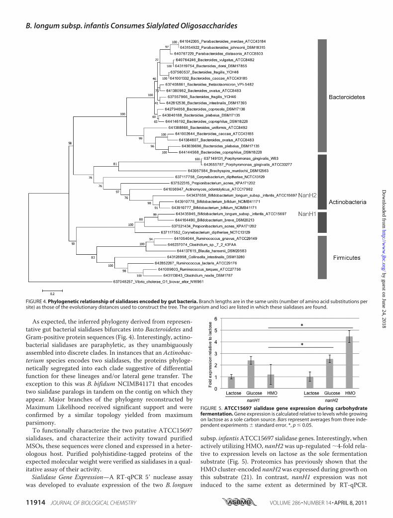
As expected, the inferred phylogeny derived from represen-tative gut bacterial sialidases bifurcates into Bacteroidetes andGram-positive protein sequences (Fig. 4). Interestingly, actino-bacterial sialidases are paraphyletic, as they unambiguouslyassembled into discrete clades. In instances that an Actinobac-terium species encodes two sialidases, the proteins phyloge-netically segregated into each clade suggestive of differentialfunction for these lineages and/or lateral gene transfer. Theexception to this was B. bifidum NCIMB41171 that encodestwo sialidase paralogs in tandem on the contig on which theyappear. Major branches of the phylogeny reconstructed byMaximum Likelihood received significant support and wereconfirmed by a similar topology yielded from maximumparsimony.To functionally characterize the two putative ATCC15697
sialidases, and characterize their activity toward purifiedMSOs, these sequences were cloned and expressed in a heter-ologous host. Purified polyhistidine-tagged proteins of theexpected molecular weight were verified as sialidases in a qual-itative assay of their activity.Sialidase Gene Expression—A RT-qPCR 5� nuclease assay
was developed to evaluate expression of the two B. longum
subsp. infantisATCC15697 sialidase genes. Interestingly, whenactively utilizing HMO, nanH2 was up-regulated 4-fold rela-tive to expression levels on lactose as the sole fermentationsubstrate (Fig. 5). Proteomics has previously shown that theHMO cluster-encoded nanH2was expressed during growth onthis substrate (21). In contrast, nanH1 expression was notinduced to the same extent as determined by RT-qPCR.
FIGURE 4. Phylogenetic relationship of sialidases encoded by gut bacteria. Branch lengths are in the same units (number of amino acid substitutions persite) as those of the evolutionary distances used to construct the tree. The organism and loci are listed in which these sialidases are found.
FIGURE 5. ATCC15697 sialidase gene expression during carbohydratefermentation. Gene expression is calculated relative to levels while growingon lactose as a sole carbon source. Bars represent averages from three inde-pendent experiments standard error. *, p 0.05.
B. longum subsp. infantis Consumes Sialylated Oligosaccharides
11914 JOURNAL OF BIOLOGICAL CHEMISTRY VOLUME 286 • NUMBER 14 • APRIL 8, 2011
by guest on June 24, 2018http://w
ww
.jbc.org/D
ownloaded from

Growth on glucose resulted in a 2-fold induction of bothnanH1or nanH2 expression. This is consistent with a previouslyobserved preference for lactose over glucose that may represssialidases and other carbohydrate-active enzymes.Substrate Specificity and Kinetics of B. longum subsp. infantis
Sialidases—A library consisting of para-nitrophenol (pNP)-tagged sialylgalactosides was screened to characterize NanH1and NanH2 substrate specificities. This sialosides library wassynthesized in a one-pot, three-enzyme reaction to yield sia-logalactosides with different sialyl linkages, and various sialicacid forms. The substrate specificity study was conducted in ahigh throughput screen format using a 384-well plate (43, 44).Briefly, removal of the terminal appropriate sialic acid form bysialidase activity exposes a terminal galactose residue that canbe cleaved by excess �-galactosidase in the reaction mixture torelease para-nitrophenol. Upon adjusting the pH of the reac-tionmixture to�9.5, themajority of the para-nitrophenol pro-duced will be converted to para-nitrophenolate that could bequantified by a plate reader at A405 nm.
The two purified sialidases cleaved both �2–3- and �2–6-linked sialosides, with a consistent preference for the �2–6
linkage (Fig. 6). Both sialidases do not have significant activity(�5%) in cleaving 2-keto-3-deoxy-D-glycero-D-galacto-nonu-losonic acid (Kdn) or its derivatives in either �2–3- or �2–6-linkage. NanH1 generally exhibited lower activity on allassayed substrates. Interestingly, both sialidases cleave�2–3- and �2–6-linked Neu5Gc from compoundsNeu5Gc�2–3Gal�pNP 5a and Neu5Gc�2–6Gal�pNP 5b,but a 5C glycolyl reduces their activities. Surprisingly, anazido group addition on the N-acetyl group of N-acetyl-neuraminic acid (Neu5Ac) does not change the activity ofeither sialidases when Neu5AcN3�2–6Gal�pNP 4b is usedas a substrate, but the activity of both sialidases decreaseswhen Neu5AcN3�2–3Gal�pNP 4b is used as a substrate. Inaddition, a fluorine substitution of one of the hydrogens inthe N-acetyl group of Neu5Ac in both �2–3- and �2–6-linked sialosides Neu5AcF�2–3/6Gal�pNP 2a and 2b doesnot affect the activity of both sialidases. In contrast, the addi-tion of an O-methyl group at the N-acetyl of Neu5Ac in both�2–3- and �2–6-linked sialosides Neu5AcOMe�2–3/6Gal�pNP 3a and 3b decreases the activity of both sialidasessignificantly (Fig. 6).
FIGURE 6. B. longum subsp. infantis ATCC15697 sialidase substrate specificity assay. NanH1 (A) and NanH2 (B) were assayed for activity on a library ofsialosides with either �2,3- or �2,6-sialyl linkages depicted in white and black, respectively. The error bars in the graphs represent the standard errors ofexperimental values obtained from duplicated samples in an individual experiment. Significant differences were determined with a Student’s t test. *, p � 0.05;**, p � 0.01; ***, p � 0.001.
B. longum subsp. infantis Consumes Sialylated Oligosaccharides
APRIL 8, 2011 • VOLUME 286 • NUMBER 14 JOURNAL OF BIOLOGICAL CHEMISTRY 11915
by guest on June 24, 2018http://w
ww
.jbc.org/D
ownloaded from

Sialidase kinetics were characterized using both �2–3-and �2–6-linked sialyllactosyl 4-methylumbelliferol (MU)(Neu5Ac�2–3/6Lac�-Mu). Interestingly, both sialidases hadsimilar affinities toward these two substrates with similar Kmvalues (supplemental Fig. S4). NanH1 was approximately twiceas efficient at hydrolyzing �2–6-linked Neu5Ac (0.22 s�1
mM�1) as �2–3 sialyl linkages (0.13 s�1 mM�1) because ofgreater affinity for this substrate (Table 1). The samepreferencefor �2–6 linkages was exhibited by NanH2, though with a con-siderably higher efficiency reflecting a 175-fold greater turn-over (kcat) rate than NanH1. NanH2 hydrolyzed �2–3 linkagesmore efficiently than NanH1 as well. Interestingly, both siali-dases had similar affinities toward the two� linkages as approx-imated by Km. Furthermore, both sialidases were more activeunder acidic conditions (pH 4.5–6.0), a pH range typical ofseveral bacterial sialidases (Fig. 4).B. longum subsp. infantisNanH1andNanH2SialidaseActiv-
ity on Purified MSO—To link exo-sialidase function as thecommitted step in MSO utilization, NanH1 and NanH2 wereassayed for hydrolysis of purified MSO substrates by massspectrometry. Accordingly, single isomers of SLNT and di-fu-cosylayed sialylated lacto-N-hexaose-like oligosaccharide(DFS-LNH) were isolated from pooled HMO by solid phase
extraction prior to liquid chromatography (Fig. 7a). These twoMSO species, as well as commercially obtained �2–3- and�2–6-linked disialyl-lacto-N-tetraose (DSLNT) (Fig. 7e) wereincubated with both recombinant ATCC15697 sialidases and acontrol �(2–3)-neuraminidase. All three purified MSO specieswere rapidly digested by NanH2 and the control �(2–3)-neuraminidase (Fig. 7, b, c and f, g). Significantly contrastingwith this, NanH1 did not hydrolyze any of the three purifiedsialylated milk oligosaccharides (Fig. 7, d and h) following anextended incubation of 1 h.
DISCUSSION
Gyorgy et al. (1) reported that sialyl moieties protect HMOfrom bacterial consumption. Their HMO-utilizing B. bifidumstrain (strict �2–3 sialidase activity) did not ferment sialylatedmilk oligosaccharides without an in vitro hydrolysis of �2–6linkages predominant inmilk glycans. Therefore it is significantthat B. longum subsp. infantis ATCC15697 utilizes SLNT andproduces the efficient NanH2 �2–6 sialidase active on MSOlinkages. This functionally links substrates incorporated intomilk with the 43-kbp gene cluster previously predicted toenable HMO catabolism (21). To this end, HMO (neutral
FIGURE 7. Digestion of MSO species with 3 recombinant sialidases for 30 min. SLNT and DFSLNH were purified from a pool of HMO and DSLNT was obtainedfrom a commercial source. HPLC fraction before digestion of purified HMO (i.e. SLNT and DFSLNH) (a); digestion of purified HMO with an �2–3-neuraminidase(b); digestion of purified HMO with NanH2 (c); digestion of purified HMO with NanH1 (d); commercially obtained DSLNT prior to digestion (e); DSLNT digestionwith an �2–3-neuraminidase (f); DSLNT digestion with NanH2 (g); DSLNT digestion with NanH1 (h).
B. longum subsp. infantis Consumes Sialylated Oligosaccharides
11916 JOURNAL OF BIOLOGICAL CHEMISTRY VOLUME 286 • NUMBER 14 • APRIL 8, 2011
by guest on June 24, 2018http://w
ww
.jbc.org/D
ownloaded from
and/or acidic) fermentation likely initiates a signal cascade toup-regulate nanH2 expression to utilize MSO.Long regarded as a virulence factor, sialic acid catabolism
confers an in vivo competitive advantage regarded to be criticalto pathogenesis, as recently described in a murine Vibrio chol-erae infection model (45). While saccharolytic commensalspervade the distal infant GIT, utilization of sialylated glycansdelivered to their niche through nursing was previouslyunknown, and may be a significant factor involved in coloniza-tion and persistence. That SLNT ismetabolized in the presenceof highly preferred neutral HMOs signifies that they are of anequivalent value to B. longum subsp. infantis thus tantalizinglysuggestive of a lifestyle predicated on the acquisition of MSO.The progressive disappearance of SLNT during HMO fer-
mentation is a consequence of ATCC15697 metabolism and isnot confounded by extracellular degradation. If spurious diges-tion had occurred, all four monitored MSO compositionswould have been eliminated to the same extent as SLNT, likelyapproaching 100% disappearance as observed in the in vitroassays on purified MSO. This would be similar to the nonspe-cific glycoprofile of the prominent GIT commensal Bacteroidesfragilis that employs several extracellular glycoside hydrolasesto utilizeHMO (46).Moreover, an increase in the abundance ofan LNT-like tetrasaccharide, concomitant with SLNT desialy-lation was not observed (data not shown). Rather, both LNTand free sialic acids disappear during pooled HMO fermenta-tion, with the latter previously demonstrated to enter the fruc-tose-6-phosphate phosphoketolase pathway when utilized as asole carbon source (19, 21).Bacteria competent for sialic acid metabolism derive energy
and biomass from Neu5Ac or sequester it to be deployed asextracellular decoration, cloaked from immunogenic responses(reviewed in Refs. 13, 47). Recently,B. fragiliswas characterizedwith a peculiar Neu5Ac catabolism in which N-acetylman-nosamine epimerase (pfam07221) converts manNAc to N-acetylglucosamine to be subsequently phosphorylated (48).This contrasts with the canonical pathway in which ManNAcis phopshorylated prior to epimerization (49). While theATCC15697 chromosome does not encode an epimerase withsignificant identity, ROK kinases appear in the sialic acid utili-zation cluster (Blon_0644) and upstream of nagA and nagB(Blon_0880) clustering with N-acylglucosamine 2-epimerase(pfam07221; Blon_0875). These kinases, and putative epi-merase, may function in a similar manner to the B. fragilis sys-tem, mitigating the lack of a clearly identifiable nanK genewithin the ATCC15697 chromosome (50).In contrast to NanH2, a linkage between NanH1 and MSO
utilization is less evident, though consistentwith the physiologyof other bifidobacterial species. Specifically, B. breve strainstested to date do not subsist on HMO/MSO despite thepresence of a nanH1 homolog within its genome. B. bifidumstrains, in contrast, utilize HMO and degradeMSO at its extra-cellular surface, ostensibly accomplished with secreted NanH2homologs (19, 42). NanH1 may be active on other sialylatedglycans encountered in the infant gut, be it delivered bymilk orendogenously present. Possible NanH1 substrates include var-ious milk glycopeptides, monosialoganglioside, or colostrumdisialoganglioside, or sialylated mucins.
In any event, the importance of translocation toMSOmetab-olism is accentuated by intracellular localization of B. longumsubsp. infantis sialidase activity. Whereas bacterial sialic acidtransporters have been elucidated (i.e. SiaT (51), NanT (52),SatABCD (38)), transport of acidic oligosaccharides remainsunexplored. The multitude of transporters encoded within theATCC15697 genome present several attractive candidates,including several transport genes within the HMO cluster orothers unlinked in cis. Interestingly, the ATCC15697 locuscomprised of a predicted epimerase and ROK genes upstreamof nagAB are proximal to a solute binding protein, which bindsLNT-like carbohydrates, thus potentially involved in SLNTtransport.5
Until now, direct microbial interactions with milk sialyloli-gosaccharides have been solely characterized in terms of innateimmune function. Inherently shielded by their acidic moiety,MSOs are frequently fucosylated to present a multi-layeredbarrier to microorganisms seeking to metabolize HMO corecomponents (e.g. LNT), an insurmountable prospect in theabsence of fucosidase and sialidase activities. RecalcitrantMSOs are therefore likely concentrated in the distal GIT,potentiallymore so than neutral species that are consumed by amore diverse assortment of microbial scavengers. Thus theability to access these highly restricted carbohydrates may con-tribute to the establishment of bifidobacteria in the nursinginfant gut. If digestible by the infant, HMOs would be a calori-cally dense nutrient for the infant, irrespective of sialylation. Assuch, HMOs are either appropriated for other early develop-mental functions (e.g. immune or neural function) or do notincrease progeny fitness, with the latter in need of reconcilia-tion with the considerable maternal resources expended intheir synthesis. At any rate, the provision of HMO to the neo-nate has evolved either reciprocally or has arisen unilaterally inthe bifidobacteria likely to the benefit of the host.
Acknowledgments—We thank Eric Vimr for helpful discussions andKaren Kalenetra for technical assistance.
REFERENCES1. Gyorgy, P., Jeanloz, R. W., von Nicolai, H., and Zilliken, F. (1974) Eur.
J. Biochem. 43, 29–332. Ward, R. E., Ninonuevo, M., Mills, D. A., Lebrilla, C. B., and German, J. B.
(2006) Appl. Environ. Microbiol. 72, 4497–44993. Bao, Y., Zhu, L., and Newburg, D. S. (2007) Anal. Biochem. 370, 206–2144. Engfer,M. B., Stahl, B., Finke, B., Sawatzki, G., andDaniel, H. (2000)Am. J.
Clin. Nutr. 71, 1589–15965. Bode, L. (2006) J. Nutr. 136, 2127–21306. Morrow, A. L., Ruiz-Palacios, G.M., Altaye, M., Jiang, X., Guerrero, M. L.,
Meinzen-Derr, J. K., Farkas, T., Chaturvedi, P., Pickering, L. K., and New-burg, D. S. (2004) J. Pediatr. 145, 297–303
7. Ruiz-Palacios, G. M., Calva, J. J., Pickering, L. K., Lopez-Vidal, Y., Volkow,P., Pezzarossi, H., and West, M. S. (1990) J. Pediatr. 116, 707–713
8. Barthelson, R., Mobasseri, A., Zopf, D., and Simon, P. (1998) Infect. Im-mun. 66, 1439–1444
9. Mahdavi, J., Sonden, B., Hurtig, M., Olfat, F. O., Forsberg, L., Roche, N.,Angstrom, J., Larsson, T., Teneberg, S., Karlsson, K. A., Altraja, S., Wad-strom, T., Kersulyte, D., Berg, D. E., Dubois, A., Petersson, C., Magnusson,K. E., Norberg, T., Lindh, F., Lundskog, B. B., Arnqvist, A., Hammarstrom,L., and Boren, T. (2002) Science 297, 573–578
10. Simon, P. M., Goode, P. L., Mobasseri, A., and Zopf, D. (1997) Infect.
B. longum subsp. infantis Consumes Sialylated Oligosaccharides
APRIL 8, 2011 • VOLUME 286 • NUMBER 14 JOURNAL OF BIOLOGICAL CHEMISTRY 11917
by guest on June 24, 2018http://w
ww
.jbc.org/D
ownloaded from
Immun. 65, 750–75711. Yolken, R. H., Peterson, J. A., Vonderfecht, S. L., Fouts, E. T., Midthun, K.,
and Newburg, D. S. (1992) J. Clin. Invest. 90, 1984–199112. Schauer, R. (2000) Glycoconj. J. 17, 485–49913. Vimr, E. R., Kalivoda, K. A., Deszo, E. L., and Steenbergen, S. M. (2004)
Microbiol. Mol. Biol. Rev. 68, 132–15314. Lewis, A. L., Desa, N., Hansen, E. E., Knirel, Y. A., Gordon, J. I., Gagneux,
P., Nizet, V., and Varki, A. (2009) Proc. Natl. Acad. Sci. U.S.A. 106,13552–13557
15. LoCascio, R. G., Ninonuevo, M. R., Freeman, S. L., Sela, D. A., Grimm, R.,Lebrilla, C. B., Mills, D. A., and German, J. B. (2007) J. Agric. Food Chem.55, 8914–8919
16. Le Blay, G., Michel, C., Blottiere, H. M., and Cherbut, C. (1999) J. Nutr.129, 2231–2235
17. Falony, G., Vlachou, A., Verbrugghe, K., and De Vuyst, L. (2006) Appl.Environ. Microbiol. 72, 7835–7841
18. Lievin, V., Peiffer, I., Hudault, S., Rochat, F., Brassart, D., Neeser, J. R., andServin, A. L. (2000) Gut. 47, 646–652
19. Ward, R. E., Ninonuevo, M., Mills, D. A., Lebrilla, C. B., and German, J. B.(2007)Mol. Nutr. Food Res. 51, 1398–1405
20. LoCascio, R. G., Ninonuevo, M., Kronewitter, S. R., Freeman, S. L., Ger-man, J. B., Lebrilla, C. B., and Mills, D. A. (2009) Microb. Biotechnol. 2,333–342
21. Sela, D. A., Chapman, J., Adeuya, A., Kim, J. H., Chen, F.,Whitehead, T. R.,Lapidus, A., Rokhsar, D. S., Lebrilla, C. B., German, J. B., Price, N. P.,Richardson, P.M., andMills, D. A. (2008) Proc. Natl. Acad. Sci. U.S.A. 105,18964–18969
22. Markowitz, V. M., and Kyrpides, N. C. (2007) Methods Mol. Biol. 395,35–56
23. Caspi, R., Foerster, H., Fulcher, C. A., Kaipa, P., Krummenacker, M., La-tendresse, M., Paley, S., Rhee, S. Y., Shearer, A. G., Tissier, C., Walk, T. C.,Zhang, P., and Karp, P. D. (2008) Nucleic Acids Res. 36, D623–D631
24. Quevillon, E., Silventoinen, V., Pillai, S., Harte, N., Mulder, N., Apweiler,R., and Lopez, R. (2005) Nucleic Acids Res. 33,W116–W120
25. Wilson, D., Madera, M., Vogel, C., Chothia, C., and Gough, J. (2007) Nu-cleic Acids Res. 35, D308–313
26. Tamura, K., Dudley, J., Nei, M., and Kumar, S. (2007)Mol. Biol. Evol. 24,1596–1599
27. Jones, D. T., Taylor, W. R., and Thornton, J. M. (1992) Comput. Appl.Biosci. 8, 275–282
28. Felsenstein, J. (1985) Evolution 39, 783–79129. Smith, P. K., Krohn, R. I., Hermanson, G. T., Mallia, A. K., Gartner, F. H.,
Provenzano, M. D., Fujimoto, E. K., Goeke, N. M., Olson, B. J., and Klenk,D. C. (1985) Anal. Biochem. 150, 76–85
30. Xie, Y., Liu, J., Zhang, J., Hedrick, J. L., and Lebrilla, C. B. (2004) Anal.Chem. 2004, 5186–5197
31. Clowers, B. H., Dodds, E. D., Seipert, R. R., and Lebrilla, C. B. (2008) Anal.Biochem. 381, 205–213
32. Dodds, E. D., Tassone, F., Hagerman, P. J., and Lebrilla, C. B. (2009) Anal.Chem. 81, 5533–5540
33. Dodds, E. D., German, J. B., and Lebrilla, C. B. (2007) Anal. Chem. 79,9547–9556
34. Parche, S., Beleut,M., Rezzonico, E., Jacobs, D., Arigoni, F., Titgemeyer, F.,and Jankovic, I. (2006) J. Bacteriol. 188, 1260–1265
35. Zhang, J. H., Lindsay, L. L., Hedrick, J. L., and Lebrilla, C. B. (2004) Anal.Chem. 76, 5990–6001
36. Ninonuevo,M., An, H., Yin, H., Killeen, K., Grimm, R.,Ward, R., German,B., and Lebrilla, C. (2005) Electrophoresis 26, 3641–3649
37. Tao,N., DePeters, E. J., Freeman, S., German, J. B., Grimm, R., and Lebrilla,C. B. (2008) J. Dairy Sci. 91, 3768–3778
38. Post, D.M.,Mungur, R., Gibson, B.W., andMunson, R. S., Jr. (2005) Infect.Immun. 73, 6727–6735
39. Taylor, G., Dineley, L., Glowka, M., and Laver, G. (1992) J. Mol. Biol. 225,1135–1136
40. Gaskell, A., Crennell, S., and Taylor, G. (1995) Structure 3, 1197–120541. Mavromatis, K., Chu, K., Ivanova,N.,Hooper, S. D.,Markowitz, V.M., and
Kyrpides, N. C. (2009) PLoS One 4, e797942. Ashida, H., Miyake, A., Kiyohara, M., Wada, J., Yoshida, E., Kumagai, H.,
Katayama, T., and Yamamoto, K. (2009) Glycobiology 19, 1010–101743. Cao, H., Li, Y., Lau, K., Muthana, S., Yu, H., Cheng, J., Chokhawala, H. A.,
Sugiarto, G., Zhang, L., and Chen, X. (2009) Org. Biomol. Chem. 7,5137–5145
44. Chokhawala,H.A., Yu,H., andChen,X. (2007)Chembiochem.8, 194–20145. Almagro-Moreno, S., and Boyd, E. F. (2009) Infect. Immun. 77, 3807–381646. Marcobal, A., Barboza, M., Froehlich, J. W., Block, D. E., German, J. B.,
Lebrilla, C. B., andMills, D. A. (2010) J. Agric. Food Chem. 58, 5334–534047. Severi, E., Hood, D. W., and Thomas, G. H. (2007) Microbiology 153,
2817–282248. Brigham, C., Caughlan, R., Gallegos, R., Dallas, M. B., Godoy, V. G., and
Malamy, M. H. (2009) J. Bacteriol. 191, 3629–363849. Vimr, E. R., and Troy, F. A. (1985) J. Bacteriol. 164, 845–85350. Brigham, C. J., and Malamy, M. H. (2005) J. Bacteriol. 187, 890–90151. Allen, S., Zaleski, A., Johnston, J. W., Gibson, B. W., and Apicella, M. A.
(2005) Infect. Immun. 73, 5291–530052. Martinez, J., Steenbergen, S., and Vimr, E. (1995) J. Bacteriol. 177,
6005–601053. Deleted in proof
B. longum subsp. infantis Consumes Sialylated Oligosaccharides
11918 JOURNAL OF BIOLOGICAL CHEMISTRY VOLUME 286 • NUMBER 14 • APRIL 8, 2011
by guest on June 24, 2018http://w
ww
.jbc.org/D
ownloaded from
German, Xi Chen, Carlito B. Lebrilla and David A. MillsDavid A. Sela, Yanhong Li, Larry Lerno, Shuai Wu, Angela M. Marcobal, J. Bruce
SialyloligosaccharidesAn Infant-associated Bacterial Commensal Utilizes Breast Milk
doi: 10.1074/jbc.M110.193359 originally published online February 2, 20112011, 286:11909-11918.J. Biol. Chem.
10.1074/jbc.M110.193359Access the most updated version of this article at doi:
Alerts:
When a correction for this article is posted•
When this article is cited•
to choose from all of JBC's e-mail alertsClick here
Supplemental material:
http://www.jbc.org/content/suppl/2011/02/02/M110.193359.DC1
http://www.jbc.org/content/286/14/11909.full.html#ref-list-1
This article cites 52 references, 20 of which can be accessed free at
by guest on June 24, 2018http://w
ww
.jbc.org/D
ownloaded from
VOLUME 285 (2010) PAGES 35206 –35215DOI 10.1074/jbc.A110.171769
Plasma kallikrein promotes epidermal growth factorreceptor transactivation and signaling in vascularsmooth muscle through direct activation ofprotease-activated receptors.Rany T. Abdallah, Joo-Seob Keum, Hesham M. El-Shewy, Mi-Hye Lee,Bing Wang, Monika Gooz, Deirdre K. Luttrell, Louis M. Luttrell,and Ayad A. Jaffa
Dr. Hesham M. El-Shewy was inadvertently left off of the authorlist. The correct author list is shown above. Dr. El-Shewy’s affiliationis the Department of Medicine, Medical University of South Caro-lina, Charleston, South Carolina 29425.
VOLUME 286 (2011) PAGES 11909 –11918DOI 10.1074/jbc.A110.193359
An infant-associated bacterial commensal utilizes breastmilk sialyloligosaccharides.David A. Sela, Yanhong Li, Larry Lerno, Shuai Wu, Angela M. Marcobal,J. Bruce German, Xi Chen, Carlito B. Lebrilla, and David A. Mills
The grant information footnote should read as follows. Thisworkwassupported, in whole or in part, by National Institutes of Health NICHDAwards R01HD059127, R01HD065122, and R01HD061923 andNIGMS Grant R01GM076360. This work was also supported by grantsfrom theUniversity of CaliforniaDiscoveryGrant Program,DairyMan-agement Inc., and the California Dairy Research Foundation and byUnited States Department of Agriculture National Research Initiative-Cooperative State Research, Education, and Extension Service Award2008-35200-18776.
THE JOURNAL OF BIOLOGICAL CHEMISTRY VOL. 286, NO. 26, p. 23620, July 1, 2011© 2011 by The American Society for Biochemistry and Molecular Biology, Inc. Printed in the U.S.A.
23620 JOURNAL OF BIOLOGICAL CHEMISTRY VOLUME 286 • NUMBER 26 • JULY 1, 2011
ADDITIONS AND CORRECTIONS This paper is available online at www.jbc.org
We suggest that subscribers photocopy these corrections and insert the photocopies in the original publication at the location of the origi-nal article. Authors are urged to introduce these corrections into any reprints they distribute. Secondary (abstract) services are urged tocarry notice of these corrections as prominently as they carried the original abstracts.