animal sources of human campylobacteriosis
TRANSCRIPT

Copyright is owned by the Author of the thesis. Permission is given for a copy to be downloaded by an individual for the purpose of research and private study only. The thesis may not be reproduced elsewhere without the permission of the Author.

ANIMAL SOURCES OF HUMAN
CAMPYLOBACTERIOSIS
A thesis presented in partial fulfIlment of the requirements for the
degree of
Doctor of Philosophy
in
Veterinary Public Health
At Massey University, Palmerston North,
New Zealand.
FASIUDDIN AHMED
1999

This thesis is dedicated to the memory Of
My Beloved Father

ABSTRACT
New Zealand has one of the highest reported rates of Campylobacter infections in
humans in the developed world. It is the single largest notifiable disease in all regions of
the country. Consumption of poultry meat has been widely implicated both overseas
and in New Zealand as the main cause of human infections. The potential contribution
of other animals especially cattle and sheep is less well known. The present study was
undertaken to fill this gap in knowledge.
Faecal samples from 300 cattle and 158 sheep were collected from local abattoirs and
farms plus 50 samples from the sheep slaughterhouse environment and examined for the
presence of thermophilic Campylobacter spp. Campylobacter spp. were isolated from
45% of the cattle, 44% of the sheep and 56% of the environmental samples. C. jejuni
and C. hyointestinalis were the predominating species isolated from cattle followed by
C. coli and C. lari. In sheep and in environmental samples from the sheep abattoir C.
jejuni was the only species isolated. The isolation rate and the species of
Campylobacter varied between beef and dairy cattle, bull and heifer calves, age of the
heifer calves, and time of the year. The high isolation rate of Campylobacter from the
cattle, sheep and their environment strongly suggests the possibility of these micro
organisms finding their way into milk and meat, as faecal contaminants at the farm and
slaughter level. There is also the potential to contaminate the environment and water
following disposal of abattoir effiuents and run off from farms.
The species of the isolates from human diarrhoeal cases were found to be predominantly
C. jejuni (95%) and C. coli (5%). Molecular typing of C. jejuni using Sma I generated
pulsed-field gel electrophoresis (pFGE) profiles yielded 13 to 16 different patterns in
the cattle, sheep and human isolates showing a large inter-species variation in the
isolates even from the same sources. However, indistinguishable as well as closely
related profiles (pulsotypes) were found across the isolates from cattle, sheep and
humans. The results obtained from the PFGE typing strongly indicate that cattle and
sheep may be important reservoirs of human campylobacter infections. It was also
observed that a few closely related types mostly dominate the C. jejuni populations in

the host animal species. The possibility of faecal contamination from these animals at
slaughter and thus C. jejuni entering the meat was studied.
Retail packs of beef (25), lamb (25) and chicken (50) mince purchased from local
supermarkets were examined. A combined selective enrichment and PCR based
method was evaluated to offer a rapid, sensitive and specific detection method for the
identification of C. jejuni from meats. C. jejuni was detected by culture and PCR in
44% of the chicken, 16% of the lamb and 12% of the beef mince samples. These results
lend credibility to our contention that faecal contamination of sheep and beef carcasses
at slaughter has significant implications for food safety. The much higher· rate of
detection in chicken mince may be related to a higher prevalence of infection in
chickens or to the method of processing which may facilitate spread between birds and /
or between product.
The C. jejuni isolates from the animal and human sources were also examined for
antibiotic resistance by the disc diffusion method to antibiotics commonly used for the
treatment of campylobacter infections in humans. No resistance was detected in the
cattle and sheep isolates. Two human isolates exhibited resistance to tetracycline with
MrCs of>128 J..I.g/ml. All other human isolates were found susceptible to the antibiotics
tested. The nil to negligible resistance detected in the animal and human isolates of C.
jejuni suggest that it is not a major problem in New Zealand at the present time
however, further work is required to examine the situation in more intensively farmed
species and monitor any changes in human isolates over time.
11

Acknowledgements
I consider it a privilege to record my deepest sense of gratitude to Professor Colin
Wilks for his generous support, constant encouragement and constructive counsel all
along the course of investigation and preparation of this manuscript. I would also like
to thank him for his genuine concern for my welfare.
I am indeed thankful to my co-supervisor Stan Fenwick for his help and guidance
throughout my studies as well as to my other supervisors Per Madie and Alan Mu"ay
who have been so generous with their time.
I have been associated with several people during this long period and every one has
contributed something towards this investigation. I would like to thank Jane Hunter,
Eammon Gormley, Kevin Stafford, Jacek Gwozdz and Hassan Hussein for their help.
Thanks are also due to the team of microbiologists at the Palmerston North Medical
laboratory who have provided the human isolates of Campylobacter and to Ayad
Alkaissi at the Meat works in collection of samples from animals.
Assistance in the laboratory provided by Magda Gwozdz, Kylie Walker, Jan Schrama
and Laurie Sandall is highly appreciated Special mention should be made of Peter
Wildbore who had been most helpful in procuring the most important to the trivial with
equal zeal.
I would like to specially thank Allain Scott for her readiness to help and advise in
matters of academic complexities and also to the other secretarial staff at the institute
for their help.
A big thanks to all my friends in room 2.01 and in the university for their enjoyable
company and support.
A special thanks to my wife Kavita for her constant encouragement and support in times
of great stress and in giving two lovely children Shireen and Rehan during this period
who have been the source of infinite joy.
III

The completion of this thesis is also due to the support, encouragement, and sound
advice I received from my family especially my beloved parents to all of them I am
deeply indebted
And last but not the least, I remember the Almighty who gave me strength, courage and
perseverance to achieve this goal.
IV

CONTENTS
Page
ABSTRACT ........... ........ . . . . . . .......................... . . ... . . . . . ........ . . . . . . ............ . . . . . . . . ..... i
ACKNOWLEDGEMENTS . . . . . . . . . . . . . . . . . . . . . . . . . . . . . . . . . . ............ .. . . . . . ... ........ iii
TABLE OF CONTENTS .. ...... .................... . . . . . . ..... . .. . . . . .. .. . . . . .. .. ............. . . . .. . .. v
LIST OF TABLES . .. ... .. ........ ........................... ..... . . . ...... . ....... . ... . .... ... ..... . ...... . x
LIST OF FIGURES .......................... . . .......... .. . . .. ......... ..... . . . ........ . . ................. xi LIST OF ABBREVIATIONS . . .. . . . .... . . . . . .... ........ .. .. . . .. .. ........ .. . . . . . . . . . . ........ .. .... xii
CHAPTER ONE: INTRODUCTION . . . .. . . . . . . .. . . ... . . ...... ........... .. ...... ....... . .. .... 1
1. 1 Introduction . ... . . . . . . . . . . . . . . . . . . ....... . . . . . . . ........... .... . . .. . . . . . . . . . .......... . . ............ ........ . 1
1.2 Objectives of this study....... ......... . . . . . . . . . . . . . . ................... ...... ............. . . . . . ...... 5
CHAPTER TWO: REVIEW OF LITERATURE . ... .......... .... . . .... .. .......... . . .. 6
2�General characteristics of Campylobacter .................................................... 6
2.2 Species of the genus Campylobacter ..... .. ..... . . . . ... . ... . . . . . . ... ............. .. ...... ...... 6
j' ources of infection for people .. . . . ........... . . . . . .... . . . . . . . . . ...... . . . . . . . . ... ..... ............ 7
2.4 Animal carriage of Campylobacter species... . . . . . ........ . . ..... .. ... . . . . ..... . . . . . . . . . . . . . 9
2.5 Milk and meat as sources of infection . .... . . . ... .. . ... ...... . . . . . . . . ............... ... . . . ... .. 1 1
2.6 Isolation and identification of Campylobacter .. .... ... .... ...... . . . . .... .... .. . . ...... ... 13
2.6. 1 Isolation .......... ..... . . ... . . . . . . .... .... . . . . . . . . . .................................. . .... . . . . . . . . . . .. . 14
2.6.2 Serotyping . . . . . . . . . .......... . . . ... . . . . . . . .. . . . . . . . . ... .. . ... ...... . . . . . . . . ...... . . .... .. . . . . . . . . .... 15
2.6.3 Biotyping ..... . . . . . . . ..... . . . . . . . ............ . . . .................. . . . .................... . . . . . . . . .... 16
2.6.4 Phagetyping . ... .... . .. ........... .. .... . . . . . . . . . . . . . . .. . . . . . . . . . . . . ........ . . . . ..... . . . .......... . . . 17
2.6.5 Whole cell protein profiles .. . . . . . ..... . . . ...... ................... . . . . . . . . ................. . 17
2.6.6 Plasmid profile analysis . .. . . . . . . ... . . ..... . . .......... ... . .. .. . ............. ....... . ... . . . .... 18
2.6.7 Multilocus enzyme electrophoresis .. . . ... . .. ... . ........ .. . . .... ........ . ... .. . . . . ..... .l8
v

2.6.8 Restriction endonuclease analysis .. .......... . . .. ... ........... . . .. .. .. ... .... ........... 19
2.6.9 Ribotyping . ....... . . ... .... ............. . . . . . .. ... ... . .... ... ....... . . . . . .. . .. .............. ...... .... 20
2.6. 10 Pulsed field gel electrophoresis . . . ..... ....... ...... . . .. ... .. . ... ....... ...... . .......... 2 1
2.6. 1 1 Random amplified polymorphic DNA................................... ... . ... ..... 22
2.6. 12 Polymerase chain reaction ...................... . .. ...... ................. ............... . . .. 24
2.6. 13 Nucleic acid sequence based amplification ................. ............ . ... ........ 27
2.8 Antibiotic resistance among Campylobacter species .... . .. . ......... ................... 29
CHAPTER THREE: ISOLATION AND IDENTIFICATION OF
CAMPYLOBACTER SPECIES FROM FAECES OF CATTLE, SHEEP AND HUMANS •....•••••••.•••••••••.•••••.••••••••.••••••••...••.•....••••..••..•••...•...•.•.•.•• •.••••••.••••••••.•••. 34
3.1 Introduction .... .............. ........... . ..... .... ................ ...... ......................... ...... ...... 34
3.2 Materials and methods................................................................................ 36
3.2. 1 Sampling . ............... ................... ..... ........... .......... . ...... ..... ........ ............ ... 36
3.2.2 Human clinical isolates .. ........ ................ ...... .................. ....... ........... ...... 37
3.2.3 Isolation techniques ......... ...... ..... .. . . ................ ..... ......................... .......... 37
3.2.4 Identification of Campylobacter ........ .. ............... ....... .... .. .. ........ ............ . 38
3.2.4.1 Presumptive identification of intestinal thermophilic
Campylobacter ... ...... .. . .. . ...... . ........ ... .. .. . . .... . . .. .... . ..... .. . . ............... . 38
3.2.4.2 Confirmative identification and species differentiation of the
thermophilic Campylobacter . ..... . . ... .... . .. . . ..... .............................. 39
3.3 Results . . . ............ ........................... . . . .. . . . . .. . . . . ........ . . . . . . . ................... ................. 40
3.3. 1 Results of Campylobacter isolation from beef cattle . . . .......... .. . .. . . ... ...... 40
3.3.2 Results ofCampylobacter isolation from dairy cattle .... ... ... ........ .......... 4 1
3.3.3 Results ofCampylobacter isolation from heifer calves . . ............ ... ......... 4 1
3.3.4 Results ofCampylobacter isolation from bull calves .................... ... ...... 45
3.3.5 Results of Campylobacter isolation from sheep .... ........ ...... ............... .... 45
3.5.6 Results of Campylobacter isolation from sheep environmental
samples . . ....... .... ........ . . .. . .. ........ ....... . . . .. . . . . ................... . . . .. . . . . . . . .. ............... 45
3.5.7 Results of identification of Campylobacter spp. from humans .......... . . . . . 45
3.4 Discussion .......... ............ ................ . . ...... ..... ...... ..... .............. ... .... ..................... 46
VI

CHAPTER FOUR: TYPING OF CAMPYLOBACTER JEJUNI ISOLATES FROM
CATTLE, SHEEP AND HUMANS BY PULSED-FIELD GEL
ELECTROPHORESIS (pFGE) ............... _ .. _ ............................ _.............. 52
4.1 Introduction....... ...................... . . . . . . . . .. .. . . . . . ....... . . . . . . . . . . . . . . . . ........ . . . . . .. . . . . . .. 52
4.2 Materials and Methods......................................................................... 53
4.2. 1 Bacterial isolates ................... .... . . . . .... . . . . . . . . . . . . . . . . . . . . . . . . . . . . . .. ... . . . . . .. . . . .. 53
4.2.2 DNA preparation.. ............................ . . . . . . . .. . . . . .. ... . . . . .. . ... . . . . . . . . .. . . . . . . .. 53
4.2.3 Restriction endonuclease digestion of plug-incorporated DNA ..... 54
4.2.4 Pulsed-field gel electrophoresis of digested DNA ....... .................. 55
4.2.5 Interpretation of pulsed-field profiles ............................................ 56
4.3 Results .................................................................................................... 56
4.3. 1 Results ofPFGE of C. jejuni strains isolated from cattle.. . ....... . . . . . 56
4.3.2 Results ofPFGE of C. jejuni strains isolated from sheep ............... 64
4.3.3 Results ofPFGE of C. jejuni strains isolated from humans ............ 64
4.4.4 Comparison of the PFGE profiles of C. jejuni isolates from animal
and human sources.............................. ............................................. 64
4.4 Discussion ........... . . . . . . . . . . . . . . . . . . . . . . . . . . . . . . . . . . . . . . . . . ... . . . . . . . . . . . . . . . . ....... . . . . . . . .. . . ..... . 65
CHAPTER FIVE: ANTIMICROBIAL SUSCEPTIBILITY PATTERNS OF
CAMPYLOBACTER JEJUNI ISOLATED FROM CATTLE, SHEEP AND
HUMANS....................................................................................................... 70
5.1 Introduction. ...... . . . ... . . . . . . . . . . . . . . . . . . .. . . .. . . . . . . ...... . . . . . . .. . . .... ... . . . . . . . . . . . . . . ... . . .. . . . . . . . 70
5.2 Materials and Methods......... ...... . . . . . . . . . ... . . . . . . . . .. . . . . . . .. .... ..... . . . . . . . .. . . . . . . . . . . . . . 7 1
5.2. 1 Bacterial strains .............................. .............................. ..................... 7 1
5.2.2 Antibiotic susceptibility testing ............................. ............ . ... . . ...... .... 7 1
5.2.3 Minimal inhibitory concentration ............................. ........................ 72
5.3 Results ................................. ............... ........................................................ 73
5.3. 1 Results of antimicrobial susceptibilities of human, cattle and
sheep isolates .............................................................. ....................... 73
vu

5.3.4 Results of determination of the minimal inhibitory concentrations
of the resistant isolates ...................................................................... 73
5.5 Discussion .. ............. .. ................ ... .. .... ..... ..... ............ .. .... ....... . .................... 74
CHAPTER SIX: DEVELOPMENT OF POLYMERASE CHAIN REACTION
FOR IDENTIFICATION OF CAMPYLOBACTER JEJUNI FROM MEAT
SAMPLES . ............... ........ ... ......... ...... . .......... ....... .... ...... ... ... .. ...... ... ....... ... .. .... ... 80
6.1 Introduction............. .. .. .......... ....... ....... ...................... .... .............. .............. ... 80
6.2 Materials and Methods............................................................................... 80
6.2.1 Oligonucleotide primer selection............................ .............................. 80
6.2.2 DNA extraction from bacteria................. .............................................. 81
6.2.2.1 DNA extraction based on proteinase-K digestion and
phenol-chloroform purification.................................................. 81
6.2.2.2 DNA extraction by boiling method ........... .... .... .................. ....... 82
6.2.2.3 DNA extraction using QIAGEN DNA kit ........... .... ................... 82
6.2.3 peR reaction components ....................................................................... 83
6.2.3.1 Taq DNA polymerase system ..................................................... 83
6.2.3.2 HotStarTaq DNA polymerase system ......................................... 83
6.2.4 Optimisation procedures .......... .............. .................. ............ .......... ...... .... 83
6.2.4.1 Magnesium concentration ........ ..... ....... ..... .......... .... ..................... 83
6.2.4.2 Denaturation, annealing and extension times and temperature .... 83
6.2.4.3 Number of amplification cycles.... ..... ...... ........ ...... .............. .... ..... 84
6.2.5 Standard peR reaction components and conditions .................................. 84
6.2.6 Specificity and sensitivity of the PCR ....................................................... 85
6.2.7 Generation and labelling of probe .............................................................. 87
6.2.8 Validation and identity of735 bp PCR product ......................................... 87
6.2.9 peR product analysis ................................................................................. 88
6.3 Results ... . . ......... ............. .......................................... . . . .... ............... . . .. . . . . ...... . . ..... 89
6.3.1 DNA extraction .......................................................................................... 89
6.3.2 Polymerase systems.... ................. ........ .......... .............. .......... .......... ....... .... 89
6.3.3 Optimisation of peR ............................................................................... 95
Vlll

6.3.4 Sensitivity and specificity of PCR ........... .... ............... ...... .................. .... 95
6.3.5 Validation and identity of PCR product ................ ............ . .................. ... 95
6.4 Discussion ..... ........ ....................... ........................ ............................. ............... 96
CHAPTER SEVEN: SURVEY OF RETAIL MEATS FOR THE PRESENCE OF
CAMPYLOBACTER JEJUNI BY CULTURE AND POLYMERASE CHAIN
REACTION METHODS ....................... _ .................. _.......................................... 99
7.1 Introduction ................. . . . . . ........................... .. ....... ............................................. 99
7.2 Materials and methods .... ................... ...... ............. ...... ..................................... 100
7.2.1 Collection of samples. .... .................... .............. ................ ...... ........ ...... ...... 100
7.2.2 Processing of samples. ....... ...... ........ ............ .... ................ ........ ...... ............ 100
7.2.3 Phenotypic identification of isolates .............. ............................................. 101
7.2.4 Extraction of DNA for PCR .... .......... ......... ..... ....................... .... . .. . .. ... .... . . . 101
7.2.5 Measurement of DNA concentration .......... ........... . ................ .. ................. 101
7.2.6 Polymerase chain reaction .............. .... . . .. ..... ...... .............. ..... .. ................. ... 102
7.2.7 Gel electrophoresis of PCR products ......................................................... 102
7.2.8 Dot blot hybridisation ............. ................................................................... 102
7.3 Results ....................... ............ ............................................................................. 103
7.3.1 Results of the screening of poultry samples .............................................. 103
7.3.2 Results of the screening of beef samples . .............. ........ ........ ..................... 103
7.3.3 Results of the screening of lamb samples ........... ....... ............... ................... 103
7.3.4 Results of the screening by PCR ....... ......... ...... ..... ........................ ..... ......... 103
7.4 Discussion . . . . . . . . ........ . . ............... .............. .................... ................. ...................... . . 108
CHAPTER EIGHT: GENERAL DISCUSSION / SUMMARy.......................... 1 12
REFERENCES ...................................................................... ....................................... 120
APPEND.IX. .•.................................................................•.......•..............•..............•..... 144
IX

LIST OF TABLES
Chapter 3
3.1 Reported carriage rate of Campylobacter species in adult
cattle in various countries . . . . . . . . . . . . . . . . . . . . . . . . . . . . . . . . . . . . . . . . . . .. ...... .... 35
3.2 Protocol used for the identification of thennophilic
Campylobacter species . . . . . . . . . . . . . . . . . . . . . . . . . . . . . . . . . . . . . . . . . . . . . . . . ........... 40
3.3 Showing the percentages of isolations of Campylobacter species
from adult cattle, calves and sheep . . . . . . . . . . . . . . . . . . . . . . . . . . . . . . . . .... ......... 42
3.4 Results of Sheep abattoir environment sampling . . . . . . . . . . . . . . . . . . . . . ........ 43
3.5 Isolations of Campylobacters in different months in cattle . . . . . ........... 44
Chapter 4
4.1 Gel running parameters . . . . . . . . . . . . . . . . . . . . . . . . . . . . . . . . . . . . . . . . . . . . . . . . .......... 55
4.2 Types and pulsotypes of C.jejuni among 35 cattle isolates . . . . . . ........... 57
4.3 Types and pulsotypes of C.jejuni among 50 sheep isolates . . . . . . ........... 58
4.4 Types and pulsotypes of C.jejuni among 50 human isolates . . . . . . .......... 59
Chapter 6
6.1 Bacterial species and sources of isolates used in the
assessment of the PCR specificity and sensitivity . . . . . . . . . . . . . . . . . . . . ... . . ... 86
Chapter 7
7.1 Results of isolation of C. jejuni from different meat
samples by culture and PCR . . . . . . . . . . . . . . . . . . . . . . . . . . . . . . . . . . . . . . . . . . ......... 104
x

LIST OF FIGURES
Chapter 4
4.1 Representative PFGE profiles of Cjejuni cattle isolates.. . . . . . . . . . ..... .... . 60
4.2 Representative PFGE profiles of Cjejuni sheep isolates . .. . .. .. . . . . . . . . . .. . . 6 1
4.3 Representative PFGE profiles of Cjejuni human isolates . . . .... .......... . . . . 62
4.4 Common PFGE profiles in humans, cattle and sheep isolates . . . . . . . . . . . . .. .. 63
Chapter 5
5.1 PCR for Tet 0 gene in resistant C jejuni isolates . . . . .. . ... . . . .. . . . . . . . . . . . ... . ... . .. . 75
Chapter 6
6.1 1. 5% agarose gel with PCR products amplified by the AH 1
and AH2 primers using DNA extracted by different methods
from C.jejuni ....... . . . . . .. .' . . .. . .. . .. . . . . . . . .. . . .. .. .. . . .. .. . .. . . . . .. . . . ...... .......... 90
6.2 1. 5% agarose gels showing PCR products using the standard
Taq polymerase and HotStarTaq polymerase systems . .. . .. .. . . . . . . . ... . .... .. .. . 9 1
6.3 Optimisation of magnesium concentration .. .. . .. .. . . . . . .. . . .. . .. . . .. . ... ... . . . . . . . . 92
6.4 Sensitivity of the PCR using purified DNA . .. . .. . . . . .. . . . . . . . .. . .... ....... .. . . . . . 93
6.5 Specificity of the PCR . . .. .. . . . . . . . . . .. . .. . . . . .. . . . . . . . . . . . . . . . . . . . . . .. . . . .. . ... .. .. .. . 94
Chapter 7
7.1 PCR of chicken mince . . . . . . .. .. . .. .. . . . . . . .. . . . . .. .. .. ... . .. .. . .. ... . . . . ... . ... . . ... .. 1 05
7.2 PCR of lamb mince . . . . . .. . .. . . . .. . .. . .. . . . . . . . . . . . . . . . . . . . .. . . .. . . . . .. .. . .. . .. . . . .. . . . 1 06
7.3 PCR of beef mince . . . . . . . . . . . . . . . . . . . . . . . . . . . . . . . . . . . . . . . . . . . . . . . . . . . .................. 1 07
Xl
PFGE
PCR
NASBA
CCDA
CCVA
CSM
HS
HL
. FBP
aMP
SDS-PAGE
DNA
MEE
REA
RNA
HACCP
RFLP
ERIC
RAPD
REP
GBS
ELGA
CBF
TSI
EDTA
BSA
CHEF
TBE
MIC
DIG
bp
LIST OF ABBREVIATIONS
Pulsed-field gel electrophoresis
Polymerase chain reaction
Nucleic acid sequence based amplification
Charcoal-cefoperazone-desoxycholate agar
Campylobacter-cefoperazone-vancomycin amphotericin
Charcoal selective media
Heat stable
Heat labile
Ferrous metabisulphite pyruvate medium
Outer membrane protein
Sodium dodecyl sulphate-polyacrylamide gel electrophoresis
Deoxyribonucleic acid
Multilocus enzyme electrophoresis
Restriction endonuclease analysis
Ribonucleic acid
Hazard analysis critical control point
Restriction fragment length polymorphism
Enterobacterial repetitive intergenic consensus
Random amplified polymorphic DNA
Repetitive extragenic palindrome
Guillain-Barre syndrome
Enzyme l inked gel assay
Campylobacter blood free agar
Triple sugar iron agar
Ethylene diamine tetra acetic acid
Bovine serum albumin
Contour clamped homogenous electric field
Tris borate EDT A
Minimal inhibitory concentration
Digoxigenin
Base pair
xi i

Chapter-l Introduction
CHAPTER 1
1 .1 . INTRODUCTION
The epidemiology of foodbome diseases is rapidly changing. In the past two decades,
newly recognised pathogens such as Campylobacter jejuni, Escherichia coli 0157:H7
and Yersinia enterocolitica have emerged as important public health problems
worldwide. Well-recognised pathogens such as Salmonella have increased in
prevalence or become associated with new food sources and some pathogens are
becoming increasingly resistant to antimicrobial agents. Many pathogens, including
Salmonella spp., Escherichia coli 0157:H7, Campylobacter spp. and Yersinia
enterocolitica have reservoirs in healthy food animals from which they spread to an
increasing variety of foods (Todd, 1 997).
These pathogens cause millions of cases of acute, sporadic illness and chronic
complications as well as large and challenging outbreaks in many countries worldwide.
In the past the main challenge of foodbome disease lay in preventing the contamination
of animal-derived food for human consumption. It is likely that in the future, prevention
of foodbome disease will be increasingly dependent on controlling contamination of
feed and water consumed by the animals themselves (Buzby and Roberts, 1 997).
Evolving trends in foodbome diseases are being driven by the same factors that have led
to the emergence of other infectious diseases including changes in the demographic
characteristics of populations, human behaviour, industry and technology and the shift
towards a global economy, microbial adaptation, and the breakdown in the public health
infrastructure (Kaferstein and Abdussalam, 1 999).

Chapter-l Introduction
Many of the re-emerging or newly-recognised pathogens are foodbome or have the
potential to be transmitted by food and/or drinking water. The emergence of "new"
foodborne pathogens can be expected because of changing production methods and
processes, food handling practices and eating habits. Foodbome illness of microbial
origin is a serious problem in the United States with 79% of outbreaks between 1 987-92
being bacterial and with improper holding temperature and poor personal hygiene of
food handlers contributing most to the disease incidence (Collins, 1 997). The global
economy also, has facilitated the rapid transport of perishable foods, increasing the
potential for exposure to foodborne pathogens from other parts of the world (Altekruse
and Swerdlow, 1 996).
However, an even greater challenge to food safety will come from changes resulting
directly in degradation of sanitation and in the transformation of the immediate human
environment. Changes include the increased age of the human population, unplanned
urbanisation and migration, and mass production of food due to population growth and
changed food habits (Kaferstein and Abdussalam, 1 999).
A pathogen may emerge as an important public health problem because of changes in
the pathogen itself or in its transmission pathways. Alternatively there may be changes
in host susceptibility to infection. Factors influencing host susceptibility within the
population as a whole include increases In the number of immunocompromised patients,
increased use of immunosuppressive agents, ageing of the population and malnutrition
(Morris and Potter, 1 997).
A better understanding of how the pathogens persist in animal reservoirs and enhanced
methods of laboratory identification are important in successful long-term prevention.
Sensitive and timely surveillance that combines rapid subtyping methods, cluster
identification and collaborative epidemiological investigations can identify and halt
large, dispersed outbreaks.
Campylobacter species have long been recognised as a cause of illness and abortion in
animals (MacFadyean and Stockman, 1 9 1 3). In the last two decades, awareness of the
2

Chapter-J Introduction
important role of Campylobacter species in human disease has been increasing (Healing
et al. , 1 992). Campylobacter jejuni and Campylobacter coli are now recognised
amongst the most common bacterial causes of diarrhoea in the world (Tauxe, 1992).
More recently, there has been a growing appreciation that many Campylobacter species
besides C. jejuni may also be human pathogens (Mishu et al., 1 992), but the incidence
of infection and relative importance of these other species is unclear (ACMSF, 1 993).
Campylobacter infection can result in serious illness that may last for more than a week
(Ketley, 1 995). The clinical signs in the majority of patients are characterised by acute
abdominal pain often with fever and general malaise which progresses to a profuse
diarrhoea (Humphrey, 1995). Bacteremia is infrequently reported. The disease is
usually self limiting (Reina et al., 1 994), but a few patients develop late complications,
which are probably triggered by an auto-immune response to specific campylobacter
antigens, notably reactive arthritis (Highton and Priest, 1996), or more seriously,
peripheral polyneuropathy known as the Guillain-Barre' syndrome (Kuroki et al., 1 99 1 ),
and the Miller-Fischer syndrome (Salloway et al., 1 996) a neuropathy associated with
ataxia, areflexia and ophthalmoplegia.
Between 1980 and 1995 surveil lance data of food-borne disease in Australia shows that
Campylobacter species were the most commonly reported pathogen (Crerar et aI., 1996)
and similarly Campylobacter has been the commonest enteric pathogen isolated from
humans in England and Wales since 198 1 (Pebody et al., 1 997). Campylobacteriosis is
a serious public health concern in all areas of New Zealand. It is the most common
notifiable, foodborne disease and appears to be increasing in frequency. The incidence
of human cases in this country is one of the highest in the industrialised world (Ikram et
al., 1994). The cost of the disease to the New Zealand society is estimated to be $ 1 6
million per year (Withington and Chambers, 1 997).
In a recent national study of sporadic disease, campylobacter infection was mostly
associated with consumption of raw milk or undercooked chicken (Eberhart-Philips et
al. , 1 997). Outbreaks of the disease have also been attributed to consumption of
untreated water (Ikram et aI. , 1 994; Stehr-Green et al., 1 99 1 ; Brieseman, 1 987) and raw
3

Chapter-i introduction
milk (Brieseman, 1984). Campylobacter organisms found in surface water most
probably originate from wild and domestic animals (Park et aI., 1 99 1 ), water run-off
from farmland following heavy rainfall or effluent from sewage purification plants
(Koenraad et aI., 1995; Jones et aI., 1990).
Analysis of the literature indicates that the majority of the cases of human
campylobacteriosis are associated with poultry, however, no single source of food of
animal origin can be excluded as a potential vehicle of infection for humans. Outbreaks
of campylobacteriosis have also been associated with consumption of unpasteurised
cows milk in many parts of the world as well as New Zealand, but there is little
information about the intestinal carriage of Campylobacter spp. in cattle, including dairy
cows.
An alarming trend found overseas has been the increase in resistance of Campylobacter
spp. to antibiotics used in the treatment of human infections (Roge et al. , 1 998;
Velazquez et aI., 1995). This has been attributed to the use of antibiotics in animals as
growth promoters and in prophylaxis against diseases particularly in intensively housed
species like pigs and poultry (Tollefson et al., 1 998). It has been demonstrated that the
flow of resistance genes in the environment from animals to human beings is possible
(John son et aI., 1 994; Linton, 1986; Scoli et aI., 1 980). In a recent study, low level
resistance to erythromycin, ciprofioxacin and doxcycline was seen in Campylobacter
isolates from people living in Auckland (Dowling et al., 1 998).
The traditional approach to the identification of Campylobacter species is based on
biochemical tests, resistance patterns and growth temperatures. Variation in a single test
may result in misidentification of the strain. The methods are also labour intensive and
time consuming. Therefore a rapid method of identification of the organism would be of
immense value.
4

Chapter-l Introduction
1 .2. OBJECTIVES OF THIS STUDY:
The overall objective was to detennine whether or not cattle and sheep constitute a
source of Campylobacter spp. for humans in New Zealand. The specific objectives to
address this major question were therefore:
1 ) To determine the prevalence of Campylobacter spp. in the intestinal contents of
cattle and sheep.
2) To determine if Campylobacter spp. are present in retail meat derived from these
species in New Zealand.
3) To determine whether or not the animal isolates of C. jejuni are the same as human
isolates by molecular typing using Pulsed-Field Gel Electrophoresis (PFGE) of
genomic DNA.
4) To develop a rapid method of detection and identification of C. jejuni from meats
by utilisation of the polymerase chain reaction (peR).
5) To study the antimicrobial susceptibility patterns of Campylobacter isolates derived
from animals and from human clinical cases of campylobacteriosis.
5

Chapter-2 Review of Literature
C HAPTER 2
2. REVIEW OF LITERATURE
2.1. GENERAL CHARACTERISTICS OF CAMPYLOBACTER
The genus name Campylobacter, derived from a Greek word for curved rod, was
proposed by Sebald and Veron (1963), to include microaerophilic bacteria that were
different from Vibrio cholerae and other vibrios.
Campylobacter are Gram-negative bacteria, 0.5 to 0.8 !J,m long and 0.2 to 0.5 !J,m wide
with characteristically curved, spiral or S-shaped cells (Sebald and Vei-on, 1963). The
motility of the bacteria is characteristically rapid and darting in a corkscrew fashion, a
feature by which they can be distinguished from other motile bacteria (Karmali and
Fleming, 1979). Microaerophilic characteristics require propagation in an atmosphere
comprising 5 % oxygen, 10% carbon dioxide and 85 % nitrogen (prescott and Munroe,
1982).
2.2. SPECIES OF THE GENUS CAMPYLOBACTER
The genus Campylobacter contains eighteen species and subspecies, most of which have
been isolated from humans, but not all are associated with disease (Skirrow, 1994).
C. jejuni and C. coli, are common causes of bacterial diarrhoeal disease in humans in
many countries around the world (Tauxe, 1992). C. jejuni has also been isolated from
cases of bacteraemia, appendicitis and recently has been associated with the Guillain
BaITt� syndrome (Fujimoto et al., 1992).
The type species, C. fetus, is an important cause of abortion in sheep and cattle but is
infrequently recovered from apparently healthy humans. The typical manifestation of the
6

Chapter-2 Review of Literature
disease caused by C. fetus in humans is that of a low-grade bacteraemia, especially in the
elderly or in patients receiving immuno-suppresive therapy. It may cause septic abortion,
meningitis, salpingitis and diarrhoea (Lior, 1 994).
C. lari was first described by Skirrow and Benjamin ( 1 980), as a thermophilic
Campylobacter species that is distinct from C. jejuni and C. coli in several ways, most
notably in its resistance to nalidixic acid. C. lari is now recognised as a potential human
pathogen frequently isolated from stools of diarrhoeal patients and also reported to cause
fatal bacteraemia in immuno-compromised patients ( Borczyk et aI., 1 987; Tauxe et al.,
1985).
In 1 98 1 , Sandstedt and Weirup, reported the isolation of thermotolerant, catalase
negative Campylobacter spp. from dogs with and without diarrhoea. C. upsaliensis is
now implicated in cases of diarrhoea and bacteraemia in immuno-compromised and
healthy humans (Goossens et aI., 1 990).
Another thermophilic species, C. hyointestinalis, has been identified as a possible cause
of proliferative enteritis in pigs (Gebhart et al. , 1983), and has been frequently isolated
from other animals. It is suggested to be an important cause of diarrhoeal disease in
homosexual men, immuno-compromised patients and the population at large (Edmonds
et al. , 1 987; Fennel et al. , 1 986).
2.3. SOURCES OF INFECTION FOR PEOPLE
The presence of Campylobacter spp. as commensals in the intestinal tracts of a wide
range of birds and mammals (Glunder et aI., 1 992; Yogasundram et al. , 1 989), including
animals most widely used for food production (Stem et aI., 1 992), and as pets (Whelan
et aI. , 1 988), open up several pathways of infection to humans. Campylobacter spp. can
survive in the environment for several weeks at temperatures around 4°C and can cause
human infection when consumed in untreated water and milk (Skirrow, 1 994). Infection
can also be acquired by direct contact with infected animals and is usually associated
with particular occupational groups.
7

Chapter-2 Review of Literature
Campylobacter enteritis is essentially a food borne disease and the principal vehicle of
infection is raw or undercooked meat (Deming et al., 1987) . Any raw meat may be
contaminated with Campylobacter and poultry products are by far the most important
source (Bryan and Doyle, 1 995; Harris et al. , 1 986) . Retail chickens have been found to
have contamination rates of 60-88% with counts in the region of 1 03_ 105 cfu per chicken
carcass (Oosterom et al. , 1 983) .
Neilsen et at. ( 1 997), in a nation-wide survey in Denmark, found the intestinal carriage
rate of Campylobacter in cattle to be 47%, chicken 36% and' swine 46%. C. jejuni was
found to be the predominant species, accounting for 83 to 91 % of the cattle and poultry
isolates while C. coli accounted for 95% of the isolates from pigs. In human patients
with campylobacter enteritis, 94% of the isolates were C. jejuni and 6% C. coli.
Serotyping of the isolates revealed identical serotypes in chickens and cattle and also
these serotypes overlapped with the serotypic distribution of human isolates.
Seventy four serogroups of thermophilic Campylobacter spp. were isolated from human
and animal sources in Israel (Rogol and Sechter, 1 987). Six of these serogroups were
found frequently in chickens and four were common to cattle. C. jejuni accounted for
86. 7% to 92. 1 % of these isolates.
Rosef et at. ( 1 985), compared 42 serotypes from different animal species to human
clinical isolates. The highest degree of similarity was observed in the poultry isolates
followed by strains from wild birds, flies and pigs.
Jones et al. ( 1 984), compared biotypes and serotypes of Campylobacter spp. isolated •
from patients with enteritis and from animal and environmental sources. They found that
most of the human strains were C. jejuni and common serotypes were frequent among
strains isolated from animal and environmental sources. However, they could be
different strains that are of the same serotype and would be possibly differentiated by
molecular methods.
8

Chapter-2 Review of Literature
2.4. ANIMAL CARRIAGE OF CAMPYLOBACTER SPECIES
As the recognition of the importance of Campylobacter spp. as a' cause of diarrhoea in
humans grew, investigations of carriage of Campylobacter in animals became more
urgent. Aeschbacher and Piffaretti (1989) , have indicated in a report that every animal
strain of Campylobacter should be considered a potential human pathogen, However,
the pathogenic potential of many Campylobacter strains has yet to be established so it is
not possible to arrive at this conclusion with certainty.
Jones et al. (1999) , studied the shedding of Campylobacter spp. by sheep at pasture in
the UK, Overall shedding of campylobacters was between a third and half of the carriage
rate of 92 % determined at slaughter. Rates of shedding depended on the type of pasture,
and time of year. The highest rates (100%) were observed at the time of lambing,
weaning and movement onto new pasture and the lowest rates (0%) occurred when the
sheep were fed on hay and silage. C. jejuni was the main species isolated and was found
to survive for up to four days in sheep faeces,
Hald and Madsen (1997) , sampled 72 healthy puppies and 42 healthy kittens for faecal
campylobacter shedding in Denmark and found 29% of the puppies positive with a
species distribution of 76% C. jejuni, 5% C. coli and 19% C. upsaliensis. Only two
(5%) of the kittens excreted campylobacters and both were C. upsaliensis.
In Japan, Ono et at. ( 199 5) , examined 279 samples of caecal contents from slaughtered
cattle and pigs. Campylobacter spp. were isolated from 31.3 % of 176 cattle and 93.2 %
of the 103 pig samples.
Giacoboni et al. (1993) , examined faeces from calves and adult cattle in Japan for
Campylobacter and found 97 % of calves and 46% of adult cattle positive. C. jejuni, C.
hyointestinalis and C. fetus were isolated from 61.8 , 26.5 and 26 ,5% of the calves
respectively. However these three species were detected at a much lower rate (11. 7 to
15%) in adult cattle.
9

Chapter-2 Review of Literature
Kakkar and Dogra ( 1 990), studied the prevalence of Campylobacter spp. in the faeces of
cattle, buffaloes, pigs and chickens in India, and found 63% of the samples were positive
for Campylobacter spp. Thirty two isolates were fully identified; 23 were C. jejuni, 8 C.
coli and one C. lari.
Turkson et al. ( 1 988), cultured rectal swabs from 992 domestic animals and 97 human
patients in Nairobi, Kenya. The isolation rate for thermophilic campylobacters was
55 . 1% from pigs, 5 1 . 5% from chickens, 47.2% from dogs, 29.4% from ducks, 6% from
goats, 5.8% from cattle, 3. 1% from humans and 2% from sheep.
Grau ( 1 988), examined the prevalence of Campylobacter spp. in calves and cattle in
Australia. C. jejuni was commonly detected in both rumen (74%) and faecal (54%)
samples obtained at slaughter from four-week-old calves but was less frequently found in
adult cattle. However there was a high prevalence (47 to 88%) of C. hyointestinalis in
samples from the intestinal tracts of both calves and cattle.
Manser and Dalzeil ( 1 985), found 259 (31%) of 846 samples of faeces and rectal
contents of domestic animals were positive for Campylobacter spp. in the u.K. The
highest rate of isolation was in pigs (66%) followed by cattle (24%) and sheep (22%).
Garcia et al. ( 1 985), reported that many of the serotypes of C. jejuni isolated from cattle
are similar to those found in human disease in Canada. This is further supported by
Dilworth et al. ( 1 988), who identified cattle as the reservoir of infection in two unrelated
cases of human gastroenteritis. Biotyping and serotyping of isolates from both the
humans and suspected cattle provided support for this connection.
Waterman et al. ( 1 984), screened the faeces of healthy cows for C. jejuni in the u.K. Of
74 cows 1 3% were positive during the summer when they were on pasture and during
winter when they were housed, 5 1 % were positive.
In a study of the prevalence of enteric pathogens in the faeces of healthy beef calves in
the United States, Myers et al. ( 1 984), found C. jejuni in 28% of animals sampled.
10
Chapter-2 Review of Literature
Infection with unidentified Campylobacter spp. occurred in 20. 5% of calves between 1
and 4 weeks of age.
The above studies show that campylobacters of the same species, biotype and serotypes
found in diseased humans are commonly found in food-producing animals. What is not
known is whether these classifications discriminate sufficiently and whether they are
really the same organisms or whether there are animal species-specific strains.
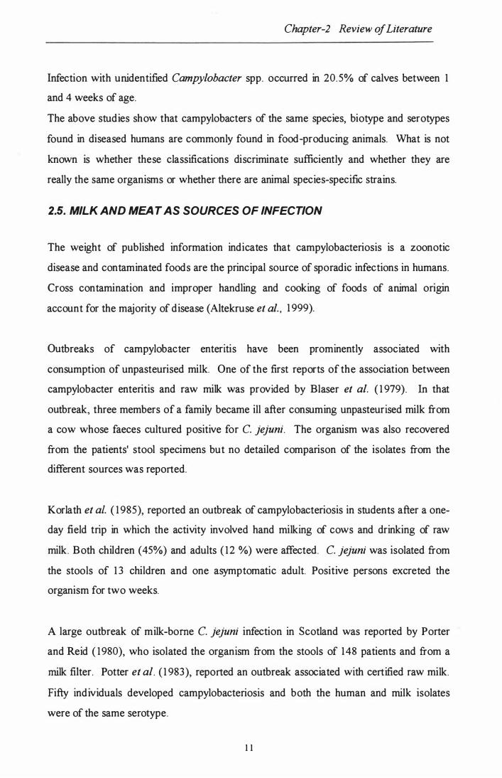
2.5. MILK AND MEA T AS SOURCES OF INFECTION
The weight of published information indicates that campylobacteriosis is a zoonotic
disease and contaminated foods are the principal source of sporadic infections in humans.
Cross contamination and improper handling and cooking of foods of animal origin
account for the majority of disease (Altekruse et al. , 1 999).
Outbreaks of campylobacter enteritis have been prominently associated with
consumption of unpasteurised milk. One of the first reports of the association between
campylobacter enteritis and raw milk was provided by Blaser et al. ( 1 979). In that
outbreak, three members of a family became ill after consuming unpasteurised milk from
a cow whose faeces cultured positive for C. jejuni. The organism was also recovered
from the patients' stool specimens but no detailed comparison of the isolates from the
different sources was reported.
Korlath et al. ( 1 985), reported an outbreak of campylobacteriosis in students after a one
day field trip in which the activity involved hand milking of cows and drinking of raw
milk. Both children (45%) and adults ( 1 2 %) were affected. C. jejuni was isolated from
the stools of 1 3 children and one asymptomatic adult. Positive persons excreted the
organism for two weeks.
A large outbreak of milk-borne C. jejuni infection in Scotland was reported by Porter
and Reid ( 1980), who isolated the organism from the stools of 148 patients and from a
milk filter. Potter et al. ( 1 983), reported an outbreak associated with certified raw milk.
Fifty individuals developed campylobacteriosis and both the human and milk isolates
were of the same serotype.
1 1

Chapter-2 Review of Literature
Major outbreaks involving consumption of contaminated or inadequately pasteurised
milk have been regularly recorded over the years (Fahey et aI. , 1995 ; Pearson and
Healing, 1992). Evans et al. (1996) , reported an outbreak of milkborne
campylobacteriosis in school children and adult helpers on a visit to a dairy farm after
drinking raw milk . C. jejuni was isolated from most of the cases as well as from dairy
cattle and bird faeces obtained at the farm.
Contamination of milk with Campylobacter spp. can be faecal in origin or could occur
through a mastitis infection. Lander and Gill (19 80) , provided evidence by way of an
experimental infection showing that such a condition could exist in cows. Raw milk from
these cows could infect humans. Subsequently Morgan et al. (19 85 ) , reported the
isolation of C. jejuni from a naturally occurring case of mastitis in association with
human illness involving the same serotype.
Diker et al. (19 87) , studied the survivability of C. coli in unpasteurised milk. The
organisms were found to survive for 2 -5 days at 3 7° C and for 1 8 days at refrigeration
temperature.
Piazza and Lasta (19 86 ) , described the isolation of C. jejuni and C. sputorum from
intestinal contents ( 1. 7% and 6.9% respectively) and C. jejuni (3.2 %) from swabbing of
carcasses of clinically healthy cattle and pigs destined for human consumption.
Several reports describe the association of Campylobacter species and beef products.
One of the earliest reports was from England describing a study in which the organism
was isolated from 1.6 % of more than 6000 meat samples ( Tumbull and Rose, 19 82).
Stem et al. (19 85 ) , using enrichment methods found that approximately 5 % of all red
meat samples from retail outlets throughout the U. S. yielded Campylobacter spp.
Lammerding et al. (19 88) also reported high levels of Campylobacter spp. in a variety of
foods of animal origin in Canada.
12

Chapter-2 Review of Literature
Bolton et al. ( 1985), examined 730 samples of offal, mince meat and sausage meat from
abattoirs and retail butchers shop for Campylobacter species. C. jejuni and C. coli were
isolated from 30.6%, 1 0 .5% and 6% of sheep, cattle, and pig offal samples respectively.
Bucci and Maini ( 1 988), isolated over a two year period thermophilic Campylobacter
spp. from 6% of 1 680 stool specimens of patients with enteritis, 5 1 .7% of 325 faecal
specimens of healthy animals, 5 1 .7% of 1 06 meat samples of 6 different species, and
32. 1 % of 1 94 samples of raw milk . Typing of the 286 isolates revealed 64% to be C.
jejuni, 29.7% C. coli and 5.6% remained unclassified.
Tomancova et al. ( 1 987), studied the survivability of C. jejuni in artificially contaminated
meat. With standard packing the organisms survived six to seven days and in vacuum
packaged meat about ten to eleven days.
Zanetti et al. ( 1 996), studied the prevalence of thermophilic bacteria in 57 manually
shelled eggs and 1 30 raw meat samples. No bacteria were found in the egg samples, but
1 6 strains of C. jejuni and 4 strains of C. coli were detected from the meat samples.
Thus food products of animal ongm have commonly been reported to contain
Campylobacter spp. and there is epidemiological evidence linking human disease with
food source. However, there is a need to demonstrate, whether or not the strains in food
are the same as those associated with human disease.
2.6. ISOLA TION, IDENTIFICATION AND TYPING OF CAMPYLOBACTER
Campylobacter speCIes represent a taxonomically heterologous group. Their
identification can be difficult since strains have relatively fastidious growth requirements
and are asaccharolytic, and since only a limited number of biochemical tests give
adequate discrimination.
Resistance to various agents (antibiotics, chemicals and dyes), temperature tolerances
and growth requirements are among the phenotypic tests used in characterising
13

Chapter-2 Review of Literature
campylobacters. However, no standard methods for the performance of such tests have
been published, and therefore most workers use methods peculiar to their own
laboratories.
The conventional approach to identification of Campylobacter spp. by culture methods is
a labour intensive and time-consuming process. Recent developments in molecular
techniques offer attractive alternatives for the detection of the organism based on
identification of specific segments of the genome. The utilisation of the polymerase chain
reaction (PCR) and nucleic acid sequence based amplification (NASBA) offer a rapid
method of identification of the organism from clinical samples and foods .
• 2.6.1 . ISOLATION
.
The isolation of Campylobacter species involves plating of specimens on selective media.
These include the blood-containing Skirrow medium (Skirrow, 1 977), and
Campylobacter-cefoperazone-vancomycin-amphotericin (CVA) , (RelIer et al. , 1 983),
and the blood-free charcoal-cefoperazone-deoxycholate agar (CCDA) , (Hutchinson and
Bolton, 1 984) and charcoal-based selective medium (CSM) , (Karmali et al. , 1 986).
Highest yield of Campylobacter spp. from stool samples was obtained by either CCDA
or CSM (Endtz et al. , 1 99 1b; Gun-Monro et al. , 1 987). Zanetti et al. ( 1 996) found
CCDA media to be better than both Butzler and Preston media for the isolation of
Campylobacter species.
Also enrichment culture for 24 to 48 h at 42°C is recommended for the isolation of C.
jejuni when only small numbers of organisms are present in ,meat and faecal specimens
(Rogol et aI., 1 985). A number of enrichment broths have been formulated to enhance
the recovery of Campylobacter spp. such as Campy-thio (Blaser et al. , 1 979),
Campylobacter enriclunent broth (Martin et al. , 1 983) and Preston enrichment broth
(Bolton and Robertson, 1 982) .
14

Chapter-2 Review of Literature
Most Campylobacter specIes reqUIre a microaerophilic atmosphere containing
approximately 5 % oxygen, 10% carbon dioxide and 85 % nitrogen for optimal recovery
(prescott and Munroe, 1982 ). Several manufacturers produce microaerobic gas
generator packs that are suitable for routine use.
Depending on the medium used Campylobacter spp. produce grey, flat, and irregular,
spreading colonies on fresh media. As the moisture content of the media decreases,
colonies may become round, convex, and glistening with little spreading (Nachamkin,
1995 ).
Phenotypic tests routinely used for identification of Campylobacter spp. include growth
temperature studies, oxidase, presence of catalase, hippurate hydrolysis, nitrate
reduction, production of H2S, and antibiotic sensitivity by the disc diffusion method
(Isenberg, 1992 ; Barret et al. , 1988; Morris and Patton, 1985 ).
2.6.2. SEROTYPING
Two serotyping systems have gained international recognition for C. jejuni and C. coli.
The Penner and Hennessey ( 1980) scheme based on heat stable (HS) soluble (LPS)
antigens is a passive haemagglutination assay, that currently recognises 65 serotypes in
total, and comprises 47 antisera for C. jejuni and 18 antisera for C. coli (penner et al. ,
1983 ).
The Lior scheme based on thermolabile antigens (HL) is a slide agglutination method
with about 130 serotypes covering C. jejuni, C. coli and C. lari (Lior et al. , 1982 ). A
separate scheme for C. upsaliensis has been proposed by Lior and Woodward (1993 ).
A renewed interest in the use of the HS antigen typing system has arisen since HS
antigens of particular serotypes have been implicated as possible pathogenic factors in
the development of Guillain-Barre' syndrome and Miller-Fisher syndrome (Kuroki et al. ,
1993 ; Yuki et a!., 1994 ).
1 5

Chapter-2 Review of Literature
The main disadvantage of these methods is the lack of commercially available, high
quality antisera. Production of antisera to the large numbers of strains for either one or
both systems would be too time consuming, costly and impractical for most clinical or
reference labs. Therefore, most of the reports of the isolation and identification of
campylobacters from various animal species and food sources do not include a
comprehensive identification of all the isolates that are made. It is difficult to conclude
from many published papers exactly what is the precise identity of the isolates made.
2.6.3. BIOTYPING
A biotyping scheme which was an extension of the scheme of Skirrow and Benjamin
(1980) , was proposed by Lior (198 4) , for the differentiation of campylobacters. By this
scheme based on hippurate hydrolysis, production of lhS on FBP mediums and
resistance to nalidixic acid, C. jejuni was divided into 4 biogroups; C. coli into two and
C. lari into two biogroups. This scheme has been widely used by various workers in
discriminating Campylobacter spp. (Nicholson and Patton, 1993 ; Owen et al. , 199 4;
Jimenez et aI. , 199 4).
The Preston biotyping scheme uses 12 tests, including 10 resistotyping tests that
determine resistance to antibiotics, dyes and chemicals. Fifty five biotypes of C. jejuni
have been identified (patton and Wachsmuth, 1992)
The biotyping scheme is simple and available to most laboratories, although it produces
only a few markers among strains when used alone. In recognition of this, biotyping is
suggested for use in conjunction with serotyping (Smith et al. , 199 7; Tay et aI., 1995 ).
However, its use in epidemiological studies to trace the source of infection is extremely
limited.
16

Chapter-2 Review of Literature
2.6.4. PHAGE-TYPING
G rajews ki et al. ( 1 985), was the fi rs t to des cri be a phage- typing s ys tem for C. jejuni and
C. coli bas ed on a panel of 14 phages is olated from poultry faeces.
There are two phage- typing s ys tems curr ently in us e, both of which incorporate s om e or
all of Gr ajews ki phages. The firs t, propos ed by K hakhr ia and L ior ( 1992), ex tends to 1 9
phages and typed 78% of 30 1 C. jejuni s trains from worldwide s ources exam ined. The
s econd des cribed by Salam a et al. ( 1990), com bined s ix of G rajews ki phages with ten
is olated in the UK.
E xcellent res ults have been obtained when the thr ee typing s chem es, s erotyping,
biotyping and phage- typing have been com bined, s upport ing the com plem entary us e of
phage- typing in conjunction with other typing s chem es for the epidem iological analys is
of Campylobacter inf ections but from a practical s tandpoint, the com bination of all thes e
m ethods m ay be too tim e cons um ing and cos tly for m os t labs (p atton et al. , 199 1).
2.6.5. WHOLE CELL PROTEIN PROFILES
Total protein profile and outer m em brane protein (OMP) profile analys es that have been
valuable in s tudies of s pecifi c Campylobacter proteins, s uch as porins and t lagellin.
However, thes e two m ethods are com plex and have been found to dem ons trate only low
to m oderate dis crim ination between cam pylobacters (p atton and W achsm uth, 1992).
The us e of OMP profile analys is with SDS-P AGE could not dis crim inate between s trains
of C. jejuni and C. coli in two s eparate s tudies (Blas er et al. , 1 983 a; L ogan and Trus t,
1982). But 9 s ub- types of C. jejuni could be dis tinguis hed on the bas is of m igr ation of
m ajor bands. However, total protein profi le analys is by polyacrylam ide gel
electrophores is (P AGE) has been us ed to identify and dis tinguis h between
Campylobacter s pecies, including C. coli and C. jejuni and for res olving taxonom ic
problems at the s peci es and s ubs pecies level (V andamm e and D eL ey, 199 1).
17

Chapter-2 Review of Literature
2.6.6. PLASMID PROFILE ANALYSIS
Plasmids have been observed in about 30-50% of C. jejuni and C. coli isolates, but the
instability of the plasmids may diminish their potential value in epidemiological
investigations (Taylor, 1992).
Fraser et al. ( 1992) , examined a number of C. coli, serogroup 20 (Lior) strains and
found that 24 of 27 examined carried one or more plasmids, suggesting a high degree of
genetic exchange in the strains of this serogroup.
Stanley et al. (1994 ) , described the typing of human and canine isolates of C. upsaliensis
by 16 S rRNA genotyping and plasmid profiling. Plasmids were found in 60% of the
strains, ranging in size from 1. 5 to 100 KB, and gave 15 distinct plasmid profiles. All
isolates from humans contained one or more plasmids, as did strains isolated from dogs
with sporadic diarrhoea. The two commonest 16 S ribotypes were divided into eight and
nine serogroups by plasmid profiling and the authors concluded that a combination of
16 S ribotyping with plasmid profiling would be valuable for detailed epidemiological
studies of C. upsaliensis
Bopp et al. (1985 ) , studied 31 C. jejuni strains from 11 outbreaks for plasmids and 19
possessed plasmid DNA. Four of the strains containing plasmids were sensitive to all the
antimicrobial agents used. Tetracycline resistant strains were found to contain 38 -
megadalton plasmids and these plasmids shared common nucleic acid sequences.
2.6.7. MUL TILOCUS ENZYME ELECTROPHORESIS (MEE)
MEE is based on the electrophoretic migration distance of enzymes present in bacteria.
Enzyme mobility differences relate directly to allelic variation in the structural gene locus
for each enzyme (Selander et aI. , 198 6). From this analysis, an estimate of the genetic
relatedness among strains can be determined.
18

Chapter-2 Review of Literature
In an application of MEE to Campylobacter strains, 50 and 1 4 electrophoretic types
were identified among 1 04 C. jejuni & 2 1 C. coli strains respectively, demonstrating a
high degree of genetic diversity within these two species (Aeschbacher and Piffaretti,
1 989).
MEE, though a highly sensitive technique for differentiation of epidemic-associated
strains (patton et al. , 1 99 1 ), is highly complex and depends on the number of enzymes
analysed and the use of appropriate electrophoretic parameters and, in comparison with
other genetic methods, is relatively time consuming.
Methods that examine chromosomal DNA and detect rrunor changes in nucleotide
sequences have been applied to Campylobacters. Restriction endonuclease analysis
(REA), southern blot and hybridisation of DNA fragments produced by REA with rRNA
or rDNA (ribotyping), PCR based DNA profiling and Pulsed field gel electrophoresis
(pFGE) measure relatively stable chromosomal differences and do not depend on
phenotypic expression of bacterial cell products.
2.6.8. RESTRICTION ENDONUCLEASE ANALYSIS (REA)
Analysis of restriction endonuclease digest patterns produced by high frequency cutting
enzymes is referred to as chromosomal restriction-enzyme analysis (REA) or bacterial
restriction-endonuclease analysis (BRENDA) and was applied to strains within various
species of Campylobacter because of limitations of biochemical and other phenotypic
methods (Bradbury et aI., 1 984; Owen et al. , 1 989; Owen and Hernandez, 1 990).
Owen et at. ( 1 990), has applied REA for the differentiation of Campylobacter strains,
and this method was successfully used in three outbreaks investigated. Fraser et al.
( 1 992), have examined a number of C. coli strains of serogroup 20 and reported that
REA demonstrated the greatest degree of discrimination among the strains. The enzyme
Hha I yielded the greatest differences between the strains. Hha I produced 1 6 different
profiles each showing a distinct banding pattern among 27 C. coli strains. Some strains,
1 9

Chapter-2 Review of Literature
which displayed identical plasmid profiles, had different restriction profiles. A
disadvantage of this method however, is the fact that chromosomal restriction
endonuclease digests, in many instances, will generate large numbers of bands that may
be difficult to interpret.
2.6.9. RIBOTYPING
Ribotyping is based on hybridisation of a rRNA or rDNA probe to restriction digested
bacterial chromosomal DNA, measuring chromosomal differences. The discriminatory
power of ribotyping is dependent on the choice of both enzyme and specific probe for
hybridisation. The pattern of three to six restriction fragments or bands produced in most
ribotyping experiments is much easier to interpret than REA patterns (patton et al.,
1 99 1 ; Wachsmuth et al. , 1 99 1 ).
Owen et al. ( 1 990), determined the DNA restriction endonuclease (Hae III and Hind III)
total digests and 1 6S and 23 S rRNA gene patterns for 1 8 isolates of C. jejuni. An
excellent correlation was found between the genomic DNA fingerprint data and the
Preston bacteriophage group. An E. coli 1 6S +23 S rRNA probe was more sensitive than
C. jejuni 1 6 S rDNA probe.
In another study involving 72 strains of C. jejuni, C. coli, C. upsaliensis and C. lari,
Owen et al. ( 1 993), came to the conclusion that the choice of restriction endonuclease is
of critical importance when examining different species of Campylobacter. Hae III
ribopatterns were the most effective means of typing strains of different species and,
when combined with Pst I ribopatterns offered a highly discriminating basis for molecular
typing.
Fitzgerald et al. ( 1 996), described a ribotyping scheme in which strains belonging to all
47 heat-stable serotypes of C. jejuni were examined for polymorphism around the 1 6S
rRNA genes and complete typability was obtained.
20

Chapter-2 Review of Literature
lackson et al. ( 1 996), examined C. jejuni serogroup reference strains and a collection of
outbreak-associated isolates for RFLPs using C. jejuni random chromosomal and
1 6sRNA gene probes. Both probes were able to differentiate between certain random
isolates of the same serogroups but greater discrimination was obtained with RFLP than
ribotyping. Genotyping also distinguished between related and unrelated strains when
applied to outbreaks.
Iriarte and Owen ( 1 996), examined 47 strains of C. jejuni by PCR -RFLP analysis of 23 S
rRNA genes. 83% of the strains, including those with different Penner serotypes and
from different hosts had the same molecular profiles. The authors suggested that this
could be because of the high degree of conservation within the 23 S rRNA sequences,
and the technique would be more useful for species-specific identification assays but not
for subtypic discrimination within C. jejuni.
2.6.1 0. PULSED-FIELD GEL ELECTROPHORESIS (PFGE)
The development of PFGE allows the analysis of a smaller number of large molecular
weight chromosomal DNA fragments generated by appropriate digestion by rare-cutting
restriction enzymes (Schwartz and Cantor, 1 984). Van et al. ( 1 99 1 ), reported the
investigation of C. jejuni and C. coli strains digested with Sma I enzyme and found that
PFGE analysis can be an alternative method useful in epidemiological investigations for
differentiating isolates of C. jejuni and C. coli.
During the last few years PFGE is being increasingly applied for the typing of
Campylobacter organisms. On et al. ( 1 999), used PFGE to demonstrate persistence of
C. sputorum infection in cows over a period of time. In another study, On et al. ( 1 998),
examined C. jejuni strains from humans, water, poultry and cattle and demonstrated a
clear link between human and the animal isolates and found the technique robust and
accurate.
2 1

Chapter-2 Review of Literature
PFGE has been compared with other techniques for the epidemiological typing of
Campylobacter spp. by various workers. Steele et al. ( 1 998), compared PFGE to fatty
acid profile typing, biotyping and serotyping and found PFGE the most discriminatory.
Shi et al. ( 1 996), compared PFGE and enterobacterial repetitive intergenic concensus
(ERIC) and Slater and Owen ( 1 998), restriction fragment length polymorphism (RFLP)
analysis based on polymerase chain reaction technique and found PFGE a better
discriminatory technique in both the cases.
The technique has been successfully applied in epidemiological studies pertaining to
outbreaks of Campylobacter infections in humans (Hanninen et al. , 1 998b) in Finland
and in identifying the source of the transmission of infection in a small dairy herd
(Hanninen et at. 1 998a).
Some workers have utilised PFGE for discrimination within the species C. upsaliensis
(Bourke et al. , 1 996), C. hyointestinalis (Salama et al. , 1 992a) and between two
subspecies as done by Salama et al. ( 1 992b), to differentiate between C. fetus ssp. fetus
and C. fetus ssp. venerealis.
2.6.1 1 . RANDOM AMPLIFIED POLYMORPHIC DNA (RAPD)
The analysis of random amplified polymorphic DNA by simple gel-electrophoretic
procedures revealed that, for different organisms, highly diverse DNA banding patterns
could be generated.
These DNA fingerprints allow discrimination between species and enable differentiation
of isolates belonging to a single species. Also, PCR primers aimed at bacterial DNA
repeat motifs proved to be highly useful in this type of assay.
Mazurier et al. ( 1 992), described a protocol for the differentiation of campylobacters by
RAPD fingerprinting and peR without the need to purify DNA, using three different,
randomly designed, 1 0-mer primers. Nine distinct RAPD profiles were obtained with one
22

Chapter-2 Review of Literature
of the primers and 10 other profiles with another primer. Distinct RAPD profiles were
identified among strains belonging to the same serotype.
Giesendorf et al. (1993 ), reported the development of species-specific DNA probes by
PCR fingerprinting of C. jejuni, C. coli and C. lari. PCR primers aimed at arbitrary
sequences, in combination with primers directed against the repetitive extragenic
palindrome (REP) or enterobacterial repetitive intergenic consensus (ERIC) motifs, and
were shown to generate isolate-specific banding patterns. Analysis of these PCR
fingerprints obtained from 33 isolates of C. jejuni, 30 isolates of C. coli and eight
isolates of C. lari revealed that, besides generation of isolate specific fragments, species
specific DNA fragments of identical size were synthesised, which could be used as
species-specific probes in southern blots.
This combination of PCR fingerprinting and probe hybridisation resulted in a highly
specific identification assay and provides an example of species-specific test development
without the prior need for DNA sequence information.
Madden et al. (199 6), used the RAPD method of typing based on a l O-mer primer for
sub-typing animal and human Campylobacter species and found it an effective method
with high discrimination and reproducibility and, unlike serotyping, no untypable strains
were found out of a total of 2 69 isolates.
Chuma et al. (1997 ), analysed the distribution of C. jejuni and C. coli in broiler chickens
by using restriction fragment length polymorphism (RFLP) of the flagellin gene and
found it a useful technique for the epidemiological studies of campylobacter
contamination of broilers in different flocks and different growth cycles on the farm.
Nishimura et al. (199 6), used PCR-based RFLP analysis based on the flagellin gene in
typing 179 isolates from Japan and China and were successful in typing most (98.7 %) of
the isolates into 2 5 separate RFLP groups as compared to serotyping (6 l.7 %). Also 11
isolates of HS- 019 strains, frequently isolated from Guillain-Barre' syndrome (GBS),
were found to show an identical RFLP pattern.
23

Chapter-2 Review of Literature
Fujimoto et al. ( 1 997), used RFLP and RAPD techniques to analyse C. jejuni serotype
0 1 9 strains associated with GBS and other strains. Their data indicated that all the
strains were closely related to one another whether or not they were associated with
GBS.
2.6.1 2. POLYMERASE CHAIN REACTION (PCR)
The polymerase chain reaction has been applied extensively to the detection of infectious
agents. peR allows amplification of a pre-selected region of DNA and can be a highly
specific and sensitive detection technique. peR has also been used for direct
identification of Campylobacter species from complex substrates without prior isolation
and purification of the organisms (Wegmuller et al. , 1 993 ).
The difficulties in routine detection, isolation and identification of Campylobacter species
make these organisms ideal candidates for peR identification. One campylobacter gene,
which has the potential to allow for organism identification at the level of species and at
the narrower level of strain, is the flagellum gene. The flagella of C. jejuni and C. coli
are composed of two subunit flagellins, the products of the fla A and fla B genes. These
genes have been cloned and sequenced and shown to contain highly variable regions that
could be used for strain-specific detection. They also contain other regions that are
highly conserved among C. coli and C. jejuni strains. Therefore these genes are
potentially useful for the detection of the Campylobacter species most commonly
associated with human diarrhoeal disease.
Oyofu et al. ( 1 997), described the development of a routine detection assay for C. jejuni
and C. coli in clinical specimens by using the peR. An oligonucleotide primer pair from a
conserved 5' region of the fl.a A gene of C. coli was used to amplify a 450bp region by
peR. The primer pair specifically detected four strains of C. coli and 47 strains of C.
jejuni, but it did not detect strains of C. fetus, c. lari, C. upsaliensis, C . cryoaerophilia
and C. hyointestinaiis. In stools seeded -with C. coli cells, the probe could detect
between 30-60 bacteria per peR assay. The assay was also successfully used to detect C.
24

Chapter-2 Review of Literature
coli in rectal swab specimens from experimentally infected rabbits and C. jejuni in human
stool samples.
Birkenhead et al. ( 1 993), used the peR method for detection and typing of
campylobacters. Thefia gene was amplified. Primers were chosen which amplified 1.3kb
of the fia gene in C. jejuni and C. coli. The C. upsaliensis amplimer was approximately
1. 7kb in size and was easily distinguishable. Other species of campylobacter failed to
yield amplimer. The amplimer was digested with Alu I, which demonstrated considerable
restriction fragment length polymorphism and suggested that it might allow the
development of a rapid and novel typing scheme.
Waegel and Nachamkin ( 1 996), described a peR based on primers from fia A gene for
detection of C. jejuni from stool samples, which required the faeces to be purified by
column chromatography before subjecting it to peR.
Genes for 1 6S r RNA have often been used as target sequences in peR assays for
identification of fastidious bacteria including campylobacter species. rRNA genes have a
typical mosaic structure of phylogenetically conserved and variable regions. The latter
may vary considerably among different bacterial species and therefore are excellent
targets for species or even subspecies-specific primers.
Eyers et al. ( 1 994), reported the use of a 23 S rRNA fragment as a target for peR
amplification and the primers used detected all thermophilic campylobacter species and
also allowed discrimination among the thermophilic species. The rRNA, an essential part
of prokaryotic and eukaryotic ribosomes, is genetically stable and consists of
conservative and variable regions. The latter may vary considerably among different
bacterial species and therefore can be targets for specific oligonucleotide probes.
Giesendorf et al. ( 1 992),. described a method for rapid detection and identification of
Campylobacter in chicken products using peR after a short enrichment culture. They
use a 1 6S rRNA gene sequence of C. jejuni as a primer and probe combination. With this
25

Chapter-2 Review of Literature
primer set and probe, 426-bp fragments from C. jejuni, C. coli and C. lari could be
amplified. The detection limit of the PCR was 1 2 . 5 bacteria.
Docherty et al. ( 1 996), described the magnetic immuno-PCR assay for the detection of
campylobacter in milk and poultry products in which the target bacteria are captured
from the food sample by magnetic particles coated with a specific antibody. The bound
bacteria are then lysed and subjected to PCR.
Van Camp et al. ( 1 993), also reported the development of a method utilising PCR
amplification and hybridisation analysis for detection of enteropathogenic Campylobacter
species based on 1 6S rRNA genes but concluded that it was not possible to differentiate
between the species on the basis of 16S rRNA as C. jejuni, C. coli, C. lari and C.
upsaliensis fonn a tight taxonomic cluster.
Wegmuller et al. ( 1 993), described a PCR method designed to sensitively detect and
identify C. jejuni and C. coli, without the need of isolating and culturing strains. The
intergenic sequence between the flagellin genes fla A and fla B was amplified and
characterised with a triple primer or seminested primer approach. A total of 50 bacterial
strains, 27 of C. jejuni and C. coli and 23 of other species were tested, giving no false
positive or false negative results. The detection limit as determined by ethidium bromide
staining of amplification products on agarose gels was 1 0 bacteria or less in artificially
contaminated water, milk and soft cheese samples with the seminested primer PCR
assay. As an application of the PCR system, 93 samples of milk and dairy products were
screened for the presence of C. jejuni and C. coli out of which 6 positive samples were
identified while none were found positive with a conventional culture method.
Hum et al. ( 1 997), evaluated the PCR assay and traditional phenotypic methods for the
identification and differentiation of C. fetus subspecies and found agreement between
strain identities suggested by both the assays to be 80.8%. The PCR was found to be a
reliable, rapid technique for the species and subspecies identification of C. fetus.
26

Chapter-2 Review of Literature
Mahendru et al. ( 199 7), used peR with primers based on fla A gene and were successful
in identification of 15 C. jejuni strains isolated from human faeces. RFLP of the
amplified product classified the 15 strains into 5 types.
Wesley et al. ( 199 7), used a multiplex peR to distinguish C. jejuni and C. coli
associated with reproductive failure in pigs. They found it useful in identifying the
Campylobacter species from atypical cases and suggested that multiplex PCR, in
conjunction with other conventional assays, may be useful for verifying unusual instances
of campylobacteriosis.
Linton et al. ( 199 7), used the PCR for detection and identification to species level of C.
jejuni and C. coli isolates direct from diarrhoeic samples using three different sets of
primers and a simple technique to purify DNA. The results concurred with culture and
phenotypic identification to species level. The peR offered a rapid method to define the
occurrence and species prevalence.
Winters et al. ( 199 7), described a method for the rapid detection of C. jejuni from
chicken washes using a nested PCR and detected C. jejuni in 80% of four groups of
chicken. peR methods have the potential to be very fast and sensitive and can be
designed to be group-reactive or quite specific depending on what is required in a
particular investigation and on which primers are chosen. They are also able to detect
non-cultivable organisms.
2.6.1 3. NUCLEIC ACID SEQUENCE BASED AMPLIFICATION (NASBA)
An alternative approach to PCR is NASBA, a technique to selectively amplify RNA.
Isothermal nucleic acid amplification of RNA in NASBA is achieved through the
concerted action of avian myeloblastosis virus reverse transcriptase ( AMV-RT), T7
RNA polymerase and RnaseH ( Compton, 199 1 ).
27

Chapter-2 Review of Literature
The reaction starts with a non-cyclic phase, in which a downstream primer, containing a
tail sequence of the T7 -promoter sequence, anneals to the single stranded target
sequence. Through the action of AMV-RT, cDNA is formed. The RNaseH hydrolyses
the RNA from the RNA-DNA hybrid, which results in a single strand of DNA to which
the upstream primer can anneal. The AMV-RT synthesis, through its DNA polymerase
activity, produces the double-stranded DNA intermediate with a transcriptionally active
promoter sequence. TheT7 RNA polymerase generates from these, intermediate single
stranded RNA copies ( 1 00- 1 000) which serve as a template in the now cyclic phase of
NASBA.
Major advantages of NASBA over peR are that NASBA is performed isothermally
which precludes the use of a special thermocycler, and that no separate R T step is
required for RNA amplification (Kievits et aI. , 1 99 1 ) .
Uyttendale et al. ( 1 994), evaluated the NASBA technique for the specific identification
of C. jejuni, C. coli and C lari. A set of primers and a probe were chosen from the 1 6S
rRNA sequence of campylobacter. The probe was hybridised in solution with the
amplified nucleic acids of 1 2 Campylobacter species and 9 other Gram-negative bacteria.
The probe was shown to hybridise specifically to the amplified single-stranded RNA of
C. jejuni, C. coli and C. lari in an enzyme-linked gel assay (ELGA).
More recently Uyttendale et al. ( 1 995), reported the utilisation of the NASBA
amplification of RNA and a non-radioactive in-solution hybridisation combined with a
short selective enrichment for the detection of C. jejuni in foods. Interference by food
components was eliminated by centrifugation following the enrichment step. The
prevalence of high numbers of indigenous flora affected the sensitivity of the assay.
However, identification of C. jejuni was possible up to a ratio of indigenous flora to C.
jejuni of 1 0,000: 1 . The base of enrichment culture and NASBA shortened the analysis
time from 6 days to 26 hours.
28

Chapter-2 Review of Literature
2.7. ANTIBIOTIC RESITANCE AMONG CAMPYLOBACTER SPECIES
Erythromycin has long been regarded as the antibiotic of choice for treating
campylobacter infections in humans. Although the use of this antibiotic may reduce the
severity of the symptoms in children and curtail the convalescent carriage state, it
generally does little to alter the course of the illness (Mandal et al. , 1 984).
Gaudreau and Gilbert ( 1 997), compared the disc diffusion and agar dilution methods for
antibiotic susceptibility testing and came to the conclusion that disc diffusion is a reliable,
easy and inexpensive susceptibility method for C. jejuni and C. coli for erythromycin,
ciprofloxacin and tetracycline.
The potential problem of antibiotic resistance has been illustrated by a study in Thailand
(Taylor et al. , 1 987), where erythromycin treatment, in acute diarroheal disease among
1 00 infants in an orphanage had no effect on the duration of diarrhoea. Campyiobacter
strains isolated from the children before treatment showed a high level of resistance to
erythromycin (53% for C. jejuni and 91% for C. coli). In developed countries
erythromycin resistance is commonly reported at a 2% level for C. jejuni and 1 0% for C.
coli (Doyle and Jones, 1 992). The report of such high rates of erythromycin resistance
in Thailand is disturbing and may indicate what could happen if antibiotics become
indiscriminately used in developed countries for the treatment of campylobacter
infections (Taylor et al. , 1 987).
Dowling et al. ( 1 998), examined 202 Campylobacter isolates from the Auckland area by
the disc diffusion method and found 1% (2) resistant to doxcycline, 1 . 5% (3) to
erythromycin and 2.5% (5) to ciprofloxacin. All the isolates were susceptible to
gentamicin. Unpublished studies by the author earlier, had detected 2- 1 1 % of the
isolates resistant to erythromycin.
Gaudreau and Gilbert ( 1 998), compared the antimicrobial resistance of C. jejuni strains
isolated from humans in Canada during 1 985-87 and 92-93 to isolates from 1 995-97, and
found an increase in resistance to tetracycline, nalidixic acid and ciprofloxacin over the
29
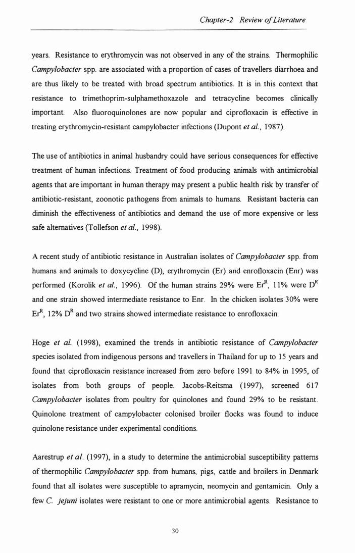
Chapter-2 Review of Literature
years. Resistance to erythromycin was not observed in any of the strains. Thermophilic
Campylobacter spp. are associated with a proportion of cases of travellers diarrhoea and
are thus likely to be treated with broad spectrum antibiotics. It is in this context that
resistance to trimethoprim-sulphamethoxazole and tetracycline becomes clinically
important. Also fluoroquinolones are now popular and ciprofloxacin is effective in
treating erythromycin-resistant campylobacter infections (Dupont et al. , 1 987).
The use of antibiotics in animal husbandry could have serious consequences for effective
treatment of human infections. Treatment of food producing animals with antimicrobial
agents that are important in human therapy may present a public health risk by transfer of
antibiotic-resistant, zoonotic pathogens from animals to humans. Resistant bacteria can
diminish the effectiveness of antibiotics and demand the use of more expensive or less
safe alternatives (Tollefson et al. , 1 998).
A recent study of antibiotic resistance in Australian isolates of Campylobacter spp. from
humans and animals to doxycycline (D), erythromycin (Er) and enrofloxacin (Enr) was
performed (Korolik et al. , 1 996). Of the human strains 29% were Et, 1 1% were DR
and one strain showed intermediate resistance to Enr. In the chicken isolates 30% were
Et, 1 2% DR and two strains showed intermediate resistance to enrofloxacin.
Roge et al. ( 1 998), examined the trends in antibiotic resistance of Campylobacter
species isolated from indigenous persons and travellers in Thailand for up to 1 5 years and
found that ciprofloxacin resistance increased from zero before 1 99 1 to 84% in 1 995, of
isolates from both groups of people. J acobs-Reitsma ( 1 997), screened 6 1 7
Campylobacter isolates from poultry for quinolones and found 29% to be resistant.
Quinolone treatment of campylobacter colonised broiler flocks was found to induce
quinolone resistance under experimental conditions.
Aarestrup et al. ( 1 997), in a study to determine the antimicrobial susceptibility patterns
of thermophilic Campylobacter spp. from humans, pigs, cattle and broilers in Denmark
found that all isolates were susceptible to apramycin, neomycin and gentamicin. Only a
few C. jejuni isolates were resistant to one or more antimicrobial agents. Resistance to
30

Chapter-2 Review oj Literature
tetracycline was more common among C. jejuni isolates from humans ( 1 1 %) than among
isolates from animals (0 to 2%). Among C. coli isolates especially from pigs there was a
high level resistance to macrolides and streptomycin.
Sjogren et al. ( 1 997), examined 1659 isolations made of C. jejuni and C. coli from
Swedish patients with diarrhoea from 1 992 to 1995 for norfloxacin resistance by the disc
diffusion method. 1 8 .6% of the isolates were found to be resistant.
Huysmans and Turnidge ( 1 997), in Australia examined 1 00 clinical human isolates of
thermophilic Campylobacter spp. including 79 strains of C. jejuni, 1 9 of C. coli and 2 of
C. lari by the disc susceptibility method. They found no resistance to quinolones in any
of the species except C. lari. Resistance to erythromycin, chloramphenicol or gentamicin
was not found in any isolate, while nine isolates were resistant to tetracycline.
Gaunt and Piddock ( 1 996), examined 2209 isolates of Campylobacter spp. collected
over a 1 year period from UK public health labs and found 4. 1 % of the isolates to be
resistant to ciprof1oxacin although none of the 9 1 patients involved had taken a
quinolone.
Tadano et al. ( 1996), studied the susceptibilities of 600 clinical isolates of C. jejuni in
Tokyo hospitals over a 6-year period to six antibiotics. The resistance rates were,
norfloxacin (7.5%), of1oxacin (7.5%), ciprofloxacin (7.3%), nalidixic acid ( 1 0 .3%),
Erythromycin (0.6%) and tetracycline (43 .2%). It was observed that the resistance to
f1uoroquinolones had increased significantly since 1 993 but susceptibility to erythromycin
had remained at the same level during the past 6 years.
Sicinschi ( 1996), studied the susceptibility to antibiotics of 39 C. jejuni/Coli isolates
using the disc diffusion method. All strains were resistant to carbenicillin, rifampicin,
polymyxin M sulphate, ketokonazole, cephalotin, and cephazolin. All the strains were
sensitive to ciprof1oxacin, chloramphenicol, gentamycin, kanamycin, streptomycin and
tobramycin. Lekowska-Kochaniak et al. ( 1 996), found almost all of 47 human and 52
poultry isolates to be susceptible to erythromycin, chloromphenical, gentamicin and
3 1

Chapter-2 Review of Literature
nitrofuratoin. Koenraad et al. ( 1 995), tested the in vitro susceptibility of 209
Campylobacter strains isolated from sewage and poultry abattoir drain water to the
quinolones, nalidixic acid, flumequine, ciprofloxacin and enrofloxacin. They found
resistance ranging from 28% for enrofloxacin to 50% for nalidixic acid.
Gomez-Garces et al. ( 1 995), reported that resistance of C. jejuni strains to
fluoroquinolones is frequent in Spain and reaches nearly 50%, but some 1 2 other oral
antimicrobial agents studied like macrolides, amoxicillin, clindamycin and fosfomycin
displayed good in vitro activities. Velazquez et al. ( 1 995), in Spain, studied 1 02 clinical
C. jejuni isolates and found in vitro resistance to erythromycin ( 1 .9%), three
fluoroquinolones (3 1 .3%- 34.3%), tetracycline (43%), kanamycin (4.8%), and ampicillin
( 1 8 .6%).
Coker and Adefeso ( 1994), found that in Nigeria resistance to erythromycin in clinical
isolates of C. jejuni was 79.2% as compared to an earlier study wherein 82% sensitivity
was observed (Coker et al. , 1 989). Schwartz et al. ( 1 993), examined 30 C. jejuni strains
isolated from stools of Israeli children with enteritis for antimicrobial susceptibility to
eight agents. It was found that all the isolates were sensitive to ciprotloxacin, otloxacin,
furazolidone and erythromycin, but 70% of the strains were found to be resistant to
tetracycline.
Endtz et al. ( 1 993), studied the in vitro susceptibilities of both quinolone susceptible and
resistant C. jejuni to three new macrolide antibiotics, azithromycin, clarit.hromycon and
roxithromycin. They found no difference in their activity against the quinolone
susceptible and resistant strains and suggested that these new antibiotics might replace
erythromycin for the treatment of campylobacter infections.
Ansary and Radu ( 1 992), studied six C. jejuni isolates for the occurrence of plasmids in
association with antibiotic resistances as well as conjugal transfer. All isolates seemed to
carry three similar plasmids and multiple resistance to three of the antibiotics tested was
observed with resistance to tetracycline most common. Conjugal transfer of donor
resistances was observed with erythromycin, neomycin, chloramphenical, kanamycin and
tetracycline.
32

Chapter-2 Review of Literature
Raute lin et al. ( 199 1 ), c ompare d the in vitro susce ptibilitie s of human Campyiobacter
str ai ns is olate d in 1978-80 and duri ng 1990 to ci pr ofloxaci n, norfl oxaci n, er ythromycin,
ge ntamyc in, and doxyc ycline. Re sults indicate d that the susce ptibility to er ythromyc in,
ge ntam ici n and doxyc yc li ne had rem ai ne d the s ame dur ing the last te n ye ar s but the
number of norfl oxaci n-re sistant str ains i ncre ase d from 4% to 1 1% and c ipr ofloxacin
re si stant str ai ns incre ase d from none to 9% i n the last 10 ye ar s.
Ak thar ( 1988), perf or me d antimi cr obial susce ptibi li ty studie s on C. jejuni is olate s from
patie nts with diarr hoe a, asymptom ati c c arr ier s and dome sti c animals. All isolate s were
se nsi tive to ery thr omyci n, ge ntamicin, furazoli done and kanamyci n and 7% of the
i solate s were re si stant to te tr acycline which was obser ve d to be plasmid me di ate d.
The analysis of the above liter ature shows that antibiotic re sistance i s an unavoidable
c onse que nce of the use of anti biotics in hum an ther apy and is obser ve d in most c ountrie s
at various le ve ls de pe ndi ng on the usage. E xam ple s of this are the unus ually high le ve ls
of re si stance to er ythromycin re por te d i n Campylobacter spp. from Thailand and Nigeri a
most pr obably due to its indiscrim inate use and re lative ly le sser in other de ve lope d
countrie s. Howe ver, i t has bee n obser ve d late ly that resis tance to an im port ant clas s of
anti biotic s, the quinolone s which was ne gligible or pre se nt at ver y l ow le ve ls incre ase d
dr am ati cally e ver si nce it was i ntroduce d as a growth pr om oter i n poultr y. This
com bi ne d with the i ncre ase d use of anti bi oti cs i n hum an ther apy as we ll as their use i n
the tre atme nt of di se ase s and pr ophylaxi s i n f ood pr oducing anim als and the possibility of
tr ansfer of re si stance ge ne s in the e nvir onme nt, could have ser ious i mplic ati ons f or the
e ffe cti ve tre atme nt of hum an di se ase s.
To com bat thi s e mergi ng situation i t i s im por tant to minimi se the use of antibi oti cs in
animal fee ds and give more em phasis on the achie ve me nt of m anageme nt goals through
incre ase d sanitation and hygie ne and be tter di se ase contr ol me thods i n li ve stoc k re ar ing.
Simi lar ly in human me dic ine, use of antib iotic s for the tre atme nt of infec tious dise ase s
nee ds to be re stri cte d to avoi d i nappropr iate use. W here possi ble the c hoice of
anti biotics should be base d on i de ntifi cation and se nsi ti vi ty of the. offe ndi ng b acteri um or
at le ast knowle dge of the li ke ly c ausative age nt and its curre nt se nsitivity pr ofile.
33

Chapter 3 Isolation of Campylobacter
CHAPTER 3
3. ISOLATION AN D I DENTI FICATION OF CAMPYLOBACTER SPECIES FROM FAECES OF CATTLE, SHEEP AN D HUMANS.
3.1. I NTRODUCTION
Campylobacter (previously tenned Vibrio) spp. were known to cause abortion in sheep
and cattle from the early 1 900s (McFadyean and Stockman, 1 9 1 3). It was only in the
1 970s after a suitable isolation method was developed (DeKeyser et aI., 1 972), that C.
jejuni was recognised as an enteric pathogen in humans. At present, Campylobacter
spp. especially C. jejuni and C. coli constitute the leading cause of gastrointestinal
disease in humans in the developed world (McNicholas et al., 1 995; Pearson and
Healing, 1 992; Tauxe, 1 992; Skirrow, 1 99 1 ) . C. jejuni also causes infertility and
abortion in cattle, sheep (van Donkersgoed et al., 1 990; Varga et al., 1 986; Prescott and
Munroe, 1 982), and other animals (Prescott and Bruin-Mosch, 1 98 1 ) .
The major reservoirs of thennophilic campylobacter are believed to be the intestines of
birds and other warm-blooded animals (Park et aI., 1 99 1 ), including adult ruminants
(Stern, 1 992), in which, it is thought, they exist as commensals with the gut flora
A major cause of campylobacteriosis in humans IS the consumption of raw or
undercooked foods . of animal origin (Harris et aI., 1 986). Outbreaks of
campylobacteriosis have been prominently associated with consumption of
unpasteurised milk (Tauxe, 1 992; Korlath et aI., 1 985; Breiseman, 1 984; Robinson and
Jones, 1 98 1 ; Porter and Reid, 1 980). Dairy cows are frequently Campylobacter positive
and appear to acquire the organism by the consumption of contaminated water
(Humphrey and Becket, 1 987). Some strains of C. jejuni can cause mastitis in dairy
cows and such infections have led to milk-borne outbreaks (Hutchinson et al., 1 985).
34

Chapter 3 Isolation of Campylobacter
More usually however, milk becomes infected with Campylobacter as a consequence of
faecal contamination during milking (Humphrey and Becket, 1 987).
Table 3.1 Reported carriage rates of Campylobacter species in adult cattle in
various countries
COUNTRY CARRIAGE RATE ( % ) REFERENCE
Australia 59.3 Grau, 1 988
Canada 22.34 van Donkersgoed et aI.,
1 990
Japan 46.7 Giacoboni et al., 1 993
Norway 0.8 Rosef et al., 1 983
Portugal 1 9.5 Cabrita et al., 1 992
United Kingdom 2 1 Bolton et aI. , 1 982
" 23.5 Manser and Dalzeil , 1 985
" 89.4 Stanley et al., 1 998a
The Campylobacter colonisation of cattle is not only significant, because of its potential
contamination of milk on the farm and the carcass at slaughter, but also with regards to
the environmental and water contamination during disposal of abattoir effluents and
slurries to land or run off from farms (Korhonen and Martikainen, 1 99 1 ; Terzieva and
McFeters, 1 99 1 ; Bolton et aI. , 1 987; Carter et al., 1 987)
The present survey of adult cattle, calves and sheep was done to estimate the prevalence
of thermophilic Campylobacter spp. in the Manawatu region of New Zealand and to
better understand the importance of cattle and sheep in the epidemiology of these
microorganisms.
35

Chapter 3 Isolation of Campylobacter
3.2. MATERIALS AND M ETHODS
3.2. 1. SAMPLING
To detennine the prevalence of intestinal thennophilic Campylobacter in cattle and
sheep three types of intestinal material were examined; rectal faeces, intestinal content
from the large intestine and samples of fresh faeces collected on defecation.
A total of 300 samples from adult cattle and calves were collected for the survey. The
samples from beef cattle were collected from an abattoir 25 km from Palmerston North.
The samples from dairy cattle were collected at Massey University dairy farms, and the
samples from the new-born calves were collected from both Massey farms and a private
fann near Palmerston North. '
One hundred and fifty eight sheep faecal samples and 58 samples of the sheep abattoir
environment were collected from an abattoir about 35 km from Palmerston North. This
work was done with the help of a summer studentship recipient Rachel Logan.
Sampling of Beef Cattle: Samples of the intestinal contents from 1 20 beef cattle
were collected after evisceration by making an incision into the tenninal part of the
colon of slaughtered animals and collecting about 1 09 of contents into a sterile plastic
container. All samples were transported to the laboratory on ice and were processed
within 2 hours of collection.
Sampling from Dairy Cattle: Faecal samples from 80 dairy cattle from two farms
were collected during milking in most instances directly into sterile plastic containers
when the animals defaecated. The samples were transported on ice and processed
immediately on arrival .
Sampling from Calves: Sterile cotton wool swabs were used to collect rectal
contents from 1 00 calves at the fanns. The heifer calves were 1 day old to 1 week old
36

Chapter 3 Isolation of Campylobacter
whereas the bull calves were about 4 weeks old. Each swab was rotated within the
rectum for approximately 30 seconds, ensuring that it was in contact with the mucosa.
Rectal swabs were transported on ice in steri le tubes on ice and processed within 2 hr of
collection.
Sampl ing from Sheep: Samples were collected from the rectum with the help of
steri le swabs and then placed in transport medium. The environmental samples were
collected by swabbing different sources and placed in the transport medium and
transferred to the laboratory.
3.2.2. HUMAN CLINICAL ISOLA TES
A total of 1 05 human clinical isolates were obtained form the Palmerston North Medical
Laboratory. These isolates had been presumptively identified as Campylobacter spp. on
the basis of their characteristic growth on campylobacter selective agar and Gram
staining. The isolates were transported to the l�boratory on the selective agar plates,
purified by subculture and further biochemical tests were performed.
3.2.3. ISOLA TION TECHNIQUES
An enrichment and selective plating method was used for the isolation of thermophilic
Campylobacter spp. from the faecal samples. About 1 g of the faeces or intestinal
contents was inoculated into 1 0 ml of Preston enrichment broth (Nutrient broth No.2,
Oxoid) supplemented with Campylobacter growth supplement (Oxoid), Preston
selective supplement (Oxoid) and 5% defibrinated sheep blood) and incubated for 48 hr
at 42°C in a microaerophilic atmosphere produced by using CampyPak Plus (BBL)
envelopes in a GasPak (BBL) jar.
Preston broth was inoculated into campylobacter blood-free agar (CBF) with
cefoperazone charcoal desoxycholate agar (CCDA) selective supplement (Oxoid) and
the plates incubated for 48 hr at 42°C in a GasPak jar with CampYPak Plus (BBL,
Cockeysvil le,USA) gas generating envelopes.
37

Chapter 3 Isolation of Campylobacter
3.2.4. IDENTIFICA TION OF CAMPYLOBA CTER
3.2.4.1 . PRESUM PTIVE IDENTIFICATION OF INTESTINAL THERMOPHILIC
CAMPYLOBA CTER
Typical colonies grown on campylobacter selective agar were examined by Gram
staining and for motility by the hanging drop method, by taking a loop-full of culture on
a glass sl ide with a drop of saline and putting a cover slip and watching under high
magnification under a microscope.
Oxidase reaction: The oxidase test was performed by using Spot test oxidase
reagents (Difco laboratories, Detroit, USA). Two or three drops of the reagent were put
on the colonies to be tested directly on the plate. A positive reaction (presence of
cytochrome oxidase) was indicated by the appearance of a deep purple colour within a
few seconds. Negative reactions remained colourless or turned l ight pink or l ight
purple after 30 seconds.
Catalase activity: The catalase activity was tested by a slide test. One drop of a 3.5%
solution of hydrogen peroxide was added to a microscope slide, placed over a black
background and a small amount of growth was transferred with a loop to the drop. The
reaction was positive when effervescence and bubbles appeared within a few seconds
(Skirrow and Benjamin 1 980)
Preparation of pure cultures: Preparations of presumptive intestinal thermophilic
Campylobacter were made by subculturing individual colonies from the selective media
on to blood agar plates (Oxoid Blood agar base no 2 and 7% defibrinated sheep blood),
which were incubated for 48 h at 42°C. These pure cultures provided the material for
the subsequent tests.
38

Chapter 3 Isolation of Campylobacter
3.2.4.2. CONFIRMATIVE IDENTIFICATION AND
DIFFERENTIATION OF THE THERMOPHILIC CAMPYLOBACTER
SPECIES
The presumptively identified Campylobacter were further tested to confirm their
identity and to differentiate to species level by the following tests:
Hippurate Hydrolysis Test: The hippurate hydrolysis test was done by a rapid
method using Bacto differentiation disc hippurate (Difco laboratories, Detroit, USA). It
involved emulsifying a loop-ful of culture in a small tube containing 0.4 ml distilled
water and a bacto-differentiation disc and incubation at 37°C for 2 h. Then 0.2 ml of
SpotTest Ninhydrin reagent (Difco laboratories, Detroit, USA) was added. After gentle
mixing and re-incubating for 1 0 minutes the reaction was read. A positive hippurate
reaction was indicated by the formation of a deep purple blue colour. A l ight purple
colour or no colour change indicated negative hippurate reactions.
Sensitivity to 30f.19 Nalidixic Acid and Cephalothin Discs: The sensitivity to
30f.1g nalidixic acid and cephalothin discs (Oxoid) was determined by a disc diffusion
test. A loop-full of pure growth was taken from the blood agar plates and rotamixed in
2 ml sterile saline. A sterile cotton swab was then used to inoculate a blood agar plate
and the discs impregnated. The plates were then incubated at 42°C for 48 h in a
microaerophilic environment. The absence of a clear zone of inhibition around the discs
was indicative of resistance.
Hydrogen Sulphide Production: The butt and slant of a triple sugar iron (TSI) agar
in a tube were stab inoculated with a straight wire. A lead acetate indicator paper was
suspended over the medium. The tube was capped and incubated at 42°C for 5 days
under microaerophilic conditions. The appearance of black colour on the lead acetate
indicator paper, or in the medium and the indicator, was recorded as positive.
39

Chapter 3 Isolation of Campylobacter
Table: 3.2 Protocol used for the Identification of Thermophilic Campylobacter
species
Campylobacter Grams Motility Oxidase Hippurate H2S Sensit. Sensit.
species Staining hydrolysis Prod. Cephal Nalidixic
Cell form -othin acid
C. jejuni G-ve + + + - R S
s-shape
C. coli "
+ + R S - -
C. lari "
+ + R R - -
C. hyointestinals "
+ + + S R -
3.2.5. PRESERVA TION OF CUL TURES
Pure growth of cultures on a blood agar plate, resulting from the incubation of a
thermophilic Campylobacter spp. at 42°C for 48 h, were harvested with a sterile cotton
swab in 3 ml 15% glycerol broth. Approximately 2 ml of the suspension was
transferred into sterile 2 rnl Nunc cryo-tubes and stored at -70°C till further use.
3.3. RESULTS
3.3. 1. RESUL TS OF CAMPYLOBACTER ISOLA TION FROM BEEF CA TTLE
Intestinal contents from a total of 1 20 beef cattle slaughtered at the abattoir were
cultured for thermophil ic Campylobacter spp. over a period of 5 months. The cattle
originated from farms in the Manawatu region.
Fifty (4 1 .6%) of the samples were positive for Campylobacter spp. The predominant
species recovered were C. hyointestinalis 26 (52%) followed by C. jejuni 9 ( 1 8%), C.
coli 8 ( 1 6%), and C. lari 7 ( 1 4%). (Table 3 .3).
40

Chapter 3 Isolation of Campylobacter
3.3.2. RESUL TS OF CAMPYLOBA CTER ISOLA TION FROM DAIRY CA TTLE
Faecal samples from a total of 80 (30+50) cows from two Massey University dairy
fanns were screened for thennophilic Campylobacter spp. over a period of 3 months.
Campylobacter spp. were isolated from 43 (53.7%) faecal samples. C. jejuni
constituted the largest number of isolations 25 (58. 1 %), followed by C. hyointestinalis
1 3 (30.2%) and C. coli 5 ( 1 1 .6%). (Table 3 .3).
Individual isolation rates from the two dairy farms were 70% (2 1 /30) and 44% (22/50)
respectively.
3.3.3. RESUL TS OF CAMPYLOBACTER ISOLA TION FROM HEIFER CAL VES
Faecal samples from 50 heifer calves aged from 0 to 3 weeks from Massey University
farms were screened for thermophilic Campylobacter spp.
Nineteen (38%) of the samples were positive for Campylobacter spp. The predominant
species was C. hyointestinalis 1 5 (78 .9%) followed by C. lari 2 ( 1 0.5%) and C. jejuni
(5.2%) and C. coli 1 (5 .2%). (Table 3.3)
New-born calves ( 1 -3 days old) had an isolation rate of 20% (5/25) whereas 2-3 week
old calves had an isolation rate of 56% ( 1 4/25).
4 1

Table 3.3 Showing the percentages of isolations of Campylobacter spp. from adult cattle, calves and sheep
% Positive % C. jejuni % C. coli 0/0 C. hyointe- % C. lari stinalis
Beef Cattle 4 l .6 1 8 1 6 52 14 (n=120) (n=50)
Dairy Cows 53 .7 58 . 1 1 l .6 30.2 -
(n=80) (n=43)
Heifer calves 38 5 .2 5 .2 78.9 1 0.5 (n=50) (n= 19)
Bull Calves 50 - - 1 00 -
(n=50) (n=25)
Sheep 44.3 1 00 - - -
(n=158) (n=70)

Chapter 3 Isolation of Campylobacter
Table 3.4 Results of sheep abattoir environment sampling
DESCRIPTION NO. OF SAMPLES NO. OF POSITIVE
Gut Room:
Aprons 1 1 Drain 2 2 Water tank 1 1 Casings tank 1 1 Floor 1 1 Chute 1 1 Basin 1 1 Steps 1 1 Bench top 1 0 Machinery
Roller 1 1 Knife washer 1 0 SI puller 1 1 Hide puller rails 2 2 Drinking fountain 1 1 Floors, Walls etc:
Paunch table 2 1 Aprons 6 1
Drip tray 1 1 Caecum spray screen 1 1 Gut puller floor 2 0 Wall by gut trays 1 1 Tail cutting area 2 0 Gut trays 2 0 Foot/apron wash area 2 2 Manual skinner 2 2 Water source 1 0 Floor by gut washer 1 1 Floor by stunner 3 1 Drain 2 1 Carcass post-perineum 6 2 Total 50 28
43

Chapter 3 Isolation of Campylobacter
Table 3.5 Isolations of Campyiobacter spp. during different months in cattle.
MONTH NO. OF SAMPLES NO. OF POSITlVES( % )
April 1 5 4 (27)
May 30 1 4 (47)
June 25 1 0 (40)
July 25 1 0 (40)
August 50 1 9 (38)
September 50 25 (50)
October 25 1 2 (48)
November 30 2 1 (70)
December 25 1 2 (48)
January 25 1 0 (40)
44

Chapter 3 Isolation of Campylobacter
3.3.4. RESUL TS OF CAMPYLOBACTER ISOLA TION FROM BULL CAL VES
Faecal samples from 50 bull calves from a local fann were screened for thermophilic
Campylobacter spp.
Twenty five (50%) of the samples were positive for Campylobacter spp. and all the
isolates were found to be C. hyointestinalis. (Table 3 .3)
3.3.5. RESUL TS OF CAMPYLOBA CTER ISOLA TION FROM SHEEP
Faecal samples from 1 58 sheep were screened for thermophilic Campylobacter spp.
Seventy (44.3%) of the samples were found positive for Campylobacter spp. All the
isolates were typed as C. jejuni (Table 3.3) .
3.5.6. RESUL TS OF CAMPYLOBA CTER ISOLA TION FROM SHEEP ENVIRONMENTAL SAMPLES
Fifty samples from the environment of the sheep abattoir were taken and screened for
Campylobacter spp.
Twenty eight (56%) were found positive for Campylobacter spp. All of them were
typed as C. jejuni (Table 3 .4).
3.3. 7. RESUL TS OF IDENTIFICA TION OF CAMPYLOBACTER SPP. FROM HUMANS
A total of 1 05 isolates obtained from clinical samples and presumptively identified as
Campylobacter spp. were procured from the Palmerston North Medical Laboratory.
45
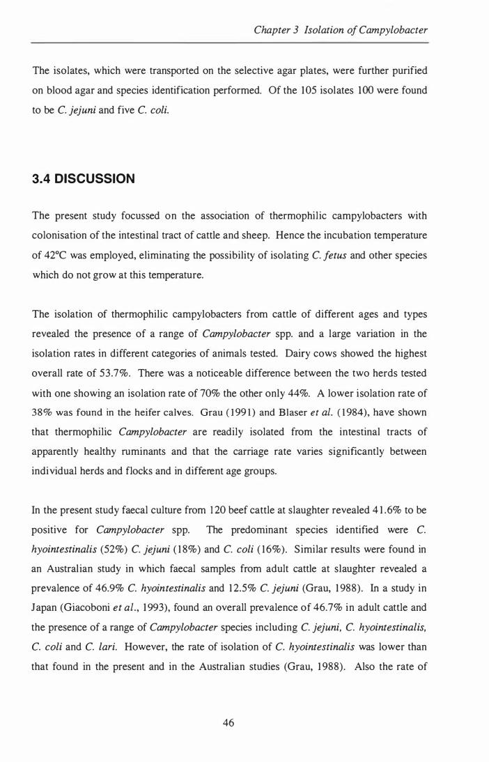
Chapter 3 Isolation of Campylobacter
The isolates, which were transported on the selective agar plates, were further purified
on blood agar and species identification performed. Of the 1 05 isolates 100 were found
to be C. jejuni and five C. coli.
3.4 DISCUSSION
The present study focus sed on the association of thermophil ic campylobacters with
colonisation of the intestinal tract of cattle and sheep. Hence the incubation temperature
of 42°C was employed, eliminating the possibility of isolating C. fetus and other species
which do not grow at this temperature.
The isolation of thermophilic campylobacters from cattle of different ages and types
revealed the presence of a range of Campylobacter spp. and a large variation in the
isolation rates in different categories of animals tested. Dairy cows showed the highest
overall rate of 53.7%. There was a noticeable difference between the two herds tested
with one showing an isolation rate of 70% the other only 44%. A lower isolation rate of
38% was found in the heifer calves. Grau ( 1 99 1 ) and Blaser et al. ( 1 984), have shown
that thermophilic Campylobacter are readily isolated from the intestinal tracts of
apparently healthy ruminants and that the carriage rate varies significantly between
individual herds and flocks and in different age groups.
In the present study faecal culture from 1 20 beef cattle at slaughter revealed 4 1 .6% to be
positive for Campylobacter spp. The predominant species identified were C.
hyointestinalis (52%) C. jejuni ( 1 8%) and C. coli ( 1 6%). Similar results were found in
an Austral ian study in which faecal samples from adult cattle at slaughter revealed a
prevalence of 46.9% C. hyointestinalis and 1 2.5% C. jejuni (Grau, 1 988). In a study in
Japan (Giacoboni et aI., 1 993), found an overall prevalence of 46.7% in adult cattle and
the presence of a range of Campylobacter species including C. jejuni, C. hyointestinalis,
C. coli and C. lari. However, the rate of isolation of C. hyointestinalis was lower than
that found in the present and in the Australian studies (Grau, 1 988). Also the rate of
46

Chapter 3 Isolation of Campylobacter
isolation of C. jejuni was lower compared with that observed by other workers
(Kursteiner et al., 1 985; Doyle and Roman, 1 982;).
The isolation rate of Campylobacter spp. from dairy cattle was 53.7% in the present
study. The predominant species was C. jejuni (58. 1 %) followed by C. hyointestinalis
(30.2%) and C. coli ( 1 1 .6%). Similar results have been obtained by a number of
workers (Stanley et al., 1998a; Cabrita et aI. , 1 992; Humphrey and Beckett, 1 987).
The faecal samples collected from 50 heifer calves revealed 1 9 (38%) positive for
Campylobacter spp. However it was seen that samples from new-born calves ( 1 -3 days
old) had an isolation rate of 20% compared to 56% in calves 2-3 weeks old. It seems
that the animals acquire the bacteria at quite an early age when there is less resistance
from the stil l developing gut microflora. The predominant species isolated was C.
hyointestinalis (78.9%) followed by C. lari ( 1 0.5%), C. jejuni (5.2%) and C. coli
(5.2%). In comparison Adesiyun et al. ( 1 992), isolated Campylobacter from 20.5% of
calves in Trinidad and C. jejuni accounted for 53.3% and C. Coli for 46.7% of the
isolations.
The predominant species isolated from both the bull and heifer calves was C.
hyointestinalis. In a study by Atabay and Cory ( 1 998), C. hyointestinalis was isolated at
a rate of 32%. The reason for the high incidence o( the C. hyointestinalis in calves found
in the present study is difficult to explain. Maybe the population of Campylobacter in
the gut changes with the age of the animal as the normal gut microflora develops and
perhaps in the older population there is a subsequent re-infection. Myers et al. ( 1 984),
reported the isolation of C. hyointestinalis in 20.5% of faecal samples from calves
between 1 -4 weeks of age, but did not detect it in any other groups. Also it was
observed that the bull calves did not carry any C. jejuni as opposed to 5% of the heifer
calves, this might be attributed to the rearing of bull calves separate from the adult dairy
herds after birth. Dairy cows have been frequently found to be Campylobacter positive
and appear to acquire the organism by the consumption of contaminated water
(Humphrey and Becket, 1987) and this may be the main source of infection for calves.
47

Chapter 3 Isolation of Campylobacter
Faecal samples from 1 58 slaughtered sheep in the present study revealed the presence of
Campylobacter in 70 (44.3%) of the samples. All the isolates were characterised as C.
jejuni. Two recent studies in UK have revealed carriage rates of 9 1 .7% from the small
intestines of lambs at slaughter (Stanley et al., 1 998b) and 88% of these isolates were C.
jejuni. In the other study by lones et al. ( 1 999), to assess the shedding of campylobacter
in grazing sheep at different times of the year showed a highest rate of 1 00% coinciding
with lambing, weaning and movement on to a new pasture and the lowest (0%) occurred
when the sheep were fed on hay and silage. C. jejuni was the predominant species (90%)
recovered from the above study. Reported results from similar studies overseas revealed
considerable variation, between 0- 94% (Adesiyun et aI., 1 992; Green et aI., 1 990;
Turkson et al. 1988; Elegbe et al., 1 987; Sarkar et aI. , 1 984; Rosef et al., 1 983). As no
other studies on large numbers of sheep have been carried out and reported before in
New Zealand, it is assumed that the findings from this study are indicative of the true
carriage rate. However it is noted that this rate is influenced by many factors, including
age, sex, weather, season, geography, time and immune status of animals.
The occurrence of a C. jejuni in the environment of the abattoir and the equipment
(56%) is understandable given the high prevalence of the organism in the sheep faeces.
An overseas study (Bolton et al. , 1 982) isolated Campylobacter spp. from the external
surfaces of 70% of sheep carcasses, and 27-38% of implements e.g. : knives, cleavers,
saws, cutting blocks, hooks, racks and scales. Another survey conducted in an abattoir
in Calcutta found that 86.7% of floors and 50% of walls were contaminated. The author
stated that the extensive contamination of the slaughterhouse represented a phenomenal
risk to operating staff (Sarkar et al., 1 984). The difference between the isolation rate
from sheep and the environment may reflect a build up of the organism, indicating a
higher risk for meatworkers, especially in the highly contaminated areas.
The Campylobacter isolates from human clinical samples were identified as C. jejuni
( 1 00) and C. coli (5). The predominance of C. jejuni as a cause of human disease is
well known throughout the world (Griffith and Park, 1 990). Similar findings have been
made in different countries where C. jejuni constitute more than 95% of the isolations
from clinical cases and C. coli account for roughly 5% (Pearson and Healing, 1 992).
48

Chapter 3 Isolation of Campylobacter
The overall prevalence of thennophilic campylobacters in our study was observed to be
on the higher side when compared to other studies in different countries (Table 3 . 1 ).
This can be attributed to different factors. 1 ) Increased sensitivity and specificity of
culture methods for campylobacter have shown to lead to a significant increase in the
rate of isolations (McNicholas, 1 995). The method of isolation used by us involved a
selective enrichment step using Preston broth, which might have enhanced the isolation
rate particularly by el iminating other competitive flora and also by reviving injured
cells. Under certain environmental conditions for example, stationary phase of growth
or exposure to atmospheric oxygen, campylobacters become round or coccoid in shape.
This shape change has been associated with a transition from a viable culturable fonn to
a viable but unculturable state (Roll ins and Col well, 1986), and this phenomenon might
contribute to the differences in the isolation rates depending upon the cultural conditions
employed.
2) Difference in the number and isolation rate between different countries might be due
to differences in environmental and husbandry conditions of cattle (Giacoboni et al.,
1 993). The conditions in which cattle are raised in New Zealand on open pastures are
different from those found in many other parts of the world and might contribute to the
differences in the isolation rates and the species isolated. Stern ( 1 98 1 ), has pointed out
that variations such as herd type, season, animal age, feeding regimen, crowding and
geography can account for significant differences in isolation rates. Campylobacter are
nonnal components of the intestinal flora of several wild rodents and wild birds. These
reservoirs are believed to spread Campylobacter strains in nature. Exposure to their
excrements or consumption of contaminated water constitutes the main routes of
infection for food producing animals (Blaser et al., 1983b).
The incidence of campylobacteriosis in humans in New Zealand in shown to fol low a
seasonal trend (Brieseman, 1 990). It is reported to be highest in summer and lowest in
winter (Ikram et al., 1 994). Meanger and Marshall ( 1989), found the highest prevalence
of thennophilic campylobacter in dairy cattle in New Zealand to be in autumn (3 1 %)
followed by summer (24%) and the lowest ( 1 2%) in winter. This partially follows the
49
Chapter 3 Isolation of Campylobacter
pattern for human clinical episodes so would be consistent with cattle being an
important source of human infections.
Although we did not set out to determine the prevalence of Campylobacter spp. in
different seasons, the sampling in the present study was done over a period of 1 0 months
(April to January) thus incorporating a part of all seasons. Analysis of data reveals that
there was no substantial difference in the isolation rates of thermophilic campylobacter
recovered from the animals over different seasons although isolations made in the
summer months show a slight increase (Table 3 .5) .
The high incidence of human infections in summer could be attributed to the general
population undertaking more outdoor activities in this season and thus exposing
themselves to contaminated stream waters or to drinking untreated water (Bohmer,
1 997; Brieseman, 1 987) and milk (Brieseman, 1 984). A change in the food habits from
well-cooked meats at home to eating barbecued and less thoroughly cooked meats
(Ikram et al., 1994), may be of more significance than any postulated increase in the
excretion of the organism from the animal reservoirs.
Campylobacter outbreaks due to consumption of contaminated water have been
documented in New Zealand (Bohmer, 1 997; Stehr-Green et al., 1 99 1 ; Brieseman,
1 987). It has been suggested that heavy rain may wash campylobacter organisms from
the surrounding beef and sheep farms into rivers and facilitate seepage into springs and
other drinking water sources.
The isolation of C. hyointestinalis and C. lari from cattle is an important finding of the
present study. C. hyointestinalis was first isolated from pigs but is now being
increasingly isolated from other animals especially cattle (Grau, 1 988; Gebhart et al. ,
1 985 ; Myers et al., 1 984; Ursing et al. , 1 984) . C. lari was first described as a group of
strains isolated from seagulls (Skirrow and Benjamin, 1 980) and later from other birds
and animals (Lindblom et al., 1 990). Both C. hyointestinalis and C. lan have been
isolated from human clinical cases overseas and are now recognised as infrequent but
50

Chapter 3 Isolation of Campylobacter
important human enteric pathogens (Borczysk et al., 1 987; Edmonds et al., 1 987; Tauxe
et al., 1 985).
The occurrence of these species in cattle makes them potential pathogens for humans in
this country. Since most medical laboratories in New Zealand, do not identify
campylobacter to species level we do not have sufficient information on the
involvement of the different species of campylobacter in the cases of human infection.
If this collection of 1 05 isolates from Palmerston North is indicative of the national
status then a small but significant proportion of cases of campylobacteriosis is due to C.
coli.
A high percentage of people in New Zealand are involved in farming and occupations
associated with handling animals and raw meat such as butchers, and staff at freezing
works and supermarkets. There are thus many opportunities for infection with
Campylobacter spp. (Brieseman, 1985). If the high prevalence of campylobacteriosis in
the human population is a reflection of the high prevalence of infection in cattle and
sheep found in this study then measures need to be implemented to break this cycle of
infection. Such measures might include education about the potential sources of
infection and the need to ensure safe water and milk supplies as well as the importance
of properly cooking meats. There is therefore a need to determine whether or not other
species such as deer are reservoirs and, if so, to implement appropriate control measures
to prevent spread to the human population. While it is assumed at this stage that the
species of campylobacter in farm animals are the same as those involved in human
disease this is not always certain . There is also a need to characterise isolates more
completely at the molecular level to determine whether or not the human clinical
isolates are the same as the animal isolates.
5 1

Chapter 4 Typing of Campylobacter Isolates
CHAPTE R 4
4. TYPING OF CAMPYLOBACTER JEJUNI ISOLATES FROM
CATTLE, SHEEP AND HUMANS BY PULSED-FI E LD G E L
ELECTROPHORESIS.
4.1 . I NTRODUCTION
Campylobacter spp. represents a taxonomically heterogeneous group. The identification
of these campylobacters can be difficult since strains have relatively fastidious growth
requirements, are asacchrolytic and only a few biochemical tests give adequate
discrimination (Goossens and Butzler, 1992).
Serotyping has been the most common typing method (Lior, 1 984; Penner et al., 1 983),
although biotyping and phagetyping have also been employed (Patton and Wachsmuth,
1 992) to show the epidemiological association of isolates from patients or to trace the
possible routes of transmission from animals to humans. However, strain
discrimination using phenotypic typing methods may be significantly compromised due
to poor reproducibility or inadequate discriminatory power (Arbeit, 1995).
A feature of the epidemiology of human campylobacteriosis is that most infections are
sporadic cases with relatively few community outbreaks. The sources of the sporadic
infections are rarely established with strains being widely distributed in both wild and
domesticated birds and mammals, as well as in sewage and untreated water.
Pulsed-field gel electrophoresis (PFGE) of genomic DNA restricted with low frequency
cutting endonucleases, has proved to be a valuable tool in the epidemiological
investigations of a wide range of medically important bacteria (Tenover et al., 1 995),
probably as a consequence of its ability to examine restriction polymorph isms along the
entire bacterial chromosome.
52

Chapter 4 Typing of Campylobacter Isolates
This method has been found more distinguishing than serotyping because several
genotypes are found within a serotype (Gibson et aI. , 1 995; Suzuki et aI., 1 994). PFGE
has been found to be the most discriminatory of several typing methods employed by
various workers in characterising campylobacter isolates (Steele et aI., 1 998; Shi et aI.,
1 996) and has considerable potential for tracing the possible routes of transmission of
the campylobacters causing sporadic infections.
The aim of the present study was to type the Campylobacter jejuni isolates from cattle
and sheep by PFGE using the restriction endonuclease Sma I and to generate a PFGE
profile of these isolates to asses their relatedness and also compare them with C. jejuni
isolates from human clinical cases in the Manawatu region. This exercise would give us
an idea as �o the similarities or differences between the C. jejuni strains prevalent in
animals and humans and lead us to better understand the epidemiology of campylobacter
infections in this region.
4.2. MATERIALS AND M ETHODS
4.2. 1. BA CTERIAL ISOLA TES
C. jejuni isolates from humans, cattle and sheep were used for this study. The isolates
were stored in glycerol broth at -70°C until used. They were revived by thawing, plating
on blood agar plates and incubating in microaerophilic environment at 42°C for 24 h.
4.2.2. DNA PREPARA TION
Intact genomic DNA was prepared by a modification of the methods described by Barret
et al. ( 1 994) and Olsen et al. ( 1 994).
Single colonies were selected from purity blood agar plates and grown in trypticase soya
broth at 42°C for 48 h. One millilitre aliquot of each broth culture was pipetted into a
sterile micro-centrifuge tube and centrifuged at 10,000 rpm for 4 min. The supematant
was removed, the pellet resuspended in 1 .5 ml ice-cold Pett IV buffer O M NaCI, I OmM
53

Chapter 4 Typing of Campylobacter Isolates
Tris HCl pH 8.0, 1 0mM EDT A pH 8.0) and the suspension centrifuged at 1 0,000 rpm
for a further 4 minutes. The supernatant was removed and the pellet resuspended in 50
III of cold Pett IV buffer and kept on ice until required. Eighty III of molten, 1 %,
pulsed-field certified (PFC) agarose (10 mg rnI·1 agarose in Pett IV buffer) was added to
the suspension and mixed carefully by pipetting to avoid air bubbles. The resultant
agaroselbuffer suspension was transferred quickly into a plug mould and left to solidify
on ice for 20 min. Then, the solid plugs were removed into fresh micro-centrifuge tubes
containing 1 ml Urea-ESP buffer (6M Urea, 50mM Tris-HCI pH 8.0, 50 mM EDTA pH
8 .0, 1 % sodium lauryl sarcosine, 0.2% sodium deoxycholate 0.5 mg rnI·! Proteinase K)
and incubated overnight in a waterbath at 56°C. After the incubation the plugs were
washed 8 times in ice-cold TE pH 8.0, for at least 30 min each wash and stored in TE
pH 8 .0 at 4°C until required.
4.2.3. RESTRICTION ENDONUCLEASE DIGESTION OF PLUG-
INCORPORA TED DNA
The restriction endonuclease enzyme Sma I was selected based on reports by other
workers (Salama et al., 1 992; Yan et al., 1 99 1 ) about its cutting ability in
Campylobacter spp. and after preliminary trials with a few other restriction enzymes.
An optimum concentration of the enzyme and other parameters were determined by
initial experimentation (data not presented).
Approximately one quarter of each plug was cut off using a sterile scalpel blade, placed
in l OO III of I x restriction buffer ( 1 2 1l1 1 Ox New England Biolabs Buffer 4, 1 111 1 00 mg
mrl Bovine Serum Albumin (BSA), 87 III sterile distilled water) and equilibrated for at
least 45 min on ice. About 20 plugs were processed at one time and a cocktail of the
restriction enzyme was made up, depending on the number of plugs being digested, and
aliquotted accordingly. The restriction buffer was decanted and replaced with 80 III
cutting buffer (8 III 10x restriction buffer, 0.8111 l00mg rnI-1 BSA, 20 U restriction
endonuclease, distilled water). Plugs and buffers were equilibrated for 45 min before
incubation overnight in a waterbath at 25° C.
54

Chapter 4 Typing of Campylobacter Isolates
4.2.4. PULSED-FIELD GEL ELECTROPHORESIS OF DIGESTED DNA
A contour-clamped homogenous electric field (CHEF) apparatus, including variable
speed pump, gel chamber, power module and mini chiller (CHEF Mapper, B io- Rad
Laboratories, Richmond, California, USA) was the system used for PFGE. A gel
running protocol was optimised for maximum resolution of the digested DNA fragments
(Table 4. 1 ).
After completion of digestion, plug slices were loaded into the wells of a 1 % PFC
agarose gel (800 mg in 80 ml 0.5x TBE buffer), which had been equilibrated in the
electrophoresis chamber with the buffer (0.5x TBE) circulating, for at least 2 h . A
molecular weight marker was also loaded in each gel .
After electrophoresis, gels were stained in fresh aqueous ethidium bromide (80 J..lI of
1 0mg mrl stock solution in 800 rnI-distilled water) for I h, then destained overnight.
Gels were examined under UV light and photographed.
Table 4.1 : Gel Running Parameters
PARAMETER
Agaraose Type / Concentration 1 % Pulsed Field Grade Agarose
Buffer Type / Concentration 0.5X TBE
Voltage gradient 6.0 V cm-I
Buffer temperature 1 4° C
Pulse angle 1 20°
Initial switch time 1 sec
Final switch time 28 sec
Run time 24 h
55

Chapter 4 Typing of Campylobacter Isolates
4.2.5. INTERPRETA TION OF PULSED-FIELD PROFILES
The gels were analysed visually for differences in the banding patterns. The first isolate
examined from each source was arbitrarily designated as Type 1 and all other isolates
were compared with this "type strain". Bands were identified numerically from the
highest molecular weight downwards, with positions fixed in relation to Type 1 . Bands
were also described by their sizes in kilobases relative to one of the molecular weight
markers. Isolates were subsequently classified into Types and Pulsotypes on the basis of
· their band differences to the Type 1 strain. Following the guidelines published by
Tenover et al . ( 1 995) on the interpretation of chromosomal DNA restriction patterns
produced by PFGE, isolates were assigned to new Types when they differed by four or
more bands from the Type 1 strain, and to pulsotypes when they differed by 1 -3 bands
from the Type 1 strain.
4.3. RESULTS
4.3. 1. RESUL TS OF PFGE OF C. JEJUNI STRAINS ISOLA TED FROM
CA TTLE
Genomic DNA from 35 C. jejuni isolates from cattle was characterised by PFGE after
digestion with the restriction enzyme Sma 1. The resolution of Sma I digested DNA
yielded between 5 and 9 fragments ranging in size from 40 - 480 kbp. The 35 isolates
were classified into 1 1 different Types (Type 1 to 1 1 ) having a difference in 4 or more
bands (Fig 4. 1 ; Table 4.2). Type 1 was subdivided into 3 pulsotypes each, having up to
three band differences from the principal strain (Table 4.2).
Types l e, and 2 were found in both beef and dairy animals. Whereas Types 1 , I B, 3 and
5 were unique to beef cattle and Types 4, 6, 7, 8, 9, 1 0, and 1 1 , to dairy cattle.
56
/

Chapter 4 Typing of Campylobacter Isolates
Table 4.2 Types and Pulsotypes of Campylobacter jejuni among 35 cattle isolates
TYPES NUMBER OF ISOLATES (%)
Type 1 (Pulsotype lA) 1 (3)
Pulsotype Is 1 (3)
Pulsotype le S (14)
Type 2 12 (34)
Type 3 1 (3)
Type 4 6 (17)
Type S 1 (3)
Type 6 1 (3)
Type 7 1 (3)
Type S 2 (6)
Type 9 1 (3)
Type 10 1 (3)
Type 11 2 (6)
57

Chapter 4 Typing of Campylobacter Isolates
Table 4.3 Types and Pulsotypes of Campylobacter jejuni among 50 Sheep isolates
TYPES NUMBER OF STRAINS (%)
Type 1 (Pulsotype lA) 2 (4)
Pulsotype Is 10 (20)
Pulsotype le S (10)
Pulsotype 10 4 (8)
Pulsotype h 5 (10)
Type 2 1 (2)
Type 3 2 (4)
Type 4 5 (10)
Type S 3 (6)
Type 6 4 (8)
Type 7 3 (6)
Type 8 1 (2)
Type 9 1 (2)
Type 10 1 (2)
Type 11 1 (2)
Type 12 2 (4)
58

--------
Chapter 4 Typing of Campylobacter Isolates
Table 4.4 Types and Pulsotypes of Campylobacter jejuni among 50 human isolates
TYPES NUMBER OF ISOLATES (%)
Type 1 (Pulsotype lA) 9 (18)
Pulsotype Is 1 1 (22)
Pulsotype le 2 (4)
Pulsotype 1D 3 (6)
Pulsotype h 2 (4)
Pulsotype h 2 (4)
Type 2 1 (2)
Type 3 9 (18)
Type 4 3 (6)
Type S 2 (4)
Type 6 1 (2)
Type 7 1 (2)
Type S 1 (2)
Type 9 1(2)
Type 10 1 (2)
Type 1 1 1 (2)
59

Chapter 4 Typing of Campylobacter Isolates
Figure 4.1 Representative PFGE profiles C. jejuni cattle isolates
M 1 2 3 4 5 6 7 8 9 10 1 1 1 2 1 3
M : Molecular size marker (Lambda ladder)
Lanes: I-Type I (PuIsotype 1 ), 2-Type 2, 3- Pulsotype I B, 4- Pulsotype le , 5- Type 3,
6-Type 4, 7- Type 5, 8- Type 6, 9- Type 7, 10- Type 8, 11 - Type 9, 12- Type 1 0
13- Type 1 1 .
60

Chapter 4 Typing of Campylobacter Isolates
FIGURE 4.2 Representative PFGE profiles of C. jejuni sheep isolates
M 1 2 3 4 5 6 7 8 9 10 1 1 1 2 1 3 1 4 1 5 1 6
kb
M: Molecular size marker (Lambda ladder)
Lanes : 1- Type l (Pulsotype l A), 2- Pulsotype I s. 3- Type 2, 4- Type 3, 5- Type 4,
6- Pulsotype l e. 7- Pulsotype ID. 8- Type 5, 9- Type 6, 10- Pulsotype l E.
1 1 - Type 7, 12- Type 8, 13- Type 9, 14- Type 10, 15- Type 1 1 , 16- Type 1 2.
6 1

Chapter 4 Typing of Campylobacter Isolates
FIGURE 4.3 Representative PFGE profiles of C. jejuni human isolates
M 1 2 3 4 5 6 7 8 9 10 1 1 1 2 1 3 14 15 1 6
kb
M: Molecular size marker (Lambda ladder)
Lanes: 1- Type I (Pulsotype 1 ), 2- Pulsotype 1 B, 3- Type 2, 4- Type 3, 5- Type 4,
6- Pulsotype l e, 7- Type 5, 8- Type 6, 9- Pulsotype I D, 10- Pulsotype l E,
11 - Type 7, 12- Type 8, 13- Type 9, 14- Type 10, 15- Pulsotype I F,
16- Type 1 1
62

Chapter 4 Typing of Campylobacter Isolates
Figure 4.4 Common PFGE profiles in human, cattle and sheep isolates
of C. jejuni
M 1 2 3 4 5 6 1 8 9 10 J 1 1 2 13 1 4 1 5
kb
388
194
48-5
M: Molecular size marker (Lambda ladder)
Lanes 1 and 2 Human and cattle isolates- pulsotypes
Lanes 2 and 3 cattle and human isolates - indistinguishable
Lanes 4 and 5 human and cattle - indistinguishable
Lanes 5 and 6 cattle and human - pulsotypes
Lanes 7 and 8 human and cattle - indistinguishable
Lanes 10 and 12 human and sheep- indistinguishable
Lanes 13 and 15 human and sheep- indistinguishable
Lanes 14 and 15 cattle and sheep - pulsotypes
63

Chapter 4 Typing of Campylobacter Isolates
4.3.2. RESULTS OF PFGE OF C. JEJUNI STRAINS ISOLA TED FROM
SHEEP
Genomic DNA from a total of 50 C. jejuni isolates from sheep were digested with the
restriction endonuclease Sma I and subjected to PFGE. The analysis of digested DNA
yielded six to nine fragments ranging in size from 40- 480 kbp. The isolates were
grouped in to 1 2 different Types (Type 1 to 1 2) based on a difference of four or more
bands (Fig 4.2; table 4 .3). Type 1 was further subdivided into five pulsotypes having up
to 3 band difference from the principal type (Fig 4.2; Table 4.3).
4.3.3. RESULTS OF PFGE OF C. JEJUNI STRAINS ISLOA TED FROM
HUMANS
Genomic DNA from 50 C. jejuni isolates from humans was digested with the restriction
endonuclease enzyme Sma I and resolved by pulsed-field gel electrophoresis. The
strains could be grouped into 1 1 different Types (Type 1 to 1 1 ) based on a difference of
4 or more bands (Fig 4.3 ; Table 4.4). Type 1 (Pulsotype l A) was further subdivided
into five pulsotypes (Pulsotypes IB to J p) having up to 3-band difference from the
principal strain (Fig 4.3; Table 4.4).
4.3.4. COMPARISON OF THE PFGE PROFILE OF C. JEJUNI ISOLA TES
FROM ANIMAL AND HUMAN SOURCES
The PFGE profiles of the cattle, sheep and human isolates were compared visually to
determine their relatedness to each other.
A total of six cattle and human C. jejuni isolates were found to have identical PFGE
patterns. Another 25 isolates were found to differ by less than three bands from each
other and were classified as pulsotypes and considered to be closely related (Fig 4.4).
64

Chapter 4 Typing of Campylobacter Isolates
Among the sheep and human isolates there were two groups consisting of a total of ten
isolates, which had identical patterns to each other (Fig 4.4).
The comparison of cattle and sheep PFGE profile revealed three sheep isolates to be
pulsotypes of a cattle isolate.
4.4. DISCUSSION
PFGE has been used for typing various organisms (Wrights et al., 1 998; Feizabadi et al.,
1 997; Hosaka et al., 1 997; Harsono et al., 1 993). Digestion of chromosomal DNA with
a restriction endonuclease that has infrequent DNA recognition sites results in fewer but
larger fragments and greatly simplifies analysis compared to restriction endonuclease
analysis (REA) and standard electrophoresis (Kakoyiannis et al., 1 988).
The aim of the present study was to characterise the large number of C. jejuni strains
isolated from cattle and sheep at meat works and farms in the Manawatu region and to
compare them with C. jejuni strains isolated from clinical cases in humans at the
Palmerston North Public Hospital.
Gibson et al. ( 1 995) reported the first use of PFGE in an epidemiological investigation
of C. jejuni. The results indicated that this technique was most discriminatory of other
typing methods used namely ribotyping and phage-typing and was capable of
discriminating not only between strains of different serotypes but also between strains
within a single serotype. The present study confirmed that PFGE of chromosomal DNA
was a useful technique in separating large molecular weight fragments with high
resolution into simple restriction profiles, which could be easily compared.
The restriction enzyme Sma I provided the best discrimination among the enzymes
tested in the preliminary trial runs, with the most easily interpreted patterns consisting of
6- 10 fragments between approximately 40 kbp to 500 kbp. This was subsequently used
in the restriction analysis of all the isolates.
65

Chapter 4 Typing of Campylobacter Isolates
Some workers have suggested that the use of two restriction enzymes might give better
discrimination (Gibson et aI., 1 995) however, Steele et al. ( 1 998) compared restriction
analysis using Sma I and Sal I and came to the conclusion that the discriminatory index
of PFGE with either of the enzymes was excellent and could be enhanced only slightly
by combining the two enzyme groups.
The technique was found to be robust with good reproducibility as demonstrated by the
consistent patterns obtained when a number of isolates where run several times during
the investigation (data not presented).
In this study the PFGE analysis of isolates of C. jejuni from cattle, sheep and humans
with the Sma I restriction endonuclease typically resolved 5- 1 0 bands ranging in size
from 40 kbp to 480 kbp. Gibson et al . ( 1 997) made similar observations in an
epidemiological investigation of C. jejuni in the UK.
The phenotypically identical C. jejuni cattle isolates were characterised into 1 1 different
types, which reflects the high degree of discrimination obtained by PFGE and the
di versity of the organism within a limited geographic area. However, the majority of the
isolates (more than 70%) fell under three predominant types (Type 1 , 2 and 4) showing
the occurrence of a few dominant clonal types in most of the animals. The other eight
types were found to be represented by only one or two strains each.
In a similar study On et al. ( 1 998), separated 34 Danish strains of C. jejuni from cattle,
poultry, humans and water and assigned them to one of six Sma I profile groups. Two
groups of indistinguishable isolates contained randomly isolated strains from the human
cattle and poultry isolates providing evidence of identical clones infecting humans and
animals.
Some strains having identical PFGE profiles were common to beef and dairy cattle,
showing the widespread presence and dissemination of the same clonal types in the
region, probably originating from a common ancestry.
66

Chapter 4 Typing of Campylobacter Isolates
Types l e, and 2 were not only found most predominant but were found in both beef and
dairy animals which belonged to different farms from different locations. There were
also Types which were either unique to beef cattle (Types 1 , I s, 3 and 5) or to dairy
cattle (Types 4, 6, 7, 8, 9, 10, 1 1 , ) .
The sheep isolates were characterised into 12 different types based on a difference of
four or more bands in their PFGE patterns. There was only one major type (Type
1IPulsotype l A) with its four pulsotypes ( 1 s, l e, 1 0 & l E) which encompassed more than
52% of the isolates. But unlike the cattle strains the sheep isolates were found to be
more varied with the other Types (2 to 1 2) being represented by two to ten percent of
the isolates each.
The human isolates more or less followed a distribution similar to the sheep isolates and
were categorised into 1 1 Types based on their band differences. Type 1 and its five
pulsotypes ( 1 s to I F) were found to be the most predominant types with about 58 % of
the isolates falling under this group. This could suggest a common origin or source of
infection, which is represented by infecting C. jejuni strains showing more genetic
homogeneity.
Hanninen et al. ( 1 998b), identified a total of 69 PFGE types among 1 76 C. jejuni
isolates from Finnish patients. Five predominant types were found to comprise 40% of
the isolates. In investigations of small outbreaks, identical PFGE patterns were
demonstrated, indicating a common source of infection.
The comparison of the animal strains with the human isolates provided quite
illuminating data with a large number of cattle strains having identical patterns to the
human isolates implying a clonal relationship between them.
Similarly the common PFGE patterns and closely related pulsotypes found between the
sheep and human isolates strongly suggest that the sheep isolates are the same clone as
the human isolates. The occurrence of only one cattle isolate which had three
pulsotypes in the sheep isolates suggests that the cattle and sheep isolates are more
67

------------- -
Chapter 4 Typing of Campylobacter Isolates
distantly related and probably have diverged very early from each other In their
evolution.
Most cases of campylobacteriosis in humans occur sporadically with the principal route
of infection believed to be foodborne (Skirrow, 1 994; Tauxe, 1 992). Our results
provide substantial genotypic evidence for a l ink between sporadic human infection and
two food animal sources; cattle and sheep in the region.
PFGE has been used successfully in earlier studies to establish links between food
animal sources and human disease. Lorenz et al. ( 1 998), used Sma I PFGE profiles to
characterise chicken and human isolates and was able to detect similar types between
the two sources. Similarly in another study Hanninen et al., ( 1 998b), found identical
PFGE patterns in human and poultry isolates. However four of the ten most common
human PFGE types were not found among chicken isolates, which suggests that in
addition to poultry other sources like cattle and sheep might contribute to human
infections as has been demonstrated by the occurrence of closely related strains between
the cattle, sheep and human isolates in the present study.
Although poultry are traditionally regarded as the principal source of such infections
(Shane, 1 992 ) cases linked to bovine and ovine sources have been described previously
(Dilworth et al., 1 988; Orr et al., 1 995). Also the data presented in the present study
indicate the need for further investigation concerning the infectious potential of other
foodstuffs such as beef and mutton as a source of human infections or as a reservoir for
C. jejuni strains pathogenic to humans.
Laboratory and epidemiological methods used previously to establish chains of
transmission in foodborne infections have been limited to serotyping and classical
epidemiological case studies, all of which are not sufficiently sensitive to distinguish
individual strains (Patton et al., 1 99 1 ; Arbeit, 1 995). In addition, a substantial number
of isolates have been found to be untypable by Lior and Penner serotyping and phage
typing (Patton and Wacshsmuth, 1 992).
68

Chapter 4 Typing of Campylobacter Isolates
The ability to rapidly identify certain clones of known pathogenicity may thus be more
relevant to public health protection than simply detecting the presence of C. jejuni. The
application of PFGE has been shown to provide precise information that can be used to
accept or reject epidemiological associations to a high degree of confidence (Smith and
Cantor, 1 987; Smith et al., 1 988).
The present study has endeavoured to classify the cattle, sheep and human strains in the
region and identify the important types prevalent in the region and to enhance our
understanding of the epidemiology of campylobacter infections in the area. The close
relationship found between certain animal Types with the human isolates has stressed
the importance of monitoring the bovine and ovine sources also as an important source
of human campylobacter infections.
The use of PFGE in typing Campylobacter spp. isolated from human and animal sources
and maintenance of a national database should offer an excellent means of
epidemiological monitoring of the disease situation and make comparisons between the
human and animal strains possible. This would ultimately help in better understanding
of the infectious potential of various animal species and offer a means of control of
human infections.
69

Chapter 5 Antimicrobial Susceptibilities
CHAPTER S
5. ANTIMICROBIAL SUSCEPTIBILITY PATTERNS OF
CAMPYLOBACTER JEJUN/ ISOLATED FROM CATTLE, SHEEP,
AND HUMANS
5.1 . INTRODUCTION
Campylobacter jejuni is recognised as one of the most important causes of acute
diarrhoeal disease in humans throughout the world (Taylor and Blaser, 199 1 ).
Affected persons usually recover without antimicrobial therapy but in some patients
with prolonged illness therapy may be indicated. In these circumstances erythromycin
or fluroquinolones are often recommended and tetracyclines used as alternatives
(Blaser, 1997; Blaser, 1 990; Goodman et al. , 1 990) .
An alarming trend has been the increase in resistance in Campylobacter spp. isolated
from humans to antibiotics which are used in the treatment of human infections (Roge
et aI., 1 998; Velazquez et aI. , 1995). Resistant bacteria can diminish the effectiveness
of antibiotics and demand the use of more expensive or less safe alternatives.
This has been attributed to the use of antibiotics in food producing animals as growth
promoters, in prophylaxis and treatment with antimicrobial agents that are important in
human therapy (Tollefson et al. , 1 998; Reina et al. , 1 992). It has been reported that
worldwide growth promotants account for 42% of the mass of veterinary
pharmaceuticals used (Gold and Moellering, 1 996) .
In addition, it has been demonstrated that the flow of resistance genes in the
environment from animals to man is possible (Johnson et al. 1994; Nijsten et al. , 1 994;
Linton 1 986; Scoli et al. 1 980).
70

Chapter 5 Antimicrobial Susceptibilities
In New Zealand, the incidence of campylobacter infections in humans is amongst the
highest in the world and it is the most commonly diagnosed cause of acute diarrhoeal
disease (McNicholas et ai. , 1 995). In a recent study, low level resistance to
erythromycin, ciprofloxacin and doxcycline was detected in Campylobacter isolates
from Auckland (Dowling et ai. , 1 998) However, very little information is available on
the prevalence of antimicrobial resistance in other areas of New Zealand in animal and
human isolates.
The aim of this study was to determine the antimicrobial profile of C. jejuni isolated
from humans, cattle and sheep to six antibiotics commonly used in the treatment of
campylobacter infections in humans.
5.2. MATERIALS AND M ETHODS
5.2.1. BACTERIAL STRAINS
One hundred C. jejuni human isolates from human patients with diarrhoea were
procured from the Palmerston North Medical Laboratory. The 3 5 cattle and 1 00 sheep
isolates were isolated from faecal samples collected at local meat-works and farms
from apparently healthy animals. All the isolates were stored frozen in glycerol broth at
-70°C and revived by culturing on blood agar. The control organism was E. coli
NCTC 1 04 1 8 .
5.2.2. ANTIBIOTIC SUSCEPTIBILITY TESTING
Susceptibility testing was performed by the controlled disc diffusion method (Hunter,
1 994) on Muller-Hinton agar plates supplemented with 5% sheep blood. A well
isolated colony was selected from a blood agar plate and inoculated into 3 rnl of sterile
7 1

Chapter 5 Antimicrobial Susceptibilities
saline and briefly rotainixed. A suspension for the control E. coli was similarly
prepared. A sterile swab was used to apply the control and the test suspension spirally
on the agar medium using a rotary plater (pbi International) . The control was spread
from the area of the disc to the outside of the plate and the test sample from the centre
of the plate to the area of the discs.
The six antibiotic discs (Oxoid) of erythromycin ( 1 5 J..Lg), tetracycline ( 1 0 J..Lg),
enrofloxacin ( 1 0 J..Lg), chloramphenicol ( 1 0 J..Lg), gentamicin ( 1 0 J..Lg) and neomycin ( 10
J..Lg) were applied on the agar between the test sample and control using a disc
dispenser (Oxoid). The plate was allowed to dry for 30 minutes and then incubated in
an anaerobic jar with a Campy-Pak (BBL) gas envelope at 42°C. The plates were read
after 48 h and the diameter of the zones of inhibition around the antibiotic discs
recorded.
5.2.3. MINIMAL INHIBITORY CONCENTRA TION (MIC)
The MICs of the resistant isolates was determined by an agar dilution method (Mendez
et aI. , 1 980). Isolates were grown in blood agar plates containing 1 J..Lg/ml of
tetracycline. Growth was suspended in 0. 1 M phosphate buffer saline (PBS) to 0.5
McFarland standard. The suspension was diluted 1 : 1 0 in PBS and inoculated into
Muller-Hinton agar plates with doubling concentration of tetracycline ranging from 2
to 1 28 J..Lg/ml in duplicates. The plates were incubated at 42°C and observed after 24 h
and 48 h for growth. The highest concentration in which growth was absent was taken
as the MIC of that strain.
72

------- -
Chapter 5 Antimicrobial Susceptibilities
5.3. RESU LTS
5.3.1. RESULTS OF ANTIMICROBIAL SUSCEPTIBILITIES OF HUMAN,
CA TTLE AND SHEEP ISOLA TES
All the 100 human isolates were found sensitive to erythromycin, enrofloxacin,
gentamicin, chloramphenicol and neomycin. Two isolates were found resistant to
tetracycline.
All 35 C. jejuni isolates from cattle as well as the 1 00 sheep C. jejuni isolates were
found sensitive to all the six antibiotics tested.
5.3.2. RESULTS OF DETERMINATION OF THE MINIMAL INHIBITORY
CONCENTRA TIONS (MICs) OF THE RESISTANT ISOLA TES
The minimal inhibitory concentrations of the tetracycline resistant isolates of C. jejuni
were determined by the agar dilution method. MICs of the two resistant isolates were
found to be � 1 28 �glml. Tetracycline susceptible isolates demonstrated MICs of � 2
�glml .
The two resistant isolates were in the process of testing sub-cultured 3 times after the
first determination of resistance and still found resistant.
73

Chapter 5 Antimicrobial Susceptibilities
5.4. DISCUSSION
In modern breeding of farm animals, practised in most countries, large amounts of
antibiotics are used for a variety of reasons, including growth promotion, disease
prophylaxis, and therapy and there is an increasing concern that such widespread use of
antibiotics is contributing to the emergence of resistant bacteria that are contaminating
the food supply (WHO Report, 1 997). However, this is not at all the New Zealand
situation except for intensive industries such as pigs and poultry. Many of the 40
million sheep in New Zealand would never see antibiotics.
Infections with quinolone-resistant strains of Campylobacter organisms are now found
with increasing frequency in clinical settings, following a trend of food borne
pathogens that have become resistant to antibiotics and have caused an increase in
morbidity and mortality among humans (Tee et al. , 1995; Alder-Mosca et aI. , 1 99 1 ;
Endtz et aI. , 1 99 1 a)
In the United States, Smith et al. ( 1 999) found that resistance to nalidixic acid
increased approximately eight-fold from l .3% in 1 992 to 1 0.2% in 1 998. All isolates
that were resistant to nalidixic acid were also resistant to ciprofloxacin also 2% were
resistant to erythromycin and 6 1 % resistant to tetracycline.
Data from other countries provide evidence that the use of fluoroquinolones in poultry
production has a primary role in increasing resistance to quinolones among C. jejuni
isolates from humans. In the Netherlands, an increase in ciprofloxacin resistant human
isolates from 0% through 1 985 to 1 1 % in 1 989 paralleled the increase in ciprofloxacin
resistant isolates from retail poultry products (Endtz et al. , 1 99 1 a) .
74

Chapter 5 Antimicrobial Susceptibilities
Likewise in Spain, an increase in the prevalence of ciprofloxacin-resistant human
isolates from between 0% - 3% in 1 989 to 30% - 50% in 1 99 1 coincided with the
introduction of enrofloxacin for veterinary use in 1 990 (Reina et ai. , 1992; Sanchez et
ai., 1 994).
In the present study the antimicrobial susceptibility of C. jejuni isolates to six
antibiotics was examined by the disc diffusion method. It was simple to perfonn and
results were easily interpreted. Standard methods for susceptibility testing and
interpretation of resistance in Campylobacter spp. are lacking and different workers
have employed various methods. However, the disc diffusion method has been widely
applied with good results for campylobacter testing (Gaudreau and Gilbert, 1 997,
Huysmans and Tumidge, 1 997; Koenraad et al. , 1 995b; Vanhoof et al. , 1 984). If there
is a need to obtain quantitative data regarding the resistant isolates, then minimal
inhibitory concentrations could be determined by an agar dilution method.
All the 35 cattle and 1 00 sheep isolates were found to be susceptible to all the six
antibiotics used including enrofloxacin. This is contrary to findings in many countries
where resistance to antibiotics is being increasingly detected in animal isolates
particularly against the quinolones.
Smith et al. ( 1999) found 20 % of Campylobacter isolates from poultry to be resistant
to ciprofloxacin and Jacobs-Reitsma ( 1 997) found 29 % to be resistant to quinolones.
This may be due to the extensive systems in which these animals are raised in New
Zealand with minimal to no use of antibiotics.
The likely times when antibiotics are used in these animals would be to treat sick
animals. Antibiotic resistant bacteria are more frequently isolated from pigs and
pOUltry than from extensively grazed cattle or sheep indicating the greater use of
antimicrobial agents in intensively housed species for growth promotion (Threlfall,
1 993). A study of Campylobacter isolates from pigs and poultry in New Zealand may
give very different results and should be conducted in a suitable range of isolates.
75

Chapter 5 Antimicrobial Susceptibilities
In our study only two human isolates (2%) of C. jejuni were found resistant to
tetracycline. No resistance to the other five antibiotics was detected in human, cattle
or sheep isolates. The findings agree with a recent study done in Auckland which
reported two of 202 human isolates to be resistant to tetracycline (Dowling et al. ,
1 998).
Overseas research shows that tetracycline resistance is more common in human isolates
than among isolates from animals. In studies by Aarestrup et al. ( 1 997) 1 1 % of the
human isolates as compared to 2 % of the animal isolates were resistant to tetracycline.
Huysmans and Turnidge ( 1 997) in Australia studied 1 00 clinical isolates of
thermophilic Campylobacter species from humans and did not detect resistance to
erythromycin, gentamicin or chloramphenicol but nine strains were found resistant to
tetracycline.
The absence of resistance to important antibiotics like erythromycin and enrofloxacin
seen in the present study speaks well for the country although low level resistance was
seen in human isolates for erythromycin (1 . 5%) and ciprofloxacin (2.5%) in the
Auckland study (Dowling et aI. , 1 998). This is still very much at a lower level when
compared to the extent of resistance observed overseas.
Levels as high as 84% in Thailand (Hoge et aI., 1 998) to 34% in Spain (Velazquez et
aI. , 1 995) and 1 8 .6% in Sweden (Sjogren et aI. , 1997) to fluoroquinolones have been
reported. Similarly rates as high as 79% resistance to erythromycin have been reported
in Nigeria (Coker and Adefeso, 1 994) to 0.6 % in Japan (Tadano et al. , 1 996). It is
however advisable to monitor this situation so that appropriate action could be
instituted should resistant strains be detected in the future.
Further studies on the antibiotic susceptibility patterns of C. jejuni from intensively
farmed species in New Zealand such as poultry and pigs are needed to better
76

Chapter 5 Antimicrobial Susceptibilities
understand the situation in this country with regard the prevalence of resistance, to
come to definite conclusions about the extent of the problem if any.
77

Chapter 6 Development of PCR
CHAPTER 6
6. DEVELOPMENT OF A POLYM ERASE CHAI N R EACTION
(PCR) FOR IDENTI FICATION OF CAMPYLOBACTER JEJUNI
FROM M EAT SAM PLES.
1 . I NTRODUCTION
. The demanding growth requirements of Campylobacter spp. make their microbiological
detection and identification a time consuming and onerous task. Discrimination among
closely related thermophilic campylobacters is by biochemical and serological tests and
is labour intensive. The detection and identification of these organisms by PCR is now
being recognised as a reliable alternative to the traditional tests (Harmon et al., 1 997).
The use of PCR to screen samples of retail poultry and red meats as a rapid method of
analysis of samples from the supermarkets in addition to the culture method was
evaluated in the present study. Therefore the initial task of developing and optimising
the PCR was undertaken.
6.2. MATERIALS AND METHODS:
6.2. 1 . OLIGONUCLEOTIDE PRI M E R SELECTIO N
The objective of the development of the PCR was to identify C. jejuni, the principal
Campylobacter spp. responsible for the overwhelming majority of infections in humans.
After perusal of available literature it was decided to select and use primers directed
against the hippuricase gene previously described by Linton et al., ( 1 997), which is
unique and highly conserved in C. jejuni (Slater and Owen, 1 997; Hani and Chan,
1 995).
80

Chapter 6 Development of PCR
The primer pair was as follows:
PRIMER AH 1 : 5'- GAA GAG GGT TTG GGT GGT G -3'
PRIMER AH 2: 5 '- AGC TAG crr CGC ATA ATA ACT TG -3'
The primers were synthesised by Life technologies, Auckland, New Zealand and were
reconstituted in sterile distilled water to give 200 J.lM solution of each primer. Primers
were stored in aliquots of 50 III at -20°C.
6.2.2. DNA EXTRACTIO N FROM BACTERIA
Preparation of genomic DNA from C. jejuni was carried out by three different methods
for use as templates for the PCR. The bacteria were grown from the frozen cultures on
blood agar for 48 h at 42°C and broth cultures made by growing in Preston broth for 1 8
h at 42°C.
6.2.2. 1. DNA extraction based on proteinase-K digestion and phenol
chloroform purification
One millilitre of the broth culture was taken in a microfuge tube and centrifuged for 10
min at 2000 rpm and the supematant discarded and the pellet washed two times with TB
buffer. To the pellet was added 500 III of TE buffer and 1 00 III ( 10 mg/ml) of Lysosyme
mixed well and the tube was incubated at 37°C in a waterbath for 30 min and 1 00 III of
1 0% sodium dodecyl sulphate (SDS) added and incubated for a further 30 min.
Thereafter 5 III ( 1 0 mg/ml) Proteinase K (Boehringer Mannheim, Germany) was added
to the tube and incubated again at 56°C for 1 h.
After the incubation an equal volume of 25:24: 1 phenol:chlorofom:isoamyl alcohol
solution was added to the tube, mixed well by vortexing and centrifuged at 1 2000 rpm
for 1 0 min. The upper phase of this solution, which contained the DNA, was transferred
to a fresh tube and the extraction process repeated. Then to the solution was added an
8 1

Chapter 6 Development of PCR
equal volume of chlorofonn, mixed well and centrifuged at 1 2000 rpm for 1 0 min and
the upper phase transferred to a new tube.
The DNA in the solution was precipitated by adding an equal volume of absolute
ethanol and 1 1 1 0 volume of 3 M sodium acetate and left for 1 h at - 80°C and then
centrifuged at 1 2000 rpm for 20 min. The pellet was washed with 70 % ethanol and
vacuum dried and resuspended in 200 /-LI of TB and the DNA concentration measure
directly by the GeneQuant (Pharrnacia Biotech, Cambridge, England) DNA calculator
and 1 -5 /-LI used in the PCR or stored at -20°C for subsequent use.
6.2.2.2. DNA extraction by boiling method
The DNA was extracted by whole cell lysis brought about by boiling the bacteria. 1 ml
of the broth culture was taken in a microfuge tube and centrifuged at 8000 rpm for 5
min. Supematant was discarded and the pellet washed twice in 1 ml TB buffer. A 200
/-LI of the suspension was taken in a tube and boiled for 10 min and immediately cooled
on ice and 5 /-LI of this was used as a template in the PCR reaction.
6.2.2.3. DNA extraction using QIAGEN DNA kit
The DNA was extracted using QIAamp (QIAGEN, Gennany) rapid DNA extraction kits
using the manufacturers protocol. The procedure in brief was as follows: About I ml of
the enrichment culture was pipetted out into a 1 .5 ml microfuge tube and centrifuged for
5 min at 7500 rpm. The supematant was discarded and to the pellet was added 1 80 /-LI of
buffer ATL supplied in the kit and 20 !-ll of proteinase K stock solution (20 mglml) and
mixed by vortexing and incubated at 55°C for 1 h. After the incubation 200 /-LI of buffer
AL supplied with the kit was added and mixed thoroughly and incubated at 70°C for 10
min in a water bath. Then 2 1 0 /-LI of ethanol (96- 100%) was added to the sample and
vortexed. This mixture was applied carefully to QIAamp spin column with a collection
tube and centrifuged at 8000 rpm for 1 min. The filtrate was collected in the collection
tube and was discarded along with the collection tube. The spin column was washed
twice with 500 /-LI of buffer A W supplied with the kit at 8000 rpm for 1 min and each
time the filtrate was collected in a clean collection tube and discarded. After the wash
82

Chapter 6 Development of PCR
steps the DNA was eluted twice by applying 200 III of buffer AE preheated to 70°C to
the spin column and centrifuging at 8000 rpm for 1 min. The DNA was eluted into a
microfuge tube and the concentration determined directly by the GeneQuant DNA
calculator and used as template in the PCR or stored in the same buffer at -20°C till
further use.
6.2.3. PCR R EACTION COMPON ENTS
6.6.3. 1. Taq DNA polymerase system
During optimisation of the PCR the reaction mixture consisted of 1 0 mM Tris-HCl (pH
8 .3), 50 mM KC1, 0.2 mM dNTPs, and 2.5 U of Taq DNA polymerase (Boehringer
Mannheim, Germany) in a total volume of 25 Ill .
6.2.3.2. HotStarTaqTM DNA polymerase system.
The HotStarTaq DNA polymerase system (QIAGEN, Germany) is a modified form of
Taq DNA polymerase, which is supplied in the inactive form and is activated by heating
to 95°C for 1 5 min just before the thermal cycling begins. It was used with a 1 0x PCR
buffer (Supplied with the polymerase) and 0.2 JlM dNTPs (Boehringer Mannheim,
Germany).
6.2.4. OPTI M ISATION PROCEDU RES
6.2.4. 1. Magnesium concentration
Magnesium concentrations in the reaction were increased by the addition of sterile
MgC}z (25 mM, Boehringer Mannheim, Germany). The following concentrations were
tested: 1 .5 , 2.0, 2.5 and 3 .0, mM
6.2.4.2. Denaturation, annealing and extension times and temperature
A standard temperature of 94°C was used for denaturation. Initially, a temperature of
55°C was chosen for annealing, however, this was raised to 60°C and finally to 65°C to
eliminate non-specific priming during PCR. Primer extension was performed at a
83

Chapter 6 Development of PCR
standard 72°C. The times of denaturation, annealing and extension were kept at 1 min
each.
6.2.4.3. Number of amplification cycles
The number of cycles can affect the yield of amplification product so 25, 30 and 35
cycles were performed and evaluated.
6.2.5. STANDARD PCR REACTION COM PO NENTS AND
CONDITIONS
The components of the PCR reaction were assembled in 25 J.lI volumes containing the
following -
Addition order Quantity (J.Ll) Final Concentration
Distilled water 1 3 .8- 1 7.8
10x reaction buffer 2.5 I x
MgCl2 1 .0 3 .0 mM
dNTP's 0.5 200 Jl.M each
Primer AH 1 1 .0 1 .0 f..ll
Primer AH 2 1 .0 1 .0 f..ll
Hotstar DNA polymerase 0.2 I V
Template 1 -5 1 nglJ.lI
Master mixes of all reagents were made and aliquots placed in 0.2 ml PCR reaction
tubes to avoid reagent losses. Tubes were kept on ice until template was added. The
reactions were performed using sterile procedures and following contamination-free
guidelines. Separate rooms, equipment, reagents and disposables were used for making
up reagent mixtures, adding template, running the PCR and analysis of PCR products.
The addition of template was conducted in a class IT bio-safety cabinet (Gelman
Science, Australia) .
84

Chapter 6 Development of PCR
Amplifications were performed in 0.2 m1 PCR tubes in a programmable DNA thermal
cycler (Perkin-Elmer Cetus Instruments, Connecticut, USA). An initial temperature of
95°C for 1 5 min was used to activate the Hotstar Taq polymerase followed by 35 cycles
of denaturation, annealing and extension as follows -
peR steps
Denaturation
Annealing
Extension
Temperature
94°C
65°C
72°C
Time
1 min
1 min
1 min
After completion of the amplification cycles the tubes were cooled to 4°C to stop the
reaction.
6.2.6. SPECIFICITY AND SENSITIVITY OF TH E PCR
The in vitro detection limit or sensitivity of the PCR (defined as the lowest quantity of
template detectable in the assay) was assessed using DNA extracted from C. jejuni
reference strain NCTC 1 1 35 1 . Starting from a known quantity of DNA, estimated by
GeneQuant DNA Calculator (Pharmacia Biotech, UK), ten-fold dilutions were made
from approximately 1 ng to 1 fg and amplified under standard PCR cycling conditions.
The specificity of the PCR (defined as the ability of the PCR to amplify only DNA from
C. jejuni) was assessed using bacterial genomic DNA as a template in a standard PCR.
Species of bacteria used in the present study and their source are presented in Table 6. 1
85

Chapter 6 Development of PCR
Table 6.1 Bacterial species and sources of isolates used in the assessment of the
PCR specificity and sensitivity
BACTERIAL STRAINS
1. Salmonella brandenburg
2. Escherichia coli 0157:H7
3. Escherichia coli NCTC 10418
4. Yersinia enterocolitica
5. Yersinia pseudotuberculosis
6. Streptococcus agalactiae
7. Staphylococcus aureus
8. Brucella ovis
9. Campylobacter jejuni NCTC 1 1 35 1
10. Campylobacter coli NCTC 1 1 366
1 1 . Campylobacter lari
12. Campylobacter hyointestinalis ]
SOURCE
Reference and clinical isolates obtained
from the Veterinary Microbiology
Laboratory, Massey University,
Palmerston North.
Isolated in the present study
86

Chapter 6 Development of PCR
6.2.7. GENERATION AN D LABELLING OF PROBE
The primers AH 1 and AH 2 were employed to produce a 735 bp fragment that served
as a template of a probe, which was used to confirm the identity of the PCR products.
The template of the probe was generated by PCR using the standard PCR conditions.
After amplification, 1 5 III aliquots of amplification mixture were electrophoresed in 2%
agarose gel in TBE with molecular size markers (cI>X 1 74 RF DNNHae ID Fragments).
The 735 bp band of ethidium bromide-stained DNA was visualised on a 3 12 nm UV
transilluminator, carefully excised from the gel and transferred to an Eppendorf tube.
The PCR product was purified using a CONCER'fTM gel extraction system (Life
Technologies) according to the manufacturers instructions. After elution in TE the
concentration of the purified probe was estimated directly by GeneQuant DNA
calculator (Pharmacia Biotech, Cambridge, UK).
The purified probe was labelled using a DIG High Prime DNA labelling Kit (Boehringer
Mannheim). One Ilg of purified probe was suspended in 1 6 III of sterile distilled water
in a reaction vial and heated in a boiling water bath for 1 0 min to denature the DNA.
The probe was then rapidly cooled on ice and 4 III DIG-High Prime added and mixed
and centrifuged briefly. The mixture was incubated for 20 h at 37°C and the reaction
stopped by heating to 65°C for 1 0 min.
The efficiency of the DIG labelling was quantified by comparison with DIG labelled
control DNA supplied with the kit.
6.2.8. VALI DATION AND IDENTITY OF 735-bp PCR PRODUCT
After purification of the 735-bp PCR product using CONCER'fTM rapid PCR
purification system the sequencing of the amplicons was performed by the Massey
University DNA analysis service.
87

Chapter 6 Development of PCR
6.2.9. PCR PRODUCT ANALYSIS
After amplification, the analysis consisted of electrophoresis of PCR products in 1 .5%
agarose gel and then the identity of the amplicons generated by the AH 1 and AH 2
primers confirmed by hybridisation with the DIG-labelled probe.
Electrophoresis of peR products
Aliquots ( 1 O�l) of each amplification mixture were mixed with 2 �l of loading dye and
were electrophoresed in 1 .5% Agarose gels in 1 %TBE at 1 00 volts for 1 hr. Molecular
size markers (ct>X 1 74 RF DNAlHae III fragments, GIBCO BRL, USA) were run
concurrently. The ethidium bromide stained DNA bands were visualised on a 3 1 2 nm
UV transilluminator, and photographed using Polaroid 667 film.
Dot blot hybridisation
DNA-DNA hybridisation was performed to confirm the identity of the amplicons.
Amplification mixtures in each PCR tube were heated for 4 min at 94°C, immediately
transferred on ice, and then 1 �l aliquots of each reaction mixture deposited manually
onto a Hybond-N membrane (Amersham, OK). After DNA cross-linking by ultraviolet
l ight irradiation for 3.5 min, the membrane was hybridised for 2 h at 65°C in 1 0 ml of
Rapid-hyb buffer (Amersham, UK) containing 5 ng of the DIG-labelled 735 bp probe
per 1 ml of the buffer. After hybridisation, the membrane was washed under stringent
conditions: once in 2 X SSC, 0. 1 % SDS at room temperature for 1 0 min, fol lowed by
one wash in 1 X SSC, 0. 1 % SDS at 65°C for 10 min and finally in 0.7 X SSC, 0. 1 %
SDS at 65°C for 1 5 min.
After post-hybridisation washes, the membrane was equilibrated at room temperature
for 1 min in 40 m} of washing buffer ( 1 0 mM maleic acid, 1 5 mM NaCl; pH 7.5, 0.3%
(v/v) Tween® 20) and blocked for 60 min at room temperature in 40 ml of blocking
solution ( 1 0 mM NaCI; pH 7.5 1 % (w/v) blocking reagent). The blot was then
incubated at room temperature for 30 min in freshly prepared antibody solution (40 ml
blocking solution to which 4 �l anti-digoxigenin antibody was added), washed twice ( 1 5
min per wash) in washing buffer, equilibrated for 2 min in 40 rnl of detection buffer ( 1 0
88

Chapter 6 Development of PCR
mM Tris-HCI, 1 0 mM NaCl pH 9.5). After equilibration, the membrane was incubated
at room temperature for 2 h in 40 ml of freshly prepared colour substrate solution
containing detection buffer to which 4.5 JlI of NBT solution (75 mglml nitroblue
tetrazolium salt in 70% (v/v) dimethylformide) and 3 .5 JlI of BCIP solution (50 mglml
5-bromo-4-chloro-3-indolyl phosphate toluidinium salt in 1 00% dimethylformide) was
added per 1 ml of the buffer. The colour was developed in the dark and was stopped by
washing the membranes with 1 00 ml of TE for 1 0 min.
6.3. R ESULTS
6.3.1 . DNA EXTRACTION
DNA extracted by the phenol chloroform method resulted in a good amount of pure
DNA and the process required about 3 h to complete and the use in the amplification
reaction gave a good product. (Fig 6. 1 )
The DNA extraction using the QIAamp kit gave consistently good amounts of highly
pure DNA and the whole process took less than 90 min to complete and produced a
strong signal (product) on the gel (Fig 6. 1 ).
The use of crude DNA extracted by boiling did not give good amplification of the
product (Fig 6. 1 ).
6.3.2. POLYMERASE SYSTEMS
Two polymerase systems were evaluated. The Taq polymerase system (Bohreinger
Manheim) when used for amplifying DNA extracted from the enrichment medium in the
PCR gave a large number of non specific amplifications (Fig 6.2A).
The use of Hotstar Taq polymerase (QIAGEN) system was found to specifically amplify
the target region and the non specific reactions were eliminated (Fig 6.2B).
89

Chapter 6 Development of PCR
Figure 6.1 1.5 % Agarose gel with peR products amplified by the AH 1 and AH2 primers using DNA extracted by different methods from C.
jejuni.
735 bp
M 1 2 3 4 5 6 7
M: Molecular marker 0X 1 74 RF DNA / Hae ill fragments
8
Lane 1 and 2: 735 bp product amplified from Phenol chlorofonn extracted DNA
Lane 3 Negative reagent control
Lane 4 and 5: 735 bp product amplified from QIAGEN Kit extracted DNA
Lane 6 and 7: Faint bands visible from DNA extracted by boiling method.
90

Chapter 6 Development of PCR
Figure 6.2 1.5 % Agarose gels showing peR products using the standard Taq Polymerase system and HotStarTaq polymerase system.
M 1 A
2 3
B M 1 2 3 4
4 5 6 7
5 6 7
M : Molecular marker 0X 174 RF DNA / Hae ill fragments
Fig A: Amplification of non specific products when Taq polymerase system is used for amplification of DNA extracted from mince incoculated with C. jejuni in lanes 1 ,2,3,6
and 7 and lanes 4 and 5 containing uninoculated mince samples as negative controls.
Fig B: The same with HotStar Taq polymerase shows the elimination of non specific product amplification.
9 1

Chapter 6 Development of PCR
Figure 6.4 Sensitivity of the peR using purified DNA
M 2 3 4 5
735 bp
M: Molecular marker 0X 1 74 RF DNA! Hae ill fragments
Lane 1 : l ng of C. jejuni DNA
Lane 2: 1 pg of C. jejuni DNA
Lane 3: 1 00 fg of C. jejuni DNA
Lane 4: I fg of C. jejuni DNA
Lane 5: reagent negative control.
92

Chapter 6 Development of PCR
Figure 6.3 Optimisation of magnesium concentration
M 1
735 bp
2 3 4 5
M : Molecular weight marker-0X 1 74 RP DNA! Hae ITI fragments
Lane 1 : C. jejuni + 1 .5 mM Mg (commercial buffer)
Lane 2 : C. jejuni +2.0 mM Mg
Lane 3 : C. jejuni + 2.5 mM Mg
Lane 4 : C. jejuni + 3.0 mM Mg
Lane 5 : reagent negative control
93

Figure 6.5 Specificity of the peR
M
735 bp
1 2 3 4
Chapter 6 Development of PCR
5 6 7 8 9
M: Molecular size marker 0X 1 74 RF DNA! Hae ITI fragments Lane 1 : C. jejuni NCTC 1 1 35 1 Lane 2 : C. coli NCTC 1 1 36 1 Lane 3 : C. lari Lane 4: C. hyointestinalis Lane 5 : E. coli Lane 6: Salmonella brandenburg Lane 7: Yersinia enterocolitica Lane 8: Brucella ovis Lane 9: Reagent negative control
94

Chapter 6 Development of PCR
6.3.3. OPTIMISATION OF PCR
A range of magnesium concentrations starting from 1 .5, 2.0, 2.5 and 3.0 � were
evaluated. A good amplification was seen at a concentration of 3 .0 � (Fig 6.3) and
was considered to be optimum for the PCR reaction.
A denaturation temperature of 94°C, annealing temperature of 65°C and extension
temperature of 72°C with a time of 1 min each were found optimum and used in the
standard PCR reaction.
Amplification using 35 cycles was seen to produce the strongest signal on the gel and
used in the standard assay.
6.3.4. SENSITIVITY AND SPECIFICITY OF PCR
The sensitivity of detection or the lowest amount of DNA that could be detected in the
standard PCR was seen to be 100 fg (Fig 6.4).
DNA from no organisms (Table 6. 1 ) except C. jejuni was amplified using the primers
AH 1 and AH 2 (6.5)
6.3.5. VALIDATION AN D I DENTITY OF PCR PRODUCT
The analysis of the sequence of the 735 bp product using the BLAST GenBank database
program confirmed the identity of the PCR product generated by the AH 1 and AH 2
primers .
95

Chapter 6 Development of PCR
6.4 DISCUSSION
Detection of Campylobacter spp. in foods takes 5-6 days and involves selective cultural
enrichment followed by isolation from selective agar (Skirrow, 1 977; Humphrey 1 986)
and finally identification by a range of biochemical and serological tests.
These limitations emphasise the need for a rapid reliable and sensitive technique for the
detection of Campylobacter in foods. The PCR has been used for the detection and
typing of Campylobacter (Birkenhead et al., 1 993), but its use in the food industry has
been l imited by the inhibition of Taq polymerase by components from food (Giesendorf
et aI., 1 992).
Therefore in the development of the PCR for the detection of C. jejuni in meat, it was
decided to employ a short enrichment before extracting the DNA so as to increase the
number of target organisms as well as remove the target bacteria from the food matrix
and thereby resolve any likely problems with inhibition of peR reaction from the
substance in the meat.
The hippuricase gene sequence was selected as the primer as it was unique to C. jejuni.
Slater and Owen ( 1 997) have shown by restriction fragment length polymorphism
analysis that the hippuricase gene of C. jejuni is highly conserved.
It was observed in the study that the PCR products were consistent and of the correct
size indicating that the amplified sequence is conserved among different strains of C.
jejuni.
The sensitivity of the peR assay was seen to be in femtogram quantities of genomic
DNA. This corresponds to fewer than four bacterial cells, on the basis of a 1 ,700 kb
genome in Campylobacter spp. (Chang and Taylor, 1 990).
t'
96

Chapter 6 Development of PCR
The assay did not generate a detectable PCR product with DNA from e. fetus, e. lan,
e. coli, e. hyointestinalis, e. upsaliensis or from other Gram-negative bacteria likely to
be encountered in the same clinical environment as e. jejuni and can therefore be
considered to be highly specific for the identification of e. jejuni.
A standard PCR buffer is suitable for most PCR assays but better results were obtained
with higher MgCh concentrations (final concentration 3.0mM) than are generally found
in commercially prepared buffers ( l .SmM). The high Mg2+ concentration can affect
primer annealing, strand disassociation temperature, product specificity and enzyme
activity and fidelity. When the Mg2+ concentration increased to 3 .0 mM the products of
the size amplified were clearly visible.
The use of chromosomal templates extracted by boiling whole cells has the advantage of
speed and convenience but is dependant on the inoculum size as too much inoculum
(>SO,OOO cel ls) can inhibit the PCR reaction (Nacharnkin et al., 1 996). It was seen that
compared to the other two methods of DNA extraction it produced a very faint signal
which could suggest a decrease in sensitivity of the PCR when it is applied to crude
DNA extracts and is in agreement with other studies (Wards et al., 1 995; Fenwick,
1 997). Possible explanations include insufficient extraction of DNA from e. jejuni, the
presence of PCR inhibitors, or a combination of both of these.
The use of HotStarTaq ™ DNA polymerase system in the PCR assay had a very
beneficial effect in eliminating the non specific amplifications seen when standard Taq
polymerase was used in the assays with DNA extracted from enrichment broth with
meat mince present. HotStarTaq ™ is a modified form of Taq DNA polymerase and is
supplied in an inactive state and can be activated by IS min incubation at 9SoC just
before the thermal cycling begins. Since it is inactive during PCR setup and prior to the
initial denaturation step non-specifically annealed primers are not extended, which
reduces amplification of nonspecific products, background, and primer-dimer formation
in each PCR cycle.
97

Chapter 6 Development of PCR
The objective of the present study was to design a peR assay for the detection of C.
jejuni in meat samples. This was successful and the application of the PCR assay to
retail meat samples is presented in Chapter 7.
98

Chapter 7 Survey of Retail Meats
CHAPTER 7
7. S U RVEY OF RETAIL M EATS FOR TH E PRESENCE OF
CAMPYLOBACTER JEJUNI BY CULTURE AND POLY M E RASE
CHAI N REACTION (PCR) M ETHODS.
7.1 . INTRODUCTION
The microbiological detection and identification of Campylobacter spp. by conventional
methods is a time consuming and delicate process due to the demanding growth
requirements of campylobacters. Discrimination among closely related thennophil ic
campylobacters by biochemical and serological tests is cumbersome. Also the
possibilities for reliable species differentiation is very limited due to the fastidious and
asaccharolytic nature of the organism. Differentiation between species is sometimes
based on a single test for example C. jejuni is differentiated from C. coli by a positive
hippurate hydrolysis reaction.
Genotype-based identification systems like the polymerase chain reaction (PCR) offer a
reliable means of identification of organisms and can cut down on the amount of time
involved. This methodology allows the amplification of a specific gene fragment by in
vitro enzymatic amplification of the target DNA. In the past few years this approach has
been evaluated by various researchers as a means of identification of Campylobacter in
faeces (Linton et al., 1 997; Oyofo et al., 1 997; Rasmussen et al., 1 996), water (Jackson
et al. , 1 996; Hemandez et aI., 1 995), sewage (Koenraad et aI., 1 995a), dairy products
(Docherty et aI., 1 996; Wegmul ler et aI., 1 993) and poultry (Ng et al., 1 997; Docherty et
al. , 1 996; Manzano et al., 1995).
Constraints commonly encountered in the PCR assays were those due to genomic
anomalies which affect the size of the PCR product, result in a false negative test or fail
99

Chapter 7 Survey of Retail Meats
to differentiate species with the primers used. Sometimes the efficacy of the assays is
affected by agents present in faecal and tissue samples with purification of bacterial
DNA required for optimum performance. Specialised materials and equipment may be
required when radiolabelled probes are used for detection of PCR products. However,
the advantages of specific and rapid detection outweigh these problems.
This study was undertaken to estimate the prevalence of C. jejuni in retail samples of
red meat and poultry mince in local supermarkets by culture and enrichment PCR. It is
possible that red meats and pOUltry are a major source of campylobacter infections
especially so since the high isolation rates of Campylobacter spp. from cattle and sheep
faeces in the Manawatu region in the earlier studies. The use of PCR and culture
simultaneously permitted a comparison between the two methods and enabled an
evaluation of the PCR with an initial enrichment step as a rapid means for identification
of C. jejuni in meat samples.
7.2. MATERIALS AN D M ETHODS
7.2. 1. COLLECTION OF SAMPLES
Sampling was carried out over a six-week period and retail packs of beef mince (n=25),
lamb mince (n=25) and pOUltry mince (n-50), were bought from five supermarkets in
Palmerston North. The mince packs were transported on ice in insulated boxes, and
processed immediately upon arrival at the laboratory.
7.2.2. PROCESSING OF SAMPLES
About 1 0 9 of each sample were homogenised in 40 ml of peptone water in a Colwarth
stomacher 400 (A.J. Seward, London, UK) for 5 minutes. One ml of the contents of the
stomacher bag was inoculated into 9 ml Preston broth (Oxoid) and incubated
microaerobically at 42°C. After about 1 8 h, a I ml portion of the broth was pipetted out
1 00

Chapter 7 Survey of Retail Meats
to be used for DNA extraction and the remainder of the broth was reincubated for
another 30 h at 42°C. After the 48 h incubation a loop-full of the Preston broth was
streaked onto a Cefoperazone charcoal deoxycholate agar (CCDA) plate. The plates
were incubated for 48 h at 42°C in a microaerophilic atmosphere. Colonies showing
typical morphology were picked up and purified by inoculating onto blood agar plates
for a further 48 h and identified biochemically.
7.2.3. PHENOTYPIC IDENTIFICA TION OF ISOLA TES
. Colonies grown on CCDA plates were presumptively identified as Campylobacter spp.
as described in Chapter 3 (3.2.3) according to their colony form, Gram stain, cell form,
motility and oxidase reaction. The species identification was carried out as described in
Chapter 3 (3 .2.4) by the following tests: hippurate hydrolysis, H2S production (triple
sugar iron agar) and sensitivity to 30 Jlg of nalidixic acid and cephalothin discs.
7.2. 4. EXTRA CTION OF DNA FOR peR
The DNA was extracted using QIAamp (QIAGEN, Germany) rapid DNA extraction kits
according to the manufacturers protocol. The procedure as given in chapter 6 (6.2.2.3)
was followed.
7.2.5. MEASUREMENT OF DNA CONCENTRA TION
The DNA concentration was measured directly by the GeneQuant DNA Calculator
(Pharmacia Biotech, Cambridge, England).
1 0 1

Chapter 7 Survey of Retail Meats
7.2. 6. POL YMERASE CHAIN REACTION
The Polymerase chain reaction was conducted on the samples using a Perkin Elmer
therrnocycler. Hot start Taq polymerase and buffer and magnesium chloride buffer was
obtained from QIAgen; dNTPs were procured from Boehringer Mannheim, primers
were synthesised by Life Technologies, Auckland, NZ and were reconstituted in sterile
distilled water to give a 200 J.lM solution of each primer. Primers were stored in
al iquots of 50 III at -20°C.
Primer 1 : 5'- GAA GAG GGT TTG GGT GGT G -3'
(Length 19 bases, GC content 57%)
Primer 2: 5'- AGC TAG CTT CGC ATA ATA ACT TG -3'
(Length 23 bases, GC content 39%)
PCR reactions were carried out in 25 III volumes as described in chapter 6 with 35
cycles of amplification.
7.2. 7. GEL ELECTROPHORESIS OF PCR PRODUCTS
Aliquots ( l01l1) of each amplification mixture were mixed with 2 III of loading dye and
were electrophoresed in 1 .5% Agarose gels in 1 %TBE at 1 00 volts for 1 hr. Molecular
size markers (<l>X I 74 RF DNAfHae III fragments, GIBCO BRL, USA) were run
concurrently. The ethidium bromide stained DNA bands were visualised on a 3 1 2· nm
UV transi l luminator, and photographed using Polaroid 667 film.
7.2.8. DOT BLOT HYBRIDISA TION
DNA-DNA hybridisation was performed to confirm the identity of the amplicons.
Amplification mixtures in each PCR tube were heated for 4 min at 94°C, immediately
transferred on ice, and then 1 III aliquots of each reaction mixture deposited manually
onto a Hybond-N membrane (Amersham, UK). The same procedure given in chapter 6
(6.2.9) was followed.
1 02

Chapter 7 Survey of Retail Meats
7.3. RESULTS
7.3. 1. RESUL TS OF THE SCREENING OF POUL TRY SAMPLES
Selective enrichment and isolation for Campylobacter spp. revealed 22 (44%) of 50
samples processed positive for Campylobacter spp.
phenotypically as 20 C. jejuni and 2 C. coli (Table 7. 1 ).
They were identified
7.3.2. RESUL TS OF THE SCREENING OF BEEF SAMPLES
The 25 beef mince samples examined revealed 4 ( 1 6%) positive for Campylobacter spp.
All the 4 isolates were typed as C. jejuni.
7.3.3. RESUL TS OF THE SCREENING OF LAMB SAMPLES
Three Iamb mince samples ( 1 2%) out of the 25 sampled were found positive for
Campylobacter spp. All of them were typed as C. jejuni.
7.3.4. RESUL TS OF THE SCREENING BY PCR
The screening of the chicken, lamb and beef mince samples after selective enrichment
and PCR with primers directed against the hippuricase gene present in C. jejuni
amplified an approximately 735 bp product in 22 chicken mince, 4 beef mince and 3
lamb mince samples as seen on the agarose gel after electrophoresis (Figs 7. 1 . 7.2 and
7.3). The products were confirmed by dot blot hybridisation. The isolates that yielded
positive results in the PCR and dot blots were those that were phenotypically identified
as C. jejuni plus the two positive samples from chicken mince that were phenotypically
identified as C. coli.
103

Table 7.1 Results of isolation of Campylobacter jejuni from different meat samples by culture and peR
MINCE SAMPLE CULTURE ISOLATION POSITIVE ( % ) PCR POSITIVE ( % )
Poultry 22 (44) 22 (44)
(n=50)
Lamb 4 ( 1 6) 4 ( 1 6)
(n=25)
Beef 3 ( 1 2) 3 ( 1 2)
(n=25)

Figure 7.1 peR of Poultry mince samples
A
1 353 bp 1 078 bp 872 bp
M 1 2 3 4 5 6 7 8 9 10 1 1 1 2 1 3 1415
Chapter 7 Survey of Retail Meats
735 bp
M: Molecular size marker 0X 1 74 RF DNN Hae ill fragments Lanes 1, 2, 3, 4, 7, 11, 12 & 13: Poultry Mince samples showing a 735 bp product Lanes 5, 6, 8, 9 & 10: poultry mince sample DNA showing negative Lane 14: positive control C. jejuni NCTC 1 1 35 1 DNA Lane 15: reagent negative control
M 1 2 3 4 5 6 7 8 9 10 1 1 12 M
B
735 bp
M: molecular size marker 0X 174 RF DNNHae ill fragments Lanes 2, 3, 7 & 8: DNA Poultry mince samples showing a 735 bp product Lanes 1, 4, 5, 6, 9 & 10: Poultry mince samples DNA showing no product Lane 1 1 : positive control C. jejuni NCTC 1 1 35 1 DNA Lane 12: reagent negative control
1 05

Chapter 7 Survey of Retail Meats
Figure 7.2 peR of Lamb mince samples
bp
1 353 1078 872
M 1 2 3 4 5 6 7 8 9 10 1 1 1 2
M : molecular size marker 0X 1 74 RP DNA Hae ID fragments Lanes 1, 3 & 8: Lamb mince samples DNA showing a 735 bp product. Lane 1 1 : positive control C. jejuni NCTC 1 1 35 1 DNA Lane 12: reagent negative control.
106
bp

Chapter 7 Survey of Retail Meats
Figure 7.3 peR of beef mince samples
M 1 2 3 4 5 6 7 8 9 1 0 1 1 1 2 1 3 1 4 1 5 bp
1 353 1 078 872
M: molecular size marker 0X 1 74 RP DNA! Hae ill fragments Lanes 7, 8, 11 & 13: Beef mince samples DNA showing a 735 bp product Lanes 2, 3, 4, 5, 6, 9, 10, 12 & 14: Beef mince samples showing negative Lane 1: positive control C. jejuni NCTC 1 1 35 1 DNA Lane 15: reagent negative control.
1 07

Chapter 7 Survey of Retail Meats
7.4. DISCUSSION
It has been postulated that C. jejuni, a commensal in the intestinal tract of poultry and
warm blooded animals may find its way into meat as a faecal contaminant during
processing (Doyle, 1 990). Food is recognised as the most significant vehicle of human
infections (Stem and Kazmi, 1989). The examination of mince meat samples from
chicken, beef and lamb in the present study revealed the presence of C. jejuni in 44% of
the chicken, 1 6% of the beef and 12% of the lamb samples making all three types of
meat potential sources of transmission of campylobacter to consumers.
Studies of campylobacteriosis both in New Zealand and overseas (Eberhart-Phil lips et
aI., 1 997) have implicated chicken (Stem and Line, 1 992) beef (Larnmerding et aI.,
1988) and lamb (Vamam and Evans 1 99 1 ) with chicken being considered the most
common source.
Investigations of retai l meats in other countries have found the occurrence of C. jejuni in
retail meats more or less comparable to the levels of contamination seen in our study.
Fukushima et al. ( 1 987) in Japan found 50% of chicken and 1 .7% of beef samples
positive for C. jejuni and more recently Ono and Yamamoto ( 1 999), found 45.8% of
retail pOUltry positive but failed to detect the organisms in beef. In Canada Larnmerding
et aI., ( 1 988) found 5% of raw ground beef and 40% of veal specimens to be positive for
C. jejuni. However Madden et al. ( 1 998) in Northern Ireland could not detect C. jejuni
in red meats but 38% of retail packs of chicken were found positive. In Australia,
sampling of sheep carcasses and frozen bulk packs revealed 1 .3% of 885 samples
positive for Campylobacter spp. (Vanderlinde et aI., 1 999) and in the U.K. Little et al .
e 1 999) found 28% of raw meat products positive for Campylobacter spp.
The levels of contamination in the beef and lamb mince samples were found to be
higher than those found in other studies probably because our study used mince meat for
isolation, which is usually prepared from trimmings from a variety of sources and is
handled extensively. It has also been observed that one heavily contaminated piece of
108
Chapter 7 Survey of Retail Meats
meat is sufficient to contaminate the entire batch as it passes through the grinder (Jay,
1 992).
In our study the mince samples were procured from supermarkets which had their own
butchery section where the mince was made and packed. Therefore the possibility of
cross contamination of the beef and lamb mince from the chicken meat during the
mincing process cannot be ruled out.
There was concurrence between the two methods used, culture and PCR identification
in detection of Campylobacter spp. Linton et al . ( 1 997) and Giesendorf et al., ( 1992)
observed similar findings of the concurrence of culture isolation and PCR with regards
to identification of C. jejuni.
Phenotypic discrimination of the Campylobacter isolates revealed two of the isolates as
C. coli, on the basis of a negative hippurate hydrolysis test. However PCR identified
these isolates as C. jejuni.
The PCR primers used in the present study for the hippuricase gene, which is present
only in C. jejuni strains (Hani and Chan, 1 995) are capable of hybridising to genomic
DNA of phenotypically hippurate hydrolysis negative strains (Linton et al., 1 997).
There is the likel ihood that two isolates identified as C. coli on the basis of negative
hippurate hydrolysis reaction could have been atypical C. jejuni (Nicholson and Patton,
1 993). Thus the PCR is potentially more reliable in detecting all isolates of C. jejuni. It
is also possible that other surveys which have relied on isolation and identification may
have under-estimated the true prevalence and called some isolates C. coli when in fact
they were hippuricase-negative strains of C. jejuni.
Although the culture isolation method usmg Preston enrichment broth and
Campylobacter selective agar with CCDA selective supplement was found an effective
and efficient technique for the isolation of Campylobacter spp. from meat, the process is
labour intensive and time consuming. The isolation and identification process taking
about 6 days to complete. The detection of C. jejuni from meat samples util ising the
1 09

Chapter 7 Survey of Retail Meats
enrichment step and peR could be completed in less than 24 h without compromising
the accuracy and rel iability of the results. The enrichment-PeR identification using
HotstarTaq polymerase system as used in the present study has the potential to offer an
excellent means of rapidly identifying the organism from meat samples.
A short selective enrichment step in the present study was used to overcome l ikely
problems of low numbers for detection, growth of background flora and inhibition of the
peR reaction due to substances in the food. The use of pre-enrichment has been shown
to be effective in detection of Salmonella from chicken mince (Mahon et al., 1 994) and
Campylobacter from faeces (Giesendorf et al., 1 992) by peR. .
The infective dose for humans of C. jejuni is very small . It has been estimated that as
few as 500 cells of C. jejuni can cause human illness (Black et al., 1 988; Robinson,
1 98 1 ) . This means that even a very small number of Campylobacter cells in the meat
may be a potential health hazard, so a sensitive detection method is required.
The high percentage of C. jejuni contamination found in the retail meats may help to
explain the high incidence of enteritis in humans caused by Campylobacter spp. in New
Zealand (Ikram et al., 1 994; Lane and Baker 1 993). If contamination of the end product
is a reflection of infection in live animals, then control of Campylobacter contamination
on the farm, may reduce the contamination of carcass, pOUltry and red meat products at
the retail level (Kapperud et al., 1 992).
Several measures have been advocated for control of Campylobacter at the farm level.
These include adoption of strict hygiene measures including thorough cleaning and
disinfecting procedures especially at pOUltry farms and monitoring of the environment
of the farms for Campylobacter spp. (van de Giessen et al., 1 998). Another program
finding increased acceptance in certain countries is the specific pathogen free (SPF)
breeding programs to supply animals free of enteropathogens like Campylobacter
(Kuiper and Martens, 1 994). However, the big challenge is then to maintain freedom
from infection during the growing phase on farms.
1 1 0

Chapter 7 Survey of Retail Meats
At the abattoir level, the slaughtering process presents many opportunities for
contamination of the carcass by intestinal material . Practices such as 'rodding' and
'bunging', which involve sealing the oesophagus and anus in cattle and sheep, can
greatly reduce the risk of contamination during carcass dressing process. Hanging and
chilling of the carcass after slaughter can cause the surface to dry out and result in
reduction of the number of Campylobacter present due to the sensitivity of the organism
to desiccation. Radical approaches such as irradiation may also serve to eliminate the
organisms (Lambert and Maxey, 1 984).
The meat industry has now generally adopted quality assurance systems and is
implementing the Hazard Analysis Critical Control Point (HACCP) concept. There is a
need for rapid microbiological monitoring systems, which can be adapted to the
technology and logistics of specific production processes whereby contribution of
critical phases in the production chain towards end product contamination can be
estimated in order to take the necessary intervention or corrective steps. Traditional
microbiological methods are highly unlikely to meet these requirements. A PCR-based
identification of Campylobacters would offer a definite advantage.
At the retail level, in butcheries and supermarkets, the equipment associated with
grinding of meat should be thoroughly cleaned to prevent successive build up of
microbial numbers and also cleaned between grinding different meats to prevent cross
contamination. Cooked and raw meat should be kept separately with staff maintaining
adequate hygiene requirements.
Finally, consumers carry an equal responsibility in ensuring that the meat they eat is free
from Campylobacter by eating thoroughly cooked meats and taking care to avoid cross
contamination between cooked and raw meats.
1 1 1

Chapter 8 General Discussion
CHAPTER 8
8. G E N E RAL DISCUSSION I SUMMARY
The emergence of Campylobacter spp. as the leading cause of gastrointestinal infections
in humans worldwide has been swift and dramatic. They are recognised as the principal
agents of bacterial intestinal infections in many countries surpassing the traditional
organisms Salmonellae (Tauxe, 1 992). In New Zealand over the last two decades
Campylobacter has been the chief cause of human gastrointestinal infections and the
most commonly notified disease (McNicholas et al., 1 995). Since 1 993 notifications of
campylobacteriosis have consistently exceeded 200 cases per 1 00 000 population per
annum (Withington and Chambers, 1997).
Campylobacter infections vary from a mild attack of diarrhoea lasting 24 hours to a
severe il lness lasting more than a week characterised by persistent colicky abdominal
pain, blood-stained faeces and general malaise (Butzler and Oosterom, 1 99 1 ; Healing et
al., 1 992). Serious ongoing sequelae include reactive arthritis and Guillain-Barre'
syndrome (GBS). The former is estimated to complicate campylobacter infection in
approximately 1 -4% of cases and is associated with considerable ongoing morbidity
(Fan and Yu, 1993 ; Highton and Priest, 1 996). GBS is a rare but potentially fatal
neurological condition usually requiring extensive periods in hospital and prolonged
rehabilitation (Mishu and Biaser, 1993). Some 20-30% of cases of GBS have been
associated with campylobacter infection (Rees et al., 1 995).
Studies both overseas and in New Zealand have identified consumption of undercooked
chicken as a major risk factor for human infections (Eberhart-Phillips et al., 1 996;
Ikram et al., 1 994). However, the potential of cattle and sheep as a contributor to
human infection has attracted little attention. The major reservoirs of thermophilic
Campylobacter are believed to be the intestines of birds and warm-blooded animals
(Park et al., 1 99 1 ), including ruminants (Stern, 1992). The intestinal colonisation of
cattle and sheep with campylobacters is significant not only for the contamination of
1 1 2

Chapter 8 General Discussion
milk on the farm and the carcass at slaughter .but also with regards to environmental and
water contamination during disposal of abattoir effluents and slurries to land or run off
from farms (Terzieva and McFeters, 1 99 1 ) . This is of direct concern in the New
Zealand situation.
The present study was envisaged to explore the possible role of cattle and sheep in the
epidemiology of human campylobacter infections in the Manawatu region with the
overall objective to determine whether or not cattle and sheep constitute a source of
Campylobacter for humans in New Zealand. To achieve this goal the work was devised
into several parts, each represented by a chapter in the thesis. The primary work
involved the isolation of Campylobacter spp. from faeces of cattle and sheep. This was
undertaken chiefly from animals presented at abattoirs in the region, and also from some
dairy and beef farms.
The results of this study indicated an overall prevalence of 45% for Campylobacter spp.
in the faeces of cattle and sheep which was quite unexpected and contrasted with the
l imited information already published about the prevalence of Campylobacter in cattle
and sheep in New Zealand (Meanger and Marshall , 1 989). This previous study done to
estimate the seasonal prevalence of thermophil ic Campylobacter found a rate of
isolation of only 1 2% in winter to 3 1 % in autumn. The faecal sampling of cattle was
done from both young and adult groups of dairy cattle and from beef cattle sent for
slaughter from different farms in the Manawatu region with a view to get a
representative prevalence rate of Campylobacter spp. in different groups of cattle
populations in the region. The rate of Campylobacter isolations from the different
groups was found to vary between the two different dairy herds, between the different
age groups of calves and also between the bull and heifer calves. It is difficult to
speculate why this differences might occur probably factors such as access to clean
water, contact with other animals and wild birds might play a role in dissemination of
the organism. Control programs could probably look at these factors and the possibility
of rearing calves away from adult cattle.
A range of thermophillic Campylobacter species were identified from the cattle with C. jejuni and C. hyointestinalis predominating followed by C. coli and C. lari. A similar
1 1 3

Chapter 8 General Discussion
observation was made in an Australian study which estimated the prevalence of
Campylobacter in adult cattle (Grau, 1 988). However, other studies have reported a
lower isolation rate of C. hyointestinalis in Japan (Giacoboni et al., 1 993) and the U.K.
(Doyle and Roman, 1 982). The difference in the number and isolation rate between
different countries might be due to variations such as herd type, season, animal age,
feeding regimen, crowding and geography (Stem, 1 98 1 ) . The above features of sheep
and cattle farming seen in New Zealand contrast with those overseas and may account
for the higher prevalence found in this present study.
The occurrence of C. hyointestinalis and C. lari in the faeces of cattle in the present
study recognises the importance of these species in the epidemiology of human
infections here in New Zealand. These species which were originally isolated from pigs
and birds are being increasingly isolated from other animals especially cattle (Lindblom
et al., 1 990; Grau, 1 988) and are now recognised as infrequent but important human
enteric pathogens (Borczyk et al., 1 987; Edmonds et al., 1 987). It is difficult to say
whether these constitute an emerging disease or just that they were present in the past
but not isolated or identified due to unavailability of proper selective media or use of
some antibiotics in the media which suppressed their growth.
The occurrence of an isolation rate of 44% Campylobacter from sheep faecal samples
with al l isolates being typed as C. jejuni was significant as no other studies on large
numbers of sheep have been carried out and reported before in New Zealand. C. jejuni
was the only species isolated from the sheep samples and this suggests that there is
potentially an important role for sheep in the epidemiology of human infections. The
likely routes of infection in addition to contamination of meat and surface waters could
be through the direct contact of infected animals with people engaged in farming.
If there is a considerable build up of C. jejuni in the abattoir environment and
equipment then this could have serious implications for the safety of the operating staff
(Sarkar et al., 1 984), and make the contamination of meat a genuine possibility.
The occurrence of a range of thermophilic Campylobacter species in animals makes it
important that Campylobacter isolated in the medical laboratories be typed to the
1 14

Chapter 8 General Discussion
species level to ascertain the involvement of different species In causing human
infections. The current trend is to presumptively identify campylobacter based on
growth on selective media and confirmation of typical morphology. However, of the
1 05 isolates supplied from the medical laboratories for this study all were C. jejuni
except for five isolates, which were C. coli. Therefore, even if the correct species of
Campylobacter is determined this may not indicate the animal species of the source.
There is thus a need for a subtyping scheme of sufficient discriminatory ability to allow
epidemiological tracing of human and animal isolates.
A previous New Zealand study by Meanger and Marshall ( 1 989), to study the seasonal
. influence on the isolation of thermophilic Campylobacter spp. from cattle demonstrated
the highest prevalence in autumn followed by summer and winter. However data from
this present study showed that the isolation rates were more or less the same over
different seasons with only a slight increase in the summer months. This increase in
summer seems to coincide with the pattern of human clinical episodes observed which
increase in the summer months (Brieseman, 1 990) . This would suggest that cattle may
be an important source of human infection. However, the vehicle of infection may not
always be cattle meat but rather contaminated water, which may be encountered more
frequently during the summer months due to more outdoor activities.
The next phase of the study involved collection from the medical laboratory at the
Palmerston North public hospital of presumptively identified Campylobacter isolates
from diarroheal cases and these were then typed to species level in our laboratory. The
majority of the isolates were found to be C. jejuni (95%) and the rest C. coli. Our
findings are consistent with those observed elsewhere which implicate C. jejuni as the
principal campylobacter species isolated from clinical cases (Pearson and Healing,
1 992). Therefore for clinical management of human infections, presumptive
identification of isolates is probably sufficient but this is not so for epidemiological
studies.
Thereafter C. jejuni isolates from cattle, sheep and human sources were typed at the
molecular level by pulsed field gel electrophoresis (PFGE) employing the restriction
endonuclease Sma I to determine the relationship between each other and from the
1 1 5

Chapter 8 General Discussion
different isolation sources. The technique was found to be reproducible and allowed
good discrimination between the isolates.
The PFGE profiles of the C. jejuni isolates revealed a large variation both between and
within source species, with each source exhibiting between 1 3 to 1 6 different genotypic
profiles. However, a majority of the isolates of each species were found to have a few
profiles, which were more predominant suggesting the presence of a few dominant
clones. There were profiles which were indistinguishable and pulsotypes (clonally
related) to each other, between the human and cattle, and human and sheep groups as
well as a few which showed a close relationship between cattle and sheep isolates.
The appearance of closely related isolates as observed by PFGE typing suggests that
cattle and sheep offer alternative sources of Campylobacter infections in humans in
New Zealand. This is more so because of the mainly pastoral lifestyle and high number
of beef and sheep farms found in this country and the large number of people involved
in farming activities. If the rates of isolation of Campylobacter in cattle and sheep in
the Manawatu region are taken as a representative of the whole of New Zealand then
these animal species could potentially be very important sources of human infections.
The potential for Campylobacter contamination of meat from the faeces of cattle and
sheep during the slaughtering process from carriers among cattle and sheep is ever
present. To investigate the presence of these microorganisms at the retail level closest
to the consumer chain, samples of beef a.,d lamb mince as well as pOUltry mince were
examined for the presence of Campylobacter spp.
As the cultural isolation of Campylobacter is time consuming and difficult due to the
relatively fastidious nature of the organism, a short enrichment and peR based method
of identification was developed to examine the meat samples for the presence of C.
jejuni.
A survey of retail meat from six local supermarkets, revealed that both lamb and beef
mince in addition to chicken mince were contaminated with C. jejuni, reflective of the
occurrence of this organism in sheep and cattle faeces observed in our study. The
1 16

Chapter 8 General Discussion
enrichment-aided peR detection offered a sensitive, rapid and highly specific
identification method for detection of C. jejuni from meat samples
The presence of a large collection of C. jejuni isolates from animal and human sources
coupled with the burning issue of increasing occurrence of resistance in Campylobacter
spp. to antibiotics, which has been attributed to the indiscriminate use of antimicrobial
agents as growth promoters in animals, lead us to investigate this aspect with respect to
our isolates. Six antibiotics, which are commonly used in the treatment of human
infections, were chosen to test by a modified disc diffusion method.
The results indicated that resistance to antibiotics in the animal and human isolates was
notably absent except for two human isolates, which showed resistance to tetracycline.
This finding, though contrary to the findings in many countries where resistance to
antibiotics is being increasingly detected in animal isolates (Jacobs-Reitsma, 1 997),
may be due to the extensive systems in which these animals are raised in New Zealand
with minimal to no use of antibiotics. However, it is important to monitor this situation
in the future to take advantage of this fortunate status by encouraging responsible use of
antibiotics in animals and humans.
Significant practices designed to reduce the emergence of resistant bacteria in animals
include emphasis on the preventive strategies such as quality assurance programmes,
appropriate husbandry and hygiene, routine health monitoring and immunisation. Use
of therapeutic antimicrobials only when it is known or suspected that an infectious agent
is present which will be susceptible to therapy and taking care to use narrow spectrum
antimicrobials and only for as long as necessary and as short as possible.
On the basis of the results obtained and the inferences made from them, the following
summary of this thesis is presented.
1 ) There is a considerable presence of Campylobacter spp. in the faeces of cattle and
sheep that has the potential to contaminate milk on the farm, carcasses at slaughter
and surface waters and pass into the food supply.
1 1 7

Chapter 8 General Discussion
2) PFGE typing of the C. jejuni from the human and animal sources showed that there
are profiles which are indistinguishable between isolates from the same sources as
well as between the cattle and human and sheep and human sources which suggest a
close relationship between them. There is thus a good possibility that cattle and
sheep derived C. jejuni contribute to human infections.
3) The presence of C. jejuni in the beef, lamb and chicken mince samples lends
credibility to the hypothesis that faecal contamination of carcasses at slaughter by
Campylobacter from the cattle and sheep is a strong possibility and this has serious
public health implications.
4) The enrichment-based peR method of identification of C. jejuni from meat is a
viable, rapid, sensitive and highly specific method for detection of C. jejuni and
could offer an alternative to cultural isolation.
5) The antimicrobial susceptibility studies of the C. jejuni isolates from the human and
animal sources show that the resistance to the commonly used antibiotics for
treatment of human infections is infrequent but vigilance is required to monitor and
maintain this favoured status.
The study emphasised the need for affective monitoring of the prevalence of
Campylobacter spp. in the animal populations in New Zealand for the containment of
human infections. The methods used in this study can be effectively utilised to achieve
these goals.
Molecular typing using PFGE looks a promising tool which can be utilised along with
the newer computer software programs available now to type the Campylobacter
organisms isolated from various sources and maintain a national database which can be
utilised for developing epidemiological strategies to combat this disease in humans.
1 1 8

Chapter 8 General Discussion
peR aided identification of C. jejuni in meats offered a rapid alternative to culture and
could be further evaluated with the other Campylobacter spp. to estimate the extent of
their presence in food given their substantial presence in the animal populations.
Lastly, the need for a co-ordinated surveillance for the presence of antimicrobial
resistance cannot be over emphasised. If the present favourable status is to be
maintained constant efforts are required to monitor the situation in cattle and sheep and
study the extent of the problem in poultry and pigs and take effective action including
passing appropriate legislation's to limit the use of antimicrobials for growth promotion
in animals.
1 1 9

References
Aarestrup,F.M. , Nielsen, E.M. , Madsen,M. and Engberg,J . ( 1 997) Antimicrobial susceptibil ity patterns of thermophilic Campylobactel spp. from humans, pigs, cattle, and broilers in Denmark. Antimicrob. Agents Chemother. 41 , 2244-2250.
ACMSF. Interim report on Campylobactel. 1 993. HMSO, London. Ref Type: Report
Adesiyun,A.A. , Kaminjolo,J .S. , Loregnard,R. and Kitson-Piggott,W. ( 1992) Campylobacter infections in calves, piglets, lambs and kids in Trinidad. Br. Vet. J. 1 48, 547-556.
Aeschbacher,M . and Piffaretti ,J.C. (1 989) Population genetics of human and animal enteric Campylobactel strains. Infect. Immun. 57, 1 432-1 437.
Akhtar,S.Q. (1 988) Antimicrobial sensitivity and plasmid-mediated tetracycl ine resistance in Campylobacter jejuni isolated in Bangladesh. Chemotherapy 34, 326-331 .
Alder-Mosca,H. , Luthy-Hottenstein,J. , Martinetti-Luchini ,G., Bumens,A. and Altwegg,M. (1 991 ) Development of resistance to quinolones in five patients with Campylobacteriosis treated with norfloxacin or ciprofloxacin . Eur. J. Clin. Microbiol. Infect. Dis. 10, 953-957.
Altekruse,S.F. and Swerdlow,D.L. (1 996) The changing epidemiology of foodborne diseases. American Journal of Medical Science 31 1 , 23-29 .
Altekruse,S.F. , Stem,N.J . , Fields,P. 1 . and Swerdlow,D.L. (1 999) Campylobacter jejum--an emerging foodborne pathogen. Emerg. Infect. Dis. 5, 28-35.
Ansary,A. and Radu,S. (1 992) Conjugal transfer of antibiotic resistances and plasmids from Campylobacter jejum clinical isolates. FEMS Microbiol. Lett. 70, 1 25-1 28.
Arbeit, R . D. Laboratory proceedings for the epidemiologic analysis of microorganisms. Murray, P . R . , Baron, E. J . , Pfaller, M . A. , Tenover, F. C . , and Yolken, R . H . 6, 1 90-208. 1 995. Washingtion DC, American SOCiety for Microbiology. Manual of Clinical Microbiology. Ref Type: Serial (Book,Monograph)
Atabay,H . 1 . and Corry,J .E. (1 998) The isolation and prevalence of campylobacters from dairy cattle using a variety of methods. J. Appl. Microbiol. 84, 733-740.
Barret,T.J . , Patton,C.M. and Morris,G.K. (1 988) Differentiation of Campylobacter species using phenotypic characterisation. Lab. Med. 19, 96- 1 02.
Barret,T.J . , Lior,H . and Green ,J .H . (1 994) Laboratory investigation of a multistate foodborne outbreak of Escherichia colJ 01 57:H7 by using pulsed-field gel electrophoresis and phagetyping. J. Clin. Microbiol. 32, 301 3-301 7.
1 20

Birkenhead,D. , HawkeY,P.M. , Heritage,J . , Gascoyne-Binzi,D .M . and Kite,P. (1 993) PCR for the detection and typing of campylobacters. Left. Appl. Microbiol. 1 7, 235-237.
Birnboim,H .C. and Doly,J. (1 979) A rapid alkaline extraction procedure for screening recombinant plasmid DNA. Nucleic Acids Res. 7, 1 51 3-1 523.
Black,R .E . , Levine, M.M. , Clements,M.L. , Hughes,T.P. and Blaser,M.J. (1 988) Experimental Campylobacter jejum infection in humans. J. Infect. Dis. 1 57, 472-479.
Blaser,M.J . , Cravens,J . , Powers,B.W., LaForce,F.M. and Wang,W.L. (1 979) Campylobacter enteritis associated with unpasteurized milk. Am. J. Med. 67, 71 5-71 8.
Blaser,M.J . , Hopkins,JA, Berka,R.M. , Vasi l ,M.L. and Wang,W.L. (1 983) Identification and characterization of Campylobacter jejuni outer membrane proteins. Infect. Immun. 42, 276-284.
Blaser,M.J. , Taylor,D .N . and Feldman, RA (1 983) Epidemiology of Campylobacter jejuni infections. Epidemiol. Rev. 5, 1 57-1 76.
Blaser,M .J . , Hoverson,D . , Ely, l .G. , Duncan,D.J. , Wang,W.L. and Brown,W.R . (1 984) Studies of Campylobacter jejuni i n patients with inflammatory bowel disease. Gastroenterology 86, 33-38.
Blaser,M.J . (1 990) Epidemiology and pathophysiology of Campylobacter pylori infections. Rev. Infect. Dis. 12 Suppl 1 , S99-1 06.
Blaser,M.J . (1 997) Epidemiologic and clinical features of Campylobacter jejuni infections. J. Infect. Dis. 1 76 Suppl 2 , 81 03-81 05.
Bohmer,P . (1 997) Outbreak of Campylobacteriosis at a school camp linked to a water supply. The New Zealand Public Health Reporl 4, 58-59.
Bolton,F.J. and Robertson,L. (1 982) A selective medium for isolating Campylobacter jejunilcoli. J. Clin. Pathol. 35, 462-467.
Boiton,F.J . , Dawkins,H.C. and Robertson,L. (1 982) Campylobacterjejunilcoli in abattoirs and butchers shops. J. Infect. 4, 243-245.
Bolton, F.J . , Dawkins,H .C . and Hutchinson,D.N. (1 985) Biotypes and serotypes of thermophilic campylobacters isolated from cattle, sheep and pig offal and other red meats. J. Hyg. (Lond) 95, 1 -6.
Bolton ,F.J . , Coates,D. , Hutchinson ,D .N. and Godfree,A. F. (1 987) A study of thermophil ic campylobacters in a river system. J. Appl. Bacteriol. 62, 1 67-1 76.
Bopp,C.A. , Birkness,KA, Wachsmuth, l .K. and Barrett,T.J. (1 985) In vitro antimicrobial susceptibility, plasmid analysis, and serotyping of epidemic-associated Campylobacter jejum. J. Clin. Microbiol. 21 , 4-7.
Borczyk,A.T.S. , Smith,D. and Lior,H . (1 987) Waterborne outbreak of Campylobacter laridis associated gastroenteritis. Lancet i , 1 64-1 65.
1 21

Bourke,B. , Sherman ,P .M. , Woodward,D. , Lior,H . and Chan,V.L. ( 1 996) Pulsed-field gel electrophoresis indicates genotypic heterogeneity among Campylobacter upsaliensis strains. FEMS Microbiol. Lett. 1 43, 57-61 .
Bradbury,W.C. , Pearson,A.D . , Marko,M.A. , Congi ,R.V. and Penner,J .L. ( 1984) Investigation of a Campylobacter jejuni outbreak by serotyping and chromosomal restriction endonuclease analysis. J. Clin. Microbiol. 1 9, 342-346.
Brieseman,M .A. (1 984) Raw milk consumption as a probable cause of two outbreaks of campylobacter infection. N. Z. Med. J. 97, 41 1 -4 13.
Brieseman,M .A. (1 985) The epidemiology of campylobacter infections in Christchurch 1 981 -83. N. Z. Med. J. 98, 391 -393.
Brieseman,M.A. (1 987) Town water supply as the cause of an outbreak of Campylobactef infection. N. Z. Med. J. 100, 21 2-21 3.
Brieseman,M.A. (1 990) A further study of the epidemiology of Campylobacter jejum infections. N. Z. Med. J. 103, 207-209.
Bryan,F.L. and Doyle,M.P. (1 995) Health risks and consequences of Salmonella and Campylobacter jejum in raw poultry. J. Food Prot. 58, 326-344.
Bucci,G. and Maini ,P. (1 988) [Isolation in the Ferrara province of Campylobacter thermophile from man, animals, food and the environment during the 2-year period 1 984- 1 985]. Boil. 1st. Sieroter. Milan 67, 49-56.
Butzler,J .P. and Oosterom,J. (1 99 1 ) Campylobactef: pathogenicity and significance in foods. Int. J. Food Microbiol. 12 , 1 -8.
BuzbY,J.C. and Roberts,T. (1 997) Economic costs and trade impacts of microbial foodborne il lness. World Health Statistics Quartely 50, 57-66.
Cabrita,J. , Rodrigues,J . , Braganca,F. , Morgado,C., Pires, l . and Goncalves,A.P. (1 992) Prevalence, biotypes, plasmid profile and antimicrobial resistance of Campylobacter isolated from wild and domestic animals from northeast Portugal. J. Appl. Bacteriol. 73, 279-285.
Carter,A.M . , Pacha,R . E. , Clark,G.W. and Williams,E.A. (1987) Seasonal occurrence of Campylobactef spp. in surface waters and their correlation with standard indicator bacteria. Appl. Environ. Microbiol. 53, 523-526.
Chang,N. and Taylor,D.E. (1 990) Use of pulsed-field agarose gel electrophoresis to size genomes of Campylobactef species and to construct a Sail map of Campylobacterjejuni UA580. J. Bacteriol. 172, 521 1 -521 7.
Chuma,T. , Makino,K. , Okamoto,K. and Yugi ,H . (1 997) Analysis of distribution of Campylobacter jejuni and Campylobacter coil in broilers by using restriction fragment length polymorphism of flagellin gene. J. Vet. Med. Sci. 59, 1 01 1 -1 01 5.
Coker,A.O., Olaiya,B. , Obi ,C.L. and Alabi,S.A. ( 1989) Characterization and antibiotic sensitivity of Campylobacter jejuni and Campylobacter coil isolated
1 22

from children at the Lagos University Teaching Hospital (LUTH) , Lagos, Nigeria. J. Trop. Med. Hyg. 92, 1 04-1 07.
Coker,A.O. and Adefeso,A.O. (1 994) The changing patterns of Campylobacter jejunilcoh in Lagos, N igeria after ten years. East Afr. Med. J. 71 , 437-440.
Collins,J .E. (1 997) I mpact of changing consumer lifestyles on the emergence remergence of foodborne pathogens. Emerging Infectious Diseases 3, 471 -479.
Compton,J. (1 991 ) Nucleic acid sequenced based amplification. Nature(London) 350, 91 -92.
Crerar,S.K. , Dalton,C.B. , Longbottom,H.M. and Kraa,E. (1 996) Foodborne disease: current trends and future surveillance needs in Australia. Med. J. Aust. 1 65, 672-675.
DeKeyser,P . , Gossuin-Detrain ,M. , Butzler,J .P . and Sternon,J. (1 972) Acute enteritis due to related vibrio: first positive stool cultures. J. Infect. Dis. 1 25, 390-392.
Deming,M.S. , Tauxe,R.V. , Blake,P.A. , Dixon,S.E. , Fowler,B.S., Jones,T.S. , LockamY,EA, Patton,C.M. and Sikes,R.O. ( 1 987) Campylobacter enteritis at a university: transmission from eating chicken and from cats [published erratum appears in Am J Epidemiol 1 987 Dec; 1 26(6) : 1 220]. Am. J. Epidemiol. 1 26, 526-534.
Diker,K.S. , Erdeger,J. and Arda,M. (1 987) [The survival of Campylobacter coli in raw milk]. Mikrobiyol. Bul. 21 , 200-205.
Dilworth ,C. R . , Lior, H . and Belliveau, MA (1 988) Campylobacter enteritis acquired from cattle. Can. J. Public Health 79, 60-62.
Docherty,L. , Adams,M .R . , Patel ,P . and McFadden,J. (1 996) The magnetic immuno-polymerase chain reaction assay for the detection of Campylobacter in milk and poultry. Lett. Appl. Microbiol. 22, 288-292.
Dowling,J . , MacCulloch,D. and Morris,A.J. (1 998) Antimicrobial susceptibility of Campylobacterand Yersinia enterocolitica isolates [letter]. N. Z. Med. J. 1 1 1 , 281 .
Doyle,M .P. and Roman,D .J . (1 982) Recovery of Campylobacter jejuni and Campylobacter coli from inoculated foods by selective enrichment. Appl. Environ. Microbiol. 43, 1 343-1 353.
Doyle, M . P . CampylobacterjejunJ. Cliver, D. O. 1 , 209. 1 990. San Deigo, Academic Press. Ref Type: Serial (Book,Monograph)
Dupont,H.L . , Ericsson,C.D. , Robinson,A. and Johnson, P.C. (1 987) Current problems in antimicrobial therapy for bacterial enteric infection. Am. J. Med. 82, 324-328.
1 23

Eberhart-Phillips,J . , Walker,N . , Garrett ,N. , Bell ,D. , Sinclair,D. , Rainger,W. and Bates,M. (1 997) Campylobacteriosis in New Zealand: results of a case-control study. J. Epidemiol. Community Health 51 , 686-691 .
Edmonds,P. , Patton,C.M. , Griffin ,P.M. , Barrett,T.J. , Schmid ,G.P . , Baker,C .N . , Lambert,MA and Brenner,D.J. (1 987) Campylobacter hyointestinalis associated with human gastrointestinal disease in the United States. J. Clin. Microbiol. 25, 685-691 .
Elegbe, l .A. , Juba,A. and Adebayo,J.O. (1 987) Species and serotypes of Campylobactel from domestic animals in N igeria. J. Diarrhoeal Dis. Res. 5, 97- 101 .
Endtz ,H .P . , Ruijs,G.J . , van Klingeren,B. , Jansen,W.H. , van der,RT. and Mouton ,RP. (1 99 1 ) Quinolone resistance in campylobacter isolated from man and poultry following the introduction of fluoroquinolones in veterinary medicine. J. Antimicrob. Chemother. 27, 1 99-208.
Endtz,H . P., Ruijs,G.J . , Zwinderman,A.H . , van der,RT., Biever,M . and Mouton, R.P . (1 991 ) Comparison of six media, including a semisolid agar, for the isolation of various Campylobactel species from stool specimens [see comments] . J. Clin. Microbiol. 29, 1 007-1 01 0.
Endtz ,H .P . , Broeren,M. and Mouton,RP. (1 993) In vitro susceptibility of quinolone-resistant Campylobacter jejunito new macrolide antibiotics. Eur. J. Clin. Microbiol. Infect. Dis. 12 , 48-50.
Evans,M.R . , Roberts,R.J. , R ibeiro,C.D. , Gardner,D. and KembreY,D. ( 1 996) A milk-borne campylobacter outbreak following an educational farm visit. Epidemio/' Infect. 1 17, 457-462.
Eyers,M. , Chapelle,S., Van Camp,G. , Goossens,H. and De Wachter,R ( 1 994) Discrimination among thermophilic Campylobacter species by polymerase chain reaction amplification of 23S rRNA gene fragments. J. Clin. Microbiol. 32, 1 623.
FaheY,T. , Morgan,D. , Gunneburg,C. , Adak,G.K. , Majid,F. and Kaczmarski,E . (1 995) An outbreak of Campylobacter jejuni enteritis associated with failed milk pasteurisation. J. Infect. 31 , 1 37-1 43.
Fan, P. T. and Vu, D. T. Y. Reiter's Syndrome. Kelly, W. N . , Harris, E. D . , Ruddy, S. , and Sledge, C . B. 1 993. Philadelphia, Saunders. Textbook of Rheumatology. Ref Type: Serial (Book,Monograph)
Feizabadi ,M.M. , Robertson, l .D. , Hope,A. , Cousins,D.V. and Hampson,D.J. (1 997) Differentiation of Australian isolates of Mycobacterium tuberculosis using pulsed-field gel electrophoresis. Aust. Vet. J. 75, 887-889.
Fennel l ,C.L. , Rompalo,A.M. , Totten,PA, Bruch ,K.L. , Flores,B.M. and Stamm,W. E. (1 986) Isolation of " Campylobacter hyointestinalis" from a human. J. Clin. Microbiol. 24, 1 46-1 48.
1 24

Fenwick, S. G. Yersinia enterocolitica. infections in people and other animals. A New Zealand study. 1 42-1 75. 1 997. Massey University, Palmerston North, New Zealand. Ref Type: Thesis/Dissertation
Fitzgerald,C. , Owen, R.J. and StanleY,J . (1 996) Comprehensive ribotyping scheme for heat-stable serotypes of Campylobacter jejunJ. J. Clin. Microbiol. 34, 265-269.
Fraser,A.D. , Brooks,B.W. , Garcia ,M.M. and Uor,H. (1 992) Molecular discrimination of Campylobacter coli serogroup 20 biotype I (Uor) strains. Vet. Microbiol. 30, 267-280.
Fujimoto,S. , Vuki ,N . , Itoh,T. and Amako,K. (1 992) Specific serotype of Campylobacter jejum associated with Guil lain- Barre syndrome [letter] . J. Infect. Dis. 165, 1 83.
Fujimoto,S. , Allos,B.M . , Misawa,N . , Patton,C .M. and Blaser,M.J. (1 997) Restriction fragment length polymorphism analysis and random amplified polymorphic DNA analysis of Campylobacter jejuni strains isolated from patients with Guillain-Barre syndrome. J. Infect. Dis. 176, 1 1 05-1 1 08.
Fukushima,H . , Hoshina,K. , Nakamura,R. and Ito,V. (1 987) Raw beef, pork and chicken in Japan contaminated with Salmonella sp. , Campylobacter sp. , Yersinia enterocolitica, and Clostridium perfringens- -a comparative study. Zentralbl. Bakteriol. Mikrobiol. Hyg. [B) 1 84, 60-70.
Garcia,M.M., Uor,H . , Stewart ,R.B. , Ruckerbauer,G.M. , Trudel,J .R. and Skljarevski ,A. (1 985) Isolation, characterization , and serotyping of Campylobacter jejum and Campylobacter coli from slaughter cattle. Appl. Environ. Microbiol. 49, 667-672.
Gaudreau,C. and Gilbert,H. (1 997) Comparison of disc diffusion and agar di lution methods for antibiotic susceptibility testing of Campylobacter jejuni subsp. jejuni and Campylobacter colJ. J. Antimicrob. Chemother. 39, 707-71 2.
Gaudreau,C. and Gilbert,H . (1 998) Antimicrobial resistance of clinical strains of Campylobacter jejuni subsp. jejum isolated from 1 985 to 1 997 in Quebec, Canada. Antimicrob. Agents Chemother. 42, 21 06-21 08.
Gaunt, P .N . and Piddock,L.J. (1 996) Ciprofloxacin resistant Campylobacter spp. in humans: an epidemiological and laboratory study. J. Antimicrob. Chemother. 37, 747-757.
Gebhart,C.J . , Ward,G.E. , Chang,K. and Kurtz,H.J. (1 983) Campylobacter hyointestinalis (new species) isolated from swine with lesions of proliferative ileitis. Am. J. Vet. Res. 44, 361 -367.
Gebhart,C .J. , Edmonds,P. , Ward,G.E., Kurtz,H.J . and Brenner,D.J. (1 985) " Campylobacter hyointestinalis" sp. nov. : a new species of Campylobacter found in the intestines of pigs and other animals. J. Clin. Microbiol. 21 , 71 5-720.
1 25

Giacoboni,G . I . , Itoh,K., Hirayama,K., Takahashi ,E . and Mitsuoka,T. (1 993) Comparison of fecal Campylobactel in calves and cattle of different ages and areas in Japan. J. Vet. Med. Sci. 55, 555-559.
Gibson,J. , Lorenz,E. and Owen,R .J . (1 997) Lineages within Campylobacter jejuni defined by numerical analysis of pulsed-field gel electrophoretic DNA profiles. J. Med. Microbiol. 46, 1 57-1 63.
Gibson,J.R . , Fitzgerald ,C. and Owen,R .J . (1 995) Comparison of PFGE, ribotyping and phage-typing in the epidemiological analysis of Campylobacter jejum serotype HS2 infections. Epidemiol. Infect. 1 1 5, 21 5-225.
Giesendorf,B.A. , Quint,W.G., Henkens,M.H . , Stegeman,H. , Huf,F.A. and N iesters,H .G. (1 992) Rapid and sensitive detection of Campylobacterspp. in chicken products by using the polymerase chain reaction. Appl. Environ. Microbiol. 58, 3804-3808.
Giesendorf,B.A., Van Belkum,A., Koeken,A., Stegeman,H. , Henkens,M.H. , Van der,P.J . , Goossens,H. , Niesters,H.G. and Quint,W.G. (1 993) Development of species-specific DNA probes for Campylobacter jejuni, Campylobacter coli, and Campylobacter lari by polymerase chain reaction fingerprinting. J. Clin. Microbiol. 31 , 1 541 -1 546.
Glunder,G. , Neumann,U. and Braune,S. (1 992) Occurrence of Campylobacter spp. in young gulls, duration of Campylobacter infection and reinfection by contact. Zentralbl. Veterinarmed. [8j 39, 1 1 9- 1 22.
Gold,H.S. and Moellering,R.C. (1 996) Antimicrobial-drug resistance. New England Journal of Medicine 335, 1 445-1 453.
Gomez-Garces,J.L. , Cogollos,R. and Alos,J.L. (1 995) Susceptibilities of fluoroquinolone-resistant strains of Campylobacter jejunl to 1 1 oral antimicrobial agents. Antimicrob. Agents Chemother. 39, 542-544.
Goodman,D.J. and Wise,K.A. (1 990) Peritonitis caused by Campylobacter jejum and serologically confirmed in a patient being treated with continuous ambulatory peritoneal dialysis. J. Infect. 21 , 71 -75.
Goossens,H. , Vlaes,L. , De Boeck,M. , Pot,B., Kersters,K. , LeVY,J . , de Mol,P . , Butzler,J.P. and Vandamme,P. ( 1990) Is "Campylobacter upsaliensis" an unrecognised cause of human diarrhoea? Lancet 335, 584-586.
Goossens,H. and Butzler,J .P . (1 99 1 ) Isolation of Campylobacter spp. from stool specimens with a semisolid medium [letter; comment] . J. Clin. Microbiol. 29, 2681 -2682.
Goossens, H. and Butzler, J. Isolation and Identification of Campylobacter species. Nachamkin, I , Blaser, M . J . , and Tomkins, L. S. 1 , 93-1 09. 1 992. Washington DC, American Society for Microbiology. Campylobacter jejuni:Current Status and Future Trends. Ref Type: Serial (Book,Monograph)
1 26

Grajewski,B.A., Kusek,J .W. and Gelfand,H. M. (1 985) Development of a bacteriophage typing system for Campylobacter jejunJ and Campylobacter coli. J. Clin. Microbiol. 22, 1 3-1 8.
Grau,F.H. (1 988) Campylobacter jejum and Campylobacter hyointestinalis in the intestinal tract and on the carcasses of calves and cattle. J. Food Prot. 51 , 857-861 .
Grau, F. H . CampylobacterjejunVcoh. Buckle, K. A. 4, 1 36-1 51 . 1 99 1 . NSW, AIFST Food Microbiology Group. Foodborne Microrganisms of Pulbic Health Significance. Ref Type: Serial (Book,Monograph)
Green ,L.E. , Wyat,J .M. and Morgan,K.L. (1 990) Terminal ileitis in lambs. Vet. Rec. 127, 1 1 9.
Griffiths, P.L. and Park,R .W. (1 990) Campylobacters associated with human diarrhoeal disease. J. Appl. Bacteriol. 69, 281 -301 .
Gun-Munro,J . , Rennie,R .P. , ThornleY,J .H . , R ichardson,H.L. , Hodge,D . and Lynch,J. (1 987) Laboratory and clinical evaluation of isolation media for Campylobacter jejum. J. Clin. Microbiol. 25, 2274-22n.
Gur,D . , Hascelik,G. , Akyon,Y., Akalin ,H .E. and Diker,S. (1 989) [In vitro susceptibil ity of Campylobacter jejum and Campylobacter coli to quinolone antibiotics]. Mikrobiyol. Bul. 23, 1 85-1 89.
Hald ,B. and Madsen,M. (1 997) Healthy puppies and kittens as carriers of Campylobacter spp., with special reference to Campylobacter upsaliensis. J. Clin. Microbiol. 35 , 3351 -3352.
Hani ,E.K. and Chan,V.L. (1 995) Expression and characterization of Campylobacter jejuni benzoylglycine amidohydrolase (Hippuricase) gene in Escherichia coil. J. Bacteriol. 1n, 2396-2402.
Hanninen,M.L. , Niskanen,M. and Korhonen,L. (1 998) Water as a reservoir for Campylobacter jejum infection in cows studied by serotyping and pulsed-field gel electrophoresis (PFGE). Zentralbl. Veterinarmed. [Bj 45, 37-42.
Hanninen,M.L. , Pajarre,S. , Klossner,M .L. and Rautelin ,H. (1 998) Typing of human Campylobacter jejum isolates in Finland by pulsed- field gel electrophoresis. J. Clin. Microbiol. 36, 1 787-1 789.
Harmon,K.M. , Ransom,G.M. and Wesley , l .v . (1 997) Differentiation of Campylobacter jejum and Campylobacter coh by polymerase chain reaction. Mol. Cell Probes 1 1 , 1 95-200.
Harris,N .V. , Thompson,D. , Martin,D.C. and Nolan,C.M. (1 986) A survey of Campylobacter and other bacterial contaminants of pre- market chicken and retai l poultry and meats, King County, Washington. Am. J. Public Health 76, 401 -406.
1 27

Harsono,K.D . , Kaspar,C.W. and Luchansky,J.B. (1 993) Comparison and genomic sizing of Escherichia coli 01 57:H7 isolates by pulsed-field gel electrophoresis. Appl. Environ. Microbiol. 59, 3141 -31 44.
Healing,T.D . , Greenwood ,M.H. and Pearson,A.D. (1 992) Campylobacter and enteritis. Rev. Med. Microbiol. 3, 1 59-1 67.
Hernandez,J . , Alonso,J .L. , Fayos,A. , Amoros, l . and Owen,R .J . ( 1 995) Development of a PCR assay combined with a short enrichment culture for detection of Campylobacter jejuni in estuarine surface waters. FEMS Microbiol. Left. 127, 201 -206.
Highton,J. and Priest ,D. (1 996) Reactive Arthritis:characterstics in southern New Zealand. N. Z. Med. J. 109, 93-95.
Hoge,C.W., Gambel,J.M. , Srijan,A. , Pitarangsi,C. and Echeverria, P. ( 1 998) Trends in antibiotic resistance among diarrheal pathogens isolated in Thailand over 1 5 years. Clin. Infect. Dis. 26, 341 -345.
Hosaka,S., Uchiyama,S., Akahoshi,T. , Kondo,H. , Shimauchi,C. , Sasahara,T. and Inoue,M. (1 997) Yersinia enterocolitica serotype 0:8 septecemia in an otherwise healthy adult:analysis of chromosomal DNA pattern by pulsed-field gel electrophoresis. J. Clin. Microbiol. 35, 3346-3347.
Hum,S. , Quinn,K. , Brunner,J . and On,S.L. (1 997) Evaluation of a PCR assay for identification and differentiation of Campylobacter fetus subspecies. Aust. Vet. J. 75, 827-831 .
Humphrey,T.J. (1 986) Techniques for the optimum recovery of cold injured Campylobacter jejuni from milk or water. J. Appl. Bacterio/. 61 , 1 25-1 32.
Humphrey,T.J. and Beckett,P. (1 987) Campylobacter jejuni in dairy cows and raw milk. Epidemio/. Infect. 98, 263-269.
Humphrey,T.J . (1 995) Human campylobacter infections: epidemiology and control. Sci. Prog. 78 ( Pt 2), 135-1 46.
Hunter, J . E. B . Apramycin-resistant Escherichia coli in farm animals and man . 53-54. 1 994. University of Liverpool, United Kingdom. Ref Type: Thesis/Dissertation
Hutchinson,D. N. and Bolton,F.J. (1 984) Improved blood free selective medium for the isolation of Campylobacter jejuni from faecal specimens [letter] . J. Clin. Pathol. 37, 956-957.
Hutchinson,D.N. , Bolton, F.J . , H inchliffe, P .M. , Dawkins,H.C. , HorsleY,S.D. , Jessop,E.G. , Robertshaw,P.A. and Counter,D .E. (1 985) Evidence of udder excretion of Campylobacter jejuni as the cause of milk-borne campylobacter outbreak. J. Hyg. (Lond) 94, 205-21 5.
Huysmans,M.B. and Turnidge,J .D. ( 1 997) Disc susceptibility testing for thermomphilic campylobacters. Pathology 29, 209-21 6.
1 28

Ikram,R . , Chambers,S. , Mitchel l ,P . , Brieseman,MA and I kam,O.H. (1 994) A case control study to determine risk factors for campylobacter infection in Christchurch in the summer of 1 992-3. N. Z. Med. J. 107, 430-432.
Iriarte,P. and Owen, R.J. (1 996) PCR-RFLP analysis of the large subunit (23S) ribosomal RNA genes of Campylobacter jejum. Lett. Appl. Microbial. 23, 1 63-1 66.
Isenberg, H. D. The clinical microbiology procedure handbook. 1 992 . Washington DC, American Society for Microbiology. Ref Type: Serial (Book,Monograph)
Jackson,C.J. , Fox,A.J. , Wareing,D .R . , Hutchinson,D.N. and Jones,D.M. (1 996) The application of genotyping techniques to the epidemiological analysis of Campylobacterjejum. Epidemiol. Infect. 1 17, 233-244.
Jacobs-Reitsma,W.F. (1 997) Aspects of epidemiology of Campylobacter in poultry. Vet. Q. 19, 1 1 3-1 1 7.
Jay, J. M . Modern Food Microbiology. 4. 1 992. New York, Van Nostrand Reinhold. Ref Type: Serial (Book,Monograph)
Jimenez,A. , Velaquez,J.B. , Rodriguez,J . , Chomon,B. and Vil la,T.G. ( 1 994) Biotyping of Campylobacter jejuni and Campylobacter coli infections in Spain. J. Infect. 29, 305-31 0.
Johnson,A.P . , Burns,L. , Woodford,N. , Threlfal l ,E.J. , Naidoo,J. , Cooke,E.M. and George,R .O. (1 994) Gentamicin resistance in clinical isolates of Escherichia coli encoded by genes of Veterinary origin. J. Med. Microbiol. 40, 221 -226.
Jones, D.M. , Abbott,J .D . , Painter,M .J. and Sutcliffe,E.M. ( 1 984) A comparison of biotypes and serotypes of Campylobactel sp. isolated from patients with enteritis and from animal and environmental sources. J. Infect. 9, 51 -58.
Jones,K. , Betaieb,M. and Telford,D.R. (1 990) Correlation between environmental monitoring of thermophilic campylobacters in sewage effluent and the incidence of Campylobacter infection in the community. J. Appl. Bacteriol. 69, 235-240.
Jones,K. , Howard,S. and Wallace,J.S. (1 999) Intermittent shedding of thermophilic campylobacters by sheep at pasture. J. Appl. Microbiol. 86, 531 -536.
Kaferstein, F. and Abdussalaam,M. (1 999) Food safety in the 21 st century. Bulletin World Health Organisation n, 347-351 .
Kakkar,M. and Dogra,S.C. (1 990) Prevalence of Campylobactel infections in animals and children in Haryana, India. J. Diarrhoeal Dis. Res. 8, 34-36.
Kakoyiannis,C.K., Winter,P.J. and Marshal l ,R.B. (1 988) The relationship between intestinal Campylobactel species isolated from animals and humans as determined by BRENDA. Epidemiol. Infect. 100, 379-387.
1 29

Kapperud,G., Skjerve,E. , Bean,N .H . , Ostroff,S.M. and Lassen,J. (1 992) Risk factors for sporadic Campylobacter infections: results of a case- control study in southeastern Norway. J. Clin. Microbiol. 30, 31 1 7-31 21 .
Karmali ,M .A. and Fleming,P.C. (1 979) Application of the Fortner principle to isolation of Campylobactel from stools. J. Clin. Microbiol. 1 0, 245-247.
Karmali ,M .A. , Simor,A. E. , Roscoe,M. , Fleming, P.C. , Smith,S.S. and Lane,J . (1 986) Evaluation of a blood-free, charcoal-based, selective medium for the isolation of Campylobactel organisms from feces. J. Clin. Microbiol. 23, 456-459.
Ketley ,J . M. (1 995) The 1 6th C. L. Oakley Lecture. Virulence of Campylobacter species: a molecular genetic approach. J. Med. Microbiol. 42, 31 2-327.
Khakhria, R . and Lior,H. (1 992) Extended phage-typing scheme for Campylobacter jejuni and Campylobacter coh. Epidemiol. Infect. 1 08, 403-41 4.
Kievits,T., van Gemen,B. , Schukkink,R . , Dirks,M . , Adriaanse,H . , Malek,L . , Sooknanan,R. and Lens,P. (1 991 ) NASBA, isothermal enzymatic in vitro nucleic acid amplification optimised for the diagnosis of HIV-1 infection. J. Virological Methods 35, 273-286.
Koenraad,P .M. , Ayling,R . , Hazeleger,W.C. , Rombouts,F.M. and Newel l ,D.G. (1 995) The speciation and subtyping of campylobacter isolates from sewage plants and waste water from a connected poultry abattoir using molecular techniques. Epidemiol. Infect. 1 1 5, 485-494.
Koenraad,P .M. , Jacobs-Reitsma,W.F., Van der,L.T., Beumer,R .R . and Rombouts,F.M. (1 995) Antibiotic susceptibility of campylobacter isolates from sewage and poultry abattoir drain water. Epidemiol. Infect. 1 1 5, 475-483.
Korhonen,L.K. and Martikainen, P .J. (1 991 ) Comparison of the survival of Campylobacter jejum and Campylobacter coli in culturable form in surface water. Can. J. Microbiol. 37, 530-533.
Korlath,J.A., Osterholm,M.T., JudY,L.A. , Forfang,J .C. and Robinson,R .A. (1 985) A point-source outbreak of campylobacteriosis associated with consumption of raw milk. J. Infect. Dis. 152, 592-596.
Ko rol ik, V. , Chang, G. and Coloe, P. J. Antibiotic resistance in Australian isolates of Campylobacter from humans and animals. PO 3.28. 1 996. Sydney,Australia, Australian Society of Microbiology. Ref Type: Conference Proceeding
KUiper,C.J . and Martens,M.R . (1 994) [Specific pathogen-free health programs]. Tijdschr. Diergeneeskd. 1 1 9, 390-393.
Kuroki ,S. , Haruta,T. , Yoshioka,M. , Kobayashi,Y . , Nukina,M. and Nakanishi ,H . (1 991 ) Guil lain-Barre syndrome associated with Campylobactel infection. Pediatr. Infect. Dis. J. 1 0, 149-1 51 .
Kuroki ,S. , Saida,T. , Nukina,M. , Haruta,T. , Yoshioka,M . , Kobayashi,Y. and Nakanishi,H . (1 993) Campylobacter jejuni strains from patients with
1 30

Guillain-Barre syndrome belong mostly to Penner serogroup 1 9 and contain beta-N- acetylglucosamine residues. Ann. Neurol. 33, 243-247.
Kursteiner, P. , Schifferli ,D . , Lanz ,E . , Burgi. l . and Erb,C. (1 985) [Isolation of Campylobacter species in slaughter animals and in a slaugtherhouse environment]. Schweiz. Arch. Tierheilkd. 1 27, 231 -238.
Lambert,J .D. and Maxey, R.B. (1 984) Effect of gamma radiation on Campylobacter jejum. J. Food Sci. 49, 665-667.
Lammerding,A.M. , Garcia,M.M. , Mann,E.D. , Robinson,Y., Dorward ,W.J. and Truscott ,R .B. (1 988) Prevalence of Salmonella and thermophilic Campylobacter in fresh pork, beef, veal and poultry in Canada. J. Food Prot. 51 , 47-52.
Lander,K.P. and GiII ,K.P. (1 980) Experimental infection of the bovine udder with Campylobacter coliljejum. J. Hyg. (Lond) 84, 421 -428.
Lane,L. and Baker,M. (1 993) "Are we experiencing an epidemic of Campylobactel infections". Communicable Diseases New Zealana 93.
Lee,C.Y. , Tai ,C.L. , Lin,S.C . and Chen,Y.T. (1 994) Occurrence of plasmids and tetracycline resistance among Campylobacter jejuni and Campylobacter coli isolated from whole market chickens and clinical samples. Int. J. Food Microbiol. 24, 1 61 -1 70.
Lekowska-Kochaniak,A. , Rozynek,E. and Popowski,J. (1 996) Antibiotic resistance of Campylobacter jejum with reference to plasmid profi les of clinical and chicken isolates. Acta Microbiol. Pol. 45, 249-259.
Lind,L. , Sjogren,E. , Welinder-Olsson,C. and Kaijser,B. (1 989) Plasmids and serogroups in Campylobacter jejum. APMIS 97, 1 097-1 1 02 .
Lindblom,G.B. , JohnY,M . , Khal i l ,K. , Mazhar,K. , Ruiz-Palacios,G.M . and Kaijser,B. (1 990) Enterotoxigenicity and frequency of Campylobacter jejuni, C. coli and C. laridis in human and animal stool isolates from different countries. FEMS Microbiol. Left. 54, 1 63-1 67.
Linton,A.H. (1 986) Flow of resistance genes in the environment from animals to man. J. Antimicrob. Chemother. 18 Suppl C, 1 89-1 97.
Linton ,D. , Lawson,A.J . , Owen,R.J. and StanleY,J. (1 997) PCR detection, identification to species level, and fingerprinting of Campylobacter jejum and Campylobacter coli direct from diarrheic samples. J. Clin. Microbiol. 35, 2568-2572.
Lior,H . , Woodward,D.L. , Edgar,J.A. , Laroche,L.J. and GiII ,P . ( 1982) Serotyping of Campylobacter jejuni by slide agglutination based on heat- labile antigenic factors. J. Clin. Microbiol. 1 5 , 761 -768.
Lior,H. (1 984) New, extended biotyping scheme for Campylobacter jejuni, Campylobacter coli, and " Campylobacter laridis' . J. Clin. Microbiol. 20, 636-640.
1 31

Lior,H. and Woodward,D.L. (1 993) A serotyping scheme for Campylobacter upsaliensis. Acta gastroenterol belg 56(Suppl), 28.
Lior,H. (1 994) Campylobacters- epidemiological markers. Dairy Food and Environmental Sanitation 14, 31 7-324.
Little ,C . , Gillespie , l . , de Louvois,J. and Mitchel l ,R. (1 999) M icrobiological investigation of halal butchery products and butchers' premises [In Process Citation]. Commun. Dis. Public Health 2, 1 1 4-1 1 8.
Logan,S.M. and Trust,T.J. (1 982) Outer membrane characteristics of Campylobacter jejum. Infect. Immun. 38, 898-906.
Lorenz,E . , Lastovica,A. and Owen,R.J. (1 998) Subtyping of Campylobacter jejuni Penner serotypes 9, 38 and 63 from human infections, animals and water by pulsed field gel electrophoresis and flagellin gene analysis. Left. Appl. Microbiol. 26, 1 79-1 82.
MacFadyean, J. and Stockman, S. Report of the Departmental Committee Appointed by the Board of Agriculture and Fisheries to Enquire into Epizootic Abortions. 3. 1 91 3. London, HMSO. Ref Type: Report
Madden ,R .H . , Moran,L. and Scates, P. ( 1996) Sub-typing of animal and human Campylobacter spp. using RAPD . Left. Appl. Microbiol. 23, 1 67-1 70.
Madden, R .H ., Moran,L. and Scates,P. (1 998) Frequency of occurrence of Campylobacter spp. in red meats and poultry in Northern Ireland and their subsequent subtyping using polymerase chain reaction-restriction fragment length polymorphism and the random amplified polymorphic DNA method. J. Appl. Microbiol. 84, 703-708.
Mahendru ,M. , Prasad,K.N. , Dhole,T.N. and Ayyagari ,A. (1 997) Rapid identification of Campylobacter jejum strains by polymerase chain reaction & their restriction fragment length polymorphism analysis. Indian J. Med. Res. 1 05, 9-1 4.
Mahon,J. , Murphy,C.K. and Barrow,P.A. (1 994) Comparison of multiplex PCR and standard bacteriological methods of detecting Salmonella on chicken skin. Left. Appl. Microbiol. 19 , 1 69.
Manavathu ,E.K. , H iratsuka,K. and Taylor,D. E. (1 988) Nucleotide sequence analysis and expression of a tetracycline- resistance gene from Campylobacter jejum. Gene 62, 1 7-26.
Mandal ,B.K. , Ell is ,M.E . , Dunbar,E.M. and Whale,K. ( 1 984) Double-blind placebo-controlled trial of erythromycin in the treatment of clinical campylobacter infection. J. Antimicrob. Chemother. 1 3, 61 9-623.
Manser,P.A. and Dalziel , R.W. (1 985) A survey of Campylobacter in animals. J. Hyg. (Lond) 95, 1 5-21 .
1 32

Manzano,M . , Pipan,C. , Botta,G. and Comi,G. (1 995) Polymerase chain reaction assay for detection of Campylobacter coli and Campylobacter jejuni in poultry meat. Zentralbl. Hyg. Umweltmed. 197, 370-386.
Martin ,W.T. , Patton,C.M. , Morris,G.K., Potter,M .E. and Puhr,N .D. (1 983) Selective enrichment broth medium for isolation of Campylobacter jejunl. J. Clin. Microbiol. 17 , 853-855.
Mazurier,S . , van de,GA, Heuvelman,K. and Wernars,K. (1 992) RAPD analysis of Campylobactel isolates: DNA fingerprinting without the need to purify DNA. Lett. Appl. Microbiol. 14, 260-262.
McNicholas,AM. , Bates,M., Kiddle, E. and Wright,J. (1 995) Is New Zealand's recent increase in campylobacteriosis due to changes in laboratory procedures? A survey of 69 medical laboratories. N. Z. Med. J. 1 08, 459-461 .
Meanger,J.D. and Marshal l ,R .B. (1 989) Seasonal prevalence of thermophilic Campylobacter infections in dairy cattle and a study of infection of sheep. New Zealand Veterinary Journal 37, 1 8-20.
Mendez,B. , Tachibana,C. and Levy,S.B. (1 980) Heterogeneity of tetracycline resistance determinants. Plasmio 3, 99-1 08.
Mishu, B . , Patton, C. M. and Tauxe, R. V. Clinical and epidemiologic features of non-jejuni and non-coli Campylobactel species. Nachamkin, I , Blaser, M . J . , and Tompkins, L . S. 31 -41 . 1 992. Washington DC, American Society for Microbiology. Campylobacter jejuni: current status and future trends. Ref Type: Serial (Book,Monograph)
Mishu ,B. and Blaser,M.J. (1 993) Role of infection due to Campylobacter jejuni in the initiation of Guillain-Barre syndrome. Clin. Infect. Dis. 17, 1 04-1 08.
Morgan,G. , Chadwick,P. , Lander,K.P. and GiII ,K.P. (1 985) Campylobacter jejuni mastitis in a cow: a zoonosis-related incident [letter] . Vet. Rec. 1 16, 1 1 1 .
Morris, G. K. and Patton, C. M. Campylobactel. Lennette, E. H . , Balows, A, Hausler, W. J . , and Shadomy, H . J. 4, 302-308. 1 985. Washington DC, American Society for Microbiology. Manual of Clinical M icrobiology. Ref Type: Serial (Book,Monograph)
Morris,J.G. and Potter,M. (1 997) Emergence of new pathogens as a function of changes in host susceptibility. Emerging Infectious Diseases 3, 435-441 .
Myers,L.L. , Firehammer,B.D. , Border,M .M. and Shoop,D.S. ( 1984) Prevalence of enteric pathogens in the feces of healthy beef calves. Am. J. Vet. Res. 45, 1 544-1 548.
Nachamkin, I . Campylobactel and Acrobactel. Murray, P . R . , Baron, E. J . , Pfaller, M . A , Tenover, F . C . , and Yolken, R. H . 6 , 483-491 . 1 995. Washington DC, American· Society for Microbiology. Manual of Cl inical Microbiology. Ref Type: Serial (Book,Monograph)
133

Nachamkin, l . , Ung,H. and Patton,C.M. (1 996) Analysis of HL and 0 serotypes of Campylobacter strains by the flagellin gene typing system. J. Clin. Microbiol. 34, 277-281 .
Ng,L.K. , Kingombe,C . I . , Yan,W. , Taylor,D.E. , Hiratsuka,K., Malik,N . and Garcia ,M .M. (1 997) Specific detection and confirmation of Campylobacter jejum by DNA hybridization and PCR. Appl. Environ. Microbiol. 63, 4558-4563.
Nicholson ,MA and Patton,C.M. ( 1 993) Evaluation of commercial antisera for serotyping heat-labile antigens of Campylobacter jejuni and Campylobacter coIl. J. Clin. Microbiol. 31 , 900-903.
Nielsen,E .M. , Engberg,J. and Madsen,M. (1 997) Distribution of serotypes of Campylobacter jejuni and C. coli from Danish patients, poultry, cattle and swine. FEMS Immunol. Med. Microbiol. 19, 47-56.
Nijsten,R . , London ,N . , van den Bofaard,A. and Stobberingh, E (1 994) Antibiotic resistance among Escherichia coli isolated from pig farmers and abattoir workers. Epidemiol. Infect. 1 1 3, 45-52.
Nishimura ,M. , Nukina,M. , Yuan,J .M . , Shen,B.Q. , Ma,J.J . , Ohta,M. , Saida,T. and Uchiyama,T. (1 996) PCR-based restriction fragment length polymorphism (RFLP) analysis and serotyping of Campylobacter jejuni isolates from diarrheic patients in China and Japan. FEMS Microbiol. Lett. 142, 1 33-1 38.
Olsen,J .E . , Skov,M .N . , Threlfall ,E .J . and Brown,D.J . (1 994) Clonal lines of Salmonella enterica serotype Enteritidis documented by IS200-, ribo-, pulsed-field gel electrophoresis and RFLP 1yping. J. Med. Microbiol. 40, 1 5-22.
On,S.L . , N ielsen,EM. , Engberg,J. and Madsen,M. (1 998) Validity of Smal-defined genotypes of Campylobacter jejum examined by Sai l , Kpnl , and BamHI polymorph isms: evidence of identical clones infecting humans, poultry, and cattle. Epidemiol. Infect. 120, 231 -237.
On ,S.L. , AtabaY,H . 1 . and Corry,J,E. (1 999) Clonality of Campylobacter sputorum bv. paraureolyticus determined by macrorestriction profiling and biotyping, and evidence for long-term perSistent infection in cattle. Epidemiol. Infect. 1 22, 1 75-1 82.
Ono,K., Masaki ,H. and Tokumaru,Y. ( 1 995) Isolation of Campylobacter spp. from slaughtered cattle and swine on blood-free selective medium. J. Vet. Med. Sci. 57, 1 085-1 087.
Ono,K. and Yamamoto,K. (1 999) Contamination of meat with Campylobacter jejuni in Saitama, Japan. Int. J. Food Microbiol. 47, 21 1 -2 19 .
Oosterom,J . , De Wilde,G A, De Boer, E, De Blaauw,L.H . and Karman,H. (1 983) Survival of Campylobacter jejuni during poultry processing and pig slaughtering. J. Food Prot. 46, 702-706.
Orr,K.E. , Lightfoot ,N .F. , Sisson,P .R . , Harkis,BA, Tweddle,J .L, Boyd ,P . , Carroll ,A., Jackson,C.J . , Wareing,D .R . and Freeman,R. (1 995) Direct milk excretion of Campylobacter jejunl in a dairy cow causing cases of human
1 34

enteritis [published erratum appears in Epidemiol lnfect 1 995 Aug ; 1 1 5( 1 ) :2 1 3] . Epidemiol. Infect. 1 14, 1 5-24.
Owen,RJ. , Costas,M . and Dawson,C. (1 989) Application of different chromosomal DNA restriction digest fingerprints to specific and subspecific identification of Campylobacter isolates. J. Clin. Microbiol. 27, 2338-2343.
Owen,RJ. , Hernandez,J. and Bolton,F. (1 990) DNA restriction digest and ribosomal RNA gene patterns of Campylobacter jejum: a comparison with bio-, sero-, and bacteriophage-types of United Kingdom outbreak strains. Epidemiol. Infect. 1 05, 265-275.
Owen, RJ. and Hernandez,J. (1 990) Genotypic variation in 'Campylobacter upsa/iensis' from blood and faeces of patients in different countries. FEMS Microbiol. Left. 60, 5-1 0.
Owen,R.J . , Desai ,M. and Garcia,S. (1 993) Molecular typing of thermotolerant species of Campylobactel with ribosomal RNA gene patterns. Res. Microbiol. 144, 709-720.
Owen,RJ. , Fitzgerald,C. , Sutherland,K. and Borman,P. ( 1 994) Flagellin gene polymorphism analysis of Campylobacter jejum infecting man and other hosts and comparison with biotyping and somatic antigen serotyping. Epidemiol. Infect. 1 1 3, 221 -234.
Oyofo,BA, Abd el Salam,S.M. , Churilla,A.M. and Wasty,M.O. (1 997) Rapid and sensitive detection of Campylobactel spp. from chicken using the polymerase chain reaction. Zentralbl. Bakteriol. 285, 480-485.
Park,R .W., Griffiths,P .L. and Moreno,G.S. ( 1 99 1 ) Sources and survival of campylobacters: relevance to enteritis and the food industry. Soc. Appl. Bacteriol. Symp. Ser. 20, 97S-1 06S.
Patton,C.M. , Wachsmuth, I .K. , Evins,G.M. , Kiehlbauch,JA , Plikaytis ,B .D . , Troup,N . , Tompkins,L. and Lior,H . (1 99 1 ) Evaluation of 10 methods to distinguish epidemic-associated Campylobacter strains. J. Clin. Microbiol. 29, 680-688.
Patton, C. M . and Wachsmuth, I . K. Typing Schemes:Are current methods useful? Nachamkin, I, Blaser, M . I . , and Tomkins, L. S. 1 , 1 1 0- 1 28. 1 992. Washington DC, American Society for M icrobiology. Campylobacter jejuni: Current Status and Future Trends. Ref Type: Serial (Book,Monograph)
Pearson,A.D . and Healing,T.D. (1 992) The surveillance and control of campylobacter infection. Commun. Ois. Rep. COR Rev. 2, R 1 33-R 1 39.
PebodY,R.G. , Ryan,M.J. and Wall, P.G. (1 997) Outbreaks of campylobacter infection: rare events for a common pathogen. Commun. Ois. Rep. COR Rev. 7, R33-R37.
Penner,J .L . and HennessY,J.N . (1 980) Passive hemagglutination technique for serotyping Campylobacter fetus subsp. jejunl on the basis of soluble heat-stable antigens. J. C/in. Microbiol. 12, 732-737.
1 35

Penner,J.L. , Hennessy,J.N. and Congi,R .V. (1 983) Serotyping of Campylobacter jejum and Campylobacter coh on the basis of thermostable antigens. Eur. J. Clin. Microbio/. 2, 378-383.
Piazza,D. and Lasta,JA (1 986) Presence of zoonotic campylobacters in cattle and swine for consumption in Argentina. Vet. Res. Commun. 10, 303-308.
Porter, l .A. and Reid,T.M . (1 980) A milk-borne outbreak of Campylobacter infection. J. Hyg. (Lond) 84, 41 5-41 9.
Potter ,M.E. , Blaser,M .J . , Sikes,R .K. , Kaufmann,A.F. and Wells,J.G. ( 1 983) Human Campylobacter infection associated with certified raw milk. Am. J. Epidemiol. 1 1 7, 475-483.
Prescott ,J . F. and Bruin-Mosch,C.W. (1 981 ) Carriage of Campylobacter jejuni in healthy and diarrheic animals. Am. J. Vet. Res. 42, 1 64-1 65.
Prescott,J.F. and Munroe,D.L. (1 982) Campylobacter jejum enteritis in man and domestic animals. J. Am. Vet. Med. Assoc. 181 , 1 524-1 530.
Rasmussen,H.N . , Olsen,J . E. , Jorgensen,K. and Rasmussen,O.F. (1 996) Detection of Campylobacter jejunJ and Campylobacter coli in chicken faecal samples by PCR. Left. Appl. Microbiol. 23, 363-366.
Rautelin,H . , Renkonen,O.V. and Kosunen,T.U. (1 99 1 ) Emergence of fluoroquinolone resistance in Campylobacter jejuni and Campylobacter coh in subjects from Finland. Antimicrob. Agents Chemother. 35, 2065-2069.
Rees,J .H . , Soudain,S.E . , Gregson, NA and Hughes,RA (1 995) Campylobacter jejuni infection and Guillain-Barre syndrome [see comments] . N. Engl. J. Med. 333, 1 374-1 379.
Reina,J . , Borrell ,N . and Serra,A. ( 1 992) Emergence of resistance to erythromycin and fluoroquinolones in thermotolerant Campylobacter strains isolated from feces 1 987-1 991 . Eur. J. Clin. Microbiol. Infect. Dis. 1 1 , 1 1 63-1 1 66.
Reina,J . , Ros,M.J. and Serra,A. (1 994) Susceptibi lities to 1 0 antimicrobial agents of 1 ,220 Campylobactel strains isolated from 1 987 to 1 993 from feces of pediatric patients. Antimicrob. Agents Chemother. 38, 291 7-2920.
Reller, L. B . , Mirret, S. and Reimer, L. G. Controlled evaluation of an improved selective medium for isolation of Campylobacter jejum. Abst . C-274, 357. 1 983. Annual meeting of American Soceity of Microbiology. 1 983. Ref Type: Conference Proceeding
Robinson,DA and Jones,D.M. (1 981 ) Milk-borne campylobacter infection. Br. Med. J. (Clin. Res. Ed) 282, 1 374-1 376.
Robinson,D.A. (1 981 ) Infective dose of Campylobacter jejum in milk. Br. Med. J. (Clin. Res. Ed) 282, 1 584.
1 36
Rogol ,M . , Shpak,B. , Rothman,D. and Sechter, l . (1 985) Enrichment medium for isolation of Campylobacter jejuni-Campylobacter coIl. Appl. Environ. Microbiol. 50, 1 25-1 26.
Rogol,M . and Sechter, l . (1 987) Serogroups of thermophilic campylobacters from humans and from non- human sources, Israel 1 982-1 985. Epidemiol. Infect. 99, 275-282.
Rol lins,D.M. and Colwell , R .R . (1 986) Viable but nonculturable stage of Campylobacter jejum and its role in survival in the natural aquatic environment. Appl. Environ. Microbiol. 52, 531 -538.
Rosef,O. , Gondrosen, B., Kapperud,G. and Underdal,B. (1 983) Isolation and characterization of Campylobacter jejuni and Campylobacter coli from domestic and wild mammals in Norway. Appl. Environ. Microbiol. 46, 855-859.
Rosef,O. , Kapperud,G. , Lauwers,S. and Gondrosen,B. (1 985) Serotyping of Campylobacter jejuni, Campylobacter coli, and Campylobacter laridis from domestic and wild animals. Appl. Environ. Microbiol. 49, 1 507-1 51 0.
Salama,S.M. , Bolton,F.J. and Hutchinson,D.N. (1 990) Application of a new phagetyping scheme to campylobacters isolated during outbreaks. Epidemiol. Infect. 1 04, 405-41 1 .
Salama,S.M . , Tabor,H . , R ichter,M . and Taylor,D .E . (1 992) Pulsed-field gel electrophoresis for epidemiologic studies of Campylobacter hyointestinalis isolates. J. Clin. Microbiol. 30, 1 982-1 984.
Salama,S.M. , Garcia,M.M. and Taylor,D.E. (1 992) Differentiation of the subspecies of Campylobacter fetus by genomic sizing. Int. J. Syst. Bacteriol. 42, 446-450.
Salloway,S. , Mermel,L.A. , Seamans,M. , Aspinall ,G.O. , Nam Shin,J .E . , Kurjanczyk,L.A. and Penner,J .L. (1 996) Miller-Fisher syndrome associated with Campylobacter jejuni bearing lipopolysaccharide molecules that mimic human ganglioside GD3. Infect. Immun. 64, 2945-2949.
Sanchez ,R . , Fernandez-Baca,V. , Diaz,M.D. , Munoz,P. , Rodriguez-Creixems,M. and Bouza,E. (1 994) Evolution of susceptibi l ities of Campylobactef spp. to quinolones and macrolides. Antimicrob. Agents Chemother. 38, 1 879-1 882.
Sandstedt,K. and Wierup,M. (1 981 ) Concomitant occurrence of Campylobactef and parvoviruses in dogs with gastroenteritis. Vet. Res. Commun. 4, 271 -273.
Sarkar,R .K. , Chowdhury,S. , Nair,G.B. and Pal ,S.C. (1 984) Prevalence of Campylobacter jejum in an abattoir. Indian J. Med. Res. 80, 41 7-420.
Schwartz,D. , Goossens,H. , LeVY,J . , Butzler,J .P . and Goldhar,J. (1 993) Plasmid profiles and antimicrobial susceptibility of Campylobacter jejuni isolated from Israeli children with diarrhea. Zentralbl. Bakteriol. 279, 368-376.
1 37

Schwartz,D.C. and Cantor,C.R. (1 984) Seperation of Yeast chromosome sized DNAs by pulsed-field gel electrophoresis. Cell 37, 67-75.
Scoli ,C. , Esposito ,S. , Anzilott i ,G .P .A. and Pennucci,C. ( 1 980) Antibiotic resistance and transfer in enterobacteriaceae of avian origin. Boil. 1st. Sieroter. Milan 59, 4-1 1 .
Sebald,M. and Veron,M. (1 963) Teneur en bases de 1 ADN et classification vibrions. Ann. Inst. PasteUl 1 05, 897-91 0.
Selander,R .K. , Caugant,D.A., Ochman,H. , Musser,J .M . , Gilmour,M.N . and Whittam,T.S. (1986) Methods of multilocus enzyme electrophoresis for bacterial population genetics and systemics. Applied and Environmental Microbiology 51 , 873-884.
Shane,S.M. ( 1 992) The significance of Campylobacter jejuni infection in poultry: a review. Avian Pathology. 21 , 1 89-2 1 3.
Shi,Z.Y. , UU,P.Y. , Lau,Y.J. , Un,Y.H . , HU,B.S. and Tsai,H .N . ( 1 996) Comparison of polymerase chain reaction and pulsed-field gel electrophoresis for the epidemiological typing of Campylobacter jejuni. Diagn. Microbiol. Infect. Dis. 26, 1 03-1 08.
Sicinschi,L. ( 1 996) [The antibiotic susceptibil ity of Campylobactel strains isolated in Moldova. The possibilities for estimation and the results] . Bacteriol. Virusol. Parazitol. Epidemiol. 41 , 1 23-1 29.
Sjogren,E . , Lindblom,G.B. and Kaijser,B. (1 997) Norfloxacin resistance in Campylobacter jejum and Campylobactel coh isolates from Swedish patients. J. Antimicrob. Chemother. 40, 257-261 .
Skirrow,M.B. (1 977) Campylobactel enteritis: a "new" disease. Br. Med. J. 2, 9-1 1 .
Skirrow,M.B. and Benjamin ,J. (1 980) '1 001 ' Campylobacters: cultural characteristics of intestinal campylobacters from man and animals. J. Hyg. (Lond) 85, 427-442 .
Skirrow,M.B. (1 991 ) Epidemiology of Campylobacter enteritis. Int. J. Food Microbiol. 12 , 9-1 6.
Skirrow,M.B. (1 994) Diseases due to Campylobacter, Helicobacter and related bacteria. J. Comp Pathol. 1 1 1 , 1 1 3-1 49.
Slater,E . and Owen,R .J . (1 998) Subtyping of Campylobacter jejuni Penner heat-stable (HS) serotype 1 1 isolates from human infections. J. Med. Microbiol. 47, 353-357.
Slater, E.R. and Owen,R.J. ( 1 997) Restriction fragment length polymorphism analysis shows that the hippuricase gene of Campylobacter jejum is highly conserved. Left. Appl. Microbiol. 25, 274-278.
Smith,C.L. and Cantor,C.R. (1 987) Purification, specific fragmentation and seperation of large DNA molecules. Meth. Enzymol. 1 55, 449-467.
1 38

Smith, C. L . , Klco, S. R. and Cantor, C. R. Pulsed-field gel electrophoresis and the technology of large DNA molecules. Davies, K. 1 988. Washington DC, IRL Press. Genomic analysis: a practical approach. Ref Type: Serial (Book,Monograph)
Smith,K.E. , Besser,J .M. , Hedberg,C.W., Leano,F.T., Bender,J.B. , Wicklund,J .H. , Johnson,B.P. , Moore,K.A. and Osterholm,M.T. (1 999) Quinolone-resistant Campylobaeter jejum infections in Minnesota, 1 992- 1 998. Investigation Team [see comments). N. Engl. J. Med. 340, 1 525-1 532.
Smith,S. / . , Coker,A.O. and Olukoya,D.K. (1 997) Biotyping of Campylobaeter strains isolated in Lagos, Nigeria using the modified Preston biotype. Z. Naturforseh. [C. j 52, 259-263.
StanleY,J. , Jones,C., Burnens,A. and Owen,R .J . (1 994) Distinct genotypes of human and canine isolates of Campylobacter upsaliensis determined by 1 6S rRNA gene typing and plasmid profi ling. J. Clin. Microbiol. 32, 1 788-1 794.
StanleY,K.N . , Wallace,J.S., Currie,J . E. , Diggle ,P .J . and Jones,K. (1 998) The seasonal variation of thermophilic campylobacters in beef cattle, dairy cattle and calves. J. Appl. Microbiol. 85, 472-480.
StanleY,K.N . , Wal/ace,J.S., Currie,J .E . , Diggle,P.J . and Jones,K. (1 998) Seasonal variation of thermophi lic campylobacters in lambs at slaughter. J. Appl. Microbiol. 84, 1 1 1 1 -1 1 1 6.
Steele,M. , McNab,B. , Fruhner,L . , DeGrandis,S. , Woodward,D. and Odumeru,J.A. (1 998) Epidemiological typing of Campylobaetel isolates from meat processing plants by pulsed-field gel electrophoresis, fatty acid profi le typing, serotyping, and biotyping. Appl. Environ. Microbiol. 64, 2346-2349.
Stehr-Green,J.K., Nichol/s,C., McEwan,S. , Payne,A. and Mitchel/ ,P. (1 991 ) Waterborne outbreak of Campylobacter jejum in Christchurch: the importance of a combined epidemiologic and microbiologic investigation . N. Z Med. J. 1 04, 356-358.
Stern,N.J . (1 981 ) Recovery rates of Campylobacter fetus ssp.jejunJ on eviserated pork, lamb and beef carcasses. J. Food Sei. 46, 1 29 1 .
Stern ,N.J . , Hernandez,M.P. , Blankenship,L. , Deibel ,K.E. , Dorres,S. , Doyle,M.P. , Ng,H. , Pierson,M.D. , Sofos,J.N . , Sveum,W.H . and Westhoff,D.C. (1 985) Prevalence and distribution of Campylobacter jejum and C.co/J in meats. J. Food Prof. 48, 595-599.
Stern , N . J. and Kazmi, S. U. CampylobacterjejunJ. Doyle, M. P. 71 -1 1 0. 1 989. Ney York, Marcel Dekker. Bacterial Foodborne Pathogens. Ref Type: Serial (Book,Monograph)
Stern , N. J. Reservoirs for Campylobacter jejuni and approaches for intervention in poultry. 49-60. 1 992. Washington DC, American Society for M icrobiology. Campylobacter jejuni:current status and future trends. Ref Type: Serial (Book,Monograph)
1 39

Stern.N.J. and Line,J .E. (1 992) Comparison of three methods for recovery of Campylobactel species from broiler carcasses. J. Food Prot. 55, 663-666.
Suzuki ,Y., Ishihara,M. , Saito,M., Ishikawa,N. and Yokochi,T. (1 994) Discrimination by means of pulsed-field gel electrophoresis between strains of Campylobacter jejum Lior type 4 derived from sporadic cases and from outbreaks of infection. J. Infect. 29, 1 83-1 87.
Tadano,K., Shingaki ,M. , Saito,K. , Takahashi ,M . , Kai ,A, Yanagawa,Y. , Itoh,T., Ohta,K. and Kudoh,Y. (1 996) [Evolution of susceptibi l ities of Campylobacter jejum isolated from diarrhoeal cases to fluoroquinolones in Tokyo] . Kansenshogaku Zasshl 70, 1 227-1 233.
Tauxe,R.v., Patton,C.M. , Edmonds,P. , Barrett,T.J . , Brenner,D.J. and Blake,P.A. (1 985) I l lness associated with Campylobacter laridis, a newly recognized Campylobactel species. J. Clin. Microbiol. 21 , 222-225.
Tauxe, R. V. Epidemiology of Campylobacter jejuni infections in the United States and other industrial nations. Nachamkin, I . , Blaser, M . J . , and Tompkins, L. S. 9-1 2. 1 992. Washington DC, American Society for Microbiology. Campylobacter jejuni:current and future trends. Ref Type: Serial (Book,Monograph)
Tay,S.T. , Puthucheary,S.D. , Devi,S. and Kautner, 1 . ( 1 995) Characterisation of Campylobacters from Malaysia. Singapore Med. J. 36, 282-284.
Taylor,D.E. , Hiratsuka,K. , Ray,H. and Manavathu ,E.K. ( 1 987) Characterization and expression of a cloned tetracycline resistance determinant from Campylobacter jejuni plasmid pUA466. J. Bacteriol. 1 69, 2984-2989.
Taylor,O.E . (1 992) Genetics of Campylobactel and Helicobactel. Annu. Rev.
Microbio/. 46, 35-64.
Taylor, D. E. ( 1 992) Antimicrobial Resistance of Campylobacter jejuni and Campylobacter coli to Tetracycline, Chloramphenicol, and Erythromycin. In Campylobacter jejuni:Current Status and Future Trends ed. Nachamkin , l . , Blaser,M.J. and Tomkins,L.S. pp. 74-86. Washington DC: American SOCiety for Microbiology.
Taylor,D.N . , Blaser,M.J . , Echeverria,P .E. , Pitarangsi ,C. , Bodhidatta,L. and Wang,W.L. (1 987) Erythromycin-resistant Campylobactel infections in Thailand. Antimicrob. Agents Chemother. 31 , 438-442.
Taylor, D. N. and Blaser, M. P. Campylobactel infections. Evans, A. S. and Brachman , P. S. 1 51 - 1 72. 1 991 . New York, P lenum Publishing Corp. Bacterial Infections in Humans. Ref Type: Serial (Book,Monograph)
Tee,W. , M ijch,A., Wright,E. and Yung,A (1 995) Emergence of multidrug resistance in Campylobacter jejum isolates from three patients infected with human immunodeficiency virus. Clin. Infect. Dis. 21 , 634-638.
Tenover,F.C. , Arbeit ,R.D. , Goering,R.V. , Mickelsen,P.A, Murray,B.E . , Persing,D.H. and Swaminathan,B. (1 995) I nterpreting chromosomal DNA
1 40

restriction patterns produced by pulsed field gel electrophoresis: Criteria for bacterial strain typing. J. Clin. Microbiol. 33, 2233-2239.
Terzieva,S. 1 . and McFeters,GA (1 991 ) Survival and injury of Escherichia coli, Campylobacter jejum, and Yersinia enterocolitica in stream water. Can. J. Microbiol. 37, 785-790.
Threlfal l ,E .J . , Rowe,B. and Ward,L.R. (1 993) A comparison of multiple drug resistance in Salmonellas from humans and food animals in England and Wales, 1 987 and 1 990. Epidemiol. lnfect. 1 1 1 , 1 89-1 97.
Todd E.C.D. (1 997) Epidemiology of foodborne diseases: a worldwide review. World Health Statistics Quarterly 50, 30-50.
Tollefson,L. , Angulo,F.J. and Fedorka-Cray,P.J. (1 998) National surveillance for antibiotic resistance in zoonotic enteric pathogens. Vet. Clin. North Am. Food Anim Pr act. 14, 1 41 -1 50.
Tomancova, l . , Steinhauser, l . and Matyas,Z. (1 987) [Campylobacter jejuni and its incidence in meat packaged under warm conditions]. Vet. Med. (Praha) 32, 541 -546.
Turkson,P.K. , Lindqvist,K.J. and Kapperud,G. (1 988) Isolation of Campylobacter spp. and Yersinia enterocolitica from dom.estic animals and human patients in Kenya. APMIS 96, 141 -1 46.
Tumbul l ,R.C.B. and Rose.P (1 982) Campylobacter jejunl in raw red meats: a Public Health Laboratory Survey. J. Hyg. Camb 88, 29-37.
Ursing,J . , Sandstedt,K. and Hansson,E. (1 984) Genetic and phenotypic characteristics of a new group of Campylobactel isolated from pigs and cattle. Acta Pathol. Microbiol. Immunol. Scand. rBj 92, 71 -72.
Uyttendaele,M . , Schukkink,R . , van Gemen,B. and Debevere,J. (1 994) Identification of Campylobacter jejuni. Campylobacter coh and Campylobacter lari by the nucleic acid amplification system NASBAR. J. Appl. Bacteriol. n, 694-701 .
Uyttendaele,M . , Schukkink,R . , van Gemen,B. and Debevere,J. (1 995) Detection of Campylobacter jejuni added to foods by using a combined selective enrichment and nucleic acid sequence-based amplification (NASBA) . Appl. Environ. Microbiol. 61 , 1 341-1 347.
Van Camp,G . , De Peer,V.V. , Nicolai,S. , Neefs,J .M . , Vandamme,P. and DeWachter,R. (1 993) Stucture of 1 6S and 23S ribosomal RNA genes in Campylobacter species:phylogenetic analysis of the genus Campylobactel and presence of internal transcribed spacers. Syst. Appl. Microbiol. 16, 361 -368.
van de Giessen,A.W. , Tilburg,J.J . , R itmeester,W.S. and Van der,P.J. ( 1 998) Reduction of campylobacter infections in broiler flocks by application of hygiene measures. Epidemiol. Infect. 121 , 57-66.
1 41

Van Donkersgoed,J. , Jantzen.E. and Chirinotrejo,M. ( 1990) Prevalence of Campylobacter jejum in pronghorns and muledeer in Southern Saskatchewen. Can. Vet. J. 31 , 302-303.
Vandamme,P. and deLeY,J. (1 991 ) Proposal for a new family, Campylobacteraceae. Int. J. Syst. Bacteriol. 41 , 451 -455.
Vanderlinde,P.B. , ShaY,B. and MurraY,J. (1 999) Microbiological status of Australian sheep meat [ In Process Citation]. J. Food Prot. 62, 380-385.
Vanhoof, R . , Dierickx,R. , Coignau,H . , Hubrechts,J .M. , Kaufman,L. and Butzler,J .P . (1 984) Disk sensitivity testing for Campylobacter jejum [letter] . Eur. J. Clin. Microbiol. 3, 1 60-1 62.
Varga,J . , Fodor,L. , Fazekas,B. , Rady,M. and Ha�os, l . (1 986) Biochemical and serological characterization of Campylobacter strains isolated from aborted bovine fetuses. Acta Vet. Hung. 34, 55-59.
Varnam, A. H. and Evans, M. G. ( 1 991 ) Foodborne Pathogens: an illustrated text Wolfe Publishing Ltd .
Velazquez,J .B. , Jimenez,A., Chomon,B. and Villa,T.G. (1 995) Incidence and transmission of antibiotic resistance in Campylobacter jejum and Campylobacter coh. J. Antimicrob. Chemother. 35, 1 73-1 78.
Wachsmuth, l .K. , Kiehlbauch,J.A., Bopp,C.A., Cameron,D .N . , Strockbine,N .A. , Wells,J .G. and Blake,P.A. (1 991 ) The use of plasmid profi les and nucleic acid probes in epidemiologic investigations of foodborne, diarrheal diseases. Int. J. Food Microbiol. 12, 77-89.
Waegel,A. and Nachamkin , l . (1 996) Detection and molecular typing of Campylobacter jejuni in fecal samples by polymerase chain reaction. Mol. Cell Probes 10, 75-80.
Wards,B.J . , Collins,D.M. and deLisle,G.W. ( 1 995) Detection of Mycobacterium bovis in tissues by polymerase chain reaction. Vet. Microbiol. 43, 227-240.
Waterman,S.C., Park,R . W. and Bramley,A.J. (1 984) A search for the source of Campylobacter jejuni in milk. J. Hyg. (Lond) 93, 333-337.
Wegmuller,B. , LuthY,J . and Candrian,U. (1 993) Direct polymerase chain reaction detection of Campylobacter jejuni and Campylobacter coli in raw milk and dairy products [published erratum appears in Appl Environ Microbiol 1 993 Dec;59(1 2) :4378]. Appl. Environ. Microbiol. 59, 2 1 61 -2 1 65.
Wesley , l .V. , Sanderson,T.P. , Larson,D.J. , Harmon,K.M. , Andrews,J .J. , Miskimins, D.W. and Zeman,D .H. ( 1 997) Application of multiplex polymerase chain reaction for rapid identification of Campylobacter jejuni and C coli associated with reproductive failure. Am. J. Vet. Res. 58, 1 070-1 075.
Whelan,C.D. , Monaghan,P . , Girdwood,R.W. and Fricker,C .R . (1 988) The significance of wild birds (Larus sp.) in the epidemiology of Campylobacter infections in humans. Epidemiol. Infect. 101 , 259-267.
1 42

------ --
WHO. The medical impact of the use of anti microbials in food animals. 1 997. Ref Type: Report
Winters,D.K. , O'Leary,A. E. and Slavik,M.F. (1 997) Rapid PCR with nested primers for direct detection of Campylobacter jejuni in chicken washes. Mol. Cell Probes 1 1 , 267-271 .
Withington,S.G. and Chambers,S.T. (1 997) The cost of campylobacteriosis in New Zealand in 1 995. N. Z Med. J. 1 10, 222-224.
Wrights,J .M. , Brett,M. and Bennet,J. (1 998) Laboratory investigation and comparison of Salmonella brandenburg cases in New Zealand. Epidemiol. Infect. 121 , 49-55.
Yan,W., Chang,N. and Taylor,D.E. (1 991 ) Pulsed-field gel electrophoresis of Campylobacter jejum and Campylobacter coli genomic DNA and its epidemiologic application. J. Infect. Dis. 163, 1 068-1 072.
Yogasundram,K. , Shane,S.M. and Harrington,K.S. (1989) Prevalence of Campylobacter jejum in selected domestic and wild birds in Louisiana. Avian Dis. 33, 664-667.
Yuki ,N . , Taki ,T. , Takahashi ,M . , Saito,K., Tai,T., M iyatake,T. and Handa,S. (1 994) Penner's serotype 4 of Campylobacter jejum has a lipopolysaccharide that bears a GM1 ganglioside epitope as well as one that bears a GD1 a epitope. Infect. Immun. 62, 21 01 -21 03.
Zanetti , F. , Varol i ,O. , Stampi ,S. and De Luca,G. ( 1 996) Prevalence of thermophilic Campylobacter and Arcobacter butzlen in food of animal orig in . Int. J. Food Microbiol. 33, 31 5-321 .
1 43

Preston Enrichment Broth
Composition per litre:
Nutrient broth No.2 (Oxoid) 25 g
Distilled water 925 ml
APPENDIX
A. MEDIA
Dissolve in distilled water, sterilise and cool to 50°C.
Add 50 ml of sterile defibrinated sheep blood.
Add I vial of reconstituted Preston campylobacyter supplement (Oxoid S 1 1 7E)
Add 1 vial of reconstituted campylobacter growth supplement (Oxoid SR 084E)
Aseptically dispense in 1 0 rnl amounts into sterile bijoux bottles.
Campylobacter Blood-Free Selective Agar Base
Campylobacter blood free selective agar base (Oxoid CM 739)
Typical formula (gIL)
Nutrient broth No 2 25.0
Bacteriological charcoal 4.0
Casein hydrolysate 3.0
Sodium desoxycholate 1 .0
Ferrous sulphate 0.25
Sodium pyruvate 0.25
Agar 1 2.0
22.75 g is dissolved in 500ml of distilled water and autoclaved.
Cooled to 50°C and aseptically added 1 vial of CCDA selective supplement SR 1 55
(Oxoid)
1 44

Blood Agar No 2 (Oxoid)
Protease peptone
Liver digest
Yeast extract
Sodium chloride
Agar
g/litre
1 5 .0
2.5
5.0
5.0
1 2.0
Suspend 40 g in 1 litre distilled water and autoclave.
Cool to 50°C and add 7% defibrinated sheep blood.
Muller-Hinton Agar
gilt
Beef dehydrated infusion form 300.0
Casein hydrolysate
Starch
Agar
1 7.5
1 .5
1 7 .0
Suspend 38 g in 1 litre distilled water. Bring to boil to dissolve.
Steril ise by autoclaving.
Trypticase Soya Broth (Difco)
gII
Bacto Tryptone 1 7.0
Bacto Soytone 3.0
Bacto Dextrose 2.5
Sodium chloride 5.0
Dipotassium phosphate 2.5
Suspend 30 g in 1 litre distilled water and sterilise by autoclaving.
1 45

Muller-Hinton Broth
Composition g!l
Beef, dehydrated infusion form 300.0
Casein hydrolysate
Starch
pH 7.4
1 7.5
1 5 .0
Dissolve 2 1 g in 1 litre distilled water and sterilise by autoclaving.
15 % Glycerol Broth
Nutrient broth No 2 (Oxoid) 25 gIlt
1 5 % vlv glycerol
Triple Sugar Iron Agar (Oxoid)
g!l
Lab lemco powder 3.0
Yeast extract 3 .0
Peptone 20.0
NaCI 5.0 Lactose 1 0.0
Sucrose 1 0.0
Glucose 1 .0
Ferric citrate 0.3
Sodium thiosulphate 0.3
Phenol red q.s
Agar 1 2.0
pH 7.4
Suspend 65 g in 1 It of distilled water. Bring to dissolve and mix well and distribute and
autoclave. Allow the medium to set in sloped form with a butt 1 inch deep.
1 46

B. BUFFERS
PETT IV BUFFER
1 M NaCI
1 0 mM TRIS.C1 pH 8.0
10 mM di-Na+ EDTA pH 8 .0
UREA -ESP BUFFER
6 M Urea
50 mM Tris pH 8.0
50 mM di-Na+ EDTA pH 8.0
Just prior to use add
Sodium lauroyl sarcosinase to 1 %
Sodium desoxycholate to 0.2%
Proteinase-K to 0.5 mg! ml
5 X TBE BUFFER (Tris-Borate-EDTA)
1 M TRIS base
1 M HB03
2 mM di-Na + EDT A pH8.0
10/1 T.E. BUFFER pH 8.0
1 0 mM TRIS.C1 pH 8.0
1 mM di-Na+ EDTA pH 8.0
147