animal responses: candidates should be able to: (a) discuss why animals need to respond to their...
TRANSCRIPT

Animal Responses: Candidates should be able to: (a) discuss why animals need to respond to their environment; (b) outline the organisation of the nervous system in terms of central and peripheral systems in humans; (c) outline the organisation and roles of the autonomic nervous system; (d) describe, with the aid of diagrams, the gross structure of the human brain, and outline the functions of the cerebrum, cerebellum, medulla oblongata and hypothalamus; (e) describe the role of the brain and nervous system in the co-ordination of muscular movement; (f) describe how co-ordinated movement requires the action of skeletal muscles about joints, with reference to the movement of the elbow joint; (g) explain, with the aid of diagrams and photographs, the sliding filament model of muscular contraction; (h) outline the role of ATP in muscular contraction, and how the supply of ATP is maintained in muscles; (i) compare and contrast the action of synapses and neuromuscular junctions; (j) outline the structural and functional differences between voluntary, involuntary and cardiac muscle; (k) state that responses to environmental stimuli in mammals are co-ordinated by nervous and endocrine systems; (l) explain how, in mammals, the ‘fight or flight’ response to environmental stimuli is co-ordinated by the nervous and endocrine systems.
The Brain
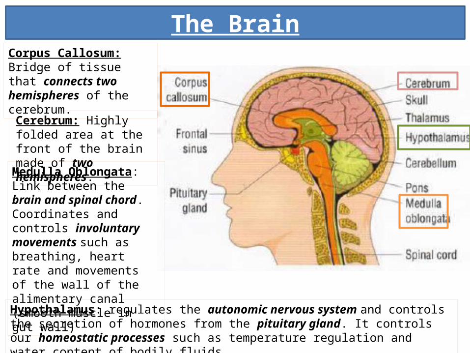
Cerebrum: Highly folded area at the front of the brain made of two hemispheres.
Corpus Callosum: Bridge of tissue that connects two hemispheres of the cerebrum.
Medulla Oblongata: Link between the brain and spinal chord. Coordinates and controls involuntary movements such as breathing, heart rate and movements of the wall of the alimentary canal (smooth muscle in gut wall)
Hypothalamus: regulates the autonomic nervous system and controls the secretion of hormones from the pituitary gland. It controls our homeostatic processes such as temperature regulation and water content of bodily fluids.

Cerebrum: Structure & Function
Primary visual area
Wernicke’s area: understanding language: association area
Sensory areaMotor area
Broca’s area: produces language: motor area
Auditory lobe
Auditory association area Visual
Association Area

Cerebrum: Structure & FunctionThe cerebrum is divided into two hemispheres which are connected via the corpus callosum. The outermost layer is folded and consists of a thin layer of nerve cell bodies known as the cerebral cortex. It is this area of the brain that is in control of higher order brain functions such as conscious thought and emotional response, the ability to override some reflexes and features associated with intelligence such as reasoning and judgement.
Cerebral
cortex
Sensory Areas
Association areas
Motor Areas
Receive impulses directly from receptors.Somatosensory area mapping shows it is subdivided according to areas of body from which it receives information.
Link between sensory and motor areas.Compares input with previous experience in order to interpret what the input means and judge an appropriate response.
Send impulses to effectors (muscles and glands) via motor neurones
The cerebral cortex is subdivided into areas responsible for specific activities:

The BrainFrontal Lobe controls conscious motor movement, speech, thought and personality.
Parietal Lobe interprets information from the sensory cortex about touch, pressure, pain and temperature.
Occipital Lobe receives sensory information from the eyes.
Temporal Lobe receives sensory input from ears – perception of language.
Cerebellum controls the coordination of movement and posture. The cerebellum receives impulses from the ears, eyes and stretch receptors in muscles and this information is integrated and used to coordinate the timing of skeletal muscle contraction and relaxation. When we go on ‘autopilot’, we are using our cerebellum: it contains programmed information. The cerebellum is responsible for keeping us upright, judging the position of objects and limbs, tensioning of muscles to manipulate tools and instruments and operation of antagonistic muscles to coordinate relaxing and contracting.

Organisation of the Nervous SystemLiving organisms are able to respond to changes in both external and internal environments. We need a method of communication between sensors and effectors. Animals need to coordinate lots of different responses, from coordinated voluntary muscle action to fine control of balance, posture and temperature regulation.
Central nervous System
Brain and Spinal Chord
Peripheral Nervous System
Somatic nervous system
All sensory neurones and
motor neurones to skeletal muscle
Autonomic nervous system
Sympathetic
Motor neurones that supply the internal organs
(viscera)
Parasympathetic
Motor neurones that supply the internal organs
(viscera)Receptors and sensory neurones
transmit impulses to the CNS
In the spinal chord, there is a region ion the centre that
contains unmyelinated neurones (grey matter)
and a regions around the outside that contains axons
and dendrons that are mainly myelinated and
therefore appears white.
Sensory neurones that carry impulses in from the receptors and motor neurones that that carry action potentials out to effectors.
All of the neurones that carry impulses into and out of the CNS:

Organisation of the Peripheral Nervous SystemThe peripheral nervous system is split into two systems: the somatic nervous system and the autonomic nervous system. The somatic nervous system includes all the sensory neurones and motor neurones that take information to skeletal muscle. All of the neurones in a typical reflex arc are part of the somatic nervous system. The autonomic nervous system is itself divided into 2 components: theses are the sympathetic and parasympathetic.
Autonomic nervous System: Sympathetic
The axons of the preganglionic neurones pass out of the spinal chord through the ventral route
The preganglionic neurones synapse with the motor neurones in an autonomic
ganglion
From these autonomic ganglia, motor neurone axons pass to all of the organs in the body.
The neurotransmitter that carries impulses across the
synapses in the sympathetic nervous
system is noradrenaline.
The sympathetic nervous system is activated when we encounter stimuli that make us scared or put us under stress . The sympathetic nervous system therefore helps to prepare the body for fight or flight.

Autonomic nervous System: Parasympathetic
All of the nerve pathways involved in the parasympathetic all begin in the brain, the top of the spinal chord or the very base of the spinal chord. The neurone that carries the impulse out of the brain or spinal chord carries on going until it is inside the wall of the organ that it will stimulate. The motor neurone will synapse with an effector neurone inside of the organ.
Many of the axons of the neurones of the parasympathetic system are in the vagus nerve.
The neurotransmitter at synapses in the parasympathetic system is acetylcholine. This often has an inhibitory effect on some organs. In general, the parasympathetic system helps the body to ‘rest and digest’.

Parasympathetic Sympathetic
Most active in sleep and relaxation Most active in times of stress
Neurones from CNS linked at a ganglion within the target tissue: pre-ganglionic neurones vary in length
Neurones from CNS linked at an autonomic ganglion just outside of the spinal chord: pre-ganglionic neurones are therefore very short.
Neurotransmitter is acetylcholine Neurotransmitter is noradrenaline
Autonomic nervous System: Comparing the Sympathetic and Parasympathetic
Effects of Sympathetic Stimulation: Increases rate and force of contraction of heart Dilates pupils Ciliary muscles relax so lens is thinner for distant vision Sphincter muscles contract Liver releases glucose into blood Increases sweating Pili erector muscles contract so hair stands on end Vasoconstriction of arterioles to gut and skin Ventilation rate and depth increases
Effects of Parasympathetic Stimulation: Reduces rate and force
of contraction of heart Pupil constricts Increase in glycogen
production

Neuromuscular JunctionSkeletal muscle contracts when it receives an impulse from a neurone. Neurones and muscles meet at specialised synapses called neuromuscular junctions.
1. Action potential arrives at neurone: calcium ion channels open and calcium ions diffuse into presynaptic neurone. This causes vesicles, containing neurotransmitter (generally acetylcholine) to move to and fuse with the presynaptic membrane. Neurotransmitter released into cleft by exocytosis.
2. Neurotransmitter binds to receptors on the sarcolemma. Sodium ions channels open and Na+ ions flood in. This causes a depolarisation across the membrane and initiates an action potential.
3. Depolarisation of sarcolemma spreads down the T tubules. Ca2+
channels open and Ca2+
ions diffuse out of the sarcoplasmic reticulum. The ions will bind to troponin molecules that are attached to actin filaments. This causes contraction.

Comparing synapses and neuromuscular junctions
Synapses Neuromuscular Junction
Neurone to Neurone Neurone to Sarcomere
Post synaptic stimulation leads to action potential in post synaptic neurone
Action potential leads to depolarisation of sarcolemma and muscle contraction
Synaptic knob is smooth and round
End plate is flattened up to muscle fibre with microvilli appearance
Neurotransmitter located in vesicles in presynaptic membrane
Vesicles release neurotransmitter into cleft on stimulation
Neurotransmitter diffuses across the cleft and binds to post synaptic membrane
Binding of neurotransmitter results in depolarisation of post synaptic membrane
Enzyme present to degrade neurotransmitter to avoid continual stimulation of post synaptic membrane.

Structure of Skeletal Muscle Skeletal muscle is described
as being striated. It appears to have stripes or bands of
light and dark.
Region Description
A band Darker regions of the sarcomere, where the thick protein filament myosin is present.
Darkest part of A band
Where myosin and actin filaments overlap
H band Lighter areas of the A band where only myosin is present
M line Provides attachment for myosin filaments
I band Lighter areas of the sarcomere where the thin actin filaments are
Z line Provides attachment for actin filaments
M line

When a muscle contracts...
H band gets shorter – there is less area in which there is only myosin present.
I band and A band stay the same: the actin and myosin filaments themselves don’t get shorter.
Darkest part of the A band (where the actin and myosin overlap) gets longer.
The distance between Z lines decreases when a muscle contracts – the sarcomere gets shorter.

The Sliding Filament ModelAs muscles contract, the sarcomeres in each myofibril get shorter as the Z lines are pulled closer together.

The Sliding Filament Model
When a muscle is relaxed,
the tropomyosin and troponin
are in such a
position in the actin
filament that
prevents
myosin from
binding.
When the
action potential stimulates the release of Ca2+
ions from the
sarcoplasmic
reticulum, the Ca2+
flood the muscle
cell. The Ca2+
bind to troponin: the
troponin
changes shape and the tropomyosin moves in order
to expose
the myosin binding site. The myosin is then able to bind to actin,
forming a cross bridge.
The myosin heads
tilt, causing the actin
to be pulled
along so overlap more
with the myosin
filament. ADP
and Pi are
released. This is
the power
stroke.
New ATP attaches
to the myosin head as
the cross
bridge is
broken. The ATP
is hydroly
sed, providing enough energy
to force the
myosin heads
to release
the actin.
They tip back to
their previous condition and are
then able to repeat
the process, forming a cross bridge further along the
filament.

The role of ATP in muscle contractionATP is required for muscle contraction: after the power stroke, ATP must be hydrolysed in
order to provide enough energy to break the cross bridge connection between actin and myosin and re-set the myosin head forwards. The myosin head can then attach to the next binding site along the actin molecule and bend again.
ATP is also necessary in the presynaptic neurone, prior to the neuro-muscular junction. Energy is required to release neurotransmitter into the cleft by exocytosis.
ATP
ATP supplies in muscles are used up very rapidly once the muscle starts working. There are 3 mechanisms by which ATP supply is maintained: 1. Aerobic respiration in muscle cell mitochondria (dependent on
supply of oxygen to the muscles and the availability of respiratory substrates)
2. Anaerobic respiration in the muscle cell sarcoplasm – this leads to the production of lactic acid, which is toxic. This stimulates increased blood supply to the muscles.
3. Creatine Phosphate: in the muscle sarcoplasm, there are stores of this molecule. A phosphate group can easily be removed from the Creatine phosphate and combined with ADP to produce more ATP by the enzyme creatine phosphotransferase.

When a person dies, respiration stops and ATP production ceases. Calcium ions can no longer be pumped into the cisternae of the sarcoplasmic reticulum, so they build up in the sarcoplasm. This causes troponin and tropomyosin to move away from their blocking positions on the actin filaments, so myosin heads are able to bind with the actin. There is no ATP left to provide energy to break the cross links, so the myosin heads remain firmly attached to the actin filament.
Rigor mortis lasts for up to 3 days. By the end of this time, enzymes leaking out of lysosomes will have partially destroyed the cells, and the actin-myosin bridges will have broken apart.
Rigor Mortis: the rigidity of deathIn resting muscle, most myosin heads are not attached to actin filaments. Transporter proteins pump calcium ions into the cisternae of the sarcoplasmic reticulum, so troponin and tropomyosin cover the attachment sites.
No ATP, so the cross links between the myosin and actin filaments cannot be broken, so muscles are held rigidly.

Voluntary Muscles Location?Attached to skeleton
Function?Movement of skeleton at joints
Structure? Striated Muscle cells form fibres
containing several nuclei Each fibre is surrounded by a
plasma membrane called a sarcolemma. The sarcoplasm of a muscle cell contains lots of mitochondria, extensive sarcoplasmic reticulum (specialised endoplasmic reticulum) and myofibrils comprised of actin and myosin myofilaments.
Stimulus?Somatic Nervous System
Contraction/fatigue?Contracts quickly and powerfully; fatigues quickly.
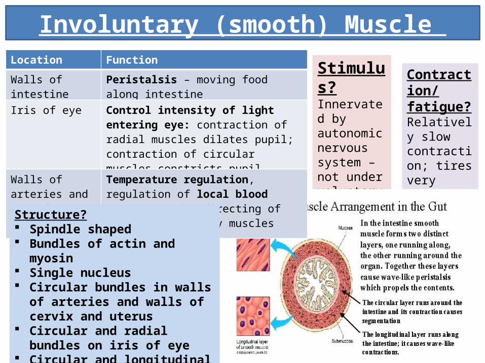
Involuntary (smooth) Muscle
Stimulus?Innervated by autonomic nervous system – not under voluntary control.
Contraction/fatigue?Relatively slow contraction; tires very slowly
Location Function
Walls of intestine Peristalsis – moving food along intestine
Iris of eye Control intensity of light entering eye: contraction of radial muscles dilates pupil; contraction of circular muscles constricts pupil.
Walls of arteries and around arterioles
Temperature regulation, regulation of local blood pressure and redirecting of blood to voluntary muscles during exercise.
Structure? Spindle shaped Bundles of actin and myosin Single nucleus Circular bundles in walls of arteries
and walls of cervix and uterus Circular and radial bundles on iris of
eye Circular and longitudinal bundles in
walls of intestine

Location?Muscular part of heart: atrial muscle, ventricular muscle and specialised excitatory and conductive muscle fibres (SAN, AVN)
Function?Coordinated contraction of heart to pump blood
Structure? Striated Intercalated discs fuse in such a way that
there are gap junctions with free diffusion of ions so action potentials can pass quickly and easily between cardiac muscle fibres.
Contraction/ fatigue?Never fatigues; powerful contraction
Cardiac Muscle Stimulus?Some cardiac muscle capable of stimulating contraction without nervous impulse – this is myogenic.Autonomic nervous system regulates rate of contraction. Sympathetic nerves increase rate; parasympathetic nerves decrease rate.

Muscles, Nerves and Hormones: The Fight or Flight Response
1. Cerebral understanding of threat activates hypothalamus
2. Hypothalamus stimulates increased activity of sympathetic nervous system
3. Sympathetic nervous system activates the adrenal medulla: adrenaline is released
4. Hypothalamus releases Corticotrophin Releasing Factor which stimulates…
5. Anterior Pituitary gland to release Adreno-corticotrophic hormone (ACTH)
6. ACTH arrives at adrenal cortex. This stimulates the release of approximately 30 corticosteroid hormones. The fight of flight response
makes an organism ready for the actions that lead to confrontation of the danger or escape from it.
A stressor is a stimulus that causes the stress response.