andrew’s diseases of the skin epidermal nevi, neoplasms, and cysts part 3 chapter 29
Post on 20-Dec-2015
219 views
TRANSCRIPT
Andrew’s Diseases of the Skin
Epidermal Nevi, Neoplasms, and Cysts Part 3 Chapter 29
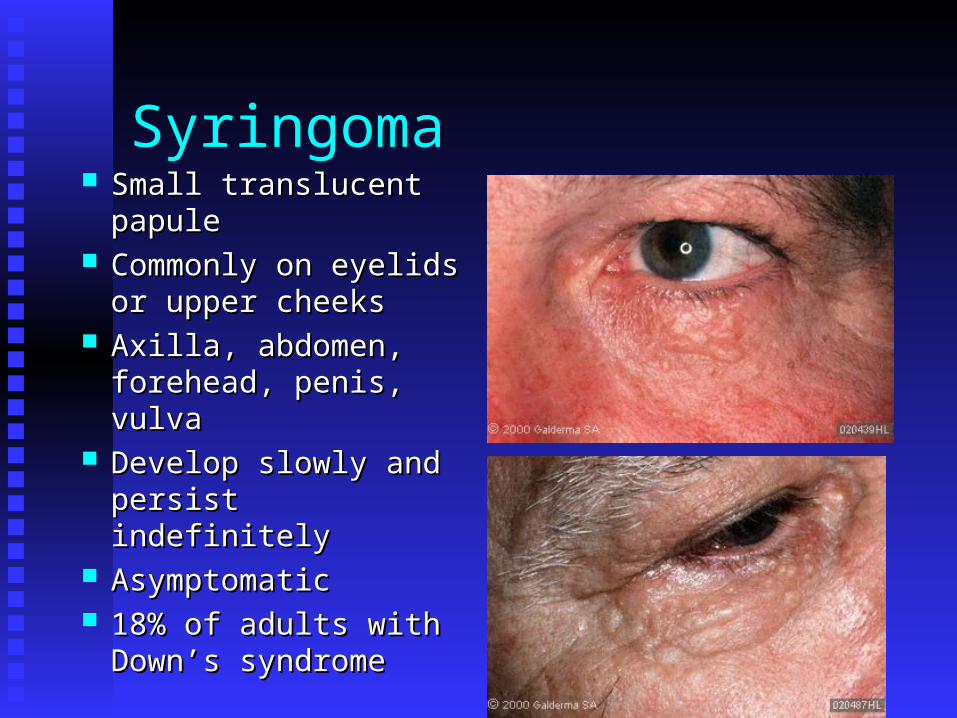
Syringoma Small translucent Small translucent
papulepapule Commonly on eyelids Commonly on eyelids
or upper cheeksor upper cheeks Axilla, abdomen, Axilla, abdomen,
forehead, penis, vulvaforehead, penis, vulva Develop slowly and Develop slowly and
persist indefinitelypersist indefinitely AsymptomaticAsymptomatic 18% of adults with 18% of adults with
Down’s syndromeDown’s syndrome
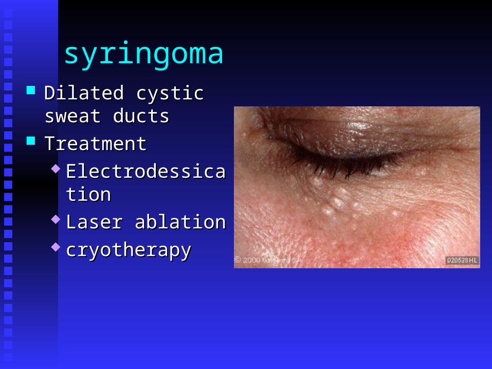
syringoma Dilated cystic sweat Dilated cystic sweat
ductsducts TreatmentTreatment
ElectrodessicationElectrodessication Laser ablationLaser ablation cryotherapycryotherapy
Variants of Syringoma
Clear cell syringomaClear cell syringoma Associated with diabetes mellitusAssociated with diabetes mellitus Identical lesions, histological differenceIdentical lesions, histological difference
Other clinical variantsOther clinical variants Limited to the scalp causing alopeciaLimited to the scalp causing alopecia Unilateral linear or nevoid distributionUnilateral linear or nevoid distribution Limited to vulva and penis Limited to vulva and penis Limited to distal extremitiesLimited to distal extremities
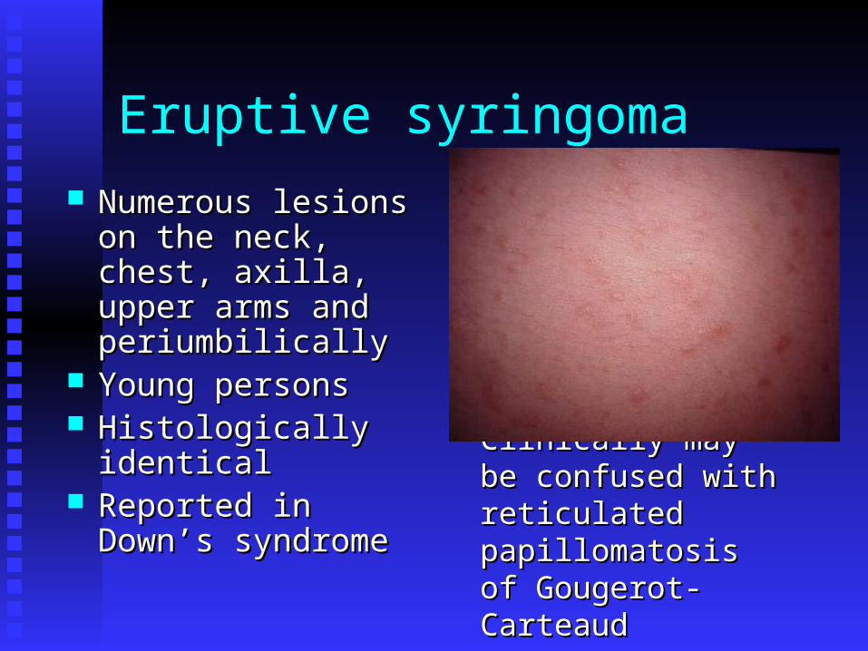
Eruptive syringoma
Numerous lesions on Numerous lesions on the neck, chest, axilla, the neck, chest, axilla, upper arms and upper arms and periumbilicallyperiumbilically
Young personsYoung persons Histologically Histologically
identicalidentical Reported in Down’s Reported in Down’s
syndromesyndrome
Clinically may be Clinically may be confused with confused with reticulated reticulated papillomatosis of papillomatosis of Gougerot-CarteaudGougerot-Carteaud
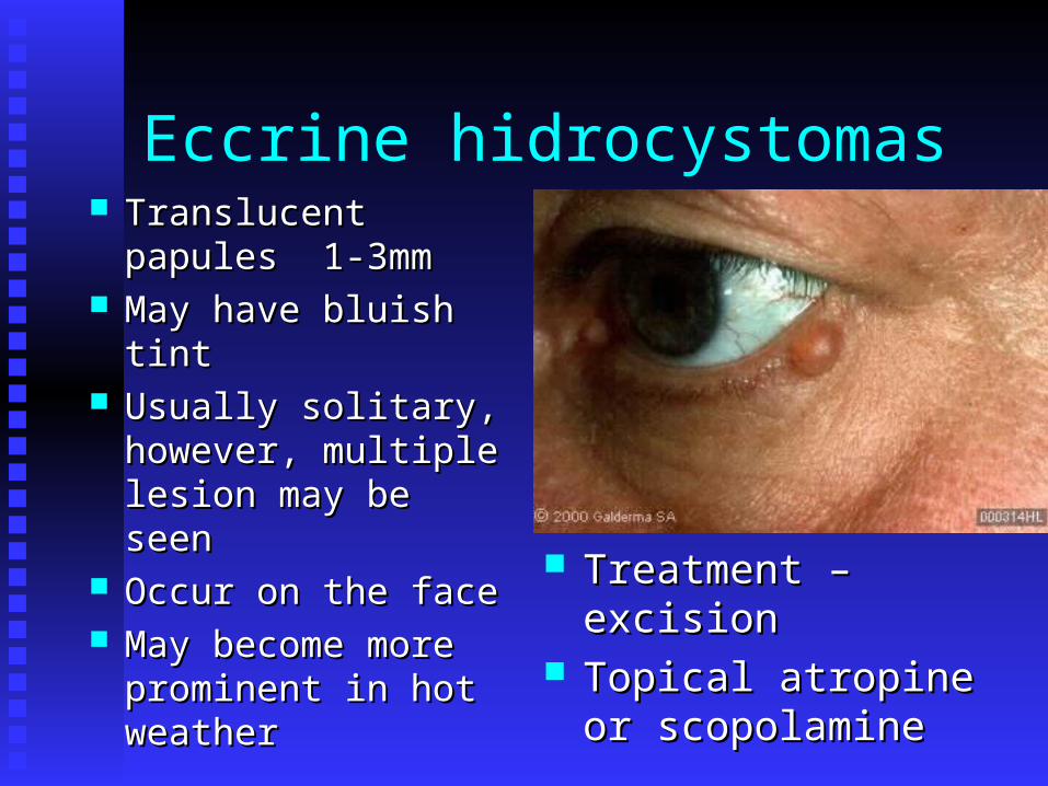
Eccrine hidrocystomas Translucent papules Translucent papules
1-3mm1-3mm May have bluish tintMay have bluish tint Usually solitary, Usually solitary,
however, multiple however, multiple lesion may be seen lesion may be seen
Occur on the faceOccur on the face May become more May become more
prominent in hot prominent in hot weatherweather
Treatment – excisionTreatment – excision Topical atropine or Topical atropine or
scopolaminescopolamine
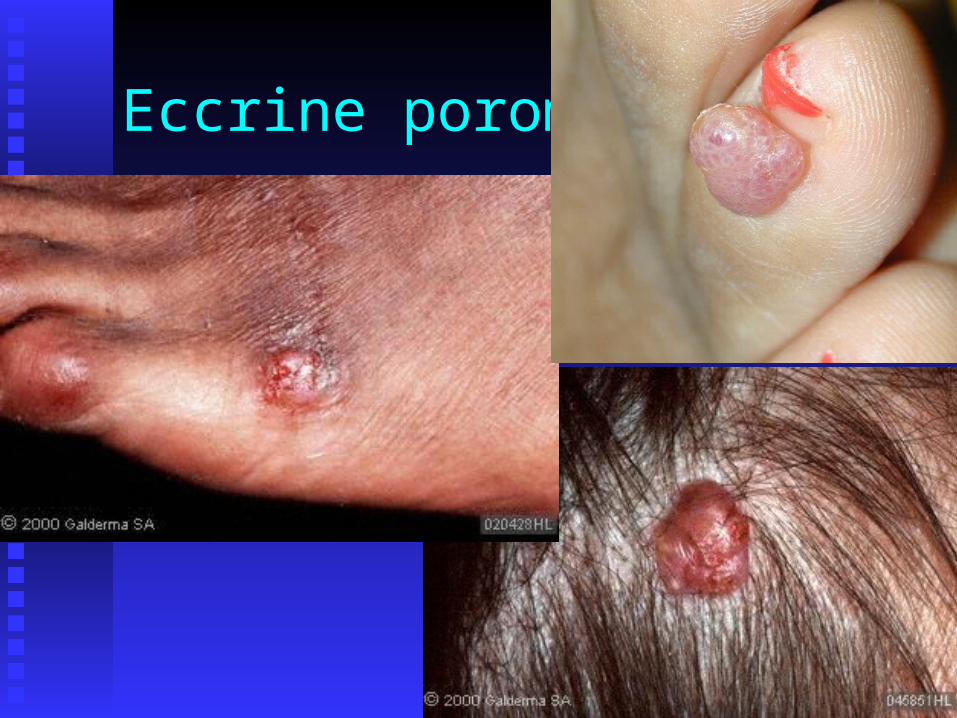
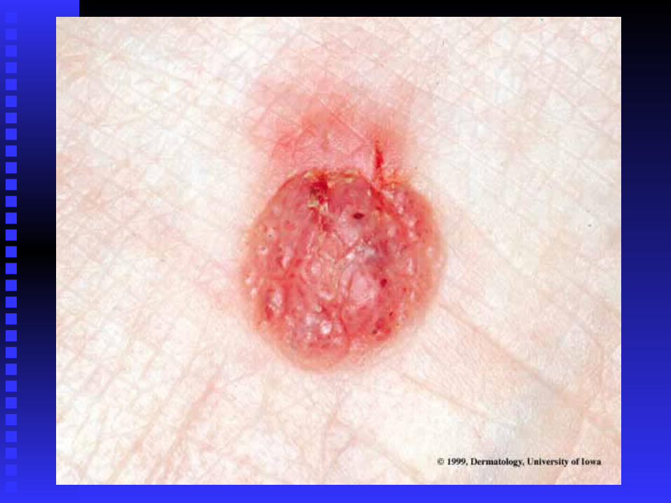
Eccrine poroma
Benign, slow-growing, slightly protruding, Benign, slow-growing, slightly protruding, sessile, soft, reddish tumorsessile, soft, reddish tumor
Most commonly occur on the sole or the Most commonly occur on the sole or the side of the foot. May occur anywhereside of the foot. May occur anywhere
Bleeds with slight traumaBleeds with slight trauma Frequent cup-shaped shallow depression Frequent cup-shaped shallow depression
from which the tumor growsfrom which the tumor grows Benign – simple excisionBenign – simple excision Eccrine poromatosisEccrine poromatosis
Eccrine poroma
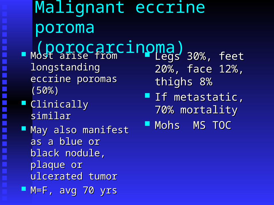
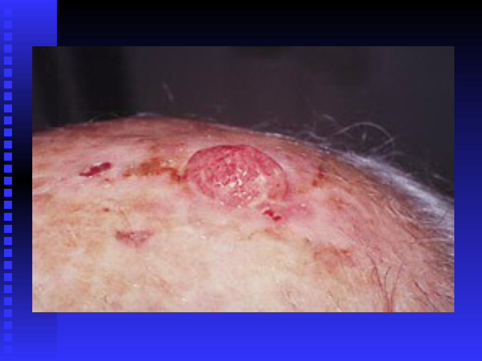
Malignant eccrine poroma(porocarcinoma)
Most arise from Most arise from longstanding eccrine longstanding eccrine poromas (50%)poromas (50%)
Clinically similarClinically similar May also manifest as a May also manifest as a
blue or black nodule, blue or black nodule, plaque or ulcerated plaque or ulcerated tumortumor
M=F, avg 70 yrsM=F, avg 70 yrs
Legs 30%, feet 20%, Legs 30%, feet 20%, face 12%, thighs 8%face 12%, thighs 8%
If metastatic, 70% If metastatic, 70% mortalitymortality
Mohs MS TOCMohs MS TOC
Chondroid Syringoma and Malignant Chondroid Syringoma
Firm intradermal or Firm intradermal or subcutaneous nodulesubcutaneous nodule
Most commonly Most commonly located on the nose or located on the nose or cheekscheeks
80 % involving the 80 % involving the head and neckhead and neck
Symptomatic 5-30mmSymptomatic 5-30mm Felt to be of eccrine Felt to be of eccrine
originorigin
Malignant mixed tumor Malignant mixed tumor of the skinof the skin
Most occur on Most occur on extremities. Reported on extremities. Reported on face, scalp, back, buttocksface, scalp, back, buttocks
Grow rapidly. Metastasis Grow rapidly. Metastasis more the 50%more the 50%
Aggressive surgical Aggressive surgical excision, Adjuvant excision, Adjuvant radiation therapy w/wo radiation therapy w/wo chemotherapychemotherapy

Clear cell hidradenoma(nodular hidradenoma)
Classified as an Classified as an eccrine sweat gland eccrine sweat gland tumortumor
Single nodular, solid Single nodular, solid or cystic, occasionally or cystic, occasionally protruding massprotruding mass
Flesh colored or Flesh colored or reddishreddish
Anywhere. Most Anywhere. Most common site is the common site is the headhead
20% c/o pain on pressure20% c/o pain on pressure Multiple lesions reportedMultiple lesions reported Women 2X menWomen 2X men Extirpation is TOCExtirpation is TOC
Malignant clear cell hidradenoma(hidradenocarcinoma)
Extremely rareExtremely rare Presents as a solitary nodule Presents as a solitary nodule Lower extremity 32.9 %, upper extremity Lower extremity 32.9 %, upper extremity
27.6 %, trunk 11.9 %, head 26.3 %27.6 %, trunk 11.9 %, head 26.3 % Metastasis occurs 60%Metastasis occurs 60% Tx wide local excision, radiation and Tx wide local excision, radiation and
chemotherapychemotherapy
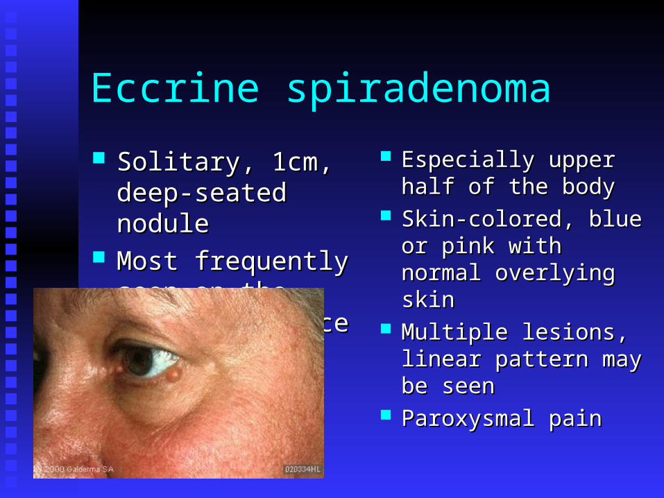
Eccrine spiradenoma
Solitary, 1cm, deep-Solitary, 1cm, deep-seated noduleseated nodule
Most frequently seen Most frequently seen on the ventral surfaceon the ventral surface
Especially upper half Especially upper half of the bodyof the body
Skin-colored, blue or Skin-colored, blue or pink with normal pink with normal overlying skinoverlying skin
Multiple lesions, Multiple lesions, linear pattern may be linear pattern may be seenseen
Paroxysmal painParoxysmal pain
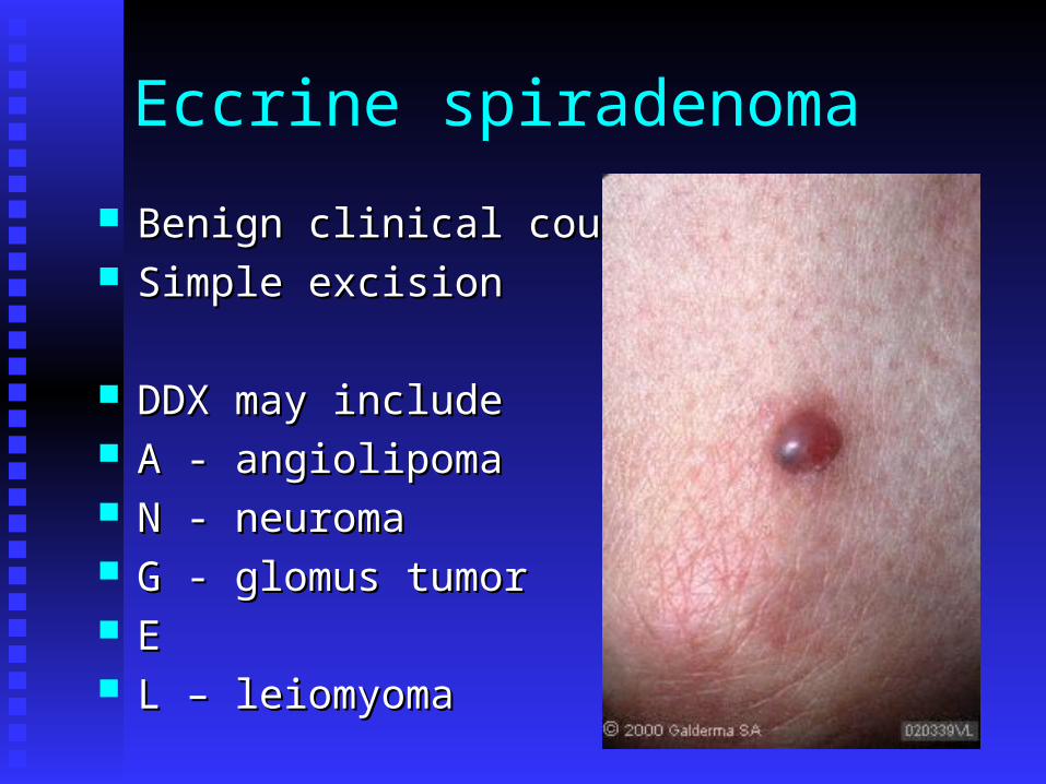
Eccrine spiradenoma
Benign clinical courseBenign clinical course Simple excisionSimple excision
DDX may includeDDX may include A - angiolipomaA - angiolipoma N - neuromaN - neuroma G - glomus tumorG - glomus tumor EE L – leiomyomaL – leiomyoma
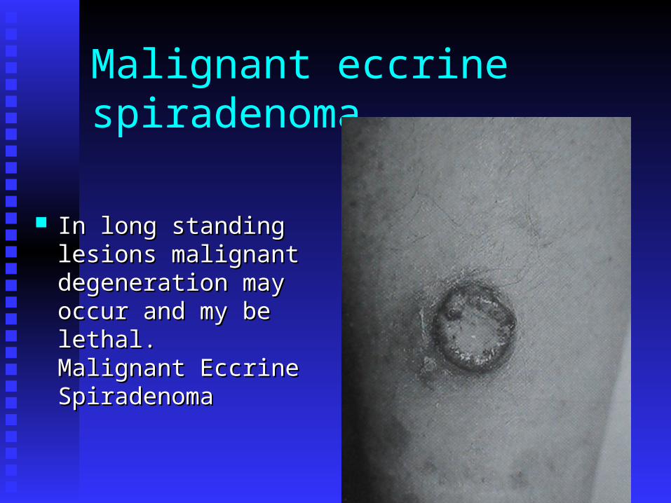
Malignant eccrine spiradenoma
In long standing In long standing lesions malignant lesions malignant degeneration may degeneration may occur and my be occur and my be lethal. Malignant lethal. Malignant Eccrine SpiradenomaEccrine Spiradenoma

Papillary eccrine adenoma
Uncommon benign lesionUncommon benign lesion Dermal nodulesDermal nodules Extremities of black patientsExtremities of black patients Tendency to recurTendency to recur Complete surgical excisionComplete surgical excision

syringoacanthoma
Extremely rare (21 cases)Extremely rare (21 cases) Seborrheic keratosis-like neoplasmSeborrheic keratosis-like neoplasm Significant tissue destruction if left Significant tissue destruction if left
untreateduntreated Classification remains controversialClassification remains controversial

Eccrine syringofibroadenoma Most presentations are Most presentations are
a solitary, a solitary, hyperkeratotic nodule hyperkeratotic nodule or plaque involving or plaque involving the extremitiesthe extremities
Characteristic marker Characteristic marker of Schopf syndromeof Schopf syndrome Hydrocystomas of Hydrocystomas of
the eyelids, the eyelids, hypotrichosis, hypotrichosis, hypodontia, and hypodontia, and nail abnormalitiesnail abnormalities
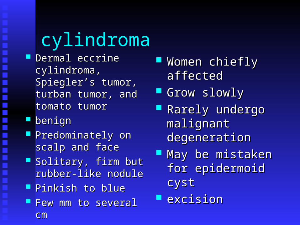
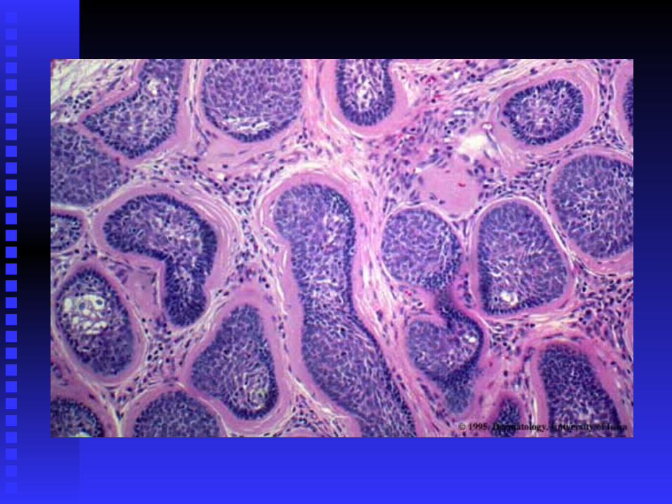
cylindroma Dermal eccrine Dermal eccrine
cylindroma, Spiegler’s cylindroma, Spiegler’s tumor, turban tumor, tumor, turban tumor, and tomato tumorand tomato tumor
benignbenign Predominately on Predominately on
scalp and facescalp and face Solitary, firm but Solitary, firm but
rubber-like nodulerubber-like nodule Pinkish to bluePinkish to blue Few mm to several cmFew mm to several cm
Women chiefly Women chiefly affectedaffected
Grow slowlyGrow slowly Rarely undergo Rarely undergo
malignant malignant degenerationdegeneration
May be mistaken for May be mistaken for epidermoid cystepidermoid cyst
excisionexcision
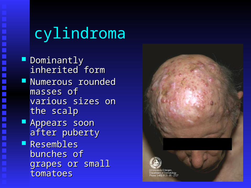
cylindroma
Dominantly inherited Dominantly inherited formform
Numerous rounded Numerous rounded masses of various masses of various sizes on the scalpsizes on the scalp
Appears soon after Appears soon after pubertypuberty
Resembles bunches of Resembles bunches of grapes or small grapes or small tomatoestomatoes
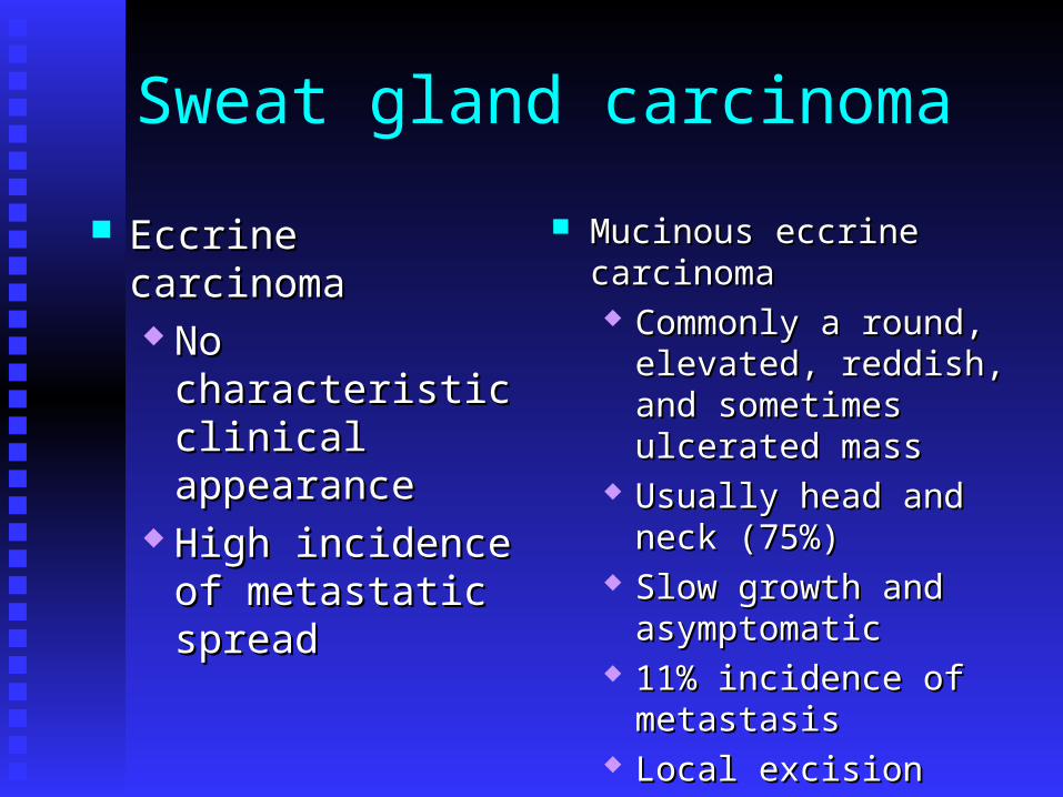
Sweat gland carcinoma
Eccrine carcinomaEccrine carcinoma No characteristic No characteristic
clinical appearanceclinical appearance High incidence of High incidence of
metastatic spreadmetastatic spread
Mucinous eccrine carcinomaMucinous eccrine carcinoma Commonly a round, Commonly a round,
elevated, reddish, and elevated, reddish, and sometimes ulcerated masssometimes ulcerated mass
Usually head and neck Usually head and neck (75%)(75%)
Slow growth and Slow growth and asymptomaticasymptomatic
11% incidence of metastasis11% incidence of metastasis Local excisionLocal excision
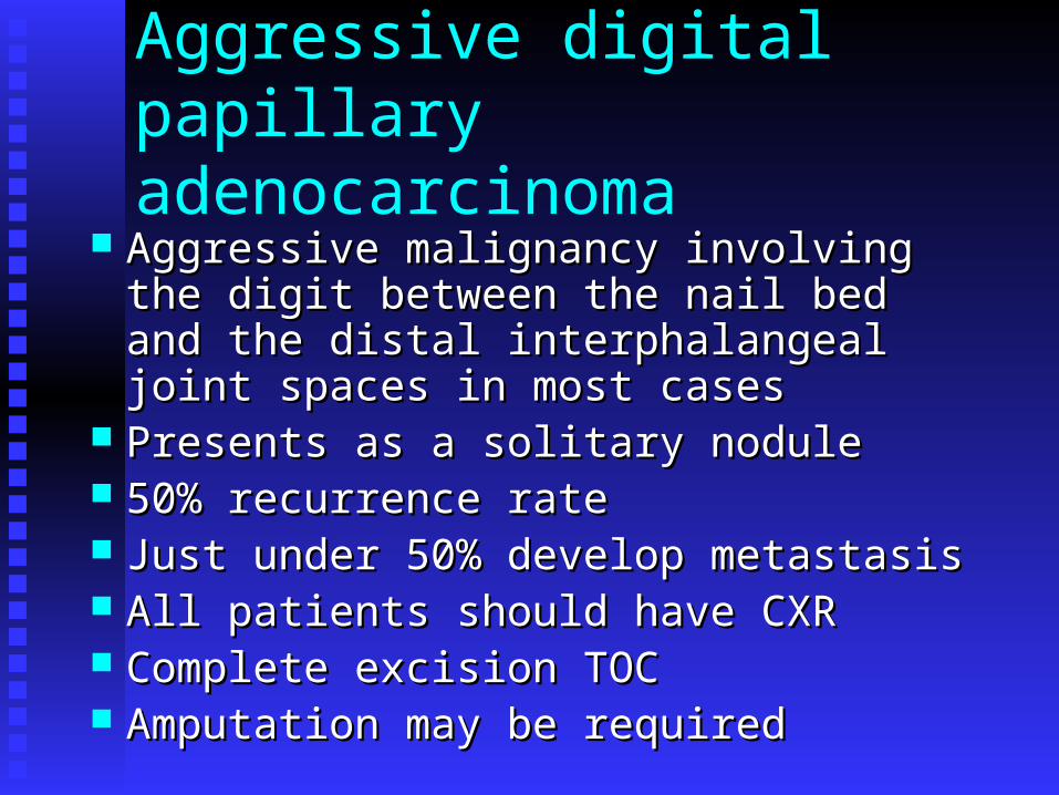
Aggressive digital papillary adenocarcinoma
Aggressive malignancy involving the digit Aggressive malignancy involving the digit between the nail bed and the distal between the nail bed and the distal interphalangeal joint spaces in most casesinterphalangeal joint spaces in most cases
Presents as a solitary nodulePresents as a solitary nodule 50% recurrence rate50% recurrence rate Just under 50% develop metastasisJust under 50% develop metastasis All patients should have CXRAll patients should have CXR Complete excision TOCComplete excision TOC Amputation may be requiredAmputation may be required
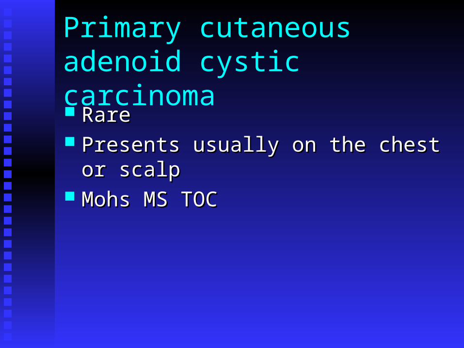
Primary cutaneousadenoid cystic carcinoma Rare Rare Presents usually on the chest or scalpPresents usually on the chest or scalp Mohs MS TOCMohs MS TOC
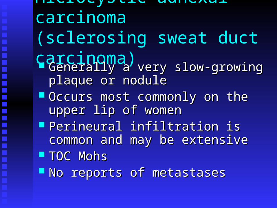
Microcystic adnexal carcinoma(sclerosing sweat duct carcinoma)
Generally a very slow-growing plaque or Generally a very slow-growing plaque or nodulenodule
Occurs most commonly on the upper lip of Occurs most commonly on the upper lip of womenwomen
Perineural infiltration is common and may Perineural infiltration is common and may be extensivebe extensive
TOC MohsTOC Mohs No reports of metastasesNo reports of metastases
APOCRINE GLANDS
ceruminoma
Rare apoeccrine tumor that rarely becomes Rare apoeccrine tumor that rarely becomes malignantmalignant
Firm nodular mass in the EACFirm nodular mass in the EAC Ulceration and crusting may occurUlceration and crusting may occur ObstructionObstruction Questionable true entityQuestionable true entity Treatment - excisionTreatment - excision
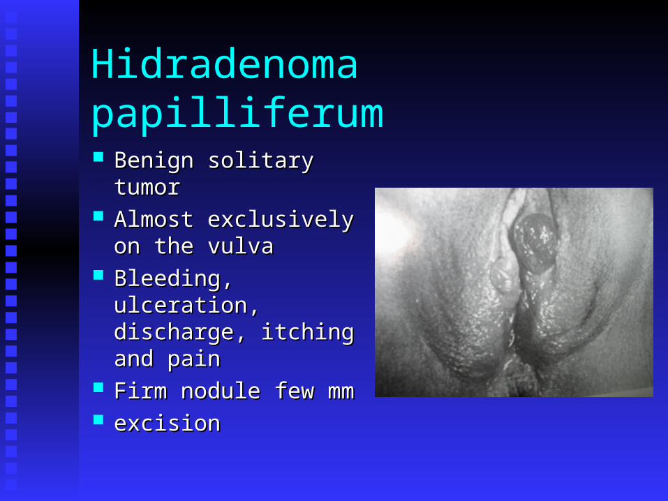
Hidradenoma papilliferum
Benign solitary tumorBenign solitary tumor Almost exclusively on Almost exclusively on
the vulvathe vulva Bleeding, ulceration, Bleeding, ulceration,
discharge, itching and discharge, itching and painpain
Firm nodule few mm Firm nodule few mm excisionexcision
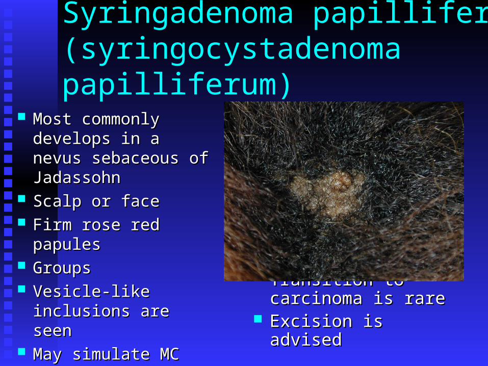
Syringadenoma papilliferum(syringocystadenoma papilliferum)
Most commonly Most commonly develops in a nevus develops in a nevus sebaceous of sebaceous of JadassohnJadassohn
Scalp or faceScalp or face Firm rose red papulesFirm rose red papules GroupsGroups Vesicle-like inclusions Vesicle-like inclusions
are seen are seen May simulate MCMay simulate MC
Transition to Transition to carcinoma is rarecarcinoma is rare
Excision is advisedExcision is advised
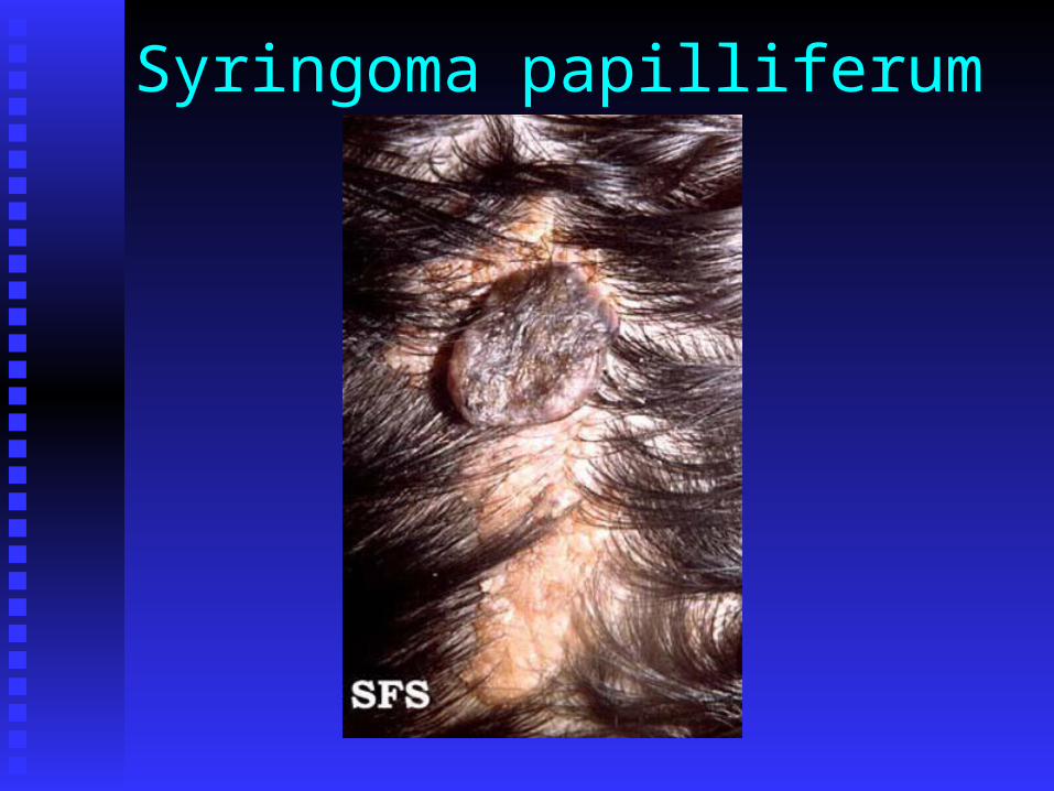
Syringoma papilliferum
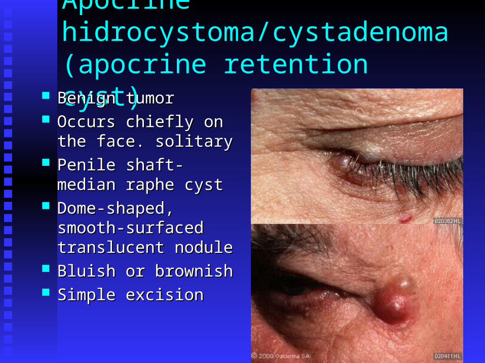
Apocrine hidrocystoma/cystadenoma(apocrine retention cyst)
Benign tumorBenign tumor Occurs chiefly on the Occurs chiefly on the
face. solitaryface. solitary Penile shaft- median Penile shaft- median
raphe cystraphe cyst Dome-shaped, Dome-shaped,
smooth-surfaced smooth-surfaced translucent noduletranslucent nodule
Bluish or brownishBluish or brownish Simple excisionSimple excision
Apocrine gland carcinoma
RareRare Axilla is the most common siteAxilla is the most common site May be seen in the nipple, vulva and EACMay be seen in the nipple, vulva and EAC May originate from aberrant mammary May originate from aberrant mammary
glandsglands Widespread metastases may occurWidespread metastases may occur

HAIR FOLLICLE NEVI AND TUMORS

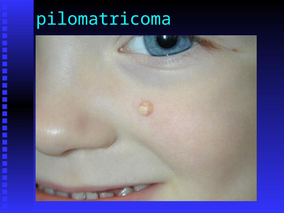
Pilomatricoma(calcifying epithelioma of Malherbe)
Usually a single tumorUsually a single tumor Most commonly on Most commonly on
the face, neck or armsthe face, neck or arms Deeply seated firm Deeply seated firm
nodule, covered with nodule, covered with normal or pink skinnormal or pink skin
AsymptomaticAsymptomatic Stretching may show Stretching may show
“tent sign”“tent sign”
Derived from hair Derived from hair matrix cellsmatrix cells
Clinical DDX is Clinical DDX is impossibleimpossible
Simple excisionSimple excision Familial patterns do Familial patterns do
occuroccur Multiple in Multiple in
Rubinstein-Taybi and Rubinstein-Taybi and Gardner syndromeGardner syndrome
pilomatricoma


Malignant pilomatricoma
Extremely rareExtremely rare Do not behave aggressivelyDo not behave aggressively
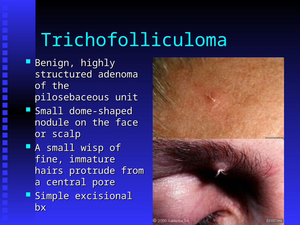
Trichofolliculoma Benign, highly Benign, highly
structured adenoma of structured adenoma of the pilosebaceous unitthe pilosebaceous unit
Small dome-shaped Small dome-shaped nodule on the face or nodule on the face or scalpscalp
A small wisp of fine, A small wisp of fine, immature hairs immature hairs protrude from a protrude from a central porecentral pore
Simple excisional bxSimple excisional bx
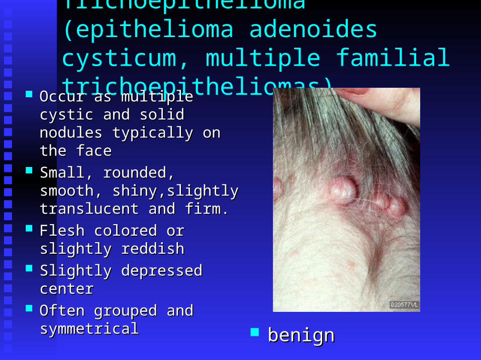
Trichoepithelioma(epithelioma adenoides cysticum, multiple familial trichoepitheliomas)
Occur as multiple cystic Occur as multiple cystic and solid nodules and solid nodules typically on the facetypically on the face
Small, rounded, smooth, Small, rounded, smooth, shiny,slightly translucent shiny,slightly translucent and firm.and firm.
Flesh colored or slightly Flesh colored or slightly reddishreddish
Slightly depressed centerSlightly depressed center Often grouped and Often grouped and
symmetricalsymmetrical benignbenign
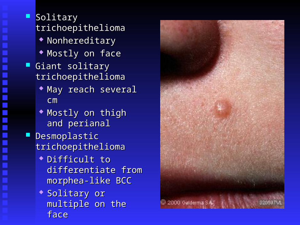
Solitary trichoepitheliomaSolitary trichoepithelioma NonhereditaryNonhereditary Mostly on faceMostly on face
Giant solitary Giant solitary trichoepitheliomatrichoepithelioma May reach several cmMay reach several cm Mostly on thigh and Mostly on thigh and
perianalperianal Desmoplastic Desmoplastic
trichoepitheliomatrichoepithelioma Difficult to Difficult to
differentiate from differentiate from morphea-like BCCmorphea-like BCC
Solitary or multiple on Solitary or multiple on the face the face
trichoblastoma
Benign neoplasms of follicular germinative Benign neoplasms of follicular germinative cellscells
AsymptomaticAsymptomatic Scalp and faceScalp and face Surgical excisionSurgical excision
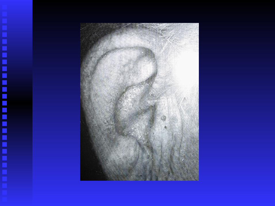
Trichilemmoma andCowden’s disease (multiple hamartoma syndrome)
Benign neoplasm of the Benign neoplasm of the hair folliclehair follicle
Small solitary papule on Small solitary papule on the facethe face
Nose and cheeksNose and cheeks MultipleMultiple
Marker for Cowden,s Marker for Cowden,s syndromesyndrome
Generally limited to the Generally limited to the head and neckhead and neck
87% of patients with 87% of patients with Cowden’sCowden’s
38% develop 38% develop malignanciesmalignancies Breast 25-36%Breast 25-36% Thyroid 7%Thyroid 7% Colon Colon
adenocarcinomaadenocarcinoma Tumor suppressor geneTumor suppressor gene
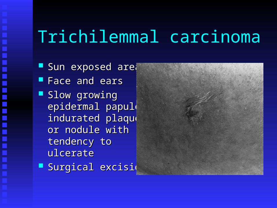
Trichilemmal carcinoma
Sun exposed areasSun exposed areas Face and earsFace and ears Slow growing Slow growing
epidermal papule, epidermal papule, indurated plaque or indurated plaque or nodule with tendency nodule with tendency to ulcerateto ulcerate
Surgical excisionSurgical excision
Trichodiscoma and fibrofolliculoma
Hundreds of flat or Hundreds of flat or dome-shaped, skin-dome-shaped, skin-colored asymptomatic colored asymptomatic papulespapules
Face, trunk and Face, trunk and extremitiesextremities
Autosomal dominant Autosomal dominant traittrait
Controversial entityControversial entity
2-4 mm skin-colored 2-4 mm skin-colored to white papulesto white papules
Solitary, more Solitary, more commonly multiplecommonly multiple
Scattered over the Scattered over the face, trunk and face, trunk and extremitiesextremities
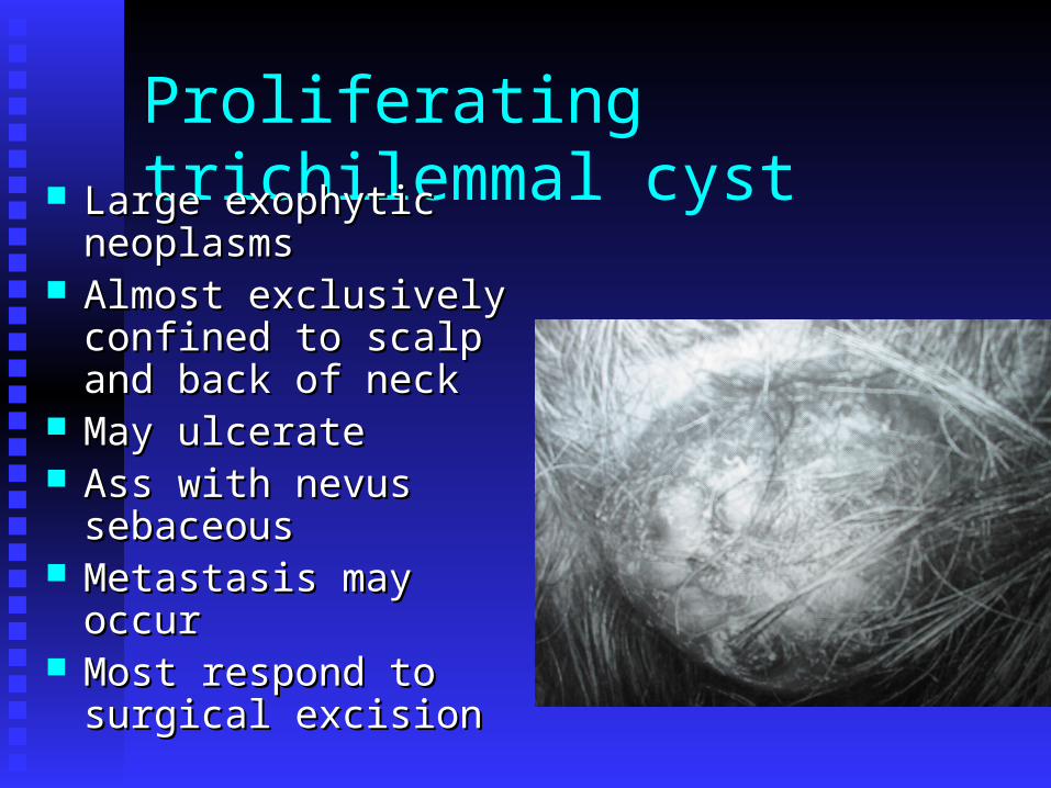
Proliferating trichilemmal cyst Large exophytic Large exophytic
neoplasmsneoplasms Almost exclusively Almost exclusively
confined to scalp and confined to scalp and back of neckback of neck
May ulcerateMay ulcerate Ass with nevus Ass with nevus
sebaceous sebaceous Metastasis may occurMetastasis may occur Most respond to surgical Most respond to surgical
excisionexcision
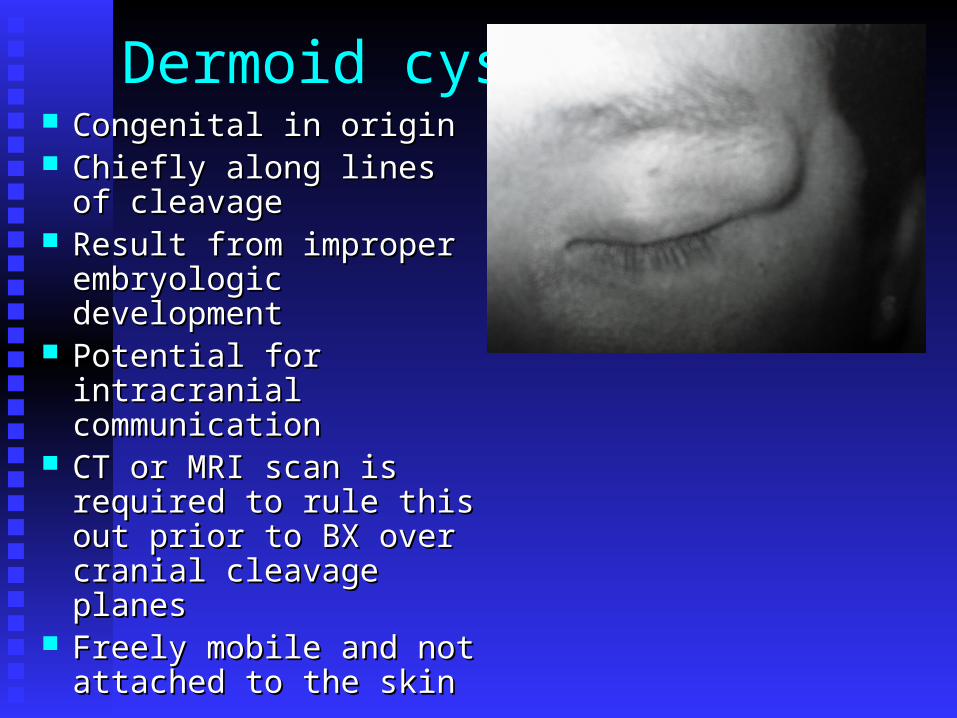
Dermoid cyst Congenital in originCongenital in origin Chiefly along lines of Chiefly along lines of
cleavagecleavage Result from improper Result from improper
embryologic developmentembryologic development Potential for intracranial Potential for intracranial
communicationcommunication CT or MRI scan is required CT or MRI scan is required
to rule this out prior to BX to rule this out prior to BX over cranial cleavage over cranial cleavage planesplanes
Freely mobile and not Freely mobile and not attached to the skinattached to the skin
Pilonidal cyst Midline hairy patch or pit in the sacral region with Midline hairy patch or pit in the sacral region with
a sinus orifice in the bottom, or a cyst beneath ita sinus orifice in the bottom, or a cyst beneath it Usually becomes symptomatic during adolescenceUsually becomes symptomatic during adolescence Opening cyst widely, debriding it, and packing it Opening cyst widely, debriding it, and packing it
with silver nitrate crystalswith silver nitrate crystals More advanced surgical intervention may be More advanced surgical intervention may be
requiredrequired SCC has been reported to arise from chronic SCC has been reported to arise from chronic
inflammatory pilonidal diseaseinflammatory pilonidal disease

Pilonidal sinus
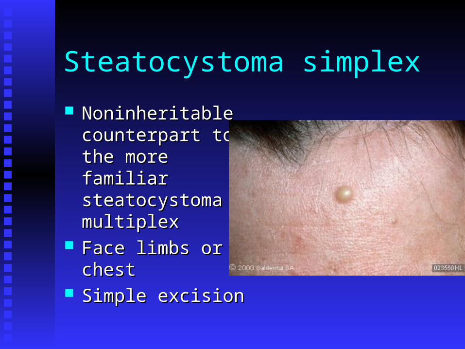
Steatocystoma simplex
Noninheritable Noninheritable counterpart to the counterpart to the more familiar more familiar steatocystoma steatocystoma multiplexmultiplex
Face limbs or chestFace limbs or chest Simple excisionSimple excision
Steatocystoma multiplex Multiple, small, yellowish, cystic nodules 2-6 mm Multiple, small, yellowish, cystic nodules 2-6 mm Principally on the upper anterior trunk, upper Principally on the upper anterior trunk, upper
arms, axillae and thighsarms, axillae and thighs Lesions may be generalizesLesions may be generalizes High familial tendencyHigh familial tendency Contain a syruplike, yellowish, odorless oily Contain a syruplike, yellowish, odorless oily
materialmaterial Likely autosomal dominant inheritanceLikely autosomal dominant inheritance Tx- excision of individual lesionsTx- excision of individual lesions Incision and expression or aspirationIncision and expression or aspiration

Steatocystoma multiplex

Eruptive vellus hair cysts
Autosomal dominant Autosomal dominant inheritanceinheritance
Yellowish to reddish Yellowish to reddish brown, small papules of brown, small papules of the chest and proximal the chest and proximal extremitiesextremities
Disseminated lesions Disseminated lesions reportedreported

Pigmented follicular cysts
Face or neckFace or neck Suggested to be a variant of multiple Suggested to be a variant of multiple
pilosebaceous cystspilosebaceous cysts
milia
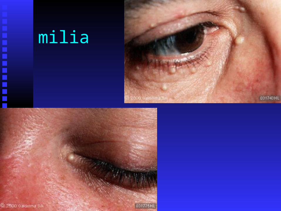
White keratinous cysts, 1-4 mmWhite keratinous cysts, 1-4 mm Chiefly on the face esp under eyesChiefly on the face esp under eyes May occur in great numbersMay occur in great numbers Occur in up to 50 % of newbornsOccur in up to 50 % of newborns Primarily develop without a predisposing Primarily develop without a predisposing
conditioncondition Can develop in inflammatory conditions and skin Can develop in inflammatory conditions and skin
diseases such as epidermolysis bullosa, diseases such as epidermolysis bullosa, pemphigus, bullous pemphigoid, PCT, herpes pemphigus, bullous pemphigoid, PCT, herpes zoster, contact dermatitis, and after prolonged use zoster, contact dermatitis, and after prolonged use of NSAIDSof NSAIDS
milia
Variants include MEM (multiple eruptive Variants include MEM (multiple eruptive milia)milia)
MEP (milia en plaque)MEP (milia en plaque) Tx- incision and expressionTx- incision and expression Tretinoin and minocycline for MEPTretinoin and minocycline for MEP
milia

Pseudocyst of the auricle
Fluctuant, tense, noninflammatory swelling Fluctuant, tense, noninflammatory swelling of the upper earof the upper ear
Believed to be ass with traumaBelieved to be ass with trauma Tx – drainageTx – drainage ILI steroidILI steroid
Cutaneous columnar cysts
Four types of cyst that occur in the skin are Four types of cyst that occur in the skin are lined by columnar epitheliumlined by columnar epithelium
Branchiogenic cystBranchiogenic cyst Small solitary lesions just above the Small solitary lesions just above the
sternal notchsternal notch Thyroglossal duct cystsThyroglossal duct cysts
Anterior aspect of the neckAnterior aspect of the neck Malignancies reported 1%Malignancies reported 1%

Cutaneous columnar cysts
Cutaneous ciliated cystsCutaneous ciliated cysts Usually located on the legs of femalesUsually located on the legs of females Perineum vulva and foot regionsPerineum vulva and foot regions
Median raphe cystMedian raphe cyst Developmental defects lying in the Developmental defects lying in the
ventral midline of the penis, usually on ventral midline of the penis, usually on the glansthe glans
Surgical intervention is standard therapySurgical intervention is standard therapy

The End