and some chemical properties
TRANSCRIPT

ISOLATION AND SOME CHEMICAL PROPERTIES OFASPERMATOGENIC SUBSTANCE FROM BULL
SEMINAL VESICLE FLUID
J. DOST\l=A'\Land J. MATOU\l=S%v\EK
Laboratory of Animal Genetics, The Czechoslovak Academy of Sciences,Libechov, Czechoslovakia
(Received 22nd March 1972)
Summary. Aspermatogenic substance (AS) was isolated from bullseminal vesicle fluid by precipitation with acetic acid and ammoniumsulphate, and subjected to CM Sephadex C-50 and Sephadex G-100column chromatography. The molecular weight of the monomer of ASwas 22,000 and of the dimer was 44,000 as determined by gel filtration.The purity of the AS was controlled by disc electrophoresis in acrylamidegel, starch-gel electrophoresis, immunoelectrophoresis in agar gel, bypeak homogeneity at the last step of separation and by the ultra-centrifugation pattern. The N-terminal amino acid was lysine. Theabsorbance of 0\m=.\1%AS at 280 nm in pH 5\m=.\5buffer was 0\m=.\510.The s
value obtained for 0\m=.\5%AS solution was 2\m=.\7.
INTRODUCTIONIn 1966, Matousek reported impairment of the spermatogenic process in rams
and rabbits following intramuscular and subcutaneous administration of bullseminal vesicle fluid. Considerable degenerative changes were recorded in thetestes ofguinea-pigs, rabbits and rams following a single intratesticular injectionof this fluid without adjuvant: the index weight of the testes decreased, therewas disturbance of spermatogenesis and, in more severe cases, a distortion andcontraction of the seminiferous tubules (Matousek, 1969). Antibodies producedby animals which were immunized subcutaneously did not cause sperm-agglutination, immobilization or spermatotoxic reactions. No circulating anti¬bodies could be found in guinea-pigs and rabbits which had been immunizedby a single intratesticular injection.
An inhibition effect of bull seminal vesicle fluid affecting the fertility offemale mice and hens was observed by Matousek & Petrovská (1969). Thefirst attempt to characterize an aspermatogenic substance in the seminalvesicle fluid of bulls was carried out by Matousek, Stanëk & Veselsky (1972)and its antiembryonic effect on mice was recorded by Matousek (1973).
Purification of the substance from bull seminal vesicle fluid which causedaspermatogenesis in the males of some species and embryonic mortality infemale mice was previously described by Dostál & Matousek (1972).
263Downloaded from Bioscientifica.com at 12/13/2021 03:28:28PM
via free access

264 J. Dostal and J. Matousek
The present report deals with the isolation and properties of aspermatogenicsubstance (AS) from bull seminal vesicle fluid.
MATERIALS AND METHODSCollection of bull seminal vesicle fluid
Bull seminal vesicles were obtained from mature slaughtered bulls. Theseminal vesicle fluid was collected as described by Matousek (1969).Isolation of bull seminal vesicle aspermatogenic substance
Ina method devised for the isolation of AS from bull seminal vesicle fluid,1 vol. of the bull seminal vesicle fluid was diluted by 2 5 vol. of 2% acetic acid.The protein precipitate was removed by centrifugation and the supernatantwas adjusted with solid ammonium sulphate to 3 M-concentration. It was thendialysed against water and freeze-dried. The protein material obtained (BS)was further purified on a CM Sephadex C-50 column in 0 5 M-sodiumphosphate buffer, pH 80, using a linear gradient of 0-1 to 0-5 M-NaCl. Themajor aspermatogenic activity peak (ASCM) was liberated at a concentrationof 0-36 M-NaCl. This fraction was applied on a column of Sephadex G-100equilibrated with 0-1 M-tris-hydrochloric acid buffer, pH 7-5.
ElectrophoresisDisc electrophoresis (Ornstein, 1964) constituted a control technique for the
separation procedures and was carried out on an apparatus devised by Davis(1964). For the separation of 0-3 mg protein, the standard conditions for electro¬phoresis at pH 9-4 and 4-3 were chosen according to instructions provided byCanalco (U.S.A.).
Starch-gel electrophoresis was performed using 27 g hydrolysed potato starch,32 g urea and 100 ml buffer, pH 1-7, according to the method described byAschaffenburg (1966).
The immunoelectrophoretic method described by Scheidegger (1955) wasused with tris-boric acid-EDTA buffer, pH 8·9, in a 1% solution of Bacto agarDifco.
Preparation of antiserumFour rabbits were immunized over a period of 3 weeks by twice weekly
subcutaneous or intramuscular injections into the cervical region of the dialysedand freeze-dried supernatant (BS) obtained by treatment of bull seminalvesicle fluid with 2% acetic acid and 3 M-ammonium sulphate. The amountper injection was 10 mg in 1 ml 0-9% NaCl. Blood was collected from theanimals 7 days after the last injection.Molecular weight determination
The molecular weight of AS was estimated by the gel filtration method(Andrews, 1964) on Sephadex G-75 in 0-1 M-citric acid-sodium phosphate,pH 3-5, in 0-1 M-sodium acetate-acetic acid, pH 5-5, in 0-1 M-tris-hydrochloricacid, pH 7-6, in 0-05 M-boric acid-0-05 M-potassium hydroxide-0025 m-
Downloaded from Bioscientifica.com at 12/13/2021 03:28:28PMvia free access

Isolation of bull aspermatogenic substance 265sodium hydroxide, pH 91, and in 0-05% ammonium hydroxide, pH 10-5; andon Sephadex G-100 in 01 M-sodium acetate-acetic acid-8 M-urea, pH 5-5.As reference proteins, ovalbumin, chymotrypsinogen and ribonuclease (Koch-Light Laboratories Ltd, England) were used.
Oxidation cleavageThe oxidation of AS was carried out at 0° C with performic acid according
to the method of Keil & Sormová (1959). At the end of the reaction, theperformic acid was removed by freeze-drying after the addition of water.Oxidized samples of AS are designated AS-O.
Reduction cleavageReduction of disulphide bridges in AS was performed using /?-mercapto-
ethanol in 8 M-urea at laboratory temperature according to the method des¬cribed by Needleman (1970). Reduced samples of AS are designated AS-R.
Amino acid analysisSamples of 20 mg AS and of 20 mg AS-O were subjected to acid hydrolysis
in 5-7 N-HC1 at 105° C for 24 hr. The amino acid analysis was performed on aMikrotechna Praha AAA Analyzer. The results of four analyses were averagedand the extrapolated values were used for calculation of residue numbers.
Tryptophane was estimated using a colorimetrie procedure with /»-dimethyl-aminobenzaldehyde (DAB), as described by Needleman (1970).
Thiol-group estimation was performed using the 5,5'-dithiobis (2-nitro-benzoic acid) (DTNB) method, as described by Needleman (1970).The N-terminal amino acid determination
The modified method of Sanger (1952) was used for the determination ofN-terminal amino acid, dissolving 20 mg AS in 1 ml 1% sodium bicarbonateand adding 2 ml 2% 2,4-dinitrofluorobenzene (DNFB) in ethanol. The reactionmixture was stirred at laboratory temperature in the dark for 3 hr. The additionof a few drops of HC1 stopped the reaction. Excessive DNFB was extractedwith ether and the sample was freeze-dried. Dry DNP-protein was hydrolysedin 5-7 N-HC1 for 16 hr at 105° C in nitrogen. The hydrolysed sample wasdiluted twice with water and DNP-amino acid was extracted by ether. Theether phase was evaporated in a water bath and the DNP-amino acid was
compared with the standards on silica gel sheets ('Silufol', Kavalier, Sávava,Czechoslovakia) in toluene : chloroethanol : pyridine : 0-8 M-ammonium hydrox¬ide (50:30:15:30) and 1-5 M-phosphate buffer, pH 6-0.
To check the results, the extracted DNP-amino acid was hydrolysed inammonium hydroxide at 105° C for 14 hr in nitrogen, and free N-terminalamino acid was determined by paper chromatography in butanol : aceticacid:water (4:1:5).Absorption spectrum analysis
The samples of 0-1% AS in 0-1 N-NaOH, in sodium acetate-acetic acidbuffer, pH 5-5, ionic strength 0-1, and in 2% acetic acid, were measured in an
Opton spectrophotometer at 220 to 320 nm.
Downloaded from Bioscientifica.com at 12/13/2021 03:28:28PMvia free access
266 J. Dostal and J. Matousek
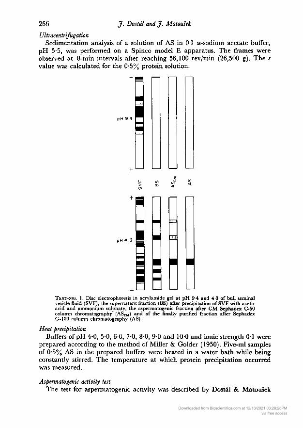
UltracentrifugationSedimentation analysis of a solution of AS in 0-1 M-sodium acetate buffer,
pH 5-5, was performed on a Spinco model E apparatus. The frames wereobserved at 8-min intervals after reaching 56,100 rev/min (26,500 g). The s
value was calculated for the 0-5% protein solution.
pH 9 4
>
pH 4-3
Text-fig. 1. Disc electrophoresis in acrylamide gel at pH 9-4 and 4-3 of bull seminalvesicle fluid (SVF), the supernatant fraction (BS) after precipitation of SVF with aceticacid and ammonium sulphate, the aspermatogenic fraction after CM Sephadex C-50column chromatography (ASCM) and of the finally purified fraction after SephadexG-100 column chromatography (AS).
Heat precipitationBuffers of pH 40, 50, 6-0, 70, 8-0, 9-0 and 100 and ionic strength 0-1 were
prepared according to the method of Miller & Golder (1950). Five-ml samplesof 0-5% AS in the prepared buffers were heated in a water bath while beingconstantly stirred. The temperature at which protein precipitation occurredwas measured.
Aspermatogenic activity testThe test for aspermatogenic activity was described by Dostál & Matousek
Downloaded from Bioscientifica.com at 12/13/2021 03:28:28PMvia free access

Isolation of bull aspermatogenic substance 267
(1972). This involved the injection of 0-02 ml of the dialysed and freeze-driedmaterial (10 mg/ml 0-9% NaCl) into the left testes of four males. On the 10thday after the injection, the mice were killed and both their testes were weighed.The right testes were used as controls. The degree of testicular damage was
determined histologically (Matousek, 1969).
RESULTSThe active fraction of purified bull seminal vesicle fluid was shown to be free ofcontaminating protein on disc electrophoresis (Text-fig. 1), starch-gel electro¬phoresis (Text-fig. 2) and immunoelectrophoresis. The isolation procedure and
I'
' ' ,1
illinium
Origin
.'I'1'
.
SVF BS AS S cm
Text-fig. 2. Starch-gel electrophoresis of bull seminal vesicle fluid and its fractions. Forabbreviations, see the legend to Text-fig. 1.
Chromatographie homogeneity of the AS peak on Sephadex G-100 are illus¬trated in Text-fig. 3.
The molecular weight of AS was estimated by gel filtration as approximately22,000 for the monomer and 44,000 for the dimer (Text-fig. 4).
Lysine (di-DNP-lysine) was determined as the N-terminal amino acid ofAS by means of thin-layer chromatography.
After splitting of extracted DNP-amino acid, three spots only were detectedon paper chromatography. The spots were designated lysine, di-DNP-lysine andDNFB.
The results of AS amino acid analysis are given in Table 1. No tryptophane
Downloaded from Bioscientifica.com at 12/13/2021 03:28:28PMvia free access

J. Dostal and J. Matousek
Bull seminal vesicle fluid
+ 25 vols of 2% acetic acid
Centrifugation 7000y, 45min
I
Supernatant Precipitate
+ Ammonium sulphate to 3m
Stirring 60 min
Centrifugation 7000p,l5min
Supernatant Precipitate
Dialysis
Freeze drying
?CM Sephodex C-50
0 05m-
sodium phosphate pH 8 0NaCI gradient
Fraction no.
Sephadex G-1000 lM-trisHCI pH 75
Elution volume
Text-fic. 3. The isolation procedure for aspermatogenic substance (AS) from bull seminalvesicle fluid.
Downloaded from Bioscientifica.com at 12/13/2021 03:28:28PMvia free access

Isolation of bull aspermatogenic substance 269residue was found by the DAB method and absorption analysis of AS solutionin 0-1 N-NaOH. The half-cystine content was calculated from the value forcysteic acid in AS-O samples and agreed well with the value for half-cystinein native AS samples. The contents of basic, dicarboxylic and sulphur-containingamino acids were about 18-1%, 19-1% and 11-0% of the total residues, respec-
7 8 9mol wtx 10"
7 8 9
Text-fig. 4. Molecular weight determination of bull seminal vesicle fluid aspermatogenicsubstance (interrupted lines) on a 1 -5 X 35-cm column of Sephadex G-75 equilibrated bybuffers of pH 3-5 (a), pH 5-5 (b), pH 7-6 (c), pH 91 (d) and pH 10-5 (e) ; and on a
3 X 50-cm column of Sephadex G-100 in 8 M-urea buffer ofpH 5-5 (f). For the compositionof the buffers, see text. The reference proteins used were ribonuclease (Ri), chymotryp-sinogen (Ch) and ovalbumin (Ov).
Downloaded from Bioscientifica.com at 12/13/2021 03:28:28PMvia free access

270 J. Dostal and J. Matousek
tively. The content of aromatic amino acids (tyrosine and phenylalanine) was
about 5%. Samples of AS did not contain any thiol groups as determined by theDTNB method.
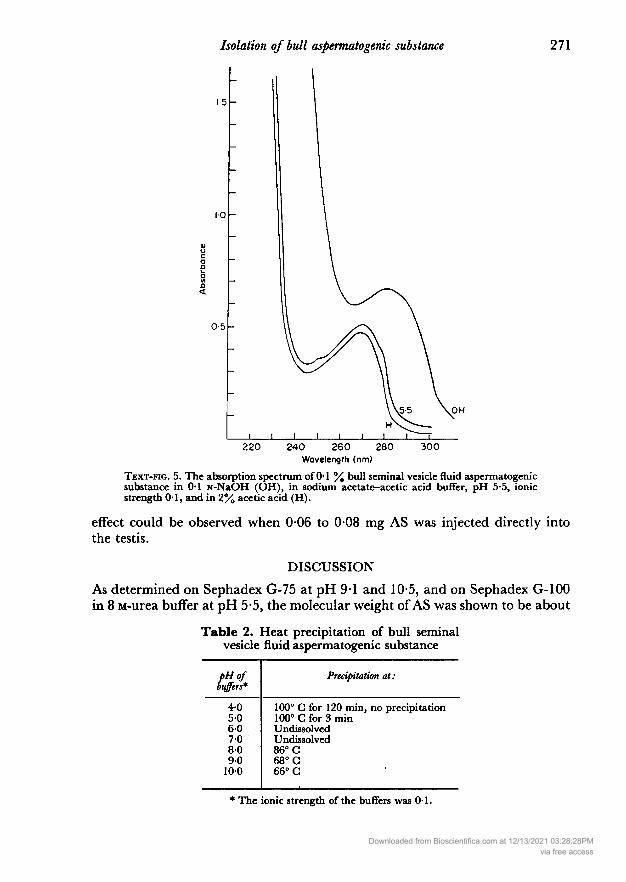
Absorption spectrum analysis of a 0-1% solution of AS (calculated on dryweight) in sodium acetate-acetic acid buffer, pH 5-5 and ionic strength 0-1,gave an absorption maximum at 278 to 280 nm and an absorption minimumat 254 to 256 nm. The absorbance of this solution measured at 280 nm was
0-510. The shift of the absorption maximum and minimum of the 0-1% solutionof AS in 01 N-NaOH and the small difference in the absorption curve of the0-1% solution of AS in 2% acetic acid (Text-fig. 5) confirmed the estimatedcontent of tyrosine residues.
Table 1. Amino acid composition of bull seminalvesicle fluid aspermatogenic substance
Amino acid
LysineHistidineArginineAspartic acidThreonineSerineGlutamic acidProlineGlycineAlanineHalf-cystine*ValineMethionineIsoleucineLeucineTyrosinePhenylalanineTryptophanefTotal residues
nmol
86-618021-762-744-977-467-528-840-450-155-757-022014-714-917-318-80
Mean no.
of residuescalculated on
22,000 mol. wt
25-045-206-27
181212-9722-3719-518-32
11-6814-48161016-476-364-254-315005-430
Nearestinteger
2556
18132220
812141616644550
199
* Determined as cysteic acid and as half-cystine.t Determined by colorimetrie method.
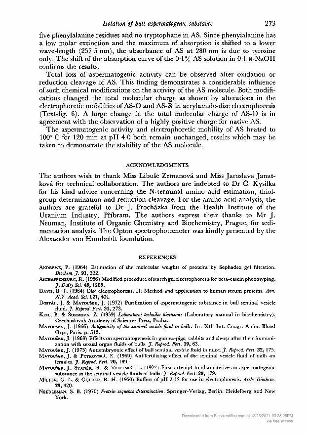
Heat precipitation of AS was found to be pH dependent. The results of thisexperiment are shown in Table 2. In samples of AS heated to 100° C for 120min at pH 40, the aspermatogenic activity remained unchanged. No asperma¬togenic activity was observed after oxidation and reduction cleavage. Electro¬phoresis of AS-O and AS-R samples (Text-fig. 6) demonstrated a change oftotal molecular charge. The ultracentrifugation patterns of the 0-5% ASsolution in the pH 5-5 buffer showing a homogeneous peak (s = 2-7), and theelectrophoresis of AS-O and AS-R confirmed the purity of the isolated AS.
When purified AS was prepared, the concentration of AS/ml 0-9% NaCl was
gradually lowered from 10 mg to 1 mg in order to find the minimal amount ofAS which would cause a 50% decrease in the weight of mouse testes and arrest
spermatogenesis within 10 days of the injection. It was found that such an
Downloaded from Bioscientifica.com at 12/13/2021 03:28:28PMvia free access
Isolation of bull aspermatogenic substance 271
OH
220 240 260 280Wavelength (nm)
300
Text-fig. 5. The absorption spectrum of 0-1 % bull seminal vesicle fluid aspermatogenicsubstance in 0-1 N-NaOH (OH), in sodium acetate-acetic acid buffer, pH 5-5, ionicstrength 0-1, and in 2% acetic acid (H).
effect could be observed when 006 to 0-08 mg AS was injected directly intothe testis.
DISCUSSIONAs determined on Sephadex G-75 at pH 9-1 and 10-5, and on Sephadex G-100in 8 M-urea buffer at pH 5-5, the molecular weight of AS was shown to be about
Table 2. Heat precipitation of bull seminalvesicle fluid aspermatogenic substance
pHofbuffers*
4-05-06-07-08-090
100
Precipitation at:
100° C for 120 min, no precipitation100° C for 3 minUndissolvedUndissolved86° C68° C66° C
* The ionic strength of the buffers was 0-1.
Downloaded from Bioscientifica.com at 12/13/2021 03:28:28PMvia free access

272 J. Dostal and J. Matousek
22,000. When buffers of pH 3-5 and 5-5 without urea were used, the elutionpatterns of AS corresponded to a molecular weight of 44,000. It seems that themonomer AS (22,000) associates to the dimer (44,000) under certain conditions.
To support this result, the performic acid-oxidized sample (AS-O) was usedfor the study of its elution behaviour. The elution patterns of AS-O corres¬
ponded to a molecular weight of 27,000. While it is clear that the molecularweight of the AS monomer is about 22,000, the dimer molecule is held together
pH 9-4
< <
pH 4-3
Text-fig. 6. Disc electrophoresis in polyacrylamide gel at pH 9-4 and 4-3 of bull seminalvesicle fluid aspermatogenic substance (AS), performic acid-oxidized aspermatogenicsubstance (AS-O), and reduced aspermatogenic substance (AS-R).
by relatively weak forces which can easily be dissociated by a change of pH.At pH 7-6, incomplete dissociation probably occurs and the AS behaves as a
protein with a molecular weight of 29,000.The absence of thiol groups in AS, the finding that lysine is the one N-
terminal amino acid and the small difference between the molecular weightsof AS and AS-O indicates the presence of intrasubunit disulphides only. Onepolypeptide chain in this case is equal to one AS subunit and to AS monomer.
The relatively low absorbance value of 0-510 for a 01% AS solution at pH 5-5estimated at 280 nm seems to agree with the presence of five tyrosine residues,
Downloaded from Bioscientifica.com at 12/13/2021 03:28:28PMvia free access
Isolation of bull aspermatogenic substance 273five phenylalanine residues and no tryptophane in AS. Since phenylalanine hasa low molar extinction and the maximum of absorption is shifted to a lowerwave-length (257-5 nm), the absorbance of AS at 280 nm is due to tyrosineonly. The shift of the absorption curve of the 0-1% AS solution in 0-1 N-NaOHconfirms the results.
Total loss of aspermatogenic activity can be observed after oxidation orreduction cleavage of AS. This finding demonstrates a considerable influenceof such chemical modifications on the activity of the AS molecule. Both modifi¬cations changed the total molecular charge as shown by alterations in theelectrophoretic mobilities of AS-O and AS-R in acrylamide-disc electrophoresis(Text-fig. 6). A large change in the total molecular charge of AS-O is inagreement with the observation of a highly positive charge for native AS.
The aspermatogenic activity and electrophoretic mobility of AS heated to100° C for 120 min at pH 4-0 both remain unchanged, results which may betaken to demonstrate the stability of the AS molecule.
ACKNOWLEDGMENTS
The authors wish to thank Miss Libuse Zemanová and Miss Jaroslava Janat-ková for technical collaboration. The authors are indebted to Dr C. Kysilkafor his kind advice concerning the N-terminal amino acid estimation, thiol-group determination and reduction cleavage. For the amino acid analysis, theauthors are grateful to Dr J. Procházka from the Health Institute of theUranium Industry, Pfibram. The authors express their thanks to Mr J.Neuman, Institute of Organic Chemistry and Biochemistry, Prague, for sedi¬mentation analysis. The Opton spectrophotometer was kindly presented by theAlexander von Humboldt foundation.
REFERENCES
Andrews, P. (1964) Estimation of the molecular weights of proteins by Sephadex gel filtration.Biochem. J. 91, 222.
Aschaffenburg, R. (1966) Modified procedure ofstarch gel electrophoresis for beta-casein phenotyping.J. Dairy Sci. 49, 1285.
Davis, B. T. (1964) Disc electrophoresis. II. Method and application to human serum proteins. Ann.NT. Acad. Sci. 121, 404.
Dostál, J. & MatouSek, J. (1972) Purification of aspermatogenic substance in bull seminal vesiclefluid. J. Reprod. Fert. 31, 273.
Keil, . & Sormová, . (1959) Laboratorni technika biochemie (Laboratory manual in biochemistry),Czechoslovak Academy of Sciences Press, Praha.
Matouüek, J. (1966) Antigenicity of the seminal vesicle fluid in bulls. In: Xth Int. Congr. Anim. BloodGrps, Paris, p. 513.
MatouSek, J. (1969) Effects on spermatogenesis in guinea-pigs, rabbits and sheep after their immuni¬zation with sexual organ fluids of bulls. J. Reprod. Fert. 19, 63.
Matousek, J. (1973) Antiembryonic effect of bull seminal vesicle fluid in mice. J. Reprod. Fert. 32, 175.Matousek, J. & Petrovská, E. (1969) Antifertilizing effect of the seminal vesicle fluid of bulls on
females. J. Reprod. Fert. 20, 189.MatouSek, J., Stanék, R. & Veselskv, L. (1972) First attempt to characterize an aspermatogenic
substance in the seminal vesicle fluids of bulls. J1. Reprod. Fert. 29, 179.Miller, G. L. & Golder, R. H. (1950) Buffers of pH 2-12 for use in electrophoresis. Archs Biochem.
29, 420.Needleman, S. B. (1970) Protein sequence determination. Springer-Verlag, Berlin, Heidelberg and New
York.
Downloaded from Bioscientifica.com at 12/13/2021 03:28:28PMvia free access

274 J. Dostal and J. MatousekOrnstein, L. (1964) Disc electrophoresis. I. Background and theory. Ann. N.T. Acad. Sci. 121, 366.Sanger, F. (1952) The arrangement of amino acids in proteins. In: Advances in Protein Chemistry, Vol. 7,
p. 2. Eds. M. L. Anson, K. Bailey and J. T. Edsall. Academic Press, New York.Scheidegger, J. (1955) Une micro-méthode de l'immunoélectrophorèse. Int. Archs Allergy appi. Immun.
7, 103.
Downloaded from Bioscientifica.com at 12/13/2021 03:28:28PMvia free access